Embrione e tubo cardiaco primitivo al 22-23 giorno ...
51
*
Transcript of Embrione e tubo cardiaco primitivo al 22-23 giorno ...

*

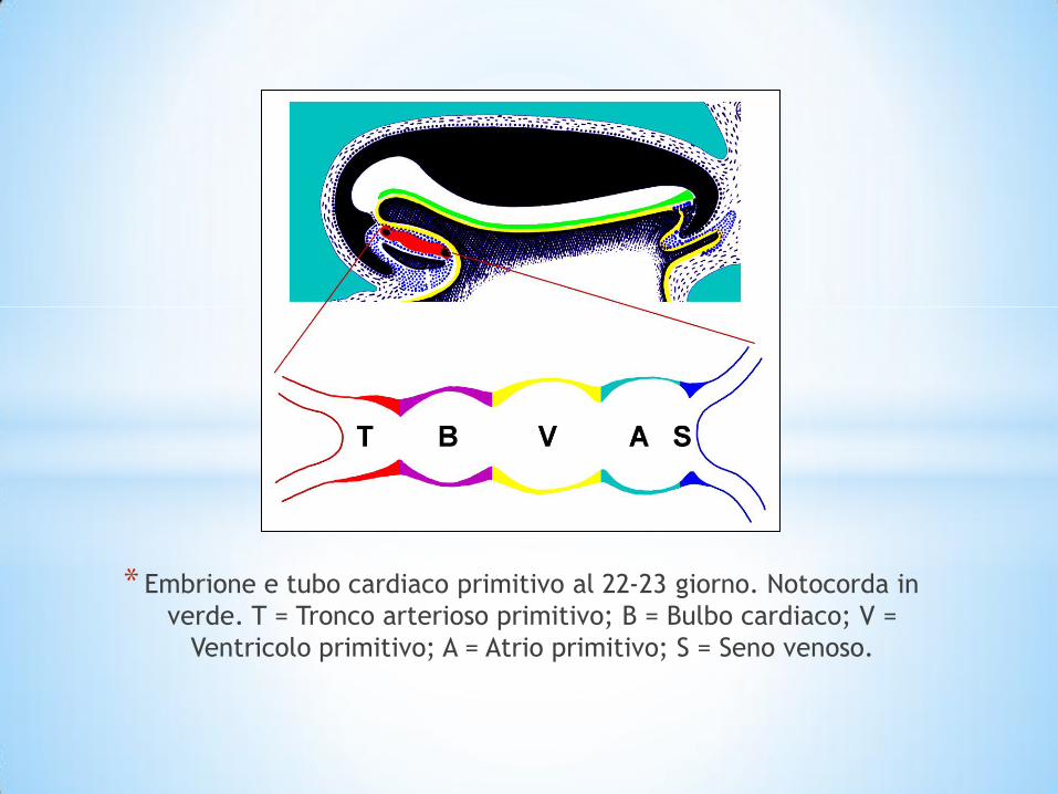
* Embrione e tubo cardiaco primitivo al 22-23 giorno. Notocorda in
verde. T = Tronco arterioso primitivo; B = Bulbo cardiaco; V =
Ventricolo primitivo; A = Atrio primitivo; S = Seno venoso.


* Primi stadi di sviluppo del cuore.
a) Tubo cardiaco;
b) Accrescimento antero-infero-laterale del Bulbo cardiaco, seguito dal Ventricolo
primitivo;
c) Spostamento antero-inferiore per accrescimento del Bulbo cardiaco-Ventricolo
primitivo e formazione dell’ansa a U, con conseguente posteriorizzazione
dell’Atrio primitivo-Seno venoso;
d) Situazione al 28-29° giorno con il Bulbo cardiaco, a destra, e il Ventricolo
primitivo, a sinistra, antero-inferiormente alll’Atrio primitivo. T = Tronco arterioso
primitivo; B = Bulbo cardiaco; V = Ventricolo primitivo; A = Atrio primitivo; S =
Seno venoso.

* Il tessuto miocardico

*Le fibre cardiache

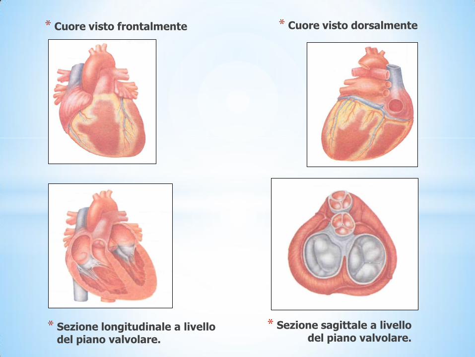
* Cuore visto frontalmente * Cuore visto dorsalmente
* Sezione longitudinale a livello del piano valvolare.
* Sezione sagittale a livello del piano valvolare.

*Struttura interna ed esterna del cuore
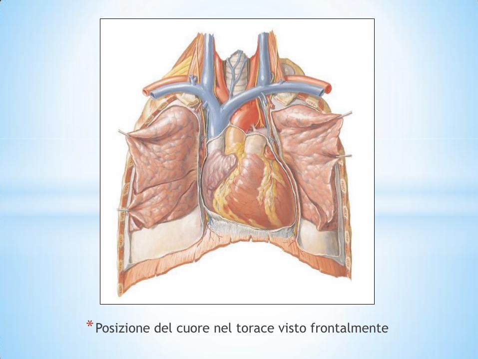
*Posizione del cuore nel torace visto frontalmente
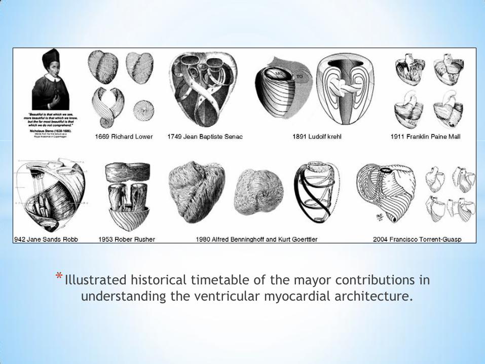
* Illustrated historical timetable of the mayor contributions in
understanding the ventricular myocardial architecture.

* Francisco (Paco) Torrent-Guasp.

*‘Unfolding’ of the myocardial band according to Torrent-
Guasp (A—D); (E) the sequential segments of the basal and
apical loops of the fully extended band.

*Unfolded segments of the helical heart showing the two
segments of the basal and apical loops.

* In the cartoon, the green line represents the fictional cleavage plane, never demonstrated histologically, which was supposed to serve as a hypothetical gliding plane to permit the necessary freedomof motion of a rope in a pulley. Torrent-Guasp had assumed that, by resecting one segment (yellow) from the right ventricular wall, the resulting increment in tension would be transmitted all along the band. Based upon this misassumption, he presumed that a dilated left ventricle would start to shrink. As demonstrated in the lower cross-section through the walls of both ventricles, there is no evidence supporting the existence of such a ‘cleavage plane’. The assumed freedom of motion of the ‘rope in the pulley’ is nothing but fiction because all segments of the alleged rope, in reality, are unified within the overall spatially netted ventricular mesh. RVC: right ventricular cavity (blue); LVC: left ventricular cavity (red); alleged cleavage plane (green); resected segment from the right ventricular free wall (yellow).

*Right ventricular dissection.
Dissection following the
ventricular muscular band
to separate the right and
left ventricular outflow
tract (A) with magnification
to better show the details
(B). Ao, aorta; LMCA, left
main coronary artery; PA,
pulmonary artery; RCA,
right coronary artery; S1,
first septal coronary artery
branch

*Fiber orientation relationship of the septum, composed of oblique
fibers that arise from the descending and ascending segments of the
apical loop, surrounded by the transverse muscle orientation of the
basal loop that composes the free right ventricular wall. Note the
conical arrangement of the septum muscle and the basal loop wrap,
forming the RV cavity.

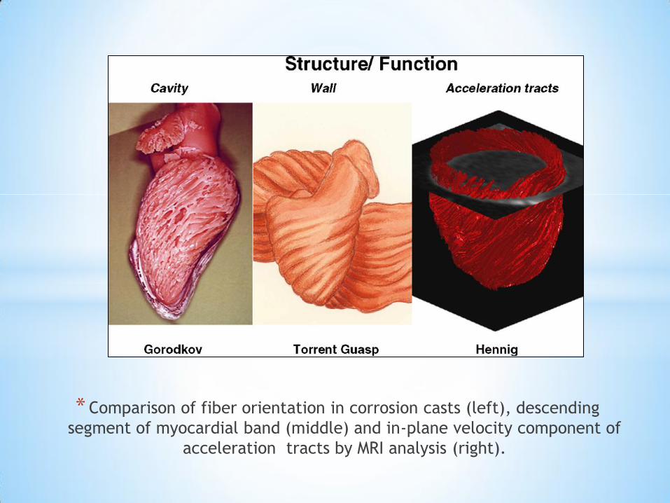
* Comparison of fiber orientation in corrosion casts (left), descending
segment of myocardial band (middle) and in-plane velocity component of
acceleration tracts by MRI analysis (right).

*Rotation of cardiac base and apex during sequential
contraction of myocardial band described by Torrent-Guasp .

Il modello di circolazione del sangue secondo Galeno

Leonardo da Vinci. Cuore. Matita e inchiostro su carta (1500 dc). The Royal
Collection, Windsor (U.K)
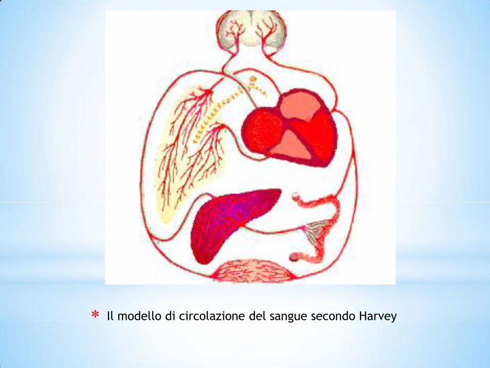
Il modello di circolazione del sangue secondo Harvey


*TRAPIANTO DI CUORE Fase Clinica
Ciclosporina “A”
Viventi
Barnard
Hardy 1964
3-12-1967
1969
1978
Xenotrapianto ortotopico
Omo-trapianto ortotopico
Più di 100 trapianti in 17 centri
20 %
3000 trapianti
1968
1986
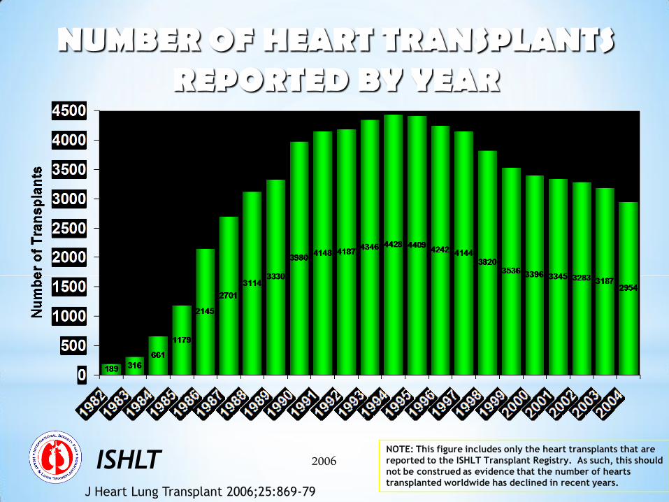
NUMBER OF HEART TRANSPLANTS REPORTED BY YEAR
ISHLT 2006 NOTE: This figure includes only the heart transplants that are
reported to the ISHLT Transplant Registry. As such, this should
not be construed as evidence that the number of hearts
transplanted worldwide has declined in recent years. J Heart Lung Transplant 2006;25:869-79
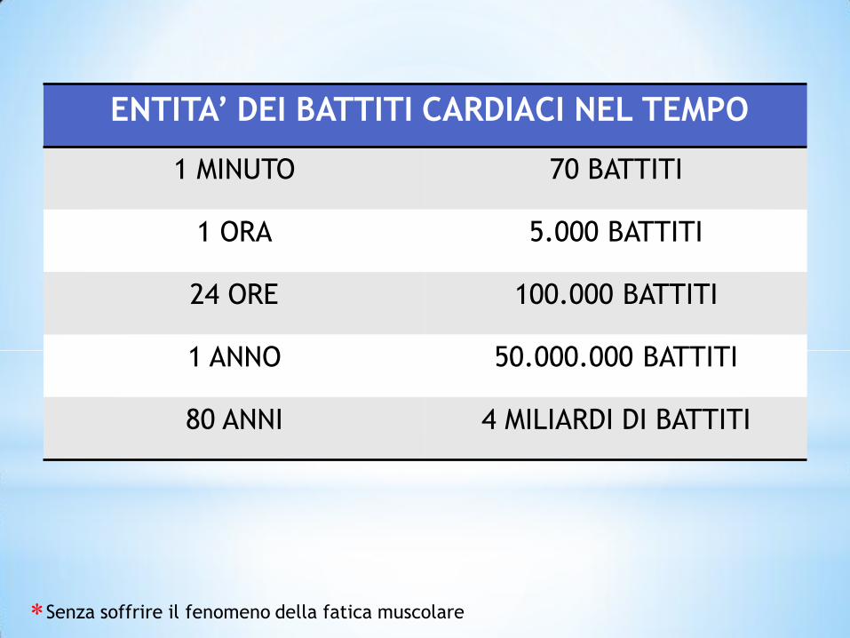
ENTITA’ DEI BATTITI CARDIACI NEL TEMPO
1 MINUTO 70 BATTITI
1 ORA 5.000 BATTITI
24 ORE 100.000 BATTITI
1 ANNO 50.000.000 BATTITI
80 ANNI 4 MILIARDI DI BATTITI
Senza soffrire il fenomeno della fatica muscolare
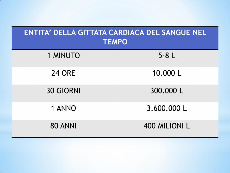
ENTITA’ DELLA GITTATA CARDIACA DEL SANGUE NEL
TEMPO
1 MINUTO 5-8 L
24 ORE 10.000 L
30 GIORNI 300.000 L
1 ANNO 3.600.000 L
80 ANNI 400 MILIONI L

Grotta di El Pindal. Graffito del proboscidato (a). Rielaborazione
grafica (b)

Psicostasia. Dal Libro dei Morti di Ani (1275 a.C. ca.) British Museum, Londra
(particolare). Si notino il cuore nel piatto destro della bilancia e la piuma nel sinistro
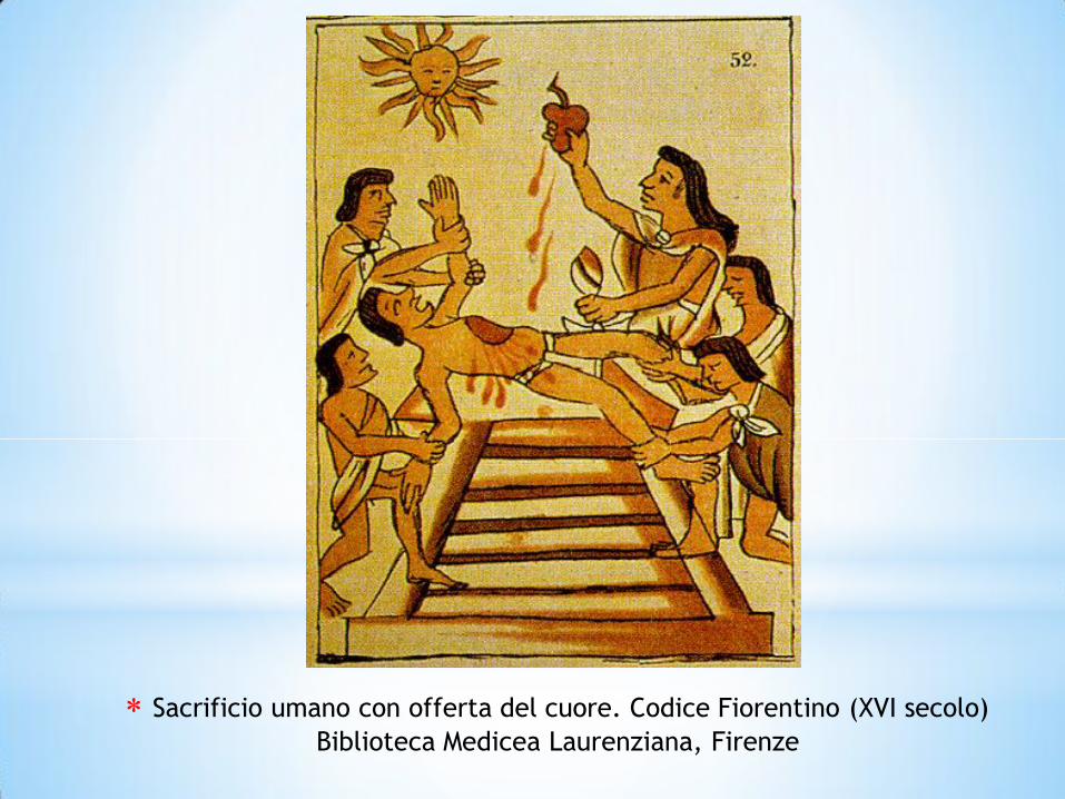
Sacrificio umano con offerta del cuore. Codice Fiorentino (XVI secolo)
Biblioteca Medicea Laurenziana, Firenze

L’icona classica del cuore

Curve matematiche “a cuore”. Da [36].


Edvard Munch. La ragazza col
cuore. Xilografia (1899)
Henry Matisse. Icaro.
Gouache Découpé (1946) Centre
Pompidou, Parigi

Barbara Krafft. Wolfgang Amadeus Mozart. Olio su tela (1819).
Sammlung Alter Musikinstrumente, Vienna


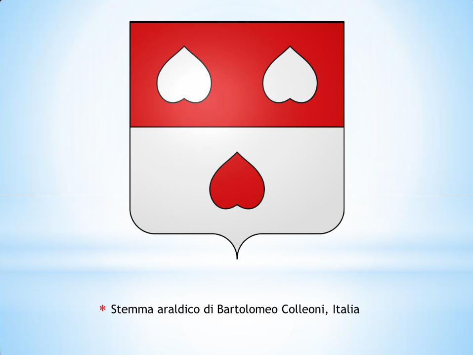
Stemma araldico di Bartolomeo Colleoni, Italia
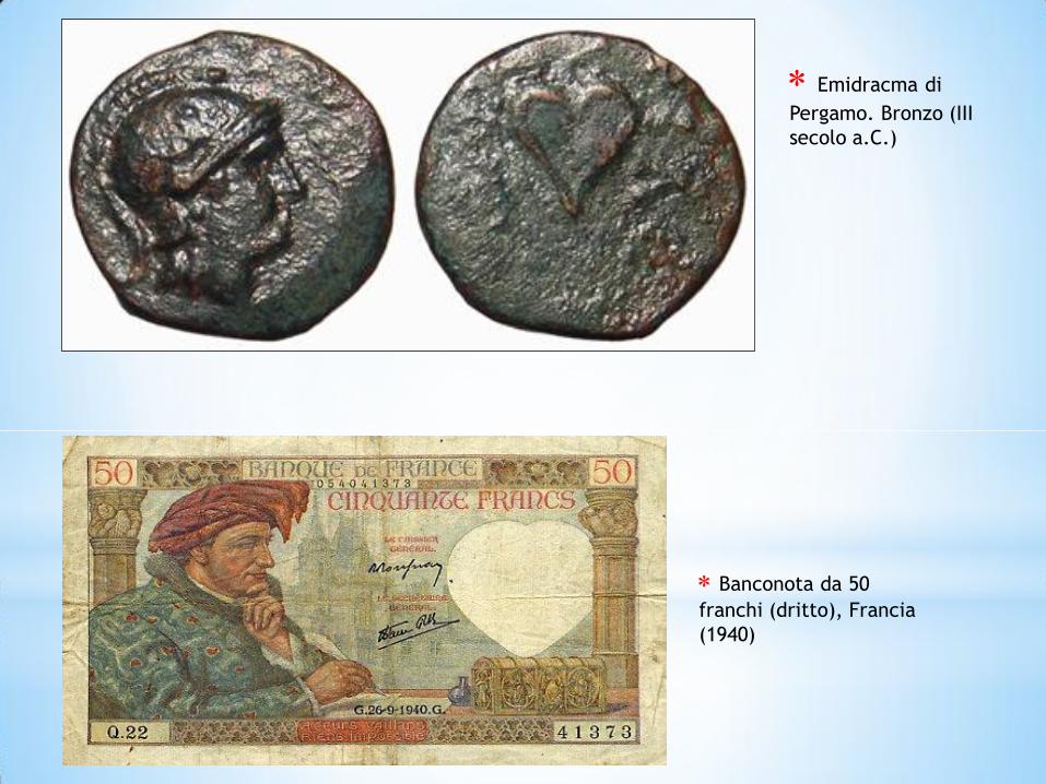
Emidracma di
Pergamo. Bronzo (III
secolo a.C.)
Banconota da 50
franchi (dritto), Francia
(1940)
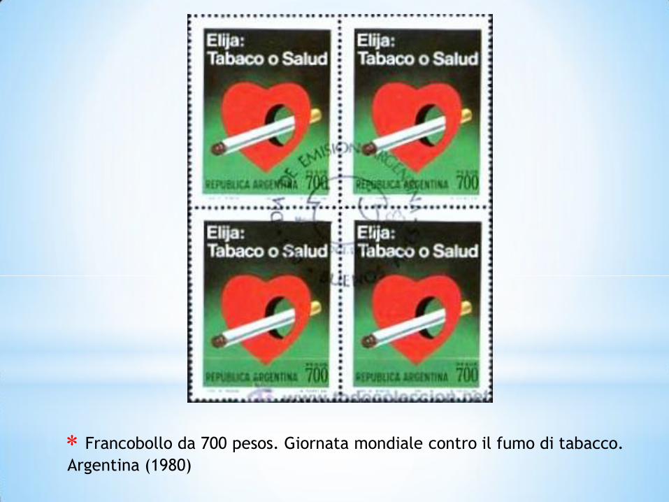
Francobollo da 700 pesos. Giornata mondiale contro il fumo di tabacco.
Argentina (1980)


Logo di “Cuore granata”


Raffaello Sanzio. La Scuola di Atene. Affresco
(1509-11) (particolare). Musei Vaticani, Roma
Marie France Louvel e Catherine
Atans. The Heart Brain (2011)

*


![John Zerzan - Futuro Primitivo (Edizioni Nautilus) [ITA]](https://static.fdocumenti.com/doc/165x107/5571f99049795991698fe0ea/john-zerzan-futuro-primitivo-edizioni-nautilus-ita.jpg)