Genetica 7 - Profs Area Scienze ed Ingegneriaprofs.scienze.univr.it/delledonne/Insegnamenti/Genetica...
59
Genetica 7
Transcript of Genetica 7 - Profs Area Scienze ed Ingegneriaprofs.scienze.univr.it/delledonne/Insegnamenti/Genetica...
Genetica 7
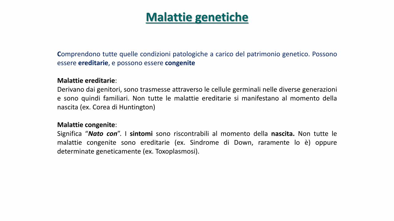
Comprendono tutte quelle condizioni patologiche a carico del patrimonio genetico. Possonoessere ereditarie, e possono essere congenite
Malattie ereditarie:Derivano dai genitori, sono trasmesse attraverso le cellule germinali nelle diverse generazionie sono quindi familiari. Non tutte le malattie ereditarie si manifestano al momento dellanascita (ex. Corea di Huntington)
Malattie congenite:Significa “Nato con”. I sintomi sono riscontrabili al momento della nascita. Non tutte lemalattie congenite sono ereditarie (ex. Sindrome di Down, raramente lo è) oppuredeterminate geneticamente (ex. Toxoplasmosi).
Malattie genetiche
Le malattie genetiche sono frequenti
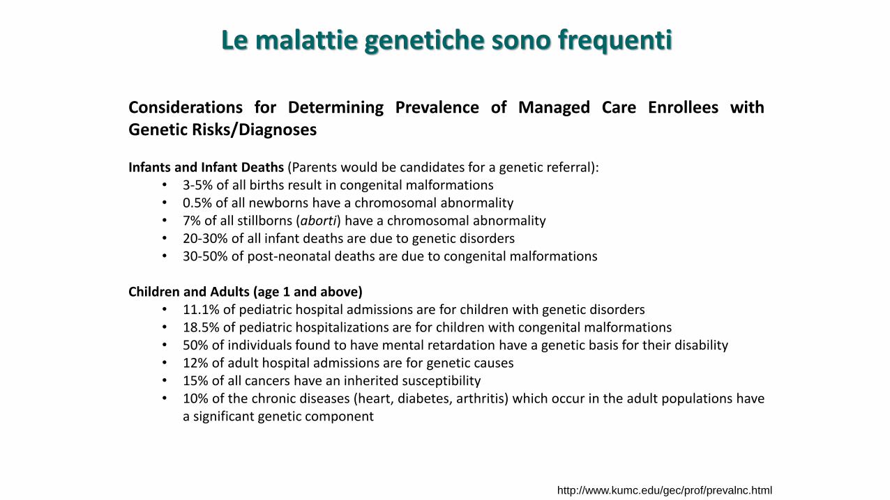
Considerations for Determining Prevalence of Managed Care Enrollees withGenetic Risks/Diagnoses
Infants and Infant Deaths (Parents would be candidates for a genetic referral):• 3-5% of all births result in congenital malformations• 0.5% of all newborns have a chromosomal abnormality• 7% of all stillborns (aborti) have a chromosomal abnormality• 20-30% of all infant deaths are due to genetic disorders• 30-50% of post-neonatal deaths are due to congenital malformations
Children and Adults (age 1 and above)• 11.1% of pediatric hospital admissions are for children with genetic disorders• 18.5% of pediatric hospitalizations are for children with congenital malformations• 50% of individuals found to have mental retardation have a genetic basis for their disability• 12% of adult hospital admissions are for genetic causes• 15% of all cancers have an inherited susceptibility• 10% of the chronic diseases (heart, diabetes, arthritis) which occur in the adult populations have
a significant genetic component
http://www.kumc.edu/gec/prof/prevalnc.html

Another possibility for estimating members who may benefit from genetic services would be toconsider the more common diagnoses, or reasons for referral, and estimate the prevalence ofenrollees with, or at risk of, these conditions based on known incidence figures. For example:
• Down syndrome (1/600 live births and increases with advanced maternal age)• Cystic Fibrosis (1/2500 Caucasian Americans)• Fragile X syndrome (1/1,000 males and 1/800 female carriers of which 30% will be mentally
retarded)• Sickle cell disease (1/500 of African American births)• Hemophilia - Factor VIII Deficiency (48/100,000 male births)• Duchenne muscular dystrophy (1/5,000 male births)• Hemochromatosis (1/450 individuals)• Breast cancer (1/8 women of which 5-10% of will have a genetic predisposition)
One could apply these figures to the enrolled population to generate prevalence estimates for clientsenrolled in a managed care plan. For example, assuming an enrolled population of 55,000, and thatone half are female members, and recognizing the incidence of breast cancer of 1/8, an estimated3400 members will develop breast cancer and 170-340 of these individuals will have a genetic basisfor their disease. Their predisposition could be identified through a detailed family history obtainedthrough a genetic evaluation and those high risk families may benefit from genetic testing for theknown breast cancer genes
http://www.kumc.edu/gec/prof/prevalnc.html
Le malattie genetiche sono frequenti
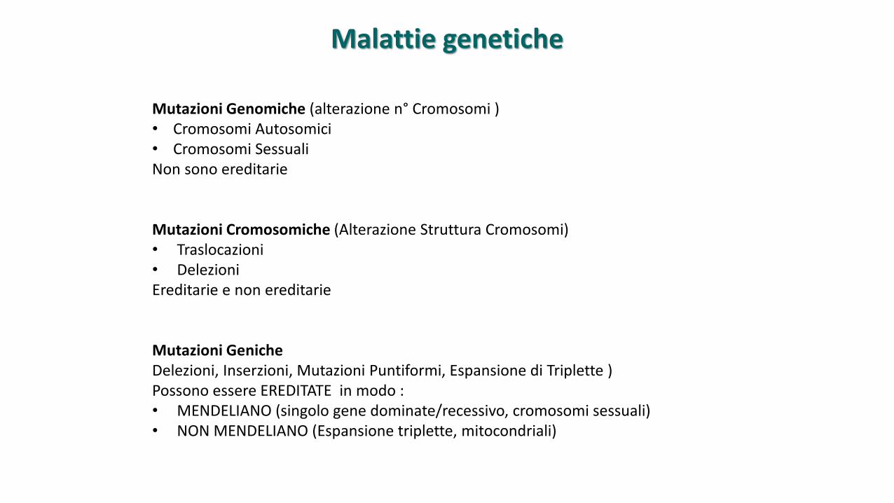
Mutazioni Genomiche (alterazione n° Cromosomi )• Cromosomi Autosomici• Cromosomi SessualiNon sono ereditarie
Mutazioni Cromosomiche (Alterazione Struttura Cromosomi) • Traslocazioni• DelezioniEreditarie e non ereditarie
Mutazioni GenicheDelezioni, Inserzioni, Mutazioni Puntiformi, Espansione di Triplette ) Possono essere EREDITATE in modo :• MENDELIANO (singolo gene dominate/recessivo, cromosomi sessuali)• NON MENDELIANO (Espansione triplette, mitocondriali)
Malattie genetiche
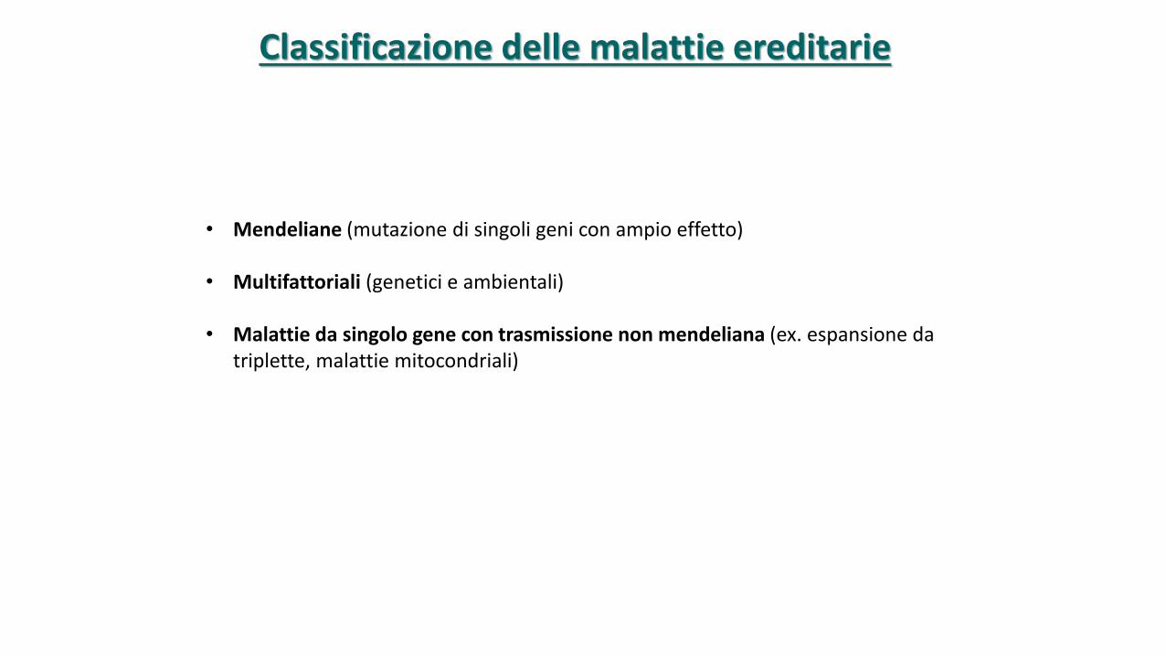
• Mendeliane (mutazione di singoli geni con ampio effetto)
• Multifattoriali (genetici e ambientali)
• Malattie da singolo gene con trasmissione non mendeliana (ex. espansione da triplette, malattie mitocondriali)
Classificazione delle malattie ereditarie
• Risultanti dall’interazione fra le condizioni ambientali e un numero di genipartecipanti alla determinazione del fenotipo >1
• Il rischio di presentare un fenotipo clinico é proporzionale al numero di genimutati ereditato
• Molte delle piú comuni malattie (diabete, ipertensione, celiachia) hannoorigine multifattoriale
Malattie genetiche multifattoriali
100%
50%
0%
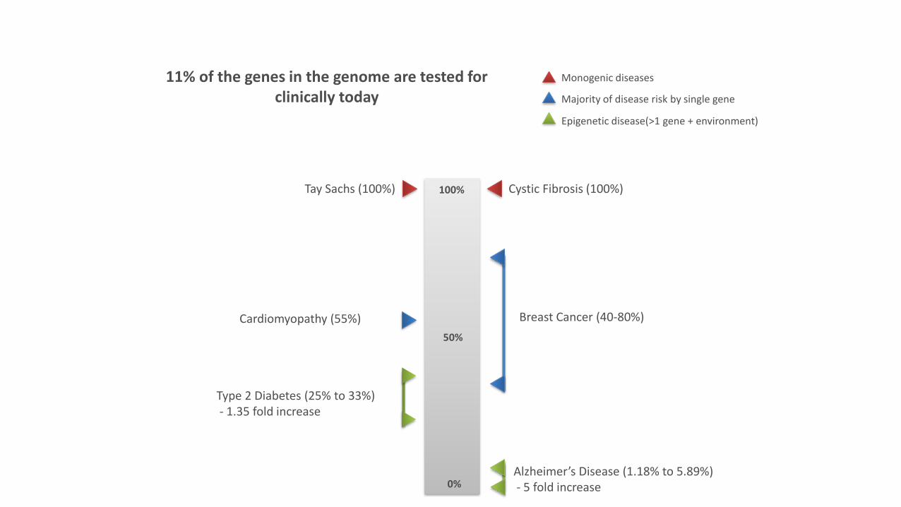
Cystic Fibrosis (100%)Tay Sachs (100%)
Breast Cancer (40-80%)
11% of the genes in the genome are tested for clinically today
Cardiomyopathy (55%)
Type 2 Diabetes (25% to 33%)- 1.35 fold increase
Alzheimer’s Disease (1.18% to 5.89%)- 5 fold increase
Monogenic diseases
Majority of disease risk by single gene
Epigenetic disease(>1 gene + environment)
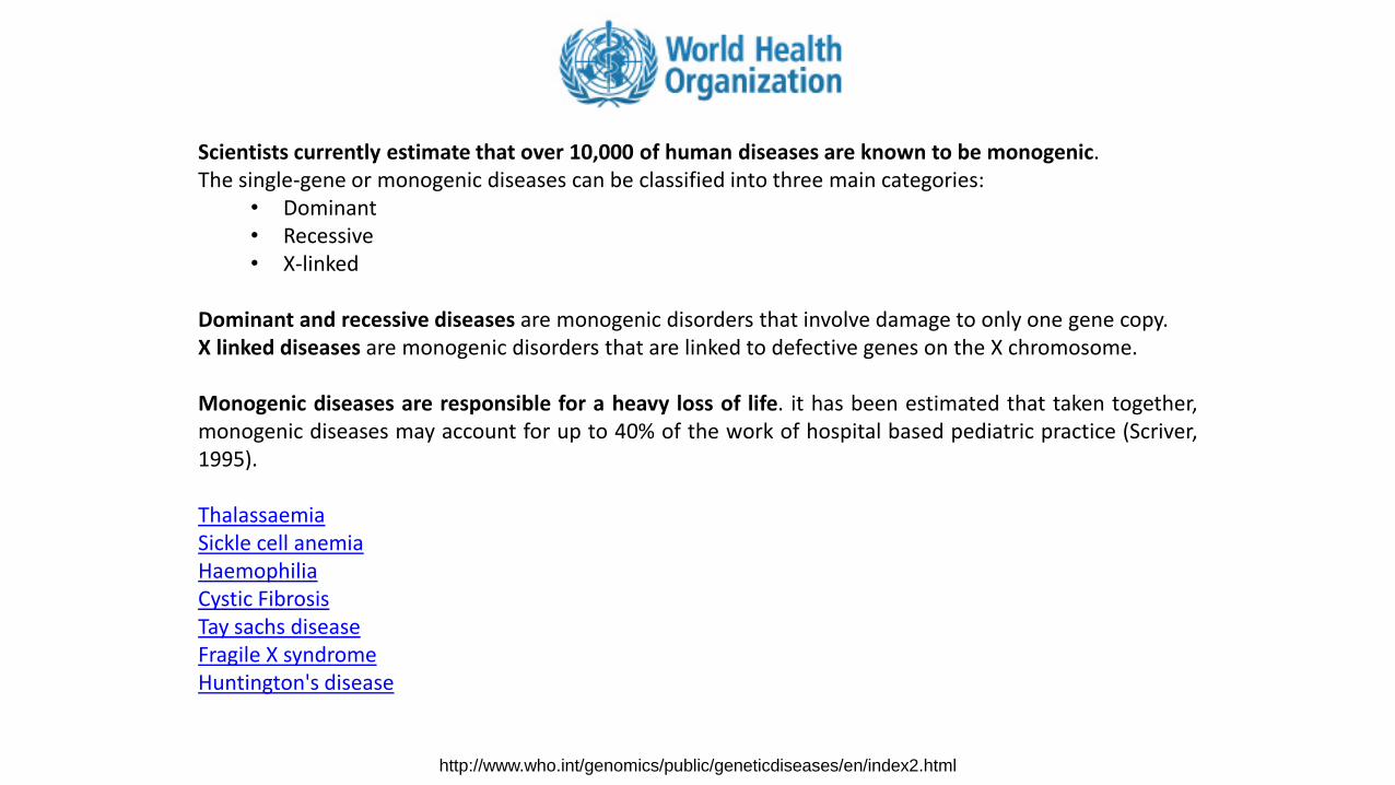
Scientists currently estimate that over 10,000 of human diseases are known to be monogenic.The single-gene or monogenic diseases can be classified into three main categories:
• Dominant• Recessive• X-linked
Dominant and recessive diseases are monogenic disorders that involve damage to only one gene copy.X linked diseases are monogenic disorders that are linked to defective genes on the X chromosome.
Monogenic diseases are responsible for a heavy loss of life. it has been estimated that taken together,monogenic diseases may account for up to 40% of the work of hospital based pediatric practice (Scriver,1995).
ThalassaemiaSickle cell anemiaHaemophiliaCystic Fibrosis Tay sachs diseaseFragile X syndromeHuntington's disease
http://www.who.int/genomics/public/geneticdiseases/en/index2.html
http://www.ncbi.nlm.nih.gov/books/NBK22183/
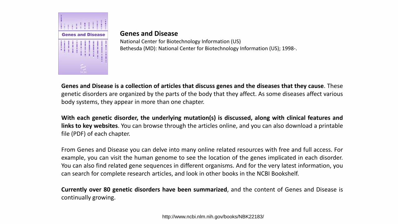
Genes and DiseaseNational Center for Biotechnology Information (US)Bethesda (MD): National Center for Biotechnology Information (US); 1998-.
Genes and Disease is a collection of articles that discuss genes and the diseases that they cause. Thesegenetic disorders are organized by the parts of the body that they affect. As some diseases affect variousbody systems, they appear in more than one chapter.
With each genetic disorder, the underlying mutation(s) is discussed, along with clinical features andlinks to key websites. You can browse through the articles online, and you can also download a printablefile (PDF) of each chapter.
From Genes and Disease you can delve into many online related resources with free and full access. Forexample, you can visit the human genome to see the location of the genes implicated in each disorder.You can also find related gene sequences in different organisms. And for the very latest information, youcan search for complete research articles, and look in other books in the NCBI Bookshelf.
Currently over 80 genetic disorders have been summarized, and the content of Genes and Disease iscontinually growing.
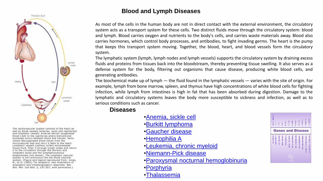
Blood and Lymph Diseases
As most of the cells in the human body are not in direct contact with the external environment, the circulatorysystem acts as a transport system for these cells. Two distinct fluids move through the circulatory system: bloodand lymph. Blood carries oxygen and nutrients to the body's cells, and carries waste materials away. Blood alsocarries hormones, which control body processes, and antibodies, to fight invading germs. The heart is the pumpthat keeps this transport system moving. Together, the blood, heart, and blood vessels form the circulatorysystem.The lymphatic system (lymph, lymph nodes and lymph vessels) supports the circulatory system by draining excessfluids and proteins from tissues back into the bloodstream, thereby preventing tissue swelling. It also serves as adefense system for the body, filtering out organisms that cause disease, producing white blood cells, andgenerating antibodies.The biochemical make up of lymph — the fluid found in the lymphatic vessels — varies with the site of origin. Forexample, lymph from bone marrow, spleen, and thymus have high concentrations of white blood cells for fightinginfection, while lymph from intestines is high in fat that has been absorbed during digestion. Damage to thelymphatic and circulatory systems leaves the body more susceptible to sickness and infection, as well as toserious conditions such as cancer.
Diseases
•Anemia, sickle cell
•Burkitt lymphoma
•Gaucher disease
•Hemophilia A
•Leukemia, chronic myeloid
•Niemann-Pick disease
•Paroxysmal nocturnal hemoglobinuria
•Porphyria
•Thalassemia
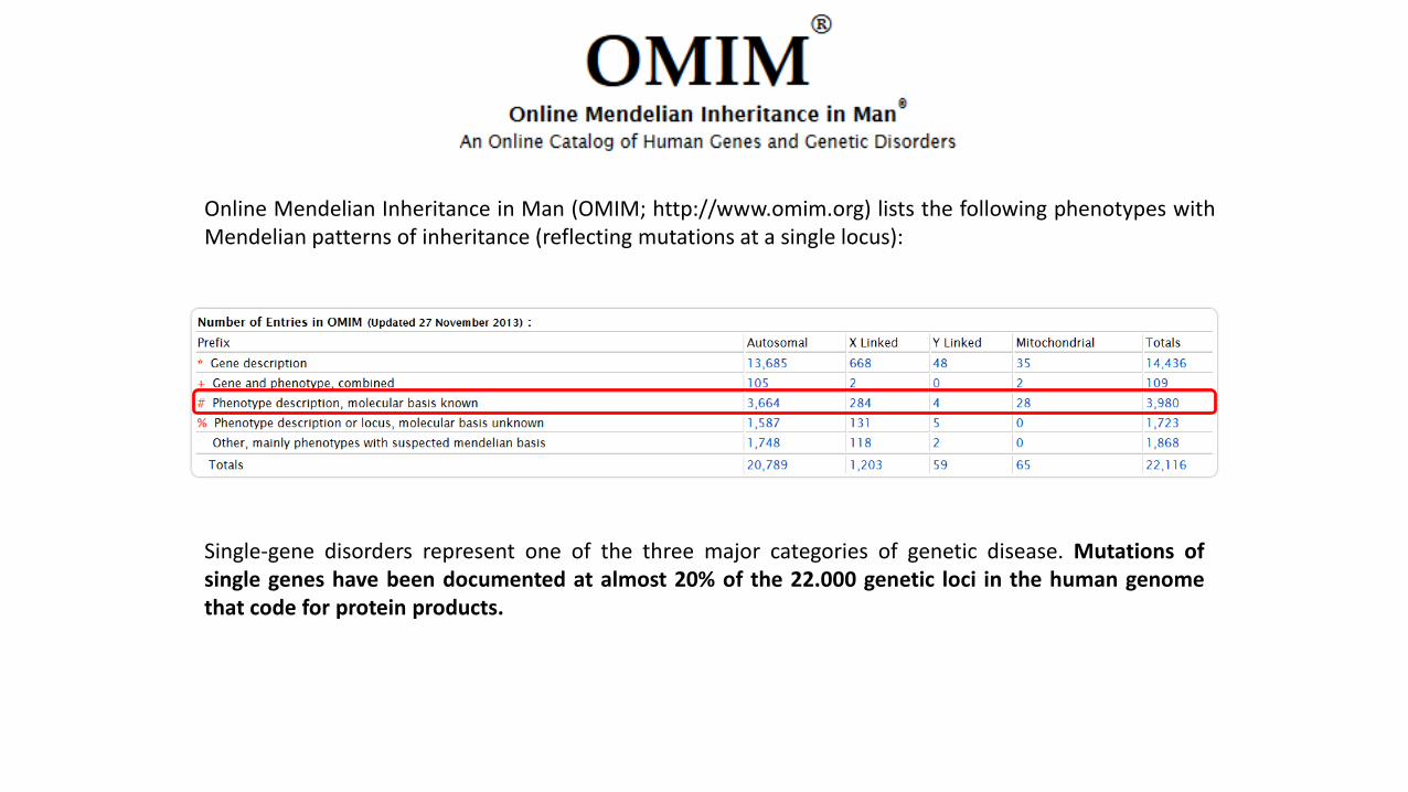
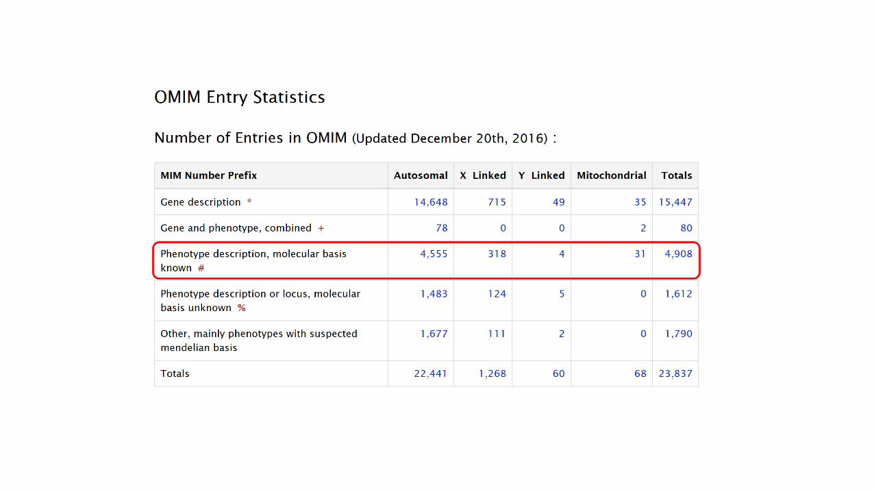
Online Mendelian Inheritance in Man (OMIM; http://www.omim.org) lists the following phenotypes withMendelian patterns of inheritance (reflecting mutations at a single locus):
Single-gene disorders represent one of the three major categories of genetic disease. Mutations ofsingle genes have been documented at almost 20% of the 22.000 genetic loci in the human genomethat code for protein products.
Copyright © 2012, Elsevier Inc. All rights Reserved.
Single Gene Defects (monogenic diseases)
Human Genes and GenomesLeon E. Rosenberg and Diane Drobnis Rosenberg
Academic Press
ISBN 978-0-12-385212-0
Copyright © 2012, Elsevier Inc. All rights Reserved.
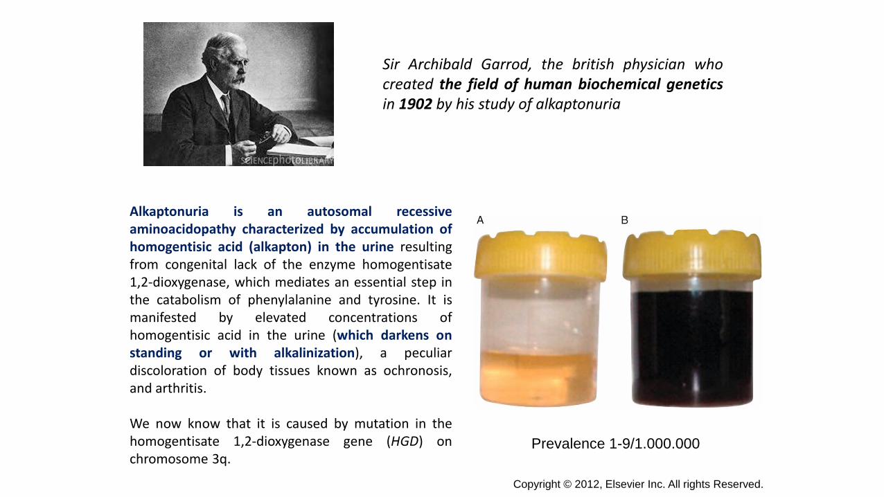
Sir Archibald Garrod, the british physician whocreated the field of human biochemical geneticsin 1902 by his study of alkaptonuria
Alkaptonuria is an autosomal recessiveaminoacidopathy characterized by accumulation ofhomogentisic acid (alkapton) in the urine resultingfrom congenital lack of the enzyme homogentisate1,2-dioxygenase, which mediates an essential step inthe catabolism of phenylalanine and tyrosine. It ismanifested by elevated concentrations ofhomogentisic acid in the urine (which darkens onstanding or with alkalinization), a peculiardiscoloration of body tissues known as ochronosis,and arthritis.
We now know that it is caused by mutation in thehomogentisate 1,2-dioxygenase gene (HGD) onchromosome 3q.
Prevalence 1-9/1.000.000
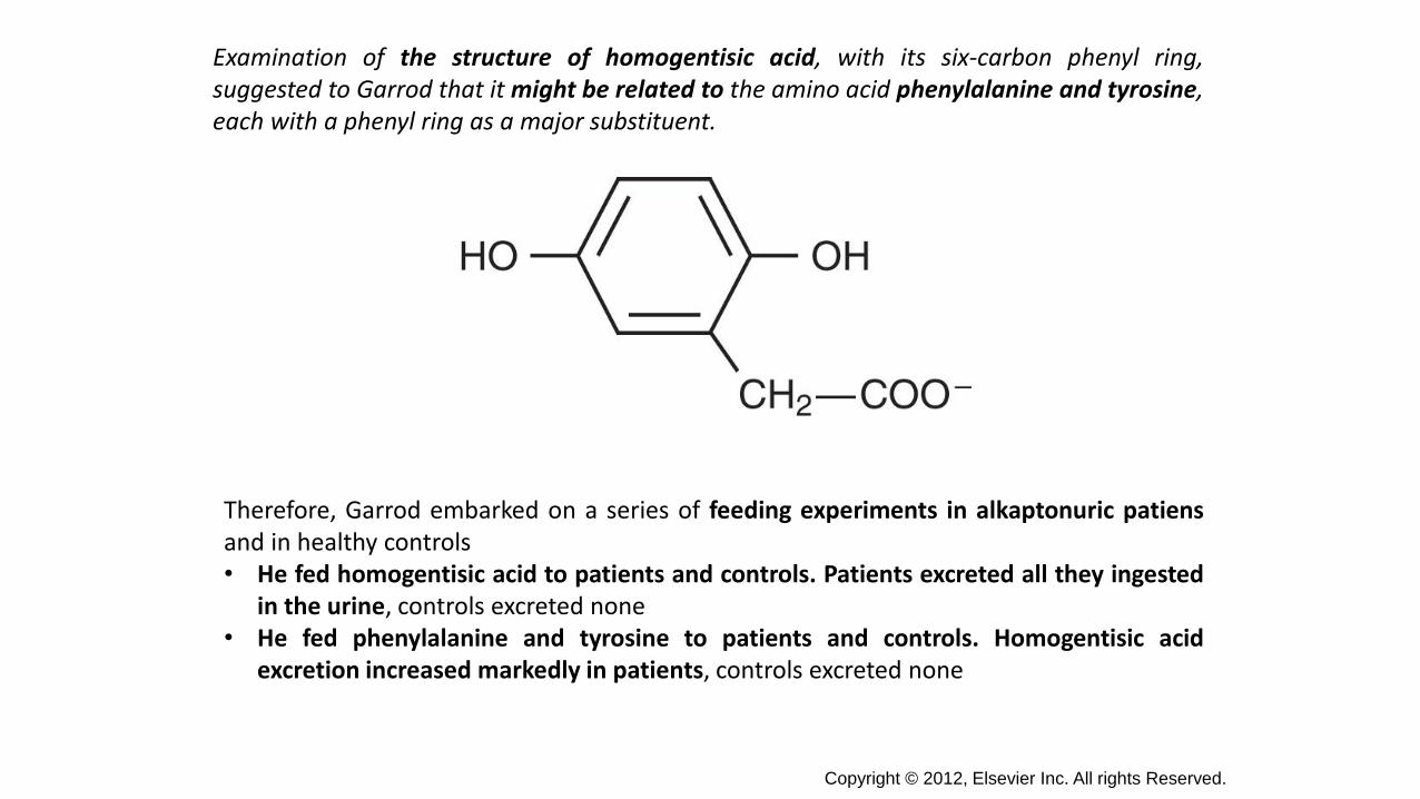
Examination of the structure of homogentisic acid, with its six-carbon phenyl ring,suggested to Garrod that it might be related to the amino acid phenylalanine and tyrosine,each with a phenyl ring as a major substituent.
Therefore, Garrod embarked on a series of feeding experiments in alkaptonuric patiensand in healthy controls• He fed homogentisic acid to patients and controls. Patients excreted all they ingested
in the urine, controls excreted none• He fed phenylalanine and tyrosine to patients and controls. Homogentisic acid
excretion increased markedly in patients, controls excreted none
Copyright © 2012, Elsevier Inc. All rights Reserved.
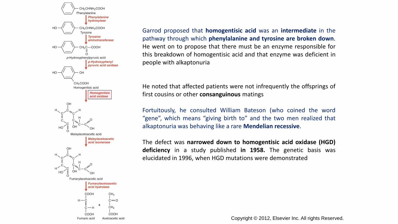
Garrod proposed that homogentisic acid was an intermediate in thepathway through which phenylalanine and tyrosine are broken down.He went on to propose that there must be an enzyme responsible forthis breakdown of homogentisic acid and that enzyme was deficient inpeople with alkaptonuria
He noted that affected patients were not infrequently the offsprings offirst cousins or other consanguinous matings
Fortuitously, he consulted William Bateson (who coined the word“gene”, which means “giving birth to” and the two men realized thatalkaptonuria was behaving like a rare Mendelian recessive.
The defect was narrowed down to homogentisic acid oxidase (HGD)deficiency in a study published in 1958. The genetic basis waselucidated in 1996, when HGD mutations were demonstrated
Copyright © 2012, Elsevier Inc. All rights Reserved.
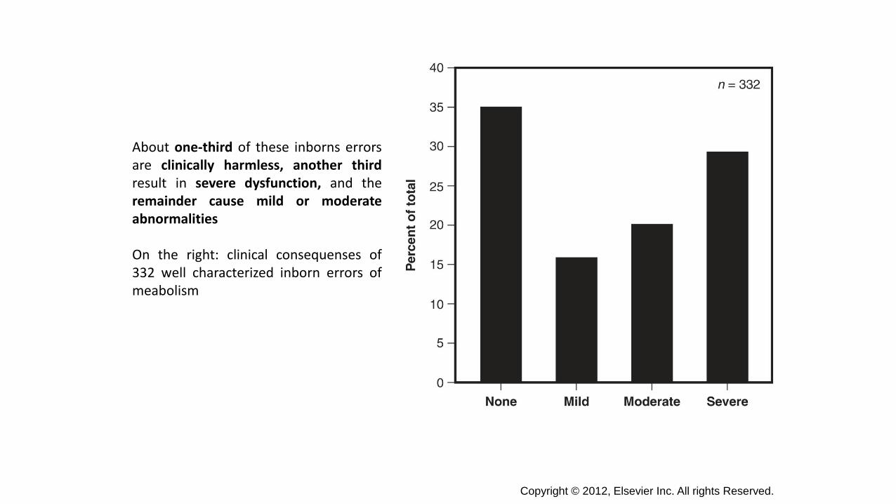
About one-third of these inborns errorsare clinically harmless, another thirdresult in severe dysfunction, and theremainder cause mild or moderateabnormalities
On the right: clinical consequenses of332 well characterized inborn errors ofmeabolism
Copyright © 2012, Elsevier Inc. All rights Reserved.
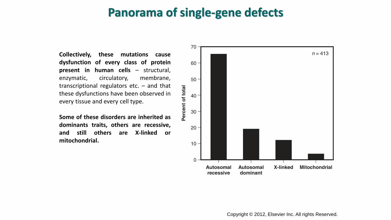
Collectively, these mutations causedysfunction of every class of proteinpresent in human cells – structural,enzymatic, circulatory, membrane,transcriptional regulators etc. – and thatthese dysfunctions have been observed inevery tissue and every cell type.
Some of these disorders are inherited asdominants traits, others are recessive,and still others are X-linked ormitochondrial.
Panorama of single-gene defects
Copyright © 2012, Elsevier Inc. All rights Reserved.

Hemoglobin (Hb) is the iron-containing oxygen-transport metalloprotein in the red blood cellsof all vertebrates (with the exception of the fishfamily Channichthyidae) as well as the tissues ofsome invertebrates.
In mammals, the protein makes up about 97%of the red blood cells' (RBCs) dry content (byweight).
Hemoglobin is a tretramer: two alpha and twobeta globin chains. Each globin chain contains aheme molecule
Copyright © 2012, Elsevier Inc. All rights Reserved.
Hemoglobinopathies
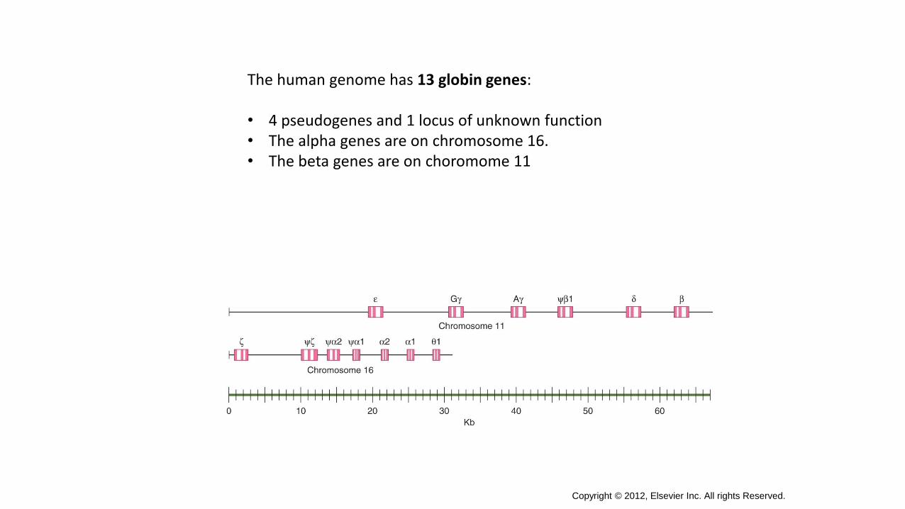
The human genome has 13 globin genes:
• 4 pseudogenes and 1 locus of unknown function• The alpha genes are on chromosome 16.• The beta genes are on choromome 11
Copyright © 2012, Elsevier Inc. All rights Reserved.
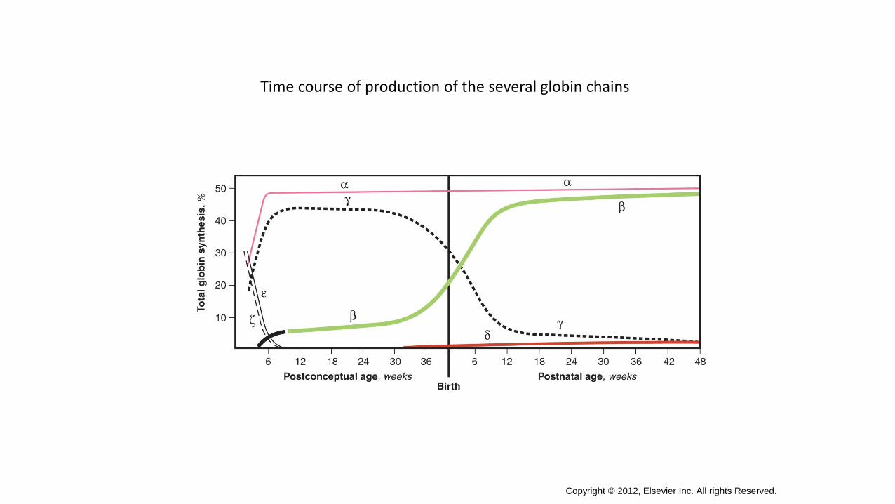
Time course of production of the several globin chains
Copyright © 2012, Elsevier Inc. All rights Reserved.
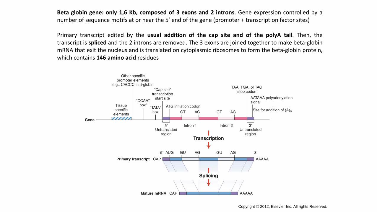
Beta globin gene: only 1,6 Kb, composed of 3 exons and 2 introns. Gene expression controlled by anumber of sequence motifs at or near the 5’ end of the gene (promoter + transcription factor sites)
Primary transcript edited by the usual addition of the cap site and of the polyA tail. Then, thetranscript is spliced and the 2 introns are removed. The 3 exons are joined together to make beta-globinmRNA that exit the nucleus and is translated on cytoplasmic ribosomes to form the beta-globin protein,which contains 146 amino acid residues
Copyright © 2012, Elsevier Inc. All rights Reserved.
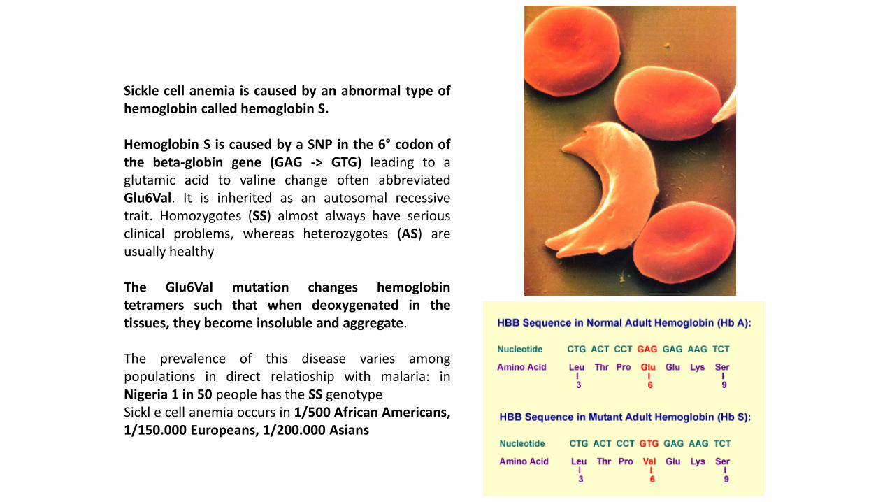
Sickle cell anemia is caused by an abnormal type ofhemoglobin called hemoglobin S.
Hemoglobin S is caused by a SNP in the 6° codon ofthe beta-globin gene (GAG -> GTG) leading to aglutamic acid to valine change often abbreviatedGlu6Val. It is inherited as an autosomal recessivetrait. Homozygotes (SS) almost always have seriousclinical problems, whereas heterozygotes (AS) areusually healthy
The Glu6Val mutation changes hemoglobintetramers such that when deoxygenated in thetissues, they become insoluble and aggregate.
The prevalence of this disease varies amongpopulations in direct relatioship with malaria: inNigeria 1 in 50 people has the SS genotypeSickl e cell anemia occurs in 1/500 African Americans,1/150.000 Europeans, 1/200.000 Asians
Sickle Cell Anemia
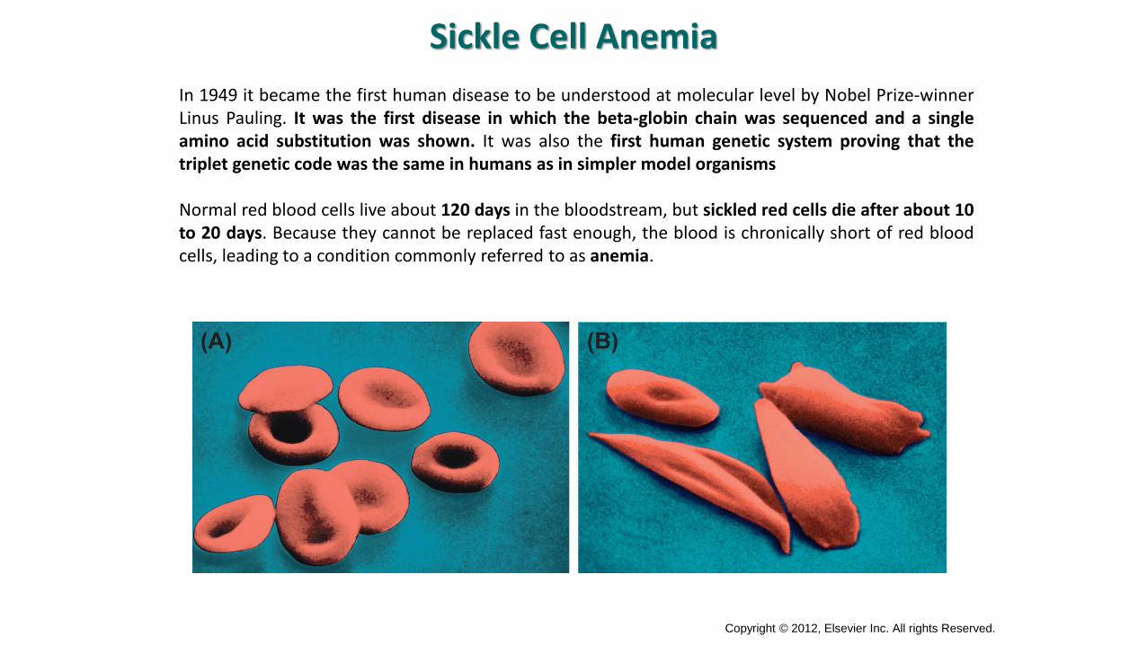
In 1949 it became the first human disease to be understood at molecular level by Nobel Prize-winnerLinus Pauling. It was the first disease in which the beta-globin chain was sequenced and a singleamino acid substitution was shown. It was also the first human genetic system proving that thetriplet genetic code was the same in humans as in simpler model organisms
Normal red blood cells live about 120 days in the bloodstream, but sickled red cells die after about 10to 20 days. Because they cannot be replaced fast enough, the blood is chronically short of red bloodcells, leading to a condition commonly referred to as anemia.
Copyright © 2012, Elsevier Inc. All rights Reserved.
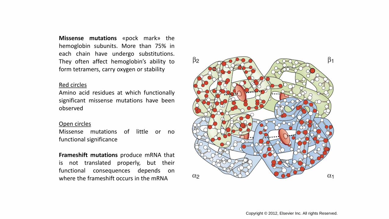
Missense mutations «pock mark» thehemoglobin subunits. More than 75% ineach chain have undergo substitutions.They often affect hemoglobin’s ability toform tetramers, carry oxygen or stability
Red circlesAmino acid residues at which functionallysignificant missense mutations have beenobserved
Open circlesMissense mutations of little or nofunctional significance
Frameshift mutations produce mRNA thatis not translated properly, but theirfunctional consequences depends onwhere the frameshift occurs in the mRNA
Copyright © 2012, Elsevier Inc. All rights Reserved.
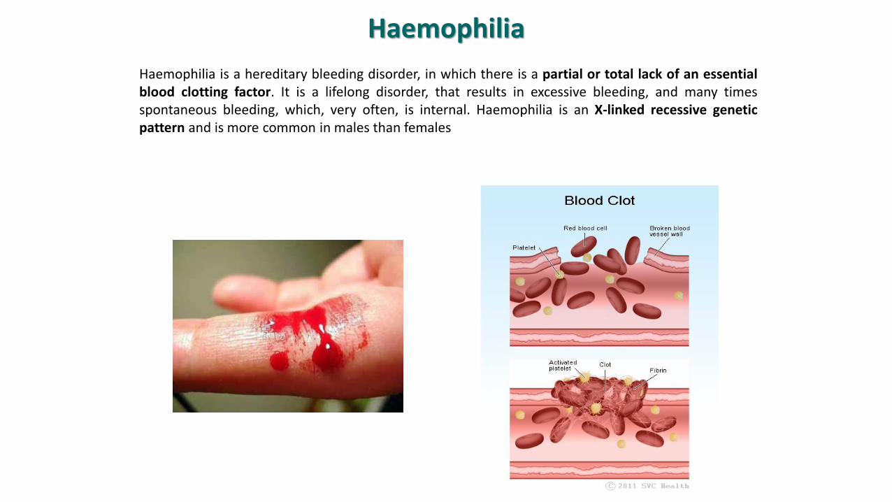
Haemophilia
Haemophilia is a hereditary bleeding disorder, in which there is a partial or total lack of an essentialblood clotting factor. It is a lifelong disorder, that results in excessive bleeding, and many timesspontaneous bleeding, which, very often, is internal. Haemophilia is an X-linked recessive geneticpattern and is more common in males than females
There are mainly two types of Haemophilia, they are Haemophilia A and Haemophilia B.Rarely there is another type of haemophilia known as haemophilia C.
• Haemophilia A is caused by the deficiency of clotting factor VIII• Haemophilia B is caused by the deficiency of clotting factor IX (Christmas Factor)• Haemophilia C is caused by the deficiency of clotting factor XI. In all the above cases,
patient will have abnormal bleeding tendency.
Incidence• Haemophilia A: 1:5000 males. It is four times more common than Type B.• Haemophilia B: 1: 34,000 males.
Haemophilia in girls is very rare condition. This condition develops in girls only when boththe X genes are defective. If a girl has one defective X gene and a normal X gene, she is notaffected but her male children have 50% chance of getting the disease.
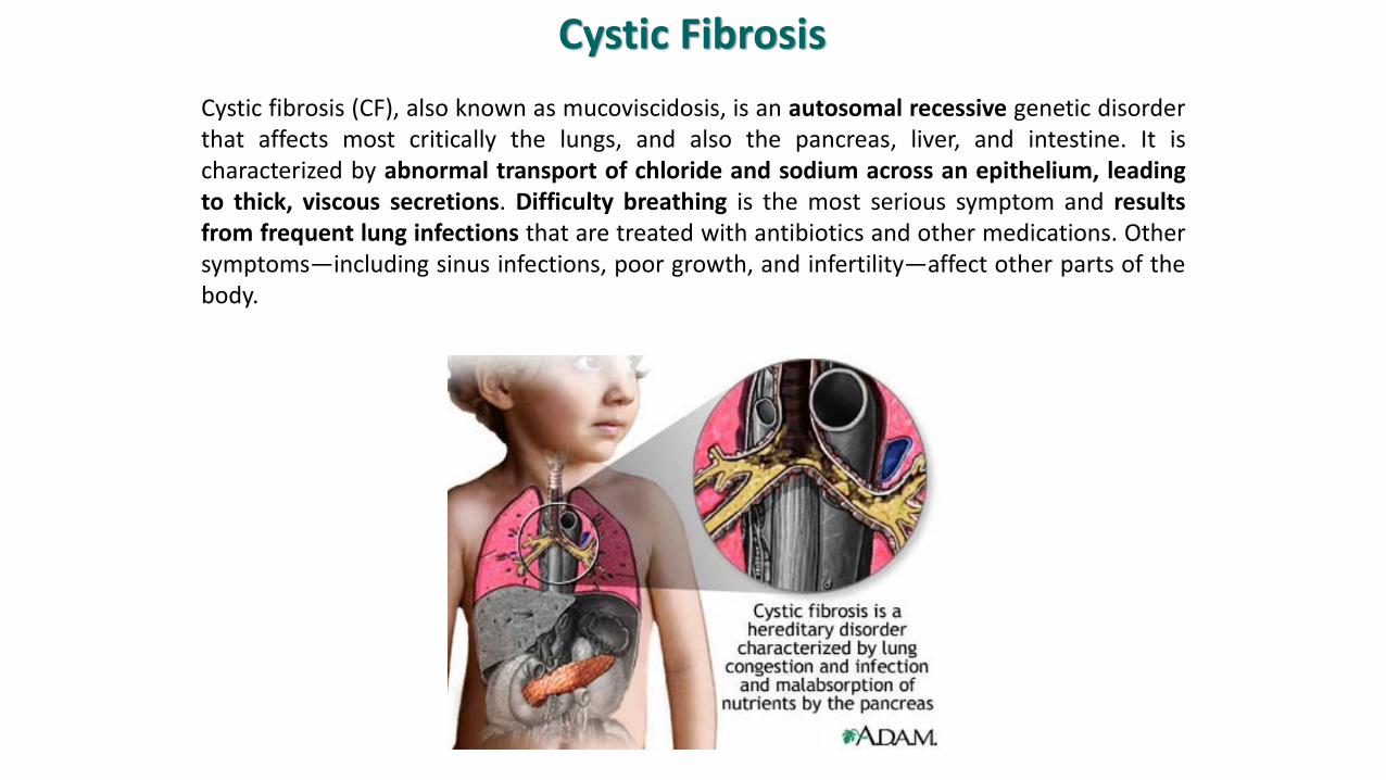
Cystic Fibrosis
Cystic fibrosis (CF), also known as mucoviscidosis, is an autosomal recessive genetic disorderthat affects most critically the lungs, and also the pancreas, liver, and intestine. It ischaracterized by abnormal transport of chloride and sodium across an epithelium, leadingto thick, viscous secretions. Difficulty breathing is the most serious symptom and resultsfrom frequent lung infections that are treated with antibiotics and other medications. Othersymptoms—including sinus infections, poor growth, and infertility—affect other parts of thebody.
CF is caused by a mutation in the gene for the protein cystic fibrosis transmembrane conductanceregulator (CFTR). This protein is required to regulate the components of sweat, digestive fluids, andmucus. CFTR regulates the movement of chloride and sodium ions across epithelial membranes, suchas the alveolar epithelia located in the lungs.
Both CFTR copies must be missing for CF to develop, and therefore has autosomal recessiveinheritance.
CF is most common among people of Central and Northern European ancestry, but occurs in manydemographic groups around the world. The prevalence of CF is the rarest in Asia and the Middle East.Individuals with cystic fibrosis can be diagnosed before birth by genetic testing, or by a sweat test inearly childhood. Ultimately, lung transplantation is often necessary as CF worsens.
Although it is severely underdiagnosed in Asia, existing evidence indicates that the prevelance of CF israre. In the European Union 1 in 2000-3000 new borns is found to be affected by CF. In the UnitedStates of America the incidence of CF is reported to be 1 in every 3500 births.
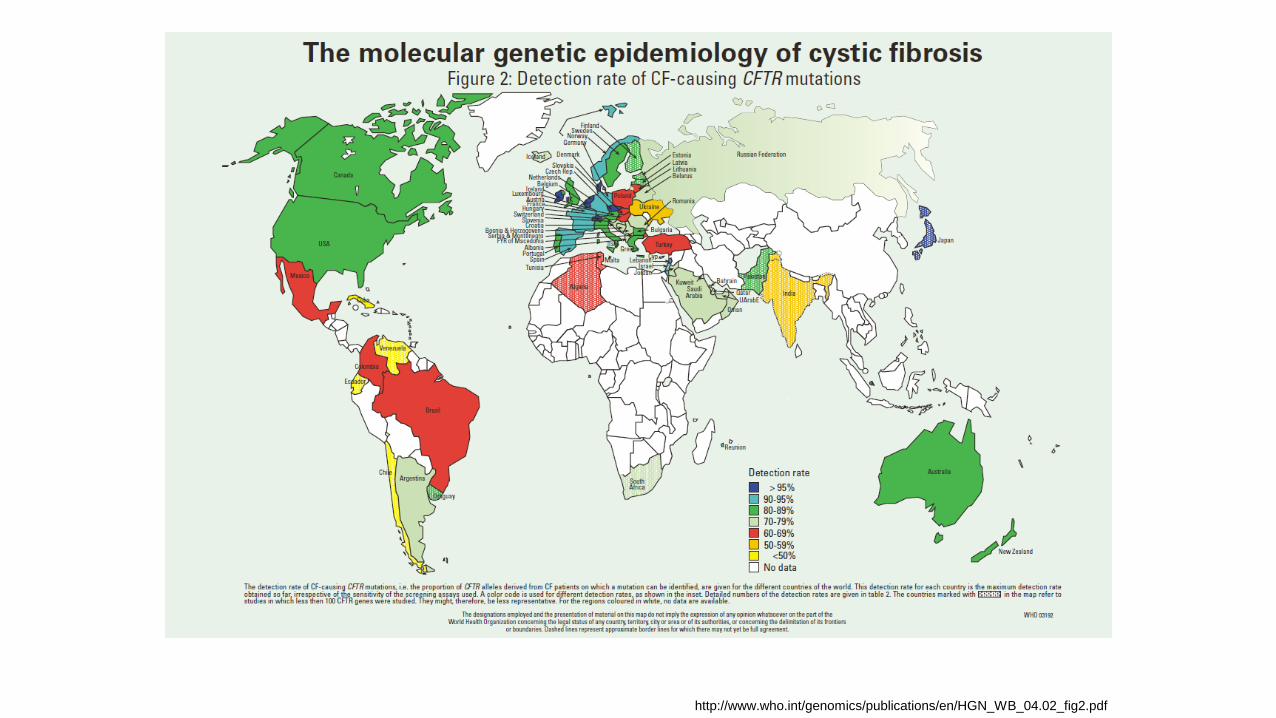
http://www.who.int/genomics/publications/en/HGN_WB_04.02_fig2.pdf
Tay sachs disease
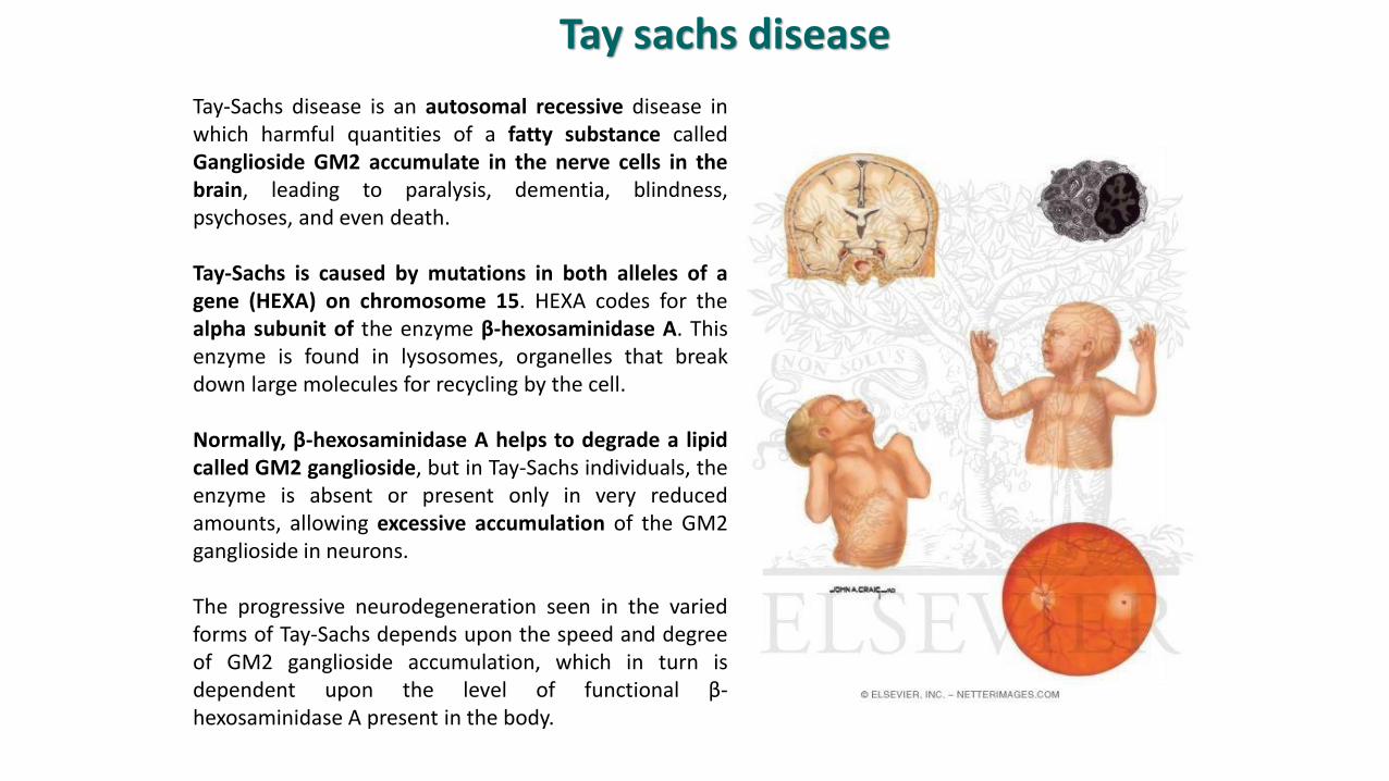
Tay-Sachs disease is an autosomal recessive disease inwhich harmful quantities of a fatty substance calledGanglioside GM2 accumulate in the nerve cells in thebrain, leading to paralysis, dementia, blindness,psychoses, and even death.
Tay-Sachs is caused by mutations in both alleles of agene (HEXA) on chromosome 15. HEXA codes for thealpha subunit of the enzyme β-hexosaminidase A. Thisenzyme is found in lysosomes, organelles that breakdown large molecules for recycling by the cell.
Normally, β-hexosaminidase A helps to degrade a lipidcalled GM2 ganglioside, but in Tay-Sachs individuals, theenzyme is absent or present only in very reducedamounts, allowing excessive accumulation of the GM2ganglioside in neurons.
The progressive neurodegeneration seen in the variedforms of Tay-Sachs depends upon the speed and degreeof GM2 ganglioside accumulation, which in turn isdependent upon the level of functional β-hexosaminidase A present in the body.
This disease is autosomal recessive.
Prevalence:
The frequency of the condition is much higher in Ashkenazi Jews of Eastern Europeanorigin than in others.
Approximately one in every 27 Jews in the United States of America is a carrier of the TSDgene. There is also a noticeable incidence of TSD in non-Jewish French Canadians livingnear the St. Lawrence River and in the Cajun community of Louisiana.
Among Jews of Sephardic origin and in the general, non-Jewish population, the carrier rateis about 1 in 250. There are certain exceptions. French-Canadian and the Cajuncommunity of Louisiana have the same carrier rate as Ashkenazi Jews, one in 27. Also,individuals with ancestry from Ireland are at increased risk for the Tay-Sachs gene.Current research indicates that among Irish Americans, the carrier rate is about one in 50.
Fragile X syndrome
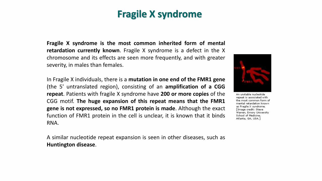
Fragile X syndrome is the most common inherited form of mentalretardation currently known. Fragile X syndrome is a defect in the Xchromosome and its effects are seen more frequently, and with greaterseverity, in males than females.
In Fragile X individuals, there is a mutation in one end of the FMR1 gene(the 5' untranslated region), consisting of an amplification of a CGGrepeat. Patients with fragile X syndrome have 200 or more copies of theCGG motif. The huge expansion of this repeat means that the FMR1gene is not expressed, so no FMR1 protein is made. Although the exactfunction of FMR1 protein in the cell is unclear, it is known that it bindsRNA.
A similar nucleotide repeat expansion is seen in other diseases, such asHuntington disease.
Although it is a X-linked recessive trait with variable expression and incompletepenetrance, 30% of all carrier women are affected.
Prevalence:
According to the Fragile X association of Southern California, Fragile X syndrome isthe single most common inherited cause of mental impairment affecting 1 in 3600males and 1 in 4000 to 6000 females with full mutation worldwide.Some studies also suggest that fragile X affects 1 in every 2000 males and 1 in every4000 females of all races and ethnic groups.
Studies have also revealed that 1 in 259 women of all races carry fragile X. Thenumber of men who are carriers is thought to be 1 in 800 of all races and ethnicity.Carrier females have a 30% to 40>% chance of giving birth to a retarded male childand a 15 to 20% chance of having a retarded female.
Huntington disease
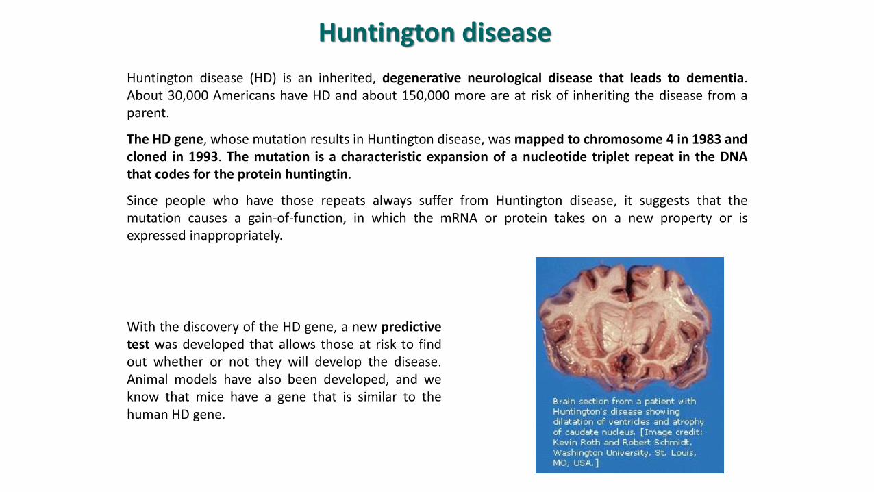
Huntington disease (HD) is an inherited, degenerative neurological disease that leads to dementia.About 30,000 Americans have HD and about 150,000 more are at risk of inheriting the disease from aparent.
The HD gene, whose mutation results in Huntington disease, was mapped to chromosome 4 in 1983 andcloned in 1993. The mutation is a characteristic expansion of a nucleotide triplet repeat in the DNAthat codes for the protein huntingtin.
Since people who have those repeats always suffer from Huntington disease, it suggests that themutation causes a gain-of-function, in which the mRNA or protein takes on a new property or isexpressed inappropriately.
With the discovery of the HD gene, a new predictivetest was developed that allows those at risk to findout whether or not they will develop the disease.Animal models have also been developed, and weknow that mice have a gene that is similar to thehuman HD gene.
Copyright © 2012, Elsevier Inc. All rights Reserved.
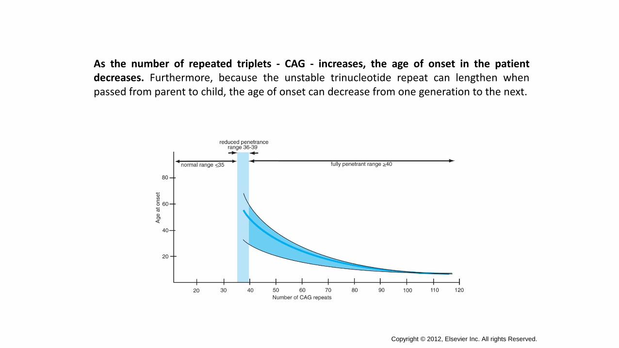
As the number of repeated triplets - CAG - increases, the age of onset in the patientdecreases. Furthermore, because the unstable trinucleotide repeat can lengthen whenpassed from parent to child, the age of onset can decrease from one generation to the next.
Huntington's is an autosomal dominant genetic disorder which means that if one parentcarriers the defective Huntington's gene, his/her offspring have a 50/50 chance of inheritingthe disease. Everyone who carries the gene will develop the disease.
The function of the huntingtin gene, also called the HTT or HD (Huntington disease) gene,is unclear. It is essential for development, and absence of huntingtin is lethal in mice. In itswild-type (normal) form, it contains 6-35 glutamine residues. However, in individualsaffected by Huntington's disease (an autosomal dominant genetic disorder), it containsmore than 36 glutamine residues (highest reported repeat length is about 250)
Prevalence:
Huntington's disease (HD) affects males and females equally and crosses all ethnic and racialboundaries. It typically begins in mid-life, between the ages of 30 and 45, though onset mayoccur as early as the age of 2. Children who develop the juvenile form of the disease rarelylive to adulthood.
In Western countries, it's estimated that about 5 to 7 people per 100,000 are affected byHD. A very high concentration of HD has also been found in the Lake Maracaibo region ofVenezuela where the prevalence of HD is about 700 per 100,000
Duchenne Muscular Dystrophy
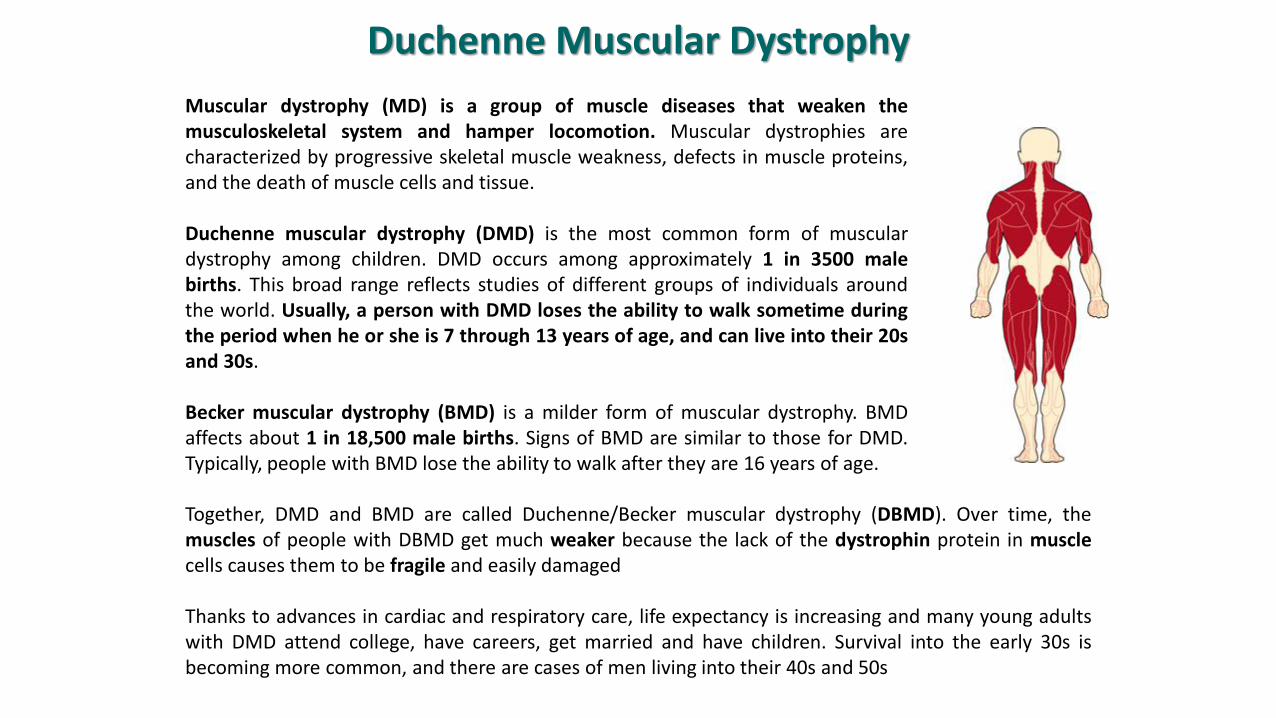
Muscular dystrophy (MD) is a group of muscle diseases that weaken themusculoskeletal system and hamper locomotion. Muscular dystrophies arecharacterized by progressive skeletal muscle weakness, defects in muscle proteins,and the death of muscle cells and tissue.
Duchenne muscular dystrophy (DMD) is the most common form of musculardystrophy among children. DMD occurs among approximately 1 in 3500 malebirths. This broad range reflects studies of different groups of individuals aroundthe world. Usually, a person with DMD loses the ability to walk sometime duringthe period when he or she is 7 through 13 years of age, and can live into their 20sand 30s.
Becker muscular dystrophy (BMD) is a milder form of muscular dystrophy. BMDaffects about 1 in 18,500 male births. Signs of BMD are similar to those for DMD.Typically, people with BMD lose the ability to walk after they are 16 years of age.
Together, DMD and BMD are called Duchenne/Becker muscular dystrophy (DBMD). Over time, themuscles of people with DBMD get much weaker because the lack of the dystrophin protein in musclecells causes them to be fragile and easily damaged
Thanks to advances in cardiac and respiratory care, life expectancy is increasing and many young adultswith DMD attend college, have careers, get married and have children. Survival into the early 30s isbecoming more common, and there are cases of men living into their 40s and 50s
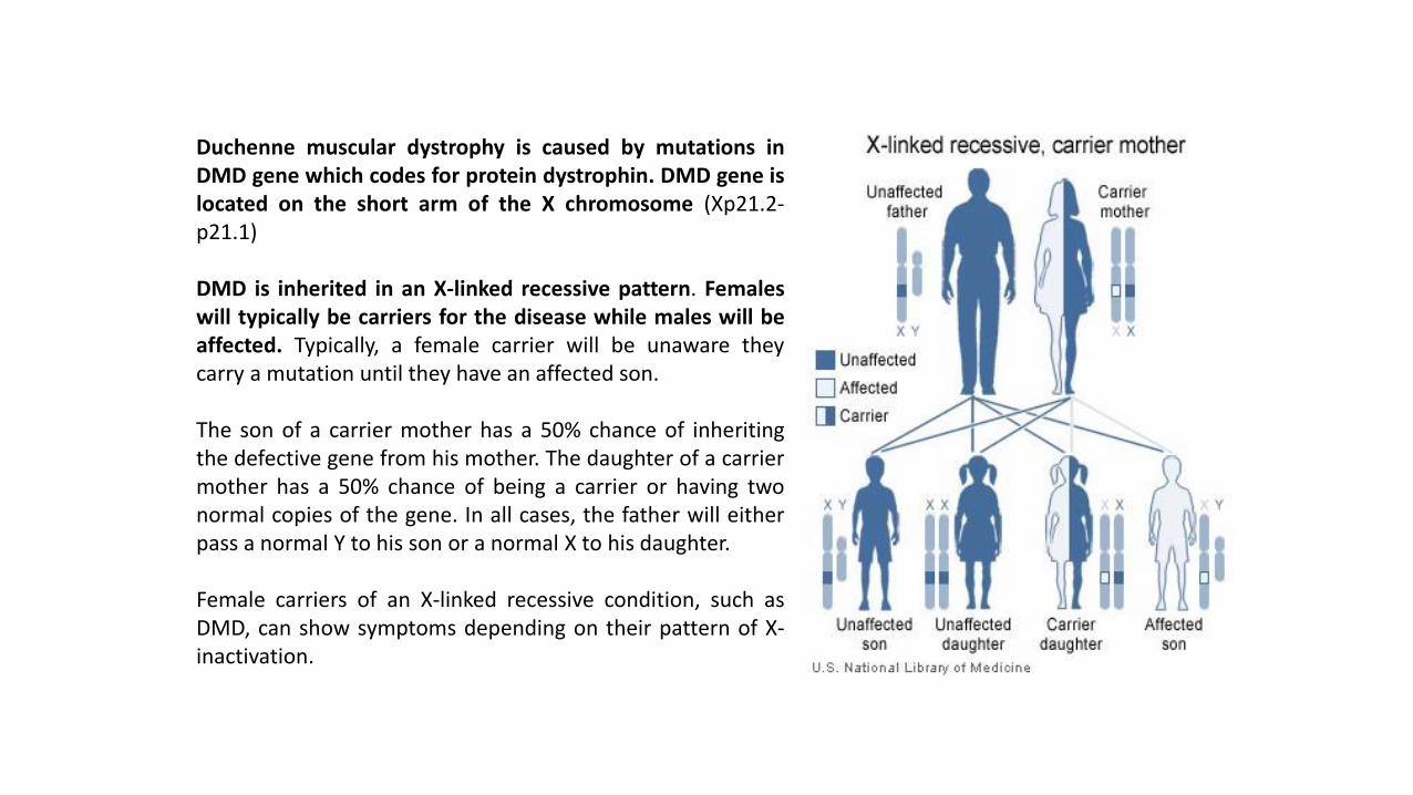
Duchenne muscular dystrophy is caused by mutations inDMD gene which codes for protein dystrophin. DMD gene islocated on the short arm of the X chromosome (Xp21.2-p21.1)
DMD is inherited in an X-linked recessive pattern. Femaleswill typically be carriers for the disease while males will beaffected. Typically, a female carrier will be unaware theycarry a mutation until they have an affected son.
The son of a carrier mother has a 50% chance of inheritingthe defective gene from his mother. The daughter of a carriermother has a 50% chance of being a carrier or having twonormal copies of the gene. In all cases, the father will eitherpass a normal Y to his son or a normal X to his daughter.
Female carriers of an X-linked recessive condition, such asDMD, can show symptoms depending on their pattern of X-inactivation.
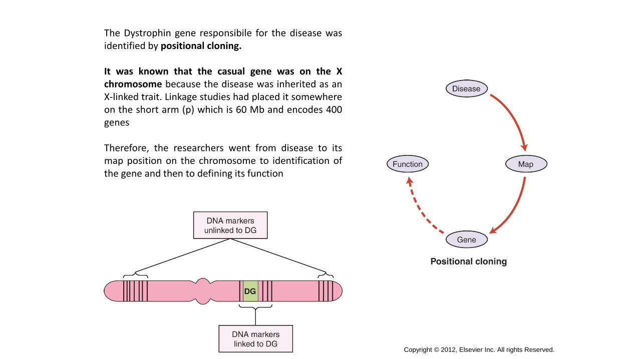
The Dystrophin gene responsibile for the disease wasidentified by positional cloning.
It was known that the casual gene was on the Xchromosome because the disease was inherited as anX-linked trait. Linkage studies had placed it somewhereon the short arm (p) which is 60 Mb and encodes 400genes
Therefore, the researchers went from disease to itsmap position on the chromosome to identification ofthe gene and then to defining its function
Copyright © 2012, Elsevier Inc. All rights Reserved.
Copyright © 2012, Elsevier Inc. All rights Reserved.
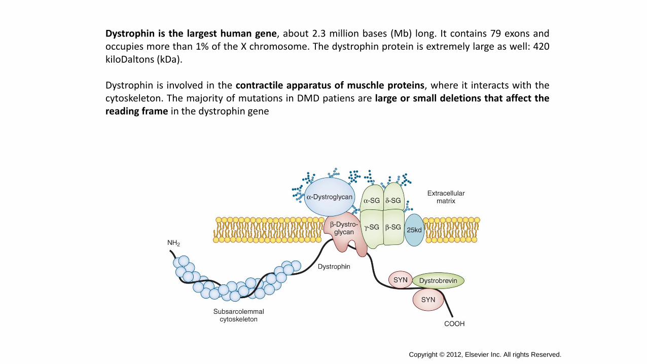
Dystrophin is the largest human gene, about 2.3 million bases (Mb) long. It contains 79 exons andoccupies more than 1% of the X chromosome. The dystrophin protein is extremely large as well: 420kiloDaltons (kDa).
Dystrophin is involved in the contractile apparatus of muschle proteins, where it interacts with thecytoskeleton. The majority of mutations in DMD patiens are large or small deletions that affect thereading frame in the dystrophin gene
Copyright © 2012, Elsevier Inc. All rights Reserved.
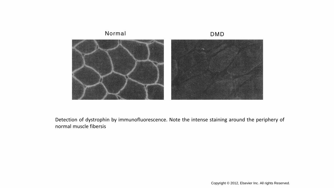
Detection of dystrophin by immunofluorescence. Note the intense staining around the periphery ofnormal muscle fibersis
Amyotrophic Lateral Sclerosis (ASL)
A-myo-trophic comes from the Greek language. "A" means no or negative. "Myo" refers to muscle, and"Trophic" means nourishment–"No muscle nourishment." When a muscle has no nourishment, it"atrophies" or wastes away. "Lateral" identifies the areas in a person's spinal cord where portions of thenerve cells that signal and control the muscles are located. As this area degenerates it leads to scarringor hardening ("sclerosis") in the region.
About 90% of patients with adult-onset ALS have no family history of ALS and present as an isolatedcase in their family. This is called sporadic ALS (SALS), and although there is likely a geneticpredisposition involved, SALS is not directly inherited. The remaining 10% of people with ALS have afamily member with ALS, and this is referred to as familial ALS (FALS).
There are several inheritance patterns, but the most common for FALS is autosomal dominant. The mostcommon genes currently known to be associated with FALS include SOD1, TDP-43, FUS and the morerecently discovered C9ORF72 and UBQLN2. Other genetic causes of ALS affect relatively few people.Nonetheless, understanding how they cause the disease may offer large insights into the diseaseprocess. These genes include VCP (valosin-containing protein), alsin, senataxin, and angiogenin andoptineurin.
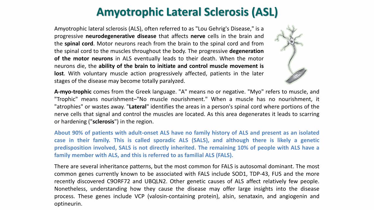
Amyotrophic lateral sclerosis (ALS), often referred to as "Lou Gehrig's Disease," is aprogressive neurodegenerative disease that affects nerve cells in the brain andthe spinal cord. Motor neurons reach from the brain to the spinal cord and fromthe spinal cord to the muscles throughout the body. The progressive degenerationof the motor neurons in ALS eventually leads to their death. When the motorneurons die, the ability of the brain to initiate and control muscle movement islost. With voluntary muscle action progressively affected, patients in the laterstages of the disease may become totally paralyzed.
C9ORF72. This gene, discovered in 2011, is the most common genetic cause of ALS. (Its name refers to the position ofan “open reading frame” on chromosome 9). Mutations in this gene account for between 25% and 40% of all familialALS cases (depending on the population), and also approximately 4% to 6% of sporadic cases. As noted above, theseapparently sporadic cases are in fact genetic. The gene mutation appears to act in a dominant manner. How this genecauses ALS is unknown, and is the subject of a great deal of intense research.
Cu/Zn Superoxide Dismutase 1 (SOD1). Mutations in SOD1 were first described in 1993, and SOD1 was the first geneknown for ALS. It accounts for about 10% of familial ALS, or 1.5% to 2% of all ALS. It is inherited in a dominant manner.How SOD1 mutations cause ALS is unknown. It is clear that disease is not due to lack of function of the protein, sincedeleting the gene in animal models doesn’t cause ALS. Instead, it appears to take on some new toxic function, possiblyrelated to an increase in the tendency of mutant SOD1 molecules to aggregate and form clumps in motor neurons. It isalso possible that SOD1 causes ALS through actions in nearby cells called astrocytes, not in motor neurons themselves.Astrocytes help maintain motor neurons, and SOD1 mutation may impair their ability to do so. Read more about SOD1
TDP-43. TAR DNA binding protein 43 (TDP-43) was linked to ALS in 2008. Mutations in TDP-43 cause a dominant form ofALS. The normal role of the TDP-43 protein includes binding to RNA, the genetic messenger molecule. Mutations in theTDP-43 gene cause the TDP-43 protein to mislocalize in motor neurons, away from the nucleus where it is normallyfound, and into the cytoplasm (the material surrounding the nucleus), where it aggregates into clumps that can be seenunder the microscope. Even in ALS not caused by TDP-43 mutations, the protein is found in these aggregates,suggesting it may play a pivotal role in many forms of ALS.
FUS. Fused in sarcoma (FUS) was also discovered to play a role in ALS in 2008. Like TDP-43, it is inherited in a dominantmanner. It is also an RNA binding protein, and may play a similar normal role in the cell. FUS and TDP-43 may in factinteract as part of their normal function.
Ubiquilin-2. Ubiquilin-2 was linked to ALS in 2011. Unlike all other known ALS genes, the ubiquilin-2 gene resides on theX chromosome. Despite this, both men and women develop ALS due to ubiquilin-2 mutations. The normal function ofthe protein is to help degrade damaged or defective proteins in the cell. It is likely that mutations in the gene interferewith this function, and may lead to accumulation of harmful material within the cell.
Copyright © 2012, Elsevier Inc. All rights Reserved.
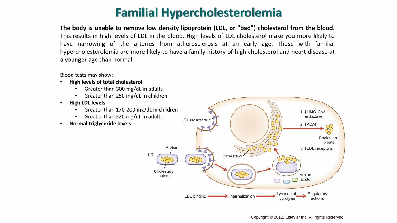
Familial HypercholesterolemiaThe body is unable to remove low density lipoprotein (LDL, or "bad") cholesterol from the blood.This results in high levels of LDL in the blood. High levels of LDL cholesterol make you more likely tohave narrowing of the arteries from atherosclerosis at an early age. Those with familialhypercholesterolemia are more likely to have a family history of high cholesterol and heart disease ata younger age than normal.
Blood tests may show:• High levels of total cholesterol
• Greater than 300 mg/dL in adults• Greater than 250 mg/dL in children
• High LDL levels• Greater than 170-200 mg/dL in children• Greater than 220 mg/dL in adults
• Normal triglyceride levels
Copyright © 2012, Elsevier Inc. All rights Reserved.
Men who have familial hypercholesterolemia have heart attacks in their 40's to 50's, and 85 percentof men with the disorder have a heart attack by age 60. Women who have familialhypercholesterolemia also have an increased risk for heart attack, but it happens 10 years later than inmen (so in their 50's and 60's).
It is inherited in families in an autosomal dominant manner.
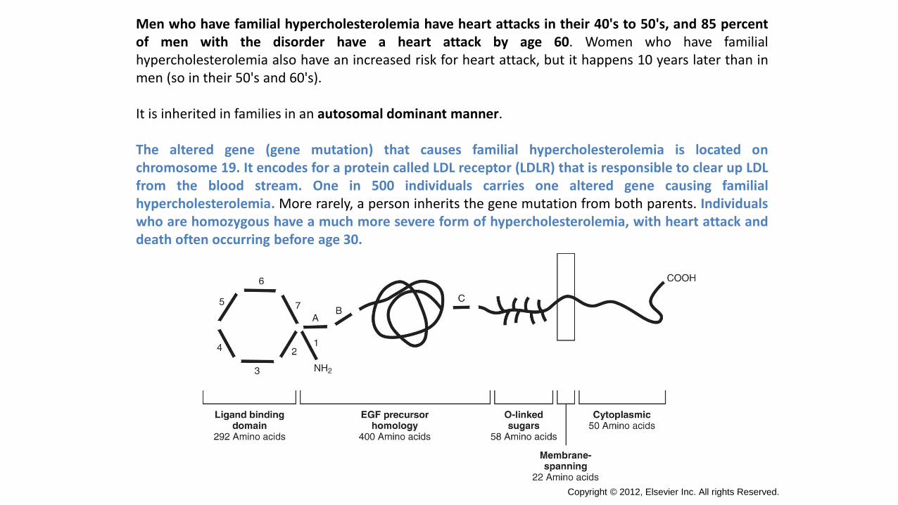
The altered gene (gene mutation) that causes familial hypercholesterolemia is located onchromosome 19. It encodes for a protein called LDL receptor (LDLR) that is responsible to clear up LDLfrom the blood stream. One in 500 individuals carries one altered gene causing familialhypercholesterolemia. More rarely, a person inherits the gene mutation from both parents. Individualswho are homozygous have a much more severe form of hypercholesterolemia, with heart attack anddeath often occurring before age 30.
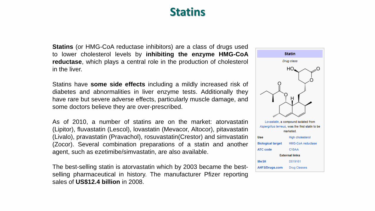
Statins (or HMG-CoA reductase inhibitors) are a class of drugs used
to lower cholesterol levels by inhibiting the enzyme HMG-CoA
reductase, which plays a central role in the production of cholesterol
in the liver.
Statins have some side effects including a mildly increased risk of
diabetes and abnormalities in liver enzyme tests. Additionally they
have rare but severe adverse effects, particularly muscle damage, and
some doctors believe they are over-prescribed.
As of 2010, a number of statins are on the market: atorvastatin
(Lipitor), fluvastatin (Lescol), lovastatin (Mevacor, Altocor), pitavastatin
(Livalo), pravastatin (Pravachol), rosuvastatin(Crestor) and simvastatin
(Zocor). Several combination preparations of a statin and another
agent, such as ezetimibe/simvastatin, are also available.
The best-selling statin is atorvastatin which by 2003 became the best-
selling pharmaceutical in history. The manufacturer Pfizer reporting
sales of US$12.4 billion in 2008.
Statins

Malattie legate al dna mitocondriale
• Il DNA genomico mitocondriale é circolare, costituito da ~ 16000 b.p. e codifica per gli enzimidella fosforilazione ossidativa.
• Ovociti: 200.000-300.000 copie di mt DNA
• Spermatozoi: perdono i mitocondri nella fertilizzazione
• Eteroplasmia: coesistono molecole di mtDNA mutate e normali
• POLIPLASMIA
• ETEROPLASMIA
• EFFETTO SOGLIA
• SEGREGAZIONE MITOTICA
• EREDITÀ MATERNA
Caratteristiche dell’ mtDNA
In ogni cellula sono presenti molti mitocondri ed ogni mitocondrio contiene multiple copia delsuo genoma (eccetto piastrine e ovulo non fertilizzato) → migliaia di copie mtDNA / cell.
Durante la divisione cellulare i mitocondri vengono distribuiti casualmente alle cellule figlie equindi la genetica mitocondriale è più simile alla genetica di popolazione che alla geneticamendeliana.
In tessuti normali tutte le copie di mtDNA sono identiche → omoplasmia.
Nel caso di una mutazione del mtDNA questa può colpire tutte le copie oppure essere presentesolo in una percentuale di genomi → eteroplasmia.
Generalmente i polimorfismi neutrali sono omoplasmici mentre la maggior parte delle mutazioni-malattia sono eteroplasmiche
Poliplasmia ed eteroplasmia mitocontriale
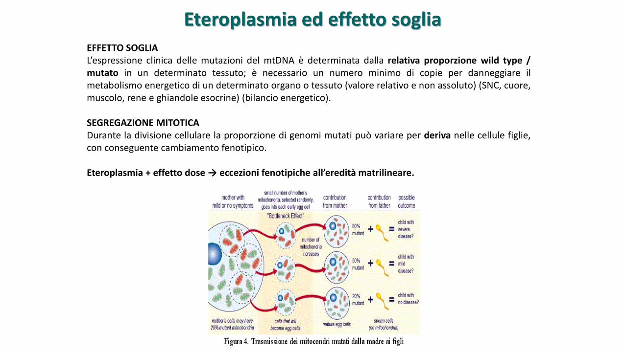
EFFETTO SOGLIAL’espressione clinica delle mutazioni del mtDNA è determinata dalla relativa proporzione wild type /mutato in un determinato tessuto; è necessario un numero minimo di copie per danneggiare ilmetabolismo energetico di un determinato organo o tessuto (valore relativo e non assoluto) (SNC, cuore,muscolo, rene e ghiandole esocrine) (bilancio energetico).
SEGREGAZIONE MITOTICADurante la divisione cellulare la proporzione di genomi mutati può variare per deriva nelle cellule figlie,con conseguente cambiamento fenotipico.
Eteroplasmia + effetto dose → eccezioni fenotipiche all’eredità matrilineare.
Eteroplasmia ed effetto soglia
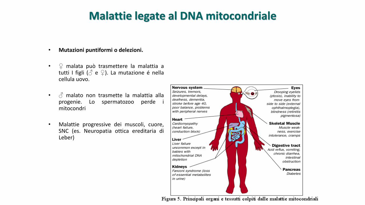
• Mutazioni puntiformi o delezioni.
• ♀ malata può trasmettere la malattia atutti I figli (♂ e ♀). La mutazione é nellacellula uovo.
• ♂ malato non trasmette la malattia allaprogenie. Lo spermatozoo perde imitocondri
• Malattie progressive dei muscoli, cuore,SNC (es. Neuropatia ottica ereditaria diLeber)
Malattie legate al DNA mitocondriale
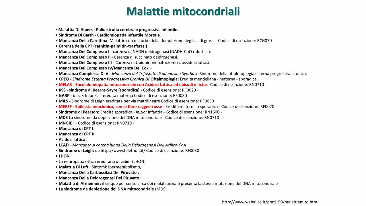
• Malattia Di Alpers - Polidistrofia cerebrale progressiva infantile. -• Sindrome Di Barth - Cardiomiopatia Infantile Mortale.• Mancanza Della Carnitina: Malattie con disturbo della demolizione degli acidi grassi - Codice di esenzione: RCG070 -• Carenza della CPT (carnitin-palmitin-trasferasi)• Mancanza Del Complesso I - carenza di NADH deidrogenasi (NADH-CoQ riduttasi).• Mancanza Del Complesso II - Carenza di succinato deidrogenasi.• Mancanza Del Complesso III - Carenza di Ubiquinone-citocromo c ossidoriduttasi.• Mancanza Del Complesso IV/Mancanza Del Cox –• Mancanza Complessa Di V - Mancanza del Trifosfato di adenosina Synthase:Sindrome della oftalmoplegia esterna progressiva cronica.• CPEO - Sindrome Esterna Progressiva Cronica Di Oftalmoplegia: Eredità mendeliana - materna - sporadica .• MELAS - Encefalomiopatia mitocondriale con Acidosi Lattica ed episodi di ictus- Codice di esenzione: RN0710 -• KSS - sindrome di Kearns-Sayre (sporadica) - Codice di esenzione: RF0020 -• NARP - Inizio: Infanzia - eredità materna Codice di esenzione: RF0030• MILS - Sindrome di Leigh ereditata per via matrilineare Codice di esenzione: RF0030• MERFF - Epilessia mioclonica, con le fibre ragged-rosse - Eredità materna o sporadica - Codice di esenzione: RF0020 -• Sindrome di Pearson: Eredità sporadica - Inizio: Infanzia - Codice di esenzione: RN1600 -• MDS La sindrome da deplezione del DNA mitocondriale - Codice di esenzione: RN0710 -• MNGIE : - Codice di esenzione: RN0710 -• Mancanza di CPT I• Mancanza di CPT II• Acidosi lattica :• LCAD - Mancanza A catena lunga Della Deidrogenasi Dell'Acilico-CoA• Sindrome di Leigh: da http://www.telethon.it/ Codice di esenzione: RF0030• LHON• La neuropatia ottica ereditaria di Leber (LHON)• Malattia Di Luft : Sintomi: Ipermetabolismo,• Mancanza Della Carbossilasi Del Piruvato :• Mancanza Della Deidrogenasi Del Piruvato :• Malattia di Alzheimer: il cinque per cento circa dei malati anziani presenta la stessa mutazione del DNA mitocondriale• La sindrome da deplezione del DNA mitocondriale (MDS)
http://www.webalice.it/prati_50/malattiemito.htm
Malattie mitocondriali
55
Sindrome Di Barth - Cardiomiopatia Infantile Mortale.La sindrome di Barth è una malattia metabolica caratterizzata da cardiomiopatia dilatativa, più raramente ditipo ipertrofico, neutropenia, miopatia scheletrica, difetto di crescita. Successivamente alla suacaratterizzazione clinica e biochimica, il gene-malattia è stato mappato in Xq28, utilizzando il clonaggioposizionale; l'analisi mutazionale ha identificato il gene G4,5. Sono state identificate oltre 20 mutazioni.
CPEO - Sindrome Esterna Progressiva Cronica Di Oftalmoplegia.Sindrome oftalmoplegica esterna cronica progressiva. E’ caratterizzata da una debolezza progressiva deimuscoli oculari e del muscolo elevatore della palpebra superioreCausa: Mutazione puntiforme del DNA mitocondriale: A3243G (la più comune)La maggior parte di questi quadri clinici è dovuta a mitocondriopatie, ma la causa della disfunzionemitocondriale è variabile (mutazioni puntiformi, delezioni del DNA mitocondriale, mutazione di geninucleari, con effetti sul DNA mitocondriale, come la timidina fosforilasi nella MNGIE).
MELASNella sindrome di MELAS (Encefalomiopatia mitocondriale con Acidosi Lattica ed episodi di ictus ), ai sintomiclinici i associano, in modo più o meno incostante, emiparesi, emianopsia, deficit motorio, emicrania,vomito, demenza, convulsioni, sordità, diabete, disturbi della memoria. La mutazione più frequente èA3243G del tRNA per la Leucina del DNA mitocondriale.
56
KSS - sindrome di Kearns-SayreInsorge prima dei 20 anni. Comporta oftalmoplegia, ptosi, retinite pigmentosa, segni associati a miopatia,disturbi della conduzione cardiaca, iperproteinorrachia. Nei casi sporadici possono essere presenti altrisintomi come sordità, cardiomiopatia, crisi epilettiche, disturbi del transito intestinale, ipoparatiroidismo,ritardo nella crescita, diabete, insufficienza renale. Queste patologie sono associate ad ampie delezionieteroplasmiche del DNA mitocondriale, la più frequente delle quali è lunga 4,9 kilobasi. Il rapporto tra lapercentuale degli organelli mutati/non mutati può essere molto elevato e quando raggiunge la soglia del60% circa (ad es: nel muscolo) determina un fenotipo patologico con deficit a carico dei complessi dellacatena respiratoria, anomalie istochimiche, "Ragged Red Fibers''.
NARPLa sindrome Neuropatia, Atassia e Retinite Pigmentosa (NARP) è clinicamente eterogenea, ma spesso ècaratterizzata dalla combinazione tra neuropatia sensoriale-motoria, atassia cerebellare e cecità notturna.La sua prevalenza è stimata in circa 1/12.000. La sintomatologia clinica comprende: retinopatia precoce"sale e pepe''; retinite pigmentosa; pupille "pigre''; nistagmo; cecità; debolezza muscolare prossimale;ritardo di sviluppo; atrofia cortico-spinale; demenza; ipoacusia; convulsioni; atassia; neuropatia sensoriale;debolezza muscolare prossimale neurogena. La sindrome NARP è una malattia ad eredità materna,dovuta alla mutazione 8993T>G nel gene del mtDNA, MTATP6, che codifica per la subunità 6 dell'ATPasi.La mutazione 8993T>G comporta un cambio aminoacidico dalla leucina 156, altamente conservata, inarginina (L156R), e causa un grave deterioramento della sintesi dell'ATP mitocondriale, che riduce l'energiae produce la morte cellulare, in particolare nei tessuti che dipendono fortemente dal metabolismo dellafosforilazione ossidativa, come il cervello e la retina.
Copyright © 2012, Elsevier Inc. All rights Reserved.
Red Hair
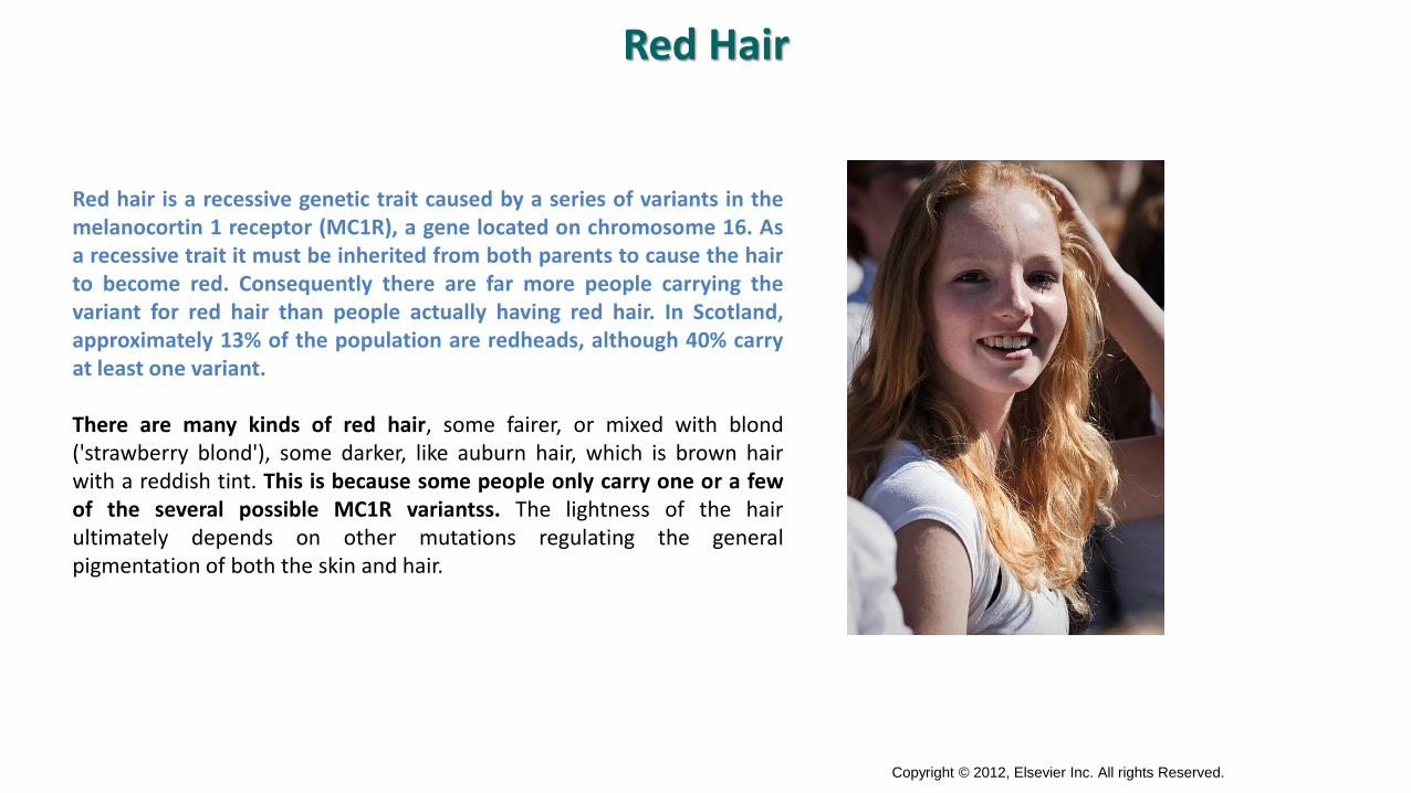
Red hair is a recessive genetic trait caused by a series of variants in themelanocortin 1 receptor (MC1R), a gene located on chromosome 16. Asa recessive trait it must be inherited from both parents to cause the hairto become red. Consequently there are far more people carrying thevariant for red hair than people actually having red hair. In Scotland,approximately 13% of the population are redheads, although 40% carryat least one variant.
There are many kinds of red hair, some fairer, or mixed with blond('strawberry blond'), some darker, like auburn hair, which is brown hairwith a reddish tint. This is because some people only carry one or a fewof the several possible MC1R variantss. The lightness of the hairultimately depends on other mutations regulating the generalpigmentation of both the skin and hair.
Copyright © 2012, Elsevier Inc. All rights Reserved.
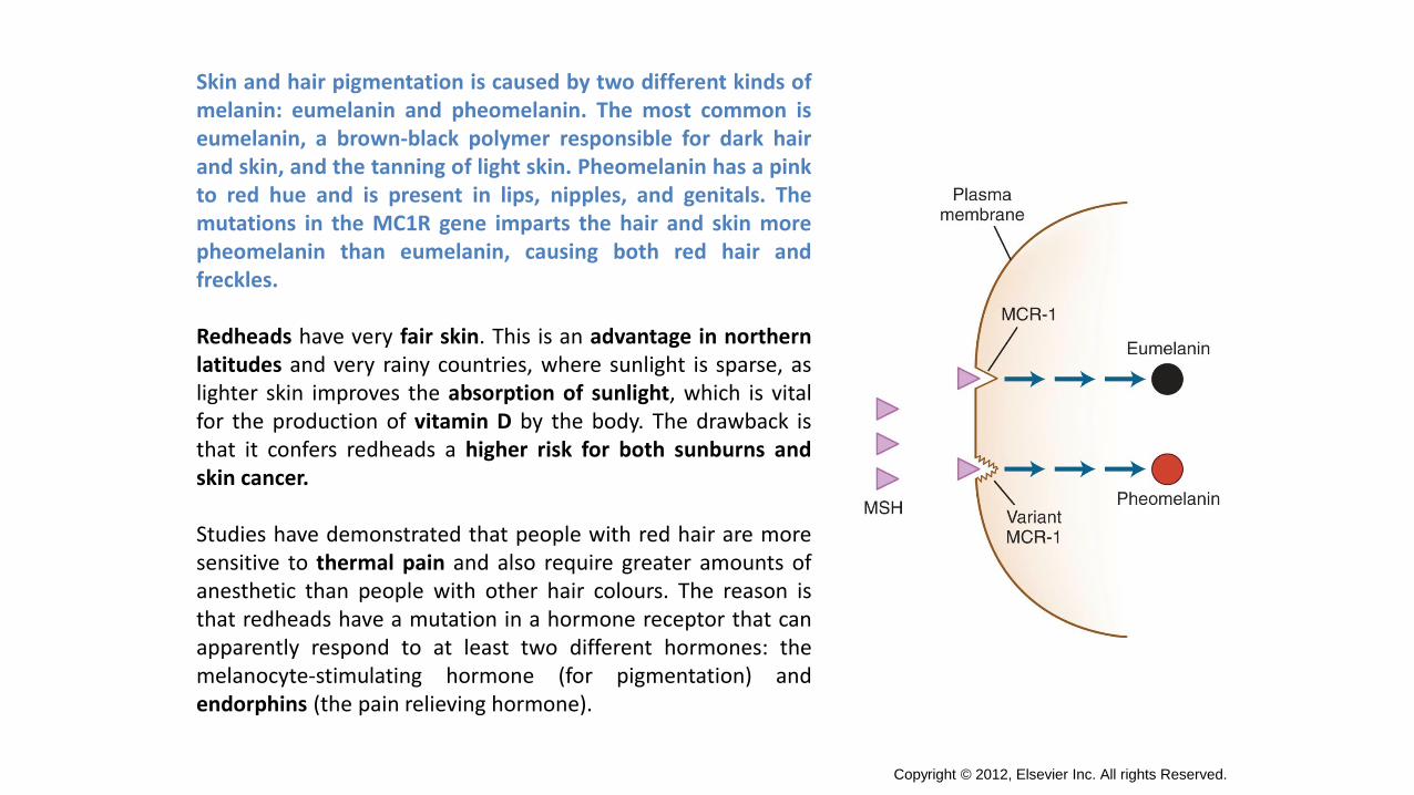
Skin and hair pigmentation is caused by two different kinds ofmelanin: eumelanin and pheomelanin. The most common iseumelanin, a brown-black polymer responsible for dark hairand skin, and the tanning of light skin. Pheomelanin has a pinkto red hue and is present in lips, nipples, and genitals. Themutations in the MC1R gene imparts the hair and skin morepheomelanin than eumelanin, causing both red hair andfreckles.
Redheads have very fair skin. This is an advantage in northernlatitudes and very rainy countries, where sunlight is sparse, aslighter skin improves the absorption of sunlight, which is vitalfor the production of vitamin D by the body. The drawback isthat it confers redheads a higher risk for both sunburns andskin cancer.
Studies have demonstrated that people with red hair are moresensitive to thermal pain and also require greater amounts ofanesthetic than people with other hair colours. The reason isthat redheads have a mutation in a hormone receptor that canapparently respond to at least two different hormones: themelanocyte-stimulating hormone (for pigmentation) andendorphins (the pain relieving hormone).
Copyright © 2012, Elsevier Inc. All rights Reserved.
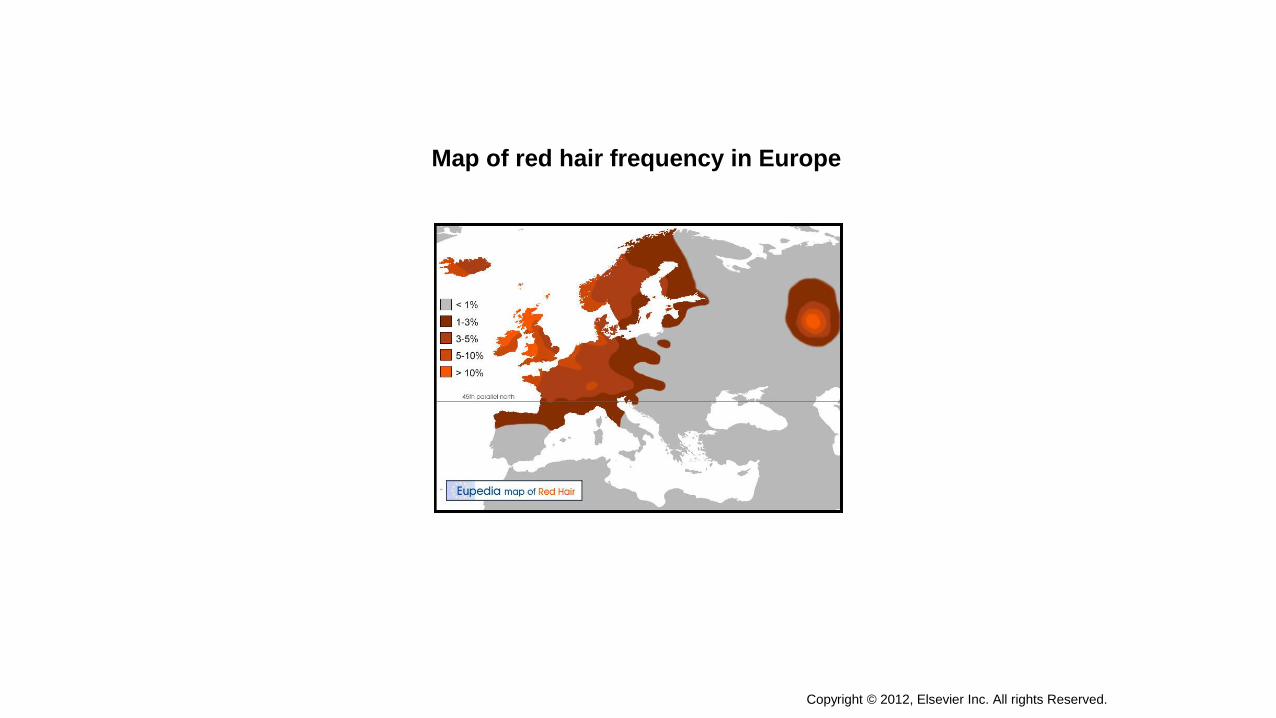
Map of red hair frequency in Europe