
Versione alternativa della tesi di dottorato: “The dilp2/5 ... · Versione alternativa della tesi...
136
Versione alternativa della tesi di dottorato: “The dilp2/5 genes control diapause inducibility” Autore Luca Schiesari Questo documento rappresenta una versione differente rispetto a quello depositato a conclusione del Ciclo di Dottorato. Può presentare semplici correzioni tipografiche, errata corrige, ma anche revisioni dei processi interni e sostituzione di formule. Può presentarsi in formato diverso dall’edizione originale, con layout e impaginazione differenti, e il testo può essere stato riassunto o ampliato. In caso di pubblicazione e in accordo con gli editori, l’autore può sottoporre il testo sotto forma di pre-print o nel formato pubblicato. In ogni caso, ai fini amministrativi, resta immutata e convalidata la versione originale della tesi depositata.
Transcript of Versione alternativa della tesi di dottorato: “The dilp2/5 ... · Versione alternativa della tesi...
Versione alternativa della tesi di dottorato:
“The dilp2/5 genes control diapause inducibility”
Autore
Luca Schiesari
Questo documento rappresenta una versione differente rispetto a quello depositato
a conclusione del Ciclo di Dottorato. Può presentare semplici correzioni tipografiche,
errata corrige, ma anche revisioni dei processi interni e sostituzione di formule. Può
presentarsi in formato diverso dall’edizione originale, con layout e impaginazione
differenti, e il testo può essere stato riassunto o ampliato. In caso di pubblicazione e
in accordo con gli editori, l’autore può sottoporre il testo sotto forma di pre-print o
nel formato pubblicato. In ogni caso, ai fini amministrativi, resta immutata e
convalidata la versione originale della tesi depositata.


Abstract Many holometabolous insects hibernate by triggering diapause, an “actively-induced” dormancy that blocks
developmental functions. Yet, the nature of signals enhancing the plasticity of developmental system and
underlying diapause inducibility is still elusive. We show that the “Insulin/IGF” dilp2/5 genes, encoding for
developmental hormones, antagonize diapause switch in D. melanogaster and their modulation is pivotal in
sensitizing the developmental system to environmental perturbations. Functional impairment of dilp2/5
signaling results in the appearance, or inhibition, of the inducible diapause polyphenism, revealing that they
are at the core of the gene network regulating diapause inducibility, beyond the control of developmental
time. DILP2/5, as dispensable developmental hormones, cover a latent and hidden plasticity of development,
underlying the evolution of an inducible diapause polyphenism through genetic accommodation. Such
hormonal mechanism might be the putative target to bioengineer diapause inducibility.

Abstract (Italian version)
Molti insetti olometaboli innescano la diapausa, una dormienza attivamente indotta che blocca lo sviluppo al
fine di ibernare. La natura dei segnali che aumentano la plasticità del sistema di sviluppo e che sottendono
l’inducibilità della diapausa rimane largamente sconosciuta. Qui, noi riportiamo che dilp2/5, due geni
“Insulin/IGF” simili codificanti per ormoni di crescita, reprimono l’induzione della diapausa in D. melanogaster
e che la loro modulazione è cruciale nel sensibilizzare il sistema di sviluppo alle perturbazioni ambientali.
Modificazioni funzionali di dilp2/5 provocano l’induzione, o l’inibizione, del polifenismo reversibile di
diapausa, rivelando che, oltre il loro ruolo nella modulazione del tasso di sviluppo, questi geni sono al cuore
del network genico che regola la dormienza. DILP2/5, come ormoni di crescita dispensabili per il normale
sviluppo, mascherano una latente plasticità di sviluppo e la loro modificazione può provocare l’evoluzione
della diapausa attraverso accomodazione genica. Questo controllo ormonale potrebbe costituire un
promettente bersaglio per un’ingegnerizzazione genetica dell’inducibilità della diapausa.
Contents Introduction 1
1. Diapause: alternative developmental trajectory 1
2. Embryonic Diapause and hormonal pulses 2 2.1 ECD signaling in embryos 2
2.2 ECD induces diapause in late embryos 2
2.3 ECD fails in diapausing early embryos 3
� � 2.4 Maternal control of embryonic diapause 4
3.Pupal Diapause and Developmental Plasticity 5 3.1 ECD failure induces pupal diapause 6
3.2 Seasonal morphs linked to diapause 10
4. JH/ECD interplay in Larval diapause 12 4.1 Hormonal control of larval life 12
4.2 JH maintains diapause 14
4.3 ECD failure induces diapause 15
4.4 JH/ECD interplay controls larval morphs 15
5.JH/ECD signaling in Imaginal Diapause 16
6. Insulin/IGFs signaling pathway (IIS) 18 6.1 Insulin/IGFs growth factors 18
6.2 Insulin/IGFs signaling pathway 19
6.3 Functions of Insulin/IGFs signaling 20
Scope of the PhD project 23 Results 24
1. Reduced sensitivity to Insulin/IGFs induces diapause 24
2. Loss of MNCs induces diapause 25
3. Loss of dilp2/5 induces diapause 26
4. Overexpression of dilp2/5 antagonizes diapause 27
5. dilp2/5 genes control developmental competence 29
6. dilp2/5 genes are paradoxically up-regulated during diapause 30
7. Downstream IIS impedance during dormancy 31
8. The release of DILP2/5 signals is reduced during diapause 32
9. IIS-Feedback on MNCs modulates diapause 33
10. dilp2/5 genes are hierarchically upstream of JH 34
Discussion 35 1. dilp2/5 genes link diapause to modifications of development 35
2. Evolution of diapause trajectory by genetic accommodation: the cardinal role of dilp2/5 signaling 37
3. Recycling dilp2/ signaling in evolution of diapause? 39
4. Neurosecretion control in inducing alternative development 40
5. Further directions: IGF signaling as putative target of diapause bioengineering 42
Materials and Methods 43 References 52 Figures 68

Abbreviations AEL After Egg Laying APF After Puparium formation CA Corpora Allata or Corpus Allatum CC Corpora Cardiaca or Corpus Cardium DA Dopamine DDC Dopa-Decarboxylase DH Diapause Hormone DILP Drosophila Insulin-like Protein dilp Drosophila Insulin-like Protein genes (Insulin/IGFs genes) ECD Į-Ecdysone 20E 20-Hydroxy-Ecdysone E2 17-ȕ-Estradiol EPPase Ecdysteroid-phospate phosphatase ERK Extracellular signal-Regualted Kinase FoxO Forkhead Box-O IIS Insulin/IGFs signaling or Insulin-like signaling Imp-L2 Imaginal morphogenesis protein-Late 2 INR Insulin/IGFs Receptor or Insulin-like Receptor Insulin/IGFs Insulin/Insulin-like Growth Factors IRS Insulin/IGFs Receptor Substrate Protein JH Juvenile Hormone JHA Juvenile Hormone analogue MNCs Median Neurosecretory Cells NEFs Newly Eclosed Females OPIF Orange-Pupa-Inducing-Factor PCMH Pupal-Melanizing-Hormone PG or PGs Prothoracic Glands PTTH Prothoracicotropic Hormone RTK Receptor Tyrosine Kinase SMPH Summer-Morph-Producing-Hormone SOG Sub-Oesophageal Ganglion SDH2 Sorbitol Dehydrogenase-2 TGF-ȕ Transforming Growth Factors ȕ TH Tyrosine-Hydroxylase TOR Target of Rapamycin TS Target Size

Gal4 drivers specific for the MNCs dilp2> since the late third (last) larval instar under temporal control of dilp2 gene promoter dilp2(p)> since early larval life (2nd instar) under temporal control of dilp2 gene promoter dilp3> since post-larval stages under temporal control of dilp3 gene promoter
Definitions Imaginal discs: Imaginal discs are sacs of cells which are the primordia of adult parts (such as wings and legs).
Canalization of Development: Developmental canalization is the inherited genetic buffering that stabilizes the phenotype and decreases its variability. Canalized developmental systems produce the same phenotype despite environmental and genetic perturbations. Canalization evolves under stabilizing selection in order to confer robustness to the developmental system. Canalization is evident, as examples, in the low penetrance of null or hypomorphic mutations, or in the absence of “genotype-by-environment interaction” (Hornstein and Shomrom 2007; Moczek 2007).
Robustness: Robustness of developmental systems is the resistance to genetic and environmental perturbations, resulting from the action of evolutionary and genetic mechanisms (Hornstein and Shomrom 2007; Moczek 2007).
Developmental Plasticity (Environmental Sensitivity or Flexibility): Developmental plasticity is the ability of a developmental system to react to endogenous or exogenous environmental perturbations with a change in form, state or physiology. Plasticity may or may not be adaptive. In the last case it is a consequence of natural selection (Moczek 2007; West-Eberhard 2003).
Cryptic Genetic Variation: Cryptic (or hidden) genetic variation is the genetic variability that is invisible to natural selection since it does not produce phenotypic variants. Thus, individuals within a population can be genetically different without exhibiting phenotypic differences. Cryptic genetic variation may be exposed under specific conditions (during the evolutionary process of genetic accommodation). Cryptic genetic variation may be due to strong epistatic interactions among loci, developmental canalization or genetic capture of genetically linked traits (Moczek 2007; West-Eberhard 2003).
Sensitizing Mutations: Sensitizing mutations are mutations in hormonal regulatory pathway which lowers the levels of a developmental hormone, in such a way that an environmental perturbation decrypts the genetic variation to selection (Suzuki and Nijhout 2006; Nijhout 2003).
Genetic accommodation: Genetic accommodation is a mechanism of evolution wherein a novel phenotype introduced through a mutation is molded into adaptive phenotype through quantitative genetics changes. Genetic accommodation results in an increased environmental sensitivity of a plastic phenotype. (Suzuki and Nijhout 2006).
Polyphenism: Polyphenisms are adaptations in which the same genotype produces inducible discrete alternative phenotypes in transient, or stable, different environments. In this case, the phenotypic plasticity is discontinuous. Polyphenisms are adaptations to reliable and predictable variations in the environment. The polyphenism-inducing environment may not coincide with the selective one. Diapause is an example of adaptive polyphenism. In nature, the phenotypic discontinuity of polyphenisms can be produced by discrete developmental switches or by discontinuities in the inducing environment that uncover only a small portion of a continuous reaction norm (Nijhout 2003).
Reaction Norm: Reaction Norms are adaptations in which the same genotype produces inducible continuous alternative phenotypes in transient, or not, different environment. In this case, the phenotypic plasticity is continuous (Nijhout 2003; West-Eberhard 2003).

Token stimulus: Token stimulus(i) is the environmental variable that induces the alternative phenotype. It is exploitable as predictor of the selective environment (i.e. an adverse season) of the polyphenism. The token stimulus is not, in itself, a stressful condition. Usually, the token stimulus reprograms development by changing either the hormone secretion or the pattern of hormone sensitivity (hormone sensitive period), resulting in the execution of the inducible alternative developmental trajectory (Nijhout 2003; Saunders et al. 2002).
Antagonistic pleiotropy: Different pleiotropic fitness effects of a trait or a gene are opposite in sign, positive in one context of expression and negative in another (West-Eberhard 2003).
1��
Introduction
1. Diapause: alternative developmental trajectory
Holometabolous insects (such as the genetic models Bombyx mori and Drosophila melanogaster)
undergo deep metamorphosis by developing from immature larval phases to an imaginal one
(Fig. 1) (Gilbert 2012; Gilbert 2009; Dubrovsky 2005; Truman and Riddiford 2002; Riddiford
1993). Hormonal pulses set the timing of transition trough each stage; yet, the timing of these
hormonal inductions fails when insects trigger diapause to hibernate (Denlinger et al. 2012;
Schiesari et al. 2011; Saunders et al. 2002).
Diapause is an “actively-induced” dormancy that blocks developmental functions to precede the
adverse season. Diapausing phase is genetically specified but it is elicited by environmental
factors (mainly the seasonal changes of photoperiod) perceived during the earlier developmental
stages (Kostal 2012; Saunder and Bertossa 2011; Saunder 2010). Thus, diapause is hormonally
programmed in advanced of its onset (Denlinger et al. 2012; Schiesari et al. 2011; Saunders et al.
2002).
Once dormancy is induced, the diapausing entity needs a “genetically-specified” period of chilling
prior to be re-activated and acquire the competence to develop in optimal environment (such as in
dormant pupae of Samia cynthia, which need 3-5 months below 4°C) (Denlinger et al. 2012;
Nakamura et al. 2011; Hahn and Denlinger 2007; Denlinger 2002). Dormant entities become
extremely resistant to low temperatures by undergoing supercooling (resistance to low
temperatures without freezing by reducing the freezing point of body fluids) or freezing (Denlinger
et al. 2012; Hahn and Denlinger 2007;Saunder et al. 2002; Lee and Denlinger 1999). As example,
pupae of the Papilionidae Papilio machaon reach temperatures of -25°C after which they freeze to
-30°C, still remaining alive for months (Shimada 1980). As well, larvae of Lyamantridae
Gymnaephora groenlandica undergo supercooling until -7°C, but they can freeze until -70°C
(Kukal et al. 1988). Embryos of the Bombycidae Bombyx mori resist until -32°C for months only
by supercooling (Suzuki et al. 1983).
Frequently, appearance of alternative morphs (polyphenism) is deeply linked to diapause
induction undergoing modulation of common hormonal signaling, as reported for the embryonic
morphs in Orgyia thyellina, for “immaculate” larvae in Diatrea grandiosella, or for the pupal
pigmentation in Papilio xuthus. Many seasonal morphs are also determined during diapause
development, such as wing diphenism of many Lepidopterans (i.e. Araschnia levana, Papilio
xuthus), or the “dark/red-spotted” morphs of larvae hatched from diapausing eggs in Orgyia
thyellina (Denlinger et al. 2012; Saunders et al. 2002). Thus, diapause is a dynamic modulation of
all holometabolous phases since its hormonal induction orchestrates as well functions linked to all
developmental transitions. Diapause is not simply a block of development but, rather, a dynamic
and plastic process (Denlinger et al. 2012; Saunders et al. 2002).

2��
2. Embryonic Diapause and hormonal pulses
Embryonic (or egg) diapause may arrest embryogenesis at any stage. Some species diapause as
early embryo, others overwinter as pharate first instar larvae within eggshell (Denlinger et al.
2012; Saunders et al. 2002). Thus, embryonic diapauses are enormously diverse in regulation,
albeit the nature of hormonal signaling is almost the same. Steroid hormone Ecdysone (ECD) is a
key regulator of insect diapauses, although its action (repressive or promoting) depends by the
stage at which dormancy is induced (Denlinger et al. 2012; Saunders et al. 2002). A simplified
scheme of the production of ECD peaks is shown in Fig. 2.
2.1 ECD signaling in embryos
In holometabolous insects, a large ECD pulse sustains embryonic development through mid-early
embryogenesis until later stages. Hence, it decays allowing embryos to hatch, once development
is completed (Truman and Riddiford 2002).
Loss of ECD causes developmental aberrations. In both Drosophila melanogaster and Bombyx
mori, mutant embryos for “Halloween” genes (i.e. shade, disembodied, shadow, ecc. - which
encode for enzymes involved in ECD biosynthesis) lack of ECD and they exhibit defects in head
involution and dorsal closure of midgut. Ultimately, these embryos die during late embryogenesis
and they fail to hatch as first instar larvae (Niwa et al. 2010; Ono et al. 2006; Yoshiyama et al.
2006; King-Jones and Thummel 2005; Gilbert et al. 2004; Warren et al. 2004; Petryk et al. 2003;
Chàvez et al. 2000). Thus, the timing of ECD peak plays a key role in driving embryonic
transitions; yet ECD controls diapause during specific developmental stages, without causing any
growth aberrations (Denlinger et al. 2012; Saunders et al. 2002).
2.2 ECD induces diapause in late embryos
The gypsy moth, Lymantria dispar, diapauses before hatching as pharate first instar larva (fully
formed) until spending the mandatory chilling period. Enhanced levels of ECD block pharate
larvae in dormancy. In fact, diapause induction fails when embryos are injected with KK-42, an
“ECD-inhibitor” thought to block ECD biosynthesis. However, this effect is reversed by ectopic
applications of ECD. In contrast, diapausing pharate larvae fail to resume competence at the end
of chilling when treated with ECD, remaining dormant. In line, “Non-Diapausing” mutants
(genetically deficient of dormancy) elicit diapause after exposure to exogenous ECD, revealing a
latent hormonal responsiveness. Since ECD control developmental transitions, it is a key gate of
signaling to elicit dormant phase. In dormant pharate larvae of Lymantria, high levels of ECD are
produced by developing Prothoracic Gland (PG, a neurohaemal gland) and this activity persist
until dormancy breaking, when ECD drops. Conversely, PG of non-diapausing larvae fails to
synthesize ECD beyond early embryogenesis provoking, in turn, larval hatching (Lee et al. 2002,
1997; Lee and Denlinger 1997, 1996; Suzuki et al. 1993). A similar mechanism is thought to
control embryonic diapause in the European skipper Thymelicus lineola (McNeil and Fields 1985).

3��
Similarly, enhanced ECD pulse sustains diapause in pharate first instar larva of the silkmoth
Antheraea yamamai (Suzuki et al. 1990). The excision of thorax/head complex (where PG is
located) from diapausing pharate larvae induces isolated abdomen to resume growth, unlike the
thorax/head complex. Moreover, ECD inhibition (by injecting KK-42) in dormant Antheraea
embryos breaks dormancy and resumes growth (Suzuki et al. 1990). Thus, timing of ECD pulse is
key to induce and sustain diapause, by avoiding the inductive processes of late embryogenesis.
2.3 ECD fails in diapausing early embryos
The silkworm, Bombyx mori, triggers diapause as early embryo (immediately after mesoderm
segmentation). At the onset of diapause, glycogen is converted into sorbitol and glycerol which
function as cryoprotecting agents during chilling at -28 to -32°C. Sorbitol plays as well regulative
functions, since it elicit dormancy when exogenously applied to “non-diapausing” embryos and its
removal resumes growth in dormant ones. Hence, dormant embryo needs a period of chilling
(<5°C for 2-3 months) to reduce sorbitol prior to end diapause (Horie et al. 2000). Once diapause
ends, ERK/MAPK pathway promotes sorbitol-glycogen conversion and ECD synthesis by
activating two key enzymes, sorbitol dehydrogenase-2 (SDH2) and ecdysteroid-phosphate
phosphatase (EPPase). On this early stage, embryo lacks of developed PG and the steroid
sources available are only made by the maternal Inactivated Ecdysteroid-Phosphates (IEPs).
Then, EPPase activates IEPs eliciting ECD pulses to de-repress development in dormant embryo.
Subsequently, embryo develops following a normal embryogenesis (Fujiwara et al. 2006a, 2006b,
2006c; Iwata et al. 2005; Horie et al. 2000).
Intriguingly, steroids control also diapause in Vertebrate embryo. The South American annual
Killifish, Austrofundulus limnaeus, lives in ephemeral ponds that seasonally undergo fast
desiccation. Survival of the species depends entirely from buried embryos that hatch during the
next rainy season once the ponds are re-inundated (Berois et al. 2012; Podrabsky et al. 2007,
2001; Podrabsky and Hand 1999). Killifish embryos survive by eliciting diapause that blocks
development at three diverse stages: early embryo (diapause I), at 38-somites embryo (diapause
II, embryos have beating heart, optic cups and developing CNS) or prior to the hatching (diapause
III, embryo is fully developed) (Berois et al. 2012; Podrabsky et al. 2010) (Fig. 3). Non diapausing
embryos diverge from diapausing ones in higher pulses of estrogens (17-ȕ-estradiol, E2) at 5/10-
somites stage as well as in the timing of traits which do not develop until several days after
diapause end (i.e. melanocytes, vasculature of yolk, otholite primordia). Diapausing embryos
restore development when incubated with E2 without reporting developmental anomalies.
Diapause is imposed to embryos by estrogen levels of the mother: older Killifishes elicit lower
levels of E2 and they lay more diapausing embryos than younger ones (Pri-Tal et al. 2011).
Steroids might also control diapause in the African Killifishes, Nothobranchius guentheri and
Nothobranchius korthausae, in which diapause evolved independently (Pri-Tal et al. 2011;
Murphy et al. 1997; Levels et al. 1986; Inglima et al. 1981).

4��
In sum, diapausing disruption of the timing of ECD pulse preserves insect embryo from the
inductive phenomena of morphogenesis; hence, development can restart after dormancy
(Denlinger et al. 2012; Saunders et al. 2002). An analogous control re-emerges in Vertebrate
diapausing embryos, still under control of steroid hormones. Hence, the bioengineering of
hibernation in embryonic/fetal stage should act before the key inductions of development by
preventing the hormonal signals that trigger the systemic changes of growth.
2.4 Maternal control of embryonic diapause
Likewise in Killifish, diapause is imposed on B. mori embryos inside mother by Diapause
Hormone (DH), a 24-amino acid protein produced by Sub-Oesophageal Ganglion (SOG) released
in haemolymph via the Corpora Cardiaca Neurohaemal gland (Shiomi et al. 2007; Yamashita et
al. 2001; Sato et al. 1998, 1994, 1993; Suwan et al. 1994; Yamashita 1996). DH is regulated by
maternal experience of environment: larvae grown in summer (under long days and high
temperatures) eclose in autumn as moths laying diapausing embryos (“diapause” moths) by
producing high levels of DH (Fig. 4) (Shiomi et al. 2007; Sato et al. 1994, 1993; Nakagaki et al.
1991; Fukuda 1951; Hasegawa 1951). DH signals in developing gonads through a G-coupled
receptor, DHR, to enhance glycogen (then, sorbitol and glycerol) inside the eggs (Homma et al.
2006; Horie et al. 2000). “Non-diapause” pupae injected with DH develop as moths laying
diapausing eggs, suggesting the existence of a sensitive phase during pupal life (Uehara et al.
2011). In addition, “diapause” moths lay developing embryos when SOG is excised from pupal
life, whereas “diapause” moths fail to lay diapausing eggs after implant (at pupal stage) of the
SOG of “non-diapause” ones (Fukuda 1951).
In “diapausing” animals, exceptional peaks of BmDH gene expression occurs during both the mid-
late larval instars (4°/5°) and the pupal life (Morita et al. 2004; Xu et al. 1995a) (Fig. 4), under
control of transcription factors of the pituitary homeobox gene family (i.e. Ptix1, a bicoid-like
homeobox transcription factor that binds cis-regulatory elements in the promoter region of BmDH)
and members of the POU transcription factor family (Shiomi et al. 2007; Zhang et al. 2004; Xu et
al. 1995b).
During late larval and early pupal life, Ddc gene (encoding for Dopamine Decarboxylase, key
enzyme in Dopamine synthesis) is overexpressed in “diapausing” animals, coherently with high
levels of Dopamine (DA) released into haemolymph. “Non-diapausing” larvae of B. mori fed with
L-DOPA (the DA precursor) develop as moth which exhibit high levels of DH and lay diapausing
embryos, similarly to those injected with DA. Thus, DA control DH signaling and functions
(Noguchi and Hayakawa 2001).
In addition, “non-diapause” moths injected with 2O-Hydroxyecdysone (20E, the active form of
ECD) lay giant developing eggs (larger and heavier than normal), similarly to diapausing ones
(Kawaguchi et al. 1989). Synergism between ECD and DH may regulate diapause: ECD dosage
modulate embryonic diphenism; whereas DH imposes dormancy.

5��
Similarly, DH elicits embryonic diapause in the tussock moth Orgyia thyellina which exhibits as
well seasonal morphs linked to dormancy. Larvae grown at long days emerge as winged
Macropteres (summer morph), but those developed at short days eclose as Brachypteres with
short wings (autumnal morph). Moreover, pupae exhibit diphenism: pale morph for the
Macropteres and dark one for the Brachypteres. Macropteres lay developing eggs whereas
Brachypteres lay diapausing embryos that are heavier and darker (Kimura and Masaki 1977)(Fig.
5). “Macropteres pupae” injected with DH develop as moths that lay diapausing embryo whereas
“Brachypteres pupae” injected with anti-DH (inhibiting hormone signaling) lay developing eggs
once they eclose (Uehara et al. 2011). These results may also mean that both pupal and moth
diphenism in Orgyia is specified by an earlier action of DH or by an unknown hormone during
larval life, since pupal DH affect only embryonic morph (Uehara et al. 2011). Similarly, dormancy-
linked diphenism occurs in B. mori where moth laying diapausing embryos exhibit many brown
scales on the wings, and diapausing embryos have thick chorion and dark pigmentation
(Tsumaraki et al. 1999). In such species, non-diapause-fated pupae (short days, SD) develop into
autumn moths or intermediate morphs (typical of diapausing moths developed under long days,
LD) and not into correct summer ones, when the brain is removed from early pupal life. Yet, the
same microsurgery has not effects on “diapause-fated” pupae which develop normally into
autumn morphs. Similarly, “non-diapausing” pupae (SD) develop as autumn moths (LD) rather
than summer ones when they “receive” the brain of “diapause-fated” (LD) larvae, during the early
5th instar life. In addition, injection of DH induces “non-diapausing” (SD) pupae to develop into
autumn or intermediate morphs, revealing the role of DH in the control of polyphenism in B. mori,
perhaps, through direct signaling into imaginal wing discs (Yamanaka et al. 2000b) (Fig. 6).
In sum, diapause is strictly linked to the appearance of seasonal morphs, under a common
hormonal control.
3.Pupal Diapause and Developmental Plasticity
Metamorphosis (transition larva-to-pupa) is initiated and sustained by “time-established” pulses of
ECD. Both production and release of ECD is mainly regulated by a small neurohormone, known
as Prothoracicotropic Hormone (PTTH), which peaks earlier than ECD setting the timing of
development in response to developmental and environmental cues (Smith and Rybczynski 2012;
Yamanaka et al. 2012; Rybczynski 2009). Once released into the haemolymph (via neurohaemal
glands), PTTH targets the larval Prothoracic Gland to elicit the ECD pulse required for the onset
of metamorphosis (Smith and Rybczynski 2012; Dubrovsky 2005; Truman and Riddiford 2002;
Mizoguchi et al. 2001, 2002; Riddiford 1993). Later, an additional pulse of ECD sustains
metamorphic transition by promoting the prepupal-to-pupal transition (corresponding to the “head
eversion” stage in Drosophila melanogaster) (Smith and Rybczynski 2012; Rybczynski 2009;
Mizoguchi et al. 2001, 2002;Dai et al. 1995; Riddiford 1993)(Fig. 7).
Disruption of ECD signaling during pupal life causes several developmental defects (Mou et al.
2012; Rewitz et al. 2010; McBrayer et al. 2007; Delanoue et al. 2010; King-Jones et al. 2005;
6��
Bialecki et al. 2002; Broadus et al. 1999; Lam et al. 1999; White et al. 1997) (Fig. 7); yet,
diapause orchestrates ECD and development without provoking any aberrations (Denlinger et al.
2012; Hahn and Denlinger 2010; Schiesari et al. 2011; Denlinger 2002; Saunders et al. 2002).
3.1 ECD failure induces pupal diapause
Diapause prolongs pupal life by inhibiting ECD peaks and, in turn, blocking the ongoing
metamorphosis (Denlinger et al. 2012; Schiesari et al. 2011; Saunders et al. 2002). In these
conditions, the pupa tolerates extremely freezing environment by supercooling at temperatures as
low as -20°C (Lee and Denlinger 1999). As example, dormant pupae of Papilio machaon can
reach temperatures of -25°C after which they freeze to -30°C, still remaining alive for months
(Shimada 1980). Dormant pupae need to be chilled to resume the competence for breaking
diapause; once chilling of winter is ended, they resume normally metamorphic growth when
exposed to optimal conditions (Denlinger et al. 2012; Saunders et al. 2002).
Failure of the ECD peak induces pupal diapause in the hornworm moth, Manduca sexta, exposed
to short days. “Diapause-fated” larvae exhibit no differences in ECD levels compared to the
developing ones through all last larval phases (5th instar, apolysis and pupation) although
photoperiodic perception occur as far back as the first larval instar. Yet, ECD pulse falls in
diapausing pupae avoiding the metamorphic progression through pharate stage (Fig. 8) (Smith et
al. 1986; Bowen et al. 1984, 1985). According to this scenario, injections of ECD agonist
(RH5849) breaks pupal dormancy reactivating the metamorphic growth (Sielezniew and
Cymborowski 1997).
Similarly, the failure of ECD signaling induces pupal diapause in many other insect species,
including Pieris brassicae (Calvez 1976), Mima tiliae (Highnam 1958), Hyalophora cecropia
(Roxström-Lindquist et al. 2005; Williams 1952, 1946), Antheraea mylita (Mishra et al. 2008),
Helicoverpa zea and Heliotis virescens (Zhang and Denlinger 2012; Loeb 1982), Mamestra
brassicae (Islam et al. 2005; Agui 1975) and Mamestra configurata (Bodnaryk 1985), Samya
cynthia (Williams 1968). Consistent with the concept that the absence of ecdysteroids is central in
inducing and sustaining the diapause phase, injections of 20E or ECD-analogous break pupal
dormancy in many insect species including Mamestra configurata (Bodnaryk 1985), Antheraea
mylita (Mishra et al. 2008), Samya cynthia (Williams 1968), and Helicoverpa zea (Zhang and
Denlinger 2012). In P. brassicae, the profile of ECD doesn’t change in 5th last instar larvae
programmed for diapause, resembling the typical pattern of developing larvae; yet, ECD pulse is
elicited only in early non-diapausing pupae. As expected, injections of ECD in dormant pupae
resumes development by breaking dormancy, while removal of Prothoracic gland induce a
permanent pupal diapause (Pullin and Bale 1989; Calvez 1976).
The flesh fly (higher Dipetera), Sarcophaga argyrostoma, enters pupal diapause immediately after
head eversion (inside the puparium), after being stimulated by short days as intra-uterine
embryos and young larvae. In the “Developing” pupae (raised under long days) following the
pupariation, ECD pulses in two distinct time: a first ECD pulse sets both pupariation and pupation,

7��
the other one drives the pharate adult development. Conversely, “diapausing” pupae (raised
under short days) exhibit the prepupal/pupal pulse, but they fail to generate the second ECD peak
as the pupae trigger dormancy (Richard et al. 1987).
Since the failure of ECD pulses blocks metamorphosis, the shutdown of PTTH signaling (PTTH
synthesis or release) is crucial to elicit diapause by avoiding the stimulation of ECD pulses
(Denlinger et al. 2012; Smith and Rybczynski 2012; Schiesari et al. 2011; Saunders et al. 2002).
In M. sexta, MsPtth gene (encoding PTTH) is normally regulated along larval life, albeit its
expression changes during the pupal phase in relation to diapause: it is strongly downregulated in
diapausing pupae and overexpressed in developing ones (Xu and Denlinger 2004). Similarly, Ptth
expression is downregulated in diapausing pupae of Heliothis virescens (Xu and Denlinger 2003)
and Helicoverpa armigera (Wei et al. 2005).
Since the shutdown of PTTH signaling is crucial to block ECD pulse and antagonize dormancy,
the gain of function of PTTH by injecting rPTTH (recombinant PTTH) terminates pupal dormancy
in noctuids of Heliothis/Helicoverpa complex (Wei et al. 2005). Still, in both M. sexta and
bombycidae Anthaerea pernyi, injections of exogenous PTTH resumes the metamorphic growth
in diapausing pupae as well in brain-less dormant ones that are blocked in permanent diapause
(Shionoya et al. 2003, Sauman and Reppert 1996). The failure of PTTH signaling is thought to
induce pupal diapause in Hyalophora cecropia (Denlinger et al. 2012; Smith and Rybczynski
2012; Williams 1952, 1946) and Samya cinthyia (Denlinger et al. 2012; Smith and Rybczynski
2012; Williams 1968). In fact, the implantation of a “chilled-activated” brain into “brainless” (brain
surgically removed) diapausing pupae of the Cecropia giant-silkworm causes the termination of
diapause, (Williams 1952, 1947, 1946). This experiment is in line with the model in which the
pupal brain, when chilled, becomes competent to release PTTH which, in turn, restore the ECD
pulses needed to antagonize pupal diapause (Denlinger et al. 2012).
Without the brain, the diapausing pupae remain still alive into a state of permanent dormancy, in
which they are unable to reactivate metamorphosis. Since PTTH pulses play key role in
promoting pupal/adult transition shortly after time of pupation, decerebration prior to PTTH
release locks pupae of giant silkworm, Hyalophora cecropia, into a permanent “diapause-like”
state. Similarly, decerebration of Manduca sexta, Pieris rapae, Pieris brassicae and Antheraea
polyphemus induces a “permanent diapause” albeit only within the first month of diapause. After
this time, diapausing pupae of these Lepidopteran species can break dormancy and complete
metamorphic development even in absence of their brains (Judy 1972; McDaniel and Berry 1967;
Wilson and Larsen 1974; Maslennikova 1970; Kind 1976). Remarkably, diapausing pupae of the
Helicoverpa zea remain permanently dormant when the brain is removed within the first 4 hours
since pupation. However, the “brainless” diapausing pupae are independent from the brain and
they have all the potential to resume metamorphosis after chilling, when debrained after 24 hours
(Denlinger et al. 2012; Meola and Adkisson 1977). This phenomenon depends on the
autonomous role of the Prothoracic glands (PGs) that become independent by neural secretions.
In fact, the PGs of dormant pupae can undergo progressive and spontaneous re-activation,

8��
assuming functions of a regulatory organ, depending on pupal age. Once chilled, PGs can
autonomously resume metamorphic growth, in a “species-specific” manner (Denlinger et al. 2012;
Denlinger 2002; Saunders et al. 2002). According to this model, PG of debrained diapausing
pupae of Papilio xuthus is directly activated by cold (Ozeki 1954) and the PG of Samia cynthia
larvae retains a high degree of independence from the brain (Mizoguchi and Ishizaki 1982). The
autonomous role of PG is well supported also by its refractoriness to PTTH during pupal
dormancy, depending on the age of the diapausing pupae (Denlinger et al. 2012; Saunders et al.
2002). In Manduca sexta, the PG of diapausing pupae becomes independently responsive to
environmental stimuli and it enters a state of a diapause-programmed refractoriness to PTTH
signal from the day of larva-to-pupa molt, perhaps as result of inductive events occurring in the
late larval stages (Bowen et al. 1985, 1984). A similar refractoriness of PG occur also in
Mamestra brassicae (Agui 1975) and Pieris brassicae (Calvez 1976).
With respect to this hormonal integration, it is possible that an inhibitor factor synergizes with the
PTTH failure to induce both diapause and PG refractoriness. Under this scenario, the time of
chilling may remove the hormonal block resuming the competence to reactivate the metamorphic
growth, gradually with the age of diapausing pupa (Denlinger et al. 2012; Schiesari et al. 2011).
Dopamine (DA) may be this unknown factor since it antagonizes metamorphic growth by inducing
diapause. In fact, larvae of M. brassicae developing as diapausing pupae exhibit levels of
haemolymphatic dopamine 4 times higher than non-diapausing ones, since the molt period. As
well, dopamine strongly increases in dormant P. brassicae pulsing on day 3 of pupal life too
remaining high trough all diapausing phase. “Non-diapausing” larvae (at last larval instar) fed with
L-DOPA (a DOPA precursor) develop as diapausing pupae (Isabel et al. 2001; Puiroux et al.
1990). Interestingly, injection of DA induces “developing” pupae of B. mori to develop as moth
which exhibit an up-regulation of BmDH gene and lay dormant embryos, revealing a conserved
function of DA signaling in the evolution of diapause. Similarly, non-diapause-type silkworms fed
with L-DOPA during the final larval instar developed as moths laying diapausing embryos
(Noguchi and Hayakawa 2001). Notably, haemolymphatic DA steadily decreases in diapausing
pupae of the silkmoth Antheraea pernyi exposed to chilling (Matstumoto and Takeda 2002). This
suggests that DA may be a chilling-removed block which arrests pupal development once
dormancy is induced. The chilling period may remove DA signaling, allowing the downstream
PTTH to elicit its anti-diapause function and re-activate the post-diapause growth.
In addition to PTTH, Diapause Hormone (DH) signaling is required to reactivate post-diapause
development, since it can resumes metamorphic growth in the “not-chilled” dormant pupae
(competence is still lost) of noctuids belonging to the Heliothis/Helicoverpa complex (Heliothis
virescens, Helicoverpa armigera, and Helicoverpa zea) (Denlinger et al. 2012; Schiesari et al.
2011). As in Bombyx mori (see above), DH gene of these heliotine species is expressed in
Neurosecretory cells (DHPCs) of the SOG and it encodes for the Diapause Hormone (DH), which
is a 22-amino acid peptide with a FXPRL motif at the C-terminus (X=G, T, V, S, I), released via

9��
Corpus Cardium (CC) in the haemolymph (Sun et al. 2005; Zhang et al. 2004b, 2004c; Sun et al.
2003).
Indeed, injection of DH-like peptides in “non-chilled” diapausing pupae of these noctuids ends
promptly dormancy in a dose-dependent manner, revealing that DH is pivotal for a diapause
escape (Zhang et al. 2011; Zhang et al. 2008; Zhang et al. 2004b, 2004c; Xu and Denlinger
2003). However, DH has a temperature-dependent action: it is unable to arrest diapause when
injected in dormant pupae exposed to 20°C, but does so in those shifted to 25°C (Zhang et al.
2004b, 2004c); yet, whether this occurs through direct temperature control on DH or through PG
responsiveness to DH, is still unknown. DH also induces Ecd in the PG, suggesting that DH/Ecd
co-operation terminates diapause (Schiesari et al. 2011).
According to the “anti-diapause” role of DH, HzDH gene declines on day 7 of pupal diapause in H.
zea, whereas it is upregulated in metamorphic pupae. When one year old “chilled” pupae
(activated) perceive optimal temperature (25°C), they break promptly dormancy by increasing the
DH gene expression (Zhang and Denlinger 2012). Similarly, DH gene expression falls down in
diapausing pupae of M. sexta at day 9 of pupal life, whereas it persists in developing pupae with
exceptional and transient drop on day 3 (stage in which pupae are progressing through pupal-to-
pharate stage) (Xu and Denlinger 2004).
In sum, DH seem to synergistically work with PTTH, upstream of ECD signaling, to reactivate the
metamorphic growth in dormant pupae, and produce a fine regulation of hibernating functions
(Denlinger et al. 2012). The conserved role of DH in the reactivation of post-diapause
development in a variety of Lepidotera indicates that this hormone play a crucial role in
reactivating post-diapause growth of the pupa and, together to PTTH, it might help to reveal the
evolutionary dynamics of diapause evolution (Schiesari et al. 2011). In addition, non-diapause
fated larvae of H. armigera delay development and, as consequence, increase the time dedicated
to the storage of energy for pupal diapause when they are injected with DH (Sun et al. 2005).
Similarly, in the silkworm Bombyx mori, individuals that are programmed to lay diapausing eggs,
prolong the feeding period during the larval stage in relation to enhanced levels of BmDH gene
expression (Xu et al. 1995a). These DH effects on larval development suggest an additional role
for this hormone in the modifications of the holometabolous growth which precede diapause onset
(Schiesari et al. 2011).
In sum, a comprehensive model of pupal diapause might involve the Dopamine repression of
metamorphic growth acting synergistically with the failure of PTTH/ECD signaling cascade.
Hence, the chilling period may be crucial to remove DA inhibition and restore the ECD pulse and
DH signaling. Similar regulation might occur in the Cecropia giant silkworm in which the surgical
implantation of the brain from “chilled activated” pupae (committed to end dormancy, see above)
into brainless diapausing pupae breaks the “permanent” dormancy and resumes metamorphosis
(Denlinger et al. 2012; Smith and Rybczynski 2012; Williams 1952, 1947, 1946).

10��
Blockage of ECD pulse is clearly at the core of the mechanism disrupting pupal metamorphosis;
yet, disruption of ECD may affect collaterally the timing of other phenomena that are hormonally
linked to diapause (Schiesari et al. 2011; Saunders et al. 2002).
3.2 Seasonal morphs linked to diapause
Diapause is strictly linked to the control of seasonal polymorphism (polyphenism), a phenomenon
extremely widespread among insects (Hartfelder and Emlen 2012; Nijhout 2012, 2003; Schiesari
et al. 2012; Saunders et al. 2002). In Hylophila prasinana, green silver-lined prasinana moth
emerges from dormant pupae, whereas “non-diapausing” pupa develops hongarica form, which is
so diverse from the first one that it was thought a different species (Danilevksii 1965). In Lycaena
phlaeas, the autumnal moths differ from the summer black-pigmented ones (developed from
diapausing larvae) in carrying wings with an orange background with black spots and a narrow
marginal band (Sakai and Masaki 1965). Further examples are reported for many Lepidoptera
such as the nymphalids Lycaena phlaeas (Endo and Kanata 1985; Sakai and Masaki 1965) and
Polygonia c-aureum (Saunders et al. 2002; Endo et al. 1988), in the pierid Colias eurytheme
(Hoffmann 1974, 1973; Watt 1969) and Eurema hecabe mandarina (Saunders et al. 2002), and
the bombycidae moths belonging to Orgyia genus (Uheara et al. 2011, Kimura and Masaki 1977).
The moth Pieris napi exists only as venosa morph (with heavy black scaling on the veins of the
hind wings) in the inland of California where this moth don’t need to enter pupal diapause (due to
optimal environment). Yet, they have the latent potential to develop as dormant pupae from which
the lighter castoria morph emerges, revealing a deep functional link between diapause and
imaginal development (Shapiro 1977, 1975).
ECD signaling plays a key role in this phenomena, as reported for the nymphalid Araschnia
levana (Koch and Bückmann 1987). Last instar caterpillar can develop in two butterfly forms:
levana and prorsa. Under short days of autumn, larvae develop into dark dormant pupae from
which levana morphs (orange/dark with black spots) eclose in spring, once dormancy ends.
Larvae grown at long days (in summer) produce light “developing” pupae from which the white
and black prorsa forms emerge. Injection of 20E in 3-days old diapausing pupae (levana) breaks
promptly dormancy and allows to develop prorsa moths. Conversely, injections of 20E in 14 days-
old levana pupae breaks promptly dormancy causing the development of the correct levana form.
In line, “non-diapausing” pupae (prorsa) develop normally when injected with 20E on day 3 of
pupal life. Yet, when prorsa pupa are PG-excised on day 3 and subsequently injected with 20E on
day 14, they emerge as levana morph. Thus, the timing of ECD release in haemolymph regulates
both dormancy and imaginal morphs. In line, when 1/1.5-days old prorsa pupae (“non-diapausing)
are linked in parabiosis to 2/8-days or 5-months old levana pupae (“diapausing”), these last break
dormancy developing both levana and intermediate morphs. Yet, when parabiotic levana pupae
are 1/1.5-days old, they emerge as prorsa butterflies (Koch and Bückmann 1987). A simplified
model of Araschnia seasonal morphs bound to diapause is shown in Fig. 9.

11��
Another nymphalid, the buckeye butterfly Precis coenia, exhibits a seasonal color polyphenism.
Under warm temperatures and long photoperiods of summer, pupae develop into linea morph,
with the ventral surface of the hind wing and the exposed one of the fore wing are light beige.
Conversely, the autumnal pupae develop into rosa forms which have a dark reddish-brown wings.
Debraining the young pupae of the linea morph causes them to develop into summer rosa forms,
but as autumnal linea forms when injected with 20E between 28 and 48 hours after pupation.
According to a role of hormonal timing, ecdysteroids rise in linea morphs on 20 hours of pupal life
whereas they are delayed until 60 hours in rosa pupae (Rountree and Nijhout 1995).
Similarly, the mimetic polyphenism of the swallowtail butterfly, Papilio xuthus, is elicited in
coincidence with “diapause” development, under control of ECD (Endo and Funatsu 1985). Under
short autumn days, the last 5th instar caterpillars develop into dormant pupae which can exhibit
two cryptic morphs (a green morph or an orange one); both pupal forms emerge in spring as red-
winged butterfly. On the contrary, a larger summer form (with wings carrying dark bands on a
light-yellow/white background) ecloses from “non-diapausing” pupae, under long days. In this
species, diapause is antagonized by ECD signaling. When 0-day old or chilled diapause pupae
are linked in parabiosis with a 0-day old non-diapause pupae, the first ones develop into summer
or intermediate (with wings carrying a “reddish” background) butterfly morphs. According to the
role of an humoral factor, no changes can be induced in 0-day old or chilled pupae when they are
joined in parabiosis with 0-day old or chilled diapause pupae. Furthermore, 0-day old, 30-day old
or chilled diapause pupae develop into black-winged summer or intermediate morphs, when they
receive the brain of diapause-destinated 5th instar larvar, pharate pupae or pupae (by
transplantation into abdomen) (Endo and Funatsu 1985). Thus, an unknown neural factor
specifies the summer morph and it may be secreted at the onset of metamorphosis, in
coincidence with the pro-metamorphic peak of PTTH. Under this scenario, non-diapausing pupae
develop into intermediate spring/summer morphs, when they are debrained on day 0 of pupal life.
Moreover, diapausing pupae debrained at day 0 of pupal life enter permanent dormancy which
can be rescued by injections of 20E at that time. However, the resulting butterflies do not exhibit
any morph-switching by developing the correct spring morph (Endo and Funatsu 1985). In
addition, when 30-days old diapausing pupae are linked in parabiosis to newly formed “non-
diapausing” ones (0-days old), the first ones break dormancy but they develop into summer (non-
diapausing) morph; whereas, 0-days old diapausing pupae escape from dormancy in 15 days
when injected with 20E, emerging as spring morph (Endo and Funatsu 1985). Interestingly, the
development of summer morphs can be modified by exposing pupae to chilling (4°C) early during
pupal life. When non-diapausing pupae are chilled at 4°C from 12 hours since pupation and, then,
allowed imaginal development at 25°C, about half of them develop without pupal dormancy into
spring or intermediate morphs (diapausing morphs).
The timing of ECD signaling controls pupal diapause of P. xuthus, which is linked to seasonal
morphs (Fig. 10). However, the injection of an extract of the Brain/SOG complexes isolated from
developing pupae in de-brained diapausing pupae causes diapause termination and the
development of summer morphs (Ito et al. 2001), revealing that an unknown neural factor acts in

12��
synergism with ECD to determine the seasonal morphs. This hormone is the SMPH (Summer-
Morph-Producing Hormone) which is strictly related to Bombyxin, an Insulin/IGF protein of
Lepidoptera (Ito et al. 2001; Endo et al. 1988). A similar mechanism seems to control the
seasonal morphs of Lycaena phlaeas (Endo and Kanata 1985). Interestingly, “late” larvae of
many species of Lepidoptera (such as in Papilionidae butterflies) switch color patterning through
each molt (see for a review Hartfelder and Emlen 2012; Nijhout 2012, 2003). Yet, ECD drives
ecdysis changes, revealing the deep hormonal link existing between color switching and moults
(see below).
In sum, diapausing functions inhibit ECD signaling disrupting the metamorphic transition. Yet, the
timing of ECD pulses are also crucial to set the appearance of specific seasonal morphs. Thus,
diapausing functions orchestrate holistically diverse aspects of development (Saunders et al.
2002).
4. JH/ECD interplay in Larval diapause
4.1 Hormonal control of larval life
Larvae of holometabolous insects are immature forms which progress throughout a genetically
specified number of molts in order to set the imaginal (adult) size. Hence, larval growth controls
as well the duration of the larval phase which ends with the metamorphic molt and the onset of
metamorphosis (pupal life). ECD sets the timing of each molt; yet, the nature of the molt is
imposed by pulses of Juvenile Hormone (JH or Neotenin). Peaks of JH drive ECD pulses to
induce larva-to-larva molt, but the metamorphic molt starts only when ECD rises synchronous to
the JH falling down (Jindra et al. 2012; Riddiford 2012, 1993; Goodman and Granger 2009;
Truman and Riddiford 2002). Once larvae reached a threshold known as “minimum viable weight”
(at which larvae can develop into adult if food is completely withdrawn), the timely initiation of
metamorphosis depends on the accomplishment of Target Size (TS) which is genetically
determine (Edgar 2006; Nijhout 2003b; D’amico et al. 2001; Day and Lawrence 2000; Stern and
Emlen 1999). Once larvae reach TS, JH declines and an ECD pulse sets the end of larval
feeding, promotes premetamorphic behaviour (i.e. cocoon spinning) and commits larvae to initiate
metamorphosis (Jindra et al. 2012; Mirth and Riddiford 2007; Dubrovsky 2005; Truman and
Riddiford 2002).
Since JH has an “anti-metamorphic” role, the alteration of JH/ECD interplay during larval phases
causes several modifications on both developmental time and growth, so that ectopical
persistence of JH signaling in last larval instar causes the repetition of this stage (Jindra et al.
2012; Goodman and Granger 2009). Implantation of extra Corpora Allata (CA) glands (which
produce and release JH into haemolymph) into penultimate and last larval instars of Galleria
melonella elicits supernumerary molts (Goodman and Granger 2009; Sehnal and Granger 1975;
Granger and Sehnal 1974). As well, inhibition of JH-Esterase (JHE, which degrades JH and sets
the end of JH pulse) or topical application of JH analogoue allows M. sexta larvae to delay
13��
metamorphosis and develop giant larvae (Lonard et al. 1996; Abdell-Aal and Hammock 1986).
Conversely, 3rd instar larvae of B. mori lacking of CA initiate precociously metamorphosis at 3rd or
4th instars failing to the reach last 5th larval stage. Transgenic silkworm, Bombyx mori, having
enhanced JHE develop as precocious pupae or as larva/pupa intermediate forms; yet, they reach
the last 5th instar when treated with a JH mimic (JHA) although they die within several days (Tan
et al. 2005). The anti-metamorphic action of JH is not required during the first three larval instars,
whose larval traits appears to be independent of JH (Jindra et al. 2012). In Mamestra brassicae,
larvae at the penultimate instar exhibit one ECD pulse 2 days after larval molt, prior the last larval
molt. However, this larvae trigger a precocious metamorphosis when the CA is surgically
removed. Larvae lacking of CA fail to elicit the normal ECD pulse and they exhibit a large peak 7
days later just before precocious pupation, resembling the ECD pulse of the normal last instar.
Yet, JH application restore the normal larval development (Hiruma 1986).
Moreover, several silkworm mutants exhibit variation in the number of molts (moltinism), between
three and seven: precocius metamorphosis occurs in mod (dimolting), rt (recessive trimolting) and
M3 (Moltinism) mutants while M5 strain develops extra larval molting (Daimon et al. 2012; Banno
et al. 2005). In particular, the mod locus encodes CYP15C1 (a cytochrome P450 monooxigenase
involved in JH biosynthesis) and mutations at this locus induce larvae to initiate metamorphosis
as 3rd or 4th instar or develop as larval/pupal intermediate forms (Fig. 11) (Daimon et al. 2012). As
well, an increase of JH pulses in young Bombyx larvae (3rd or 4th instars) induces a perfect extra
larval molting as 6th instar larvae that metamorphoses normally (Fig. 11). This extra molt persists
from 8 to more than 20 days, whereas larvae remain in 5th stage for 5 days, like the 4th instar of
normal silkworms. Similarly, extra molting larvae exhibit ECD pulses during the 6th (last) stage
that are similar to those of normal 5th (last) instar, whereas ECD peaks in extra molting larvae
during 5th (penultimate) stage in a way typical of the normal 4th (penultimate) larval instar
(Kamimura and Kiuchi 2002). Thus, failure of JH signaling is key to set the onset of
metamorphosis.
Switches in both intensity and timing of JH/ECD signaling disrupt the metamorphic transition and
elicit diapause in specific stage of the last larval instar, without provoking developmental
aberrations (Denlinger et al. 2012; Schiesari et al. 2011; Saunders et al. 2002).
Pupal stage is specified later in response to a second large pulse of ECD, synchronously to a
transient reappearance of JH. This second transient pulse of JH (around the time of pupariation)
avoids precocious differentiation of imaginal discs and other imaginal precursors. After this
transient pulse, JH decays and CA remains inactive during the entire pupal life (Goodman and
Granger 2009; Dubrovsky 2005; Truman and Riddiford 2002; Baker et al. 1987). When pupae of
Antheraea polyphemus chilled just prior to the initiation of adult development receive xeno-CAs
from Hyalophora cecropia moths, they develop into “mixed” 2nd-pupae which exhibited only few
traces of adult characteristics, and consequently die precociously. Similar results were
recapitulated when pupae of both Samya cynthia and Hyalophora cecropia received CA from
Polyphemus moths (Riddiford, 1972; Williams 1961). Thus, transient pulse of JH prevents the

14��
differentiation of adult moth trough prepupal life. Hence, excision of CA (allatectomy) in larvae of
both Hyalophora cecropia and Manduca sexta at the late-last instar (when JH is low and before
the transient JH pulse) induces the development of pupae with premature adult traits (Champlin et
al. 1999; Champlin and Truman 1998a,b; Kiguchi and Riddiford 1978; Williams 1961).
4.2 JH maintains diapause
Post-feeding larvae of the Pyralid Diatraea grandiosella enter diapause by molting from the
“spotted” (non diapause) form to an “immaculate” (diapause) morph (Fig. 13). Some diapausing
larvae undergo “stationary” extra-molts developing into a second (§ 50%) or a third (§ 14%)
immaculate stage. Immaculate larvae remains unchanged in size through each molt. They modify
completely their physiology: feeding stops, respiration decreases, and fat accumulation,
dehydratation, cold hardiness and fat storage enhance. Injections of ECD into immaculate larvae
elicit only further immaculate-to-immaculate molts failing to induce metamorphosis. Moreover,
injections of ECD into the body of neck-legated diapausing larvae induce premature pupation of
the part behind the ligature. Thus, ECD action is restricted in a similar way as under the JH
control. Accordingly, topical application of JH to “spotted” (non diapausing) larvae induce them to
develop as immaculate morphs by triggering dormancy. Protracted gain of JH maintain diapause
enhancing the number of “stationary” molts. Haemolymphatic levels of JH remain at high levels
during diapause after immaculate molt. Once diapause ends, JH decays and larvae
metamorphose (Saunders et al. 2002; Chippendale 1984, 1977; Chippendale and Yin 1973; Yin
and Chippendale 1976a, 1976b, 1975, 1974, 1973). Stationary molts trough diapause stage also
occur in many other Lepidoptera such as Spilarctia imparilis (Sugiki and Masaki 1972), Diatraea
lineolata (Kevan 1944), and Busseola fusca (Usua 1973). Similarly, larvae of the stem borer Chilo
supressalis trigger extra molts by inducing diapause, under a protracted and intense pulse of JH.
In this species, both non diapausing and chilled-activated (at 5°C for 40 days) diapausing larvae
re-induces diapause after exposure to JH (Yagi and Fukaya 1974).
Larvae of the Noctuid Sesamia nonagriodes trigger diapause during which they feed slowly and
undergo up to 12 (usually 3-4) stationary larval molts without rising body size. Conversely, “non-
diapausing larvae” pupate in the 5th or 6th larval instars. Diapause 6th instars exhibit enhanced
levels of ECD which lead to supernumerary larval molts despite the high levels of JH. ECD rises
irregularly during extra larval molts in diapausing larvae and topical application of ECD mimic
accelerates larval molts in diapausing larvae. In line, non-diapausing 5th instar larvae induce extra
larval molts increasing body weight, when fed with a JH mimic. Naturally, diapause is specified
only prior to 3rd instar after which JH starts to increase precociously in 4th and 5th instars without
developmental changes, revealing as well the far “inductive” nature of JH switching (Fig. 12
bottom) (Eizaguirre et al. 2005, 1998). Similarly, high levels of JH maintain larval diapause in the
yellow-spotted longicorn beetle, Psacothea hilaris (Munyiri and Ishikawa 2004).
15��
4.3 ECD failure induces diapause
Lepidoptera that do not undergo larval “stationary” molts during diapause do not need to elicit a
protracted JH pulse (Denlinger et al. 2012). In larvae of both Laspeyresia polmonella and Ostrinia
nubilalis, diapause is induced by failure of PTTH/ECD cascade (Denlinger et al. 2012; Saunders
et al. 2002; Bean and Beck 1983, 1980; Sieber and Bentz 1980, 1977). Larvae of O. nubilalis
exhibit an intense JH pulse at the onset of diapause which decays immediately after diapause
induction. As well, ECD drops dramatically in diapausing larvae (Fig. 12 top) in which injections
of ECD elicit pupation, not a stationary molt (Gelman et al. 1992; Gadenne et al. 1990; Peypelut
et al. 1990; Gelman and Brents 1984; Gelman and Woods 1983). Why JH is high before diapause
initiation is still unknown. Yet, JH is not necessary to induce diapause since its injection to non-
diapausing larvae fails to induce diapause (Denlinger et al. 2012).
Interestingly, nm-g (non-molting glossy) mutant of Bombyx mori exhibits decreased levels of ECD
and mutant larvae remain small failing to progress beyond the first instar (Niwa et al. 2010;
Tanaka 1998; Nagata et al. 1987). Similarly, the knockdown of shroud (sro, an “Halloween” gene)
in the Prothoracic Gland (PG) induces larvae of Drosophila melanogaster to die prior to 144 hours
after egg laying (pupation normally starts around 100 hours AEL) as 2nd instar (Niwa et al. 2010).
In addition, knockdown of smad2 (downstream effector of TGF-ȕ signals) in PG leads to down
regulation of ECD during the last 3rd instar by blocking the “Halloween” cascade needed to initiate
metamorphosis. Smad2-lacking larvae fail to metamorphose arresting at the last third larval instar
for more than 2 weeks, continuing to feed and grow to a very large size. These larvae pupate only
when fed with 20E (Gibbens et al. 2011). Thus, breakdown of ECD signaling impedes larvae (in
which diapause is not “inducible”) to progress into metamorphic transitions. Yet, this failure is not
sufficient per se to induce dormancy. Thus, bioengineering of insect hibernation should consider
this aspect to dissect the genetic architecture of diapausing functions and to understand why
diapause-induced disruption of growth does not cause any developmental aberration.
4.4 JH/ECD interplay controls larval morphs
Both JH and ECD signaling modulate larval diapause as well as other developmental switches
(Schiesari et al. 2011; Hiruma and Riddiford 2009; Saunders et al. 2002). As an example, the
intensity of JH signaling controls color switching in larvae of Papilio xuthus (in which the timing of
ECD controls pupal polyphenism linked to diapause, see above). Body markings of Papilio larva
change deeply during the last 4th larval molt (transition to last 5th instar): young caterpillars (from
1st to 4th larval instar) are mimics of birds droppings (with white and black markings), but they
develop cryptic pattern (green camouflage color) for masking inside the foliage of the host plants.
This developmental switching is modulated by JH which is known to be a strong effector of
developmental plasticity. Last instar caterpillars (5th stage) treated with JHA at 4th instar fail to
develop the cryptic (green) pattern after 4th molt and they reproduce into mimetic form; yet, this
switch occurs only when JH is high within 20 hours after appearance of the 4th instar (which lasts
up to 96 hours). Thus, a JH-sensitive phase exists around 0 to 20 hours after the 3rd ecdysis. The
16��
ectopic JH pulse induces the expression of “tubercles” genes (which control formation of tubercles
of the mimetic form) and inhibits bbp (Bilin binding protein, controlling green color) later at the 4th
molt (4th-to-5th instar transition). In addition, high levels of JH modify the spatial pattern of
expression of both tyrosine hydroxylase (TH) and dopa decarboxylase (DDC) genes (which
produce melanic markings), resembling the pattern of mimetic form (Futahashi and Fujiwara
2008a).
Normally, decreasing levels of JH are necessary to activate BBP (and, then, green pigmentation),
inhibit “tubercles” genes and develop the proper melanic markings. JH sets the last larval pattern
just at the beginning of penultimate instar; yet, the ECD pulse sets timing of both last 4th molt (to
the final 5th instar) and appearance of the JH-dependent pattern. When 20E is applied during the
mid-phase of the molting period (during ECD decaying), the pigmentation is completely inhibited
until the cessation of treatment. In fact, the protracted ECD pulse represses TH and DDC genes
needed for the pigmentation (Futahashi and Fujiwara 2008b, 2007, 2005). This dynamism in
JH/ECD interplay explains inter-specific differences of larval body markings in three Papilionidae
species (between P.machaon, P.xuthus and P.polytes) (Shirataki et al. 2010). In addition to larval
color switches, pupae of the swallowtail Papilio xuthus can exhibit color polymorphism bound to
diapause. Caterpillars reared under short days develop as dormant pupae which can exhibit a
green or an orange pigmentation (see above), in relation with specific pupation sites. The
seasonal morphs are produced by the interplay of two factors, a pupal melanizing hormone
(PCMH) and the orange-pupa-inducing factor (OPIF) which are released by the nerve cord of
larvae at the last 5th instar in different thresholds (Yamanaka et al. 2004, 2000, 1999). Since ECD
signaling controls pupal diapause (Endo and Funatsu 1985), its signaling timing modulates wing
patterning (Endo and Funatsu 1985) and sets the pigmentation in relation to each ecdysis in
Papilio butterflies and other Lepidoptera (Hiruma and Riddiford 2009), ECD could drive the action
of both PCMH and OPIF, in relation to diapause trajectory.
Since ECD links hormonally diapause to seasonal morphs, changes in ECD signaling may also
explain inter-specific variation in pupal dormancy. Moreover, JH modulation of larval color
switches may explain the appearance of larval morphs linked to diapause.
5.JH/ECD signaling in Imaginal Diapause
Despite its tropical origin, Drosophila melanogaster has evolved an imaginal diapause and,
therefore, it has spread in temperate regions all over the world (Schimdt and Paaby 2008;
Schimdt and Conde 2006; Schimdt et al. 2005a, 2005b; Saunders et al. 1989). Newly emerged
females (within 6-8 hours from eclosion) trigger diapause when they perceive short days
(light/dark cycles, LD, inferior to 12:12) at temperature lower than 14°C; whereas they resume
growth under long days (up to LD16:8) or when shifted to higher temperatures, without requiring
a defined period of chilling. A 11-days old diapausing female, carrying a wild-type genotype,
breaks dormancy within 1 day when shifted to 25°C, within 2 days when exposed to 18°C, and
within 6 days under long days at 12°C (Saunders et al. 1989).

17��
Similarly to other kinds of dormancy, Drosophila diapause is induced upon synergic failure of JH
and ECD which block oocytes maturation in the gonads prior to yolk deposition. In fact, both JH
and ECD are low in diapausing females and both break promptly dormancy when delivered by
injections (Richard et al. 2001, 1998; Saunders et al. 1990). However, the dynamics of the
JH/ECD interplay remain to clarify (Denlinger et al. 2012; Saunders et al. 2002).
Likewise the fruit fly, the failure of JH/ECD signaling is crucial to induce reproductive diapause in
other Insects, including the beetle Leptinotarsa decemlineata (Lefevere et al. 1989; Lefever 1989;
Briers et al. 1982), the butterfly Speyeria idalia (Kopper et al. 2001), the moth Caloptilia fraxinella
(Evenden et al. 2007), the curculio Conotrachelus nenuphar (Hoffman et al. 2007), and the
mosquito Culex pipiens (Sim and Denlinger 2009, 2008; Radio et al. 1999).
Intriguingly, North American populations of Drosophila melanogaster exhibit a latitudinal cline in
diapause response. In Drosophila, wild-type females of northern latitudes exhibit a strong
diapause which is linked to changes in other life-history traits such as delayed developmental
time, slow growth rate, reduced aging, enhanced cold and starvation resistances and decreased
fecundity. An opposite pattern occurs in females of southern latitudes which trigger a weak
diapause (Schimdt and Paaby 2008; Schimdt and Conde 2006; Schimdt et al. 2005a, 2005b),
suggesting that diapausing signaling may also control larval phases (Fig. 14). Since Insulin/IGFs
signaling control multiple functions of growth, its inhibition may be a crucial event to orchestrate
diapausing and developmental functions and link diverse life phases upstream to hormonal
switches.
Similarly, the monarch butterfly, Danaus plexippus exhibit developmental changes in relation to
diapause. Indeed, larvae develop as migrant diapausing butterflies by shutting down JH signaling
and triggering developmental and physiological changes (such as enhanced longevity, fat storage
and cold resistance, and overpowering drive to fly south) to reach their overwintering grounds
(distant more than 4000 km) (Zhan et al. 2011). Microarrays analysis revealed that Insulin/IGF-1
signaling is downregulated in diapausing migrants, suggesting that the activation of the
transcription factor FoxO (Forkhead Box-O, an antigrowth factor) may be the key event inducing
diapause (Zhan et al. 2011). Insulin/IGFs signaling might act as master control of diapause in
Lepidoptera, since injections of mammalian Insulin breaks dormancy and promotes development
in both the Saturniidae Samia cynthia ricini (Wang et al. 1986), which genome encodes five
Insulin/IGFs (Antonova et al. 2012), and the Pieridae Pieris brassicae pupae by eliciting ECD
pulses (Arpagus 1987). Similarly, RNAi knockdown of both Insulin/IGF-1 (ilp-1) or Insulin/IGFs-
receptor (InR) genes induces dormancy in non-diapausing females (Sim and Denlinger 2009,
2008). Still, Insulin/IGFs signaling antagonize the induction of dauer diapause in the nematode C.
elegans, in which diapause is associated to changes in both growth and developmental timing
(Fielenbach and Antebi 2008).
18��
6. Insulin/IGFs signaling pathway (IIS)
6.1 Insulin/IGFs growth factors
Insulin/IGFs growth factors have been identified in many species of invertebrates, including
echinodermates and urochordates (Antonova et al. 2012). In line with these findings, growth
functions of Insulin/IGF signaling (IIS¸ Insulin-like signaling) are extremely conserved in
Metazoans (Antonova et al. 2012; Baker and Thummel 2007; Edgar 2006; Wu and Brown 2006).
Genome of Drosophila melanogaster has eight Insulin/IGF genes (known as dilp genes) encoding
the corresponding Insulin/IGFs (DILP1 to -8), which are homologous (at protein level) to both
Vertebrate Insulin/IGFs and retain many of the Vertebrate functions (Colombani et al. 2012;
Garelli et al. 2012; Toivonen and Partridge 2009; Ikeya et al. 2002; Rulifson et al. 2002).
In Drosophila, dilp genes are independently regulated (by different enhancer elements) and
exhibit specific expression patterns along both developmental stages (Fig. 15 top) and organs,
suggesting they may differ in functions (Okamoto et al. 2009; Slaidina et aI. 2009; Ikeya et al.
2002). The newly discovered dilp8 gene is strongly expressed in larval imaginal discs during
larval phases (Colombani et al. 2012; Garelli et al. 2012). dilp6 is strongly expressed in fat bodies
(analogues to the adipose tissue of Vertebrates) during pupal development (Okamoto et al. 2009;
Slaidina et aI. 2009); dilp1,2,3,5 genes are mainly expressed in two clusters of neurosecretory
cells of the brain, known as Median Neurosecretory Cells (MNCs) from the early larval stages to
imaginal ones (except for dilp1, which is not detected in the adult MNCs), likely reflecting their
clustering within a 26 kb region of chromosome III (Gronke et al., 2010; Zhang et al., 2009;
Broughton et al., 2008; Ikeya et al. 2002; Rulifson et al. 2002). Only dilp2/3/5 genes continue to
be expressed in MNCs after metamorphosis (Slaidina et al. 2009; Broughton et al. 2008). In
details, dilp2 is expressed from first instar, dilp5 from second instar and dilp3 from late third instar,
likely to reflect the requirement for higher Insulin/IGFs levels to support the extensive growth
which characterize later larval stages in particular (Grönke et al. 2010; Rulifson et al. 2002; Ikeya
et al. 2002; Brogiolo et al. 2001).
The deletion of single dilp genes induce compensatory modulation in expression of the others
(Zhang et al. 2009; Grönke et al. 2010), revealing the existence of feedback regulatory
mechanism within the Insulin/IGFs system. Interestingly, the expression of dilp genes in MNCs
shares common pattern in Insects, suggesting a key conserved role for these cells.
MNCs develop from a neuroectoderm region that shares a developmentally analogy with the
hypothalamic-pituitary axis of Vertebrates, based on marker gene expression such as the
Vertebrate Nkx2.1/2 homolog vnd (ventral nervous defective), and the Vertebrate Sim1 homolog
sim (single-minded) (de Velasco et al. 2007). In addition, the Pax6 homolog eyeless (ey) is also
required for MNCs development, since the loss of ey gene phenocopies a strong loss of
Insulin/IGFs signaling, causing the development of hyperglycemic larvae with growth defects
(Okamoto et al. 2012; Clements et al. 2008).

19��
Insulin/IGFs are released by MNCs in haemolymph via both the Corpus Cardium (CC, a
neurohaemal gland) and the aorta (Rulifson et al. 2002). In nutrient deprivation, the modulation of
DILPs release from MNCs override on their transcriptional regulation to limit Insulin/IGFs
signaling. However, haemolymphatic release of DILPs can be prompt and complete within 30 min
from the triggering stimuli (Géminard et al. 2009). Once released, Insulin/IGFs may be
transported to target organs by binding proteins (IGFBPs, IGF Binding Proteins) that promote
DILP action, as reported in Vertebrates (Antonova et al. 2012). In fact, Vertebrate IGFBPs play
cryptic functions in controlling the biological activity of IGF-1 (Rosenfeld et al., 2000; Duan and
Xu, 2005) by enhancing the half-life of this growth factor and modulating the activation of its
signaling (Arquier et al. 2008; Duan and Xu, 2005; Domene et al., 2005). In Drosophila larvae,
dALS,the fly homolog of the Vertebrate IGFBP acid-labile subunit (ALS), can binds both DILP2
(in complex with co-factor Imp-L2) and DILP5 (in complex with an unknown co-factor) and
antagonize their functions to control growth as well as carbohydrate and fat metabolism (Arquier
et al. 2008; Colombani et al. 2003). dALS is one of the two functional IGFBP protein currently
known in Drosophila and the dynamics of DILP processing, storage and release remain mostly
unknown. Similarly, Imp-L2 is another inhibitor of both DILP2 and DILP5 which results down-
regulated in triple dilp2-3,5-/- knockouts, upon control of systemic feedbacks (Grönke et al. 2010).
Intriguingly, the gain of function of Imp-L2 causes the up-regulation of dilp2, dilp3 and dilp5 genes
in MNCs. Still, the genetic ablation of the gonads induces the up-regulation of Imp-L2 and
dilp2/3/5 genes (Alic et al. 2011; Flatt et al. 2008; Honneger et al. 2008). Taken together, these
reports reveal that dilp2/5 genes undergo up-regulation in their expression whenever the
downstream IIS is inhibited (insulin impedance), similarly to what reported for the Insulin/IGFs
counterparts in the mouse model (El-Bakri et al. 2004; Brünning et al. 2000).
6.2 Insulin/IGFs signaling pathway
The signaling initiates upon binding of ligands to the Insulin/IGF-receptor (INR) belonging to the
Receptor Tyrosine Kinase (RTK) family (Fig. 15 bottom). Only one Insulin/IGFs Receptor (INR) is
encoded by Drosophila genome (Shingleton et al. 2005; Brogiolo et al. 2001; Chen et al. 1996;
Yenush et al. 1996; Fernandez et al. 1995; Ruan et al. 1995), albeit it may form hybrid complexes
with unknown co-receptors to mediate the signal of diverse DILPs (similarly to the Vertebrate
Insulin/IGF system), as revealed by the pleiotropic functions of dilp genes (Antonova et al. 2012;
Gronke et al. 2010; Teleman 2010; Belfiore et al. 2009; Taniguchi et al. 2006).
Once DILPs bind to the Į-subunit of INR, the ȕ-subunit autophosphorylates recruiting two adaptor
scaffold Insulin-Receptor Substrates (IRSs), CHICO (homolog of Vertebrate IRS1) and SH2B (the
fly homolog of Vertebrate SH2B adaptor protein 1), which are docking sites for PI3-Kinase
possessing src-homology-2 (SH2) domains (Song et al. 2010; Werz et al. 2009; Taniguchi et al.,
2006; Bohni et al. 1999). Once CHICO is activated upon phosphorylation of tyrosine residue,
PI3K (encoded by dp110 gene) is recruited to the cell membrane and activated. Hence, PI3K
activates the second messenger phosphatidylinosiol (3,4,5)-triphospate (PIP3) which, in turn,

20��
recruits the two kinases PDK1 (3-phosphoinositide-dependent protein kinase-1) and AKT to the
plasma membrane (via their lipid-binding PH-Pleckstrin Homology-domains) inducing their
activation (Antonova et al. 2012; Teleman 2010; Hyun et al. 2009; Orme et al. 2006).
Subsequently, AKT inhibits the nuclear translocation of the transcription factor Forkhead Box-O
(FoxO). FoxO functions as “antigrowth factor” activating many target genes (involving in growth
inhibition, developmental timing, stress response), albeit its action appears modulated by the
organs, developmental stage and regulative context (Antonova et al. 2012; Tang et al. 2011;
Wang et al. 2011; Teleman 2010; Hwangbo et al. 2004).
AKT activity is at the crosstalk core with other growth signaling pathways, the most important of
which is transduced by TOR (Target Of Rapamycin), which promotes growth and development
timing through nutrient sensing. In fact, AKT removes the TSC1/2 repression of TOR signaling
which, in turn, inhibits Thor gene (a target gene of FOXO, encoding the translational inhibitor 4E-
BP) and activates S6 Kinsase (S6K, activator of the 40S ribosomal protein S6) promoting the
initiation of translation (Antonova et al. 2012; Teleman 2010). Fine feedbacks balance the
IIS/TOR crosstalking signaling. Insulin/IGFs controls indirectly ATP pools, which control TOR
activity, whereas TOR feeds back on IIS by promoting AKT activation (Antonova et al. 2012;
Edgar 2006). Metabolic and growth abnormalities result from aberrant signaling through IIS/TOR
cross-talking pathways (Layalle et al. 2008; Teleman et al. 2008; Luong et al. 2006;
Wulleschleger et al. 2006).
6.3 Functions of Insulin/IGFs signaling
Drosophila develops through three larval instars (of about 5 days, at 25°C) and one pre/pupal
phase (12 hours in prepupal stage and 3.5 days in pupal one) prior to hatch as adult flies (Fig. 1).
Insulin/IGFs signaling plays an holistic role along holometabolous phases by coupling energy
state to both growth and developmental functions (Colombani et al 2005; Walkiewicz and Stern
2009; Zhang et al. 2009; Géminard et al. 2009; Grönke et al. 2010; Wessels et al. 2004; Britton et
al. 2002).
Insulin/IGFs are key regulators of growth and developmental time, especially during larval life, by
controlling the speed of growth (Walkiewicz and Stern 2009; Caldwell et al. 2005; Colombani et
al. 2005; Mirth et al. 2005). Genetic ablation of early larval MNCs causes growth defects (smaller
larvae and adult), developmental delay, growth retardation, elevated circulating sugars (mainly
threalose) and disruptions of normal glycogen and lipid levels (Rulifson et al. 2002). However, the
removal of late larval MNCs cause more attenuated aberrations of development. In fact, these
MNCs-lacking larvae delay slightly development of about 24 hours with respect to the normal
normal 216 ones and they metamorphose into slightly small (but proportionate) adults with a 30%
weight loss emphasized in abdominal size (Ikeya et al. 2002). As well, these adults exhibit
hyperglycemia and enhanced storage of lipid and glycogen (Broughton et al. 2008).
All of the defects due to the ablation of early larval MNCs can be rescued by ectopic expression of
dilp2 gene, suggesting that there is genetic redundancy among dilps (Rulifson et al. 2002).
21��
Overexpression of each dilp1to6 genes promote growth, being dilp2, dilp5 and dilp6 genes the
most powerful ones (Ikeya et al. 2002). Mutants for single dilp gene metamorphose normally and
exhibit none or slight decrease (in dilp2 mutants) in body weight, with exception for those lacking
of dilp6 (see below). Only dilp2 single mutants exhibit a slight delay (about 17 hours) in
developmental time (Gronke et al. 2010). Yet, deletion of dilp1-to-5 genes (Df[dilp1-5]) produces
viable homozygous that are developmentally delayed and, once metamorphosed, proportionally
small with reduced body mass, revealing balancing and/or redundant functions among dilp genes
(Grönke et al. 2010; Zhang et al. 2009). Ubiquitous overexpression of a dilp2 transgene is
sufficient to rescue growth defects in Df[dilp1-5] mutants and none RNAi lines targeting each of
these five dilps is effective in causing growth aberrations (Zhang et al. 2009). Therefore, there is a
high degree of functional redundancy among dilp genes on the control of growth rate (Grönke et
al. 2010; Zhang et al. 2009).
Mutant larvae lacking of dilp1to5 genes are still able to metamorphose in viable and not sterile
adults although they need 4 days more than normal ones to reach the end of third (last) larval
instar, and additional time to attain pupation. Also, mutants exhibit many other defects such as
reduced metabolic activity, disrupted levels of both sugar and triglycerides, and allometric
aberrations (Zhang et al. 2009). Similarly, null triple mutants for dilp2/3/5 genes delay
development from 8 to 17 days more than normal; once metamorphosed, they exhibit a 42% loss
in adult body weight, high levels of both lipid and glycogen and high oxidative stress resistance
(Gronke et al. 2010).
Mutants lacking of dilp1to5,7 genes (except dilp6) still develop into adults, whereas the multiple
knockout for dilp2,3,5,6 genes is lethal, revealing that redundancy may occur among this last
subset of dilp genes (Gronke et al. 2010). Both mutations chico or dSH2B cause as well growth
defects (growth retardation, high levels of lipid and extension of longevity), whereas the double
knockout is lethal. In line, InR knockout results in early larval mortality (Chen et al. 1996;
Fernandez et al. 1995), but heteroallelic hypomorphic mutations for this gene produce larval
growth delay, adult dwarfism, hyperthrealosemia and hypertriglycemia (obesity) (Shingleton et al.
2005; Brogiolo et al. 2001). Indeed, FoxO overexpression (or AKT inhibition) in larvae
phenocopies starvation, leading to growth arrest and causing larvae to wander away from food
(wandering behavior). In addition, it decreases organ size by reducing cell number (Géminard et
al. 2009; Kramer et al. 2003; Edgar et al. 2006; Wulleschleger et al. 2006). Thus, the precocious
disruption of Insulin/IGFs signaling provokes metabolic and developmental aberrations along all
holometabolous phases (Antonova et al. 2012; Gronke et al. 2010; Edgar 2006).
Insulin/IGFs control larval growth rate as well as ECD functions. Increasing IIS in PG enlarges PG
growth and promotes ECD synthesis provoking an advance of metamorphosis initiation and lethal
reduction of body growth (larvae overestimate the size so that they initiate metamorphosis before
surpassing the minimal viable weight necessary to survive pupation). Conversely, decreasing IIS
has opposite effects. In this scenario, ECD antagonizes systemically the larval growth rate
promoted by Insulin/IGFs signaling, providing a molecular link between the control of growth and
22��
developmental transitions (Colombani et al. 2005; Mirth et al. 2005; Caldwell et al. 2005). This
model can explain why the overexpression of dilp2 gene since early larval stages can accelerate
development without decreasing body weight: dilp2 may promote a fast growth rate and advances
the initiation of metamorphosis by promoting ECD signaling (Walkiewicz and Stern 2009).
Interestingly, DILP8 is a signal produced by growing imaginal discs under conditions that perturbs
the imaginal discs growth program. In these conditions, DILP8 delays metamorphosis by inhibiting
ECD to signal abnormal growth and regenerate normal size. How the specific DILP8 function
takes place at molecular levels is still unknown. Thus, Insulin/IGFs signaling are key factors to
couple growth control to developmental timing (Colombani et al. 2012; Garelli et al. 2012). In
addition, DILP6 promotes growth during pupal life (a “non-feeding” period) under the positive
control of both ECD and FoxO (here, FoxO transduces a “starvation-like” signal under “driving
action” of ECD), revealing the intimate link between these hormones. Loss of dilp6 gene prevent
pupal growth resulting in about 20% loss of body weight in adult stage (Okamoto et al. 2009;
Slaidina et al. 2009). In adult stage, fat body-derived DILP6 inhibits the release of DILP2/5 factors
from the MNCs, conferring a fine modulation inside IIS (Bai et al. 2012).
Moreover, Insulin/IGFs signaling induces also JH synthesis in adult stage by promoting the
expression of HMGCR (3-hydroxy-3-methylglutaryl CoA reductase, an enzyme that catalyzes the
synthesis of Mevalonate, upstream JH precursor) in Corpora Allata, although developmental
control of this regulative function is not well understood (Jones et al. 2010; Belcacem et al. 2007).
Accordingly, hypomorphic mutants for InR gene exhibit low haemolymphatic levels of JH (Tatar et
al. 2001).
In addition, Insulin/IGFs signaling control gonads maturation. Both heteroallelic hypomorphic
mutations of InR and the loss of chico causes complete sterility in females by arresting oocytes in
previtellogenic phases, a condition resembling diapause (Lafevere and Drummond-Barbosa 2005;
Tatar et al. 2001). Both multiple dilp1to5 and triple dilp2,3,5 knockouts exhibit reduced eggs
production, without any aberrations of gonadic growth (Gronke et al. 2010; Zhang et al. 2009).
Conversely, both dilp2 and dilp5 knockouts exhibit only slight defects, since other dilp genes are
up-regulated in these mutants and compensate these gene-loss (Gronke et al. 2010; Zhang et al.
2009).
Insulin/IGFs signaling may be a master control of diapausing functions since 1) it controls both
ECD and JH signaling; 2) it directly links growth, developmental timing and energy demands,
coupling as well diverse phases of development; 3) its functions are extremely retained among
Insects.

23��
Scope of the PhD project A deeper knowledge of hibernating genetics might trace the basis for dormancy bio-engineering.
Dormancy is inducible only by coupling the enhancing levels of cryoprotecting agents to the
inhibition of inductive processes of development. Without blocking holometabolous growth, the
alone cryoprotecting potential fails to enhance cold resistance.
Then, diapause is hormonally programmed in advanced of its onset to block development before
stage transitions and modify holistically the other holometabolous phases. Yet, the nature of
signals inducing hormonal switches and linking diapausing functions to the growing ones is still
elusive. Why and How is diapause inducible?
The aim of my PhD project is the understanding of genetic nature of “diapause inducibility” and its
linkage to modifications of holometabolous growth. To achieve this aim, I have used the genetic
model Drosophila melanogaster, in which adult diapause is strictly linked to modifications of larval
growth. Thus, I have performed a functional study on Insulin/IGFs signaling (IIS) since it is known
as key control of the growth rate along holometabolous phases.
24��
Results It has been recently reported that two allelic variants of timeless gene affect moderately diapause
incidence in wild-type populations of Drosophila melanogaster (Tauber et al. 2007). The timeless
gene has two allelic forms, ls-tim (long/short timeless) and s-tim (short timeless) (see Materials
and Methods). Females homozygous for ls-tim variant exhibit diapause at levels moderately
higher than s-tim homozygous ones (Tauber et al. 2007). Hence, I have genotyped all of the
strains used in this PhD project so that all the experimental lines were uniformed for the timeless
genotype with their respective controls, to rule out the effects of timeless polymorphism.
1. Reduced sensitivity to Insulin/IGFs induces diapause
To investigate if IIS controls genetically diapause, I decided to assess the effects produced by the
loss of both InR (Insulin/IGFs Receptor) and chico (InR Substrate) genes on diapause phenotype.
If Insulin/IGFs signaling antagonizes diapause by promoting growth, its fall down should enhance
the proportion of dormant females (diapause incidence). However, diapause assay couldn’t be
performed with mutants carrying strong or complete removal of InR or chico because of their
phenotype. Null mutants for InR dye during early larval life whereas larvae carrying heteroallelic
hypomorphic mutations in InR gene delay strongly metamorphosis and develop into dwarf sterile
females which are deficient of gonadic maturation (Shingleton et al. 2005; Brogiolo et al. 2001;
Chen et al. 1996; Fernandez et al. 1995). As well, the dwarf mutant females for chico are sterile,
resembling the diapause phenotype (Böhni et al. 1999). In addition, other genetic toolkits were
limited in their use due to both the experimental temperatures (12°C) and the kind of diapausing
stage. In details:
1) no ubiquitous Gal4 drivers are currently available to efficiently drive UAS-InR-RNAi
transgene specifically in post-larval phases (FlyBase);
2) the Gal4/UAS system cannot be induced in the target stages under inhibitory control of
tsGal80, since the latter requires temperatures higher than 28°C to be inactivated
(Slaidina et al. 2009);
3) temperature sensitive mutants for InR gene exhibit a suppression of Insulin/IGFs
signaling only at temperature higher than 18°C (Shingleton et al. 2005).
These experimental limits were extendable throughout all my analysis. On these basis, I assayed
diapause phenotype in hypomorphic mutants for InR (InRhyp) carrying a P-element insertion in the
promoter region of the gene (see Materials and Methods) which leads to a weak perturbation of
normal gene function (Bellen et al. 2004). Homozygous InRhyp mutants developed into adult
females with no dwarfism and sterility (data not shown). Once developed at 25°C, newly eclosed
females (NEFs) were exposed at 12°C for 11 days under long days (LD16:8, “growth-promoting”
photoperiod). In these conditions, a significantly higher proportion of homozygous InRhyp mutants
(Mean±SD: 90.3% ± 6.2, p<0.001) were dormant, when compared to wild-type flies that share the

25��
same genetic background (Co-InRhyp, Mean±SD: 22.7% ± 4.3) (Fig. 17). Moreover, the proportion
of “non-diapausing” InRhyp mutants exhibit reduced ovaries with only few oocytes in early
vitellogenic phases (data not shown).
Analogously, I assayed diapause phenotype of chico deficient females, in which the chico gene
was disrupted by a P-element insertion (designated as chicohyp, see Materials and Methods) that
reduced the expression by 60% in homozygous chicohyp flies, without causing strong dwarfism
and sterility (mutants exhibit also hypertriglycemia) (Song et al. 2010). A significantly high
proportion of homozygous chicohyp mutants were diapausing (Mean±SD: 88.5% ± 3.7, p<0.001) as
compared to wild-type flies that share the same genetic background (44.3% ± 4.2), and their
phenotype resembles that of InRhyp mutants (p=0.99) (Fig. 17).
In sum, these results suggest the role of Insulin/IGFs signaling as antagonist of dormancy. The
sensitivity to Insulin/IGFs signals is still effective, but still active, during diapause since its
reduction is enough to increase diapause.
2. Loss of MNCs induces diapause
To investigate the possible role of Insulin/IGFs signaling on diapause, I disrupted the levels of a
subset of Insulin/IGFs ligands by removing the Median Neurosecretory Cells (MNCs), which
express dilp2, dilp3 and dilp5 genes since larval life (Grönke et al. 2010; Zhang et al. 2009;
Broughton et al. 2005; Rulifson et al. 2002; Ikeya et al. 2002). To achieve this aim, I have
genetically induced the ablation of MNCs by using dilp2-Gal4 transgene (dilp2>, see Materials
and Methods) to drive the expression of two pro-apoptotic transgenes, UAS-head involution
defective (hid) and UAS-reaper (rpr) (Abrams et al. 1993), from late third (last) larval phase.
Under this Gal4-driver, these genes are effective in causing the complete loss of MNCs at that
stage (Ikeya et al. 2002) since such neurons are not still present after metamorphosis (Broughton
et al. 2005). It is also reported that these MNCs-lacking larvae delay slightly development of 24
hours more than the normal 216 ones and they metamorphose into slightly small adults with a
30% of weight loss (emphasized in abdominal size). These weak effects are presumably due to
the late developmental onset of expression of the dilp2-Gal4 driver transgene used in these
experiments (Broughton et al. 2005, Ikeya et al. 2002). Then, the effects of MNCs-loss observed
in adult phase depend only slightly by larval life (Broughton et al. 2005). Once developed at 25°C,
NEFs were exposed at 12°C for 11 days under long days (LD16:8, “growth-promoting
photoperiod”) to promote the appearance of “non-diapause” trajectory. Yet, most of MNCs-lacking
flies were dormant (Mean±SD: 97.6% ± 2.9, p<0.001) whereas both controls heterozygous for the
transgenes (dilp2-Gal4/+ (dilp2>+) and uas-hid/+,uas-rpr/+ (+>hid,rpr)) entered diapause at
significantly lower incidence (Fig. 17). These results indicate that Insulin/IGFs signals released by
MNCs have to fail for inducing diapause. Since larvae lacking of MNCs from the onset of second
larval instar (dilp2(p)>hid,rpr) exhibited lethality at the end of larval life (data not shown, and
Rulifson et al. 2002), I couldn’t assess the effects of early MNCs loss on diapause in relation to
strong modifications of larval development.

26��
Subsequently, I asked if the loss of MNCs after larval life may induce high levels of diapause
similar to those observed in dilp2>hid,rpr line, although causing none defects on larval growth. I
used dilp3-Gal4 driver in combination with UAS-hid and UAS-rpr to ablate MNCs specifically from
post larval phases. Larval MNCs are not eliminated in this line, and larval growth and viability are
not affected (Buch et al. 2008). Once metamorphosed at 25°C, MNCs-lacking females
(dilp3>hid,rpr) exhibited a significant increase of diapause incidence (Mean±SD: 97.3% ± 1.7,
p<0.001), similarly to those of dilp2>hid,rpr line (p=0.99)(Fig. 17). Thus, loss of MNCs functions
from post-larval stages is effective to induce diapause at high incidence.
In sum, loss of Insulin/IGFs functions of MNCs is crucial to induce dormancy and link such
functions to the developmental ones: when MNCs signals fail since late larval stages, diapause
reflects modifications of larval growth. Yet, larval functions of MNCs are dispensable in
modulating diapause.
3. Loss of dilp2/5 induces diapause
If the effects of MNCs ablation reflect the loss of dilp2, dilp3 and dilp5 genes, knockouts of these
dilps should enhance strongly diapause incidence.
Null homozygous mutants for dilp1to5 genes (Df[dilp1-5], here designated as dilp1-5-/-) are
proportionally small with reduction of 64% in body weight (Zhang et al. 2009). As well, they are
developmentally delayed since they need 4 days more (9 days total) than normal to end larval life,
and additional time to reach the pupation (prepupa-to-pupa). However, the time from pupation to
adult eclosion is not affected, revealing that these genes control pre-pupal growth and larval
developmental time. These mutants develop into viable and not sterile adults (Zhang et al. 2009).
To confirm the antagonistic role of Insulin/IGFs ligands on diapausing functions, I assayed the
effects of the null Df[dilp1-5] deficiency (designated as dilp1-5-/-, see material and methods) on
diapause response. Once developed at 25°C, NEFs were exposed at 12°C for 11 days under long
days (LD16:8, “growth-promoting” photoperiod). All homozygous dilp1-5-/- mutants were dormant
(Mean±SD: 100% ± 0, p<0.001) whereas both heterozygous mutants (dilp1-5+/-) and control
(w1118) entered diapause to significantly lower levels (respectively to 38.1% ± 1.9 and 36.8% ±
4.0) (Fig. 17). Likewise, dilp1-5-/- homozygous mutants fail to respond to photoperiodic perception.
Subsequently, I assessed the effects in diapause response of the multiple loss of dilp2, dilp3 and
dilp5 to find out the diapause role of specific Insulin/IGFs signals. Thus, I used null Df[dilp2-3],
dilp53 mutants (designated as dilp2-3,5-/-) which are completely deficient in the expression of
dilp2, dilp3, dilp5 genes (see material and methods, Grönke et al. 2010). These null mutants
delay development from 8 to 17 days more than normal and they exhibit 42% loss in adult body
weight, once metamorphosed as viable and fertile adults (Grönke et al. 2010).
As expected, all homozygous dilp2,3-5-/- mutants were dormant (Mean±SD: 100% ± 0, p<0.001)
at 12°C under long days (LD16:8), resembling the phenotype of both dilp1-5-/- mutants and
MNCs-lacking flies. Intriguingly, both dilp2,3-5-/- and dilp1-5-/- mutants were homozygous for the s-
27��
tim allele at the timeless locus, indicating that these mutations strongly override the timeless
effect.
As none of the single dilp2, dilp3 or dilp5 null mutants were effective in enhancing diapause to
levels of 100% (p<0.01), dilp genes have retained a genetic redundancy in antagonizing
diapause. However, only single dilp2-/- and dilp5-/- null mutants (see material and methods) were
effective to significantly enhance diapause incidence at levels (respectively to 59.8% ± 7.0,
p<0.001 and 57.5% ± 3.7, p<0.001) higher than the controls that share the same genetic
background (designated as Co-null, 44.3% ± 4.2) (Fig. 17). Since dilp1-to-5 genes are clustered
within a 26 kb region of chromosome III (FlyBase; Brogiolo et al. 2001), I couldn’t easily generate
double dilp2/dilp5 mutants by genetic recombination since dilp2/5 genes are located within about
17 kb each other.
Subsequently, I have silenced both dilp2 and dilp5 in MNCs by driving the expression of UAS-
dilp2-RNAi and UAS-dilp5-RNAi transgenes under control of dilp2-Gal4 driver. Both RNAi lines
were effective in the silencing of dilp2 and dilp5 expression with dilp2-Gal4 driver (Broughton et
al. 2008; Söderberg et al. 2012). Both dilp2 (dilp2>dilp2-RNAi) and dilp5 (dilp2>dilp5-RNAi)
knockdowns caused a significant, but moderate, increase of diapause incidence (respectively to
65.5% ± 8.1, p<0.001 and 74.2% ± 7.4, p<0.001) as compared to controls (Fig. 17). As well,
another independent dilp2 RNAi line (dilp2>dilp2-RNAi-B) (Broughton et al. 2008) was effective in
enhancing diapause incidence (69.3% ± 3.0, n=428, p<0.001, data not shown). Relative dilp2
transcript levels in adult female heads were decreased by approximately 80% of control levels in
both dilp2>dilp2-RNAi and dilp2>dilp2-RNAi-B lines (Broughton et al. 2008, see Material and
Methods). Likewise single dilp2-/- and dilp5-/- knockouts, none of the dilp knockdowns resembled
the phenotype of dilp2,3-5-/- and dilp1-5-/- mutants (p<0.01).
Since dilp2-/- females are known to exhibit 11% decrease of adult body weight and 17 hours of
developmental delay (Grönke et al. 2010), I wanted to rule out indirect diapause effects of dilp2
gene due to its role in developmental functions. Then, I have indirectly approached the problem
because genetic toolkits useful to rule out directly these effects were not exploitable for diapause.
I have assayed diapause phenotype of null mutants for dilp6 gene (designated as dilp6-/-) which
exhibit growth defects during pupal growth, resulting in 20% reduction of adult body weight
without any developmental delay (Okamoto et al. 2009; Slaidina et al. 2009). The dilp6-/- null
mutants didn’t exhibited an increase of diapause levels (29.6% ± 4.2, p=1.0) with respect to Co-
dilp6-/- controls (Fig. 17), revealing that a proportional reduction of growth dependent by
Insulin/IGFs signaling is ineffective to increase diapause. Similarly, the dilp64591 hypomorphic
mutants didn’t exhibit effects on diapause response (27.4% ± 4.8, n=215)(data not shown).
Moreover, dilp2,3-/- null double mutants did not exhibit increase of diapause incidence (38.2% ±
3.0, p=1.0, Fig. 17) with respect to controls, although it exhibit a 7% loss of body weight (Grönke
et al. 2010). Since dilp5 gene is strongly upregulated in dilp2,3-/- mutants (Grönke et al. 2010),
their paradoxical diapause phenotype may be due to a compensatory interplay between dilp
genes.

28��
Taken together, these results reveal a genetic redundancy between dilp2 and dilp5 genes in
antagonizing diapause. Then, only a complete loss of these signals trigger a strong diapause
response avoiding a proper photoperiodic perception. Since dilp2 regulates growth rate and
developmental time in larval phases, it links genetically diapause to modifications of
holometabolous growth.
4. Overexpression of dilp2/5 antagonizes diapause
If dilp2 and dilp5 genes retain genetic redundancy in modulation of diapause, overexpression of
these signals should be effective in inhibiting dormancy. Thus, I have overexpressed UAS-dilp2 or
UAS-dilp5 transgenes by using dilp2(p)-Gal4 transgene (Rulifson et al. 2002) which specifically
drives UAS-transgene expression in MNCs since early 2nd larval stage (dilp2(p)-Gal4). Once
developed at 25°C, NEFs were exposed to 12°C for 11 days under short days (LD8:16, “growth-
inhibiting” photoperiod), to strongly induce dormancy. Yet, both dilp2 (dilp2(p)>dilp2) and dilp5
(dilp2(p)>dilp5) overexpression decreased strongly diapause incidence to levels inferior to 10%
(respectively of 6.0% ± 2.4, p<0.001 and 9.1% ± 2.0, p<0.001) (Fig. 18) and induce full gonadic
growth (data not shown). Conversely dilp-gained flies, both all homozygous dilp1-5-/- homozygous
mutants (100% ± 2.5, p<0.001) and the most of MNCs-lacking females (dilp2>hid,rpr, 96.4% ±
1.8) were dormant (Fig. 18). Thus, dilp2 and dilp5 genes antagonize diapause by inducing
growth. Interestingly, dilp2(p)>dilp2 females accelerate both larval growth and developmental time
(they reach pupariation about 84 hours earlier than controls) without increasing body size
(Walkiewicz and Stern 2009), supporting the role of this gene as a genetic link between
diapausing functions and developmental time.
To exclude possible indirect and early developmental effects of dilp2/5 overexpression on
diapause, I have overexpressed both dilp2 and dilp5 in MNCs from late third (last) larval instar
(when the developmental time and larval growth rate are already set) by using the dilp2-Gal4
driver. Both dilp2- (dilp2>dilp2) and dilp5-gained (dilp2>dilp5) flies reduced significantly diapause
incidence to about 5-10% (5.6% ± 2.5, p<0.001 and 7.4% ± 4.7, p<0.001 respectively), and they
exhibited fully-grown gonads (Fig. 18).
Subsequently, I have overexpressed both dilp2 and dilp5 genes in the neurosecretory cells of the
brain since early larval phases by using c929-Gal4 driver (Hemes et al. 2006), to bypass possible
controls of DILP2/5 release. As expected, most of dilp2-gained flies (c929>dilp2) decreased
strongly diapause incidence (0.7% ± 0.7, p<0.001) (Fig. 18) and exhibit fully grown ovaries (data
not shown). Similarly, overexpression of dilp5 gene (c929>dilp5) decreased significantly diapause
levels (10.1% ± 3.2, p<0.001). The non-diapausing proportion of these lines exhibited fully-grown
gonads (data not shown). Taken together, these results support a master role of both dilp2 and
dilp5 in antagonizing dormancy by preventing the diapausing antigrowth switch of development.
When DILP2/5 are over the signaling threshold since early larval life, they strongly repress
diapause and modify larval development. However, the larval effects of dilp2/5 are dispensable to
induce diapause.

29��
5. dilp2/5 genes control developmental competence
Despite other types of diapause, dormant adults maintain the competence to develop when
exposed to optimal conditions, without requiring a defined period of chilling prior to terminate
dormancy (see Introduction). Saunders et al. (1989) reported that dormant wild-type flies need
about 2 days to reactivate completely growth when shifted from 12°C to long days at 18°C, and
less than 1 day when shifted to 25°C.
If this response is mediated by dilp2/5 signaling, dilp1-5-/- null mutants should fail to promptly
break dormancy once shifted out from “diapause-inducing” conditions. To test this hypothesis, I
have exposed the NEFs to 12°C under LD16:8 for 11 days and, then, they were shifted to 15°C
(LD16:8), 18°C (LD16:8) or 22°C (LD12:12) for 5 days, a time interval which is sufficient to break
dormancy.
Once shifted to 15°C, all dilp1-5-/- mutants maintained dormancy and failed in reactivating growth
(100% ± 0, p<0.001), while controls showed a different response (6.4% ± 3.8, p<0.001) (Fig. 19).
In contrast, dilp2-gained flies (dilp2(p)>dilp2) were mostly not dormant (0.2% ± 0.5, p<0.001)
significantly more than controls (6.0% ± 1.3, p<0.001). As well, dilp2-gained flies exhibited well
grown ovaries (data not shown). Similarly, most of the dilp1-5-/- mutants remained dormant
(76.4% ± 2.4, p<0.01) once shifted to 19°C whereas controls were not. At these same conditions,
both dilp2- and dilp5-gained (dilp2(p)>dilp5) flies escaped completely from dormancy (0% ± 0,
p<0.001) and exhibited fully grown ovaries. Only when shifted to 22°C, dilp1-5-/- mutants
reactivated completely gonadic growth exhibiting diapause incidence to levels of 0% (p=1.0),
similarly to other lines and dilp2-3,5-/- (0%, n=138, data not shown) (Fig. 19, 20). Notably, all
dilp1-5-/- mutants were in diapause (100% ± 0, p<0.001) when maintained at 12°C for a total of 16
days (negative control). These results provide evidences that dilp2/5 signaling are key to preserve
the competence for resuming post-diapause development. In addition, dilp2/5 genes becomes
limiting in coldness since their loss is dispensable in optimal environment.
If dilp2/5 signaling is a master control of dormancy, overexpression of dilp2 and dilp5 genes since
early larval life should promptly prevent dormancy induction just after 5 days of exposure to short
days (LD8:16) at 12°C. As expected, both dilp2-gained (dilp2(p)>dilp2) and dilp5-gained
(dilp2(p)>dilp5) flies were unable to trigger diapause (respectively 4.1% ± 1.2, p<0.001 and 7.0%
± 2.7, p<0.001) and exhibited fully developed gonads (Fig. 21, 22). Conversely, all dilp1-5-/-
mutants were dormant (100% ± 0, p<0.001) with respect to controls (white1118, 54.9% ± 0.9).
Similarly, flies lacking of MNCs since late third instar (dilp2>hid,rpr) exhibited significantly high
levels of diapause (95.2% ± 3.5). These results provide evidences that an increase in dilp2/5
dosage has all the potential to promptly prevent diapause induction (Fig. 21). Intriguingly, both
dilp2-gained (dilp2(p)>dilp2) and dilp5-gained (dilp2(p)>dilp5) flies exhibited significantly lower
levels of diapause (respectively 2.5% ± 2.5, p<0.001 and 10.1% ± 3.4, p<0.001) just after 3 days
since eclosion (Fig. 21), revealing powerful anti-diapause effects of dilp2/5.

30��
In addition, both dilp2-/- and dilp5-/- single mutants exhibited moderate but significant levels of
diapause (respectively 67.8% ± 7.1, p<0.01 and 59.9% ± 1.1, p<0.01) after 5 days of exposure to
long days (LD16:8) with respect to controls. Conversely, all dilp1-5-/- mutants and most of MNCs-
lacking flies (dilp2>hid,rpr) were dormant (at levels respectively of 100% ± 7.1, p<0.01 and 97.6%
± 2.9, p<0.01) (Fig. 21).
Since Insulin/IGFs signaling accelerate growth and control heterochrony through insect
development (Hartfelder and Emlen 2012; Edgar 2006), overexpression of dilp2/5 genes might
induce a precocious (heterochronic) growth of the ovaries. To rule out this hypothesis, I have
dissected both dilp2-gained and dilp5-gained females within 5 hours since eclosion at 22°C. As
expected, none heterochronic effects were observed and all females of both lines exhibited
immature (“diapausing-like”) gonads, similarly to controls (Fig. 21, 22).
Taken together, these results provide evidence that dilp2/5 genes are required to promptly revert
diapause polyphenism into normal development once flies are shifted out from “diapause-
inducing” environment. Without dilp2/5 genes, flies delay, or fail to trigger such resumption.��
6.dilp2/5 genes are paradoxically up-regulated during
diapause
Since the loss of dilp2/5 genes is effective in enhancing diapause, I reasoned that dormancy
might be induced by down-regulating the expression of these genes. To test this hypothesis, I
have measured mRNA abundance for both dilp2 and dilp5 by performing a qPCR analysis (see
Materials and Methods) on heterozygous dilp2-Gal4/+ females which were reared at two different
temperatures for 11 days since eclosion under short days (LD8:16). One batch was exposed to
diapause-inducing temperature of 12°C (“diapausing batch”), the other one was reared at the
optimal 25°C (“developing batch”). Then, the two batches were compared for gene expression.
“Diapausing batch” exhibited dormancy with an incidence of §40% (41.3% ± 6.3). Paradoxically, I
found that both dilp2 and dilp5 were significantly up-regulated in full body of “diapausing batch”
flies, respectively of 2 folds (2.3 folds ± 1.3, p<0.01) and 4 folds (4.1 folds ± 0.8, p<0.01) (Fig. 23)
more than “developing one”. As well, dilp2, dilp3 and dilp5 they were up-regulated in the
“diapausing batch” respectively of 1.7 (p<0.01), 2,7 (p<0.01) and 3.8 folds (p<0.01), when the
assay was performed in isolated heads, revealing that all dilp genes expressed in MNCs are
upregulated during diapause (Fig. 23). Since dosage modifications of dilp2/5 genes are effective
on diapause, this expression profile is totally unexpected. Yet, dilp2/3/5 genes are strongly
upregulated in lines lacking of Germ Cells (then, lacking of gonads) that exhibit markers of
Insulin/IGFs impedance (Flatt et al. 2008), accordingly to their up-regulation during diapause (in
which ovaries are atrophic). The mechanism for which dilp2/5 genes are up-regulated during
dormancy is still a “black box”, but it might be symptomatic of a block of Insulin/IGFs signaling
downstream of dilp2/5 expression. Thus, the Insulin/IGFs signaling impedance might resolve the
paradox.
31��
7. Downstream IIS impedance during dormancy
If Insulin/IGFs impedance of the gonads is causal to the up-regulation of dilp2/5 genes in MNCs,
FoxO activity should enhance in that body region during diapause. To test this hypothesis, I have
measured FoxO transcriptional activity in “diapausing” FoxO.REs-Luciferase transgenic flies
(FREs-Luc) carrying a Luciferase reporter gene under the control of FoxO response elements
(FREs) in homozygosis (Tang et al. 2011; Kramer et al. 2008, see also Materials and Methods).
This gene reporter assay is more accurate to directly determine FoxO activity than a qPCR
performed on FoxO target genes, since these last are also controlled by other transcriptional
factors (especially under drastic physiological changes)(Kramer et al. 2008; Layalle et al. 2008;
Teleman et al. 2008; Wullschleger et al. 2006; Colombani et al. 2003). FREs-Luc females were
reared at two different temperatures for 11 days since eclosion under LD8:16. One batch was
exposed to diapause-inducing temperature of 12°C (“diapausing batch”), the other one was
reared at the optimal 25°C (“developing batch”). Then, the two batches were compared for both
the activity of FREs-Luc reporter gene and the expression levels dilp2/5 genes.
As expected, the activity of FoxO.RE-Luciferase reporter gene assayed in abdomen (the body
compartment containing the gonads) was 6 folds higher (6.0 folds ± 1.6, p<0.01) in FREs-Luc of
“diapausing batch” than in those of the “developing” one (Fig. 24). Nevertheless, both dilp2 and
dilp5 genes were significantly up-regulated in full body of “diapausing” FREs-Luc flies,
respectively of 2 folds (1.8 folds ± 0.3, p<0.01) and 4 folds (3.3 folds ± 0.3, p<0.01)(Fig. 23) more
than the “developing one”, despite the strong FoxO activity in abdomen (marker of IIS
impedance). Coherently, FREs-Luc females of “diapausing batch” exhibited dormancy with an
incidence of §50% (49.5% ± 4.8, n=549) whereas they are completely developing at 25°C (data
not shown). As well, InR gene was more expressed (2.2 folds ± 0.6, p<0.01) in “diapausing”
FREs-Luc females than in those of “developing batch” (Fig. 23), suggesting that the IIS receptivity
is not impaired in diapausing flies at the transcriptional level. These results indicate that IIS
decreases where DILP2/5 are required to antagonize dormancy. I am currently trying to generate
the double w1118;FoxO.RE-Luciferase;Akh-Gal4 transgenic line to assess if functional ectopic
expression of dilp2/5 genes (FREs-Luc;Akh>dilp2 or FREs-Luc;Akh>dilp5) can reduce the activity
of FREs-Luc reporter gene in abdomen (work in progress). In addition, I couldn’t assay FRE-Luc
activity directly in gonads for technical reasons (the time needed to dissection does not avoid
sample degradation and it does not maintain intact the ovaries). Intriguingly, the activity of
FoxO.RE-Luciferase reporter gene assayed in both full body and isolated head/thorax complex
was higher in FREs-Luc females of “developing batch” than in those of “diapausing one”
(respectively to 2.1 folds ± 0.4, p<0.01 and 2.1 folds ± 0.3, p<0.01)(Fig. 24). These results
indicate that IIS is still required in head/thorax compartments during diapause, may be due to
compensatory effects of systemic DILPs. In line, expression of dilp6 remains unchanged (p=0.37)
in full body of diapausing dilp2>+ transgenic flies (Fig. 23). Taken together, these results indicate
that FoxO activity is spatially repressed by IIS in driving the diapausing switch of growth, despite
the up-regulation of dilp2/5 genes.
32��
8. The release of DILP2/5 signals is reduced during diapause
Since dilp2/5 were upregulated in the presence of IIS impedance, I wondered if the release of
DILP2/5 signals might be specifically modulated during diapause, similarly to what happens in
animals exposed to nutritional deprivation (Géminard et al. 2009). The rapid response obtained
upon shifted diapausing females in optimal environment (Saunders et al. 1989) suggests that the
mechanism of DILP2/5 modulation might involve the modulation of DILP2/5 release.
Unfortunately, previous efforts to quantify DILPs by Western blot analysis have failed because of
low ligand abundance (Broughton et al. 2005; Flatt et al. 2008), and current technology does not
permit sensible detection of differences in circulating DILPs (Michael O’Connor and Pierre
Leopold, personal communication). In addition, a double DILP2/5 immunostaining on MNCs
(Géminard et al. 2009) might be meaningless to detect a differential release, since the dilps are
up-regulated in diapause-inducing environment. Similarly, an approach which rely on the
expression of secreted-GFP (UAS-secGFP, Géminard et al. 2009) might fail since its expression
must be driven under fragments of dilp2 promoter.
Thus, I have genetically modified MNCs excitability (and, in turn, their neurosecretion) in order to
alter diapause response. I have first induced the ectopic expression of UAS transgene carrying a
bacterial Sodium Channel (UAS-Na+Ch, designated as Na+Ch) in the MNCs to enhance neuronal
excitability and force the release of DILP2/5 hormones (Géminard et al. 2009, see Materials and
Methods). As expected, both Na+Ch expression both since early larval stages (dilp2(p)>Na+Ch) or
since late third instar (dilp2>Na+Ch) inhibited strongly diapause (respectively to 8.7% ± 3.5,
p<0.001 and 0.8% ± 1.0, p<0.001), although the flies were exposed to short days (LD8:16) (Fig.
25). In addition, non-diapausing proportion of both lines exhibited fully grown ovaries (Fig. 25).
Then, I have induced the ectopic expression of the Ork1.¨C transgene encoding a Potassium
Channel (UAS-Ork1.¨C, designated as K+Ch), that functions as neuronal silencer by reducing the
neurohormones release from the MNCs (see Materials and Methods). Coherently, females
expressing Ork1.¨C transgene since early larval stages (dilp2(p)>K+Ch) enhanced significantly
diapause levels (91.9% ± 2.8, p<0.001) higher than both controls and flies expressing the
negative form Ork1.¨C transgene (dilp2(p)>Neg-K+Ch)(Fig. 25). In sum, these results indicate
that the modulation of DILP2/5 release along both late larval and imaginal phases is a key
regulatory mechanism of DILP2/5 which promote, or prevent, diapause trajectory.
Since the loss of dilp2/5 genes inhibited a correct photoperiodic perception (see above), I have
assayed the same lines under long days (LD16:8). Both Na+Ch expression since early larval
stages (dilp2(p)>Na+Ch) or since late third instar (dilp2>Na+Ch) inhibited strongly diapause
(respectively to 8.3% ± 1.5, p<0.001 and 0.4% ± 0.6, p<0.001) (Fig. 25). Conversely, females
expressing the Ork1.¨C transgene since early larval stages (dilp2(p)>K+Ch) enhanced
significantly diapause levels (87.8% ± 7.3, p<0.001), although the flies were exposed to long days
(LD16:8). None of the lines exhibited significant differences in diapause incidence between long
and short days conditions, revealing that the photoperiodic perception is abolished. In addition,
33��
dilp2>Na+Ch females promptly terminated dormancy just after 5 days since eclosion at both
photoperiod (to 6.8% ± 2.5, p<0.001 under short days and 5.3% ± 2.7, p<0.001 under long days).
Conversely, dilp2(p)>K+Ch females exhibited high levels of diapause at both photoperiods (to
94.8% ± 2.8, p<0.001 under short days and 90.4% ± 1.0, p<0.001 under long days) (Fig. 25).
Taken together, these results reveal that the modulated release of DILP2/5 signals from MNCs
orchestrates diapausing functions, leading to proper perception of environmental stimuli.
To enhance haemolymphatic levels of dilp2/5 genes bypassing MNCs secretive control, I have
induced ectopic expression of such genes in both Neurohaemal glands and imaginal fat bodies,
which normally process polypeptide hormones and release them directly into haemolymph
(Slaidina et al. 2009; Okamoto et al. 2009; Kim and Rulifson 2004). Once developed at 25°C,
newly eclosed females were exposed to 12°C for 11 days under short days (LD8:16). As
expected, ectopical enhance of dilp2 gene dosage into adult fat bodies (DJ634>dilp2) induced
most of the females to prevent dormancy (1.7% ± 1.3, p<0.001), indicating that the amount of
circulating DILP2 is below the threshold necessary to promote growth in wild type flies (Fig. 26).
As well, overexpression of both dilp2 (Akh>dilp2) or dilp5 (Akh>dilp5) transgenes in Corpora
Cardiaca cells since early larval life decreased strongly diapause incidence (3.7% ± 3.5, p<0.001
and 5.9% ± 2.2, p<0.001) as compared to controls (Fig. 26). In addition, overexpression of dilp2
transgene (hmgcr(11)>dilp2) in Corpora Allata cells since early larval phases inhibited significantly
diapause to levels of 3.7% ± 3.5 (p<0.001)(Fig. 26). These results indicate that an
haemolymphatic increase of DILP2/5 signals antagonizes strongly diapause. Yet, the modulated
release and the effectiveness of dilp2/5 misregulation might be conflicting. However, the release
of DILP2/5 is not completely blocked, during diapause so that the strong increase of dilp2/5
dosage might enhance DILP2/5 signals over the threshold for FoxO-inhibition into haemolymph at
each weak pulse of release. In line, the sensitivity of IIS is not impaired in diapause-inducing
environment (see chicohyp and InRhyp in Fig. 17), supporting this scenario. In addition, the increase
of dilp2/5 expression along development might have a synergic role in antagonizing the diapause
switch in advance of its onset by reprogramming development.
Taken together, these results suggest that DILP2/5 produced in MNCs could be subjected to a
specific neurosecretion control that override their transcriptional up-regulation.
9. IIS-Feedback on MNCs modulates diapause
Since IIS is active in isolated head/thorax complex of diapausing females (see section 7), I
wondered if IIS-feedbacks of systemic DILPs might modulate DILP2/5 release, as reported in
other contexts (Bai et al. 2012; Grönke et al. 2010; Géminard et al. 2009). A similar mechanism
should reinforce a state of low circulating DILPs increasing the regulative options on DILP2/5
release, when these last decreased into haemolymph.
To test this hypothesis, I have induced the MNCs-expression of the UAS-InRDN transgene that
encodes for a Dominant Negative INR and, therefore, causes the complete loss of IIS sensitivity
in MNCs (Broughton et al. 2005). If IIS-feedbacks are moderately effective, loss of IIS sensitivity
34��
into MNCs should antagonize dormancy induction, although the females are exposed to 12°C
under short days (LD8:16). As expected, both loss of INR in MNCs since early larval life
(dilp2(p)>InRDN) or since late third instar (dilp2>InRDN) decreased significantly diapause incidence
(respectively to 8.3% ± 1.0, p<0.001 and 19.4% ± 2.7, p<0.01) as compared to the controls (Fig.
27).
Since FoxO is inhibited by INR signaling, its action should also inhibit diapause. Coherently,
overexpression of FoxO transgene in MNCs since early larval life (dilp2(p)>FoxO) or since late
third instar (dilp2>FoxO) decreased significantly diapause incidence (respectively to 5.1% ± 2.5,
p<0.001 and 7.9% ± 1.9, p<0.01) as compared to the controls (Fig. 27). In addition, no significant
increase of expression of dilp2/5 genes was observed in dilp2>FoxO line compared to control line
at 12°C (data not shown), suggesting that FoxO-effects on DILP2/5 signaling might be
downstream of their transcriptional control. Both FoxO gain and INR loss since late third instar
were effective in decreasing diapause just after 5 days since eclosion (respectively to 5.3% ± 0.7,
p<0.01 and 7.1% ± 3.6, p<0.01). Taken together, these results indicate that IIS feedback
reinforces the reduction of DILP2/5 signals, when these last are limiting factors into haemolymph
during diapause.
10. dilp2/5 genes are hierarchically upstream of JH
Since diapause is induced by a failure of JH release from Corpora Allata (CA) cells, dilp2/5 genes
might act upstream of JH signaling in the genetic hierarchy that modulate diapause. It is reported
that IIS directly controls JH in CA (Belgacem et al. 2009, 2006; Tatar et al. 2001) and IIS/JH
interplay occurs along larval/pupal life to orchestrate growth and developmental transitions (Jones
et al. 2010, Riddiford et al. 2010, see also Introduction). If dilp2/5 genes are hierarchically
upstream of JH in diapause control, reduction of IIS-sensitivity of Corpora Allata cells should
enhance diapause incidence, although the females are exposed to 12°C under long days
(LD16:8). To test this hypothesis, I have induced the MNCs-expression of UAS-InRDN or UAS-
FoxO transgenes in CA cells by CA-specific Gal4 drivers. As predicted, both FoxO
overexpression (Aug21>FoxO and hmgcr(11)>FoxO) or loss of INR (Aug21>InRDN and
hmgcr(11)>InRDN) since early larval stages significantly enhanced diapause to levels higher than
80% (control §30%) (Fig. 27). These results support the role of dilp2/5 genes as negative master
controls in the diapause gene network, hierarchically upstream of JH signaling.

35��
Discussion�1. dilp2/5 genes link diapause to modifications of
development
This work contributes to identify the genetic basis of diapause inducibility which transcends the
pool of signals that coordinate the seasonal change of growth. What makes biologically relevant
diapause phenomenon lies in its plasticity in blocking development in an inducible dormant state
that can hibernate for years, without causing any developmental aberration (Saunders et al. 2002;
Lee and Denlinger 1999). I found dilp2/5 to be diapause-antagonist genes, beyond their roles in
the coordination of both growth and developmental timing. In autumn, once the limitation imposed
by DILP2/5 signaling is below signaling threshold, diapause signaling can induce the appearance
of a dormant state and a temporally shift of the timing of development. The dilp2/5 genes
antagonize redundantly diapausing functions, and only the multiple dilp2/5 loss elicits a strong
diapause. As in the case of other genetic systems (Rohner et al. 2009; Nowak et al. 1997),
genetic redundancy of dilp2/5 confer robustness (resistance to genetic and environmental
perturbations) to the modulation of diapause strength by ensuring the antagonizing dilp2/5
functions, once genetic or environmental perturbations target one of these signals. In this
scenario, dilp2/5 genes act as back-up devices to ensure the functional regulation of essential
developmental function (diapause modulation), and, on the other side, they can be recruited for
pleiotropic functions along holometabolous phases. Variation of dilp2/5 gene dosage modify both
developmental timing and larval growth rate in synergism with changes in their anti-diapause
functions (Fig. 28).
During larval development, dilp2 is dynamically expressed earlier than dilp5 ensuring a “climax”
of IIS during the third larval instar, when the adult growth size is determined (Ikeya et al. 2002;
Rulifson et al. 2002). An adaptation to the northern temperate environments might involve a
reduction of dilp2/5 signaling to elicit a strong diapause response. According to this model, the
multiple or single loss of dilp2/5 promotes diapausing functions. Yet, mild genetic modifications in
dilp2/5 signaling causes a slowdown of larval developmental time, by initially limiting dilp2 function
during both 1st and 2nd instars and, subsequently, controlling the intensity of both dilp2 and dilp5
signaling during the third larval instar. In this dynamism, the larval growth is not strongly impaired
as for developmental timing, since dilp1 is co-expressed with dilp2 from the 1st instar and the
“climax” of dilp2/5 signaling persists synergistically with the increase of dilp3 expression during
the third larval instar. Both dilp1 and dilp3 can promote larval growth (acting at distinct
times)(Grönke et al. 2010; Ikeya et al. 2002; Rulifson et al. 2002) compensating the moderate
decrease of dilp2/5 signaling. Multiple dilp1-5-/- null mutants lose about 53% of body weight and
delay development of several days; yet, the multiple dilp1-4-/-knockouts exhibit only the 11% loss
of the weight delaying development of about 25 hours, revealing that the redundancy between
dilp genes is strongly effective in balancing dilp-loss effects (Grönke et al. 2010). Conversely,

36��
developmental timing is exclusively impaired by the dynamic and simultaneous decrease of
dilp2/5 signaling, as revealed by multiple dilp2-3,5-/- or single dilp2-/- knockouts (Grönke et al.
2010). In line with this model, dilp1 overexpression increases larval growth (Ikeya et al. 2002)
whereas dilp1-/- knockouts lose the 7% of body weight without reporting any developmental delay.
Similarly, dilp2-/- null mutants lose about the 11% of the weight, but they delay developmental
timing of about 17 hours (Grönke et al. 2010). However, an effective knockdown of dilp2 during
the third larval instar does not impair larval growth and, in turn, the body weight (Broughton et al.
2008). Although dilp5-/- mutants do not exhibit any developmental delay or growth defects, the
multiple dilp2-3,5-/- null mutants delay strongly developmental time (up to 17 days) and lose 40%
of body weight (Grönke et al. 2010). According to such dilp2/5 effects on larval developmental
time, I have found that multiple dilp1-5-/- or triple dilp2-3,5-/- knockouts are strongly effective in
inducing diapause, similarly to the MNCs-lacking flies.
In northern temperate habitats, the selective pressure acting on diapause (in turn, on dilp2/5
signaling) override those positively selecting for faster development (Schmidt et al. 2005a,
2005b); yet, the dilp2/5-dependent delay of developmental time might physiologically synergize
with diapause in advance of its onset. Coherently, long-term cold acclimated larvae delay the
onset of metamorphosis enhancing the haemolymphatic levels of cryoprotecting agents. Once
metamorphosed, such animals exhibit an increased cold resistance (Kostal et al. 2011). Similarly,
flies adapted to northern temperate environments are more resistant to the cold and develop
slower than those adapted to southern ones (Schmidt et al. 2009; 2005), resembling the lowering
of dilp2/5 signaling.
In the opposite pattern, the adaptation of southern temperate environments might involve an
enhanced dilp2/5 signaling, inducing a weak diapause in synergism with the acceleration of
developmental timing. Under this scenario, genetic modifications increasing dilp2/5 signaling
advance the onset of metamorphosis but, simultaneously, increase the larval growth rate. The
final result is the acceleration of development without an increase of adult weight. Accordingly, the
overexpression of dilp2 in larval MNCs advances the onset of metamorphosis of about 80 hours,
without any effect on body weight (Walkiewicz and Stern 2009), while it strongly reduces the
expression of diapause phenotype, similarly to the overexpression of dilp5.
Interestingly, Drosophila adapted to northern temperate environments trigger a strong diapause in
relation to genetically-determined modifications of development. They are more resistant to cold
and starvation stresses, they have higher egg-to-adult viability, slower aging and higher early-life
fecundity, and they develop at slower rate than flies adapted to southern temperate environments
(Schmidt et al. 2009; Schmidt et al. 2005). Notably, the development delays about 15 hours in
“diapausing” genotypes than “non-diapausing” ones (Schmidt et al. 2005), resembling the loss of
dilp2 signaling. In sum, my work provide evidences on dilp2/5 as genes at the molecular core of
the mechanism underlying both the link between diapause and other developmental functions,
and the adaptive developmental plasticity originating by the link itself. Modifications in the timing
or in the levels of dilp2/5 signaling modify development and, in turn, diapause (Fig. 28). It will be

37��
of interest to determine if a sort of “hormonal memory” due to the dynamic larval experience of
dilp2/5 signaling may synergize with imaginal regulation of such signaling in eliciting diapause.
2. Evolution of alternative diapause trajectory by genetic
accommodation: the cardinal role of dilp2/5 signaling
Mary-Jane West-Eberhard (West-Eberhard 2003), an evolutionary biologist at the University of
Costa Rica, inquired a stimulating and fundamental question on the appearance of alternative
development: “How does a novel, environmentally sensitive trait originate?” My work reveals that
the effects of genetic modifications in dilp2/5 signaling are masked until the environmental triggers
reveal them to produce the alternative diapause development. The regulatory mechanism of
dilp2/5 signaling may underlie the evolution of diapause by genetic accommodation, similarly to
other genetic systems (Rajakumar et al. 2012; Suzuki and Nijhout 2008; Moczek 2007; Suzuki
and Nijhout 2006; Nijhout 2003; West-Eberhard 2003; Abouheif and Wray 2002; Wheeler and
Nijhout 1983).
Once the developmental system is genetically sensitized by changes in dilp2/5 signaling
regulatory pathway, the developmental plasticity (diapause polyphenism) remains latent and it is
triggered only when the animal encounters the transient environmental perturbation. Without the
“sensitizing genetic modifications” on dilp2/5 signaling, the developmental system buffers the
effects of environmental perturbations avoiding the appearance of alternative diapause trajectory
by maintaining high levels of dilp2/5 signals (Fig. 28). In line with this model, dilp2/5-gained flies
fail to enter diapause once exposed to the environmental perturbation by buffering the perturbing
effects. Conversely, the developmental system fails to buffer the effects of perturbations when the
sensitizing mutations in dilp2/5 signaling regulatory pathway shift the levels of these hormones.
This phenomenon thrusts the developmental system over the phenotypic threshold when the
environmental perturbation occurs. Coherently, dilp2-3,5-/- and dilp1-5-/- knockouts or any
decrease of IIS-sensitivity (chicohyp and InRhyp) elicit a strong diapause response. Moreover, the
multiple dilp1-5-/- knockouts exhibit an extremely enhanced sensitivity in inducing the alternative
diapause trajectory once exposed to environmental perturbation, since they strongly trigger this
inducible response under weak perturbing conditions (as spring-like temperatures). In these
mutants, the developmental system is shifted close to the threshold of inducibility.
Many signaling traits make dilp2/5 as genes exploitable in improving developmental plasticity and
the inducibility of alternative diapause trajectory: the developmental dynamism in their regulation,
their pleiotropy, and their redundancy with other DILPs. Sensitizing genetic modifications shifting
the levels, or the timing, of dilp2/5 signaling modify as well diapause-linked developmental
functions. Hence, the plasticity in eliciting diapause trajectory is genetically integrated with
plasticity of other developmental functions that can adaptively synergize with the inducible
diapause. This phenomenon is a sort of antagonistic pleiotropy and it might genetically stabilizes
the link between developmental traits and maintain diapause trajectory as inducible
38��
“polyphenism”. In this model, dilp2/5 genes set a global adaptive plasticity by holistically molding
all developmental processes for the inducibility of diapause.
Diapause might quickly evolve by sensitizing the developmental system to environmental
perturbation, upon genetic changes in dilp2/5 signaling. Inducible diapause arises by modulating
dilp2/5 signaling gradient as a sort of “signaling gate” for FoxO-dependent switches of
development, sensitizing the developmental system and, eventually, leading to qualitative
switches into diapause trajectory. This functional control underlies the inducibility of diapause, its
multiple independent evolution (Danks in 2006) and its diversity in hormonal control (Saunders et
al. 2002).
The dilp2/5-dependent link of developmental functions is pivotal in facilitating the origin of novel
and inducible alternative diapause trajectory by capturing both cryptic and non-cryptic genetic
variation of linked developmental processes. Coherently, the variability in diapause response
reflects the variance in the expression of linked developmental traits (Schmidt and Paaby 2009;
Schmidt et al. 2005a, 2005b), revealing that diapause captures the genetic variability of the other
traits by maintaining the linkage between developmental traits themselves. In addition, the genetic
variance for diapause phenotype is associated with the third chromosome (Schmidt et al. 2005a,
2005b), in which dilp2/5 genes are clustered. Hence, dilp2/5 genes act as capacitors for the
cryptic genetic variation underlying the evolution of alternative diapause trajectory by genetic
accommodation, similarly to the role of IIS in other developmental systems (Emlen et al. 2012;
Snell-Rood and Moczek 2012). Since IIS signaling cascade is differentially modulated in
polyphenic morphs, it is thought to be the main source of developmental plasticity in many
developmental systems such as in caste forms of paper wasps (Toth et al. 2007), sexual morphs
in horned beetle (Emlen et al. 2012; Snell-Rood and Moczek 2012; Emlen et al. 2006), diapausing
migrant morphs of monarch butterfly (Zhan et al. 2011), nurse castes of honey bees (Amen et al.
2008; de Azevedo et al. 2008). In the horned beetles, Trypoxylus dichotomus and Ontophagus
nigriventris, IIS links diverse developmental processes and facilitates the imaginal sneaker-fighter
polyphenism by capturing the cryptic genetic variability of linked larval traits (Emlen et al. 2012;
Snell-Rood and Moczek 2012).
Inducible polyphenisms occur by changing either the timing of hormone secretion, the timing of
hormone sensitivity or the timing of a hormone sensitive period upon exposure to transiently
perturbing environments. The coincidence of hormone levels and the timing of hormone sensitivity
lead to the appearance of the accommodated alternative phenotypes (Nijhout 2003). During larval
or pupal phases, sensitizing mutations altering the timing, or the levels, of developmental
hormones can alter the course of following development by shifting, or sensitizing, the
hormonally sensitive period of a diapause-linked trait, improving the genetic accommodation of
this last. Such phenomenon might underlie the evolution of diapause-linked traits under control of
dilp2/5 genes in fruit fly and, more in general, the diapause-bound polyphenism occurring in other
insect species under regulation of other developmental hormones (Orgyia thyellina, Araschnia
levana, Papilio xuthus, see introduction). Functionally, diapause trajectory might modify the
39��
hormone levels, the threshold or the hormone sensitive period of other developmental networks
sensitizing the genetic system for the appearance of diapause-linked polyphenisms. Also, this
functional mechanism support the hypothesis for which the genetic modifications in
developmental hormone signaling accelerate the evolution of developmental trajectories (Pfennig
et al. 2010; Heyland et al. 2004).
To quote Nijhout (2003) in its excellent review on polyphenism: “The origin of polyphenisms can
be understood in developmental terms as being due to the origin or loss of a coincidence between
a peak of hormone secretion and a hormone-sensitive period. Variation in the timing of hormone
secretion or receptor expression (or the threshold of hormone sensitivity) could produce an
occasional partial or full mismatch, which results in new phenotypes. Genetic stabilization of the
mismatch in response to some environmental signals (but not others) could then fix the
polyphenism. Evolutionary adaptation of alternative morphs of a polyphenism is most likely
facilitated by the fact that hormone-sensitive periods are time and tissue specific, so that
developmental regulation is effectively compartmentalized in both time and space”.
Mechanisms underlying the adaptive alternative diapause trajectory might extremely enhance the
environmental sensitivity of other developmental modules and accelerate the evolution of multiple
polyphenic traits upon exposure to the same, or temporally different, environmental perturbations.
Although this conjecture is coherent with the appearance of many discrete polyphenism regulated
by diapause trajectory (Hunt et al. 2007, see also introduction), it will be of interest to perform
both evolutionary and functional genetic studies on organisms exhibiting multiple polyphenic traits
linked to diapause, in order to confirm such hypothesis.
3. Recycling dilp2/5 signaling in evolution of diapause?
Diapause is a novelty in D. melanogaster evolution, as adaptive response to northern temperate
environments (Schmidt et al. 2005a, 2005b). The original old-tropical habitats are seasonally
homogeneous in terms of food availability and temperature cycles. In fact, animals adapted to
such environments don’t express diapause (Schmidt et al. 2005a, 2005b). How dilp2/5 signaling
originated an evolutionary novelty? dilp2/5 signaling might be evolutionary co-opted from a pre-
existing regulatory mechanism during the evolution of alternative diapause trajectory. Below the
delayed development, diapause is strictly linked to an enhanced starvation resistance. Indeed,
animals adapted to northern temperate habitats exhibit high starvation resistance and delayed
developmental time (Schmidt and Paaby 2008; Schmidt et al. 2005a, 2005b). Notably, fruit flies
selected for a faster developmental time are less resistant to starvation and longer lived than
“non-selected” ones (Burke et al. 2010; Chippendale et al. 1996), suggesting a functional or
molecular link between such functions. DILP2/5 signaling have to fail in eliciting starvation
resistance (for a review, Teleman 2010) and they might have been evolutionary recruited as
diapause antagonists from such pre-existing function. Starvation blocks larval growth and strongly
suppresses dilp5 gene transcription (Ikeya et al. 2002) although dilp2 transcript, which contribute
approximately to 80% of dilp genes levels in MNCs (Buch et al. 2008), remains unchanged (Ikeya

40��
et al. 2002), revealing the existence of additional controls. Indeed, the release of both DILP2 and
DILP5 from MNCs is strongly reduced upon nutritional deprivation, in order to limit larval growth
and slowdown developmental time (Géminard et al. 2009). Upon nutritional deprivation, a forced
MNCs release of DILP2 or an excessive ectopic increase of DILP2 signaling in larvae causes a
50% and 100% reduction in adult emergence, respectively (Géminard et al. 2009; Honegger et al.
2008). Additionally, the MNCs ablation in late third instar larvae increased starvation resistance in
the adult stage (Broughton et al. 2008), supporting the predisposition of dilp2/5 signaling to be
evolutionary co-opted in antagonizing diapausing functions. Coherently, I have found that such
larvae develop into adults triggering a strong diapause response, resembling the link between
starvation and diapause observed in nature. Likewise starvation, dilp2/5 signaling have to fail
under a threshold (via their neurosecretion control) to allow a full induction of diapausing
functions.
4. Neurosecretion control in inducing alternative
development
Surprisingly, I discovered that diapause leads to the up-regulation of dilp2/5 genes. This
phenomenon occurs also in flies in which Germ Cells (GCs) are genetically removed. Yet, these
flies exhibit traits of IIS-impedance such as the up-regulation of both FoxO-target genes and Imp-
L2 (Imaginal morphogenesis protein-Late 2). The latter encodes for an Insulin/IGF Binding Protein
(IGFBP) that specifically modulates the biological activity of DILP2 and DILP5 into the
haemolymph (Alic et al. 2011; Flatt et al. 2008; Honegger et al. 2008). Similarly, transgenic flies
carrying an extra-dosage of Imp-L2 exhibit typical traits of the loss of dilp2/5 signaling along all
larval phases (strong reduction of both proportional growth and fecundity, enhanced storage
lipids, increased stress resistance and slowed down aging), although dilp2/5 genes are strongly
up-regulated in MNCs (Alic et al. 2011). However, Imp-L2 is downregulated in the triple dilp2-3,5-/-
null mutants (Grönke et al. 2010), suggesting that Imp-L2 is causally recruited to antagonize
dilp2/5 signaling, independently on IIS system, in GCs-lacking flies. In synergism with the
reduction of DILP2/5 release, Imp-L2 might also limit the circulating DILP2/5 and, in turn, reduce
the IIS-antagonism of diapause. Interestingly, MNCs-produced DILP2/5 can undergo release-
modulation, which overrides their transcriptional up-regulation. Upon nutritional deprivation, the
control of DILP2/5 release plays a key role in modulating the haemolymphatic levels of IIS in
larvae, under remote control of positive messenger by the larval fat body (Géminard et al. 2009).
In such conditions, Imp-L2-/- knockout larvae fail to survive (Honegger et al. 2008), revealing that
an haemolymphatic control is coupled to the neurosecretion control of DILP2/5. In addition,
blocking neurosecretion in larval MNCs causes growth and metabolic aberrations, such as a
proportional reduction of both size and body weight (about 40% loss) due to the slowing down of
larval growth rate (Géminard et al. 2009). Likewise other physiological responses (Bai et al. 2012;
Géminard et al. 2009), I discover the release of DILP2/5 signals to be negatively modulated (but
not blocked) in response to autumn-like temperatures during diapause induction, abolishing the

41��
anti-diapause switch mediated by such signaling. Decreasing MNCs neurosecretion causes the
induction of a strong diapause whereas increasing DILP2/5 release inhibits diapause, revealing
that the brain MNCs have the peculiar ability to couple neurosecretion with environmental stimuli
triggering diapause. Thus, modifications of hormone secretion yield shift in the timing of hormone
signaling and, in turn, set the appearance of both diapause and polyphenism. Similar
mechanisms are widespread among holometabolous insects, such as Araschnia levana, Papilio
xuthus, Orgya thyellina and many others that trigger diapause (Saunders et al. 2002, see
introduction). Independently by the hormonal system involved, the functional and evolutionary
mechanism underlying the control of alternative developmental trajectory seems to act in the
same way.
Some evidences support the reduction under a signaling threshold of DILP2/5 via a modulation of
neurosecretion rather than a complete blockage, in the fruit fly. First, negative modulation of
DILP2/5 neurosecretion is still effective. Second, misregulations of dilp2/5 dosage modulate
strongly diapause. Finally, the sensitivity to IIS is not affected during diapause as revealed by the
up-regulation of InR gene and the effectiveness of the loss of IIS-sensitivity (chicohyp and InRhyp
mutants) in inducing diapause, suggesting that DILP2/5 are still present, but reduced under a
signaling threshold. Upon failure of DILP2/5 release, diapausing functions modify simultaneously
the spatial pattern of the anti-IIS action of FoxO, dependent on the signaling of the other DILPs.
Since the activation of INR is required for gonadic maturation (LaFever and Drummond-Barbosa
2005; Tatar et al. 2001) and its CA-targeted loss induces diapause, DILP2/5 might specifically
signal in such organs once released into haemolymph. These findings are coherent with previous
reports on FoxO role in controlling organ-specific changes (Antonova et al 2012; Hawangbo et al.
2004; Kramer et al. 2003; Puig et al. 2003; Brogiolo et al. 2001; Bohni et al. 1999) and in eliciting
the development of polymorphic traits in other developmental systems (Snell-Rood and Moczek
2012).
Since dilp2/5 are controlled by different enhancers (Ikeya et al. 2002), DILP2/5 might be
independently released during diapause induction, improving the regulative responsiveness to
developmental (endogenous) and environmental (exogenous) stimuli. Additionally, Imp-L2 might
enforce DILP2/5 modulation by controlling the haemolymphatic levels of such signals. In this
scenario, DILP2/5 signaling are at the core of the mechanism in which time signaling (seasonal
changes of photoperiod and temperature) are transduced in alternative developmental pathways
by improving the plasticity of the system. DILP2/5 release is reactivated upon perception of
“spring-like” temperature, leading to resume post-diapause development. The multiple loss of
dilp2/5 genes causes the failure of such recovery, revealing that the absence of these signals
robustly ensures a reversable state of “permanent diapause-like”, which resembles the hormonal
misregulation of some other types of diapause (i.e. pupal diapause) in many insect species (see
introduction). Yet, the effectiveness of the multiple dilp2/5 loss is dispensable at warm
temperatures (summer-like), revealing that dilp2/5 functions become limiting in coldness. A further
understanding of the genetic basis of such phenomenon might be crucial for inducing a dormant-
like state in non-diapause-inducing conditions.

42��
5. Further directions: IGF signaling as putative target of
diapause bioengineering
The understanding of the evolutionary and developmentally mechanisms underlying diapause
inducibility might trace the basis for the genetic bioengineering of dormancy. Since diapause is a
polyphenism, it is fundamental to know the temporal and genetic dynamics of genes that override
the developmental switches. Moreover, the evolutionary trajectories of diapause might uncover
the gene networks underlying the latent potential of diapause appearance, its evolutionary loss or
its non-inducibility, in order to identify the developmental barriers which makes diapause inducible
in a species and not in another. Such barriers have to fail, or to be modulated, to induce diapause
in non-diapausing organisms.
Pioneer efforts in the bioengineering of diapause have been carried out in Drosophila. The 3rd
instar larvae do not trigger diapause and they strongly slow down development until the initiation
of a quiescent state (passively-induced dormancy). Quiescent larvae do not survive after being
exposed to freezing. Similarly, non-quiescent larvae increased in their cryoprotecting potential (by
enhancing the levels of the free L-proline) fail to hibernate and die upon freezing. Yet, quiescent
larvae can hibernate by freezing if their cryoprotecting potential is improved. Upon thawing, such
larvae develop and metamorphose normally, revealing that the block of development is crucial in
making effective the enhanced cryoprotecting potential. Since dilp2/5 genes control imaginal
diapause and larval growth, their loss might be the signals required to induce a quiescent-like
state in the larvae. It will be of interest to hibernate such dilp2-3,5-/- null mutants, once their
cryoprotecting potential is increased.
Diapause also occurs in lower Vertebrates, such as in killifish embryos which arrest development
and enhance extremely the hypoxic resistance by triggering diapause (see introduction)
(Podbrasky et al. 2007). Such phenomenon might be arisen from modifications of developmental
hormones that modulate developmental time and sensitize the genetic system to environmental
perturbations. The evolutionary conserved IGF signaling pathway might be the putative target to
uncover a latent potential of diapause inducibility in fishes. Zebrafish lacks of any form of
diapause; yet, zebrafish embryos slow down, or arrest, embryonic growth and development, and
delay the onset of organogenesis upon exposure to chronic hypoxia. Hypoxia represses IGF1 by
increasing the levels of IGFBP1 (IGF-Binding-Protein-1) that, in turn, impedes IGF1 to signal.
Such timed inhibition does not provoke patterning abnormalities or growth defects, since it only
delays the timing of development. Re-oxygenation after hypoxia accelerates developmental and
growth rates upon resumption of IGF1 signaling, a compensatory phenomenon known as “catch-
up growth” (or “compensatory growth”) (Kamei et al. 2010). This regulatory pathway resembles
diapause II and the resulting anoxia/hypoxia resistance of killifish embryos (Podbrasky et al.
2007, see introduction). Despite the igfr1-deficient zebrafish embryos (although such embryos do
not exhibit patterning defects they are dwarf, developmental delayed, and they do not advance
beyond a developmental stage corresponding to 18hpf in a wild type ones) (Schlueter et al.
43��
2007), the transient and timed loss of IGF signaling confers an enhanced degree of
developmental plasticity. Such phenomenon suggests that the hypoxia gene network might
underlies a latent and hidden “IGFs-based mechanism” of developmental plasticity which might
be evolutionarily exploitable for diapause inducibility in specific developmental stages
(evolutionary cooption). The targeting of putative modifier genes in IGF-signaling in both zebrafish
(non-inducible diapause) and killifish (inducible diapause) will be crucial to identify the differential
gene networks underlying such developmental systems.
Materials and Methods
1. Drosophila strains
All Drosophila stocks and crosses were maintained at 22°C under 12 hrs light/dark cycles (LD) on
the standard cornmeal food (50 g/l inactivated yeast powder, 8.5 g/l agar, 72 g/l corn flour, 79.3
g/l sucrose, 13.5 ml/l Tego in 75% ETOH). Flies were reared inside atoxic fly-culture tubes.
1.1 Mutants used in this study
The w1118 line, which is usually used as control, carries a mutation causing the loss of eye
pigmentation. This mutation might compromise the photoperiodic perception and, in turn,
diapause phenotype. Thus, transgenic w1118 lines carrying two independent P-elements in
heterozygosis (generic genotype w1118;P-element/+;P-element/+) were used as control for the
knockouts (except for dilp6-/- and dilp1-5-/- lines) and the hypomorphic mutants. Likewise mutants,
the control lines carry a white transgene that rescues w1118 mutation. Two w1118 lines, carrying
different alleles of timeless gene, were used to generate the heterozygous P-element controls
according to the timeless genotype of both mutants and GAL4/UAS lines. One w1118 line carries
the s-tim allele (y+w1118;s-tim) whereas the other one carries the ls-tim variant (y+w1118;ls-tim) (see
below, ARMS PCR). The w1118;s-tim line was a gift from Dr. Charlotte Helfrich-Förster (University
of Regensburg, Germany). The y1,w67c23;+;P{EPgy2}InREY00681 line (designated as InRhyp for
homozygote), in which a P-element (§10 kb) was inserted to disrupt the InR gene (FlyBASE;
Bellen et al. 2004), was obtained from Bloomington Drosophila Stock Center (#15306). The
y1,w1118;pdf-Gal4/+ line was designated as Co-InRhyp and used as controls for the
P{EPgy2}InREY00681 line. As well, the y1/+,w1118;UAS-hid,UAS-rpr/+ line (designated as +>hid,rpr)
was used as control for the P{EPgy2}InREY00681 line. The y1,w67c23;P{SUPor-P}ChicoKG00032 line
(designated as chicohyp for the homozygote), in which a P-element (§11 kb) was inserted to
disrupt the chico gene (FlyBASE; Song et al. 2010; Bellen et al. 2004), was obtained from
Bloomington Drosophila Stock Center (#14337). The mRNA abundance was reduced by 60% in
homozygous chicohyp mutants (Song et al. 2010). The y1/+,w1118;UAS-sNPF/+;UAS-sNPF/+ line
(designated as Co-chicohyp) was used as controls for the P{SUPor-P}ChicoKG00032 line. The
Df[dilp1-5] line (designated as dilp1-5-/- for homozygote) carries a deletion of approximately of
44��
60.5kb that cause the complete loss of dilp1to5 genes. Df[dilp1-5] line was a gift from Dr. Leslie
Pick (University of Maryland). The w1118;s-tim line (designated as w1118) was used as control for
Df[dilp1-5] line, since this last exhibited white eyes.
The w1118;+;Df[dilp2-3],dilp54 (designated as dilp2-3,5-/- for homozygote), and the w1118;+; dilp51
(designated as dilp5-/- for homozygote), and the w1118;+; dilp21 (designated as dilp2-/- for
homozygote), and the w1118;+; dilp31 (designated as dilp3-/- for homozygote) lines were generated
by ends out homologous recombination (Gronke et al. 2010). These mutations cause the
complete loss of dilp genes and they were specific for the targeted dilp genes (Gronke et al.
2010). These lines were obtained from Bloomington Drosophila Stock Center (#30890, #30884,
#30881, #30882 respectively). The dilp2-3-/- line, which completely lacks of dilp2/3 genes, was
generated by ends out homologous recombination (Gronke et al. 2010) and it was a gift from Dr.
Linda Partridge (University College London). The w1118;s-tim (designated as w1118), and
w1118;dilp2-Gal4/+ (designated as dilp2>+), and the w1118;dilp2(p)-Gal4/+ (designated as
dilp2(p)>+) lines were used as controls for w1118;+;Df[dilp2-3],dilp54 line. The y1/+,w1118;UAS-
sNPF/+;UAS-sNPF/+ line (designated also as Co-null) was used as controls for the w1118;+;
dilp51, and w1118;+; dilp31, and w1118;+; dilp21, and the dilp2-3-/- lines. The dilp63932 and dilp64591
lines were generated by a imprecise excision of a P-element insertion. The dilp63932 is a null
mutation with 9.5kb deletion downstream of the insertion site that delete the whole dilp6 locus.
The dilp64591 is a strong hypomorphic mutation with 2.3kb deletion downstream of the insertion
site that removes the entire dilp6 5’ untranslated region. The dilp6 mRNA was reduced to 7% in
dilp64591 mutants (Okamoto et al. 2009). The dilp64941 is a P-element precise excision line used as
genotypically matched control (Okamoto et al. 2009).
1.2 The GAL4/UAS binary system and the RNA interference (RNAi)
In this study, I have used the yeast-derived binary GAL4/UAS system to induce the conditional
expression of target trangenes. The driver-Gal4 transgene is composed by the yeast Gal4 gene
(encoding for GAL4 transcriptional activator) fused with the promoter of a specific gene (driver)
that controls both temporal and spatial patterns of expression of Gal4 gene. The UAS-target
transgene is composed by the yeast Upstream Activator Sequence (UAS) fused with the target
transgene. Once Gal4 gene is expressed, GAL4 binds UAS to activate the transcription of the
target transgene. Thanks to the GAL4/UAS system, it is possible to increase the dosage of a
target gene (overexpression), induce RNA interference (RNAi) for silencing a target gene,
express pro-apoptotic genes to cause the genetic ablation of specific subset of cells, express the
dominant negative form of a target protein, or express the constitutively active form of a specific
protein (Fig. 16) (Brand and Perrimon 1993; Ma and Ptashne 1987).
In this study, I have used RNA interference (RNAi) to knock down the expression of target genes
by using UAS-target-RNAi line. The dsRNA is degraded upon an ATP-dependent enzymatic cut
operated by Dicer that produces short 21-23bp RNA oligonucleotide defined as siRNA (short
interfering RNAs). Hence, siRNAs bind the complement target mRNAs inducing their degradation

45��
into the RISC complex (RNA-induced silencing complex) and silencing the expression of the
target gene (Carthew 2001).
1.3 The GAL4/UAS lines used in this study
The dilp2-Gal4 driver (designated as dilp2>) was a gift of Dr. Linda Partridge (University College
London). The expression of dilp2-gal4 driver is determined by a fragment of the dilp2 promoter
which expresses Gal4 exclusively in MNCs since the late third larval instar (Ikeya et al. 2002;
Broughton et al. 2005). The dilp2(p)-Gal4 driver was a gift of Dr. Dr. Eric J. Rulifson (Stanford
University, HHMI). The expression of dilp2(p)-Gal4 driver is determined by a fragment of the dilp2
promoter which expresses exclusively in MNCs since the early larval phases (Rulifson et al.
2002). The w1118;P{GawB}dimm929crc929 line (designated as c929-Gal4) was a gift of Dr. Michael
B. O’Connor (university of Minnesota, HHMI). The P-element c929-Gal4
(w1118;P{GawB}dimm929crc929) is inserted within the gene cryptocephal and lies §13kb upstream of
dimm gene (Hewes et al. 2003). The P{GawB}dimm929crc929 line expresses Gal4 following the
expression pattern of dimm gene (Park et al. 2008) and it expresses in many peptidergic cells of
larval and adult brain (Hewes et al. 2003, 2006; Park et al. 2008). The InsP3-Gal4 line
(designated as dilp3-Gal4 (dilp3>)) was a gift of Dr. Michael J. Pankratz (University of Bonn). The
expression of dilp3-Gal4 driver is under control of a fragment of dilp3 promoter to specifically
express in MNCs (Buch et al. 2008). The y1,w1118;+; P{Akh-gal4.L} line (designated as Akh>)
expresses GAL4 in the AKH-producing cells of Corpora Cardiaca since early larval stages,
following the temporal profile of Akh expression (Lee and Park 2004). The P{Akh-gal4.L} line was
obtained by Bloomington Drosophila Stock Center (#25684).
Both w1118;p[hmgcr(DI-3)-Gal4] (designated as hmgcr(11)>) and w1118;+;p[hmgcr(DI-11)-Gal4]
(designated as hmgcr(11)>) lines were a gift of Dr. Jean-René Martin (Centre National de la
Recherche Scientifique (CNRS), Gif-sur-Yvette). Both lines contain E-boxes and and SRE (Sterol
Responding Element Binding Protein) and they were constructed by selecting the two putative
mRNA promoter of endogenous hmgcr gene, respectively a sequence of 3.8kb upstream to the
first exon (non-coding) initiation site and a sequence of 10.8kb upstream to second exon (non-
coding). These lines correspond respectively to hmgcr RNAA ad RNAB promoters (Belgacem and
Martin 2007). Both lines drive Gal4 within the endogenous hmgcr expression pattern. Both such
lines drive Gal4 expression in the Corpora Allata (CA) cells (Belgacem and Martin 2007; Jones et
al. 2010). The p[hmgcr(DI-3)-Gal4] line follows partially and temporally the expression of
endogenous hmgcr, while p[hmgcr(DI-11)-Gal4] expresses Gal4 specifically in the CA. The
w1118;+;p{GawB}Lk6DJ634 line (designated ad DJ634>) was a gift of Dr. Mauro Zordan (University
of Padova), previously obtained from Bloomington Drosophila Stock Center (#8614). The
w1118;+;p{GawB}Lk6DJ634 line expresses Gal4 specifically in the adult fat bodies (Kapahi et al.
2004; Serude et al. 2002). The UAS-dilp2 and UAS-dilp5 were a gift of Dr. Ernst Hafen (Unversity
of Zürich) and they were generated by inserting the PCR-amplified genomic DNA encompassing
the coding regions of each of dilp genes in the pUAST vector (Ikeya et al. 2002). The

46��
y1,w1118;P{UAS-FoxO.P}2 line (designated as UAS-FoxO (>FoxO)) was obtained from
Bloomington Drosophila Stock Center (#9575) and it expresses a wild type FoxO under UAS
control (FlyBASE, Mattila et al. 2009; Puig et al. 2003, 2005). The y1,w1118;+;P{UAS-
InR.K1409A}3 line (designated as UAS-InRDN (>InRDN)) was obtained from Bloomington
Drosophila Stock Center (#8253) and it expresses a dominant negative InR under the control of
UAS (Tang et al. 2011; Demontis and Perrimon 2009). The w1118;UAS-dilp5-RNAi was obtained
from Vienna Drosophila RNAi Center (VDRC, v49520). This transformant is effective in the
silencing of dilp5 gene (Söderberg et al. 2012, 2011) and it has not recorded off-targets. Both
UAS-dilp2-RNAi-A (designated as UAS-dilp2-RNAi) and UAS-dilp2-RNAi-B RNAi lines were a gift
of Dr. Linda Partridge (University College London). Relative dilp2 transcript levels in adult female
heads were decreased by approximately 80% of control levels in both dilp2-Gal4/UAS-dilp2-RNAi
and dilp2-Gal4/UAS-dilp2-RNAi-B genotypes. These RNAi transformants do not generate off-
targets (Broughton et al. 2008).
2. Diapause assay
First instar larvae were reared on the standard cornmeal food at 22°C under LD12:12 until pupal
hatching. Newly-eclosed adults (females and males together) were collected in “fly-culture” tubes
(about 60 flies for each vial) within 5 hours since eclosion (at 22°C, LD12:12). Immediately,
samples were exposed to diapausing temperature of 12°C at two different photoperiodic
conditions, depending on the experiments: 12°C under LD8:16 (short days, to enhance diapause
incidence) or 12°C under LD16:8 (long days, to decrease diapause levels). After 11 days or 5
days, females were anesthetized, killed in ETOH 70% and immediately analyzed in PBS. The
dissection of each gonad was performed at 40X zoom with a LeicaMZ6 stereomicroscope.
Diapause was scored on the basis of the developmental state of both gonads, according to
Saunders et al. (1989, 1990): each female was scored as “diapausing” in complete absence of
vitellogenic oocytes (late stage 8/early stage 9) in its gonads. Diapause levels were presented as
the proportion of diapausing females (diapause incidence). For the experiments of “temperature
shifting”, newly eclosed flies were exposed to 12°C at LD16:8 for 11 days and then shifted to
12°C, or 15°C, or 19°C or 22°C at LD16:8 for 5 days. Diapause was scored at the end of these
additional 5 days. All data were transformed in arcsen value, prior to be statistically analyzed.
One way ANOVA and Student t-test were performed using R statistical software version 2.15.1
(2012-06-22). Images of ovaries isolated from “non-diapausing” female, qualitatively showing an
average growth of “non-diapausing” proportion of the strain, were shown. Images were taken at
40X zoom with a LeicaMZ6 stereomicroscope. Bar on top of images represents 0.2 millimeter.
3. Extraction of genomic DNA for the ARMS PCR
I have extracted genomic DNA from single adult males. A total of 10 males were sampled and
analyzed in the following ARMS PCR. Samples were collected and stored at -20°C until
processing. For each fly, it was added 50 microL of Solution A (Tris HCl pH 8.2 10mM, EDTA
2mM, NaCl 25mM). After homogenizing, it was added 1 microL of Proteinase K (10 mg/mL) to
47��
each sample. Samples were incubated in thermal cycler at 37°C for 45 min and, then immediately
placed to 100°C for 3 min. Samples were centrifuged 3 min at maximal speed. The supernatant
was stored at -20°C until processing.
4. ARMS (Amplification Refractory Mutation System) PCR
This method was used to genotype the Drosophila lines for two alleles at the timeless locus,
which slightly affect diapause response. The timeless gene has two allelic variants, ls-tim
(long/short-timeless) and s-tim (short-timeless). In the ls-tim variant, an upstream initiating
methionine codon and a second ATG 23 codons downstream generate both full-length L-TIM1421
and truncated S-TIM1398 proteins. The s-tim allele generates the only S-TIM1398 protein from the
downstream ATG, since a deletion of the G nucleotide at position 294 interrupts the upstream
reading frame with a stop codon (Sandrelli et al. 2007; Tauber et al. 2007). ARMS PCR was
performed according to Tauber et al. (2007). Females homozygous for ls-tim variant enhance
diapause at levels moderately higher than s-tim homozygous flies (Tauber et al. 2007). Hence, I
have genotyped all of the strains used in this PhD project in order to genetically uniform all the
experimental lines with their respective controls for the timeless genotype. Genomic DNA was
extracted from ten males for each strain that were independently analyzed in ARMS PCR. Two
different PCR reactions were performed by using different combinations of allele-specific
oligonucleotide primers. In this way, it was possible to amplify the timeless region that contain the
polymorphic site. The following “forward” primers were used:
GA: 5’-TGGAATAATCAGAACTTTGA-3’ (specific for ls-tim allele)
AT: 5’-TGGAATAATCAGAACTTTAT-3’ (specific for s-tim allele)
Each of these “forward” primers were coupled to the single “reverse” primer:
TIM-3’: 5’-AGATTCCACAAGATCGTGTT-3’
The product size was 689bp. Another timeless region was also amplified together with one
described above, as internal control of reaction efficiency. To achieve this aim, the following
primers were used:
C5: 5’-CATTCATTCCAAGCAGTATC-3’
C3: 5’-TATTCATGAACTTGTGAATC-3’
The product size was 488bp. Each “reaction mix” (20 microL of total volume) had the following
composition:
- 0.5 microL dNTPs (1.25mM each)
- 1 microL “forward” primer allele-specific (GA or AT)(10mM)
- 1 microL “reverse” primer TIM-3’ (10mM)
- 1 microL “forward” primer C5 (10mM)
- 1 microL “reverse” primer C3 (10mM)
- 2 microL Genomic DNA (sample)

48��
- 9.1 microL milliQ water
- 0.4 microL Promega GoTaq Polimerase (Promega) (5 units/microL)
- 4 microL 5X GoTaq Buffer (Promega)
Samples were incubated in the Thermal Cycler under the following program:
step 1: 95°C for 2 min
step 2: 95°C for 2 min
step 3: 52°C for 1 min
step 4: 72°C for 45 sec
step 5: 30 cycles from step 2 to 4
step 6: 72°C for 10 min
step 7: 4°C for �
Reaction products were analyzed in 1,5% agarose gel of electrophoresis to determine the
genotype of each samples.
5. Luciferase Assay
FoxO proteins activate transcription by binding to a FoxO response element (FRE) (Puig et al.
2003). The FoxO-Luciferase transgenic line contains a firefly luciferase reporter gene under the
control of 8 consecutive FREs (FRE-Luc) (Tang et al. 2011; Kramer et al. 2008) inserted on the
2nd chromosome. FRE-Luc strain was provided by Brian Staveley. FRE-Luc larvae were reared at
25°C under LD12:12 until eclosion in non limiting conditions of food. Then, adults were collected
within 5 hours since eclosion and they were subjected to 12°C under LD8:16 or 25°C under
LD8:16. After 11 days, flies were frozen at -80°C until processing.
Luciferase assay was performed using the Luciferase Assay System (Firefly Luciferase) provided
by Promega. Females were dissected in dry ice to isolate abdomen and head/thorax complexes
and frozen at-80°C until processing. Protein extractions were performed according to the
Promega Luciferase Assay system manual and adapted to the protocol of Kramer et al. 2008.
Frozen samples were thawed in the following volumes of Promega cell culture lysis reagent (Lysis
Buffer) and ground with a pestle:
- Full body: 200 microL of Lysis Buffer for 20 females for each sample (for 11
independent biological replicates);
- Head/thorax complex: 280 microL of Lysis Buffer for 40 head/thorax
complexes for each sample (for 9 independent biological replicates);
- Abdomen: 150 microL of Lysis Buffer for 40 abdomen for each sample (for 15
independent biological replicates).
Samples were frozen in liquid nitrogen and thawed in a 37°C water bath 3 times and then
centrifuged to remove debris. This process was repeated and the two resulting supernatants were
combined and stored at -70°C. Luciferase activity of adult protein extracts was measured using a
Luminometer microplate scintillation and counter provided by Berthold Technologies. 100

49��
microlitres of Promega Luciferase Assay Reagent was added to 20 microL of larval extract and
light production was measured in relative light units (RLU) emitted over a 10 sec time period.
Final Luciferase values (Lv) were normalized to the protein concentration (RLU/microg of protein)
and they were presented as fold changes (Lv12°C/Lv25°C when Lv12°C>Lv25°C or
Lv25°C/Lv12°C when Lv25°C>Lv12°C). The protein concentration was determined using the
Pierce BCA Protein Assay Kit - Reducing Agent Compatible. Absorbance was read at 562 nm and
the sample absorbances were normalized for that of Sample Blank (Lysis Buffer without protein).
Protein concentration was estimated in microg/microL from absorbance readings using a standard
curve derived from known concentration of BSA dissolved in Lysis Buffer. Protein extracts with
absorbance readings that were higher than those within the range of the standard curve were
diluted 5-fold to accurately estimate protein concentration. All data were statistically analyzed with
a Student t-test performed with R statistical software version 2.15.1.
6. RNA extraction and cDNA synthesis for qPCR
Once developed at 25°C under LD12:12, adults were collected within 5 hours since eclosion and
they were subjected to 12°C under LD8:16 or 25°C under LD8:16. After 11 days, flies were frozen
in liquid nitrogen and stored at -80°C until processing. Heads of the heterozygous dilp2-Gal4/+
(dilp2>+) females were isolated from the bodies in dry ice by using a funnel with a fine mesh.
Samples were frozen at -80°C until processing. I have prepared total RNA from three independent
biological replicates per each experimental condition (12°C and 25°C), each replicate with 50
isolated heads, using TRIzol reagent (Invitrogen). Because heads can thaw rapidly and mRNA
degrades, all sample preparations were performed with iced reagents. Each sample was added
to 1 milliL of TRIzol and they were ground with a pestle for max. 5 min. Then, samples were
incubated at room temperature for 5 min. 200 microL of Chlorophorm were added to each sample
and mixed by inversion for 15 sec. Subsequently, the samples were incubated in ice for 15 min
and centrifuged at 4°C at 12000g for 15 min. Surfactant water phase of each sample was added
to an iso-volume of Isopropanol, mixed by inversion for 15 sec and incubated in ice for 15 min.
Then, samples were centrifuged a 14000g for 20 min at 4°C. Resulting pellet was washed with
500 microL of 75% ETOH solution. Samples were centrifuged at 14000g for 10 min at 4°C. The
resulting pellet was diluted in 15 microL of RNAase-free milliQ water and frozen at -80°C until
processing. Both heterozygous dilp2-Gal4/+ (dilp2>+) and FRE-Luciferase females were frozen in
liquid nitrogen and stored at -80°C until processing. I have prepared total RNA from five
independent biological replicates per each experimental condition (12°C and 25°C), each
replicate with 30 full bodies, using Rneasy Mini Kit and RNase-Free DNase Set (QIAGEN). 350
microL of RLT solution was added at each sample. Each sample was incubated at room
temperature for 3 min, mixed with 350 microL of 70% ETOH and, then, transferred to the mini
column on 2 mL collection tube. Column was spinned down at 12000g for 15 sec, flow-through
was discharged and 350 microL of RW1 solution were added to each column. Columns were
spinned down at 12000g for 15 sec, flow-through was discharged, and 10 microL DNAse mix
solution were added to each column. Columns were incubated for 15 min at room temperature.

50��
Columns were centrifuged at 12000g for 15 sec, after the addition of 350 microL of RW1 solution.
Flow-through was discharged and 500 microL of RPE solution were added to each column that
were therefore centrifuged at 12000g for 1 min to dry the silica-gel membrane. This step was
repeated one more time. To elute, 40-50 microL of RNAase-free milliQ water were added to the
membrane of each column. Columns were transferred in new collection tubes and they were
spinned down at 12000g for 1 min. This step was repeated one more time after that the flow-
through was re-charged into the column.
The purity and concentration of RNA (microg/milliL) was determined using the Nanodrop (ND-
1000) spectrophotometer. The Invitrogen SuperScript III First-Strand Synthesis SuperMix was
used for the first-strand cDNA synthesis from total RNA of each samples. The procedure is
designated to convert 0.1 picog to 5 microg of total RNA into first-strand cDNA. The following
were combined in a 0.2 milliL PCR tubes on ice:
- up to 5 microg of total RNA
- 1 microl of Primer Oligo(dT)20
- 1 microL of Annealing Buffer
- to 8 microL of RNAase-free milliQ water
Then, reaction samples were incubate in a thermal cycler at 65°C for 5 min, and then immediately
place on ice for at least 1 min. The contents of each tube was collected by brief centrifugation.
The following was added to the tube on ice:
- 10 microL of 2X First-Strand Reaction Mix
- 2 microL of SuperScript III/RNAaseOUT
Samples were incubated 50 min at 50°C (Oligo(dT)20 primed) and, then, at 85°C for 5 min.
Samples were frozen at -20°C until processing.
7. qPCR (quantitative Real-Time PCR)
Quantitative Real-Time PCR (qPCR) was performed by using LightCycler DNA Master SYBR
Green I (Roche) on Roche LightCycler 480 System (Roche). Each PCR was performed by using
three to four biological replicates; each biological replicate was replicated three times (technical
replicates). For each dilp transcript, we normalized message levels relative to a rpL23 (for full
body) or rp49 (for isolated heads) controls by the method of 2 -¨¨CT (Livak and Schmittgen 2001).
Previous work, confirmed by independent reports, suggested that rp49 (ribosomal protein 49)
and rpL23 (ribosomal protein L23) genes are robust “house-keeping” control when analyzing dilp
transcript (Okamoto et al. 2009; Slaidina et al. 2009). The Master Mix that contains all of the
reaction reagents except the DNA template was prepared as follows, in a final volume of 10
microL for each replicate:
- 7.5 microL of 2X Enzyme Mix
- 1 microL of Primer Mix (“forward” and “reverse” primers)
- 1.5 microL of milliQ PCR water

51��
Subsequently, 5 microL of cDNA template 1/5 diluted of each replicate were added to 10 microL
of Master Mix in the Multiwell Plate. PCR reaction was performed as follows:
step1: 5 min at 95°C
step2: 10 sec at 95°C
step3: 10 sec at 60°C
step4 10 sec at 72°C
sep5: 40 cycles from step2 to 4
The primers used for qPCR are following:
1. For isolated heads:
dilp2 (183bp) forward: 5’-GTATGGTGTGCGAGGAGTAT-3’
reverse: 5’-TGAGTACACCCCCAAGATAG-3’
dilp3 (216bp) forward: 5’-AAGCTCTGTGTGTATGGCTT-3’
reverse: 5’-AGCACAATATCTCAGCACCT-3’
dilp5 (211bp) forward: 5’-AGTTCTCCTGTTCCTGATCC-3’
reverse: 5’-CAGTGAGTTCATGTGGTGAG-3’
rp49 (122bp) forward: 5’-AGGGTATCGACAACAGAGTG-3’
reverse: 5’-CACCAGGAACTTCTTGAATC-3’
These primers are published in Lee et al. 2008
2. For full bodies:
dilp2 (159bp) forward: 5’-ACGAGGTGCTGAGTATGGTGTGCG-3’
reverse: 5’-CACTTCGCAGCGGTTCCGATATCG-3’
dilp5 (148bp) forward: 5’-TGTTCGCCAAACGAGGCACCTTGG-3’
reverse: 5’-CACGATTTGCGGCAACAGGAGTCG-3’
dilp6 (184bp) forward: 5’-TGCTAGTCCTGGCCACCTTGTTCG-3’
reverse: 5’-GGAAATACATCGCCAAGGGCCACC-3’
InR (158bp) forward: 5’-TCGGTATCGTCTGTGATGGA-3’
reverse: 5’-GGTGCAGCTCTCGATTCTAA-3’
rpL23 (804bp) forward: 5’-GACAACACCGGAGCCAAGAACC-3’
reverse: 5’-GTTTGCGCTGCCGAATAACCAC-3’
These primers are used in Michael O’Connor’s LAB (University of Minnesota,
HHMI).
Final normalized expression levels (E) were shown as fold changes (E12°C/E25°C when
E12°C>E25°C or E25°C/E12°C when E25°C>E12°C). All data were statistically analyzed with a
Student t-test performed with R statistical software version 2.15.1 (2012-06-22).

52��
References A
Abdell-Aal, Y.A.I., and Hammock, B.D. (1986). Transition state analogs as ligands for affinity purification of juvenile hormone esterase. Science 233, 1073.
Abouheif, E., and Wray, G.A. (2002). Evolution of the gene network underlying wing polyphenism in ants. Science 297, 249
Abrams, J.M., White, K., Fessler, L.I., and Steller, H. (1993). Programmed cell death during Drosophila embryogenesis. Development 117, 29.
Agui, N. (1975). Activation of prothoracic glands by brains in vitro. J. Insect Physiol. 21, 903.
Alic, N., Hoddinott, M.P., Vinti, G., and Partridge, L. (2011). Lifespan extension by increased expression of the Drosophila homologue of the IGFBP7 tumor suppressor. Aging Cell 10, 137.
Ament, S.A., Corona, M., Pollock, H.S., and Robinson, G.E. (2008) Insulin signaling is involved in the regulation of worker division of labor in honey bee colonies. PNAS 105, 4226.
Antonova, Y., Arik, A.J., Moore, W., and Riehle, M.A. (2012). Insulin-like peptides: structure, signaling, and function. In: Gilbert, L.I. (Eds). Insect endocrinology. Elsevier. Chapter 2, pp. 63.
Arpagaus, M. (1987). Vertebrate insulin induces diapause termination in Pieris brassicae pupae. Roux’s Arch. Dev. Biol., 196, 527.
Arquier, N., Géminard, C. Bourouis, M., Jarretou, G., Honneger, B., Paix, A., and Léopold, P. (2008). Drosophila ALS regulates growth and metabolism through functional interaction with Insulin-like peptides. Cell Metabolism 7, 333.
B
Bai, H., Kang, P., and Tatar, M. (2012). Drosophila insulin-like peptide-6 (dilp6) expression from fat body extends lifespan and repress secretion of Drosophila insulin-like peptide 2 from the brain. Aging Cell, doi: 10.1111/acel.12000.
Baker, F.C., Tsai, L.W., Reuter, C.C., and Schooley, D.A. (1987). In vivo fluctuation of JH, JH acid, and ecdysteroid titer, and JH esterase activity during development of the fifth stadium Manduca sexta. Insect Biochem. 17, 989.
Baker, K.D., and Thummel, C.S. (2007). Diabetic larvae and obese flies-emerging studies of metabolism in Drosophila. Cell Metab. 6, 257.
Banno Y, Fujii H, Kawaguchi Y, Yamamoto K, Nishikawa K, et al. (2005). A guide to the silkworm mutants 2005-gene name and gene. Fukuoka, Japan: Silkworm Genetics Division, Institute of Genetic Resorces, Kyushu University.
Bean, D.W., and Beck, S.D. (1980). The role of juvenile hormone in the larval diapause of the European com borer, Ostrinia nubilalis. J. Insect Physiol. 26, 579.
Bean, D.W., and Beck, S.D. (1983). Haemolymph ecdysteroid titres in diapause and nondiapause larvae of the European corn borer, Ostrinia nubilalis. J. Insect Physiol. 29, 687.
Belfiore, A., Frasca, F., Pandini, G., Sciacca, L., and Vigneri, R. (2009). Insulin receptor isoforms and insulin receptor/insulin-like growth factor receptor hybrids in physiology and disease. Endocr. Rev. 30, 586.
Belgacem, Y.H., and Martin, J-R. (2007). Hmgcr in the Corpus Allatum controls sexual dimorphism of locomotor activity and body size via the Insulin Pathway in Drosophila. PLos One 1, e187.
Belgacem, Y.H., Martìn, J-R. (2006). Disruption of insulin pathways alters trehalose level and abolishes sexual dimorphism in locomotor activity in Drosophila. J Neurobiol 66, 19.
Bellen, H.J., Levis, R.W., Liao, G., He, Y., Carlson, J.W., Tsang, G., Evans-Holm, M., Hiesinger, P.R., Schulze, K.L., Rubin, G.M., Hoskins, R.A., and Spradling, A.C. (2004). The BDGP Gene Disruption Project: single transposon insertions associated with 40% of Drosophila genes. Genetics 167, 761.
Berois, N., Arezo, M.J.., Papa, N.G., and Clivio, G.A. (2012). Annual fish: developmental adaptations for an extreme environment. Dev. Biol. 1, 595.
Bialecki, M., Shilton, A., Fichtenberg, C., Segraves, W.A., and Thummel, C.S. (2002). Loss of the Ecdysteroid-inducible E75A Orphan Nuclear Receptor uncouples molting from metamorphosis in Drosophila. Developmental Cell 3, 209.
Bodnaryk, R. P. (1985). Ecdysteroid levels during postdiapause development and 20-hydroxyecdysoneinduced development in male pupae of Mamestra configurata Wlk. J. Insect. Physiol. 31, 53.
53��
Böhni, R., Riesgo-Escovar, J., Oldham, S., Brogiolo, W., Stocker, H., Andruss,B.F., Beckingham, K., and Hafen, E. (1999). Autonomous control of cell and organ size by CHICO, a Drosophila homolog of vertebrate IRS1-4. Cell 97,865.
Bowen, M. F., Bollenbacher, W. E., and Gilbert, L. I. (1984). In vitro studies on the role of the brain and prothoracic glands in the pupal diapause of Manduca sexta. J. Exp. Biol.,108, 9.
Bowen, M.F., Irish, R., Whishenton, L.R., Granger, N.A., Gilbert, L.I. and Bollenbacher, W.E. (1985) Endocrine events during pre-diapause and non-diapause larval-pupal development of the tobacco hornworm, Manduca sexta. J. Insect Physiol. 31, 83-90.
Briers, T., Peferoen, M., & de Loof, A. (1982). Ecdysteroids and adult diapause in the Colorado potato beetle, Leptinotarsa decemlineata. Physiol. Entomol. 7, 379.
Britton, J. S., Lockwood, W. K., Li, L., Cohen, S. M. and Edgar, B. A. (2002) Drosophila’s insulin/PI3-kinase pathway coordinates cellular metabolism with nutritional conditions. Dev. Cell 2, 239.
Broadus, J., McCabe, J.R., Endrizzi, B., Thummel, C.S., and Woodard, C.T. (1999). The Drosophila bFTZ-F1 Orphan Nuclear Receptor Provides Competence for Stage-Specific Responses to the Steroid Hormone Ecdysone. Molecular Cell, Vol. 3, 143.
Brogiolo, W., Stocker, H., Ikeya, T., Rintelen, F., Fernandez, R. and Hafen, E. (2001). An evolutionarily conserved function of the Drosophila insulin receptor and insulin-like peptides in growth control. Curr. Biol. 11, 213.
Broughton, S., Alic, N., Slack, C., Bass, T., Ikeya, T., Vinti, G., Tommasi, A.M., Driege, Y., Hafen, E. and Partridge, L. (2008). Reduction of DILP2 in Drosophila triages a metabolic phenotype from lifespan revealing redundancy and compensation among DILPs. PLoS ONE 3, e3721.
Broughton, S.J., Piper, M.D., Ikeya, T., Bass, T.M., Jacobson, J., Driege, Y., Martinez, P., Hafen, E., Withers, D.J., Leevers, S.J. and Partridge, L. (2005). Longer lifespan, altered metabolism, and stress resistance in Drosophila from ablation of cells making insulin-like ligands. PNAS 102, 3105.
Brüning, J.C., Gautam, D., Burks, D.J., Gillette, J., Schubert, M., Orban, P.C. Klein, R., Krone, W., Müller-Wieland, D., and Kahn, C.R. (2000). Role of brain insulin receptor in control of body weight and reproduction. Science 289, 2122.
Buch, S., Melcher,C.,Bauer,M., Katzenberger,J.,and Pankratz, M.J. (2008). Opposing Effects of Dietary Protein and Sugar Regulate a Transcriptional Target of Drosophila Insulin-like Peptide Signaling. Cell Metabolism 7, 321.
Buszczak, M., Freeman, M.R., Carlson, J.R., Bender, M. and Cooley, L. (1999). Ecdysone response genes govern egg chamber development during midoogenesis in Drosophila. Development 126, 458.
C
Caldwell, P.E., Walkiewicz, M. and Stern, M. (2005). Ras activity in the Drosophila prothoracic gland regulates body size and developmental rate via ecdysone release. Curr. Biol. 15, 1785.
Calvez, B. (1976). Circulating ecdysone levels in late larval stages and the induction of the nymphal diapause in Pieris brassicae [Taux d'ecdysone circulante aux derniers ages larvaires et induction de la diapause nymphales chez Pieris brassicae]. CR. Acad. Sci. (Paris, series D) 282, 1367.
Carthew, R.W. (2001). Gene silencing by double strand RNA. Curr. Opin. Cell. Biol. 13, 244.
Champlin, D.T., and Truman, J.W. (1998a). Ecdysteroids govern two phases of eye development during metamorphosis of the moth, Manduca sexta. Development 125, 2009.
Champlin, D.T., and Truman, J.W. (1998b). Ecdysteroid control of cell proliferation during optic lobe neurogenesis in the moth, Manduca sexta. Development 125, 269.
Champlin, D.T., Reiss, S.E., and Truman, J.W. (1999). Hormonal control of ventral diaphragm myogenesis during metamorphosis of the moth, Manduca sexta. Dev. Genes Evol. 209, 265.
Chávez, v.m., Marqués, G., Delbecque, J.B., Kobayashi, K., Hollingsworth, M., Burr, J., Natzle, J.E., O’Connor, M.B. (2000). The Drosophila disembodied gene controls late embryonic morphogenesis and codes for a cytochrome P450 enzyme that regulates embryonic Ecdysone levels. Development 127, 4115.
Chen, C., Jack, J. and Garofalo, R. S. (1996). The Drosophila insulin receptor is required for normal growth. Endocrinology 137, 846.
Chippendale, A.K., Alipaz, J.A., Chen, H.-W., and Rose, M.R. (1997). Experimental evolution of accelerated development in Drosophila. 1. Developmental speed and larval survival. Evolution 51, 1536
Chippendale, A.K., Chu, T.J.F., and Rose, M.R. (1996). Complex trade-offs and the evolution of starvation resistance in Drosophila melanogaster. Evolution 50, 753
54��
Chippendale, G.M. (1977) Hormonal regulation of larval diapause. Annu. Rev. Ent. 22, 121.
Chippendale, G.M. (1984) Environmental signals, the neuroendocrine system, and the regulation of larval diapause in the southwestern com borer, Diatraea grandiosella. In: Photoperiodic Regulation of Insect and Molluscan Hormones, Ciba Foundation Symposium 104, 259.
Chippendale, G.M. and Yin, C-M. (1973) Endocrine activity retained in diapause insect larvae. Nature 246, 511.
Clements, J., Hens, K., Francis, C., Schellens, A., and Callaerts, P. (2008). Conserved role for the Drosophila Pax6 homolog Eyeless in differentiation and function of insulin-producing neurons. PNAS 105, 16183.
Colombani, J., Andersen, D.S., and Léopold, P. (2012). Secreted peptide Dilp8 coordinates Drosophila tissue growth with developmental timing. Science 4, 336.
Colombani, J., Bianchini, L., Layalle, S., Pondeville, E., Dauphin-Villemant, C., Antoniewski, C., Carre, C., Noselli, S. and Leopold, P. (2005). Antagonistic actions of ecdysone and insulins determine final size in Drosophila. Science 310, 667.
Colombani, J., Raisin, S., Pantalacci, S., Radimerski, T., Montagne, J. and Leopold, P. (2003). A nutrient sensor mechanism controls Drosophila growth. Cell 114, 739.
D
D’Amico, L. J., Davidowitz, G. and Nijhout, H. F. (2001). The developmental and physiological basis of body size evolution in an insect. Proc. Biol. Sci. 268, 1589.
Dai, J. -D., Mizoguch, A., Satake, S., Ishizaki, H., and Gilbert, L. I. (1995). Developmental changes in the prothoracicotropic hormone content of the Bombyx mori brain-retrocerebral complex and hemolymph: analysis by immunogold electron microscopy, quantitative image analysis, and time-resolved fluoroimmunoassay. Dev. Biol. 171, 212.
Daimon, T., Kozaki, T., Niwa, R., Kobayashi, I., Furuta, K., Namiki, T., Uchino, K., Banno, Y., Katsuma, S., Tamura, T., Mita, K., Sezutsu, H., Nakayama, M., Itoyama, K., Shimada, T., and Shinoda, T. (2012). Precocious metamorphosis in the Juvenile Hormone–Deficient Mutant of the silkworm, Bombyx mori. PLoS Genetics 8, e1002486.
Danilevskii, A. S. (1965). Photoperiodism and Seasonal Development of Insects, 1st English edition. Oliver & Boyd, Edinburgh and London.
Danks, H.V. (2005). Insect adaptations to cold and changing environments. Canad. Entomol. 138, 1
Day, S.J., and Lawrence, P.A. (2000). Measuring dimensions: the regulation of size and shape. Development 127, 2977.
de Azevedo, S.V., and Hartfelder, K. (2008) The insulin signaling pathway in honey bee (Apis mellifera) caste development - differential expression of insulin-like peptides and insulin receptors in queen and worker larvae. J Insect. Physiol. 54, 1064.
de Velasco, B., Erclik, T., Shy, D., Sclafani, J., Lipshitz, H., McInnes, R. and Hartenstein, V. (2007). Specification and development of the pars intercerebralis and pars lateralis, neuroendocrine command centers in the Drosophila brain. Dev. Biol. 302, 309.
Delanoue, R., Slaidina, M., and Leòpold, P. (2010). The steroid hormone Ecdysone controls systemic growth by repressing dMyc function in Drosophila Fat cells. Dev. Cell 18, 1012.
Demontis, F., and Perrimon, N. (2009). Integration of Insulin receptor/FoxO signaling and dMyc activity during muscle growth regulates body size in Drosophila. Development 136, 983.
Denlinger, D.L. (2002). Regulation of diapause. Annu. Rev. Entomol. 47, 93.
Denlinger, D.L., Yocum, G.P., and Rinehart, J.P. (2012). Hormonal control of diapause. In: Gilbert, L.I. (Eds). Insect endocrinology. Elsevier. Chapter 10, pp. 430.
Domene, H.M., Bengolea, S.V., Jasper, H.G., and Boisclair, Y.R. (2005). Acidlabile subunit deficiency: phenotypic similarities and differences between human and mouse. J. Endocrinol. Invest. 28, 43.
Duan, C., and Xu, Q. (2005). Roles of insulin-like growth factor (IGF) binding proteins in regulating IGF actions. Gen. Comp. Endocrinol. 142, 44.
Dubrovsky, E.B. (2005). Hormonal cross talk in insect development . TRENDS in Endo. Metab. 16, 6.
E
Edgar, B.A. (2006). How flies get their size: genetics meets physiology. Nat. Rev. Genet. 7, 907.
Eizaguirre, M., Prats, J., Abellana, M., Carmen, L., Llovera, M. and Canela, R. (1998). Juvenile hormone and diapause in the Mediterranean corn borer, Sesamia nonagrioides. J. Insect Physiol. 44, 419.

55��
Eizaguirre, M., Schafellner, C., Lopez, C. and Sehnal, F. (2005). Relationship between an increase of juvenile hormone titer in early instars and the induction of diapause in fully grown larvae of Sesamia nonagrioides. J. Insect Physiol. 51, 1127.
El-Bakri, N.K., Islam, A., Suliman, I., Lindgren, U., Winblad, B., and Adem, A. (2004). Ovariectomy and gonadal hormone treatment: effects on insulin-like growth factor-1 receptors in the brain. Growth Horm. IGF Res. 14, 388.
Emlen,�D.J., Warren, I.A., Johns, A., Dworkin, I., Lavine, L.C. (2012). A mechanism of extreme growth and reliable signaling in sexually selected ornements and weapons. Science 337, 860
Endo, K. and Funatsu, S. (1985). Hormonal control of seasonal morph determination in the swallowtail butterfly, Papilio xuthus L. (Lepidoptera: Papilionidae). J. Insect Physiol. 31, 669.
Endo, K. and Kanata, Y. (1985). Hormonal control of seasonal morph determination in the small copper butterfly, Lycaena phlaeas daimio Seitz. J. Insect Physiol. 31, 701.
Endo, K., Masaki, T., and Kumagai, K. (1988). Neuroendocrine regulation of the development of seasonal morphs in the Asian comma, Polygonia c-aureum L.: Difference in activity of summer morph-producing hormone from brain-extracts of long-day and short days pupae. Zool. Sci. 5, 145.
Evenden, M. L., Armitage, G., & Lau, R. (2007). Effects of nutrition and methoprene treatment upon reproductive diapause in Caloptilia fraxinella (Lepidoptera: Gracillariidae). Physiol. Entomol., 32, 275.
F
Fernandez, R., Tabarini, D., Azpiazu, N., Frasch, M. and Schlessinger, J. (1995). The Drosophila insulin receptor homolog: a gene essential for embryonic development encodes two receptor isoforms with different signaling potential. EMBO J. 14, 3373.
Fielenbach, N., and Antebi, A. (2008). C. elegans dauer formation and the molecular basis of plasticity, Genes Dev. 22, 2149.
Flatt, T., Min, K.J., D’Alterio, C., Villa-Cuesta, E., Cumbers, J., Lehmann, R., Jones, D.L. and Tatar, M. (2008). Drosophila germ-line modulation of insulin signaling and lifespan. PNAS 105, 6368.
FlyBASE Database: www.flybase.org
Fujiwara, Y. and Shiomi, K. (2006b). Distinct effects of different temperatures on diapause termination, yolk morphology and MAPK phosphorylation in the silkworm, Bombyx mori. J. Insect Physiol. 54, 1194.
Fujiwara, Y., Shindome, C., Takeda, M. and Shiomi, K. (2006a). The roles of ERK and P38 MAPK signaling cascades on embryonic diapause initiation and termination of the silkworm, Bombyx mori. Insect Biochem. Mol. Biol. 36, 47.
Fujiwara, Y., Tanaka, Y., Iwata, K., Rubio, R., Yaginuma, T., Yamashita, O. and Shiomi, K. (2006c). ERK/MAPK regulates ecdysteroid and sorbitol metabolism for embryionic diapause termination in the silkworm, Bombyx mori. J. Insect Physiol. 52, 569.
Fukuda, S. (1944). The hormonal mechanism of larval moulting and metamorphosis in the silkworm. J. Fac. Sci. Tokyo Univ. Sect. IV 6, 477.
Fukuda, S. (1951). Production of the diapause eggs by transplanting the suboesophageal ganglion in the silkworm. Proc. Imp. Acad. Japan 27, 672.
Futahashi, R., and Fujiwara, H. (2007). Regulation of 20-hydroxyecdysone on the larval pigmentation and the expression of melanin synthesis enzymes and yellow gene of the swallowtail butterfly, Papilio xuthus. Insect Bioch. Mol. Biology 37, 855.
Futahashi, R., and Fujiwara, H. (2008a). Juvenile hormone regulates butterfly larval pattern switches. Science 319, 1061.
Futahashi, R., and Fujiwara, H. (2008b). Identification of stage-specific larval camouflage associated genes in the swallowtail butterfly, Papilio xuthus. Dev Genes Evol 218, 491.
Futahashi, R., Fujiwara, H., (2005). Melanin-synthesis enzymes coregulate stage-specific larval cuticular markings in the swallowtail butterfly, Papilio xuthus. Dev. Genes Evol. 215, 519.
G
Gadenne, C., Varjas, L., & Mauchamp, B. (1990). Effects of the non-steroidal ecdysone mimic, RH-5849, on diapause and non-diapause larvae of the European corn borer, Ostrinia nubilalis. J. Insect Physiol., 36, 555.
Garelli, A., Gontijo, A.M., Miguela, V., Caparros, E., and Dominguez, M. (2012). Imaginal discs ssecrete insulin-like peptide 8 to mediate plasticity of growth and maturation. Science 336, 579.
56��
Gelman, D. B., & Brents, L. A. (1984). Haemolymph ecdysteroid levels in diapause- and nondiapause-bound fourth and fifth instars and in pupae of the European corn borer, Ostrinia nubilalis (Hubner). Comp. Biochem. Physiol. A, 78, 319.
Gelman, D. B., & Woods, C. W. (1983). Haemolymph ecdysteroid titers of diapause- and nondiapause-bound fifth instars and pupae of the European corn borer, Ostrinia nubilalis (Hubner). Comp. Biochem. Physiol. A 76, 367.
Gelman, D. B., Thyagaraja, B. S., Kelly, T. J., Masler, E. P.,Bell, R. A., et al. (1992). Prothoracicotropic hormone levels in brains of the European corn borer, Ostrinia nubilalis: diapause vs. the non-diapause state. J. Insect Physiol. 38, 383.
Géminard, C., Rulifson, E.J., Leopold, P. (2009). Remote control of insulin secretion by fat cells in Drosophila. Cell Metab. 10, 199.
Gibbens, Y.Y., Warren, J.T., Gilbert, L.I., O’Connor, M.B. (2011). Neuroendocrine regulation of Drosophila metamorphosis requires TGF-ȕ/Activin signaling. Development 138, 2693.
Gilbert, L.I. (2004). Halloween genes encode P450 enzymes that mediate steroid hormone biosynthesis in Drosophila melanogaster. Mol. Cel. Endo. 215, 1.
Gilbert, L.I. (2009). Insect Development: morphogenesis, molting and metamorphosis. Elsevier.
Gilbert, L.I. (2012). Insect Endocrinology. Elsevier.
Goodman, W.G., and Granger, N.A. (2009). The Juvenile hormones. In: Gilbert, L.I. (Eds). Insect Development: morphogenesis, molting and metamorphosis. Elsevier. Chapter 8, pp. 306.
Granger, N.A., and Sehnal, F. (1974). Regulation of larval corpora allata in Galleria mellonella. Nature 21, 415.
Grönke, S., Clarke, D.-F., Broughton, S., Andrews, T. D., and Partridge, L. (2010). Molecular evolution and functional characterization of Drosophila Insulin-like peptides. PLos Genetics 6, e1000857.
H
Hahn, D.A., and Denlinger, D.L. (2007). Meeting the energetic demands of insect diapause: nutrient storage and utilization. J. Insect. Physiol. 53,760.
Hahn, D.A., and Denlinger, D.L. (2010). Energetics of insect diapause. Annu. Rev. Entomol. 56, 103.
Hartfelder, K., and Emlen, D.J. (2012). Endocrine control of Insect Polyphenism. In: Gilbert, L.I. (Eds). Insect endocrinology. Elsevier. Chapter 10, pp. 465.
Hasegawa, K. (1951) Studies in voltinism in the silkworm, Bombyx mori, with special reference to the organs concerning voltinism (a preliminary note). Proc. Imp. Acad. Japan 27, 667–671.
Hewes, R.S., Gu, T., Brewster, J.A., Qu, C., Zhao, T. (2006). Regulation of secretory protein expression in mature cells by DIMM, a basic helix-loop-helix neuroendocrine differetation factor. J. Neurosci. 26, 7860.
Hewes, R.S., Park, D., Gauthier, S.A., Schaefer, A.M., Taghert, P.H. (2003). The bHLH protein Dimmed controls neuroendocrine cell differentiation in Drosophila. Development 130, 1771.
Heyland,�A., Hodin, J., Reitzel, A.M. (2005). Hormone signaling in evolution and development: a non-model system approach. Bioessays 27, 64
Highnam, K. C. (1958). Activity of the brain/corpora cardiaca system during pupal diapause 'break' in Mimas tiliae (Lepidoptera). Q. Jl microsc. Sci. 99, 73.
Hiruma, K. (1986). Regulation of prothoracicotropic hormone release by juvenile hormone in the penultimate and last instar larvae of Mamestra brassicae. Gen. Comp. Endo. 63, 201.
Hiruma, K., and Riddiford, L.M. (2009). The molecular mechanisms of cuticular melanization: the Ecdysone cascasde leading to dopa decarboxylase expression in Manduca sexta. Insect Bioch. Mol. Biology 39, 245.
Hoffmann, E. J., VanderJagt, J., & Whalon, M. E. (2007). Pyriproxyfen activates reproduction in pre-diapause northern
strain plum curculio (Conotrachelus nenuphar Herbst). Pest Manag. Sci., 63, 835.
Hoffmann, R. J. (1973). Environmental control of seasonal variation in the butterfly, Colias eurytheme. I. Adaptive aspects of a photoperiodic response. Evolution 27, 387.
Hoffmann, R. J. (1974). Environmental control of seasonal variation in the butterfly Colias eurytheme: effects of photoperiod and temperature on pteridine pigmentation. Insect Physiol. 20, 1913.
Homma, T., Watanabe, K., Tsurumaru, S., Kataoka, H., Imai, K., Kamba, M., Niimi, T., Yamashita, O. and Yaginuma, T. (2006). G protein-coupled receptor for diapause hormone, an inducer of Bombyx embryonic diapause. Biochem. Biophys. Res. Commun. 344, 386.

57��
Honneger, B., Galic, M., Köhler, K., Wittwer, F., Brogiolo, W., Hafen, E., and Stocker, H. (2008). Imp-L2, a putative homolog of Vertebrate IGF-binding protein 7, counteracts insulin signaling in Drosophila and is essential for starvation resistance. J. Biol. 7, 10
Horie, Y., Kanda, T. and Mochida, Y. (2000). Sorbitol as an arrester of embryonic development in diapausing eggs of the silkworm, Bombyx mori. J. Insect Physiol. 46, 1009.
Hunt, J.H., Kensinger, B.J., Kossuth, J.A., Henshaw, M.T., Norberg, K., Wolschin, F., and Amdam, G.V. (2007). A diapause pathway underlies the gyne phenotype in Polistes wasps, revealing an evolutionary route to caste-containing insect societies. PNAS 104, 14020
Hwangbo, D.S., Gershman, B., Tu, M.P., Palmer, M., Tatar, M. (2004). Drosophila dFoxO controls lifespan and regulates insulin signaling in brain and fat body. Nature 429, 562.
Hyun, S., Lee, J.H., Jin, H., Nam, J., Namkoong, B., Lee, G., Chung, J., and Kim, V.N. (2009). Conserved MicroRNA miR-8/miR-200 and its target USH/FOG2 control growth by regulating PI3K. Cell. 139, 1096.
I
Ikeya, T., Galic, M., Belawat, P., Nairz, K. and Hafen, E. (2002). Nutrient-dependent expression of insulin-like peptides from neuroendocrine cells in the CNS contributes to growth regulation in Drosophila. Curr. Biol. 12, 1293.
Inglima, K., Perlmutter, A., Markofsky, J. (1981). Reversible stage-specific embryonic inhibition mediated by the presence of adults in the annual fish Nothobranchius guentheri. J. Exp. Zool. 215, 23.
Isabel, G., Gourdoux, L., and Moreau, R. (2001). Changes of biogenic amine levels in haemolymph during diapausing and non-diapausing status in Pieris brassicae L. Comp. Bioch. Physiol. A 28,117.
Islam, A.T., Nankaku, N., Marui, Y., Shahjahan, R., Kong, W., Yamanaka, A., and Endo, K. (2005). Neuroendocrine roles of the brain in the regulation of 20-hydroxyecdysone responsiveness in two types of diapause pupae of the cabbage armyworm, Mamestra brassicae. Zool. Sci. 22, 775.
Ito, T., Yamanaka, A., Tanaka, H., Watanabe, M., and Endo, K. (2001). Evidence for the presence of the Summer-Morp-Producing Hormone in the Swallotail butterfly, Papilio xuthus L. (Lepidoptera: Papilionidae). Zoo. Sci. 18, 1117.
Iwata, K., Shindome, C., Kobayashi, Y., Takeda, M., Yamashita, O., Shiomi, K. and Fujiwara, Y. (2005). Temperature-dependent activation of ERK/MAPK in yolk cells and its role in embryonic diapause termination in the silkworm Bombyx mori. J. Insect Physiol. 51, 1306.
J
Jindra, M., Palli, S.R., and Riddiford, L.M. (2012). The Juvenile Hormone signaling pathway in insect development. Annu. Rev. Entomol. DOI: 10.1146/annurev-ento-120811-153700.
Jones, D., Jones, G., Teal, P., Hammac, C., Messmer, L., Osborne, K., Belgacem Y.H., Martin, J-R. (2010). Suppressed production of methyl farnesoid hormones yields developmental defects and lethality in Drosophila larvae. Gen.Comp. Endo. 165, 244.
Judy, K.J. (1972). Diapause termination and metamorphosis in brainless tobacco hornworms (Lepidoptera). Life Sci. 11, 605.
K
Kamei, H., Ding, Y., Kajimura, S., Wells, M., Chiang, P., and Duan, C. (2011). Role of IGF signaling in catch-up growth and accelerated temporal development in zebrafish embryos in response to oxygen availability. Development 138, 777
Kamimura, M., and Kiuchi, M. (2002). Applying fenoxycarb at the penultimate instar triggers an additional ecdysteroid surge and induces perfect extra larval molting in the silkworm. Gen. Comp. Endo. 128, 231.
Kapahi, P., Zid, B.M., Harper, T., Koslover, D., Sapin, V., Benzer, S. (2004). Regulation of lifespan in Drosophila by modulation of genes in the TOR signaling pathway, Curr. Biol. 14, 885.
Kawaguchi Y, Banno Y, Doira H, Fujii H (1989). Manifestation of characteristics in the ‘‘Giant Egg’’ mutant of Bombyx mori (Lepidoptera: Bombycidae) 2. Artificial induction of large eggs by application of large amounts of 20-hydroxyecdysone. Odokon 33, 63.
Kevan, D.K.M. (1944). The bionomics of the neotropical cornstalk borer, Diatraea lineolata Wlk in Trinidad, B.W.I. Bull ent. Res. 35, 23.
Kiguchi, K., and Riddiford, L. M. (1978). The role of juvenile hormone in pupal development of the tobacco hornworm, Manduca sexta. J. Insect Physiol. 24,673.
58��
Kim, S.K. and Rulifson, E.J. (2004) Conserved mechanisms of glucose sensing and regulation by Drosophila corpora cardiaca cells. Nature 431, 316.
Kimura, T. and Masaki, S. (1977). Brachypterism and seasonal adaptation in Orgyia thyellina Butler (Lepidoptera, Lymantriidae). Kontyu, 45, 97.
Kind, T.V. (1978). A study of the reactivation of diapausing pupae of Acronycta rumicis L. (Lepidoptera, Noctuidae). II. Reactivation of intact and brainless pupae at various temperatures. Ent. Rev 57, 163.
King-Jones, K., and Thummel, C.S. (2005). Nuclear receptors- a perspective from Drosophila. Nat. Gen. Rev. 6, 31.
King-Jones, K., Charles, J-P., Lam, G., and Thummel, C.S. (2005). The Ecdysone-induced DHR4 Orphan Nuclear Receptor coordinates growth and maturation in Drosophila. Cell, Vol. 121, 773.
Koch, P.B. and Buckmann, D. (1987). Hormonal control of seasonal morphs by the timing of ecdysteroid release in Araschnia levana L. (Nymphalidae: Lepidoptera). J. Insect Physiol. 33, 823.
Kopper, B. J., Shu, S., Charlton, R. E., & Ramaswamy, S. B. (2001). Evidence for reproductive diapause in the fritillary Speyeria idalia (Lepidoptera: Nymphalidae). Ann. Entomol. Soc. America 94, 42.
Koštál, V. (2010). Insect photoperiodic calendar and circadian clock: independence, cooperation or unity? J. Insect Physiol. doi:10.1016/j.insphys.2010.10.006.
Koštál, V., Šimek, P., Zahradníþková, H., Cimlová, J., ŠtČtina, T. (2012). Conversion of the chill susceptible fruit fly (Drosophila melanogaster) to a freeze tolerant organism. PNAS 109, 3270.
Koštál, V., Korbelová, J., Rozsypal, J., Zahradníþková, H., Cimlová, J., Tomþala, A., Šimek, P. (2011). Long-term cold acclimatation extends serviva time at 0°C and modifies the metabolomic profiles of the larvae of the fruit fly Drosophila melanogaster. PloS One 6, e25025.
Kramer, J. M., Davidge, J. T., Lockyer, J. M. and Staveley, B. E. (2003). Expression of Drosophila FOXO regulates growth and can phenocopy starvation. BMC Dev. Biol. 3, 5
Kramer, J.M., Slade, J.D., and Staveley, B.E. (2008). FoxO is required for resistance to amino acid starvation in Drosophila. Genome 51,668.
Kukal, O., Serianni, A.S., Duman, G.J. (1988). Glycerol metabolism in a freeze-tolerant arctic insect: an in vivo 13 C NMR study. J. Comp. Physiol. 158, 175.
Kuwano, E., Takeya, R., and Eto, M. (1985). Synthesis and anti-juvenile hormone activity of 1-substituted-5-[(E)-2, 6-dimethyl-1, 5-heptadienyl]imidazoles. Agric Biol Chem 49, 483
L
LaFever, L. and Drummond-Barbosa, D. (2005). Direct control of germ line stem cell division and cyst growth by neural insulin in Drosophila. Science 309, 1071.
Lam, G., Hall, B.L., Bender, M., and Thummel, C.S. (1999). DHR3 is required for the prepupal-pupal transition and differentiation of adult structures during Drosophila metamorphosis. Dev. Biol. 212, 204.
Layalle, S., Arquier, N. and Leopold, P. (2008) The TOR pathway couples nutrition and developmental timing in Drosophila. Dev. Cell 15, 568.
Lee, E.Jr., and Denlinger, D.L. (1999). Insects at low temperature. Chapman and Hall.
Lee, G., and Park, J.H. (2004). Haemolymph sugar homeostasis and starvation-induced hyperactivity affected by genetic manipulations of the adipokinetic hormone-encoding gene in Drosophila melanogaster. Genetics 167, 311.
Lee, K. S., Kwon, O. Y., Lee, J. H., Kwon, K., Min, K. J., Jung, SA., Kim, AK, You, K-H. Tatar, M., and Yu, K. (2008). Drosophila short neuropeptide F signalling regulates growth by ERK-mediated insulin signalling. Nat. Cell Biol. 10, 468.
Lee, K.-Y., and Denlinger, D. L. (1997). A role for ecdysteroids in the induction and maintenance of the pharate first instar diapause of the gypsy moth, Lymantria dispar. J. Insect Physiol. 43, 289.
Lee, K.-Y., and Denlinger, D. L. (1996). Diapause-regulated proteins in the gut of pharate first instar larvae of the gypsy moth, Lymantria dispar, and the effects of KK-42 and neck ligation on expression. J. Insect Physiol. 42, 423.
Lee, K.-Y., Horodyski, F. M., Valaitis, A. P., and Denlinger, D. L. (2002). Molecular characterization of the insect immune protein hemolin and its high induction during embryonic diapause in the gypsy moth, Lymantria dispar. Insect Biochem. Mol. Biol. 32, 1457.
Lee, K.-Y., Valaitis, A. P., and Denlinger, D. L. (1997). Further evidence that diapause in the gypsy moth, Lymantria dispar, is regulated by ecdysteroids: a comparison of diapause and non-diapause strains. J. Insect Physiol. 43, 897.
59��
Lefevere, K. S. (1989). Endocrine control of diapause termination in the adult female Colorado potato beetle, Leptinotarsa decemlineata. J. Insect Physiol. 35, 197.
Lefevere, K. S., Koopmanschap, A. B., & de Kort, C. A.D. (1989). Juvenile hormone metabolism during and after diapause in the female Colorado potato beetle, Leptinotarsa decemlineata. J. Insect Physiol 35, 129.
Levels, P.J., Van Tits, L.J.H., Denucé, J.M. (1986). The effect of the presence of adult fishes, gonad homogenates, and embryo homogenates on the dispersionreaggregation phase during early embryonic development of the annual fish Nothobranchius korthausae, J. Exp. Zool. 240, 259.
Livak, K.J., Schmittgen, T.D. (2001). Analysis of relative gene expression data using real-time quantitative PCR and the 2 -¨¨CT method. Methods 25:402–408.
Loeb, M. J. (1982). Diapause and development in the tobacco budworm, Heliothis virescens: A comparison of haemolymph ecdysteroid titres. J. Insect Physiol. 28, 667.
Lonard, D.M., Bhaskaran, G., and Dahm, K. H. (1996). Control of prothoracic gland activity by juvenile hormone in fourth instar Manduca sexta larvae. J. Insect Physiol. 42, 205.
Luong, N., Davies, C.R., Wessells, R.J., Graham, S.M., King, M.T., Veech, R., Bodmer, R., Oldham, S.M. (2006). Activated FOXO-mediated insulin resistance is blocked by reduction of TOR activity. Cell Metab. 4, 133.
M
Ma, J., and Ptashne, M. (1987). The carboxy-terminal 30 amino acids og GAL4 are recognized by GAL80. Cell 50, 137.
Maslennikova, V.A. (1970). Hormonal regulation of diapause in Pieris brassicae L. Dokl. Akad. Nauk. SSSR 192, 942.
Matsumoto, M., and Takeda, M. (2002). Changes in brain monoamine contents in diapause pupae of Antheraea pernyi when activated under long-day and y chilling. J. Insect Physiol. 48, 765.
Mattila, J., Bremer, A., Ahonen, L., Kostiainen, R., and Puig, O., (2009). Drosophila FoxO regulates organism size and stress resistance through an adenylate cyclase. Mol. Cell Biol., 29,5357.
McBrayer, Z., Ono, H., Shimell, M., Parvy, J.P., Beckstead, R.B., Warren, J.T., Thummel, C.S., Dauphin-Villemant, C., Gilbert, L.I. and O’Connor, M.B. (2007). Prothoracicotropic hormone regulates developmental timing and body size in Drosophila. Dev. Cell 13, 857.
McDaniel, C.N. and Berry, S.J. (1967). Activation of the prothoracic glands of Antheraea polvphemus. Nature 214, 1032.
McNeil, J.N. and Fields, P.G. (1985). Seasonal diapause development and diapause termination in the European skipper, Thymelicus lineola (Ochs.). J. Insect Physiol. 31, 467.
Meola, R. W., and Adkisson, P. L. (1977). Release of prothoracicotropic hormone and potentiation of developmental ability during diapause in the bollworm, Heliothis zea. J. Insect Physiol. 23, 683.
Mirth, C., and Riddiford, L. M. (2007). Size assessment and growth control: how adult size is determined in insects. Bioessays 29, 344.
Mirth, C., Truman, J.W. and Riddiford, L.M. (2005) The role of the prothoracic gland in determining critical weight for metamorphosis in Drosophila melanogaster. Curr. Biol. 15, 1796.
Mishra, P. K., Sharan, S. K., Kumar, D., Singh, B. M.K., Subrahmanyam, B., and Suryanarayana, N. (2008). Effect of ecdysone on termination of pupal diapause and egg production in Antheraea mylita Drury. J. Adv. Zool. 29, 128.
Mizoguchi, A., and Ishizaki, H. (1982). Prothoracic glands of the saturniid moth Samia cynthia ricini possess a circadian clock controlling gut purge timing. PNAS 79, 2726.
Mizoguchi, A., Dedos, S. G., Fugo, H., & Kataoka, H. (2002). Basic pattern of fluctuation in hemolymph PTTH titers during larval-pupal and pupal-adult development of the silkworm, Bombyx mori. Gen. Comp. Endocrinol., 127, 181.
Mizoguchi, A., Ohashi, Y., Hosoda, K., Ishibashi, J.,and Kataoka, H. (2001). Developmental profile of the changes in the prothoracicotropic hormone titer in hemolymph of the silkworm Bombyx mori: correlation with ecdysteroid secretion. Insect Biochem. Mol. Biol. 31, 349.
Moczek, A.P., and Nijhout, H.F. (2003). Rapid evolution of a polyphenic threshold. Evol. Dev. 5, 259
Morita, A., Niimi, T., & Yamashita, O. (2004). Physiological differentiation of DH-PBAN-producing neurosecretory cells in the silkworm embryo. J. Insect Physiol., 49, 1093.
Mou, X., Duncan, D.M., Baehrecke, E.H., and Duncan, I. (2012). Control of target gene specificity during metamorphosis by the steroid response gene E93. PNAS 109, 2949.
60��
Mozcek, A.P. (2007). Developmental capacitance, genetic accommodation, and adaptive evolution. Evol. Dev. 9, 299.
Munyiri, F. N., & Ishikawa, Y. (2004). Endocrine changes associated with metamorphosis and diapause induction in the yellow-spotted longicorn beetle, Psacothea hilaris. J. Insect Physiol. 50, 1075.
Murphy, W.J., and Collier, G.E. (1997). A molecular phylogeny for aplocheiloid fishes (Atherinomorpha, Cyprinodontiformes): the role of vicariance and the origins of annualism. Mol. Biol. Evol. 14, 790.
N
Nagata, M., Tsuchida, K., Shimizu, K. and Yoshitake, N. (1987). Physiological aspects of nm-g mutant: An ecdysteroid-deficient mutant of the silkworm, Bombyx mori. J. Insect Physiol. 33, 723.
Nakagaki, M., Takei, R., Nagashima, E., & Yaginuma, T. (1991). Cell-cycles in embryos of the silkworm, Bombyx mori: G2 arrest at diapause stage. Roux’s Arch. Devel. Biol., 200, 223.
Nakamura, A., Miyado, K., Takezawa, Y., Ohnami, N., Sato, M., Ono, C., Harada, Y., Yoshida, K., Kawano, N., Kanai, S., Miyado, M., Umezawa, A. (2011). Innate immune system still works at diapause, a physiological state of dormancy in insects. Bioch. Biophys. Res. Comm. doi:10.1016/j.bbrc.2011.06.015.
Nijhout, H.F. (2003). Development and of evolution of adaptive polyphenisms. Evol. Dev. 5, 9.
Nijhout, H.F. (2003b). The control of body size in insects. Dev. Biol. 261, 1.
Nijhout, H.F. (2010). Molecular and physiological basis of colour pattern formation. Advances in Insect Physiology 38, 219.
Nijhout, H.F. (2012). Development and of evolution of adaptive polyphenisms. Elsevier DOI: 10.1016/B978-0-12-415831-3.00010-0
Niwa, R., Namiki, T., Ito, K., Shimada-Niwa, Y., Kiuchi, M., Kawaoka, S., Kayukawa, T., Banno, Y., Fujimoto, Y., Shigenobu, S., Kobayashi, S., Shimada, T., Katsuma, S., and Shinoda, T. (2010). Non-molting glossy/shroud encodes a short-chain dehydrogenase/reductase that functions in the ‘Black Box’ of the ecdysteroid biosynthesis pathway. Development 137, 1991.
Noguchi, H., & Hayakawa, Y. (2001). Dopamine is a key factor for the induction of egg diapause of the silkworm, Bombyx mori. Eur. J. Biochem. 268, 774.
Nowak, M.A., Boerlijst, M.C., Cooke, J., Smith, J.M. (1997). Evolution of genetic redundancy. Nature 338, 167. Okamoto, N., Nishimori, Y., and Nishimura, T. (2012). Conserved role for the Dachshund protein with Drosophila Pax6 homolog Eyeless,in insulin expression. PNAS 109, 2406.
O
Okamoto, N., Yamanaka, N., Yagi, Y., Nishida, Y., Kataoka, H., O’Connor, M.B. and Mizoguchi, A. (2009). A fat body-derived IGF-like peptide regulates postfeeding growth in Drosophila. Dev. Cell 17, 885.
Ono, H., Rewitz, K.R., Shinoda, T., Itoyama, K., Petryk, A., Rybczynski, R., Jarcho, M., Warren, J.T., Marqués, G.M., Shimell, M.J., Gilbert, L.I., O'Connor, M.B. (2006). Spook and Spookier code for stage-specific components of the Ecdysone biosynthetic pathway in Diptera. Developmental Biology 298, 555.
Orme, M. H., Alrubaie, S., Bradley, G. L., Walker, C. D. and Leevers, S. J. (2006) Input from Ras is required for maximal PI3K signalling in Drosophila. Nat. Cell Biol. 8, 1298.
Ozeki, K. (1954). Experiments on the formation of imaginal structures in the pupae of the swallowtail, Papilio xuthus Linnaeus. Scient. Pap. Coll. gen. Educ. Tokyo 4, 47.
P
Park, D., Veenstra, J.A., Park, J.H., and Taghert, P.H. (2008). Mapping peptidergic cells in Drosophila: where DIMM fits in. PLos ONE 3, e1896.
Pennisi, E. (2006). Evolution. Hidden genetic variation yields caterpillar of a different color. Science 311, 591
Petryk, A., Warren, J.T., Marqué, G., Jarcho, M.P., Gilbert, L.I., Kahler, J., Parvy, J.-P., Li, Y., Dauphin-Villemant, C., and O’Connor, M.B. (2003). Shade is the Drosophila P450 enzyme that mediates the hydroxylation of ecdysone to the steroid insect molting hormone 20-hydroxyecdysone. PNAS 24, 13773.
Peypelut, L., Beydon, P., & Lavenseau, L. (1990). 20-Hydroxyecdysone triggers the resumption of imaginal wing disc development after diapause in the European corn borer, Ostrinia nubilalis. Arch. Insect Biochem. Physiol. 15, 1.
Pfennig,�D.W., Wund, M.A., Snell-Rood, E.C., Cruickshank, T., Schlichting, C.D., Moczek, A.P. (2010). Phenotypic plasticity’s impact on diversification and speciation. Trends Ecol. Evol. 25, 459

61��
Podrabsky, J. E., and Hand, S. C. (1999). The bioenergetics of embryonic diapause in an annual killifish, Austrofundulus limnaeus. J. Exp. Biol. 202, 2567.
Podrabsky, J. E., Carpenter, J. F., and Hand, S. C. (2001). Survival of water stress by annual killifish embryos: dehydration avoidance and amyloid fibrils in the egg envelope. Am. J. Physiol. Regul. Integr. Comp. Physiol. 280, R123.
Podrabsky, J. E., Lopez, J. P., Fan, T. W. M., Higashi. R. and Somero, G. N. (2007). Extreme anoxia tolerance in embryos of the annual killifish Austrofundulus limnaeus: Insights from a metabolomics analysis. J. Exp. Biol. 210, 2253.
Podrabsky, J.E., Garrett, I.D.F., Kohl, Z.F. (2010) Alternative developmental pathways associated with diapause regulated by temperature and maternal influences in embryos of the annual killifish Austrofundulus limnaeus, J. Exp. Biol. 213, 3280.
Prasad,�N.G., Shakarad, M., Anitha, D., Rajamani, M., and Joshi, A. (2001). Correlated responses to selection for faster development and early reproduction in Drosophila: the evolution of larval traits. Evolution 55, 1363
Pri-Tal, B.M., Blue, S., Pau F., K-Y., Podrabsky, J.E. (2011). Hormonal components of altered developmental pathways in the annual killifish, Austrofundulus limnaeus. Gen. Comp. Endo. 174, 166.
Prud’Homme, B., and Gompel, N. (2012). Evolution: return of the ant supersoldiers. Curr. Biol. 22, R165
Puig, O., and Tjian, R. (2005). Transcriptional feedback control of insulin receptor by dFOXO/FOXO1. Genes Dev. 19, 2435.
Puig, O., Marr, M.T., Ruhf, M.L., and Tjian, R. (2003). Control of cell number by Drosophila FoxO: downstream and feedback regulation of the insulin receptor pathway. Genes Dev. 17, 2006.
Puiroux, J., Moreau, R., and Gourdoux, L. (1990). Variations of biogenic amine levels in the brain of Pieris brassicae pupae during non-diapausing and diapausing development. Arch. Insect Biochem. Physiol. 14, 57.
Pullin, A. S., and Bale, J. S. (1989). Effects of ecdysone, juvenile hormone and haemolymph transfer on cryoprotectant metabolism in diapausing and non-diapausing pupae of Pieris brassicae. J. Insect Physiol. 35, 911.
R
Rajakumar,�R., San Mauro, D., Dijkstra, M.B., Huang, M.H., Wheeler, D.E., Hiou-Tim, F., Khila, A., Cournoyea, M., Abouheif, E. (2012). Ancestral developmental potential facilitates parallel evolution in ants. Science 335, 79.
Readio, J., Chen, M.-H., & Meola, R. (1999). Juvenile hormone biosynthesis in diapausing and nondiapausing Culex pipiens (Diptera: Culicidae). J. Med. Entomol., 36, 355.
Rewitz, K.F., Yamanaka, N., and O’Connor, M.B. (2010). Steroid hormone inactivation is required during the juvenile-adult transition in Drosophila. Dev. Cell 19, 895.
Richard, D.S., Jones, J.M., Barbarito, M.R., Cerula, S., Detweiler, J.P., Fisher, S.J., Brannigan, D.M. and Scheswohl, D.M. (2001). Vitellogenesis in diapausing and mutant Drosophila melanogaster: further evidence for the relative roles of ecdysteroids and juvenile hormones. J. Insect Physiol. 47, 905.
Richard, D.S., Warren, J.T., Saunders, D.S. and Gilbert, L.I. (1987). Haemolymph ecdysteroid titres in diapause and nondiapause destined larvae and pupae of Sarcophaga argyrostoma. J. Insect Physiol. 33, 115.
Richard, D.S., Watkins, N.L., Serafin, R.B. and Gilbert, L.I. (1998). Ecdysteroids regulate yolk protein uptake by Drosophila melanogaster oocytes. J. Insect Physiol. 44, 637.
Riddiford, L.M, Hiruma, K., Zhou, X., Nelson, C.A. (2003). Insights into the molecular basis of the hormonal control of molting and metamorphosis from Manduca sexta and Drosophila melanogaster. Insect Bioch. Mol. Biol. 33, 1327.
Riddiford, L.M. (1972). Juvenile hormone in relation to the larval-pupal transformation of the cecropia silkworm. Biol. Bull. 142, 310.
Riddiford, L.M. (1993). Hormones and Drosophila development. In: Bate, M. and Arias, A.M. (Eds.), The Development of Drosophila melanogaster. Cold Spring Harbor Laboratory Press, Vol. 2, pp. 899.
Riddiford, L.M. (2012). How does juvenile hormone control insect metamorphosis and reproduction. Gen. Comp. Endo., http://dx.doi.org/10.1016/j.ygcen.2012.06.001.
Riddiford, L.M., Truman, J.W., Mirth, C.K. and Shen, Y.C. (2010). A role for juvenile hormone in the prepupal development of Drosophila melanogaster. Development 137, 1117.
Rohner, N., Bercsényi, M., Orbán, L., Kolanczyk, M.E., Linke, D., Brand, M., Nüsslein-Volhard, C., Harris, M.P. (2009). Duplication of fgfr1 permits Fgf signaling to serve as a target for selection during domestication. Curr. Biol. 19, 1642.

62��
Rountree, D.B. and Nijhout, H.F. (1995). Hormonal control of a seasonal polyphenism in Precis coenia (Lepidoptera: Nymphalidae). J. Insect Physiol. 41, 987.
Roxström-Lindquist, K., Assefaw-Redda, Y., Rosinska, K., and Faye, I. (2005). 20-Hydroxyecdysone indirectly regulates Hemolin gene expression in Hyalophora cecropia. Insect Molecular Biology 14, 645.
Ruan, Y., Chen, C., Cao, Y. and Garofalo, R. S. (1995). The Drosophila insulin receptor contains a novel carboxyl-terminal extension likely to play an important role in signal transduction. J. Biol. Chem. 270, 4236.
Rulifson, E.J., Kim, S.K. and Nusse, R. (2002). Ablation of insulin-producing neurons in flies: growth and diabetic phenotypes. Science 296, 1118.
Rybczynski, R. (2009). Prothoracicotropic Hormone. In Gilbert, L.I. (Eds.). Insect Development: morphogenesis, molting and metamorphosis. Elsevier.
S
Saito, H., Takeuchi, Y., Takeda, R., Hayashi, Y., Watanabe, K., et al. (1994). The core and complementary sequence responsible for biological activity of the diapause hormone of the silkworm, Bombyx mori. Peptides, 15, 1173.
Sakai, T. and Masaki, S. (1965). Photoperiod as a factor causing seasonal forms in Lycaena phlaeas daimio Seitz. (Lepidoptera: Lycaenidae). Kontyu, 33, 275.
Sandrelli, F., Tauber, E., Pegoraro, M., Mazzotta, G., Cisotto, P., Landskron, J., Stanewsky, R., Piccin, A., Rosato, E., Zordan, M., Costa, R. and Kyriacou, C.P. (2007) A molecular basis for natural selection at the timeless locus in Drosophila melanogaster. Science 316, 1898.
Sato, Y., Ikeda, M. and Yamashita, O. (1994). Neurosecretory cells expressing the gene for common precursor for diapause hormone and pheromone biosynthesis-activating neuropeptide in the suboesophageal ganglion of the silkworm, Bombyx mori. Gen. Comp. Endocrinol. 96, 27.
Sato, Y., Oguchi, M., Menjo, N., Imai, K., Saito, H., Ikeda, M., Isobe, M. and Yamashita, O. (1993). Precursor polyprotein for multiple neuropeptides secreted from the suboesophageal ganglion of the silkworm Bombyx mori: characterization of the cDNA encoding the diapause hormone precursor and identification of additional peptides. PNAS 90, 3251.
Sato, Y., Shiomi, K., Saito, H., Imai, K., & Yamashita, O. (1998). Phe-X-Pro-Arg-Leu-NH2 peptide producing cells in the central nervous system of the silkworm, Bombyx mori. J. Insect Physiol. 44, 333.
Sauman, I., and Reppert, S. M. (1996). Molecular characterization of prothoracicotropic hormone (PTTH) from the giant silkmoth Antheraea pernyi: developmental appearance of PTTH-expressing cells and relationship to circadian clock cells in central brain. Dev. Biol. 178, 418.
Saunders, D.S. (2010). Photoperiodism in insects: migration and diapause responses. In Nelson, R. J., Denlinger, D.L., and Somers, D.E., (Eds.), Photoperiodism. Oxford University Press. pp. 218–257.
Saunders, D.S., and Bertossa, R.C. (2011). Deciphering time measurement: the role of circadian ‘clock’ genes and formal experimentation in insect photoperiodism. J. Insect Physiol., doi:10.1016/j.jinsphys.2011.01.013
Saunders, D.S., Henrich, V.C. and Gilbert, L.I. (1989) Induction of diapause in Drosophila melanogaster: photoperiodic regulation and the impact of arrhythmic clock mutations on time measurement. PNAS 86, 3748.
Saunders, D.S., Richard, D.S., Applebaum, S.W., Ma, M. and Gilbert, L.I. (1990). Photoperiodic diapause in Drosophila melanogaster involves a block to the juvenile hormone regulation of ovarian maturation. Gen. Comp. Endocrinol. 79, 174.
Saunders, D.S., Steel, C.G.H., Vafopoulou, X. and Lewis, R.D. (2002). Insect Clocks, Elsevier Science. Chapter: Photoperiodism and seasonal cycles of development, pp. 271-298.
Schiesari, L., Kyriacou, C.P., and Costa, R. (2011). The hormonal and circadian basis for insect photoperiodic timing. FEBS Letters 585, 1450.
Schlueter, P. J., Royer, T., Farah, M. H., Laser, B., Chan, S. J., Steiner, D. F., and Duan, C. (2006). Gene duplication and functional divergence of the zebrafish insulin-like growth factor 1 receptors. FASEB J. 20, 1230.
Schmidt, P.S., and Conde, D.R. (2006). Environmental heterogeneity and the maintenance of genetic variation for reproductive diapause in Drosophila melanogaster. Evolution 60:1602.
Schmidt, P.S., and Paaby, A.B. (2008). Reproductive diapause and life-history clines in north American populations of Drosophila melanogaster. Evolution, 62, 1204.
Schmidt, P.S., Matzkin, L.M., Ippolito, M., and Eanes, W.F. (2005a). Geographic variation in diapause incidence, life history traits and climatic adaptation in Drosophila melanogaster. Evolution 59, 1721.

63��
Schmidt, P.S., Paaby, A.B., and Heschel, M.S. (2005b). Genetic variance for diapause expression and associated life histories in Drosophila melanogaster. Evolution 59, 2616.
Sehnal, F., and Granger, N.A. (1975). Control of corpora allata functions in the last instar larvae of Galleria mellonella. Biol. Bull. 148, 106.
Serude, L., Brummel, T., Kapahi, P., Benzer, S. (2002). Spatio-temporal analysis of gene expression during aging in Drosophila melanogaster. Aging Cell 1,47.
Shapiro, A. M. (1975). Developmental and phenotypic responses to photoperiod in uni- and bivoltine Pieris napi in California. Trans. R. ent. Soc. Lond. 127, 65.
Shapiro, A. M. (1977). Phenotypic induction in Peris napi L.: role of temperature and photoperiod in a coastal California population. Ecol. Ent. 2, 217.
Shimada, K. (1980). Some physiological properties associated with freeze-tolerance in diapausing pupae of Papilio machaon. Low temp. Sci. 38, 53.
Shingleton, A. W., Das, J., Vinicius, L. and Stern, D. L. (2005). The temporal requirements for insulin signaling during development in Drosophila. PLoS Biol. 3, e289.
Shiomi, K., Fujiwara, Y., Yasukochi, Y., Kajiura, Z., Nagagaki, M. and Yaginuma, T. (2007). The Pitx homeobox gene in Bombyx mori: regulation of DH-PBAN neuropeptide hormone gene expression. Mol. Cell. Neurosci. 34, 209.
Shionoya, M., Matsubayashi, H., Asahina, M., Kuniyoshi, H., Nagata, S., Riddiford, L. M., and Kataoka, H. (2003). Molecular cloning of the prothoracicotropic hormone from the tobacco hornworm, Manduca sexta. Insect Biochem. Mol. Biol. 33, 795.
Shirataki, H., Futahashi, R., and Fujiwara, H. (2010). Species-specific coordinated gene expression and trans-regulation of larval color pattern in three swallowtail butterflies. Evolution and Development 12, 305.
Sieber, R., and Benz, G. (1977) Juvenile hormone in larval diapause of the codling moth, Laspeyresia pomonella L. (Lepidoptera, Tortricidae). Experientia 33, 1598.
Sieber, R., and Benz, G. (1980) The hormonal regulation of the larval diapause in the codling moth, Laspeyresia pomonella (Lep. Tortricidae). J. Insect Physiol. 26, 213.
Sielezniew, M., and Cymborowski, B. (1997). Effects of ecdysteroid agonist RH-5849 on pupal diapause of the tobacco hornworm (Manduca sexta). Arch. Insect Biochem. Physiol. 35, 191.
Sim, C., and Denlinger, D. L. (2008). Insulin signaling and FOXO regulate eh overwintering diapause of the mosquito Culex pipiens. PNAS 105, 6777.
Sim, C., and Denlinger, D. L. (2009). A shut-down in expression of an insulin-like peptide, ILP-1, halts ovarian maturation during overwintering diapause in the mosquito Culex pipiens. Insect Mol. Biol., 18, 325.
Slaidina, M., Delanoue, R., Gronke, S., Partridge, L. and Leopold, P. (2009). A Drosophila insulin-like peptide promotes growth during nonfeeding states. Dev. Cell 17, 874.
Smith, W. A., Bowen, M. F., Bollenbacher, W. E., and Gilbert, L. I. (1986). Cellular changes in the prothoracic glands of diapausing pupae of Manduca sexta. J. Exp. Biol. 120, 131.
Smith, W., and Rybczynski, R. (2012). Prothoracicotropic Hormone. In: Gilbert, L.I. (Eds). Insect endocrinology. Elsevier. Chapter 1, pp. 1.
Snell-Rood, E.C., and Moczek, A.P. (2012). Insulin signaling as a mechanism underlying developmental plasticity: the role of FOXO in a nutritional polyphenism. PLoS One 7, e34857
Söderberg, J.A.E., Birse, R.T., Nässel, D.R. (2011). Insulin production and signaling in renal tubules of Drosophila is under control of tachynin-related peptide and regulates stress resistance. PLOS One 6, e19866.
Söderberg, J.A.E., Carlsson, M.A., Nässel, D.R. (2012). Insulin-producing cells in the Drosophila brain also express satiety-inducing cholecystokinin-like peptide, drosulfakinin. Frontiers in Endocrinology 3,109.
Song, W., Ren, D., Li, W., Jiang, L., Won Cho, K., Huang, P., Fan, C., Song, Y., Liu, Y., and Rui, L. (2010). SH2B Regulation of Growth, Metabolism, and Longevity in Both Insects and Mammals. Cell Metabolism 11, 427.
Stern, D. L. and Emlen, D. J. (1999). The developmental basis for allometry in insects. Development 126,1091.
Sugiki, T. and Masaki, S. (1972). Photoperiodic control of larval and pupal development in Spilarctia imparilis Butler (Lepidoptera: Arctiidae). Kontyu, 40, 269.

64��
Sun, J.S., Zhang, Q.R., Zhang, T.Y., Zhu, Z.L., Zhang, H.M., Teng, M.K., Niu, L.W. and Xu, W.H. (2005). Developmental expression of FXPRLamide neuropeptides in peptidergic neurosecretory cells of diapause- and nondiapause-destined individuals of the cotton bollworm, Helicoverpa armigera. Gen. Comp. Endocrinol. 141, 48.
Sun, J.S., Zhang, T.Y., Zhang, Q.R. and Xu, W.H. (2003). Effect of the brain and suboesophageal ganglion on pupal development in Helicoverpa armigera through regulation of FXPRLamide neuropeptides. Regul. Pept. 116, 163.
Suwan, S., Isobe, M., Yamashita, O., Minakata, H., & Imai, K. (1994). Silkworm diapause hormone, structure activityrelationships, indispensable role of C-terminus amide. Insect Biochem. Mol. Biol., 24, 1001.
Suzuki, K., Fujita, M., Miya, K. (1983). Changes in supercooling point of silworm eggs. J. Seric. Sci. Jap. 52, 385.
Suzuki, K., Minagawa, T., Kumagai, T., Naya, S.-I., Endo, Y., et al. (1990). Control mechanism of diapause of the pharate first-instar larvae of the silkmoth Antheraea yamamai. J. Insect Physiol. 36, 855.
Suzuki, K., Nakamura, T., Yanbe, T., Kurihara, M., and Kuwano, E. (1993). Termination of diapause in pharate first instar larvae of the gypsy moth Lymantria dispar japonica by an imidazole derivative KK-42. J. Insect Physiol. 39,107.
Suzuki, Y., and Nijhout, H.F. (2006). Evolution of a polyphenism by genetic accommodation. Science 311, 650.
Suzuki, Y., and Nijhout, H.F. (2007). Genetic basis of adaptive evolution of a polyphenism by genetic accommodation. J. Evol. Biol. 21, 57.
Suzuki, Y., and Nijhout, H.F. (2008). Constraint and developmental dissociation of phenotypic integration in a genetically accommodated trait. Evol. Dev. 10, 690.
T
Taguchi, A. and White, M. F. (2008) Insulin-like signaling, nutrient homeostasis, and life span. Annu. Rev. Physiol. 70, 191.
Tan, A., Tanaka, H., Tamura, T., and Shiotsuki, T. (2005).Precocious metamorphosis in transgenic silkworms overexpressing juvenile hormone esterase. PNAS 102, 11751
Tanaka, Y. (1998). Induction of larval ecdysis by ecdysone in the non-molting glossy (nm-g) mutant larvae of Bombyx mori. J. Seric. Sci. Jpn. 67, 109.
Tang, H.Y., Smith-Calds, M.S., Driscoll, M.V., Salhadar, S., and Shingleton, A.W. (2011). FoxO regulates organ-specific phenotypic plasticity in Drosophila. PLOS Genetics 11, e1002373.
Taniguchi, C. M., Emanuelli, B., and Kahn, C. R. (2006). Critical nodes in signalling pathways: Insights into insulin action. Nat. Rev. Mol. Cell Biol. 7, 85.
Tatar, M., Kopelman, A., Epstein, D., Tu, M.-P., Yin, C.-M., and Garofalo, R.S. (2001). A mutant Drosophila Insulin receptor homolog that extends life-span and impairs neuroendocrine function. Science 292, 107.
Tauber, E., Zordan, M., Sandrelli, F., Pegoraro, M., Osterwalder, N., Breda, C., Daga, A., Selmin, A., Monger, K., Benna, C., Rosato, E., Kyriacou, C.P. and Costa, R. (2007). Natural selection favors a newly derived timeless allele in Drosophila melanogaster. Science 316, 1895.
Teleman, A.A. (2010). Molecular mechanisms of metabolic regulation by insulin in Drosophila. Biochem. J. 425, 13.
Teleman, A.A., Hietakangas, V., Sayadian, A.C. and Cohen, S.M. (2008). Nutritional control of protein biosynthetic capacity by insulin via Myc in Drosophila. Cell Metab. 7, 21–32.
Toivonen, J.M. and Partridge, L. (2009) Endocrine regulation of aging and reproduction in Drosophila. Mol. Cell. Endocrinol. 299, 39.
Toth, A.L., Varala, K., Newman, T.C., Miguez, F.E., Hutchison, S.K., Willoughby, D.A., Simons, J.F., Egholm, M., Hunt, J.H., Hudson, M.E., Robinson, G.E. (2007). Wasp gene expression supports an evolutionary link between maternal behavior and eusociality. Science 318, 441
Truman, J.W., and Riddiford, L.M. (2002). Endocrine insights into the evolution of metamorphosis in Insects. Annu. Rev. Entomol. 47, 467.
Tsumaraki, J., Ishiguro, J. and Yamanaka, A. (1999). Effects of photoperiod and temperature on seasonal morph development and diapause egg oviposition in a bivoltine race (Daizo) of the silkmoth, Bombyx mori. J. Insect Physiol. 45, 101.
U
Uehara, H, Senoh, Y., Yoneda, K., Kato, Y., and Shiomi, K. (2011). An FXPRLamide neuropeptide induces seasonal reproductive polyphenism underlying a life-history tradeoff in the tussock moth. PLoS One 6, e24213.
65��
Usua, E.J. (1973). Induction of diapause in the maize stemborer, Busseola fusca. Ent. exp. appl. 16, 322.
W
Walkiewicz, M.A., and Stern, M. (2009). Inceased insulin/insulin grow factor signaling adavances the onset of metamorphosis in Drosophila. PLoS One 4, e5072.
Wang, B., Moya, N., Niessen, S., Hoover, H., Mihaylova, M.M., Shaw, R.J., Yates, J.R. 3rd, Fischer, W.H., Thomas, J.B., and Montminy, M. (2011). A hormone-dependent module regulating energy balance. Cell 145, 596.
Wang, Z. S., Zheng, W. H., & Guo, F. (1986). Effect of Bombyx 4K-PTTH and bovine insulin on testis development of indian silkworm, Philosamia cynthia ricini. In A. B. Borkovec, & D. B. Gelman (Eds.), Insect Neurochemistry and Neurophysiology. Humana Press pp. 335–338.
Watt, W. B. (1969). Adaptive significance of pigment polymorphisms in Colias butterflies. II. Thermoregulation and photoperiodically controlled melanism variation in Colias eurytheme. PNAS 63.767.
Wei, Z. J., Zhang, Q. R., Kang, L., Xu, W. H., and Denlinger, D. L. (2005). Molecular characterization and expression of prothoracicotropic hormone during development and pupal diapause in the cotton bollworm, Helicoverpa armigera. J. Insect. Physiol. 51, 691.
Werz, C., Kohler, K., Hafen, E. and Stocker, H. (2009). The Drosophila SH2B family adaptor Lnk acts in parallel to chico in the insulin signaling pathway. PLoS Genet. 5, e1000596.
Wessells, R.J., Fitzgerald, E., Cypser, J.R., Tatar, M., Bodmer, R. (2004). Insulin regulation of heart function in aging fruit flies. Nat. Genet. 36, 1275.
West-Eberhard, M.J. (2003). Developmental plasticity and evolution, Oxford University Press.
White, K.P., Hurban, P., Watanabe, T., Hogness, D.S. (1997). Coordination of Drosophila metamorphosis by two Ecdysone-induced Nuclear Receptors. Science 276, 114.
Williams, C. M. (1946). Physiology of insect diapause. I. The role of the brain in the production and termination of pupal dormancy in the giant silkworm Platysamia cecropia. Biol. Bull. Mar. Biol. Lab. (Woods Hole) 90, 2343.
Williams, C. M. (1947). Physiology of insect diapause II. Interaction between the pupal brain and prothoracic glands in the metamorphosis of the giant silkworm, Platysamia cecropia. Biol. Bull. 93, 89.
Williams, C. M. (1952). Physiology of insect diapause. IV. The brain and prothoracic glands as an endocrine system in the cecropia silkworm. Biol. Bull. Mar. Biol. Lab.,( Woods Hole) 103, 120.
Williams, C.M. (1961). The juvenile hormone. II. Its role in the endocrine control of molting, pupation, and adult development of the cecropia silkworm. Biol. Bull. 116, 323.
Williams, C.M.(1968). Ecdysone and ecdysone-analogues: their assay and action on diapausing pupae of the Cynthia silkworm. Biological Bulletin 134, 344.
Wilson, G.R., and Larsen, J.R. (1974). Debraining and diapause development in Manduca sexta pupae. J.Insect Physiol 20, 2459.
Wu, Q., and Brown, M.R. (2006). Signaling and function of Insulin-like peptides in Insects. Annu. Rev. Entomol. 51, 1.
Wullschleger, S., Loewith, R. and Hall, M.N. (2006). TOR signalling in growth and metabolism. Cell 124, 471.
X
Xu, W.-H., and Denlinger, D.L. (2003). Molecular characterization of prothoracicotropic hormone and diapause hormone in Heliothis virescens during diapause, and a new role for diapause hormone. Insect Molecular Biology 12, 509.
Xu, W.-H., and Denlinger, D.L. (2004). Identification of a cDNA encoding DH, PBAN and other FXPRL neuropeptides from the tobacco hornworm, Manduca sexta, and expression associated with pupal diapause. Peptides 25, 1099.
Xu, W.-H., Sato, Y., Ikeda, M., & Yamashita, O. (1995a). Stage-dependent and temperature controlled expression of the gene encoding the precursor protein of diapause hormone and pheromone biosynthesis activating neuro peptide in the silkworm, Bombyx mori. J. Biol. Chem., 270, 3804.
Xu, W.-H., Sato, Y., Ikeda, M., & Yamashita, O. (1995b). Molecular characterization of the gene encoding the precursor protein of diapause hormone and pheromone biosynthesis activating neuropeptide (DH-PBAN) of the silkworm, Bombyx mori and its distribution in some insects. Biochim. Biophys. Acta, 1261, 83.

66��
Y
Yagi, S. and Fukaya, M. (1974) Juvenile hormone as a key factor regulating larval diapause of the rice stem borer, Chilo suppressalis (Lepidoptera: Pyralidae). Appl. Ent. Zool. 9, 247.
Yamanaka, A., Endo, K., Nishida, H., Kawamura, N., Hatase, Y., Kong, W., Kataoka, H., Suzuki, A. (1999). Extraction and partial characterizations of pupal-cuticle melanizing-hormone (PCMH) in the swallowtail butterfly, Papilio xuthus L. (Lepidoptera, Papilionidae). Zool Sci 16, 261.
Yamanaka, A., Imai, H., Adachi, M., Komatsu, M., Islam, F.A.T.M., Kodama, I., Kitazawa, C., Endo, K. (2004). Hormonal control of the orange coloration of diapause pupae in the Swallotail butterfly, Papilio xuthus L. (Lepidoptera: Papilionidae). Zool. Sci. 21, 1049.
Yamanaka, A., Tsurumaki, J., Endo, K. (2000b). Neuroendocrine regulation of seasonal morph development in a bivoltine race (Daizo) of the silkmoth, Bombyx mori L. J. Insect Physiol. 46, 803.
Yamanaka, A., Yoshitomi, A., Endo, K. (2000) Effects of hormonal factors on pupal cuticle coloration in the swallowtail butterfly, Papilio xuthus L. (Lepidoptera, Papilionidae). Proc Arthropod Embryol Soc Jpn 35, 13.
Yamanaka, N., Rewitz, K.F., O’Connor, M.B. (2012). Ecdysone control of developmental transitions: lessons from Drosophila research. Annu. Rev. Entomol. DOI: 10.1146/annurev-ento-120811-153608.
Yamashita, O. (1996). Diapause hormone of the silkworm, Bombyx mori: structure, gene expression and function. J. Insect Physiol 42, 669.
Yamashita, O., Shiomi, K., Ishida, Y., Katagiri, N., & Niimi, T. (2001). Insights for future studies on embryonic diapause promoted by molecular analyses of diapause hormone and its action in Bombyx mori. In D. L. Denlinger, J. Giebultowicz, and D. S. Saunders (Eds.), Insect Timing: Circadian Rhythmicity to Seasonality. Elsevier. pp. 145–153.
Yenush, L., Fernandez, R., Myers, Jr, M. G., Grammer, T. C., Sun, X. J., Blenis, J., Pierce, J. H., Schlessinger, J. and White, M. F. (1996). The Drosophila insulin receptor activates multiple signaling pathways but requires insulin receptor substrate proteins for DNA synthesis. Mol. Cell. Biol. 16, 2509.
Yin, C.M. and Chippendale, G.M. (1974). Juvenile hormone and the inducion of larval polymorphism and diapause of the southwestern corn borer, Diatrea grandiosella. J. Insect Physiol. 20, 1833.
Yin, C.M. and Chippendale, G.M. (1975) Insect prothoracic glands: function and ultrastructure in diapause and non-diapause larvae of Diatrea grandiosella. Can. J. Zool. 53, 124.
Yin, C.M. and Chippendale, G.M. (1976a). Hormonal control of larval diapause and metamorphosis of the southwestern corn borer Diatrea grandiosella. J. Exp. Biol. 64, 303.
Yin, C.M. and Chippendale, G.M. (1976b). Diapause of the southwestern corn borer, Diatrea grandiosella: further evidence showing juvenile hormone to be the regulator. J. Insect Physiol. 25, 513.
Yin, C-M., and Chippendale, G.M. (1973). Juvenile hormone regulation of the larval diapause of the southwestern com borer, Diatraea grandiosella. J. Insect. Physiol. 19, 2403.
Yoshiyama, T., Namiki, T., Mita, K., Kataoka, H.,and Niwa, R. (2006). Neverland is an evolutionally conserved Rieske-domain protein that is essential for ecdysone synthesis and insect growth. Development 133, 2565.
Z
Zhan, S., Merlin, C., Boore, J.L., and Reppert, S.M. (2011). The monarch butterfly genome yields into long-distance migration. Cell 147, 1171
Zhang, H., Liu, J., Li, C.R., Momen, B., Kohanski, R. A., and Pick, L., (2009). Deletion of Drosophila insulin-like peptides causes growth defects and metabolic abnormalities. PNAS 106, 19617.
Zhang, Q., and Denlinger, D.L. (2012). Dynamics of diapause hormone and prothoracicotropic hormone transcript expression at diapause termination in pupae of the corn earworm, Helicoverpa zea. Peptides 34, 120.
Zhang, Q., Nachman, R.J., Kaczmarek, K., Zabrocki, J., and Denlinger, D.L. (2011). Disruption of insect diapause using agonists and an antagonist of diapause hormone. PNAS 108, 16922.
Zhang, Q., Zdarek, J., Nachman, R. J., and Denlinger, D. L. (2008). Diapause hormone in the corn earworm, Helicoverpa zea: optimum temperature for activity, structureactivity relationships, and efficacy in accelerating flesh fly pupariation. Peptides 29, 196.
Zhang, T. Y., Kang, L., Zhang, Z. F., & Xu, W. H. (2004a). Identification of a POU factor involved in regulating the neuron-specific expression of the gene encoding diapause hormone and pheromone biosynthesis-activating neuropeptide in Bombyx mori. Biochem. J., 380, 255.

67��
Zhang, T. Y., Sun, J. S., Zhang, Q. R., Xu, J., Jiang, R. J., and Xu, W. H. (2004b). The diapause hormone-pheromone biosynthesis activating neuropeptide gene of Helicoverpa armigera encodes multiple peptides that break, rather than induce, diapause. J. Insect Physiol. 50, 547.
Zhang, T.Y., Sun, J.S., Zhang, L.B., Shen, J.L. and Xu, W.H. (2004c). Cloning and expression of the cDNA encoding the FXPRL family of peptides and a functional analysis of their effect on breaking pupal diapause in Helicoverpa armigera. J. Insect Physiol. 50, 25.

68 �
Figures
Figure 1. Temporal dynamics of 20E signaling in holometabolous growth Holometabolous development of D. melanogaster is shown in relation to hormonal pulses. D. melanogaster progresses through three larval stages (or instars) for a total of 116 hours of growth (at 25°C). Larva-to-Larva transitions occur only through the moults, under the control of pulses of the Steroid Hormone 20-Hdroxy-Ecdysone (20E, red bars). Once the larval growth is completed, a rise in 20E commits the last third-instar larva to enter metamorphosis inside a pupal cage produced by the larva itself. Metamorphosis involves deep morphogenetic events to reorganize the larval body plan and it goes on for about 4 days through two consecutive stages: the prepupa and pupa. 20E sets the onset of metamorphosis and the prepupa-to-pupa transition. Finally, D. melanogaster emerges as the mature adult form (reproductive imaginal phase). The 20E signals in target cells through an heterodimer of two nuclear receptors, the Ecdysone Receptor (EcR) and Ultraspiracle (USP), which are hortologous to the Vertebrate Farnesoid X Receptor (FXR) and the Retinoid X Receptor (RXR), respectively. EcR gene encodes three protein isoforms, two of which (EcR-A and EcR-B1) are expressed in distinct sets of cells during the onset of metamorphosis and they exhibit a distinct temporal profile of expression through holometabolous growth (expression profile is shown in blue). Most of the effects of 20E are mediated by the transcriptional regulatory functions of the activated EcR/USP complex, thereby orchestrating downstream gene expression. Most of the 20E-responsive genes are members of the nuclear receptor superfamily including E75, E78, Drosophila Hormone Receptor 3 (DH3), “fushi-tarazu” transcription factor 1 (ftz-f1). The expression of these target genes oscillates in response to 20E (expression profile is shown in blue). Modified from King-Jones and Thummel (2005). Image of adult D. melanogaster is modified from http://carriearnold.wordpress.com

FIGURE 1

69 �
Figure 2. Production of 20E pulses
Pulses of 20E (20-Hydroxy-Ecdysone) signaling are produced by two processes. First, the release of Į-Ecdysone (ECD) into the haemolymph and the following conversion into biologically active 20E set the onset of the pulse. Once signaling is completed, 20E is inactivated by its own feedback, inducing the end of the hormonal signaling. The steroidogenesis in an insect is shown as a model for holometabolous insects.
Top. The steroid hormone Į-Ecdysone (ECD) is synthesized from the precursor Cholesterol by a series of redox-reactions which are catalyzed by the “Halloween” enzymes. ECD is the circulating inactive steroid released into the haemolymph by the Prothoracic gland (PG) during larval and pupal phases. Since PG regresses through metamorphosis, ECD pulses are produced by the gonads of the adult which release ECD into the haemolymph.
Bottom. Once it reaches the peripheral target cells (Ts, in red), Į-Ecdysone is converted by the P450 monooxygenase SHADE (encoded by the Halloween gene shade) into the biologically active 20-Hydroxy-Ecdysone (20E), via hydroxylation at carbon 20. Next, 20E activates the EcR/USP receptor complex and, in turn, the expression of target genes, including those encoding for factors that inhibit 20E signaling itself. These 20E-inhibitor factors might be differs in their temporal and spatial regulation. In D.melanogaster, the cytochrome P450 Cyp18a1 Cyp18a1 is known to inactivate 20E by converting it into 20,26-Dihydroxy-Ecdysone (20,26E) during the prepupa-to-pupa transition. These feedbacks confer the transient nature of ECD signaling which drives the unidirectional progressions of development through the oscillation of the ECD-responsive genes.

FIGURE 2

70 �
Figure 3. Modifications of development in diapausing embryos of the killifish Austrofundulus limnaeus.
Model of the two developmental trajectories in embryos of A. limnaeus. Diapause is strictly associated with modifications of development. Early embryogenesis normally occurs until 10-somite stage, without any difference between the two trajectories. By the 18-somite stage, non-diapausing embryos (in diapause II) differ morphologically from diapausing ones. At the end of the “escape” trajectory, the fully-developed embryos enter obligate diapause III, which occurs directly prior to hatching. Note the morphological differences at level of trunk and cephalic development. From Podrabsky et al. (2010).

FIGURE 3
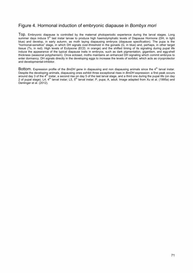
71 �
Figure 4. Hormonal induction of embryonic diapause in Bombyx mori
Top. Embryonic diapause is controlled by the maternal photoperiodic experience during the larval stages. Long summer days induce 5th last instar larvae to produce high haemolymphatic levels of Diapause Hormone (DH, in light blue) and develop, in early autumn, as moth laying diapausing embryos (diapause specification). The pupa is the “hormonal-sensitive” stage, in which DH signals over-threshold in the gonads (G, in blue) and, perhaps, in other target tissue (Ts, in red). High levels of Ecdysone (ECD, in orange) and the shifted timing of its signaling during pupal life induce the appearance of the typical diapause traits in embryos, such as dark pigmentation, gigantism, and egg-shell thickness (seasonal polyphenism). Once eclosed, moths maintains an enhanced DH signaling which commit embryos to enter dormancy. DH signals directly in the developing eggs to increase the levels of sorbitol, which acts as cryoprotector and developmental-inhibitor.
Bottom. Expression profile of the BmDH gene in diapausing and non diapausing animals since the 4th larval instar. Despite the developing animals, diapausing ones exhibit three exceptional rises in BmDH expression: a first peak occurs around day 3 of the 4th instar, a second rise on day 5 of the last larval stage, and a third one during the pupal life (on day 2 of pupal stage). L4, 4th larval instar; L5, 5th larval instar; P, pupa; A, adult. Image adapted from Xu et al. (1995a) and Denlinger et al. (2012).

FIGURE 4

72 �
Figure 5. Hormonal control of diapause-bound seasonal morphs in Orgyia thyellina
Top. Photoperiodic experience of the 4th/5th instar larvae modulates the appearance of seasonal morphs in relation to diapause. A - Developing Macropterus moth develops from a larva reared under long days (top). Diapausing Brachypterus morphs develop under short days (bottom); B - Pale Macropterus (left) and dark Brachypterus pupae (right); C - nondiapausing (from Macropterus morph, left) and diapausing (from Brachypterus) eggs. D - image C under higher magnification. From Kimura and Masaki (1977) and Saunders et al. (2002).
Middle. Model of hormonal control of diapause. Diapausing Brachypterus pupae are the hormonal sensitive stage, in which high levels of Diapause Hormone (DH) induce diapausing embryos. Once eclosed, Brachypterus moth maintains high levels of DH and lays diapausing embryos, which exhibit enhanced cryoprotecting potential. Dormant embryos are darker, heavier, and thicker than developing ones.
Bottom. Effects of the misregulation of DH on embryonic diapause. Eggs laid just after oviposition (day 0, A-D), 8 days since laying (E-H), and embryos (I-L). Macropterus moths lay developing embryos, when reared under long days (LD) during larval life (A, E, I). Conversely, Brachypterus moths reared under short days (SD) lay diapausing embryos (B, F, J). Injection of DH into Macropterus moth induces the production of diapausing embryos (C, G, K), instead of the developing ones. On the contrary, injection of anti-DH induces Brachypterus moths to lay developing eggs (D, H, L). The white arrow and arrowhead indicate diapause and nondiapause eggs, respectively. The black arrows and arrowheads indicate telson and cephalic lobes, respectively. Scale bar = 5 mm. Reproduced from Uehara et al. (2011).

FIGURE 5

73 �
Figure 6. Hormonal control of polyphenism in Bombyx mori
Top. High levels of Diapause Hormone (DH) induce the development of autumn morph, by signaling in Imaginal Wing Discs (ImD). The autumn moths exhibit brown scales on the wings.
Bottom. Seasonal morphs (autumn and spring) of B. mori. Dorsal sides of male (upper) and female (lower) adults of autumn (left) and spring (right) morphs. From Tsurumaki et al. (1999).
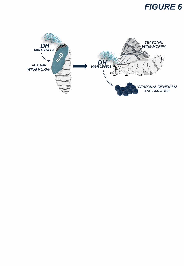
FIGURE 6
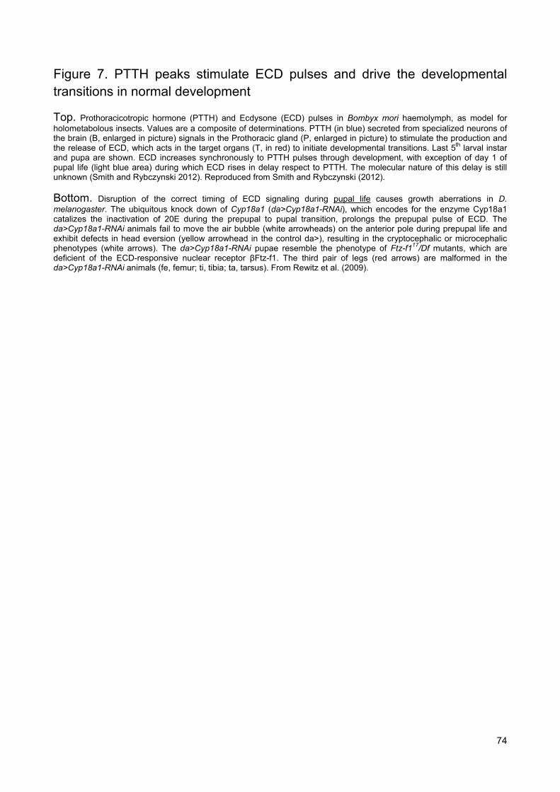
74 �
Figure 7. PTTH peaks stimulate ECD pulses and drive the developmental transitions in normal development
Top. Prothoracicotropic hormone (PTTH) and Ecdysone (ECD) pulses in Bombyx mori haemolymph, as model for holometabolous insects. Values are a composite of determinations. PTTH (in blue) secreted from specialized neurons of the brain (B, enlarged in picture) signals in the Prothoracic gland (P, enlarged in picture) to stimulate the production and the release of ECD, which acts in the target organs (T, in red) to initiate developmental transitions. Last 5th larval instar and pupa are shown. ECD increases synchronously to PTTH pulses through development, with exception of day 1 of pupal life (light blue area) during which ECD rises in delay respect to PTTH. The molecular nature of this delay is still unknown (Smith and Rybczynski 2012). Reproduced from Smith and Rybczynski (2012).
Bottom. Disruption of the correct timing of ECD signaling during pupal life causes growth aberrations in D. melanogaster. The ubiquitous knock down of Cyp18a1 (da>Cyp18a1-RNAi), which encodes for the enzyme Cyp18a1 catalizes the inactivation of 20E during the prepupal to pupal transition, prolongs the prepupal pulse of ECD. The da>Cyp18a1-RNAi animals fail to move the air bubble (white arrowheads) on the anterior pole during prepupal life and exhibit defects in head eversion (yellow arrowhead in the control da>), resulting in the cryptocephalic or microcephalic phenotypes (white arrows). The da>Cyp18a1-RNAi pupae resemble the phenotype of Ftz-f117/Df mutants, which are deficient of the ECD-responsive nuclear receptor ȕFtz-f1. The third pair of legs (red arrows) are malformed in the da>Cyp18a1-RNAi animals (fe, femur; ti, tibia; ta, tarsus). From Rewitz et al. (2009).

FIGURE 7

75 �
Figure 8. The failure of PTTH/ECD cascade induces pupal diapause in Manduca sexta
Top. Model of hormonal control of pupal diapause in Manduca sexta. The photoperiodic induction of diapause occurs as early as 1st larval instar upon perception of autumnal short days. Diapausing last 5th instar larvae exhibit normal temporal profiles of both Ecdysone (ECD, in red) and Prothoracicotropic hormone (PTTH, in blue), but they metamorphose into diapausing pupae. In diapausing pupae, PTTH fall down and the ECD pulse is not elicited any more blocking the metamorphic progression and triggering dormancy. After an established period of chilling, dormant pupae can resume development once exposed to optimal environmental conditions. PTTH and ECD pulses are resumed and the development progresses without reporting growth defects. (B, brain; PG, Prothoracic Gland; Ts, target cells).
Bottom. Haemolymph ecdysteroid titers in M. sexta during the last 5th larval instar and during the pupal life, of diapausing (red arrowheads) and non-diapausing (dark blue arrowheads) animals. During the last 5th larval instar (left), there are no differences between diapausing (red) and non-diapausing (dark blue) animals (W, wandering stage; A, apolysis; E, pupal ecdysis). During the pupal stage (right), diapausing animals exhibit low levels of 20-Hydroxy-Ecdysone (20E) compared to non diapausing ones. Each data point represents Mean ± SD of 4 to 8 animals. Modified from Saunders et al. 2002.

FIGURE 8

76 �
Figure 9. Hormonal control of seasonal polyphenism linked to diapause in Araschnia levana.
Top. Ecdysone-induced morphs in A. levana. Normal spring levana (left), summer prorsa (right) and intermediate (middle) forms are shown. The intermediate morphs develop depending on the timing of Ecdysone injections into presumptive spring form pupae.
Middle. Model of pupal diapause in A. levana. In short days of autumn, the last larval instar develops into a dark diapausing pupa (diapause trajectory) carrying numerous tubercles, upon failure of the pupal Ecdysone (ECD) signaling. After a period of chilling, the dormant pupa breaks diapause and resumes growth by eliciting a pulse of ECD, which is delayed along the pupal life because of dormancy. This “delayed” pupal pulse of ECD induces pupae to develop into levana morphs, which have orange/dark wings with black spots.
Bottom. Conversely, the light pigmented non-diapausing pupae (development trajectory) elicits a precocious pulse of ECD which induces the progression through metamorphosis. Hence, the developing pupa ecloses as prorsa butterfly morph with white and black wings.
Images of the wings are modified from Nijhout (2010, 2003).

FIGURE 9

77 �
Figure 10. Hormonal control of polyphenism linked to diapause in the swallowtail butterfly, Papilio xuthus.
Top. Model of pupal diapause in P. xuthus. In short days of autumn, the last larval instar develops into green diapausing pupa (diapause trajectory), upon failure of the pupal Ecdysone (ECD) signaling. After a period of chilling, the dormant pupa breaks diapause and resumes growth by eliciting a pulse of ECD, which is delayed along the pupal life because of dormancy. This “delayed” pupal pulse of ECD induces pupae to develop into the spring butterfly morphs.
Bottom. Conversely, the brown pigmented non-diapausing pupae (development trajectory) progress through metamorphosis, upon pulse of ECD. Hence, the developing pupae eclose as summer forms.
Images of the wings are modified from http://www.jpmoth.org

FIGURE 10

78 �
Figure 11. Juvenile hormone controls the nature of moults
Top. Extramolt induction by an ectopic and prolonged pulse of Juvenile Hormone (JH). The ectopic JH pulse at the end of the 5th larval instar of Bombyx mori induces an additional molt to a perfect and giant 6th larval instar, which exhibits the endocrine profile typical of the last instar. In the 5th instar larvae, the prolonged pulse of JH drives the molting action of ECD to commit a larva-to-larva moult rather than the onset of metamorphosis, resulting in a perfect additional molt. The giant 6th larvae metamorphose normally after additional 8-20 days of larval life.
Bottom. The loss of JH induces a precocious metamorphosis in Bombyx mori. The mod mutants (control, ET14 and UAS-CYP15C1 animals) are lacking of CYP15C1 (an enzyme involved in JH byosynthesis) and they start precociously metamorphosis resulting in small pupae (left) and moths (right). However, the transgenic expression of CYP15C1 (ET14>UAS-CYP15C1) rescues the phenotype of mod mutants, resulting in normal pupae and moths. From Daimon et al. (2012).

FIGURE 11

79 �
Figure 12. Endocrine control of two types of larval diapause
Top. Hormonal control of larval diapause in Ostrinia nubilalis. Haemolymph ecdysteroid titers during the last larval stage in pre-diapausing (red arrowhead, closed squares) and non-diapausing (dark blue arrowhead, closed circles) larvae (each data point represents mean ± SEM of 3 to 5 biological replicates). The last larval instar triggers diapause upon Ecdysone (ECD) failure which prevents the progression to the metamorphic moult without causing additional extra-molts. Modified from Saunders et al. (2002) and Bean and Beck (1983).
Bottom. Larval diapause in Sesamia nonagriodes. Juvenile hormone (JH) levels in haemolymph in diapausing (Diap, red arrowhead) and non-diapausing (Non-diap, dark blue arrowhead) larvae. Numbers following the letters L and d indicate instar stage and age in days, respectively. Mean ± SD values are represented. The levels of JH remain high in diapausing animals trough all extra-molt periods, whereas they fall down at day 6 of the last 6th larval instar of non-diapausing animals which metamorphose. The last larval instar enters diapause by eliciting a prolonged pulse of Juvenile Hormone (JH) which drives the molting action of Ecdysone (ECD) to induce additional larva-to-larva molts and to arrest the onset of metamorphosis. Modified from Eizaguirre et al. (2005).

FIGURE 12

80 �
Figure 13. Hormonal control of larval diapause in Diatraea grandiosella
Top. Haemolymph Juvenile Hormone (JH) levels related to the onset of diapause and rate of post-diapause pupation. Levels of JH are high in pre-diapause spotted larvae (white arrowhead) and in the following diapausing immaculate ones (red arrowhead). Diapause is broken and pupation starts only when JH falls down. The time of ecdysis from the spotted morph to the immaculate one is about 50 days. Modified from Saunders et al. (2002).
Middle. Model of diapause induction. The spotted larva at the last instar enter dormancy under high levels of JH which drive the molting action of Ecdysone (ECD) to induce larva-to-larva molt rather than the metamorphic one. The dormant immaculate morph develops from the spotted larva and it exhibits enhanced cold resistance, increased fat accumulation, and low respiration rate. Immaculate larvae fail to initiate metamorphosis.
Bottom. Model of diapause termination. The drop of JH arrests diapause and allows the ECD pulse to initiate normally metamorphosis by inducing a larva-to-pupa molt. Hence, the immaculate larvae metamorphose normally, without reporting developmental aberrations.

FIGURE 13

81 �
Figure 14. Imaginal (adult) diapause of Drosophila melanogaster is linked to modifications of larval development
Diapause is strictly linked to modifications of larval development in D. melanogaster. In North America, natural populations of the fruit fly exhibit a latitudinal cline in diapause and larval traits. Larvae of northern latitudes exhibit a delayed developmental time (due to the slowdown of larval growth rate and the onset of metamorphosis) and develop as adult triggering a strong diapause response in relation to slow aging and low early-life fecundity. The opposite pattern occurs in flies of southern latitudes. These variations represent the genetic adaptation to different environments and might be the results of the genetic and functional link between holometabolous phases, controlled by unknown growth factors. Image of adult D. melanogaster is modified from http://carriearnold.wordpress.com

FIGURE 14

82 �
Figure 15. Insulin/IGFs (IIS) signaling pathway of Drosophila melanogaster
Top. An expression time course of dilp1to6 genes along holometabolous phases of D.melanogaster. The grey area inside each graph represents the pupal stage (non-feeding period of development). The dilp2 and dilp5 genes are strongly expressed during all holometabolous phases (the profiles are emphasized by the light blue/grey area). For each profile, fold changes are calculated relative to the minimal level. No cross-quantification is provided between the different dilp genes. Mean ± SD. From Slaidina et al. (2009).
Bottom. The canonical Insulin/IGFs signaling cascade (on left). The DILPs (INSULIN-LIKE PROTEINS or INSULIN/IGFs FACTORS, in red) bind and activate the single Insulin/IGFs Receptor (INR, a Receptor Tyrosine Kinase) in the extracellular surface of the target cells. INR activates its receptor substrates, CHICO and SH2B, which, in turn, activate the canonical PI3K cascade. CHICO and SH2B act synergistically to induce a full responsiveness to the DILPs. The final step of the pathway is the activation of the AKT kinase which inhibits the nuclear translocation of the transcription factor FoxO (Forkhead box-O) and prevents the activation of stress/antigrowth FoxO-responsive genes. In the larva phases (on right), DILP2/5 are secreted by the Median Neurosecretory Cells (Ms, in red) via the Neurohaemal glands complex (Ns), which includes the Prothoracic glands, the Corpus Cardium, and the Corpora Allata. These neural DILPs signal in the larval target cells (Ts) promoting the acceleration of both developmental time and growth rate, and the increase of larval metabolism rate. The larval phases have all the potential to modulate the developmental time and prepare the cold acclimatation (see text for details).
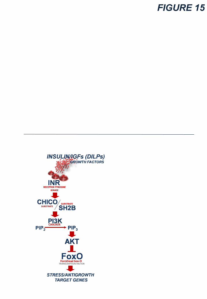
FIGURE 15

83 �
Figure 16. Misregulating gene functions with the yeast-derived Gal4/UAS binary system
A fly carrying a driver-Gal4 transgene is crossed with one carrying a UAS-target sequence transgene. The progeny of the cross carries both transgenes. The enhancer of a specific gene (driver) controls both temporal and spatial pattern of expression of Gal4 gene (blue areas). Once Gal4 gene is expressed, GAL4 binds the Upstream Activator Sequence (UAS) to activate the transcription of the target sequence (red areas), so that the expression of this last changes along space and time depending on the enhancer. Thanks to the Gal4/UAS system, it is possible to increase the dosage of target genes (overexpression), induce RNA interference (RNAi) for the silencing of target genes, and express pro-apoptotic genes to cause the genetic ablation of specific subset of cells. Image of the adult fly is modified from http://carriearnold.wordpress.com.

FIGURE 16
84 �
Figure 17. Disruption of Insulin/IGFs signaling induces diapause
Effects of the disruption of IIS on diapause response at 12°C under LD16:8 (long days) after 11 days since eclosion. The proportion of diapausing females (diapause incidence) is shown in percentage. Both InRhyp and chicohyp hypomorphic homozygous mutants increase diapause incidence compared with their controls (Co-InRhyp and Co-chicohyp, respectively). The Gal4 drivers express in MNCs neurons but they differ in their temporal expression: the dilp2-gal4 line (dilp2>) expresses Gal4 specifically since late third larval instar, and the dilp3> since the end of larval life. The MNCs-lacking flies (dilp2>hid,rpr and dilp3>hid,rpr) exhibit high levels of diapause respect to controls (dilp2>+ and +>hid,rpr and dilp3>+). All multiple dilp1-5-/- and dilp2-3,5-/- knockouts are dormant compared to white1118 and transgenic (+>hid,rpr and dilp2>+) controls. Reducing dilp2 and dilp5 signaling (dilp2-/- and dilp5-/- knockouts or dilp2>dilp2-RNAi and dilp2>dilp5-RNAi) increase moderately diapause without resembling the phenotype of multiple knockouts. The dilp6-/- knockouts do not increase diapause incidence although they are defective for about 20% in body weight.
Red bars represent the key experiments. The arrowheads on the bar represent the homozygous ls-tim/ls-tim (red arrowhead) and the heterozygous ls-tim/s-tim (dark blue arrowhead) genotypes. The absence of arrowhead indicates the homozygous s-tim/s-tim genotype. Mean ± SD of 5 biological replies (at least 60 females for each) are shown. Numbers inside the bars represent the total number of dissected flies. ** = (p<0.001).

FIGURE 17

85 �
Figure 18. Gain of function of dilp2/5 genes antagonizes diapause
Top. Effects of the increase of Insulin/IGFs signaling on diapause response at 12°C under LD8:16 (short days) after 11 days since eclosion. The Gal4 drivers express in MNCs neurons but they differ in their temporal expression: the dilp2-gal4 line (dilp2>) expresses Gal4 specifically in MNCs since late third larval instar, and the dilp2(p)> since early larval life. Overexpression of dilp2 (dilp2>dilp2 and dilp2(p)>dilp2) or dilp5 (dilp2>dilp5 and dilp2(p)>dilp5) decreases strongly diapause incidence, as compared with the controls (dilp2>+ and dilp2(p)>+ and +>dilp2 and +>dilp5). The MNCs-lacking and dilp1-5-/- flies are dormant. Ectopic overexpression of dilp2 (c929>dilp2) or dilp5 (c929>dilp5) in all neuroendocrine cells since early larval life strongly decreases diapause incidence, compared to controls (c929>+). Red bars represent the key experiments. All the lines were homozygous for s-tim allele. Mean ± SD of 5 biological replicates (at least 60 females for each) are shown. Numbers inside the bars represent the total number of dissected flies. ** = (p<0.001).
Bottom. Effects of dilp2/5 genes misregulation on gonads maturation of non-diapausing flies reared at the same conditions as above. A representative example is shown for each genotype. The dilp2-gain (dilp2>dilp2) and dilp5-gain (dilp2>dilp5) non-diapausing females have fully grown ovaries, compared to non-diapausing controls (dilp2>+) which exhibit few early vitellogenic egg-chambers inside the gonads. The gonads of both MNCs-lacking flies (dilp2>hid,rpr) and dilp2-3,5-/- knockouts are completely atrophic (DIAP, diapausing). Numbers I and II indicate the two gonads. White bar is 0.2mm. Red arrowhead indicates the vitellogenic egg chambers.

FIGURE 18
DIAPDIAP
86 �
Figure 19. DILP2/5-mediated reactivation of post diapause growth
Reactivation of diapausing flies by transfer for 5 days to 12°C, 15°C, 19°C, and 22°C under LD16:8, after a period of 11 days at 12°C under LD16:8. Despite the white1118 controls and the dilp2/5 gained flies (dilp2(p)>dilp2 and dilp2(p)>dilp5), the dilp1-5-/- knockouts fail to break dormancy at both 15°C and 19°C resuming the growth only at 22°C. The dilp2(p)-gal4 line (dilp2(p)>) expresses Gal4 specifically in MNCs since early larval life. Red bars represent the key experiments. All the lines were homozygous for s-tim allele. Mean ± SD of 4 or 5 biological replicates (over 60 females for each reply) are shown. Numbers inside the bars represent the total number of dissected flies. ** = (p<0.001).
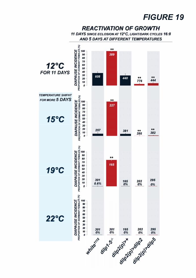
FIGURE 19

FIGURE 19

87 �
Figure 20. DILP2/5-mediated reactivation of post diapause growth
Effects of the loss of dilp2/5 genes on gonads maturation of non-diapausing dilp1-5-/- flies shown in Figure 19. A representative example is shown for each genotype. The gonads of both dilp1-5-/- and dilp2-3,5-/- mutants are completely atrophic (DIAP, diapausing) after a shift for 5 days at 12°C. After 5 days of shift to 22°C, the dilp1-5-/- mutants break completely diapause but rescue moderately the ovarian maturation (3 representative examples are shown). A full ovarian growth in dilp1-5-/- and dilp2-3,5-/- mutants appears after 11 days of shift to 22°C. Numbers I and II indicate the two gonads. White bar is 0.2mm. Red arrowheads indicate the vitellogenic egg chambers.
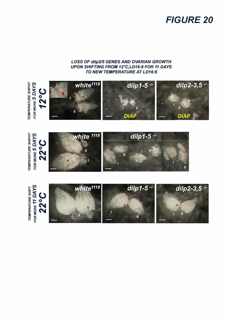
FIGURE 20
DIAP DIAP

88 �
Figure 21. Gain of dilp2/5 induces a prompt activation of growth, without causing heterochronic effects.
Prompt activation of growth in “diapausing” flies reared for 5 days in diapause-inducing (LD8:16, 12°C) or in diapause-breaking (LD16:8, 12°C) conditions. After 5 days in diapause-breaking (LD16:8, 12°C) conditions, dilp1-5-/-, dilp2-3,5-/- and MNCs-lacking (dilp2>hid,rpr) flies fail to activate growth maintaining high levels of diapause as compared to white1118
and dilp2>+ controls, respectively. Despite the dilp2(p)>+ control, dilp2/5 gained flies (dilp2(p)>dilp2 and dilp2(p)>dilp5) promptly activate growth. The dilp2(p)-gal4 line (dilp2(p)>) expresses Gal4 specifically in MNCs since early larval life. With respect to dilp1-5-/- and dilp2-3,5-/- knockouts, both single dilp2-/- and dilp5-/- mutants weakly activate growth. Despite the dilp2(p)>+ control, dilp2/5 gained flies (dilp2(p)>dilp2 and dilp2(p)>dilp5) promptly activate gonads growth after 3 and 5 days since eclosion, although reared in diapause-inducing conditions (LD8:16 at 12°C). After 5 days in the same conditions, both dilp1-5-/- and MNCs-lacking (dilp2>hid,rpr) flies fail to activate growth maintaining high levels of diapause as compared to white1118 and dilp2>+ controls, respectively. After 5 hours since eclosion at 22°C, newly eclosed females of all genotypes exhibit immature (“diapause-like”) gonads. Numbers inside the grey area represent the percentage and the total number of females carrying immature gonads. The “NOT DETERMINED” tag indicates strains for which the assay in those conditions was considered meaningless. Red bars represent the key experiments. The red arrowhead on the bar represents the homozygous ls-tim genotype whereas the absence of arrowhead indicate the homozygous s-tim genotype. Mean ± SD of 4 or 5 biological replicates (at least 60 females for each) are shown. Numbers inside bars represent the total number of dissected flies. ** = (p<0.001). Red ** tag represents p<0.001 obtained by comparison with the dilp1-5-/- and dilp2-3,5-/- multiple knockouts.
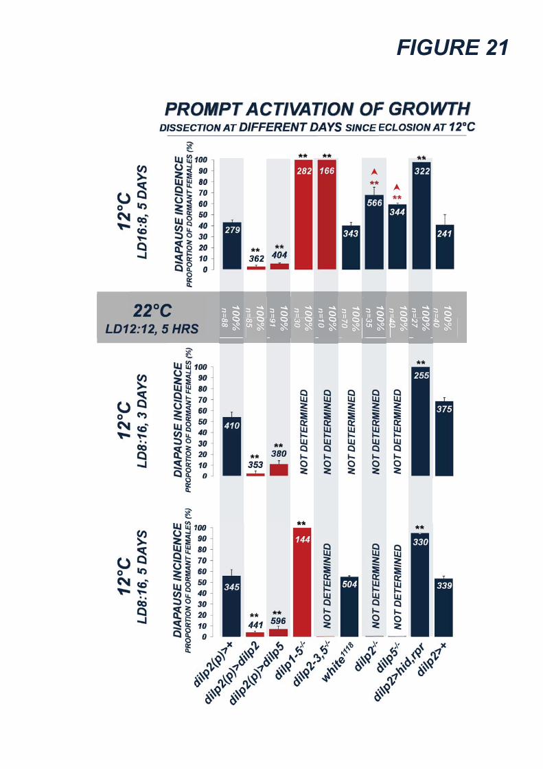
FIGURE 21

89 �
Figure 22. Gain of dilp2/5 induces a prompt activation of growth, without causing heterochronic effects.
Effects of the gain of dilp2/5 genes in MNCs on gonads maturation of non-diapausing flies shown in Figure 21. A representative example is shown for each genotype. Numbers I and II indicate the two gonads. White bar is 0.2mm. Red arrowheads indicate the vitellogenic egg chambers. After 5 hours since eclosion at 22°C under LD12:12, all newly eclosed flies exhibit atrophic and immature gonads independently on the genotype. After 5 days in diapause-inducing conditions (LD8:16 at 12°C), dilp2-gain (dilp2(p)>dilp2) and dilp5-gain (dilp2(p)>dilp5) flies exhibit fully grown ovaries, with respect dilp2>+ controls which exhibit only early vitellogenesis.

FIGURE 22

90 �
Figure 23. Up-regulation of dilp2/5 genes in diapausing flies Expression levels of dilp genes at 12°C and 25°C, under LD8:16. The “up-bar” represents the up-regulation of the gene at 12°C (<12°C) whereas the “down-bar” represents the downregulation of the gene at 12°C (<25°C). Final normalized expression levels (E) are presented as fold changes (E12°C/E25°C when E12°C>E25°C or E25°C/E12°C when E25°C>E12°C). The point at which the expression levels of the two batches are identical is set to 1. Mean ± SD are shown. Numbers inside the bars represent the average fold change. ** = (p<0.01).
Top. qPCR performed from mRNA extracted from 30 full bodies of females. Five biological replicates were performed. Each gene analyzed is indicated on the bottom of the graph. Analysis performed in two different genotypes: the FoxO.RE-Luciferase (in red) and the dilp2>+ (in blue) genotypes. In both genotypes, expression levels of dilp2 and dilp5 genes are increased at 12°C. Levels of dilp6 are unchanged in dilp2>+ flies. InR gene is overexpressed in FoxO.RE-Luciferase flies.
Bottom. qPCR performed from mRNA extracted from 50 isolated heads of dilp2>+ females. Five biological replicates were performed. Each gene analyzed is indicated on the bottom of the graph. Analysis performed in two different genotypes: the FoxO.RE-Luciferase (in red) and the dilp2>+ (in blue) genotypes. Expression levels of dilp2, dilp3 and dilp5 genes are increased at 12°C.
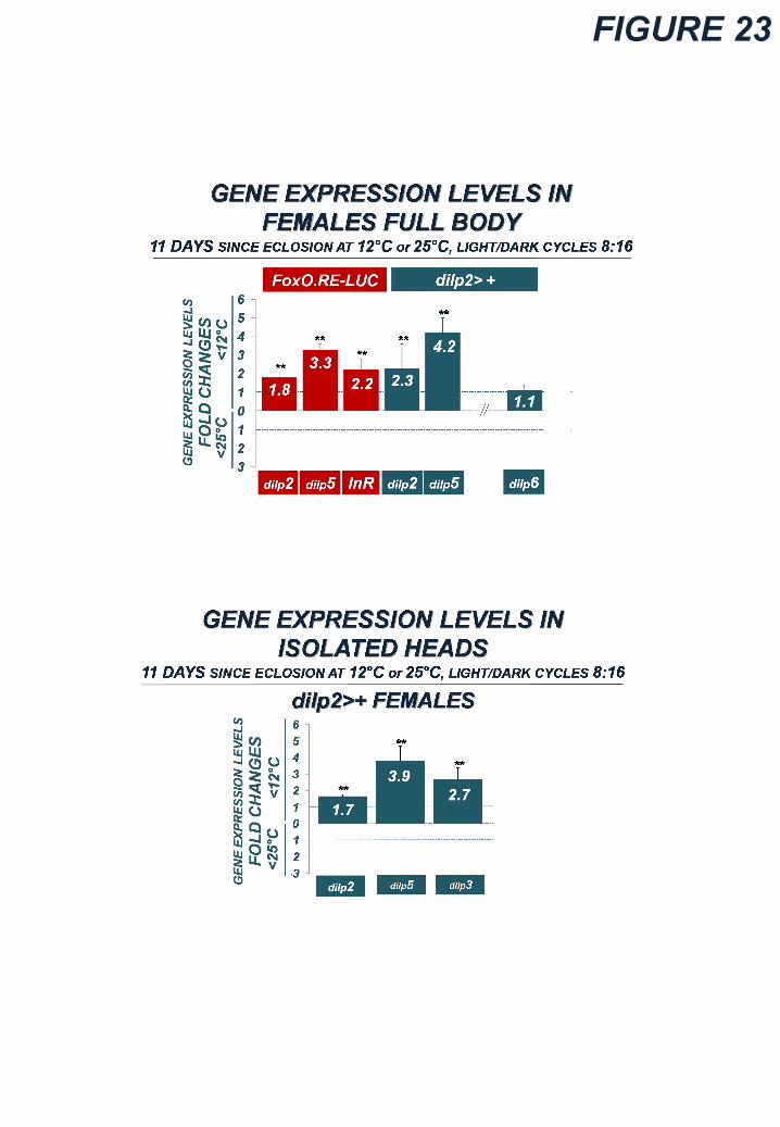
FIGURE 23

91 �
Figure 24. FoxO-responsive reporter gene reveals a diapause-specific profile of IIS
Top. Model of activity of the FoxO-responsive reporter gene, FoxO.RE-Luciferase. Transgenic flies carry a firefly Luciferase transgene fuse to 8 consecutive FoxO-responsive elements (FREs), which transcript Luciferase gene upon FoxO activation. FoxO is activated upon failure of Insulin/IGFs signaling. The levels of Luciferase are a reverse marker of Insulin/IGFs signaling.
Bottom. Levels of Luciferase at 12°C and 25°C, under LD8:16. The “up-vertical bar” represents the up-regulation of Luciferase at 12°C (<12°C) whereas the “down-vertical bar” represents the down-regulation of Luciferase at 12°C (<25°C). Final normalized levels of Luciferase (L) are presented as fold changes (L12°C/L25°C when L12°C>L25°C or L25°C/L12°C when L25°C>L12°C). The point at which the Luciferase levels of the two batches are identical is set to 1. Mean ± SD are shown. Numbers inside the bars represent the average fold change, those inside the parenthesis indicate the number of biological replies. The body region analyzed is indicated at the bottom of each column in the graph. ** = (p<0.01).

FIGURE 24

92 �
Figure 25. The misregulation of MNCs release of DILPs modulates diapause
Top. Effects of misregulation of MNCs release on diapause response at 12°C under LD16:8 (long days) after 5 and 11 days since eclosion. The Gal4 drivers express in MNCs neurons but they differ in their temporal expression: the dilp2-gal4 line (dilp2>) expresses Gal4 specifically since late third larval instar, and the dilp2(p)> since the early larval life. Overexpression of the Na+Ch Sodium Channel (dilp2>Na+Ch and dilp2(p)>Na+Ch) inhibit promptly diapause after 5 and 11 days since eclosion, as compared with the controls (dilp2>+ and dilp2(p)>+ and +>Na+Ch). Conversely, the overexpression of K+Ch Potassium Channel (dilp2(p)>K+Ch) increases diapause incidence, as compared with the negative controls (dilp2(p)>Neg-K+Ch) and the transgenic controls (dilp2(p)>+ and +>K+Ch and +>Neg-K+Ch), albeit the exposure to “diapause-breaking” photoperiod. The “NOT DETERMINED” tag indicates strains for which the assay in those conditions was meaningless. Red bars represent the key strains. All the lines were homozygous for s-tim allele. Mean ± SD of more than 5 biological replicates (at least 60 females for each) are shown. Numbers inside the bars represent the total number of dissected flies. ** = (p<0.001).
Middle. Effects of the misregulation of MNCs release on gonads maturation of non-diapausing flies shown on top. A representative example is shown for each genotype. The I and II indicates the two gonads. White bar is 0.2mm. Red arrowheads indicate the vitellogenic egg chambers. Note that the gonads of dilp2(p)>K+Ch flies are completely atrophic (DIAP, diapausing).
Bottom. Effects of misregulation of MNCs release on diapause response at 12°C under LD8:16 (short days) after 5 and 11 days since eclosion. Overexpression of the Na+Ch Sodium Channel (dilp2>Na+Ch and dilp2(p)>Na+Ch) inhibits promptly diapause after 5 and 11 days since eclosion, as compared to the controls (dilp2>+ and dilp2(p)>+ and +>Na+Ch), albeit the exposure to “diapause-inducing” photoperiod. Conversely, the overexpression of K+Ch Potassium Channel (dilp2(p)>K+Ch) induces diapause at high levels, as compared with the negative controls (dilp2(p)>Neg-K+Ch) and the transgenic controls (dilp2(p)>+ and +>K+Ch and +>Neg-K+Ch).

FIGURE 25
DIAP
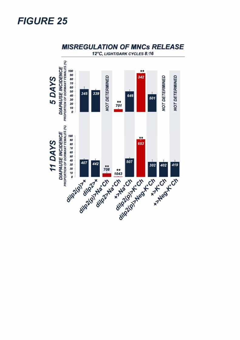
FIGURE 25

93 �
Figure 26. Haemolymphatic increase of DILP2/5 antagonizes diapause
Top. Effects of ectopic overexpression of dilp2/5 genes on diapause at 12°C under LD8:16 (short days) after 5 or 11 days since eclosion. Overexpression was induced in organs which release growth-factor protein into haemolymph. The Akh> driver expresses specifically in CC cells since the early larval life, the DJ634> expresses in adult fat body cells, and the hmgcr(11)> drives specifically in CA cells since early larval life. Overexpression of dilp2 and dilp5 genes decreases strongly diapause incidence, albeit the exposure to “diapause-inducing” photoperiod. Red bars represent the key experiments. The dark blue arrowhead on the bar represent the heterozygous ls-tim/s-tim whereas the absence of arrowheads indicates the homozygous s-tim genotype. Mean ± SD of 5 biological replicates (at least 60 females for each) are shown. Numbers inside the bars represent the total number of dissected flies. ** = (p<0.001).
Bottom. Effects of ectopic expression of dilp2/5 genes in Corpora Cardiaca cells on gonads maturation of non-diapausing flies shown on top. A representative example is shown for each genotype. The I and II indicates the two gonads. White bar is 0.2mm. Red arrowheads indicate the vitellogenic egg chambers. Noteworthy, gonads of Akh>dilp2 and Akh>dilp5 flies exhibit fully grown ovaries, as compared to the control Akh>+.
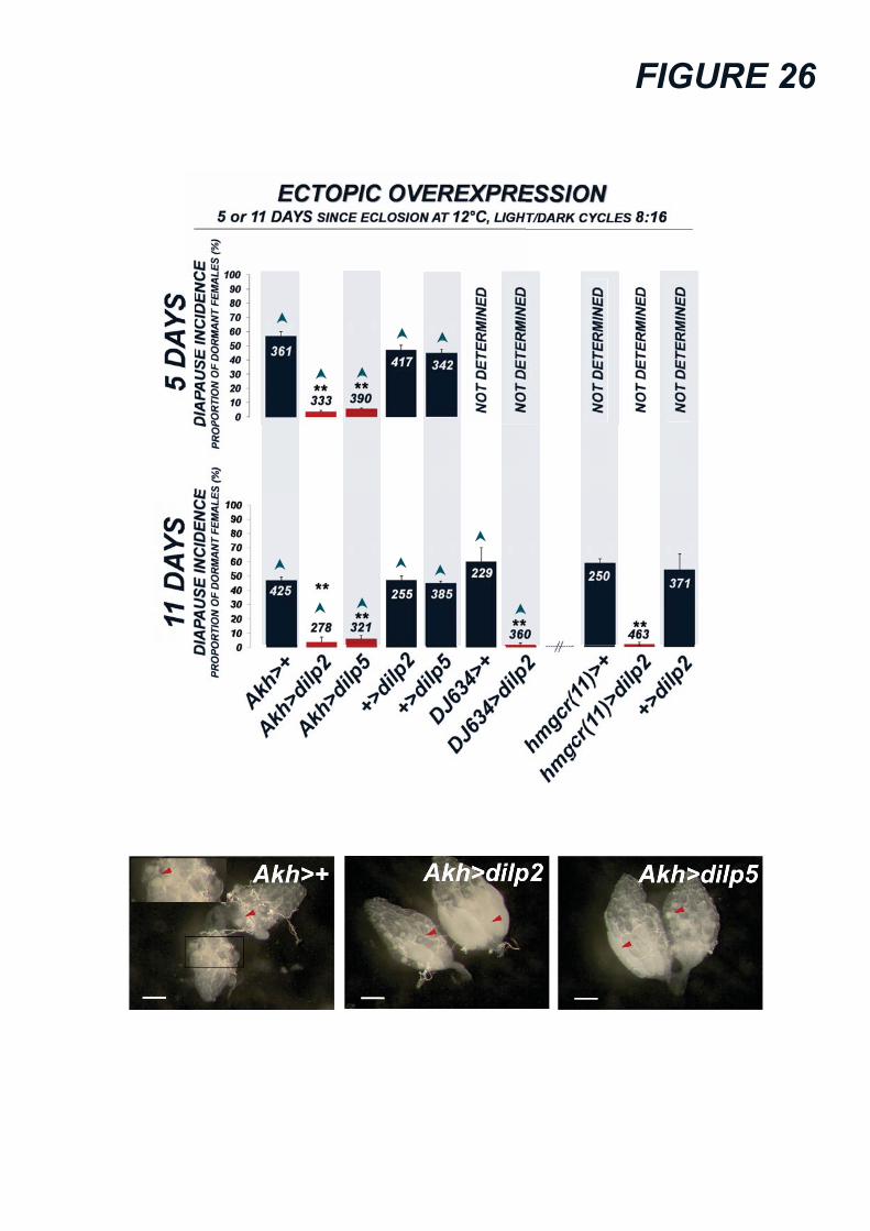
FIGURE 26

94 �
Figure 27. Misregulation of IIS-responsiveness in MNCs and CA affects diapause All lines are homozygous for s-tim variant. Mean ± SD of 5 biological replicates (at least 60 females for each) are shown. Numbers inside the bars represent the total number of dissected flies. ** = (p<0.001).
Top. IIS feedback is active during diapause. Effects of INR loss or FoxO overexpression in MNCs on diapause at 12°C under LD8:16 (short days) after 5 or 11 days since eclosion. FoxO overexpression (dilp2>FoxO and dilp2(p)>FoxO) or INR-loss (dilp2>InRDN and dilp2(p)>InRDN) decreases diapause incidence, as compared with the controls.
Bottom. IIS is hierarchically upstream of JH signaling. The Aug21>, hmgcr(11)>, and hmgcr(3)> drivers expresses in CA but they differ in their temporal expression. The INR loss (Aug21> InRDN and hmgcr(11)> InRDN) and FoxO-overexpression (Aug21>FoxO and hmgcr(3)> FoxO) induces a strong diapause response, as compared with the controls, in spite of the exposure at 12°C and a “diapause-breaking” photoperiod (LD16:8) for 11 days.

FIGURE 27

95 �
Figure 28. Final model: dilp2/5 genes link diapause to modifications of holometabolous growth DILP2/5 act as developmental/growth hormones. Genetic modifications reducing dilp2/5 dosage or DILP2/5 signaling cause the slowdown of both developmental time and larval growth rate, and enhance the environmental sensitivity of developmental system (potential for diapause inducibility). In perturbing environment (grey area), DILP2/5 decrease below signaling threshold leading FoxO free to override and inducing imaginal diapause (reversible polyphenism). Modifications increasing DILP2/5 signaling accelerate developmental time and reduce developmental plasticity by preventing the fall down of such signaling under threshold. High levels of DILP2/5 buffer the perturbing effects and make the flies unable to induce the diapausing switch (loss of diapause inducibility). The image of adult female is modified by http://carriearnold.wordpress.com.

FIGURE 28

Acknowledgements I want to thank Michael B. O’Connor for having me in his talented team (University of Minnesota, Dept. Genetics, Cell
Biology and Development, HHMI). Michael has deeply improved and stimulated my scientific trusts and his contribution
was fundamental to improve my scientific growth. I want also to thank Fondazione “Ing. Aldo Gini” (University of Padova)
for his financial support to the 6-months experience in O’Connor’s lab. Finally, I want to thank Stefano Piccolo (University
of Padova, Dept. Biology) for supporting me during my PhD project, Prof. Telmo Pievani (University of Padova) for his
critical comments on the evolutionary topic and Gabriele Andreatta (University of Padova, Dept. Biology) for his enduring
help to improve this project.


![Giugno 2016 – versione sintetica€¦ · combustibili alternativi con potenzialità di lungo termine in termini di alternativa al petrolio. La Direttiva 2014/94/UE [3] stabilisce](https://static.fdocumenti.com/doc/165x107/605f6e847707da253658784c/giugno-2016-a-versione-combustibili-alternativi-con-potenzialit-di-lungo-termine.jpg)