Valutazione con metodica OSNA del linfonodo sentinella ... · OSNA del linfonodo sentinella Anna...
30
Valutazione con metodica OSNA del linfonodo sentinella Anna Sapino Università di Torino AO-U San Giovanni Battista di Torino
-
Upload
nguyenquynh -
Category
Documents
-
view
224 -
download
0
Transcript of Valutazione con metodica OSNA del linfonodo sentinella ... · OSNA del linfonodo sentinella Anna...

Valutazione con metodica OSNA del linfonodo sentinella
Anna SapinoUniversità di TorinoAO-U San Giovanni Battista di Torino

J Clin Pathol 2004;57:695–701.
Le procedure anatomo-patologiche per la valutazione del LS mancano di standardizzazione• riduzione macroscopica• utilizzo dell’esame al congelatore• numero di sezioni istologiche da esaminare• utilizzo di colorazioni ancillari• stesura del referto

pN1
Based on AJCC/UICC TNM, 7th editionProtocol web posting date: October 2009
pN1 a: METASTASES in 1 to 3 axillary lymph nodes, at least 1 metastasis greater than 2.0 mm
pN1mi: MICROMETASTASES (greater than 0.2 mm and/or more than 200 cells, but none greater than 2.0 mm).
(sn): Only sentinel node(s) evaluated. If 6 or more sentinel nodes and/or nonsentinel nodes are removed, this modifier should not be used
pN0No regional lymph node metastasis histologically, no additional examination for isolated tumor cellspN0(i–) No regional lymph node metastases histologically, negative IHCpN0(i+) Malignant cells in regional lymph node(s) not greater than 0.2 mm or single tumor cells, or a cluster of fewer than 200 cells in a single histologic cross-section (detected by H&E or IHC including ITC)

LS FISSATO IN FORMALINA ED INCLUSO IN PARAFFINA

5
2 (HE+IHC)2 (HE+IHC)150µ150µ
150µ150µ
150µ150µ
Etc.Etc.
150µ150µ
150µ150µ
2 (HE + IHC)2 (HE + IHC)
2 (HE +IHC)2 (HE +IHC)
2 (HE +IHC)2 (HE +IHC)
2 (HE + IHC)2 (HE + IHC)
serial (step) sectioning
1 paraffin block
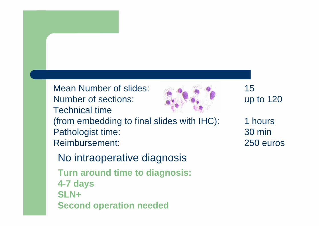
Mean Number of slides: 15 Number of sections: up to 120Technical time (from embedding to final slides with IHC): 1 hoursPathologist time: 30 minReimbursement: 250 euros
No intraoperative diagnosisTurn around time to diagnosis: 4-7 daysSLN+Second operation needed

PROCESSAZIONE
ALLESTIMENTO
LETTURA E PROBLEMI DI INTERPRETAZIONE DIAGNOSTICA
LS NEL CARCINOMA DELLA MAMMELLA E STANDARDIZZAZIONE
CELLULE TIMORALI ISOLATEMICROMETASTASIMACROMETASTASI
Ha maggior peso la quantità di tumore nel linfonodo di come sono disposte le cellule!

FIG. 2 Lobular carcinoma (test case #56). A dispersed pattern of lobular carcinoma withfewer cells than the case illustrated in Figure 2 also caused disagreement in classification. On (A) pre-test, three MDs chose micrometastasis, one chose “other”, and two choseisolated tumor cells (ITC). On (B) post-test, all six MDs chose ITC [(N0(i)].

392 patients with an invasive lobular carcinoma and positive SN and axillary lymph node dissection
SNs with multiple single cells and clusters arranged in a discontinuous manner but dispersed homogeneously in a definable part of the lymph node, classified
as micrometastases according to the EWGBSP interpretations vs. ITCaccording to Turner et al.

Frequency and comparison of nonnon--SN involvement SN involvement according to two different interpretations of the N staging system.
SN classification Number of patients with non-SN involvement (%; 95% CI))
Difference% (95% CI)
EWGBSP EWGBSP Turner
ITC 3/27 (11%;3.93/27 (11%;3.9--28.1)28.1) 11/71 (15%; 8.9 -25.7) 4% (-13.8-16.9)
Micrometastases 22/107 (21%;14.022/107 (21%;14.0--29.2)29.2) 28/96 (29%; 21.0-38.9) 9% (-3.3-20.4)
Macrometastases 158/258 (61%;55.2158/258 (61%;55.2--67.0)67.0)
144/225 (64%;57.5-70.0)
3% (-5.9-11.3)

OSNA OSNA
• PROCEDURA AUTOMATIZZATA DI AMPLIFICAZIONE DEGLI ACIDI NUCLEICI
• VERIFICA LA PRESENZA DEL GENE DELLA CK19 NEL TESSUTO LINFONODALE
• CONSENTE UNA DIAGNOSI MOLECOLARE DEL LS INTRAOPERATORIA
• FORNISCE UN RISULTATO DI NEGATIVO, MICROMETASTASI O MACROMETASTASI

++
+
-
++
+
-
Macro-Metastasis
Micro-Metastasis
ITC / background
Macro-Metastasis
Micro-Metastasis
ITC / background
CK19 mRNACK19 mRNASize of metastasis Size of metastasis
250 copies mRNA/µL – CK19
(5.0 x 10⁶)
5.000 copies mRNA/µL – CK19
(1.0 x 10⁸)
OSNA diagnosis
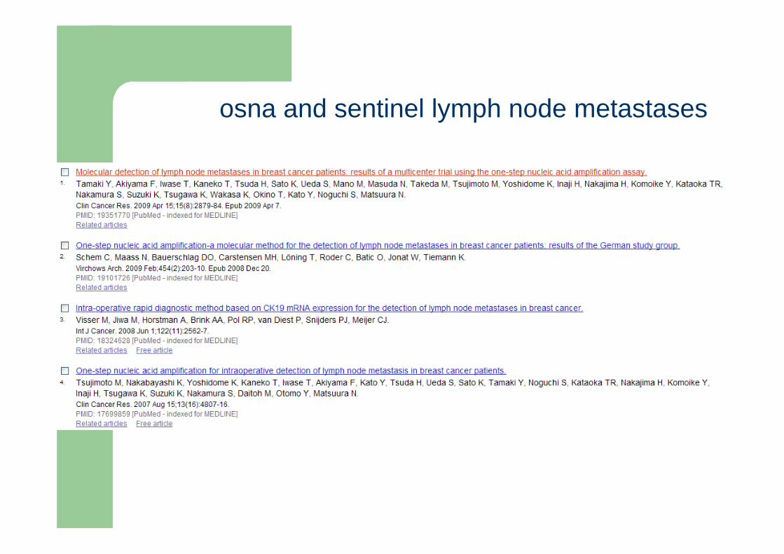
osna and sentinel lymph node metastases

Tsujimoto et al 2007 - Clinical Cancer ResearchVisser et al 2008 - Int J Cancer Schem et al 2009 - Virchows Arch Tamaki et al 2009 – Clinical Cancer Research
Grado di concordanza molecolare-istologico: 92- 98.2%Sensibilità: 95- 98.1% Specificità dal 94.7-100%
METODICHE A CONFRONTO: ISTOLOGIA E OSNA
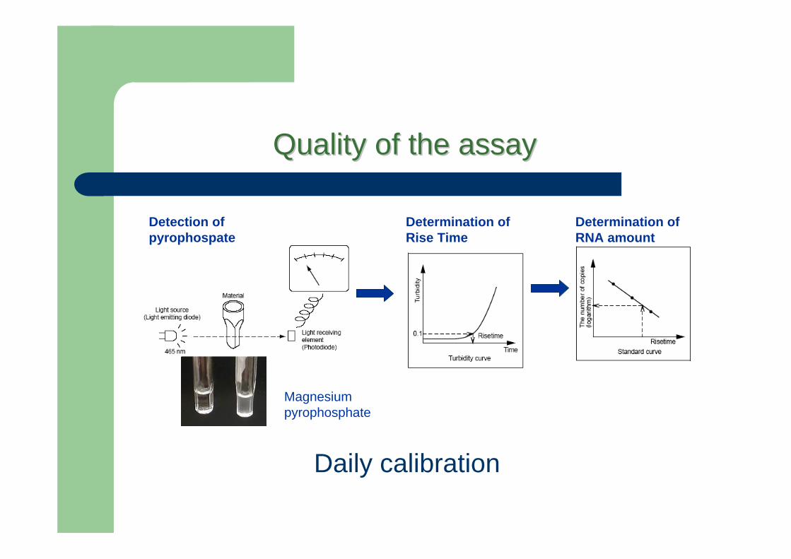
QualityQuality of the of the assayassay
Detection of pyrophospate
Determination of RNA amount
Determination of Rise Time
Magnesium pyrophosphate
Daily calibration

Undesired amplification false positive results. of genomic DNA is avoided due to:
• 6 different primers which have been specifically designed toavoid the amplification of CK19 pseudogenes or theirtranscripts,
•precipitation of DNA at low pH during sample preparationand the isothermal reaction temperature of 65°C.
False negative?CK19 negative
QualityQuality of the of the assayassay

Lymph nodes are simply homogenised in a special homogenising reagent.
The liquid phase is taken and inserted in the RD-100i which automatically performs pipetting, amplification, and detection.
The total time required starting from the preparation of the lymph node until resultsare displayed is about 30 minutes for one lymph node and about 40 minutes for fourlymph nodes.
Workflow of the OSNA-assay
Clin Cancer Res 2007;13(16) August15, 2007
Time of executionTime of execution
fat tissue clearing
Weight

Molinette utilizzo dell’intero linfonodo


Ns55 (32)114 (67)
35 (32)75 (68)
Ki670-10%>10%
Ns144 (85)25 (14)
108 (98)2 (2)
HER2negativepositive
Ns38 (22)131 (77)
22 (20)88 (80)
Progesterone Receptor0-10%>10%
Ns14 (8)155 (92)
10 (9)100 (91)
Estrogen Receptor0-10% >10%
Ns118 (70)51 (30)
80 (73)30 (27)
Vascular invasionAbsentPresent
Ns109 (64)29 (17)31 (18)
81 (74)16 (14)13 (12)
Histological TypeDuctalLobularSpecial Type
Ns66 (39)78 (46)25 (15)
46 (42)48 (44)16 (14)
Histological Grade1 2 3
Ns41 (24)45 (27)83 (49)
33 (30)19 (17)58 (53)
Tumor Size (mm)<101.1-1.5>1.5
Ns61.2 (23-86)17 (10)35 (21)45 (26)72 (43)
66.7(38-82)5 (5)
30 (27)32 (29)43 (39)
Age yrMedian (range)<4546-5556-65>65
P-valueNON OSNA169 (%)
OSNA110 (%)
PATHOLOGICAL PARAMETERS

66%71%Negativo7 %/ITC8%18%Micrometastasi
20 %11%Macrometastasi
Metodo Tradizionale
169 casi
OSNA 110 casi
P<0.01

48%42%Cavo ascellare positivo
Metodo TradizionaleOSNA Macrometastasi
22%22%Cavo ascellare positivo
Metodo TradizionaleOSNA Micrometastasi

991Negative7723Micrometastases ( +)1783Macrometastases (++)
Negative%Positive %OSNA Assay
Cytology (HE/IHC)
RISULTATI OSNA 2010

Yes (0/12)Positive28.927.3-(L)<250 +L71
Yes (0/14)Positive25.425.1++2.3x 104-L52
Yes (0/19)Positive25.023.3++1.6x 104-L3
NoPositive25.131.1+(I)1.3x 10³-L33
NoBorderline31.530.8+(I)1.0x 10³-L31b
Yes (0/19)Positive24.825.1+(I)3.3x 102-L2b
NoBorderline3432+(I)4.9x 10²-L26
NoBorderline3433.2+(I)4.1x 10³-L6
NoPositive21.621+(I)3.4x 10²-L31
NoBorderline32.931.3+2.8x 10²-L50
Yes (0/13)Positive26.126.3+6.9x 102-L35 b
NoBorderline3231.8+2.9x 10²-L75
Yes (2/18)Positive25.426.4+2.7x 102-L28
Yes (0/13)Positive24.725.4+1.4x 103-L35
NoBorderline32.330+3.4x 10³-L38
Yes (0/23)Positive27.728.1+2.0x 103-L13
NoBorderline32.932.8+6.6x 10²-L26
NoPositive25.725.8+4.7x 10³-L12
Yes (0/8)Positive27.527.1+4.9x 102-L32
Yes (0/20)Borderline32.632+4.6x 103-L2
ResultSPDEFCut-off 31.6
Ct
CK19Cut-off 31.5
Ct
ResultCK19Copy
number/µl
ALN(positive LN/Total LN)
SYBR-Green RT-PCROSNAImprintCytologyCases
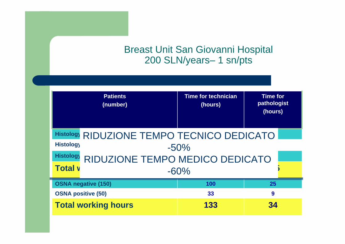
Breast Unit San Giovanni Hospital200 SLN/years– 1 sn/pts
933OSNA positive (50)25100OSNA negative (150)
105210Total working hours
1428Histology False negative (28)
34133Total working hours
1632Histology Positive (32)
75150Histology Negative (150)
Time forpathologist
(hours)
Time for technician(hours)
Patients(number)
RIDUZIONE TEMPO TECNICO DEDICATO-50%
RIDUZIONE TEMPO MEDICO DEDICATO -60%

Costi OSNA caso per 200 pazienti / anno
200 pazienti con biopsia del linfonodo sentinella / annoMedia di 1 LN / PazienteCosto medio OSNA: circa € 350 / paziente inclusi:
– Noleggio strumentazione automatica e accessori– Full risk– Reagenti e consumabili dedicati per eseguire 200 pazienti o linfonodi
La variabilità dei costi dipende da:Numero pazienti con biopsia del LS / annoNumero di linfonodi /pazienteNumero di giornate OSNA /settimanaNumero di settimane lavorative /annoNumero anni di contratto

GRAZIE PER LGRAZIE PER L’’ATTENZIONEATTENZIONE

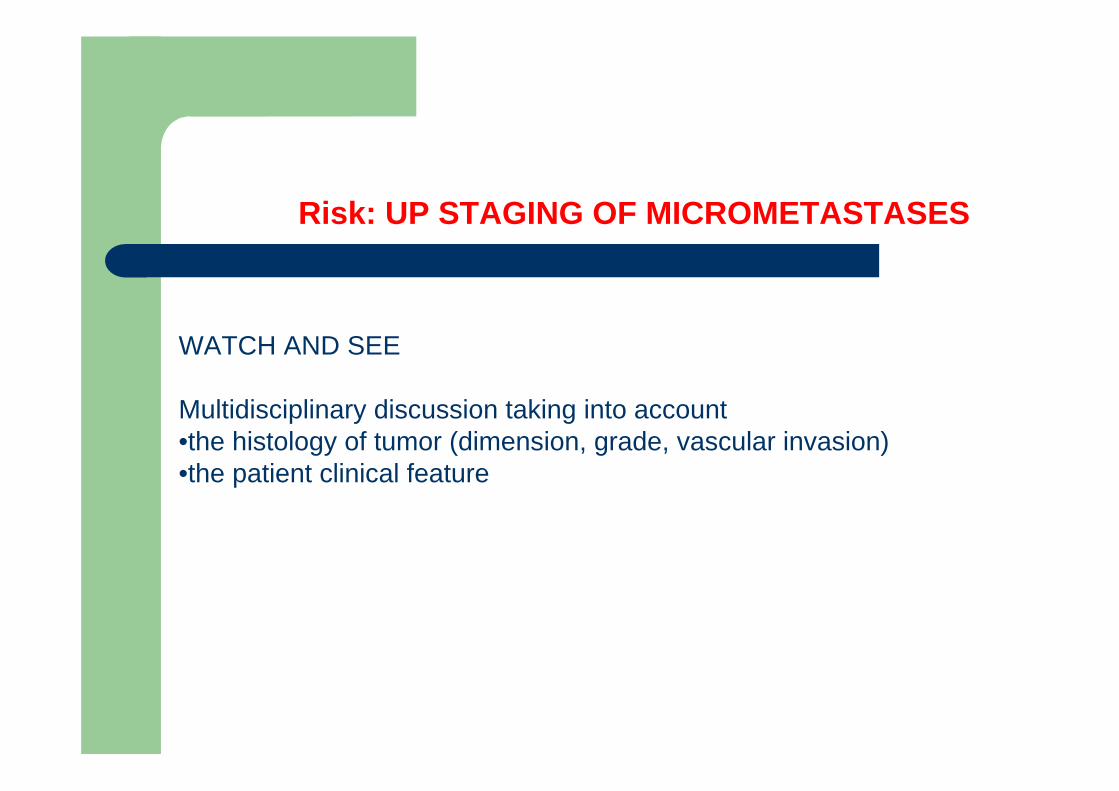
Risk: UP STAGING OF MICROMETASTASES
WATCH AND SEE
Multidisciplinary discussion taking into account•the histology of tumor (dimension, grade, vascular invasion)•the patient clinical feature

Axillary recurrence is low in patients with breast cancer who do not undergocompletion axillary lymph node dissection for micrometastases in sentinel lymph
nodes.Rayhanabad J, Yegiyants S, Putchakayala K, Haig P, Romero L, Difronzo LA.
Am Surg. 2010 Oct;76(10):1088-91.