
STRUCTURAL AND FUNCTIONAL ANALYSIS OF … · funzionali a livello di proteine sono spesso carenti....
120
UNIVERSITÀ DEGLI STUDI DI MILANO SCUOLA DI DOTTORATO IN MEDICINA MOLECOLARE CICLO XXVII Anno Accademico 2013/2014 TESI DI DOTTORATO DI RICERCA settore scientifico disciplinare: BIO13 STRUCTURAL AND FUNCTIONAL ANALYSIS OF COMPLEMENT FACTOR H: A CRUCIAL PROTEIN IN SEVERAL DISORDERS Dottorando: Silvia BERRA Matricola N° R09657 TUTORE: Prof. Alberto CLIVIO COORDINATORE DEL DOTTORATO: Prof. Mario CLERICI
Transcript of STRUCTURAL AND FUNCTIONAL ANALYSIS OF … · funzionali a livello di proteine sono spesso carenti....

UNIVERSITÀ DEGLI STUDI DI MILANO
SCUOLA DI DOTTORATO IN MEDICINA MOLECOLARE
CICLO XXVII
Anno Accademico 2013/2014
TESI DI DOTTORATO DI RICERCA
settore scientifico disciplinare: BIO13
STRUCTURAL AND FUNCTIONAL ANALYSIS OF
COMPLEMENT FACTOR H: A CRUCIAL PROTEIN IN
SEVERAL DISORDERS
Dottorando: Silvia BERRA
Matricola N° R09657
TUTORE: Prof. Alberto CLIVIO
COORDINATORE DEL DOTTORATO: Prof. Mario CLERICI


I
Sommario
Il Fattore H del complemento (FH) è un importante regolatore della via alternativa del complemento: protegge infatti le cellule dell’ospite dall’attacco del sistema del complemento e carenze di FH sia qualitative e quantitative dovute a mutazioni nel gene CFH sono spesso associate ad una serie di malattie umane, come la glomerulonefrite membranoproliferativa (MPGN), la sindrome emolitico-uremica atipica (aHUS) e la degenerazione maculare della retina legata all’età (AMD). Mentre esiste una caratterizzazione genetica per tutte queste malattie, i dati funzionali a livello di proteine sono spesso carenti. Inoltre, il FH gioca un ruolo significativo nelle malattie infettive: molti agenti patogeni sono infatti in grado di reclutare il FH sulla loro superficie sfruttandolo per proteggersi dagli attacchi del complemento. Mentre per alcuni agenti patogeni l'interazione con il FH è stata ben descritta, per gli altri gli "interattori" diretti sono ancora sconosciuti. Tuttavia, lo studio del FH è complicato dalla presenza di proteine FH-related (FHRs) che posseggono un elevato grado di somiglianza con il FH e ne rendono quindi difficile la purificazione e la analisi diretta. Il primo obiettivo di questo progetto è stato lo sviluppo di saggi quantitativi e funzionali FH-specifici, utilizzando un anticorpo monoclonale (Mab 5H5) prodotto nel nostro laboratorio, che si è dimostrato essere specifico per FH. Il secondo era la produzione del FH intero e di tre frammenti, contenenti diverse porzioni della proteina, sia in un sistema di espressione eucariotico che procariotico. L’anticorpo 5H5 è stato prodotto in grandi quantità utilizzando un mini-fermentatore ed è stato utilizzato per purificare il FH mediante cromatografia di affinità. La purezza e l’attività funzionale del FH purificato sono state valutate e confrontate con gli altri metodi di purificazione (ad es. affinità su Eparina, HPLC, HIC). È stato inoltre sviluppato un test ELISA per la corretta quantificazione del FH da fluidi biologici, utilizzando il 5H5 insieme ad un anticorpo policlonale anti-FH, anch’esso prodotto nel nostro laboratorio, e al FH purificato per affinità. Questo test di quantificazione è risultato affidabile ed è stato utilizzato per valutare diversi campioni di pazienti aHUS e AMD. Inoltre, sono stati sviluppati anche due micro-metodi di purificazione del FH. Questi metodi consentono di purificare una buona quantità di FH altamente purificato partendo da una piccola quantità di campione e sono quindi uno strumento utile per studiare il FH specialmente in quelle patologie, quali aHUS, che colpiscono soprattutto i bambini. Infine, sono stati sviluppati due saggi funzionali per valutare sia l'attività cofattoriale N-terminale che l’attività protettiva C-terminale del FH. Il FH è una grossa glicoproteina solubile che possiede funzioni diverse in differenti parti della molecola. Quindi diversi test sono necessari per valutare la completa funzionalità del FH.

II
Il primo test, il "cofactor assay", basato sul fatto che il FH possiede attività cofattoriale per il taglio del C3b mediato dal FI, è completamente funzionante ed è già stato utilizzato per lo screening di diversi campioni. Il secondo, il test di emolisi, si basa sul fatto che il FH è in grado di proteggere gli eritrociti di pecora dalla emolisi mediata dal complemento. In questo test il FH purificato dai pazienti verrà utilizzato insieme ad un siero di controllo depleto di FH, al fine di avere un saggio in cui l'unica variabile sia il FH. Questo test è stato già messo a punto utilizzando un siero di controllo al fine di verificare tutte le condizioni, ed è ora nella fase finale di sviluppo, dopo di che sarà utilizzabile su materiale dei pazienti. I quattro cloni contenenti la sequenza completa del FH e i frammenti parziali (SCRs 1-7, SCRs 8-14, SCRs 15-20) sono stati prodotti e saranno utilizzati per esprimere FH sia in forma glicosilata che non glicosilata. Sarà quindi analizzata la capacità di queste proteine ricombinanti di interagire con le proteine dei patogeni o di ripristinare la funzione di un FH difettoso.

III
Abstract
Complement Factor H (FH) is a major regulator of the Alternative Pathway of Complement: it protects host cells from complement attack and qualitative and quantitative deficiencies of FH due to mutations in the CFH gene are frequently associated with a number of human diseases, such as membranoproliferative glomerulonephritis (MPGN), atypical haemolytic uraemic syndrome (aHUS) and age-related macular degeneration (AMD). A genetic characterisation is available for all these diseases, whereas functional data on protein levels are often missing. Furthermore, FH plays a significant role in infectious diseases, as many pathogens are able to recruit FH on their surface, thus protecting themselves from complement attack. Whereas interaction of FH with certain pathogens has been well described, for others the direct “interactors” still remain unknown. However, the study of FH is complicated by the presence of FH-related proteins (FHRs) that share a high degree of similarity rendering its purification and direct analysis challenging. The first focus of this project was the development of quantitative and functional FH-specific assays, using a monoclonal antibody (MAb 5H5) produced in our laboratory, which has been shown to be specific for FH. The second aim was the production of the whole recombinant FH and of three fragments, containing different parts of the protein, in both a eukaryotic and a prokaryotic expression system. MAb 5H5 was produced in high amounts using a mini-fermentator and used to purify FH by affinity chromatography. Purity and functional activity of this affinity-purified FH were assessed and compared with FH obtained by other purification methods (eg. Heparin affinity, HPLC, HIC). An home made ELISA for the correct quantification of FH from biological fluids, making use of 5H5 together with a polyclonal anti-FH antibody, also produced in our lab, and affinity purified FH was also developed. This reliable quantification assay was used to screen samples from aHUS and AMD patients. Moreover, two micro-methods to purify FH were also developed. These methods enable to purify a good quantity of highly purified FH from a small amount of sample and are therefore a useful tool to study FH especially in those pathologies, such as aHUS, which mainly affect children. Finally, two functional assays to assess both the N-terminal cofactorial activity and C-terminal protective activity of FH have been set up. FH is a big soluble glycoprotein that bears different functions in different parts of the molecule. Therefore different tests are needed to assess full functionality of FH. The first test, the “cofactor assay”, based on the fact that FH possesses cofactor activity for the FI-mediated cleavage of C3b, is now fully implemented and has already been used to screen different samples.

IV
The second one, the “haemolysys test” is based on the fact that FH is able to protect sheep erythrocytes from haemolysis mediated by complement. In this test FH purified from patients will be used together with a control FH-depleted serum, in order to set up an assay where the only variable is FH. This test has already been set up with a control serum in order to test all the conditions, and is now in its fine tuning phase, after which it will be applicable to patient’s material. The four clones containing the complete FH sequence and partial fragments (SCRs 1-7, SCRs 8-14, SCRs 15-20) have been produced and will be used to express FH in a glycosylated or unglycosylated form. These recombinant proteins will be screened for their ability to interact with proteins from pathogens or to restore the function of a defective FH.
V
CONTENTS
ABBREVIATIONS 1
INTRODUCTION 3
1. THE COMPLEMENT SYSTEM 3
1.1. Complement activation 5 1.1.1. Classical Pathway 5 1.1.2. Alternative Pathway 6 1.1.3. Lectin Pathway 7 1.1.4. Properdin Pathway 8
1.1. The terminal pathway 9
2. REGULATION OF THE COMPLEMENT SYSTEM 10
2.1. Regulatory proteins 10 2.1.1. Non-RCA regulators 11 2.1.2. RCA regulators 15
3. COMPLEMENT FACTOR H 21
3.1. FH functions 21 3.2. FH structure and functional domains 22 3.3. CFH gene 25 3.4. Factor H protein family 26
4. FACTOR H-ASSOCIATED DISEASES 30
4.1. Atypical Hemolytic Uremic Syndrome 30 4.1.1. FH in aHUS 32
4.2. Age-Related Macular Degeneration 34 4.2.1. FH in AMD 37
4.3. Interaction with Pathogens 38
AIM OF THE WORK 41
MATERIALS AND METHODS 43
5. PRODUCTION OF 5H5 MONOCLONAL ANTIBODY 43
5.1. Suspension cell culture 43 5.2. High density cell culture 43 5.3. Purification of 5H5 antibody 44
6. PURIFICATION OF FACTOR H 45

VI
6.1. Preparation of 5H5-affinity column 45 6.2. Affinity purification of factor H 46
7. FACTOR H ELISA 47
8. IGG AND ALBUMIN DEPLETION 48
9. WESTERN BLOT (WB) 48
10. FACTOR H IMMUNOPRECIPITATION (IP) 50
11. HEPARIN AND HYDROPHOBIC INTERACTION CHROMATOGRAPHY (HIC) PURIFICATION 51
11.1. Heparin purification 51 11.2. Hydrophobic interaction chromatography (HIC) 53
12. FACTOR H INTEGRITY ASSAY 55
12.1. Trypsin cleavage of FH 55 12.2. IP of Trypsin-cleaved FH 55 12.3. Integrity assay 56
13. FACTOR H FUNCTIONAL ASSAYS 56
13.1. Quantification of Purified FH 56 13.2. Cofactor assay 57 13.3. Haemolysis test 57
14. CLONING OF FACTOR H 58
14.1. Retrotranscription 58 14.1.1. RNA denaturation 58 14.1.2. Retrotranscription reactions 59
14.2. Amplification 59 14.3. Features of the pFastBac™/HBM-TOPO® vector 60 14.4. Blunt-End TOPO® Cloning Reaction 61 14.5. Transformation and analysis of tranformants 61 14.6. Cloning into a prokaryotic system 62
RESULTS 63
15. PRODUCTION AND PURIFICATION OF 5H5 ANTIBODY 63
16. PURIFICATION OF FH 64
17. FH QUANTITATIVE ELISA 68

VII
18. FACTOR H IMMUNOPRECIPITATION (IP) 69
19. TRYPSIN CLEAVAGE AND INTEGRITY ASSAY 70
19.1. Cleavage of FH by trypsin 70 19.2. Affinity of 5H5 for cleaved and uncleaved FH 71 19.3. FH integrity assay 71
20. Quantification of Purified Factor H 73
21. FUNCTIONAL ASSAYS 73
21.1. Cofactor assay 73 21.2. Haemolysis test 76
22. OTHER PURIFICATION METHODS 79
22.1. Heparin affinity purification 79 22.2. Hydrophobic Interaction Chromatography (HIC) 80
23. CLONING OF FACTOR H 80
DISCUSSION 84
CONCLUSIONS 90
REFERENCES 92
APPENDIX 106
1
ABBREVIATIONS
aHUS atypical Hemolitic Uremic Syndrome
AMD Age-related Macular Degeneration
AP Alternative Pathway
AP Alkaline Phosphatase
BCIP 5-bromo-4-chloro-3'-indolyphosphate p-toluidine
C1-INH C1 inhibitor
C4bp C4 binding protein
CA Cofactor activity
CD Cluster of differentiation
CFH Factor H gene
CFHRs Factor H related proteins genes
C Complement
C“n” Complement component (from 1 to 9)
Cn Clusterin(SP-40,40, apolipoprotein J)
CP Classical Pathway
CR1 Complement receptor 1
CR2 Complement receptor 2
CRP C-reactive protein
DAA Decay accelerating activity
DAF Decay-accelerating factor
DDD Dense deposit disease
ELISA Enzyme-linked immunosorbent assay
ESh Sheep erythrocyte
FB Factor B
FD Factor D
FH Factor H
FHL-1 Factor H-like protein 1
FHRs Factor H related proteins
FI Factor I
GAG Glycosaminoglycan
GlcNAc N-acetyl-glucosamine
GalNAc N-acetyl-galactosamine

2
GBM Glomerular basement membrane
GPI Glycosylphosphatidylinositol
HRP Horseradish Peroxidase
iC3b Inactive C3b
iC4b Inactive C4b
Ig Immunoglobulin
IP Immunoprecipitation
LP Lectin Pathway
LPS Lipopolysaccharide
MAb Monoclonal antibody
MAC Membrane attack complex
MASPs MBL-associated serine proteases
MBL Mannose-binding lectin
MCP Membrane cofactor protein
NBT nitro-blue tetrazolium chloride
P Properdin
PAb Polyclonal antibody
RCA Regulators of complement activation
RPE Retinal pigment epithelium
SCR Short consensus repeat domain
SDS-PAGE Sodium dodecyl sulfate – polyacrylamide gel
electrophoresis
SPR Surface Plasmon resonance
TM Thrombomodulin
TMB Tetramethylbenzidine
Vn Vitronectin (S-protein)
WB Western Blot
3
INTRODUCTION
1. The complement system
The complement system is a central component of innate immunity and is
one of the principal mechanisms of defence against infections.
Complement was first identified in the 1890s by Jules Bordet as a heat-
labile principle in serum that “complemented” antibodies in the killing of
bacteria [1, 2]. Now we know that it consists of more than 30 soluble
plasma and membrane-associated proteins interacting with each other to
activate and regulate the system [3].
The complement system has three main physiologic activities: defence
against bacterial infection, bridging of innate and adaptive immunity, and
disposal of immune complexes and the products of inflammatory injury [4].
The activation of complement consists in an enzymatic cascade in which
complement proteins, normally present in serum as inactive enzyme
precursors (zymogens), are activated sequentially. Each component
recognises and activates, generally by proteolysis, the following one.
Complement can be activated by three different pathways: the classical
(CP), the lectin (LP) and the alternative (AP) pathways, according to the
nature of recognition [5]. All these pathways converge after the activation
of the central component C3 and are followed by a common non-
enzymatic process consisting on the assembly of a cytolytic complex named
the “membrane attack complex” (MAC), which forms a hole in the
membrane and initiates cell lysis [6, 7].
Control of these enzymatic cascades is essential to prevent rapid
consumption of complement in vivo, therefore a group of plasma and
membrane-bound proteins act to inhibit the system at multiple stages.
Some of the proteolytic complement protein fragments, such as C3b, are
also able to opsonize microbes thus stimulating phagocytosis and B-cell

4
responses, whereas other fragments, named “anaphylatoxins”, can attract
and activate macrophages and neutrophils to the site of infection [8–10].
Two other important roles of the complement system are the clearance of
apoptotic and necrotic cells by promoting their phagocytosis and the
clearance of immune complexes from circulation by solubilizing and
transporting them to the reticuloendothelial system [11, 12].
1.1. Complement activation
Depending on the stimuli, complement activation can follow three
different pathways (Fig.1):
- Classical Pathway is activated by surface-bound antibodies (IgM and
certain isotypes of IgG)
- Alternative Pathway is constitutively active but can be enhanced by
certain molecules (carbohydrates, lipids or proteins) bound to foreign
surfaces.
- Lectin Pathway is stimulated by the mannose-binding lectin (MBL)
bound to mannose residues on the pathogen surface
Fig. 1 Schematic representation of the activation of complement cascade. After recognizing the target all the three pathways converge to covalent attachment of C3b on the surface.
The names classical and alternative arose because the classical pathway
was discovered first but the alternative pathway is phylogenetically older.
All the pathways converge in the formation of an enzymatic complex called
C3-convertase, composed of different parts in the various pathways but all
with the same target, the molecule C3 which is hydrolysed into C3b and
covalently attached to the target cell surface.
5
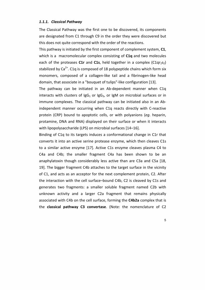
1.1.1. Classical Pathway
The Classical Pathway was the first one to be discovered, its components
are designated from C1 through C9 in the order they were discovered but
this does not quite correspond with the order of the reactions.
This pathway is initiated by the first component of complement system, C1,
which is a macromolecular complex consisting of C1q and two molecules
each of the proteases C1r and C1s, held together in a complex (C1qr2s2)
stabilized by Ca2+
. C1q is composed of 18 polypeptide chains which form six
monomers, composed of a collagen-like tail and a fibrinogen-like head
domain, that associate in a "bouquet of tulips"-like configuration [13].
The pathway can be initiated in an Ab-dependent manner when C1q
interacts with clusters of IgG1 or IgG3, or IgM on microbial surfaces or in
immune complexes. The classical pathway can be initiated also in an Ab-
independent manner occurring when C1q reacts directly with C-reactive
protein (CRP) bound to apoptotic cells, or with polyanions (eg. heparin,
protamine, DNA and RNA) displayed on their surface or when it interacts
with lipopolysaccharide (LPS) on microbial surfaces [14–16].
Binding of C1q to its targets induces a conformational change in C1r that
converts it into an active serine protease enzyme, which then cleaves C1s
to a similar active enzyme [17]. Active C1s enzyme cleaves plasma C4 to
C4a and C4b; the smaller fragment C4a has been shown to be an
anaphylatoxin though considerably less active than are C3a and C5a [18,
19]. The bigger fragment C4b attaches to the target surface in the vicinity
of C1, and acts as an acceptor for the next complement protein, C2. After
the interaction with the cell surface–bound C4b, C2 is cleaved by C1s and
generates two fragments: a smaller soluble fragment named C2b with
unknown activity and a larger C2a fragment that remains physically
associated with C4b on the cell surface, forming the C4b2a complex that is
the classical pathway C3 convertase. (Note: the nomenclature of C2
6
fragments is different from the other complement proteins as for historical
reasons the attached bigger fragment was called “a” and the released one
“b”. An attempt to reverse these “a” and “b” designations was not
universally accepted by experts in the complement field leading to
confusion in textbooks and scientific literature) [20].
The classical pathway C3 convertase binds C3 thanks to the C4b
component whereas the C2a component catalyses its proteolysis into C3a
and C3b [21]. The small C3a fragment is an anaphylatoxin that stimulates
inflammation [10]. The bigger fragment, C3b, can diffuse away and form
covalent bonds with cell surfaces or with the antibody where complement
activation was initiated, functioning as an opsonin, or it can remain bound
to the complex to form a trimolecular complex C4b2a3b, called the
classical pathway C5 convertase.
1.1.2. Alternative Pathway
The Alternative Pathway results in the proteolysis of C3 as the Classical
Pathway does but without any need for antibody for initiation. This
pathway of complement activation requires only four serum proteins: C3,
factor B (FB), factor D (FD), and properdin (P) [22].
This pathway is normally constitutively active: C3 in plasma is in fact being
continuously cleaved at a low rate to generate C3b in a process that is
called “tick-over”. C3 is spontaneously converted to metastable C3* that
undergoes hydrolysis to form C3(H2O) which in turn interacts with factor B
forming the initial convertase [23]. The complex C3(H2O)B is a target for
factor D which cleaves factor B releasing the Ba fragment and forming the
enzyme C3(H2O)Bb. The enzyme C3(H2O)Bb has a short half-life (90 sec.) but
can be stabilized by the binding of properdin that acts as a positive
regulator of complement [24]. C3(H2O)Bb is a C3 convertase enzyme that
cleaves C3 to C3a and to metastable C3b* that exposes a highly reactive
thioester bond. The thioester domain can react with the amino or hydroxyl

7
groups of cell surface proteins or polysaccharides enabling C3b to become
covalently attached to cell surfaces [25]. If these bonds are not formed,
the C3b remains in the fluid phase, where is quickly hydrolysed and can
form with FB the fluid phase C3b(H2O)Bb that can cleave more C3. The C3b
bound to the membrane can react with FB which is cut by FD with the
release of Ba and the formation of the complex C3bBb that is the
alternative pathway C3 convertase (Fig.2). C3bBb can cleave more C3
molecules, thus generating an amplification loop. Some of the C3b
molecules originated can then bind to the convertase itself leading to the
formation of the complex C3bBbC3b, which is the alternative pathway C5
convertase.
Fig. 2 Schematic representation of the AP activation. Details are discussed in the text.
1.1.3. Lectin Pathway
The lectin pathway is initiated in the absence of antibody by the binding of
microbial polysaccharides to circulating lectins such as mannan-binding
lectin (MBL) or ficolins (L, H or M). These soluble lectins are collagen-like
proteins that structurally resemble C1q [26].
MBL, L-ficolin, and H-ficolin are plasma proteins whereas M-ficolin is
mainly secreted by activated macrophages in tissues. These lectins are able

8
to recognize microbial sugar structures; MBL binds to mannose residues on
glycoproteins or carbohydrates present on the surface of microorganisms
whereas the ficolin fibrinogen-like domain can bind to different substances
such as GlcNAc, GalNAc, fucose, sialic acid, lipoteichoic acid (LTA) [27, 28].
MBL and ficolins associate with MBL-associated serine proteases (MASPs)
including MASP1, MASP2, and MASP3. The MASP proteins are structurally
homologous to the C1r and C1s proteases and have a similar function but
they are not able to bind C1q.
MASP-1 can cleave very efficiently C2 but not C4, a weak activity in cutting
C3 has also been shown, and also has a role in activating MASP-2; MASP-2
can also cleave with a high efficiency C2 and to a lower extent C4; MASP-3
does not cleave C2 or C4 and its function is still uncertain but it seems to
activate FD in vitro [26, 29]. MASP proteins lead to the formation of the
same C3 convertase of the classical pathway (C4b2a) and the rest of the
pathways are identical.
1.1.4. Properdin Pathway
Properdin role in stabilizing components of the alternative pathway
convertases has been shown since the 1970’s. More recently it has been
discovered that properdin is also able to activate an “additional” pathway
of complement that utilizes the same components as the alternative
pathway, but that is initiated directly by properdin [30]. Properdin is able
to bind directly to target cells and microbes, thus providing a platform for
the assembly of convertase and leading to phagocytosis. Properdin bound
on a surface has been shown to bind C3, the resulting C3bP complex can
then bind factor B forming C3bBP that can in turn be cleaved by factor D,
releasing Ba and generating surface-bound C3bBbP. This convertase is
similar to the one of the alternative pathway, but its formation is initiated
by properdin rather than nascent C3b [30]. The significance of this
properdin-driven pathway is still elusive, but it has raised a lot of attention
as a novel activation pathway.

9
1.2. The terminal pathway
The C3 convertase of the CP and LP (C4b2a) or that of the AP (C3bBb)
perform a proteolytic cleavage of C3, releasing a small fragment, C3a and
producing C3b. The C3 convertase is able to activate more C3 molecules
leading to an amplification step that is necessary for proceeding to the
terminal pathway. The terminal pathway of complement starts when large
amounts of C3b have been produced and some bind directly to the C3
convertase forming the C5 convertase of the CP and LP (C4b2a3b) or that
of the AP (C3bBbC3b) [31–33]. The addition of this C3b molecule on C3
convertases changes the substrate specificity of the active sites, located in
C2a and Bb, from C3 to C5 component. The C5 is then hydrolysed by the C5
convertases forming the C5a and C5b fragments. C5a that is released to
plasma is a powerful anaphylatoxin and chemotaxin, even more potent
than C3a [34].
The generation of C5b activates a non-enzymatic process of assembling of
the terminal complement components leading to the formation of the
membrane attack complex (MAC) [6, 35]. C5b, which is extremely labile,
serves as a platform for the sequential binding of C6 and C7 forming the
C5b-7 complex. After the binding of C7, the C5b-7 complex becomes
amphipathic and exposes hydrophobic regions which enable its insertion
into the membrane [36]. C5b-7 anchored in the lipid bilayer is a receptor
for plasma C8, the formed C5b-8 complex functions as receiver of multiple
molecules of C9 (from 16 to 18) that create a cylindrical pore of about 30
nm in the membrane [37, 38].The C5b-9 complex, also called membrane
attack complex (MAC), allows the diffusion of small molecules across the
membrane to occur, and leads to rapid osmotic lysis of the target cell [39,
40].
10
2. Regulation of the complement system
The complement system is the first line of defence against infections, it is
rapidly activated and ends in the lysis of foreign cells. Activation of
complement could also be potentially harmful to the individual so, in order
to prevent unwanted damage to host cells, the complement system needs
to be tightly regulated. Complement is regulated by a set of proteins, both
circulating or membrane-associated, that act at different steps of the
cascade and enable fine tuning of complement activation in different
physiological circumstances. Regulators of the complement system ensure
that the activation is both effective and localized on foreign surfaces.
2.1. Regulatory proteins
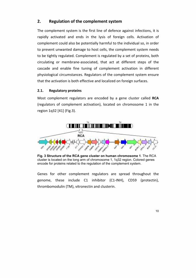
Most complement regulators are encoded by a gene cluster called RCA
(regulators of complement activation), located on chromosome 1 in the
region 1q32 [41] (Fig.3).
Fig. 3 Structure of the RCA gene cluster on human chromosome 1. The RCA cluster is located on the long arm of chromosome 1, 1q32 region. Colored genes encode for proteins related to the regulation of the complement system.
Genes for other complement regulators are spread throughout the
genome, these include C1 inhibitor (C1-INH), CD59 (protectin),
thrombomodulin (TM), vitronectin and clusterin.
11
2.2. Non-RCA regulators
C1-INH is a 105 KDa single-chian glycoprotein that belongs to the
superfamily of serine proteinase inhibitor (serpin). It’s composed of two
domains: a C-terminal serpin domain that contains the protease
recognition region and provides the inhibitory activity and an unique,
heavily glycosylated N-terminal domain (~100 aa), not present in other
serpins, that has no role in protease inhibition but is important in
maintaining functional integrity [42].
C1-INH is the main regulator of both the CP and LP: it irreversibly binds and
inactivates the serine proteases C1r, C1s [43] and MASP-1, MASP-2 [44].
C1-INH is the only plasma protease inhibitor of the CP, it disassembles C1
dissociating C1r and C1s and it can also suppress spontaneous activation of
C1 via a reversible interaction with zymogen C1r and C1s. It can also
regulate the LP via inactivation of MASP2, although α2 macroglobulin also
is able to inhibit MASP2 [45].
C1-INH is a suicide protease inhibitor that irreversibly binds to its targets,
its reactive loop behaves as a pseudo-substrate for the protease that
cleaves the peptide bond and covalently binds to the inhibitor. After
binding, C1-INH acts as a mousetrap swinging the inactivated protease
from the upper to the lower pole and inserting the reactive loop of the
protease inside its structure [46].
C1INH is also the major inhibitor of factor XIIa and kallikrein of the contact
system, lack of inhibition of these enzymes results in inappropriate
bradykinin generation and increase of vascular permeability. Inherited
deficiency of C1-INH is the basis of hereditary angioedema [47].
CD59 also known as MAC-inhibitory protein (MAC-IP), membrane inhibitor
of reactive lysis (MIRL), or protectin is the key regulator of the terminal
pathway. CD59 is a cell surface GPI-anchored glycoprotein of 18-25 KDa
that is heavily glycosylated.

12
CD59 can be found on all circulating blood cells (erythrocytes and
leukocytes), endothelial cells, fibroblasts, and in most epithelial cells (in
pancreas, epidermis, bronchi, kidney, and salivary glands, moreover it has
also been found on sperm.
CD59 functions by binding to the α-chain of C8 in the C5b-8 complex and
preventing subsequent binding of C9, and/or by binding to C9 in the C5b-9
complex and preventing recruitment of additional C9 molecules, it
therefore blocks the polymerization of C9 and the formation of MAC [48].
Inhibition of MAC assembly by CD59 is species-specific, it only occurs if the
MAC components are from the same species as the target cell.
This protein also plays a role in signal transduction pathways in the
activation of T cells.
Deficiency of CD59 results in paroxysmal nocturnal hemoglobinuria, a rare
disease characterized by destruction of red blood cells by the complement
system [49].
Thrombomodulin (TM) is a transmembrane glycoprotein abundantly
expressed on the surface of vascular endothelial cells and is mainly
involved in anticoagulant pathway. TM binds thrombin forming a 1:1
stoichiometric complex thus sequestering it from its substrates, platelets
and fibrinogen. Moreover, after the binding, thrombin changes its
specificity from fibrinogen to protein C activating it. Activated protein C
makes a complex with protein S and cleaves the activated factors V and
VIII, inactivating them, thus acting as an antithrombotic factor.
TM can also act as a complement regulator in two ways: it enhances FI-
mediated inactivation of C3b on the cell surface in the presence of the
cofactor FH [50], and activates TAFI-mediated inactivation of the
anaphylatoxins C3a and C5a [51].
Protein S has also been shown to participate in complement regulation by
binding C4BP. Protein S in complex with C4BP loses its cofactor activity for
protein C but enables C4BP to bind to negatively charged glycolipids [52].
13
This interaction thus promotes coagulation while controlling complement
CP and LP activation.
Vitronectin (Vn, S-protein) and clusterin (Cn, SP-40,40, apolipoprotein J)
are two soluble regulators of the terminal pathway that block the
incorporation of MAC into membranes. Vitronectin binds preferentially to
C5b-7 and interacts with C9 inhibiting its polymerization and preventing
membrane insertion [53]. Clusterin binds not only to C7 in the complex of
C5b-7 preventing its insertion into the cell membrane, but it can also bind
to C8 and to C9, inhibiting the polymerization of C9 [54].
Soluble complexes of C5b-9 with vitronectin or clusterin (SC5b-9 complex)
are normally present in serum during complement activation, thus a
specific enzyme-linked immunosorbent assay (ELISA) for this complex can
be used to monitor complement consumption.
Properdin as discussed earlier is a stabilizer of the AP.
Factor I (FI) is an 88 KDa serum glycoprotein predominantly synthesised by
the liver and is a key regulator of complement activation. FI is a serine
protease that cleaves C3b and C4b to their inactive forms iC3b and iC4b by
releasing C3f or C4c fragments to the fluid phase. To exert its proteolityc
functions FI requires cofactor proteins such as Factor H (FH), C4b-Binding
Protein (C4BP), Complement Receptor 1 (CR1/CD35) and Membrane
Cofactor Protein (MCP/CD46) [55]. Cleavage of C3b is mediated by FH, MCP
or CR1, whereas in the proteolysis of C4b cofactor activity (CA) is
performed by C4BP, MCP or CR1. These cofactor proteins will be discussed
in more detail later.
Inactivating C3b and C4b, FI prevents the formation of the C3 and C5
convertases of both the AP and the CP, thus acting as regulator for both
pathways. Moreover, since the AP comprises a feedback loop mechanism
14
triggered by C3b deposition on a target, inactivation of C3b by FI regulates
the pathway also at a different level.
FI is synthesized as a single chain precursor (pro-I) that undergoes post-
translational glycosylation and proteolytic processing before secretion [56].
The mature protein is composed of two polypeptide chains linked by a
single disulfide bond [57]. The heavy chain (50KDa) comprises an N-
terminal region, an FI membrane attack complex (FIMAC) domain, a CD5
domain (also known as the scavenger receptor cysteine-rich domain), two
low-density lipoprotein receptor (LDLr) domains and a C-terminal region of
unknown function. The light chain (38KDa) contains the serine protease
(SP) domain with the conserved catalytic residues.
Inactivation of C3b
FI is able to hydrolyse both membrane bound and soluble C3b in the
presence of its cofactors.
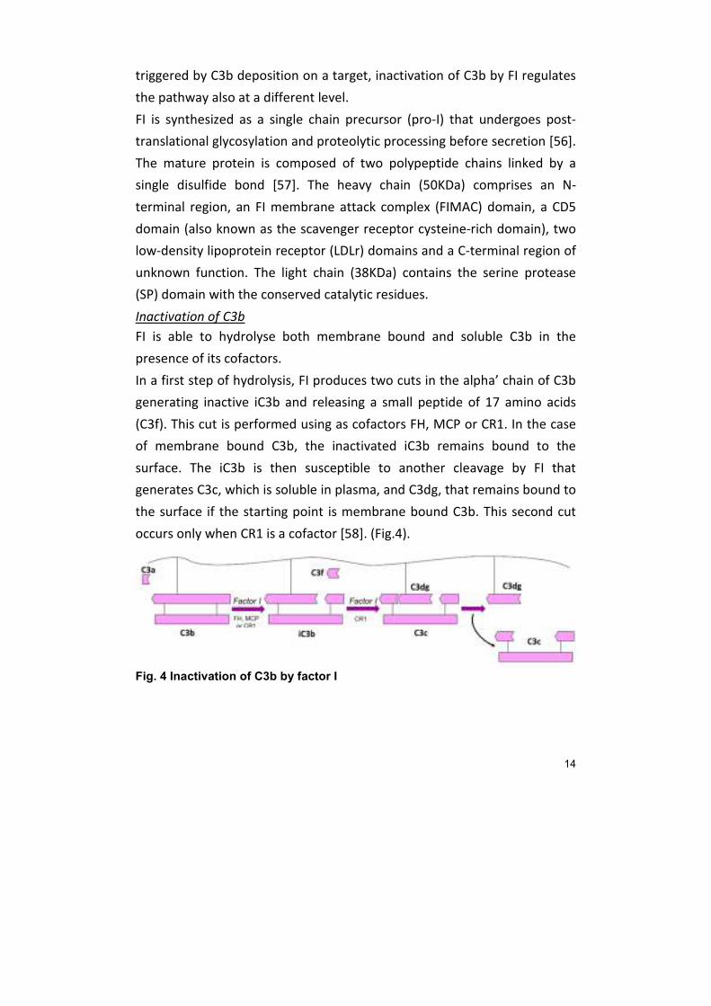
In a first step of hydrolysis, FI produces two cuts in the alpha’ chain of C3b
generating inactive iC3b and releasing a small peptide of 17 amino acids
(C3f). This cut is performed using as cofactors FH, MCP or CR1. In the case
of membrane bound C3b, the inactivated iC3b remains bound to the
surface. The iC3b is then susceptible to another cleavage by FI that
generates C3c, which is soluble in plasma, and C3dg, that remains bound to
the surface if the starting point is membrane bound C3b. This second cut
occurs only when CR1 is a cofactor [58]. (Fig.4).
Fig. 4 Inactivation of C3b by factor I

15
C3dg can be further cleaved by plasma proteases to C3d that is thought to
have similar biological activity than C3dg.
None of these fragments can activate the complement cascade, but the
hydrolysis products iC3b, C3c and C3dg can bind to specific complement
receptors and mediate functions such as the clearance of immune
complexes, opsonization, B cell stimulation and immunological memory.
Inactivation of C4b
In the presence of the cofactors C4BP, CR1, or MCP, FI cleaves the
membrane bound C4b inactivating it to iC4b. After a second cleavage the
soluble fragment C4c is released and the residual C4d, with unknown
biologic activity, remains bound to the membrane. (Fig. 5)
Fig. 5 Inactivation of C4b by factor I
2.2.1. RCA regulators
RCA regulators are a family of genetically, structurally and functionally
related proteins.
They are composed of 4 to 30 homologous short consensus repeat (SCR)
domains, also known as complement control protein (CCP), that have
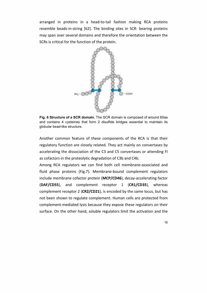
probably arisen by gene duplication events [59]. SCRs are small domains,
composed of approximately 60 amino acids each, that are characterized by
a high degree of conservation. They all contain four invariant cysteines that
form two disulfide bridges essential to maintain a globular bead-like
structure of the domain [60, 61]. (Fig. 6). The SCR domains are usually
16
arranged in proteins in a head-to-tail fashion making RCA proteins
resemble beads-in-string [62]. The binding sites in SCR- bearing proteins
may span over several domains and therefore the orientation between the
SCRs is critical for the function of the protein.
Fig. 6 Structure of a SCR domain. The SCR domain is composed of around 60aa
and contains 4 cysteines that form 2 disulfide bridges essential to maintain its
globular bead-like structure.
Another common feature of these components of the RCA is that their
regulatory function are closely related. They act mainly on convertases by
accelerating the dissociation of the C3 and C5 convertases or attending FI
as cofactors in the proteolytic degradation of C3b and C4b.
Among RCA regulators we can find both cell membrane-associated and
fluid phase proteins (Fig.7). Membrane-bound complement regulators
include membrane cofactor protein (MCP/CD46), decay-accelerating factor
(DAF/CD55), and complement receptor 1 (CR1/CD35), whereas
complement receptor 2 (CR2/CD21), is encoded by the same locus, but has
not been shown to regulate complement. Human cells are protected from
complement-mediated lysis because they expose these regulators on their
surface. On the other hand, soluble regulators limit the activation and the
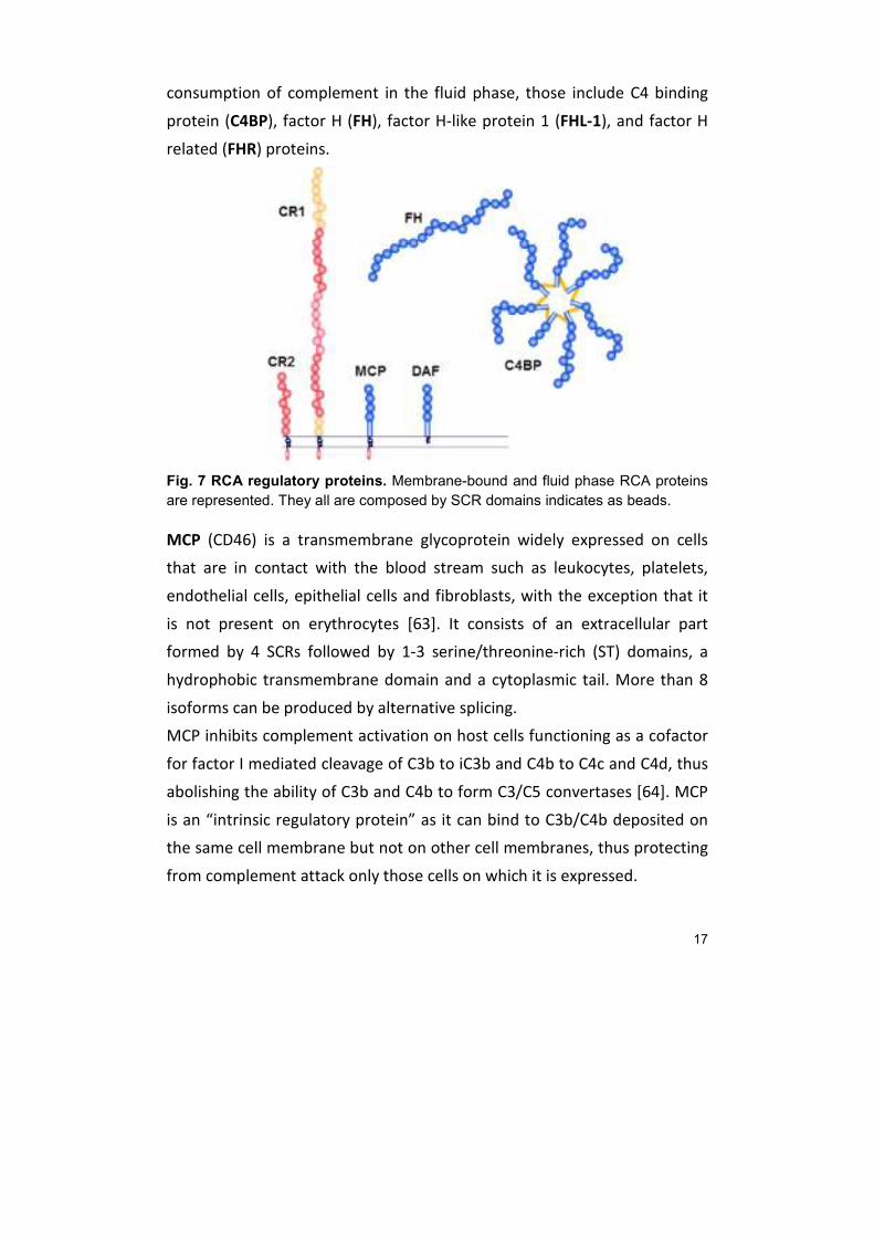
17
consumption of complement in the fluid phase, those include C4 binding
protein (C4BP), factor H (FH), factor H-like protein 1 (FHL-1), and factor H
related (FHR) proteins.
Fig. 7 RCA regulatory proteins. Membrane-bound and fluid phase RCA proteins
are represented. They all are composed by SCR domains indicates as beads.
MCP (CD46) is a transmembrane glycoprotein widely expressed on cells
that are in contact with the blood stream such as leukocytes, platelets,
endothelial cells, epithelial cells and fibroblasts, with the exception that it
is not present on erythrocytes [63]. It consists of an extracellular part
formed by 4 SCRs followed by 1-3 serine/threonine-rich (ST) domains, a
hydrophobic transmembrane domain and a cytoplasmic tail. More than 8
isoforms can be produced by alternative splicing.
MCP inhibits complement activation on host cells functioning as a cofactor
for factor I mediated cleavage of C3b to iC3b and C4b to C4c and C4d, thus
abolishing the ability of C3b and C4b to form C3/C5 convertases [64]. MCP
is an “intrinsic regulatory protein” as it can bind to C3b/C4b deposited on
the same cell membrane but not on other cell membranes, thus protecting
from complement attack only those cells on which it is expressed.

18
DAF (CD55) is a 70 KDa glycoprotein bound to the cell membrane by a GPI-
anchor followed by a serine/threonine-rich (ST) region and 4 consecutive
extracellular SCRs. It’s expressed on the surface of all blood cells and in
various tissues and organs [65]. A soluble isoform generated by alternative
splicing is present in body fluids [66].
DAF can control complement activation at different levels accelerating the
dissociation of both C3 and C5 convertases. DAF binds to the AP C3
convertase, C3bBb, accelerating its dissociation and also decreases the
stability of the CP/LP C3 convertase, C4bC2a, by accelerating its
dissociation to C4b and C2a [67]. In this way C3b peptides can no longer be
produced to bind to the surface of the cells. DAF can also bind and
dissociate C5 convertases on the surface of cells, thus protecting cells from
lysis mediated by the membrane-attack complex [67].
Complement receptor type 1 (CR1) is a large, multifunctional,
transmembrane glycoprotein. It is composed by a cytoplasmic domain, a
transmembrane region and a large extracellular domain composed of 30
SCRs in the most frequent allele. Three other alleles are possible containing
respectively 27, 37 or 44 SCRs. On the basis of the degree of homology the
SCRs are further grouped into larger units, called long homologous repeats
(LHRs) and designated LHR-A, -B, -C, and -D. The number of LHRs in the
different allelic forms varies from three to six [58].
CR1 is expressed on all peripheral blood cells with the exception of
platelets, natural killer cells and most T cells [68] and is also found in
glomerular podocytes, astrocytes and hyalocytes (vitreal macrophages).
CR1 binds C3b and C4b, and, with a lower affinity, iC3b and C3dg [58].
Different LHRs bind to different substrates and bear different activities.
CR1 on circulating cells acts mainly as an immune adherence receptor to
facilitate the removal of C3b/C4b-opsonized immune complexes and
pathogens from the circulation, which after binding are transported to
liver and spleen where they are processed by macrophages [12].

19
CR1 also exerts complement inhibitory activities: it possesses decay-
accelerating activity (DAA) for both CP and AP C3 and C5 convertases and it
irreversibly inactivates all four convertases, due to its co-factor activity (CA)
for factor I-mediated cleavage of C3b and C4b. CR1 is not particularly
efficient in generating iC3b, but is the only co-factor protein able to carry
out the third cleavage of C3b to generate C3c which is released to fluid
phase and C3dg [58].
CR1 has also been reported to bind to the collagenous portions of C1q [69]
and to mannan-binding lectin (MBL) [70].
Complement receptor 2 (CR2) which is expressed on B-lymphocytes binds
to iC3b, C3dg, or C3d and has a role in B-cell activation, bridging innate and
acquired immunity [71], but a specific role in complement regulation has
not yet been discovered.
C4 binding protein (C4BP) is a large soluble glycoprotein, present in plasma
at a concentration of approximately 200 µg/ml. C4BP exists in several
isoforms, the major form (α7/β1) is composed of seven identical α-chains
(70 KDa) and a single β-chain (45 KDa) all linked together at the C-termini
by disulfide bridges in a unique “octopus-like” structure (Fig. 8) [72]. Other
less abundant forms are composed of six α-chains and one β-chain (α6/β1)
or exclusively of seven α-chains (α7/β0) or of six α-chains (α6/β0). Both α-
and β-chains are composed of multiple SCRs domains, the α-chain contains
eight and the β-chain three. The SCRs domains in both chains are followed
by a C-terminal extension (~60 amino acid residues long) which contain an
amphiphatic α-helix region and two cysteines that are required for
intracellular polymerization and interchain disulfide bridging [73].
The genes encoding for both the α-chain (C4BPA) and the β-chain (C4BPB)
are located in the RCA gene cluster in close proximity.

20
Each of the α-chains contains a binding site for the activated complement
protein C4b, whereas the β-chain binds to coagulation system protein S
[74, 75].
Fig. 8 Structure of C4BP. The major isoform (α7/β1) composed of 7 α-chains and
1 β-chain is indicated. This protein has a unique “octopus-like” structure.
C4BP is the main inhibitor of the CP and LP of complement, where it
controls C4b-mediated reactions in several ways.
C4BP functions as a cofactor to factor I in the regulation of the complement
system, with C4b as the primary target. C4b bound to C4BP α-chain is a
substrate for FI, that cleaves it into inactive C4b (iC4b) [72]. However, C4BP
can also serve as cofactor to FI in the regulation of the complement protein
C3b, even though other proteins such as FH, MCP, and CR1 are more
efficient in supporting C3b degradation.
Moreover, C4BP prevents the assembly of the classical C3-convertase by
binding nascent C4b and it also accelerates the natural decay of the
complex [76]. On the other hand, C4BP does not seem to be an inhibitor of
the assembled alternative C3-convertase [77].
The binding of C4BP β-chain with protein S enables the S-C4BP complex to
bind to negatively charged phospholipid membranes, thus providing C4BP
with the ability to interact with certain cell surfaces, where it can express
its complement regulatory activities.
During the apoptotic process, negatively charged phospholipids, that are
normally located in the inner part of the cell membranes, are exposed on
21
the surface of the apoptotic cell. The protein S-C4BP complex can bind and
inhibit the activation of complement on the apoptotic cell, which may be
important for the controlling the inflammatory process. However, the
protein S-C4BP complex does not stimulate phagocytosis of the apoptotic
cells [78].
Factor H (FH), factor H-like protein 1 (FHL-1), and factor H related (FHR)
proteins will be discussed in more detail.
3. Complement Factor H
Complement Factor H (FH), the main regulator of AP, was first identified in
1965 by Nilsson and Müeller-Eberhard as β1H globulin [79]. Factor H is a
single polypeptide chain plasma glycoprotein of 155 KDa composed of 20
SCR domains [80].
FH is synthesized mainly by the liver [81], but extra-hepatic synthesis has
been demonstrated in a variety of tissues including endothelial and
epithelial membranes, glomerular mesangial cells, platelets, mesenchymal
stem cells and retinal pigment epithelium [82–86].
FH plasma levels in humans is very variable (116-562 μg/ml) and it has
been shown to be influenced by environmental factors such as age and
smoke, although the genetic component accounts for the major part
(~62%) [87].
3.1. FH functions
The main function of FH is down-regulation of AP which is achieved in
three different ways: FH competes with FB for binding to C3b, thus
preventing formation of C3 and C5 convertases of the AP [88]; FH also
possesses decay acceleration activity (DAA) and promotes the dissociation
of these convertases once they have formed [89]; finally it acts as an
essential cofactor for FI in the proteolytic conversion of C3b to iC3b [90]. In
22
the absence of FH, spontaneous activation of AP occurs in plasma and
leads to consumption of complement components C3 and FB [22].
The regulatory function of FH, which is mainly due to its interaction with
C3b, is performed both in the fluid phase and on self-surfaces where C3b is
bound. However, while FH binds and inactivates C3b promptly in fluid
phase, the inactivation of surface-bound C3b by factor H is dependent on
the chemical composition of the surface.
FH is able to distinguish between self and foreign surfaces and this is
mainly due to its capacity to bind polyanions such as sialic acid and
glycosaminoglycans (GAGs) such as heparin. The presence of these markers
on self surfaces increases the affinity of FH for surface-bound C3b as a
consequence of the simultaneous recognition of both types of molecules
by the same FH molecule [91–93]. Thus, FH acts by selectively limiting the
progression of the complement cascade on the surfaces of self-tissues and
allowing amplification of the AP only on foreign surfaces.
3.2. FH structure and functional domains
FH mature protein is composed of 1213 amino acid residues that are
organized in 20 SCRs of ~60 residues each joined in a “string-of-beads”
arrangement by 19 linkers of 3-8 residues.
A multiple alignment of the 20 SCRs highlights four invariant Cys residues
and a near-invariant Trp residue between Cys(III) and (IV). This consensus
sequence also occurs multiple times in other members of the RCA family.
Each of the 20 SCRs is presumed to fold into a distinct three-dimensional
(3D) structure termed the complement control protein module (CCP)
stabilized by two disulphide bounds between Cys(I)-(III) and Cys(II)–(IV)
[94].
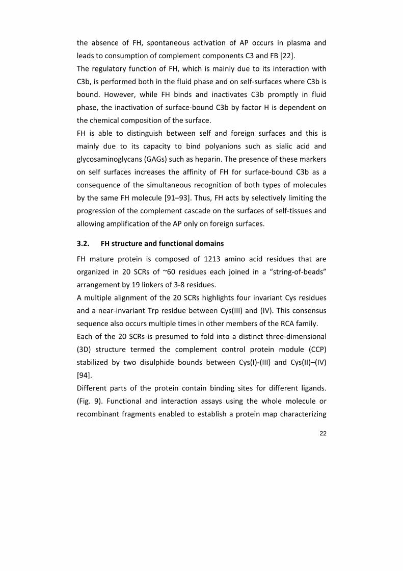
Different parts of the protein contain binding sites for different ligands.
(Fig. 9). Functional and interaction assays using the whole molecule or
recombinant fragments enabled to establish a protein map characterizing
23
the binding sites for C3b and polyanions [95–102]. FH contains three
regions important for binding C3b; two main binding sites located at each
end of the molecule and a third one in the central part of FH. The binding
site present in the N-terminal part (SCRs 1-4) is the only one necessary and
sufficient to perform the cofactor function for FI. It also accelerates the
dissociation of the convertase, although other regions of FH must
contribute, as the dissociation capacity of the fragment FH-SCRs 1-4 is 100
times lower than that of the whole FH [100]. The binding site for C3b
localised in the C-terminal region (SCRs 19-20) has the highest affinity to
C3b, however, both sites at opposite ends must cooperate in binding to
C3b. SPR interaction assays demonstrate that affinity of the entire FH is
much higher than that of the N- and C-terminal fragments taken separately
[103]. Moreover, SPR interaction assays also demonstrated that the three
different binding sites on FH interact with distinct sites on C3b [99], and
they can also bind C3b degradation fragments. The C3d fragment of C3b
binds to SCRs 19-20 whereas the central binding site is specific for C3c.
The interaction of FH with GAGs and polyanions has been located mainly to
two regions: SCR 7 with the contribution of SCRs 6 and 8, and SCRs 19-20,
in which SCR20 is the main heparin-binding site [103]. Another central
binding site (SCRs 12-15) has been proposed but never been confirmed.
These sites enable the binding of FH to host cell surfaces, but not to the
surfaces of pathogens, thus leading to the complement regulatory
protection of host cells.
FH also binds C-reactive protein (CRP) at SCR 6-8 and SCR 16-20 [104]. CRP
bound to damaged host cells can bind FH leading to C3b regulation.
Weak zinc binding sites are primarily located within SCR 6-8; these sites
lead to the precipitation of FH–C3b complexes in the pathophysiological
concentrations of zinc found in the retina [105]. In addition to these ligand
interactions, FH self-associates with itself to form dimers and higher
oligomers [106].
24
Fig. 9 Structure of FH. FH is composed of 60 SCR domains. Binding sites for
different ligands are indicated.
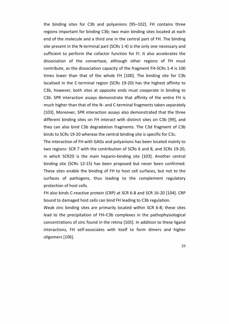
The structure of FH, composed by 20 SCRs linked by short sequences,
unlike it was previously believed, is not completely flexible, however N and
C-termini are brought close to each other (Fig. 10). This architecture must
be functionally relevant to allow both ends of FH to interact with the same
molecule of C3b. This would explain how C3b binding sites, SCRs 1-4 and
SCRs 19-20, cooperate and confer avidity. Several models of FH-C3b-
Heparin interaction have been proposed. Morgan HP et al hypothesised a
model of FH-C3b in which a single molecule of FH interacts with one of C3b
in non-overlapping regions through binding sites located at opposite ends
of FH (FHSCR1-4-C3b and FHSCR19-20-C3d/TED), and further enables the C-
terminal region of FH (SCRs 19-20) to bind to polyanions on the surface
[107].
Fig. 10 FH-C3b-heparin complex. FH is a rigid structure but can fold in on itself, bringing the two ends close to each other and enabling interaction with C3b and polyanions.

25
The highly complex FH structure can be essentially divided into two
functional regions:
- The N-terminal region, which binds to C3b and bears the regulatory
activities, such as cofactor activity (CA) and decay acceleration activity
(DAA)
- The C-terminal region, which binds to C3b and polyanions and provides
the selective ability to interact with surfaces
The correct activity of FH depends on cooperation between these two
regions and with the other interaction sites.
3.3. The CFH gene
FH is encoded by a single gene (CFH), located within the RCA in the region
1q32. The CFH gene contains 23 exons, the first exon codes for the 5'UTR
and signal peptide. Each of the other 22 exons code for one of the 20 SCR
domains except SCR 2 which is encoded by exons 3 and 4 (Fig. 11).[5]
Fig. 11 Structure of the CFH gene. Structure of CFH gene and different exons
coding for the two isoforms FH (white boxes) and FHL-1 (black boxes) are
indicated. Location of the CFH gene in the RCA cluster, in close proximity to
CFHRs genes, is also indicated.
From the same gene an alternative transcript is produced that codes for a
43 KDa protein named FH-like 1 (FHL-1). FHL-1 shares with FH the first 9
26
exons, which code for the first seven SCRs, plus exon 10, which is absent in
the mature messenger coding for FH. Exon 10 possesses a premature stop
codon and codes for a short tail of four amino acids (Ser Phe-Thr-Leu)
[108]. Both proteins are mainly expressed by the liver, but the plasma
concentration of FHL-1 is much lower (10-50 µg/ml). FHL-1 has
complement regulatory functions, such as cofactor activity for FI and
convertase C3bBb dissociation activity. FHL-1 is also capable of binding to
heparin, various microbial proteins, and cell surfaces, being involved in cell
motility processes [100, 109].
Despite FH and FHL-1 share the promoter region and the transcription start
site, the regulation of their expression is different depending on the
cellular type. The reason for this difference is unknown but it probably
depends on different transcriptional and post-transcriptional mechanisms
[110]. This suggests that there are specific functions for FH and FHL-1,
where both the tissue and the local concentration can be very important.
However, the precise role of FHL-1 in vivo is not determined.
3.4. Factor H protein family
In the RCA cluster, just downstream of the CFH gene, we can find five
genes (CFHR1, CFHR2, CFHR3, CFHR4, CFHR5) coding for the FH-related
proteins (FHRs) (Fig. 10). These proteins, together with FH and FHL-1 form
a family of glycoproteins (“Factor H protein family”) which are structurally
and functionally related [111].
The chromosomal segment containing CFH and CFHR genes is
characterized by several large genomic repeat regions, including different
exons of CFH and CFHRs 1-5, which have a high degree of sequence
identity. This situation favours non-homologous recombination processes
that result in chromosomal rearrangements and can lead to both deletions
and duplications. Chromosomal rearrangements can result in the loss of a
27
single or several CFHR genes, but can also give rise to new gene-
compositions and hybrid genes including in some cases also the CFH gene.
Rearrangements within the FH gene family may lead to defects associated
with pathology [5, 112].
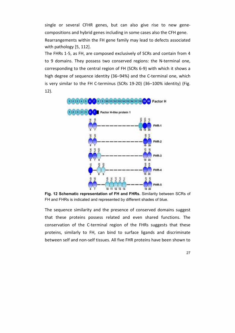
The FHRs 1-5, as FH, are composed exclusively of SCRs and contain from 4
to 9 domains. They possess two conserved regions: the N-terminal one,
corresponding to the central region of FH (SCRs 6-9) with which it shows a
high degree of sequence identity (36–94%) and the C-terminal one, which
is very similar to the FH C-terminus (SCRs 19-20) (36–100% identity) (Fig.
12).
Fig. 12 Schematic representation of FH and FHRs. Similarity between SCRs of
FH and FHRs is indicated and represented by different shades of blue.
The sequence similarity and the presence of conserved domains suggest
that these proteins possess related and even shared functions. The
conservation of the C-terminal region of the FHRs suggests that these
proteins, similarly to FH, can bind to surface ligands and discriminate
between self and non-self tissues. All five FHR proteins have been shown to
28
bind to C3b and to C3d. Of the two C-terminal SCRs in FHR proteins, the
one that corresponds to SCR19 in FH is highly conserved whereas the one
corresponding to SCR20 is more diverse. SCR20 of FH harbours an
important heparin binding site, suggesting that the FHRs may recognize
and bind to different cell surfaces.
On the other hand, the lack of a regulatory region such as the one present
in SCRs 1-4 of FH, with cofactor and decay activity, implies that FHRs are
not very potent in regulating complement [111].
According to their conserved domains, FHR proteins are divided into two
major groups: group I includes FHR-1, FHR-2 and FHR-5 whereas group II is
composed by FHR-3 and FHR-4 [113].
Proteins in group I are composed of a different number of SCR domains:
FHR-1 has five, FHR-2 four SCRs and FHR5 nine. This group is characterized
by their conserved N-termini (SCR1 and SCR2) which have more than 80%
sequence identity. These proteins circulate in plasma exclusively as dimers
(homodimers or heterodimers), and their dimerization is mediated by the
two conserved N-terminal SCRs.
FHR-1 is composed of five short SCRs, it possesses a dimerization domain in
the N-terminus and forms homodimers and likely also heterodimers with
FHR-2 and FHR-5.
Two variants of FHR-1 have been identified, an acidic isoform (FHRA) that
expresses the amino acids HLE in SCR3 and a basic isoform (FHRB)
expressing YVQ in SCR3. FHR-1 circulates in human plasma in two
glycosylated forms: FHR-1α with one and FHR-1β with two carbohydrate
side chains. The plasma concentration of CFHR1 is about 70 to 100 μg/ml.
FHR-2 consists of four SCRs, it forms homodimers and likely also
heterodimers with FHR-1 but not with FHR-5. The protein has two
glycosylated forms, a single glycosylated (24 KDa) and a double
glycosylated form (28 KDa). The plasma concentration is estimated to be
about 1/10 of FH, around 50 μg/ml.
29
FHR-5 is composed of nine SCRs and is the longest FHR protein, it circulates
in the plasma as homodimer and likely also as a heterodimer with FHR-1.
FHR-5 is the only FHR protein that has five SCR domains with high sequence
identities to SCRs 10–14 of FH. FHR-5 is a glycosylated protein of 62 KDa.
The group II FHR proteins, as well as factor H, lack the N-terminal
dimerization domains. FHR-3 and FHR-4 show a high degree of amino acid
identity to the SCRs 6–8 of FH (63 to 95%). SCRs 6–8 of factor H mediates
binding to heparin and to monomeric C-reactive protein (CRP), which is
conserved in FHR-4 [111].
FHR-3 consists of five SCR domains that show high amino acid identity to
SCRs 6-7-8 and SCRs 19-20 of FH. It is detected in plasma in multiple
variants (ranging from 35 to 56 KDa), reflecting the existence of four
different glycosylated variants of FHR-3. The plasma concentration is
estimated to be similar to FHR-1 (70–100 μg/ml).
FHR-4 is composed of nine SCRs and appears with an internal duplication
similar to an internal dimer, as SCRs 1–4 show a high level of identity to
SCRs 5–8 of the same protein. As FHR-3, it also has sequence identity to FH
SCRs 6-7-8 and SCRs 19-20. The human CFHR4 gene codes for two proteins,
FHR-4A and FHR-4B. FHR-4B derives from an alternatively spliced transcript
of the CFHR4 gene and is composed of five SCRs that correspond to half of
the SCRs of FHR-4A, in detail SCR1 and SCRs 5–9. FHR-4B has a mobility of
42 KDa and FHR-4A of 86 KDa. The plasma concentration of the two
variants has not yet been determined.
The high amino acid identity among the family members is also reflected
by the fact that antibodies raised against FH also detect different FHRs in
plasma and that the antibodies generated against FHRs cross-react with
other FHRs. Moreover, auto-antibodies against factor H in aHUS recognize
not only factor H but also certain FHRs [114]. This cross-reactivity is a

30
challenge for FHRs purification from plasma, their concentration
determination and in depth analysis.
The exact function of FHRs is still uncertain, but the functional relationship
between them and FH is now clearer: they can cooperate with FH in the
regulation of complement activation or compete for binding to ligands and
surfaces, which would result in less regulation.
4. Factor H-Associated Diseases
Complement activation and its regulatory mechanisms are in strict balance,
such that the effector activities of complement are normally directed
towards foreign surfaces and are avoided against self ones. Any imbalance
can lead to tissue damage, susceptibility to infection, as well as chronic
pathology.
Complement regulatory defects due to FH mutations or anti-FH
autoantibodies have been described in certain pathological conditions, and
CFH polymorphisms have also been associated with disease.
Among FH-associated diseases, the best studied are atypical Hemolytic
Uremic Syndrome (aHUS), Dense Deposit Disease (DDD) which affect
primarily the kidney with predominant glomerular dysfunction and Age-
related Macular Degeneration (AMD), a late onset eye dysfunction causing
impairment of the central vision. aHUS and AMD well be discussed in more
detail here.
4.1. Atypical Hemolytic Uremic Syndrome
The Hemolytic Uremic Syndrome (HUS) is a microvascular dysfunction
mainly affecting the kidney, but also the gastrointestinal and central
nervous systems can be involved. This condition is clinically defined as
hemolytic angiopathic anemia, thrombocytopenia and acute renal failure.
The main events triggering the pathogenic process seem to be endothelial
lesions in glomerular microvasculature due to either infection or other

31
exogenous damaging causes. These lesions show thickening of arterial and
capillary walls, endothelial inflammation and detachment of the glomerular
basal membrane (GBM), in addition to sub-endothelial accumulation of
proteins and cellular debris as well as formation of microclumps of
platelets and fibrin which may cause vascular occlusion. Generation of
schistocysts (fragmented erythrocytes) due to the circulation through
partially occluded renal microvasculature is another feature of HUS [115].
Two types of HUS are traditionally distinguished. The most frequent form
(90% of the observed cases), called typical, mainly affects children and has
an incidence of 2 cases per year every 100.000 inhabitants. Typical HUS is
associated with infection by an entero-haemorrhagic strain of Escherichia
coli (0157:H), which produces a highly toxic Shiga-like toxin. Although the
mechanism of action of this toxin has not yet been totally clarified, its role
in causing haemorrhagic diarrhoea and the evolution of this disease is not
under dispute. After crossing the intestinal barrier and entering the
systemic circulation, the Shiga toxins bind to receptors on the endothelial
surface causing lesions and triggering an inflammatory status and pro-
thrombotic episodes. Most cases of typical HUS have a positive outcome
and kidney function is restored in at least 75% of the patients. The
remaining cases can develop chronic kidney disease and in some cases
might even die (3-5%). [115].
Approximately 10% of cases of the hemolytic–uremic syndrome are
classified as atypical HUS (aHUS), since they are not caused by either Stx-
producing bacteria or streptococci. Atypical HUS can affect both children
and adults, its incidence corresponds to two cases per year per 1.000.000
inhabitants. Based on histological findings, the lesions observed in aHUS
are indistinguishable from those of the typical form; however, aHUS has a
much worst prognosis. Most patients show recurrent episodes and half of
them develop chronic kidney disease (CKD). aHUS mortality is
approximately 25% [116].

32
No special association has been found for aHUS, but a number of risk
factors have been found which predispose to this disease. Among them,
immune suppressing or anti-cancer therapies, oral contraceptives,
pregnancy and post-partum.
On the other hand, several studies have found, in familiar cases of aHUS,
and less frequently in sporadic cases, a clear genetic component. In some
cases of aHUS, mutations were found in genes coding for pro-coagulant
proteins, such as Thrombomodulin (TM), most patients were found
carrying mutations on genes coding for the alternative pathway of
complement (AP) [117].
The involvement of complement in HUS is known since 1974, but only in
recent years a clear association between aHUS and specific complement
proteins has been assessed. In many instances aHUS-associated mutations
and polymorphisms have been identified in regulatory proteins such as FH,
MCP, or FI and more recently to components FB and C3 [117]. In most of
these mutations functional defects have been identified which affect up to
half the aHUS cases. This implies that complement AP is somehow directly
involved in the pathogenesis of this disease.
In spite of all this evidence, in most cases a specific genetic or
environmental cause which might be considered the triggering event has
not been found. This calls for the existence of additional factors that have
not yet been identified. Among the mutations that have been described,
most are located in the CFH gene, which deserves special attention in the
pathogenesis of this disease [118].
4.1.1. FH in aHUS
Factor H mutations affect approximately 30% of aHUS patients, more than
100 mutations have already been described. An updated record of all
mutations associated with aHUS can be found at the FH–HUS online
database (http://www.fh-hus.org) [119].
33
Heterozygous mutations affecting various domains of FH have been
associated with aHUS, however missense mutations in the C-terminal
region (SCRs 19-20) are the most prevalent genetic alterations among aHUS
patients. Furthermore, in a significant number of patients mutations
affecting the functionality of the C-terminal region are the result of gene
conversion events between exon 23 of CFH and the homologous exon 6 of
CFHR1 [120] or of genomic rearrangements creating CFH::CFHR1 hybrid
genes [121].
FH mutations associated with aHUS rarely result in decreased FH plasma
levels, the majority of mutant proteins express normally and present
normal co-factor activity for the FI-mediated proteolytic inactivation of C3b
in plasma [122]. Functional analyses of several of these mutants showed an
altered interaction with C3b, heparin and endothelial cells [122–124].
As mentioned above, aHUS-associated FH mutations cluster in the C-
terminus of the protein, a region that is critical to control activation of
complement on cell surfaces. Consistent with this location, carriers of these
mutations express FH molecules that present normal regulatory activity in
plasma but a limited capacity to protect self cells from complement attack
[125].
Recent structural studies have provided new insights into how these
mutations impair the function of factor H in host/non-host discrimination
[107, 126]. Certain mutations in SCR20 were also found to reduce the
binding of FH to CRP [127] and to PTX3 [114]. For many mutations,
however, there is no functional effect known to date.
Moreover, anti-FH autoantibodies are detected in approximately 10% of
aHUS patients, mostly children. These antibodies have functional
consequences similar to those caused by the mutations in the C-terminal
region of FH [128, 129].
Several studies using recombinant FH fragments have been performed to
determine the binding sites of these autoantibodies and have shown that
34
they bind mainly to SCRs 19-20, although in some cases reactivity with
other domains, such as SCRs 8-11, was also observed [130].
Functional studies indicated that the autoantibodies directed against SCRs
19-20 interfere with the FH recognition functions impairing its interaction
with surface-bound C3b and thus inhibiting its complement regulatory
activity on host surfaces [130]. This reduced protection from complement-
mediated damage is likely to be involved in the endothelial injury
associated with aHUS.
Interestingly, 90% of the affected individuals presenting anti-FH antibodies
are also homozygous for the deletion of the CFHR1 and CFHR3 genes, it’s
still unclear whether the deletion of these genes predisposes to the
development of FH autoantibodies or if they are independent risk factors
for aHUS [129, 131, 132]. What is certain is that, due to the similar C-
terminal SCRs of FH and FHR-1, most of the studied autoantibodies
recognize both host complement regulators. FHR-1 in fact can hijack
autoantibodies and rescue host cells when added to anti-factor H
autoantibody-positive plasma [133].
A mouse model of aHUS is available, in which a truncated form of FH
(FHΔ16-20) that lacks the terminal five SCR domains, equivalent to the
location of the majority of aHUS-associated FH human mutations, is
produced [134].
4.2. Age-Related Macular Degeneration
AMD is the main cause of elder blindness in the industrialized countries. It
is an eye lesion affecting the central part of the retina, also known as
macula. AMD diagnosis is based on the degeneration signs that are evident
in the macula, independently from vision sharpness. Although substantial
histological changes are associated with AMD in the retina retinal pigment
epithelium (RPE), choriorcapillaris and choroid in middle age, AMD does
not manifest clinically before 55 years of age [135].
35
The typical clinical sign of AMD are the drusen, the origin of which is still
today totally unknown. As seen through the ophthalmoscope, they appear
as white to yellow dots in the retina, sometimes with a crystalline and
shiny aspect. They are extracellular deposits that accumulate between the
RPE basal membrane and the inner collagen layer of the Bruch membrane
[135].
The drusen are found in different eye diseases, and constitute a relevant
risk factor for the development of AMD. This association is very strong, and
as a consequence the presence of drusen is considered as a sign of an
early-stage of AMD, even in the absence of vision loss. Both the size and
the number of drusen are related to AMD severity. Drusen composition is
variable and not well defined, and only some of its components are known.
Their inside mainly contains glycoproteins and phospholipids, and in their
outer layer several proteins have been identified: fibronectin, chaperones,
apolipoprotein E, vitronectin, Amyloid P and complement proteins such as
C1q, C4, C5, C3 (and its degradation fragments), as well as the C5b-9
complex. The have also been shown to contain RPE cellular debris [136].
There is an early, almost asymptomatic stage in AMD, which is
characterised by the appearance of drusen as well as mild changes in RPE
pigmentation. In the later stage there is a severe vision loss, and the
disease may be classified as non-exsudative “dry”, or atrophic and
exsudative “wet” or neovascular. In the former case, well-defined
hypopigmented regions are produced, something known as “geographic
atropohy”, and the typical symptoms are blind holes or spots of vision. The
most advances stage is characterised by the presence of serum or
hemorrhagic fluid in the extracellular space, which causes the detachment
of the neuro-retina or of the RPE.
A typical sign is the choroidal neovascularisation, which can diffuse through
the Bruch membrane until towards the RPE. These changes generate
distorted images, which is often a sign of the wet form of AMD. Both types
36
of AMD can coexist in the same patient, and one type can change into the
other. The general progression is from the dry to the wet form: from 10 to
15% of patients evolve from the dry to the wet form [135].
The RPE is the main component of the retina which is involved in the
pathogenesis of AMD. The drusen structurally and functionally modify the
RPE, induce thickening of the monolayer, removing it from the intimate
vascular support of the choroidal region. This causes the interruption of
nutrient and metabolite supply and disposal of waste between the retina
and the choroid capillaries. Moreover, the modifications of the Bruch
membrane cause the impairment of cell adhesion, a reduced regenerative
capacity of the neuroretina by the RPE cells. All this, together with the
effect of neovascularisation, has a deep impact on the photoreceptors with
a direct negative effect on central vision. The extracellular deposits and
some of the components present in the drusen, such as activated
complement proteins and complement regulators, in addition unchain the
local inflammatory reactions that are involved in the pathogenesis of AMD
[135]. Changes in these eye structures, such as thickening of walls and loss
of permeability are naturally occurring as an aging feature. However, the
influence of environmental and genetic factors is very important.
AMD is a late onset complex disease caused, like many other multifactorial
diseases, by the convergent action of multiple risk factors. Among these,
we can quote aging, cigarette smoke, a high body mass index, and
especially genetics [135]. Multiple genes appear to be associated with
AMD, and particularly of note are Fib15, ABCA4, ApoE, HTRA1, ARMS2 and
different complement genes: CFH, CFB, C2, C3 and CFI [137].
HTRA1 and ARMS2 are on locus 10q26 and are strongly associated with
AMD. The former codes for a serine protease localised in the retina (as well
as in other tissues) and the latter is a hypothetical gene with unknown
function. An ARMS2 haplotype has been described with a sequence change
(Ala69Ser) which increases the risk of AMD up to 6.3 times in homozygous
37
carriers. However, the existence of this theoretical protein has not been
confirmed and its correlation with AMD pathogenesis is still elusive [137].
The most significative genetic association with AMD was found with the
locus 1q32, where the CFH gene is found.
4.2.1. FH in AMD
The common FH polymorphism Tyr402His has been associated with AMD
and the 402H variant represents the higher risk factor for developing the
disease. This association has been confirmed in different human
populations with an Odds Rate between 2.45 and 4.6 in heterozygous
subjects and up to 7.4 in homozygotes [138–140]. The frequency of the
402H varies among different populations, which explains the differences in
the incidence of this polymorphism for AMD.
Functional studies revealed different binding properties of the of the two
FH variants (402Y and 402H). The 402H variant has a reduced binding
affinity to CRP and a higher affinity for DNA and necrotic cells compared to
the 402Y variant. Since residue 402 is situated in SCR7 which is involved in
binding to the GAGs, there are also subtle differences between the variants
in their interaction with heparin and GAGs [141, 142].
No difference in binding to retinal pigment epithelial cells was found, but
the disease-associated variant binds less efficiently to both the
extracellular matrix protein fibromodulin and the Bruch’s membrane in the
retina [143].
Additional SNPs in the CFH gene associated with AMD were identified.
A polymorphism in exon 2, resulting in a Isoleucin to Valine exchange at
amino acid position 62 (I62V) of FH and FHL-1 has been reported. The 62I
variant is associated with the protective form for AMD and most likely has
a higher thermal stability [144].
The protective 62I variant has been shown to possess an increased affinity
for C3b and enhanced cofactor activity for the FI-mediated cleavage of C3b
38
[145], even though, as shown by crystallographic studies, residue 62 is
positioned outside the interaction interface of FH with C3b.
Recently, a new rare polymorphism (R1210C) was described in which the C
allele confers a 20-fold higher risk of AMD and appears to be involved in a
significantly earlier onset of the disease [146]. The same polymorphism
was previously shown to be involved in atypical Hemolytic Uremic
Syndrome [147].
In addition, a protective haplotype associated with the deletion of a 84-kbp
fragment directly downstream of CFH gene, that includes CFHR1 and
CFHR3 genes has been described [148].
4.3. Interaction with Pathogens
Since complement plays an important role in protection against infections,
and is generally regarded as the first line of defence against pathogenic
microorganisms it is not surprising that numerous pathogens (viruses,
bacteria, fungi and parasites) have evolved strategies for attenuating or
escaping complement attack.
Their mechanisms of action can be condensed to a few successful
strategies: the recruitment or mimicking of complement regulators; the
modulation or inhibition of complement proteins by direct interactions;
and inactivation by enzymatic degradation.
By acquiring complement regulators onto their surfaces microorganisms
gain the ability to regulate and control complement activation and can
evade the destructive effects of this powerful defense system. Many
pathogens are able to sequester FH on their surface in order to camouflage
themselves as host-like cells and thus are protected from complement
attack.
Binding of FH down regulates opsonization and lead to a rapid inactivation
of the newly formed C3b at the microbial surface, thus preventing further
amplification of the complement cascade and the formation of MAC. While
39
prevention of opsonization and subsequent phagocytosis is beneficial for
practically all microbes, evasion of MAC formation is especially important
for Gram-negative bacteria and spirochetes. Acquisition of FH is important
or even essential for pathogens; increasing numbers of them have been
shown to bind FH [149].
There are two main interaction sites on FH for microbial binding: one is
within SCRs 6–7 and the other in the C-terminal SCRs 19–20.
Microbes such as group A streptococci [150] and Neisseria [151] have been
shown to bind to the first site, for example the FH binding protein (FHbp)
of Neisseria meningitidis binds to SCR6 [152].
Binding via SCRs 6–7 facilitates also utilization of FHL-1, which contains
SCRs 1–7 of FH. Many microbes have been shown to bind both FH and FHL-
1 [153].
The mechanisms of interaction with FH/FHL-1 and complement protection
have been studied in more detail with the Gram-positive bacterium
Streptococcus pyogenes. Both FH and FHL-1 bind to the streptococcal M5
and M6 proteins, and under physiological conditions a preferential binding
of FHL-1 has been observed. In an M-protein bound conformation FHL-1
retains its cofactor activity and by promoting C3b degradation it can
contribute to resistance against phagocytosis. The sites required for the
interaction of the two proteins have been mapped: the surface-exposed
hypervariable region of the streptococcal M protein interacts with the C-
terminal SCR7 of the FHL-1 protein. Probably the M protein has an
additional binding site for FH, but despite all the studies no other sites have
been identified so far.
However, it seems that the majority of microbes utilize both sites to bind
to FH: for instance, B. burgdorferi sensu stricto, which causes Lyme disease,
binds to FH SCR7 using protein CRASP-1 [154] and to SCRs 19–20 using
outer surface protein E (OspE) and its paralogs [155]. This ability for dual
binding facilitates efficient protection against the AP attack.

40
Due to the high homology between the C-terminus of FH and C-termini of
FH-related proteins (FHRs), some microbes bind also certain FHRs but the
significance of this phenomenon for immune evasion is not yet clear.
The binding site in the C-terminal was analysed in more detail in a study
comparing Gram-negative, Gram-positive, and eukaryote microbes known
to bind FH, and using three microbial proteins, OspE (from B. burgdorferi
sensu stricto), FhbA (from B. hermsii), and Tuf (from P. aeruginosa). All the
pathogens and the proteins analysed share a common binding site in SCR
20 that overlaps but is not identical with the heparin and cellular binding
sites [156].
Interestingly, recent studies indicate that the FH 402H polymorphism may
relate to a better resistance from certain bacterial infections. Haapasalo et
al. showed that the AMD-associated factor H 402H variant has a lower
binding affinity to various streptococci compared to the 402Y variant,
resulting in a more efficient opsonization and phagocytosis [157, 158].
These data point to a pathogen-driven establishment of this common
polymorphism in the human population, with evolutionary advantage
against bacterial infection at the expense of late-age adverse effects in
developing AMD. This example raises the possibility that other factor H
polymorphisms have similarly spread in the human population because of
the evolutionary race between humans and their pathogens.
Moreover, interaction of FH with HIV has been previously described by our
group [159–161]. Envelope glycoproteins gp41 and gp120 have been
shown to bind to FH and target regions on these proteins have been
characterized, a possible biding region in FH has been identified in SCR13
but a full characterisation is still needed.
41
AIM OF THE WORK
Quantification of FH serum levels and assessment of its functionality are
crucial for patients characterisation in all FH-associated diseases but, due
to the high degree of sequence similarity between FHR proteins and FH,
tests specific only for FH are difficult to develop. Currently available
methods for the quantification of serum FH are often not specific as they
recognise both FH and FHR proteins, thus leading to an overestimation of
real FH levels. Assays for the structural and functional characterisation of
FH from individual subjects are available, but are usually cumbersome or
time-consuming and require complex purification procedures [162].
Many methods for the purification of FH have been reported, these are
mainly based on multiple chromatography steps needed to obtain a pure
fraction with minor contamination from FHRs proteins, but they require
long time and often lead to a partial degradation of FH.
FH has been shown to interact with several pathogens, but whereas for
some of them interaction with FH has been well described, for others the
direct “interactors” still remain unknown. Interaction of FH with HIV has
been previously reported by our group [159, 160] but up to the present
only target regions on HIV envelope glycoproteins have been characterized
whereas an analysis of the binding domains on FH is still needed.
The present work had a dual purpose with the common aim to better
understand the role of FH in different pathologies.
The first aim of my project was to develop FH-specific assays, using a
monoclonal antibody (MAb 5H5) produced in our laboratory [160]. The
antibody was first characterised and its ability to recognise specifically FH
without any cross-reaction with FHR proteins was verified. The antibody
was then used to purify large amounts of highly purified FH and to set up a
group of FH-specific assays.
Two tests respectively for the specific quantification of FH in patients’ sera
and for the analysis of its structural integrity were first developed and used

42
to screen aHUS and AMD patients. Later two functional assays to assess
both the N-terminal and C-terminal activity of FH have been set up and
used to test different samples.
The second purpose was the cloning and expression of the entire FH or
partial fragments in both an eukaryotic and a prokaryotic system, in order
to produce glycosylated and unglycosylated FH.
The recombinant proteins will be useful for different purposes. First of all
they can be used to screen which parts of FH are involved in interaction
with proteins of known pathogens and to explore interaction with other
pathogens, and also to test the importance of glycosylation in these
interactions. Secondly, their ability to restore the function of a defective FH
or to protect against FH autoantibodies can be analysed, with the final goal
to use them for therapy in many diseases caused by FH defects for which
the only possible treatment is currently plasma exchange.
43
MATERIALS AND METHODS
5. Production of 5H5 Monoclonal antibody
5.1. Suspension cell culture
A murine hybridoma (5H5) producing a monoclonal antibody (MAb) against
human FH was thawed and cultivated in T25 flasks with RPMI1640 medium
(Euroclone) containing 20% Fetal Bovine serum (FBS, Euroclone), 1x
PenStrep (Euroclone) and 4 mM L-glutamine (Euroclone) in a humidified
37°C, 5% CO2 incubator. Subsequently cells were subcloned and
maintained in T75 flasks in RPMI1640 complete medium with FBS reduced
to 10%. Aliquots were periodically harvested by centrifugation, frozen in 1
ml FBS aliquots containing 10% (v/v) DMSO (Sigma) and stored at -135°C.
Production of MAb was periodically checked by a home-made ELISA on
supernatants.
5.2. High density cell culture
In order to enhance antibody production the CELLine Bioreactor (CL1000)
(INTEGRA Bioscience) has been used. This system permits to cultivate cells
at high densities (up to 107-10
8 cells/ml) for a long periods allowing for a
high production of antibodies, 50-100 times higher than in static culture.
The bioreactor is a flask with two compartments, a cell compartment and a
medium compartment, divided by a 10 KDa semipermeable membrane.
This membrane allows small molecules, as nutrients and waste products, to
diffuse from one compartment to the other, whereas higher molecular
weight molecules secreted by the proliferating cells are retained within the
cell compartment.
The medium compartment was filled with 1 L of RPMI1640 medium
containing 1x PenStrep and 4 mM L-glutamine which was completely
changed every 14 days. 2.5x10⁷ hybridoma cells were collected by
centrifugation from static suspension cultures, resuspended in 15 ml
44
RPMI1640 medium containing 10% FBS, 1x PenStrep and 4 mM L-glutamine
and seeded in the cell compartment. Every 7 days all liquid was collected
from the cell compartment, cells were splitted 1:5 into fresh medium and
reseeded into the same chamber. The supernatant was checked for the
presence of antibody by ELISA test before storing it for subsequent
antibody purification.
5.3. Purification of 5H5 antibody
A ProteinG-Agarose column was prepared in order to purify 5H5
monoclonal antibody from both static or high density culture supernatant.
1 g of Protein G-Agarose resin (Sigma P7700) was rehydrated in 25 ml of
deionized water in agitation for 30 min. The resin was then thoroughly
washed on a Büchner funnel with gentle vacuum using 50 ml of deionized
water in order to remove the lactose stabilizer. The washed resin was then
resuspended in PBS buffer and packed into the column.
For antibody purification the ProteinG-Agarose column was attached to the
Econo-System (Bio-Rad). Column was washed at 2 ml/min rate first with
PBS for 15 min, then with 10 ml of 0.1 M Glycine pH 2.7 and again with PBS
until pH reached 7.4. 20 ml of cell culture supernatant were added to the
column until they completely entered the column then the Ab-depleted
fraction (Flow-through) was collected and column was washed again with
PBS for 10min.
The antibody was then eluted in 0.1 M Glycine pH 2.7 monitoring OD in
order to decide when to start collecting fractions. The eluates were
immediately neutralised with Tris-HCl 1.5 M pH 8.8 (20 µl/ml). The
collected fractions (Eluate) containing 5H5 antibody were stored for further
analysis. Finally the column was washed with PBS for 15 min and stored in
PBS-NaN3 (2%) at 4°C until next purification.
Eluted fractions were analysed by SDS-PAGE on a 12% gel followed by
Coomassie staining with BioSafe Coomassie (Bio-Rad).
45
Fractions from different runs containing the antibody were pooled
together and concentrated using Centriprep-3 (Amicon) or Vivaspin-20 (30
KDa cut-off) (Sartorius) concentrators following manufacturer's
instructions.
6. Purification of factor H
6.1. Preparation of 5H5-affinity column
Concentrated antibody preparations were desalted using EconoPac DG10
columns (Bio-Rad), according to the following protocol:
1. Remove the upper cap, and pour off the excess buffer above the top frit.
2. Add 20 ml of coupling buffer (0.1 M NaHCO3, 0.5 M NaCl, pH 8.4) to the
column (fill to the 30 ml mark), and snap off the bottom tip to start the
column flowing.
3. Allow the buffer to drain to the top frit. The column will not run dry.
Flow will stop when the buffer level reaches the top frit.
4. Add 3.0 ml of concentrated antibody to the column.
5. Allow entire sample to enter the column and discard the first 3.0 ml of
effluent.
6. Add 4.0 ml of coupling buffer to elute the antibody, while collecting the
4.0 ml fraction from the column.
Protein amount from concentrated fractions before and after desalting was
determined by Bradford Protein assay (Bio-Rad).
Column preparation:
1. Collect 500 mg of Cyanogen Bromide-Activated Sepharose 4B resin
(Sigma) into a 50-ml tube
2. Washed four times with 50 ml of 1 mM HCl
3. Leave the last wash in incubation for 10 min
4. Filter the resin on a Büchner funnel
5. Wash with milliQ water
6. Wash with coupling buffer (0.1 M NaHCO3, 0.5 M NaCl, pH 8.4)

46
7. Collect washed resin into a 15-ml tube and resuspend it in 10 ml of
coupling buffer containing 5 mg of desalted 5H5 antibody
8. Incubate at RT in agitation for 2h
9. After incubation, collect the coupling buffer with unbound antibody
10. Wash the resin twice with coupling buffer
11. Incubate the resin in 0.2 M Glycine pH 8.0 overnight at 4°C
12. Wash the resin alternatively with coupling buffer/acetate buffer (0.1 M
acetic acid, 0.5 M NaCl, pH 4.0) four times
13. Put the resin into a column and equilibrate with PBS for 15min.
14. Store the prepared 5H5-Sepharose column in PBS-NaN3 (2%) at 4°C
until use.
6.2. Purification of factor H
Human factor H was purified from human serum by affinity
chromatography on 5H5-Sepharose column. A set up of the protocol was
necessary to achieve an efficient purification without any contaminant. It
was necessary to test different elution protocols changing pH and salt
concentration of solutions in order to obtain a perfectly purified protein.
The final protocol is the following.
The 5H5-Sepharose column was attached to the Econo-System (Bio-Rad)
and washed at 1 ml/min first with PBS for 10 min, then with 10 ml of 0.1 M
Glycine pH 2.7 and finally with 0.5 M NaCl in PBS until pH reached 7.4.
Before adding it to the column 2 ml of human serum were brought to a
final concentration of 0.5 M NaCl and filtered through 0.22 µm filters. The
filtered human serum was allowed to completely enter the column at 0.25
ml/min rate, then the column was washed with 0.5 M NaCl in PBS at 1
ml/min rate and the fraction of FH-depleted serum (about 15 ml) was
collected until OD dropped. Finally FH was eluted from the column with 0.1
M Glycine pH 3.0 at 1 ml/min monitoring OD to decide when to start the
collection, typically 6 ml of eluate (Purified Factor H) were collected. The
eluate was immediately neutralised with Tris-HCl 1.5 M pH 8.8 ( 20 µl/ml).
47
The column was further washed with 0.1 M Glycine pH 2.7 in order to
eliminate possible contaminants and finally washed with PBS and stored in
PBS-NaN3 (2%) at 4°C until next purification.
Eluted fractions were analysed by SDS-PAGE on a 8% gel in reducing and
non-reducing conditions followed by Coomassie staining with BioSafe
Coomassie (Bio-Rad).
Positive fractions containing purified FH were pooled together and
concentrated using Vivaspin-20 (30 KDa cut-off) concentrators (Sartorius)
following manufacturer's instructions.
7. Factor H ELISA
A home-made ELISA test to determine FH concentration in biological fluids
was developed. This assay is a sandwich ELISA that uses MAb 5H5 as the
catcher and a FH-specific chicken antibody, also produced in our lab, as the
tracer together with 5H5-affinity purified FH as a standard.
Several adaptations have been made to the protocol to achieve the best
results. Tris-Buffered Saline (TBS) pH 7.65 has been shown to be a better
coating buffer than the traditional carbonate/bicarbonate buffer pH 9.6 for
5H5 antibody. The use of an HRP-conjugated secondary antibody instead of
the previously used AP-conjugated, and the consequent change of
chromogenic substrate from BluePhos® to TMB enhanced the sensitivity of
the test.
Controls without coating and without FH were also included during the set
up to assess test’s specificity.
The final detailed protocol is described in the appendix.
When aqueous humour and vitreous humour samples were used in the
test, working dilutions were as follows:
- aqueous humour: 1:5 and 1:10
- vitreous humour: 1:5, 1:10 and 1:50

48
8. IgG and Albumin Depletion
Albumin and IgG were depleted from normal human serum using Hi-Trap™
Albumin & IgG depletion columns (GE Healthcare) according to the
following protocol.
Buffers:
- Binding buffer: 20 mM sodium phosphate, 0.15 M NaCl, pH 7.4
- Elution buffer: 0.1 M glycin-HCl, pH 2.7
Sample preparation:
Human serum was centrifuged at 14,000 g for 10 min. at 4°C to eliminate
debris and 150 µl were diluted 1:10 in binding buffer.
Depletion procedure:
The column was connected to a peristaltic pump and a flow rate of 1
ml/min was used for the entire procedure.
A modified protocol from the manufacturer datasheet was used as follows:
1. Fill the pump tubing with binding buffer. Remove the stopper and the
snap-off end from the column and connect it to the pump tubing
'drop-to-drop' to avoid introducing air into the system.
2. Wash the column with 5 ml binding buffer to remove the 20% ethanol
storage solution.
3. Equilibrate with 10 ml of binding buffer.
4. Apply the diluted human serum to the column aspirating it directly
from the tube and then wash with 7 ml of binding buffer. Collect the
flow through (depleted serum) during sample application and wash
(total 8.5 ml).
5. Elute albumin and IgG from the column with 5 ml elution buffer and
immediately neutralise with 100 µl Tris-HCl 1.5 M pH 8.8.
9. Western Blot (WB)
Serum samples and samples deriving from different purification
procedures were subjected to SDS-PAGE in both reducing and non-
reducing conditions. For SDS-PAGE precast 4-12% or 12% NuPAGE® Novex®

49
Bis-Tris Protein Gel (LifeTechnologies) Mini or Midi were used and run in
MOPS 1X buffer at 200 V for 40-60 min. depending on the different gels
and formats.
Gels were blotted on a PVDF membrane using iBLOT (LifeTechnologies)
with protocol P3 for 7-10 min. Membranes were blocked with 3% BSA in
TBS/Tween 0.5% 2h RT or overnight at 4°C.
Different primary antibodies were used (Table 1) to reveal FH, FHRs, C3b
and FI. Primary antibodies were typically diluted in 1% BSA in TBS/Tween
0.5% and applied to the membranes 1h RT in constant agitation.
Membranes were then washed 3 times with TBS/Tween 0.1% and
incubated 1h RT in constant agitation with the corresponding Alkaline
phosphatase (AP) conjugated secondary antibody diluted in 1% BSA in
TBS/Tween 0.5%. After 3 washes in TBS/Tween 0.1%, specific signal was
revealed with SigmaFast™ BCIP/NBT (Sigma).
Table 1. Antibodies
Protein Primary Antibody Company Dilution Notes
FH 5H5 MAb Our laboratory 1:15000 Specific for FH
chicken anti-FH PAb Our laboratory 1:10000 Specific for FH
sheep anti-FH PAb TheBindingSite 1:2000 Sees also two
unspecified bands
around 75 KDa
FHR-1 rabbit anti-CFHL1 PAb Abcam 1:1000 Sees also FHR-2
rabbit anti-CFHR1 PAb Abnova 1:500 Sees also FHR-2
FHR-2 rabbit anti-CFHR2 PAb Abnova 1:500 Sees also FHR-1
FHR-3 rabbit anti-CFHR3 PAb ProteinTech 1:500 Sees FH, FHR-3
band is uncertain
FHR-4 rabbit anti-CFHR4 PAb Abcam 1:500 Sees also FH
FHR-5 rabbit anti-CFHR5 PAb Abnova 1:500 Specific for FHR-5
FI mouse anti-FI
(Supernatant)
MAb Serotec 1:100
sheep anti-FI PAb Sigma 1:2000
C3b goat anti-C3 PAb Sigma 1:2000 Sees different
fragments of C3

50
10. Factor H Immunoprecipitation (IP)
To isolate FH from patients’ sera an IP micro-method was developed using
5H5-Sepharose resin, in order to allow purification from small amounts of
sample. The final standard IP protocol was as follows:
1. Incubate in a 2.0-ml eppendorf tube 50 µl of serum or plasma with 50 µl
of 5H5-sepharose resin in 500 µl of PBS for 30 min on a rotator mixer
2. Centrifuge samples for 2 min at 13,000 rpm in a table-top
microcentrifuge
3. Collect in a new tube supernatant containing FH-depleted serum
4. Wash resin once with PBS/Triton 0.1% and three more times with PBS,
at each round centrifuge the tubes for 1 min at 13,000 rpm to pellet the
resin and discard the wash buffer
5. Elute FH from the resin with 200 µl of 0.1 M Glycine pH 2.7, incubating it
for 15 min on a rotator mixer and centrifuge the tubes for 1 min at
13,000 rpm to recover the eluate
6. Transfer the eluate into a new tube contain 4 µl of Tris-HCl 1.5 M pH 8.8
to neutralise it
7. Perform a second elution step in the same conditions to ensure that all
the FH is eluted from the resin
8. Wash the resin several times with PBS
The method was further developed by the use of Micro Bio-Spin™ Columns
(Bio-Rad) which allow for a faster procedure. The final protocol was as
follows:
1. Incubate in the capped micro column 50 µl of serum or plasma with 50
µl of 5H5-sepharose resin in 500 µl of PBS for 30 min on a rotator mixer
2. Snap off the tip and place the column in a 1.5 ml eppendorf tube to
collect the “FH-depleted serum”. Remove the cap and push it back on
the column to start the flow. Allow the sample to flow by gravity
through the column and eventually centrifuge for 30-60 sec. at 1,000 g
to allow all the sample to pass.
51
3. Wash resin once with PBS/Triton 0.1% and three more times with PBS
allowing the buffer to flow by gravity through the column, after the final
wash centrifuge for 30-60 sec. at 1,000 g to allow all the buffer to pass.
4. Place the column into a new tube contain 4 µl of Tris-HCl 1.5 M pH 8.8
and elute FH from the resin with 200 µl of 0.1 M Glycine pH 2.7,
allowing the sample to flow by gravity through the column and
eventually centrifuge for 30-60 sec. at 1,000 g to allow all the sample to
pass.
5. Repeat the previous step once
6. Wash the resin several times with PBS
11. Heparin and hydrophobic interaction chromatography
(HIC) purification
In order to compare our results, factor H was purified using two other
methods (Heparin and HIC) which don’t provide a complete separation of
FH from FHRs proteins.
11.1. Heparin purification
Heparin purification was performed by the use of HiTrap Heparin HP
Columns (GE Healthcare).
Method overview:
As FH and FHRs contain binding sites for Heparin with different binding
affinity, they can be purified from serum by affinity chromatography on
Heparin. Other serum proteins are also able to bind heparin are they can
therefore be purified together with FH and FHRs. To separate proteins
according to their binding capacity different salt concentrations can be
used in the elution steps. The resin in the column is composed of Heparin
covalently coupled to highly cross-linked agarose beads. Heparin used as a
ligand is a naturally occurring sulphated glucosaminoglycan which is
extracted from the native proteoglycan of porcine intestinal mucosa. It
52
consists of alternating units of uronic acid and D-glucosamine, most of
which are substituted with one or two sulphate groups.
Protocol:
The protocol of the manufacturer was slightly modified as follow.
Buffers:
- Binding buffer: 10 mM sodium phosphate, pH 7 + 50 mM NaCl
- Elution buffer: 10 mM sodium phosphate pH7 + NaCl
� E1 100 mM NaCl
� E2 150 mM NaCl
� E3 200 mM NaCl
� E4 250 mM NaCl
� E5 300 mM NaCl
� E6 400 mM NaCl
� E7 500 mM NaCl
� E8 1 M NaCl
Sample preparation:
300 µl of human serum were diluted 1:10 in binding buffer and filtered
through a 0.45 μm filter immediately before to be applied to the column.
15 ml IgG-Albumin depleted serum, which correspond to 300 µl of
undiluted serum were brought to a final NaCl concentration of 50 mM
before being applied to the column.
Purification procedure:
The column was connected to a peristaltic pump, a flow rate of 0.5 ml/min
was used for sample loading whereas 1 ml/min was used for washing and
elution.
1. Fill the pump tubing with binding buffer. Remove the stopper and
connect the column to the pump tubing, “drop to drop” to avoid
introducing air into the column.
2. Remove the snap-off end at the column outlet.
3. Wash out the preservative and equilibrate the column with 10 column
volumes of binding buffer.
53
4. Apply the sample into the column aspirating it directly from the tube
5. Collect the flow through (F.T.) into a new tube
6. Wash with 10 ml of binding buffer
7. Elute different fractions using 5 ml of each elution buffer and wash after
each step with 5 ml of the same buffer
8. Store all the samples (F.T. and Eluates) for further analysis
All the fractions were then concentrated and desalted in PBS Buffer using
Vivaspin-500 (5 KDa cut-off) (Sartorius) concentrators following
manufacturer's instructions. Fractions were subsequently analysed by SDS-
PAGE followed by Coomassie staining with BioSafe Coomassie (Bio-Rad),
and later by WB.
11.2. Hydrophobic interaction chromatography (HIC)
For HIC purification a column containing Macro-Prep Methyl HIC Support
(Bio-Rad) was prepared, serum was loaded in the presence of ammonium
sulphate and different fractions were eluted by gradually diminishing the
salt concentration.
Method overview:
Hydrophobic interaction chromatography (HIC) separates proteins on the
basis of their relative hydrophobicity. The principle for protein adsorption
to HIC media is complementary to ion exchange and size exclusion
chromatography. Sample molecules containing hydrophobic and
hydrophilic regions are applied to an HIC column in a high-salt buffer. The
salt in the buffer reduces the solvation of sample solutes. As solvation
decreases, hydrophobic regions that become exposed are adsorbed by the
media. The more hydrophobic the molecule, the less salt is needed to
promote binding. Usually a decreasing salt gradient is used to elute
samples from the column in order of increasing hydrophobicity.
FH has been shown to bind to hydrophobic resin and therefore can be
isolated with this procedure [163].
54
The Macro-Prep Methyl HIC Support which is ideal for purifying proteins
that have strong hydrophobic regions was chosen for this procedure.
Buffers:
- Buffer A: 10 mM Tris, pH 7.2 + 1.5M Ammonium Sulphate
- Buffer B: 10 mM Tris, pH 7.2
Column preparation:
The Macro-Prep Methyl HIC Resin was resuspended into storage buffer
(50% slurry) and packed into a column. The column was extensively
washed with Buffer B and later equilibrated with Buffer A.
Sample preparation:
1 ml of human serum was diluted 1:3 into Buffer A and brought to 1.5 M
(NH4)2SO4 adding 200 mg Ammonium Sulphate. The tube was left in the
end-to-end platform until the salt was dissolved. The sample was
centrifuged at 15,000 g for 15 min. to recover precipitates whereas the
supernatant was used for chromatography.
Setting up the Econo Chromatography System:
The column was connected to Econo-System (Bio-Rad) and the sample
supernatant (about 2.5 ml) was directly loaded on the Methyl HIC column.
Flow through which doesn’t bind to the column was collected (100%
Fraction). A gradient, mixing Buffer A and Buffer B in order to gradually
diminish Ammonium Sulphate content, was set on the instrument and
three fractions (66%, 33% and 0%) were collected monitoring OD280.
Collected fractions:
• 100% 1.5 M Ammonium Sulphate
• 66% 1 M Ammonium Sulphate
• 33% 0.5 M Ammonium Sulphate
• 0% no Ammonium Sulphate
All the fractions were concentrated and desalted in Buffer B using Vivaspin-
500 (5 KDa cut-off) (Sartorius) concentrators following manufacturer's
instructions. Fractions were subsequently analysed by SDS-PAGE followed
by Coomassie staining with BioSafe Coomassie (Bio-Rad), and later by WB.

55
12. Factor H integrity assay
FH (150 KDa) can be cleaved by a serum protease between amino acids
341-342, thus generating two fragments (142 KDa and 38 KDa) that are
held together by a disulphide bridge and are visible as separate molecular
species only after reduction. This cleavage can be mimicked in vitro by
trypsin. In order to develop a test capable of detecting the presence of the
cleaved form of FH it was necessary to test the ability of 5H5 to bind it,
using trypsin-cleaved FH.
12.1. Trypsin cleavage of FH
Reagents:
• Trypsin TPCK treated from bovine pancreas (Sigma - T1426) - 1mg/ml in
1mM HCl
• Trypsin inhibitor from Glycine max (soybean) (Sigma - T9003) - 1mg/ml
in H2O
• 5H5-affinity purified FH
Protocol:
Trypsinization of FH was performed with a FH:trypsin ratio of 100:1 (w/w)
(1 µg of FH + 10 ng of trypsin) in TBS pH 7.5, incubating at 37°C for different
times.
Digestion was stopped by adding twofold weight excess of soybean trypsin
inhibitor.
12.2. IP of Trypsin-cleaved FH
A sample with 50% of trypsin-cleaved FH was immunoprecipitated with
5H5-coupled Sepharose resin, as described previously. Samples before IP,
and different fractions of IP were analysed by SDS-PAGE followed by
Coomassie staining and densitometric analysis with the program TotalLab.

56
12.3. Integrity assay
After demonstrating that 5H5 recognizes and immunoprecipitate with the
same affinity both the uncleaved and cleaved form of FH, this important
feature was used to set up a test for the integrity of FH in different
samples.
The test consists in the 5H5-affinity immunoprecipitation of FH from serum
samples or from FH preparation followed by SDS-PAGE and Coomassie
staining. A 50% trypsin-cleaved FH is used as a control.
13. Factor H functional assays
Two tests to screen N-terminal cofactorial activity and C-terminal
protection activity of FH have been set up, namely the “Cofactor assay”
and the “Haemolysis test”.
13.1. Quantification of Purified Factor H
The exact quantification of purified FH samples was a crucial point in order
to perform the functional assays. Different quantification methods were
used and compared: Bradford method with Bio-Rad protein assay, our
home-made FH ELISA and spectrophotometric determination at 280 nm.
Bio-Rad protein assay was performed following manufacturer instructions,
the home-made ELISA developed for serum and plasma sample was used
as described previously.
Spectrophotometric determination was done with a Nanodrop instrument.
PBS was used as a blank and absorbance at 280 nm was measured. To
establish FH concentration an extinction coefficient for a 1% solution (E1%)
of 12.4 was used, as reported by others [164].
This formula was used to calculate the concentration expressed in µg/ml:
c = (A280/E1% * 10) * 1000
57
13.2. Cofactor assay:
This test is based on the fact that FI can cut C3b in vitro only in the
presence of FH; if FH is not functional C3b cannot be cleaved.
All the samples to be used in this test were brought into PBS buffer and
their concentration was established as previously reported.
In the test we used purified C3b and FI (Merck Millipore) and 5H5-affinity
purified FH.
The test was set up with different amounts of purified proteins and with
different incubation times. FH concentration was gradually scaled down to
assess test sensitivity.
The final protocol used to screen samples was as follows:
500 ng of purified C3b and 250 ng of purified FI were incubated in PBS for
30 min at 37°C alone or in the presence of different amounts of 5H5-
affinity purified FH. After incubation samples were put on ice and SDS
sample buffer was immediately added to stop the reaction. Samples were
loaded on a SDS-PAGE gel and analysed by WB using a polyclonal anti-C3b
antibody (Sigma) or by Coomassie staining.
13.3. Haemolysis test
This test to assess the activity of the C-terminus is based on the fact that FH
is able to protect Sheep erythrocytes from complement attack. If
complement is activated erythrocytes undergo lysis that can be measured
by the release of haemoglobin using a microplate reader.
The test has been first set up using a control serum with known amount of
functional FH. Protocol was adapted from “AH50 Assay for Total Alternative
Pathway Hemolytic Activity” Giclas (2001) [165].
Due to the impossibility to purchase Veronal buffer that is normally used in
this kind of test it was necessary to set up the test using two different
buffers:
58
- A modified Veronal buffer adapted using a commercially available buffer
similar to the one needed: Veronal Buffer 5X (Lonza)
- HEPES buffer adapted from Moreno-Indias et al. [166]
The final detailed protocol is described in the appendix.
The test was then performed adding different concentration of EDTA to
serum. This was necessary to test the minimum concentration of EDTA that
still allows lysis, since depleted serum needs to be prepared in the
presence of EDTA in order to prevent complement activation during the
procedure.
The final part of the test that makes use of FH-depleted serum alone or
with different amounts of FH to be tested is still under development.
14. Cloning of factor H
The sequence of human factor H RNA (NM_000186.3) has been divided
into three main fragments containing approximately 7 SCR each (SCR 1-7,
SCR 8-14, SCR 15-20) and specific PCR primers were designed (Table 2).
Quality and specificity of primers was checked with Primer Premier
software (Primer Biosoft).
14.1. Retrotranscription
Human Liver total RNA (Life Thechnologies) was retrotranscribed with two
methods: iScript cDNA Synthesis Kit (Bio-Rad) which contains a mix of
Random Hexamers and Oligo(dT) or with ImProm-II™ Reverse Transcription
System (Promega) which enables the use of Oligo(dT) or Specific primers in
different reactions. A modified protocol was used.
14.1.1. RNA denaturation
Samples for different retrotranscription reactions were prepared as follows
and denatured at 70°C for 5 min followed by a 10 min incubation on ice.

59
cDNA-1
(ImProm)
cDNA-2
(ImProm)
cDNA-3
(iScript)
Oligo(dT)15 1 µl - -
FH-specific
primers
- 1 µl -
RNA 1 µl 1 µl 1 µl
Nuclease free
H2O
3 µl 3 µl 4 µl
14.1.2. Retrotranscription reactions
Different reaction mixes were prepared and added to the samples:
RT mix ImProm RT mix iScript
5x ImProm reaction buffer 4 µl 5x reaction mix 4 µl
MgCl2 2.5 µl iScript RT 1 µl
dNTPs 1 µl Nuclease free H2O 5 µl
RNAse Inhibitor 1 µl
ImProm RT 1 µl
Nuclease free H2O 5.5 µl
Retrotranscription was performed with the following thermic protocol:
25° C 5’
42° C 60’
85° C 5’
cDNA quality was checked by PCR using beta-actin primers when possible.
14.2. Amplification
The entire FH and the three fragments have been amplified using Phusion™
DNA Polymerase (Thermo). Regular HF buffer was substituted with CG
Buffer, which is normally used for difficult templates, as FH cDNA tends to
form secondary structures and amplification results challenging.
60
Mix Thermic protocol
5x CG buffer 4 µl 98° C 30”
dNTPs (10mM) 0.4 µl 98° C 10”
Primer F (10µM) 1 µl * 64° C 20” X 35
Primer R (10µM) 1 µl 72° C 50”*
Phusion 0.2 µl 72° C 5’
cDNA 2 µl
dH2O up to 20 µl
* for the amplification of the entire FH 0.8 µl of dNTPs and 2’ extension
were used.
Amplification products were checked and quantified on a agarose gel.
14.3. Features of the pFastBac™/HBM-TOPO® vector
The pFastBac™/HBM-TOPO® vector (Life Technologies) was chosen for
cloning and expression into insect cells. The vector contains the following
elements:
• Strong polyhedrin (PH) promoter for high-level baculovirus-based
protein expression in insect cells
• TOPO® Cloning site for rapid and efficient cloning of blunt-end PCR
products amplified with proofreading polymerases
• N-terminal Honey Bee Mellitin (HBM) secretion signal coding sequence
for secretion of the cloned gene product into the extracellular medium
• C-terminal polyhistidine tag for simple purification of recombinant
proteins
• TEV protease cleavage site for removal of the polyhistidine tag
following protein purification using AcTEV™ protease
• SV40 polyadenylation signal for efficient transcription termination and
polyadenylation of the recombinant transcript
• Mini-Tn7 elements for site-specific transposition of the gene of interest
into the baculovirus shuttle vector (bacmid DNA) propagated in E. coli
• Ampicillin (bla) resistance gene (β-lactamase) for selection of
transformants in E. coli
61
• pUC origin for high copy replication and maintenance of the plasmid in
E. coli
• Gentamicin resistance gene for selection of transformants containing
recombinant bacmid DNA
14.4. Blunt-End TOPO® Cloning Reaction
The amplified fragments were inserted into the pFastBac™/HBM-TOPO®
vector (Life Technologies) using TOPO-cloning.
All the samples were brought to a concentration of 5 ng/µl and an
insert:vector ratio of 2:1 was used in the in a TOPO® Cloning reaction. The
6-µl reactions were prepared as follows:
PCR Product (5 ng/µl) 4 µl
Salt Solution (200 mM NaCl, 10 mM MgCl2) 1 µl
pFastBac™/HBM-TOPO® vector 1 µl
and incubated 5 min at RT.
14.5. Transformation and analysis of tranformants
The TOPO reactions were used to transform Mach1™ T1R Chemically
Competent E. coli (Life Technologies) following the manufacturer protocol.
Plates were incubated overnight at 37°C.
Some colonies were screened by Colony PCR to check the orientation of
the insert, two parallel PCR reactions were performed:
- PCR Forward: which detects the fragment inserted in the correct
orientation.
In this PCR a forward primer on the insert together with SV40 reverse
primer on the vector is used. (Table 2)
- PCR Reverse: which detects the fragment inserted in the correct
orientation
In this PCR a reverse primer on the insert together with SV40 reverse
primer on the vector is used, if the fragment is in the wrong orientation
the reverse primer on the insert functions as a forward primer giving
amplification. (Table 2)

62
After this first screening, plasmid DNA from positive ones was isolated by
Mini-prep using Eurogold Plasmid kit (Euroclone) according to the
manufacturer protocol.
Plasmid DNA was then sequenced using primers located on the vector
(Table 2) and sequences were analysed with CLC Sequence Viewer (CLC
bio).
14.6. Cloning into a prokaryotic system
Primers to clone the same fragments into a prokaryotic system were also
designed. (Table 2)
These primers will be used to amplify the fragments directly from the
previously generated pFastBac™/HBM clones, fragments will be
subsequently inserted into E. coli expression vector using the StarGate® IBA
System (IBA BioTAGnology).
Table 2. Primers
Primer Sequence (5’-3’)
PCR primers for cloning into the eukaryotic system
FH-SCR1-F GAAGATTGCAATGAACTTCCTC
FH-SCR7-R GACACGGATGCATCTGG
FH-SCR8-F GTCAAAACATGTTCCAAATCAAG
FH-SCR14-R TTTTTCAACACAGAGTGGTATTG
FH-SCR15-F AAAATTCCATGTTCACAACCAC
FH-SCR20-R TCTTTTTGCACAAGTTGGATAC
Colony PCR primers
SV40 polyA-rev GGTATGGCTGATTATGATC
Used together with PCR specific primers
Sequencing primers
pFASTBAC-F TCCGGATTATTCATACCGTCCC
pFASTBAC-R CCTCTACAAATGTGGTATGGCTG
PCR primers for cloning into the prokaryotic system
Star-FH-SCR-1F AGCGGCTCTTCAATGGAAGATTGCAATGAACTTCCTC
Star-FH-SCR-7R AGCGGCTCTTCTCCCGACACGGATGCATCTGGGA
Star-FH-SCR-8F AGCGGCTCTTCAATGGTCAAAACATGTTCCAAATCAAG
Star-FH-SCR-14R AGCGGCTCTTCTCCCTTTTTCAACACAGAGTGGTATTG
Star-FH-SCR-15F AGCGGCTCTTCAATGAAAATTCCATGTTCACAACCAC
Star-FH-SCR-20R AGCGGCTCTTCTCCCTCTTTTTGCACAAGTTGGATAC

63
RESULTS
15. Production and Purification of 5H5 antibody
High density hybridoma cultures with CELLine Bioreactor enabled to
produce a consistent amount of MAb 5H5, mean antibody production was
about 1.5-2 mg/day.
The quality of purified antibody checked by SDS-PAGE in reducing
conditions followed by Coomassie staining was good, and purity was about
90-100%. Two major bands are visible on the gel, a 50 KDa band
corresponding to antibody’s heavy chains and a 25 KDa band
corresponding to light chains, an upper band due to incomplete reduction
can also be present. (Fig. 13)
Fig. 13 Coomassie of purified MAb 5H5: two elution fractions are shown (El.1
and El.2), antibody elute in the first fraction and bands corresponding to heavy (50
KDa) and light (25 KDa) chains are clearly visible.
MAb 5H5 antibody has been tested in Western Blot and fractions of total
human serum, albumin- and IgG-depleted serum and albumin and IgG
purified from serum have been analysed. Under non-reducing conditions a
clear band around 150 KDa corresponding to FH is visible in total serum
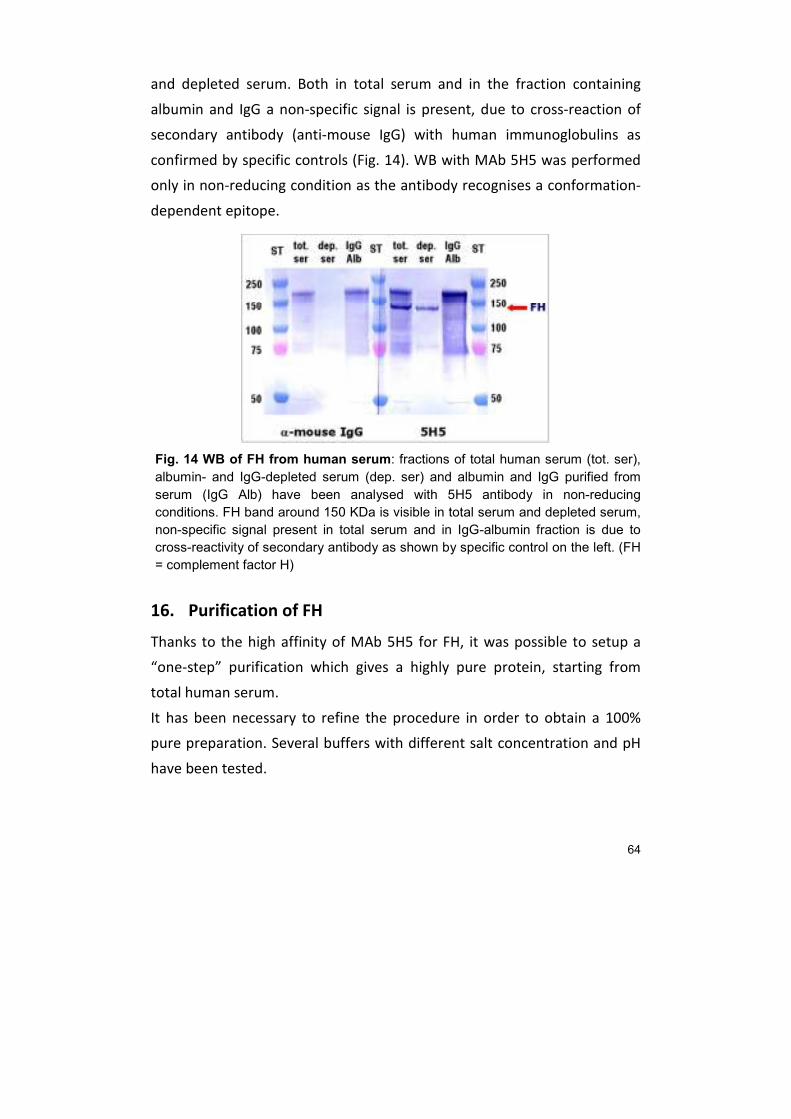
64
and depleted serum. Both in total serum and in the fraction containing
albumin and IgG a non-specific signal is present, due to cross-reaction of
secondary antibody (anti-mouse IgG) with human immunoglobulins as
confirmed by specific controls (Fig. 14). WB with MAb 5H5 was performed
only in non-reducing condition as the antibody recognises a conformation-
dependent epitope.
Fig. 14 WB of FH from human serum: fractions of total human serum (tot. ser),
albumin- and IgG-depleted serum (dep. ser) and albumin and IgG purified from
serum (IgG Alb) have been analysed with 5H5 antibody in non-reducing
conditions. FH band around 150 KDa is visible in total serum and depleted serum,
non-specific signal present in total serum and in IgG-albumin fraction is due to
cross-reactivity of secondary antibody as shown by specific control on the left. (FH
= complement factor H)
16. Purification of FH
Thanks to the high affinity of MAb 5H5 for FH, it was possible to setup a
“one-step” purification which gives a highly pure protein, starting from
total human serum.
It has been necessary to refine the procedure in order to obtain a 100%
pure preparation. Several buffers with different salt concentration and pH
have been tested.
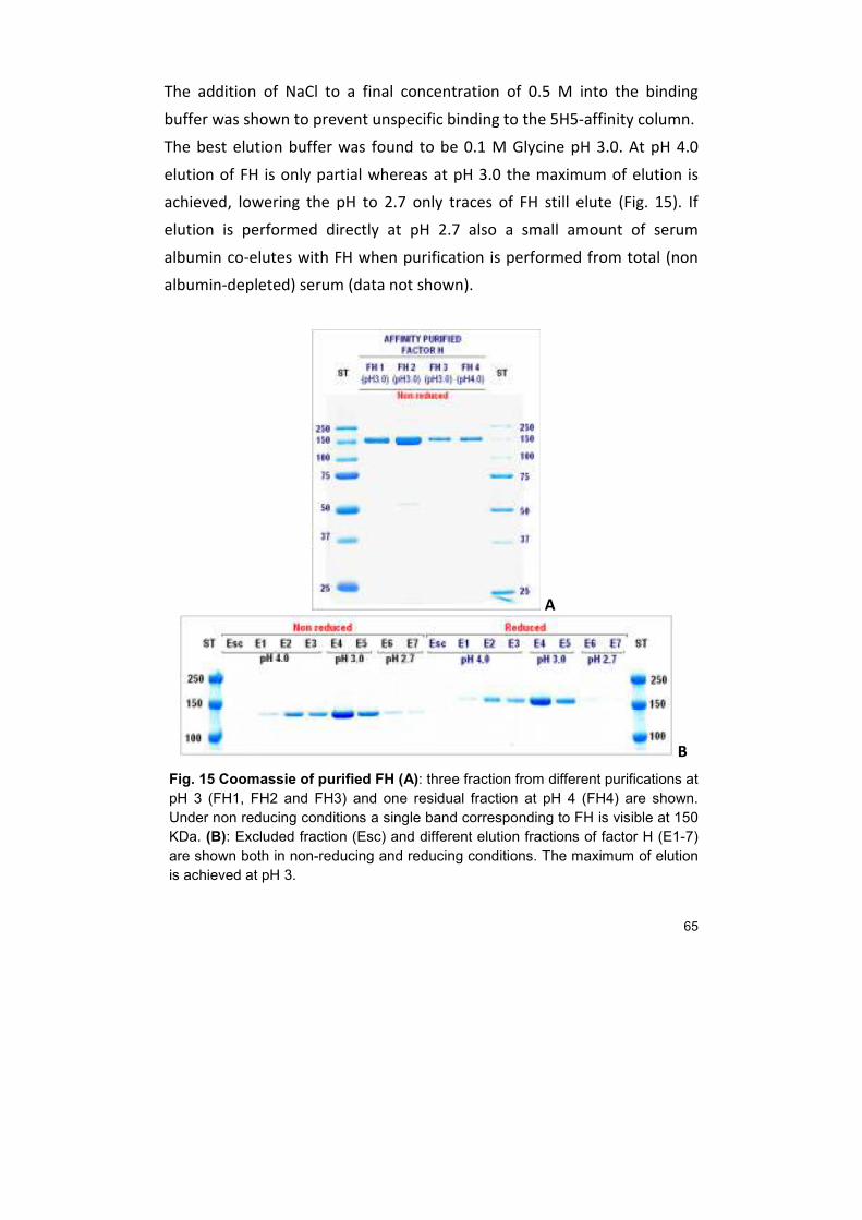
65
The addition of NaCl to a final concentration of 0.5 M into the binding
buffer was shown to prevent unspecific binding to the 5H5-affinity column.
The best elution buffer was found to be 0.1 M Glycine pH 3.0. At pH 4.0
elution of FH is only partial whereas at pH 3.0 the maximum of elution is
achieved, lowering the pH to 2.7 only traces of FH still elute (Fig. 15). If
elution is performed directly at pH 2.7 also a small amount of serum
albumin co-elutes with FH when purification is performed from total (non
albumin-depleted) serum (data not shown).
A
B
Fig. 15 Coomassie of purified FH (A): three fraction from different purifications at
pH 3 (FH1, FH2 and FH3) and one residual fraction at pH 4 (FH4) are shown.
Under non reducing conditions a single band corresponding to FH is visible at 150
KDa. (B): Excluded fraction (Esc) and different elution fractions of factor H (E1-7)
are shown both in non-reducing and reducing conditions. The maximum of elution
is achieved at pH 3.

66
Efficiency of the method and purity of the affinity purified FH were checked
by Western Blot.
Western blot on total serum, purified FH fractions and FH-depleted serum
was performed in non-reducing conditions with both MAb 5H5 and
polyclonal chicken anti-FH. With both antibodies, a 150 KDa band is
present in purified FH fractions whereas it is missing in FH-depleted serum,
confirming that depletion was complete (Fig. 16). Non-specific signal due to
cross-reaction of the polyclonal or the secondary antibodies with human
immunoglobulins is present in total serum and FH-depleted serum (Fig. 16).
A B
Fig. 16 (A) WB of FH from human serum: fractions of total human serum (tot.
ser), albumin- and IgG-depleted serum (dep. ser) and albumin and IgG purified
from serum (IgG Alb) have been analysed with 5H5 antibody in non-reducing
conditions. FH band around 150 KDa is visible in total serum and depleted serum,
non-specific signal present in total serum and in IgG-albumin fraction is due to
cross-reactivity of secondary antibody as shown by specific control on the left. (FH
= complement factor H) (B) WB of purified FH: fractions of purified FH (FH) and
FH-depleted serum (dep. ser) have been analysed in non-reducing conditions with
both MAb 5H5 and polyclonal chicken anti-FH antibodies. FH band (150 KDa) band
is present in purified FH fractions whereas it is missing in FH-depleted serum.
Depletion of FH from serum and efficiency of purification was also checked
in an ELISA test using purified FH as a standard for precise quantification
(Fig. 17).
As shown in Figure 17, a single step of affinity chromatography was able to
almost totally deplete the sample of FH, less than 1 µg/ml is still present in
depleted serum.

67
A B
Fig. 17 FH ELISA from depleted serum: OD at different dilutions of FH-depleted serum compared to purified FH (A) and quantification of residual FH in FH-depleted serum alone or compared to total serum (B) are shown.
Moreover purity of the fractions and absence of “contaminating” FHRs
proteins was demonstrated by WB using antibodies against different FHRs
proteins. The FH purified by 5H5-affinity chromatography was compared
with fractions obtained with other purification methods such as Heparin-
affinity chromatography and HIC chromatography. In fractions obtained by
5H5-affinity, unlike the ones coming from other purification methods, no
FHRs proteins are present (Fig. 18).
Fig.18 WB of total serum and different fractions of 5H5-affinity purification and Heparin purification developed with antibodies for FH and FHRs: FHRs are completely absent in the 5H5-affinity purified FH indicated with the red arrow.
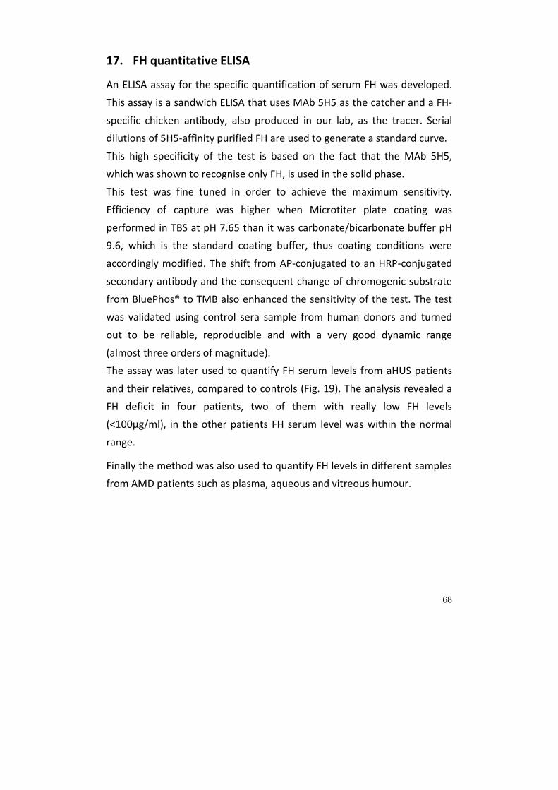
68
17. FH quantitative ELISA
An ELISA assay for the specific quantification of serum FH was developed.
This assay is a sandwich ELISA that uses MAb 5H5 as the catcher and a FH-
specific chicken antibody, also produced in our lab, as the tracer. Serial
dilutions of 5H5-affinity purified FH are used to generate a standard curve.
This high specificity of the test is based on the fact that the MAb 5H5,
which was shown to recognise only FH, is used in the solid phase.
This test was fine tuned in order to achieve the maximum sensitivity.
Efficiency of capture was higher when Microtiter plate coating was
performed in TBS at pH 7.65 than it was carbonate/bicarbonate buffer pH
9.6, which is the standard coating buffer, thus coating conditions were
accordingly modified. The shift from AP-conjugated to an HRP-conjugated
secondary antibody and the consequent change of chromogenic substrate
from BluePhos® to TMB also enhanced the sensitivity of the test. The test
was validated using control sera sample from human donors and turned
out to be reliable, reproducible and with a very good dynamic range
(almost three orders of magnitude).
The assay was later used to quantify FH serum levels from aHUS patients
and their relatives, compared to controls (Fig. 19). The analysis revealed a
FH deficit in four patients, two of them with really low FH levels
(<100µg/ml), in the other patients FH serum level was within the normal
range.
Finally the method was also used to quantify FH levels in different samples
from AMD patients such as plasma, aqueous and vitreous humour.
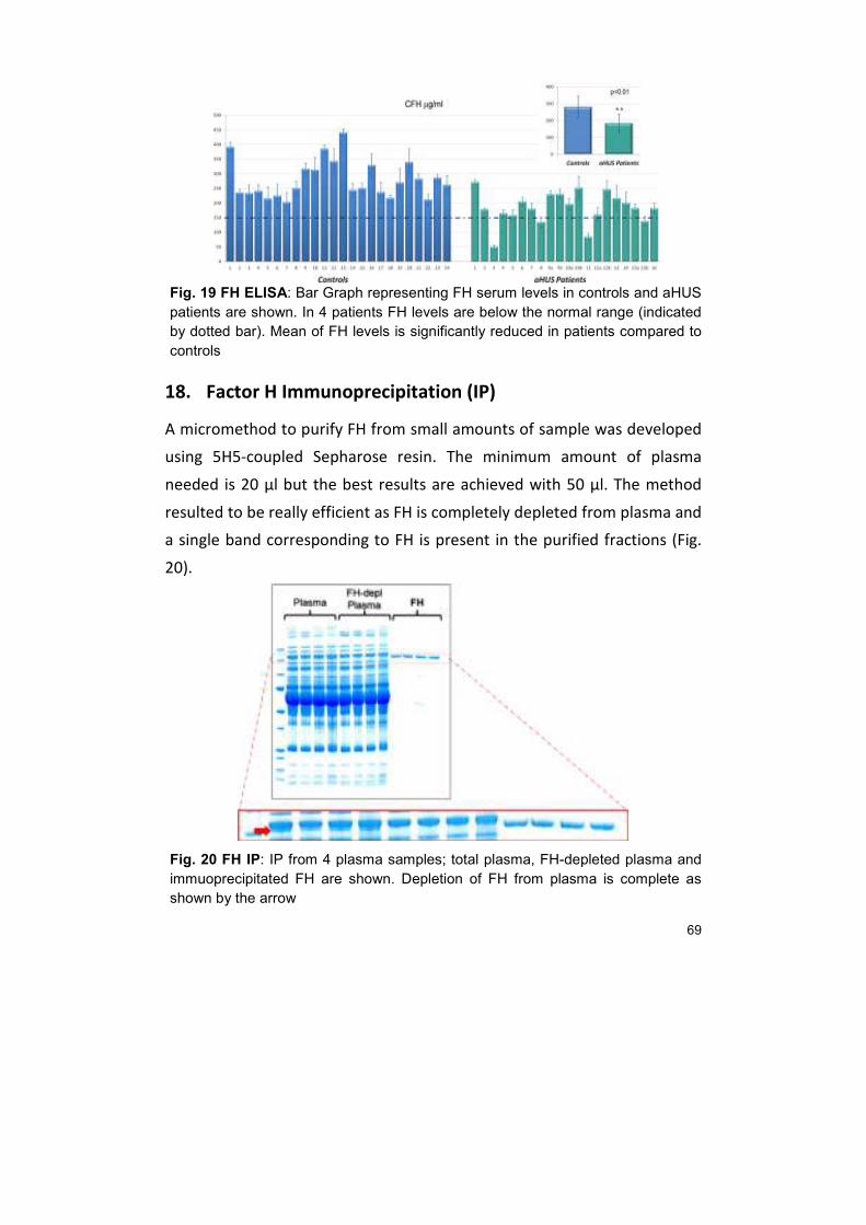
69
Fig. 19 FH ELISA: Bar Graph representing FH serum levels in controls and aHUS
patients are shown. In 4 patients FH levels are below the normal range (indicated
by dotted bar). Mean of FH levels is significantly reduced in patients compared to
controls
18. Factor H Immunoprecipitation (IP)
A micromethod to purify FH from small amounts of sample was developed
using 5H5-coupled Sepharose resin. The minimum amount of plasma
needed is 20 µl but the best results are achieved with 50 µl. The method
resulted to be really efficient as FH is completely depleted from plasma and
a single band corresponding to FH is present in the purified fractions (Fig.
20).
Fig. 20 FH IP: IP from 4 plasma samples; total plasma, FH-depleted plasma and
immuoprecipitated FH are shown. Depletion of FH from plasma is complete as
shown by the arrow

70
The method was further improved by the use of Micro Bio-Spin™ Columns
(Bio-Rad) which allow for a faster procedure.
19. Trypsin cleavage and integrity assay
19.1. Cleavage of FH by trypsin
FH (150 KDa) can be cleaved by a plasmatic protease into two fragments
(142 KDa and 38 KDa) that are held together by a disulphide bond. This
cleavage can be mimicked in vitro by trypsin. In order to develop a test to
detect the presence of cleaved FH it was necessary to obtain trypsin-
cleaved FH. 5H5-affinity purified FH was subjected to digestion with trypsin
using different incubation times and samples were analysed by SDS-PAGE
under reducing conditions followed by Coomassie staining.
As the incubation time increases the band corresponding to intact FH (180
KDa, in reducing conditions) decreases, whereas the bands corresponding
to the two cleaved fragments (142 KDa and 38 KDa) appear. The 50% of
cleavage is achieved after only 5 min. Increasing again the time other
bands due to other cuts performed by trypsin also appear fractions (Fig.
21).
Fig. 21 Trypsin cleavage of FH: trypsin cuts FH into two fragments, as the
incubation time increases the cleaved form increases compared to the uncleaved
form.
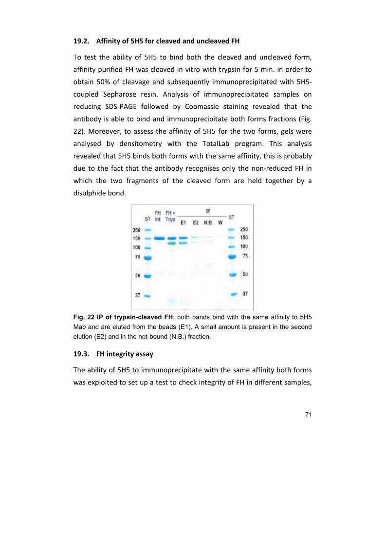
71
19.2. Affinity of 5H5 for cleaved and uncleaved FH
To test the ability of 5H5 to bind both the cleaved and uncleaved form,
affinity purified FH was cleaved in vitro with trypsin for 5 min. in order to
obtain 50% of cleavage and subsequently immunoprecipitated with 5H5-
coupled Sepharose resin. Analysis of immunoprecipitated samples on
reducing SDS-PAGE followed by Coomassie staining revealed that the
antibody is able to bind and immunoprecipitate both forms fractions (Fig.
22). Moreover, to assess the affinity of 5H5 for the two forms, gels were
analysed by densitometry with the TotalLab program. This analysis
revealed that 5H5 binds both forms with the same affinity, this is probably
due to the fact that the antibody recognises only the non-reduced FH in
which the two fragments of the cleaved form are held together by a
disulphide bond.
Fig. 22 IP of trypsin-cleaved FH: both bands bind with the same affinity to 5H5
Mab and are eluted from the beads (E1). A small amount is present in the second
elution (E2) and in the not-bound (N.B.) fraction.
19.3. FH integrity assay
The ability of 5H5 to immunoprecipitate with the same affinity both forms
was exploited to set up a test to check integrity of FH in different samples,
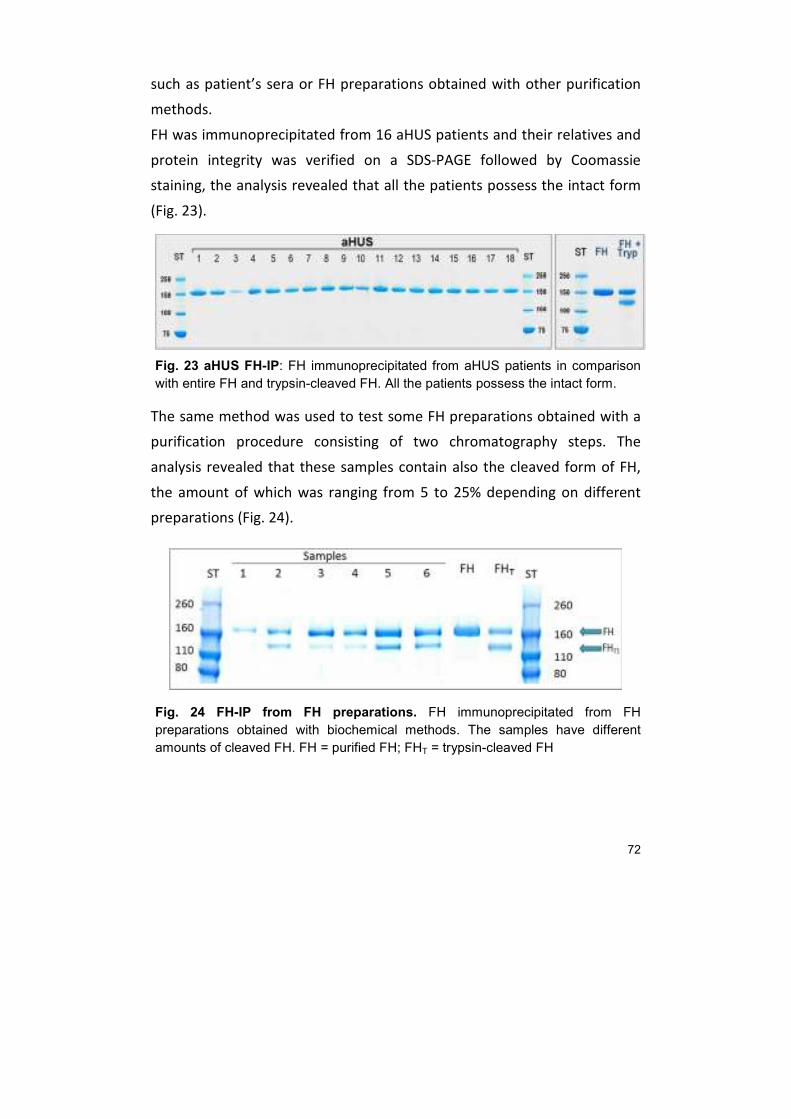
72
such as patient’s sera or FH preparations obtained with other purification
methods.
FH was immunoprecipitated from 16 aHUS patients and their relatives and
protein integrity was verified on a SDS-PAGE followed by Coomassie
staining, the analysis revealed that all the patients possess the intact form
(Fig. 23).
Fig. 23 aHUS FH-IP: FH immunoprecipitated from aHUS patients in comparison
with entire FH and trypsin-cleaved FH. All the patients possess the intact form.
The same method was used to test some FH preparations obtained with a
purification procedure consisting of two chromatography steps. The
analysis revealed that these samples contain also the cleaved form of FH,
the amount of which was ranging from 5 to 25% depending on different
preparations (Fig. 24).
Fig. 24 FH-IP from FH preparations. FH immunoprecipitated from FH
preparations obtained with biochemical methods. The samples have different
amounts of cleaved FH. FH = purified FH; FHT = trypsin-cleaved FH

73
20. Quantification of Purified Factor H
The exact quantification of purified FH samples was a crucial point in order
to perform the functional assays and to compare different samples.
Different quantification methods were compared. Bradford assay turned
out to be totally unreliable for this purpose, whereas ELISA and
spectrophotometric determination at 280 nm were more reliable and gave
comparable results. ELISA resulted to be the most sensitive method as it
was possible to quantify low FH concentrations. However, OD280 was
sensitive enough to quantify FH samples to be used for functional assays
and was in general preferred as it is faster.
21. FUNCTIONAL ASSAYS
Two assays to test respectively the N-terminal cofactorial activity of FH and
its C-terminal protective activity were developed.
21.1. Cofactor assay
A “cofactor assay” to assess N-terminal functionality of FH making use of
purified C3b and FI was set up. FI is able to cut C3b converting it to the
inactive form iC3b only in the presence of its cofactor FH. C3b is composed
of two chains: α’-chain (104 KDa) and β-chain (70 KDa), the α’ is cleaved by
FI into two fragments, α-68 (68 KDa) and α-43 (43 KDa).
Purified C3b was incubated with FI alone or together with different
amounts of FH and the presence of different bands was checked on a
reducing SDS-PAGE followed by WB with a polyclonal antibody against C3b.
In the absence of FH (C3b and C3b + FI) the α’-chain is not cleaved and a
single band around 104 KDa (α’) is observed whereas in the presence of FH
(C3b + FI + FH) α’ is cleaved and a faint band around 104 KDa is still visible
but α-68 and α-43 are also visualized (Fig. 25). In all the samples a band of
70 KDa corresponding to β-chain is also present and remains constant.
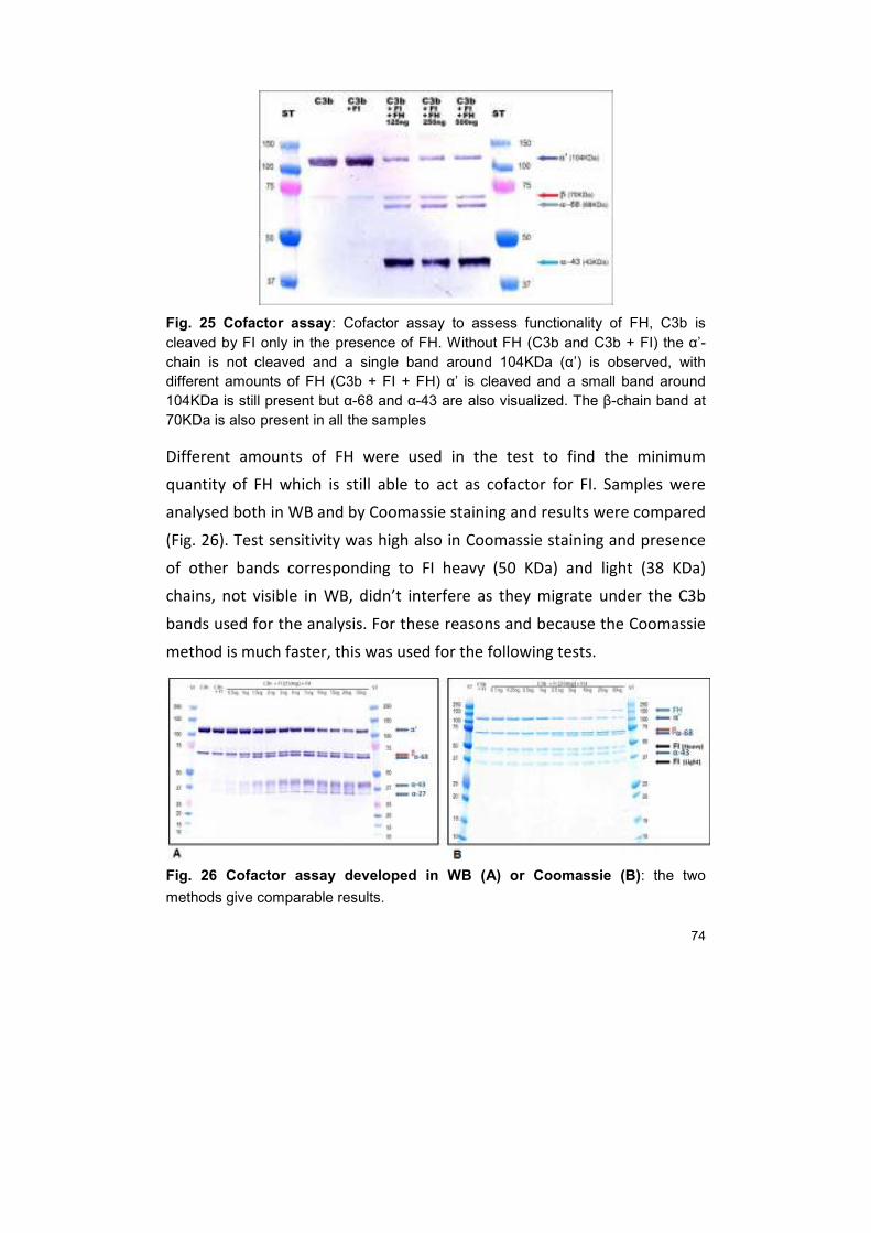
74
Fig. 25 Cofactor assay: Cofactor assay to assess functionality of FH, C3b is
cleaved by FI only in the presence of FH. Without FH (C3b and C3b + FI) the α’-
chain is not cleaved and a single band around 104KDa (α’) is observed, with
different amounts of FH (C3b + FI + FH) α’ is cleaved and a small band around
104KDa is still present but α-68 and α-43 are also visualized. The β-chain band at
70KDa is also present in all the samples
Different amounts of FH were used in the test to find the minimum
quantity of FH which is still able to act as cofactor for FI. Samples were
analysed both in WB and by Coomassie staining and results were compared
(Fig. 26). Test sensitivity was high also in Coomassie staining and presence
of other bands corresponding to FI heavy (50 KDa) and light (38 KDa)
chains, not visible in WB, didn’t interfere as they migrate under the C3b
bands used for the analysis. For these reasons and because the Coomassie
method is much faster, this was used for the following tests.
Fig. 26 Cofactor assay developed in WB (A) or Coomassie (B): the two
methods give comparable results.
75
Gels were analysed by densitometry with the TotalLab program and the
intensity of α’-chain or α-68 band was compared to that of β-chain (70
KDa) which remains constant. The cofactor activity was calculated as a
decrease of α’-chain or an increase of α-68. The first one was preferred as
the α’-chain is subject to a single cut by FI, whereas α-68 can be further
cleaved into α-27 and C3dg.
To assess functionality of different FH samples, cofactor assay was
performed in parallel on test samples and 5H5-affinity purified FH as a
control. A sample in which FH was absent was always included for
quantification of the results. At least three different concentrations of FH
were used for each sample.
The percentage of α’/β was calculated as follows: the intensity of α’ band
was first normalised to the intensity of β band in each lane and then
divided by the α’/β ratio obtained in the control where FH was absent as
this corresponds to 100% of α’-chain.
Data corresponding to the percentage of α’/β relative to the ng of FH used
were put on a graph and a Logarithmic regression model was used to build
a curve using the equation: y = a + b ln(x), where y = % α’/β and x = ng FH.
The curve was then visualized in a graph in which x-axis is in logarithmic
scale (Log10) in order to visualize a straight line. The curve obtained allows
the direct comparison of different samples.
This test was used to compare different samples of FH purified by 5H5-
affinity IP from sera of normal donors. The analysis revealed that cofactor
activity in these samples was normal and comparable to that of the control
(Fig. 27-A).
This test was also used to analyse some samples of FH, purified with a
biochemical method consisting in a two-step chromatography, that were
produced for therapeutic purpose.
Different samples and different fractions coming from different purification
steps were analysed and compared with a 5H5-affinity purified FH as a

76
control. The analysis revealed a decrease in cofactor activity in some of
these samples, probably due to the purification procedure (Fig. 27-B).
A
B
Fig. 27 Cofactor assay: (A) on 5H5-affinity purified FH and (B) on FH purified with
biochemical methods.
21.2. Haemolysis test
An “Haemolysis test” to test the protective activity of FH, which depends
on the C-terminal part of the molecule, was set up for sera samples.
This test is based on the fact that FH is able to protect sheep erythrocytes
from complement attack; in the absence of FH or in the presence of a
defective FH erythrocytes undergo lysis which can be measured
spectrophotometrically following the release of haemoglobin.
A minimum of haemolysis is still observed using high concentrations of sera
and can be used to test alternative pathway (AP) activation.
The assay was first set up using a control serum to test all the conditions
needed to develop the final test.
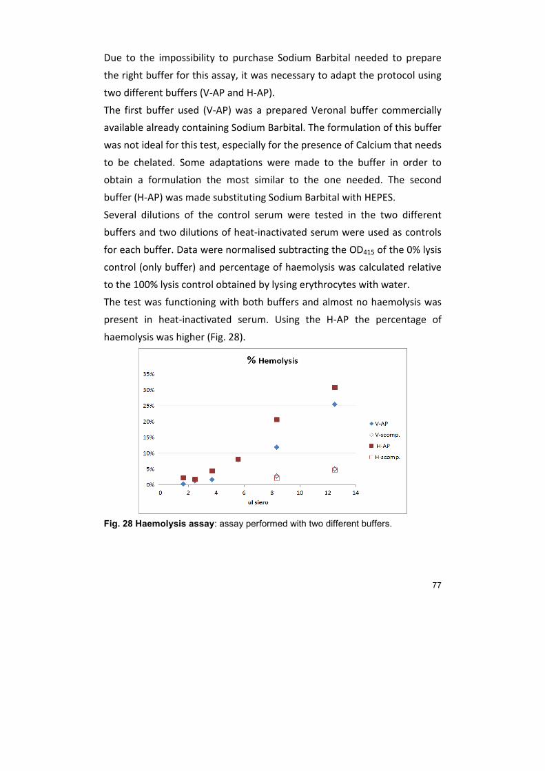
77
Due to the impossibility to purchase Sodium Barbital needed to prepare
the right buffer for this assay, it was necessary to adapt the protocol using
two different buffers (V-AP and H-AP).
The first buffer used (V-AP) was a prepared Veronal buffer commercially
available already containing Sodium Barbital. The formulation of this buffer
was not ideal for this test, especially for the presence of Calcium that needs
to be chelated. Some adaptations were made to the buffer in order to
obtain a formulation the most similar to the one needed. The second
buffer (H-AP) was made substituting Sodium Barbital with HEPES.
Several dilutions of the control serum were tested in the two different
buffers and two dilutions of heat-inactivated serum were used as controls
for each buffer. Data were normalised subtracting the OD415 of the 0% lysis
control (only buffer) and percentage of haemolysis was calculated relative
to the 100% lysis control obtained by lysing erythrocytes with water.
The test was functioning with both buffers and almost no haemolysis was
present in heat-inactivated serum. Using the H-AP the percentage of
haemolysis was higher (Fig. 28).
Fig. 28 Haemolysis assay: assay performed with two different buffers.

78
In the final test FH-depleted serum needs to be used. Since subtracting FH
from serum leads to an indiscriminate activation of complement with the
consequence of consuming all the complement proteins, activation needs
to be inhibited by adding EDTA throughout the depletion procedure. On
the other hand, EDTA will also chelate Magnesium ions needed to activate
AP in the haemolysis test. For these reasons, different concentrations of
EDTA were tested in this assay in order to establish the minimum
concentration that prevents activation of complement in FH-depleted
serum but still allows the test to function.
The test was performed diluting the serum 1:5 in H-AP buffer with the
addition of different amounts of EDTA. As a negative control PBS was used
instead of serum and prepared in the same way. Percentage of haemolysis
was calculated as before and visualized on a graphic relative to final EDTA
concentration (Fig. 29).
Fig. 29 Haemolysis assay: assay performed with different EDTA concentrations.
Haemolysis resulted to be inhibited by the presence of EDTA and decreases
as the concentration of EDTA increases. A final concentration of 1 mM still
allows the test to function. Considering that serum is used at a 1:5 dilution
and is then diluted 1:1 with erythrocyte preparation, the final dilution is
1:10. That allows to prepare depleted serum with 10 mM of EDTA and to
perform the test with a final concentration of 1 mM.

79
22. Other purification methods
22.1. Heparin affinity purification
Human serum was applied to a Heparin column in order to purify heparin
binding proteins such as FH and FHRs. Fractions were analysed by WB with
different antibodies for FH, FHRs and C3b (Fig. 30). All the proteins were
present in serum before purification and some traces were still present in
the flow through probably due to column overload. FH eluted at 250 mM
NaCl with some residual at higher salt concentrations (300-400 mM). FHR-1
eluted both at 250 and 300 mM NaCl with some residual at 400 mM,
whereas FHR-2 eluted at 250 mM together with FH and no residuals were
visible at higher salt concentrations. FHR-5 had a different pattern as is
eluted at higher salt concentrations with a maximum at 300 and 400 mM
and only a small amount eluted earlier. Finally FHR-4 didn't seem to bind to
the Heparin column as all the signal was present in the flow through.
Fig. 30 WB of Heparin fractions developed with antibodies for FH and FHRs.
C3b eluted together with FH at 250 mM NaCl with some traces at higher
salt concentrations. (data not shown)

80
22.2. Hydrophobic Interaction Chromatography (HIC)
Serum proteins were also isolated with a hydrophobic column (HIC) and
fractions were analysed by Coomassie staining and by WB with different
antibodies for FH, FHRs and C3b (Fig. 31).
Albumin did not bind to HIC column and is present in the 100% fraction
containing 1.5 M ammonium sulphate. FH and FHRs eluted in the 33%
fraction containing 0.5 M ammonium sulphate except FHR-5 which was
present in the 0% fraction. Finally C3b was present both in the 66% and 33
% fractions (1 and 0.5 m sodium sulphate) with a residual of α-43 in the 0%
fraction.
Fig. 31 Coomassie e WB of HIC fractions.(A) Coomassie, (B) WB for C3b and
(C) WB for FH and FHRs.
23. CLONING OF FACTOR H
The entire FH and three fragments containing approximately seven SCRs
each (SCR 1-7, SCR 8-14, SCR 15-20) have been amplified from human liver
RNA using Phusion™ DNA Polymerase (Thermo).
In order to obtain the right amplification of all the fragments in was
necessary to introduce several changes in the protocols. cDNA was
produced using both Oligo(dT) or specific primers: SCR-7R, SCR-14R, SCR-
20R. For SCR 1-7 fragment the best results were obtained using cDNA
retrotranscribed with the specific primer SCR-14R whereas for fragments
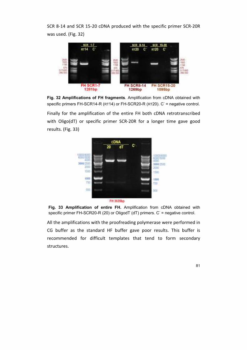
81
SCR 8-14 and SCR 15-20 cDNA produced with the specific primer SCR-20R
was used. (Fig. 32)
Fig. 32 Amplifications of FH fragments. Amplification from cDNA obtained with
specific primers FH-SCR14-R (RT14) or FH-SCR20-R (RT20). C- = negative control.
Finally for the amplification of the entire FH both cDNA retrotranscribed
with Oligo(dT) or specific primer SCR-20R for a longer time gave good
results. (Fig. 33)
Fig. 33 Amplification of entire FH. Amplification from cDNA obtained with
specific primer FH-SCR20-R (20) or OligodT (dT) primers. C- = negative control.
All the amplifications with the proofreading polymerase were performed in
CG buffer as the standard HF buffer gave poor results. This buffer is
recommended for difficult templates that tend to form secondary
structures.
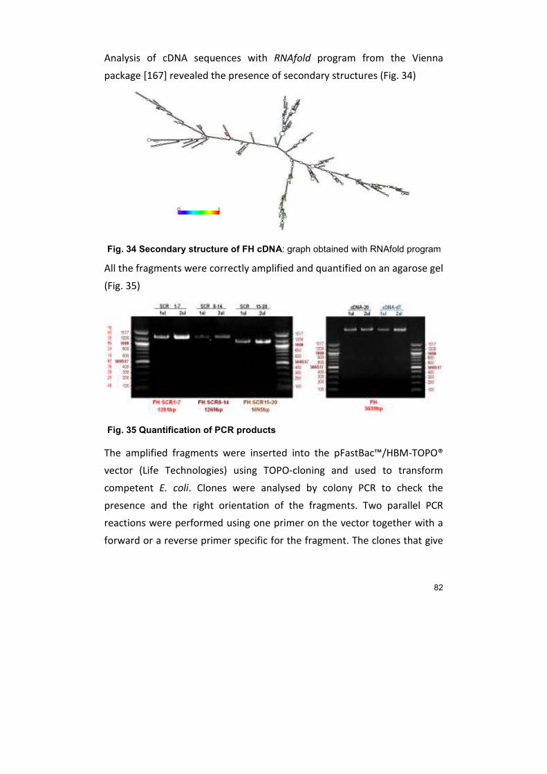
82
Analysis of cDNA sequences with RNAfold program from the Vienna
package [167] revealed the presence of secondary structures (Fig. 34)
Fig. 34 Secondary structure of FH cDNA: graph obtained with RNAfold program
All the fragments were correctly amplified and quantified on an agarose gel
(Fig. 35)
Fig. 35 Quantification of PCR products
The amplified fragments were inserted into the pFastBac™/HBM-TOPO®
vector (Life Technologies) using TOPO-cloning and used to transform
competent E. coli. Clones were analysed by colony PCR to check the
presence and the right orientation of the fragments. Two parallel PCR
reactions were performed using one primer on the vector together with a
forward or a reverse primer specific for the fragment. The clones that give
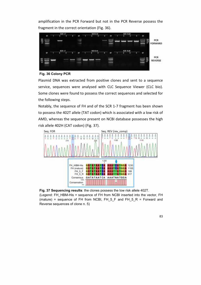
83
amplification in the PCR Forward but not in the PCR Reverse possess the
fragment in the correct orientation (Fig. 36).
Fig. 36 Colony PCR
Plasmid DNA was extracted from positive clones and sent to a sequence
service, sequences were analysed with CLC Sequence Viewer (CLC bio).
Some clones were found to possess the correct sequences and selected for
the following steps.
Notably, the sequence of FH and of the SCR 1-7 fragment has been shown
to possess the 402T allele (TAT codon) which is associated with a low risk of
AMD, whereas the sequence present on NCBI database possesses the high
risk allele 402H (CAT codon) (Fig. 37).
Fig. 37 Sequencing results: the clones possess the low risk allele 402T.
(Legend: FH_HBM-His = sequence of FH from NCBI inserted into the vector, FH
(mature) = sequence of FH from NCBI, FH_5_F and FH_5_R = Forward and
Reverse sequences of clone n. 5)

84
DISCUSSION
Complement Factor H (FH) is a crucial regulator of complement activation.
Mutations in the CFH gene are frequently associated with a number of
human diseases, furthermore it has an important role in infectious diseases
as many pathogens are able to recruit it on their surface to protect
themselves from complement attack.
However, the study of this protein is complicated by the presence of other
homologous proteins belonging to the “FH protein family”. The high amino
acid identity among the family members is a cause of cross-reactivity for
antibodies. Antibodies raised against FH often detect also different FHRs
and antibodies generated against FHRs can cross-react with FH or other
FHRs. This cross-reactivity is thus a challenge for correct quantification
and/or purification of FH from plasma.
The aim of this study was mainly to develop some quantitative and
functional FH-specific assays, using a monoclonal antibody (MAb 5H5)
produced in our laboratory that was shown to be specific for FH. Another
important focus was the production of recombinant FH in both an
eukaryotic and a prokaryotic expression system, in order to study the
importance of protein glycosylation and to further and more precisely
refine structure-function relationships, as well as to develop safer
therapeutic tools: in fact, current therapies for FH defects only rely on
blood derivatives.
By the use of high-density hybridoma cultures with CELLine Bioreactor, it
was possible to produce a consistent amount of MAb 5H5. This antibody
was the starting point for the development of FH-specific assays.
The first use of MAb 5H5 was the purification of FH using the antibody
conjugated with Sepharose resin. Thanks to the high affinity of MAb 5H5
for FH, it was possible to set up a “one-step” purification procedure, which
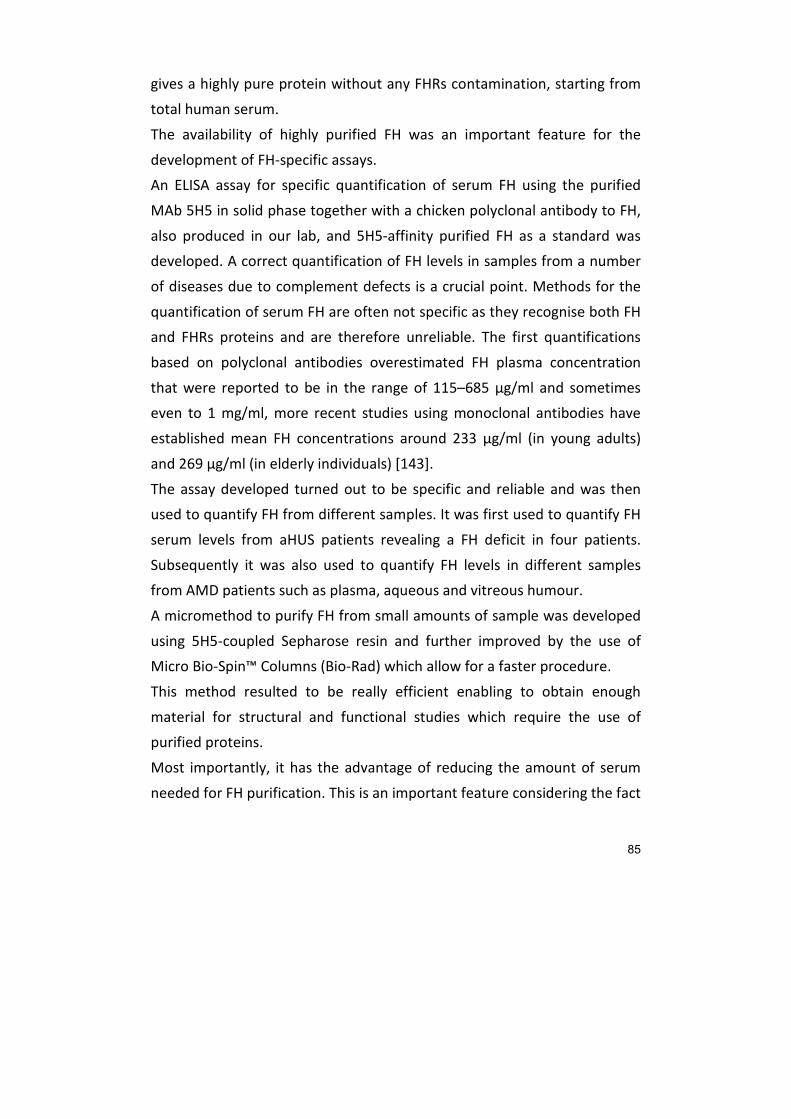
85
gives a highly pure protein without any FHRs contamination, starting from
total human serum.
The availability of highly purified FH was an important feature for the
development of FH-specific assays.
An ELISA assay for specific quantification of serum FH using the purified
MAb 5H5 in solid phase together with a chicken polyclonal antibody to FH,
also produced in our lab, and 5H5-affinity purified FH as a standard was
developed. A correct quantification of FH levels in samples from a number
of diseases due to complement defects is a crucial point. Methods for the
quantification of serum FH are often not specific as they recognise both FH
and FHRs proteins and are therefore unreliable. The first quantifications
based on polyclonal antibodies overestimated FH plasma concentration
that were reported to be in the range of 115–685 μg/ml and sometimes
even to 1 mg/ml, more recent studies using monoclonal antibodies have
established mean FH concentrations around 233 μg/ml (in young adults)
and 269 μg/ml (in elderly individuals) [143].
The assay developed turned out to be specific and reliable and was then
used to quantify FH from different samples. It was first used to quantify FH
serum levels from aHUS patients revealing a FH deficit in four patients.
Subsequently it was also used to quantify FH levels in different samples
from AMD patients such as plasma, aqueous and vitreous humour.
A micromethod to purify FH from small amounts of sample was developed
using 5H5-coupled Sepharose resin and further improved by the use of
Micro Bio-Spin™ Columns (Bio-Rad) which allow for a faster procedure.
This method resulted to be really efficient enabling to obtain enough
material for structural and functional studies which require the use of
purified proteins.
Most importantly, it has the advantage of reducing the amount of serum
needed for FH purification. This is an important feature considering the fact

86
that FH-related diseases, e.g. aHUS, affect mainly children and the material
available is poor.
FH can be cleaved by a plasmatic protease into two fragments that are held
together by a disulphide bond, this can be mimicked in vitro by trypsin.
The ability of MAb 5H5 to recognise both the intact and the trypsin-cleaved
form of FH, was demonstrated by IP and further exploited to set up an
integrity assay to be used on different samples. The analysis on aHUS
patients revealed that they all possess the intact form, whereas analysis on
chromatography purified FH revealed the presence of the cleaved form.
The question whether the cleaved FH form has a reduced activity is still
controversial. The cut is within the SCR6 domain and separates the
domains with cofactor activity from the rest of the molecule, even if the
structure is still held together by disulfide bridges.
It has been shown that the cofactor activity resides in the 38 KDa N-
terminal fragment and that this fragment is also able to work alone [168].
However, the data available in the literature on the cofactor activity of the
cleaved form are different and often conflicting. Some works show that the
cofactor activity in the fluid phase seems to be increased by the cleavage,
whereas the cofactor activity for the degradation of membrane-bound C3b
is certainly decreased [169].
The presence of the cleaved form could also be due to an artefact of the
purification methods.
During the various steps, necessary for FH purification with biochemical
methods, the plasmatic protease inhibitor is first removed thus enabling
the plasmatic protease to cut FH.
With affinity purification methods, such as the method developed here
based on MAb 5H5, the presence of the cleaved form is generally not
observed. Thus, the integrity test is an important tool to assess the
presence of the cleaved form especially in those FH preparations obtained

87
with biochemical methods that are used to prepare FH for substitution
therapies.
The ability of MAb 5H5 to bind with the same affinity both the intact and
the cleaved form of FH is probably due to the fact that this antibody
recognises a structural epitope that remains the same in the two forms but
is lost after reduction of disulphide bridges. In fact, MAb 5H5 recognises FH
only under unreducing conditions. This feature is probably also the reason
why MAb 5H5 is specific for FH and does not recognise any of the FHRs
proteins.
The epitope recognised by MAb 5H5 has not yet been identified; being a
conformational mapping the epitope is challenging. Recombinant proteins
that are discussed later, will be probably useful for this purpose.
Finally, MAb 5H5 was used to develop two functional assays to assess both
the N-terminal cofactorial activity and C-terminal protective activity of FH.
The “cofactor assay” is based on the fact that FI can cut C3b in vitro only in
the presence of a functional FH; if CFH is not functional C3b cannot be
cleaved. The test was set up using purified C3b and FI together with
different amounts of affinity purified FH. The test was used to screen
different serum samples or fractions derived from a biochemical
purification of FH and revealed a reduced activity in samples which
underwent different chromatography steps.
This reduced activity is probably due to the long procedure used to purify
FH, consisting in two sequential chromatography steps, that somehow
modify the protein. It doesn’t appear to be related to the presence of the
cleaved form, but it is probably due to other modifications that occur in the
protein during the procedure.
The “Haemolysis test” is useful to assess the protective activity of FH that is
dependent on the C-terminal part of the molecule. This test is based on the
fact that FH is able to protect erythrocytes from complement attack; in the
absence of FH or in the presence of a defective FH erythrocytes undergo
88
lysis which can be measured spectrophotometrically following the release
of haemoglobin. The test will be performed making use of FH-depleted
serum, which causes lysis of the erythrocytes, and 5H5-affinity purified FH
from patient’s sera in order to assess the ability of those FH to restore
protection.
For the moment the assay has been set up using a normal control serum in
order to test all the conditions. It was first necessary to test different buffer
preparations as the correct buffer needed for this kind of test is not
commercially available in Italy. FH is an important inhibitor of complement
activation and its depletion from serum causes the indiscriminate
activation of complement leading to the consumption of complement
proteins. To avoid this activation that would render FH-depleted serum
useless for this test, it is necessary to produce it in the presence of EDTA
that blocks both the CP and the AP. However, the presence of EDTA will
inhibit erythrocytes lysis thus precluding the possibility to perform the
“Haemolysis test”. For these reasons it was necessary to perform the test
in the presence of different concentrations of EDTA in order to establish
the minimum concentration that still prevents complement to be activated
in serum but allows the test to function. The set up of the last conditions
needed to perform the final test is still under development.
This test will be useful to assess N-terminal functionality especially in aHUS
patients in which most mutations are located in the last two SCRs (SCR 19-
20) and cause haemolysis. The genetic characterisation of this group of
aHUS patients has already been performed whereas functional data at
protein level are still missing. Therefore, the functional data obtainable
with this test will be fundamental to complement the characterisation of
these patients and their relatives performed by DNA sequencing, thus
allowing for a detailed description of the structure-function relationship.
The other aim of this project was the cloning and expression of FH in both
an eukaryotic and a prokaryotic system.
89
As FH is a glycoprotein and a correct glycosylation might be important for
all its functions, I planned to use a modified insect expression system in
which the glycosylation pattern is very similar to that of humans.
Unglycosylated proteins to be used as a control will be produced in E. coli.
FH is a big soluble protein with 20 SCR domains of about 60 aa each that
bear different functions. I decided to clone separately both the entire
protein and three main fragments containing approximately 7 SCR each
(SCR 1-7, SCR 8-14, SCR 15-20).
Four clones containing respectively the entire FH and the three fragments
were obtained and confirmed by sequencing to possess the correct
sequences. These clones will be used to produce the recombinant proteins
into the two different systems.
The recombinant proteins produced will be useful for different purposes.
First of all they can be used to screen which parts of FH are involved in
interaction with proteins of known pathogens and to explore interaction
with other pathogens. Secondly their ability to restore the function of a
defective FH or to protect against FH autoantibodies will be analysed. The
final goal is to use them for therapy in many diseases caused by CFH
defects for which the only possible treatment is currently plasma exchange.
Furthermore, these recombinant fragments will also be used to try to
identify the so far elusive conformational epitope recognised by MAb 5H5.
90
CONCLUSIONS
The study of complement factor H (FH) is complicated by the presence of
FHR proteins that share a high degree of similarity.
Thanks to the use of a monoclonal antibody (MAb 5H5) produced in our
lab, and shown to be specific for FH, it was possible to develop several FH-
specific tests.
A fast and efficient method for the purification of FH was developed and
further scaled down to be applicable to small samples. A home-made ELISA
for the correct quantification of FH in biological fluids, without any
interference from FHR proteins, was developed and used to screen
different samples. A test for the assessment of FH structural integrity,
based on the fact that FH can be cleaved by a protease into a probably less
active form, was also developed. This test used to screen both FH purified
from patient’s sera and samples deriving from different purification
procedures.
Finally two functional assays to test both the C-terminal cofactorial activity
and the N-terminal protective activity of FH were developed.
The “cofactor assay”, based on the fact that FI can cut its substrate C3b
only in the presence of a functional FH, has been set up and was used to
screen different samples. This test was important especially to assess
functionality of some FH preparations obtained with a biochemical
purification method, that were intended to be used for therapeutic
purposes.
The second one, the “haemolysis test” based on the fact that a functional
FH is able to protect sheep erythrocytes from haemolysis, is now in its fine
tuning phase. This test is a modification of a test normally used to assess
AP activation in sera. For the test a FH-depleted serum from a normal
subject will be used together with FH purified from patients.
91
The developed tests that have been used in the present work on aHUS
patients and FH preparations produced for therapeutic purposes, will be
used in the future also for a bigger screening on AMD patients for whom a
pilot study just started. The study of AMD patients will be part of a larger
project that will involve other methods such as the study of retinal
epithelial cells which have been shown to produce FH locally in the eye [86]
and the genetic screening of some SNPs which have been shown to
correlate with the development of the disease [138].
Another aim of this project was the cloning of the entire FH and some
fragments for the expression into both a prokaryotic and a eukaryotic
system in order to produce both glycosylated and unglycosylated proteins.
The clones have been produced and are ready to be introduced into the
expression hosts.
The recombinant proteins produced will be useful for different purposes.
First of all they can be used to screen which parts of FH are involved in
interaction with proteins of known pathogens and to explore interaction
with other pathogens. Secondly their ability to restore the function of a
defective FH or to protect against FH autoantibodies will be analysed. The
final goal is to use them for therapy in many diseases caused by FH defects
for which the only possible treatment is currently plasma exchange

92
REFERENCES
1. Bracke J., and Gengou O. , "Sur l’existence de substances sensibilisatrices dans la plupart des serum antimicrobiens.", Ann. Inst. Pasteur, Vol. 15, 1901, pp. 289−302.
2. Lachmann P. , "Complement before molecular biology", Molecular Immunology, Vol. 43, no. 6, 2006, pp. 496–508.
3. Carroll M.C. , "The complement system in regulation of adaptive immunity", Nature Immunology, Vol. 5, no. 10, 2004, pp. 981–986.
4. Walport M.J. , "Complement -First of two parts", New England Journal of Medicine, Vol. 344, no. 14, 2001, pp. 1058–1066.
5. Rodríguez de Córdoba S., Esparza-Gordillo J., Goicoechea de Jorge E., Lopez-Trascasa M., and Sánchez-Corral P. , "The human complement factor H: functional roles, genetic variations and disease associations", Molecular immunology, Vol. 41, no. 4, 2004, pp. 355–367.
6. Müller-Eberhard H.J. , "The membrane attack complex of complement", Annual Review of Immunology, Vol. 4, 1986, pp. 503–528.
7. Cole D.S., and Morgan B.P. , "Beyond lysis: how complement influences cell fate", Clinical Science, Vol. 104, no. 5, 2003, pp. 455.
8. Fearon D.T., and Carroll M.C. , "Regulation of B lymphocyte responses to foreign and self-antigens by the CD19/CD21 complex", Annual Review of Immunology, Vol. 18, 2000, pp. 393–422.
9. Tofte R.W., Peterson P.K., Schmeling D., Bracke J., Kim Y., and Quie P.G. , "Opsonization of four Bacteroides species: role of the classical complement pathway and immunoglobulin", Infection and Immunity, Vol. 27, no. 3, 1980, pp. 784–792.
10. Hugli T.E., and Müller-Eberhard H.J. , "Anaphylatoxins: C3a and C5a", Advances in Immunology, Vol. 26, 1978, pp. 1–53.
11. Poon I.K.H., Hulett M.D., and Parish C.R. , "Molecular mechanisms of late apoptotic/necrotic cell clearance", Cell Death and Differentiation, Vol. 17, no. 3, 2010, pp. 381–397.
12. Schifferli J.A., Ng Y.C., and Peters D.K. , "The role of complement and its receptor in the elimination of immune complexes", The New England Journal of Medicine, Vol. 315, no. 8, 1986, pp. 488–495.
13. Strang C.J., Siegel R.C., Phillips M.L., Poon P.H., and Schumaker V.N. , "Ultrastructure of the first component of human complement: electron microscopy of the crosslinked complex", Proceedings of the National Academy of Sciences of the United States of America, Vol. 79, no. 2, 1982, pp. 586–590.
14. Agnello V., Winchester R.J., and Kunkel H.G. , "Precipitin reactions of the C1q component of complement with aggregated gamma-globulin and immune complexes in gel diffusion", Immunology, Vol. 19, no. 6, 1970, pp. 909–919.
15. Claus D.R., Siegel J., Petras K., Osmand A.P., and Gewurz H. , "Interactions of C-reactive protein with the first component of human complement", Journal of Immunology (Baltimore, Md.: 1950), Vol. 119, no. 1, 1977, pp. 187–192.
16. Loos M., Bitter-Suermann D., and Dierich M. , "Interaction of the first (C1), the second (C2) and the fourth (C4) component of complement with different

93
preparations of bacterial lipopolysaccharides and with lipid A", Journal of Immunology (Baltimore, Md.: 1950), Vol. 112, no. 3, 1974, pp. 935–940.
17. Sim R.B., Porter R.R., Reid K.B., and Gigli I. , "The structure and enzymic activities of the C1r and C1s subcomponents of C1, the first component of human serum complement", The Biochemical Journal, Vol. 163, no. 2, 1977, pp. 219–227.
18. Gorski J.P., Hugli T.E., and Müller-Eberhard H.J. , "C4a: the third anaphylatoxin of the human complement system", Proceedings of the National Academy of Sciences of the United States of America, Vol. 76, no. 10, 1979, pp. 5299–5302.
19. Tsuruta T., Yamamoto T., Matsubara S., Nagasawa S., Tanase S., Tanaka J., Takagi K., and Kambara T. , "Novel function of C4a anaphylatoxin. Release from monocytes of protein which inhibits monocyte chemotaxis", The American Journal of Pathology, Vol. 142, no. 6, 1993, pp. 1848–1857.
20. Kemper C., Pangburn M.K., and Fishelson Z. , "Complement Nomenclature 2014", Molecular Immunology, Vol. 61, no. 2. XXV International Complement Workshop September 14-18, 2014 - Rio de Janeiro, Brazil, 2014, pp. 56–58.
21. Cooper N.R. , "Enzymatic activity of the second component of complement", Biochemistry, Vol. 14, no. 19, 1975, pp. 4245–4251.
22. Schreiber R.D., Pangburn M.K., Lesavre P.H., and Müller-Eberhard H.J. , "Initiation of the alternative pathway of complement: recognition of activators by bound C3b and assembly of the entire pathway from six isolated proteins", Proceedings of the National Academy of Sciences of the United States of America, Vol. 75, no. 8, 1978, pp. 3948–3952.
23. Pangburn M.K., and Müller-Eberhard H.J. , "Initiation of the alternative complement pathway due to spontaneous hydrolysis of the thioester of C3", Annals of the New York Academy of Sciences, Vol. 421, 1983, pp. 291–298.
24. Fearon D.T., and Austen K.F. , "Properdin: binding to C3b and stabilization of the C3b-dependent C3 convertase", The Journal of Experimental Medicine, Vol. 142, no. 4, 1975, pp. 856–863.
25. Sim R.B., Twose T.M., Paterson D.S., and Sim E. , "The covalent-binding reaction of complement component C3", The Biochemical Journal, Vol. 193, no. 1, 1981, pp. 115–127.
26. Thiel S. , "Complement activating soluble pattern recognition molecules with collagen-like regions, mannan-binding lectin, ficolins and associated proteins", Molecular Immunology, Vol. 44, no. 16, 2007, pp. 3875–3888.
27. Ikeda K., Sannoh T., Kawasaki N., Kawasaki T., and Yamashina I. , "Serum lectin with known structure activates complement through the classical pathway", The Journal of Biological Chemistry, Vol. 262, no. 16, 1987, pp. 7451–7454.
28. Matsushita M. , "Ficolins: complement-activating lectins involved in innate immunity", Journal of Innate Immunity, Vol. 2, no. 1, 2010, pp. 24–32.
29. Sekine H., Takahashi M., Iwaki D., and Fujita T. , "The role of MASP-1/3 in complement activation", Advances in Experimental Medicine and Biology, Vol. 735, 2013, pp. 41–53.
94
30. Kemper C., and Hourcade D.E. , "Properdin: New roles in pattern recognition and target clearance", Molecular immunology, Vol. 45, no. 16, 2008, pp. 4048–4056.
31. Takata Y., Kinoshita T., Kozono H., Takeda J., Tanaka E., Hong K., and Inoue K. , "Covalent association of C3b with C4b within C5 convertase of the classical complement pathway", The Journal of Experimental Medicine, Vol. 165, no. 6, 1987, pp. 1494–1507.
32. Kinoshita T., Takata Y., Kozono H., Takeda J., Hong K.S., and Inoue K. , "C5 convertase of the alternative complement pathway: covalent linkage between two C3b molecules within the trimolecular complex enzyme", Journal of Immunology (Baltimore, Md.: 1950), Vol. 141, no. 11, 1988, pp. 3895–3901.
33. Rawal N., and Pangburn M.K. , "Structure/function of C5 convertases of complement", International Immunopharmacology, Vol. 1, no. 3, 2001, pp. 415–422.
34. Meuer S., Hugli T.E., Andreatta R.H., Hadding U., and Bitter-Suermann D. , "Comparative study on biological activities of various anaphylatoxins (C4a, C3a, C5a). Investigations on their ability to induce platelet secretion", Inflammation, Vol. 5, no. 4, 1981, pp. 263–273.
35. Podack E.R., Esser A.F., Biesecker G., and Müller-Eberhard H.J. , "Membrane attack complex of complement: a structural analysis of its assembly", The Journal of Experimental Medicine, Vol. 151, no. 2, 1980, pp. 301–313.
36. Hammer C.H., Nicholson A., and Mayer M.M. , "On the mechanism of cytolysis by complement: evidence on insertion of C5b and C7 subunits of the C5b,6,7 complex into phospholipid bilayers of erythrocyte membranes", Proceedings of the National Academy of Sciences of the United States of America, Vol. 72, no. 12, 1975, pp. 5076–5080.
37. Podack E.R., Biesecker G., Kolb W.P., and Müller-Eberhard H.J. , "The C5b-6 complex: reaction with C7, C8, C9", Journal of Immunology (Baltimore, Md.: 1950), Vol. 121, no. 2, 1978, pp. 484–490.
38. Tschopp J., Podack E.R., and Müller-Eberhard H.J. , "The membrane attack complex of complement: C5b-8 complex as accelerator of C9 polymerization", Journal of Immunology (Baltimore, Md.: 1950), Vol. 134, no. 1, 1985, pp. 495–499.
39. Müller-Eberhard H.J. , "The killer molecule of complement", The Journal of Investigative Dermatology, Vol. 85, no. 1 Suppl, 1985, pp. 47s–52s.
40. Podack E.R., and Tschopp J. , "Membrane attack by complement", Molecular Immunology, Vol. 21, no. 7, 1984, pp. 589–603.
41. Rodríguez de Córdoba S., Díaz-Guillén M.A., and Heine-Suñer D. , "An integrated map of the human regulator of complement activation (RCA) gene cluster on 1q32", Molecular Immunology, Vol. 36, no. 13-14, 1999, pp. 803–808.
42. Bos I.G.A., Lubbers Y.T.P., Roem D., Abrahams J.P., Hack C.E., and Eldering E. , "The functional integrity of the serpin domain of C1-inhibitor depends on the unique N-terminal domain, as revealed by a pathological mutant", The Journal of Biological Chemistry, Vol. 278, no. 32, 2003, pp. 29463–29470.
43. Ratnoff O.D., Pensky J., Ogston D., and Naff G.B. , "The inhibition of plasmin, plasma kallikrein, plasma permeability factor, and the C’1r subcomponent
95
of the first component of complement by serum C’1 esterase inhibitor", The Journal of Experimental Medicine, Vol. 129, no. 2, 1969, pp. 315–331.
44. Matsushita M., Thiel S., Jensenius J.C., Terai I., and Fujita T. , "Proteolytic activities of two types of mannose-binding lectin-associated serine protease", Journal of Immunology (Baltimore, Md.: 1950), Vol. 165, no. 5, 2000, pp. 2637–2642.
45. Davis A.E., Lu F., and Mejia P. , "C1 inhibitor, a multi-functional serine protease inhibitor", Thrombosis and Haemostasis, Vol. 104, no. 5, 2010, pp. 886–893.
46. Lomas D.A., Belorgey D., Mallya M., Miranda E., Kinghorn K.J., Sharp L.K., Phillips R.L., Page R., Robertson A.S., and Crowther D.C. , "Molecular mousetraps and the serpinopathies", Biochemical Society Transactions, Vol. 33, no. Pt 2, 2005, pp. 321–330.
47. Cugno M., Zanichelli A., Foieni F., Caccia S., and Cicardi M. , "C1-inhibitor deficiency and angioedema: molecular mechanisms and clinical progress", Trends in Molecular Medicine, Vol. 15, no. 2, 2009, pp. 69–78.
48. Davies A., and Lachmann P.J. , "Membrane defence against complement lysis: the structure and biological properties of CD59", Immunologic Research, Vol. 12, no. 3, 1993, pp. 258–275.
49. Yamashina M., Ueda E., Kinoshita T., Takami T., Ojima A., Ono H., Tanaka H., Kondo N., Orii T., and Okada N. , "Inherited complete deficiency of 20-kilodalton homologous restriction factor (CD59) as a cause of paroxysmal nocturnal hemoglobinuria", The New England Journal of Medicine, Vol. 323, no. 17, 1990, pp. 1184–1189.
50. Delvaeye M., Noris M., De Vriese A., Esmon C.T., Esmon N.L., Ferrell G., et al. , "Thrombomodulin mutations in atypical hemolytic-uremic syndrome", The New England Journal of Medicine, Vol. 361, no. 4, 2009, pp. 345–357.
51. Bajzar L., Manuel R., and Nesheim M.E. , "Purification and characterization of TAFI, a thrombin-activable fibrinolysis inhibitor", The Journal of Biological Chemistry, Vol. 270, no. 24, 1995, pp. 14477–14484.
52. Rezende S.M., Simmonds R.E., and Lane D.A. , "Coagulation, inflammation, and apoptosis: different roles for protein S and the protein S-C4b binding protein complex", Blood, Vol. 103, no. 4, 2004, pp. 1192–1201.
53. Podack E.R., Kolb W.P., and Müller-Eberhard H.J. , "The SC5b-7 complex: formation, isolation, properties, and subunit composition", Journal of Immunology (Baltimore, Md.: 1950), Vol. 119, no. 6, 1977, pp. 2024–2029.
54. Tschopp J., Chonn A., Hertig S., and French L.E. , "Clusterin, the human apolipoprotein and complement inhibitor, binds to complement C7, C8 beta, and the b domain of C9", Journal of Immunology (Baltimore, Md.: 1950), Vol. 151, no. 4, 1993, pp. 2159–2165.
55. Sim R.B., Day A.J., Moffatt B.E., and Fontaine M. , "Complement factor I and cofactors in control of complement system convertase enzymes", Methods in Enzymology, Vol. 223, 1993, pp. 13–35.
56. Goldberger G., Arnaout M.A., Aden D., Kay R., Rits M., and Colten H.R. , "Biosynthesis and postsynthetic processing of human C3b/C4b inactivator (factor I) in three hepatoma cell lines", The Journal of Biological Chemistry, Vol. 259, no. 10, 1984, pp. 6492–6497.
96
57. Fearon D.T. , "Purification of C3b inactivator and demonstration of its two polypeptide chain structure", Journal of Immunology (Baltimore, Md.: 1950), Vol. 119, no. 4, 1977, pp. 1248–1252.
58. Krych-Goldberg M., and Atkinson J.P. , "Structure-function relationships of complement receptor type 1", Immunological Reviews, Vol. 180, 2001, pp. 112–122.
59. Krushkal J., Bat O., and Gigli I. , "Evolutionary relationships among proteins encoded by the regulator of complement activation gene cluster", Molecular Biology and Evolution, Vol. 17, no. 11, 2000, pp. 1718–1730.
60. Barlow P.N., Baron M., Norman D.G., Day A.J., Willis A.C., Sim R.B., and Campbell I.D. , "Secondary structure of a complement control protein module by two-dimensional 1H NMR", Biochemistry, Vol. 30, no. 4, 1991, pp. 997–1004.
61. Norman D.G., Barlow P.N., Baron M., Day A.J., Sim R.B., and Campbell I.D. , "Three-dimensional structure of a complement control protein module in solution", Journal of Molecular Biology, Vol. 219, no. 4, 1991, pp. 717–725.
62. Perkins S.J., Nealis A.S., and Sim R.B. , "Oligomeric domain structure of human complement factor H by X-ray and neutron solution scattering", Biochemistry, Vol. 30, no. 11, 1991, pp. 2847–2857.
63. Johnstone R.W., Loveland B.E., and McKenzie I.F. , "Identification and quantification of complement regulator CD46 on normal human tissues", Immunology, Vol. 79, no. 3, 1993, pp. 341–347.
64. Liszewski M.K., Post T.W., and Atkinson J.P. , "Membrane cofactor protein (MCP or CD46): newest member of the regulators of complement activation gene cluster", Annual Review of Immunology, Vol. 9, 1991, pp. 431–455.
65. Nicholson-Weller A., and Wang C.E. , "Structure and function of decay accelerating factor CD55", The Journal of Laboratory and Clinical Medicine, Vol. 123, no. 4, 1994, pp. 485–491.
66. Medof M.E., Walter E.I., Rutgers J.L., Knowles D.M., and Nussenzweig V. , "Identification of the complement decay-accelerating factor (DAF) on epithelium and glandular cells and in body fluids", The Journal of Experimental Medicine, Vol. 165, no. 3, 1987, pp. 848–864.
67. Kuttner-Kondo L.A., Mitchell L., Hourcade D.E., and Medof M.E. , "Characterization of the active sites in decay-accelerating factor", Journal of Immunology (Baltimore, Md.: 1950), Vol. 167, no. 4, 2001, pp. 2164–2171.
68. Fearon D.T. , "Identification of the membrane glycoprotein that is the C3b receptor of the human erythrocyte, polymorphonuclear leukocyte, B lymphocyte, and monocyte", The Journal of Experimental Medicine, Vol. 152, no. 1, 1980, pp. 20–30.
69. Klickstein L.B., Barbashov S.F., Liu T., Jack R.M., and Nicholson-Weller A. , "Complement receptor type 1 (CR1, CD35) is a receptor for C1q", Immunity, Vol. 7, no. 3, 1997, pp. 345–355.
70. Ghiran I., Barbashov S.F., Klickstein L.B., Tas S.W., Jensenius J.C., and Nicholson-Weller A. , "Complement receptor 1/CD35 is a receptor for mannan-binding lectin", The Journal of Experimental Medicine, Vol. 192, no. 12, 2000, pp. 1797–1808.
97
71. Dempsey P.W., Allison M.E., Akkaraju S., Goodnow C.C., and Fearon D.T. , "C3d of complement as a molecular adjuvant: bridging innate and acquired immunity", Science (New York, N.Y.), Vol. 271, no. 5247, 1996, pp. 348–350.
72. Blom A.M. , "Structural and functional studies of complement inhibitor C4b-binding protein", Biochemical Society Transactions, Vol. 30, no. Pt 6, 2002, pp. 978–982.
73. Kask L., Hillarp A., Ramesh B., Dahlbäck B., and Blom A.M. , "Structural requirements for the intracellular subunit polymerization of the complement inhibitor C4b-binding protein", Biochemistry, Vol. 41, no. 30, 2002, pp. 9349–9357.
74. Blom A.M., Webb J., Villoutreix B.O., and Dahlbäck B. , "A cluster of positively charged amino acids in the C4BP alpha-chain is crucial for C4b binding and factor I cofactor function", The Journal of Biological Chemistry, Vol. 274, no. 27, 1999, pp. 19237–19245.
75. Härdig Y., and Dahlbäck B. , "The amino-terminal module of the C4b-binding protein beta-chain contains the protein S-binding site", J Biol Chem, Vol. 271, 1996, pp. 20861–20867.
76. Gigli I., Fujita T., and Nussenzweig V. , "Modulation of the classical pathway C3 convertase by plasma proteins C4 binding protein and C3b inactivator", Proceedings of the National Academy of Sciences of the United States of America, Vol. 76, no. 12, 1979, pp. 6596–6600.
77. Seya T., Holers V.M., and Atkinson J.P. , "Purification and functional analysis of the polymorphic variants of the C3b/C4b receptor (CR1) and comparison with H, C4b-binding protein (C4bp), and decay accelerating factor (DAF)", Journal of Immunology (Baltimore, Md.: 1950), Vol. 135, no. 4, 1985, pp. 2661–2667.
78. Kask L., Trouw L.A., Dahlbäck B., and Blom A.M. , "The C4b-binding protein-protein S complex inhibits the phagocytosis of apoptotic cells", The Journal of Biological Chemistry, Vol. 279, no. 23, 2004, pp. 23869–23873.
79. Nilsson U.R., and Mueller-Eberhard H.J. , "ISOLATION OF BETA IF-GLOBULIN FROM HUMAN SERUM AND ITS CHARACTERIZATION AS THE FIFTH COMPONENT OF COMPLEMENT", The Journal of Experimental Medicine, Vol. 122, 1965, pp. 277–298.
80. Ripoche J., Day A.J., Harris T.J., and Sim R.B. , "The complete amino acid sequence of human complement factor H", The Biochemical Journal, Vol. 249, no. 2, 1988, pp. 593–602.
81. Schwaeble W., Zwirner J., Schulz T.F., Linke R.P., Dierich M.P., and Weiss E.H. , "Human complement factor H: expression of an additional truncated gene product of 43 kDa in human liver", European Journal of Immunology, Vol. 17, no. 10, 1987, pp. 1485–1489.
82. Brooimans R.A., van der Ark A.A., Buurman W.A., van Es L.A., and Daha M.R. , "Differential regulation of complement factor H and C3 production in human umbilical vein endothelial cells by IFN-gamma and IL-1", Journal of Immunology (Baltimore, Md.: 1950), Vol. 144, no. 10, 1990, pp. 3835–3840.
83. Tu Z., Li Q., Bu H., and Lin F. , "Mesenchymal stem cells inhibit complement activation by secreting factor H", Stem Cells and Development, Vol. 19, no. 11, 2010, pp. 1803–1809.
98
84. Licht C., Pluthero F.G., Li L., Christensen H., Habbig S., Hoppe B., Geary D.F., Zipfel P.F., and Kahr W.H.A. , "Platelet-associated complement factor H in healthy persons and patients with atypical HUS", Blood, Vol. 114, no. 20, 2009, pp. 4538–4545.
85. Van den Dobbelsteen M.E., Verhasselt V., Kaashoek J.G., Timmerman J.J., Schroeijers W.E., Verweij C.L., van der Woude F.J., van Es L.A., and Daha M.R. , "Regulation of C3 and factor H synthesis of human glomerular mesangial cells by IL-1 and interferon-gamma", Clinical and Experimental Immunology, Vol. 95, no. 1, 1994, pp. 173–180.
86. Chen M., Forrester J.V., and Xu H. , "Synthesis of complement factor H by retinal pigment epithelial cells is down-regulated by oxidized photoreceptor outer segments", Experimental Eye Research, Vol. 84, no. 4, 2007, pp. 635–645.
87. Esparza-Gordillo J., Soria J.M., Buil A., Almasy L., Blangero J., Fontcuberta J., and Rodríguez de Córdoba S. , "Genetic and environmental factors influencing the human factor H plasma levels", Immunogenetics, Vol. 56, no. 2, 2004, pp. 77–82.
88. Conrad D.H., Carlo J.R., and Ruddy S. , "Interaction of beta1H globulin with cell-bound C3b: quantitative analysis of binding and influence of alternative pathway components on binding", The Journal of Experimental Medicine, Vol. 147, no. 6, 1978, pp. 1792–1805.
89. Weiler J.M., Daha M.R., Austen K.F., and Fearon D.T. , "Control of the amplification convertase of complement by the plasma protein beta1H", Proceedings of the National Academy of Sciences of the United States of America, Vol. 73, no. 9, 1976, pp. 3268–3272.
90. Pangburn M.K., Schreiber R.D., and Müller-Eberhard H.J. , "Human complement C3b inactivator: isolation, characterization, and demonstration of an absolute requirement for the serum protein beta1H for cleavage of C3b and C4b in solution", The Journal of Experimental Medicine, Vol. 146, no. 1, 1977, pp. 257–270.
91. Fearon D.T. , "Regulation by membrane sialic acid of beta1H-dependent decay-dissociation of amplification C3 convertase of the alternative complement pathway", Proceedings of the National Academy of Sciences of the United States of America, Vol. 75, no. 4, 1978, pp. 1971–1975.
92. Kazatchkine M.D., Fearon D.T., and Austen K.F. , "Human alternative complement pathway: membrane-associated sialic acid regulates the competition between B and beta1 H for cell-bound C3b", Journal of Immunology (Baltimore, Md.: 1950), Vol. 122, no. 1, 1979, pp. 75–81.
93. Pangburn M.K., Schreiber R.D., and Müller-Eberhard H.J. , "C3b deposition during activation of the alternative complement pathway and the effect of deposition on the activating surface", Journal of Immunology (Baltimore, Md.: 1950), Vol. 131, no. 4, 1983, pp. 1930–1935.
94. Schmidt C.Q., Herbert A.P., Hocking H.G., Uhrín D., and Barlow P.N. , "Translational mini-review series on complement factor H: structural and functional correlations for factor H", Clinical and experimental immunology, Vol. 151, no. 1, 2008, pp. 14–24.
95. Blackmore T.K., Hellwage J., Sadlon T.A., Higgs N., Zipfel P.F., Ward H.M., and Gordon D.L. , "Identification of the second heparin-binding domain in
99
human complement factor H", Journal of Immunology (Baltimore, Md.: 1950), Vol. 160, no. 7, 1998, pp. 3342–3348.
96. Blackmore T.K., Sadlon T.A., Ward H.M., Lublin D.M., and Gordon D.L. , "Identification of a heparin binding domain in the seventh short consensus repeat of complement factor H", Journal of Immunology (Baltimore, Md.: 1950), Vol. 157, no. 12, 1996, pp. 5422–5427.
97. Ferreira V.P., Herbert A.P., Hocking H.G., Barlow P.N., and Pangburn M.K. , "Critical role of the C-terminal domains of factor H in regulating complement activation at cell surfaces", Journal of Immunology (Baltimore, Md.: 1950), Vol. 177, no. 9, 2006, pp. 6308–6316.
98. Gordon D.L., Kaufman R.M., Blackmore T.K., Kwong J., and Lublin D.M. , "Identification of complement regulatory domains in human factor H", Journal of Immunology (Baltimore, Md.: 1950), Vol. 155, no. 1, 1995, pp. 348–356.
99. Jokiranta T.S., Hellwage J., Koistinen V., Zipfel P.F., and Meri S. , "Each of the three binding sites on complement factor H interacts with a distinct site on C3b", The Journal of Biological Chemistry, Vol. 275, no. 36, 2000, pp. 27657–27662.
100. Kühn S., and Zipfel P.F. , "Mapping of the domains required for decay acceleration activity of the human factor H-like protein 1 and factor H", European Journal of Immunology, Vol. 26, no. 10, 1996, pp. 2383–2387.
101. Pangburn M.K., Atkinson M.A., and Meri S. , "Localization of the heparin-binding site on complement factor H", The Journal of Biological Chemistry, Vol. 266, no. 25, 1991, pp. 16847–16853.
102. Sharma A.K., and Pangburn M.K. , "Identification of three physically and functionally distinct binding sites for C3b in human complement factor H by deletion mutagenesis", Proceedings of the National Academy of Sciences of the United States of America, Vol. 93, no. 20, 1996, pp. 10996–11001.
103. Schmidt C.Q., Herbert A.P., Kavanagh D., Gandy C., Fenton C.J., Blaum B.S., Lyon M., Uhrín D., and Barlow P.N. , "A new map of glycosaminoglycan and C3b binding sites on factor H", Journal of Immunology (Baltimore, Md.: 1950), Vol. 181, no. 4, 2008, pp. 2610–2619.
104. Okemefuna A.I., Nan R., Miller A., Gor J., and Perkins S.J. , "Complement factor H binds at two independent sites to C-reactive protein in acute phase concentrations", The Journal of Biological Chemistry, Vol. 285, no. 2, 2010, pp. 1053–1065.
105. Nan R., Tetchner S., Rodriguez E., Pao P.-J., Gor J., Lengyel I., and Perkins S.J. , "Zinc-induced self-association of complement C3b and Factor H: implications for inflammation and age-related macular degeneration", The Journal of Biological Chemistry, Vol. 288, no. 26, 2013, pp. 19197–19210.
106. Nan R., Gor J., and Perkins S.J. , "Implications of the progressive self-association of wild-type human factor H for complement regulation and disease", Journal of Molecular Biology, Vol. 375, no. 4, 2008, pp. 891–900.
107. Morgan H.P., Schmidt C.Q., Guariento M., Blaum B.S., Gillespie D., Herbert A.P., et al. , "Structural basis for engagement by complement factor H of C3b on a self surface", Nature Structural & Molecular Biology, Vol. 18, no. 4, 2011, pp. 463–470.

100
108. Estaller C., Schwaeble W., Dierich M., and Weiss E.H. , "Human complement factor H: two factor H proteins are derived from alternatively spliced transcripts", European Journal of Immunology, Vol. 21, no. 3, 1991, pp. 799–802.
109. Zipfel P.F., and Skerka C. , "FHL-1/reconectin: a human complement and immune regulator with cell-adhesive function", Immunology Today, Vol. 20, no. 3, 1999, pp. 135–140.
110. Friese M.A., Hellwage J., Jokiranta T.S., Meri S., Peter H.H., Eibel H., and Zipfel P.F. , "FHL-1/reconectin and factor H: two human complement regulators which are encoded by the same gene are differently expressed and regulated", Molecular Immunology, Vol. 36, no. 13-14, 1999, pp. 809–818.
111. Zipfel P.F., Skerka C., Hellwage J., Jokiranta S.T., Meri S., Brade V., Kraiczy P., Noris M., and Remuzzi G. , "Factor H family proteins: on complement, microbes and human diseases", Biochemical Society transactions, Vol. 30, no. Pt 6, 2002, pp. 971–978.
112. Skerka C., and Zipfel P.F. , "Complement factor H related proteins in immune diseases", Vaccine, Vol. 26 Suppl 8, 2008, pp. I9–14.
113. Skerka C., Chen Q., Fremeaux-Bacchi V., and Roumenina L.T. , "Complement factor H related proteins (CFHRs)", Molecular Immunology, Vol. 56, no. 3, 2013, pp. 170–180.
114. Kopp A., Strobel S., Tortajada A., Rodríguez de Córdoba S., Sánchez-Corral P., Prohászka Z., López-Trascasa M., and Józsi M. , "Atypical hemolytic uremic syndrome-associated variants and autoantibodies impair binding of factor h and factor h-related protein 1 to pentraxin 3", Journal of immunology (Baltimore, Md.: 1950), Vol. 189, no. 4, 2012, pp. 1858–1867.
115. Noris M., and Remuzzi G. , "Hemolytic uremic syndrome", Journal of the American Society of Nephrology: JASN, Vol. 16, no. 4, 2005, pp. 1035–1050.
116. Noris M., and Remuzzi G. , "Atypical hemolytic-uremic syndrome", The New England Journal of Medicine, Vol. 361, no. 17, 2009, pp. 1676–1687.
117. Loirat C., and Frémeaux-Bacchi V. , "Atypical hemolytic uremic syndrome", Orphanet journal of rare diseases, Vol. 6, 2011, pp. 60.
118. De Córdoba S.R., and de Jorge E.G. , "Translational mini-review series on complement factor H: genetics and disease associations of human complement factor H", Clinical and experimental immunology, Vol. 151, no. 1, 2008, pp. 1–13.
119. Saunders R.E., Abarrategui-Garrido C., Frémeaux-Bacchi V., Goicoechea de Jorge E., Goodship T.H.J., López Trascasa M., et al. , "The interactive Factor H-atypical hemolytic uremic syndrome mutation database and website: update and integration of membrane cofactor protein and Factor I mutations with structural models", Human Mutation, Vol. 28, no. 3, 2007, pp. 222–234.
120. Heinen S., Sanchez-Corral P., Jackson M.S., Strain L., Goodship J.A., Kemp E.J., et al. , "De novo gene conversion in the RCA gene cluster (1q32) causes mutations in complement factor H associated with atypical hemolytic uremic syndrome", Human Mutation, Vol. 27, no. 3, 2006, pp. 292–293.
101
121. Venables J.P., Strain L., Routledge D., Bourn D., Powell H.M., Warwicker P., et al. , "Atypical haemolytic uraemic syndrome associated with a hybrid complement gene", PLoS medicine, Vol. 3, no. 10, 2006, pp. e431.
122. Sánchez-Corral P., Pérez-Caballero D., Huarte O., Simckes A.M., Goicoechea E., López-Trascasa M., and de Córdoba S.R. , "Structural and functional characterization of factor H mutations associated with atypical hemolytic uremic syndrome", American Journal of Human Genetics, Vol. 71, no. 6, 2002, pp. 1285–1295.
123. Manuelian T., Hellwage J., Meri S., Caprioli J., Noris M., Heinen S., Jozsi M., Neumann H.P.H., Remuzzi G., and Zipfel P.F. , "Mutations in factor H reduce binding affinity to C3b and heparin and surface attachment to endothelial cells in hemolytic uremic syndrome", The Journal of Clinical Investigation, Vol. 111, no. 8, 2003, pp. 1181–1190.
124. Józsi M., Heinen S., Hartmann A., Ostrowicz C.W., Hälbich S., Richter H., et al. , "Factor H and atypical hemolytic uremic syndrome: mutations in the C-terminus cause structural changes and defective recognition functions", Journal of the American Society of Nephrology: JASN, Vol. 17, no. 1, 2006, pp. 170–177.
125. Sánchez-Corral P., González-Rubio C., Rodríguez de Córdoba S., and López-Trascasa M. , "Functional analysis in serum from atypical Hemolytic Uremic Syndrome patients reveals impaired protection of host cells associated with mutations in factor H", Molecular Immunology, Vol. 41, no. 1, 2004, pp. 81–84.
126. Kajander T., Lehtinen M.J., Hyvärinen S., Bhattacharjee A., Leung E., Isenman D.E., Meri S., Goldman A., and Jokiranta T.S. , "Dual interaction of factor H with C3d and glycosaminoglycans in host-nonhost discrimination by complement", Proceedings of the National Academy of Sciences of the United States of America, Vol. 108, no. 7, 2011, pp. 2897–2902.
127. Mihlan M., Stippa S., Józsi M., and Zipfel P.F. , "Monomeric CRP contributes to complement control in fluid phase and on cellular surfaces and increases phagocytosis by recruiting factor H", Cell Death and Differentiation, Vol. 16, no. 12, 2009, pp. 1630–1640.
128. Dragon-Durey M.-A., Loirat C., Cloarec S., Macher M.-A., Blouin J., Nivet H., Weiss L., Fridman W.H., and Frémeaux-Bacchi V. , "Anti-Factor H autoantibodies associated with atypical hemolytic uremic syndrome", Journal of the American Society of Nephrology: JASN, Vol. 16, no. 2, 2005, pp. 555–563.
129. Józsi M., Licht C., Strobel S., Zipfel S.L.H., Richter H., Heinen S., Zipfel P.F., and Skerka C. , "Factor H autoantibodies in atypical hemolytic uremic syndrome correlate with CFHR1/CFHR3 deficiency", Blood, Vol. 111, no. 3, 2008, pp. 1512–1514.
130. Józsi M., Strobel S., Dahse H.-M., Liu W., Hoyer P.F., Oppermann M., Skerka C., and Zipfel P.F. , "Anti factor H autoantibodies block C-terminal recognition function of factor H in hemolytic uremic syndrome", Blood, Vol. 110, no. 5, 2007, pp. 1516–1518.
131. Abarrategui-Garrido C., Martínez-Barricarte R., López-Trascasa M., de Córdoba S.R., and Sánchez-Corral P. , "Characterization of complement factor H-related (CFHR) proteins in plasma reveals novel genetic variations
102
of CFHR1 associated with atypical hemolytic uremic syndrome", Blood, Vol. 114, no. 19, 2009, pp. 4261–4271.
132. Moore I., Strain L., Pappworth I., Kavanagh D., Barlow P.N., Herbert A.P., et al. , "Association of factor H autoantibodies with deletions of CFHR1, CFHR3, CFHR4, and with mutations in CFH, CFI, CD46, and C3 in patients with atypical hemolytic uremic syndrome", Blood, Vol. 115, no. 2, 2010, pp. 379–387.
133. Strobel S., Abarrategui-Garrido C., Fariza-Requejo E., Seeberger H., Sánchez-Corral P., and Józsi M. , "Factor H-related protein 1 neutralizes anti-factor H autoantibodies in autoimmune hemolytic uremic syndrome", Kidney international, Vol. 80, no. 4, 2011, pp. 397–404.
134. Pickering M.C., de Jorge E.G., Martinez-Barricarte R., Recalde S., Garcia-Layana A., Rose K.L., et al. , "Spontaneous hemolytic uremic syndrome triggered by complement factor H lacking surface recognition domains", The Journal of Experimental Medicine, Vol. 204, no. 6, 2007, pp. 1249–1256.
135. Hageman G.S., Gehrs K., Johnson L.V., and Anderson D. , "Age-Related Macular Degeneration (AMD)", Webvision: The Organization of the Retina and Visual System, ed. Helga Kolb, Eduardo Fernandez, and Ralph Nelson, University of Utah Health Sciences Center, 1995.
136. Hageman G.S., and Mullins R.F. , "Molecular composition of drusen as related to substructural phenotype", Molecular Vision, Vol. 5, 1999, pp. 28.
137. Francis P.J., and Klein M.L. , "Update on the role of genetics in the onset of age-related macular degeneration", Clinical Ophthalmology (Auckland, N.Z.), Vol. 5, 2011, pp. 1127–1133.
138. Hageman G.S., Anderson D.H., Johnson L.V., Hancox L.S., Taiber A.J., Hardisty L.I., et al. , "A common haplotype in the complement regulatory gene factor H (HF1/CFH) predisposes individuals to age-related macular degeneration", Proceedings of the National Academy of Sciences of the United States of America, Vol. 102, no. 20, 2005, pp. 7227–7232.
139. Edwards A.O., Ritter R. 3rd, Abel K.J., Manning A., Panhuysen C., and Farrer L.A. , "Complement factor H polymorphism and age-related macular degeneration", Science (New York, N.Y.), Vol. 308, no. 5720, 2005, pp. 421–424.
140. Klein R.J., Zeiss C., Chew E.Y., Tsai J.-Y., Sackler R.S., Haynes C., et al. , "Complement factor H polymorphism in age-related macular degeneration", Science (New York, N.Y.), Vol. 308, no. 5720, 2005, pp. 385–389.
141. Herbert A.P., Deakin J.A., Schmidt C.Q., Blaum B.S., Egan C., Ferreira V.P., Pangburn M.K., Lyon M., Uhrín D., and Barlow P.N. , "Structure shows that a glycosaminoglycan and protein recognition site in factor H is perturbed by age-related macular degeneration-linked single nucleotide polymorphism", The Journal of Biological Chemistry, Vol. 282, no. 26, 2007, pp. 18960–18968.
142. Prosser B.E., Johnson S., Roversi P., Herbert A.P., Blaum B.S., Tyrrell J., et al. , "Structural basis for complement factor H linked age-related macular degeneration", The Journal of Experimental Medicine, Vol. 204, no. 10, 2007, pp. 2277–2283.
103
143. Kopp A., Hebecker M., Svobodová E., and Józsi M. , "Factor H: A Complement Regulator in Health and Disease, and a Mediator of Cellular Interactions", Biomolecules, Vol. 2, no. 1, 2012, pp. 46–75.
144. Hocking H.G., Herbert A.P., Kavanagh D., Soares D.C., Ferreira V.P., Pangburn M.K., Uhrín D., and Barlow P.N. , "Structure of the N-terminal region of complement factor H and conformational implications of disease-linked sequence variations", The Journal of biological chemistry, Vol. 283, no. 14, 2008, pp. 9475–9487.
145. Tortajada A., Montes T., Martínez-Barricarte R., Morgan B.P., Harris C.L., and de Córdoba S.R. , "The disease-protective complement factor H allotypic variant Ile62 shows increased binding affinity for C3b and enhanced cofactor activity", Human Molecular Genetics, Vol. 18, no. 18, 2009, pp. 3452–3461.
146. Raychaudhuri S., Iartchouk O., Chin K., Tan P.L., Tai A.K., Ripke S., et al. , "A rare penetrant mutation in CFH confers high risk of age-related macular degeneration", Nature genetics, Vol. 43, no. 12, 2011, pp. 1232–1236.
147. Martinez-Barricarte R., Pianetti G., Gautard R., Misselwitz J., Strain L., Fremeaux-Bacchi V., et al. , "The complement factor H R1210C mutation is associated with atypical hemolytic uremic syndrome", Journal of the American Society of Nephrology: JASN, Vol. 19, no. 3, 2008, pp. 639–646.
148. Hughes A.E., Orr N., Esfandiary H., Diaz-Torres M., Goodship T., and Chakravarthy U. , "A common CFH haplotype, with deletion of CFHR1 and CFHR3, is associated with lower risk of age-related macular degeneration", Nature Genetics, Vol. 38, no. 10, 2006, pp. 1173–1177.
149. Lambris J.D., Ricklin D., and Geisbrecht B.V. , "Complement evasion by human pathogens", Nature Reviews. Microbiology, Vol. 6, no. 2, 2008, pp. 132–142.
150. Blackmore T.K., Fischetti V.A., Sadlon T.A., Ward H.M., and Gordon D.L. , "M protein of the group A Streptococcus binds to the seventh short consensus repeat of human complement factor H", Infection and Immunity, Vol. 66, no. 4, 1998, pp. 1427–1431.
151. Madico G., Welsch J.A., Lewis L.A., McNaughton A., Perlman D.H., Costello C.E., Ngampasutadol J., Vogel U., Granoff D.M., and Ram S. , "The meningococcal vaccine candidate GNA1870 binds the complement regulatory protein factor H and enhances serum resistance", Journal of Immunology (Baltimore, Md.: 1950), Vol. 177, no. 1, 2006, pp. 501–510.
152. Schneider M.C., Prosser B.E., Caesar J.J.E., Kugelberg E., Li S., Zhang Q., et al. , "Neisseria meningitidis recruits factor H using protein mimicry of host carbohydrates", Nature, Vol. 458, no. 7240, 2009, pp. 890–893.
153. Zipfel P.F., Würzner R., and Skerka C. , "Complement evasion of pathogens: common strategies are shared by diverse organisms", Molecular Immunology, Vol. 44, no. 16, 2007, pp. 3850–3857.
154. Kraiczy P., Hellwage J., Skerka C., Becker H., Kirschfink M., Simon M.M., Brade V., Zipfel P.F., and Wallich R. , "Complement resistance of Borrelia burgdorferi correlates with the expression of BbCRASP-1, a novel linear plasmid-encoded surface protein that interacts with human factor H and FHL-1 and is unrelated to Erp proteins", The Journal of Biological Chemistry, Vol. 279, no. 4, 2004, pp. 2421–2429.
104
155. Hellwage J., Meri T., Heikkilä T., Alitalo A., Panelius J., Lahdenne P., Seppälä I.J., and Meri S. , "The complement regulator factor H binds to the surface protein OspE of Borrelia burgdorferi", The Journal of Biological Chemistry, Vol. 276, no. 11, 2001, pp. 8427–8435.
156. Meri T., Amdahl H., Lehtinen M.J., Hyvärinen S., McDowell J.V., Bhattacharjee A., Meri S., Marconi R., Goldman A., and Jokiranta T.S. , "Microbes bind complement inhibitor factor H via a common site", PLoS pathogens, Vol. 9, no. 4, 2013, pp. e1003308.
157. Haapasalo K., Jarva H., Siljander T., Tewodros W., Vuopio-Varkila J., and Jokiranta T.S. , "Complement factor H allotype 402H is associated with increased C3b opsonization and phagocytosis of Streptococcus pyogenes", Molecular Microbiology, Vol. 70, no. 3, 2008, pp. 583–594.
158. Haapasalo K., Vuopio J., Syrjänen J., Suvilehto J., Massinen S., Karppelin M., Järvelä I., Meri S., Kere J., and Jokiranta T.S. , "Acquisition of complement factor H is important for pathogenesis of Streptococcus pyogenes infections: evidence from bacterial in vitro survival and human genetic association", Journal of Immunology (Baltimore, Md.: 1950), Vol. 188, no. 1, 2012, pp. 426–435.
159. Pintér C., Siccardi A.G., Longhi R., and Clivio A. , "Direct interaction of complement factor H with the C1 domain of HIV type 1 glycoprotein 120", AIDS research and human retroviruses, Vol. 11, no. 5, 1995, pp. 577–588.
160. Pintér C., Siccardi A.G., Lopalco L., Longhi R., and Clivio A. , "HIV glycoprotein 41 and complement factor H interact with each other and share functional as well as antigenic homology", AIDS research and human retroviruses, Vol. 11, no. 8, 1995, pp. 971–980.
161. Stoiber H., Ebenbichler C., Schneider R., Janatova J., and Dierich M.P. , "Interaction of several complement proteins with gp120 and gp41, the two envelope glycoproteins of HIV-1", AIDS (London, England), Vol. 9, no. 1, 1995, pp. 19–26.
162. Brandstätter H., Schulz P., Polunic I., Kannicht C., Kohla G., and Römisch J. , "Purification and biochemical characterization of functional complement factor H from human plasma fractions", Vox sanguinis, Vol. 103, no. 3, 2012, pp. 201–212.
163. Ripoche J., Al Salihi A., Rousseaux J., and Fontaine M. , "Isolation of two molecular populations of human complement factor H by hydrophobic affinity chromatography", The Biochemical Journal, Vol. 221, no. 1, 1984, pp. 89–96.
164. Pangburn M.K., Rawal N., Cortes C., Alam M.N., Ferreira V.P., and Atkinson M.A.L. , "Polyanion-Induced Self Association of Complement Factor H", Journal of immunology (Baltimore, Md. : 1950), Vol. 182, no. 2, 2009, pp. 1061–1068.
165. Giclas P.C. , "Alternative pathway evaluation", Current Protocols in Immunology / Edited by John E. Coligan ... [et Al.], Vol. Chapter 13, 2001, pp. Unit 13.2.
166. Moreno-Indias I., Dodds A.W., Arguello A., Castro N., and Sim R.B. , "The complement system of the goat: Haemolytic assays and isolation of major proteins", BMC Veterinary Research, Vol. 8, 2012, pp. 91.

105
167. Lorenz R., Bernhart S.H., Höner Zu Siederdissen C., Tafer H., Flamm C., Stadler P.F., and Hofacker I.L. , "ViennaRNA Package 2.0", Algorithms for molecular biology: AMB, Vol. 6, 2011, pp. 26.
168. Alsenz J., Lambris J.D., Schulz T.F., and Dierich M.P. , "Localization of the complement-component-C3b-binding site and the cofactor activity for factor I in the 38kDa tryptic fragment of factor H", The Biochemical journal, Vol. 224, no. 2, 1984, pp. 389–398.
169. Hong K., Kinoshita T., Dohi Y., and Inoue K. , "Effect of trypsinization on the activity of human factor H", Journal of immunology (Baltimore, Md.: 1950), Vol. 129, no. 2, 1982, pp. 647–652.

106
APPENDIX
A. Factor H ELISA
BUFFERS:
Coating buffer: TBS, pH 7.65
Wash buffer: TBS/Tween 0.1%
Overcoating buffer: 3% BSA in TBS/Tween 0.5% (TTBS)
Sample buffer: TBS-BSA 1%, Tween 0.1%
PROTOCOL
1. Coating:
- Dilute MAb 5H5 stock solution (3 mg/ml) to 1:2000 in Coating buffer
to give a final concentration of 1.5 µg/ml
- Dispense 100 µl/well in a microtiter plate and incubate o/n at 4°C
Wash the plate three times with Wash buffer
2. Overcoating:
- Dispense 250 µl/well of Overcoating buffer
- Incubate 1 h at 37°C in an Eppendorf Thermomixer
Wash the plate three times with Wash buffer
3. Samples:
- Dilute the samples, control and standard in Sample buffer
∗ Standard Curve:
5H5-affinity purified FH (1 µg/µl)
- prepare 5 two-fold serial dilutions starting from 250 ng/ml
- Dispense 100 µl/well to the assigned microassay wells in
triplicate
∗ Samples:
Plasma or sera
- prepare 3 dilutions: 1:2.000, 1:4.000 and 1:8.000
- Dispense 100 µl/well to the assigned microassay wells in
duplicate
∗ Positive Control:
5H5-affinity purified FH (174 µg/ml)
- prepare 3 dilutions: 1:2.000, 1:4.000 and 1:8.000
107
- Dispense 100 µl/well to the assigned microassay wells in
duplicate
- Reserve two wells for blank, 100 µl/well of Sample buffer
- Incubate 1 h at 37°C in an Eppendorf Thermomixer
Wash the plate three times with Wash buffer
4. Primary antibody:
- Dilute the Chicken anti-FH antibody 1:25.000 in Sample buffer
- Dispense 100 µl/well
- Incubate 1 h at 37°C in an Eppendorf Thermomixer
Wash the plate three times with Wash buffer
5. Secondary antibody:
- Dilute the anti-chicken IgY-HRP antibody (Promega) 1:5.000 in
Sample buffer
- Dispense 100 µl/well
- Incubate 1 h at 37°C in an Eppendorf Thermomixer
Wash the plate three times with Wash buffer
6. Develop:
- Prepare the Substrate Solution: Warm the reagent to room
temperature before use. Mix equal volumes (5 ml) of TMB Peroxidase
Substrate and Peroxidase Substrate Solution B (KPL) immediately
prior to use.
(Note: TMB Peroxidase Substrate contains 3,3',5,5'- tetramethylbenzidine at
a concentration of 0.4 g/L in an organic base. Peroxidase Substrate Solution B
contains H202 at a concentration of 0.02% in a Citric Acid buffer.)
- Dispense 100 µl/well
- Incubate at 37°C in an Eppendorf Thermomixer
- Read the plate in Microplate Reader (Model 680 Bio-Rad) at 595 nm,
after 20 and 40 min.
7. Stop:
- Stop the reaction with 100 µl/well of Stop Solution (2N Sulphuric
Acid), solution will turn yellow
- Read the plate in Microplate Reader (Model 680 Bio-Rad) at 450 nm
108
B. Haemolysis test
Buffers:
• V-AP Buffer (Veronal) 1X: Veronal 1X + 5 mM MgCl2 + 10 mM EGTA +
0.1% gelatin
Veronal Buffer 5X (Lonza): Barbitol 15.58 mM, Sodium Barbitol 9.07 mM,
NaCl 727.24 mM, CaCl2 1.26 mM, MgCl2 4.13 mM
• H-AP Buffer (Hepes) 1X: Hepes 1X + 6 mM MgCl2 + 10 mM EGTA + 0.1%
gelatin
Hepes Buffer (5X): HEPES 21 mM, NaCl 725 mM
• N-Saline: 154 mM NaCl
Preparation of Sheep Erythrocytes (Esh) for Alternative Pathway Assay
1. Under aseptic conditions, remove 0.5 ml packed Sheep Erythrocytes
from stock stored in Alsever’s solution (Sigma).
2. Wash in Alsever’s solution (several times)
3. Count Erythrocytes and transfer a part into a new tube with AP buffer
(NOTE: Erythrocytes were divided into two different tubes containing the two
AP buffers: V-AP and H-AP)
4. Wash in AP buffer
5. Resuspend in the same buffer to the required concentration for assay
(108/ml)
Protocol
1. Prepare one set of disposable 1,5ml tubes for serum dilutions and one
set of disposable 2ml tubes for the assay. Include in the assay-rack
tubes for 0% controls and for total-lysis controls. Place tubes on ice.
(NOTE: two different set for the two different AP buffers were prepared)
2. Prepare six serial 1.5-fold dilutions of serum sample starting from 1:4
in ice-cold AP buffer, mixing well after each dilution before doing the
next.
3. Add 100 μl of ESh suspension (in AP buffer at 108/mL) to each assay
tube.

109
4. Transfer 100 µl of each dilution to the appropriate tube in the assay
rack. Place 100 µl AP buffer in the background-lysis control tubes.
5. Incubate 60 min at 37°C with occasional shaking to keep the cells in
suspension.
6. To all serum tubes and background-lysis control tubes add 300 μl ice-
cold N-saline to stop the reaction, to total-lysis control tubes add 400
μl H20.
7. Centrifuge 5 min at 1000 × g, 4°C, to pellet the cells.
8. Read the OD415 of each supernatant (200 μl in duplicate wells)
Calculations:
1. Subtract background-lysis control absorbance from each value to obtain
corrected absorbances (Abc)
2. Calculate fractional haemolysis in each well relative to the 100% lysis
wells: Fractional haemolysis (y) = (Abc serum/Abc 100%)

110
PRODOTTI SCIENTIFICI DEL DOTTORANDO
A tool for the rapid analysis of structural and functional integrity of Complement
Factor H in aHUS patients.
Berra S1, Formicola R
1, Tedeschi S
2, Salardi S
2, Cugno M
3, Ardissino G
4, Cicardi M
1
and Clivio A1
4th International Conference HUS-MPGN-TTP& related disorders – Innsbruck,
Austria – 9th
-11th
June 2013
111
ACKNOWLEDGEMENTS
The present work was performed in the Biology and Genetics laboratory in
the "L. Sacco" Department of Biomedical and Clinical Sciences at
Università degli Studi di Milano. Thanks to my tutor Prof. Alberto Clivio.
Part of this work was performed thanks to a grant from Kedrion S.p.a.
aHUS patient’s samples were kindly provided by Dott. Gianluigi Ardissino
from Policlinico Hospital, thanks to Dott.sa Silvana Tedeschi and Dott.sa
Stefania Salardi for the genetic analysis.
AMD patient’s samples were kindly provided by Prof. Giovanni Staurenghi
and Dott. Andrea Giani from L. Sacco Hospital.