Reazioni dermatologiche ai tatuaggi - old.iss.itold.iss.it/binary/ondi/cont/BERARDESCA.pdf · •...
31
Reazioni dermatologiche ai tatuaggi Enzo Berardesca Istituto Dermatologico San Gallicano, IRCCS Roma
Transcript of Reazioni dermatologiche ai tatuaggi - old.iss.itold.iss.it/binary/ondi/cont/BERARDESCA.pdf · •...

Reazioni dermatologiche ai tatuaggi
Enzo Berardesca
Istituto Dermatologico San Gallicano, IRCCS
Roma

Problema frequente
• 67% dei casi secondo uno studio tedesco– Prurito, sanguinamento, bruciore ecc..
• 1% dei pazienti hanno consultato un medico
• 30% hanno assunto medicamenti per il problema
• 12% delle reazioni nei primi 15 giorni dal tatuaggio
Reazioni avverse dermatologiche
• Acute– Insorgenza immediata o nelle prime settimane
dopo il tatuaggio
• Tardive– Comparse a distanza dopo mesi o anni

Acute
• Infiammazione asettica dei tessuti
• Edema delle estremità
• Reazioni dei linfonodi locali• Ematomi e porpora
• Diffusione sottocutanea dell’inchiostro• Dermatite da contatto acuta
• Infezioni acute (follicoliti, pioderma, herpes)

Infiammazione

Porpora e soffusioni
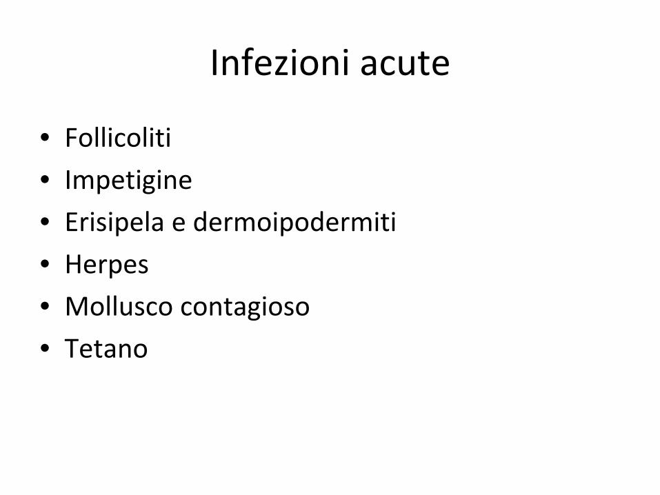
Infezioni acute
• Follicoliti• Impetigine
• Erisipela e dermoipodermiti
• Herpes• Mollusco contagioso
• Tetano

Impetigine e follicolite
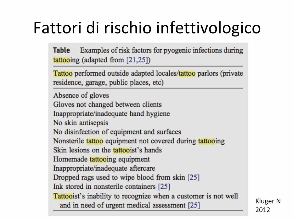
Fattori di rischio infettivologico
Kluger N2012

Tardive
• Infezioni• Reazioni allergiche, orticaria• Reazioni lichenoidi• Granulomi o reazioni granulomatose• Fotosensibilizzazione• Pseudolinfomi• Vasculiti e necrosi• Cancerizzazione, melanoma• Koebnerizzazione• Alterazioni associate alla Risonanza magnetica
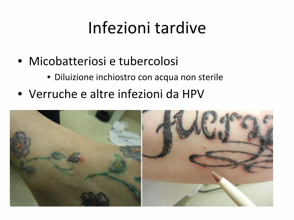
Infezioni tardive
• Micobatteriosi e tubercolosi• Diluizione inchiostro con acqua non sterile
• Verruche e altre infezioni da HPV
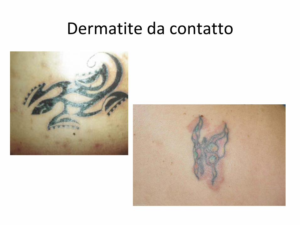
Dermatite da contatto

Dermatite da contatto

Reazioni lichenoidi
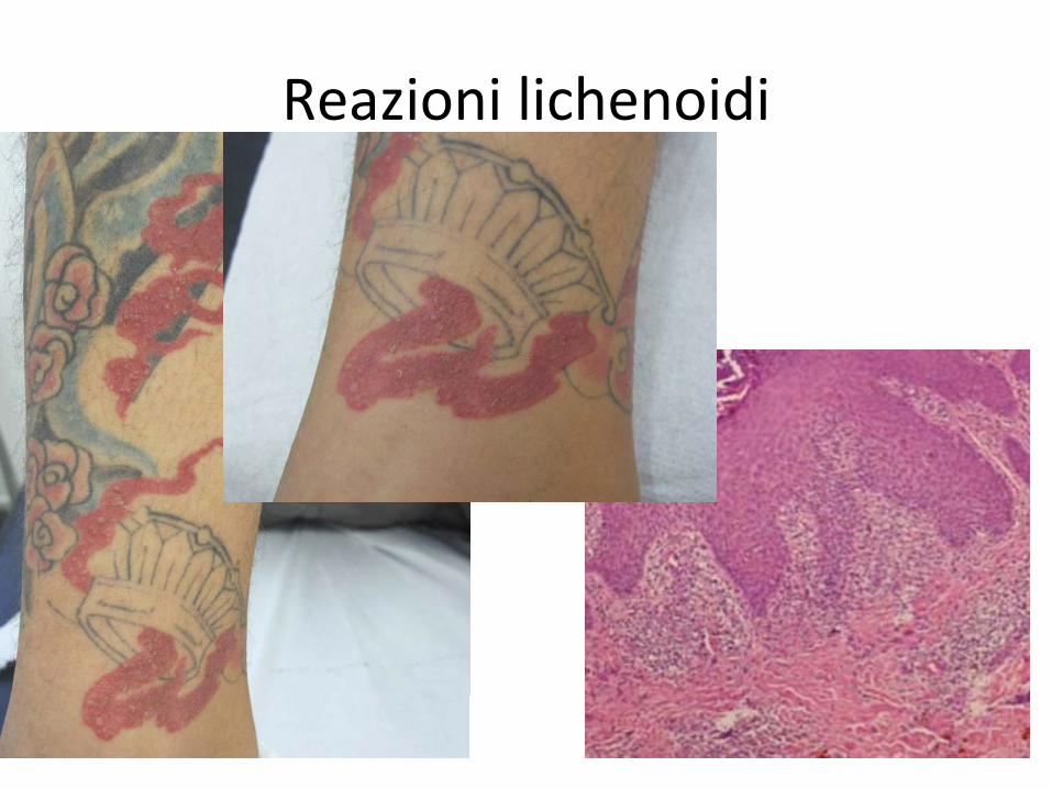
Reazioni lichenoidi

Reazioni granulomatose, Vasculiti e necrosi
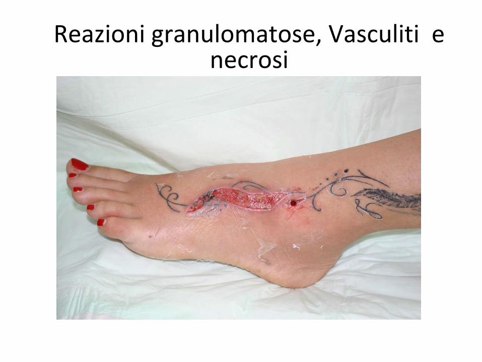
Reazioni granulomatose, Vasculiti e necrosi
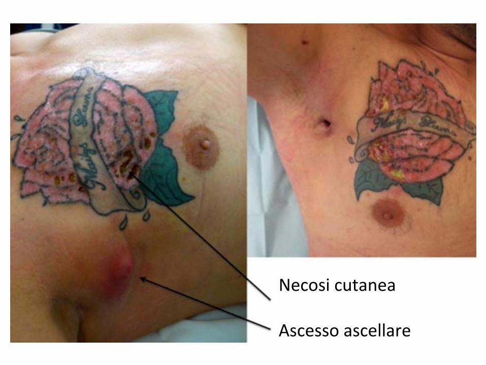
Necosi cutanea
Ascesso ascellare
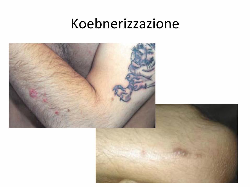
Koebnerizzazione

Granuloma sarcoideo
Juhas & English J Pediatr Adolesc Gynecol, 2013

Dermatofibromi e tumori cutanei
Bittencourt et al An Bras Dermatol, 2013
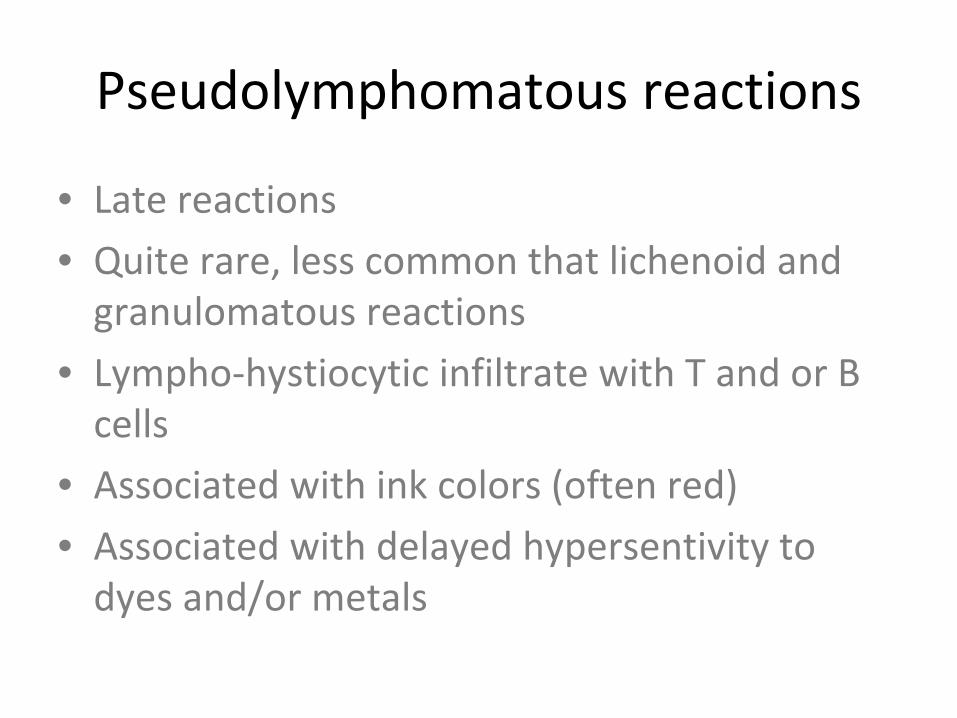
Pseudolymphomatous reactions
• Late reactions• Quite rare, less common that lichenoid and
granulomatous reactions
• Lympho‐hystiocytic infiltrate with T and or B cells
• Associated with ink colors (often red)• Associated with delayed hypersentivity to
dyes and/or metals

JEADV 2010
Case 1
• Woman, 32 yrs
• Tattoo since 1 year• Papular eruption in the reddish area of the
tattoo
• No adenopathy• Failure of previous treatment with clobetasol
• Patch test negative

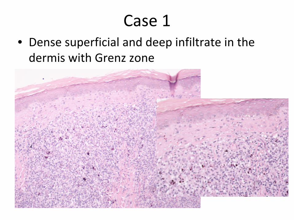
Case 1• Dense superficial and deep infiltrate in the
dermis with Grenz zone
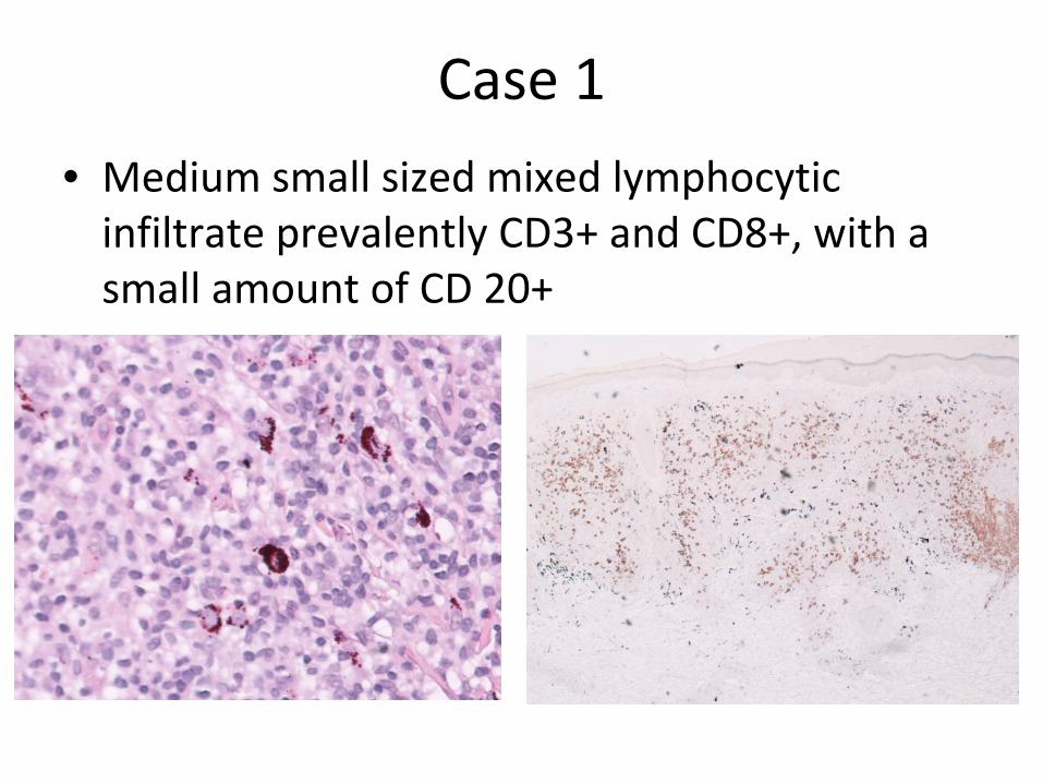
Case 1
• Medium small sized mixed lymphocytic infiltrate prevalently CD3+ and CD8+, with a
small amount of CD 20+
Ink and metal analysis (mass spectrometry)
• Ink was collected at tattoo shops (where the tattoos were done)
• Metals were analyzed from skin biopsies• Cr and Ni were found particularly high in both
ink and skin• Other metals, Hg in particular, were minimal
and have been considered impurities according to Resolution ResAP (2008)1 of the
Council of Europe
Conclusions
• In our series, reactions were all associated with red ink, as reported in the literature
• High levels of Cr and Ni were found in inks and skin biopsies
• All patients were patch test negative• Immunological reaction to these metals?• Lesions were successfully treated with surgery
or laser. No response to topical corticosteroids.

Grazie


![Ferrario ENTEROSTOMIE [modalità compatibilità]associazionemaruti.it/img/messaggi/1793363332435/Ferrario... · ALTERAZIONI CUTANEE lesione ulcerativa Follicolite peristomale ...](https://static.fdocumenti.com/doc/165x107/5c66943309d3f2c14e8c4429/ferrario-enterostomie-modalita-compatibilita-alterazioni-cutanee-lesione.jpg)