Protective Effect of the Cochlear Efferent System During Noise Exposure
7
Protective Effect of the Cochlear Efferent System During Noise Exposure GIUSEPPE ATTANASIO, a MAURIZIO BARBARA, GIUSEPPE BUONGIORNO, ALDO CORDIER, BARBARA MAFERA, FEDERICO PICCOLI, GABRIELE NOSTRO, AND ROBERTO FILIPO Institute of Otorhinolaryngology, University of Rome “La Sapienza,” Viale del Policlinico, Rome 00161, Italy ABSTRACT: The aim of the present study was to confirm the hypothesis that the cochlear efferent system is involved in the mechanisms underlying the “toughening” effect at high frequencies. The toughening effect is defined as a progressive threshold shift reduction when repeated exposures to the same noise are applied. Vestibular neurectomy was performed through a posterior cranial fossa approach in six healthy pigmented guinea pigs, and it assured the interruption of both crossed and uncrossed olivocochlear bundles to one ear only, before their entrance in the internal auditory meatus. The animals were then implanted with permanent electrodes for the electrocochleographic find- ings. Ten days after the operation the animals were exposed to octave-band toughening noise, centered at 4 kHz, at 85-dB SPL, for 10 consecutive days, 6 hours on/18 hours off. The hearing threshold was registered before and at the end of each exposure session. The behavior of the hearing threshold in the oper- ated ears was then compared to that of the controlateral, nonoperated ears. Complete recovery from TS in the control ear began after four days of expo- sure, whereas in the operated ear hearing loss increased to day 7 (55 dB), with only a partial reduction (45 dB) beyond ten days of exposure. The results of the present study clearly demonstrated that sectioning of the OCB in guinea pigs causes persistent hearing loss during noise-exposure condi- tioning, in comparison to the contralateral, nonoperated ear. Thus, one can as- sume that the lack of decrease of TS during intermittent noise exposure could be due to the loss of the protective effect of the efferent fibers, perhaps mediat- ed by the lateral OC neurons that synapse beneath the IHCs. INTRODUCTION The efferent innervation to the cochlea has been known since Rasmussen 1,2 who demonstrated the presence of a homo- and contralateral efferent olivocochlear bundle (OCB) in cats, rats, and opossum. The axons originate bilaterally in the superior oli- vary complex of the brain stem. With the application of innovative techniques such as retrograde and anterograde transport of tracers, anatomical reconstruction from serial images and immunocytochemical localization of neuroactive substances, a detailed anatomic description of the efferent auditory system has been achieved. 3–8 361 a To whom correspondence may be addressed. Phone: 39-6-4454607; fax: 39-6-4454864; e-mail: [email protected]
-
Upload
giuseppe-attanasio -
Category
Documents
-
view
214 -
download
2
Transcript of Protective Effect of the Cochlear Efferent System During Noise Exposure

Protective Effect of the Cochlear EfferentSystem During Noise Exposure
GIUSEPPE ATTANASIO,a MAURIZIO BARBARA, GIUSEPPEBUONGIORNO, ALDO CORDIER, BARBARA MAFERA, FEDERICO PICCOLI, GABRIELE NOSTRO, AND ROBERTO FILIPO
Institute of Otorhinolaryngology, University of Rome “La Sapienza,” Viale delPoliclinico, Rome 00161, Italy
ABSTRACT: The aim of the present study was to confirm the hypothesis thatthe cochlear efferent system is involved in the mechanisms underlying the“toughening” effect at high frequencies. The toughening effect is defined as aprogressive threshold shift reduction when repeated exposures to the samenoise are applied. Vestibular neurectomy was performed through a posteriorcranial fossa approach in six healthy pigmented guinea pigs, and it assured theinterruption of both crossed and uncrossed olivocochlear bundles to one earonly, before their entrance in the internal auditory meatus. The animals werethen implanted with permanent electrodes for the electrocochleographic find-ings. Ten days after the operation the animals were exposed to octave-bandtoughening noise, centered at 4 kHz, at 85-dB SPL, for 10 consecutive days, 6hours on/18 hours off. The hearing threshold was registered before and at theend of each exposure session. The behavior of the hearing threshold in the oper-ated ears was then compared to that of the controlateral, nonoperated ears.Complete recovery from TS in the control ear began after four days of expo-sure, whereas in the operated ear hearing loss increased to day 7 (55 dB), withonly a partial reduction (45 dB) beyond ten days of exposure.
The results of the present study clearly demonstrated that sectioning of theOCB in guinea pigs causes persistent hearing loss during noise-exposure condi-tioning, in comparison to the contralateral, nonoperated ear. Thus, one can as-sume that the lack of decrease of TS during intermittent noise exposure couldbe due to the loss of the protective effect of the efferent fibers, perhaps mediat-ed by the lateral OC neurons that synapse beneath the IHCs.
INTRODUCTION
The efferent innervation to the cochlea has been known since Rasmussen1,2 whodemonstrated the presence of a homo- and contralateral efferent olivocochlear bundle(OCB) in cats, rats, and opossum. The axons originate bilaterally in the superior oli-vary complex of the brain stem. With the application of innovative techniques such asretrograde and anterograde transport of tracers, anatomical reconstruction from serialimages and immunocytochemical localization of neuroactive substances, a detailedanatomic description of the efferent auditory system has been achieved.3–8
361
aTo whom correspondence may be addressed. Phone: 39-6-4454607; fax: 39-6-4454864;e-mail: [email protected]
Two populations of cochlear efferent fibers have been identified, that is, lateraland medial olivocochlear (OC) neurons. Lateral OC neurons are situated in closeproximity to, or within, the lateral superior olivary nucleus (LSO), whereas medialOC neurons are scattered medially, ventrally, and anteriorly to the medial superiorolivary nucleus (MSO). Lateral OC neurons project predominantly to the ipsilateralcochlea and terminate almost exclusively beneath the inner hair cells (IHCs), where-as medial OC neurons project predominantly to the contralateral cochlea and termi-nate mainly beneath the outer hair cells (OHCs). The so-called medial group consistsof medium-sized oval-shaped cells with diameters as large as 20 �m. The contralat-eral fibers cross the midline at the floor of the fourth ventricle. The so-called lateralgroup consists of very small fusiform cells with diameters of approximately 10 �m.They join the homolateral bundle within the vestibular root and run with the vestibu-lar nerve into the periphery. Before they cross over to the cochlear nerve deep in theinternal auditory meatus, through the anastomosis of Oort (vestibulocochlear anasto-mosis). Within the modiolus, the efferent nerves form the intraganglionic spiral bun-dle as one or several nerve bundles in the peripheral portion of the spiral ganglionfrom where the fibers radiate to the organ of Corti.
The distribution of the efferent fibers to the OHCs is essentially radial, and thetunnel-crossing fibers are relatively large with an average diameter of 1 �m. The ef-ferent innervation of the OHCs decreases from base to apex and is less abundant inthe second and third row. The efferent fibers at the level of the IHCs form the innerspiral and the tunnel spiral fibers. Morphologically, the efferent fibers of the OHCsare large and radial and synapse predominantly with the receptor cells,9–11 whereasthe efferent fibers of the IHCs system have a spiral distribution and synapse exclu-sively with dendrites of type I ganglion cells beneath the IHCs,12,5 but not with thereceptor cell. The efferent axons beneath IHCs are of two distinct morphologicaltypes.13,14
1. Unidirectional fibers, composing about 85% of IHC efferents, which enter inthe inner spiral bundle and terminate in one or more dense patches that, in thetotal basal–apical extent, span no more than 10–20% (1–2 mm) of the totallength of the organ of Corti;
2. Bidirectional fibers, which enter the organ of Corti and bifurcate into basal andapical branches over a span of more than 50% of its length, then coursing inthe inner spiral bundle for at least 80%, and sometimes more than 95% of thetotal cochlear length.
It is worth mentioning that, in several species of rodents, the LSO and the sur-rounding region show two types of olivocochlear neurons: small cells within the LSO(intrinsic neurons) and large cells (shell neurons) surrounding it, located in the later-al nucleus of the trapezoid body.14,15 It has been shown that injections of a specifictracer (biotinylated dextran amine) into the intrinsic neurons produce focal peaks oflabeling in the cochlea, whereas similar injections into the shell neurons produced adiffuse, nonfocal projection, extending for nearly the entire length of the cochlea.
A protective role has been recently suggested for the olivocochlear efferent sys-tem.16–18 In these experiments, the activation of the crossed OCB could reduce thetemporary effects of acoustic overstimulation in guinea pigs. In this regard, the role
ANNALS NEW YORK ACADEMY OF SCIENCES362
of the efferent system in the toughening phenomenon was studied. The “toughening”effect is defined as a progressive threshold shift reduction when repeated exposuresto the same noise are applied.19,20 Chinchillas with crossed OCB sectioned at the lev-el of the forth ventricle were exposed to 4-kHz toughening noise exposure, in orderto test whether or not the OCB is involved in high-frequency long-duration noise ex-posure (G. Attanasio, D. Henderson, M. Subramaniam, and Y.-H. Shen, unpublisheddata). At the end of the exposure time (10 days, 6 h/day) at 85-dB SPL, the animalsdeveloped a threshold shift slightly greater than the control group, showing less re-covery deefferented (17 dB) than the control (28 dB). Unfortunately, the high rate ofpostoperative mortality did not allow the use of an appropriate number of experimen-tal animals.
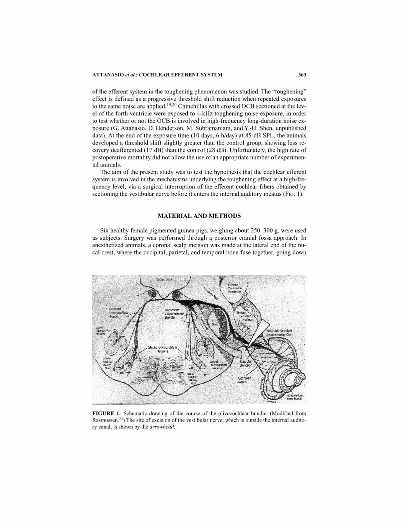
The aim of the present study was to test the hypothesis that the cochlear efferentsystem is involved in the mechanisms underlying the toughening effect at a high-fre-quency level, via a surgical interruption of the efferent cochlear fibers obtained bysectioning the vestibular nerve before it enters the internal auditory meatus (FIG. 1).
MATERIAL AND METHODS
Six healthy female pigmented guinea pigs, weighing about 250–300 g, were usedas subjects. Surgery was performed through a posterior cranial fossa approach. Inanesthetized animals, a coronal scalp incision was made at the lateral end of the nu-cal crest, where the occipital, parietal, and temporal bone fuse together, going down
ATTANASIO et al.: COCHLEAR EFFERENT SYSTEM 363
FIGURE 1. Schematic drawing of the course of the olivocochlear bundle. (Modified fromRasmussen.23) The site of excision of the vestibular nerve, which is outside the internal audito-ry canal, is shown by the arrowhead.
to the region of the occipital bone. Using a diamond bur, the region of the occipitalbone lying between the external occipital crest and the supraoccipital ridge wasopened until the exposure of the underlying dura. A careful blunt dissection of thesigmoid sinus from its sulcus in the temporal bone followed, thus allowing its medialretraction. Using neurosurgical scissors, the dura at the medial edge of the sigmoidsinus was opened, allowing the aspiration of a small amount of cerebellar tissue and adorsal–medial retraction of the cerebellum itself into the cranial cavity. The medialsurface of the petrous segment of the temporal bone was hence exposed and the re-gion of the internal auditory meatus identified. The superior vestibular nerve is thefirst tubular structure encountered through this approach. Rostrally and slightly infe-rior to it lies the canal for the facial nerve. Caudally and inferiorly to these two smallforamina is the internal auditory canal that contains the cochlear nerve, the singularnerve, and the nerve to the saccular macula. With a cutting hook, the superiorvestibular nerve was finally excised. Closure of the surgical cavity with Surgicel®and Gelfoam® ends the operation, leaving the efferent system to the cochlea inter-rupted.
The animals were then implanted with permanent evoked potential recordingelectrodes. The positive electrode, placed in the mastoid region, was attached to asocket that also incorporated connections for the reference and ground electrodesplaced in the contralateral mastoid region and at the vertex, respectively. The socketwas glued to the skull at the vertex with dental cement. Ten days after the operationthe animals were exposed to octave-band toughening noise, centered at 4 kHz, at 85-dB SPL, for 10 consecutive days, 6 h on/18 h off. The hearing threshold was regis-tered before and at the end of each exposure session. The behavior of the hearingthreshold in the operated ears was then compared to that of the contralateral, nonop-erated ears.
RESULTS
The surgical procedure did not significantly affect the hearing threshold, sinceonly a 5-dB difference between the operated and the control ear was evidenced (FIG.2). The worst hearing loss was recorded at the end of the third exposure session, inboth sides. At this time, however, the animals developed a greater initial TS at 4kHzin the operated ear (50 dB) compared to the normal one (35 dB). A complete recov-ery from TS in the control ear began 4 days after the exposure, whereas in the operat-ed ear hearing loss increased to day 7 (55 dB) with only a partial reduction (45 dB)after 10 days of exposure.
DISCUSSION
Although a protective effect of the efferent cochlear system has unequivocallybeen demonstrated in the guinea pig,16–18 ambiguities still remain as to the mecha-nisms underlying this effect.21 It has previously been reported that, when exposingthe animals to a 10-kHz tone for 1 min, at approximately 100-dB SPL of intensity,those in which the OCB is electrically stimulated during the exposure show a smaller
ANNALS NEW YORK ACADEMY OF SCIENCES364

TS than the animals exposed without electric stimulation. The protective effect of theelectric stimulation was shown to disappear when animals are treated with systemicstrychnine, a drug known to block the efferent function. Moreover, it has also beensuggested that this protective effect could be mediated by the so-called medial olivo-cochlear (MOC) system, that is, the OC projection to the OHCs.17,18 However, theuse of electric stimulation, either at the floor of the fourth ventricle or at the roundwindow level, could also cause activation of other feedback pathways to the innerear, such as the middle-ear muscle reflex or the autonomic fibers system or, alterna-tively, could be changing systemic variables, such as blood pressure or temperature,which might, in turn, affect the vulnerability of the ear to acoustic overstimulation.Finally, the widespread central and peripheral effects of strychnine make the sys-temic injection of this drug a nonspecific test for an olivocochlear involvement.
Liberman21 was unable to replicate the same findings in the cat, but reported thatwhen using a higher level of noise exposure and, consequently, inducing greater TS,the efferent fibers could play a protective role. It must also be noted that all thesestudies only involved short-duration, high-level pure-tone exposures.
The results of the present study clearly demonstrate that sectioning of the OCB inguinea pigs causes a persistent hearing loss during conditioning noise exposure, incomparison to the contralateral, nonoperated ear. The surgical excision of thevestibular nerve before its entrance into internal auditory meatus represent an opti-mal method to assure the interruption of both crossed and uncrossed olivocochlearbundles to one ear only, before their anastomosis with the cochlear nerve. Recoveryis essentially complete 10 days postsurgery, and it is now possible to measure the pre-exposure hearing threshold. The use of chronic exposure eliminated any biasing re-lated to anesthesia and/or changes of systemic variables, such as blood pressure ortemperature. In addition, an earlier study22 has demonstrated that sectioning ofstapedius muscle did not prevent TS reduction during “conditioning” exposure. Thus,one can assume that the lack of a decrease in TTS during intermittent noise exposure
ATTANASIO et al.: COCHLEAR EFFERENT SYSTEM 365
FIGURE 2. Effect of olivocochlear bundle section on threshold shift from exposure to anOBN centered at 4 kHz at 85-dB SPL for 6 h/day for 10 days.

could be due to the loss of the protective effect of the efferent fibers, perhaps mediat-ed by the lateral OC neurons that synapse beneath the IHCs.
If the OC system plays a protective role in noise-induced hearing loss, then by un-derstanding this natural “defense” mechanism and how to measure its effectiveness,it may be possible to explain the interindividual different susceptibility to acoustictrauma on the basis of differences in efferent system function.
ACKNOWLEDGMENT
This research was supported by MURST, under grant It (COFIN ’98): “Phys-iopathology of the Efferent Olivocochlear System.”
REFERENCES
1. RASMUSSEN, G. L. 1946. The olivary peduncle and other fiber projections of the superiorolivary complex. J. Comp. Neurol. 84: 141–219.
2. RASMUSSEN, G. L. 1953. Further observations of the efferent cochlear bundle. J. Comp.Neurol. 99: 61–74.
3. WARR, W. B. 1975. Olivocochlear and vestibular efferent neurons of the feline brain stem:Their location, morphology, and number determined by retrograde axonal transport andacetylcholinesterase histochemistry. J. Comp. Neurol. 161: 159–182.
4. WARR, W. B. & J. J. GUINAN, JR. 1979. Efferent innervation of the organ of Corti: Two sep-arate systems. Brain Res. 173: 152–155.
5. LIBERMAN, M. C. 1980. Efferent synapses in the inner hair cell area of the cat cochlea: anelectron microscopy study of serial sections. Hear. Res. 3: 189–204.
6. SANTI, P. 1986. Organ of surface preparations for computer assisted morphometry. Hear.Res. 24: 179–187.
7. RYAN, A. F., I. R. SCHWARTZ, R. H. HELFERT, E. KEITHLEY & Z.-X. WANG. 1987. Selectiveretrograde labeling of lateral olivocochlear neurons in the brainstem based on preferen-tial uptake of 3H-D aspartic acid in the cochlea. J. Comp. Neurol. 255: 606–616.
8. VEENMAN, C. L., A. REINER & M. G. HONING. 1992. Biotinylated dextran amine as an an-terograde tracer for single and double-labelling studies. J. Neurosci. Methods 41:239–254.
9. KIMURA, R. S. & J. WERSALL. 1962. Termination of the olivocochlear bundle in relation tothe outer hair cells of the organ of Corti in the guinea pig. Acta Otolaryngol. 55: 11–32.
10. SPOENDLIN, H. 1966. The organization of the cochlear receptor. Adv. Oto-Rhino-Laryngol.13: 1–114.
11. GINSBERG, R. D. & D. K. MOREST. 1984. Fine structure of cochlear innervation in the cat.Hear. Res. 14: 93–106.
12. SMITH, C. A. 1961. Innervation patterns of the cochlea. The internal hair cells. Ann. Otol.Rhinol. Laryngol. 70: 504–527.
13. BROWN, M. C. 1987. Morphology of labeled efferent fibers in the guinea pig cochlea. J.Comp. Neurol. 260: 605–618.
14. WARR, W. B., J. B. BOCHE & S. T. NEELY. 1997. Efferent innervation of the inner hair cellregion: origins and terminations of two lateral olivocochlear systems. Hear. Res. 198:89–111.
15. VETTER, D. E. & E. MUGNAINI. 1992. Distribution and dendritic features of three groups ofrat olivocochlear neurons. Anat. Embryol. 185: 1–16.
ANNALS NEW YORK ACADEMY OF SCIENCES366

16. RAJAN, R. & B. M. JOHNSTONE. 1983. Crossed cochlear influences on monoaural tempo-rary threshold shift. Hear. Res. 9: 279–294.
17. RAJAN, R. 1988. Effect of electrical stimulation of the crossed olivocochlear bundle ontemporary threshold shift in auditory sensitivity. I. Dependence on electrical stimulationparameters. J. Neurophysiol. 60: 549–568.
18. RAJAN, R. 1988. Effect of electrical stimulation of the crossed olivocochlear bundle ontemporary threshold shift in auditory sensitivity. II. Dependence on the level of the tem-porary threshold shift. J. Neurophysiol. 60: 569–579.
19. MILLER, J. D., C. S. WATSON & W. P. COVELL. 1963. Deafening effects of noise on the cat.Acta Otolalaryngol. (Stockh.) 176 (Suppl.): 44–52.
20. CLARK, W. W., B. A. BOHNE & F. A. BOETTCHER. 1987. Effect of periodic rest on hearingloss and cochlear damage following exposure to noise. J. Acoust. Soc. Am. 82:1253–1264.
21. LIBERMAN, M. C. 1992. Does olivocochlear feedback protect the cat’s ear from acoustic in-jury? In Noise Induced Hearing Loss, A. Dancer, D. Henderson, R. J. Salvi, and R. P.Hamernik, Eds.: 423–428. Mosby-Year Book. St. Louis, Mo.
22. HENDERSON, D., M. SUBRAMANIAM, M. PAPAZIAN & V. P. SPONGR. 1994. The role of themiddle ear muscles in the development of resistance to noise induced hearing loss. Hear.Res. 74: 22–28.
ATTANASIO et al.: COCHLEAR EFFERENT SYSTEM 367


![Seastick .ppt [modalità compatibilità] - · PDF file› Sistema operativoSistema operativo Windows Enbedded › Processore ARM11 › Web server integrato ... › Low noise for ROV](https://static.fdocumenti.com/doc/165x107/5a9ff7607f8b9a84178d8680/seastick-ppt-modalit-compatibilit-sistema-operativosistema-operativo-windows.jpg)