
La circolazione retinica e il rischio cardiovascolare · La circolazione retinica e il rischio...
43
ASST Spedali Civili di Brescia – University of Brescia, Italy La circolazione retinica e il rischio cardiovascolare Prof. ENRICO AGABITI ROSEI Clinica Medica – University of Brescia, Italy
Transcript of La circolazione retinica e il rischio cardiovascolare · La circolazione retinica e il rischio...
ASST Spedali Civili di Brescia – University of Brescia, Italy
La circolazione retinica e
il rischio cardiovascolare
Prof. ENRICO AGABITI ROSEI
Clinica Medica – University of Brescia, Italy
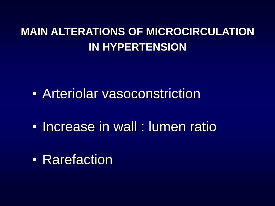
• Arteriolar vasoconstriction
• Increase in wall : lumen ratio
• Rarefaction
MAIN ALTERATIONS OF MICROCIRCULATION
IN HYPERTENSION
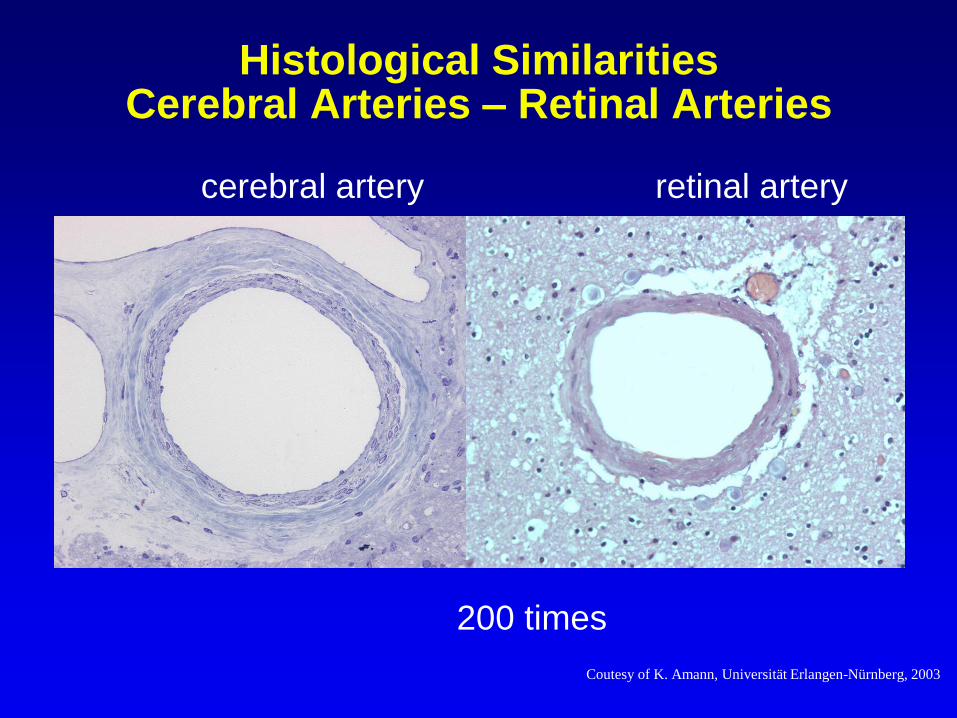
Histological Similarities Cerebral Arteries – Retinal Arteries
cerebral artery retinal artery
200 times
Coutesy of K. Amann, Universität Erlangen-Nürnberg, 2003
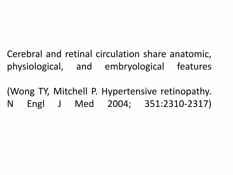
Cerebral and retinal circulation share anatomic, physiological, and embryological features
(Wong TY, Mitchell P. Hypertensive retinopathy. N Engl J Med 2004; 351:2310-2317)
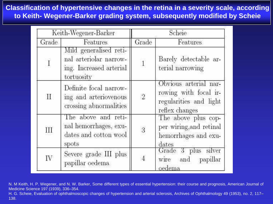
Classification of hypertensive changes in the retina in a severity scale, according
to Keith- Wegener-Barker grading system, subsequently modified by Scheie
N. M Keith, H. P. Wegener, and N. W. Barker, Some different types of essential hypertension: their course and prognosis, American Journal of
Medicine Science 197 (1939), 336–354.
H. G. Scheie, Evaluation of ophthalmoscopic changes of hypertension and arterial sclerosis, Archives of Ophthalmology 49 (1953), no. 2, 117–
138.
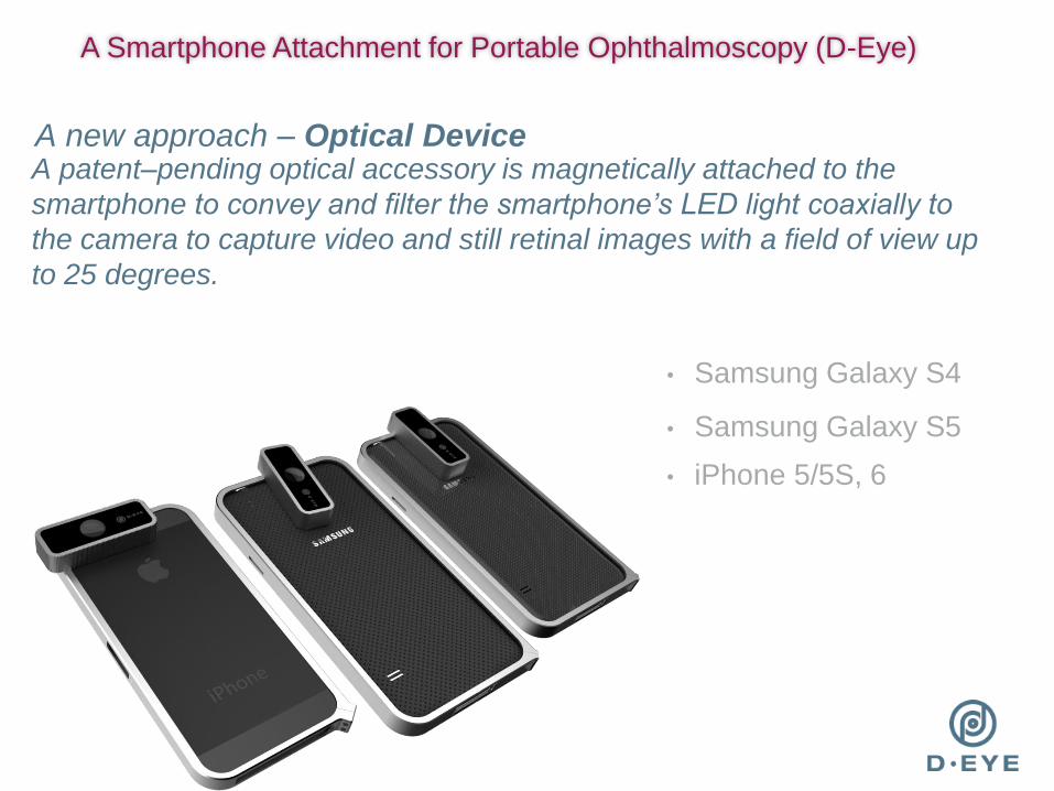
A patent–pending optical accessory is magnetically attached to the
smartphone to convey and filter the smartphone’s LED light coaxially to
the camera to capture video and still retinal images with a field of view up
to 25 degrees.
• Samsung Galaxy S4
• Samsung Galaxy S5
• iPhone 5/5S, 6
A new approach – Optical Device
A Smartphone Attachment for Portable Ophthalmoscopy (D-Eye)
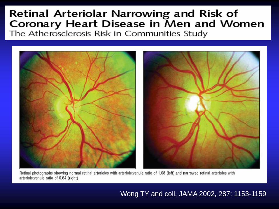
Wong TY and coll, JAMA 2002, 287: 1153-1159
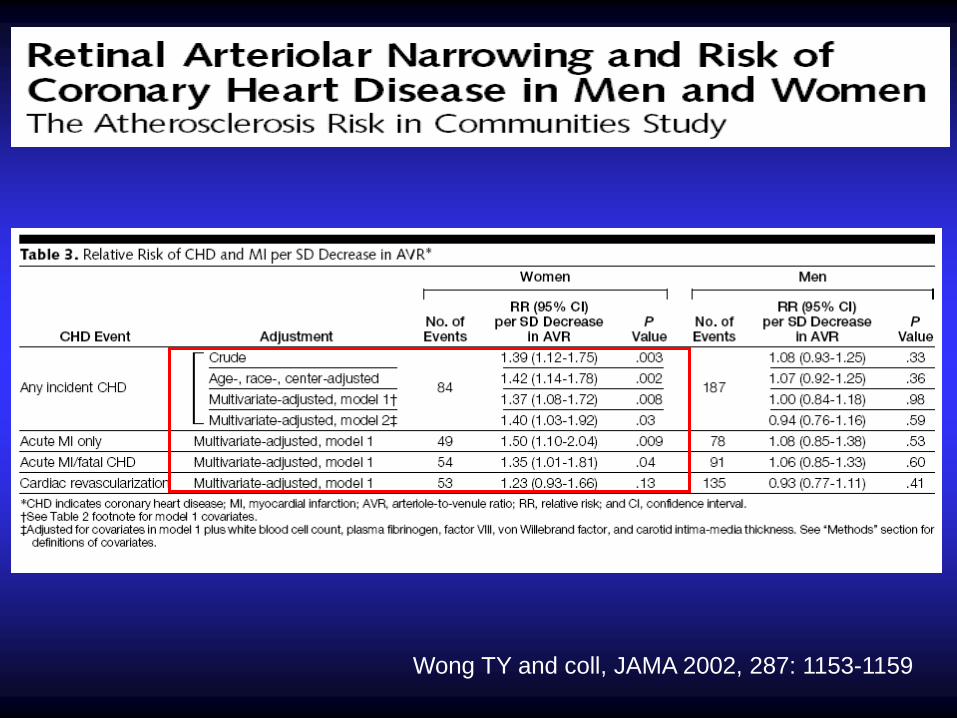
Wong TY and coll, JAMA 2002, 287: 1153-1159
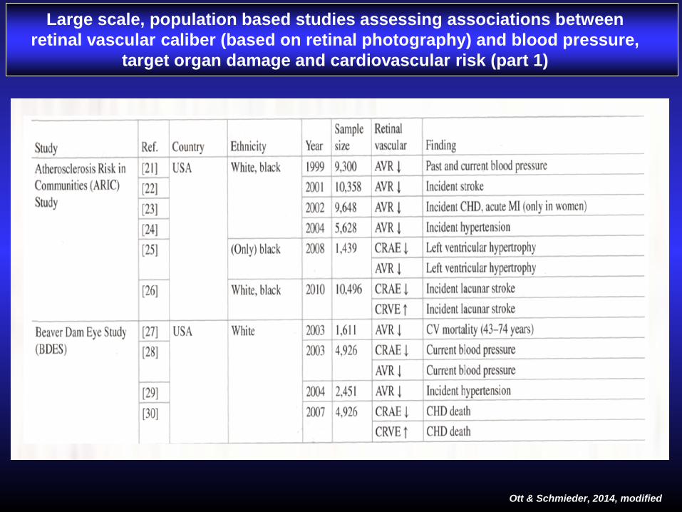
THE END THE END Large scale, population based studies assessing associations between
retinal vascular caliber (based on retinal photography) and blood pressure,
target organ damage and cardiovascular risk (part 1)
Ott & Schmieder, 2014, modified
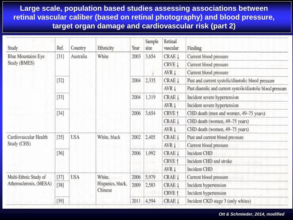
THE END Large scale, population based studies assessing associations between
retinal vascular caliber (based on retinal photography) and blood pressure,
target organ damage and cardiovascular risk (part 2)
Ott & Schmieder, 2014, modified

Quantification of topological changes in retinal vascular
architecture in essential and malignant hypertension
Hughes AD et al, J Hypertens 2006, 24:889–894
EHT was associated with an increase in the arteriolar length-to-
diameter ratio (P < 0.01). There were also alterations in
arteriolar topology indicative of rarefaction, including a marked
reduction in the number of terminal branches in EHT (P < 0.01).
These changes in the arteriolar network were exaggerated in
MHT and there was also increased venular tortuosity and
venular rarefaction in MHT compared with normotensive
subjects.
Hypertension is associated with marked topological alterations in
the retinal vasculature, and quantification of these changes may
be a useful novel approach to the assessment of target organ
damage in hypertension.

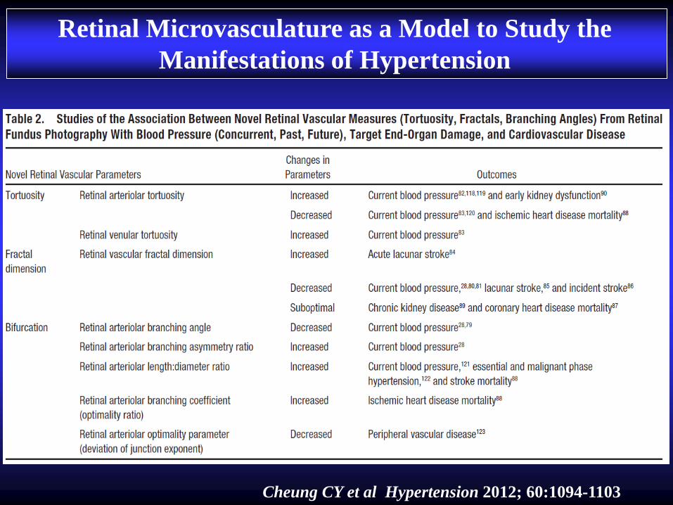
Retinal Microvasculature as a Model to Study the
Manifestations of Hypertension
Cheung CY et al Hypertension 2012; 60:1094-1103
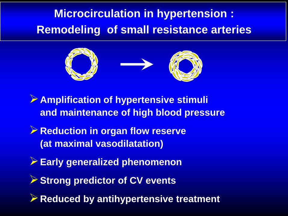
Amplification of hypertensive stimuli
and maintenance of high blood pressure
Reduction in organ flow reserve
(at maximal vasodilatation)
Early generalized phenomenon
Strong predictor of CV events
Reduced by antihypertensive treatment
Microcirculation in hypertension :
Remodeling of small resistance arteries
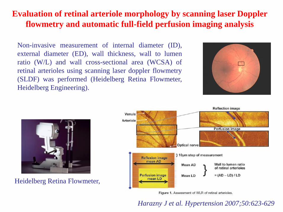
Evaluation of retinal arteriole morphology by scanning laser Doppler
flowmetry and automatic full-field perfusion imaging analysis
Non-invasive measurement of internal diameter (ID),
external diameter (ED), wall thickness, wall to lumen
ratio (W/L) and wall cross-sectional area (WCSA) of
retinal arterioles using scanning laser doppler flowmetry
(SLDF) was performed (Heidelberg Retina Flowmeter,
Heidelberg Engineering).
Heidelberg Retina Flowmeter,
Heidelberg Engineering
Harazny J et al. Hypertension 2007;50:623-629
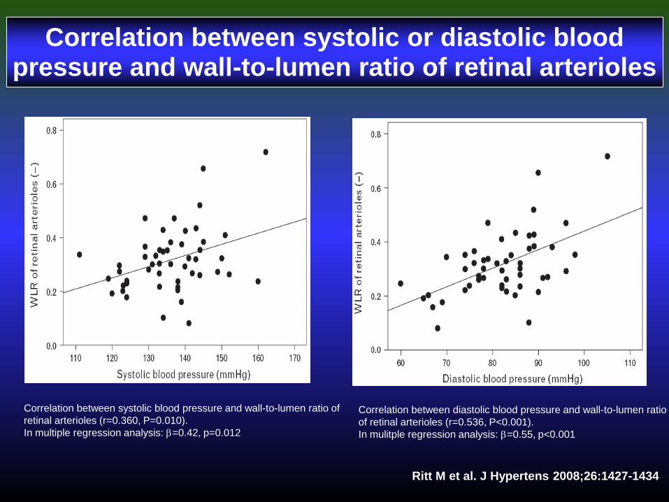
Correlation between systolic or diastolic blood pressure and wall-to-lumen ratio of retinal arterioles
Correlation between systolic blood pressure and wall-to-lumen ratio of
retinal arterioles (r=0.360, P=0.010).
In multiple regression analysis: b=0.42, p=0.012
Correlation between diastolic blood pressure and wall-to-lumen ratio
of retinal arterioles (r=0.536, P<0.001).
In mulitple regression analysis: b=0.55, p<0.001
Ritt M et al. J Hypertens 2008;26:1427-1434


Evaluation of small retinal artery morphology by scanning laser Doppler
flowmetry and automatic full-field perfusion imaging analysis
Clinica Medica, University of Brescia, Italy

Close correlation between wall to lumen ratio (W/L) of retinal arterioles and
media to lumen ratio of subcutaneous small arteries (M/L) in a population of
normotensive subjects (n=16) and hypertensive patients (n=24)
Rizzoni D, Agabiti Rosei E et al, J Hypertens, 2012
r=0.76, p<0.001
y = 6,9364x - 0,2779
R2 = 0,5702
0,00
0,10
0,20
0,30
0,40
0,50
0,60
0,70
0,80
0,04 0,06 0,08 0,10 0,12 0,14 0,16
M/L
W/L
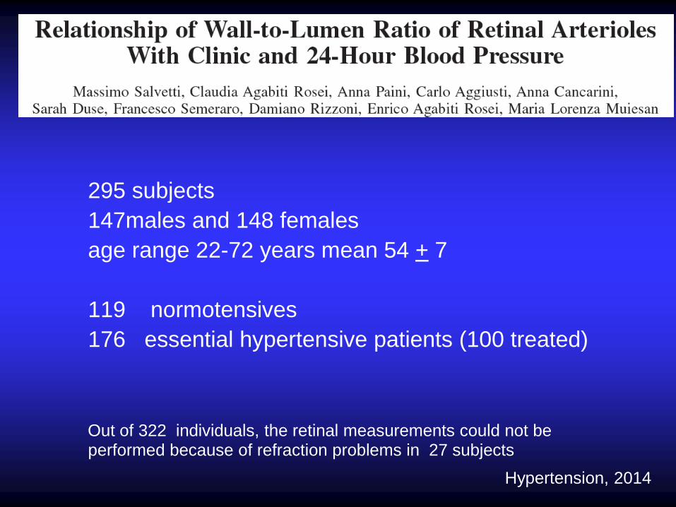
295 subjects
147males and 148 females
age range 22-72 years mean 54 + 7
119 normotensives
176 essential hypertensive patients (100 treated)
Out of 322 individuals, the retinal measurements could not be performed because of refraction problems in 27 subjects
Patients
Hypertension, 2014

Correlation between WLR and PWV
r =0.17 , p = 0.005
Salvetti, Agabiti Rosei et al, Hypertension, 2014
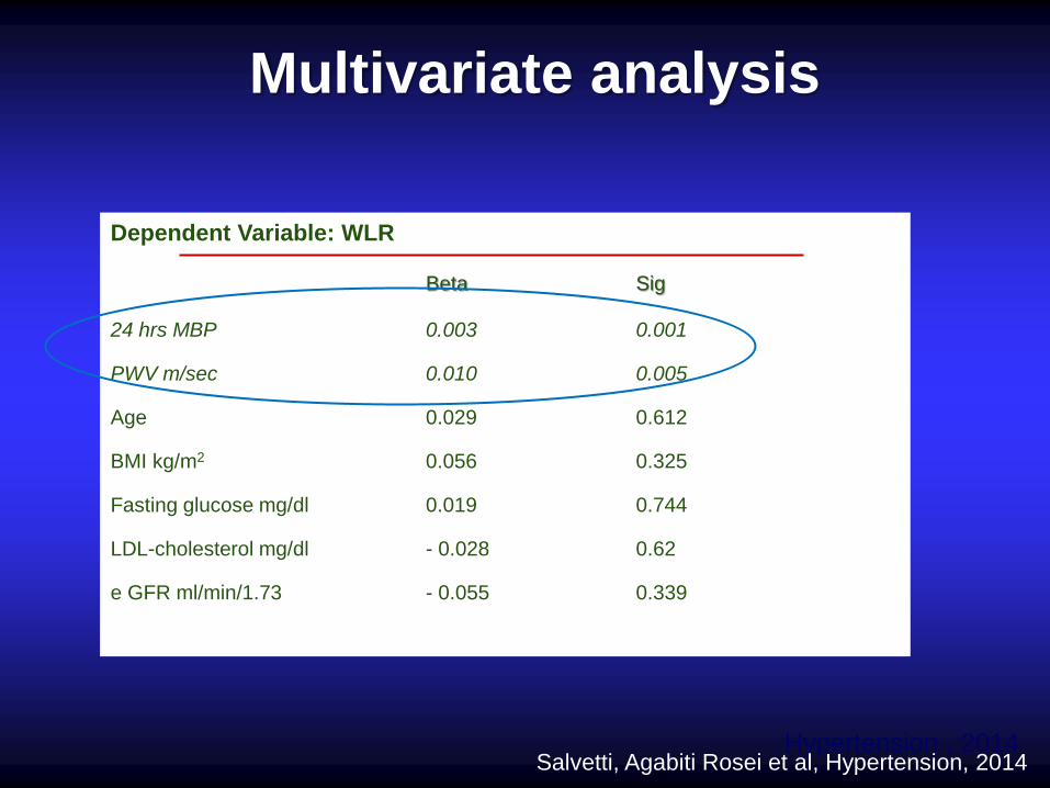
Multivariate analysis
Dependent Variable: WLR
Beta Sig
24 hrs MBP 0.003 0.001 PWV m/sec 0.010 0.005 Age 0.029 0.612 BMI kg/m2 0.056 0.325 Fasting glucose mg/dl 0.019 0.744 LDL-cholesterol mg/dl - 0.028 0.62 e GFR ml/min/1.73 - 0.055 0.339
Hypertension , 2014 Salvetti, Agabiti Rosei et al, Hypertension, 2014

* P<0.05, ** P<0.01 vs. Basal, # P<0.05 vs. Lercanidipine alone (4 weeks)
**
Basal
Lercanidipine (4 weeks)
Lercanidipine+ENA
(24 weeks)
**
Basal
Lercanidipine (4 weeks)
Lercanidipine+HCTZ
(24 weeks)
Reduction in wall to lumen ratio of retinal arterioles after lercanidipine+enalapril
treatment (n=10) and after lercanidipine+hydrochlorothiazide treatment in
hypertensive patients (n=10)

* P<0.05, ** P<0.01 vs. Basal, # P<0.05 vs. Lercanidipine alone (4 weeks)
***#
**
Basal
Lercanidipine (4 weeks)
Lercanidipine+ENA
(24 weeks)
**
** Basal
Lercanidipine (4 weeks)
Lercanidipine+HCTZ
(24 weeks)
Reduction in wall to lumen ratio of retinal arterioles after lercanidipine+enalapril
treatment (n=10) and after lercanidipine+hydrochlorothiazide treatment in
hypertensive patients (n=10)
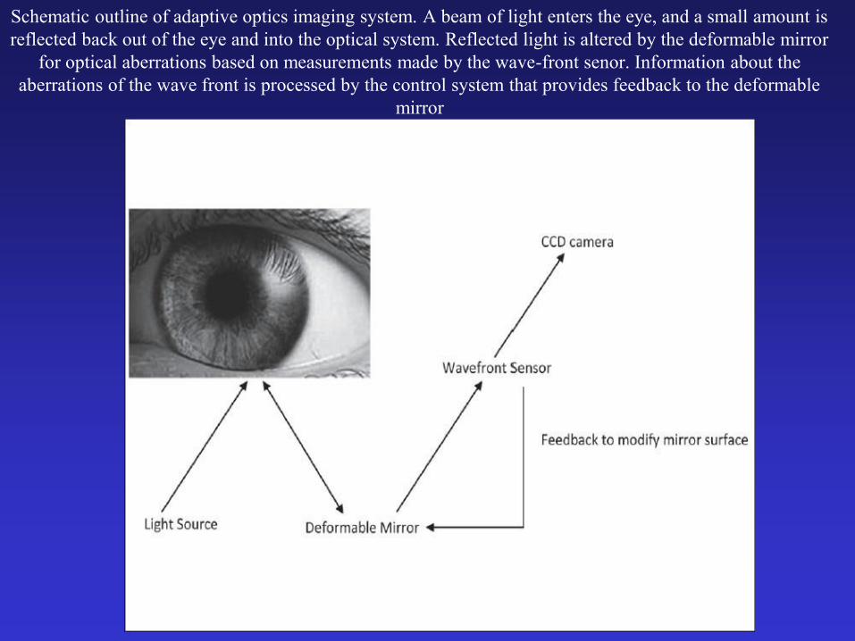
Schematic outline of adaptive optics imaging system. A beam of light enters the eye, and a small amount is
reflected back out of the eye and into the optical system. Reflected light is altered by the deformable mirror
for optical aberrations based on measurements made by the wave-front senor. Information about the
aberrations of the wave front is processed by the control system that provides feedback to the deformable
mirror
J Hypertens 2014 ; 32(4):890-898
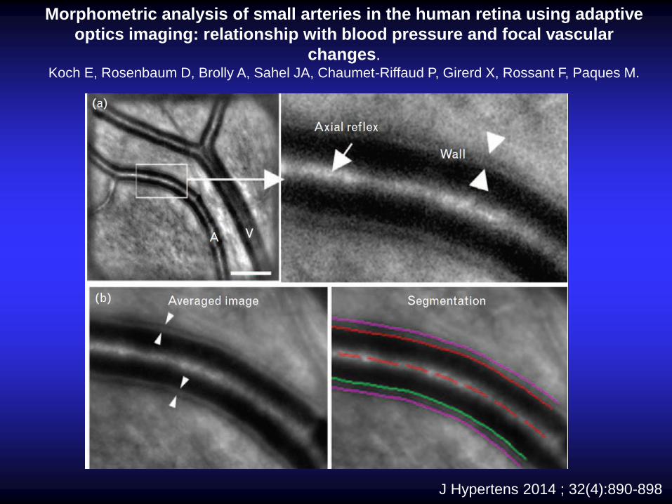
Morphometric analysis of small arteries in the human retina using adaptive
optics imaging: relationship with blood pressure and focal vascular
changes. Koch E, Rosenbaum D, Brolly A, Sahel JA, Chaumet-Riffaud P, Girerd X, Rossant F, Paques M.
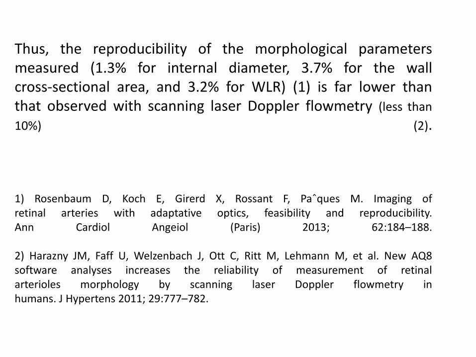
Thus, the reproducibility of the morphological parameters measured (1.3% for internal diameter, 3.7% for the wall cross-sectional area, and 3.2% for WLR) (1) is far lower than that observed with scanning laser Doppler flowmetry (less than
10%) (2).
1) Rosenbaum D, Koch E, Girerd X, Rossant F, Paˆques M. Imaging of retinal arteries with adaptative optics, feasibility and reproducibility. Ann Cardiol Angeiol (Paris) 2013; 62:184–188.
2) Harazny JM, Faff U, Welzenbach J, Ott C, Ritt M, Lehmann M, et al. New AQ8 software analyses increases the reliability of measurement of retinal arterioles morphology by scanning laser Doppler flowmetry in humans. J Hypertens 2011; 29:777–782.

EFFECTS OF AGE, BLOOD PRESSURE AND ANTIHYPERTENSIVE
TREATMENTS ON RETINAL ARTERIOLES REMODELING ASSESSED
BY ADAPTIVE OPTICS
Rosembaum D et al , J Hypertens, in press
CONCLUSIONS
…. new technologies, presently under clinical
evaluation, may help us in the near future to:
• non invasively assess microvascular structural
alterations
• better stratify cardiovascular risk of our patients
with consequent optimization of treatment.

Eyes: an open window on our inner body “ ”

THE END
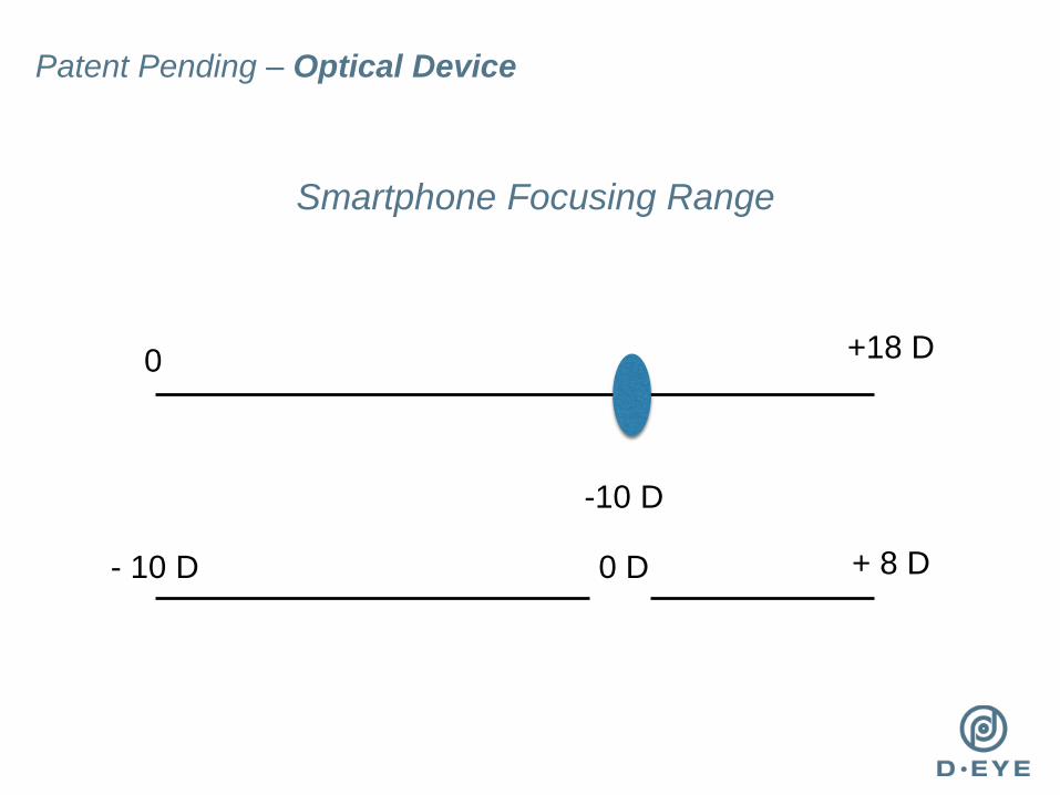
Patent Pending – Optical Device
+ 8 D
+18 D
-10 D
Smartphone Focusing Range
0
- 10 D 0 D


Representative Retinal Images
Representative retinal images taken with D-Eye.
(A) Normal optic disc in an undilated child. (B)
Normal posterior pole in a dilated 29-year-old
woman. (C) Dry age-related maculopathy in an
undilated 75-year-old man. (D) Optic nerve glioma
in a 23-year-old undilated woman. (E) Posterior
vitreous detachment in a dilated 72-year-old
pseudophakic woman. (F) Waxy disc pallor and
pigmentary changes in a 50-year-old man with
retinitis pigmentosa.
Cross-polarization-accentuated
nerve-fiber layer definition (white
arrows).

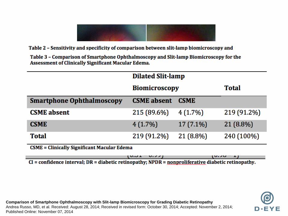
Comparison of Smartphone Ophthalmoscopy with Slit-lamp Biomicroscopy for Grading Diabetic Retinopathy
Andrea Russo, MD, et al. Received: August 28, 2014; Received in revised form: October 30, 2014; Accepted: November 2, 2014;
Published Online: November 07, 2014

Scanning Laser Doppler Flowmetry:
A tool to assess structural components
Laser class I, 690 nm
energy 0.1 mW
10-20° picture,
confocal 3D 2D
Resolution:
3D: 10 x 10 x 300 µm
2D: 10 x 10 µm
Duration of scanning : 2.048s
Intraobserver variation:
Coefficient of variation:
4.95%
Interobserver variation:
Coefficient of variation:
7.96%

THE END THE END Large scale, population based studies assessing associations between
retinal vascular caliber (based on retinal photography) and blood pressure,
target organ damage and cardiovascular risk (part 1)
Ott & Schmieder, 2014, modified

THE END

THE END