Glaucoma tra struttura e funzione: la risposta degli … · RGC & RGC & MeanMeanDeviation 5...
23
93° Congresso Nazionale SOI Corso ZEISS Extensive OCT under standing: retina, miopia, neuroftalmologia, glaucoma Glaucoma tra struttura e funzione: la risposta degli OCT Glaucoma tra struttura e funzione: la risposta degli OCT No No financial financial interest interest 1
Transcript of Glaucoma tra struttura e funzione: la risposta degli … · RGC & RGC & MeanMeanDeviation 5...

93° Congresso Nazionale SOICorso ZEISS
Extensive OCT under standing: retina, miopia, neuroftalmologia, glaucoma
Glaucoma tra struttura e funzione: la risposta degli OCTGlaucoma tra struttura e funzione: la risposta degli OCT
No No financialfinancial interestinterest1

Structure and function: not only glaucomaStructure and function: not only glaucoma
2
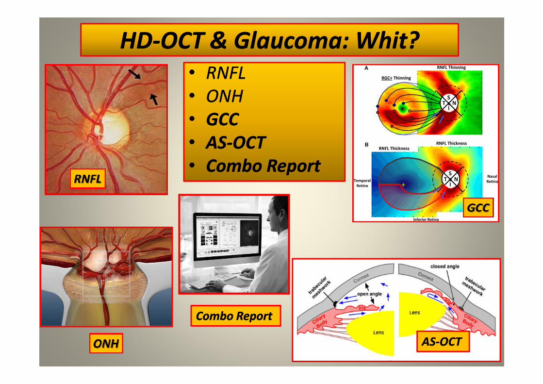
HDHD--OCT & Glaucoma: OCT & Glaucoma: WhitWhit??
•• RNFLRNFL
•• ONHONH
•• GCCGCC
•• ASAS--OCTOCT
•• Combo ReportCombo Report
ASAS--OCTOCT
Combo Report Combo Report

Glaucoma Continuum by R. WeinrebGlaucoma Continuum by R. Weinreb
4

RGC & RGC & MeanMean DeviationDeviation
5
Adapted from Medeiros FA, Lisboa R, Weinreb RN, et al. A combined index of structure
and function for staging glaucomatous damage. Arch Ophthalmol. 2012; 130 (5)
5000/9000 Retinal Ganglion Cells/5000/9000 Retinal Ganglion Cells/YearYear
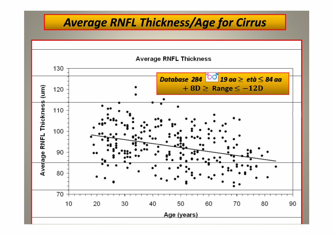
Average RNFL Thickness/Age for CirrusAverage RNFL Thickness/Age for Cirrus
6

MeanMean Deviation MD Deviation MD (dB(dB))
Average Average Thickness Thickness (µm(µm))
EstimatedEstimated RCG RCG countcount (x10.000 cells(x10.000 cells))
-- AtAt early stagesearly stages of damage of damage
(high RGC counts), (high RGC counts), changes changes
in estimatedin estimated RGCRGC counts counts
correspond to relatively correspond to relatively
smaller changes in MDsmaller changes in MD
(continuous line) (continuous line) andand
relativelyrelatively larger changes inlarger changes in
average RNFL average RNFL thickness thickness
(dashed line(dashed line).).
MD CV HFAMD CV HFA
7
Felipe A. Medeiros, Linda M. Zangwill, Christopher Bowd, Kaweh Mansouri, and Robert N. Weinreb
Investigative Ophthalmology & Visual Science, October 2012, Vol. 53, No. 11
-- AtAt advancedadvanced stagesstages of of
damage damage (low RGC counts), (low RGC counts),
changes in estimatedchanges in estimated RGCRGC
counts correspond to counts correspond to
relatively relatively large changes in large changes in
MDMD, butbut onlyonly small small
changes in average RNFLchanges in average RNFL
thickness.thickness.
Thickness Thickness μμm HDm HD--OCT CirrusOCT Cirrus

CSFI CSFI Combined Combined Structure Structure
Function Index Function Index
8
Felipe A. Medeiros, Renato Lisboa,
Robert N. Weinreb, Christopher A.
Girkin, Jeffrey M. Liebmann, Linda M.
Zangwill. Arch Ophthalmol. 2012
Douglas GR, Drance SM, Schulzer M.
A correlation of fields and discs in
open angle glaucoma. Can J. O. 1974
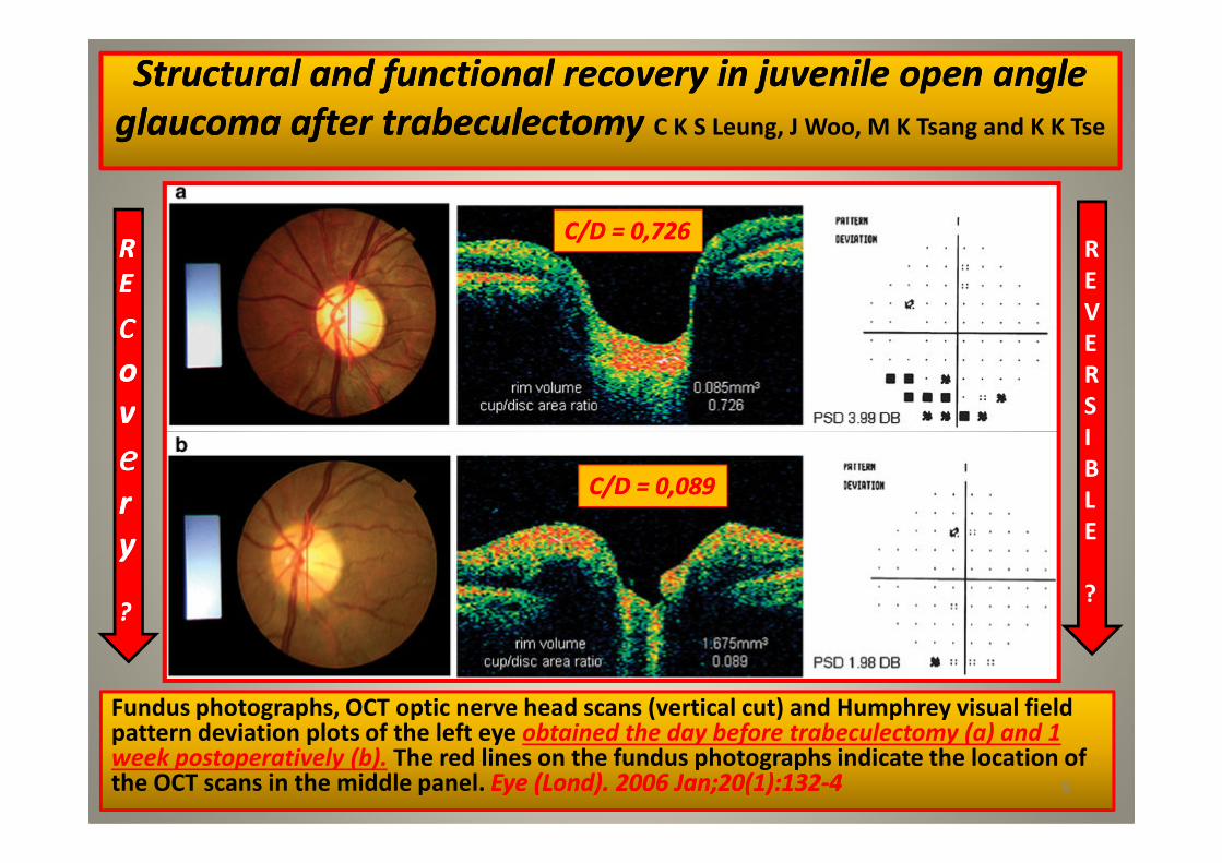
StructuralStructural and and functionalfunctional recoveryrecovery in in juvenilejuvenile open angle open angle
glaucoma glaucoma after after trabeculectomytrabeculectomy C K S Leung, J Woo, M K Tsang and K K Tse
RR
EE
ccoo
vv
ee
R
E
V
E
R
S
I
C/D = 0,726C/D = 0,726
Fundus photographs, OCT optic nerve head scans (vertical cut) and Humphrey visual field pattern deviation plots of the left eye obtained the day before trabeculectomy (a) and 1 week postoperatively (b). The red lines on the fundus photographs indicate the location of the OCT scans in the middle panel. Eye (Eye (LondLond). 2006 Jan;20(1):132). 2006 Jan;20(1):132--44 9
vv
eerr
yy
??
I
B
L
E
?
C/D = 0,089C/D = 0,089

Structural and functional recovery in juvenile open angle Structural and functional recovery in juvenile open angle
glaucoma glaucoma after after trabeculectomytrabeculectomyC K S Leung, J Woo, M K Tsang and K K Tse Eye (Lond). 2006 Jan;20(1):132-4
10

Finite Element Modeling of the Lamina Cribrosa of the Finite Element Modeling of the Lamina Cribrosa of the
Optic Nerve Head in Optic Nerve Head in GlaucomaGlaucomaDevers Eye Institute / National Institute of Health Optic Nerve Head Research Laboratory
directed by Dr. Claude Burgoyne (Portland Oregon)
11Struttura FrattaleStruttura Frattale

IOP Elevation Reduces the Waviness of the Load Bearing IOP Elevation Reduces the Waviness of the Load Bearing
Collagen Fibers in the Collagen Fibers in the Lamina Cribrosa Lamina Cribrosa
Ian Ian A. A. SigalSigal et alet al. . ARVO 2013 Annual Meeting AbstractsARVO 2013 Annual Meeting Abstracts
12
Collagen fibers with and without crimpCollagen fibers with and without crimp

Racial Racial Differences in Mechanical Strain in the Posterior Human Differences in Mechanical Strain in the Posterior Human ScleraScleraM. A. Fazio 1-2, R. Grytz 1, L. Bruno 2, J. S. Morris 3, C. A. Girkin 2, J. Crawford C. Downs 2.
1 Ophthalmology, The University of Alabama in Birmingham, Birmingham, AL;
2 Mechanical Engineering, University of Calabria, Cosenza, Italy;
3 Department of Biostatistics, The University of Texas MD Anderson Cancer Center, Houston, TX.
ARVO 2013 Annual Meeting Abstracts
Baltimore Eye Survey : African 4 times higher risk Caucasian
13
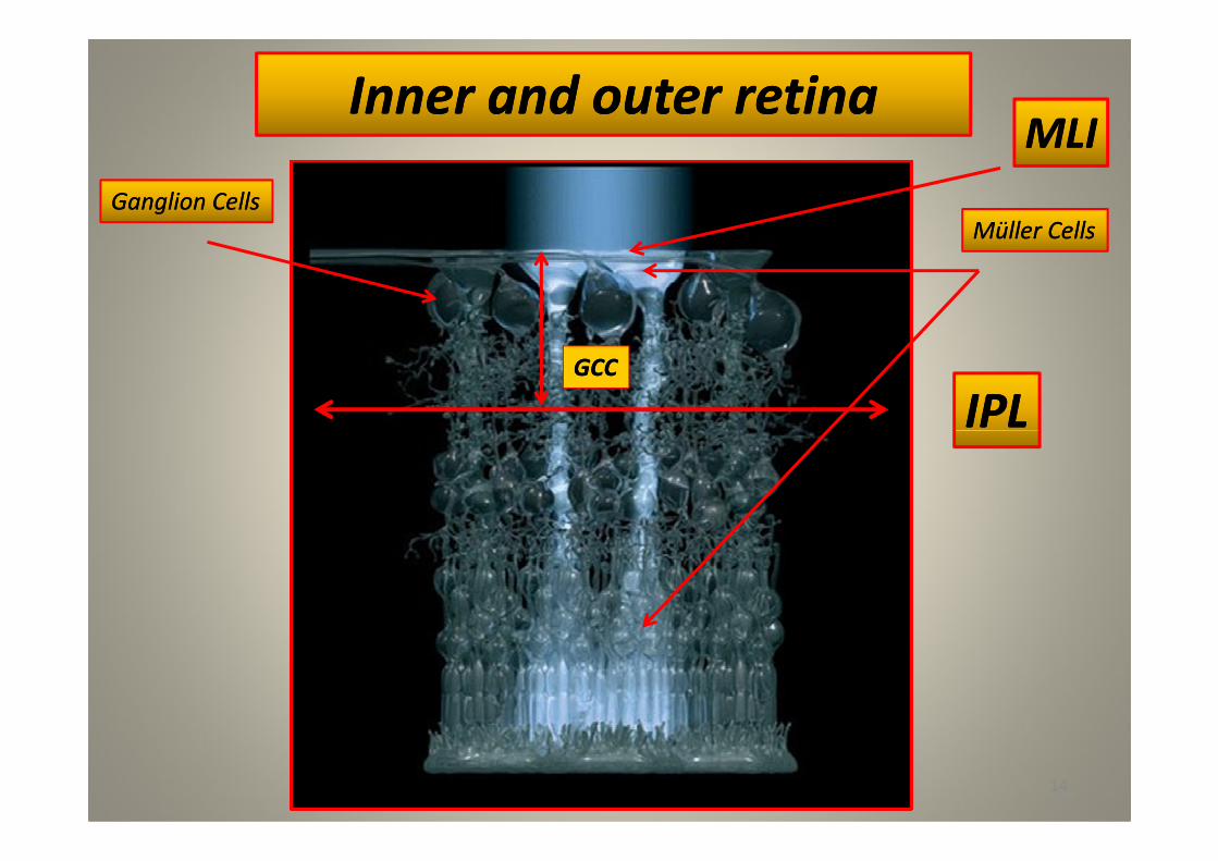
Inner Inner and and outerouter retinaretina
Müller CellsMüller Cells
Ganglion CellsGanglion Cells
IPLIPLGCCGCC
MLIMLI
14
IPLIPL
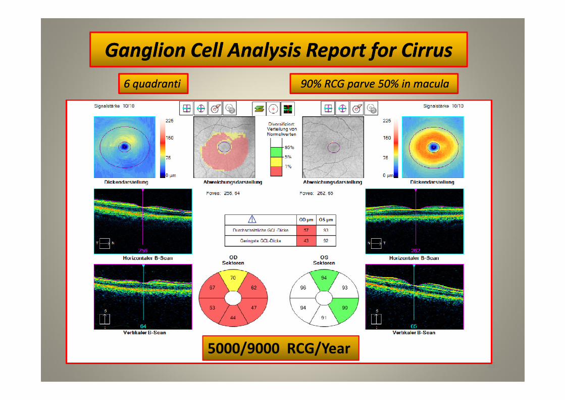
Ganglion Cell Analysis Ganglion Cell Analysis Report for Cirrus Report for Cirrus
6 quadranti6 quadranti 90% RCG parve 50% in macula90% RCG parve 50% in macula
15
5000/9000 RCG/Year

AS HDAS HD--OCTOCT
16
IOL IOL
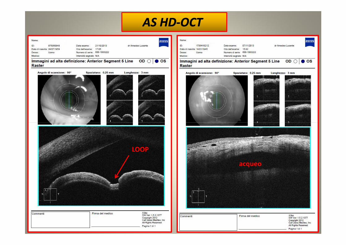
AS HDAS HD--OCTOCT
17
LOOP
acqueo
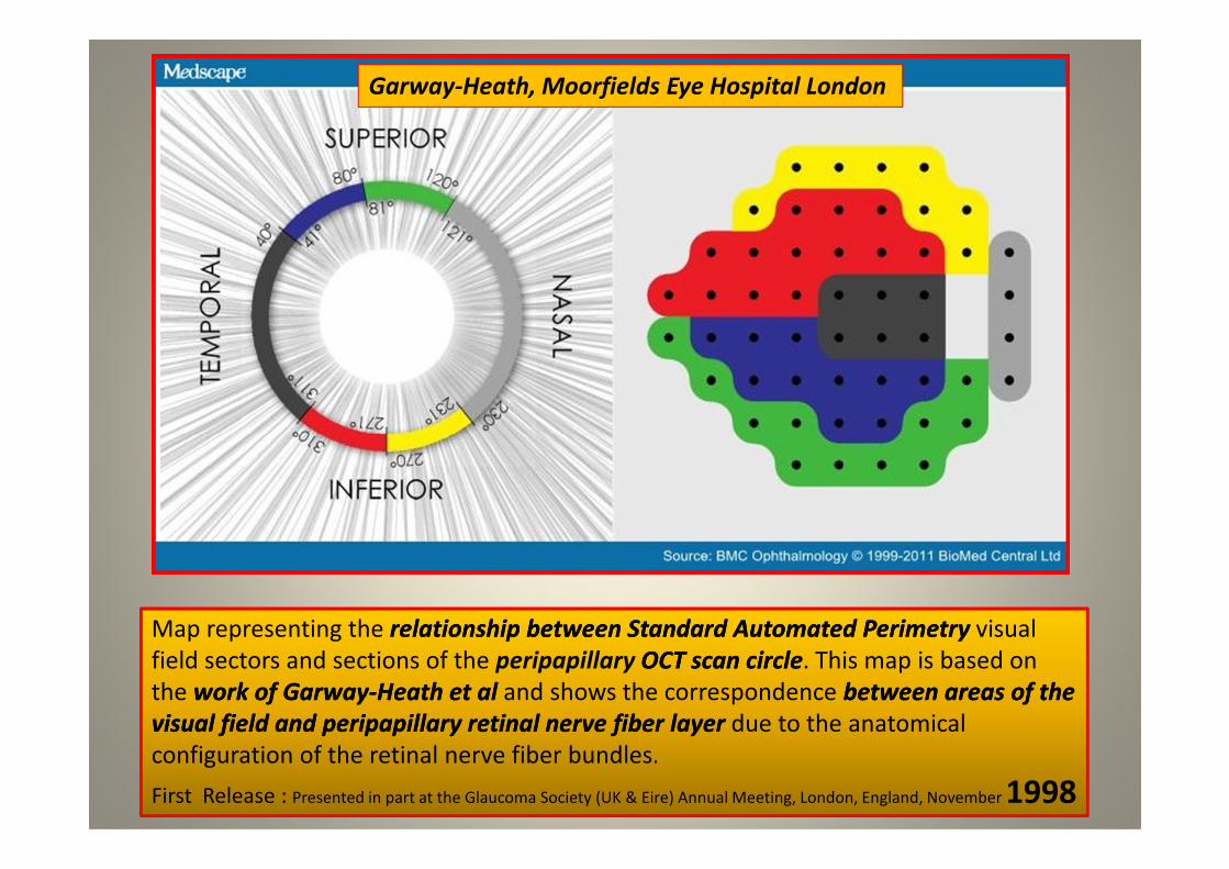
Garway-Heath, Moorfields Eye Hospital London
Map representing the relationshiprelationship betweenbetween Standard Automated Standard Automated PerimetryPerimetry visual
field sectors and sections of the peripapillary OCT scan circleOCT scan circle. This map is based on
the work of work of GarwayGarway--HeathHeath et al et al and shows the correspondence between areas of the between areas of the
visual field and peripapillary retinal nerve fiber layer visual field and peripapillary retinal nerve fiber layer due to the anatomical
configuration of the retinal nerve fiber bundles.
First Release : Presented in part at the Glaucoma Society (UK & Eire) Annual Meeting, London, England, November 1998
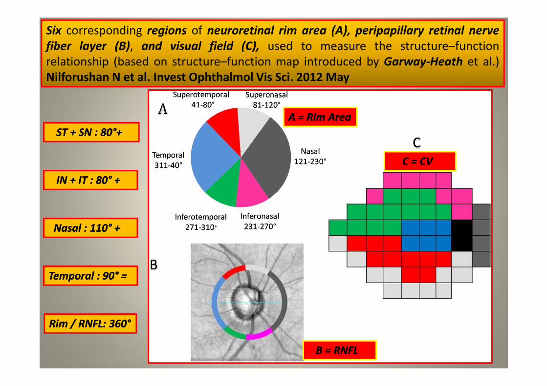
Six corresponding regions of neuroretinal rim area (A), peripapillary retinal nerve
fiber layer (B), and visual field (C), used to measure the structure–function
relationship (based on structure–function map introduced by Garway-Heath et al.)
Nilforushan N et al. Invest Ophthalmol Vis Sci. 2012 May
ST + SN : 80ST + SN : 80°°++
IN + IT : 80IN + IT : 80°° ++
A = A = RimRim AreaArea
C = CV C = CV
19
NasalNasal : 110: 110°° ++
TemporalTemporal : 90: 90°° ==
B = RNFLB = RNFL
RimRim / RNFL: 360/ RNFL: 360°°

Forum Glaucoma Forum Glaucoma WorkplaceWorkplace
Combined structure and function reportsCombined structure and function reportsA. LucenteA. Lucente
20
Database HFA : Database HFA : 422 422 ≥ 18aa età ≥ 18aa età ≤ ≤ 89aa 89aa + 5D ≥ Range ≤ + 5D+ 5D ≥ Range ≤ + 5D
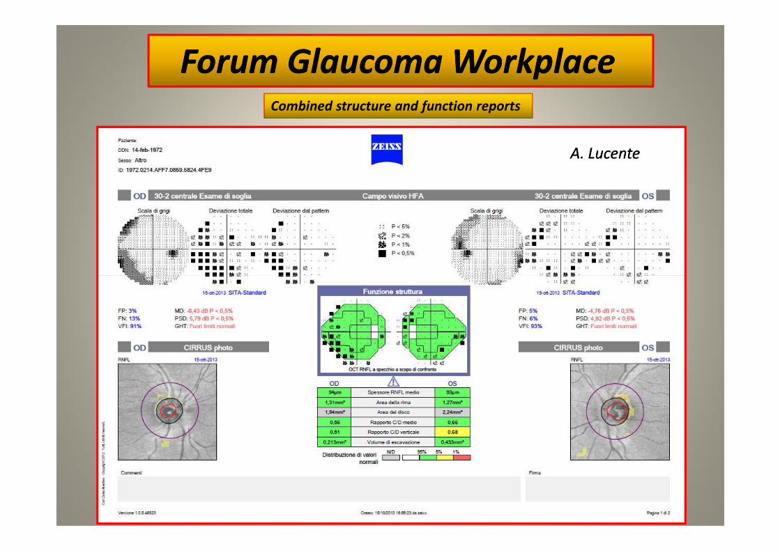
Combined structure and function reports
Forum Glaucoma Forum Glaucoma WorkplaceWorkplace
A. LucenteA. Lucente
21
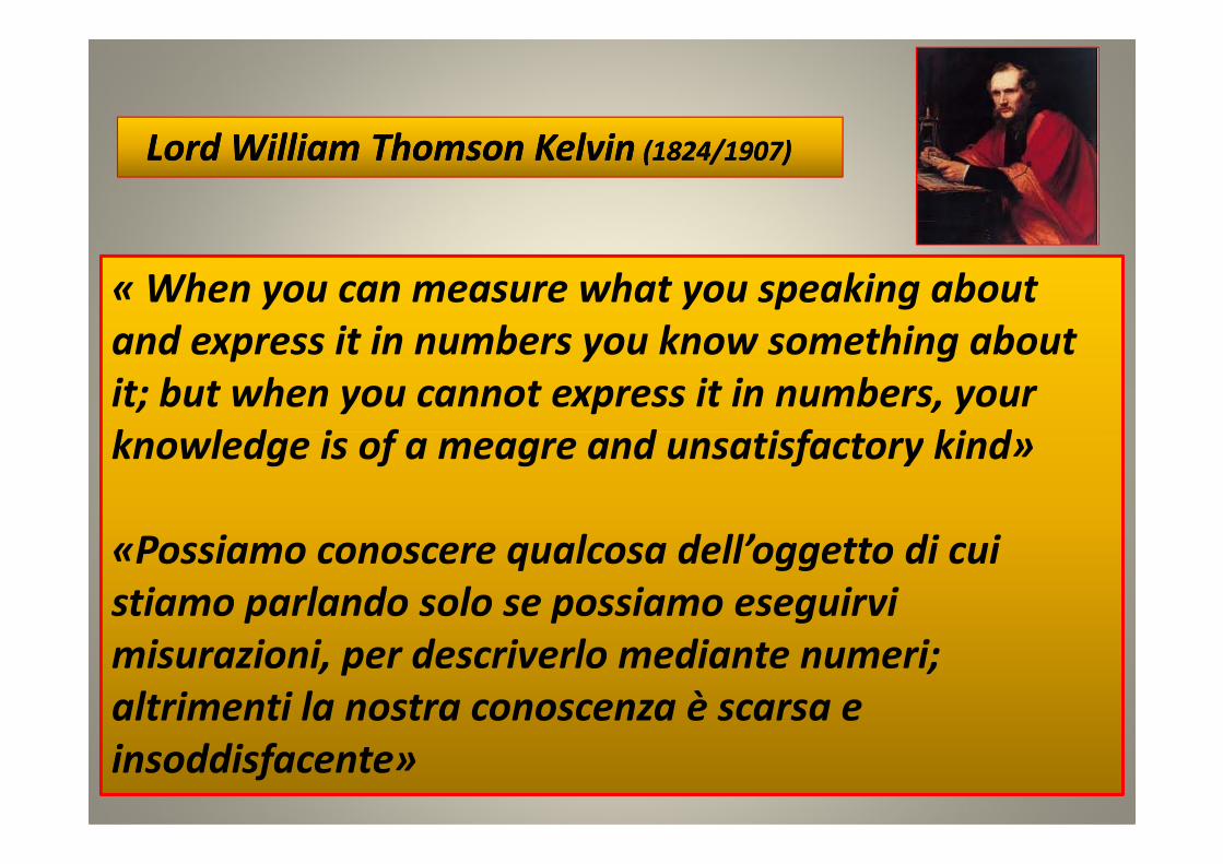
« When you can measure what you speaking about
and express it in numbers you know something about
it; but when you cannot express it in numbers, your
knowledge is of a meagre and unsatisfactory kind»
Lord Lord William Thomson KelvinWilliam Thomson Kelvin (1824/1907)(1824/1907)
22
knowledge is of a meagre and unsatisfactory kind»
«Possiamo conoscere qualcosa dell’oggetto di cui
stiamo parlando solo se possiamo eseguirvi
misurazioni, per descriverlo mediante numeri;
altrimenti la nostra conoscenza è scarsa e
insoddisfacente»
Grazie per l’attenzioneGrazie per l’attenzione
23
Grazie per l’attenzioneGrazie per l’attenzione