Funzioni di Epoe molecole terapeutiche - Docenti...
36
Funzioni di Epo e molecole terapeutiche
Transcript of Funzioni di Epoe molecole terapeutiche - Docenti...
Funzioni di Epo e molecole terapeutiche
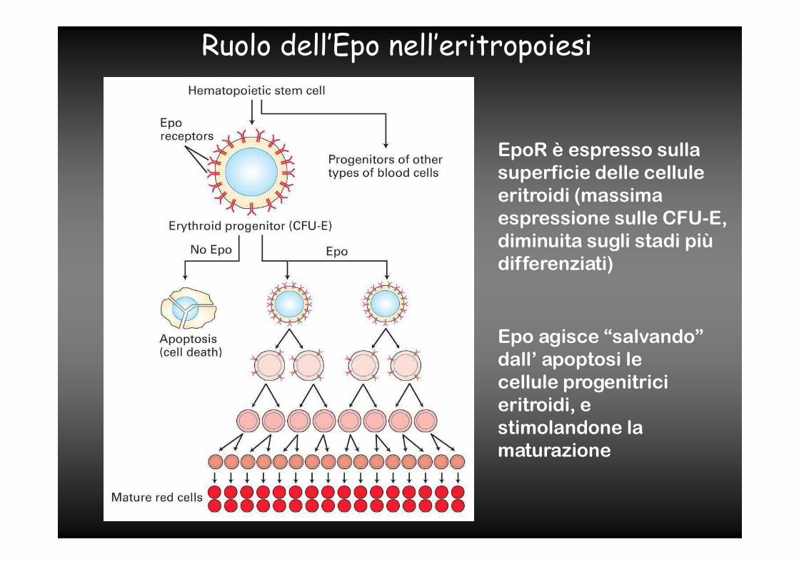
EpoR è espresso sulla
superficie delle cellule
eritroidi (massima
espressione sulle CFU-E,
diminuita sugli stadi più
differenziati)
Epo agisce “salvando”
dall’ apoptosi le
cellule progenitrici
eritroidi, e
stimolandone la
maturazione
Ruolo dell’Epo nell’eritropoiesi
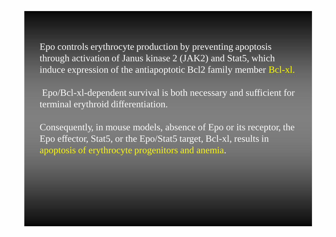
Epo controls erythrocyte production by preventing apoptosis through activation of Janus kinase 2 (JAK2) and Stat5, which induce expression of the antiapoptotic Bcl2 family memberBcl-xl.
Epo/Bcl-xl-dependent survival is both necessary and sufficient for terminal erythroid differentiation.
Consequently, in mouse models, absence of Epo or its receptor, the Epo effector, Stat5, or the Epo/Stat5 target, Bcl-xl, results in apoptosis of erythrocyte progenitors and anemia.
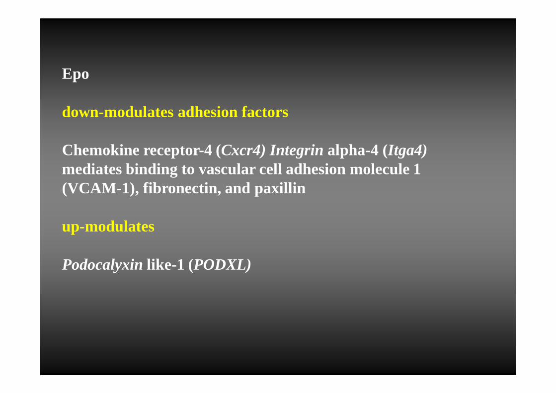
Epo
down-modulates adhesion factors
Chemokine receptor-4 (Cxcr4) Integrin alpha-4 (Itga4) mediates binding to vascular cell adhesion molecule 1 (VCAM-1), fibronectin, and paxillin
up-modulates
Podocalyxin like-1 (PODXL)
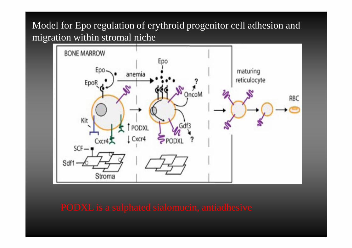
Model for Epo regulation of erythroid progenitor cell adhesion and migration within stromal niche
PODXL is a sulphated sialomucin, antiadhesive
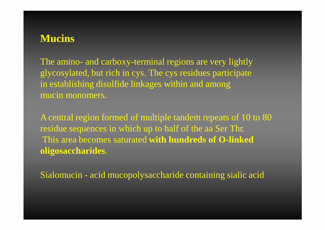
Mucins
The amino- and carboxy-terminal regions are very lightly glycosylated, but rich in cys. The cys residues participatein establishing disulfide linkages within and amongmucin monomers.
A central region formed of multiple tandemrepeats of 10 to 80 residue sequences in which up to half of the aa Ser Thr.This area becomes saturatedwith hundreds of O-linked oligosaccharides.
Sialomucin - acid mucopolysaccharide containing sialic acid
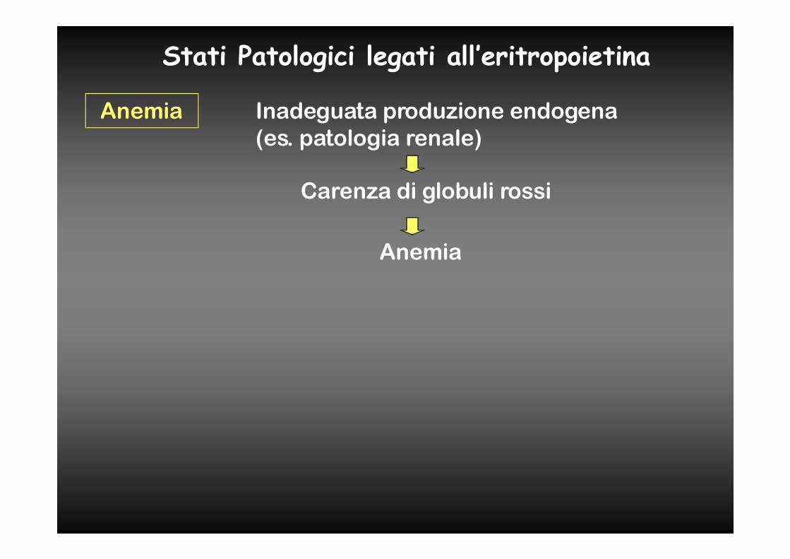
Stati Patologici legati all’eritropoietina
Anemia Inadeguata produzione endogena
(es. patologia renale)
Carenza di globuli rossi
Anemia
HIF prolyl hydroxylase inhibition results in endogenous erythropoietin induction, erythrocytosis
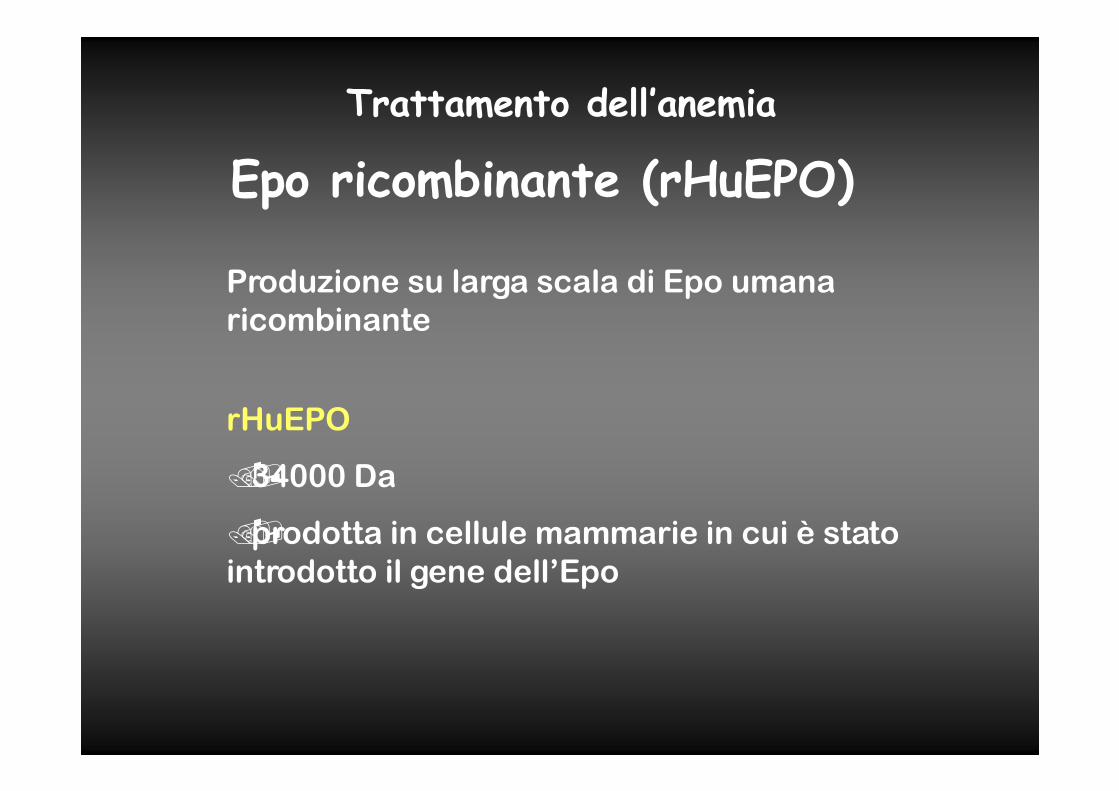
Epo ricombinante (rHuEPO)
Produzione su larga scala di Epo umana
ricombinante
rHuEPO
��34000 Da
��prodotta in cellule mammarie in cui è stato
introdotto il gene dell’Epo
Trattamento dell’anemia
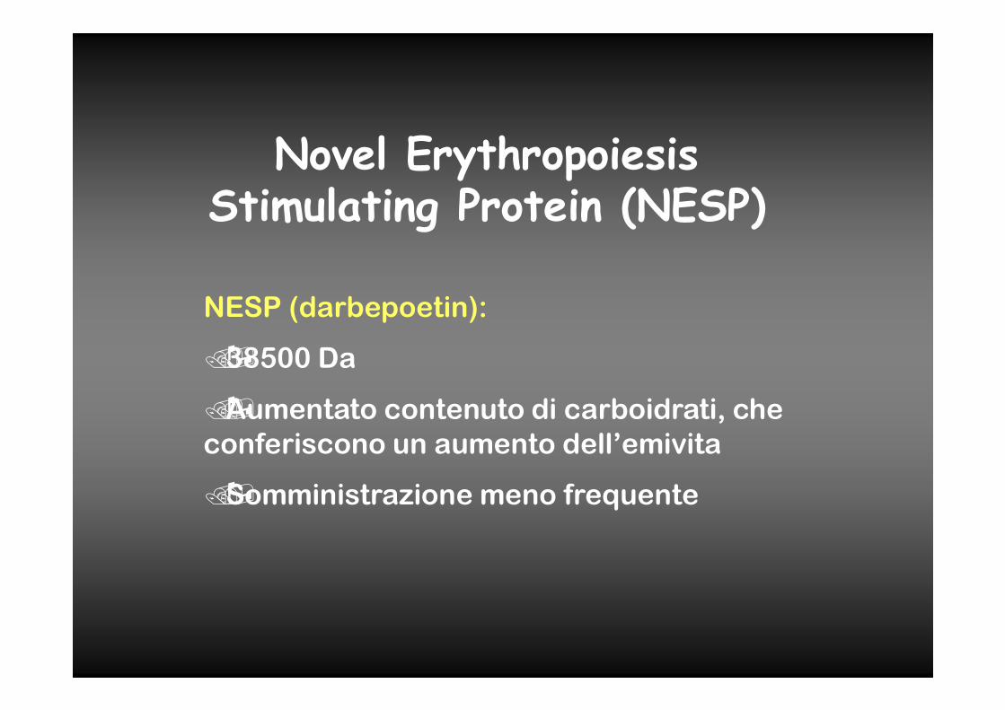
Novel Erythropoiesis Stimulating Protein (NESP)
NESP (darbepoetin):
��38500 Da
��Aumentato contenuto di carboidrati, che
conferiscono un aumento dell’emivita
��Somministrazione meno frequente
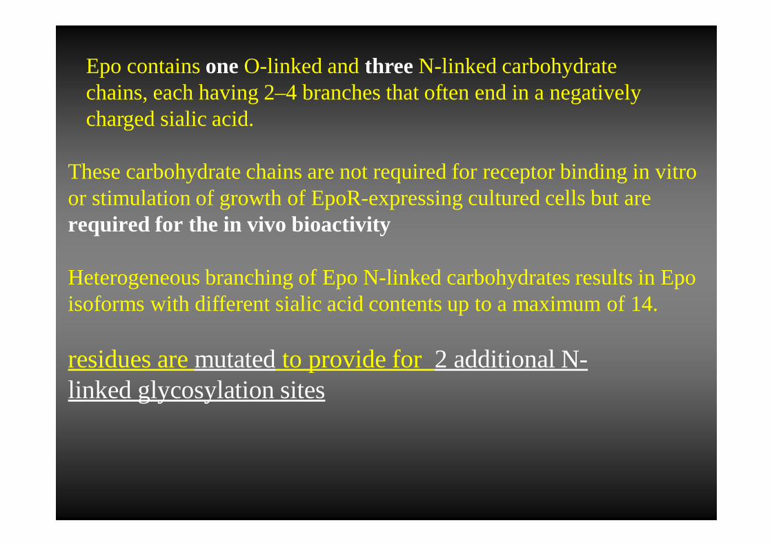
Epo containsone O-linked and three N-linked carbohydrate chains, each having 2–4 branches that often end in a negatively charged sialic acid.
These carbohydrate chains are not required for receptor binding in vitroor stimulation of growth of EpoR-expressing cultured cells but arerequired for the in vivo bioactivity
Heterogeneous branching of Epo N-linked carbohydrates results in Epo isoforms with different sialic acid contents up to a maximumof 14.
residues are mutatedto provide for 2 additional N-linked glycosylation sites
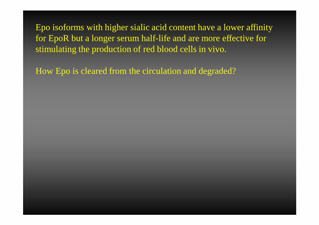
Epo isoforms with higher sialic acid content have a lower affinity for EpoR but a longer serumhalf-life and are more effective for stimulating the production of red blood cells in vivo.
How Epo is cleared fromthe circulation and degraded?
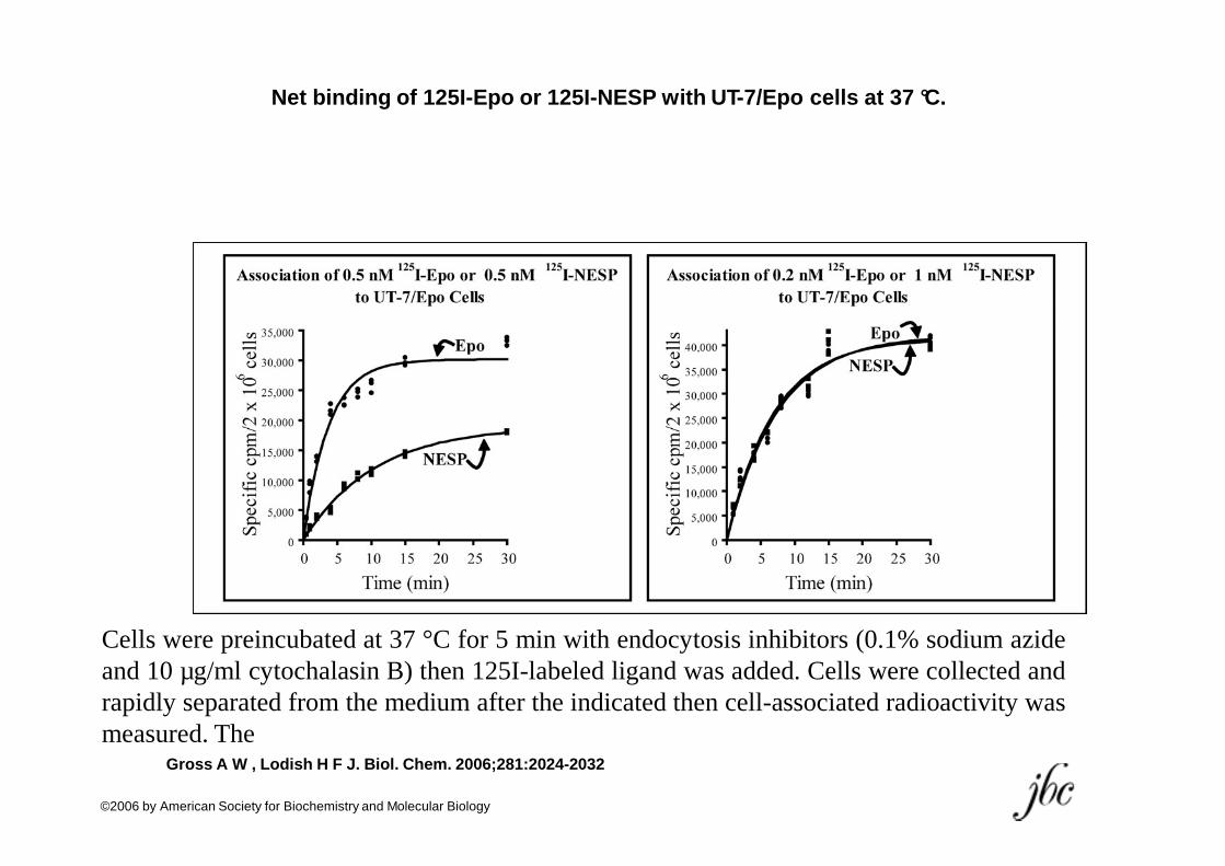
Net binding of 125I-Epo or 125I-NESP with UT-7/Epo cells at 3 7 °C.
Cells were preincubated at 37 °C for 5 min with endocytosis inhibitors (0.1% sodium azideand 10 µg/ml cytochalasin B) then 125I-labeled ligand was added. Cells were collected andrapidly separated from the medium after the indicated then cell-associated radioactivity wasmeasured. The
Gross A W , Lodish H F J. Biol. Chem. 2006;281:2024-2032
©2006 by American Society for Biochemistry and Molecular Biology
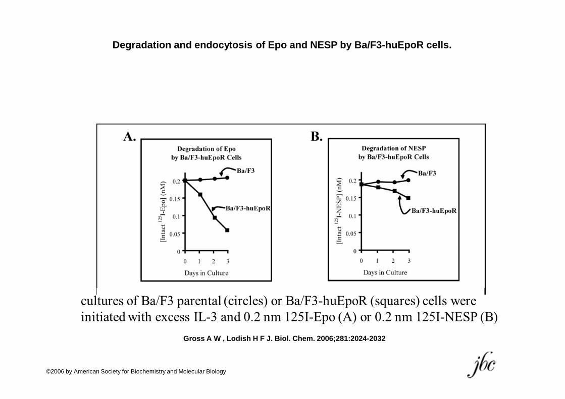
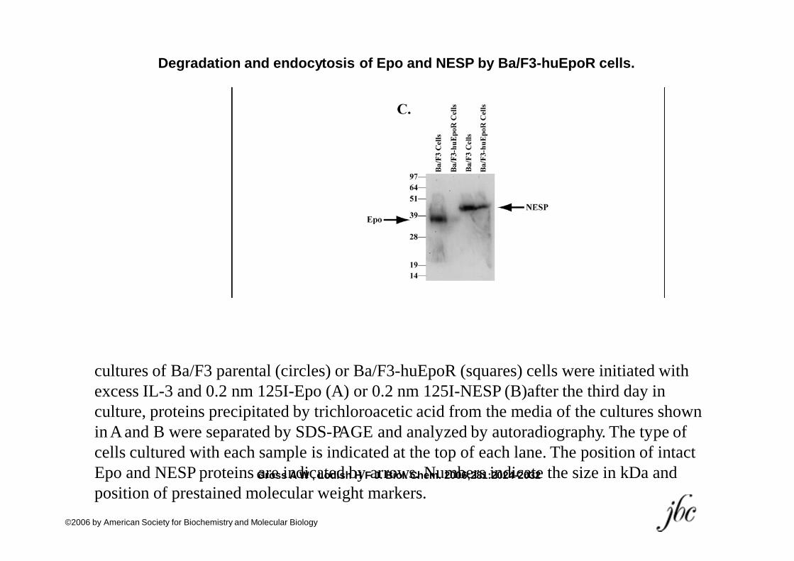
Degradation and endocytosis of Epo and NESP by Ba/F3-huEpoR cells.
Gross A W , Lodish H F J. Biol. Chem. 2006;281:2024-2032
©2006 by American Society for Biochemistry and Molecular Biology
Degradation and endocytosis of Epo and NESP by Ba/F3-huEpoR cells.
cultures of Ba/F3 parental (circles) or Ba/F3-huEpoR (squares) cells wereinitiated with excess IL-3 and 0.2 nm 125I-Epo (A) or 0.2 nm 125I-NESP (B)after the third day in culture, proteins precipitated by trichloroacetic acid from the media of the cultures shown inAand B were separated by SDS-PAGE and analyzed by autoradiography. The typeof cells cultured with each sample is indicated at the top of each lane. The position of intact Epo and NESP proteinsGarroessinAdWic,aLoteddishbHyFaJr.rBoiowl.sC.heNmu. 2m00b6e;2r8s1:i2n02d4i-c20a3t2e the size in kDa and position of prestained molecular weight markers.
©2006 by American Society for Biochemistry and Molecular Biology
Epo-Epo” -a peptide-linked head-to-tail dimer

Diagram of cDNA encoding the Epo-Epo fusion protein.
Sytkowski A J et al. J. Biol. Chem. 1999;274:24773-24778
1 2stop
©1999 by American Society for Biochemistry and Molecular Biology
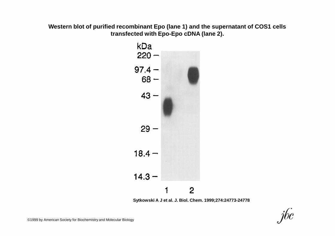
Western blot of purified recombinant Epo (lane 1) and the sup ernatant of COS1 cells transfected with Epo-Epo cDNA (lane 2).
Sytkowski A J et al. J. Biol. Chem. 1999;274:24773-24778
©1999 by American Society for Biochemistry and Molecular Biology
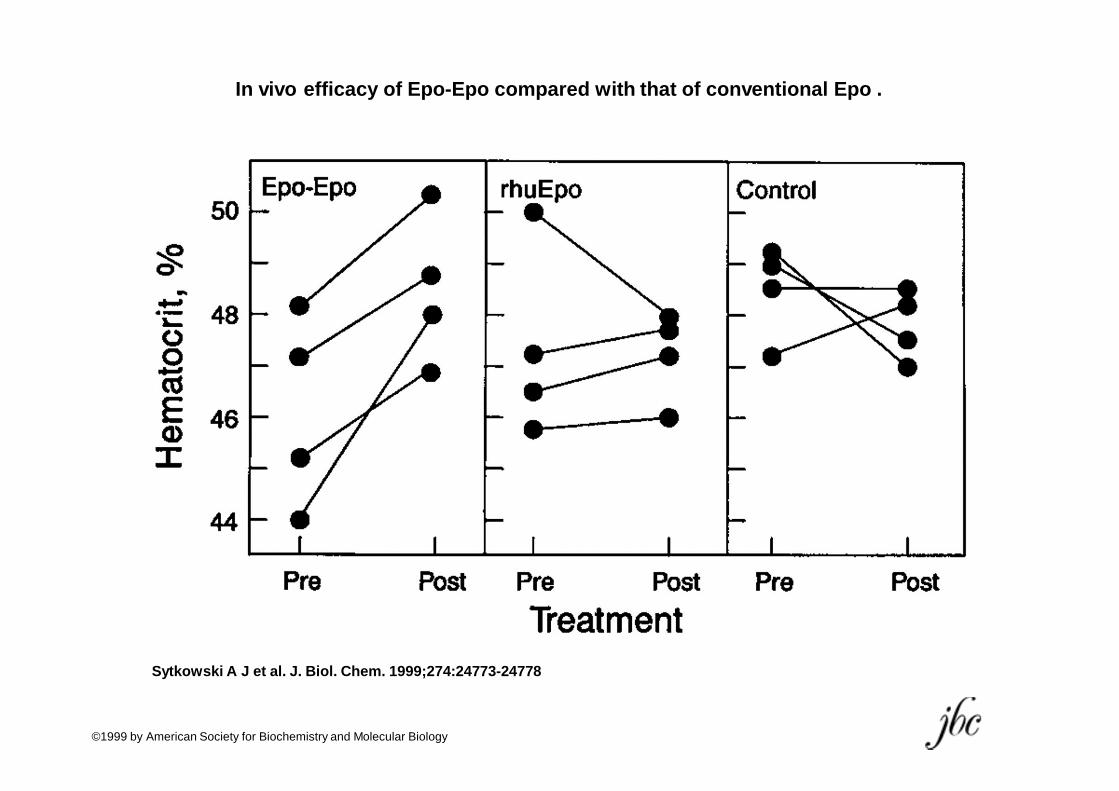
In vivo efficacy of Epo-Epo compared with that of convention al Epo .
Sytkowski A J et al. J. Biol. Chem. 1999;274:24773-24778
©1999 by American Society for Biochemistry and Molecular Biology
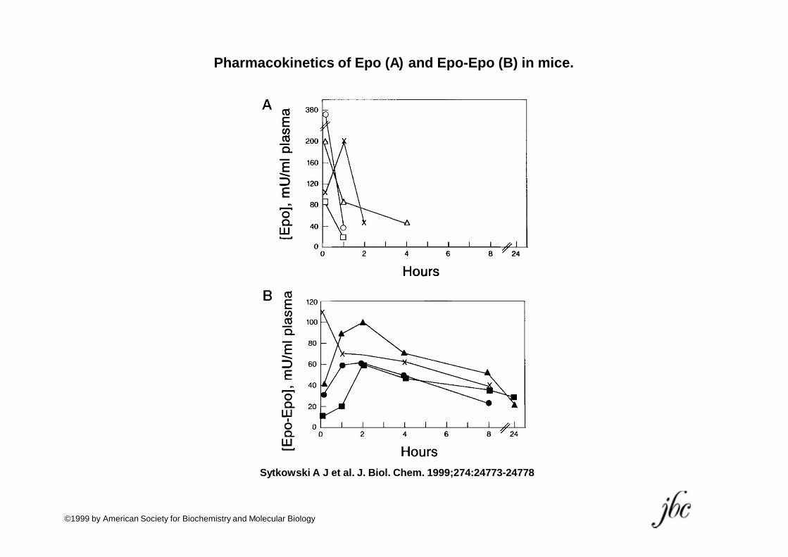
Pharmacokinetics of Epo (A) and Epo-Epo (B) in mice.
Sytkowski A J et al. J. Biol. Chem. 1999;274:24773-24778
©1999 by American Society for Biochemistry and Molecular Biology
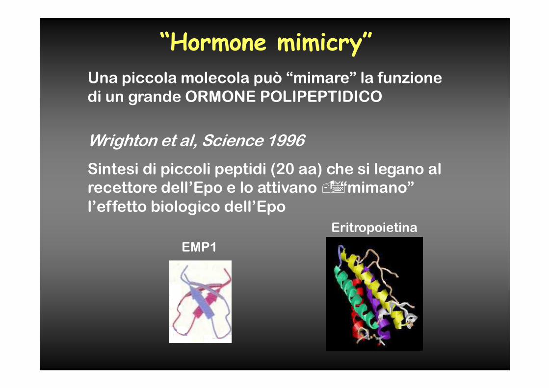
“Hormone mimicry”
Una piccola molecola può “mimare” la funzione
di un grande ORMONE POLIPEPTIDICO
Wrighton et al, Science 1996
Sintesi di piccoli peptidi (20 aa) che si legano al
recettore dell’Epo e lo attivano ��“mimano”
l’effetto biologico dell’Epo
Eritropoietina
EMP1
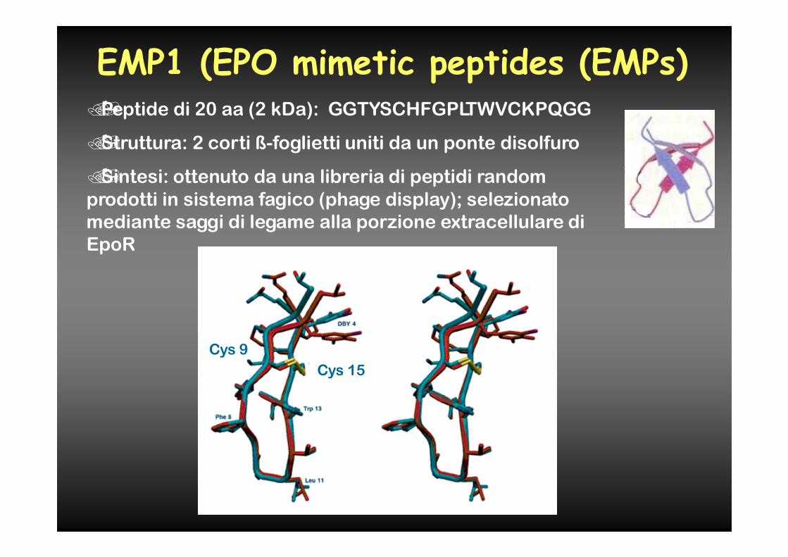
EMP1 (EPO mimetic peptides (EMPs)��Peptide di 20 aa (2 kDa): GGTYSCHFGPLTWVCKPQGG
��Struttura: 2 corti ß-foglietti uniti da un ponte disolfuro
��Sintesi: ottenuto da una libreria di peptidi random
prodotti in sistema fagico (phage display); selezionato
mediante saggi di legame alla porzione extracellulare di
EpoR
Cys 9
Cys 15
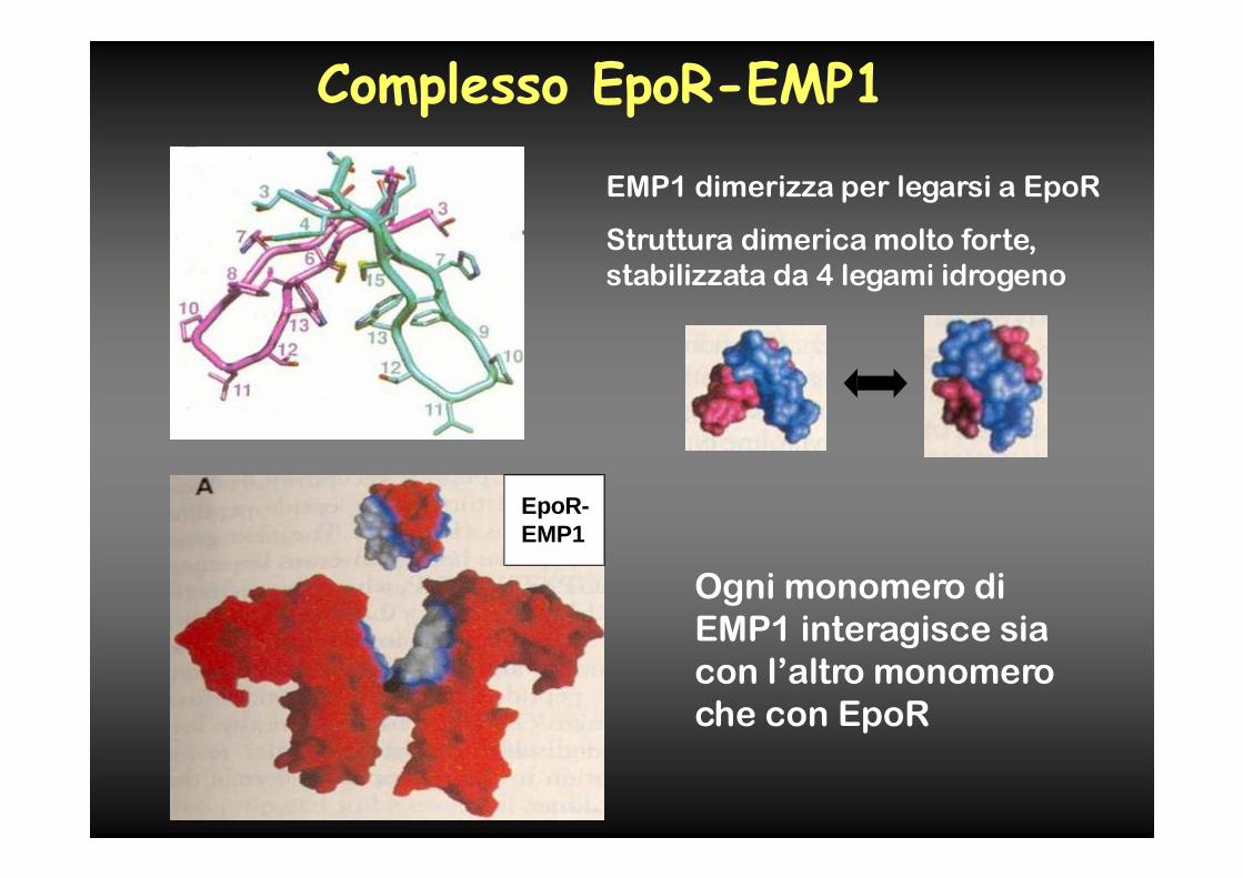
Complesso EpoR-EMP1
EMP1 dimerizza per legarsi a EpoR
Struttura dimerica molto forte,
stabilizzata da 4 legami idrogeno
EpoR-EMP1
Ogni monomero di
EMP1 interagisce sia
con l’altro monomero
che con EpoR
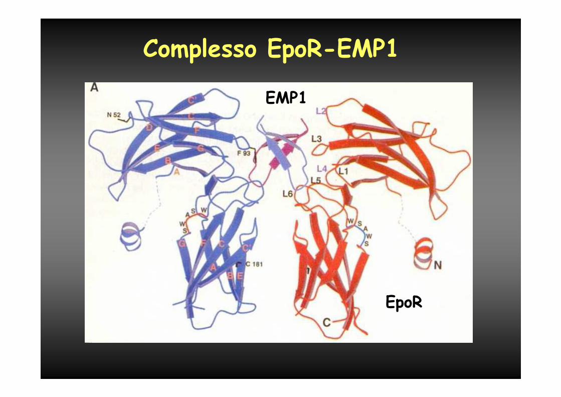
Complesso EpoR-EMP1
EMP1
EpoR
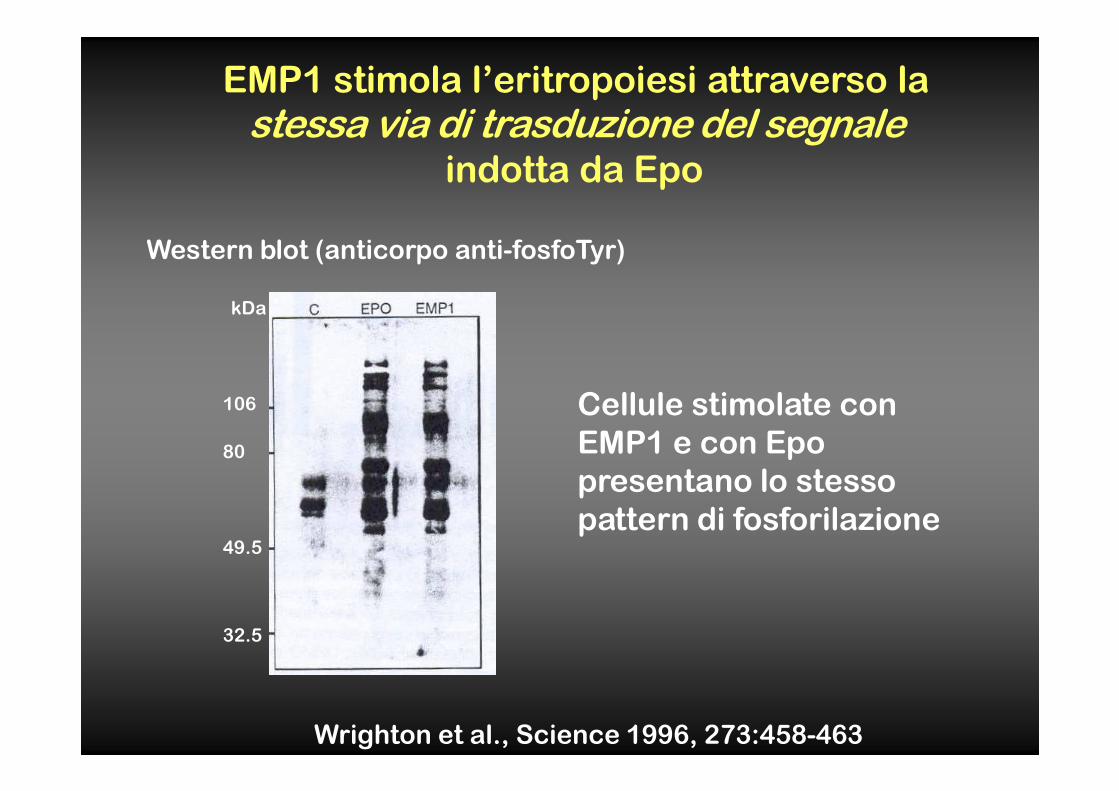
EMP1 stimola l’eritropoiesi attraverso la
stessa via di trasduzione del segnaleindotta da Epo
Western blot (anticorpo anti-fosfoTyr)
Wrighton et al., Science 1996, 273:458-463
Cellule stimolate con
EMP1 e con Epo
presentano lo stesso
pattern di fosforilazione
106
kDa
80
49.5
32.5
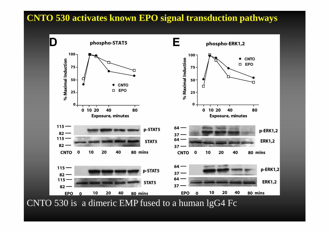
CNTO 530 activates known EPO signal transduction pathways
CNTO 530 is a dimeric EMP fused to a human lgG4 Fc
EMP1 è la dimostrazione che una molecola di 20 aa può mimare la funzione di un ormone
��Stimolando la stessa via di trasduzione del
segnale (JAK, STAT...)
��Senza avere nessuna omologia di sequenza o
struttura con l’ormone
“Hormone mimicry”
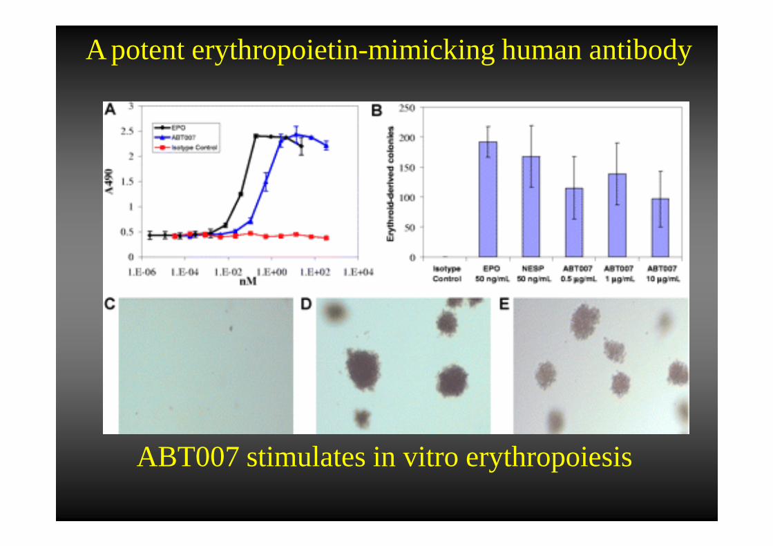
ABT007 stimulates in vitro erythropoiesis
A potent erythropoietin-mimicking human antibody
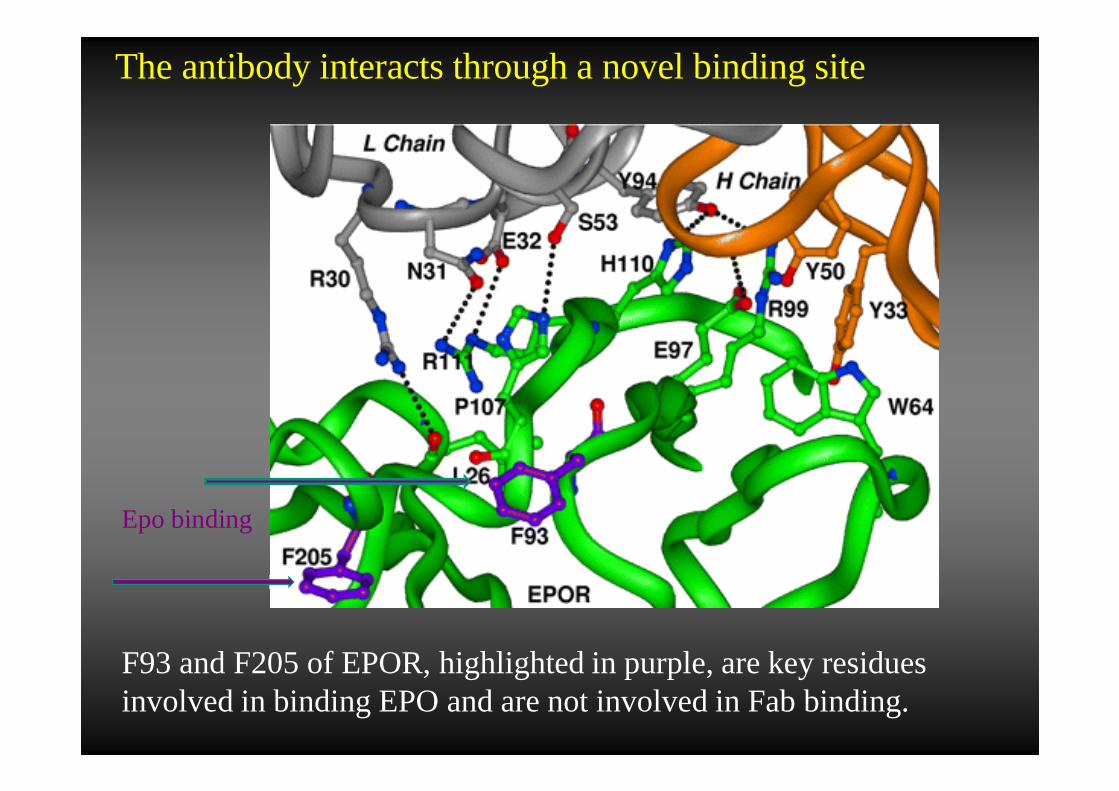
The antibody interacts through a novel binding site
F93 and F205 of EPOR, highlighted in purple, are key residues involved in binding EPO and are not involved in Fab binding.
Epo binding
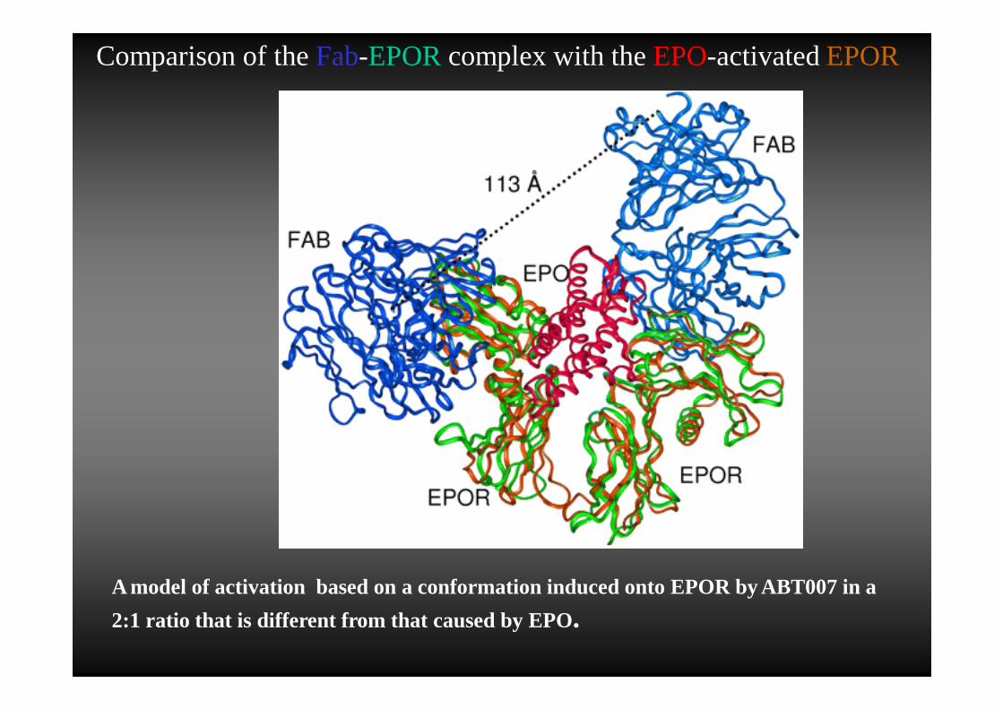
A model of activation based on a conformation induced onto EPOR by ABT007 in a
2:1 ratio that is different from that caused by EPO.
Comparison of theFab-EPOR complex with theEPO-activatedEPOR
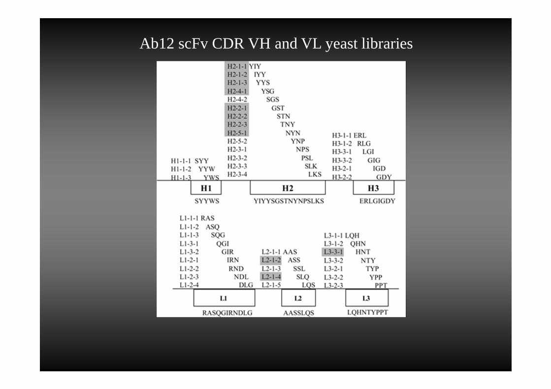
Ab12 scFv CDR VH and VL yeast libraries
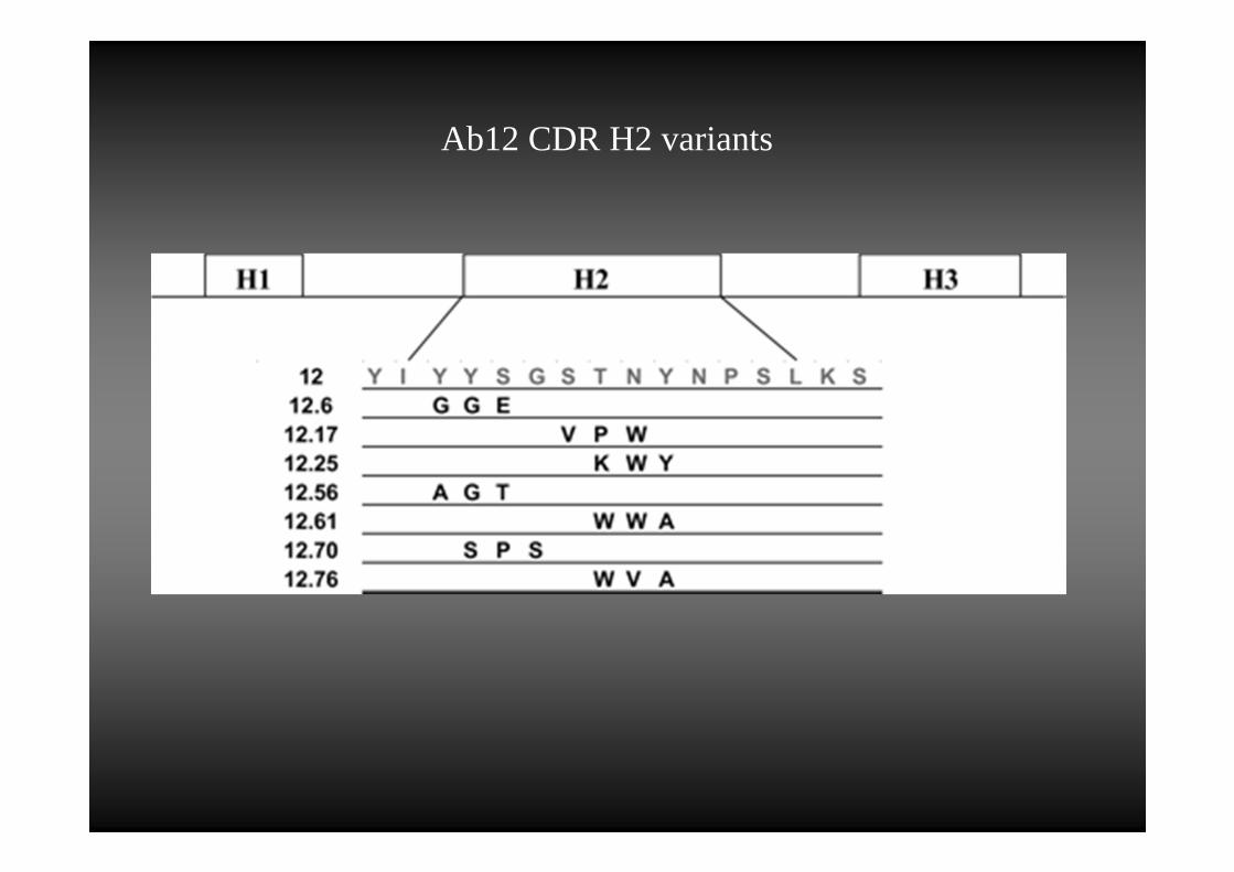
Ab12 CDR H2 variants
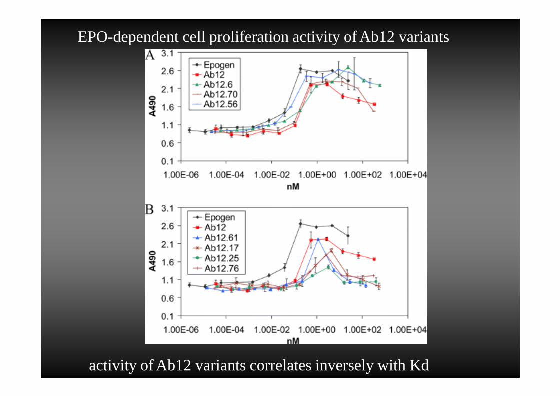
EPO-dependent cell proliferation activity of Ab12 variants
activity of Ab12 variants correlates inversely with Kd
EPO's tissue-protective actions have been shown to be mediated by a tissue-protective receptor complex consisting of the EPO receptor and theβ common-receptor (CD131) subunit that is also used by GM-CSF, IL-3, and IL-5.
helix B-surface peptide (HBSP). This peptide is composed of 11 amino acids (QEQLERALNSS) derived fromthe aqueous face of helix B of EPO and exhibits tissue-protective activities
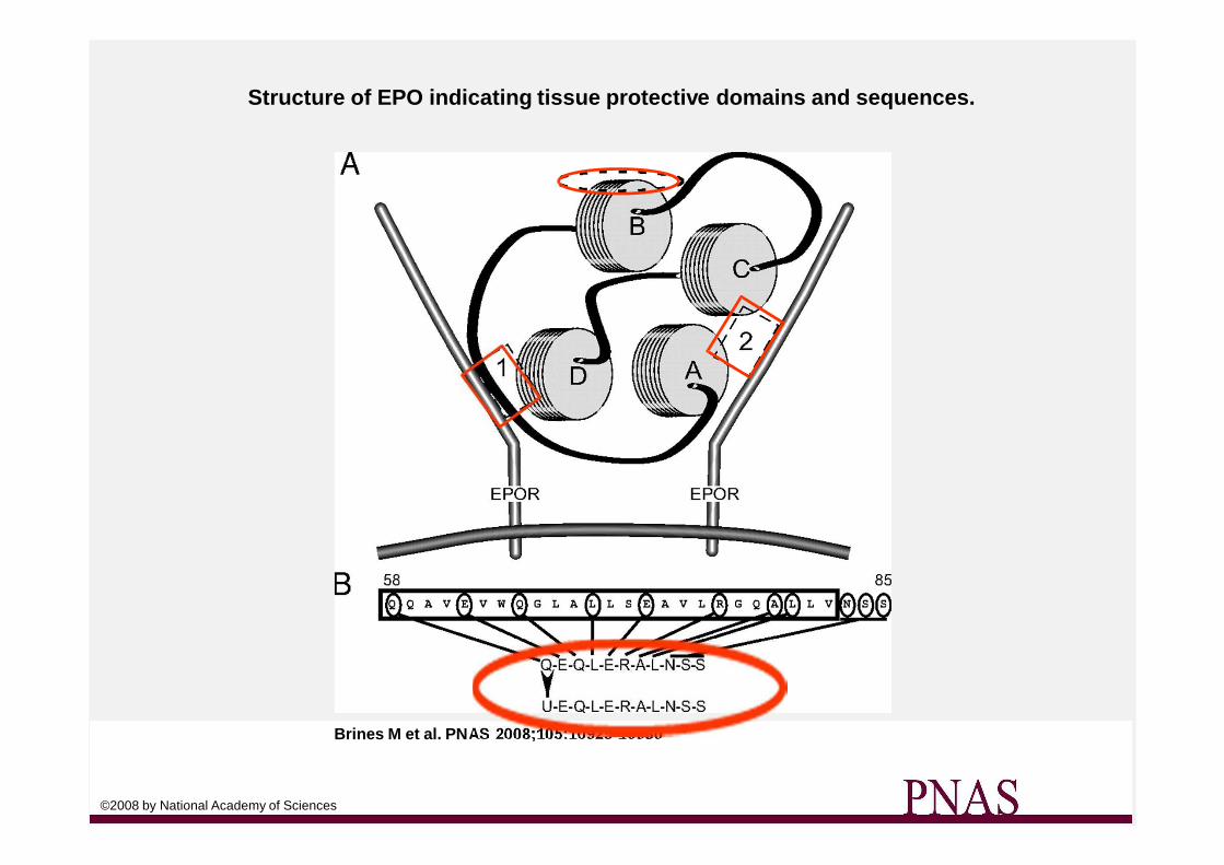
Structure of EPO indicating tissue protective domains and sequences.
Brines M et al. PN AS 2008;105:10925-10930
©2008 by National Academy of Sciences
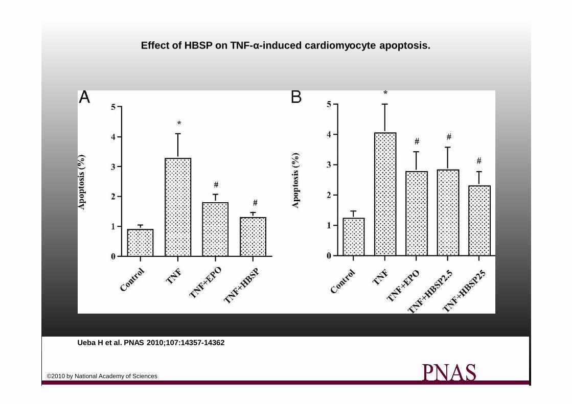
Effect of HBSP on TNF- α-induced cardiomyocyte apoptosis.
Ueba H et al. PNAS 2010;107:14357-14362
©2010 by National Academy of Sciences


![Cinetica-Biochimica2012 Branchini [modalit compatibilit ]m.docente.unife.it/francesco.bernardi/materiale-didattico... · Costante catalitica (kcat) o Numero di turnover Per il modello](https://static.fdocumenti.com/doc/165x107/5c65c04e09d3f29b6e8d3435/cinetica-biochimica2012-branchini-modalit-compatibilit-m-costante-catalitica.jpg)