Riparazione per excisione di basi (BER) - Docenti...
34
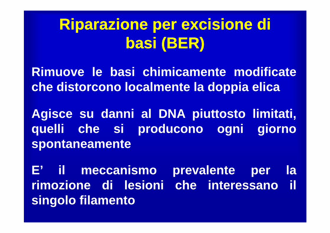
Riparazione per excisione di basi (BER) Riparazione per excisione di basi (BER) Rimuove le basi chimicamente modificate che distorcono localmente la doppia elica E’ il meccanismo prevalente per la rimozione di lesioni che interessano il singolo filamento Agisce su danni al DNA piuttosto limitati, quelli che si producono ogni giorno spontaneamente
Transcript of Riparazione per excisione di basi (BER) - Docenti...
Riparazione per excisione di basi (BER)
Riparazione per excisione di basi (BER)
Rimuove le basi chimicamente modificateche distorcono localmente la doppia elica
E’ il meccanismo prevalente per larimozione di lesioni che interessano ilsingolo filamento
Agisce su danni al DNA piuttosto limitati,quelli che si producono ogni giornospontaneamente

• The Nobel Prize in Chemistry 2015 was
awarded jointly to
• Tomas Lindahl,
• Paul Modrich and
• Aziz Sancar
"for mechanistic studies of DNA repair"
Tomas Lindahl
• In the early 1970s Tomas Lindahl demonstrated that DNA has limited
chemical stability even in the absence of external physical assaults.
• Under physiological conditions DNA is subject to a number of chemical
reactions such as hydrolytic deamination, oxidation and non-enzymatic
methylation.
• These reactions modify the bases of DNA and as a consequence increase
the risk for mutations.
• Tomas Lindahl used the term DNA decay to describe these processes and
elegantly demonstrated that under physiological conditions, spontaneous
hydrolytic DNA depurination occur at significant levels.
• The most fascinating discovery was the demonstration of high levels of
spontaneous cytosine deamination under physiological conditions, which
leads to the formation of uracil.
• Lindahl demonstrated that DNA is an inherently unstable molecule, subject to
decay even under physiological conditions. Guided by this observation, Lindahl
identified a completely new group of DNA glycosylases and described their role in
base excision repair.


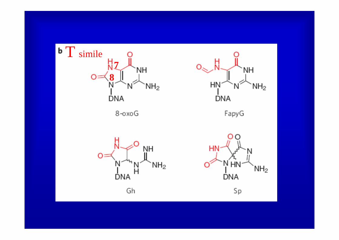
BER: 8-OXOGUANINA
78
T simile

78
8
7

• MutT and its human homologue MTH1 have an important role in preventing the incorporation of 8-oxoG, through hydrolysis of free 8-oxo-dGTP.

*MTH1
hydrolysis of free 8-oxo-dGTP
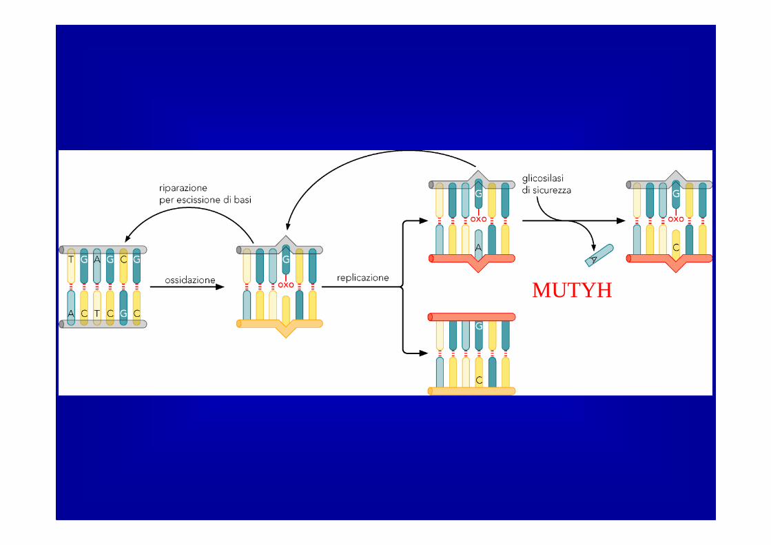
MUTYH

• The enzyme tracks rapidly along DNA, inserting a ‘probe’ amino-acid residue (green hexagonPhe 114) at various base pairs to test the stability and/or deformability of the duplex.
• OGG1 samples millions of base pairs per second!!!!!!.
The 8-oxoG lesion searchprocess.

• OGG1 was found to move along the DNA with a diffusion constant approaching the theoretical upper limit for one-dimensional diffusion,indicating that OGG1 samples millions of base pairs per second.
• On the basis of these measurements, the estimated barrier to sliding is extremely small (0.5 kcal mol–1). The smaller barrier and the observed unbiased random movement of OGG1 on DNA suggest that OGG1 rapidly searches along DNA as a consequence of brownian motion.

• the 8-oxoG is extruded to the exosite and captured in the 8-oxoG-specific pocket, where it is excised from the DNA.
The 8-oxoG lesion searchProcess (2).

Una DNA glicosilasi (l’uomo nepossiede almeno 8, specificheper varie lesioni) rompe il legametra la base errata e ildesossiriboso liberando la base
Formazione di un sito AP cheviene riconosciuto da APE1 (APendonucleasi) ���� APE1 taglia ilsingolo filamento in 5’ al sito AP
La DNA polimerasi riempie il gaplasciato dalla glicosilasi usandocome stampo l’elica parentale
La ligasi richiude l’elica riparata

OGG1 LRC with8-oxoG•C-containing
DNA. 8-oxoG is shown in red, and the C
in green.
orange (Gly 42), dark pink (Asn 149 or Cys 149), light purple (Arg 154), dark purple (Tyr 203), light blue (Arg 204), yellow (His 270), brown (Gln 315) and
black (Phe 319).

Gly 42
NH7
lesion-recognition complexes (LRCs).
Cys 253
Lys 249
∆-
(Gln 315)
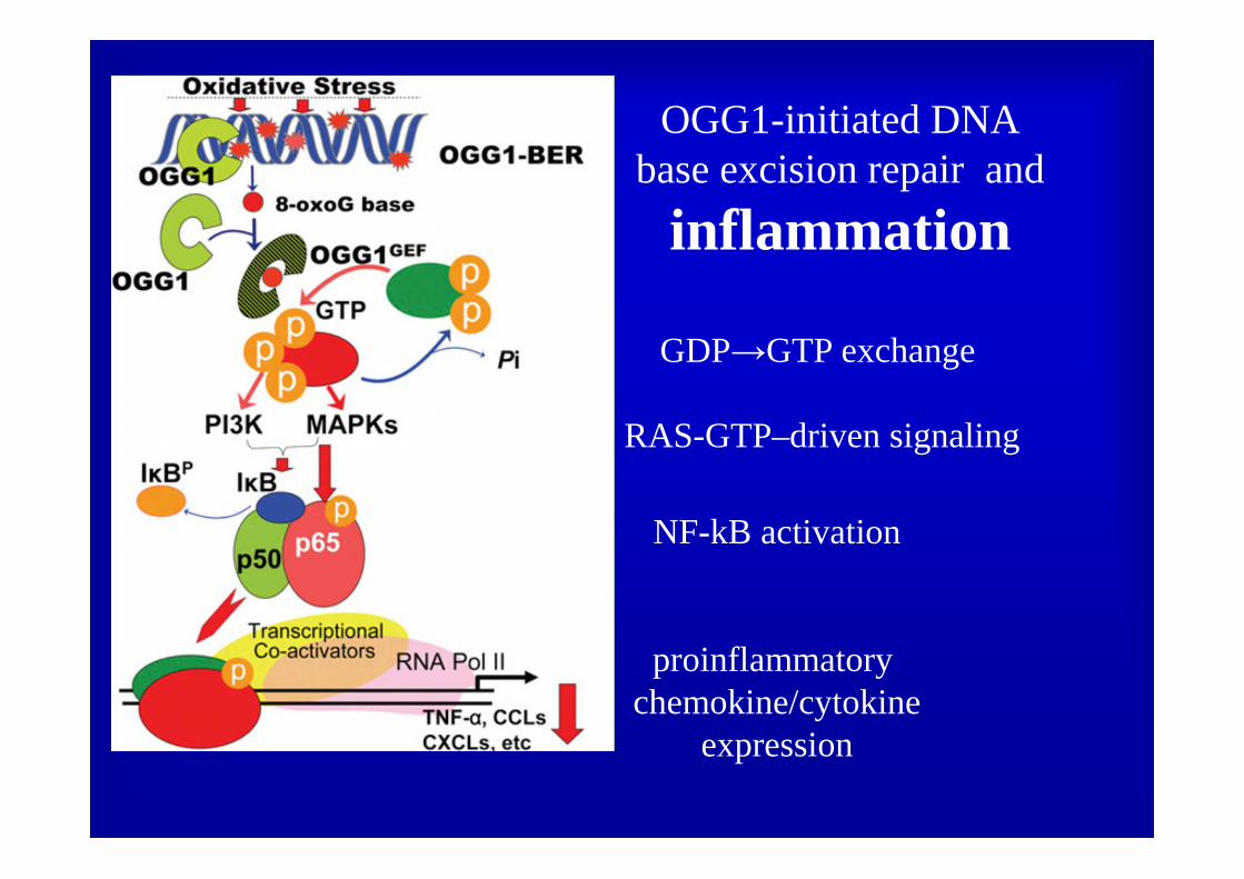
OGG1-initiated DNAbase excision repair and
inflammation
NF-kB activation
proinflammatory chemokine/cytokine
expression
GDP→GTP exchange
RAS-GTP–driven signaling

Germline mutations observed in MUTYH in individuals with polyposis
• DNA-binding motifs: helix–hairpin–helix (HhH) motif and the Fe–S cluster loop (FCL) motif

• Consistent with a global defect in 8-oxoG•A repair, a high proportion of tumours from patients with biallelic mutations in MUTYH
have been observed to contain G-to-T transversions

Uomini e Topi….
• mice that are deficient only in MUTYH do not show any atypical properties
• However, crossing MUTYH-deficientmice with multiple intestinal neoplasia (ApcMin/+) mice, which carry a nonsense mutation in Apc, resulted in greater intestinal tumorigenesis than in ApcMin/+/Mutyh+/+ mice.

neil1

• RNA editing changes the lesion specificityfor the DNA repair enzyme NEIL1
Whole transcriptome sequence analysis from various
human tissues identified over 200 possible A to I editing sites in non repeat sequences, including a site predicted to cause recoding in the mRNA for the DNA repair enzyme NEIL1 (lysine242 AAA codon edited to AIA codon for arginine)
NEIL1 plays a key role in the initiation of base excision repair of oxidized base lesions by catalyzing the cleavage of the N-glycosidic linkage to the 2’-deoxyribose
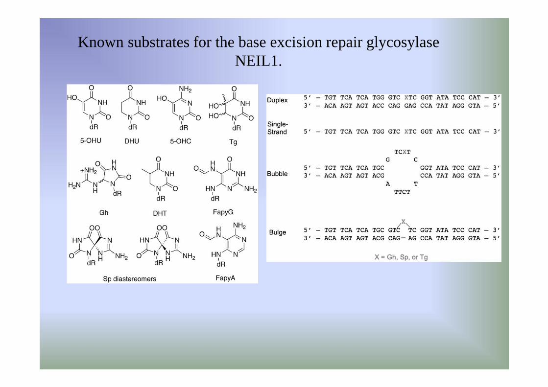
Known substrates for the base excision repair glycosylase NEIL1.
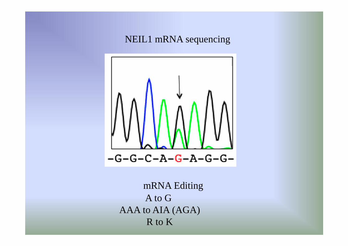
A to G AAA to AIA (AGA)
R to K
NEIL1 mRNA sequencing
mRNA Editing
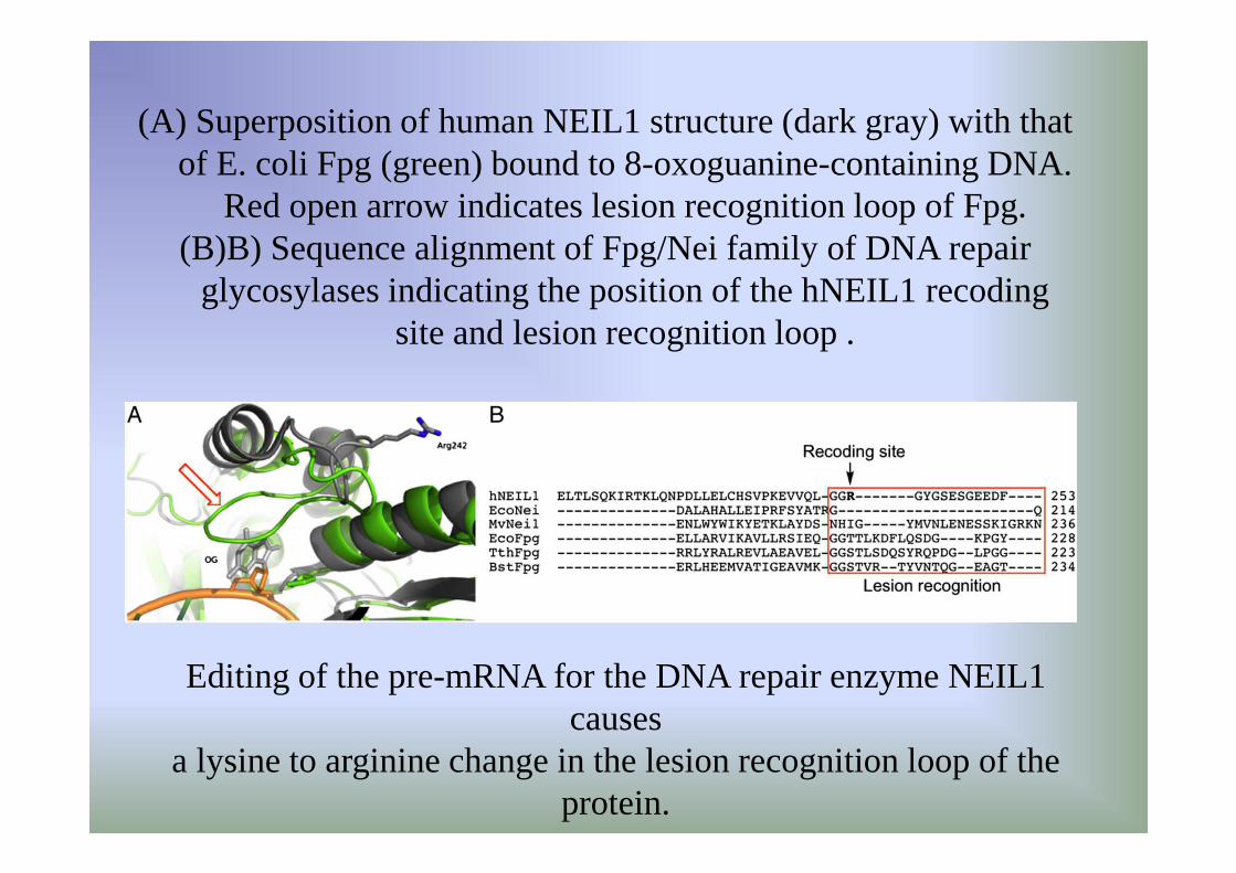
(A) Superposition of human NEIL1 structure (dark gray) with that of E. coli Fpg (green) bound to 8-oxoguanine-containing DNA.
Red open arrow indicates lesion recognition loop of Fpg. (B)B) Sequence alignment of Fpg/Nei family of DNA repair
glycosylases indicating the position of the hNEIL1 recoding site and lesion recognition loop .
Editing of the pre-mRNA for the DNA repair enzyme NEIL1 causes
a lysine to arginine change in the lesion recognition loop of theprotein.

Superposition of human NEIL1 structure (dark gray) with that of E. coli Fpg (green) bound to 8-oxoguanine-containing DNA.

Three human ADAR (adenosine deaminase acting on RNA)-family members
ADAR1L is detected mainly in the cytoplasm, whereas ADAR1S localizes in the nucleoplasmand nucleolus
ADAR2 localizes predominantly in the nucleolus
SSRNA?

Editing of the pre-mRNA for the DNA repair enzyme NEIL1 causes a lysine to arginine change in the lesion recognition loop of the protein.
In vitro editing: Sequence of products from reaction of 1 µM human ADAR
central A and third A of K242 codon

Known substrates for the base excision repair glycosylase NEIL1.
thymine glycol
guanidinohydantoin
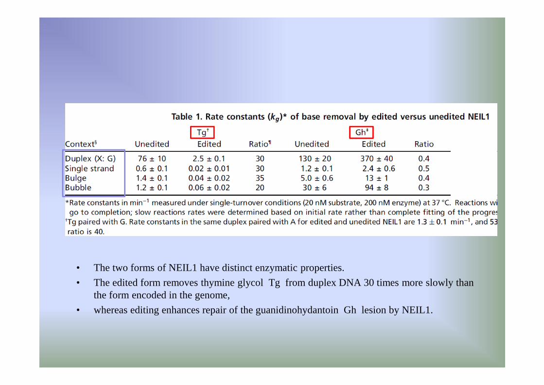
• The two forms of NEIL1 have distinct enzymatic properties.
• The edited form removes thymine glycol Tg from duplex DNA 30 times more slowly than the form encoded in the genome,
• whereas editing enhances repair of the guanidinohydantoin Gh lesion by NEIL1.

NEIL1 editing in response to IFN-α. (Left) Sequence at the recoding site in NEIL1 cDNA from U87 human
glioblastoma cells cultured in the absence of IFN-α.(Right) NEIL1 cDNA sequence from U87 cells treated with IFN-α.

Sommario Neil 1• ADAR1-catalyzed editing of the NEIL1 mRNA causes the genomically
encoded AAA lysine codon, corresponding to amino acid position 242 in the lesion recognition loop of the protein, to be converted to a codon for arginine.
• The two forms of the NEIL1 protein (edited and unedited) have distinct enzymatic properties with changes observed for both glycosylase activity and lesion specificity.
• Editing occurs in a hairpin duplex structure formed near the intron 5/exon 6 boundary in the NEIL1pre-mRNA.
• Furthermore, NEIL1 mRNA recoding is regulated extracellularly by interferon, as predicted for an ADAR1-catalyzed reaction.
• These results suggest a regulatory mechanism for DNA repair based on RNA editing.

Deciphering the functions and regulation of brain-
enriched A-to-I RNA editing Nat Neurosci. 2013..
• Adenosine-to-inosine (A-to-I) RNA editing, in which genomically encoded adenosine is changed to inosine in RNA, is catalyzed by adenosine deaminase acting on RNA (ADAR).
• This fine-tuning mechanism is critical during normal development and diseases, particularly in relation to brain functions.
• A large number of RNA editing sites have recently been identified as a result of the development of deep sequencing and bioinformatic analyses.
• Deciphering the functional consequences of RNA editing events is challenging.