
Ciclossigenasi (COX): struttura e...
27
Università degli Studi di Ferrara Facoltà di Scienze MM.FF.NN. Laurea specialistica in Scienze biomolecolari e cellulari Ciclossigenasi (COX): struttura e ruolo Guastella Giuseppe email [email protected] telefono 0532 974421
Transcript of Ciclossigenasi (COX): struttura e...

Università degli Studi di Ferrara
Facoltà di Scienze MM.FF.NN.
Laurea specialistica in
Scienze biomolecolari e cellulari
Ciclossigenasi (COX): struttura e ruolo
Guastella Giuseppe email [email protected] 0532 974421


Le isoforme

COX 1 COX 1 -- COX 2COX 2

Differenze fra le due isoforme
COX I COX II
Gene 22 Kb, cromosoma 9 mRNA 2.8 Kb
8 Kb, cromosoma 1 mRNA 4.3 Kb
Enzima 70 Kd proteina di membrana
70 Kd proteina di membrana
Substrato Acido arachidonico Acido arachidonico,
altri acidi grassi simili
Km (µmol/L) 5.4 5.6 Km (µmol/L) (Acido Arachidonico)
5.4 5.6

Differenze biologiche fra le due isoforme
COX I COX II
Espressione Costitutiva Inducibile
Funzioni • Controllo delle funzioni cellulari
• Piastrine • Stomaco • Rene
• Processi infiammatori • Macrofagi • Leucociti • Fibroblasti
Inibizione Aspirina, Farmaci
antinfiammatori non steroidei (FANS)
Aspirina, FANS coxibici

La struttura e la catalisi

Dominio “membrane-binding”
Ogni subunità ha un dominio per il legame con le membrana
Questo dominio è composto da 4 a-eliche anfipaticheche si legano ad una precisa e
ottimale profondità ad uno solo dei due strati della membrana cellulare

Il Sito Ciclossigenasico
Acido Arachidonico
Atomo di ossigeno
Legame idrogeno
Atomo di Azoto
Doppio legame
L’Acido Arachidonico (AA) viene legato in questa tasca formata da 4 α eliche e tappezzata da amminoacidi idrofobici W F L V.
La testa polare dell’acido è tenuta in posizione dai legami idrogeno formati con La testa polare dell’acido è tenuta in posizione dai legami idrogeno formati con la serina 530 e la tirosina 385 e 348.
In posizioni vicine ai carboni 13-15 dell’AA, l’idrofobicità della catena è interrotta da residui di serina, tirosina e arginina, importani per la catalisi.
Importante per il funzionamento della proteina è anche il Triptofano 387 anche se non se ne conosce la motivazione

Sintesi delle prostaglandine a Sintesi delle prostaglandine a partire dall’acido arachidonicopartire dall’acido arachidonico
Tyr 385Tyr 385 Tyr 385Tyr 385 Tyr 385Tyr 385
Fe-Eme
Acido arachidonicoAcido arachidonico
PGHPGH22
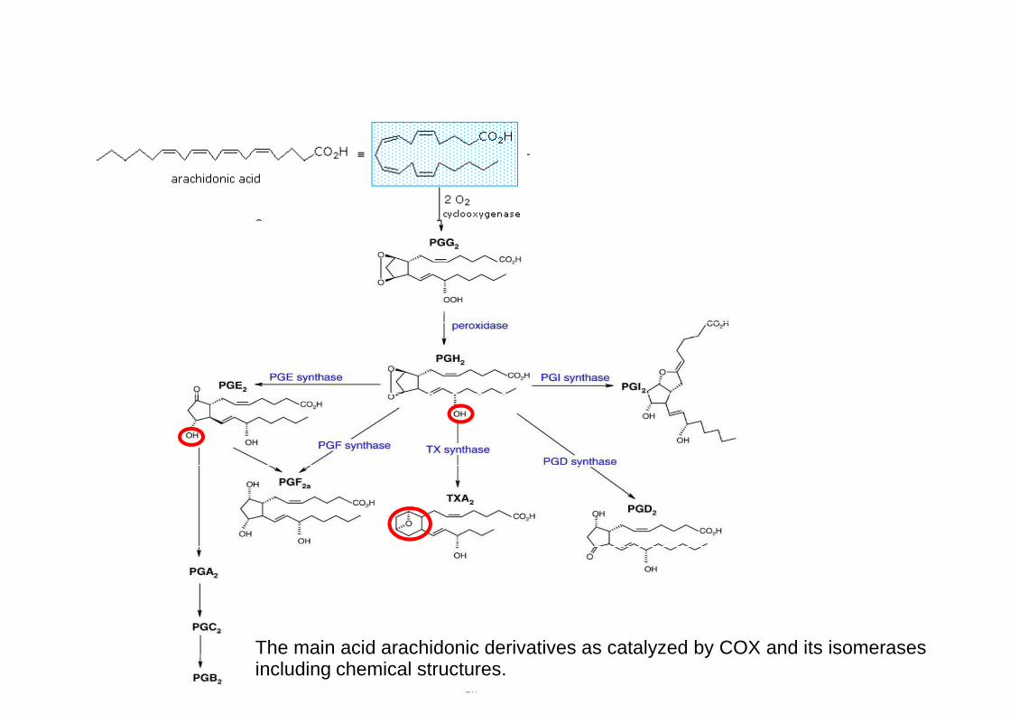
The main acid arachidonic derivatives as catalyzed by COX and its isomerases including chemical structures.

I prodotti: I PROSTANOIDI
PGI2 derivazione vasale vasodilatazione
inibiz. aggregazione piastrinica
PGE2 deriva dai macrofagi mediatore dell’ infiammazione
mediatore della febbre
PGD2 deriva dai mastociti vasodilatazione
inibiz. aggregazione piastrinica
TXA2 deriva dalle piastrine vasocostrizione
aggregazione piastrinica

Gli inibitori
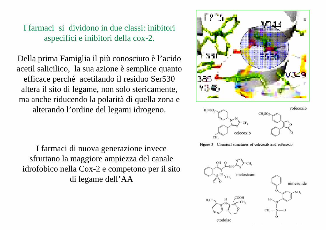
I farmaci si dividono in due classi: inibitori aspecifici e inibitori della cox-2.
Della prima Famiglia il più conosciuto è l’acido acetil salicilico, la sua azione è semplice quanto
efficace perché acetilando il residuo Ser530 efficace perché acetilando il residuo Ser530 altera il sito di legame, non solo stericamente, ma anche riducendo la polarità di quella zona e
alterando l’ordine del legami idrogeno.
I farmaci di nuova generazione invece sfruttano la maggiore ampiezza del canale
idrofobico nella Cox-2 e competono per il sito di legame dell’AA

COXCOX--11COXCOX--11 COXCOX--22COXCOX--22
Sito attivoSito attivoSito attivoSito attivo
Le Due Isoforme della CiclossigenasiLe Due Isoforme della Ciclossigenasi
Sito attivoSito attivo
CanaleCanaleIdrofobicoIdrofobico
Canale Canale IdrofobicoIdrofobico
Val523Val523apreapreuna “tasca una “tasca idrofilica”idrofilica”lateralelaterale
Sito attivoSito attivo
Arg120Arg120
Ile523Ile523chiudechiude
15
“Tasca idrofilica”“Tasca idrofilica”lateralelaterale
Arg120Arg120
Arg120Arg120
chiudechiudeuna “tasca una “tasca idrofilica”idrofilica”lateralelaterale
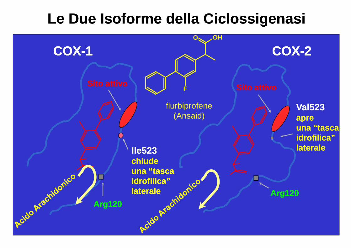
COXCOX--11COXCOX--11 COXCOX--22COXCOX--22
Sito attivoSito attivoSito attivoSito attivo
Le Due Isoforme della CiclossigenasiLe Due Isoforme della CiclossigenasiO OH
Sito attivoSito attivo
Val523Val523apreapreuna “tasca una “tasca idrofilica”idrofilica”lateralelaterale
Sito attivoSito attivo
Ile523Ile523chiudechiude
F
flurbiprofene(Ansaid)
16
Arg120Arg120Arg120Arg120
chiudechiudeuna “tasca una “tasca idrofilica”idrofilica”lateralelaterale
COXCOX--11COXCOX--11 COXCOX--22COXCOX--22
Sito attivoSito attivoSito attivoSito attivo
Le Due Isoforme della CiclossigenasiLe Due Isoforme della Ciclossigenasi
NN
OF
F
Sito attivoSito attivo
Canale Canale IdrofobicoIdrofobico
Val523Val523apreapreuna “tasca una “tasca idrofilica”idrofilica”lateralelaterale
Sito attivoSito attivo
Arg120Arg120
Ile523Ile523chiudechiude
NS
ONH2
FF
celecoxib(Celebrex)
17
Arg120Arg120
Arg120Arg120
chiudechiudeuna “tasca una “tasca idrofilica”idrofilica”lateralelaterale
Arg 513 Arg 513 e His 90His 90formano legami idrogeno con l’ossigeno nella catena solfonammidica

Selective inhibition of cyclooxygenase-2 enhances platelet adhesion in hamster arterioles in vivo.
Martin A. Buerkle, Selim Lehrer, Hae-Young Sohn, Peter Conzen,
“Our experiments demonstrate that selective inhibition of Cox-2 results in an increase in transient platelet interactions with the vessel wall in vivo, resulting in significant firm platelet
Martin A. Buerkle, Selim Lehrer, Hae-Young Sohn, Peter Conzen, Ulrich Pohl and Florian Krötz
wall in vivo, resulting in significant firm platelet adhesion that normally does not take place in these intact arterioles.”
Circulation, 2004

Regolazione infiammazione

From: Funk CD, Science, 294, 1871, 2001
COXCOX-3
(?)
COX
Gastric epithelial cellsMucus production, cytoprotection

Wnt/b-Catenin Signaling Enhances Cyclooxygenase-2 (COX2) Transcriptional Activity
in Gastric Cancer Cells
Felipe Nun˜ ez1, Soraya Bravo1, Fernando Cruzat1, Martı´n Montecino1,2, Felipe Nun˜ ez1, Soraya Bravo1, Fernando Cruzat1, Martı´n Montecino1,2, Giancarlo V. De Ferrari1,2*
Here we studied the transcriptional regulation of the COX2 gene ingastric cancer (GC) cell lines and assessed whether this phenomenon ismodulated by Wnt/b-catenin signaling. Wnt3a significantly enhancedCOX2 mRNA expression in a dose- and time-dependent manner. Serialdeletion of a 1.6 Kbp COX2 promoter fragment and gain- or loss-of-function experiments allowed us to identify a minimal Wnt/b-cateninresponsive region consisting of 0.8 Kbp of the COX2 promoter (pCOX2-responsive region consisting of 0.8 Kbp of the COX2 promoter (pCOX2-0.8), which showed maximal response in genereporter assays. Theactivity of this pCOX2-0.8 promoter region was further confirmed by DNA-protein binding assays.
Plusone 2011

The Wnt signaling pathway

COX2 gene expression and nuclear localization of b-c atenin in GC cells.
Figure 1. (A) COX2 mRNA expression in control (WI38) and GC cell lines (MKN45, AGS, SNU1, SNU16, KATOIII and N87). Total RNA was extracted from cultured cells and semiquantitative RT-PCR was used to determine COX2 and b-actin RNA levels as an internal control. Twenty-six cycles were chosen as an adequate PCR cycle. (B) Nuclear levels of b-catenin protein in the same cell-lines, as shown in (A), were examined through Western Blot analysis using nuclear extracts. The TFIIB general transcription factor was used as an internal control.

Human pCOX2-1.6 promoter activity in response to Wn t/b-catenin signaling.
Figure 3 (B & C)Gene reporter assays in MKN45 (B) and HEK293 (C) cells co-transfected Figure 3 (B & C)Gene reporter assays in MKN45 (B) and HEK293 (C) cells co-transfected with 10 ng of pCOX2 and increasing concentrations of a constitutively active b-catenin (S33Y) protein (left panel). Cells were co-transfected with 1 ng of PRL-SV40 Renilla as an internal control. Promoter activity was normalized as the ratio between firefly luciferase and Renilla luciferase units. RLU: Relative Luciferase Units. Each figure corresponds to at least three independent experiments. Statistical significance was determined through ANOVA test (* p,0.05, ** p,0.01). Nuclear levels of b-catenin protein were examined in same cell lines through Western Blot analysis (right panel). The TFIIB general transcription factor was used as an internal control.

Figure 4. (A) Schematic representationof pCOX2 deletions. (B–D) Gene reporter assay in MKN45 (B), AGS (C) and WI38 (D) cell lines transiently transfected with 50 ng pCOX deletions (pCOX2-1.2;
pCOX2-0.8 as a minimal COX2 promoter with maximum b asal response in GC cells.
50 ng pCOX deletions (pCOX2-1.2; pCOX2-0.8; pCOX2-0.65 and pCOX2-0.4) and 50 ng of empty vector. In all experiments cells were transfected with1 ng of PRL-SV40 Renilla as an internal control. Promoter activity was normalized as the ratio between firefly luciferase and Renilla luciferase units. RLU: Relative Luciferase Units. Each figure corresponds to at least three independent experiments. Statistical significance was determinedStatistical significance was determinedthrough ANOVA test (* p,0.05, ** p,0.01).

Wnt/b-catenin signaling modulates pCOX2-0.8 activit y in MKN45 cells.
Figure 5. Gene reporter assays in MKN45 cells transiently transfected with Figure 5. Gene reporter assays in MKN45 cells transiently transfected with either 10 ng pCOX2-0.8 (A) or pCOX2-0.4 (B), plus 5–10 ng of b-catenin S33Y and 10 ng of empty vector as control. Cells were transfected with increasing concentrations of pCOX2-0.8, MpCOX-08, or equal amounts of empty vector as a control. Promoter activity was normalized as the ratio between firefly luciferase and Renilla luciferase units. RLU: Relative Luciferase Units. Each figure corresponds to at least three independent experiments. Statistical significance was determined through ANOVA test (* p,0.05, ** p,0.01).
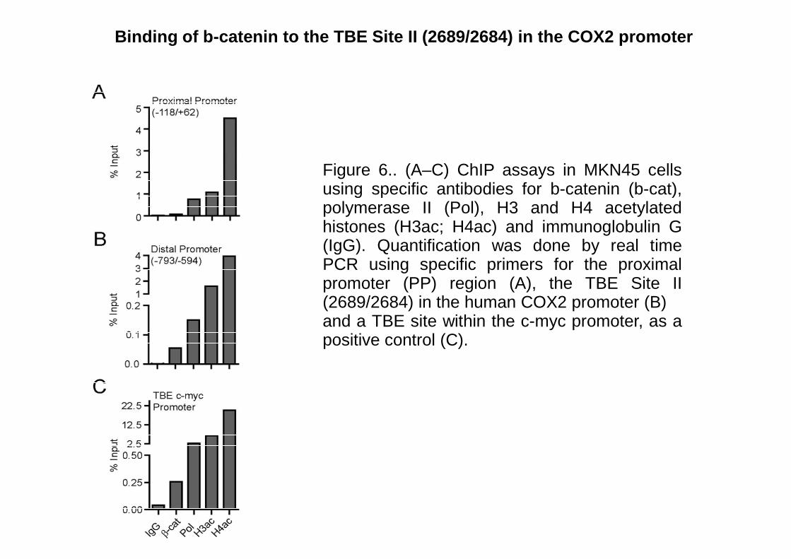
Figure 6.. (A–C) ChIP assays in MKN45 cellsusing specific antibodies for b-catenin (b-cat),
Binding of b-catenin to the TBE Site II (2689/2684) in the COX2 promoter
using specific antibodies for b-catenin (b-cat),polymerase II (Pol), H3 and H4 acetylatedhistones (H3ac; H4ac) and immunoglobulin G(IgG). Quantification was done by real timePCR using specific primers for the proximalpromoter (PP) region (A), the TBE Site II(2689/2684) in the human COX2 promoter (B)and a TBE site within the c-myc promoter, as apositive control (C).


![Lezione 2 A trasposoni [modalità compatibilità ]m.docente.unife.it/francesco.bernardi/materiale... · 2qh ri wkhvh uhwurwudqvsrvrq olnh hohphqwv lv 57/ 3(* '6* surwhdvh dfwlyh vlwh](https://static.fdocumenti.com/doc/165x107/5f4181e67d33bd67b353a0ba/lezione-2-a-trasposoni-modalitf-compatibilitf-m-2qh-ri-wkhvh-uhwurwudqvsrvrq.jpg)