1 IUSS - Ferrara 1391 IUSS - Ferrara 1391 IUSS DAY Ferrara, Lunedì 22 Marzo 2010.

Università degli Studi di Ferrara
DOTTORATO DI RICERCA IN
FARMACOLOGIA E ONCOLOGIA MOLECOLARE
CICLO XXVI
COORDINATORE Prof. Antonio Cuneo
Immunohistochemical Detection of DNA Mismatch Repair Proteins Abnormalities in
Sudanese Colorectal Cancer Patients
Settore Scientifico Disciplinare BIO/14
Dottorando Tutore
Dott. Yosef Mohamed Azzam Yosef Mohamed Zakout Prof. Giovanni Lanza
Anni 2011/2013

Università degli Studi di Ferrara
DOTTORATO DI RICERCA IN
FARMACOLOGIA E ONCOLOGIA MOLECOLARE
CICLO XXVI
COORDINATORE Prof. Antonio Cuneo
Immunohistochemical Detection of DNA Mismatch Repair Proteins Abnormalities in
Sudanese Colorectal Cancer Patients
Settore Scientifico Disciplinare BIO/14
Dottorando Tutore
Dott. Yosef Mohamed Azzam Yosef Mohamed Zakout Prof. Giovanni Lanza
_______________________________ ___________________________
(firma) (firma)
Anni 2011/2013

i
Dedication
I dedicate this work to my parents, teachers, and friends.

ii
Acknowledgments
My thanks go to all who helped me to complete this modest effort.
I am especially grateful to my supervisor, Prof. Giovanni Lanza, for his expert
supervision, patience, and valuable comments.
I would like to thank Dr. Linda Ulazzi, Mr. Roberto Mazzoni, Dr. Beatrice
Paradiso, and Ms. Iva Maestri for their help and assistance.
My special thanks go to Prof. Naser Eldin Bilal, Dean Faculty of Medical
Laboratory Sciences - University of Khartoum for his support and encouragement.
My thanks go to Dr. Saifaldin Mohammad Azain for his cooperation, and for Ms.
Samah Batran, Ms. Shahenaz Shaban Salih, Ms.Tibyan Abdalhadi
Mohammed, and Dr. Lobaina Mohammed Eissa for their assistance.
iii
Abbreviations
5-FU 5-fluorouracil
BAT 25 Big adenine tract 25
BAT 26 Big adenine tract 26
CC1 Cell conditioning 1
CC2 Cell conditioning 2
CI Confidence interval
CRC Colorectal cancer
CRCs Colorectal cancers
CT Computed tomographic
dMMR Deficient mismatch repair
DNA Deoxyribonucleic acid
dNTPs Deoxynucleotide triphosphates
EXO I Exconuclease I
FFPE Formalin fixed, paraffin-embedded
FOBTs Fecal occult blood tests
HG High grade
HNPCC Hereditary nonpolyposis colorectal cancer
HR Hazard ratio
IHC Immunohistochemistry
LG Low grade
LIGI Ligase I
MgCl2 Magnesium chloride
iv
MLH1 MutL homolog 1
MMR Mismatch repair
MMR protein - Mismatch repair protein negative
MMR protein + Mismatch repair protein positive
MR Magnetic resonance
MSH2 MutS protein homolog 2
MSH6 MutS protein homolog 6
MSI Microsatellite instability
MSI-H Microsatellite instability- high
MSI-L Microsatellite instability- low
MSS Microsatellite-stable
n Number
NCI National cancer institute
PCNA Proliferating cellular nuclear antigen
PCR Polymerase chain reaction
pMMR Proficient Mismatch repair
PMS2 Postmeiotic segregation increased 2
Pol δ Polymerase δ
RFC Replication factor C
RPA Replication protein A
SEPT9 Septin 9
μm Micrometre

v
Abstract
Background: The current study aimed to assess DNA mismatch repair (MMR)
proteins abnormalities among Sudanese colorectal cancer (CRC) patients, mainly
by immunohistochemistry (IHC).
Methods: CRC cases were retrieved from the records of two Histopathology
laboratories in Khartoum, Sudan. The total number of included cases was 42.
Sections were cut and stained by immunohistochemical method to assess the
abnormalities of four MMR proteins (MLH1, MSH2, MSH6 and PMS2) using anti-
MLH1, MSH2, MSH6 (mouse monoclonal antibodies) and anti-PMS2 (rabbit
monoclonal antibody). Microsatellite instability (MSI) analysis using mainly BAT 25
& 26 was performed for cases that showed negative or inadequate staining results
for any MMR protein by IHC.
Results: Of the study population, 25 (59.5%) were males and 17 (40.4%) were
females. Their ages ranged between 20-85 years (the age of 4 patients was not
provided). The mean age was 56.1 year, and 12 (31.5%) of the CRC patients were
among the age groups younger than 50 years.
Of the 42 included cases, 34 (80.95%) were MMR protein positive for all MMR
proteins under assessment, 3 (7.14%) MSH2 inadequate, and 1 (2.38%) MSH6
inadequate.
Abnormal MMR proteins expression was found in 4 (9.5%) cases. Of these, 2
(50%) were MSH2&MSH6 negative and 2 (50%) were MLH1&PMS2 negative.
Regarding MSI results, the three cases that were MSH2 inadequate and positive
for the rest by IHC showed stable results with both BAT 25& 26. The case that
was MSH6 inadequate, showed stable results with both BAT 25&26.
The 2 cases with MSH2&MSH6 negative results were unstable with both BAT
25&26. Of the two cases that were MLH1&PMS2 negative, one case showed non-
evaluable results with both BAT 25&26 while the other case was unstable with
BAT 26 and not evaluable with BAT 25.
Conclusion: In this study, the percentage of MMR protein negative cases in
Sudanese CRC patients appears to be relatively low compared to what has been
generally reported in certain studies done in different countries. Furthermore,
MLH1&PMS2 and MSH2&MSH6 abnormal expression detected by IHC seems to
be the most common form of MMR proteins abnormalities in Sudanese CRC

vi
patients. Concerning the results of IHC, MLH1 and MSH2 seem to be the most
inactivated MMR genes in Sudanese CRC patients.
vii
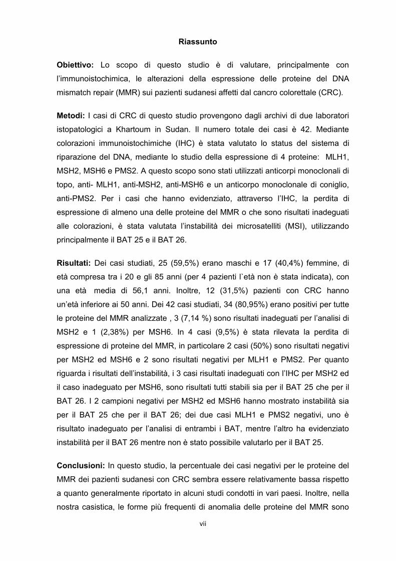
Riassunto
Obiettivo: Lo scopo di questo studio è di valutare, principalmente con
l’immunoistochimica, le alterazioni della espressione delle proteine del DNA
mismatch repair (MMR) sui pazienti sudanesi affetti dal cancro colorettale (CRC).
Metodi: I casi di CRC di questo studio provengono dagli archivi di due laboratori
istopatologici a Khartoum in Sudan. Il numero totale dei casi è 42. Mediante
colorazioni immunoistochimiche (IHC) è stata valutato lo status del sistema di
riparazione del DNA, mediante lo studio della espressione di 4 proteine: MLH1,
MSH2, MSH6 e PMS2. A questo scopo sono stati utilizzati anticorpi monoclonali di
topo, anti- MLH1, anti-MSH2, anti-MSH6 e un anticorpo monoclonale di coniglio,
anti-PMS2. Per i casi che hanno evidenziato, attraverso l’IHC, la perdita di
espressione di almeno una delle proteine del MMR o che sono risultati inadeguati
alle colorazioni, è stata valutata l’instabilità dei microsatelliti (MSI), utilizzando
principalmente il BAT 25 e il BAT 26.
Risultati: Dei casi studiati, 25 (59,5%) erano maschi e 17 (40,4%) femmine, di
età compresa tra i 20 e gli 85 anni (per 4 pazienti l`età non è stata indicata), con
una età media di 56,1 anni. Inoltre, 12 (31,5%) pazienti con CRC hanno
un’età inferiore ai 50 anni. Dei 42 casi studiati, 34 (80,95%) erano positivi per tutte
le proteine del MMR analizzate , 3 (7,14 %) sono risultati inadeguati per l’analisi di
MSH2 e 1 (2,38%) per MSH6. In 4 casi (9,5%) è stata rilevata la perdita di
espressione di proteine del MMR, in particolare 2 casi (50%) sono risultati negativi
per MSH2 ed MSH6 e 2 sono risultati negativi per MLH1 e PMS2. Per quanto
riguarda i risultati dell’instabilità, i 3 casi risultati inadeguati con l’IHC per MSH2 ed
il caso inadeguato per MSH6, sono risultati tutti stabili sia per il BAT 25 che per il
BAT 26. I 2 campioni negativi per MSH2 ed MSH6 hanno mostrato instabilità sia
per il BAT 25 che per il BAT 26; dei due casi MLH1 e PMS2 negativi, uno è
risultato inadeguato per l’analisi di entrambi i BAT, mentre l’altro ha evidenziato
instabilità per il BAT 26 mentre non è stato possibile valutarlo per il BAT 25.
Conclusioni: In questo studio, la percentuale dei casi negativi per le proteine del
MMR dei pazienti sudanesi con CRC sembra essere relativamente bassa rispetto
a quanto generalmente riportato in alcuni studi condotti in vari paesi. Inoltre, nella
nostra casistica, le forme più frequenti di anomalia delle proteine del MMR sono

viii
risultate due: la mancata espressione di MLH1 - PMS2 e quella di MSH2-MSH6.
Considerando i risultati ottenuti con l’IHC, si può concludere che nei pazienti
sudanesi con CRC, MLH1 ed MSH2 sembrano essere i geni del MMR più
frequentemente inattivati.

ix
List of contents
Dedication................................................................................................ i
Acknowledgments…….……………………………………………………… ii
Abbreviations…………………………………………………………………. iii
Abstract (English)…………………………………………...………………... v
Abstract (Italian)…………………………………………………………….... vii
List of contents……………………………………………………………...… ix
List of tables………………………………………………………………..…. xi
List of figures…………………………………………………………..……… xii
Chapter one: Introduction and literature review…………………….… 1
1. Introduction and literature review………………………………………... 2
1.1. Epidemiology of CRC…………………………………………………… 2
1.2. Clinical features of CRC.................................................................... 2
1.3. Risk factors for CRC……...…………………………………………….. 2
1.4. Screening for CRC……………………………………………………… 3
1.5. DNA MMR system............................................................................. 4
1.5.1. MSI-High (MSI-H) and MSI-Low (MSI-L)....................................... 4
1.5.2. Hereditary nonpolyposis colorectal cancer (HNPCC) and MMR.... 5
1.5.3. MMR, MSI and CRC...................................................................... 6
1.5.3.1. Pathological and clinical features of MSI CRC............................ 6
1.5.4. IHC for detection of MMR status.................................................... 7
1.5.4.1. Interpretation of IHC results for DNA MMR proteins……………. 11
Chapter two: Objectives........................................................................ 15
2. Objectives............................................................................................ 16
Chapter three: Materials and methods................................................. 17
3. Materials and methods......................................................................... 18
3.1. Study samples................................................................................... 18
3.2. Laboratory procedures...................................................................... 18
3.2.1. Cutting............................................................................................ 18
3.2.2. Immunohistochemical analysis...................................................... 18
3.2.3. Extraction of DNA……………………………………………………... 19
3.2.4. MSI analysis…………………………………………………………… 19
Chapter four: Results………………………………………………………. 21
4. Results……………………………………………………………….…...… 22

x
Chapter five: Discussion….……………………………………………….. 40
5. Discussion………………………………………………………….………. 41
Chapter six: Conclusions…………………………………………………. 44
6. Conclusions……………………………………………………………..…. 45
Chapter seven: References……………………………………………….. 46
7. References…………………………………………………………………. 47

xi
List of tables
Table 1: Description of study population by gender. 24
Table 2: Description of study population by age. 25
Table 3: Description of grade of differentiation. 26
Table 4: MMR proteins expression by IHC, and MSI status. 27
Table 5: Description of study population by MMR status. 28
Table 6: Immunohistochemical patterns of dMMR CRCs. 29
Table 7: Description of study population by MMR protein expression
and gender.
29
Table 8: Description of study population by MMR protein expression
and grade of differentiation.
29
Table 9: A summary of some studies regarding MMR protein
expression examined by IHC.
42
xii
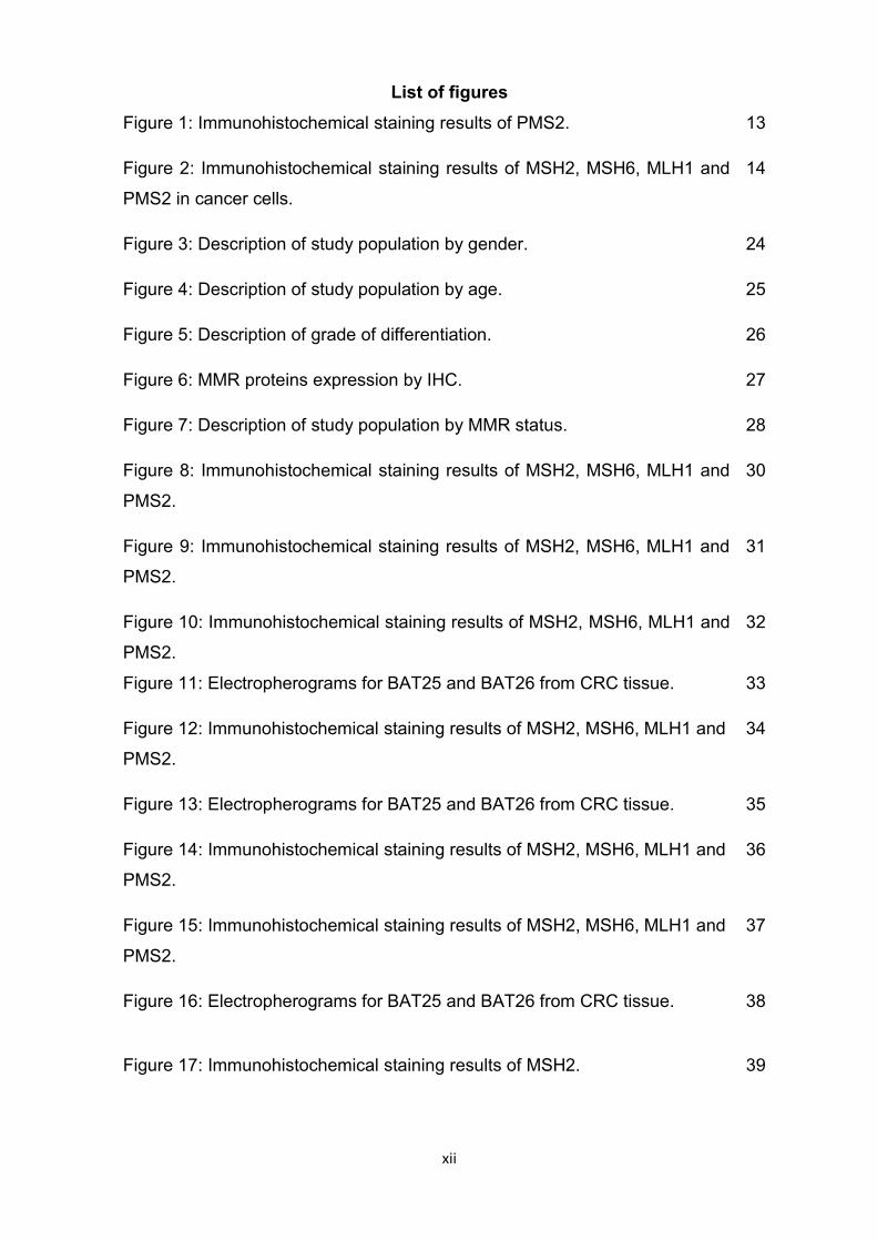
List of figures
Figure 1: Immunohistochemical staining results of PMS2. 13
Figure 2: Immunohistochemical staining results of MSH2, MSH6, MLH1 and
PMS2 in cancer cells.
14
Figure 3: Description of study population by gender. 24
Figure 4: Description of study population by age. 25
Figure 5: Description of grade of differentiation. 26
Figure 6: MMR proteins expression by IHC. 27
Figure 7: Description of study population by MMR status. 28
Figure 8: Immunohistochemical staining results of MSH2, MSH6, MLH1 and
PMS2.
30
Figure 9: Immunohistochemical staining results of MSH2, MSH6, MLH1 and
PMS2.
31
Figure 10: Immunohistochemical staining results of MSH2, MSH6, MLH1 and
PMS2.
32
Figure 11: Electropherograms for BAT25 and BAT26 from CRC tissue. 33
Figure 12: Immunohistochemical staining results of MSH2, MSH6, MLH1 and
PMS2.
34
Figure 13: Electropherograms for BAT25 and BAT26 from CRC tissue. 35
Figure 14: Immunohistochemical staining results of MSH2, MSH6, MLH1 and
PMS2.
36
Figure 15: Immunohistochemical staining results of MSH2, MSH6, MLH1 and
PMS2.
37
Figure 16: Electropherograms for BAT25 and BAT26 from CRC tissue. 38
Figure 17: Immunohistochemical staining results of MSH2. 39

1
CHAPTER ONE
INTRODUCTION AND LITERATURE REVIEW

2
1. Introduction and literature review
1.1. Epidemiology of CRC:
Worldwide, CRC was the third most common cancer in 2008 with 1.233 million
newly diagnosed cases (9.7% of the total) and about 608.000 deaths, which
makes it the fourth cause of cancer deaths (Ferlay et al., 2010a).
In United States, 142.82 new cases of CRC was estimated in 2013 makes it the
third most common cancer with approximately 50.830 deaths (Siegel et al., 2013).
In Europe, CRC was the most common cancer in 2008 constituting 436,000 cases,
13.6% of the total cancers and the second cause of cancer death constituting
212,000 deaths, 12.3% of the total (Ferlay et al., 2010b).
In Sudan, CRC represents 6.4% of the total number of malignant tumors and the
second most common malignant gastrointestinal tumor (33.69%) after esophageal
cancer (37.7%) during the period from 2000 to 2004 according to a published data
from one pathology center in Khartoum (El Hassan et al., 2008).
1.2. Clinical features of CRC:
Symptoms of CRC include alteration in bowel habit, bleeding from the rectal,
abdominal pain, intestinal obstruction (Kyle et al., 1991), diarrhoea (MacArthur and
Smith, 1984) and anaemia (Rizk and Ryan, 1994; Saidi et al., 2008).
One of the early symptoms include abdominal pain or/and vomiting, while the
more localised symptoms of the rectal and weight loss are usually related with
long delay (MacArthur and Smith, 1984).
1.3. Risk factors for CRC:
Several studies found that there are different risk factors associated with the
development of CRC. Such as increasing age and family history (Ballinger and
Anggiansah, 2007). The rate of CRC elevates throughout the fifth decade and
reaches its most extreme at the age 75; however, there are various cases in more
youthful individuals each year (Mihajlovic-Bozic, 2004).

3
Other risk factors were reported such as smoking and consumption of alcohol
(Mihajlovic-Bozic, 2004; Sanjoaquin et al., 2004; Cappellani et al., 2013), notably
the higher level of alcohol intake (Cho et al., 2004; Shimizu et al., 2003) and the
daily alcohol consumption (Wu et al., 1987) .
Less consumption of high-fibers grains and vegetables increases the risk of fatal
colon cancer (Thun et al., 1992).
Aspirin might decrease the risk of CRC in women (Giovannucci et al., 1995; Cook
et al., 2013). Another finding connected the regular use of aspirin to a decreased
risk in males for developing CRC (Giovannucci et al., 1994).
Other factors such as obesity and less physical activity measured also as risk
factors (Giovannucci, 2003). Furthermore, dietary system that is rich in fat and red
meat considered as factors increase the risk (Mihajlović-Božić, 2004).
1.4. Screening for CRC:
Screening for CRC is important for the early detection of the disease (O’Carroll et
al., 2013). There are different tests used for CRC screening, including fecal occult
blood tests (FOBTs), stool DNA tests, colonoscopy, flexible sigmoidscopy,
magnetic resonance (MR), computed tomographic (CT) colonography, double
contrast barium enema, capsule endoscopy (Flitcroft et al., 2012), and faecal
pyruvate kinase isoenzyme type M2 (Tonus et al., 2012).
Furthermore, methylated septin 9 (SEPT9) was described as a sensitive biomarker
for CRC from peripheral blood, and the detection of this biomarker in plasma was
found as a dependable method of screening for left and right sided colon cancers
(Tóth et al., 2012).
Regarding the relationship between screening with faecal-occult-blood test testing
and mortality, several studies showed a lower mortality from CRC in the screening
groups compared to control groups (Kronborg et al., 1996; Hardcastle et al., 1996;
Shaukat et al., 2013).
In comparison to no screening, both incidence and mortality were found to be
reduced, in variable percentages, in three different screening methods namely

4
colonoscopy, fecal immunochemical test and low-sensitivity guaiac fecal occult
blood test (Barouni et al., 2012).
Furthermore, CRC cases in the control group are at a more advanced stage
compared to CRC cases detected by screening (Kewenter et al., 1994).
1.5. DNA MMR system:
DNA MMR is a process that corrects mismatches that appear during DNA
replication and getaway proofreading process (Kunkel and Erie, 2005). Therefore,
this system removes any base-base mismatches as well as insertion-deletion
loops occur during replication of DNA as a result of DNA polymerase slippage
(Peltomäki, 2001).
Failure in MMR system will most likely affect functions and structure of the cell
ensuing in tumorigenesis, immortalization, malignant transformation, and/or
degenerative diseases (Conde-Pérezprina et al., 2012). MSI is the term that used
to describe the form of genomic instability that related to DNA MMR defective in
tumors (Boland et al., 1998).
1.5.1. MSI-High (MSI-H) and MSI-Low (MSI-L):
Five microsatellites markers were validated and recommended for detecting MSI
CRC in 1997 in the National Cancer Institute (NCI) sponsored conference entitled
"The International Workshop on Microsatellite Instability and RER Phenotypes in
Cancer Detection and Familial Predisposition". This panel includes
mononucleotide repeats: Big adenine tract (BAT) 25, BAT-26, and dinucleotide
repeats: D5S346, D2S123, and D17S250 (Boland et al., 1998). This panel is
known as the Bethesda panel (Vilar and Gruber, 2010).
Colorectal cancers (CRCs) are called MSI-H if there is instability in 2 or more of
the five microsatellite markers, but those with instability in only one of the five
markers are called MSI-L and share the phenotype of microsatellite-stable (MSS)
tumors, while MSI-H tumors have distinctive pathological and clinical phenotype
(Boland et al., 1998). However, expanded panel composed of 10 markers were
evaluated (Mead et al., 2007).
In 2002, one more workshop was held at the NCI in Bethesda to revise and
improve the Bethesda Guidelines. The participants in this workshop suggested

5
including more mononucleotide markers in order to increase the sensitivity of the
panel (Umar et al., 2004).
One of the proposed panels is to use a pentaplex panel of 5 quasimonomorphic
mononucleotide repeats, which includes BAT-25, BAT-26, NR-21, NR-22, and NR-
24, and provides a precise assessment of tumor MSI status with 100% specificity
and sensitivity and obviate the need to compare to normal DNA (Suraweera et al.
2002).
Bacher et al. (2004) described another method for MSI analysis. In their system,
they use five mononucleotide markers (BAT-25, BAT-26, NR-21, NR-24 and
MONO-27). They compared the results of MSI analysis in 153 CRCs using their
new MSI Multiplex System to the results obtained using a panel composed of 10
MSI markers, and they found that there is 99% concordance between the two
methods and the accuracy was almost 100% in detecting MSI-H cases.
Patil et al. (2012) examined the same previous panel (BAT-25, BAT-26, NR-21,
NR-24 and MONO-27) regarding the need of control DNA to detect MSI status.
They found that this panel is able to accurately detect 95.2% of MSS CRC cases
and all MSI-H CRC cases without using normal DNA.
Brennetot et al. (2005) found that detecting of MSI-H can be done by BAT-26 and
BAT-25 analysis even if the DNA sample contains low tumor DNA (5-10%).
Moreover, MSI can be detected using one marker, BAT 26, but this method can be
used to screen CRCs for MMR deficiency but cannot differentiate between
different degrees and types of instability (de la Chapelle, 1999).
Moreover, BAT-26 was used for MSI analysis in different studies (Chai et al.,
2004; Jover et al., 2004, Samowitz et al., 2001). Nevertheless, Oh et al. (2012)
found that using BAT-26 and BAT-25 for MSI analysis seems not to provide an
accurate assessment of MSI status, mainly in MSI-L cases.
1.5.2. Hereditary nonpolyposis colorectal cancer (HNPCC) and MMR:
HNPCC (also known as Lynch syndrome) is caused by mutations in MMR genes;
furthermore, a noteworthy percentage of sporadic cancer is connected with loss of
MMR (Hsieh and Yamane, 2008).

6
In HNPCC, hMLH1 and hMSH2 are the most frequently affected genes. Deficient
expression of these genes makes the cell at risk to the accumulation of several
molecular defects, a circumstance that can be assessed by the instability in
segments of base repeats in the genome identified as MSI (Silva et al., 2005).
1.5.3. MMR, MSI and CRC:
According to the data reported from different articles, MSI (notably the high levels)
is present in almost 15% of CRC (Boland et al., 1998; Vilar and Gruber, 2010;
Hamilton, 2013). This is due to germline mutation in one of the MMR genes
(mainly MLH1, MSH2, MSH6 or PMS2) or MLH1 epigenetic silencing (Vilar and
Gruber, 2010). Of this 15% CRCs with MSI, 12% are due to acquired
hypermetylation of the MLH1 gene promoter while 3% are related to HNPCC
(Boland and Goel, 2010).
Furthermore, MSI is present in over 90% of HNPCC patients in which MSI caused
by defects in DNA MMR (Søreide, 2007).
1.5.3.1. Pathological and clinical features of MSI CRC:
CRCs with MMR-defective that causes MSI have characteristic pathological and
clinical features as shown in different studies. They tend to be in the ascending
colon (Jover et al., 2004), have a lower risk of recurrence (Yoon et al., 2011;
Sinicrope et al., 2011), at stage that is less advanced, mucinous, poorly
differentiated and have pattern of expansive growth more often compared to
microsatellite stable (MSS) CRCs (Benatti et al., 2005).
Xiao et al. (2013) examined 1,941 CRC patients and found that poorly
differentiated CRC (PD) was more frequent among MSI CRC cases compared to
MSS CRC cases representing 23.6% and 4.2% respectively.
However, another study by Ashktorab et al. (2005) showed a different finding
regarding the histologic tumor grade. In their study, MSI analysis was performed
using 5 MSI markers (BAT25, BAT26, D2S123, D5S346 and D17S250) in 51 CRC
cases from African Americans. Of those, 22 (43%) showed MSI-H, and most of
them were well differentiated.
Moreover, CRCs with MSI-H demonstrate association with infiltration of a dense
local lymphocyte, less incidence of metastasis to distant organ, and proximal
localization of the tumor (Kloor et al., 2013). Additionally, MSI CRCs have a better

7
prognosis compared to MSS CRCs (Popat et al., 2005; Pino and Chung, 2011;
Kloor et al., 2013).
Jensen et al. (2009) detected MSI in (13.8%) of the examined CRC cases. They
found that MSI tumors were associated with lesser risk of death (hazard ratio (HR)
= 0.4; 95% CI: 0.2–0.9; P = 0.02) and recurrence (HR = 0.3; 95% CI: 0.2–0.7; P =
0.0007) compared to MSS tumors in multivariate analysis.
Ward et al. (2001) studied MSI and the clinicopathological characteristics of
sporadic CRC. They reported that 33 (10.6%) tumors out of 310 were MSI-H.
Additionally, they found that these MSI-H cases were more likely to be mucinous,
high grade, and to harbor elevated numbers of both intraepithelial and peritumoral
lymphocytes. Furthermore, these MSI-H cases were more likely to occur among
females and to be right sided.
Ziadi et al. (2013) detected 6 (13.8%) CRC tumors with MSI-H out of 44 CRC
cases. These MSI-H cases were more likely to have medullary pattern, to be
poorly differentiated, and to harbor elevated numbers of peritumoral lymphocytes.
Regarding lymph node involvement, tumor location and stage of disease, they
found no significant difference between MSI-H and MSI-L/MSS cases.
CRCs with MSI or dMMR thought to have no or poorer benefit from adjuvant 5-
fluorouracil (5-FU) based chemotherapy compared to CRCs with MSS (Laghi and
Malesci, 2012; Pino and Chung, 2011; Benatti et al., 2005). However, a study
found that any advantage of 5-FU among dMMR stage III tumors is suggested to
be limited to tumors with suspected germline mutations compared to sporadic
CRCs (Sinicrope et al., 2011).
1.5.4. IHC for detection of MMR status:
IHC is a technique where monoclonal and polyclonal antibodies are used to
identify certain components in cells or tissues (antigens), and the antibody binding
site is being recognized by labelling of the primary antibody or a suitable
secondary antibody with a marker such as enzyme, colloidal gold, radioactive
elements, fluorescent dye, and then being seen under either ordinary or
fluorescent microscope (Duraiyan et al., 2012).
8
CRCs with mutations in DNA MMR gene show elevated rate of replication errors in
simple repetitive sequences demonstrable as MSI, in which the majority are due to
somatic MMR dysfunction in addition to a subset occur due to germline mutations
(Stone et al., 2001). The availability of antibodies to MMR proteins has offered an
alternative method to molecular techniques for recognizing dMMR CRCs (Stone et
al., 2001). IHC can be a specific, sensitive, fast, and cost-effective method for
identifying MMR defects (Lindor et al., 2002).
Additionally, the other important advantage of IHC is that it could direct and guide
genetic analysis by detecting the affected gene based on the absence of protein
expression in IHC ( Zhang and Li, 2013; Rigau et al., 2003).
Lindor et al. (2002) compared between IHC and MSI analysis regarding the
phenotyping of CRC. In their study, 1,144 CRC cases were examined for
deficiency of DNA MMR using MSI analysis and immunohistochemical detection of
MSH2 and MLH1 proteins. MSI-H was detected in 350 (30.6%) by MSI analysis.
Of these MSI-H cases, 323 showed loss of expression by IHC of either MLH1 or
MSH2 (228 and 98 respectively). They concluded that IHC for MLH1 and MSH2
has sensitivity of 92.3% and specificity of 100% for dMMR screening. Furthermore,
they found that the normal expression of MMR proteins by IHC has 96.7%
predictive value for MSS/MSI-L, and it reaches 100% in regard abnormal IHC for
MSI-H.
Jover et al. (2004) reported that IHC appears to provide a reliable technique to
detect most dMMR CRCs. They performed microsatellite analysis in 172 cases of
CRCs by polymerase chain reaction (PCR) using BAT-26, and IHC for MLH1 and
MSH2. MSI was assessed in 13 (7.6%) cases and all these cases showed
negative staining by IHC for MLH1 or MSH2.
Marcus et al. (1999) compared between the results of MSI analysis and IHC for
MLH1 and MSH2 proteins in 72 CRC tumors. Of those cases, 38 were MSI-H and
34 were MSS. All MSS cases showed intact IHC staining with MLH1 and MSH2,
while loss of expression of MLH1 and/or MSH2 was detected in 37 of 38 MSI-H
tumors. They concluded that IHC can differentiate accurately between MSS and
MSI-H tumors.

9
Cunningham et al. (2001) examined MMR status among 257 CRC patients by MSI
testing and IHC for MSH2, MSH6, and MLH1. Defective MMR was detected in 51
(20%) cases, which demonstrated MSI-H. All these MSI-H cases showed loss of
expression for one or more of the assessed MMR proteins.
Stone et al. (2001) performed MSI analysis and IHC for MMR proteins (MSH2 and
MLH1) using monoclonal antibodies in 46 CRC cases. MSI was detected in 23
cases. Of these 23 cases, 22 showed loss of expression of one of the assessed
MMR proteins. MSS was detected in 23 CRC cases and all of them showed
positive staining with both antibodies
Ruszkiewicz et al. (2002) found that IHC provides an alternative relatively
inexpensive and fast technique to assess the status of MSI compared to MSI
analysis. Furthermore, they found that the sensitivity and specificity of IHC are
92% and 99.8% respectively when assessed against MSI analysis. However, they
reported that it has to be accepted that using IHC technique only; would miss a
small fraction of MSI cases.
The validity of IHC regarding MMR proteins in CRC was examined by Overbeek et
al. (2008). In their study, 100 molecularly assessed CRC cancer cases were
stained for 4 MMR proteins (MLH1, MSH2, MSH6, and PMS2) and then were
examined by 7 pathologists from 5 different laboratories. Of those pathologists, 2
have experience in Interpretation of IHC results for DNA MMR proteins. The
authors found that IHC is a valid method to recognize sporadic MSI CRC patients
as well as those patients at risk for HNPCC when the IHC stained sections are
assessed by experienced pathologists. Furthermore, they reported that MSI test is
to be performed for those cases with aberrant or indefinite staining results by IHC
in order to verify the presence of defective MMR.
Boardman et al. (2007) studied the frequency of defective MMR among Alaska
native patients with CRC by IHC (MLH1, MSH2, and hMSH6), and MSI analysis.
In their study, 329 CRC cases were studied. Of these cases, MSI and loss of
expression of MMR proteins were detected among 46 (14%). Of these 46 cases,
loss of MLH1, MSH2, and MLH1/MSH2 was detected in 42 (91%), 3 (7%), and 1
(2%) respectively.

10
Shia et al. (2009) reported that using a two-antibody panel composed of MSH6
and PMS2 is as effective as the four-antibody panel (MLH1, MSH2, MSH6 and
PMS2) in detecting the abnormalities of DNA MMR protein.
Additionally, Hall et al. (2010) found that using a two-antibody panel composed of
PMS2 and MSH6 has 100% sensitivity and specificity compared to the four-
antibody panel test (MLH1, MSH2, MSH6 and PMS2), with absence of false
positives or negatives. They concluded that the four-antibody panel should be
replaced by the two-antibody panel for screening for MMR deficiency by IHC.
Furthermore, the usefulness of using the 2 antibodies (MSH6 and PMS2) in CRC
was confirmed by Mojtahed et al. (2011).
Moreover, O'Regan et al. (2013) achieved similar findings in their study. They
suggested that the initial use of two antibodies (MSH6 & PMS2) followed by the
other two antibodies (MLH1& MSH2) ,if there is loss of expression in the initially
used antibodies, could have the same efficiency of using the four antibodies
(MLH1, MSH2, MSH6 and PMS2) to detect loss of expression of MMR gene
protein.
Regarding HNPCC, Caldés et al. (2004) studied the sensitivity of MSI analysis and
IHC for MSH2, MSH6, and MLH1 among tumors obtained from carriers of known
MMR gene mutation. They studied the germline mutations in MSH2, MSH6, and
MLH1 in 58 samples from HNPCC families. Of them, 28 were found with a real
mutation and all of them showed loss of expression by IHC for at least one of the
examined MMR proteins. MSI-H was found in 27 of the 28 cases with a real
mutation. Sensitivity by IHC was 100% compared to 96% by MSI analysis for
detection of MMR deficiency among carriers of a real pathogenic mutation in
MMR. Furthermore, they found that IHC can be used to predict the expected gene
to harbor the mutation for MSH2, MSH6, and MLH1.
Hendriks et al. (2003) found that the sensitivity of IHC in identifying MMR
deficiency among carriers of a pathogenic mutation in MMR is 89% compared to
93% by MSI analysis. Furthermore, they found that IHC have rightly forecasted the
MSH2, MSH6 and MLH1 mutation in 92%, 75% and 48% of the cases in that
order.

11
Another study by Shia et al. (2005) found that IHC has 79% sensitivity and 89%
specificity regarding the prediction of the germline mutation, compared to 97%
sensitivity and 83% specificity for MSI analysis. Moreover, they found that the low
sensitivity of IHC is particularly due to the low sensitivity of this method concerning
the detection of MLH1 gene mutation.
However, Mangold et al. (2005) mentioned that MLH1 mutation is indicated by
weak positive staining of MLH1. This observation increases the ability of IHC in
predicting MLH1 mutation carriers. In this concept, they found negative staining of
MLH1 by IHC in 66% of carriers of MLH1 mutation. This percentage of predicting
MLH1 mutation elevated to 98% when the pathologist in the study took into
account the weak staining of MLH1 as an indicator of MLH1 mutation.
Furthermore, all tumors (100%) with MSH2 mutation showed negative MSH2
staining.
1.5.4.1. Interpretation of IHC results for DNA MMR proteins:
In IHC, MLH1, MSH2, MSH6 and PMS2 protein antibodies give an idea about the
MMR system functionality (Vilar and Gruber, 2010).
Loss of expression of any of these proteins indicates the presence of dMMR and
detects the gene that is most probably to harbor a germline mutation or inactivated
due to another cause (Vilar and Gruber, 2010).
Positive staining by IHC for MMR protein defined as the presence of nuclear
staining in any malignant cells, while the adjacent normal epithelium of the colon
and lymphocytes serve as internal controls (Watson et al., 2007).
The sections considered negative for MMR protein when all malignant cells show
complete loss of staining for the examined MMR protein while there is a nuclear
staining in the adjacent normal cells (Watson et al., 2007).
Some examples of positive and negative IHC staining for MMR proteins are shown
in Figures 2&3.

12
Mutation of MSH2 often result in simultaneous loss of MSH2/MSH6 by IHC, while
mutation of MLH1 often result in simultaneous loss of MLH1/PMS2, but mutation
of MSH6 or PMS2 often result in loss of MSH6 or PMS2 only (Shia, 2008).
The Interpretation of IHC results for MMR proteins (MLH1, MSH2, MSH6, and
PMS2), the inactivated gene, and their association to MSI status are as follows
(Vilar and Gruber, 2010):
A- MLH1, MSH2, MSH6 and PMS2 positive:
- Interpretation: Intact or proficient MMR (pMMR).
- Inactivated gene: none.
- MSI status: MSS.
B- MSH2&MSH6 negative and MLH1&PMS2 positive:
- Interpretation: dMMR.
- Inactivated gene: MSH2.
- MSI status: MSI.
C- MLH1&PMS2 negative and MSH2&MSH6 positive:
- Interpretation: dMMR.
- Inactivated gene: MLH1.
- MSI status: MSI.
D- PMS2 negative and the others are positive:
- Interpretation: dMMR.
- Inactivated gene: PMS2.
- MSI status: MSI.
E- MSH6 negative and the others are positive:
- Interpretation: dMMR.
- Inactivated gene: MSH6.
- MSI status: MSI or MSS (Vilar and Gruber, 2010).

13
(a)
(b)
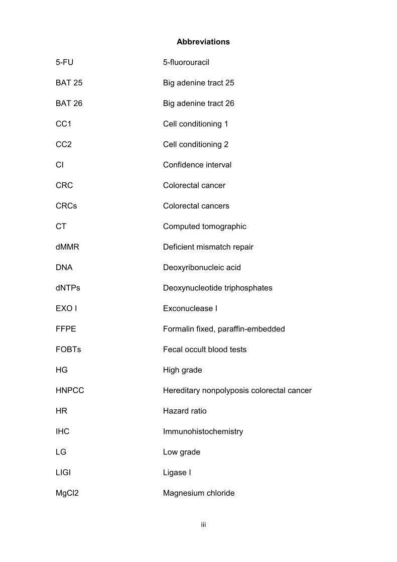
Figure 1: Immunohistochemical staining results of PMS2. (a) PMS2 negative by
IHC in malignant cells, shown at 100x. (b) Positive in normal cells in the same
case, shown at 200x. (Source: from the current study).

14
(a) (b)
(c) (d)
Figure 2: Immunohistochemical staining results of MSH2, MSH6, MLH1 and
PMS2 in cancer cells (shown at 200x). (a) MSH2 positive, (b) MSH6 positive, (c)
MLH1 positive and (d) PMS2 positive. (Source: from the current study).

15
CAHPTER TWO
OBJECTIVES
16
2. Objectives
The aims of this study were to assess the frequency of DNA MMR proteins
abnormalities among Sudanese CRC patients mainly by detection of four MMR
proteins expression (MLH1, MSH2, MSH6, and PMS2) by immunohistochemical
method, and to determine the type of abnormal MMR proteins expression among
Sudanese CRC patients.

17
CHAPTER THREE
MATERIALS AND METHODS

18
3. Materials and methods
3.1. Study samples:
In this study, CRC cases were retrieved from the records of two Histopathology
laboratories in Khartoum, Sudan. Paraffin sections were cut from paraffin wax
embedded tissue blocks. The total number of included cases was 42.
3.2. Laboratory procedures:
3.2.1. Cutting:
Special slides for IHC (SuperFrost® plus, DIAPATH), as well as ordinary frosted
end slides were used. Using a rotary microtome, 3 & 10 μm sections were cut.
3.2.2. Immunohistochemical analysis:
The sections were examined for MMR protein expression of MLH1, MSH2, MSH6
and PMS2 using anti-MLH1, MSH2, MSH6 (mouse monoclonal antibodies-CELL
MARQUE, USA) and anti-PMS2 (rabbit monoclonal antibody-CELL MARQUE,
USA). IHC was performed using a fully automated slide preparation system
(BenchMark XT, Ventana, USA), and the staining of 2 sections was repeated
again using another fully automated slide preparation system (BenchMark ULTRA,
Ventana, USA).
Positive staining defined as the presence of nuclear staining in any percentage of
malignant cells, while nuclear staining in adjacent normal cells serve as internal
positive control. Negative staining defined as the complete absence of nuclear
staining in malignant cells while normal cells show positive nuclear staining. MMR
protein positive cases also indicated that the cases are proficient MMR (pMMR),
and MMR protein negative cases indicated that the cases are dMMR (Vilar and
Gruber, 2010).
Immunohistochemical staining in some sections was inadequate to provide a good
evaluation. In such instances, immunohistochemical analysis was repeated in
another section, when available, from the same case. The repetition was
performed using a prolonged time of antigen retrieval, and 2 inadequately stained
sections were repeated for MSH2 and MSH6 using the two fully automated slide
preparation systems. Sections were treated in an oven, at 60-650 C - up to 30
19
minutes approximately, before repeating the Immunohistochemical analysis with
the antigen retrieval modification several. In BenchMark XT, the time of treatment
in cell conditioning 1 (CC1) for antigen retrieval was increased to 90 minutes for
MSH2, MLH1, and MSH6. For PMS2, the time in cell conditioning 2 (CC2) for
antigen retrieval was increased to 84 minutes. In BenchMark ULTRA, treatment in
CC1 was done for 92 minutes for both MSH2 and MSH6. After repetition, a
number of sections were evaluable. However, the immunohistochemical staining
remained inadequate to allow a good evaluation in other sections. Therefore, MSI
analysis was performed for these cases, which remained inadequate, as well as
for those cases that showed negative staining results for any MMR protein.
3.2.3. Extraction of DNA:
The areas with tumor cells were labelled. Then, DNA was extracted from formalin-
fixed, paraffin-embedded tissues from unstained sections, hematoxylin & eosin
stained sections or immunohistochemical-stained sections of different thicknesses
and number, vary from 3-10 μm of 1-3 sections. The extraction was done using the
kit “QIAamp® DNA FFPE Tissue” (Qiagen®) according to the manufacturer
instructions with some minor modifications.
3.2.4. MSI analysis:
BAT26 and BAT25 microsatellite markers were used mainly. Polymerase chain
reaction (PCR) was done using the extracted DNA in a final volume of 6 μl, 1X
Buffer, dNTPs (250 μM each), primers (0.1 μM each), MgCl2 (3.75 mM) and 1 unit
of Taq polymerase (Roche). For PCR reaction, the first step was denaturation at
95 ° C for 5 minutes before the addition of the polymerase, then 35 cycles (30
seconds at 95 ° C, 30 seconds at the annealing temperature and 30 seconds at 72
° C), and 10-minute final extension at 72 ° C.
The PCR products were marked with fluorochrome. In which, the primer of BAT 25
was labelled with fluorochrome NED, while the primer of BAT 26 was labelled with
FAM. Each sample was added to formamide and standard (ROX 500), and then
transferred to capillary automated sequencer ABI PRISM 310 Genetic Analyzer
(Applied Biosystems).
The analysis was conducted using the program GenMapper V4.0 for data
processing. Electropherograms can recognize MSI CRC by the presence of new
shorter peaks due to the shortening of the adenine repeats in cancer cells (Vilar

20
and Gruber 2010). In some instances, the electropherograms was not definite to
provide a clear MSI results for one or more of the used MSI markers. Such results
were considered non-evaluable.

21
CHAPTER FOUR
RESULTS

22
4. Results
In this study, we aimed to assess the MMR proteins abnormalities in Sudanese
CRC patients. MMR proteins were assessed by IHC in 42 Sudanese CRC
patients.
Table 1 & Figure 3 show the distribution of the study population by gender, 25
(59.5%) were males, and 17 (40.4%) were females. Male to female ratio was
1.47:1. The ages of the study population were between 20-85 years (the age of 4
patients was not provided), and the mean age was 56.1 year. The majority of the
study population were among age groups 50-59 and 60-69 years representing 7
(18.4%) for each, followed by 70-79 and 80-89 constituting 6 (15.7%) for each,
followed by age groups 20-29, 30-39, and 40-49 representing 4 (10.5%) for each
as shown in Table 2 & Figure 4. Furthermore, the ages of 12 (31.5%) of the CRC
patients were less than 50 years.
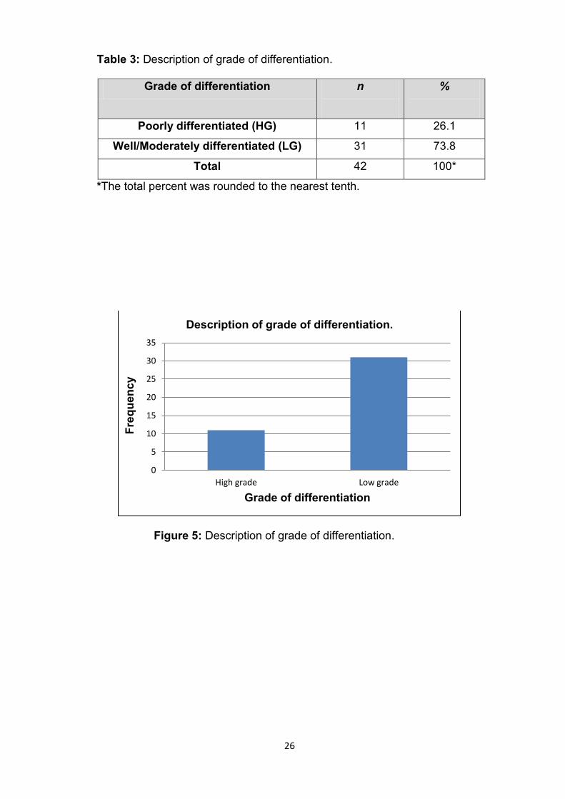
Regarding the grade of differentiation (assessed in sections stained by either
hematoxylin & eosin or IHC and obtained from one block of each case), 11
(26.1%) of the cases were poorly differentiated and classified as high grade (HG),
comparing to 31 (73.8%) well/moderately differentiated classified as low grade
(LG) as shown in Table 3 & Figure 5.
Table 4 & Figures (for selected cases) 6, 8-16 show the MMR proteins
expression by IHC, and MSI status. Of the 42 included cases, 34 (80.95%) were
MMR protein positive for MLH1, PMS2, MSH2 and MSH6, 4 (9.5%) MMR protein
negative (2 (4.76%) MLH1&PMS2 negative and 2 (4.76%) MSH2&MSH6
negative), 3 (7.14%) MSH2 inadequate and positive for the rest, and 1 (2.38%)
MSH6 inadequate and positive for the others.
Regarding MSI results, the three cases that were MSH2 Inadequate and positive
for the rest by IHC showed stable results with both BAT 25 & 26. The two cases
that showed MSH2&MSH6 negative results were unstable with both BAT 25 & 26.
Of the two cases that were MLH1&PMS2 negative, one of them showed non-
evaluable results with BAT 25 & 26, while the other case was unstable with BAT
26 and not evaluable with BAT 25. The case that was MSH6 Inadequate and
positive for the others showed stable results with both BAT 25 & 26.
Immunohistohhemical staining results in some sections were inadequate for
allowing a good evaluation. In such instances, the immunohistochemical analysis
was repeated in another section using a prolonged time of antigen retrieval. For

23
one case, there was no additional section to be re-stained. After repetition, a
number of sections were evaluable as the example shown in Figure 17. However,
the immunohistochemical staining results remained inadequate to provide a good
evaluation in other sections.
Concerning the MMR status based on the immunohistochemical results, MMR
protein negative cases (which considered dMMR) were detected among 4 (9.5%)
of the included cases as shown in Table 5 & Figure 7.
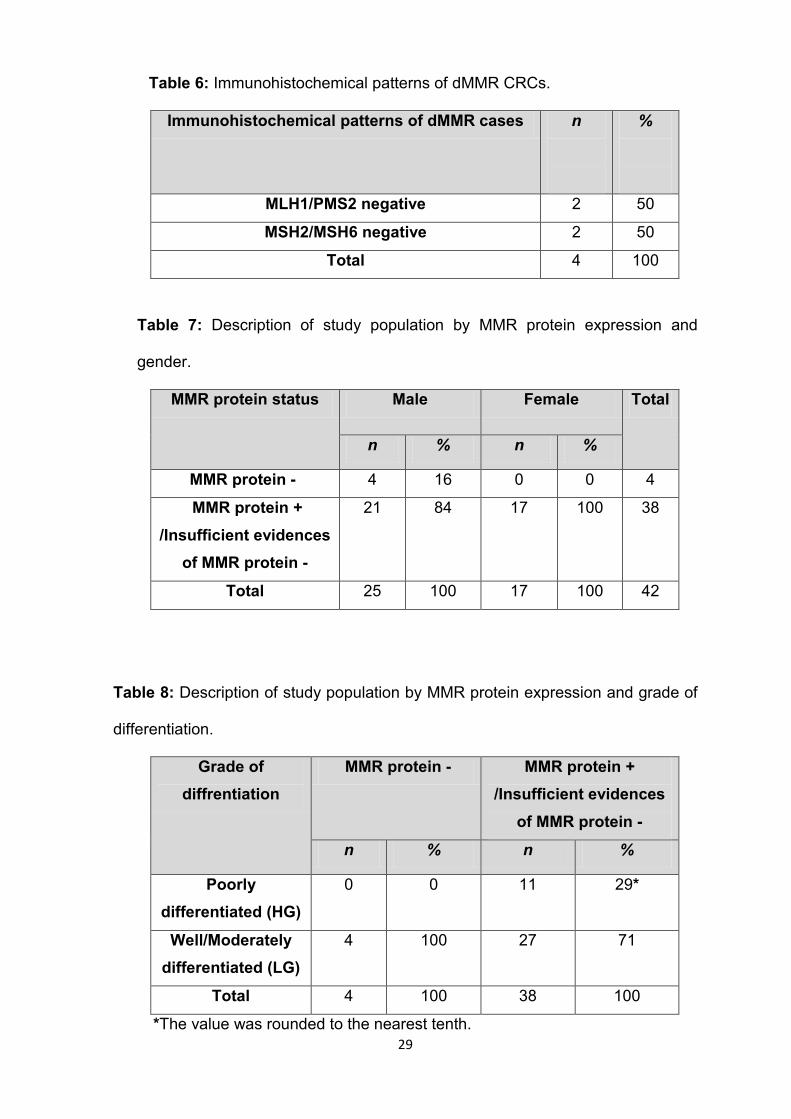
Immunohistochemical patterns of these four dMMR cases were 2 (50%)
MLH1/PMS2 negative, and 2 (50%) MSH2/MSH6 negative, as shown in Table 6.
All MMR protein negative cases were among males. In which 4 (16%) of the males
were MMR protein negative while no MMR protein negative cases were detected
among females as shown in Table 7.
All MMR protein negative cases were LG, which refer to well/moderately
differentiated CRC, as shown in Table 8.

24
Table 1: Description of study population by gender.
Gender n %
Male 25 59.5
Female 17 40.4
Total 42 100*
*The total percentage was rounded to the nearest tenth.
Figure 3: Description of study population by gender.
0
5
10
15
20
25
30
Female Male
Fre
qu
en
cy
Gender
Description of study population by gender

25
Table 2: Description of study population by age.
* The age of 4 patients was not provided.
** The total percentage was rounded to the nearest tenth.
Figure 4: Description of study population by age.
0
1
2
3
4
5
6
7
8
20-29 30-39 40-49 50-59 60-69 70-79 80-89
Fre
qu
en
cy
Age group
Description of study population by age.
Age (Years) Male
Female Total
%
20-29 2 2 4 10.5
30-39 2 2 4 10.5
40-49 2 2 4 10.5
50-59 5 2 7 18.4
60-69 5 2 7 18.4
70-79 4 2 6 15.7
80-89 3 3 6 15.7
Total 23 15 38* 100**
26
Table 3: Description of grade of differentiation.
Grade of differentiation n
%
Poorly differentiated (HG) 11 26.1
Well/Moderately differentiated (LG) 31 73.8
Total 42 100*
*The total percent was rounded to the nearest tenth.
Figure 5: Description of grade of differentiation.
0
5
10
15
20
25
30
35
High grade Low grade
Fre
qu
en
cy
Grade of differentiation
Description of grade of differentiation.

27
Table 4: MMR proteins expression by IHC, and MSI status.
Expression of MMR
protein
n
%
MSI analysis
BAT 25 BAT 26
MLH1&PMS2
negative (MMR
protein - cases)
2 4.76 1- Not evaluable
2- Not evaluable
Unstable
Not evaluable
MSH2&MSH6
negative (MMR
protein - cases)
2 4.76 Unstable Unstable
All positive (MMR
protein + cases)
34
80.95 - -
MSH2 Inadequate /
MSH6,MLH1, PMS2
positive
3
7.14 Stable Stable
MSH6 Inadequate/
MSH2, MLH1, PMS2
positive
1
2.38 Stable Stable
Total 42 100*
-
*The total percentage was rounded to the nearest tenth.
Figure 6: MMR proteins expression by IHC.
0
5
10
15
20
25
30
35
40
MSH2&MSH6 negative
MSH6 Inadequate&
MSH2 positive
MLH1& PMS2 negative
MSH2 inadequate & MSH6 positive
All positive
Fre
qu
en
cy
IHC results
MMRP expression by IHC.

28
Table 5: Description of study population by MMR status.
*The total percentage was rounded to the nearest tenth.
Figure 7: Description of study population by MMR status.
0
5
10
15
20
25
30
35
40
dMMR pMMR/Insufficient evidences for dMMR
Fre
qu
en
cy
MMR status
Description of study population by MMR status.
MMR status n
%
pMMR / Insufficient evidences of dMMR 38 90.4
dMMR 4 9.5
Total 42 100*
29
Table 6: Immunohistochemical patterns of dMMR CRCs.
Immunohistochemical patterns of dMMR cases n
%
MLH1/PMS2 negative 2 50
MSH2/MSH6 negative 2 50
Total 4 100
Table 7: Description of study population by MMR protein expression and
gender.
MMR protein status Male Female Total
n % n %
MMR protein - 4 16 0 0 4
MMR protein +
/Insufficient evidences
of MMR protein -
21 84 17 100 38
Total 25 100 17 100 42
Table 8: Description of study population by MMR protein expression and grade of
differentiation.
Grade of
diffrentiation
MMR protein - MMR protein +
/Insufficient evidences
of MMR protein -
n % n %
Poorly
differentiated (HG)
0 0 11 29*
Well/Moderately
differentiated (LG)
4 100 27 71
Total 4 100 38 100
*The value was rounded to the nearest tenth.
30
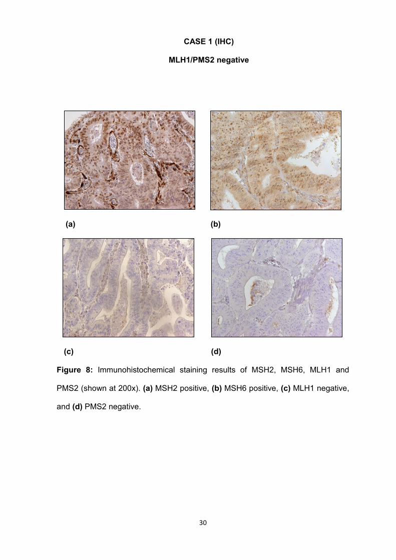
CASE 1 (IHC)
MLH1/PMS2 negative
(a) (b)
(c) (d)
Figure 8: Immunohistochemical staining results of MSH2, MSH6, MLH1 and
PMS2 (shown at 200x). (a) MSH2 positive, (b) MSH6 positive, (c) MLH1 negative,
and (d) PMS2 negative.

31
CASE 2 (IHC)
MLH1/PMS2 negative
(a) (b)
(c) (d)
Figure 9: Immunohistochemical staining results of MSH2, MSH6, MLH1 and
PMS2 (shown at 200x). (a) MSH2 positive, (b) MSH6 positive, (c) MLH1 negative,
and (d) PMS2 negative.
32
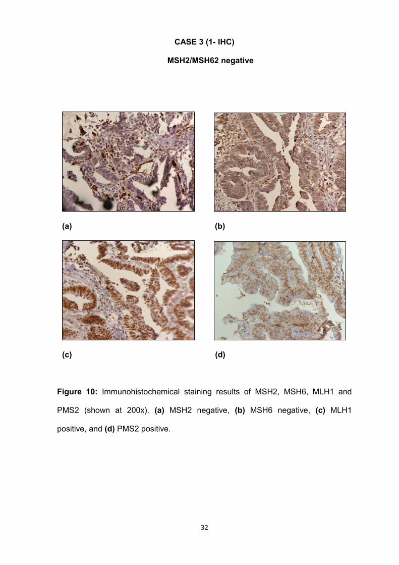
CASE 3 (1- IHC)
MSH2/MSH62 negative
(a) (b)
(c) (d)
Figure 10: Immunohistochemical staining results of MSH2, MSH6, MLH1 and
PMS2 (shown at 200x). (a) MSH2 negative, (b) MSH6 negative, (c) MLH1
positive, and (d) PMS2 positive.
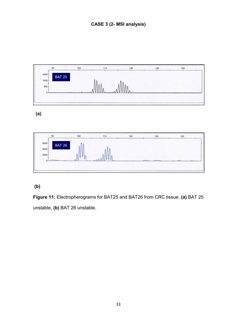
33
CASE 3 (2- MSI analysis)
(a)
(b)
Figure 11: Electropherograms for BAT25 and BAT26 from CRC tissue. (a) BAT 25
unstable, (b) BAT 26 unstable.
BAT 25
BAT 26

34
CASE 4 (1- IHC)
MSH2/MSH62 negative
(a) (b)
(c) (d)
Figure12: Immunohistochemical staining results of MSH2, MSH6, MLH1 and
PMS2 (shown at 200x). (a) MSH2 negative, (b) MSH6 negative, (c) MLH1
positive, and (d) PMS2 positive.

35
CASE 4 (2- MSI analysis)
(a)
(b)
Figure 13: Electropherograms for BAT25 and BAT26 from CRC tissue. (a) BAT 25
unstable, (b) BAT 26 unstable.
BAT 25
BAT 26
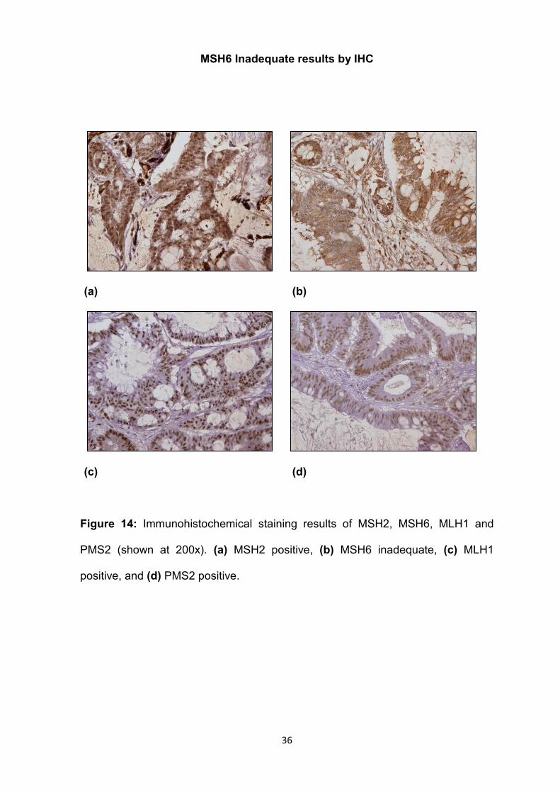
36
MSH6 Inadequate results by IHC
(a) (b)
(c) (d)
Figure 14: Immunohistochemical staining results of MSH2, MSH6, MLH1 and
PMS2 (shown at 200x). (a) MSH2 positive, (b) MSH6 inadequate, (c) MLH1
positive, and (d) PMS2 positive.

37
MSH2 inadequate results by IHC, (IHC & MSI analysis)
1- IHC
(a) (b)
(c) (d)
Figure 15: Immunohistochemical staining results of MSH2, MSH6, MLH1 and
PMS2 (shown at 200x). (a) MSH2 inadequate, (b) MSH6 positive, (c) MLH1
positive, and (d) PMS2 positive.
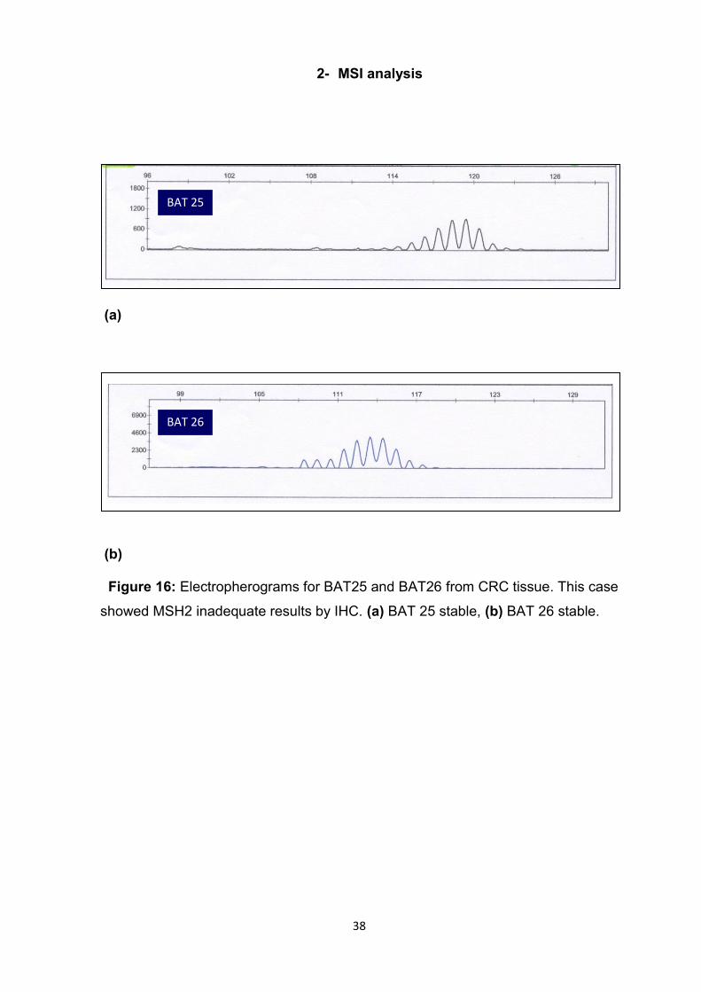
38
2- MSI analysis
(a)
(b)
Figure 16: Electropherograms for BAT25 and BAT26 from CRC tissue. This case
showed MSH2 inadequate results by IHC. (a) BAT 25 stable, (b) BAT 26 stable.
BAT 25
BAT 26
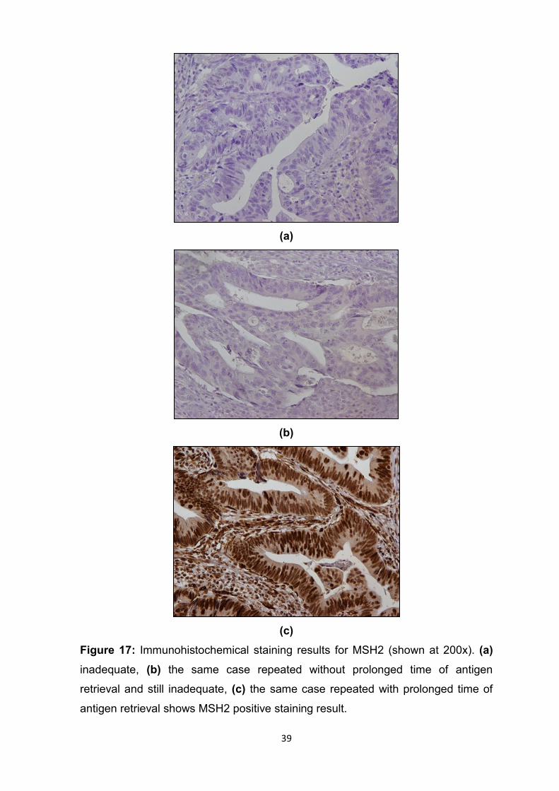
39
(a)
(b)
(c)
Figure 17: Immunohistochemical staining results for MSH2 (shown at 200x). (a)
inadequate, (b) the same case repeated without prolonged time of antigen
retrieval and still inadequate, (c) the same case repeated with prolonged time of
antigen retrieval shows MSH2 positive staining result.

40
CHAPTER FIVE
DISCUSSION

41
5. Discussion
In this study, we aimed to assess the DNA MMR proteins abnormalities among
Sudanese CRC patients. The total number of included cases was 42.
Sections were cut and assessed by IHC for the expression of 4 MMR protiens
(MLH1, MSH2, MSH6, and PMS2). Furthermore, MSI analysis using mainly BAT
25 & 26 was performed for cases that showed negative or inadequate staining
results by IHC.
In the current study, 4 (9.5%) MMR protein negative CRC cases were detected by
IHC. Different studies showed higher percentages of MMR proteins abnormalities
by IHC compared to our study. Khoo et al. (2013) found that the percentage of
abnormal IHC staining for MLH1, MSH2 and MSH6 expression was (14.4%)
among 298 Malaysian CRC cases. Furthermore, our finding in the current study is
less than what was detected by Lanza et al. (2006) in Ferrara, Italy. In their study,
718 colorectal adenocarcinoma patients were studied. Of whom, 114 (15.9%)
were found with abnormal expression of MMR protein. Of these 114 cases with
abnormal expression, 18 were MSH2 negative and 96 were MLH1 negative.
Moreover, Jensen et al. (2008) in Denmark, found that 39 (14.9%) out of 262 CRC
cases were MMR deficient by IHC or MSI analysis.
However, other studies found less percentages compared to our finding. A study
from Spain by Jover et al. (2004) reported that the percentage of MMR-defective
CRC cases was 7.6%. In their study, 172 CRC cases were studied. MSI analysis
was performed by BAT 26, while IHC was performed using antibodies against
MLH1 and MSH2 proteins. MSI was detected in 13 (7.6%) cases and all exhibited
loss of expression of one of the two examined proteins (11 MLH1 negative and 2
MSH2 negative). De Jesus-Monge et al. (2010) examined the MMR protein
expression among Hispanics from Puerto Rico. In their study, MLH1 and MSH2
protein were examined by IHC in 164 CRC cases. Of these cases, 7 (4.3%) were
negative. Table 9 shows a summary of some other studies from different countries
including the percentage of MMR protein negative cases assessed by IHC. Most
of these studies reported higher percentages of MMR protein negative CRC cases
compared to our study. However, the result obtained by Kheirelseid et al. (2013)
was almost near to our finding, while the finding of Coggins et al. (2005) was less
than our finding.

42
Table 9. A summary of some studies regarding MMR protein expression examined
by IHC.
Authors and
year
Country Number
of
included
cases
MMR protein
negative
CRCs
Examined MMR
proteins
n %
Molaei et al.
(2010)
Iran 343 48 14 MLH1, MSH2, PMS2,
and MSH6
Erdamar et
al. (2007)
Turkey 74 34 45.9 MLH1 and MSH2
Coggins et al.
(2005)
England 732 57 7.78 MLH1 and MSH2
Wright and
Stewart
(2003)
New
Zealand
458 89 19.4 MLH1 and MSH2
Jin et al.
(2008)
China 146 32 21.9 MLH1, MSH2 and
MSH6
Ashktorab et
al. (2008)
Oman 49 8 16.3 MLH1 and MSH2
Kheirelseid et
al. (2013)
Ireland 33 3 9.09 MLH1, MSH2, and
PMS2
Lindor et al.
(2002)
USA 1,144 326 28.4 MLH1 and MSH2

43
Furthermore, another study from Ghana, which is one of the West African
countries, showed a high percentage (41%) of MSI-H CRC cases (Raskin et al.,
2013). Kria Ben Mahmoud et al. (2012) examined the MSI status among 150
CRCs from Tunisians patients. They found that 15 % of these CRCs were MSI-H.
Regarding the grade of differentiation of MMR protein negative CRC cases in the
current study, all these cases were LG, which refers to well/moderately
differentiated CRC. However, this finding requires additional evaluation using a
larger sample size to find out the accurate association between MMR protein
negative cases and the grade of differentiation in Sudanese patients.
Nevertheless, this observation opposes other published data. Lanza et al. (2006)
found that MMR protein negative CRCs are characterized by poor differentiation.
Furthermore, Khoo et al. (2013) reported that CRC cases with MMR defect were
frequently found poorly differentiated compared to those cases with no defect in
MMR.
In our study, the results of IHC in several sections were inadequate to allow a
good assessment. In such instances, the immunohistochemical analysis was
repeated using successive antigen retrieval by increasing the time of this step.
After repetition, a number of sections were evaluable. However, the
immunohistochemical staining remained inadequate to provide a good evaluation
in other sections. Raskin et al. (2013) found some difficulties in their study
regarding the analysis of MMR proteins by IHC, which resulted in the presence of
a limited number of CRC cases with sufficient immunohistochemical staining. They
attributed these difficulties in achieving a good immunohistochemical staining to
the prolonged time of fixation, which makes the tissue excessively dehydrated.

44
CHAPTER SIX
CONCLUSIONS

45
6. Conclusions
The percentage of MMR protein negative cases among Sudanese CRC patients in
this study is 9.5%, which appears to be relatively low compared to what has been
generally reported in certain studies performed in different countries. However,
detection of MMR proteins by IHC is recommended to be introduced in the referral
Histopathology laboratories in Sudan due to its importance in the management of
CRC patients.
In this study, MLH1&PMS2 and MSH2&MSH6 abnormal expression detected by
IHC seem to be the most common form of MMR proteins abnormalities in
Sudanese CRC patients. Moreover, MLH1 and MSH2 seem to be the most
inactivated MMR genes in Sudanese CRC patients, concerning the results of IHC.

46
CHAPTER SEVEN
REFERENCES
47
7. References
- Ashktorab, H., Brim, H., Al-Riyami, M., Date, A., Al-Mawaly, K., Kashoub,
M., Al-Mjeni, R., Smoot, D.T., Al-Moundhri, M., Al-Hashemi, S., Ganguly,
S.S., Raeburn, S., 2008. Sporadic colon cancer: mismatch repair
immunohistochemistry and microsatellite instability in Omani subjects. Dig.
Dis. Sci. 53, 2723–2731.
- Ashktorab, H., Smoot, D.T., Farzanmehr, H., Fidelia-Lambert, M., Momen,
B., Hylind, L., Iacosozio-Dononue, C., Carethers, J.M., Goel, A., Boland,
C.R., Giardiello, F.M., 2005. Clinicopathological features and microsatellite
instability (MSI) in colorectal cancers from African Americans. Int. J. Cancer
116, 914–919.
- Bacher, J.W., Flanagan, L.A., Smalley, R.L., Nassif, N.A., Burgart, L.J.,
Halberg, R.B., Megid, W.M.A., Thibodeau, S.N., 2004. Development of a
fluorescent multiplex assay for detection of MSI-High tumors. Dis. Markers
20, 237–250.
- Ballinger, A.B., Anggiansah, C., 2007. Colorectal cancer. BMJ 335, 715–
718.
- Barouni, M., Larizadeh, M.H., Sabermahani, A., Ghaderi, H., 2012.
Markov’s modeling for screening strategies for colorectal cancer. Asian
Pac. J. Cancer Prev. 13, 5125–5129.
- Benatti, P., Gafà, R., Barana, D., Marino, M., Scarselli, A., Pedroni, M.,
Maestri, I., Guerzoni, L., Roncucci, L., Menigatti, M., Roncari, B., Maffei, S.,
Rossi, G., Ponti, G., Santini, A., Losi, L., Di Gregorio, C., Oliani, C., Ponz de
Leon, M., Lanza, G., 2005. Microsatellite instability and colorectal cancer
prognosis. Clin. Cancer Res. 11, 8332–8340.
- Boardman, L.A., Lanier, A.P., French, A.J., Schowalter, K.V., Burgart, L.J.,
Koller, K.R., McDonnell, S.K., Schaid, D.J., Thibodeau, S.N., 2007.
Frequency of defective DNA mismatch repair in colorectal cancer among
the Alaska Native people. Cancer Epidemiol. Biomarkers Prev. 16, 2344–
2350.
- Boland, C.R., Goel, A., 2010. Microsatellite instability in colorectal cancer.
Gastroenterology 138, 2073–2087.e3.
- Boland, C.R., Thibodeau, S.N., Hamilton, S.R., Sidransky, D., Eshleman,
J.R., Burt, R.W., Meltzer, S.J., Rodriguez-Bigas, M.A., Fodde, R., Ranzani,
G.N., Srivastava, S., 1998. A National Cancer Institute Workshop on

48
Microsatellite Instability for cancer detection and familial predisposition:
development of international criteria for the determination of microsatellite
instability in colorectal cancer. Cancer Res. 58, 5248–5257.
- Brennetot, C., Buhard, O., Jourdan, F., Flejou, J.-F., Duval, A., Hamelin, R.,
2005. Mononucleotide repeats BAT-26 and BAT-25 accurately detect MSI-
H tumors and predict tumor content: implications for population screening.
Int. J. Cancer 113, 446–450.
- Caldés, T., Godino, J., Sanchez, A., Corbacho, C., De la Hoya, M., Lopez
Asenjo, J., Saez, C., Sanz, J., Benito, M., Ramon Y Cajal, S., Diaz-Rubio,
E., 2004. Immunohistochemistry and microsatellite instability testing for
selecting MLH1, MSH2 and MSH6 mutation carriers in hereditary non-
polyposis colorectal cancer. Oncol. Rep. 12, 621–629.
- Cappellani, A., Zanghì, A., Di Vita, M., Cavallaro, A., Piccolo, G., Veroux,
P., Lo Menzo, E., Cavallaro, V., de Paoli, P., Veroux, M., Berretta, M., 2013.
Strong correlation between diet and development of colorectal cancer.
Front Biosci (Landmark Ed) 18, 190–198.
- Chai, S.M., Zeps, N., Shearwood, A.-M., Grieu, F., Charles, A., Harvey, J.,
Goldblatt, J., Joseph, D., Iacopetta, B., 2004. Screening for defective DNA
mismatch repair in stage II and III colorectal cancer patients. Clin.
Gastroenterol. Hepatol. 2, 1017–1025.
- Cho, E., Smith-Warner, S.A., Ritz, J., van den Brandt, P.A., Colditz, G.A.,
Folsom, A.R., Freudenheim, J.L., Giovannucci, E., Goldbohm, R.A.,
Graham, S., Holmberg, L., Kim, D.-H., Malila, N., Miller, A.B., Pietinen, P.,
Rohan, T.E., Sellers, T.A., Speizer, F.E., Willett, W.C., Wolk, A., Hunter,
D.J., 2004. Alcohol intake and colorectal cancer: a pooled analysis of 8
cohort studies. Ann. Intern. Med. 140, 603–613.
- Coggins, R.P., Cawkwell, L., Bell, S.M., Crockford, G.P., Quirke, P., Finan,
P.J., Bishop, D.T., 2005. Association between family history and mismatch
repair in colorectal cancer. Gut 54, 636–642.
- Conde-Pérezprina, J.C., León-Galván, M.Á., Konigsberg, M., 2012. DNA
mismatch repair system: repercussions in cellular homeostasis and
relationship with aging. Oxid Med Cell Longev 2012, 728430.
- Cook, N.R., Lee, I.-M., Zhang, S.M., Moorthy, M.V., Buring, J.E., 2013.
Alternate-day, low-dose aspirin and cancer risk: long-term observational
follow-up of a randomized trial. Ann. Intern. Med. 159, 77–85.
49
- Cunningham, J.M., Kim, C.Y., Christensen, E.R., Tester, D.J., Parc, Y.,
Burgart, L.J., Halling, K.C., McDonnell, S.K., Schaid, D.J., Walsh Vockley,
C., Kubly, V., Nelson, H., Michels, V.V., Thibodeau, S.N., 2001. The
frequency of hereditary defective mismatch repair in a prospective series of
unselected colorectal carcinomas. Am. J. Hum. Genet. 69, 780–790.
- De Jesus-Monge, W.E., Gonzalez-Keelan, C., Zhao, R., Hamilton, S.R.,
Rodriguez-Bigas, M., Cruz-Correa, M., 2010. Mismatch repair protein
expression and colorectal cancer in Hispanics from Puerto Rico. Fam
Cancer 9, 155–166.
- de la Chapelle, A., 1999. Testing tumors for microsatellite instability. Eur. J.
Hum. Genet. 7, 407-408.
- Duraiyan, J., Govindarajan, R., Kaliyappan, K., Palanisamy, M., 2012.
Applications of immunohistochemistry. J Pharm Bioallied Sci 4, S307–S309
- El Hassan, A., El Hassan, L., Mudawi, H., Gasim, B., Own, A., Elamin, E.,
Ibn Ouf, M., El Mekki Abdullah, M., Fedail, S., 2008. Malignant gastric
tumors in Sudan: a report from a single pathology center. Hematol Oncol
Stem Cell Ther 1, 130–132.
- Erdamar, S., Ucaryilmaz, E., Demir, G., Karahasanoglu, T., Dogusoy, G.,
Dirican, A., Goksel, S., 2007. Importance of MutL homologue MLH1 and
MutS homologue MSH2 expression in Turkish patients with sporadic
colorectal cancer. World J. Gastroenterol. 13, 4437–4444.
- Ferlay, J., Parkin, D.M., Steliarova-Foucher, E., 2010b. Estimates of cancer
incidence and mortality in Europe in 2008. Eur. J. Cancer 46, 765–781.
- Ferlay, J., Shin, H.-R., Bray, F., Forman, D., Mathers, C., Parkin, D.M.,
2010a. Estimates of worldwide burden of cancer in 2008: GLOBOCAN
2008. Int. J. Cancer 127, 2893–2917.
- Flitcroft, K.L., Irwig, L.M., Carter, S.M., Salkeld, G.P., Gillespie, J.A., 2012.
Colorectal cancer screening: why immunochemical fecal occult blood tests
may be the best option. BMC Gastroenterol 12, 183.
- Giovannucci, E., 2003. Diet, body weight, and colorectal cancer: a summary
of the epidemiologic evidence. J Womens Health (Larchmt) 12, 173–182.
- Giovannucci, E., Egan, K.M., Hunter, D.J., Stampfer, M.J., Colditz, G.A.,
Willett, W.C., Speizer, F.E., 1995. Aspirin and the risk of colorectal cancer
in women. N. Engl. J. Med. 333, 609–614.
- Giovannucci, E., Rimm, E.B., Stampfer, M.J., Colditz, G.A., Ascherio, A.,
50
Willett, W.C., 1994. Aspirin use and the risk for colorectal cancer and
adenoma in male health professionals. Ann. Intern. Med. 121, 241–246.
- Hall, G., Clarkson, A., Shi, A., Langford, E., Leung, H., Eckstein, R.P., Gill,
A.J., 2010. Immunohistochemistry for PMS2 and MSH6 alone can replace a
four antibody panel for mismatch repair deficiency screening in colorectal
adenocarcinoma. Pathology 42, 409–413.
- Hamilton, S.R., 2013. BRAF mutation and microsatellite instability status in
colonic and rectal carcinoma: context really does matter. J. Natl. Cancer
Inst. 105, 1075–1077.
- Hardcastle, J.D., Chamberlain, J.O., Robinson, M.H., Moss, S.M., Amar,
S.S., Balfour, T.W., James, P.D., Mangham, C.M., 1996. Randomised
controlled trial of faecal-occult-blood screening for colorectal cancer. Lancet
348, 1472–1477.
- Hendriks, Y., Franken, P., Dierssen, J.W., De Leeuw, W., Wijnen, J., Dreef,
E., Tops, C., Breuning, M., Bröcker-Vriends, A., Vasen, H., Fodde, R.,
Morreau, H., 2003. Conventional and tissue microarray
immunohistochemical expression analysis of mismatch repair in hereditary
colorectal tumors. Am. J. Pathol. 162, 469–477.
- Hsieh, P., Yamane, K., 2008. DNA mismatch repair: molecular mechanism,
cancer, and ageing. Mech. Ageing Dev. 129, 391–407.
- Jensen, L.H., Lindebjerg, J., Byriel, L., Kolvraa, S., Crüger, D.G., 2008.
Strategy in clinical practice for classification of unselected colorectal
tumours based on mismatch repair deficiency. Colorectal Dis 10, 490–497.
- Jensen, S.A., Vainer, B., Kruhøffer, M., Sørensen, J.B., 2009. Microsatellite
instability in colorectal cancer and association with thymidylate synthase
and dihydropyrimidine dehydrogenase expression. BMC Cancer 9, 25.
- Jin, H.-Y., Liu, X., Li, V.K.M., Ding, Y., Yang, B., Geng, J., Lai, R., Ding, S.,
Ni, M., Zhao, R., 2008. Detection of mismatch repair gene germline
mutation carrier among Chinese population with colorectal cancer. BMC
Cancer 8, 44.
- Jover, R., Payá, A., Alenda, C., Poveda, M.J., Peiró, G., Aranda, F.I.,
Pérez-Mateo, M., 2004. Defective mismatch-repair colorectal cancer:
clinicopathologic characteristics and usefulness of immunohistochemical
analysis for diagnosis. Am. J. Clin. Pathol. 122, 389–394.
- Kewenter, J., Brevinge, H., Engarås, B., Haglind, E., Ahrén, C., 1994.
51
Results of screening, rescreening, and follow-up in a prospective
randomized study for detection of colorectal cancer by fecal occult blood
testing. Results for 68,308 subjects. Scand. J. Gastroenterol. 29, 468–473.
- Kheirelseid, E.A.H., Miller, N., Chang, K.H., Curran, C., Hennessey, E.,
Sheehan, M., Kerin, M.J., 2013. Mismatch repair protein expression in
colorectal cancer. J Gastrointest Oncol 4, 397–408.
- Khoo, J.J., Gunn, A., Peh, S.C., 2013. Pattern of hMLH1, hMSH2 and
hMSH6 expression and clinical characteristics in a sample of Malaysian
colorectal carcinoma cases. Malays J Pathol 35, 45–57.
- Kloor, M., Staffa, L., Ahadova, A., von Knebel Doeberitz, M., 2013. Clinical
significance of microsatellite instability in colorectal cancer. Langenbecks
Arch Surg. [Epub ahead of print].
- Kria Ben Mahmoud, L., Arfaoui, A., Khiari, M., Chaar, I., Lounis, A.,
Sammoud, S., Ben Hmida, A.M., Gharbi, L., Mzabi, S.R., Bouraoui, S.,
2012. Evaluation of microsatellite instability, MLH1 expression and hMLH1
promoter hypermethylation in colorectal carcinomas among Tunisians
patients. Tunis Med 90, 646–653.
- Kronborg, O., Fenger, C., Olsen, J., Jørgensen, O.D., Søndergaard, O.,
1996. Randomised study of screening for colorectal cancer with faecal-
occult-blood test. Lancet 348, 1467–1471.
- Kunkel, T.A., Erie, D.A., 2005. DNA mismatch repair. Annu. Rev. Biochem.
74, 681–710.
- Kyle, S.M., Isbister, W.H., Yeong, M.L., 1991. Presentation, duration of
symptoms and staging of colorectal carcinoma. Aust N Z J Surg 61, 137–
140.
- Laghi, L., Malesci, A., 2012. Microsatellite instability and therapeutic
consequences in colorectal cancer. Dig Dis 30, 304–309.
- Lanza, G., Gafà, R., Santini, A., Maestri, I., Guerzoni, L., Cavazzini, L.,
2006. Immunohistochemical test for MLH1 and MSH2 expression predicts
clinical outcome in stage II and III colorectal cancer patients. J. Clin. Oncol.
24, 2359–2367.
- Lindor, N.M., Burgart, L.J., Leontovich, O., Goldberg, R.M., Cunningham,
J.M., Sargent, D.J., Walsh-Vockley, C., Petersen, G.M., Walsh, M.D.,
Leggett, B.A., Young, J.P., Barker, M.A., Jass, J.R., Hopper, J., Gallinger,
S., Bapat, B., Redston, M., Thibodeau, S.N., 2002. Immunohistochemistry
52
versus microsatellite instability testing in phenotyping colorectal tumors. J.
Clin. Oncol. 20, 1043–1048.
- MacArthur, C., Smith, A., 1984. Factors associated with speed of diagnosis,
referral, and treatment in colorectal cancer. J Epidemiol Community Health
38, 122–126.
- Mangold, E., Pagenstecher, C., Friedl, W., Fischer, H.-P., Merkelbach-
Bruse, S., Ohlendorf, M., Friedrichs, N., Aretz, S., Buettner, R., Propping,
P., Mathiak, M., 2005. Tumours from MSH2 mutation carriers show loss of
MSH2 expression but many tumours from MLH1 mutation carriers exhibit
weak positive MLH1 staining. J. Pathol. 207, 385–395.
- Marcus, V.A., Madlensky, L., Gryfe, R., Kim, H., So, K., Millar, A., Temple,
L.K., Hsieh, E., Hiruki, T., Narod, S., Bapat, B.V., Gallinger, S., Redston,
M., 1999. Immunohistochemistry for hMLH1 and hMSH2: a practical test for
DNA mismatch repair-deficient tumors. Am. J. Surg. Pathol. 23, 1248–1255.
- Mead, L.J., Jenkins, M.A., Young, J., Royce, S.G., Smith, L., St John,
D.J.B., Macrae, F., Giles, G.G., Hopper, J.L., Southey, M.C., 2007.
Microsatellite instability markers for identifying early-onset colorectal
cancers caused by germ-line mutations in DNA mismatch repair genes.
Clin. Cancer Res. 13, 2865–2869.
- Mihajlovic-Bozic, V., 2004. Risk factors for colorectal cancer. Arch Oncol
12, 45–49.
- Mojtahed, A., Schrijver, I., Ford, J.M., Longacre, T.A., Pai, R.K., 2011. A
two-antibody mismatch repair protein immunohistochemistry screening
approach for colorectal carcinomas, skin sebaceous tumors, and
gynecologic tract carcinomas. Mod. Pathol. 24, 1004–1014.
- Molaei, M., Mansoori, B.K., Ghiasi, S., Khatami, F., Attarian, H., Zali, M.,
2010. Colorectal cancer in Iran: immunohistochemical profiles of four
mismatch repair proteins. Int J Colorectal Dis 25, 63–69.
- O’Carroll, R.E., Steele, R.J., Libby, G., Brownlee, L., Chambers, J.A., 2013.
Anticipated regret to increase uptake of colorectal cancer screening in
Scotland (ARTICS): study protocol for a randomised controlled trial. BMC
Public Health 13, 849.
- O’Regan, T., Chau, K., Tatton, M., Smith, T., Parry, S., Bissett, I., 2013.
Immunochemistry screening for Lynch syndrome in colorectal
adenocarcinoma using an initial two antibody panel can replace a four

53
antibody panel. N. Z. Med. J. 126, 70–77.
- Oh, J.R., Kim, D.-W., Lee, H.S., Lee, H.E., Lee, S.M., Jang, J.-H., Kang, S.-
B., Ku, J.-L., Jeong, S.-Y., Park, J.-G., 2012. Microsatellite instability testing
in Korean patients with colorectal cancer. Fam. Cancer 11, 459–466.
- Overbeek, L.I.H., Ligtenberg, M.J.L., Willems, R.W., Hermens, R.P.M.G.,
Blokx, W.A.M., Dubois, S.V., van der Linden, H., Meijer, J.W.R., Mlynek-
Kersjes, M.L., Hoogerbrugge, N., Hebeda, K.M., van Krieken, J.H.J.M.,
2008. Interpretation of immunohistochemistry for mismatch repair proteins
is only reliable in a specialized setting. Am. J. Surg. Pathol. 32, 1246–1251.
- Patil, D.T., Bronner, M.P., Portier, B.P., Fraser, C.R., Plesec, T.P., Liu, X.,
2012. A five-marker panel in a multiplex PCR accurately detects
microsatellite instability-high colorectal tumors without control DNA. Diagn.
Mol. Pathol. 21, 127–133.
- Peltomäki, P., 2001. Deficient DNA mismatch repair: a common etiologic
factor for colon cancer. Hum. Mol. Genet. 10, 735–740.
- Pino, M.S., Chung, D.C., 2011. Microsatellite instability in the management
of colorectal cancer. Expert Rev Gastroenterol Hepatol 5, 385–399.
- Popat, S., Hubner, R., Houlston, R.S., 2005. Systematic review of
microsatellite instability and colorectal cancer prognosis. J. Clin. Oncol. 23,
609–618.
- Raskin, L., Dakubo, J.C.B., Palaski, N., Greenson, J.K., Gruber, S.B., 2013.
Distinct molecular features of colorectal cancer in Ghana. Cancer Epidemiol
37, 556–561.
- Rigau, V., Sebbagh, N., Olschwang, S., Paraf, F., Mourra, N., Parc, Y.,
Flejou, J.-F., 2003. Microsatellite instability in colorectal carcinoma. The
comparison of immunohistochemistry and molecular biology suggests a role
for hMSH6 [correction of hMLH6] immunostaining. Arch. Pathol. Lab. Med.
127, 694–700.
- Rizk, S.N., Ryan, J.J., 1994. Clinicopathologic review of 92 cases of colon
cancer. S D J Med 47, 89–93.
- Ruszkiewicz, A., Bennett, G., Moore, J., Manavis, J., Rudzki, B., Shen, L.,
Suthers, G., 2002. Correlation of mismatch repair genes
immunohistochemistry and microsatellite instability status in HNPCC-
associated tumours. Pathology 34, 541–547.
- Saidi, H.S., Karuri, D., Nyaim, E.O., 2008. Correlation of clinical data,
54
anatomical site and disease stage in colorectal cancer. East Afr Med J 85,
259–262.
- Samowitz, W.S., Curtin, K., Ma, K.N., Schaffer, D., Coleman, L.W., Leppert,
M., Slattery, M.L., 2001. Microsatellite instability in sporadic colon cancer is
associated with an improved prognosis at the population level. Cancer
Epidemiol. Biomarkers Prev. 10, 917–923.
- Sanjoaquin, M.A., Appleby, P.N., Thorogood, M., Mann, J.I., Key, T.J.,
2004. Nutrition, lifestyle and colorectal cancer incidence: a prospective
investigation of 10998 vegetarians and non-vegetarians in the United
Kingdom. Br. J. Cancer 90, 118–121.
- Shaukat, A., Mongin, S.J., Geisser, M.S., Lederle, F.A., Bond, J.H., Mandel,
J.S., Church, T.R., 2013. Long-term mortality after screening for colorectal
cancer. N. Engl. J. Med. 369, 1106–1114.
- Shia, J., 2008. Immunohistochemistry versus Microsatellite Instability
Testing For Screening Colorectal Cancer Patients at Risk For Hereditary
Nonpolyposis Colorectal Cancer Syndrome. J Mol Diagn 10, 293–300.
- Shia, J., Klimstra, D.S., Nafa, K., Offit, K., Guillem, J.G., Markowitz, A.J.,
Gerald, W.L., Ellis, N.A., 2005. Value of immunohistochemical detection of
DNA mismatch repair proteins in predicting germline mutation in hereditary
colorectal neoplasms. Am. J. Surg. Pathol. 29, 96–104.
- Shia, J., Tang, L.H., Vakiani, E., Guillem, J.G., Stadler, Z.K., Soslow, R.A.,
Katabi, N., Weiser, M.R., Paty, P.B., Temple, L.K., Nash, G.M., Wong,
W.D., Offit, K., Klimstra, D.S., 2009. Immunohistochemistry as first-line
screening for detecting colorectal cancer patients at risk for hereditary
nonpolyposis colorectal cancer syndrome: a 2-antibody panel may be as
predictive as a 4-antibody panel. Am. J. Surg. Pathol. 33, 1639–1645.
- Shimizu, N., Nagata, C., Shimizu, H., Kametani, M., Takeyama, N.,
Ohnuma, T., Matsushita, S., 2003. Height, weight, and alcohol consumption
in relation to the risk of colorectal cancer in Japan: a prospective study. Br.
J. Cancer 88, 1038–1043.
- Siegel, R., Naishadham, D., Jemal, A., 2013. Cancer statistics, 2013. CA
Cancer J Clin 63, 11–30.
- Silva, R.V.E., Garicochea, B., Cotti, G., Maranho, I.C., Cutait, R., 2005.
Hereditary nonpolyposis colorectal cancer identification and surveillance of
high-risk families. Clinics (Sao Paulo) 60, 251–256.
55
- Sinicrope, F.A., Foster, N.R., Thibodeau, S.N., Marsoni, S., Monges, G.,
Labianca, R., Kim, G.P., Yothers, G., Allegra, C., Moore, M.J., Gallinger, S.,
Sargent, D.J., 2011. DNA mismatch repair status and colon cancer
recurrence and survival in clinical trials of 5-fluorouracil-based adjuvant
therapy. J. Natl. Cancer Inst. 103, 863–875.
- Søreide, K., 2007. Molecular testing for microsatellite instability and DNA
mismatch repair defects in hereditary and sporadic colorectal cancers--
ready for prime time? Tumour Biol. 28, 290–300.
- Stone, J.G., Robertson, D., Houlston, R.S., 2001. Immunohistochemistry for
MSH2 and MHL1: a method for identifying mismatch repair deficient
colorectal cancer. J. Clin. Pathol. 54, 484–487.
- Suraweera, N., Duval, A., Reperant, M., Vaury, C., Furlan, D., Leroy, K.,
Seruca, R., Iacopetta, B., Hamelin, R., 2002. Evaluation of tumor
microsatellite instability using five quasimonomorphic mononucleotide
repeats and pentaplex PCR. Gastroenterology 123, 1804–1811.
- Thun, M.J., Calle, E.E., Namboodiri, M.M., Flanders, W.D., Coates, R.J.,
Byers, T., Boffetta, P., Garfinkel, L., Heath, C.W., Jr, 1992. Risk factors for
fatal colon cancer in a large prospective study. J. Natl. Cancer Inst. 84,
1491–1500.
- Tonus, C., Sellinger, M., Koss, K., Neupert, G., 2012. Faecal pyruvate
kinase isoenzyme type M2 for colorectal cancer screening: a meta-analysis.
World J. Gastroenterol. 18, 4004–4011.
- Tóth, K., Sipos, F., Kalmár, A., Patai, A.V., Wichmann, B., Stoehr, R.,
Golcher, H., Schellerer, V., Tulassay, Z., Molnár, B., 2012. Detection of
methylated SEPT9 in plasma is a reliable screening method for both left-
and right-sided colon cancers. PLoS ONE 7, e46000.
- Umar, A., Boland, C.R., Terdiman, J.P., Syngal, S., de la Chapelle, A.,
Rüschoff, J., Fishel, R., Lindor, N.M., Burgart, L.J., Hamelin, R., Hamilton,
S.R., Hiatt, R.A., Jass, J., Lindblom, A., Lynch, H.T., Peltomaki, P.,
Ramsey, S.D., Rodriguez-Bigas, M.A., Vasen, H.F.A., Hawk, E.T., Barrett,
J.C., Freedman, A.N., Srivastava, S., 2004. Revised Bethesda Guidelines
for hereditary nonpolyposis colorectal cancer (Lynch syndrome) and
microsatellite instability. J. Natl. Cancer Inst. 96, 261–268.
- Vilar, E., Gruber, S.B., 2010. Microsatellite instability in colorectal cancer-
the stable evidence. Nat Rev Clin Oncol 7, 153–162.

56
- Ward, R., Meagher, A., Tomlinson, I., O’Connor, T., Norrie, M., Wu, R.,
Hawkins, N., 2001. Microsatellite instability and the clinicopathological
features of sporadic colorectal cancer. Gut 48, 821–829.
- Watson, N., Grieu, F., Morris, M., Harvey, J., Stewart, C., Schofield, L.,
Goldblatt, J., Iacopetta, B., 2007. Heterogeneous Staining for Mismatch
Repair Proteins during Population-Based Prescreening for Hereditary
Nonpolyposis Colorectal Cancer. J Mol Diagn 9, 472–478.
- Wright, C.L., Stewart, I.D., 2003. Histopathology and mismatch repair status
of 458 consecutive colorectal carcinomas. Am. J. Surg. Pathol. 27, 1393–
1406.
- Wu, A.H., Paganini-Hill, A., Ross, R.K., Henderson, B.E., 1987. Alcohol,
physical activity and other risk factors for colorectal cancer: a prospective
study. Br. J. Cancer 55, 687–694.
- Xiao, H., Yoon, Y.S., Hong, S.-M., Roh, S.A., Cho, D.-H., Yu, C.S., Kim,
J.C., 2013. Poorly differentiated colorectal cancers: correlation of
microsatellite instability with clinicopathologic features and survival. Am. J.
Clin. Pathol. 140, 341–347.
- Yoon, Y.S., Yu, C.S., Kim, T.W., Kim, J.H., Jang, S.J., Cho, D.H., Roh,
S.A., Kim, J.C., 2011. Mismatch repair status in sporadic colorectal cancer:
immunohistochemistry and microsatellite instability analyses. J.
Gastroenterol. Hepatol. 26, 1733–1739.
- Zhang, X., Li, J., 2013. Era of universal testing of microsatellite instability in
colorectal cancer. World J Gastrointest Oncol 5, 12–19.
- Ziadi, S., Ksiaa, F., Gacem, R.B., Labaied, N., Mokni, M., Trimeche, M.,
2013. Clinicopathologic characteristics of colorectal cancer with
microsatellite instability. Pathol. Res. Pract. [Epub ahead of print].