Tecniche spettroscopiche Metodi per l'individuazione delle ... · Nuclear Magnetic Resonance--Espin...
45
Tecniche spettroscopiche Metodi per l'individuazione delle molecole • Fondamenti di interazione-luce materia. • Spettro elettromagnetico. • Struttura della materia • Fenomeni di assorbimento e luminescenza. Spettroscopia in assorbimento e fluorescenza di campioni biologici
Transcript of Tecniche spettroscopiche Metodi per l'individuazione delle ... · Nuclear Magnetic Resonance--Espin...
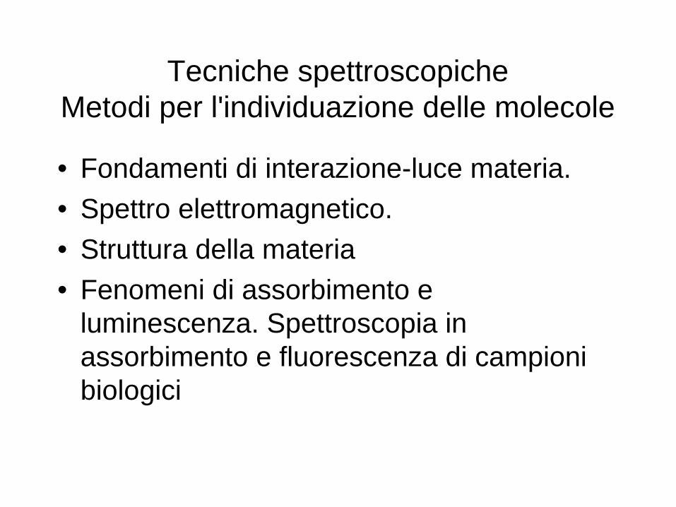
Tecniche spettroscopicheMetodi per l'individuazione delle molecole
• Fondamenti di interazione-luce materia.
• Spettro elettromagnetico.
• Struttura della materia
• Fenomeni di assorbimento e luminescenza. Spettroscopia in assorbimento e fluorescenza di campioni biologici
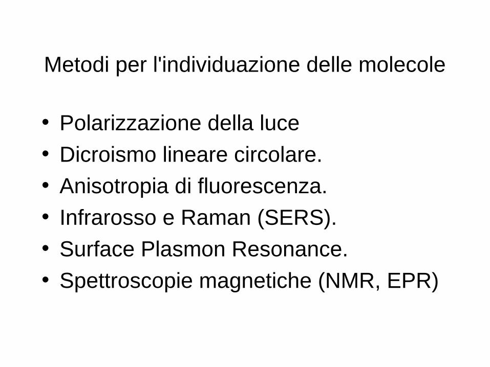
Metodi per l'individuazione delle molecole
• Polarizzazione della luce
• Dicroismo lineare circolare.
• Anisotropia di fluorescenza.
• Infrarosso e Raman (SERS).
• Surface Plasmon Resonance.
• Spettroscopie magnetiche (NMR, EPR)
Metodi per l'individuazione delle molecole
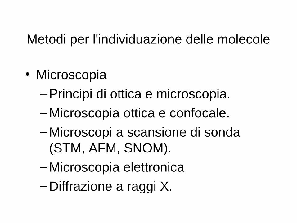
• Microscopia
–Principi di ottica e microscopia.
–Microscopia ottica e confocale.
–Microscopi a scansione di sonda (STM, AFM, SNOM).
–Microscopia elettronica
–Diffrazione a raggi X.

Light as a Wave
La luce è un'onda elettromagnetica oscillante
Ha associata un a lunghezza d'onda λ
c = 3 x 1010 cm/second
Velocita della luce nel vuoto
E= hν =h c/λ
Con h = 4.135 x 10⁻¹⁵ eV*sec = 6.625 x 10⁻²⁷ erg*sec
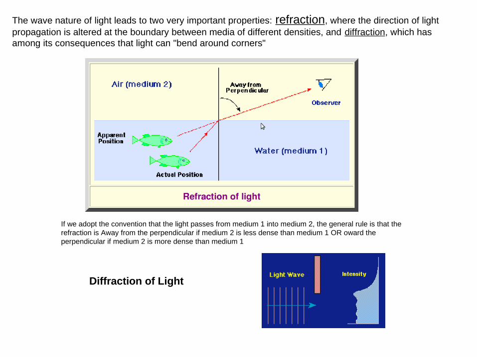
The wave nature of light leads to two very important properties: refraction, where the direction of light propagation is altered at the boundary between media of different densities, and diffraction, which has among its consequences that light can "bend around corners"
If we adopt the convention that the light passes from medium 1 into medium 2, the general rule is that the refraction is Away from the perpendicular if medium 2 is less dense than medium 1 OR oward the perpendicular if medium 2 is more dense than medium 1
Diffraction of Light
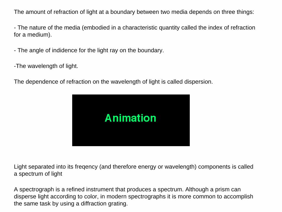
The amount of refraction of light at a boundary between two media depends on three things:
- The nature of the media (embodied in a characteristic quantity called the index of refraction for a medium).
- The angle of indidence for the light ray on the boundary.
-The wavelength of light.
The dependence of refraction on the wavelength of light is called dispersion.
Light separated into its freqency (and therefore energy or wavelength) components is called a spectrum of light
A spectrograph is a refined instrument that produces a spectrum. Although a prism can disperse light according to color, in modern spectrographs it is more common to accomplish the same task by using a diffraction grating.
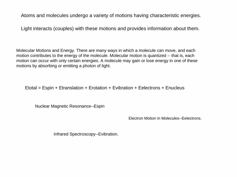
Atoms and molecules undergo a variety of motions having characteristic energies.
Light interacts (couples) with these motions and provides information about them.
Molecular Motions and Energy. There are many ways in which a molecule can move, and each motion contributes to the energy of the molecule. Molecular motion is quantized -- that is, each motion can occur with only certain energies. A molecule may gain or lose energy in one of these motions by absorbing or emitting a photon of light.
Etotal = Espin + Etranslation + Erotation + Evibration + Eelectrons + Enucleus
Infrared Spectroscopy--Evibration.
Nuclear Magnetic Resonance--Espin
Electron Motion in Molecules--Eelectrons.

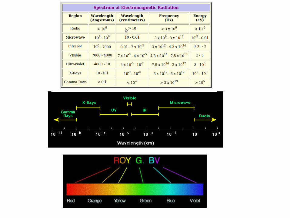
LE RADIAZIONI ELETTROMAGNETICHE (II)
Tipo Lunghezza d’onda Energia Interazione
γ < 0,1 nm emissione nucleare
raggi X 0,1-10 nm transizioni elettroniche (elettroni interni)
UV 10-380 nm transizioni elettroniche(elettroni di valenza)
Vis 380-800 nm transizioni elettroniche(elettroni di valenza)
IR 800 nm-100 µm transizioni vibrazionali
Microonde 100 µm- 1 cm transizioni rotazionali
onde radio 1 cm- metri assorbimento nucleare
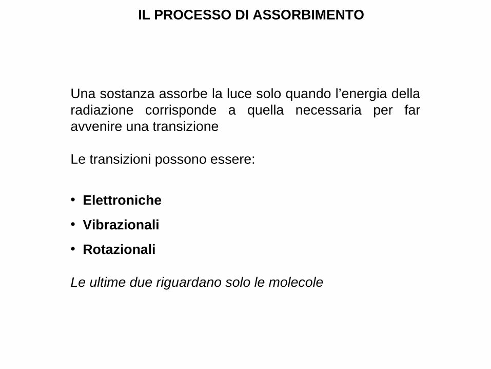
IL PROCESSO DI ASSORBIMENTO
Una sostanza assorbe la luce solo quando l’energia della radiazione corrisponde a quella necessaria per far avvenire una transizione
Le transizioni possono essere:
• Elettroniche
• Vibrazionali
• Rotazionali
Le ultime due riguardano solo le molecole
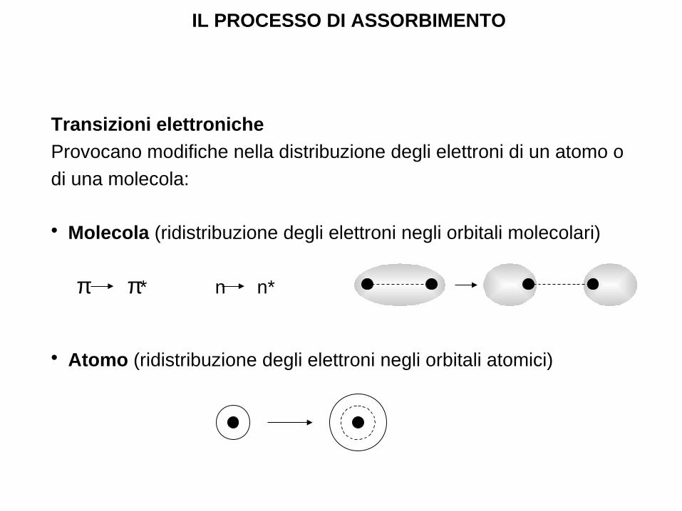
Transizioni elettroniche
Provocano modifiche nella distribuzione degli elettroni di un atomo o
di una molecola:
• Molecola (ridistribuzione degli elettroni negli orbitali molecolari)
π π* n n*
• Atomo (ridistribuzione degli elettroni negli orbitali atomici)
IL PROCESSO DI ASSORBIMENTO

TRANSIZIONI ELETTRONICHE E SPETTRO ATOMICO
S2
S1
S0
E ∆E
S0 S2
S0 S1
S0 S2 S0 S1
I
λ
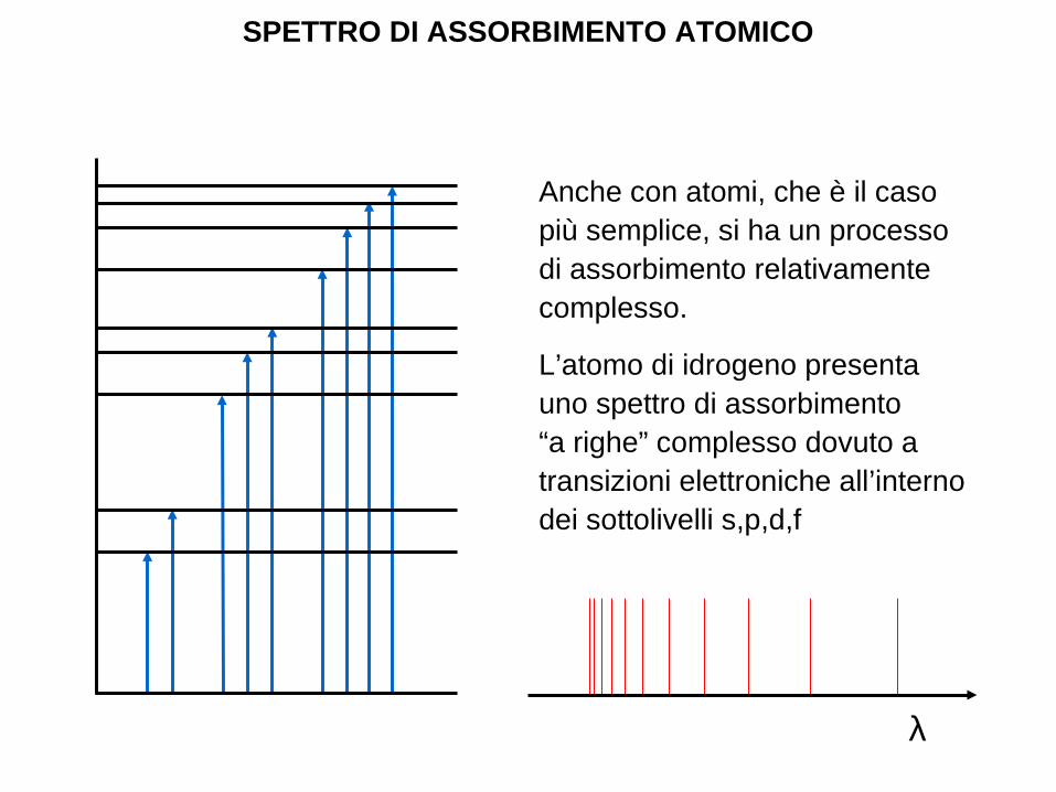
SPETTRO DI ASSORBIMENTO ATOMICO
Anche con atomi, che è il caso più semplice, si ha un processo di assorbimento relativamente complesso.
L’atomo di idrogeno presenta uno spettro di assorbimento “a righe” complesso dovuto a transizioni elettroniche all’interno dei sottolivelli s,p,d,f
λ
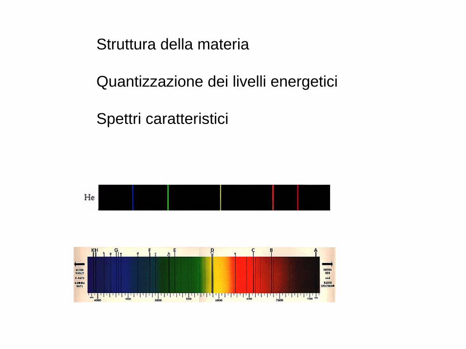
Struttura della materia
Quantizzazione dei livelli energetici
Spettri caratteristici
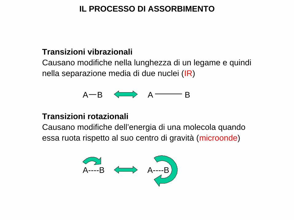
Transizioni vibrazionaliCausano modifiche nella lunghezza di un legame e quindi nella separazione media di due nuclei (IR)
A B A B
Transizioni rotazionaliCausano modifiche dell’energia di una molecola quando essa ruota rispetto al suo centro di gravità (microonde)
A----B A----B
IL PROCESSO DI ASSORBIMENTO
L’assorbimento della luce da parte delle molecole è un processo molto complesso
• In una molecola ogni livello di energia elettronica è suddiviso in un certo numero di sottolivelli vibrazionali
• In aggiunta ciascun sottolivello vibrazionale è ulteriormente suddiviso in sottolivelli rotazionali
IL PROCESSO DI ASSORBIMENTO
IL PROCESSO DI ASSORBIMENTO
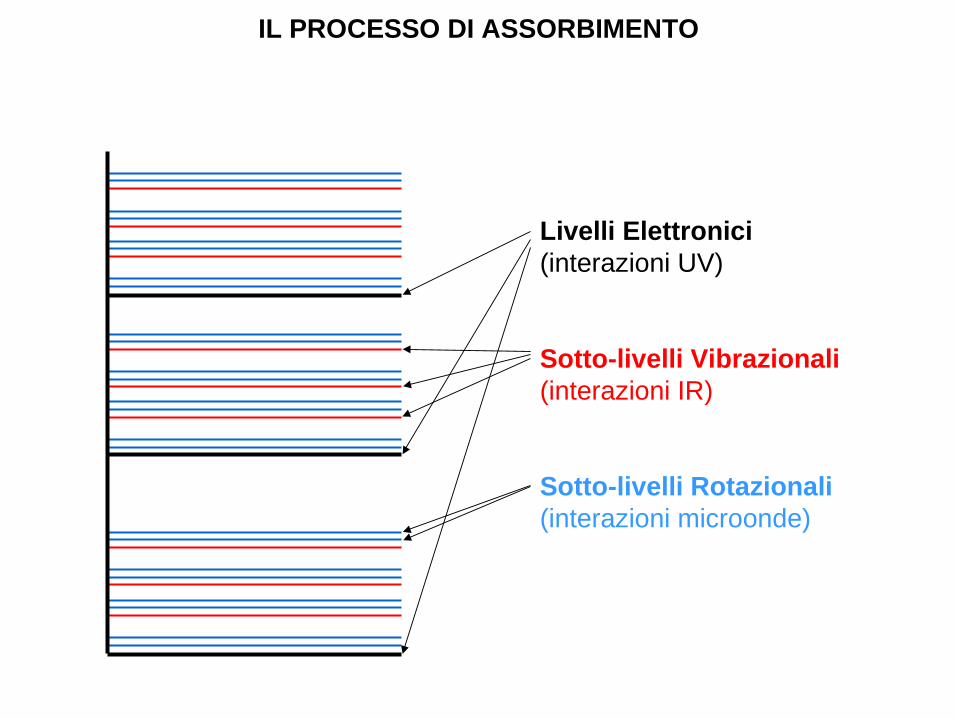
Livelli Elettronici(interazioni UV)
Sotto-livelli Vibrazionali(interazioni IR)
Sotto-livelli Rotazionali(interazioni microonde)
• L’interazione con una molecola e l’assorbimento coinvolgono quindi non solo livelli elettronici, ma anche sotto-livelli vibrazionali e rotazionali.
• Le interazioni con altre molecole avranno anch’esse un effetto, con il risultato che lo spettro di assorbimento sarà a “bande” (ci saranno talmente tante transizioni che sarà impossibile distinguere le une dalle altre).
ASSORBIMENTO MOLECOLARE
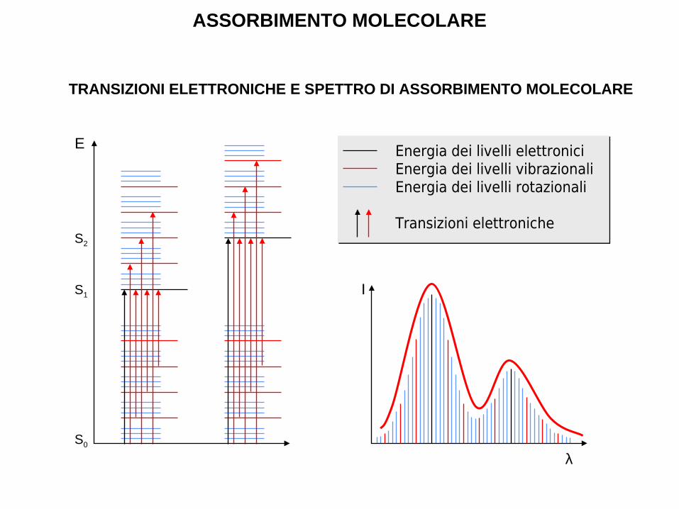
TRANSIZIONI ELETTRONICHE E SPETTRO DI ASSORBIMENTO MOLECOLARE
ASSORBIMENTO MOLECOLARE
E
S0
S1
S2
I
λ
Energia dei livelli elettroniciEnergia dei livelli vibrazionaliEnergia dei livelli rotazionali
Transizioni elettroniche
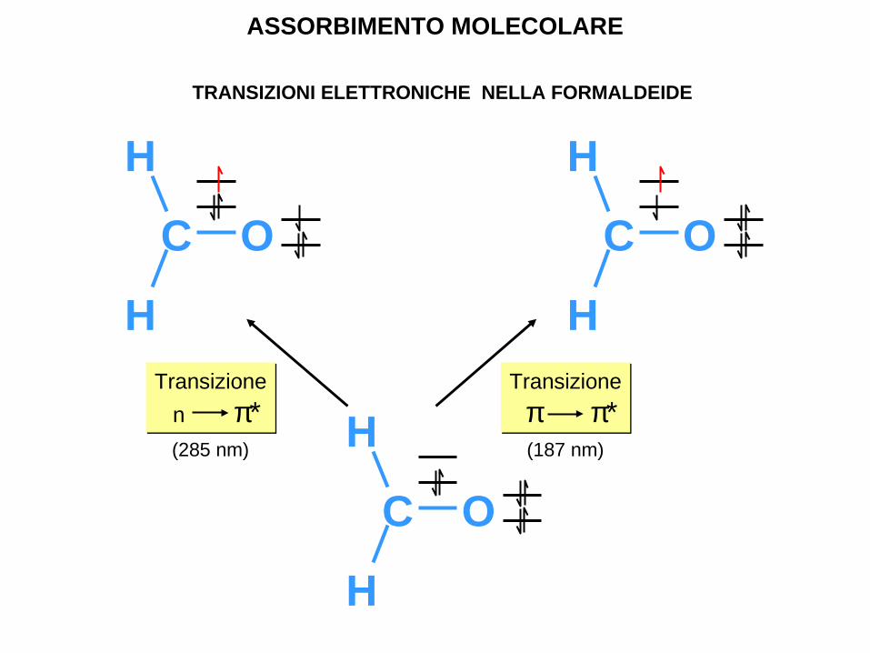
ASSORBIMENTO MOLECOLARE
TRANSIZIONI ELETTRONICHE NELLA FORMALDEIDE
Transizione
n π*Transizione
n π*(285 nm)
Transizione
π π*Transizione
π π*(187 nm)
H
C O
H
H
C O
H
H
C O
H
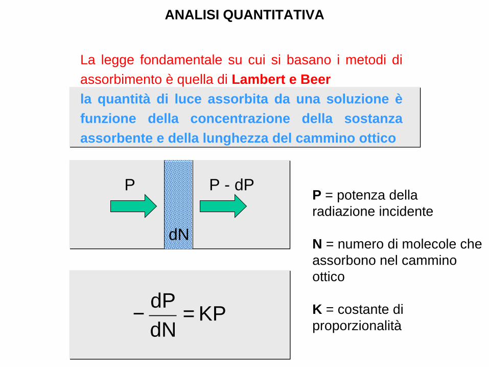
La legge fondamentale su cui si basano i metodi di
assorbimento è quella di Lambert e Beer
la quantità di luce assorbita da una soluzione è
funzione della concentrazione della sostanza
assorbente e della lunghezza del cammino ottico
ANALISI QUANTITATIVA
KPdN
dP =−
P P - dP
dN
P = potenza della radiazione incidente
N = numero di molecole che assorbono nel cammino ottico
K = costante di proporzionalità
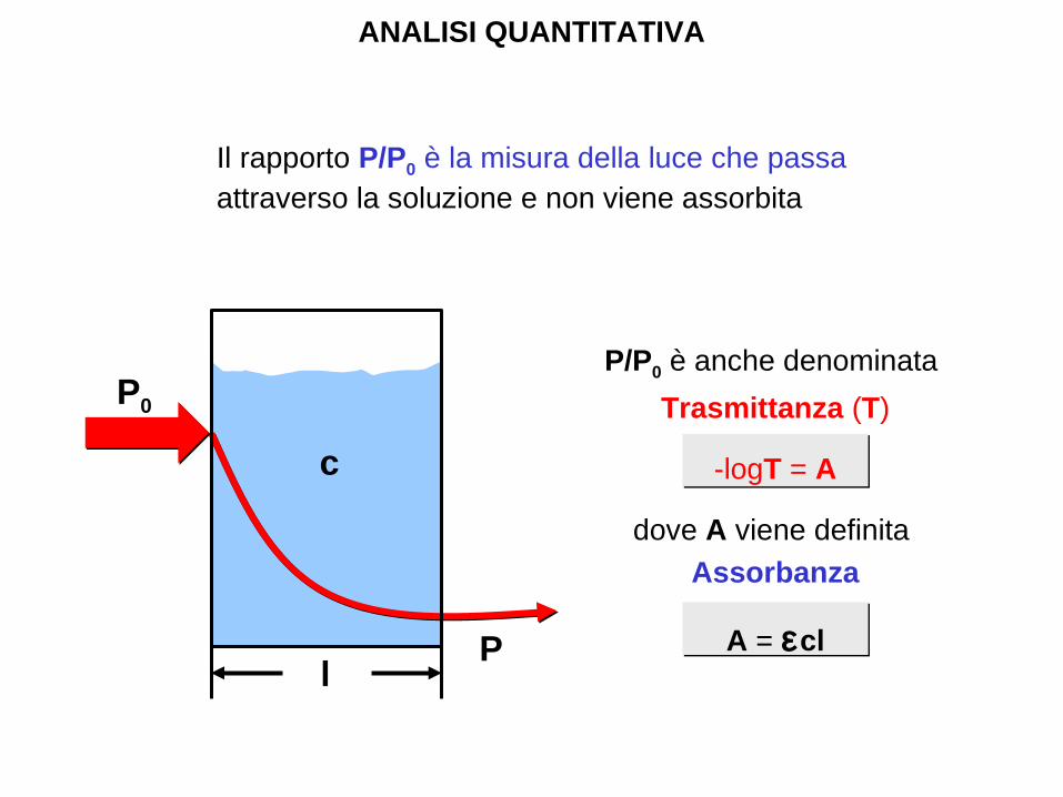
P/P0 è anche denominata
Trasmittanza (T)
-logT = A
dove A viene definita
Assorbanza
A = εcl
Il rapporto P/P0 è la misura della luce che passa attraverso la soluzione e non viene assorbita
ANALISI QUANTITATIVA
P0
c
lP

A=−log T=−log (P/P0)=log (P0/P )=ε⋅l⋅c
LA LEGGE DI LAMBERT E BEER
Concentrazione
Asso
rban
za
1.5
0.0
0.5
1.0
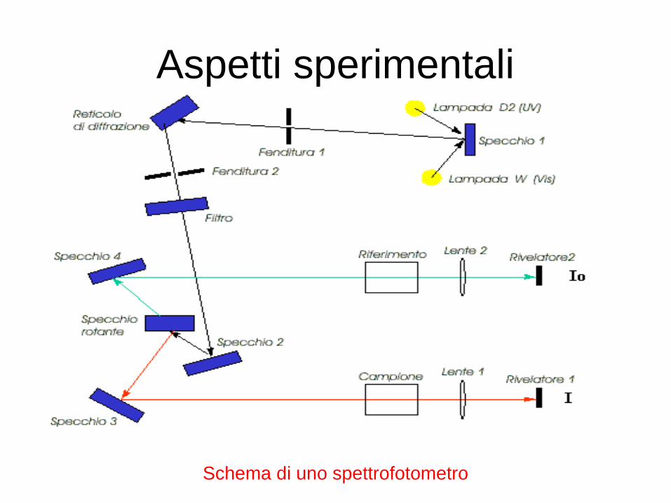
Aspetti sperimentali
Schema di uno spettrofotometro
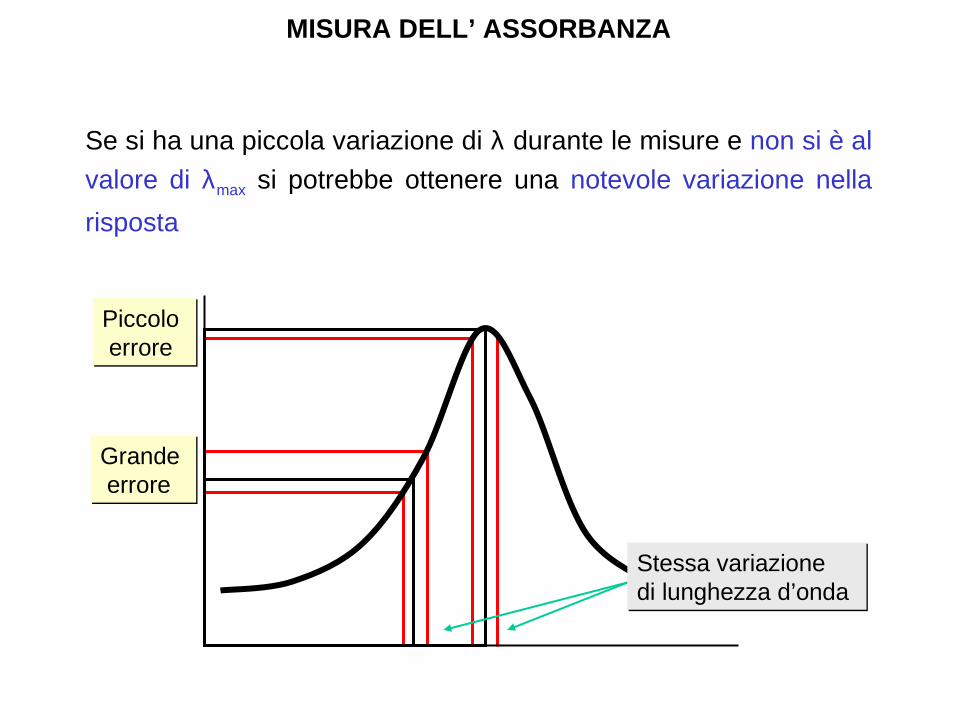
Se si ha una piccola variazione di λ durante le misure e non si è al
valore di λmax si potrebbe ottenere una notevole variazione nella
risposta
MISURA DELL’ ASSORBANZA
Piccolo errorePiccolo errore
Grande erroreGrande errore
Stessa variazione di lunghezza d’ondaStessa variazione di lunghezza d’onda
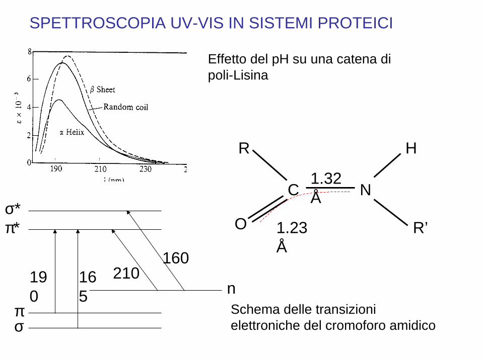
SPETTROSCOPIA UV-VIS IN SISTEMI PROTEICI
πσ
n
π∗σ∗
190
165
210160
Schema delle transizioni elettroniche del cromoforo amidico
R
C N
O R’
H
1.23 Å
1.32 Å
Effetto del pH su una catena di poli-Lisina
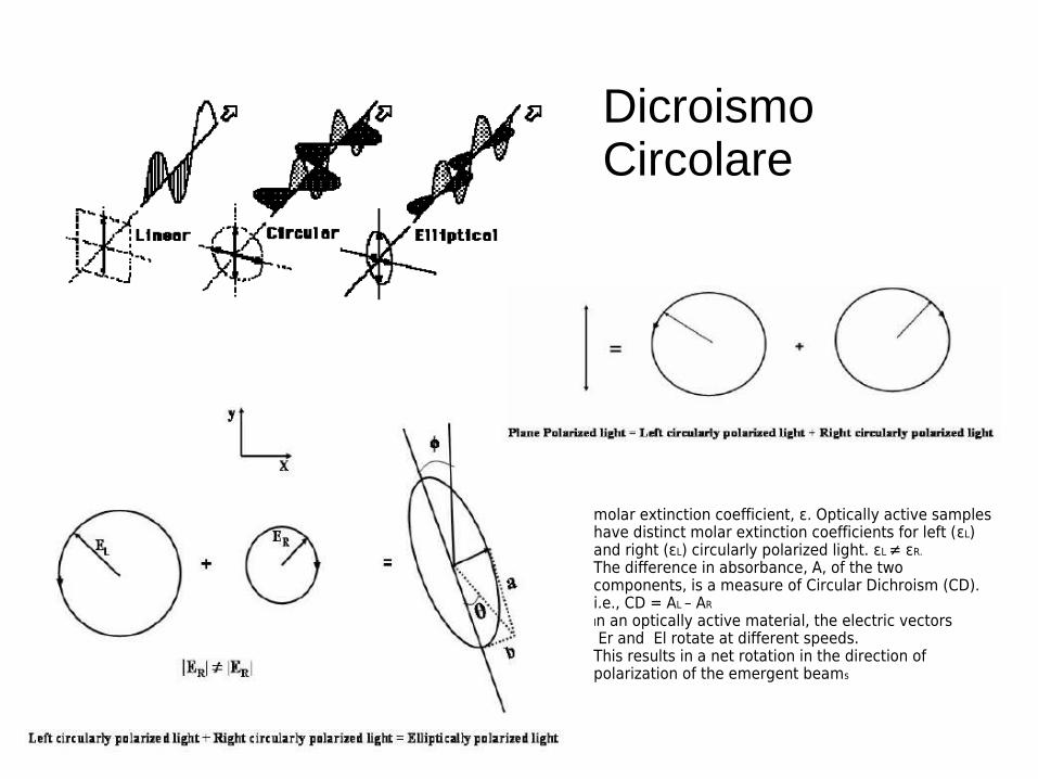
molar extinction coefficient, ε. Optically active samples have distinct molar extinction coefficients for left (εL) and right (εL) circularly polarized light. εL ≠ εR.
The difference in absorbance, A, of the two components, is a measure of Circular Dichroism (CD). i.e., CD = AL – AR
In an optically active material, the electric vectors Er and El rotate at different speeds.This results in a net rotation in the direction of polarization of the emergent beams
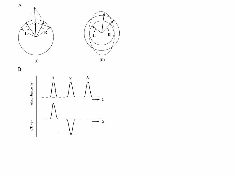
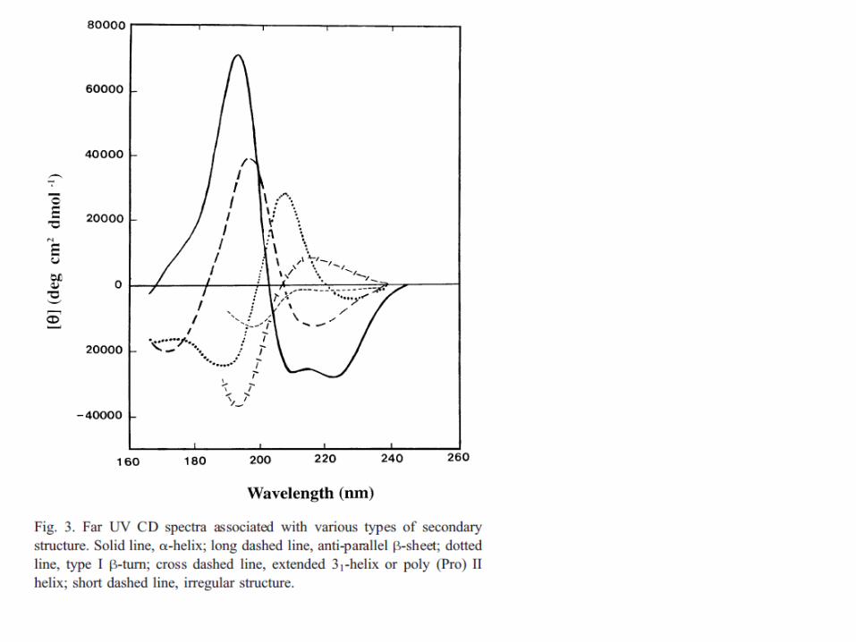
Dicroismo Circolare

Thermal stability
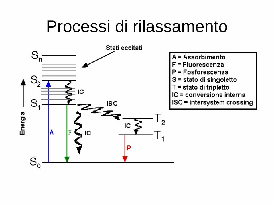
Processi di rilassamento
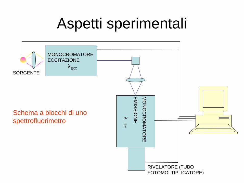
Aspetti sperimentali
MONOCROMATOREECCITAZIONE λEXC
SORGENTE
MO
NO
CR
OM
AT
OR
E
EM
ISS
ION
E
λE
M
RIVELATORE (TUBO FOTOMOLTIPLICATORE)
Schema a blocchi di uno spettrofluorimetro
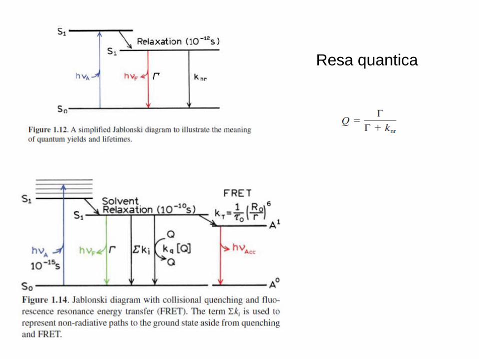
Resa quantica
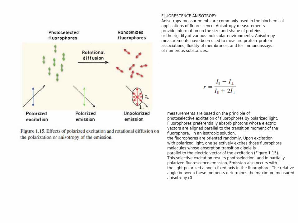
FLUORESCENCE ANISOTROPYAnisotropy measurements are commonly used in the biochemicalapplications of fluorescence. Anisotropy measurementsprovide information on the size and shape of proteinsor the rigidity of various molecular environments. Anisotropymeasurements have been used to measure protein–proteinassociations, fluidity of membranes, and for immunoassaysof numerous substances.
measurements are based on the principle ofphotoselective excitation of fluorophores by polarized light.Fluorophores preferentially absorb photons whose electricvectors are aligned parallel to the transition moment of thefluorophore. In an isotropic solution,the fluorophores are oriented randomly. Upon excitationwith polarized light, one selectively excites those fluorophoremolecules whose absorption transition dipole isparallel to the electric vector of the excitation (Figure 1.15).This selective excitation results photoselection, and in partiallypolarized fluorescence emission. Emission also occurs withthe light polarized along a fixed axis in the fluorophore. The relative angle between these moments determines the maximum measured anisotropy r0
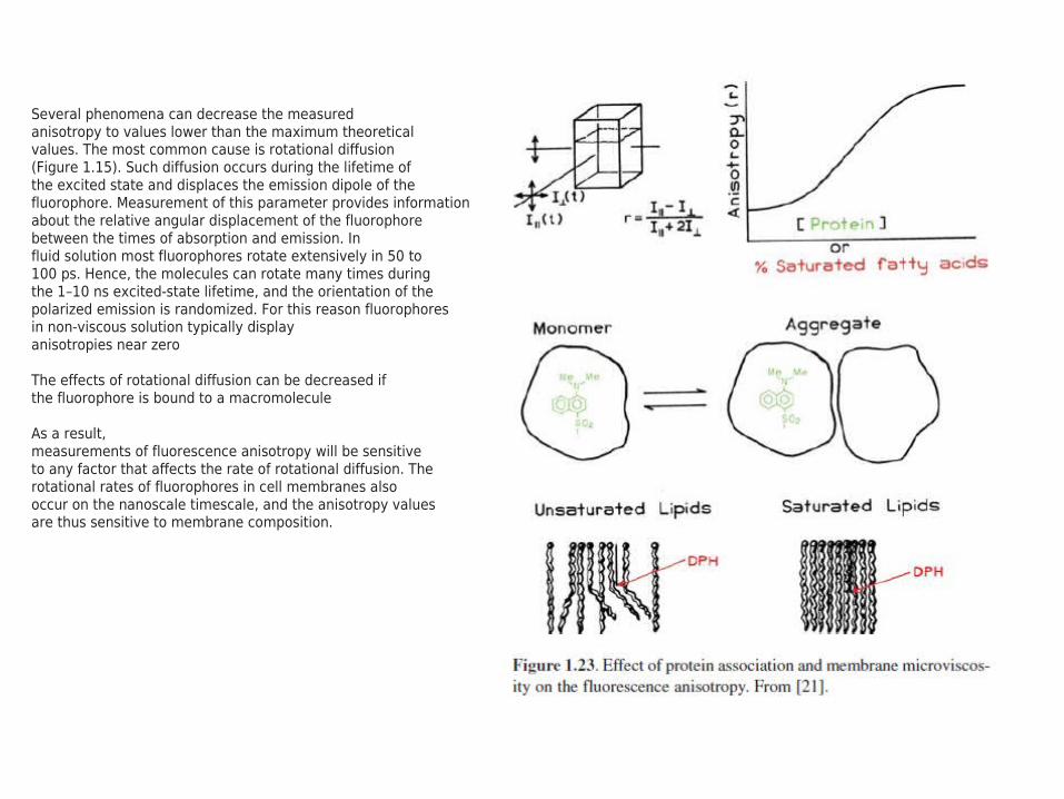
Several phenomena can decrease the measuredanisotropy to values lower than the maximum theoreticalvalues. The most common cause is rotational diffusion(Figure 1.15). Such diffusion occurs during the lifetime ofthe excited state and displaces the emission dipole of thefluorophore. Measurement of this parameter provides informationabout the relative angular displacement of the fluorophorebetween the times of absorption and emission. Influid solution most fluorophores rotate extensively in 50 to100 ps. Hence, the molecules can rotate many times duringthe 1–10 ns excited-state lifetime, and the orientation of thepolarized emission is randomized. For this reason fluorophoresin non-viscous solution typically displayanisotropies near zero
The effects of rotational diffusion can be decreased ifthe fluorophore is bound to a macromolecule
As a result,measurements of fluorescence anisotropy will be sensitiveto any factor that affects the rate of rotational diffusion. Therotational rates of fluorophores in cell membranes alsooccur on the nanoscale timescale, and the anisotropy valuesare thus sensitive to membrane composition.

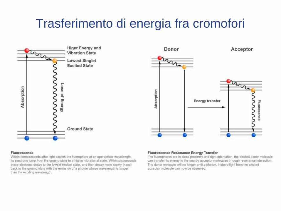
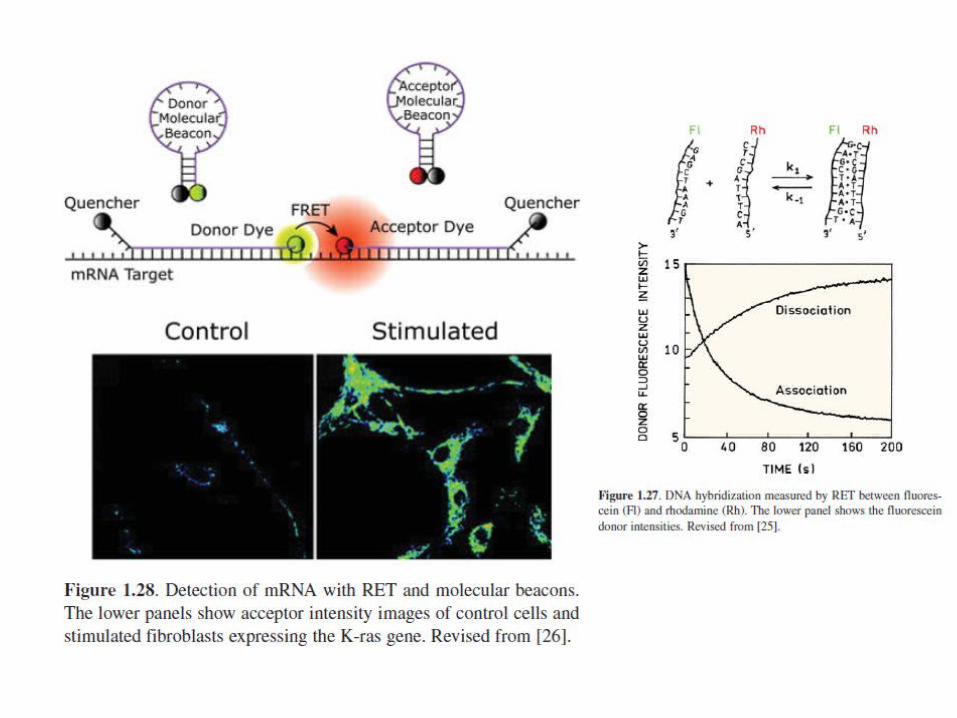
Trasferimento di energia fra cromofori

Struttura e trasferimento risonante di energia
Il cromoforo donatore assorbe a 435 nm ed emette a 475 nm. Quando l’accettore si avvicina a causa di un cambiamento strutturale l’emissione del donatore si attenua e compare una emissione a lunghezza d’onda più elevata (520 nm)
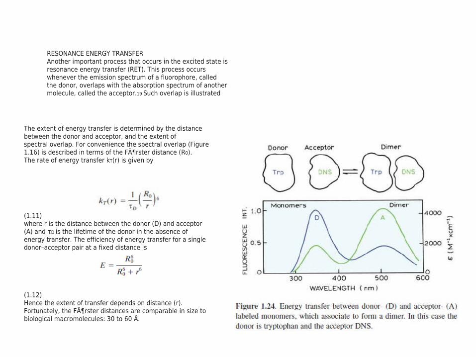
RESONANCE ENERGY TRANSFERAnother important process that occurs in the excited state isresonance energy transfer (RET). This process occurswhenever the emission spectrum of a fluorophore, calledthe donor, overlaps with the absorption spectrum of anothermolecule, called the acceptor.19 Such overlap is illustrated
The extent of energy transfer is determined by the distancebetween the donor and acceptor, and the extent ofspectral overlap. For convenience the spectral overlap (Figure1.16) is described in terms of the Förster distance (R0).The rate of energy transfer kT(r) is given by
(1.11)where r is the distance between the donor (D) and acceptor(A) and τD is the lifetime of the donor in the absence ofenergy transfer. The efficiency of energy transfer for a singledonor–acceptor pair at a fixed distance is
(1.12)Hence the extent of transfer depends on distance (r).Fortunately, the Förster distances are comparable in size tobiological macromolecules: 30 to 60 Ã.
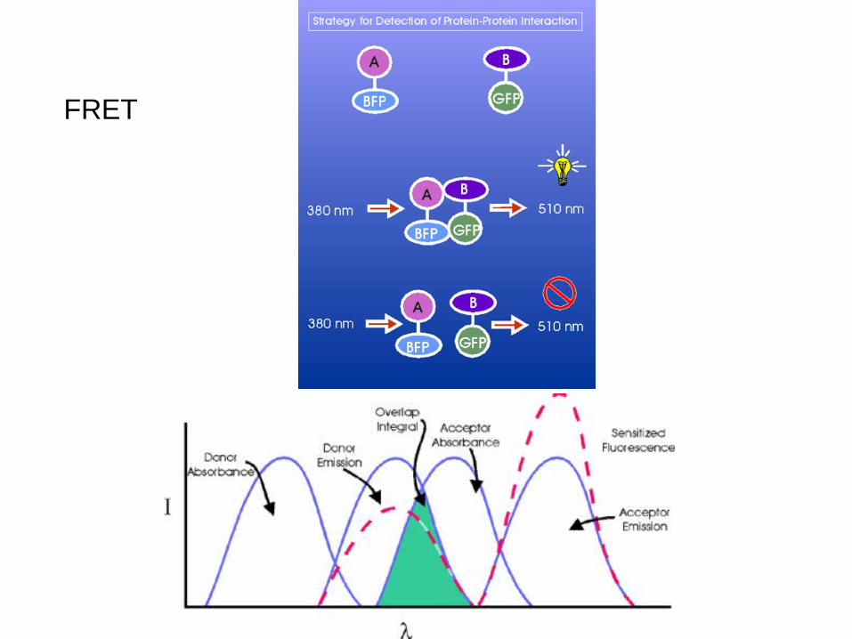
FRET