
TARGETING CD38 ANTIGEN AS HERAPEUTIC · PDF filetrattamento delle neoplasie è stato...
145
UNIVERSITA’ DEGLI STUDI DI VERONA DIPARTIMENTO DI PATOLOGIA E DIAGNOSTICA SCUOLA DI DOTTORATO DI SCIENZE BIOMEDICHE TRASLAZIONALI DOTTORATO DI RICERCA IN BIOMEDICINA TRASLAZIONALE CICLO XXV TITOLO DELLA TESI DI DOTTORATO TARGETING CD38 ANTIGEN AS A THERAPEUTIC STRATEGY FOR HEMATOLOGICAL MALIGNANCIES S.S.D. MED/04 Coordinatore: Prof. Cristiano Chiamulera Tutor: Dott. Giulio Fracasso Dottorando: Dott.ssa Monica Castagna ANNO ACCADEMICO 2012-2013
Transcript of TARGETING CD38 ANTIGEN AS HERAPEUTIC · PDF filetrattamento delle neoplasie è stato...
UNIVERSITA’ DEGLI STUDI DI VERONA
DIPARTIMENTO DI
PATOLOGIA E DIAGNOSTICA
SCUOLA DI DOTTORATO DI
SCIENZE BIOMEDICHE TRASLAZIONALI
DOTTORATO DI RICERCA IN
BIOMEDICINA TRASLAZIONALE
CICLO XXV
TITOLO DELLA TESI DI DOTTORATO
TARGETING CD38 ANTIGEN AS A THERAPEUTIC
STRATEGY FOR HEMATOLOGICAL MALIGNANCIES
S.S.D. MED/04
Coordinatore: Prof. Cristiano Chiamulera Tutor: Dott. Giulio Fracasso
Dottorando: Dott.ssa Monica Castagna
ANNO ACCADEMICO 2012-2013

II
III
ACKNOWLEDGEMENTS
My special thanks to:
Professor Marco Colombatti (Dep. Pathology and Diagnostics, Università degli Studi di
Verona) for tutoring me during my PhD and for being always open for discussion and
supportive in sharing his experience and ideas.
Dr. Matteo Pasetto and Dr. Erika Barison (Dep. Pathology and Diagnostics, Università
degli Studi di Verona) for supporting my work during the first years of my PhD course.
Dr. Giulio Fracasso and Dr. Cristina Anselmi (Dep. Pathology and Diagnostics, Università
degli Studi di Verona) for the useful suggestions and the materials provided throughout
my PhD work.
Dr. David J Flavell (University of Southampton), Dr. Aldo Ceriotti and Dr. Serena Fabbrini
(Consiglio Nazionale delle Ricerche, Milano), Dr. Rodolfo Ippoliti (Università degli Studi
dell'Aquila) and Dr. Wijnand Helfrich (University of Groningen) for the fruitful
collaboration and for the useful materials provided.

IV
V
RIASSUNTO
Il successo di terapie convenzionali come la chemioterapia e la radioterapia per il
trattamento delle neoplasie è stato limitato a causa di diversi fattori come la
chemioresistenza ai farmaci e la tossicità periferica causata dalla mancanza di specificità
di questi approcci. Per questo motivo l’interesse per le terapie selettive che prevedono
l’uso di immunotossine, specialmente per il trattamento di tumori ematologici, è in
aumento. Le immunotossine sono proteine chimeriche costituite da un ligando selettivo
per la cellula bersaglio (dominio di origine anticorpale, citochina o fattore di crescita) che
media il legame e l’internalizzazione della porzione tossica legata chimicamente o fusa
geneticamente, generalmente rappresentata da una tossina di origine vegetale o
batterica che agisce interferendo con la sintesi proteica.
In questo lavoro viene descritta la costruzione di nuove proteine di fusione ad uso
terapeutico progettate per indurre apoptosi selettivamente in neoplasie umane dei
linfociti B e la valutazione dell’effetto potenziante ottenuto attraverso l’associazione delle
immunotossine con farmaci coinvolti in meccanismi metabolici intracellulari. Il dominio di
legame delle nostre immunotossine è rappresentato da frammenti anticorpali a singola
catena (scFv) diretti verso l’antigene CD38, una molecole di superficie espressa ad alti
livelli dai linfociti B di un sottogruppo particolarmente aggressivo di Leucemia Linfatica
Cronica (CLL) che evolve in una patologia dall’esito prognostico sfavorevole, nota come
Sindome di Richter, e dalle plasmacellule tumorali immature nel Mieloma Multiplo (MM).
L’scFv è fuso ad una porzione tossica che agisce inibendo il meccanismo della sintesi
proteica negli organismi eucarioti e nel caso delle nostre immunotossine è rappresentato
da una forma tronca della Esotossina A prodotta dal batterio Pseudomonas aeruginosa
(PE40) o in alternativa dalla tossina di origine vegetale saporina.
Abbiamo inizialmente progettato una immunotossina con PE40 ed una con saporina
contenenti un scFv derivato da un anticorpo monoclonale (mAb) sviluppato e
caratterizzato nel nostro laboratorio. Tutti i costrutti ricombinanti sono stati prodotti nel
sistema di espressione di origine batterica Escherichia coli e purificati da corpi di
inclusione tramite IMAC. Tuttavia, l’scFv 1E8 non ha consentito di preservare l’efficienza
di legame dell’anticorpo parentale. Inoltre, le immunotossine ricombinanti ottenute dalla
fusione dell’scFv 1E8 con PE40 o saporina hanno mostrato una bassa affinità di legame

VI
nei confronti delle cellule bersaglio esprimenti la molecola CD38 e, di conseguenza, è
stata rilavata solo una trascurabile attività citotossica.
Con la progettazione della forma divalente dell’scFv 1E8, il nostro scopo è stato quello
di aumentare l’affinità di legame dei costrutti. Nonostante i risultati sconfortanti del
saggio di legame in citometria a flusso, la molecola DIV1E8-SAP ha dimostrato di inibire la
sintesi proteica di cellule CD38-positive con una IC50 nell’ordine del sub-nanomolare.
Successivamente abbiamo progettato due immunotossine ricombinanti dirette verso
l’antigene CD38, il cui dominio di legame era costituito da un scFv derivato da un mAb con
una specificità epitopica diversa da quella del precedentemente descritto 1E8. Le
immunotossine AT13/5-PE e AT13/5-SAP hanno dimostrato buone proprietà di legame
con una elevata affinità e specificità per l’antigene CD38 espresso sulla superficie di
cellule derivate da Linfoma di Burkitt e cellule di mieloma.
Abbiamo dimostrato l’abilità si queste immunotossine di inibire la sintesi proteica nelle
linee cellulari studiate e ne abbiamo chiaramente dimostrato un effetto dose-risposta. Il
blocco della sintesi proteica causato dalle immunotossine derivate da AT13/5 ha
determinato infine l’innesco del processo di apoptosi e la morte cellulare. Attraverso
saggi di apoptosi abbiamo dimostrato la capacità di AT13/5-PE e AT13/5-SAP di indurre
apoptosi in cellule Daudi e RPMI8226.
Abbiamo perciò provato che l’associazione delle nostre immunotossine con molecole
terapeutiche che agiscono su diversi bersagli dalla cascata di traduzione del segnale
coinvolta nella crescita cellulare, nella sopravvivenza e nella proliferazione, potrebbe
essere sinergica in alcune linee cellulari. In particolare abbiamo osservato che farmaci
coinvolti nell’inibizione di Bcl-2, Bcl-xL e Bcl-w (noti come BH3-mimetics) possono
aumentare la potenza delle nostre immunotossine.
Abbiamo infine dimostrato una prima prova di concetto riguardo l’efficacia delle
immunotossine derivate da AT13/5 su linfociti B derivati da pazienti affetti da CLL,
tuttavia questo studio necessita di essere implementato con una casistica più ampia.
VII
ABSTRACT
The success of conventional chemotherapy and radiotherapy for the treatment of
cancer has been limited due to several factors like chemoresistance to drugs and
peripheral toxicity caused by the lack of specificity of these approaches. For this reason
the interest in targeted therapies using immunotoxins (ITs) especially for the treatment of
hematological malignancies is increasing. Immunotoxins are chimeric proteins with a cell-
selective ligand (antibody-derived domain, cytokine or growth factor) which drives the
binding and internalization of a chemically linked or genetically fused toxic portion,
generally represented by a plant or bacterial toxin which acts by interfering with protein
synthesis.
Here we report on the construction of novel therapeutic fusion proteins designed to
induce target antigen-restricted apoptosis in human B-cell neoplasias and the evaluation
of the potentiating effect obtained by the association of the ITs with drugs involved in
intracellular metabolic pathways. The binding portion of our ITs is represented by a
single-chain antibody fragment (scFv) directed against CD38 antigen, a surface molecule
highly expressed by B lymphocytes of a particularly aggressive sub-group of Chronic
Lymphocytic Leukemia (CLL) leading to the prognostically unfavorable Richter’s Syndrome
and by the neoplastic immature plasma cells in Multiple Myeloma (MM). The scFv is fused
to a toxic portion which acts by inhibiting the mechanism of protein synthesis in
eukaryotes and in our ITs is represented by a truncated version of the bacterial toxin
Pseudomonas aeruginosa Exotoxin A (PE40) or alternatively by the plant toxin saporin.
We firstly designed a PE40- and a saporin-based IT comprising a scFv derived from a
monoclonal antibody (mAb) developed and characterized in our laboratory. All the
recombinant constructs were produced in the bacterial expression system E. coli and
purified from inclusion bodies by IMAC. However, the scFv format (1E8) did not allow to
preserve the binding efficiency of the parental monoclonal. Moreover, the recombinant
ITs created by the fusion of 1E8 scFv with PE40 or saporin showed a low binding affinity to
the CD38 target cells and, as a consequence, only negligible citotoxic activity was
detected.
With the creation of the divalent form of the 1E8 scFv, our purpose was to increase the
binding affinity of the constructs. Despite the discouraging results of the flow-cytometric
VIII
binding assay, DIV1E8-SAP demonstrated to inhibit protein synthesis of CD38-positive
cells with an IC50 in the sub-nanomolar range.
Then we designed two anti-CD38 recombinant ITs whose binding portion was a scFv
derived from a mAb with an epitope specificity different from that of the previously
described 1E8. AT13/5-PE and AT13/5-SAP showed good binding properties with a high
affinity and specificity for CD38 antigen expressed on the surface of Burkitt’s lymphoma
cells and myeloma cells.
We proved the ability of these ITs to inhibit protein synthesis in the cell lines studied
and we clearly demonstrated a dose-response effect of the ITs. The arrest of protein
synthesis caused by the AT13/5-derived ITs finally leads to the triggering of the apoptotic
cascade and to cell death. By using apoptosis assays we demonstrated the capability of
AT13/5-PE and AT13/5-SAP to induce apoptosis of Daudi and RPMI8226 cells.
Then we proved that the association of our ITs with therapeutic molecules acting on
different targets of the signal transduction cascade involved in cell growth, survival and
proliferation, could be synergistic in some cell lines. In particular we observed that drugs
involved in the Bcl-2, Bcl-xL and Bcl-w inhibition (BH3-mimetics) can increase the potency
of our ITs.
Finally we demonstrated a first proof of concept about the efficacy of AT13/5-derived
ITs on B-lymphocytes derived from CLL patient, but this study needs to be implemented
with a wider number of cases.
IX
INDEX
1. INTRODUCTION.......................................................................1 1.1 CONVENTIONAL THERAPY OF CANCER 3
1.2 ANTIBODY-BASED TARGETED THERAPIES 5
1.3 TUMOR CELL ANTIGENS 9
1.3.1 CD38 10
1.3.1.1 CD38 structure and function 12
1.3.1.2 CD38 as a target for immunotherapy 14
1.3.1.2.1 Chronic Lymphocytic Leukemia (CLL) 15
1.3.1.2.2 Multiple Myeloma (MM) 17
1.4 IMMUNOTOXINS 18
1.4.1 The binding domain 20
1.4.1.1 Antibodies 20
1.4.1.2 Antibody fragments 23
1.4.2 The toxic domain 26
1.4.2.1 Plant toxins 27
1.4.2.1.1 Saporin 29
1.4.2.2 Bacterial toxins 30
1.4.2.2.1 Pseudomonas Exotoxin A: structure and function 31
1.4.2.2.2 Pseudomonas Exotoxin A: Cytotoxic pathways 32
1.4.2.2.3 PE derivatives 35
1.4.3 Immunogenicity 36
1.4.3.1 Immunogenicity of the binding domain 36
1.4.3.2 Immunogenicity of the toxic portion 38
1.4.4 Expression systems 39
1.5 COMBINATION THERAPIES 41
AIM OF THE RESEARCH 47
2. MATERIALS AND METHODS……………………………………………….…..49 2.1 MICROBIOLOGY TECHNIQUES 51
2.1.1 Escherichia coli strains 51
2.1.2 E. coli growth media 51
2.1.3 Plasmid vectors 51
2.1.4 Preparation of CaCl2-competent E. coli cells 52
2.1.5 Heat-shock mediated transformation of Escherichia coli 53
2.2 HUMAN CELL LINES 53
2.2.1 Cell lines and growth media 53
2.2.2 B-lymphocytes from PBMCs 54
X
2.3 MOLECULAR BIOLOGY 54
2.3.1 RNA extraction from anti-CD38 hybridoma cells 54
2.3.2 cDNA synthesis 54
2.3.3 PCR amplification of specific DNA fragments 55
2.3.3.1 Amplification of the sequence coding for mouse β-actin 55
2.3.3.2 Amplification of the sequences coding for variable domains
of heavy and light chains 56
2.3.3.3 Amplification of the sequence coding for the truncated
version of Pseudomonas aeruginosa exotoxin A (PE40) 57
2.3.3.4 Amplification of the sequence coding for the saporin 57
2.3.4 DNA digestion with restriction enzymes 58
2.3.5 Plasmid constructs 59
2.3.5.1 Cloning strategy 59
2.3.5.2 Ligation 61
2.3.5.3 Colony-PCR screening 62
2.3.6 Plasmid DNA extraction from E. coli cultures 62
2.3.7 DNA sequencing 63
2.3.8 Oligonucleotides used 63
2.4 PROTEIN EXPRESSION IN BACTERIA 64
2.4.1 Expression of scFv and immunotoxin in Escherichia coli BL21(DE3) pLysS 64
2.5 PROTEIN PROCESSING AND ANALYSIS 64
2.5.1 Extraction of proteins from E. coli BL21(DE3) pLysS inclusion bodies 64
2.5.2 Purification of recombinant proteins by affinity chromatography 65
2.5.3 Refolding of proteins from inclusion bodies 66
2.5.4 Purification of mAb from hybridoma culture medium 67
2.5.5 Denaturing polyacrylamide gel electrophoresis (SDS-PAGE) 68
2.5.6 Immunoblotting 68
2.5.6.1 Transfer of proteins on PVDF membrane 68
2.5.6.2 Immunodetection 69
2.5.7 Protein quantification 69
2.5.7.1 Spectrophotometric quantification 69
2.5.7.2 Coomassie staining 70
2.6 ANALYSIS OF BINDING IN FLOW CYTOMETRY 70
2.6.1 Comparison between binding efficiencies of hybridomas 70
2.6.2 Competition assay for specific binding of new mAbs to CD38 on cells 71
2.6.3 Curves of binding to the CD38 antigen on cells 71
2.7 BIOLOGICAL ASSAYS 72
2.7.1 Cytotoxicity assessment by leucine incorporation 72
2.7.2 Cell proliferation assay with XTT 72
2.7.3 Apoptosis assay 73
XI
3. RESULTS…………………………………………………………………………….….75 3.1 CHARACTERIZATION OF NEW ANTI-CD38 HYBRIDOMA CLONES 77
3.2 CHARACTERIZATION OF THE MONOCLONAL ANTIBODY 1E82H11 81
3.3 CLONING, EXPRESSION AND CHARACTERIZATION OF THE 1E8 SCFV
AND DERIVED ITS 83
3.3.1 Amplification of the VH and VL domains of the anti-CD38 mAb 83
3.3.2 Expression, purification and characterization of the antibody
fragment 1E8 85
3.3.3 Expression, purification and characterization of the 1E8-derived ITs 87
3.3.4 Expression, purification and characterization of the divalent 1E8
antibody fragment and the derived ITs 90
3.4 AT13/5-DERIVED CONSTRUCTS 94
3.4.1 Expression, purification and characterization of the AT13/5-derived ITs 94
3.4.2 Cytotoxicity of the AT13/5-derived immunotoxins 96
3.4.3 Combination treatments with AT13/5-derived immunotoxins 104
3.4.4 Effect of the AT13/5-derived immunotoxins on B-CLL 107
4. DISCUSSION………………………………………………………………………...111
5. BIBLIOGRAPHY……………………………………………………………….……123

XII
XIII
ABBREVIATION USED:
Ab/Abs Antibody/antibodies
Amp Ampicillin
APS Ammonium persulfate
Cam Chloramphenicol
cpm Counts per minute
DEPC Diethyl pyrocarbonate
DMSO Dimethyl sulfoxide
FBS Fetal bovine serum
FITC Fluorescein isothiocyanate
Gln Glutamine
h hour
H2O Water
Ig Immunoglobulin
IMAC Immobilized metal ion affinity chromatography
IPTG isopropyl-beta-D-thiogalactopyranoside
IT /ITs Immunotoxin(s)
mAb Monoclonal antibody
MFI Medium Fluorescence Intensity
NaOAc Sodium acetate
NTA Nitriloacetic acid
o.n. overnight
OD Optical density
PCR Polymerase chain reaction
PE Pseudomonas aeruginosa Exotoxin A
PE40 Truncated form of Pseudomonas aeruginosa Exotoxin A
PVDF Polyvinylidene fluoride
rpm Revolutions per minute
RT Room temperature
scFv Single-chain Fragment variable
SDS Sodium dodecyl sulfate
t½ Half-life
Temed Tetramethylethylenediamine
Tet Tetracycline
VH Heavy chain variable domain
VL or Vk Light chain variable domain

XIV

1. INTRODUCTION

Introduction
3
1.1 CONVENTIONAL THERAPY OF CANCER
According to the World Health Organization (WHO), cancer is responsible for
approximately 7.6 million (13%) of the 59 million deaths that occur each year. WHO
estimates that if current cancer rates remain unchanged, new cases of cancer will
increase from 12.7 million cases (2008) to 21.4 million cases (2030) [1]. Moreover,
considering the more developed regions of the world it can be observed that cancer is
responsible for about 25% of all deaths.
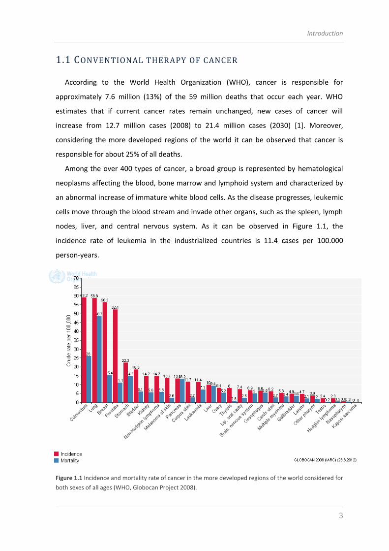
Among the over 400 types of cancer, a broad group is represented by hematological
neoplasms affecting the blood, bone marrow and lymphoid system and characterized by
an abnormal increase of immature white blood cells. As the disease progresses, leukemic
cells move through the blood stream and invade other organs, such as the spleen, lymph
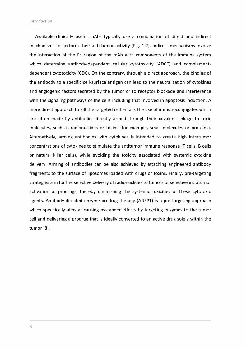
nodes, liver, and central nervous system. As it can be observed in Figure 1.1, the
incidence rate of leukemia in the industrialized countries is 11.4 cases per 100.000
person-years.
Figure 1.1 Incidence and mortality rate of cancer in the more developed regions of the world considered for
both sexes of all ages (WHO, Globocan Project 2008).
Introduction
4
The first line treatment for leukemia is represented by chemotherapy and radiation
therapy, often used in combination.
Chemotherapy agents attack rapidly dividing cells and due to their inability to
distinguish leukemia cells from other rapidly dividing but non-cancerous cells, they often
cause toxicity towards healthy tissues with a rapid cell turnover [2] such as healthy red and
white blood cells, blood-clotting platelets, hair follicles, and cells lining the
gastrointestinal tract, thus creating unpleasant side effects. Furthermore the damage to
white blood cells increases the immunodeficiency and the risk of infection.
Radiotherapy kills leukemia cells by exposing them to ionizing radiation that damages
cell DNA but the treatment can lead to DNA mutations in the by-stander normal cells
increasing the risk of the potential onset of a secondary radiation-associated cancer [3].
For the treatment of haematological malignancies, chemotherapy and radiotherapy are
often supported by hematopoietic stem cells transplantation with the double purpose to
replace disease-causing stem cells with healthy ones and to replenish a bone marrow
which is damaged by the aggressiveness of the therapeutics mentioned above. Also stem
cells therapy is not free of side effects and graft-versus-host disease is the major complication
identified in the case of allogenic transplantation.
Along with the commonly known side effects of conventional treatments, frequent poor
responses and relapses are observed especially in some indolent malignancies, due to their
slow progress and inefficacy of the therapies. Besides, tumor cells can develop a resistance to
chemotherapy drugs, hindering their mechanism of action or promoting their expulsion out
of the cell before they can act [4].
As a consequence, the need for an improved efficacy in cancer therapies has been
increasingly felt in the last few years, placing the focus of research on the development of
new drugs that combine power of action and selective targeting of cancerous cells. An
ever clearer understanding of the biochemical events that are implicated in the onset and
progression of many malignancies has allowed to design tumor-specific therapies that
directly target the molecules involved in the development of tumors or that selectively
deliver the drug into cancerous cells. The opportunity of specifically affecting a tumor is
provided by the presence of molecular targets being selectively expressed on the surface
of cancerous cells: these are the tumor marker antigens [2].
Introduction
5
1.2 ANTIBODY-BASED TARGETED THERAPIES
Antibody-based therapy for cancer has become established over the past 15 years and
is now one of the most successful and important strategies for treating patients with
hematological malignancies and solid tumors.
However, the history of true antibody therapy began about a century ago with the
discovery by von Behring that resistance to infectious diseases like tetanus and diphteria
could be transferred between animals through their sera, a strategy known as passive
serotherapy [5]. Further developments of serotherapy came from Ehrlich, who forged the
term “magic bullet” referring to immunoglobulins as the real responsible for immune
protection, and from Köhler and Milstein, who developed in 1975 the hybridoma
technology, a method for generation and large-scale production of monoclonal antibodies
(mAbs) of murine origin [6]. However, the fundamental basis of antibody-based therapy
of tumors dates back to the original observation of antigen expression by tumor cells
through serological techniques in the 1960s [7]. The definition of cell surface antigens
that are expressed by human cancers has revealed a broad array of targets that are
overexpressed, mutated or selectively expressed compared to normal tissues.
Therapeutics that target these antigens can function through mediating alterations in
antigen or receptor function (such as agonist or antagonist functions), modulating the
immune system or delivering a specific drug that is conjugated to an antibody [8].
Cancer immunotherapy can be considered from several perspectives, but one
convenient way to categorize immunotherapy is to think of active or passive approaches:
active immunotherapy involves the stimulation of the patient’s immune response
against tumor cells (vaccination) through the administration of antigens (Ag) in
various shapes (e.g. recombinant proteins, cDNAs inserted into plasmids or viral
vectors, peptides) or by using tumor cells unable to replicate (apoptotic or
necrotic) or by loaded immune cells;
passive immunotherapy consists in the administration of immunological effectors
(e.g. monoclonal antibodies alone or conjugated to drugs, toxins or cytokines,
tumor-specific T lymphocytes).
Introduction
6
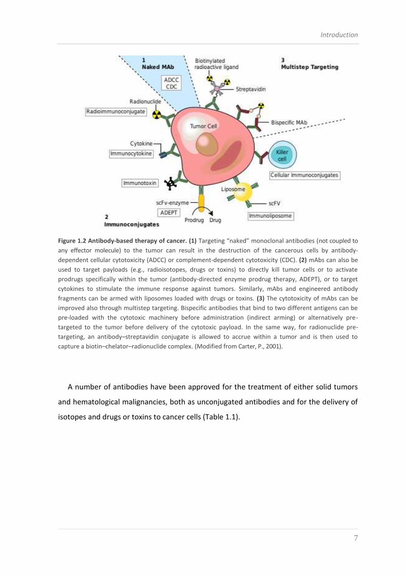
Available clinically useful mAbs typically use a combination of direct and indirect
mechanisms to perform their anti-tumor activity (Fig. 1.2). Indirect mechanisms involve
the interaction of the Fc region of the mAb with components of the immune system
which determine antibody-dependent cellular cytotoxicity (ADCC) and complement-
dependent cytotoxicity (CDC). On the contrary, through a direct approach, the binding of
the antibody to a specific cell-surface antigen can lead to the neutralization of cytokines
and angiogenic factors secreted by the tumor or to receptor blockade and interference
with the signaling pathways of the cells including that involved in apoptosis induction. A
more direct approach to kill the targeted cell entails the use of immunoconjugates which
are often made by antibodies directly armed through their covalent linkage to toxic
molecules, such as radionuclides or toxins (for example, small molecules or proteins).
Alternatively, arming antibodies with cytokines is intended to create high intratumor
concentrations of cytokines to stimulate the antitumor immune response (T cells, B cells
or natural killer cells), while avoiding the toxicity associated with systemic cytokine
delivery. Arming of antibodies can be also achieved by attaching engineered antibody
fragments to the surface of liposomes loaded with drugs or toxins. Finally, pre-targeting
strategies aim for the selective delivery of radionuclides to tumors or selective intratumor
activation of prodrugs, thereby diminishing the systemic toxicities of these cytotoxic
agents. Antibody-directed enzyme prodrug therapy (ADEPT) is a pre-targeting approach
which specifically aims at causing bystander effects by targeting enzymes to the tumor
cell and delivering a prodrug that is ideally converted to an active drug solely within the
tumor [8].
Introduction
7
Figure 1.2 Antibody-based therapy of cancer. (1) Targeting “naked” monoclonal antibodies (not coupled to
any effector molecule) to the tumor can result in the destruction of the cancerous cells by antibody-
dependent cellular cytotoxicity (ADCC) or complement-dependent cytotoxicity (CDC). (2) mAbs can also be
used to target payloads (e.g., radioisotopes, drugs or toxins) to directly kill tumor cells or to activate
prodrugs specifically within the tumor (antibody-directed enzyme prodrug therapy, ADEPT), or to target
cytokines to stimulate the immune response against tumors. Similarly, mAbs and engineered antibody
fragments can be armed with liposomes loaded with drugs or toxins. (3) The cytotoxicity of mAbs can be
improved also through multistep targeting. Bispecific antibodies that bind to two different antigens can be
pre-loaded with the cytotoxic machinery before administration (indirect arming) or alternatively pre-
targeted to the tumor before delivery of the cytotoxic payload. In the same way, for radionuclide pre-
targeting, an antibody–streptavidin conjugate is allowed to accrue within a tumor and is then used to
capture a biotin–chelator–radionuclide complex. (Modified from Carter, P., 2001).
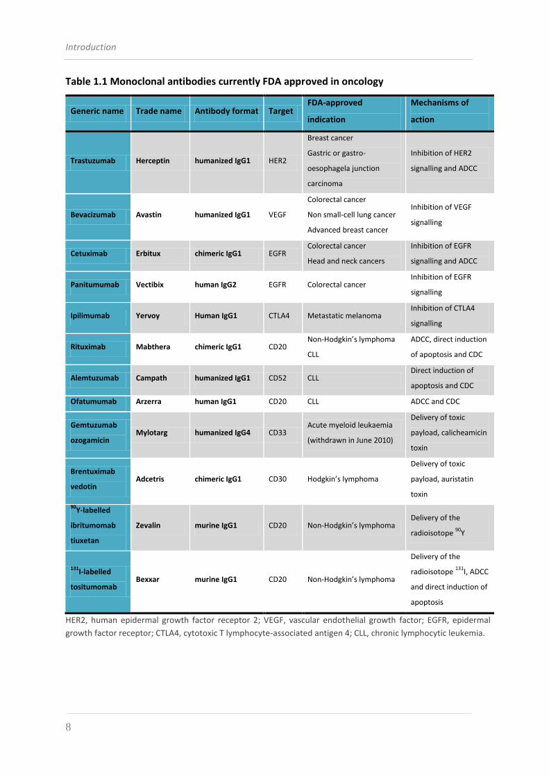
A number of antibodies have been approved for the treatment of either solid tumors
and hematological malignancies, both as unconjugated antibodies and for the delivery of
isotopes and drugs or toxins to cancer cells (Table 1.1).
Introduction
8
Table 1.1 Monoclonal antibodies currently FDA approved in oncology
Generic name Trade name Antibody format Target FDA-approved
indication
Mechanisms of
action
Trastuzumab Herceptin humanized IgG1 HER2
Breast cancer
Gastric or gastro-
oesophagela junction
carcinoma
Inhibition of HER2
signalling and ADCC
Bevacizumab Avastin humanized IgG1 VEGF
Colorectal cancer
Non small-cell lung cancer
Advanced breast cancer
Inhibition of VEGF
signalling
Cetuximab Erbitux chimeric IgG1 EGFR Colorectal cancer
Head and neck cancers
Inhibition of EGFR
signalling and ADCC
Panitumumab Vectibix human IgG2 EGFR Colorectal cancer Inhibition of EGFR
signalling
Ipilimumab Yervoy Human IgG1 CTLA4 Metastatic melanoma Inhibition of CTLA4
signalling
Rituximab Mabthera chimeric IgG1 CD20 Non-Hodgkin’s lymphoma
CLL
ADCC, direct induction
of apoptosis and CDC
Alemtuzumab Campath humanized IgG1 CD52 CLL Direct induction of
apoptosis and CDC
Ofatumumab Arzerra human IgG1 CD20 CLL ADCC and CDC
Gemtuzumab
ozogamicin Mylotarg humanized IgG4 CD33
Acute myeloid leukaemia
(withdrawn in June 2010)
Delivery of toxic
payload, calicheamicin
toxin
Brentuximab
vedotin Adcetris chimeric IgG1 CD30 Hodgkin’s lymphoma
Delivery of toxic
payload, auristatin
toxin
90Y-labelled
ibritumomab
tiuxetan
Zevalin murine IgG1 CD20 Non-Hodgkin’s lymphoma Delivery of the
radioisotope 90
Y
131I-labelled
tositumomab Bexxar murine IgG1 CD20 Non-Hodgkin’s lymphoma
Delivery of the
radioisotope 131
I, ADCC
and direct induction of
apoptosis
HER2, human epidermal growth factor receptor 2; VEGF, vascular endothelial growth factor; EGFR, epidermal
growth factor receptor; CTLA4, cytotoxic T lymphocyte-associated antigen 4; CLL, chronic lymphocytic leukemia.
Introduction
9
1.3 TUMOR CELL ANTIGENS
The targets of cancer immunotherapy can be classified as:
a. Tumor Specific Antigens (TSAs), which are present only on tumor cells and are
represented by new mutant proteins or aberrantly glycosilated versions of normal
proteins;
b. Tumor Associated Antigens (TAAs), which are proteins being over-expressed on
some tumor cells compared to healthy cells;
c. Oncofetal proteins, which are normally produced in the early stages of embryonic
development and disappear by the time the immune system is fully developed.
The safety and efficacy of therapeutic mAbs and immunoconjugates in oncology vary
depending on the nature of the target antigen. The ideal antigen for antibody-based
therapy of leukemias should exhibit certain characteristics:
its expression should be restricted to the surface of cancer cells. If the antigen is
expressed on normal cells, the loss of these cells should not result in serious
complications such as life-threatening cytopaenia or prolonged
immunosuppression;
it should not be expressed by early progenitors in bone marrow, thus allowing the
reconstitution of B lymphocyte populations after the treatment;
the target antigen should be expressed homogeneously and at high density on the
leukemic cells to provide an adequate number of antibody binding sites. Studies
suggest that tumor responses correlate with target density. The lower
responsiveness of CD20-expressing CLL to rituximab compared with follicular B cell
non-Hodgkin’s Lymphoma appears to be due to the lower level of CD20 expressed
in CLL [9];
the target antigen should be accessible and its secretion should be minimal, as
secreted antigens can bind the antibody in the circulation and could prevent
sufficient antibody from binding to the tumor;
if the desired mechanism of action is ADCC or CDC (as occurs for unmodified or
“naked” antibodies), target antigens should not undergo internalization so as to
maximize the availability of the Fc region to immune effector cells and
Introduction
10
complement proteins. By contrast, good internalization is desirable for antibodies
or proteins that deliver toxins into the cancer cell and for antibodies the action of
which is primarily based on the downregulation of cell surface receptors.
The quest for surface molecules representing optimal targets for cancer immunotherapy is
a major concern for solid tumors as well as hematologic malignancies. However, compared to
solid tumors, blood-borne neoplasias have proven easier to treat with mAbs and their
derivatives, because single circulating cells are more exposed to blood-infused drugs that can
thus work efficiently at a lower dosage [10].
In lymphoproliferative diseases like leukemias and lymphomas most tumor antigens
come into the category of proteins that constitute the Cluster of Differentiation (CD) of
lymphocytes; they are mainly tumor-associated antigens (TAAs). The marker antigens that
have turned out to be of particular interest as targets for immunotherapy of hematologic
malignancies are CD20, CD22, CD19, CD38, CD52, CD30, CD33, CD25, CD80 and CD40 [11].
1.3.1 CD38
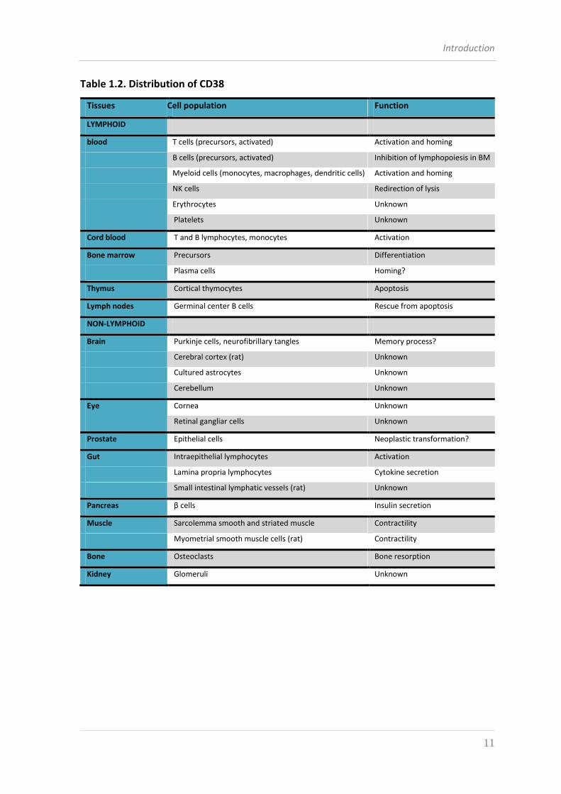
Human CD38 was originally designated as an activation marker during the quest to
identify cell surface molecules involved in T cell recognition; indeed, this concept was
validated by the observation of CD38 expression on thymocytes and T lymphocytes [12].
This definition was later proven inadequate when the molecule was shown to be neither
lineage- nor activation-restricted and now CD38 expression is considered virtually
ubiquitous with a widespread distribution either in lymphoid and nonlymphoid tissues
(Table 1.2). However, the underlying mechanism of action of the molecule’s expression in
different cell lineage is still not entirely clear, and this is reflected in the sometimes
contrasting reports in literature. A non negligible fact is that the expression of CD38
modifies significantly with age and, mostly for the hematological lineage, CD38 has been
demonstrated to be expressed by immature hematopoietic cells, downregulated by
mature cells and re-expressed at high levels by activated lymphocytes such as T cells, B
cells, dendritic cells and natural killer (NK) cells [13].
Introduction
11
Table 1.2. Distribution of CD38
Tissues Cell population Function
LYMPHOID
blood T cells (precursors, activated) Activation and homing
B cells (precursors, activated) Inhibition of lymphopoiesis in BM
Myeloid cells (monocytes, macrophages, dendritic cells) Activation and homing
NK cells Redirection of lysis
Erythrocytes Unknown
Platelets Unknown
Cord blood T and B lymphocytes, monocytes Activation
Bone marrow Precursors Differentiation
Plasma cells Homing?
Thymus Cortical thymocytes Apoptosis
Lymph nodes Germinal center B cells Rescue from apoptosis
NON-LYMPHOID
Brain Purkinje cells, neurofibrillary tangles Memory process?
Cerebral cortex (rat) Unknown
Cultured astrocytes Unknown
Cerebellum Unknown
Eye Cornea Unknown
Retinal gangliar cells Unknown
Prostate Epithelial cells Neoplastic transformation?
Gut Intraepithelial lymphocytes Activation
Lamina propria lymphocytes Cytokine secretion
Small intestinal lymphatic vessels (rat) Unknown
Pancreas β cells Insulin secretion
Muscle Sarcolemma smooth and striated muscle Contractility
Myometrial smooth muscle cells (rat) Contractility
Bone Osteoclasts Bone resorption
Kidney Glomeruli Unknown
Introduction
12
1.3.1.1 CD38 STRUCTURE AND FUNCTION
Human CD38 is a pleiotropic type II surface glycoprotein made of 300 amino acid
residues and with a molecular weight of 46 kDa comprising two to four N-linked
oligosaccharide chains containing sialic acid residues [14].
The structure of CD38 proved difficult to establish and has been accomplished only
recently thanks to a highly efficient yeast expression system which has been developed
on purpose to enable structure-function studies and to facilitate purification of much of
the cyclase for crystallography. This strategy has been used to obtain a construct with a
missing transmembrane segment and mutated glycosylation sites [15]. The resulting
extramembrane domain was fully active in terms of enzymatic functions and was
crystallized as head-to-tail dimers [14].
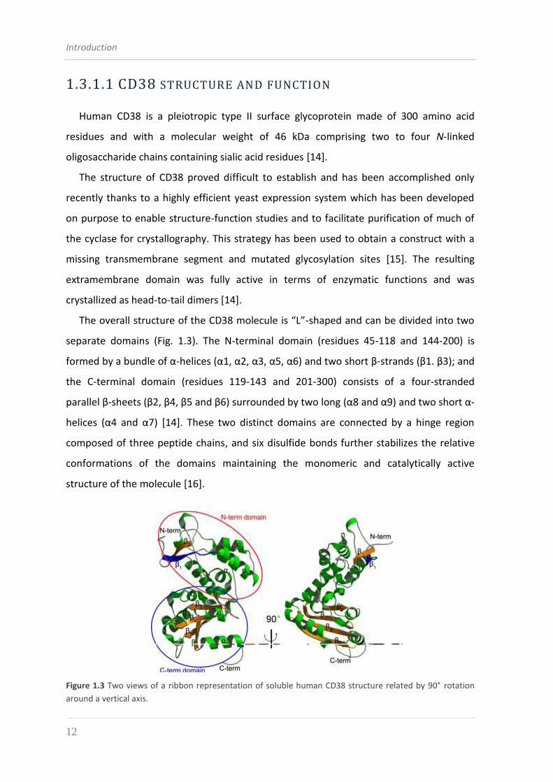
The overall structure of the CD38 molecule is “L”-shaped and can be divided into two
separate domains (Fig. 1.3). The N-terminal domain (residues 45-118 and 144-200) is
formed by a bundle of α-helices (α1, α2, α3, α5, α6) and two short β-strands (β1. β3); and
the C-terminal domain (residues 119-143 and 201-300) consists of a four-stranded
parallel β-sheets (β2, β4, β5 and β6) surrounded by two long (α8 and α9) and two short α-
helices (α4 and α7) [14]. These two distinct domains are connected by a hinge region
composed of three peptide chains, and six disulfide bonds further stabilizes the relative
conformations of the domains maintaining the monomeric and catalytically active
structure of the molecule [16].
Figure 1.3 Two views of a ribbon representation of soluble human CD38 structure related by 90° rotation
around a vertical axis.

Introduction
13
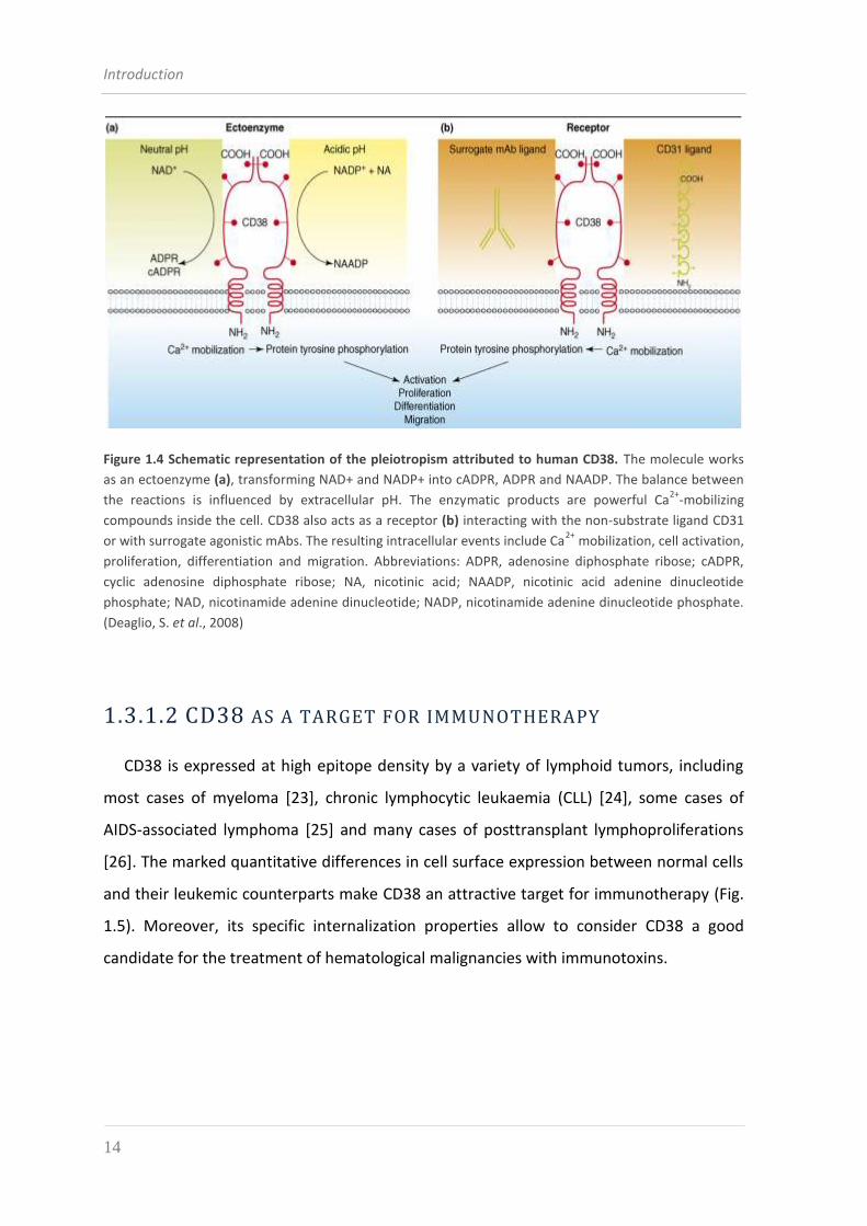
CD38 was originally defined as an ectoenzyme (enzyme of the plasmatic membrane
which catalyzes reaction taking place in the extracellular space), but during evolution it
acquired the ability to mediate cell–cell interactions, acting as a receptor [17].
As an ectoenzyme, CD38 belongs to a complex family of the cell surface enzymes
involved in the catabolism of extracellular nucleotides. With its ADP-ribosyl cyclase
activity, CD38 generates cyclic ADP ribose (cADPR) and ADPR from NAD+ and nicotinic acid
adenine dinucleotide phosphate (NAADP) from NADP+. These second messengers
cooperate in the regulation and modulation of intracellular Ca2+ that plays a key role in
several physiological processes, including cell proliferation, muscle contraction, stem cell
regeneration and hormone secretion [18] (Fig. 1.4a).
The use of agonistic mAbs demonstrated that CD38 engagement is followed by signals
that are apparently independent of CD38 enzymatic activities [19]. In fact, CD38 ligation
determine tyrosine phosphorylation of a sequential number of intracellular signal
transducers such as ZAP-70 and the proto-oncogene c-cbl (which drive life and death
messages to the cells) or the phospholipase C-γ responsible for Ca2+ mobilization [20].
Study of CD38 as a receptor also demonstrated that the cross-linking with CD31 (also
known as platelet endothelial cell adhesion molecule-1, PECAM-1), as a non-substrate
ligand for CD38, activates a signaling which leads to proliferation of lymphocyte
populations and to inhibition of apoptosis [21] (Fig. 1.4b).
Finally, studies by A. Funaro demonstrated that CD38 undergoes internalization
following ligation with agonistic (IB4) or nonagonistic (IB6) specific mAbs [22].
Internalization, along with shedding, represents a mechanism of down-regulation of CD38
and this is independent on the amount of CD38 molecules constitutively expressed by
different cells. Furthermore, this mechanism never involves the entire amount of surface
molecule; on the contrary, the internalized fraction represents an almost constant
percentage (30-40%) of the total amount of surface CD38 molecules, suggesting the
existence of two pools of molecules, one of which undergoes internalization after Ab
binding.
Introduction
14
Figure 1.4 Schematic representation of the pleiotropism attributed to human CD38. The molecule works
as an ectoenzyme (a), transforming NAD+ and NADP+ into cADPR, ADPR and NAADP. The balance between
the reactions is influenced by extracellular pH. The enzymatic products are powerful Ca2+
-mobilizing
compounds inside the cell. CD38 also acts as a receptor (b) interacting with the non-substrate ligand CD31
or with surrogate agonistic mAbs. The resulting intracellular events include Ca2+
mobilization, cell activation,
proliferation, differentiation and migration. Abbreviations: ADPR, adenosine diphosphate ribose; cADPR,
cyclic adenosine diphosphate ribose; NA, nicotinic acid; NAADP, nicotinic acid adenine dinucleotide
phosphate; NAD, nicotinamide adenine dinucleotide; NADP, nicotinamide adenine dinucleotide phosphate.
(Deaglio, S. et al., 2008)
1.3.1.2 CD38 AS A TARGET FOR IMMUNOTHERAPY
CD38 is expressed at high epitope density by a variety of lymphoid tumors, including
most cases of myeloma [23], chronic lymphocytic leukaemia (CLL) [24], some cases of
AIDS-associated lymphoma [25] and many cases of posttransplant lymphoproliferations
[26]. The marked quantitative differences in cell surface expression between normal cells
and their leukemic counterparts make CD38 an attractive target for immunotherapy (Fig.
1.5). Moreover, its specific internalization properties allow to consider CD38 a good
candidate for the treatment of hematological malignancies with immunotoxins.

Introduction
15
(e) Im
mu
no
toxin
s-m
edia
ted
cyto
toxic
ity
(a) Block of migration
(b) R
ad
ioim
mu
no
thera
py
(c) Cell-mediated cytotoxicity(d) C’-mediated cytotoxicity
Figure 1.5 Potential applications of CD38 as a therapeutic target. The mechanisms of action of therapeutic
anti-CD38 mAbs include: (a) the inhibition of a function, in this instance migration, considered critical for
CLL progression. Inhibition might be the result of the action of a mAb binding a specific domain of CD38 and
perturbing ligation of CXCL12 to its receptor. Alternatively, a divalent ligand of antibody origin might
provide a simultaneous binding to CD38 and CXCR4, increasing ligand specificity. (b) The use of mAbs as
carriers of radiopharmaceuticals delivering a lethal hit either by surface (γ emitters) or cytoplasmic (β
emitters) irradiation. (c) The elicitation of cytotoxicity mediated in v ivo by killer cells. (d) The elicitation of
cytotoxicity mediated in v ivo by complement. (e) The use of mAbs as carriers of toxins. (Modified from
Deaglio, S. et al., 2008).
1.3.1.2.1 CHRONIC LYMPHOCYTIC LEUKEMIA (CLL)
B-cell chronic lymphocytic leukemia (B-CLL) is the abnormal progressive accumulation
of functionally incompetent monoclonal B-lymphocytes in blood, bone marrow, lymph
nodes and spleen [27]. It is the most common adult leukemia in Western countries,
accounting for about 30% of total leukemias. Worldwide there are approximately 180.000
new cases every year. CLL is a pathology of the adult age which is rarely diagnosticated in
individuals earlier than 40 years and its incidence rapidly increases with age after 55
Introduction
16
years. The clinical course of CLL is extremely variable with survival ranging from 1 to more
than 15 years.
The exact causes of CLL are unknown, with epidemiological studies finding no
association with viral infection, chemical or radiation exposure [28]. Conventional
therapies are ineffective and, although hematopoietic stem cell transplantation (HSCT)
has led to complete remission for some patients [29], the development of new
therapeutic strategies is critical to improve the clinical outcome.
Most of the circulating B-CLL cells are quiescent, resting in the G0 phase of the cell
cycle, and they can survive for a few months as opposed to a few days for normal B cells
[30]. Thus, CLL can be considered a disease caused by a loss of appropriate apoptosis,
rather than increased proliferation [31], even if no defects in the apoptotic pathways can
be noticed suggesting that there are micro-environmental factors regulating death in B-
CLL cells. As a consequence of B cells functional incompetence, patients become
immunocompromised and easily exposed to recurrent infections which are often the
cause of death. 10-15 % of patients develops autoimmune hemolytic anemia, while in 2-
8% of the cases CLL tends to transform into an aggressive lymphoma called Richter’s
Syndrome which is associated with a rapid progression of the disease, chemotherapy
resistance and a poor prognosis (which leads to death in 6 months) [32].
Several published works demonstrate that some diagnostic marker of CLL are
associated with Richter transformation: overexpression of CD38 and ZAP-70 by B-CLL
lymphocytes and absence of mutations on variable domains of their IgG gene are
unfavorable characteristics which promote activation of B cells and that determine
survival and proliferation in presence of cytokines, chemokines and other signals [33, 34].
Accumulated experience with CLL indicates that:
CD38 is a marker selectively expressed by patients who are generally poorly
responsive to conventional treatment;
CD38 is a receptor for growth and survival signals mediated through interactions
with favorable microenvironments and, finally;
CD38 is a component of the multiple elements of the migratory machinery, which
depends on chemokines and their receptors.
Introduction
17
These overall observations suggest that CD38 is a potential therapeutic target for CLL
[17].
1.3.1.2.2 MULTIPLE MYELOMA (MM)
Multiple myeloma (MM) is a malignant disorder of the B cell lineage, characterized by
neoplastic monoclonal expansion of plasma cells able to spread within the bone marrow
and produce osteolytic lesions resulting in destruction of adjacent bone tissue. MM
accounts for approximately 1% of neoplastic disease and 13% of all hematological
cancers, its incidence increases with age and the median age at diagnosis is about 70
years.
In recent years, the introduction of autologous hematopoietic stem cell
transplantation (HSCT) together with the availability of novel drugs such as thalidomide,
lenalidomide and bortezomib, especially when used in combination regimens, have
dramatically improved initial response rates and prolonged overall survival [35];
nevertheless MM remains an incurable disease with a median overall survival of 4-7 years
and new therapeutic options are needed for patients.
It has been demonstrated that myeloma cells themselves are not merely less sensitive
to chemotherapy due to their dormancy but they are prone to develop multidrug
resistance (MDR) due to the overexpression of the transporter P-glycoprotein (P-gp), the
sigma receptors (σR2) and to a defective apoptosis mechanism.
The strong expression of CD38 by myeloma cells has been exploited for the
development of targeted therapies using both “naked” antibodies able to induce ADCC
(as demonstrated by the works of G.T. Stevenson [36, 37]), and immunotoxins (e.g.
IB4/saporin-S6, a mAb coupled to saporin-S6 which was created by the group of Bolognesi
[38]). In spite of their promising results, these new approaches did not lead to clinical
applications: the molecule’s widespread distribution in lymphoid, myeloid and epithelial
cells as well as in specialized tissues and organs including the eyes (see Table 1.2), caused
a general reluctance to use CD38 as a target in human therapy.
Nevertheless, further impetus for designing clinical models based on anti-CD38
molecules came from the observation by the K. Mehta group that the expression of CD38
Introduction
18
is highly sensitive to exogenous and endogenous all-trans retinoic acid (ATRA) and
derivatives and that the sensitivity is strongly magnified in tumors and leukemic cells [39].
These initial observations were reevaluated from therapeutic perspectives in acute
promyelocytic leukemia (APL), as well as in other myeloid leukemias. ATRA is an in vitro
inducer at nanomolar concentrations of the cell surface CD38 in myeloid leukemia blasts.
The same reagent is a key component in clinical differentiation therapy adopted for APL
cells, which are generally CD38- before treatment. The CD38 molecules expressed de
novo at high epitope density may be used as therapeutic targets and the combination of
ATRA with an anti-CD38 immunotoxin may result in a synergistic killing of leukemia cells
[40].
1.4 IMMUNOTOXINS
Immunotoxins (ITs) are chimeric proteins composed of a targeting portion (usually an
antibody fragment, a cytokine or a growth factor) linked to a toxin. Immunotoxins bind to
surface antigens on a cancer cell, enter the cell by endocytosis, and kill it by enzimatically
inhibiting protein synthesis. The most potent immunotoxins are made from bacterial and
plant toxins.
First-generation ITs, obtained by chemically conjugating one whole toxin to a mAb, often
showed no efficacy in animal models because they lacked specificity and they were toxic also
for normal cells. Replacement of the cell-binding domain of the toxin with an antibody-
derived domain led to the development of more specific compounds and much better
tolerated by animals. However these second-generation ITs, as their precursors
immunoconjugate, were unstable and not homogeneous in composition [41]. These
difficulties were overcome exploiting recombinant DNA techniques and the principles of
protein engineering to obtain third-generation immunotoxins, designed to contain only the
elements required to recognized and kill the tumour cells. In the last 10-15 years several
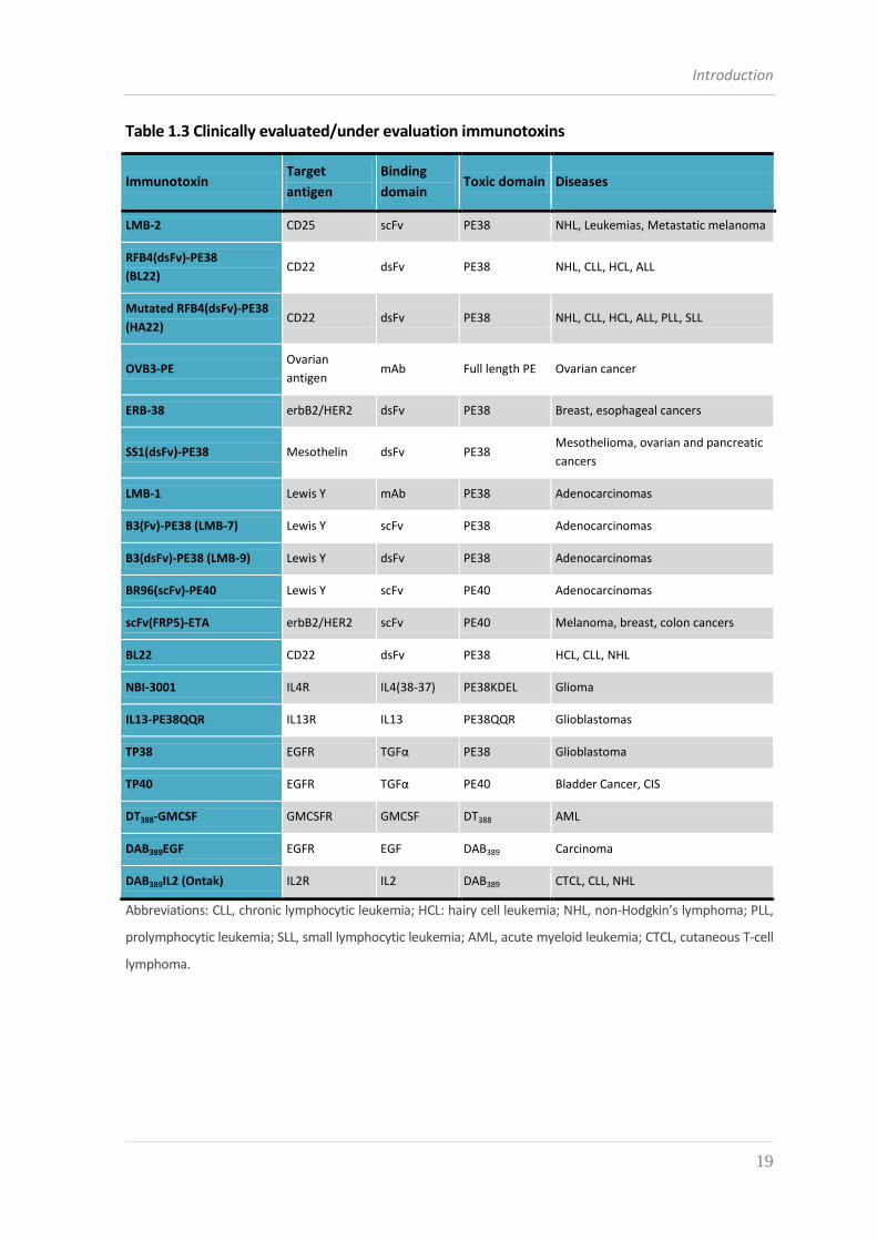
recombinant immunotoxins have been evaluated in clinical trials (Table 1.3).
Introduction
19
Table 1.3 Clinically evaluated/under evaluation immunotoxins
Immunotoxin Target
antigen
Binding
domain Toxic domain Diseases
LMB-2 CD25 scFv PE38 NHL, Leukemias, Metastatic melanoma
RFB4(dsFv)-PE38
(BL22) CD22 dsFv PE38 NHL, CLL, HCL, ALL
Mutated RFB4(dsFv)-PE38
(HA22) CD22 dsFv PE38 NHL, CLL, HCL, ALL, PLL, SLL
OVB3-PE Ovarian
antigen mAb Full length PE Ovarian cancer
ERB-38 erbB2/HER2 dsFv PE38 Breast, esophageal cancers
SS1(dsFv)-PE38 Mesothelin dsFv PE38 Mesothelioma, ovarian and pancreatic
cancers
LMB-1 Lewis Y mAb PE38 Adenocarcinomas
B3(Fv)-PE38 (LMB-7) Lewis Y scFv PE38 Adenocarcinomas
B3(dsFv)-PE38 (LMB-9) Lewis Y dsFv PE38 Adenocarcinomas
BR96(scFv)-PE40 Lewis Y scFv PE40 Adenocarcinomas
scFv(FRP5)-ETA erbB2/HER2 scFv PE40 Melanoma, breast, colon cancers
BL22 CD22 dsFv PE38 HCL, CLL, NHL
NBI-3001 IL4R IL4(38-37) PE38KDEL Glioma
IL13-PE38QQR IL13R IL13 PE38QQR Glioblastomas
TP38 EGFR TGFα PE38 Glioblastoma
TP40 EGFR TGFα PE40 Bladder Cancer, CIS
DT388-GMCSF GMCSFR GMCSF DT388 AML
DAB389EGF EGFR EGF DAB389 Carcinoma
DAB389IL2 (Ontak) IL2R IL2 DAB389 CTCL, CLL, NHL
Abbreviations: CLL, chronic lymphocytic leukemia; HCL: hairy cell leukemia; NHL, non-Hodgkin’s lymphoma; PLL,
prolymphocytic leukemia; SLL, small lymphocytic leukemia; AML, acute myeloid leukemia; CTCL, cutaneous T-cell
lymphoma.
Introduction
20
1.4.1 THE BINDING DOMAIN
In the design of an IT a variety of binding domains can be used to selectively deliver
the drug to the intended cell target; besides monoclonal antibodies and fragments thence
derived, other small proteins are appropriate to fulfil this function, e.g. growth factors
and cytokines. Such molecules impart specificity to the IT by virtue of the higher
expression level of some receptors for growth factors and cytokines on tumor cells.
Interleukin 2 (IL-2), IL-13, transforming growth factor α (TGFα) and granulocyte-
macrophage colony stimulating factor (GMCSF), for instance, have been employed in the
construction of immunotherapeutic agents to direct toxic molecules towards leukemia or
lymphoma cells [42, 43].
However, due to their remarkable molecular versatility and to the possibility of being
raised against virtually any target, antibodies are probably the best candidates as binding
domains in ITs design [44].
1.4.1.1 ANTIBODIES
Antibodies are glycoproteins belonging to the immunoglobulin (Ig) superfamily; they
are produced by B lymphocytes (B cells) in response to exposure to an antigen. They react
specifically with that antigen in vivo or in vitro and are hence a part of the humoral
adaptive immune response.
All antibody molecules share the same basic structural characteristics but display
remarkable variability in the region that bind antigens. The basic structural unit of an
antibody molecule consists of four polypeptide chains, two identical light chains (L) and
two identical heavy chains (H). The four chains are linked covalently by disulfide bonds
(Fig. 1.6). Each heavy chain has a molecular weight of 50-75 kDa and contains about 400
amino acids and the amino acid differences in its carboxy terminal portion identifies five
isotypes (IgG, IgA, IgM, IgD and IgE). Light chains have a molecular weight of
approximately 23 kDa, are composed of about 212 amino acids and are of two types, κ and
λ, based on their structural (antigenic) differences.
Both heavy and light chains consist of amino terminal variable (V) regions that
participate in antigen recognition and carboxy terminal constant (C) regions.
Introduction
21
Variable regions are so named because most of the variability in amino acids sequence,
that distinguish the antibodies made by different clones of B cells, is confined to three
short stretches in the V regions of heavy and light chains (VH and VL). These hypervariable
regions (each about 10 amino acid residues long) are called complementarity-determining
regions (CDRs) and they are held in place by more conserved framework sequences [45].
The VH is juxtaposed with the VL to form an antigen-binding site.
The C region domains are separate from the antigen-binding site and do not
participate in antigen recognition. The constant regions of the two heavy chains constitute
the so-called Fragment Crystallizable (Fc) portion which, interacting with cells of the
immune system, mediates effector functions.
(a) (b)
Antigen-
binding site
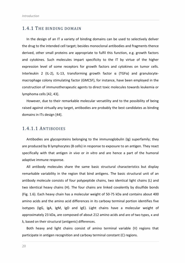
Figure 1.6 Structure of an antibody molecule. (a) Schematic diagram of a secreted IgG molecule. It is
composed of four polypeptide chains, two light and two heavy chains, each of which is organized in
domains of 110 amino acids containing a disulphide bridge that forms a loop of approximately 60 amino
acids. The heavy chain of an IgG comprises three constant domain (CH) and one variable domain (VH), while
the light chain is made by one single constant domain (CL) and one variable domain (VL). The antigen-
binding sites are formed by the juxtaposition of the VL and VH domains. (b) Structure of a human IgG
molecule as revealed by x-ray crystallography. In this ribbon diagram, the heavy chains are colored blue and
red, and the light chains are colored green and yellow.
Early studies of antibody structure relied on antibodies purified from the serum of
animals immunized with various antigens, yielding a polyclonal pool of reactive
immunoglobulins that may respond to different epitopes of an antigen. Despite their high
binding affinity, polyclonal antibodies are unsuitable for therapeutic use, due to their
Introduction
22
heterogeneous composition. The most important breakthrough in the field of antibody-based
therapies was the introduction, by Georges Köhler and Cesar Milstein in 1975, of the
hybridoma technology for producing monoclonal antibodies. Hybridoma technology is based
on the somatic fusion of myeloma cells and B lymphocytes from the spleen of an immunized
mouse. The resulting chimeras are immortalized cells capable to secrete antibodies
indefinitely (Fig. 1.7). Since each fusion yields mAbs with a unique and specific idiotype (i.e.
antigen-binding site), the hybridoma clone producing the mAb with highest affinity can be
selected and propagated, thus providing bulk amounts of a homogeneous immunoglobulin
with the desired specificity [6].
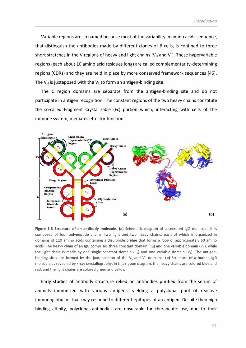
Figure 1.7 The hybridoma technology for producing monoclonal antibodies.
Abbreviations: HGPRT, hypoxanthine-guanine phosphoribosyltransferase; HAT medium, hypoxanthine-
aminopterin-thymidine medium.
The ability of IgGs to bind two antigens greatly increases their functional affinity and
confers high retention times (also called avidity) on many cell-surface receptors and
polyvalent antigens. The Fc domain recruits cytotoxic effector functions through complement
and/or through interactions with γFc receptors (Fc receptors for gamma globulins) and can
provide long serum half-lives (>10 days) through interaction with the neonatal Fc receptor
(FcRn).[46].
Introduction
23
Monoclonal antibodies can be covalently coupled to toxins or toxin derivatives by
chemical means. Generally, coupling reactions involve at least one accessible SH group. The
connection of the antibody to the toxin utilizes two types of chemical bonds for conjugation.
One class of immunotoxins contains a disulfide linker, so that the antibody and toxin separate
upon reduction within the target cell. The other class of chemical linkers contains thioether
bonds that cannot be cleaved by reduction. Different linkers for conjugation of proteins have
been developed, varying in length, stability, flexibility and chemical reactivity: which linker
class (cleavable or not) and which conjugation chemistry to be used depends first on the
nature of the toxin and an experimental evaluation is usually required remembering that
differences in linkers can greatly affect the activity of immunotoxins [47, 48].
1.4.1.2 ANTIBODY FRAGMENTS
To address some of the limitations of large IgG molecules, an ever increasing
importance has been acknowledged to the study and development of antibody fragments
(Fig. 1.8), which are smaller molecules with a potentially better tissue penetration in case
of solid tumors. Antibody fragments still maintain an unaltered binding capability and
specificity to the antigen while not having non-selective activity due to non specific
binding of the antibody Fc portion.
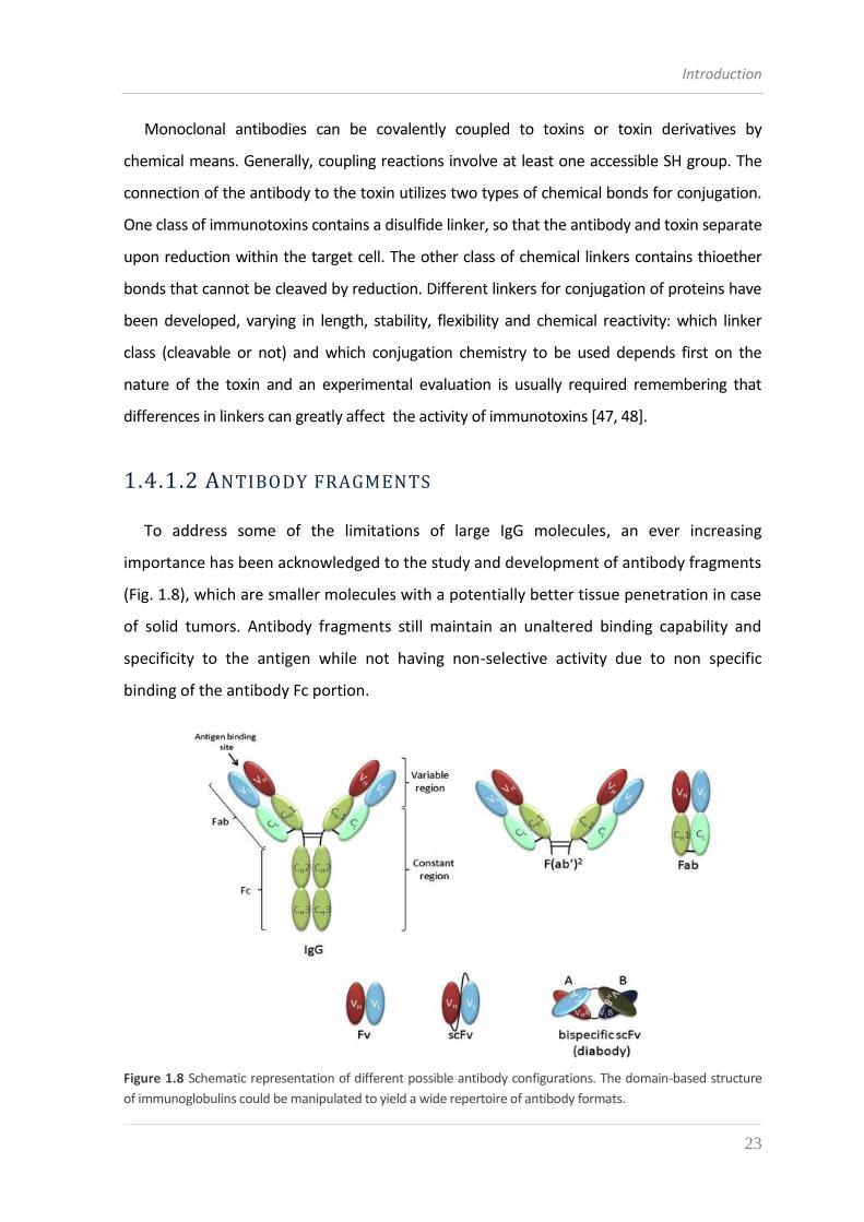
Figure 1.8 Schematic representation of different possible antibody configurations. The domain-based structure
of immunoglobulins could be manipulated to yield a wide repertoire of antibody formats.
Introduction
24
Initially, such fragments could be prepared by proteolytic digestion of whole
immunoglobulins. Treatment of IgG with proteases like papain generates three separate
fragments: the Fc region and two identical Fab (fragment, antigen binding) regions,
comprising the complete light chain (VL and CL) associated with the variable domain and
the first constant domain of the heavy chain. When pepsin (instead of papain) is used to
cleave IgG, the released fragment is called F(ab’)2 and is composed of a pair of Fab' units
connected by two disulfide bonds. Fabs and F(ab’)2 retain the ability to bind antigen
because each contains paired VL and VH domains (Fig. 1.8) and at the same time their
employment in ITs design brought about reduced levels of non-specific binding as
compared to whole mAbs. However, since antibodies show variable susceptibility to
proteolytic treatment, the final product can be highly heterogeneous, which accounts for
the poor success claimed for immunotherapeutics manufactured by this kind of
procedures [49]. The development of recombinant DNA technologies offered the chance
to synthesize Fabs and even smaller fragments in heterologous expression systems.
The smallest unit of an immunoglobulin molecule with function in antigen-binding
activities is called Fv fragment and it is represented by the heterodimer formed by the
variable domains of heavy and light chains (Fig. 1.8). The genes encoding the VH and VL
domains are usually cloned from hybridoma mRNA by reverse transcription, cDNA
synthesis and subsequent PCR amplification using degenerate primers that are
complementary to the conserved sequences at the 5’ end of genes for the VH and VL
domains and to the 3’ ends of the JH and JL regions (located at the 3’ ends of VH and VL) [50].
The PCR amplified variable regions can then be cloned into an appropriate plasmid vector for
expression in a heterologous host, especially Escherichia coli [51]. In a recombinant Fv
fragment VH and VL domains are associated through non-covalent interactions which may not
be strong enough to assure the dimer stability; in fact the dissociation constant of the VH-VL
complex, ranging between 10-5 and 10-8 M, is often not sufficient to keep the two domains
together under slightly destabilizing conditions or when the protein concentration is low [49].
To improve their stability different strategy has been developed creating scFvs (single-chain
Fragments variable) and dsFvs (disulphide-stabilized antibody fragments).
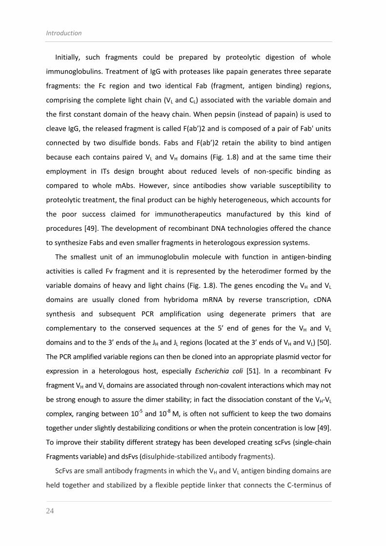
ScFvs are small antibody fragments in which the VH and VL antigen binding domains are
held together and stabilized by a flexible peptide linker that connects the C-terminus of
Introduction
25
the VH (or VL) with the N-terminus of the other domain (Fig. 1.8). Various linkers were
designed to provide flexibility and enhance solubility, with the most widely used linker
varying from 10 to 25 amino acids in length and typically including hydrophilic amino
acids; the most common linker is the decapentapeptide (Gly4Ser)3. ScFvs often show a
good binding capability, comparable to that of the mAb they derive from; while in some
cases a considerable loss of affinity can be observed, probably associated with a high
tendency to aggregate and form unstable multimers. The formation of dimers and trimers
is primarily determined by linker length and it is favored by shorter linkers (0–12 amino
acids) [52], while the VH-VL orientation can affect expression efficiency, stability, and
antigen binding activity [53].
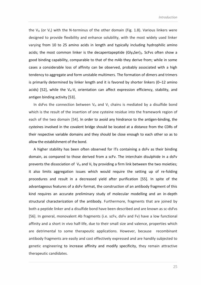
In dsFvs the connection between VH and VL chains is mediated by a disulfide bond
which is the result of the insertion of one cysteine residue into the framework region of
each of the two domain [54]. In order to avoid any hindrance to the antigen-binding, the
cysteines involved in the covalent bridge should be located at a distance from the CDRs of
their respective variable domains and they should be close enough to each other so as to
allow the establishment of the bond.
A higher stability has been often observed for ITs containing a dsFv as their binding
domain, as compared to those derived from a scFv. The interchain disulphide in a dsFv
prevents the dissociation of VH and VL by providing a firm link between the two moieties;
it also limits aggregation issues which would require the setting up of re-folding
procedures and result in a decreased yield after purification [55]. In spite of the
advantageous features of a dsFv format, the construction of an antibody fragment of this
kind requires an accurate preliminary study of molecular modelling and an in-depth
structural characterization of the antibody. Furthermore, fragments that are joined by
both a peptide linker and a disulfide bond have been described and are known as sc-dsFvs
[56]. In general, monovalent Ab fragments (i.e. scFv, dsFv and Fv) have a low functional
affinity and a short in vivo half-life, due to their small size and valence, properties which
are detrimental to some therapeutic applications. However, because recombinant
antibody fragments are easily and cost effectively expressed and are handily subjected to
genetic engineering to increase affinity and modify specificity, they remain attractive
therapeutic candidates.
Introduction
26
1.4.2 THE TOXIC DOMAIN
Several types of therapeutic agents have been used in the design of anticancer ITs and
constructs containing cytotoxic drugs, cytokines, toxins or radionuclides have been
evaluated in preclinical and clinical studies.
Standard chemotherapeutic drugs belonging to the antifolates, vinca alkaloids or
anthracyclines have been chemically linked to mAbs, but these immunoconjugates proved
to be inefficient in the clinical situation due to the moderate cytotoxic potential of these
drugs and to the limited achievement of therapeutic levels within the cells [2].
Toxins constitute another class of highly cytotoxic agents that have been conjugated to
mAbs and tested for antitumor therapy efficacy. Toxins are poisonous substances (usually
protein) produced by living cells or organisms. In contrast to the low-molecular-mass
chemical molecules adducted for chemotherapy, toxins used for anticancer therapy are
generally enzymes that exert their cytotoxic activity inside the cell; in most cases one
single molecule in the appropriate intracellular compartment is sufficient to kill the cell.
To be used therapeutically, toxins mostly have to be modified to remove their binding
sites for targets expressed in normal tissue. In addition, toxins often have to be
deglycosylated to avoid rapid clearance by liver cells expressing mannose receptors.
The toxins that are best analyzed and most commonly used for making immunotoxins
are the protein synthesis inhibiting toxins: ricin, diphtheria toxin (DT) and Pseudomonas
exotoxin (PE). All these proteins have been crystallized, their structures determined and
specific functions assigned to different structural subunits or domains of which they are
composed. Also, the genes for these toxins have been cloned and expressed as
recombinant proteins in E. coli [57]. Combining the understanding of their structure and
function with molecular cloning techniques has made it possible to generate genetically
altered toxin derivatives with improved properties for use as immunotoxins.
Introduction
27
1.4.2.1 PLANT TOXINS
Plants synthesize and accumulate in seeds and leaves a broad range of secondary
metabolites, including alkaloids and terpenoids, that are toxic to herbivores and
pathogens, and so are believed to act as defense compounds [58].
Plant toxins belong to the ribosome inactivating proteins (RIPs), a class of potent
inhibitors of protein synthesis that act by catalytically depurinating, thanks to their RNA
N-glycosidase activity, an adenine residue (A4324 in rats) present in a conserved stem-
loop region in 23/26/28S large ribosomal RNAs. The removal of this adenine, preventing
association of the ribosome with the elongation factor 2 (eEF-2), causes an irreversible
arrest of protein synthesis and consequently cell death occurs.
RIPs from plants have been classified into three main categories: type 1 are composed
of a single polypeptide chain of approximately 30 KDa, type 2 are heterodimers consisting
of an A chain, functionally equivalent to the type 1 polypeptide, linked to a B subunit,
endowed with lectin-binding properties, while type 3 are synthesized as inactive
precursors (ProRIPs) that require proteolytic processing events to form an active RIP and
are not in use for therapeutic purposes.
Type 1 RIPs, like saporin (from the seeds of the soapwort Saponaria officinalis), PAP
(pokeweed antiviral protein, from the plant Phytolacca americana) and gelonin (from the
seeds of Gelonium multiflorum), are characterized by a high basicity (pI > 9.5) and can be
glycosilated. Some of them have a N-terminal sequence that directs them to the
endoplasmic reticulum. The routing of type 1 RIPs to reach their target into the cytosol is
actually unclear but it has been demonstrated that the binding of saporin to the cell
surface is at least in part mediated by the α2-macroglobulin receptor (α2MR; also termed
low density lipoprotein-receptor-related-protein, LPR), indicating a general mechanism of
interaction of plant RIPs with the α2MR system [59]. After being endocytosed, the toxin
reaches the endo-lysosomal compartment from where it is delivered to the cytosol
following as yet unidentified pathways.
As previously described, type 2 RIPs are holotoxins containing an A chain, which is the
enzymatically active one (N-glycosidase), linked, through a disulphide bond, to a B chain
that mediates binding to the terminal galactose or N-acetylgalactosamine residues
Introduction
28
present on the surface of most mammalian cells and that promotes the translocation of
the A chain into the cytoplasm.
Among type 2 RIPs, ricin, obtained from the seeds of the castor oil plant Ricinus
communis, has been the one most widely used in preclinical and clinical studies [60]. After
entering mammalian cells by endocytosis, ricin is transported to the early endosomes and
undergoes retrograde transport via the Golgi complex to the endoplasmic reticulum (ER)
where the catalytic moiety exploits the ER-associated degradation (ERAD) pathway,
normally used for the disposal of misfolded or unassembled polypeptides, to reach and
depurinate cytosolic ribosomes. This retrograde route of transport for ricin represents a
highly effective strategy to deliver its A domain into the cytosol, which is a prerequisite
for exerting toxicity [61].
Predictably, ITs containing native ricin and other type 2 RIPs lack specificity since they
bind not only to target cells, but virtually to any other cell via the B chain. This problem
has been successfully circumvented by altering or deleting the binding domain through
two different approaches:
1) by separating the functionally active A chain from the B chain and deglycosylating
the A chain (dgA) (this latter procedure prevents liver toxicity due to glycosylated
residues on the A chain which recognize parenchymal and non parenchymal cells
in the liver, causing hepatotoxicity and poor biodistribution)[62]. Alternatively, the
A chain can be made as a recombinant protein in bacteria, but this last approach
did not lead to obtain functional immuntoxins when A chain was conjugated to an
antibody [63];
2) by attaching affinity ligands (i.e. galactose, lactose, or glycopeptides) to the sugar-
binding sites of the B chain (blocked ricin) [64].
Due to their catalytic mechanism of action, these toxins are extremely potent; it has
been estimated that a few molecules of ricin in the cytoplasm are enough to kill a cell.
Moreover, since their mechanism of cell killing is different from those of standard
chemotherapeutic agents, it is reasonable to expect that they could exert an efficient
antitumor activity against chemoresistant and/or resting neoplastic cells without
cumulative bone marrow toxicity.
Introduction
29
1.4.2.1.1 SAPORIN
The plant toxin saporin, compared to other RIPs, shows various peculiar features in
terms of remarkable stability and activity on a wide variety of substrates, that have made
it an interesting protein to be employed in the design of immunotoxins [65, 66].
The term saporin collectively identifies a family of RIP isoforms that accumulate in
different tissues of the soapwort Saponaria officinalis. Mixtures of closely related
isoforms and several cDNA and genomic clones have been isolated; among them, SO6
saporin (or saporin-6) represents the major HPLC peak of purified seed protein and
constitutes about 7% of the total proteins. Seed protein sequencing revealed
heterogeneity at two positions, with either an aspartic or a glutamic acid in position 48,
and either lysine or arginine present in position 91, indicating that the SO6 peak contains
a set of correlated isoforms. In fact, HPLC analysis confirmed the presence of at least
three different components in SO6 preparations while recombinant expression of single
seed-like isoforms demonstrated the same RIP activity, except for a leaf-derived isoform
[67].
While some characteristics of the saporin proteins, such as key catalytic residues and
overall three-dimensional fold, are shared with RTA and the other known crystallized
RIPs, other biochemical features clearly differ among type I plant RIPs and RTA. For
example, the sequence identity is low and, in particular, only 22% of residues are
conserved between RTA and saporin SO6. On the contrary, a high degree of sequence
identity (about 80%) is found between saporin SO6 and dianthin from Dianthus
caryophyllus, both of which are synthesized by plants belonging to the same subfamily of
the Caryiophyllaceae family. Despite that, all the crystallized RIPs have been shown to
share a common “RIP fold”, as can be estimated by the superimposition of the 3D
structures of several type I RIPs and RTA, which is characterized by the presence of two
major domains: an N-terminal domain, which is mainly β-stranded, and a C-terminal
domain that is predominantly α–helical [68].
Saporin cytotoxicity varies in a wide range, with concentrations inhibiting protein
synthesis by 50% (IC50) changing from nanomolar to micromolar, depending on the cell
lines investigated and on the expression of the α2-macroglobulin receptor/low-density
Introduction
30
lipoprotein receptor-related protein (LRP1) which has been proved to bind saporin in
vitro and mediate its internalization in human monocytes and in fibroblasts [67].
As compared to ricin-based ITs, saporin has been exploited relatively little as the toxin
of choice for clinical uses, so far. A small clinical study with an anti-CD30 monoclonal
conjugated to the seed extracted saporin (BERH2-SAP) for the treatment of Hodgkin’s
lymphoma, proved very encouraging [69] without reporting serious drug-related
toxicities. However, antibodies against both domains were raised in the treated patients.
ITs based on saporin have been also used by Flavell and coworkers for their clinical trials
in adult and pediatric patients with hematological malignancies: their pre-clinical studies
showed that an anti-CD19 immunotoxin, named BU12-saporin, and an anti-CD38
immunotoxin, OKT10-saporin [70], displayed selective antitumor activity both in vitro and
in vivo against malignant target hematological cells.
1.4.2.2 BACTERIAL TOXINS
Pseudomonas aeruginosa Exotoxin A (PE) and Corynebacterium diphtheria toxin (DT)
are the most widely exploited bacterial toxins for the immunotherapy of cancer. Both PE
and DT enzymatically modify eEF-2 in the cytosol by catalyzing the adenosine diphosphate
(ADP) ribosylation of residue His699 of eEF-2 which is post-translationally modified to a
diphthimide residue [71, 72]. This modification irreversibly inactivates eEF-2 causing the
arrest of cellular protein synthesis.
Both toxins are produced as single polypeptide chains and they share a similar
structure made by three principal portions: a binding domain which mediates the
interaction with the cell surface, a catalytic domain responsible for the ADP-ribosylating
enzymatic activity and a translocation domain which facilitates the transfer of the catalytic
domain into the cytosol. In each case, the toxin is proteolytically cleaved within the
translocation domain, and a disulfide bond holds the two fragments together until it is
reduced [44]. Despite their similar structure and mechanism of action, PE and DT differ
greatly in their amino acid sequences; in fact, the enzymatic domain of PE is near the
carboxyl terminus while that of DT is near the amino terminus. Conversely, the binding
domain of PE is near its amino terminus and that of DT is near its carboxyl terminus [73].
Introduction
31
1.4.2.2.1 PSEUDOMONAS EXOTOXIN A: STRUCTURE AND FUNCTION
Full-length Pseudomonas exotoxin A (PE) is a 66 kDa single-chain protein secreted by
the Gram-negative, opportunistic and pathogenic bacterium Pseudomonas aeruginosa. PE
belongs to a family of enzymes termed mono-ADP-ribosyltransferases, and more
specifically is a NAD+-diphthamide ADP-ribosyltransferase. An analysis of the 5’ and 3’
flanking regions indicated that PE is translated from a monocystronic message into a 638
amino acids precursor with a highly hydrophobic leader peptide of 25 amino acids, which
is removed during the secretion process releasing the final 613 amino acids PE protein.
Analysis of the crystal structure of PE shows that it is composed of three major
domains whose functions were assigned based on mutational analysis (Fig. 1.9). Domain
Ia is located at the amino terminal portion of PE (residues 1-252) and it mediates binding
to the eukaryotic cellular receptor which has been identified to be the large subunit of
the α2MR (LRP1). Domain II (residues 253-364) is composed of 6 consecutive α helices
and is required for the translocation of the toxin across cellular membranes: the
translocation domain is responsible for enabling the carboxyl terminal ADP-ribosylating
activity in domain III to reach the cytosol of target cells. Domain Ib is a small portion
(residues 365-404) localized between domain Ia and III. The function of this domain has
not been elucidated and may be required for the secretion of the toxin by the bacterium.
Nevertheless, its last 4 residues (aa 400-404) together with domain III (aa 405-613) form
the catalytic subunit of the protein with ADP-ribosyltransferase activity, which leads to an
inhibition of protein synthesis and finally to cell death [74].
Two important amino acid motifs inside the PE molecule have been characterized by
mutation analysis and are considered essential for its cytotoxicity. The first motif (aa 274-
280, RHRQPRG) lies inside domain II of PE and it is exposed on the exterior surface of the
protein, where it is accessible for the cleavage by the ubiquitous eukaryotic protease furin
[75]. The second motif is the pentapeptide REDLK (aa 609-613) at the C-terminus of PE,
which acts as an endoplasmic reticulum retention sequence [76].
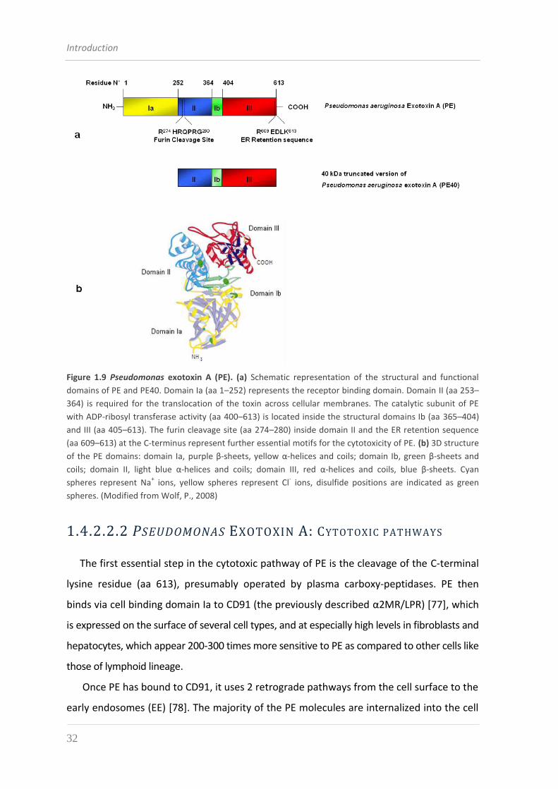
Introduction
32
Figure 1.9 Pseudomonas exotoxin A (PE). (a) Schematic representation of the structural and functional
domains of PE and PE40. Domain Ia (aa 1–252) represents the receptor binding domain. Domain II (aa 253–
364) is required for the translocation of the toxin across cellular membranes. The catalytic subunit of PE
with ADP-ribosyl transferase activity (aa 400–613) is located inside the structural domains Ib (aa 365–404)
and III (aa 405–613). The furin cleavage site (aa 274–280) inside domain II and the ER retention sequence
(aa 609–613) at the C-terminus represent further essential motifs for the cytotoxicity of PE. (b) 3D structure
of the PE domains: domain Ia, purple β-sheets, yellow α-helices and coils; domain Ib, green β-sheets and
coils; domain II, light blue α-helices and coils; domain III, red α-helices and coils, blue β-sheets. Cyan
spheres represent Na+ ions, yellow spheres represent Cl
- ions, disulfide positions are indicated as green
spheres. (Modified from Wolf, P., 2008)
1.4.2.2.2 PSEUDOMONAS EXOTOXIN A: CYTOTOXIC PATHWAYS
The first essential step in the cytotoxic pathway of PE is the cleavage of the C-terminal
lysine residue (aa 613), presumably operated by plasma carboxy-peptidases. PE then
binds via cell binding domain Ia to CD91 (the previously described α2MR/LPR) [77], which
is expressed on the surface of several cell types, and at especially high levels in fibroblasts and
hepatocytes, which appear 200-300 times more sensitive to PE as compared to other cells like
those of lymphoid lineage.
Once PE has bound to CD91, it uses 2 retrograde pathways from the cell surface to the
early endosomes (EE) [78]. The majority of the PE molecules are internalized into the cell
Introduction
33
in a nonlipid-dependent manner via clathrin-coated pits. But in some cases, as
demonstrated in HeLa cells, the association of a proportion of CD91-bound PE with a
receptor in detergent-resistant microdomains (DRM), facilitates the uptake in
caveosomes and the transport to the EE in vesicles containing the EE marker Rab5.
PE dissociates from CD91 in the acidic endosome environment, undergoes a
conformational change, and is cleaved by the protease furin between residues R279 and
G280 [79]. This results in an N-terminal fragment of 28 kDa and a C-terminal fragment of
37 kDa, the latter being composed of a portion of domain II, domain Ib and all of the
enzymatic active domain III [80]. Both fragments are still connected by the disulfide bond
between C265 and C287, encompassing the furin cleavage site. It is speculated that under
the mildly acidic conditions in the endosomes, there is an unfolding event, possibly by the
binding of chaperone proteins. This leads to a surface exposure of the disulfide bond with
subsequent reduction, perhaps by protein disulfide isomerases (PDI), which is followed by
a release of the C-terminal 37-kDa fragment [81].
The enzymatic active 37-kDa fragment travels to the trans-Golgi network (TGN) via late
endosomes and a Rab9-dependent route [82]. There, it can bind in a pH-dependent
manner to the KDEL receptor via its C-terminal KDEL-like sequence REDL (aa 609–612)
and is transported to the endoplasmic reticulum (ER) [76]. The KDEL receptor cycles
between the TGN and the ER via Golgi cisternae with the help of the tyrosine kinase Src
and is responsible for recycling proteins bearing the C-terminal amino acid code KDEL to
the ER [83]. Alternatively, the 37-kD fragment of PE, which bound to DRM at the cell
surface, can directly reach the TGN from EE in a pathway independent of the small
GTPase Rab9 and presumably reaches the endoplasmic reticulum (ER) in a lipid-
dependent sorting pathway, controlled by Rab6 [78]. Sequences in the translocation
domain II induce the dislocation of the 37-kDa fragment from the ER to the cytosol [80].
There is evidence that the Sec61p translocon, a protein used in eukaryotic cells for the
dislocation of unfolded or misfolded proteins in the ER-associated protein degradation
pathway (ERAD), is involved in this step [84].
Proteolytic cleavage and translocation into the cytoplasmic compartment are the rate-
limiting steps in the intoxication process: Ogata and colleagues demonstrated that as little
as 8-10% of the molecules interacting with a target cell are actually cleaved. This low
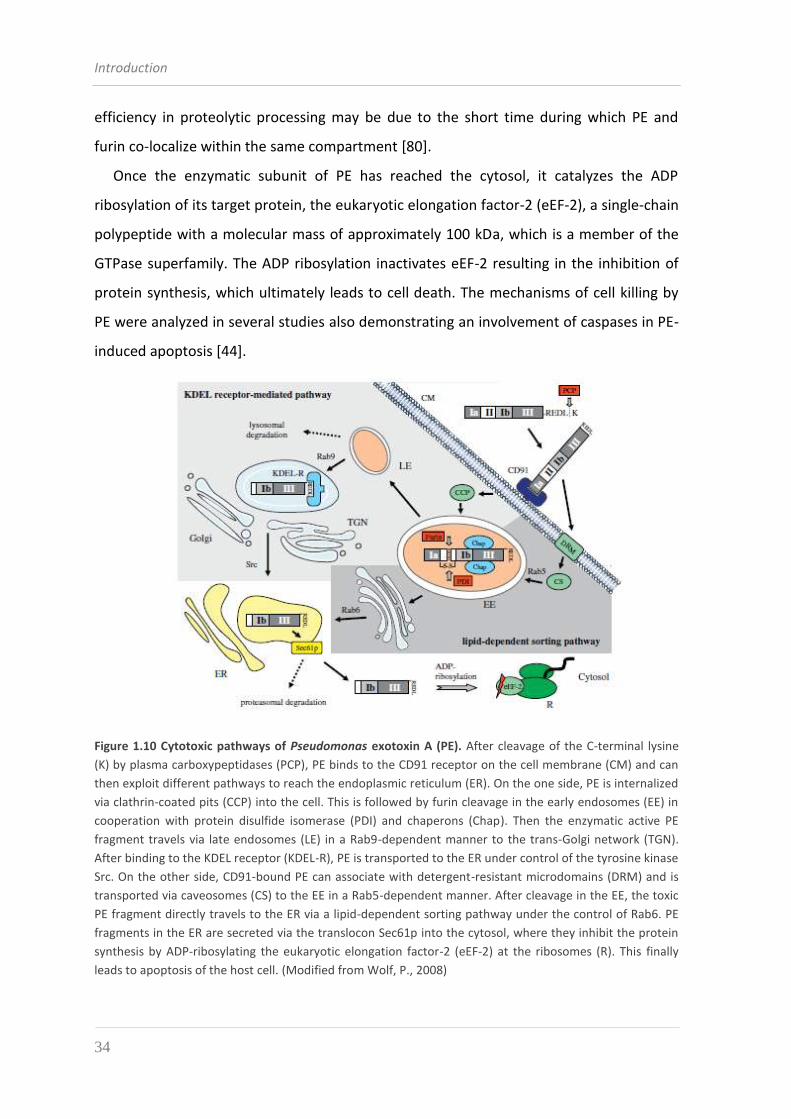
Introduction
34
efficiency in proteolytic processing may be due to the short time during which PE and
furin co-localize within the same compartment [80].
Once the enzymatic subunit of PE has reached the cytosol, it catalyzes the ADP
ribosylation of its target protein, the eukaryotic elongation factor-2 (eEF-2), a single-chain
polypeptide with a molecular mass of approximately 100 kDa, which is a member of the
GTPase superfamily. The ADP ribosylation inactivates eEF-2 resulting in the inhibition of
protein synthesis, which ultimately leads to cell death. The mechanisms of cell killing by
PE were analyzed in several studies also demonstrating an involvement of caspases in PE-
induced apoptosis [44].
Figure 1.10 Cytotoxic pathways of Pseudomonas exotoxin A (PE). After cleavage of the C-terminal lysine
(K) by plasma carboxypeptidases (PCP), PE binds to the CD91 receptor on the cell membrane (CM) and can
then exploit different pathways to reach the endoplasmic reticulum (ER). On the one side, PE is internalized
via clathrin-coated pits (CCP) into the cell. This is followed by furin cleavage in the early endosomes (EE) in
cooperation with protein disulfide isomerase (PDI) and chaperons (Chap). Then the enzymatic active PE
fragment travels via late endosomes (LE) in a Rab9-dependent manner to the trans-Golgi network (TGN).
After binding to the KDEL receptor (KDEL-R), PE is transported to the ER under control of the tyrosine kinase
Src. On the other side, CD91-bound PE can associate with detergent-resistant microdomains (DRM) and is
transported via caveosomes (CS) to the EE in a Rab5-dependent manner. After cleavage in the EE, the toxic
PE fragment directly travels to the ER via a lipid-dependent sorting pathway under the control of Rab6. PE
fragments in the ER are secreted via the translocon Sec61p into the cytosol, where they inhibit the protein
synthesis by ADP-ribosylating the eukaryotic elongation factor-2 (eEF-2) at the ribosomes (R). This finally
leads to apoptosis of the host cell. (Modified from Wolf, P., 2008)
Introduction
35
1.4.2.2.3 PE DERIVATIVES
The nonspecific toxicity of immunotoxins is often caused by binding of the toxic
portion to normal cells. When using native toxins this is mediated, in the case of PE, by
the binding domain Ia. Inactivation of this binding function was the major goal in
developing PE derived immunotoxins and over the past several years a variety of modified
PE molecules with altered cell-binding domains have been genetically engineered and
then tested as immunotoxins. One of those PE derivatives is PE glu57, in which the lysine
at position 57 in domain Ia is replaced by glutamate [85]. Another is PEglu57,246,247,249,
in which the cell-binding domain is not only inactivated by the glu57 mutation, but is also
unfolded due to the three further mutations [86]. Immunotoxins made from these PE
derivatives show diminished nonspecific toxicity and therefore a wider therapeutic
window is observed against cultured cells and in animal experiments. A further reduction
of nonspecific toxicity can be reached by removing the entire domain Ia and replacing it
with a tumor-specific ligand. The first truncated form of PE to be produced is known as
PE40 (residues 253-613) to reflect its molecular weight of 40 kDa. Additionally, a large
portion of PE domain Ib (residues 365-380) can be deleted: this modified form is called
PE38. Both these truncated forms still retain full toxic and translocation activity and when
coupled or recombinantly fused to antibodies or other targeting moieties make very
active immunotoxins [87].
Several other modified versions of PE were created; some examples are: LysPE40 and
LysPE38, which have an exposed lysine residue next to its amino terminus to facilitate
conjugation to mAbs [88], while in PE40KDEL and PE38KDEL the original “REDLK”
endoplasmic reticulum retention sequence at the carboxyl terminus of PE is replaced by a
KDEL which is a preferred sequence [89]. These last immunotoxins show increased
cytotoxic activity, but nonspecific toxicity is often increased by approximately the same
factor.
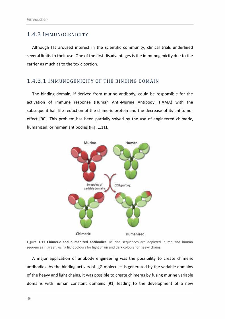
Introduction
36
1.4.3 IMMUNOGENICITY
Although ITs aroused interest in the scientific community, clinical trials underlined
several limits to their use. One of the first disadvantages is the immunogenicity due to the
carrier as much as to the toxic portion.
1.4.3.1 IMMUNOGENICITY OF THE BINDING DOMAIN
The binding domain, if derived from murine antibody, could be responsible for the
activation of immune response (Human Anti-Murine Antibody, HAMA) with the
subsequent half life reduction of the chimeric protein and the decrease of its antitumor
effect [90]. This problem has been partially solved by the use of engineered chimeric,
humanized, or human antibodies (Fig. 1.11).
Figure 1.11 Chimeric and humanized antibodies. Murine sequences are depicted in red and human
sequences in green, using light colours for light chain and dark colours for heavy chains.
A major application of antibody engineering was the possibility to create chimeric
antibodies. As the binding activity of IgG molecules is generated by the variable domains
of the heavy and light chains, it was possible to create chimeras by fusing murine variable
domains with human constant domains [91] leading to the development of a new
Introduction
37
generation of therapeutic candidates [92]. These chimeric antibodies are 70% human and
possess a fully human Fc portion, which makes them considerably less immunogenic in
humans and allows them to interact with human effector cells and the complement
cascade.
With the development of antibody engineering techniques, it became possible to
decrease even further the murine part of mAbs by replacing the hypervariable loops of a
fully human antibody with the hypervariable loops of the murine antibody of interest,
using an approach called complementarity-determining region grafting [93]. These
antibodies, called “humanized”, are 85–90% human and are even less immunogenic than
chimeric antibodies. However, complementarity-determining region grafting is more
technically demanding than a mere fusion, and directed mutagenesis approaches are
often needed to restore the affinity present in the murine parental antibody. Most of the
approved mAbs in current use are either chimeric or humanized (Table 1.1).
Another major improvement came with the development of in vitro selection
methods, the most successful being phage display. With the ever increasing power of
antibody engineering, it became possible to clone entire repertoires of antibody fragment
genes, from immunized or non-immunized animals, including humans. A powerful
selection method was therefore needed to select from this large number of potential
ligands, those able to bind the antigen of choice.
The first technique, and still by far the most common one was inspired by the work of
George Smith [94]. Like all in vitro selection methods, this technique relies on the ability
to establish a physical link between a protein and the gene encoding this protein, in this
case between a protein fused to a filamentous phage capsid protein (p3 or p8) displayed
at the surface of phage M13 and its corresponding gene contained in the encapsidated
DNA. If the molecule is immunopurified by binding to the antigen of interest, its gene is
immediately available, allowing sequencing and further multiplication of the specific
clone. Because of these in vitro selection methods, it is now possible to rapidly and
efficiently select fully human antibody fragments against virtually any antigen by using
“universal”, large, non-immunized libraries [95].
Moreover, the same approach can be used to maximize the affinity of a valuable
antibody by creating a secondary library consisting of mutants of the first candidate and
Introduction
38
performing stringent in vitro selection against the antigen of choice. Phage display and
more recently ribosome display have been used to obtain ligands with sub-picomolar
affinities for the relevant antigen, outperforming the affinities of most conventional mAbs
[96].
During the same decade, a complementary approach was developed to create fully
human antibodies. Transgenic “humanized” mice were created by replacing the entire
mouse IgG repertoire with a human repertoire [97]. Upon immunization, these
humanized mice produce human IgGs and conventional hybridoma techniques can be
used to clone human antibodies with the required properties. This approach has the
advantage of yielding in vivo matured antibodies, circumventing the need for additional
affinity maturation. However, humanized mice cannot be used effectively when the
immunogen is toxic or when the targeted antigen shares a high degree of homology with
its murine ortholog.
1.4.3.2 IMMUNOGENICITY OF THE TOXIC PORTION
Human immune response has been also observed against the toxic portion of
immunotoxins: indeed, the presence of a bacterial or plant toxin can trigger the formation
of neutralizing antibodies, hindering their efficacy. Especially PE and DT are very
immunogenic [98] and cannot be humanized with standard techniques. DT-derived
immunotoxins are in addition particularly affected because most people in developed
countries have been vaccinated against DT and many adults have neutralizing antibodies
to DT. In patients with B- or T-cell malignancies, the formation of neutralizing antibodies
is less frequent because of their immunosuppressed state; in contrast, in patients with
solid tumors, antibody responses are frequently detected as early as a few days after the
first treatment regimen, preventing re-administration of the ITs [99]. Many efforts have
been made to decrease immunogenicity of the toxin moiety; one possibility explored is
masking of the therapeutic molecules by chemically modifying the immunotoxin with high
molecular weight polyethylenglycol (PEG) [100], dextran, or other nonimmunogenic
polymers. An impressive result was obtained recently by Onda and coworkers, who
identified the major immunogenic B-cell epitopes in the truncated form of Pseudomonas
Introduction
39
aeruginosa exotoxin A (PE38) [101]. A total of eight amino acids containing large bulky
hydrophilic side chains have been replaced with smaller polar residues within these
epitopes, resulting in a new toxin endowed with much less immunogenicity than the
parental one, without any loss of cytotoxic activity also when recombinantly fused to an
anti CD22 variable fragment [102]. Finally, a current successful approach to obtain less
immunogenic or nonimmunogenic immunotoxins is the generation of a new class of
recombinant molecules in which the cytotoxic moiety is an endogenous protein of human
origin like proapoptotic protein (e.g TNF, TRAIL or granzyme B) or RNase [103].
A further complication observed in ITs administration in the past was due to
nonspecific binding of the toxin domain to vascular endothelial cells, leading to the so-
called “vascular leak syndrome” (VLS) [104], which is characterized by the damage of
vascular endothelial cells, extravasation of fluids and proteins, interstitial edema, and
organ failure. Although the mechanisms underlying this side effect are not completely
understood, proteins such as RTA and some type I RIPs contain a consensus amino acid
sequence which seems to induce vascular damage to human endothelial cells in vitro by
binding to integrin receptors [105]. Indeed, in the case of RTA, molecular modeling
suggested that these motifs were partially exposed on the surface of the molecule [105]
and a similar motif is shared by viral disintegrins, which disrupt the function of integrin
receptors [106]. Also PE-based immunotoxins have been shown to promote VLS in rats
[107]. In the perspective of eliminating VLS during therapeutical use of ricin-based ITs,
Vitetta and coworkers produced a series of RTA mutants, and identified the Asn 97 to Ala
mutation, in a region flanking the VLS-responsible motif in the three-dimensional
structure, as displaying significant less VLS in mice [108].
1.4.4 EXPRESSION SYSTEMS
Diverse prokaryotic and eukaryotic expression systems have been developed for the
production of recombinant proteins. These have included bacteria, yeast, filamentous
fungus, eukaryotic alga, insect cell, plant, mammalian cell and transgenic animal systems.
While in many instances heterologous proteins can be expressed in several different
Introduction
40
systems, there is sometimes less flexibility in terms of choice of expression system due to
structural requirements on the part of the specific protein to be produced.
The expression of heterologous proteins in eukaryotic organisms benefits by the presence
of specialized compartments for the folding and assembly and of an efficient machinery for
post-translational modifications. For this reason, eukaryotic expression systems are required
for producing molecule, such as certain therapeutic IgGs, which requires appropriate
glycosylation [109]. However, eukaryotic systems are not suitable for the expression of
most ITs made by toxic domains which target the eukaryotic elongation factor of protein
synthesis or a eukaryotic ribosomal subunit, thus resulting in a toxic effect on the host cell
itself. Nevertheless some studies had shown that certain hosts are remarkably resistant to a
number of toxins normally used in the design of ITs, while other expression systems (i.e.
Saccharomyces cerevisiae and Pichia pastoris) are optimized for the production of these same
toxins, allowing to recover fairly good amounts of the derived ITs [110].
Antibody fragments (mostly scFvs) and ITs have a much simpler structure and do not
require glycosylation. Thus, bacterial expression has been the method of choice for the
expression of these molecules. Further reasons for this choice lie in the cost-effectiveness
of bacteria, their well-characterized genetics, and the availability of many different
bacterial expression systems. Among the hosts available for recombinant expression,
Escherichia coli is in an exceptional position. This stems from the many decades of intense
research on its genetics as well as the broad scope of biotechnological tools available for
genetic engineering of this organism. As a host for recombinant expression, E. coli is
especially valued because of its rapid growth rate, capacity for continuous fermentation,
low media costs and achievable high expression levels. One consequence of this
popularity is that about 80% of all proteins used to solve three-dimensional structures
submitted to the protein data bank (PDB) in 2003 were prepared in E. coli [111] and
during 2003 and 2006, nine out of 31 approved therapeutic proteins were produced in E.
coli [112], among them important growth factors, insulins and interferons [113].
The major drawbacks of using E. coli for recombinant protein production are its lack of
secretion systems for efficient release of proteins to the growth medium, limited ability
to facilitate extensive disulfide-bond formation and other post-translational
modifications, inefficient cleavage of the amino terminal methionine which can result in
Introduction
41
lowered protein stability and increased immunogenicity, and occasional poor folding due
to lack of specific molecular chaperones [114].
Nevertheless, although refolding procedures require customization for each individual
protein, the production of ITs in bacteria as inclusion bodies, followed by solubilization
and in vitro renaturation, is still the most common route towards obtaining sufficient
amounts of protein to be used for in vitro, lab-scale evaluation or for clinical trials [44].
1.5 COMBINATION THERAPIES
Despite their potency, immunotoxins produce complete remission infrequently when
administered as single agents and relapses often occur because of the anti-apoptotic
strategies frequently employed by tumor cells to overcome chemotherapy-mediated
death [115, 116].
Programmed cell death (apoptosis) is now widely recognized as an evolutionarily
conserved, genetically controlled process for killing damaged, infected, superfluous or
potentially dangerous cells that is essential for the normal development and function of
multicellular organisms. Defects in the control of apoptosis causing either the survival of
unwanted cells or inappropriate killing of vital cells underlie a multitude of disorders,
including autoimmunity, degenerative diseases and cancers [116]. Indeed, defects in
apoptosis are now considered to be a hallmark of most, if not all, cancers.
Many cancer cells develop resistance to apoptosis by mechanisms involving members
of the B-cell lymphoma-2 (Bcl-2) protein family, which are critical regulators of apoptosis
consisting of anti- and pro-apoptotic proteins that determine the balance between
survival and programmed cells death [117]. Structural and functional characteristics
divide proteins of Bcl-2 family into three subgroups:
1) the anti-apoptotic family members Bcl-2, Bcl-xL , Bcl-W, A1 and Mcl-1 (myeloid cell
leukemia-1), which promote cell survival by counteracting the apoptotic effector
proteins, thus preventing their activation and mitochondrial outer membrane
permeabilization with the consequent release of cytochrome c and caspase
activation,
Introduction
42
2) Bax and Bak proteins, which are the essential activators of the effector phase of
apoptosis,
3) pro-apoptotic BH3 proteins, such as Bad, Bim (Bcl-2 interacting mediator of cell
death), Bid (Bcl-2-interacting domain), Noxa and Puma (p53 upregulated mediator
of apoptosis), which enable activation of Bax and Bak either by neutralizing anti-
apoptotic Bcl-2 family proteins or by direct action.
Observations in human tumors and studies with genetically modified (transgenic or
knock-out) mice have shown that tumourigenesis can be driven by gain-of-function
mutations in cell death antagonists (e.g., Bcl-2 overexpression) or loss-of-function
mutations in cell death activators (e.g., loss of Bim) [117].
Activation of the pro-apoptotic proteins Bax and Bak depends on the action of BH3-
only proteins, which are up-regulated in response to stress or cell injury. BH3-only
proteins can activate Bax/Bak directly or they can trigger apoptosis via the neutralization
of the pro-survival proteins Bcl-2, Bcl-xL and Mcl-1 (Fig. 1.12).
Therefore, BH3 mimetics were developed as potential stand-alone antitumor agents or
as sensitizers for chemotherapy, thus eliminating resistance to apoptosis. ABT-737, one
such agent, was shown to have strong binding affinity for Bcl-2 and Bcl-xL but little or
none for Mcl-1. Because inhibition of protein synthesis triggered by immunotoxins
frequently results in the loss of Mcl-1, obtaining inhibition of Bcl-2 and Bcl-xL with ABT-
737 seemed to be a good strategy to potentially sensitized tumor cells to immunotoxins
action [115].
Combinatorial therapy using ABT-737 (and its clinical analog ABT-263) and
immunotoxins has been demonstrated highly effective against molecular subgroups of
multiple myeloma [118], adult T-cell leukemia/lymphoma (ATLL) [119] and many other
types of tumor.
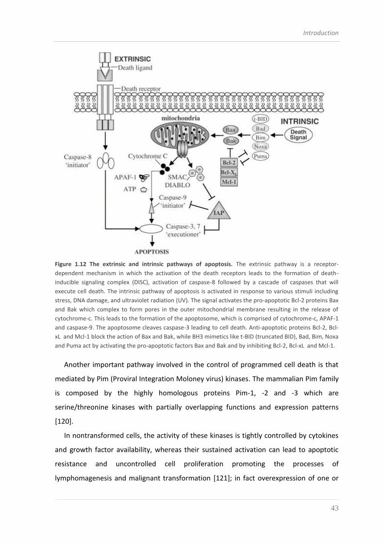
Introduction
43
Figure 1.12 The extrinsic and intrinsic pathways of apoptosis. The extrinsic pathway is a receptor-
dependent mechanism in which the activation of the death receptors leads to the formation of death-
inducible signaling complex (DISC), activation of caspase-8 followed by a cascade of caspases that will
execute cell death. The intrinsic pathway of apoptosis is activated in response to various stimuli including
stress, DNA damage, and ultraviolet radiation (UV). The signal activates the pro-apoptotic Bcl-2 proteins Bax
and Bak which complex to form pores in the outer mitochondrial membrane resulting in the release of
cytochrome-c. This leads to the formation of the apoptosome, which is comprised of cytochrome-c, APAF-1
and caspase-9. The apoptosome cleaves caspase-3 leading to cell death. Anti-apoptotic proteins Bcl-2, Bcl-
xL and Mcl-1 block the action of Bax and Bak, while BH3 mimetics like t-BID (truncated BID), Bad, Bim, Noxa
and Puma act by activating the pro-apoptotic factors Bax and Bak and by inhibiting Bcl-2, Bcl-xL and Mcl-1.
Another important pathway involved in the control of programmed cell death is that
mediated by Pim (Proviral Integration Moloney virus) kinases. The mammalian Pim family
is composed by the highly homologous proteins Pim-1, -2 and -3 which are
serine/threonine kinases with partially overlapping functions and expression patterns
[120].
In nontransformed cells, the activity of these kinases is tightly controlled by cytokines
and growth factor availability, whereas their sustained activation can lead to apoptotic
resistance and uncontrolled cell proliferation promoting the processes of
lymphomagenesis and malignant transformation [121]; in fact overexpression of one or
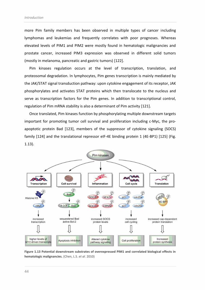
Introduction
44
more Pim family members has been observed in multiple types of cancer including
lymphomas and leukemias and frequently correlates with poor prognoses. Whereas
elevated levels of PIM1 and PIM2 were mostly found in hematologic malignancies and
prostate cancer, increased PIM3 expression was observed in different solid tumors
(mostly in melanoma, pancreatic and gastric tumors) [122].
Pim kinases regulation occurs at the level of transcription, translation, and
proteosomal degradation. In lymphocytes, Pim genes transcription is mainly mediated by
the JAK/STAT signal transduction pathway: upon cytokine engagement of its receptor, JAK
phosphorylates and activates STAT proteins which then translocate to the nucleus and
serve as transcription factors for the Pim genes. In addition to transcriptional control,
regulation of Pim mRNA stability is also a determinant of Pim activity [121].
Once translated, Pim kinases function by phosphorylating multiple downstream targets
important for promoting tumor cell survival and proliferation including c-Myc, the pro-
apoptotic protein Bad [123], members of the suppressor of cytokine signaling (SOCS)
family [124] and the translational repressor eIF-4E binding protein 1 (4E-BP1) [125] (Fig.
1.13).
Figure 1.13 Potential downstream substrates of overexpressed PIM1 and correlated biological effects in
hematologic malignancies. (Chen, L.S. et al. 2010)
Introduction
45
Pim kinases represent validated drug targets in many hematologic cancers and select
solid tumors and a number of small molecule PIM kinases inhibitors have emerged as
therapeutics for hematological malignancies. Based on the known crystal structure of
Pim-1, high-throughput screening, and lead optimization techniques, has been possible to
develop compounds specifically binding to the ATP binding site of Pim-1 and competing
with ATP binding. One of the most potent inhibitors of all 3 Pim kinases is SGI-1776, an
imidazo[1,2-b] pyridazine compound which has been demonstrated efficient in preclinical
models of human acute myeloid leukemia and CLL and prostate cancer, either as a single
agent or in combination with other chemotherapeutic agents [126].

Introduction
46
Aim of the research
47
AIM OF THE RESEARCH
The main objectives of the study here presented are:
construction and production in E. coli of a scFv binding to the B-cell surface antigen
CD38, through the molecular cloning of the variable region of heavy and light chains
(VH and VL, respectively);
generation and bacterial expression of recombinant immunotoxins by genetic fusion
of the scFv to a truncated version of Pseudomonas aeruginosa exotoxin A (PE40) or to
the plant toxin saporin;
characterization of the binding properties of the recombinant ITs on cells that
express the target antigen on their surface;
evaluation of the selective toxicity on target B cells of the ITs obtained;
study of potentiating effects of drugs inhibiting intracellular pathways in association
with recombinant ITs.

48

2. MATERIALS AND METHODS

Materials and Methods
50
Materials and Methods
51
2.1 MICROBIOLOGY TECHNIQUES
2.1.1 ESCHERICHIA COLI STRAINS
DH5α: F- Φ80lacZΔM15 Δ(lacZYA-argF) U169 recA1 endA1 hsdR17 (rK–, mK+) phoA
supE44 λ thi-1 gyrA96 relA1;
XL1-Blue: recA1 endA1 gyrA96 thi-1 hsdR17 supE44 relA1 lac [F1 proAB lacIqZΔM15
Tn10 (Tetr)];
BL21 (DE3) pLysS: F- ompT hsdS(rB- mB
-) dcm+ galλ(DE3) rne131 (pLysS Camr).
Bacterial stocks are kept at -80 °C in LB medium containing 20% glycerol.
2.1.2 E. COLI GROWTH MEDIA
LB-medium
Tryptone 10 g/l
Yeast Extract 5 g/l
NaCl 10 g/l
Adjust pH to 7.5 with 1M NaOH and sterilize at 121 °C for 20 minutes.
Agar medium for plates is prepared by addition of 15 g/l Agar before sterilization by
autoclave.
LB broth is used for transformation, growth, maintenance and protein expression with
DH5α, XL1-Blue and BL21(DE3)pLysS E. coli strains.
2.1.3 PLASMID VECTORS
For the molecular cloning of DNA sequences vector pET20b(+) (Novagen) was used.
pET20b(+)
pET20b(+) is an expression vector of 3716 bp carrying a multi-cloning site between T7
promoter and T7 terminator sequences.
In order to induce the expression of the protein coded by the sequence inserted in the
polylinker, it is necessary to use E. coli strains carrying a copy of the gene for T7 RNA
polymerase. These are lysogenic hosts of the DE3 bacteriophage, which has in its genome
Materials and Methods
52
the sequence coding for T7 RNA polymerase, under control of the promoter lacUV5. The
lacUV5 promoter is induced by the addition of IPTG and drives the transcription of the T7
RNA polymerase, which, in its turn, transcribes the sequence of interest. In this way the
basal expression, i.e. the expression of the recombinant protein before induction, is very
limited. The strain of E. coli employed for the expression also bears the plasmid pLysS,
which determines a further repression of the basal expression level through the
transcription of T7 lysozyme, a natural inhibitor of T7 RNA polymerase.
Vector pET20b(+), by virtue of the pelB signal peptide, promotes the periplasmic sorting
of the protein coded by the inserted sequence, also providing a poly-histidine carboxy-
terminal tag. pET20b(+) carries an ampicillin resistance and the pBR322 origin of
replication.
In this plasmid the final scFv-coding sequence was inserted and subsequently fused to the
fragment coding for PE40 or saporin, so as to obtain the constructs for the expression of
the immunotoxins of interest.
2.1.4 PREPARATION OF CACL2-COMPETENT E. COLI CELLS
The strains of Escherichia coli utilized for the molecular cloning and for protein expression
are made competent to transformation by plasmid DNA following treatment with CaCl2.
A glycerol stock of DH5α, XL1-Blue or BL21 (DE3) pLysS cells (kept at -80 °C) is streaked on
a LB-agar plate containing 25 μg/ml Tetracycline (for XL1-Blue), 34 μg/ml
Chloramphenicol (for BL21 (DE3) pLysS) or no antibiotics (for DH5α) and incubated o.n. at
37 °C.
The day after a single colony is picked and used to start a 10 ml LB culture supplemented
with the same antibiotics at the same concentrations used in the plates and grown o.n at
37 °C with shaking.
Few (1-2) ml of the overnight culture is then used to start a 50 ml LB culture with
appropriate antibiotics. This culture is grown to OD600 = 0.3-0.4 at 25°C with shaking.
The bacterial culture is then kept on ice for 30 minutes, after which it is centrifuged at
4000 x g, 10 minutes at 4 °C. Always keeping the cells at ice temperature, they are
recovered and resuspended in 25 ml of sterile, ice-cold 50 mM CaCl2. After a one hour
Materials and Methods
53
incubation on ice the suspension is centrifuged at 3000 x g, 5 minutes at 4°C. The
supernatant is removed and the bacterial pellet is resuspended in 3 ml of a sterile 50 mM
CaCl2, 20% glycerol solution. Cells are finally dispensed in aliquots, frozen in dry ice and
kept at -80 °C.
2.1.5 HEAT-SHOCK MEDIATED TRANS FORMATION OF ESCHERICHIA COLI
A 100 μl aliquot of CaCl2-competent cells are thawed on ice and mixed into a sterile tube
with 5-10 ng of plasmid DNA; in case of transformation with a ligation, 5 μl of the reaction
are used. The mixture is gently flicked and kept on ice for 20 minutes. Afterwards a heat
shock is carried out by dipping the tube for one minute in a 42°C water bath, followed by
a 2 minute incubation on ice. 1 ml of LB broth (without antibiotics) is then added into the
tube which is then incubated 1 hour at 37°C with shaking. The desired amount of
transformation is then plated (after centrifugation to concentrate cells if necessary) on
LB-agar plates supplemented with the appropriate selective agent (100 μg/ml Amp + 25
μg/ml Tet or 100 μg/ml Amp + 34 μg/ml Cam). Plates are incubated inverted at 37 °C and
grown o.n.
2.2 HUMAN CELL LINES 2.2.1 CELL LINES AND GROWTH MEDIA
Ramos: human line of B lymphocytes derived from Burkitt’s lymphoma (CD38-
positive);
Daudi: human line of B lymphocytes derived from Burkitt’s lymphoma (CD38-
positive);
3T3/7A: mouse fibroblasts tranfected with CD38 molecule (CD38-positive);
RPMI8226: human line of Multiple Myeloma (CD38-positive);
U266: human line of Multiple Myeloma (CD38-negative);
Anti-CD38 hybridoma 1E82H11: the hybridoma clone was derived from spleen
lymphocytes of a mouse previously immunized with intraperitoneal injection of a
membrane preparation from 3T3 cells transfected with human CD38 antigen (kindly
Materials and Methods
54
provided by Prof. Ippoliti and co-workers from the University of L’Aquila); the
myeloma cell line used in the fusion was NS-1.
All cell lines are grown in flasks at 37 °C, 5% CO2, using the following medium:
RPMI 1640 medium (with 40 mg/l folic acid, 2 g/l NaHCO3) (BiochromAG) supplemented
with 10% Fetal Calf Serum (FCS), 2 mM L-Glutamine and antibiotics (100 U/ml penicilline
and 100 μg/ml streptomycine-sulphate). Hybridoma clone 1E82H11 is grown in
Hybridomed DIF 1000 Serum free medium (BiochromAG).
All supplements are added into the medium after sterilization through 0.22 µm filters.
Each cell line is cultured as a suspension and is constantly kept at an exponential growth
phase by frequent medium changes. Cell stocks can be stored in liquid nitrogen tank at a
concentration of 1 or 2 x 10⁶ cells/ml RPMI medium with 20% FCS and 10% DMSO.
2.2.2 B-LYMPHOCYTES FROM PBMCS
B-lymphocytes were obtained by negative-selection (using EasySep Human B Cell
Enrichment Kit, StemCell Technologies) from a PBMC sampe of a CLL patient. Cells were
mantained in IMDM medium (Life Technologies) supplemented with 10% FCS, 10 nM IL-4,
20 U/ml IL-2 and 50 ng/ml CD40L.
2.3 MOLECULAR BIOLOGY 2.3.1 RNA EXTRACTION FROM ANTI-CD38 HYBRIDOMA CELLS
3 x 10⁶ cells of the anti-CD38 hybridoma are collected by centrifugation (5 minutes, 150 x
g, 5°C) and likewise washed in physiologic solution. RNA extraction is obtained using SV
Total RNA Isolation System (Promega) and finally RNA is eluted with 50 µl RNAse-free
H2O. Total RNA is quantified by spectrophotometry: 1 unit at OD260 corresponds to an
RNA concentration of 40 μg/ml.
2.3.2 CDNA SYNTHESIS
Retrotranscription to cDNA is obtained using M-MLV retrotranscriptase (Moloney Murine
Leukemia Virus Reverse Transcriptase - Invitrogen), following the manufacturer’s
Materials and Methods
55
instructions: 4 μg of total RNA are mixed with 1 μl of random primers (Invitrogen), 1 μl of
10 mM dNTPs mix (Applied Biosystem) and 8 μl RNAse-free H2O; this mixture is incubated
at 65°C for 5 minutes and then kept on ice. The following must then be added: 4 μl of 5x
First Strand Buffer (Invitrogen), 2 μl of 0.1 M DTT (Invitrogen), 1 μl of RNase-OUT
(Invitrogen); the reaction is incubated 2 minutes at 37°C, after which 1 μl of M-MLV is
added. Using a thermocycler, the reaction is then incubated 10 minutes at 25°C, followed
by 50 minutes at 37°C and finally 15 minutes at 70 °C.
The quality of the cDNA is assessed through a PCR test with a couple of primers designed
for the amplification of murine β-actine (see paragraph 2.3.3.1).
2.3.3 PCR AMPLIFICATION OF SPE CIFIC DNA FRAGMENTS
The amplification of DNA fragments by polymerase-chain-reaction (PCR) was customized
in order to fit the conditions required by specific templates and enzymes used in the
reactions. All reactions were carried out using a GeneAmp PCR system 9700 thermocycler
(Applied Biosystems).
2.3.3.1 AMPLIFICATION OF THE SEQUENCE CODING FOR MOUSE Β-ACTIN
The outcome of this PCR provides an indication regarding the quality of the cDNA as a
template, before proceeding with the amplification of the fragments of interest.
Cycling programme:
1 µl of cDNA is used as a template in a 50 µl reaction including a couple of primers (m-β-
actin fw and m-β-actin rev) at a final concentration of 0.4 µM each, dNTPs (Applied
Biosystems) at a concentration of 0.2 mM each, 2 units of Taq DNA polymerase
(Fermentas), 5 µl of 10x buffer (Fermentas) and 4 µl of 25 mM MgCl2.
94°C 5 min
94°C 45 sec
35 cycles 59°C 45 sec
72°C 45 sec
72°C 7 min
4°C end
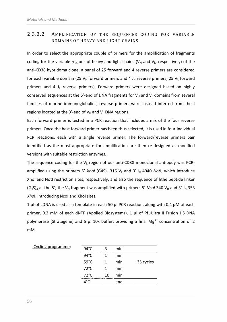
Materials and Methods
56
2.3.3.2 AMPLIFICATION OF THE SEQUENCES CODING FOR VARIABLE
DOMAINS OF HEAVY AND LIGHT CHAINS
In order to select the appropriate couple of primers for the amplification of fragments
coding for the variable regions of heavy and light chains (VH and Vk, respectively) of the
anti-CD38 hybridoma clone, a panel of 25 forward and 4 reverse primers are considered
for each variable domain (25 VH forward primers and 4 JH reverse primers; 25 Vk forward
primers and 4 Jk reverse primers). Forward primers were designed based on highly
conserved sequences at the 5’-end of DNA fragments for VH and VL domains from several
families of murine immunoglobulins; reverse primers were instead inferred from the J
regions located at the 3’-end of VH and VL DNA regions.
Each forward primer is tested in a PCR reaction that includes a mix of the four reverse
primers. Once the best forward primer has been thus selected, it is used in four individual
PCR reactions, each with a single reverse primer. The forward/reverse primers pair
identified as the most appropriate for amplification are then re-designed as modified
versions with suitable restriction enzymes.
The sequence coding for the Vk region of our anti-CD38 monoclonal antibody was PCR-
amplified using the primers 5’ XhoI (G4S)3 316 Vk and 3’ Jk 4940 NotI, which introduce
XhoI and NotI restriction sites, respectively, and also the sequence of hthe peptide linker
(G4S)3 at the 5’; the VH fragment was amplified with primers 5’ NcoI 340 VH and 3’ JH 353
XhoI, introducing NcoI and XhoI sites.
1 μl of cDNA is used as a template in each 50 µl PCR reaction, along with 0.4 µM of each
primer, 0.2 mM of each dNTP (Applied Biosystems), 1 µl of PfuUltra II Fusion HS DNA
polymerase (Stratagene) and 5 µl 10x buffer, providing a final Mg2+ concentration of 2
mM.
Cycling programme:
94°C 3 min
94°C 1 min
35 cycles 59°C 1 min
72°C 1 min
72°C 10 min
4°C end
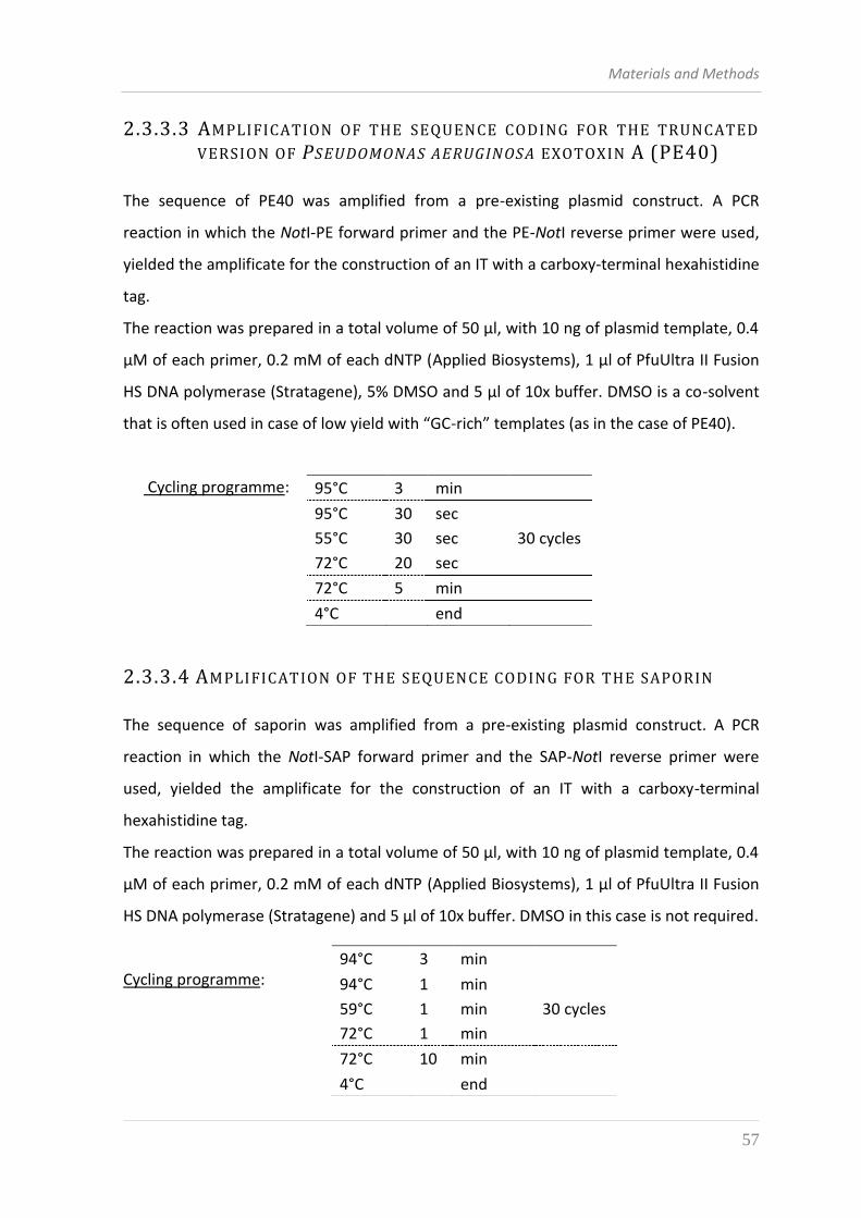
Materials and Methods
57
2.3.3.3 AMPLIFICATION OF THE SEQUENCE CODING FOR THE TRUNCATED
VERSION OF PSEUDOMONAS AERUGINOSA EXOTOXIN A (PE40)
The sequence of PE40 was amplified from a pre-existing plasmid construct. A PCR
reaction in which the NotI-PE forward primer and the PE-NotI reverse primer were used,
yielded the amplificate for the construction of an IT with a carboxy-terminal hexahistidine
tag.
The reaction was prepared in a total volume of 50 µl, with 10 ng of plasmid template, 0.4
µM of each primer, 0.2 mM of each dNTP (Applied Biosystems), 1 µl of PfuUltra II Fusion
HS DNA polymerase (Stratagene), 5% DMSO and 5 µl of 10x buffer. DMSO is a co-solvent
that is often used in case of low yield with “GC-rich” templates (as in the case of PE40).
Cycling programme:
2.3.3.4 AMPLIFICATION OF THE SEQUENCE CODING FOR THE SAPORIN
The sequence of saporin was amplified from a pre-existing plasmid construct. A PCR
reaction in which the NotI-SAP forward primer and the SAP-NotI reverse primer were
used, yielded the amplificate for the construction of an IT with a carboxy-terminal
hexahistidine tag.
The reaction was prepared in a total volume of 50 µl, with 10 ng of plasmid template, 0.4
µM of each primer, 0.2 mM of each dNTP (Applied Biosystems), 1 µl of PfuUltra II Fusion
HS DNA polymerase (Stratagene) and 5 µl of 10x buffer. DMSO in this case is not required.
Cycling programme:
95°C 3 min
95°C 30 sec
30 cycles 55°C 30 sec
72°C 20 sec
72°C 5 min
4°C end
94°C 3 min
94°C 1 min
30 cycles 59°C 1 min
72°C 1 min
72°C 10 min
4°C end
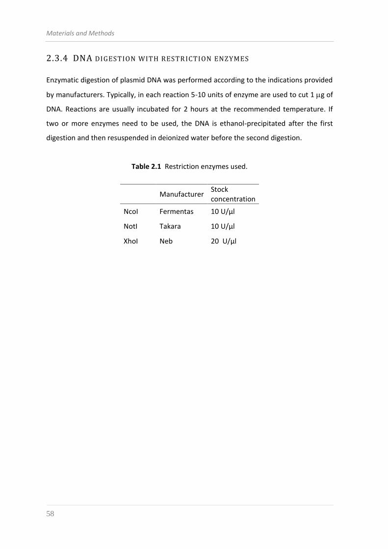
Materials and Methods
58
2.3.4 DNA DIGESTION WITH RESTRICTION ENZYMES
Enzymatic digestion of plasmid DNA was performed according to the indications provided
by manufacturers. Typically, in each reaction 5-10 units of enzyme are used to cut 1 g of
DNA. Reactions are usually incubated for 2 hours at the recommended temperature. If
two or more enzymes need to be used, the DNA is ethanol-precipitated after the first
digestion and then resuspended in deionized water before the second digestion.
Table 2.1 Restriction enzymes used.
Manufacturer Stock concentration
NcoI Fermentas 10 U/μl
NotI Takara 10 U/μl
XhoI Neb 20 U/μl
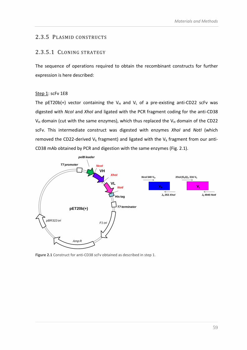
Materials and Methods
59
2.3.5 PLASMID CONSTRUCTS
2.3.5.1 CLONING STRATEGY
The sequence of operations required to obtain the recombinant constructs for further
expression is here described:
Step 1: scFv 1E8
The pET20b(+) vector containing the VH and VL of a pre-existing anti-CD22 scFv was
digested with NcoI and XhoI and ligated with the PCR fragment coding for the anti-CD38
VH domain (cut with the same enzymes), which thus replaced the VH domain of the CD22
scFv. This intermediate construct was digested with enzymes XhoI and NotI (which
removed the CD22-derived Vk fragment) and ligated with the Vk fragment from our anti-
CD38 mAb obtained by PCR and digestion with the same enzymes (Fig. 2.1).
Amp R
pBR322 ori
pET20b(+)
F1 ori
T7 promoter
T7 terminator
pelB leader
VH
VL
His tag
NcoI
XhoI
NotI VLVH
NcoI 340 VH
Jk 4940 NotI
XhoI (G4S)3 316 Vk
JH 353 XhoI
Figure 2.1 Construct for anti-CD38 scFv obtained as described in step 1.
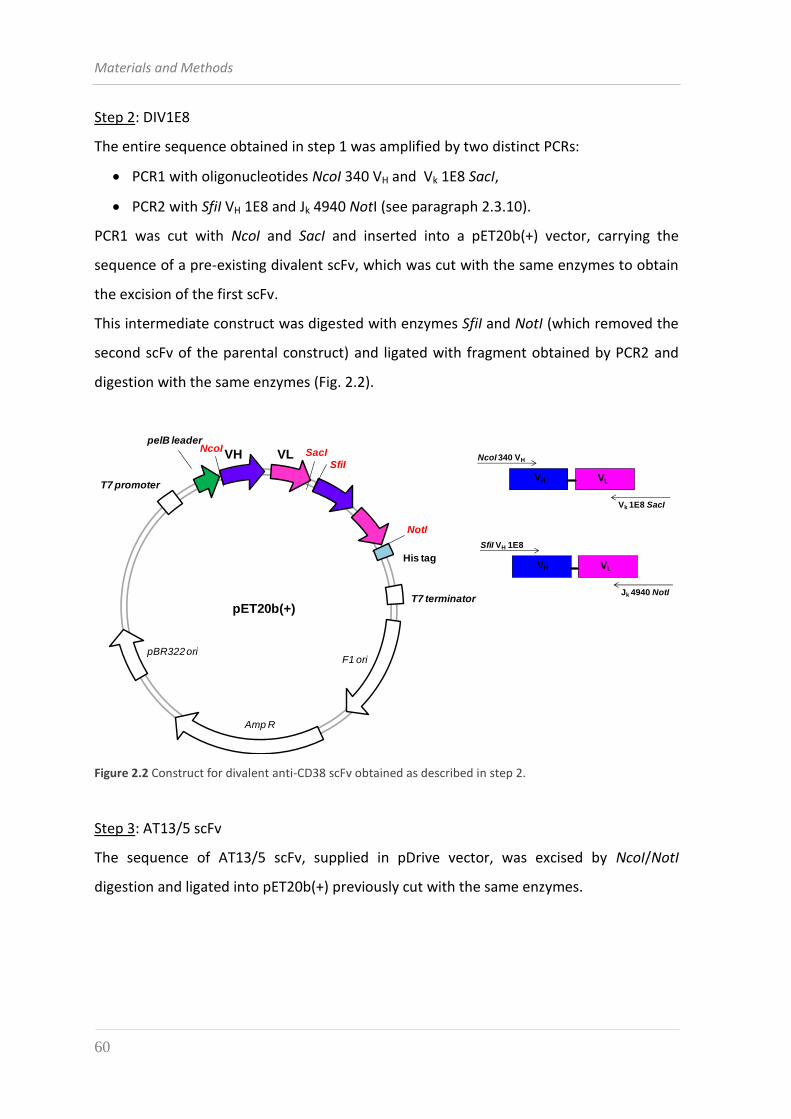
Materials and Methods
60
Step 2: DIV1E8
The entire sequence obtained in step 1 was amplified by two distinct PCRs:
PCR1 with oligonucleotides NcoI 340 VH and Vk 1E8 SacI,
PCR2 with SfiI VH 1E8 and Jk 4940 NotI (see paragraph 2.3.10).
PCR1 was cut with NcoI and SacI and inserted into a pET20b(+) vector, carrying the
sequence of a pre-existing divalent scFv, which was cut with the same enzymes to obtain
the excision of the first scFv.
This intermediate construct was digested with enzymes SfiI and NotI (which removed the
second scFv of the parental construct) and ligated with fragment obtained by PCR2 and
digestion with the same enzymes (Fig. 2.2).
VLVH
NcoI 340 VH
Vk 1E8 SacI
VLVH
SfiI VH 1E8
Jk 4940 NotI
Amp R
pBR322 ori
pET20b(+)
F1 ori
T7 promoter
T7 terminator
pelB leader
VH VL
His tag
NcoI
NotI
SacI
SfiI
Figure 2.2 Construct for divalent anti-CD38 scFv obtained as described in step 2.
Step 3: AT13/5 scFv
The sequence of AT13/5 scFv, supplied in pDrive vector, was excised by NcoI/NotI
digestion and ligated into pET20b(+) previously cut with the same enzymes.
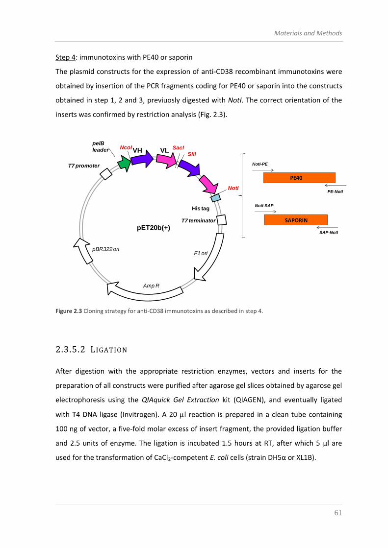
Materials and Methods
61
Step 4: immunotoxins with PE40 or saporin
The plasmid constructs for the expression of anti-CD38 recombinant immunotoxins were
obtained by insertion of the PCR fragments coding for PE40 or saporin into the constructs
obtained in step 1, 2 and 3, previuosly digested with NotI. The correct orientation of the
inserts was confirmed by restriction analysis (Fig. 2.3).
Amp R
pBR322 ori
pET20b(+)
F1 ori
T7 promoter
T7 terminator
pelB
leader VH VL
His tag
NcoI
NotI
SacI
SfiI
PE40
SAPORIN
NotI-PE
PE-NotI
NotI-SAP
SAP-NotI
Figure 2.3 Cloning strategy for anti-CD38 immunotoxins as described in step 4.
2.3.5.2 LIGATION
After digestion with the appropriate restriction enzymes, vectors and inserts for the
preparation of all constructs were purified after agarose gel slices obtained by agarose gel
electrophoresis using the QIAquick Gel Extraction kit (QIAGEN), and eventually ligated
with T4 DNA ligase (Invitrogen). A 20 l reaction is prepared in a clean tube containing
100 ng of vector, a five-fold molar excess of insert fragment, the provided ligation buffer
and 2.5 units of enzyme. The ligation is incubated 1.5 hours at RT, after which 5 μl are
used for the transformation of CaCl2-competent E. coli cells (strain DH5α or XL1B).
Materials and Methods
62
2.3.5.3 COLONY-PCR SCREENING
For a rapid screening of E. coli colonies obtained after transformation with the ligation
reaction, a PCR is performed directly on bacterial cells.
Using a sterile tip, cells from a single colony are picked from the LB-agar plate and
dissolved in 10 l sterile, deionized water in a PCR-tube; the same tip is dipped in 50-100
l of LB broth with appropriate antibiotics in a 1.5 ml eppendorf tube, so that the positive
bacterial clones can be recovered at the end of the screening. In each PCR tube is then
added 15 μl of a concentrated reaction mix with: 2.5 μl of 10x reaction buffer
(Fermentas), dNTPs mix (0.2 mM of each dNTP, Applied Biosystems), forward and reverse
primers (10 pmoles each), 1 unit of Taq DNA polymerase (Fermentas). If necessary, also
5% DMSO is included.
Cycling programme:
The PCR reactions are analysed by agarose-gel electrophoresis and staining with
ethidium bromide. Amplificates of the expected size signify recombinant clones, that can
be recovered from the small LB cultures and propagated for extraction of plasmid DNA.
2.3.6 PLASMID DNA EXTRACTION FROM E. COLI CULTURES
Single colonies picked from LB-agar plates (or from small LB cultures for colony-PCR
screening) are inoculated in 5 mL for small-scale preparations (miniprep), or in 50 mL for
medium-scale preparations (midiprep) of LB broth additioned with the appropriate
antibiotics (100 µg/ml Amp); the culture is grown o.n. at 37 °C with shaking (250 rpm) and
cells are recovered by centrifugation (5 minutes, 10000 rpm at RT in minifuge for
minipreps; 10 minutes, 4000 xg at RT for midipreps) and the bacterial pellet is processed
using the Wizard Plus SV Minipreps DNA Purification System (Promega) for minipreps and
95°C 4 min
95°C 30 sec
35 cycles 55°C 1 min
72°C 1 min
72°C 7 min
4°C end
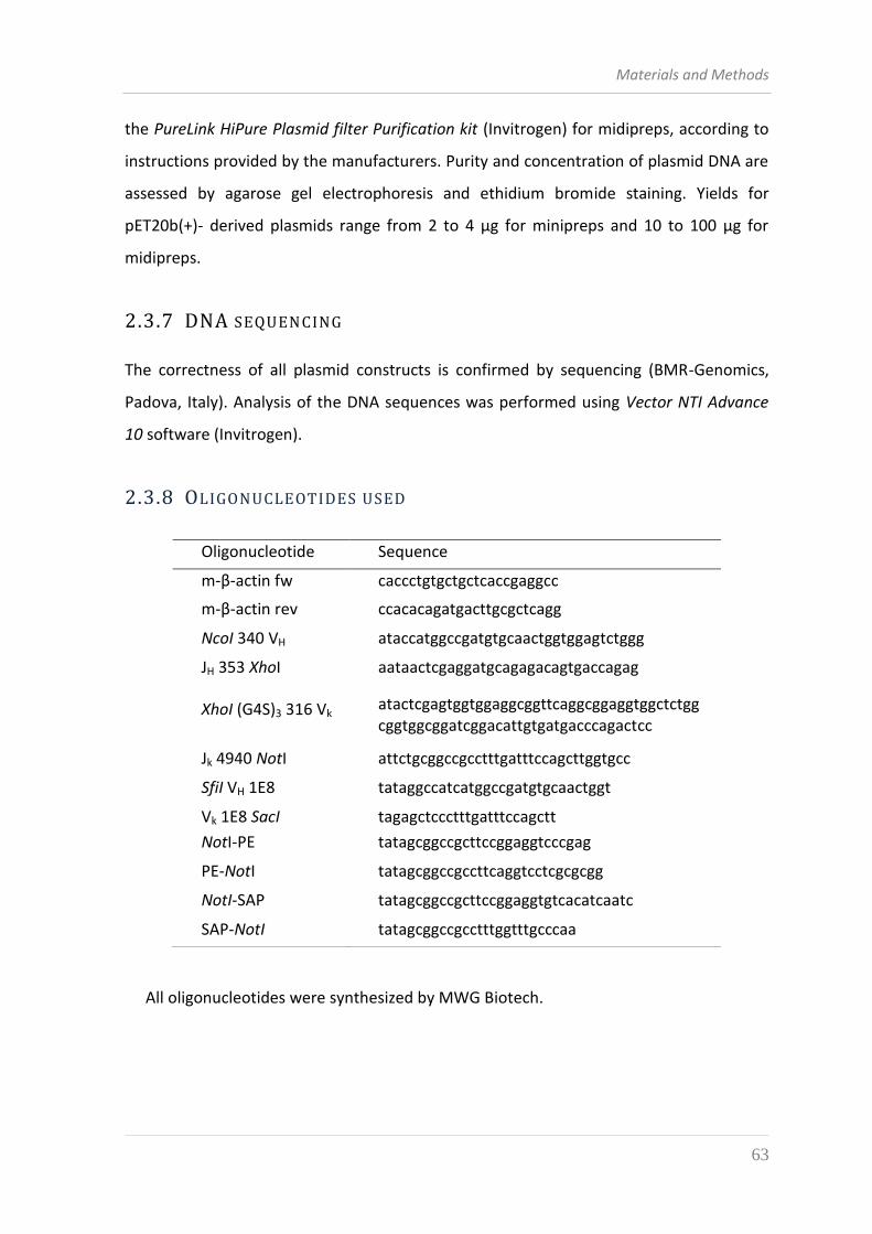
Materials and Methods
63
the PureLink HiPure Plasmid filter Purification kit (Invitrogen) for midipreps, according to
instructions provided by the manufacturers. Purity and concentration of plasmid DNA are
assessed by agarose gel electrophoresis and ethidium bromide staining. Yields for
pET20b(+)- derived plasmids range from 2 to 4 µg for minipreps and 10 to 100 µg for
midipreps.
2.3.7 DNA SEQUENCING
The correctness of all plasmid constructs is confirmed by sequencing (BMR-Genomics,
Padova, Italy). Analysis of the DNA sequences was performed using Vector NTI Advance
10 software (Invitrogen).
2.3.8 OLIGONUCLEOTIDES USED
All oligonucleotides were synthesized by MWG Biotech.
Oligonucleotide Sequence
m-β-actin fw caccctgtgctgctcaccgaggcc
m-β-actin rev ccacacagatgacttgcgctcagg
NcoI 340 VH ataccatggccgatgtgcaactggtggagtctggg
JH 353 XhoI aataactcgaggatgcagagacagtgaccagag
XhoI (G4S)3 316 Vk atactcgagtggtggaggcggttcaggcggaggtggctctgg cggtggcggatcggacattgtgatgacccagactcc
Jk 4940 NotI attctgcggccgcctttgatttccagcttggtgcc
SfiI VH 1E8 tataggccatcatggccgatgtgcaactggt
Vk 1E8 SacI tagagctccctttgatttccagctt
NotI-PE tatagcggccgcttccggaggtcccgag
PE-NotI tatagcggccgccttcaggtcctcgcgcgg
NotI-SAP tatagcggccgcttccggaggtgtcacatcaatc
SAP-NotI tatagcggccgcctttggtttgcccaa
Materials and Methods
64
2.4 PROTEIN EXPRESSION IN BACTERIA
2.4.1 EXPRESSION OF SCFV AND IMMUNOTOXIN IN ESCHERICHIA COLI
BL21(DE3) PLYSS
A single colony of E. coli BL21 (DE3) pLysS transformed with pET20b(+)-based constructs
coding for the scFv or immunotoxins was inoculated in 20 ml of Lb broth, 100 µg/ml Amp,
34 µg/ml Cam and grown o.n. at 37 °C with shaking at 250 rpm. The o.n. culture was used
to start a bigger 1 l culture in LB broth, 100 µg/ml Amp which was grown at 37 °C with
shaking to an OD600 between 0.7 and 0.8. Before induction, 0.5 ml of culture were
transferred into an eppendorf tube and the cell pellet was recovered after centrifugation
for 5 minutes at 11000 rpm in minifuge and stored at -20 °C for later SDS-PAGE analysis
(non induced sample).
Expression of the recombinant protein was induced by addition of 1 mM IPTG into the
culture and incubation was continued for 3 hours at 30 °C with shaking. 0.5 ml of culture
(induced sample) were transferred into an eppendorf tube and treated as with the non
induced sample. The bulk of the induced culture was centrifuged 15 minutes at 8000 xg, 4
°C and the bacterial pellet was frozen in liquid nitrogen and stored at -80 °C before
protein extraction (see par. 2.5.1).
2.5 PROTEIN PROCESSING AN D ANALYSIS 2.5.1 EXTRACTION OF PROTEINS FROM E. COLI BL21(DE3) PLYSS
INCLUSION BODIES
Bacterial pellets obtained from 1 l induced cultures and stored at - 80 °C (see par. 2.4.1)
were resuspended by sonication (3 bursts of 30 seconds each, with incubation of 30
seconds on ice after each burst) in 100 ml of lysis buffer (50 mM Na2HPO4 pH 7.5, 0.5 M
NaCl, 1% Triton X-100, 1 mM PMSF).
After resuspension, the following were added: 10 mM MgCl2, 20 units/ml DNAse I
(Roche), 0.1 mg/ml lysozime. This was followed by an incubation of 20 minutes at 4°C.
The insoluble material was sedimented by centrifugation for 20 minutes at 13000 xg, at 4
°C. Inclusion bodies were then dissolved by sonication in 3 ml lysis buffer, added dropwise
Materials and Methods
65
into a beaker containing 47 ml of a denaturating buffer (50 mM Na2HPO4 pH 7.5, 0.5 M
NaCl, 8 M urea) kept under magnetic stirring and then incubated for 1 hour at RT. The
solubilized inclusion bodies were centrifuged 30 minutes at 20000 xg, 4 °C to separate
persisting aggregates and the supernatant was then recovered for further purification of
the recombinant proteins or store at 4°C.
2.5.2 PURIFICATION OF RECOM BINANT PROTEINS BY AFFINITY
CHROMATOGRAPHY
Constructs for both scFv and ITs have a C-terminal hexahistidine tag, which was exploited
for purification by Immobilized metal-ion affinity chromatography (IMAC). We used the Ni
Sepharose 6 Fast Flow (GE Healthcare) that takes advantage of the coordination occurring
between the Ni2+ ions immobilized on the resin beads and the imidazole rings of
histidines.
The resin must first be equilibrated in the buffer in which inclusion bodies are
resuspended and which is used for the chromatography. Two ml of Ni Sepharose slurry
were mixed by inversion with 8 ml of denaturing buffer (see par. 2.5.1) containing 20 mM
imidazole and then centrifuged 5 minutes at 2000 rpm to recovering the equilibrated
resin. The sample (denatured inclusion bodies, see par. 2.5.1) was filtered by 0.45 μm
syringe filter to remove possible aggregates and the supernatant was mixed with the
sedimented resin into a beaker and incubated o.n. at 4°C. The day after, the sample was
loaded onto a column (sealed at both ends) and incubated few mintes to allow the resin
to settle. The column was then uncapped and the flow-through was collected. Three
washing steps followed with 10 ml of Wash buffer with 20 mM imidazole, to remove
unwanted bacterial proteins that could bind the resin aspecifically. Elution of His6-tagged
proteins was finally obtained in several steps, each time adding 1 ml of Elution buffer and
collecting the eluate in eppendorf tubes.
Materials and Methods
66
Wash buffer:
NaH2PO4 50 mM pH 7.5
NaCl 0.5 M
Urea 8 M
Imidazole 20 mM
Elution buffer:
NaH2PO4 50 mM pH 7.5
NaCl 0.5 M
Urea 8 M
Imidazole 0.5 mM
Absorbance at 280 nm of the eluted samples gave an indication of the amount of purified
protein: the samples containing a sufficient amount of protein (OD280 > 0.1), were pooled
and dialyzed in the refolding process.
2.5.3 REFOLDING OF PROTEINS FROM INCLUSION BODIE S
Refolding of urea-denatured proteins purified by IMAC was attained by multi-step dialysis
that gradually decreased the concentration of denaturant, therefore promoting protein
refolding.
Tubular membranes with a 14 kDa cutoff (Carl Roth) were used.
Each dialysis steps were performed in a volume of 500 ml, for 12 hours at 4 °C according
to the following scheme:
- Refolding buffer with 4 M urea
- Refolding buffer with 2 M urea
- Refolding buffer with 1 M urea
- Refolding buffer with 0.5 M urea
- Refolding buffer with 0.25 M urea
- Refolding buffer with no urea
- Refolding buffer without L-Arginine
- PBS
Materials and Methods
67
In case some precipitate formed during the dialysis procedure, it was removed by
centrifugation at 7000 xg, 10 minutes at 4 °C. Supernatant was recovered and dialysis
continued with the next step.
Refolding Buffer:
NaH2PO4 50 mM pH 8
NaCl 0.5 M
L-Arginine 0.4 M
10x PBS buffer:
NaCl 80 g/l
KCl 2 g/l
Na2HPO4 (2H2O) 17.9 g/l
KH2PO4 2 g/l
Adjust pH to 7.2, sterilize 20 minutes at 121 °C and store at RT. Dilute to 1x in deionized
water.
2.5.4 PURIFICATION OF MAB FROM HYBRIDOMA CULTURE MEDIUM
1E82H11 mAb was purified by affinity chromatography using the Protein G Sepharose 6
Fast Flow (GE Healthcare) which exploits the binding of protein G to the Fc region of
murine IgG.
Two ml of Protein G Sepharose slurry were mixed with 8 ml of 20 mM sodium phosphate
buffer, pH 7 and loaded into a column for resin equilibration. After sedimentation of the
resin, the buffer was discarded as flow-through. Ten ml of hybridoma culture supernatant
were centrifuged (1000 rpm, 10 minutes, 4 °C) to remove possible cell debris and aggregates
and the supernatant was loaded onto the column. The column was sealed at both ends and
incubated o.n. at 4°C on a rotating wheel. The column was then kept in a vertical position
and, after the resin settled, it was uncapped and the flow-through was collected. Three
washing steps followed with 10 ml of 20 mM sodium phosphate buffer, pH 7 to remove
unwanted proteins that could bind the resin aspecifically. Elution of IgGs was finally obtained
Materials and Methods
68
adding 900 μl of 0.1 M glycine-HCl, pH 3 and collecting the eluate in eppendorf tubes where
100 μl of neutralizing buffer (1 M Tris-HCl, pH 9) were previously added.
Absorbance at 280 nm of the eluted samples gave an indication of the amount of purified
protein: the samples containing a sufficient amount of protein were pooled and dialyzed
in 2 l PBS buffer, o.n. at 4 °C.
2.5.5 DENATURING POLYACRY LAMIDE GEL ELECTROPHORESIS (SDS-PAGE)
The electrophoretic separation of proteins from crude extracts, supernatants or purified
samples was performed according to standard laboratory procedures, using mini-gels
with 5% acrylamide stacking and 12% separating slabs. Electrophoresis was conducted in
Tris-Glycine buffer (25 mM Tris, 190 mM Glycine, 0,1% SDS), using the Mini-Protean III
Cell apparatus (BioRad) under reducing conditions (samples were denatured by boiling for
5 minutes in Sample Loading buffer (4X) - 0.32 M TrisHCl pH 6.8, 6% SDS, 50% glycerol, 2%
β-mercaptoethanol, 0.006 g Bromophenol blue). A tension of 100 V was applied until the
blue line (given by the bromophenol in Sample Loading Buffer) came out of the running
gel. A protein size standard (PageRuler Prestained Protein Ladder Plus, Fermentas) was
also loaded for molecular weight determination of the analyzed proteins.
2.5.6 IMMUNOBLOTTING 2.5.6.1 TRANSFER OF PROTEINS ON PVDF MEMBRANE
Proteins separated by SDS-PAGE were blotted on polyvinylidene fluoride (PVDF)
membranes (Immobilon–P, Millipore) following the manufacturer’s indications. The
polyacrilamide gel and PVDF membrane were assembled as a sandwich in a Mini Trans-
Blot Electrophoretic Transfer Cell (BioRad) according to the manufacturer’s instructions.
For proteins to be transferred, a tension of 100 was applied for 1 hour.
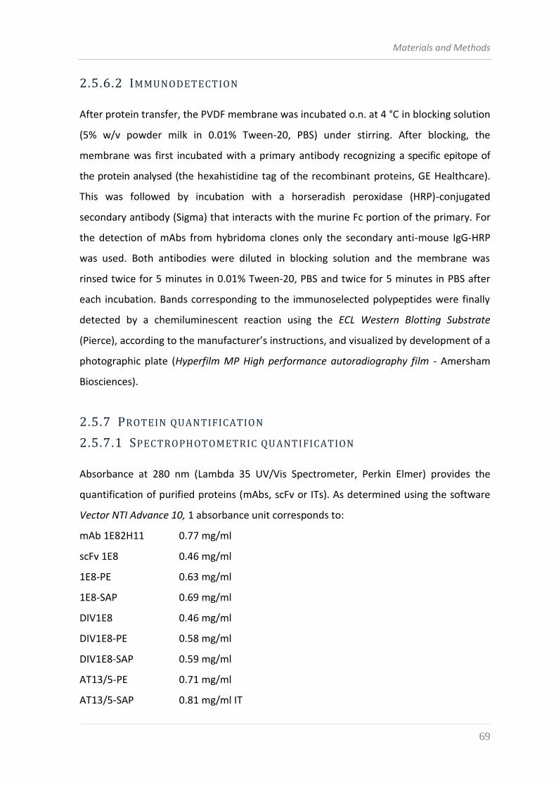
Materials and Methods
69
2.5.6.2 IMMUNODETECTION
After protein transfer, the PVDF membrane was incubated o.n. at 4 °C in blocking solution
(5% w/v powder milk in 0.01% Tween-20, PBS) under stirring. After blocking, the
membrane was first incubated with a primary antibody recognizing a specific epitope of
the protein analysed (the hexahistidine tag of the recombinant proteins, GE Healthcare).
This was followed by incubation with a horseradish peroxidase (HRP)-conjugated
secondary antibody (Sigma) that interacts with the murine Fc portion of the primary. For
the detection of mAbs from hybridoma clones only the secondary anti-mouse IgG-HRP
was used. Both antibodies were diluted in blocking solution and the membrane was
rinsed twice for 5 minutes in 0.01% Tween-20, PBS and twice for 5 minutes in PBS after
each incubation. Bands corresponding to the immunoselected polypeptides were finally
detected by a chemiluminescent reaction using the ECL Western Blotting Substrate
(Pierce), according to the manufacturer’s instructions, and visualized by development of a
photographic plate (Hyperfilm MP High performance autoradiography film - Amersham
Biosciences).
2.5.7 PROTEIN QUANTIFICATION
2.5.7.1 SPECTROPHOTOMETRIC QU ANTIFICATION
Absorbance at 280 nm (Lambda 35 UV/Vis Spectrometer, Perkin Elmer) provides the
quantification of purified proteins (mAbs, scFv or ITs). As determined using the software
Vector NTI Advance 10, 1 absorbance unit corresponds to:
mAb 1E82H11 0.77 mg/ml
scFv 1E8 0.46 mg/ml
1E8-PE 0.63 mg/ml
1E8-SAP 0.69 mg/ml
DIV1E8 0.46 mg/ml
DIV1E8-PE 0.58 mg/ml
DIV1E8-SAP 0.59 mg/ml
AT13/5-PE 0.71 mg/ml
AT13/5-SAP 0.81 mg/ml IT
Materials and Methods
70
2.5.7.2 COOMASSIE STAINING
A further means of protein quantification is provided by Coomassie staining after
elctrophoresis of purified proteins. SDS-PAGE gels are stirred for 30 minutes at RT in
Coomassie solution, followed by decoloration in Destaining solution until bands are
clearly visible. Amounts of proteins in discrete bands are assessed by comparison with
known quantities of bovine serum albumin (BSA, Sigma).
Coomassie solution
Coomassie Brilliant Blue R250 0.25% w/v
Methanol 45% v/v
Acetic acid glacial 10% v/v
in deionized H2O
Destaining solution
Methanol 45% v/v
Acetic acid glacial 10% v/v
in deionized H2O
2.6 ANALYSIS OF BINDING IN FLOW-CYTOMETRY
2.6.1 COMPARISON BETWEEN BINDING EFFICIENCIES O F HYBRIDOMAS
3T3/7A and Ramos cells were grown in T75 flasks to exponential phase and harvested by
centrifugation at 150 xg, 5 minutes, 4°C in 15 ml tubes. After counting with a
hemacytometer, cells were resuspended in a binding buffer (0.5% w/v BSA in PBS) and
incubated 20 minutes on ice. Next, 2 x 10⁵ cells were dispensed in each flow-cytometry
tube and centrifuged as before. Cells were resuspended in serial dilutions of hybridoma
supernatants and incubated on ice for 1 hour. Two washing steps follow, each with 2 ml
of blocking buffer. For the detection of bound mAbs, cells were stained with an anti-
mouse IgG-FITC antibody (goat polyclonal from Beckman Coulter, 1:100 diluted).
Materials and Methods
71
Incubation was carried out for 30 minutes on ice in 100 µl of binding buffer, followed by
two washing steps with 2 ml binding buffer each. Background fluorescence was assessed
by staining cells with anti-mouse-FITC.
At the end of the staining, cells in each tube were resuspended in 0.5 ml binding buffer
and the mean fluorescence intensity (MFI) of each sample was determined using BD Facs
Canto (BD Bioscience).
2.6.2 COMPETITION ASSAY FOR SPECIFIC BINDING OF NEW MABS TO
CD38 ON CELLS
2 x 10⁵ Ramos cells were sedimented in single flow-cytometry tubes as described above
(par. 2.6.1). Each tube was stained with 200 ng of biotinylated OKT10 mAb mixed with
100 μl of hybridoma supernatant containing an excess of IgG. After a 30 minutes
incubation period on ice, two washes with 2 ml of binding buffer each were performed. A
second staining with Streptavidin-FITC was used carried out for 30 minutes on ice,
followed by two more washes, after which samples were resuspended in 0.5 ml binding
buffer and analysed with BD Facs Canto (BD Bioscience).
The decrease of OKT10-biotin signal, evaluated by comparison with the maximal MFI
obtained by incubation without a displacing mAb, was determined using BD Facs Canto
(BD Bioscience).
2.6.3 CURVES OF BINDING TO THE CD38 ANTIGEN ON CELLS
2 x 10⁵ Daudi cells were sedimented in single flow-cytometry tubes as described above
(par. 2.6.1). Cells were resuspended in 100 µl of binding buffer containing increasing
amounts of mAb or scFv and incubated on ice for 1 hour. The staining with the
recombinant ITs was performed with a fixed concentration of protein, without
determining a curve. Two washing steps follow, each with 2 ml of blocking buffer. For the
detection of bound scFvand ITs, cells were stained first with an anti-His6 secondary
antibody (mouse mAb from GEHealthcare, 1:200 diluted in binding buffer) and then with
an anti-mouse IgG-FITC tertiary antibody (goat polyclonal from Beckman Coulter, 1:100
diluted). On the contrary, after staining with our anti-CD38 mAb or the commercial OKT10
Materials and Methods
72
mAb cells were directly incubated with anti-mouse IgG-FITC antibody. Incubations were
for 30 minutes on ice, followed by two washing steps with 2 ml binding buffer each.
Background fluorescence was assessed by staining cells with anti-mouse-FITC (for mAbs)
or anti-His6 followed by anti-mouse-FITC (for scFv and ITs). At the end of the staining, cells
in each tube were resuspended in 0.5 ml binding buffer and MFI of each sample was
determined as described above.
2.7 BIOLOGICAL ASSAYS
2.7.1 CYTOTOXICITY ASSESSMENT BY LEUCINE INCORPORATION
The effect on uptake of 14C-leucine into cells was taken as a measure of the protein
synthesis inhibition caused by treatment with a toxin or immunotoxins. Cells were
resuspended in leucine-free RPMI-1640 medium supplemented with 2% FCS and 3x10⁴
cells in a volume of 90 μl were seeded in each well of round-bottom 96-well plates
(Greiner bio-one). The molecules to be tested were dialysed in PBS, filter-sterilized
through Spin-X tubes (Costar) and diluted in the same medium used for cell resuspension.
Ten μl of differently diluted ITs were finally added in each well. The plate was incubated
for 48 hours at 37 °C, 5% CO2. Ten µl of 14C-leucine from a 1-1.2x10⁵ cpm/ml stock in
leucine-free RPMI-1640 were added 16 hours before the end time, after which the
content of each well was transferred to filter paper using a cell-harvester (Wesbart), and
radioactivity was measured using a beta counter (Wallac 1409, Pharmacia).
Results obtained for experimental cultures are expressed as a percentage of the
amount of 14C-leucine incorporation observed in untreated control cultures maintained
under identical conditions.
2.7.2 CELL PROLIFERATION AS SAY WITH XTT
The cytotoxic activity of the ITs was evaluated using a colorimetric cell proliferation assay
based on the conversion of XTT (sodium 2,3-bis(2-methoxy-4-nitro-5-sulfophenyl)-5-
[(phenylamino)-carbonyl]-2H-tetrazolium inner salt) to orange-colored formazan
compounds by cellular dehydrogenases. Thirty thousand cells, seeded in 96-well plate
Materials and Methods
73
and maintained in phenol red-free RPMI-1640 medium were treated with serial dilutions
of IT for 72 hours. After this incubation period, 50 μl of 1 mg/ml XTT solution were added
to each well and incubated for 3 hours. The absorbance was read at 490 and the 690 nm
using a plate reader.
2.7.3 APOPTOSIS ASSAY
CD38+ and CD38- cells were seeded in 24-well plates at a final concentration of 3x10⁵ and
treated with 1 μg/ml of the different ITs, while B lymphocytes were seeded in 48-well plates
at a final concentration of 2 x 10⁶ cells. Cells were incubated for 48 hours at 37 °C, 5% CO2,
after which cells were harvested an sedimented in single flow-cytometry tubes and
centrifuged at 150 xg, 5 minutes, 4°C. After wasting the supernatants, cells were stained
with Annexin V-FITC/propidium iodide (PI) following the manufacturer’s instructions for an
incubation period of 15 minutes at RT. Cells were then washed with 2 ml of PBS and
resuspended in a 0.5 final volume for further flow-cytometric analysis.

Materials and Methods
74

3. RESULTS

Results
76
Results
77
3.1 CHARACTERIZATION OF N EW ANTI-CD38 HYBRIDOMA
CLONES
A standard procedure of mice immunization and hybridoma fusion was exploited for
the generation of novel anti-CD38 clones. Briefly, Balb/c mice were first boosted with
intraperitoneal (i.p.) injection of a membrane preparation from 3T3 cells expressing the
human CD38 antigen (kindly provided by Prof. Ippoliti R. and co-workers, Università
dell’Aquila). I.p. injection was repeated after seven days and three, four, five days later
bacterially expressed CD38 ectodomain (from Prof. Ippoliti R. and co-workers) was
intravenously (i.v.) injected. The sera from all the immunized animals were then tested
and compared by immunoblotting and ELISA on the bacterially expressed CD38 (data not
shown). The mouse showing the best anti-CD38 reactivity was used to carry out PEG-
mediated fusion of spleen lymphocytes and the myeloma line Ag8. Fusions were
dispensed in 48-well plates and the CD38-reactivity of supernatants was assayed by flow-
cytometry, allowing to select positive wells for subsequent limiting-dilution cloning in 96-
well plates. Different dilutions of supernatants from monoclonal wells were likewise
tested by flow-cytometry on CD38+ 3T3 cells to assess the ability of the different clones
to bind the target antigen. As shown in figure 3.1, four of the investigated clones,
underlined with red color, were demonstrated not to bind CD38, while all the other ten
clones showed a positive staining of CD38+ 3T3 cells. In particular it can be observed that
two clones, highlighted with green color, exhibited high binding affinity. However, this
first type of investigation did not take into account the quantification of the IgG contained
into the supernatants of the different clones. Moreover, some clones may not secrete
functional IgG and this may account for the apparent absence of binding.
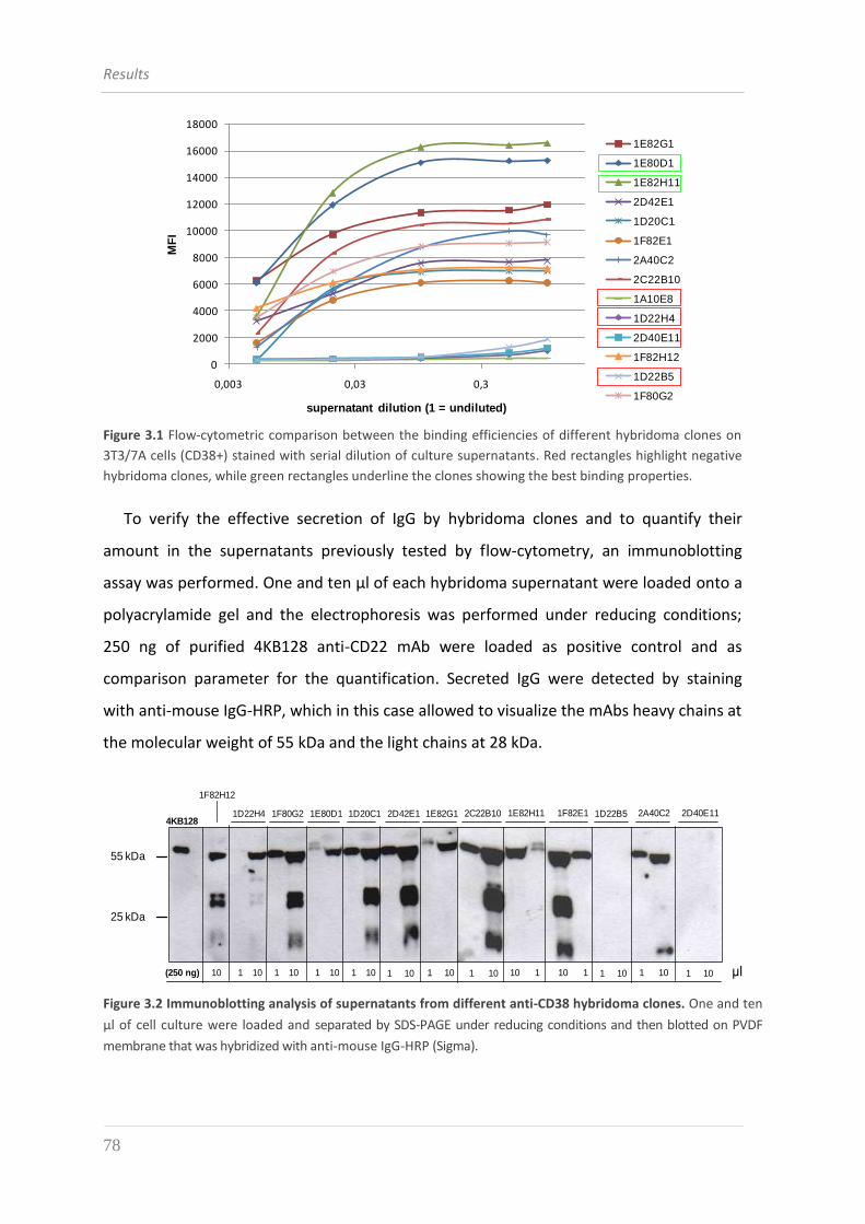
Results
78
0
2000
4000
6000
8000
10000
12000
14000
16000
18000
0,003 0,03 0,3
MF
I
supernatant dilution (1 = undiluted)
1E82G1
1E80D1
1E82H11
2D42E1
1D20C1
1F82E1
2A40C2
2C22B10
1A10E8
1D22H4
2D40E11
1F82H12
1D22B5
1F80G2
Figure 3.1 Flow-cytometric comparison between the binding efficiencies of different hybridoma clones on
3T3/7A cells (CD38+) stained with serial dilution of culture supernatants. Red rectangles highlight negative
hybridoma clones, while green rectangles underline the clones showing the best binding properties.
To verify the effective secretion of IgG by hybridoma clones and to quantify their
amount in the supernatants previously tested by flow-cytometry, an immunoblotting
assay was performed. One and ten μl of each hybridoma supernatant were loaded onto a
polyacrylamide gel and the electrophoresis was performed under reducing conditions;
250 ng of purified 4KB128 anti-CD22 mAb were loaded as positive control and as
comparison parameter for the quantification. Secreted IgG were detected by staining
with anti-mouse IgG-HRP, which in this case allowed to visualize the mAbs heavy chains at
the molecular weight of 55 kDa and the light chains at 28 kDa.
55 kDa
25 kDa
2C22B10 1E82H11 1F82E1 2A40C21D22B5 2D40E114KB128
(250 ng)
1D22H4 1F80G2 1E80D1 1D20C1 2D42E1 1E82G1
1F82H12
10 10 1 1 10 1 10 1 10 1 10 1 10 1 10 1 10 1 10 1 10 1 10 10 1 μl
Figure 3.2 Immunoblotting analysis of supernatants from different anti-CD38 hybridoma clones. One and ten
µl of cell culture were loaded and separated by SDS-PAGE under reducing conditions and then blotted on PVDF
membrane that was hybridized with anti-mouse IgG-HRP (Sigma).
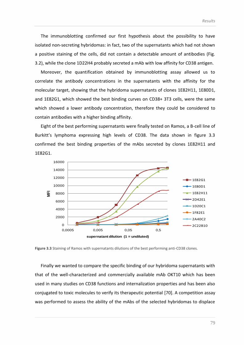
Results
79
The immunoblotting confirmed our first hypothesis about the possibility to have
isolated non-secreting hybridomas: in fact, two of the supernatants which had not shown
a positive staining of the cells, did not contain a detectable amount of antibodies (Fig.
3.2), while the clone 1D22H4 probably secreted a mAb with low affinity for CD38 antigen.
Moreover, the quantification obtained by immunoblotting assay allowed us to
correlate the antibody concentrations in the supernatants with the affinity for the
molecular target, showing that the hybridoma supernatants of clones 1E82H11, 1E80D1,
and 1E82G1, which showed the best binding curves on CD38+ 3T3 cells, were the same
which showed a lower antibody concentration, therefore they could be considered to
contain antibodies with a higher binding affinity.
Eight of the best performing supernatants were finally tested on Ramos, a B-cell line of
Burkitt’s lymphoma expressing high levels of CD38. The data shown in figure 3.3
confirmed the best binding properties of the mAbs secreted by clones 1E82H11 and
1E82G1.
0
2000
4000
6000
8000
10000
12000
14000
16000
0,0005 0,005 0,05 0,5
MF
I
supernatant dilution (1 = undiluted)
1E82G1
1E80D1
1E82H11
2D42E1
1D20C1
1F82E1
2A40C2
2C22B10
Figure 3.3 Staining of Ramos with supernatants dilutions of the best performing anti-CD38 clones.
Finally we wanted to compare the specific binding of our hybridoma supernatants with
that of the well-characterized and commercially available mAb OKT10 which has been
used in many studies on CD38 functions and internalization properties and has been also
conjugated to toxic molecules to verify its therapeutic potential [70]. A competition assay
was performed to assess the ability of the mAbs of the selected hybridomas to displace
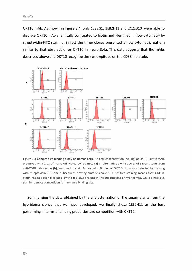
Results
80
OKT10 mAb. As shown in figure 3.4, only 1E82G1, 1E82H11 and 2C22B10, were able to
displace OKT10 mAb chemically conjugated to biotin and identified in flow-cytometry by
streptavidin-FITC staining; in fact the three clones presented a flow-cytometric pattern
similar to that observable for OKT10 in figure 3.4a. This data suggests that the mAbs
described above and OKT10 recognize the same epitope on the CD38 molecule.
OKT10-biotin OKT10 mAb+ OKT10-biotin
1D20C11E80D11F82E12D42E1 2A40C2
1E82G11E82H112C22B10
a
b
Figure 3.4 Competitive binding assay on Ramos cells. A fixed concentration (200 ng) of OKT10-biotin mAb,
pre-mixed with 2 μg of non-biotinylated OKT10 mAb (a) or alternatively with 100 μl of supernatants from
anti-CD38 hybridomas (b), was used to stain Ramos cells. Binding of OKT10-biotin was detected by staining
with streptavidin-FITC and subsequent flow-cytometric analysis. A positive staining means that OKT10-
biotin has not been displaced by the the IgGs present in the supernatant of hybridomas, while a negative
staining denote competition for the same binding site.
Summarizing the data obtained by the characterization of the supernatants from the
hybridoma clones that we have developed, we finally chose 1E82H11 as the best
performing in terms of binding properties and competition with OKT10.
Results
81
3.2 CHARACTERIZATION OF THE MONOCLONAL ANTIBODY
1E82H11
Hybridoma cells of the selected clone 1E82H11 were grown in a bioreactor (Sartorius)
and the supernatant enriched of IgG was collected weekly. The mAbs were purified from
the supernatants by affinity chromatography exploiting the binding of the Fc region of the
IgGs to the G protein immobilized on a Sepharose matrix and allowing to collect 1 mg of
purified antibody for each ml of culture supernatant.
Firstly the binding and specificity properties of the purified anti-CD38 mAb 1E82H11 to
the native cellular antigen were verified by flow-cytometry on CD38+ cell lines (the
lymphoid cell line Daudi and the multiple myeloma line RPMI8226) and the CD38- cell line
of myeloid origin U266. As shown in figure 3.5, when stained with 5 μg of the 1E82H11
mAb, Daudi and RPMI8226 caused an evident rightward shift in the MFI (Mean
Fluorescence Intensity) peak, as compared to the negative control, unlike U266 that do
not express the antigen and therefore showed a negligible MFI shift. The MFI values
suggest a high binding affinity and moreover a suitable specificity of 1E82H11 mAb for the
target antigen since no binding could be observed on CD38- cells.
We observed a higher CD38 expression level on Daudi than RPMI8226, but also an
increased expression when myeloma cells were exposed for 16 hours to 10 nM all-trans
retinoic acid (ATRA), a conventional drug used in the therapy of Acute Promyelocytic
Leukemia (PML) and described as a potent and selective inducer of CD38 expression in
myeloid leukemia cells (see paragraph 1.3.1.2.2). On the contrary ATRA did not show any
significant effect on CD38 expression of U266 cells; in fact, no increase in MFI value was
observed with respect of cells not treated with ATRA. (Fig. 3.5).
Due to the higher levels of target antigen expressed, Daudi was selected as the line of
choice for further experiments of characterization of the mAb and the other anti-CD38
constructs.
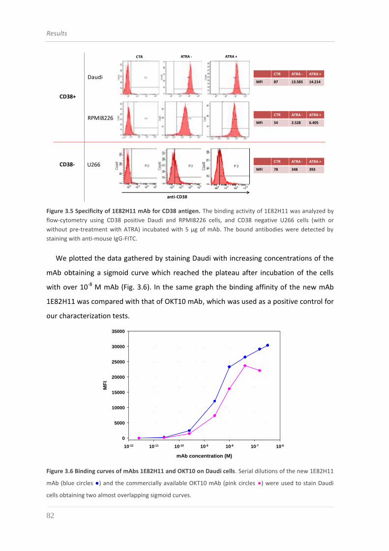
Results
82
CTR ATRA - ATRA +
MFI 87 13.583 14.214
CTR ATRA - ATRA +
MFI 78 348 393
CTR ATRA - ATRA +
MFI 54 2.528 6.405
CTR ATRA - ATRA +
Daudi
U266
anti-CD38
RPMI8226
CD38+
CD38-
Figure 3.5 Specificity of 1E82H11 mAb for CD38 antigen. The binding activity of 1E82H11 was analyzed by
flow-cytometry using CD38 positive Daudi and RPMI8226 cells, and CD38 negative U266 cells (with or
without pre-treatment with ATRA) incubated with 5 μg of mAb. The bound antibodies were detected by
staining with anti-mouse IgG-FITC.
We plotted the data gathered by staining Daudi with increasing concentrations of the
mAb obtaining a sigmoid curve which reached the plateau after incubation of the cells
with over 10-8 M mAb (Fig. 3.6). In the same graph the binding affinity of the new mAb
1E82H11 was compared with that of OKT10 mAb, which was used as a positive control for
our characterization tests. Binding mAb anti-CD38
mAb concentration (M)
10-12 10-11 10-10 10-9 10-8 10-7 10-6
MF
I
0
5000
10000
15000
20000
25000
30000
35000
1E82H11
OKT10
Figure 3.6 Binding curves of mAbs 1E82H11 and OKT10 on Daudi cells. Serial dilutions of the new 1E82H11
mAb (blue circles ●) and the commercially available OKT10 mAb (pink circles ●) were used to stain Daudi
cells obtaining two almost overlapping sigmoid curves.
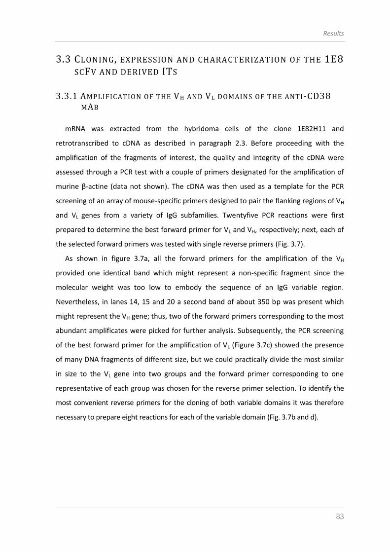
Results
83
3.3 CLONING , EXPRESSION AND CHARACTERIZATION OF THE 1E8
SCFV AND DERIVED ITS
3.3.1 AMPLIFICATION OF THE VH AND VL DOMAINS OF THE ANTI-CD38
MAB
mRNA was extracted from the hybridoma cells of the clone 1E82H11 and
retrotranscribed to cDNA as described in paragraph 2.3. Before proceeding with the
amplification of the fragments of interest, the quality and integrity of the cDNA were
assessed through a PCR test with a couple of primers designated for the amplification of
murine β-actine (data not shown). The cDNA was then used as a template for the PCR
screening of an array of mouse-specific primers designed to pair the flanking regions of VH
and VL genes from a variety of IgG subfamilies. Twentyfive PCR reactions were first
prepared to determine the best forward primer for VL and VH, respectively; next, each of
the selected forward primers was tested with single reverse primers (Fig. 3.7).
As shown in figure 3.7a, all the forward primers for the amplification of the VH
provided one identical band which might represent a non-specific fragment since the
molecular weight was too low to embody the sequence of an IgG variable region.
Nevertheless, in lanes 14, 15 and 20 a second band of about 350 bp was present which
might represent the VH gene; thus, two of the forward primers corresponding to the most
abundant amplificates were picked for further analysis. Subsequently, the PCR screening
of the best forward primer for the amplification of VL (Figure 3.7c) showed the presence
of many DNA fragments of different size, but we could practically divide the most similar
in size to the VL gene into two groups and the forward primer corresponding to one
representative of each group was chosen for the reverse primer selection. To identify the
most convenient reverse primers for the cloning of both variable domains it was therefore
necessary to prepare eight reactions for each of the variable domain (Fig. 3.7b and d).
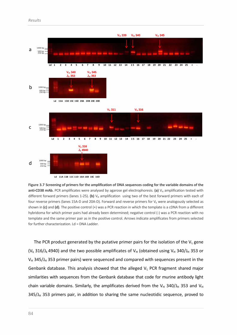
Results
84
Ld 1 2 3 4 5 6 7 8 9 10 11 12 13 14 1 5 16 17 18 19 20 21 22 23 24 25 + -
VH 339 VH 340 VH 345
Ld 15A 15B 15C 15D 20A 20B 20C 20D
VH 340
JH 353
VH 345
JH 353
a
b 1000 bp
500 bp250 bp
1000 bp
500 bp250 bp
Ld 1 2 3 4 5 6 7 8 9 10 11 12 13 14 1 5 16 17 18 19 20 21 22 23 24 25 + -
Ld 11A 11B 11C 11D 16A 16B 16C 16D
Vk 311 Vk 316
Vk 316
Jk 4940
c
d
1000 bp
500 bp250 bp
1000 bp
500 bp250 bp
Figure 3.7 Screening of primers for the amplification of DNA sequences coding for the variable domains of the
anti-CD38 mAb. PCR amplificates were analysed by agarose gel electrophoresis. (a) VH amplification tested with
different forward primers (lanes 1-25). (b) VH amplification using two of the best forward primers with each of
four reverse primers (lanes 15A-D and 20A-D). Forward and reverse primers for Vk were analogously selected as
shown in (c) and (d). The positive control (+) was a PCR reaction in which the template is a cDNA from a different
hybridoma for which primer pairs had already been determined; negative control (-) was a PCR reaction with no
template and the same primer pair as in the positive control. Arrows indicate amplificates from primers selected
for further characterization. Ld = DNA Ladder.
The PCR product generated by the putative primer pairs for the isolation of the VL gene
(Vk 316/Jk 4940) and the two possible amplificates of VH (obtained using VH 340/JH 353 or
VH 345/JH 353 primer pairs) were sequenced and compared with sequences present in the
Genbank database. This analysis showed that the alleged VL PCR fragment shared major
similarities with sequences from the Genbank database that code for murine antibody light
chain variable domains. Similarly, the amplificates derived from the VH 340/JH 353 and VH
345/JH 353 primers pair, in addition to sharing the same nucleotidic sequence, proved to
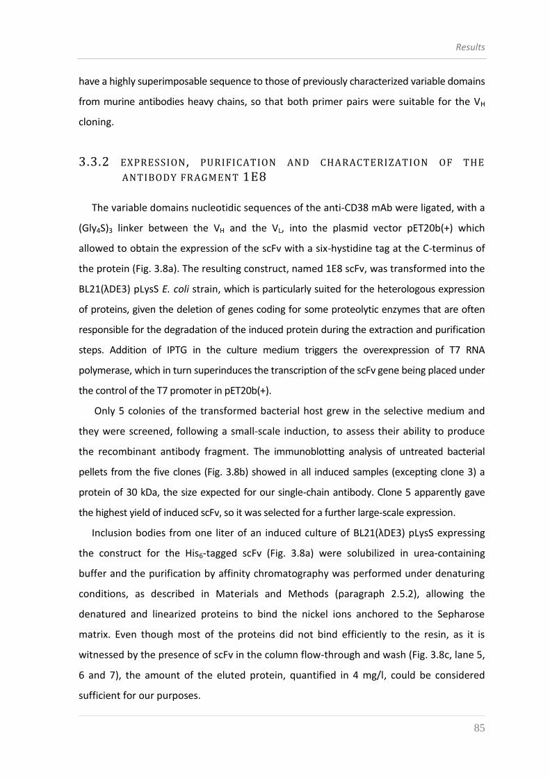
Results
85
have a highly superimposable sequence to those of previously characterized variable domains
from murine antibodies heavy chains, so that both primer pairs were suitable for the VH
cloning.
3.3.2 EXPRESSION , PURIFICATION AND CHARACTERIZATION OF THE
ANTIBODY FRAGMENT 1E8
The variable domains nucleotidic sequences of the anti-CD38 mAb were ligated, with a
(Gly4S)3 linker between the VH and the VL, into the plasmid vector pET20b(+) which
allowed to obtain the expression of the scFv with a six-hystidine tag at the C-terminus of
the protein (Fig. 3.8a). The resulting construct, named 1E8 scFv, was transformed into the
BL21(λDE3) pLysS E. coli strain, which is particularly suited for the heterologous expression
of proteins, given the deletion of genes coding for some proteolytic enzymes that are often
responsible for the degradation of the induced protein during the extraction and purification
steps. Addition of IPTG in the culture medium triggers the overexpression of T7 RNA
polymerase, which in turn superinduces the transcription of the scFv gene being placed under
the control of the T7 promoter in pET20b(+).
Only 5 colonies of the transformed bacterial host grew in the selective medium and
they were screened, following a small-scale induction, to assess their ability to produce
the recombinant antibody fragment. The immunoblotting analysis of untreated bacterial
pellets from the five clones (Fig. 3.8b) showed in all induced samples (excepting clone 3) a
protein of 30 kDa, the size expected for our single-chain antibody. Clone 5 apparently gave
the highest yield of induced scFv, so it was selected for a further large-scale expression.
Inclusion bodies from one liter of an induced culture of BL21(λDE3) pLysS expressing
the construct for the His6-tagged scFv (Fig. 3.8a) were solubilized in urea-containing
buffer and the purification by affinity chromatography was performed under denaturing
conditions, as described in Materials and Methods (paragraph 2.5.2), allowing the
denatured and linearized proteins to bind the nickel ions anchored to the Sepharose
matrix. Even though most of the proteins did not bind efficiently to the resin, as it is
witnessed by the presence of scFv in the column flow-through and wash (Fig. 3.8c, lane 5,
6 and 7), the amount of the eluted protein, quantified in 4 mg/l, could be considered
sufficient for our purposes.
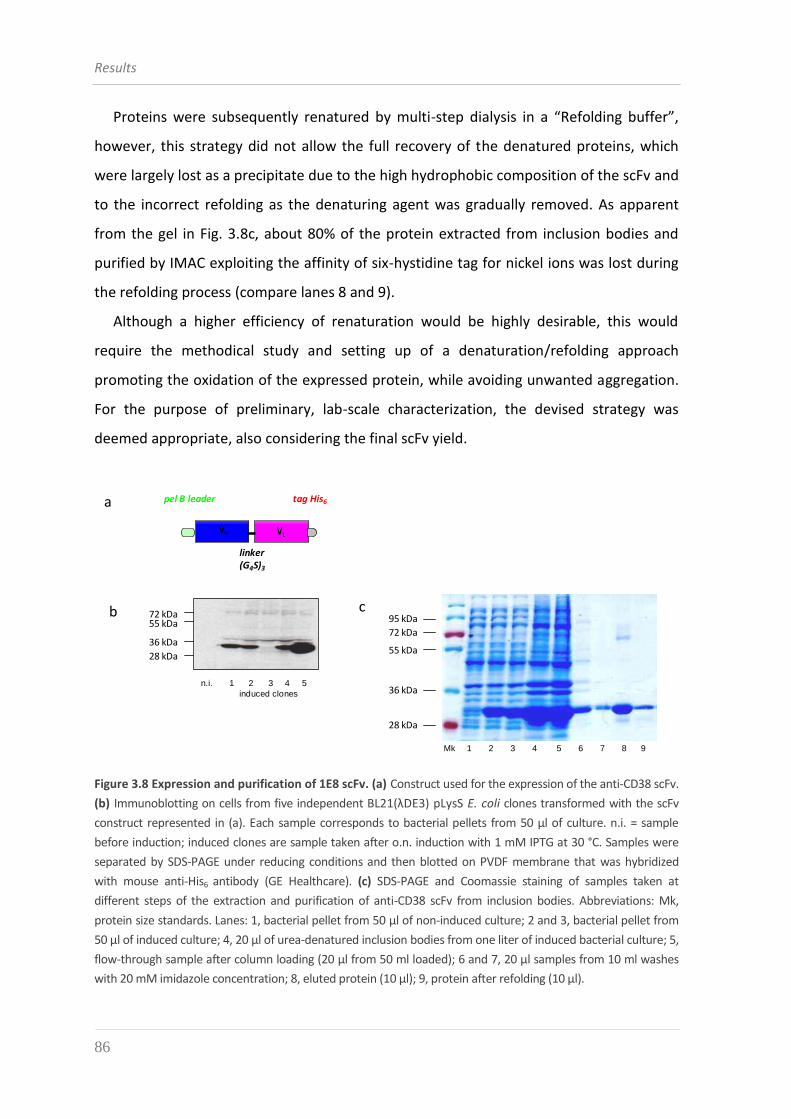
Results
86
Proteins were subsequently renatured by multi-step dialysis in a “Refolding buffer”,
however, this strategy did not allow the full recovery of the denatured proteins, which
were largely lost as a precipitate due to the high hydrophobic composition of the scFv and
to the incorrect refolding as the denaturing agent was gradually removed. As apparent
from the gel in Fig. 3.8c, about 80% of the protein extracted from inclusion bodies and
purified by IMAC exploiting the affinity of six-hystidine tag for nickel ions was lost during
the refolding process (compare lanes 8 and 9).
Although a higher efficiency of renaturation would be highly desirable, this would
require the methodical study and setting up of a denaturation/refolding approach
promoting the oxidation of the expressed protein, while avoiding unwanted aggregation.
For the purpose of preliminary, lab-scale characterization, the devised strategy was
deemed appropriate, also considering the final scFv yield.
72 kDa55 kDa
36 kDa
28 kDa
a
b c
pel B leader tag His6
VLVH
PelB leader sequence
(Gly4Ser)3 linker
His6
130 kDa
95 kDa
72 kDa
55 kDa
36 kDa
28 kDa
17 kDa
5 l 10l
linker (G4S)3
72 kDa
55 kDa
36 kDa
28 kDa
95 kDa
Mk 1 2 3 4 5 6 7 8 9
n.i. 1 2 3 4 5
induced clones
Figure 3.8 Expression and purification of 1E8 scFv. (a) Construct used for the expression of the anti-CD38 scFv.
(b) Immunoblotting on cells from five independent BL21(λDE3) pLysS E. coli clones transformed with the scFv
construct represented in (a). Each sample corresponds to bacterial pellets from 50 µl of culture. n.i. = sample
before induction; induced clones are sample taken after o.n. induction with 1 mM IPTG at 30 °C. Samples were
separated by SDS-PAGE under reducing conditions and then blotted on PVDF membrane that was hybridized
with mouse anti-His6 antibody (GE Healthcare). (c) SDS-PAGE and Coomassie staining of samples taken at
different steps of the extraction and purification of anti-CD38 scFv from inclusion bodies. Abbreviations: Mk,
protein size standards. Lanes: 1, bacterial pellet from 50 μl of non-induced culture; 2 and 3, bacterial pellet from
50 μl of induced culture; 4, 20 μl of urea-denatured inclusion bodies from one liter of induced bacterial culture; 5,
flow-through sample after column loading (20 μl from 50 ml loaded); 6 and 7, 20 μl samples from 10 ml washes
with 20 mM imidazole concentration; 8, eluted protein (10 μl); 9, protein after refolding (10 μl).
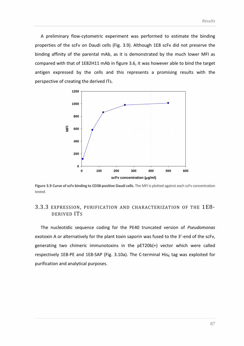
Results
87
A preliminary flow-cytometric experiment was performed to estimate the binding
properties of the scFv on Daudi cells (Fig. 3.9). Although 1E8 scFv did not preserve the
binding affinity of the parental mAb, as it is demonstrated by the much lower MFI as
compared with that of 1E82H11 mAb in figure 3.6, it was however able to bind the target
antigen expressed by the cells and this represents a promising results with the
perspective of creating the derived ITs. 2D Graph 1
scFv concentration (g/ml)
0 100 200 300 400 500 600
MF
I
0
200
400
600
800
1000
1200
Figure 3.9 Curve of scFv binding to CD38-positive Daudi cells. The MFI is plotted against each scFv concentration
tested.
3.3.3 EXPRESSION , PURIFICATION AND CHARACTERIZATION OF THE 1E8-DERIVED ITS
The nucleotidic sequence coding for the PE40 truncated version of Pseudomonas
exotoxin A or alternatively for the plant toxin saporin was fused to the 3’-end of the scFv,
generating two chimeric immunotoxins in the pET20b(+) vector which were called
respectively 1E8-PE and 1E8-SAP (Fig. 3.10a). The C-terminal His6 tag was exploited for
purification and analytical purposes.
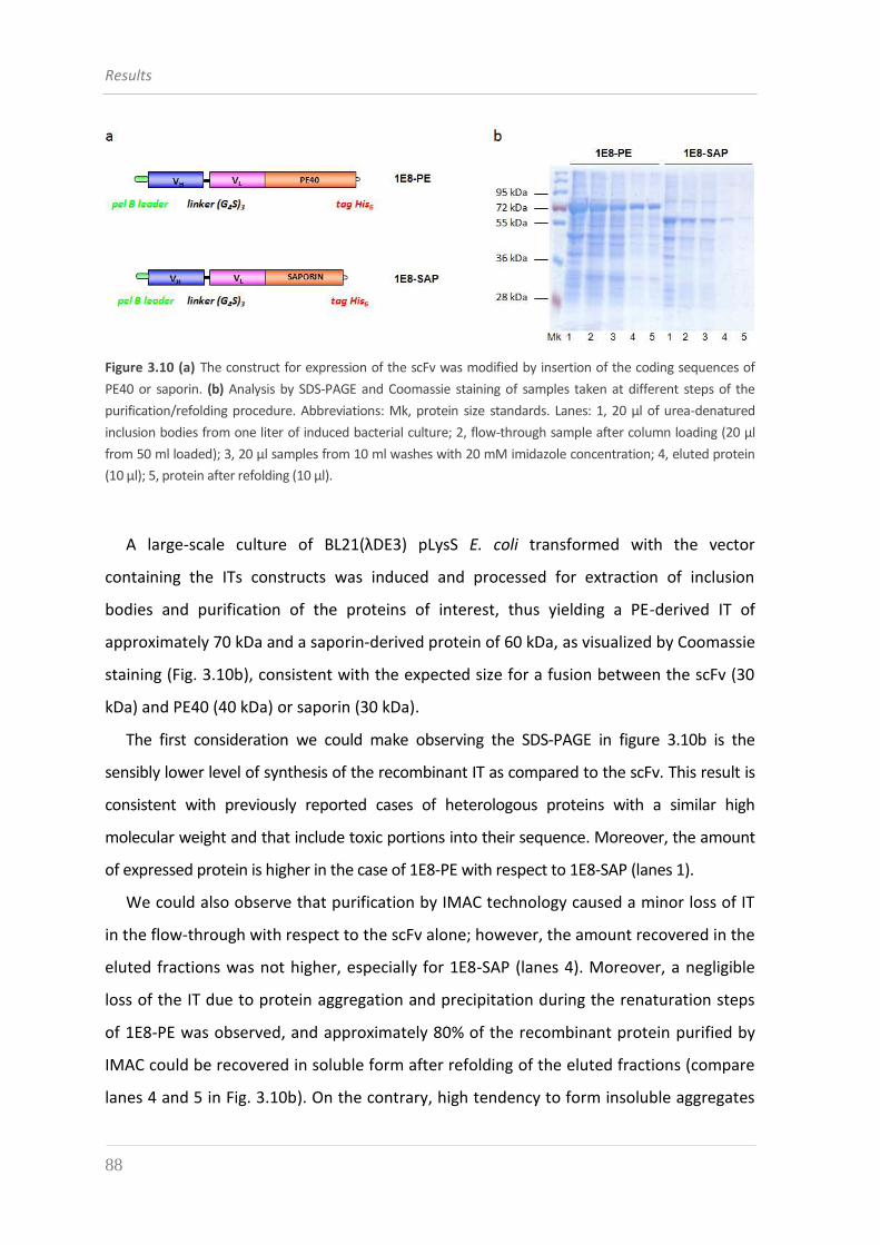
Results
88
Figure 3.10 (a) The construct for expression of the scFv was modified by insertion of the coding sequences of
PE40 or saporin. (b) Analysis by SDS-PAGE and Coomassie staining of samples taken at different steps of the
purification/refolding procedure. Abbreviations: Mk, protein size standards. Lanes: 1, 20 μl of urea-denatured
inclusion bodies from one liter of induced bacterial culture; 2, flow-through sample after column loading (20 μl
from 50 ml loaded); 3, 20 μl samples from 10 ml washes with 20 mM imidazole concentration; 4, eluted protein
(10 μl); 5, protein after refolding (10 μl).
A large-scale culture of BL21(λDE3) pLysS E. coli transformed with the vector
containing the ITs constructs was induced and processed for extraction of inclusion
bodies and purification of the proteins of interest, thus yielding a PE-derived IT of
approximately 70 kDa and a saporin-derived protein of 60 kDa, as visualized by Coomassie
staining (Fig. 3.10b), consistent with the expected size for a fusion between the scFv (30
kDa) and PE40 (40 kDa) or saporin (30 kDa).
The first consideration we could make observing the SDS-PAGE in figure 3.10b is the
sensibly lower level of synthesis of the recombinant IT as compared to the scFv. This result is
consistent with previously reported cases of heterologous proteins with a similar high
molecular weight and that include toxic portions into their sequence. Moreover, the amount
of expressed protein is higher in the case of 1E8-PE with respect to 1E8-SAP (lanes 1).
We could also observe that purification by IMAC technology caused a minor loss of IT
in the flow-through with respect to the scFv alone; however, the amount recovered in the
eluted fractions was not higher, especially for 1E8-SAP (lanes 4). Moreover, a negligible
loss of the IT due to protein aggregation and precipitation during the renaturation steps
of 1E8-PE was observed, and approximately 80% of the recombinant protein purified by
IMAC could be recovered in soluble form after refolding of the eluted fractions (compare
lanes 4 and 5 in Fig. 3.10b). On the contrary, high tendency to form insoluble aggregates
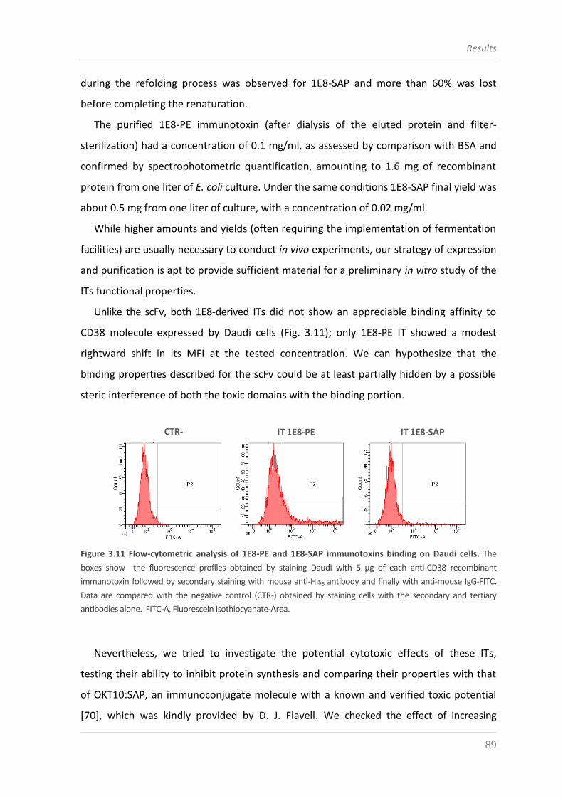
Results
89
during the refolding process was observed for 1E8-SAP and more than 60% was lost
before completing the renaturation.
The purified 1E8-PE immunotoxin (after dialysis of the eluted protein and filter-
sterilization) had a concentration of 0.1 mg/ml, as assessed by comparison with BSA and
confirmed by spectrophotometric quantification, amounting to 1.6 mg of recombinant
protein from one liter of E. coli culture. Under the same conditions 1E8-SAP final yield was
about 0.5 mg from one liter of culture, with a concentration of 0.02 mg/ml.
While higher amounts and yields (often requiring the implementation of fermentation
facilities) are usually necessary to conduct in vivo experiments, our strategy of expression
and purification is apt to provide sufficient material for a preliminary in vitro study of the
ITs functional properties.
Unlike the scFv, both 1E8-derived ITs did not show an appreciable binding affinity to
CD38 molecule expressed by Daudi cells (Fig. 3.11); only 1E8-PE IT showed a modest
rightward shift in its MFI at the tested concentration. We can hypothesize that the
binding properties described for the scFv could be at least partially hidden by a possible
steric interference of both the toxic domains with the binding portion.
CTR- IT 1E8-PE IT 1E8-SAP
Figure 3.11 Flow-cytometric analysis of 1E8-PE and 1E8-SAP immunotoxins binding on Daudi cells. The
boxes show the fluorescence profiles obtained by staining Daudi with 5 μg of each anti-CD38 recombinant
immunotoxin followed by secondary staining with mouse anti-His6 antibody and finally with anti-mouse IgG-FITC.
Data are compared with the negative control (CTR-) obtained by staining cells with the secondary and tertiary
antibodies alone. FITC-A, Fluorescein Isothiocyanate-Area.
Nevertheless, we tried to investigate the potential cytotoxic effects of these ITs,
testing their ability to inhibit protein synthesis and comparing their properties with that
of OKT10:SAP, an immunoconjugate molecule with a known and verified toxic potential
[70], which was kindly provided by D. J. Flavell. We checked the effect of increasing
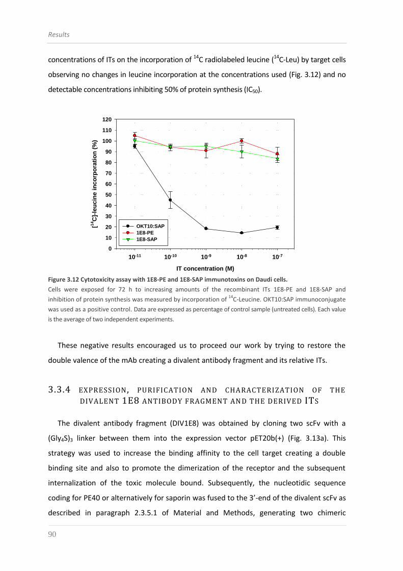
Results
90
concentrations of ITs on the incorporation of 14C radiolabeled leucine (14C-Leu) by target cells
observing no changes in leucine incorporation at the concentrations used (Fig. 3.12) and no
detectable concentrations inhibiting 50% of protein synthesis (IC50).
2D Graph 2
IT concentration (M)
10-11 10-10 10-9 10-8 10-7
[14C
]-le
ucin
e i
nco
rpo
rati
on
(%
)
10
30
50
70
90
110
0
20
40
60
80
100
120
OKT10:SAP
1E8-PE
1E8-SAP
Figure 3.12 Cytotoxicity assay with 1E8-PE and 1E8-SAP immunotoxins on Daudi cells.
Cells were exposed for 72 h to increasing amounts of the recombinant ITs 1E8-PE and 1E8-SAP and
inhibition of protein synthesis was measured by incorporation of 14
C-Leucine. OKT10:SAP immunoconjugate
was used as a positive control. Data are expressed as percentage of control sample (untreated cells). Each value
is the average of two independent experiments.
These negative results encouraged us to proceed our work by trying to restore the
double valence of the mAb creating a divalent antibody fragment and its relative ITs.
3.3.4 EXPRESSION , PURIFICATION AND CHARACTERIZATION OF THE
DIVALENT 1E8 ANTIBODY FRAGMENT AN D THE DERIVED ITS
The divalent antibody fragment (DIV1E8) was obtained by cloning two scFv with a
(Gly4S)3 linker between them into the expression vector pET20b(+) (Fig. 3.13a). This
strategy was used to increase the binding affinity to the cell target creating a double
binding site and also to promote the dimerization of the receptor and the subsequent
internalization of the toxic molecule bound. Subsequently, the nucleotidic sequence
coding for PE40 or alternatively for saporin was fused to the 3’-end of the divalent scFv as
described in paragraph 2.3.5.1 of Material and Methods, generating two chimeric
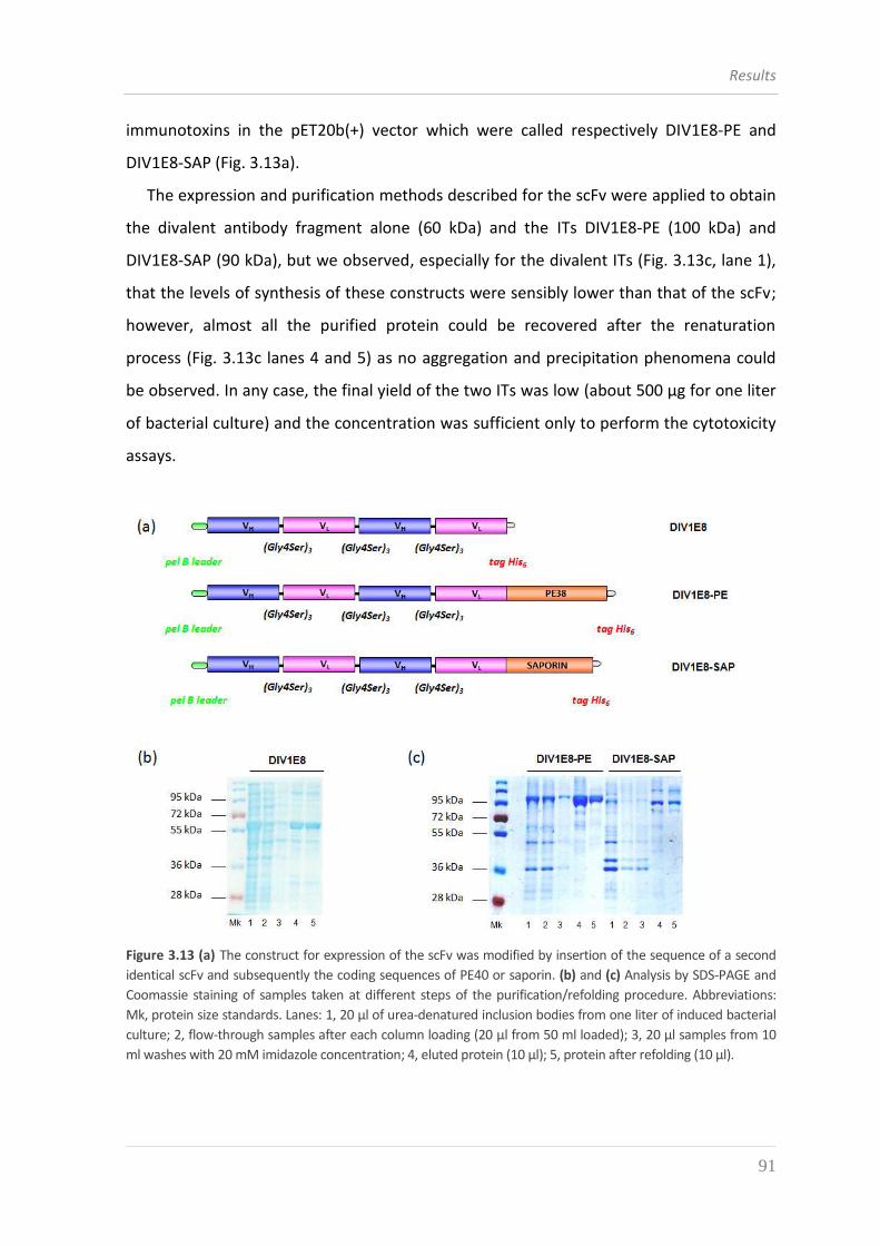
Results
91
immunotoxins in the pET20b(+) vector which were called respectively DIV1E8-PE and
DIV1E8-SAP (Fig. 3.13a).
The expression and purification methods described for the scFv were applied to obtain
the divalent antibody fragment alone (60 kDa) and the ITs DIV1E8-PE (100 kDa) and
DIV1E8-SAP (90 kDa), but we observed, especially for the divalent ITs (Fig. 3.13c, lane 1),
that the levels of synthesis of these constructs were sensibly lower than that of the scFv;
however, almost all the purified protein could be recovered after the renaturation
process (Fig. 3.13c lanes 4 and 5) as no aggregation and precipitation phenomena could
be observed. In any case, the final yield of the two ITs was low (about 500 μg for one liter
of bacterial culture) and the concentration was sufficient only to perform the cytotoxicity
assays.
Figure 3.13 (a) The construct for expression of the scFv was modified by insertion of the sequence of a second
identical scFv and subsequently the coding sequences of PE40 or saporin. (b) and (c) Analysis by SDS-PAGE and
Coomassie staining of samples taken at different steps of the purification/refolding procedure. Abbreviations:
Mk, protein size standards. Lanes: 1, 20 μl of urea-denatured inclusion bodies from one liter of induced bacterial
culture; 2, flow-through samples after each column loading (20 μl from 50 ml loaded); 3, 20 μl samples from 10
ml washes with 20 mM imidazole concentration; 4, eluted protein (10 μl); 5, protein after refolding (10 μl).
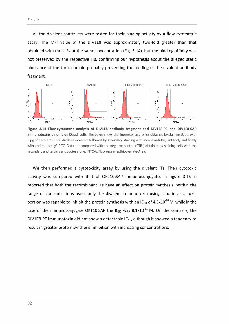
Results
92
All the divalent constructs were tested for their binding activity by a flow-cytometric
assay. The MFI value of the DIV1E8 was approximately two-fold greater than that
obtained with the scFv at the same concentration (Fig. 3.14), but the binding affinity was
not preserved by the respective ITs, confirming our hypothesis about the alleged steric
hindrance of the toxic domain probably preventing the binding of the divalent antibody
fragment.
CTR- IT DIV1E8-PE IT DIV1E8-SAPDIV1E8
Figure 3.14 Flow-cytometric analysis of DIV1E8 antibody fragment and DIV1E8-PE and DIV1E8-SAP
immunotoxins binding on Daudi cells. The boxes show the fluorescence profiles obtained by staining Daudi with
5 μg of each anti-CD38 divalent molecule followed by secondary staining with mouse anti-His6 antibody and finally
with anti-mouse IgG-FITC. Data are compared with the negative control (CTR-) obtained by staining cells with the
secondary and tertiary antibodies alone. FITC-A, Fluorescein Isothiocyanate-Area.
We then performed a cytotoxicity assay by using the divalent ITs. Their cytotoxic
activity was compared with that of OKT10:SAP immunoconjugate. In figure 3.15 is
reported that both the recombinant ITs have an effect on protein synthesis. Within the
range of concentrations used, only the divalent immunotoxin using saporin as a toxic
portion was capable to inhibit the protein synthesis with an IC50 of 4.5x10⁻¹⁰ M, while in the
case of the immunoconjugate OKT10:SAP the IC50 was 8.1x10⁻¹¹ M. On the contrary, the
DIV1E8-PE immunotoxin did not show a detectable IC50, although it showed a tendency to
result in greater protein synthesis inhibition with increasing concentrations.
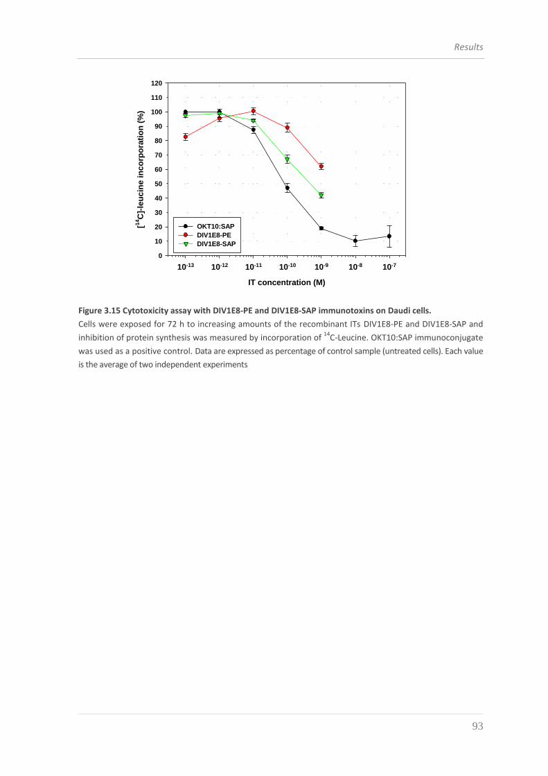
Results
93
2D Graph 1
IT concentration (M)
10-13 10-12 10-11 10-10 10-9 10-8 10-7
[14C
]-le
ucin
e i
nco
rpo
rati
on
(%
)
10
30
50
70
90
110
0
20
40
60
80
100
120
OKT10:SAP
DIV1E8-PE
DIV1E8-SAP
Figure 3.15 Cytotoxicity assay with DIV1E8-PE and DIV1E8-SAP immunotoxins on Daudi cells.
Cells were exposed for 72 h to increasing amounts of the recombinant ITs DIV1E8-PE and DIV1E8-SAP and
inhibition of protein synthesis was measured by incorporation of 14
C-Leucine. OKT10:SAP immunoconjugate
was used as a positive control. Data are expressed as percentage of control sample (untreated cells). Each value
is the average of two independent experiments
Results
94
3.4 AT13/5-DERIVED CONSTRUCTS
In our quest for functional recombinant ITs, and considering the deceiving results
obtained with 1E8 mAb, we resorted to investigating the properties of a different anti-
CD38 mAb and evaluated its suitability as the targeting element of new recombinant ITs.
The anti-CD38 hybridoma AT13/5 was produced by standard hybridoma technology by
Ellis J.H. [127], who further characterized the specificity of the AT13/5 mAb through a
series of flow-cytometric experiment. In addition to demonstrating the binding affinity of
AT13/5 for CD38-transfected CHO cells and human PBL, he also investigated the
relationship between the AT13/5 epitope and those of other anti-CD38 mAbs. He proved
that IB4, but not OKT10, could inhibit the binding of FITC-conjugated AT13/5 to CD38-
positive cells, thereby demonstrating that the new developed mAb, even if bound to the
same molecule as OKT10 mAb, probably recognized a different epitope. Accordingly, the
mAb 1E82H11 we have previously characterized (see paragraph 3.1 and 3.2) and AT13/5
would bind different epitopes on CD38 molecule.
3.4.1 EXPRESSION , PURIFICATION AND CHARACTERIZATION OF THE
AT13/5-DERIVED ITS
The sequence of AT13/5 scFv was kindly provided by Dr. W. Helfrich ((University of
Groningen, The Netherlands); it was subcloned into the vector pET20b(+) and the
sequence coding for PE40 or saporin was cloned at the 3’-end of AT13/5 scFv obtaining
the recombinant immunotoxins AT13/5-PE and AT13/5-SAP (Fig. 3.16).
The expression and purification methods were those previously described for the 1E8-
derived constructs. The AT13/5-PE purification steps are shown in figure 3.16b: we can
observe that AT13/5-PE is a protein of about 72 kDa expressed at high levels by the
bacterial host (lane 1) and it owns such chemical characteristics that allowed to recover
more than 90% of the purified protein after the renaturation process without a heavy loss
of precipitated material (compare lane 3 and 4). Despite the inefficiency of the protein
binding to the resin, which caused a strong loss of protein in the flow-through (lane 2),
the final yield of protein was approximately 10 mg from one liter of bacterial culture.
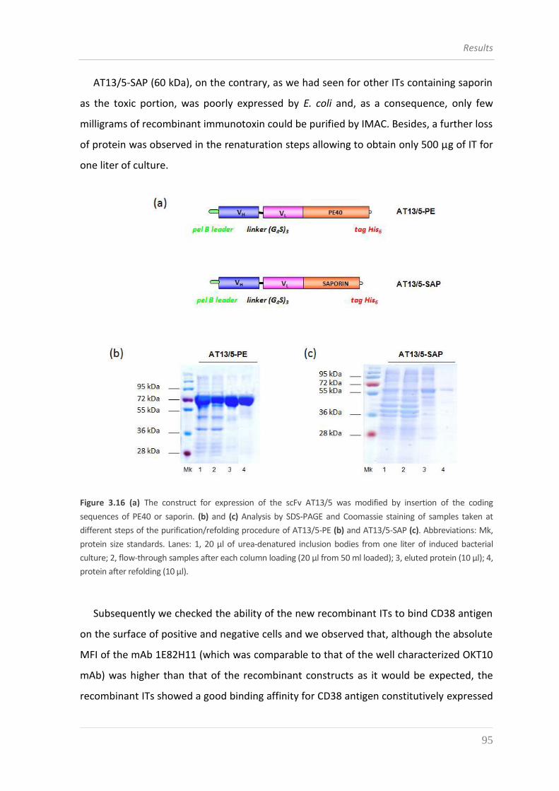
Results
95
AT13/5-SAP (60 kDa), on the contrary, as we had seen for other ITs containing saporin
as the toxic portion, was poorly expressed by E. coli and, as a consequence, only few
milligrams of recombinant immunotoxin could be purified by IMAC. Besides, a further loss
of protein was observed in the renaturation steps allowing to obtain only 500 μg of IT for
one liter of culture.
Figure 3.16 (a) The construct for expression of the scFv AT13/5 was modified by insertion of the coding
sequences of PE40 or saporin. (b) and (c) Analysis by SDS-PAGE and Coomassie staining of samples taken at
different steps of the purification/refolding procedure of AT13/5-PE (b) and AT13/5-SAP (c). Abbreviations: Mk,
protein size standards. Lanes: 1, 20 μl of urea-denatured inclusion bodies from one liter of induced bacterial
culture; 2, flow-through samples after each column loading (20 μl from 50 ml loaded); 3, eluted protein (10 μl); 4,
protein after refolding (10 μl).
Subsequently we checked the ability of the new recombinant ITs to bind CD38 antigen
on the surface of positive and negative cells and we observed that, although the absolute
MFI of the mAb 1E82H11 (which was comparable to that of the well characterized OKT10
mAb) was higher than that of the recombinant constructs as it would be expected, the
recombinant ITs showed a good binding affinity for CD38 antigen constitutively expressed
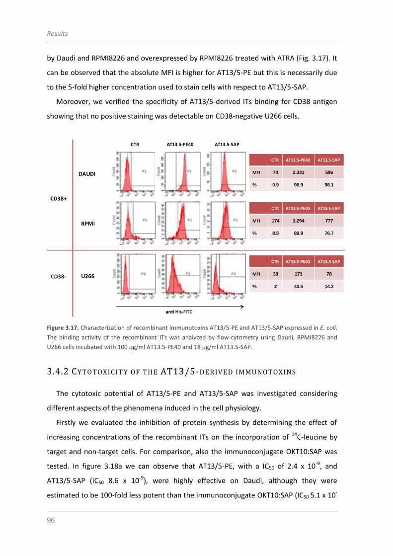
Results
96
by Daudi and RPMI8226 and overexpressed by RPMI8226 treated with ATRA (Fig. 3.17). It
can be observed that the absolute MFI is higher for AT13/5-PE but this is necessarily due
to the 5-fold higher concentration used to stain cells with respect to AT13/5-SAP.
Moreover, we verified the specificity of AT13/5-derived ITs binding for CD38 antigen
showing that no positive staining was detectable on CD38-negative U266 cells.
CTR AT13.5-PE40 AT13.5-SAP
MFI 74 2.331 596
% 0.9 98.9 98.1
CTR AT13.5-PE40 AT13.5-SAP
MFI 174 1.294 777
% 8.5 89.9 76.7
CTR AT13.5-PE40 AT13.5-SAP
MFI 39 171 78
% 2 43.5 14.2
CD38+
CD38-
DAUDI
RPMI
U266
anti His-FITC
AT13.5-SAPAT13.5-PE40CTR
Figure 3.17. Characterization of recombinant immunotoxins AT13/5-PE and AT13/5-SAP expressed in E. coli.
The binding activity of the recombinant ITs was analyzed by flow-cytometry using Daudi, RPMI8226 and
U266 cells incubated with 100 μg/ml AT13.5-PE40 and 18 μg/ml AT13.5-SAP.
3.4.2 CYTOTOXICITY OF THE AT13/5-DERIVED IMMUNOTOXINS
The cytotoxic potential of AT13/5-PE and AT13/5-SAP was investigated considering
different aspects of the phenomena induced in the cell physiology.
Firstly we evaluated the inhibition of protein synthesis by determining the effect of
increasing concentrations of the recombinant ITs on the incorporation of 14C-leucine by
target and non-target cells. For comparison, also the immunoconjugate OKT10:SAP was
tested. In figure 3.18a we can observe that AT13/5-PE, with a IC50 of 2.4 x 10⁻⁹, and
AT13/5-SAP (IC50 8.6 x 10⁻⁹), were highly effective on Daudi, although they were
estimated to be 100-fold less potent than the immunoconjugate OKT10:SAP (IC50 5.1 x 10-
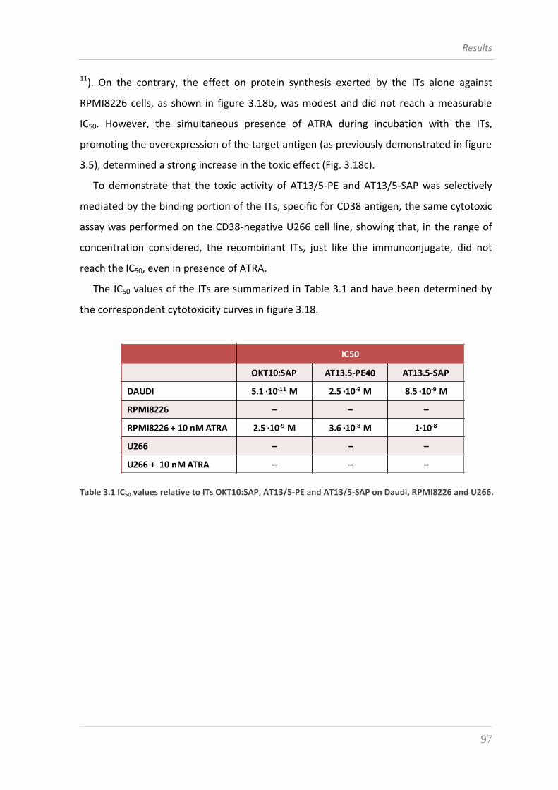
Results
97
11). On the contrary, the effect on protein synthesis exerted by the ITs alone against
RPMI8226 cells, as shown in figure 3.18b, was modest and did not reach a measurable
IC50. However, the simultaneous presence of ATRA during incubation with the ITs,
promoting the overexpression of the target antigen (as previously demonstrated in figure
3.5), determined a strong increase in the toxic effect (Fig. 3.18c).
To demonstrate that the toxic activity of AT13/5-PE and AT13/5-SAP was selectively
mediated by the binding portion of the ITs, specific for CD38 antigen, the same cytotoxic
assay was performed on the CD38-negative U266 cell line, showing that, in the range of
concentration considered, the recombinant ITs, just like the immunconjugate, did not
reach the IC50, even in presence of ATRA.
The IC50 values of the ITs are summarized in Table 3.1 and have been determined by
the correspondent cytotoxicity curves in figure 3.18.
IC50
OKT10:SAP AT13.5-PE40 AT13.5-SAP
DAUDI 5.1 ·10-11 M 2.5 ·10-9 M 8.5 ·10-9 M
RPMI8226 – – –
RPMI8226 + 10 nM ATRA 2.5 ·10-9 M 3.6 ·10-8 M 1∙10-8
U266 – – –
U266 + 10 nM ATRA – – –
Table 3.1 IC50 values relative to ITs OKT10:SAP, AT13/5-PE and AT13/5-SAP on Daudi, RPMI8226 and U266.
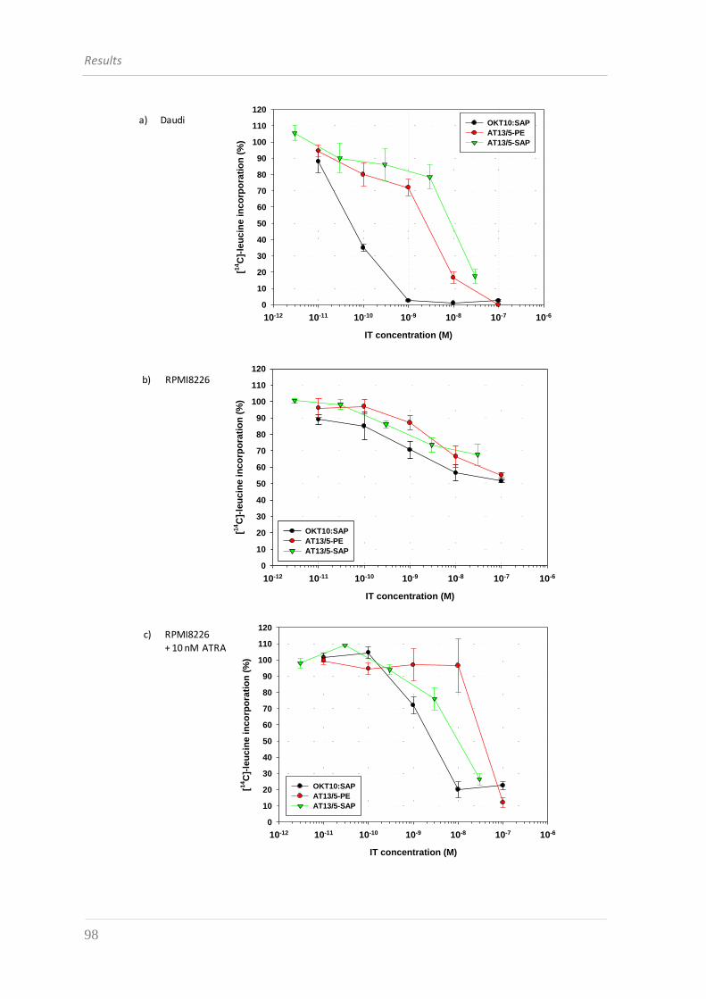
Results
98
DAUDI
IT concentration (M)
10-12 10-11 10-10 10-9 10-8 10-7 10-6
[14C
]-le
ucin
e i
nco
rpo
rati
on
(%
)
10
30
50
70
90
110
0
20
40
60
80
100
120
OKT10:SAP
AT13/5-PE
AT13/5-SAP
a) Daudi
RPMI no ATRA
IT concentration (M)
10-12 10-11 10-10 10-9 10-8 10-7 10-6
[14C
]-le
uc
ine
in
co
rpo
rati
on
(%
)
10
30
50
70
90
110
0
20
40
60
80
100
120
OKT10:SAP
AT13/5-PE
AT13/5-SAP
b) RPMI8226
RPMI + 10 nM ATRA
IT concentration (M)
10-12 10-11 10-10 10-9 10-8 10-7 10-6
[14C
]-le
uc
ine
in
co
rpo
rati
on
(%
)
10
30
50
70
90
110
0
20
40
60
80
100
120
OKT10:SAP
AT13/5-PE
AT13/5-SAP
c) RPMI8226
+ 10 nM ATRA
Results
99
U266 no ATRA
IT concentration (M)
10-12 10-11 10-10 10-9 10-8 10-7 10-6
[14C
]-le
uc
ine
in
co
rpo
rati
on
(%
)
10
30
50
70
90
110
130
0
20
40
60
80
100
120
140
OKT10:SAP
AT13/5-PE
AT13/5-SAP
d) U266
U266
IT concentration (M)
10-12 10-11 10-10 10-9 10-8 10-7 10-6
[14C
]-le
ucin
e i
nco
rpo
rati
on
(%
)
10
30
50
70
90
110
130
0
20
40
60
80
100
120
140
OKT10:SAP
AT13/5-PE
AT13/5-SAP
e) U266
+ 10 nM ATRA
Figure 3.18 Cytotoxicity assay with AT13/5-PE and AT13/5-SAP ITs on Daudi, RPMI8226 and U266 cells
treated or not with ATRA. Cells were exposed for 72 h to increasing amounts of the recombinant ITs
AT13/5-PE and AT13/5-SAP (in presence or absence of 10 nM ATRA) and inhibition of protein synthesis was
measured by incorporation of 14
C-Leucine. OKT10:SAP immunoconjugate was used as a positive control.
Data are expressed as percentage of control sample (untreated cells). Each value is the average of two
independent experiments.
As the final result of protein synthesis arrest is the inhibition of cell growth and finally
death, we proceeded our analysis by evaluating the toxic effect of the recombinant ITs on
cell proliferation. The XTT assay we used measures cell viability based on the activity of
mitochondria enzymes in live cells that reduce XTT reagent (a tetrazolium derivative) to a
highly water-soluble orange colored product. As the mitochondria enzymes are
Results
100
inactivated shortly after cell death, the amount of water-soluble compound generated
from XTT is proportional to the number of living cells in the sample and can be quantified
by measuring absorbance at wavelength of 475 nm.
The curves in figure 3.19 clearly show that the ITs can inhibit the proliferation of cells
expressing the CD38 antigen, while they have no detectable toxic activity on CD38-
negative U266 cells within the range of concentrations used.
Although the immunoconjugate OKT10:SAP had a more potent action as compared to
the recombinant ITs, we could observe that AT13/5-PE IC50 was lower than 10 nM for
both Daudi and ATRA-treated RPMI8226, while AT13/5-SAP was capable to reduce by half
the proliferation of CD38-expressing cell lines only at the higher concentration we could
obtain by the purification process (3 x 10⁻⁸ M).
Results
101
DAUDI
IT concentration (M)
10-12 10-11 10-10 10-9 10-8 10-7 10-6
via
bil
ity (
%)
10
30
50
70
90
110
130
0
20
40
60
80
100
120
140
OKT10:SAP
AT13/5-PE
AT13/5-SAP
RPMI + 10 nM ATRA
IT concentration (M)
10-12 10-11 10-10 10-9 10-8 10-7 10-6
via
bil
ity (
%)
10
30
50
70
90
110
0
20
40
60
80
100 OKT10:SAP
AT13/5-PE
AT13.5-SAP
U266
IT concentration (M)
10-12 10-11 10-10 10-9 10-8 10-7 10-6
via
bil
ity (
%)
10
30
50
70
90
110
130
0
20
40
60
80
100
120
140
OKT10:SAP
AT13/5-PE
AT13/5-SAP
a) Daudi
b) RPMI8226+ 10 nM ATRA
c) U266
Figure 3.19 Effect of treatment with ITs on proliferation of Daudi (A), ATRA-treated RPMI8226 (B), and U266 (C)
cells. Data are expressed as percentage of control sample (untreated cells). Each value is the average of two
independent experiments.
The final parameter we wanted to evaluate for the complete characterization of our
ITs was the ability to promote apoptosis of treated cells. This type of study was made by
the flow-cytometric analysis of cells after a double staining with Annexin-V-FITC and
Propidium iodide (PI). Annexin-V is a Ca2+-dependent phospholipid-binding protein with
Results
102
high affinity for phosphatidylserine which stains apoptotic as well as necrotic cells, while
PI stains DNA of leaky necrotic cells only.
Initially we had to set up the best parameters to perform the experiments and we
chose a time-point of 48 hours to better visualize the induction of apoptosis and a
standard concentration of 1 μg/ml of each IT to compare the effect of the different
molecules with each other and with the control non-treated cells. We also decided to pre-
treat RPMI8226 cells with ATRA with the purpose of maximizing CD38 expression and
obtaining the best results in terms of cell killing.
In figure 3.20a we can observe that a strong induction of apoptosis was determined by
all anti-CD38 ITs, which were able to promote the killing of about 40% to 70% of treated
Daudi cells. A similar, but stronger effect was observed in the case of RPMI8226 which
seemed to be more prone to undergo cell death: in fact, in this case the level of apoptosis
varied from 70% to 85% of treated cells. Moreover, the apoptosis induced by the
recombinant ITs was almost comparable to the effect promoted by OKT10:SAP.
Once again we demonstrated that U266 viability/apoptosis was not influenced by the
treatment with anti-CD38 ITs; in fact, as we can see in figure 3.20c, the percentage of
apoptosis of treated cells was comparable to that of the control and this was not due to a
form of apoptosis resistance because we can observe that ricin toxin at the concentration
of 1 μg/ml was able to induce apoptosis of more than 70% of U266 cells.
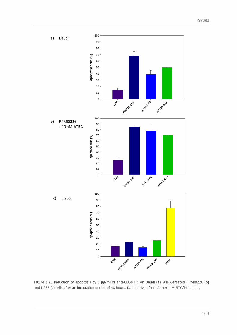
Results
103
2D Graph 2
CTR
OKT1
0:SAP
AT1
3/5-
PE
AT1
3/5-
SAP
ap
op
toti
c c
ell
s (
%)
10
30
50
70
90
0
20
40
60
80
100
2D Graph 2
CTR
OKT1
0:SAP
AT1
3/5-
PE
AT1
3/5-
SAP
Ric
in
ap
op
toti
c c
ell
s (
%)
10
30
50
70
90
0
20
40
60
80
100
2D Graph 2
CTR
OKT10
:SAP
AT13
/5-P
E
AT13
/5-S
AP
ap
op
toti
c c
ell
s (
%)
10
30
50
70
90
0
20
40
60
80
100
a) Daudi
b) RPMI8226
+ 10 nM ATRA
c) U266
Figure 3.20 Induction of apoptosis by 1 μg/ml of anti-CD38 ITs on Daudi (a), ATRA-treated RPMI8226 (b)
and U266 (c) cells after an incubation period of 48 hours. Data derived from Annexin-V-FITC/PI staining.
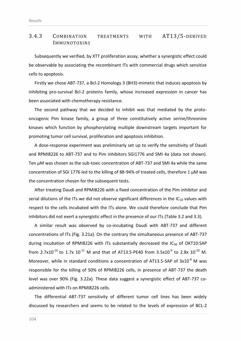
Results
104
3.4.3 COMBINATION TREATMENTS WITH AT13/5-DERIVED
IMMUNOTOXINS
Subsequently we verified, by XTT proliferation assay, whether a synergistic effect could
be observable by associating the recombinant ITs with commercial drugs which sensitize
cells to apoptosis.
Firstly we chose ABT-737, a Bcl-2 Homology 3 (BH3)-mimetic that induces apoptosis by
inhibiting pro-survival Bcl-2 proteins family, whose increased expression in cancer has
been associated with chemotherapy resistance.
The second pathway that we decided to inhibit was that mediated by the proto-
oncogenic Pim kinase family, a group of three constitutively active serine/threonine
kinases which function by phosphorylating multiple downstream targets important for
promoting tumor cell survival, proliferation and apoptosis inhibition.
A dose-response experiment was preliminarly set up to verify the sensitivity of Daudi
and RPMI8226 to ABT-737 and to Pim inhibitors SGI1776 and SMI 4a (data not shown).
Ten µM was chosen as the sub-toxic concentration of ABT-737 and SMI 4a while the same
concentration of SGI 1776 led to the killing of 88-94% of treated cells, therefore 1 μM was
the concentration chosen for the subsequent tests.
After treating Daudi and RPMI8226 with a fixed concentration of the Pim inhibitor and
serial dilutions of the ITs we did not observe significant differences in the IC50 values with
respect to the cells incubated with the ITs alone. We could therefore conclude that Pim
inhibitors did not exert a synergistic effect in the presence of our ITs (Table 3.2 and 3.3).
A similar result was observed by co-incubating Daudi with ABT-737 and different
concentrations of ITs (Fig. 3.21a). On the contrary the simultaneous presence of ABT-737
during incubation of RPMI8226 with ITs substantially decreased the IC50 of OKT10:SAP
from 2.7x10⁻¹⁰ to 1.7x 10⁻¹¹ M and that of AT13.5-PE40 from 3.5x10⁻⁹ to 2.8x 10⁻¹⁰ M.
Moreover, while in standard conditions a concentration of AT13.5-SAP of 3x10⁻⁸ M was
responsible for the killing of 50% of RPMI8226 cells, in presence of ABT-737 the death
level was over 90% (Fig. 3.22a). These data suggest a synergistic effect of ABT-737 co-
administered with ITs on RPMI8226 cells.
The differential ABT-737 sensitivity of different tumor cell lines has been widely
discussed by researchers and seems to be related to the levels of expression of BCL-2
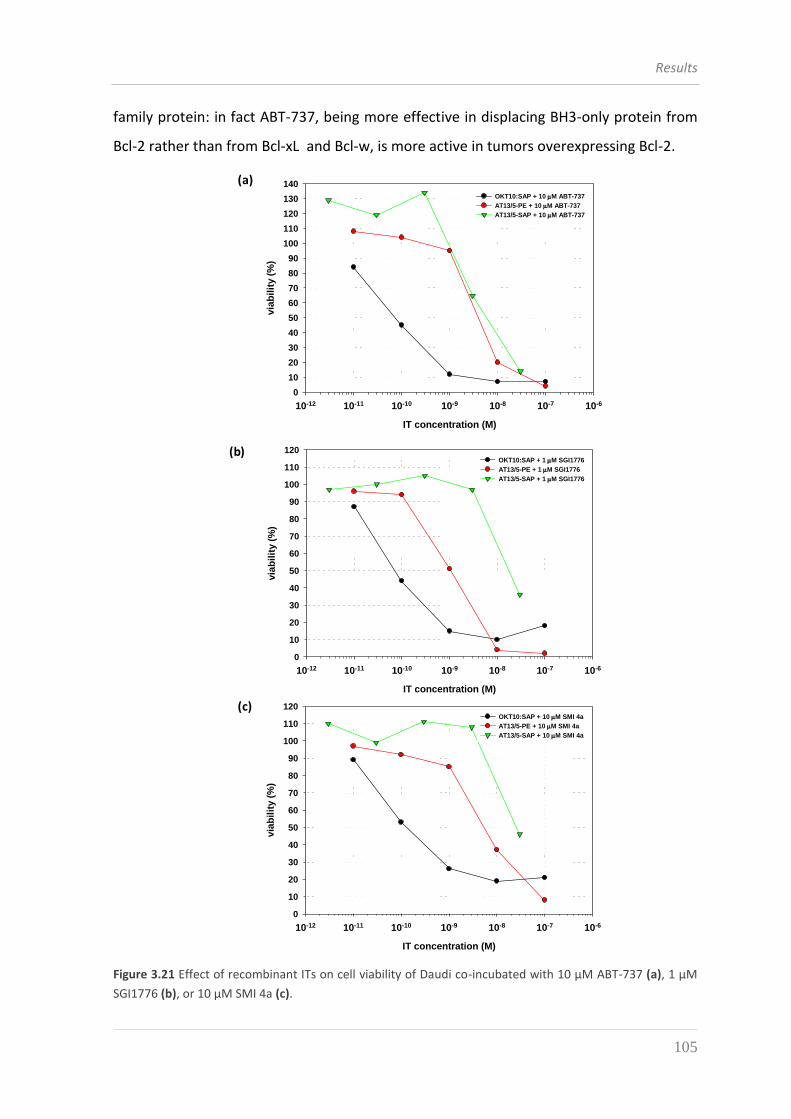
Results
105
family protein: in fact ABT-737, being more effective in displacing BH3-only protein from
Bcl-2 rather than from Bcl-xL and Bcl-w, is more active in tumors overexpressing Bcl-2. DAUDI + 10 M ABT-737
IT concentration (M)
10-12 10-11 10-10 10-9 10-8 10-7 10-6
via
bil
ity (
%)
10
30
50
70
90
110
130
0
20
40
60
80
100
120
140
OKT10:SAP + 10 M ABT-737
AT13/5-PE + 10 M ABT-737
AT13/5-SAP + 10 M ABT-737
DAUDI + 10 M ABT-737
IT concentration (M)
10-12 10-11 10-10 10-9 10-8 10-7 10-6
via
bil
ity (
%)
10
30
50
70
90
110
130
0
20
40
60
80
100
120
140
OKT10:SAP + 10 M ABT-737
AT13/5-PE + 10 M ABT-737
AT13/5-SAP + 10 M ABT-737
DAUDI + 1 M SGI
IT concentration (M)
10-12 10-11 10-10 10-9 10-8 10-7 10-6
via
bil
ity (
%)
10
30
50
70
90
110
0
20
40
60
80
100
120
OKT10:SAP + 1 M SGI1776
AT13/5-PE + 1 M SGI1776
AT13/5-SAP + 1 M SGI1776
DAUDI + 1 M SGI
IT concentration (M)
10-12 10-11 10-10 10-9 10-8 10-7 10-6
via
bil
ity (
%)
10
30
50
70
90
110
0
20
40
60
80
100
120
OKT10:SAP + 1 M SGI1776
AT13/5-PE + 1 M SGI1776
AT13/5-SAP + 1 M SGI1776
DAUDI + 10 M SMI 4a
IT concentration (M)
10-12 10-11 10-10 10-9 10-8 10-7 10-6
via
bil
ity (
%)
10
30
50
70
90
110
0
20
40
60
80
100
120
OKT10:SAP + 10 M SMI 4a
AT13/5-PE + 10 M SMI 4a
AT13/5-SAP + 10 M SMI 4a
DAUDI + 10 M SMI 4a
IT concentration (M)
10-12 10-11 10-10 10-9 10-8 10-7 10-6
via
bil
ity (
%)
10
30
50
70
90
110
0
20
40
60
80
100
120
OKT10:SAP + 10 M SMI 4a
AT13/5-PE + 10 M SMI 4a
AT13/5-SAP + 10 M SMI 4a
(a)
(b)
(c)
Figure 3.21 Effect of recombinant ITs on cell viability of Daudi co-incubated with 10 µM ABT-737 (a), 1 µM
SGI1776 (b), or 10 µM SMI 4a (c).
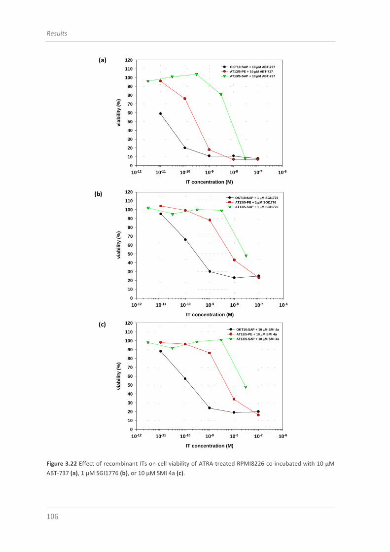
Results
106
RPMI + 10 nM ATRA
+ 10 M SMI 4a
IT concentration (M)
10-12 10-11 10-10 10-9 10-8 10-7 10-6
via
bil
ity (
%)
10
30
50
70
90
110
0
20
40
60
80
100
120
OKT10:SAP
AT13.5-PE40
AT13.5-SAP
RPMI + 10nM ATRA
+ 1 M SGI1776
IT concentration (M)
10-12 10-11 10-10 10-9 10-8 10-7 10-6
via
bil
ity (
%)
10
30
50
70
90
110
0
20
40
60
80
100
120
OKT10:SAP
AT13.5-PE40
AT13.5-SAP
RPMI + 10 nM ATRA + 10 M ABT-737
IT concentration (M)
10-12 10-11 10-10 10-9 10-8 10-7 10-6
via
bil
ity (
%)
10
30
50
70
90
110
0
20
40
60
80
100
120
OKT10:SAP
AT13/5-PE
AT13/5-SAP
DAUDI + 10 M ABT-737
IT concentration (M)
10-12 10-11 10-10 10-9 10-8 10-7 10-6
via
bil
ity (
%)
10
30
50
70
90
110
130
0
20
40
60
80
100
120
140
OKT10:SAP + 10 M ABT-737
AT13/5-PE + 10 M ABT-737
AT13/5-SAP + 10 M ABT-737
DAUDI + 1 M SGI
IT concentration (M)
10-12 10-11 10-10 10-9 10-8 10-7 10-6
via
bil
ity (
%)
10
30
50
70
90
110
0
20
40
60
80
100
120
OKT10:SAP + 1 M SGI1776
AT13/5-PE + 1 M SGI1776
AT13/5-SAP + 1 M SGI1776
DAUDI + 10 M SMI 4a
IT concentration (M)
10-12 10-11 10-10 10-9 10-8 10-7 10-6
via
bil
ity (
%)
10
30
50
70
90
110
0
20
40
60
80
100
120
OKT10:SAP + 10 M SMI 4a
AT13/5-PE + 10 M SMI 4a
AT13/5-SAP + 10 M SMI 4a
(a)
(b)
(c)
Figure 3.22 Effect of recombinant ITs on cell viability of ATRA-treated RPMI8226 co-incubated with 10 µM
ABT-737 (a), 1 µM SGI1776 (b), or 10 µM SMI 4a (c).
Results
107
DAUDI IC50
OKT10:SAP AT13.5-PE40 AT13.5-SAP
untreated 6.1 ∙ 10-11 M 6 ∙ 10-9 M 2.6 ∙ 10-8 M
10 µM ABT-737 7.6 · 10-11 M 3.9 · 10-9 M 5.9 · 10-9 M
1 µM SGI1776 7.3 · 10-11 M 1 · 10-9 M 1.7 ·10-8 M
10 µM SMI 4a 1.2 · 10-10 M 5.2 · 10-9 M 2.5∙10-8 M
Table 3.2 IC50 values relative to ITs OKT10:SAP, AT13/5-PE and AT13/5-SAP on Daudi treated with BH3-
mimetics or Pim inhibitors.
RPMI8226 IC50
OKT10:SAP AT13.5-PE40 AT13.5-SAP
untreated 2.7 ∙ 10-10 M 3.5 ∙ 10-9 M 3 ∙ 10-8 M
10 µM ABT-737 1.7 · 10-11 M 2.8 · 10-10 M 7.9 · 10-9 M
1 µM SGI1776 2.7 · 10-10 M 6.8 · 10-9 M 2.7 ·10-8 M
10 µM SMI 4a 1.6 · 10-10 M 4.8 · 10-9 M 2.7∙10-8 M
Table 3.3 IC50 values relative to ITs OKT10:SAP, AT13/5-PE and AT13/5-SAP on ATRA-treated RPMI8226
treated with BH3-mimetics or Pim inhibitors.
3.4.4 EFFECT OF THE AT13/5-DERIVED IMMUNOTOXINS ON B-CLL
We investigated the potential clinical application of our ITs by evaluating their specific
cytotoxic activity on B-lymphocytes derived from B-CLL patients. This part of the study is
now at an early stage of development for two main reasons: the first is the difficulty to
find appropriate quantity of CD38-positive samples available for research purposes and
suitable to perform reproducible experiments on samples from the same patient; the
second is the need to optimize the culture conditions of these type of cells which are not
immortalized and therefore show high level of apoptosis after 24 hours of incubation
without any stimuli.
We firstly selected a PBMCs (Peripheral Blood Mononuclear Cell) sample showing high
levels of CD38 expression and we purified B-lymphocytes by negative selection. The
Results
108
subsequent flow-cytometric analysis of the purified cells demonstrated that the sample
contained over 90% of CD38-positive cells (Fig. 3.23).
CD38CD19
Figure 3.23 Flow-cytometric analysis of B-cell purity (CD19+ cells) and CD38 expression by B-lymphocytes
after purification by negative selection from a PBMC sample from a CLL patient.
The culture conditions were optimized by the progressive addition of cytokines (IL-4,
IL-2 and CD40-ligand) and the change of the culture medium with an enriched in nutrients
one (IMDM), as shown in figure 3.24. Despite these solutions, as we can observe in Figure
3.25, untreated cells spontaneously underwent apoptosis at a level of 30% within 24
hours. However this time was sufficient to highlight the induction of apoptosis on B-cells
of the first patient we treated with recombinant ITs.
This first apoptosis experiment, although yielding a moderately success, represents
only a starting point for the future clinical development of anti-CD38 recombinant ITs.
Results
109
9.2% 23.7%
14.3%7.1%
IL4 +untreated
48 H
24 H
87.7%
69%
IMDM medium
10% FCS
10 nM IL-4
20 U/ml IL-2
50 ng/ml CD40L
43%
DAY 0
Figure 3.24 Stepwise optimization of culture condition of B-lymphocytes to diminish the level of
spontaneous apoptosis. Annexin-V-FITC/PI staining was performed after 24 and 48 hours of incubation of
cells with different cytokines
2D Graph 3
CTR
OKT10
:SAP
AT13
/5-P
E
AT13
/5-S
AP
ap
op
toti
c c
ell
s (
%)
10
30
50
70
90
0
20
40
60
80
100
Figure 3.25 Induction of apoptosis by 1 μg/ml of anti-CD38 ITs on B-lymphocytes derived from a CLL patient
after an incubation period of 24 hours. Data derived from Annexin-V-FITC/PI staining.

Results
110

4. DISCUSSION

Discussion
112
Discussion
113
Rapid progress in understanding molecular mechanisms of cancer development and
increased insights into the nature of tumor antigens made a large impact on the design
and evaluation of novel therapeutic strategies focused on the specific targeting of tumor
cells, thus obtaining an increase in efficacy with a concomitant reduction of side effects.
ITs represent a very efficacious targeted immunotherapy approach for treating cancer
patients, particularly when used in association with other therapeutic modalities. The
best success of ITs has been observed in the field of hematological malignancies; in fact,
cell from hematological tumors, being located intravascularly or perivascularly in well-
perfused lymph nodes, are more exposed to permeation by ITs and therefore more
accessible than cells of solid tumor masses.
The present thesis describes the construction and development of different
recombinant ITs targeting the CD38 antigen, whose expression is mainly linked to the
lymphoid lineage and whose overexpression has been demonstrated in hematological
malignancies such as B-CLL and Multiple Myeloma [33]. CD38 is also known to undergo
internalization upon binding of ligands to its extracellular portion [22], which is
fundamental for the uptake of an antibody drug. We also explored an important concept
about the effect on efficacy and selectivity of the association of our ITs with drugs
involved in survival and apoptosis pathways of the cell.
Despite the interest in developing immunotherapeutic drugs targeting CD38 antigen,
which led to the production of both anti-CD38 chimeric monoclonal antibodies and ITs,
none of such compounds has been yet approved for clinical therapy.
In this work we pursued the construction of a panel of recombinant ITs which can be
divided into different sub-groups depending on the nature of the binding domain as well
as the toxic domain.
Considering the binding domain, we created ITs composed of single chain antibody
fragments (scFv) directed against CD38 antigen and derived from two murine hybridomas
secreting monoclonal antibodies with different epitope specificity. Hybridoma 1E82H11
was produced and characterized in our laboratory and the purified mAb from this clone
showed good reactivity towards the native antigen expressed by lymphoma and myeloma
cell lines. Moreover, 1E82H11 mAb binding efficiency was almost comparable to that of
OKT10 mAb, a known mAb that we used as a reference standard. On the contrary AT13/5
Discussion
114
hybridoma was developed by J. H. Ellis [127] and the sequence of the derived scFv was
supplied to us by Dr. W. Helfrich (University of Groningen, The Netherlands). The
construction of the scFvs by the traditional strategy of genetic fusion of the sequences
coding for the variable domains implies that a future therapeutic utilization of such
antibody fragments will likely require a “humanization” step by further genetic
manipulation through a variety of approaches, in order to reduce their immunogenic
potential and avoid the occurrence of HAMA [128]. With this perspective, CDR-grafting
has already been employed by J.H. Ellis for the humanization of the entire AT13/5 IgG1.
The first choice for the toxic portion to be fused to our scFvs was a derivative of
Pseudomonas aeruginosa exotoxin A (PE), which is a preferred molecule for the
construction of ITs because its high toxic potential is well documented and its cytotoxic
pathways are well understood [129]. Moreover, PE40, a mutant form of PE in which the
largest portion of the cell-binding domain has been deleted and therefore, showing
diminished nonspecific toxicity when administered to animals, has been widely used in
the development of ITs and is now part of many recombinant molecules tested in clinical
trials. Nevertheless, PE is known to be immunogenic, leading to the formation of
neutralizing antibodies; it is therefore likely that a de-immunization strategy will be
needed before exploring its in vivo efficacy. This can be accomplished, as shown by M.
Onda and coworkers [101], through the replacement of the amino acids within the
epitopes determining the reactivity of the immune system.
The first approach used to obtain a potent cytotoxic effect and possibly avoid a future
immune response, was the employment of saporin as the toxic portion; indeed, saporin is
reported to be a potent toxin and one of the less immunogenic among the toxins of plant
origin.
Particular attention has been paid to setting up a suitable strategy for the expression
and purification of the antibody fragments and the derived ITs. The main concern was
manageability and final yield. For these reasons we opted for a prokaryotic host like E.
coli, which is easily grown in shake flasks and allows the rapid induction and high-level
accumulation of heterologous proteins. In order to maximize the amount of protein we
decided to select the BL21(DE3)pLysS strain, which, being deficient in the expression of
Discussion
115
several endogenous proteases, represents one of the most widely used prokaryotic systems
for heterologous production of proteins.
Furthermore, all the recombinant constructs which have been inserted into the
plasmid vector for the expression on the bacterial host present a N-terminal signal peptide
for the sorting to the periplasm; this was done to ease the recovery of the heterologous
proteins in soluble form. In spite of the presence of this signal sequence, the induced proteins
were mainly accumulated as inclusion bodies which required the dissolution of the insoluble
aggregates using a denaturing agent, such as urea, and a further renaturation process which
is generally reported to be the most critical step affecting the yield of biologically active
antibody fragments and ITs, as loss due to aggregation and precipitation can be substantial
[130]. In fact, in spite of the application of an appropriate refolding procedure (i.e. the
gradual removal of the denaturing agent and the use of reduced and oxidized glutathione and
arginine that are known to limit the occurrence of aberrant protein folding and precipitation
[131]), a sizeable loss of protein (especially for the scFv) due to formation of insoluble
aggregates was observed. Fusions with PE40 proved less prone to aggregation during the
renaturation procedure, while saporin-based ITs showed high levels of aggregation and
precipitation. These different behaviour was probably conferred by the chemical and
structural features of the toxin molecules, by their amino acid composition and their
hydrophilic/hydrophobic profile. To avoid most of the disadvantages linked to the use of E.
coli, especially the problem of protein aggregation following refolding, the use of eukaryotic
systems and particularly of the yeast Pichia pastoris, has been proposed as the best-suited
expression platform of saporin-based therapeutic molecules, allowing the recovery of
proteins in soluble form from the culture medium [132].
Finally, our scFv and IT constructs contain a C-terminal hexahistidine tag to allow the
purification by affinity chromatography. It should be noted, however, that an epitope tag
represents an artificial element, unrelated to the rest of the polypeptide and with no
pharmacological activity: a protein to be used as a therapeutic agent in humans should be
ideally free from any such non-essential portion. Moreover, as it was described by J. L.
Hessler et al., ITs with PE should have a free terminus because removal of the lysine
residue of the C-terminal REDLK sequence is essential for the binding of PE to the KDEL
receptor and therefore for the cellular intoxication of PE [133]. Thus, the presence of a
Discussion
116
peptide tag could decrease PE activity. We have however maintained it in our PE-based
ITs because in the present work we were not aiming at the best optimization obtainable
but rather at proving the suitability of our ITs in the context studied. Optimization and
further refinements will be addressed when the best candidate for use in vivo will be
selected.
We began the analysis on the binding affinity of our recombinant constructs by a first
characterization of the parental mAb. Flow-cytometric assays showed that the binding of
our 1E82H11 mAb is restricted to CD38-positive cells (i.e. Daudi and RPMI8226) and that
it targets the same epitope recognized by OKT10 mAb, by which it is displaced in a
competitive staining.
The conversion of the mAb into the scFv format did not allow to preserve the binding
efficiency of the parental monoclonal and not even that of the scFv derived from OKT10
mAb (data not shown). When generating a scFv from the immunoglobulin variable region
genes isolated from a hybridoma cell line, a decrease in the apparent affinity of the
resulting scFv as compared to the parental mAb has often been observed [134]. With
many scFv molecules, the lower affinity results from the decrease in valence (number of
binding sites) that occurs when the format is switched from the larger bivalent mAb to
the smaller monovalent scFv. This is particularly true when multiple copies of the target
epitope are present on a single antigen molecule as was the case with the CC49 MAb
[135]. Decrease in affinity can also result from structural alterations between the IgG and
scFv formats. In particular, the peptide spacer that joins the VH and VL chains can
potentially interfere with the normal alignment of the two chains. Finally, the loss of scFv
conformation due to the need of solubilizing proteins accumulated into inclusion bodies,
and the further refolding procedure can affect molecules stability as it can be
demonstrated by the high tendency to form aggregates showed by our 1E8 scFv.
We attempted to reconstitute the double valence of the parental IgG by genetically
fusing a second scFv at the C-terminal of the first one, obtaining a divalent molecule with
only a two-fold greater binding affinity with respect to the scFv and still far from the
affinity showed by the mAb. These results suggested that a different approach for the
design of multivalent antibody molecules could be considered: for example it could be
possible to obtain diabody and triabody by shortening the peptide linker connecting the
Discussion
117
VH and VL of a single scFv molecule from 15 amino acids to 5 amino acids or 0–3 amino
acids respectively [136].
Genetic fusion of monovalent or divalent scFvs to the toxic portions resulted in a
further loss of binding affinity probably ascribable to the steric hindrance of the toxic
domain preventing the binding of the antibody fragment or possibly by the interference
of the toxin sequence with the correct folding of the binding portion.
The substitution of the 1E8 scFv (derived from a mAb with the same specificity of
OKT10) with that derived from AT13/5 mAb (a simil-IB4 mAb) implies the changing of the
molecular epitope towards which the ITs were targeted. As reported by C. M. Ausiello et
al. [137], competition binding analysis identified two families of mAbs, namely IB4, IB6
and AT2 on one side and OKT10, SUN-4B7 and AT1 on the other. Each mAb family binds
epitopes that are completely or partially common. However, the functional activities of
the CD38 molecule cannot be simply attributed to the epitope engaged: for instance, IB4
and OKT10 mAbs, which bind different epitopes, perform as agonistic mAbs in inducing
PBMC proliferation and interferon (IFN)-γ secretion. On the contrary, IB4 is the only mAb
able to induce significant intracellular Ca2+ fluxes [137]. Despite the considerations on the
correlation between structural and functional properties of CD38 epitopes, we could
observe that AT13/5-derived ITs showed good affinity and specificity for CD38 antigen
expressed on the surface of Burkitt’s lymphoma cells and myeloma cells proving that the
steric organization of the scFv and the toxic domains in this case can favor the binding to
a different epitope on CD38 molecule.
The toxic molecules that we used for the construction of our ITs have been described
to induce inhibition of protein synthesis through the inactivation of the elongation factor
eEF-2 after the internalization and the intracellular routing. This is the reason that justifies
our investigation on the levels of protein synthesis inhibition (PSI) selectively induced by
our ITs on CD38-positive cells.
With the creation of the divalent 1E8-derived ITs we demonstrated that, despite the
discouraging binding properties of these constructs, they showed an acceptable
cytotoxicity against Daudi cells. This observation could be explained with the small
amount of toxic molecules needed to determine PSI. Moreover, the dimerization of the
CD38 receptor upon binding to the divalent ITs could account for a better rate of
Discussion
118
internalization of the divalent ITs with respect to the monovalent counterparts. However,
similar results in terms of PSI were obtained using AT13/5-derived ITs, showing that the
presence of a double binding site, as in the case of divalent ITs, is not essential for the
cytotoxic activity if the binding affinity of the monovalent scFv is high, as demonstrated
by AT13/5-derived ITs.
It is widely recognized that the arrest of protein synthesis is the main cause of cell
death induced by ITs [138] and the cell proliferation assays that we performed on target
cells with our anti-CD38 ITs confirmed the results obtained by the PSI assay. In fact, we
observed that ITs concentrations able to induce arrest of protein synthesis also showed a
high ability to inhibit cell proliferation. However, PE and PE-containing ITs, as well as
some plant toxins, have been proved to induce programmed cell death by the activation
of caspase-3-like protease [139]. Activated caspases can cleave structural proteins and
enzymes necessary for the survival of both proliferating and resting cells; in addition,
caspases activate the endonuclease responsible for the inter-nucleosomal cleavage of
genomic DNA and cleave so-called “death substrates”, such as poly(ADP)-ribose
polymerase (PARP), both hallmarks of apoptotic death [140]. Further molecular changes
induced during apoptosis include randomization of the distribution of phosphatidyl serine
between the inner and outer leaflets of the plasma membrane. The ability of our ITs to
induce apoptosis was investigated by detecting some of these changes, such as surface-
exposure of phosphatidyl serine with annexin V and detection of cells with subdiploid
DNA content by staining with DNA intercalating dyes. We observed high levels of
apoptosis induction both by PE- and saporin-based ITs. It should be noted that, although
Daudi cells are reported to express CD38 at a higher density on their surface, RPMI8226 cells
seem to be more prone to undergo apoptosis (Fig. 3.20). A possible explanation could be the
different sensitivity of the cell lines derived from different types of tumors. Moreover, some
cell lines, under certain culture conditions, could trigger mechanisms of resistance to
apoptosis by the over-expression of multiple survival signals.
All the cytotoxicity experiments showed that the IC50 of the immunoconjugate
OKT10:SAP was approximately 10- to 100-fold lower as compared to those of the
recombinant constructs. The higher toxicity showed by OKT10:SAP could be explained by
several factors: firstly by the double valence of the entire mAb compared to the scFvs,
Discussion
119
which accounts for a best binding performance of the mAb, secondly by the possibility of
an incorrect folding due to the renaturation process, which is a problem affecting the
recombinant ITs but not the immunoconjugate molecules. Finally, the hexahistidine tag
placed at the carboxy-terminal end of PE40 and saporin may interfere with the intracellular
routing and consequently reduce ITs potency. Indeed, the addition of 6-11 amino acids at the
C-terminus of PE40 has been demonstrated to be sufficient to bring about a tangible loss of
enzymatic functionality [141]. While cytotoxicity results can be considered encouraging, it is
quite probable that a significant increase in the ITs potency could be obtained by removing
the hexahistidine tag from our constructs. This would however require the setting up of a
different, tag-independent procedure for the purification of the resulting polypeptides.
It can be noticed that the specificity of our recombinant ITs was evaluated in PSI assays
as well as in apoptosis and viability assays which confirmed that the potent effect of the
new molecules is irrefutably mediated by the binding domain which selectively binds
CD38-expressing cells.
We finally explored an important concept related to the association of recombinant ITs
with drugs involved in the inhibition of essential intracellular pathways. The aim of this
study was to assess if an increased cytotoxic and selective effect could be achieved by the
association regimen. Combination therapy has become a mainstay of cancer
chemotherapy because it consents to potentiate such compounds which show a modest
activity when administered as single agents and allows to virtually affect different cellular
targets . The association treatment here reported offers several advantages:
it exploits drugs that have been already approved for clinical trials and whose
pharmacology has been previously studied, so that the translation of the
combination approach to the clinical development should be facilitated;
it profits by the combination of chemical drugs which demonstrated to be
functional in anti-cancer therapy;
it allows to obtain a mutual potentiating effect of both ITs and chemical
therapeutics.
The choice of a BH3-mimetic to be used in our combination therapy derived from two
main observations:
Discussion
120
1. the inhibition of protein synthesis mediated by the ITs frequently results in the loss
of Mcl-1, a short-lived pro-survival Bcl-2 protein, and this may contribute to the
potency of these protein toxins [142];
2. other pro-survival proteins such as Bcl-2 and Bcl-xL are longer lived and when
cancer cells depend on one of these, toxin-mediated killing may be more difficult
to achieve. Further, Bcl-2 and Bcl-xL are frequently associated with resistance to
chemotherapy.
Binding of ABT-737 to either Bcl-2 or Bcl-xL neutralizes their pro-survival activity,
allowing Bax or Bak to initiate the intrinsic arm of the apoptotic pathway. Here we
showed that the BH3-only mimetic ABT-737 can increase ITs efficiency on RPMI8226 cells
resulting in enhanced killing by as much as 10-fold. This activity was achieved using a
concentration of 10 µM of ABT-737 that was non toxic when added alone. The same
concentration did not determine a similar potentiating effect on Daudi cells. A possible
explanation of this result can be inferred by the work of R. W. Rooswinkel and coworkers
who demonstrated that Bcl-2, Bcl-xL and Bcl-w are not targeted with equal efficiency by
ABT-737 in the cellular context [143]. As a consequence, ABT-737 was not equally
effective in displacing BH3-only proteins or Bax from Bcl-2, as compared with Bcl-xL or
Bcl-w, offering an explanation for the differential ABT-737 sensitivity of tumor cells
overexpressing these proteins. Thus, in our experiments, the resistance of Daudi to ABT-
737-induced apoptosis could be probably due to the variable pattern of pro-apoptotic
molecules they express. This is an interesting concept that deserves to be explored in
greater detail in future works.
A similar explanation can be given to justify the inefficiency of Pim-kinases inhibitors in
potentiating the effect of recombinant ITs, which may be ascribable to the low expression
of one or more Pim-kinase isoforms or to the fact that the growth and proliferation
mechanisms induced by Pim-kinases could be poorly involved in the metabolic pathways
of the cell lines considered.
As it was predictable, the association treatment of ABT-737 and ITs did not show any
potentiating effect on U266 cells indicating that Bcl-2 mechanism of action is independent
of that promoted by the ITs.
Discussion
121
The work presented in this thesis is focalized on the functional characterization of
recombinant ITs through the use of cancer cell lines. It is well-known that cell lines are not
always true representatives of the parent tumors from which they are derived, in fact
they might show altered properties due to prolonged time in culture and hence a
heightened response to anticancer drugs; whereas tumors in vivo are influenced by
microenvironment and can develop resistance to apoptosis by induction of several
mechanisms. Here we reported only a preliminary ex vivo experiment on human cells
derived from a CLL patient, but our intent is to extend the study to a wide panel of CD38-
positive CLL and MM patients. The final goal of this work will be the in vivo evaluation of
the therapeutic effect of anti-CD38 ITs in animal models.
A further development of the study of anti-CD38 ITs will be directed to overcome the
problems related to immunogenicity of these heterologous proteins. The toxic domain
PE40, which has revealed efficient in cell killing, could be engineered by the mutagenesis
of the immunogenic epitopes or could be substituted by endogenous protein of human
origin like proapoptotic protein (e.g TNF or TRAIL) or RNase, while humanization of the
binding domain would be necessary.
Finally, to evaluate the possibility of enhancing the anti-tumor effects of our ITs,
association with other therapeutics like pro-apoptotic agents and radiotherapy sensitizers
could be explored.

Discussion
122

5. BIBLIOGRAPHY

Bibliography
124
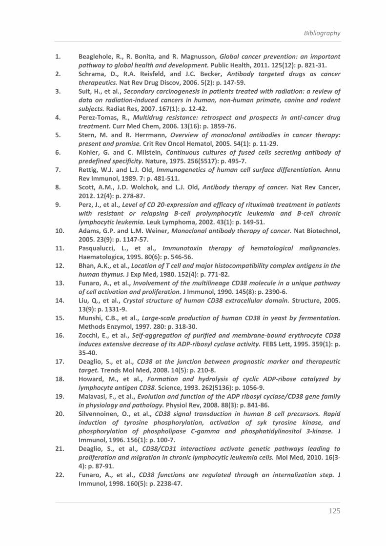
Bibliography
125
1. Beaglehole, R., R. Bonita, and R. Magnusson, Global cancer prevention: an important pathway to global health and development. Public Health, 2011. 125(12): p. 821-31.
2. Schrama, D., R.A. Reisfeld, and J.C. Becker, Antibody targeted drugs as cancer therapeutics. Nat Rev Drug Discov, 2006. 5(2): p. 147-59.
3. Suit, H., et al., Secondary carcinogenesis in patients treated with radiation: a review of data on radiation-induced cancers in human, non-human primate, canine and rodent subjects. Radiat Res, 2007. 167(1): p. 12-42.
4. Perez-Tomas, R., Multidrug resistance: retrospect and prospects in anti-cancer drug treatment. Curr Med Chem, 2006. 13(16): p. 1859-76.
5. Stern, M. and R. Herrmann, Overview of monoclonal antibodies in cancer therapy: present and promise. Crit Rev Oncol Hematol, 2005. 54(1): p. 11-29.
6. Kohler, G. and C. Milstein, Continuous cultures of fused cells secreting antibody of predefined specificity. Nature, 1975. 256(5517): p. 495-7.
7. Rettig, W.J. and L.J. Old, Immunogenetics of human cell surface differentiation. Annu Rev Immunol, 1989. 7: p. 481-511.
8. Scott, A.M., J.D. Wolchok, and L.J. Old, Antibody therapy of cancer. Nat Rev Cancer, 2012. 12(4): p. 278-87.
9. Perz, J., et al., Level of CD 20-expression and efficacy of rituximab treatment in patients with resistant or relapsing B-cell prolymphocytic leukemia and B-cell chronic lymphocytic leukemia. Leuk Lymphoma, 2002. 43(1): p. 149-51.
10. Adams, G.P. and L.M. Weiner, Monoclonal antibody therapy of cancer. Nat Biotechnol, 2005. 23(9): p. 1147-57.
11. Pasqualucci, L., et al., Immunotoxin therapy of hematological malignancies. Haematologica, 1995. 80(6): p. 546-56.
12. Bhan, A.K., et al., Location of T cell and major histocompatibility complex antigens in the human thymus. J Exp Med, 1980. 152(4): p. 771-82.
13. Funaro, A., et al., Involvement of the multilineage CD38 molecule in a unique pathway of cell activation and proliferation. J Immunol, 1990. 145(8): p. 2390-6.
14. Liu, Q., et al., Crystal structure of human CD38 extracellular domain. Structure, 2005. 13(9): p. 1331-9.
15. Munshi, C.B., et al., Large-scale production of human CD38 in yeast by fermentation. Methods Enzymol, 1997. 280: p. 318-30.
16. Zocchi, E., et al., Self-aggregation of purified and membrane-bound erythrocyte CD38 induces extensive decrease of its ADP-ribosyl cyclase activity. FEBS Lett, 1995. 359(1): p. 35-40.
17. Deaglio, S., et al., CD38 at the junction between prognostic marker and therapeutic target. Trends Mol Med, 2008. 14(5): p. 210-8.
18. Howard, M., et al., Formation and hydrolysis of cyclic ADP-ribose catalyzed by lymphocyte antigen CD38. Science, 1993. 262(5136): p. 1056-9.
19. Malavasi, F., et al., Evolution and function of the ADP ribosyl cyclase/CD38 gene family in physiology and pathology. Physiol Rev, 2008. 88(3): p. 841-86.
20. Silvennoinen, O., et al., CD38 signal transduction in human B cell precursors. Rapid induction of tyrosine phosphorylation, activation of syk tyrosine kinase, and phosphorylation of phospholipase C-gamma and phosphatidylinositol 3-kinase. J Immunol, 1996. 156(1): p. 100-7.
21. Deaglio, S., et al., CD38/CD31 interactions activate genetic pathways leading to proliferation and migration in chronic lymphocytic leukemia cells. Mol Med, 2010. 16(3-4): p. 87-91.
22. Funaro, A., et al., CD38 functions are regulated through an internalization step. J Immunol, 1998. 160(5): p. 2238-47.
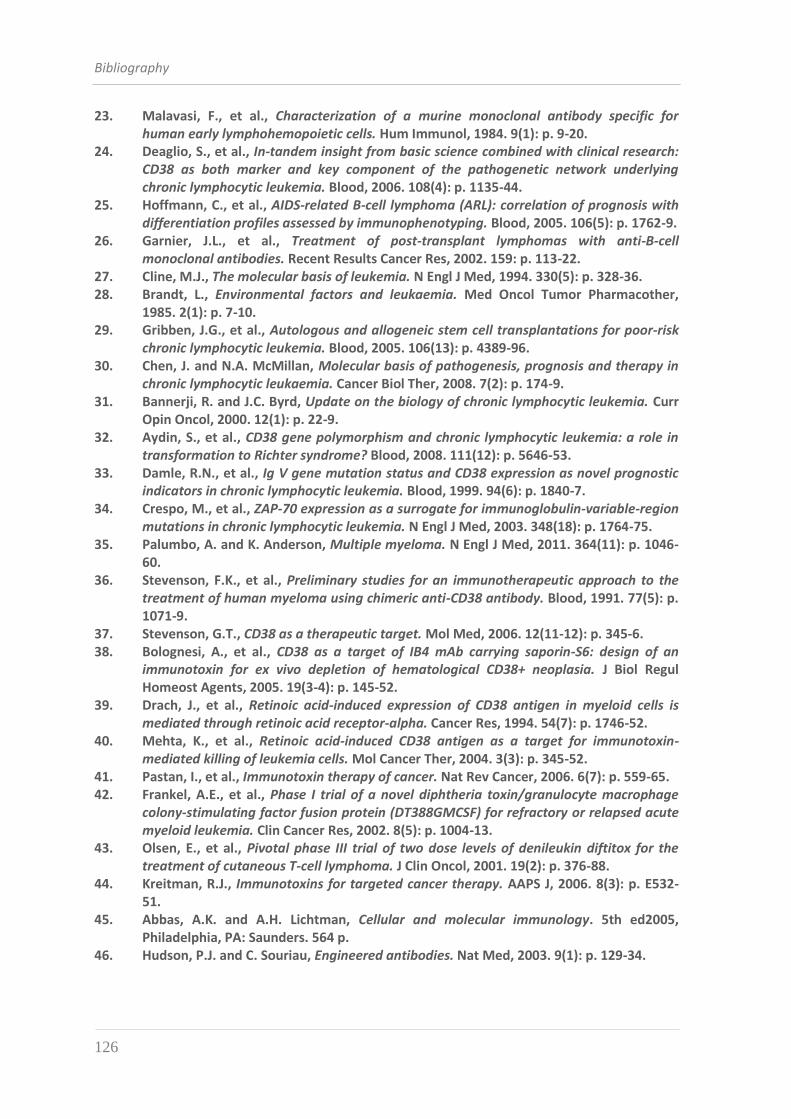
Bibliography
126
23. Malavasi, F., et al., Characterization of a murine monoclonal antibody specific for human early lymphohemopoietic cells. Hum Immunol, 1984. 9(1): p. 9-20.
24. Deaglio, S., et al., In-tandem insight from basic science combined with clinical research: CD38 as both marker and key component of the pathogenetic network underlying chronic lymphocytic leukemia. Blood, 2006. 108(4): p. 1135-44.
25. Hoffmann, C., et al., AIDS-related B-cell lymphoma (ARL): correlation of prognosis with differentiation profiles assessed by immunophenotyping. Blood, 2005. 106(5): p. 1762-9.
26. Garnier, J.L., et al., Treatment of post-transplant lymphomas with anti-B-cell monoclonal antibodies. Recent Results Cancer Res, 2002. 159: p. 113-22.
27. Cline, M.J., The molecular basis of leukemia. N Engl J Med, 1994. 330(5): p. 328-36. 28. Brandt, L., Environmental factors and leukaemia. Med Oncol Tumor Pharmacother,
1985. 2(1): p. 7-10. 29. Gribben, J.G., et al., Autologous and allogeneic stem cell transplantations for poor-risk
chronic lymphocytic leukemia. Blood, 2005. 106(13): p. 4389-96. 30. Chen, J. and N.A. McMillan, Molecular basis of pathogenesis, prognosis and therapy in
chronic lymphocytic leukaemia. Cancer Biol Ther, 2008. 7(2): p. 174-9. 31. Bannerji, R. and J.C. Byrd, Update on the biology of chronic lymphocytic leukemia. Curr
Opin Oncol, 2000. 12(1): p. 22-9. 32. Aydin, S., et al., CD38 gene polymorphism and chronic lymphocytic leukemia: a role in
transformation to Richter syndrome? Blood, 2008. 111(12): p. 5646-53. 33. Damle, R.N., et al., Ig V gene mutation status and CD38 expression as novel prognostic
indicators in chronic lymphocytic leukemia. Blood, 1999. 94(6): p. 1840-7. 34. Crespo, M., et al., ZAP-70 expression as a surrogate for immunoglobulin-variable-region
mutations in chronic lymphocytic leukemia. N Engl J Med, 2003. 348(18): p. 1764-75. 35. Palumbo, A. and K. Anderson, Multiple myeloma. N Engl J Med, 2011. 364(11): p. 1046-
60. 36. Stevenson, F.K., et al., Preliminary studies for an immunotherapeutic approach to the
treatment of human myeloma using chimeric anti-CD38 antibody. Blood, 1991. 77(5): p. 1071-9.
37. Stevenson, G.T., CD38 as a therapeutic target. Mol Med, 2006. 12(11-12): p. 345-6. 38. Bolognesi, A., et al., CD38 as a target of IB4 mAb carrying saporin-S6: design of an
immunotoxin for ex vivo depletion of hematological CD38+ neoplasia. J Biol Regul Homeost Agents, 2005. 19(3-4): p. 145-52.
39. Drach, J., et al., Retinoic acid-induced expression of CD38 antigen in myeloid cells is mediated through retinoic acid receptor-alpha. Cancer Res, 1994. 54(7): p. 1746-52.
40. Mehta, K., et al., Retinoic acid-induced CD38 antigen as a target for immunotoxin-mediated killing of leukemia cells. Mol Cancer Ther, 2004. 3(3): p. 345-52.
41. Pastan, I., et al., Immunotoxin therapy of cancer. Nat Rev Cancer, 2006. 6(7): p. 559-65. 42. Frankel, A.E., et al., Phase I trial of a novel diphtheria toxin/granulocyte macrophage
colony-stimulating factor fusion protein (DT388GMCSF) for refractory or relapsed acute myeloid leukemia. Clin Cancer Res, 2002. 8(5): p. 1004-13.
43. Olsen, E., et al., Pivotal phase III trial of two dose levels of denileukin diftitox for the treatment of cutaneous T-cell lymphoma. J Clin Oncol, 2001. 19(2): p. 376-88.
44. Kreitman, R.J., Immunotoxins for targeted cancer therapy. AAPS J, 2006. 8(3): p. E532-51.
45. Abbas, A.K. and A.H. Lichtman, Cellular and molecular immunology. 5th ed2005, Philadelphia, PA: Saunders. 564 p.
46. Hudson, P.J. and C. Souriau, Engineered antibodies. Nat Med, 2003. 9(1): p. 129-34.
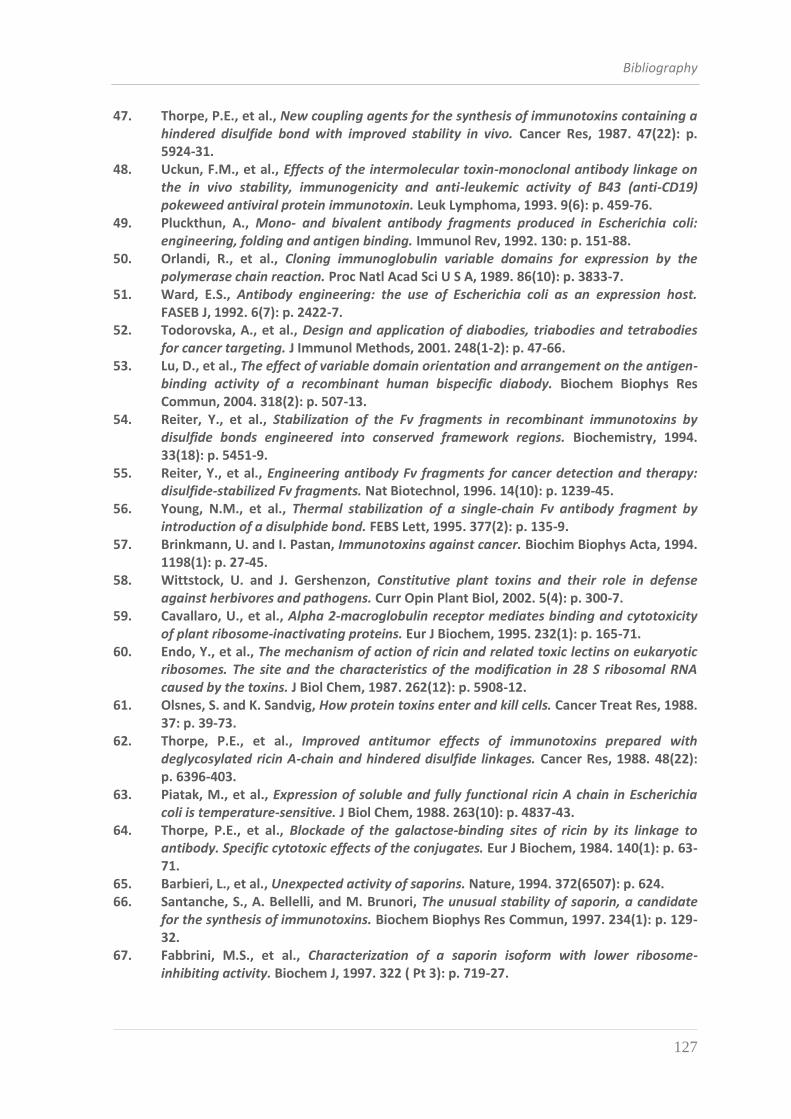
Bibliography
127
47. Thorpe, P.E., et al., New coupling agents for the synthesis of immunotoxins containing a hindered disulfide bond with improved stability in vivo. Cancer Res, 1987. 47(22): p. 5924-31.
48. Uckun, F.M., et al., Effects of the intermolecular toxin-monoclonal antibody linkage on the in vivo stability, immunogenicity and anti-leukemic activity of B43 (anti-CD19) pokeweed antiviral protein immunotoxin. Leuk Lymphoma, 1993. 9(6): p. 459-76.
49. Pluckthun, A., Mono- and bivalent antibody fragments produced in Escherichia coli: engineering, folding and antigen binding. Immunol Rev, 1992. 130: p. 151-88.
50. Orlandi, R., et al., Cloning immunoglobulin variable domains for expression by the polymerase chain reaction. Proc Natl Acad Sci U S A, 1989. 86(10): p. 3833-7.
51. Ward, E.S., Antibody engineering: the use of Escherichia coli as an expression host. FASEB J, 1992. 6(7): p. 2422-7.
52. Todorovska, A., et al., Design and application of diabodies, triabodies and tetrabodies for cancer targeting. J Immunol Methods, 2001. 248(1-2): p. 47-66.
53. Lu, D., et al., The effect of variable domain orientation and arrangement on the antigen-binding activity of a recombinant human bispecific diabody. Biochem Biophys Res Commun, 2004. 318(2): p. 507-13.
54. Reiter, Y., et al., Stabilization of the Fv fragments in recombinant immunotoxins by disulfide bonds engineered into conserved framework regions. Biochemistry, 1994. 33(18): p. 5451-9.
55. Reiter, Y., et al., Engineering antibody Fv fragments for cancer detection and therapy: disulfide-stabilized Fv fragments. Nat Biotechnol, 1996. 14(10): p. 1239-45.
56. Young, N.M., et al., Thermal stabilization of a single-chain Fv antibody fragment by introduction of a disulphide bond. FEBS Lett, 1995. 377(2): p. 135-9.
57. Brinkmann, U. and I. Pastan, Immunotoxins against cancer. Biochim Biophys Acta, 1994. 1198(1): p. 27-45.
58. Wittstock, U. and J. Gershenzon, Constitutive plant toxins and their role in defense against herbivores and pathogens. Curr Opin Plant Biol, 2002. 5(4): p. 300-7.
59. Cavallaro, U., et al., Alpha 2-macroglobulin receptor mediates binding and cytotoxicity of plant ribosome-inactivating proteins. Eur J Biochem, 1995. 232(1): p. 165-71.
60. Endo, Y., et al., The mechanism of action of ricin and related toxic lectins on eukaryotic ribosomes. The site and the characteristics of the modification in 28 S ribosomal RNA caused by the toxins. J Biol Chem, 1987. 262(12): p. 5908-12.
61. Olsnes, S. and K. Sandvig, How protein toxins enter and kill cells. Cancer Treat Res, 1988. 37: p. 39-73.
62. Thorpe, P.E., et al., Improved antitumor effects of immunotoxins prepared with deglycosylated ricin A-chain and hindered disulfide linkages. Cancer Res, 1988. 48(22): p. 6396-403.
63. Piatak, M., et al., Expression of soluble and fully functional ricin A chain in Escherichia coli is temperature-sensitive. J Biol Chem, 1988. 263(10): p. 4837-43.
64. Thorpe, P.E., et al., Blockade of the galactose-binding sites of ricin by its linkage to antibody. Specific cytotoxic effects of the conjugates. Eur J Biochem, 1984. 140(1): p. 63-71.
65. Barbieri, L., et al., Unexpected activity of saporins. Nature, 1994. 372(6507): p. 624. 66. Santanche, S., A. Bellelli, and M. Brunori, The unusual stability of saporin, a candidate
for the synthesis of immunotoxins. Biochem Biophys Res Commun, 1997. 234(1): p. 129-32.
67. Fabbrini, M.S., et al., Characterization of a saporin isoform with lower ribosome-inhibiting activity. Biochem J, 1997. 322 ( Pt 3): p. 719-27.
Bibliography
128
68. de Virgilio, M., et al., Ribosome-inactivating proteins: from plant defense to tumor attack. Toxins (Basel), 2010. 2(11): p. 2699-737.
69. Falini, B., et al., Response of refractory Hodgkin's disease to monoclonal anti-CD30 immunotoxin. Lancet, 1992. 339(8803): p. 1195-6.
70. Flavell, D.J., et al., Therapy of human B-cell lymphoma bearing SCID mice is more effective with anti-CD19- and anti-CD38-saporin immunotoxins used in combination than with either immunotoxin used alone. Int J Cancer, 1995. 62(3): p. 337-44.
71. Iglewski, B.H., P.V. Liu, and D. Kabat, Mechanism of action of Pseudomonas aeruginosa exotoxin Aiadenosine diphosphate-ribosylation of mammalian elongation factor 2 in vitro and in vivo. Infect Immun, 1977. 15(1): p. 138-44.
72. Van Ness, B.G., J.B. Howard, and J.W. Bodley, ADP-ribosylation of elongation factor 2 by diphtheria toxin. Isolation and properties of the novel ribosyl-amino acid and its hydrolysis products. J Biol Chem, 1980. 255(22): p. 10717-20.
73. FitzGerald, D. and I. Pastan, Targeted toxin therapy for the treatment of cancer. J Natl Cancer Inst, 1989. 81(19): p. 1455-63.
74. Siegall, C.B., et al., Functional analysis of domains II, Ib, and III of Pseudomonas exotoxin. J Biol Chem, 1989. 264(24): p. 14256-61.
75. Nakayama, K., Furin: a mammalian subtilisin/Kex2p-like endoprotease involved in processing of a wide variety of precursor proteins. Biochem J, 1997. 327 ( Pt 3): p. 625-35.
76. Kreitman, R.J. and I. Pastan, Importance of the glutamate residue of KDEL in increasing the cytotoxicity of Pseudomonas exotoxin derivatives and for increased binding to the KDEL receptor. Biochem J, 1995. 307 ( Pt 1): p. 29-37.
77. Kounnas, M.Z., et al., The alpha 2-macroglobulin receptor/low density lipoprotein receptor-related protein binds and internalizes Pseudomonas exotoxin A. J Biol Chem, 1992. 267(18): p. 12420-3.
78. Smith, D.C., et al., Internalized Pseudomonas exotoxin A can exploit multiple pathways to reach the endoplasmic reticulum. Traffic, 2006. 7(4): p. 379-93.
79. Ogata, M., et al., Cell-mediated cleavage of Pseudomonas exotoxin between Arg279 and Gly280 generates the enzymatically active fragment which translocates to the cytosol. J Biol Chem, 1992. 267(35): p. 25396-401.
80. Ogata, M., et al., Processing of Pseudomonas exotoxin by a cellular protease results in the generation of a 37,000-Da toxin fragment that is translocated to the cytosol. J Biol Chem, 1990. 265(33): p. 20678-85.
81. McKee, M.L. and D.J. FitzGerald, Reduction of furin-nicked Pseudomonas exotoxin A: an unfolding story. Biochemistry, 1999. 38(50): p. 16507-13.
82. Lombardi, D., et al., Rab9 functions in transport between late endosomes and the trans Golgi network. EMBO J, 1993. 12(2): p. 677-82.
83. Wilson, D.W., M.J. Lewis, and H.R. Pelham, pH-dependent binding of KDEL to its receptor in vitro. J Biol Chem, 1993. 268(10): p. 7465-8.
84. Koopmann, J.O., et al., Export of antigenic peptides from the endoplasmic reticulum intersects with retrograde protein translocation through the Sec61p channel. Immunity, 2000. 13(1): p. 117-27.
85. Jinno, Y., et al., Mutational analysis of domain I of Pseudomonas exotoxin. Mutations in domain I of Pseudomonas exotoxin which reduce cell binding and animal toxicity. J Biol Chem, 1988. 263(26): p. 13203-7.
86. Lorberboum-Galski, H., et al., IL2-PE664Glu, a new chimeric protein cytotoxic to human-activated T lymphocytes. J Biol Chem, 1990. 265(27): p. 16311-7.
87. Kondo, T., et al., Activity of immunotoxins constructed with modified Pseudomonas exotoxin A lacking the cell recognition domain. J Biol Chem, 1988. 263(19): p. 9470-5.
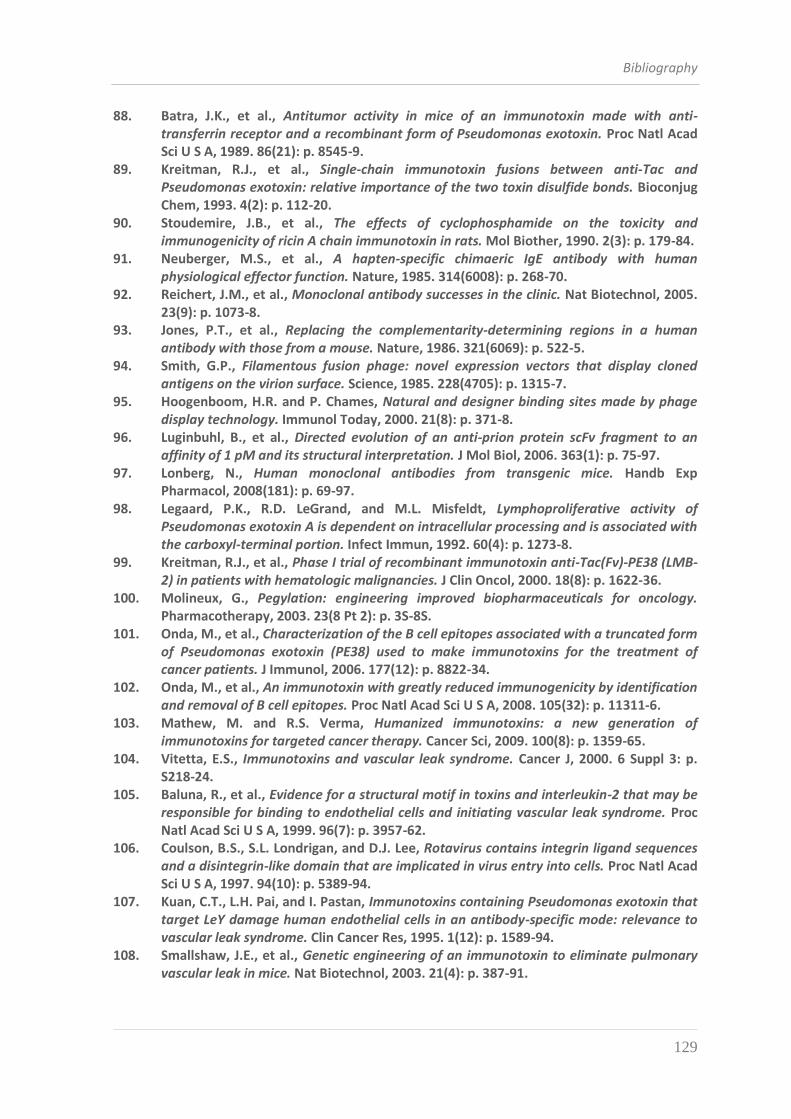
Bibliography
129
88. Batra, J.K., et al., Antitumor activity in mice of an immunotoxin made with anti-transferrin receptor and a recombinant form of Pseudomonas exotoxin. Proc Natl Acad Sci U S A, 1989. 86(21): p. 8545-9.
89. Kreitman, R.J., et al., Single-chain immunotoxin fusions between anti-Tac and Pseudomonas exotoxin: relative importance of the two toxin disulfide bonds. Bioconjug Chem, 1993. 4(2): p. 112-20.
90. Stoudemire, J.B., et al., The effects of cyclophosphamide on the toxicity and immunogenicity of ricin A chain immunotoxin in rats. Mol Biother, 1990. 2(3): p. 179-84.
91. Neuberger, M.S., et al., A hapten-specific chimaeric IgE antibody with human physiological effector function. Nature, 1985. 314(6008): p. 268-70.
92. Reichert, J.M., et al., Monoclonal antibody successes in the clinic. Nat Biotechnol, 2005. 23(9): p. 1073-8.
93. Jones, P.T., et al., Replacing the complementarity-determining regions in a human antibody with those from a mouse. Nature, 1986. 321(6069): p. 522-5.
94. Smith, G.P., Filamentous fusion phage: novel expression vectors that display cloned antigens on the virion surface. Science, 1985. 228(4705): p. 1315-7.
95. Hoogenboom, H.R. and P. Chames, Natural and designer binding sites made by phage display technology. Immunol Today, 2000. 21(8): p. 371-8.
96. Luginbuhl, B., et al., Directed evolution of an anti-prion protein scFv fragment to an affinity of 1 pM and its structural interpretation. J Mol Biol, 2006. 363(1): p. 75-97.
97. Lonberg, N., Human monoclonal antibodies from transgenic mice. Handb Exp Pharmacol, 2008(181): p. 69-97.
98. Legaard, P.K., R.D. LeGrand, and M.L. Misfeldt, Lymphoproliferative activity of Pseudomonas exotoxin A is dependent on intracellular processing and is associated with the carboxyl-terminal portion. Infect Immun, 1992. 60(4): p. 1273-8.
99. Kreitman, R.J., et al., Phase I trial of recombinant immunotoxin anti-Tac(Fv)-PE38 (LMB-2) in patients with hematologic malignancies. J Clin Oncol, 2000. 18(8): p. 1622-36.
100. Molineux, G., Pegylation: engineering improved biopharmaceuticals for oncology. Pharmacotherapy, 2003. 23(8 Pt 2): p. 3S-8S.
101. Onda, M., et al., Characterization of the B cell epitopes associated with a truncated form of Pseudomonas exotoxin (PE38) used to make immunotoxins for the treatment of cancer patients. J Immunol, 2006. 177(12): p. 8822-34.
102. Onda, M., et al., An immunotoxin with greatly reduced immunogenicity by identification and removal of B cell epitopes. Proc Natl Acad Sci U S A, 2008. 105(32): p. 11311-6.
103. Mathew, M. and R.S. Verma, Humanized immunotoxins: a new generation of immunotoxins for targeted cancer therapy. Cancer Sci, 2009. 100(8): p. 1359-65.
104. Vitetta, E.S., Immunotoxins and vascular leak syndrome. Cancer J, 2000. 6 Suppl 3: p. S218-24.
105. Baluna, R., et al., Evidence for a structural motif in toxins and interleukin-2 that may be responsible for binding to endothelial cells and initiating vascular leak syndrome. Proc Natl Acad Sci U S A, 1999. 96(7): p. 3957-62.
106. Coulson, B.S., S.L. Londrigan, and D.J. Lee, Rotavirus contains integrin ligand sequences and a disintegrin-like domain that are implicated in virus entry into cells. Proc Natl Acad Sci U S A, 1997. 94(10): p. 5389-94.
107. Kuan, C.T., L.H. Pai, and I. Pastan, Immunotoxins containing Pseudomonas exotoxin that target LeY damage human endothelial cells in an antibody-specific mode: relevance to vascular leak syndrome. Clin Cancer Res, 1995. 1(12): p. 1589-94.
108. Smallshaw, J.E., et al., Genetic engineering of an immunotoxin to eliminate pulmonary vascular leak in mice. Nat Biotechnol, 2003. 21(4): p. 387-91.
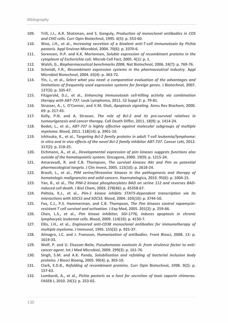
Bibliography
130
109. Trill, J.J., A.R. Shatzman, and S. Ganguly, Production of monoclonal antibodies in COS and CHO cells. Curr Opin Biotechnol, 1995. 6(5): p. 553-60.
110. Woo, J.H., et al., Increasing secretion of a bivalent anti-T-cell immunotoxin by Pichia pastoris. Appl Environ Microbiol, 2004. 70(6): p. 3370-6.
111. Sorensen, H.P. and K.K. Mortensen, Soluble expression of recombinant proteins in the cytoplasm of Escherichia coli. Microb Cell Fact, 2005. 4(1): p. 1.
112. Walsh, G., Biopharmaceutical benchmarks 2006. Nat Biotechnol, 2006. 24(7): p. 769-76. 113. Schmidt, F.R., Recombinant expression systems in the pharmaceutical industry. Appl
Microbiol Biotechnol, 2004. 65(4): p. 363-72. 114. Yin, J., et al., Select what you need: a comparative evaluation of the advantages and
limitations of frequently used expression systems for foreign genes. J Biotechnol, 2007. 127(3): p. 335-47.
115. Fitzgerald, D.J., et al., Enhancing immunotoxin cell-killing activity via combination therapy with ABT-737. Leuk Lymphoma, 2011. 52 Suppl 2: p. 79-81.
116. Strasser, A., L. O'Connor, and V.M. Dixit, Apoptosis signaling. Annu Rev Biochem, 2000. 69: p. 217-45.
117. Kelly, P.N. and A. Strasser, The role of Bcl-2 and its pro-survival relatives in tumourigenesis and cancer therapy. Cell Death Differ, 2011. 18(9): p. 1414-24.
118. Bodet, L., et al., ABT-737 is highly effective against molecular subgroups of multiple myeloma. Blood, 2011. 118(14): p. 3901-10.
119. Ishitsuka, K., et al., Targeting Bcl-2 family proteins in adult T-cell leukemia/lymphoma: in vitro and in vivo effects of the novel Bcl-2 family inhibitor ABT-737. Cancer Lett, 2012. 317(2): p. 218-25.
120. Eichmann, A., et al., Developmental expression of pim kinases suggests functions also outside of the hematopoietic system. Oncogene, 2000. 19(9): p. 1215-24.
121. Amaravadi, R. and C.B. Thompson, The survival kinases Akt and Pim as potential pharmacological targets. J Clin Invest, 2005. 115(10): p. 2618-24.
122. Brault, L., et al., PIM serine/threonine kinases in the pathogenesis and therapy of hematologic malignancies and solid cancers. Haematologica, 2010. 95(6): p. 1004-15.
123. Yan, B., et al., The PIM-2 kinase phosphorylates BAD on serine 112 and reverses BAD-induced cell death. J Biol Chem, 2003. 278(46): p. 45358-67.
124. Peltola, K.J., et al., Pim-1 kinase inhibits STAT5-dependent transcription via its interactions with SOCS1 and SOCS3. Blood, 2004. 103(10): p. 3744-50.
125. Fox, C.J., P.S. Hammerman, and C.B. Thompson, The Pim kinases control rapamycin-resistant T cell survival and activation. J Exp Med, 2005. 201(2): p. 259-66.
126. Chen, L.S., et al., Pim kinase inhibitor, SGI-1776, induces apoptosis in chronic lymphocytic leukemia cells. Blood, 2009. 114(19): p. 4150-7.
127. Ellis, J.H., et al., Engineered anti-CD38 monoclonal antibodies for immunotherapy of multiple myeloma. J Immunol, 1995. 155(2): p. 925-37.
128. Almagro, J.C. and J. Fransson, Humanization of antibodies. Front Biosci, 2008. 13: p. 1619-33.
129. Wolf, P. and U. Elsasser-Beile, Pseudomonas exotoxin A: from virulence factor to anti-cancer agent. Int J Med Microbiol, 2009. 299(3): p. 161-76.
130. Singh, S.M. and A.K. Panda, Solubilization and refolding of bacterial inclusion body proteins. J Biosci Bioeng, 2005. 99(4): p. 303-10.
131. Clark, E.D.B., Refolding of recombinant proteins. Curr Opin Biotechnol, 1998. 9(2): p. 157-63.
132. Lombardi, A., et al., Pichia pastoris as a host for secretion of toxic saporin chimeras. FASEB J, 2010. 24(1): p. 253-65.
Bibliography
131
133. Hessler, J.L. and R.J. Kreitman, An early step in Pseudomonas exotoxin action is removal of the terminal lysine residue, which allows binding to the KDEL receptor. Biochemistry, 1997. 36(47): p. 14577-82.
134. Huston, J.S., et al., Protein engineering of antibody binding sites: recovery of specific activity in an anti-digoxin single-chain Fv analogue produced in Escherichia coli. Proc Natl Acad Sci U S A, 1988. 85(16): p. 5879-83.
135. Milenic, D.E., et al., Construction, binding properties, metabolism, and tumor targeting of a single-chain Fv derived from the pancarcinoma monoclonal antibody CC49. Cancer Res, 1991. 51(23 Pt 1): p. 6363-71.
136. Adams, G.P. and R. Schier, Generating improved single-chain Fv molecules for tumor targeting. J Immunol Methods, 1999. 231(1-2): p. 249-60.
137. Ausiello, C.M., et al., Functional topography of discrete domains of human CD38. Tissue Antigens, 2000. 56(6): p. 539-47.
138. Carroll, S.F. and R.J. Collier, Active site of Pseudomonas aeruginosa exotoxin A. Glutamic acid 553 is photolabeled by NAD and shows functional homology with glutamic acid 148 of diphtheria toxin. J Biol Chem, 1987. 262(18): p. 8707-11.
139. Keppler-Hafkemeyer, A., U. Brinkmann, and I. Pastan, Role of caspases in immunotoxin-induced apoptosis of cancer cells. Biochemistry, 1998. 37(48): p. 16934-42.
140. Thornberry, N.A. and Y. Lazebnik, Caspases: enemies within. Science, 1998. 281(5381): p. 1312-6.
141. Chaudhary, V.K., et al., Pseudomonas exotoxin contains a specific sequence at the carboxyl terminus that is required for cytotoxicity. Proc Natl Acad Sci U S A, 1990. 87(1): p. 308-12.
142. Andersson, Y., S. Juell, and O. Fodstad, Downregulation of the antiapoptotic MCL-1 protein and apoptosis in MA-11 breast cancer cells induced by an anti-epidermal growth factor receptor-Pseudomonas exotoxin a immunotoxin. Int J Cancer, 2004. 112(3): p. 475-83.
143. Rooswinkel, R.W., et al., Bcl-2 is a better ABT-737 target than Bcl-xL or Bcl-w and only Noxa overcomes resistance mediated by Mcl-1, Bfl-1, or Bcl-B. Cell Death Dis, 2012. 3: p. e366.