QUALITATIVO 2.0 HCV RNA REAL TIME - iriseirise.com.ge/uploads/files/7259AA896_48 NLM7185 ital ingl...
60
NUCLEAR LASER MEDICINE S.r.l. UFFICI OPERATIVI: Viale delle Industrie, 3 – 20090 SETTALA MI (Italy) Tel (+39) 02/95. 24. 51 - Fax (+39) 02/95. 24. 52. 37 SITO INTERNET: www.nlm.it – E-MAIL: [email protected] 0459 BIOLOGIA MOLECOLARE ESTRAZIONE: AA1318 NON COMPRESA HCV RNA REAL TIME QUALITATIVO 2.0 NLM 7185 VER 01/09/2014 AA896/48 48 TEST W0105020307
Transcript of QUALITATIVO 2.0 HCV RNA REAL TIME - iriseirise.com.ge/uploads/files/7259AA896_48 NLM7185 ital ingl...

NUCLEAR LASER MEDICINE S.r.l.
UFFICI OPERATIVI: Viale delle Industrie, 3 – 20090 SETTALA MI (Italy)
Tel (+39) 02/95. 24. 51 - Fax (+39) 02/95. 24. 52. 37
SITO INTERNET: www.nlm.it – E-MAIL: [email protected]
0459
BIOLOGIA MOLECOLARE
ESTRAZIONE: AA1318 NON COMPRESA
HCV RNA REAL TIME
QUALITATIVO 2.0
NLM 7185 VER 01/09/2014
AA896/48 48 TEST
W0105020307

2 :testo modificato rispetto alla precedente versione
UTILIZZO
Il test HCV RNA REAL TIME QUALITATIVO 2.0 fornisce i reagenti necessari per la determinazione qualitativa della presenza del virus dell’Epatite C (HCV RNA) in campioni di plasma umano, mediante Real Time RT-PCR della regione 5’-UTR dell’RNA virale; contemporaneamente viene amplificata anche la regione Core, in modo da rendere compatibili gli amplificati con il kit NLM cod. AC004/24 GEN-C 2.0 per la genotipizzazione di HCV. Il Controllo Interno è endogeno (mRNA del gene costitutivo GAPDH) e viene estratto ed amplificato in ogni campione, al fine di monitorare l’intera procedura. Questo prodotto va utilizzato in abbinamento al kit di estrazione NLM AA1318 “Estrazione DNA/RNA Virale”, da ordinare separatamente. L’RNA estratto è retrotrascritto e amplificato in un unico step di RT-PCR, utilizzando lo strumento CFX (BioRad).
HCV RNA REAL TIME QUALITATIVO 2.0 è indicato per la diagnosi di infezione da HCV insieme agli altri parametri di laboratorio (es. anticorpi, ALT, etc.) ed al quadro clinico del paziente. Il dispositivo non va utilizzato come test di screening per la presenza di HCV RNA nei donatori di sangue. Il kit è da ritenersi per il solo uso professionale.
3 :testo modificato rispetto alla precedente versione
INTRODUZIONE
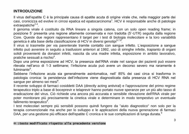
Il virus dell’epatite C è la principale causa di epatite acuta di origine virale che, nella maggior parte dei casi, cronicizza ed evolve in cirrosi epatica ed epatocarcinoma1. HCV è responsabile anche di patologie extraepatiche2-4. Il genoma virale è costituito da RNA lineare a singola catena, con un solo open reading frame; in posizione 5’ presenta una regione altamente conservata e non tradotta (5’-UTR) seguita dalla regione Core. Queste due regioni rappresentano il target per i test di biologia molecolare e la loro variabilità genetica è alla base della classificazione di HCV in diversi genotipi5,7,8. Il virus si trasmette per via parenterale tramite contatto con sangue infetto. L’esposizione a sangue infetto può avvenire in seguito a trasfusioni anteriori al 1992, uso di siringhe infette, trapianto di organi solidi provenienti da donatori infetti, nascita da una madre infetta, esposizione in ambito lavorativo, pratiche sessuali a rischio6. Dopo una prima esposizione ad HCV, la presenza dell’RNA virale nel sangue dei pazienti può essere rilevata nell’arco di 1-3 settimane; l’infezione acuta può avere un decorso severo ma raramente è fulminante6. Sebbene l’infezione acuta sia generalmente asintomatica, nell’ 85% dei casi circa si trasforma in patologia cronica: la persistenza dell’infezione viene diagnosticata dalla presenza di HCV RNA nel sangue per almeno sei mesi6. Il recente sviluppo di farmaci cosiddetti DAA (directly acting antiviral) e l'approvazione del primo regime terapeutico triplo a base di boceprevir e telaprevir hanno portato nuove speranze per un più alto tasso di eradicazione del virus. Ciò richiede una ancora più accurata e sensibile rilevazione dell'RNA virale per poter monitorare più precisamente la cinetica virale e determinare in modo tempestivo un eventuale fallimento terapeutico9. I test molecolari sempre più sensibili possono quindi fungere da "aiuto diagnostico" non solo per la terapia convenzionale ma anche per lo sviluppo e le applicazioni della nuova generazione di farmaci DAA, per una gestione più efficace dell'epatite C cronica e le sue complicazioni di lunga durata.9
4 :testo modificato rispetto alla precedente versione
BIBLIOGRAFIA
1. Sarrazin C. Diagnosis of hepatitis C: update 2004. Journal of Gastroenterology and Hepatology (2004) 19, S88-S93
2. Johnson RJ, Gretch DR, Yamabe H et al. Membranoproliferative glomerulonephritis associated with hepatitis C virus infection. N Engl J med 1993; 328: 465-470.
3. Agnello V, Chung RT, Kaplan RM. A role for hepatitis C virus infection in Type II cryoglobulinemia. N Engl J Med 1992; 327: 1490-1495.
4. Andreone P, Zignego AL, Cursaro C et al. Prevalence of monoclonal gammopathies with hepatitis C virus infection . Ann Intern Med 1998; 129: 294-298
5. Zein NN. Clinical Significance of Hepatitis C Virus Genotypes. Clinical Microbiology Reviews, Apr. 2000, p. 223-235.
6. National Institutes of Health, Consensus Conference Statement. Management of hepatitis C: 2002. 7. P.T. Hraber et al. Comparative analysis of hepatitis C virus phylogenies from coding and non-coding regions:
the 5' untranslated region (UTR) fails to classify subtypes. Virology Journal 2006, 3:103 8. M. A. Ansari et al. HCV-Core Region: Its Significance in HCV-Genotyping and Type Dependent Genomic
Expression. Macedonian Journal of Medical Sciences. 2012 Mar 15; 5(1):30-39. 9. G. Colucci Molecular diagnostic and predictive tests in the evolution of chronic hepatitis C anti-viral therapies
BMC Infect Dis. 2012; 12(Suppl 2): S8. Published online 2012 November 12

5 :testo modificato rispetto alla precedente versione
PRINCIPIO DEL TEST
Il Test HCV RNA REAL TIME QUALITATIVO 2.0 si basa su due processi: 1. estrazione dell’RNA virale 2. retrotrascrizione, amplificazione e rivelazione della sequenza bersaglio mediante Real Time RT-
PCR Il Controllo Interno (GAPDH mRNA) viene estratto insieme al campione e permette di monitorare l’andamento di tutto il saggio.
1. Estrazione dell’RNA virale
Per la purificazione di HCV RNA e del Controllo Interno a partire da campioni di plasma umano si deve utilizzare il kit “Estrazione DNA/RNA Virale” (cod. NLM AA1318).
2. RT-PCR e Rivelazione
Target selezionato per l’amplificazione
La regione 5’-UTR è stata scelta come target per l’identificazione del virus in campioni di plasma umano poiché si tratta di una regione altamente conservata fra i diversi genotipi di HCV; la co-amplificazione della regione Core è stata inserita per permettere di discriminare i genotipi 1a, 1b e 6 nel caso che gli amplificati vengano utilizzati in seguito con il kit NLM AC004/24 Gen-C ver. 2.0. Il Controllo Interno è l’mRNA del gene GAPDH, costitutivamente espresso, e viene co-estratto e co-amplificato insieme al target. La sequenza amplificata è simile per lunghezza e composizione in basi alla sequenza di HCV.
Retrotrascrizione ed amplificazione
Successivamente alla fase di estrazione, HCV RNA e CI sono retrotrascritti ed amplificati in un unico passaggio di RT-PCR, utilizzando enzimi e reagenti opportunamente selezionati.

6 :testo modificato rispetto alla precedente versione
Rivelazione
Il test si basa sulla Real-Time PCR, tecnica che permette di monitorare in tempo reale l’amplificazione dei campioni d’interesse, utilizzando sonde doppiamente marcate con un fluoroforo donatore e un quencher (chimica “dual labeled probe”). La PCR avviene in presenza di due sonde specifiche rispettivamente per il controllo interno e per la regione 5’-UTR del virus. A ciascuna sonda è legato un diverso fluoroforo donatore: FAM per HCV e JOE per il controllo interno. In presenza di una fonte luminosa, quando le sonde sono intatte e donatore e quencher su ciascuna sonda sono vicini, la fluorescenza emessa dal fluoroforo donatore è assorbita dal fluoroforo accettore. Durante l’amplificazione le sonde si appaiano ciascuna al proprio target specifico e sono soggette all’attività nucleasica 5’3’ della DNA polimerasi. Quando i fluorofori donatore ed accettore sono separati, la fluorescenza del donatore può essere rilevata alla sua lunghezza d’onda specifica: in questo modo l’amplificazione dell’RNA virale e del CI può essere monitorata nel corso della reazione.
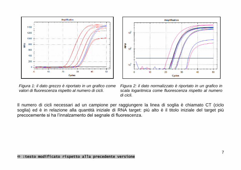
Lo strumento CFX (BioRad) comprende in un singolo strumento un termociclatore ed un dispositivo per la rilevazione della fluorescenza durante i cicli di PCR. Un computer raccoglie i dati di fluorescenza che vengono visualizzati in un grafico mediate un apposito software (Figura 1). Dopo la raccolta dei dati viene effettuata l’analisi. Il dato grezzo è normalizzato per correggere il segnale di fondo, successivamente viene impostato il livello di soglia in corrispondenza del quale viene analizzato il segnale di fluorescenza (Figura 2).
7 :testo modificato rispetto alla precedente versione
Figura 1: il dato grezzo è riportato in un grafico come valori di fluorescenza rispetto al numero di cicli.
Figura 2: il dato normalizzato è riportato in un grafico in scala logaritmica come fluorescenza rispetto al numero di cicli.
Il numero di cicli necessari ad un campione per raggiungere la linea di soglia è chiamato CT (ciclo soglia) ed è in relazione alla quantità iniziale di RNA target: più alto è il titolo iniziale del target più precocemente si ha l’innalzamento del segnale di fluorescenza.

8 :testo modificato rispetto alla precedente versione
COMPOSIZIONE DEL PRODOTTO (Conservare a -25/-15°C)
Mix A Sonde Primers Tris-EDTA
1 x 47 l
Mix B Primers Tris-EDTA
1 x 110 l
Mix C (R36,37,38) Enzimi per RT-PCR MgCl2
dNTPs
1 x 330 l
Controllo negativo Il controllo negativo contiene plasma di origine umana testato per HIV-Ab ed RNA, HCV-Ab ed RNA, HBs-Ag e DNA e risultato negativo. Il controllo negativo deve essere estratto
4 x 500 l
Controllo positivo RNA sintetico Il controllo positivo è fornito in forma liofila. Deve essere risospeso in 14 µl di acqua per biologia molecolare (RNasi, DNasi e Proteasi free) prima dell’uso per la fase di RT-PCR.
4 provette

9 :testo modificato rispetto alla precedente versione
MODULARITA'
La modularità prevista è di massimo 4 sedute: 12 campioni + 1 Controllo Positivo + 1 Controllo Negativo possono essere analizzati in ogni seduta. Se la modularità non viene rispettata i reagenti potrebbero essere insufficienti per effettuare il numero di test previsto.
STABILITA’ E CONSERVAZIONE
- Tutti i reagenti sono stabili sino alla data di scadenza riportata sull’etichetta se conservati a -25/-15°C.
- Scongelare i reagenti in ghiaccio o a +2/+8°C. - Le Mix sono stabili sino alla data di scadenza se tenute a -25/-15°C. Questi reagenti possono essere
ricongelati e scongelati al massimo 4 volte. - La Mix A contiene i fluorofori FAM e JOE che sono fotosensibili: evitare prolungate esposizioni alla
luce. - La Master Mix deve essere utilizzata subito dopo la preparazione; dopo averla dispensata nelle
provette da PCR, la Master Mix rimanente va scartata. Evitare prolungate esposizioni alla luce. - Il Controllo Positivo è un templato ad RNA, fornito già aliquotato e in forma liofila: si raccomanda di
risospendere tale reagente con acqua RNasi, DNasi e Proteasi free poco prima dell’uso. Aggiungere quindi la Master Mix nella provetta del CP risospeso e trasferire nella strip/piastra di amplificazione, il tutto dopo aver dispensato i campioni nelle rispettive provette onde evitare contaminazioni.
10 :testo modificato rispetto alla precedente versione
RACCOLTA DEI CAMPIONI
L’RNA virale viene isolato e purificato partendo da campioni di plasma preparati entro 6 ore dal momento del prelievo. - Raccogliere i campioni di sangue secondo le comuni precauzioni per i prelievi di sangue. - Utilizzare provette sterili. Usare solo EDTA come anticoagulante; altre tipologie di anticoagulanti
potrebbero interferire con la corretta esecuzione del test. Centrifugare a 1000-1500 x g per 10-15 minuti per separare il plasma.
- Prelevare il quantitativo di plasma necessario per l’estrazione dell’RNA (450 μl) sotto cappa a flusso laminare verticale, operando in modo da evitare la degradazione dell’RNA.
- Conservare i campioni a +2/+8°C fino al momento dell’estrazione. Se non si procede immediatamente, si consiglia di porre i campioni a -25/-15°C fino a 72 ore prima di congelarli a ≤ -70°C.
- Evitare ripetuti congelamenti/scongelamenti dei campioni di plasma. - I campioni vanno manipolati seguendo le buone pratiche di laboratorio e devono essere considerati
come pericolosi in quanto potenziale fonte di infezione.
PRECAUZIONI
- La procedura va eseguita da personale competente utilizzando le buone pratiche di laboratorio ed i comuni dispositivi di protezione individuale.
- Tutti i consumabili (puntali e provette) devono essere privi di DNAsi ed RNAsi. I puntali devono avere il filtro per evitare la contaminazione delle pipette. Utilizzare un nuovo puntale ogni volta che viene dispensato un volume.
- Eliminare il materiale monouso utilizzato come rifiuti speciali. - Il controllo negativo contiene plasma di origine umana negativo per HIV-Ab ed RNA, HCV-Ab ed
RNA, HBs-Ag e DNA. Tuttavia tale reagente deve essere maneggiato come campione potenzialmente infettivo, utilizzando i dispositivi di protezione individuale.
11 :testo modificato rispetto alla precedente versione
- Mix C: Irritante (R36, 37, 38) per le frasi S fare riferimento alla scheda di sicurezza - Non mangiare, bere, fumare o applicare cosmetici nelle aree preposte all’esecuzione del test. - Se vi è esposizione di occhi, cute o mucose alle sostanze utilizzate, lavare abbondantemente con
acqua e contattare al più presto un medico. - Non utilizzare reagenti scaduti. - Non mischiare reagenti di lotti diversi. - Prestare attenzione alla modularità del dispositivo (12 campioni + 1 Controllo Positivo +1 Controllo
Negativo per 4 sedute). L’utilizzo del kit con una modularità inferiore determina l’insufficienza dei reagenti per poter eseguire tutti i test previsti.
- Data l’elevata sensibilità del test prestare particolare attenzione alle cross-contaminazioni. Si consiglia di eseguire l’analisi in tre zone separate:
- Zona 1: pre-PCR (manipolazione dei campioni ed estrazione) - Zona 2: preparazione della Master Mix - Zona 3: post PCR (Real Time PCR)
e di pulire le aree di lavoro con candeggina al termine della procedura. - Non utilizzare il kit se la confezione è danneggiata. Contattare il fornitore. - E’ opportuno assicurare una temperatura il più possibile costante ed uniforme in laboratorio
ed evitare di posizionare gli strumenti in prossimità di fonti di calore/raffreddamento che possano comprometterne il corretto funzionamento.

12 :testo modificato rispetto alla precedente versione
MATERIALE NECESSARIO MA NON FORNITO
ZONA 1
Cappa a flusso laminare verticale Set dedicato di micropipette a volume variabile
Puntali con filtro e provette monouso da 2 e da 1,5ml DNAsi ed RNAsi free
Separatore magnetico
Vortex e termoblocco
Centrifuga Acqua RNasi, DNasi e Proteasi free
ZONA 2
Cappa a flusso laminare verticale Set dedicato di pipette a volume variabile Puntali con filtro DNAsi ed RNAsi free Provette DNAsi ed RNAsi free, da 0,2 ml, strip da 8 DNAsi e RNAsi free o piastre per real time PCR DNAsi e RNAsi free Acqua RNasi, DNasi e Proteasi free
ZONA 3
CFX (BioRad) CFX (BioRad) software

13 :testo modificato rispetto alla precedente versione
PROCEDIMENTO
ESTRAZIONE DELL’RNA VIRALE
(Sistema abbinato: “Estrazione DNA/RNA virale”, codice NLM AA1318)
Fare riferimento alle istruzioni del kit di estrazione per la preparazione, la manipolazione e lo
smaltimento dei reagenti, considerando 450 l di campione di partenza. Questo protocollo è per uso manuale e prevede l’utilizzo di tubi di reazione da 2 ml ed un separatore magnetico idoneo. Per l’automazione della procedura con strumento Janus (Perkin Elmer) contattare il fornitore.
Preparare il numero di provette da 2 ml da centrifuga necessario per i campioni da estrarre. Prima di procedere con la fase di estrazione portare il termoblocco a +55°C. Assicurarsi che la Proteinasi K e il Poly(A) RNA siano stati preparati come descritto.
1. Dispensare nel seguente ordine 10 l di Proteinasi K e 450 l di campione in una provetta da 2
ml. Aggiungere 450 l di Lysis Buffer Viral RNA, poi 6 l di Poly(A) RNA. Mescolare bene pipettando almeno 8 volte su e giù e incubare a 55°C per 10 minuti; vortexare brevemente i campioni a metà dell’incubazione. Per maggiore comodità è possibile preparare una miscela di Lysis Buffer Viral RNA e Poly(A) RNA calcolando la quantità necessaria per il numero totale di campioni da processare + 1. Questa miscela deve essere aggiunta al campione immediatamente (entro 15 minuti dalla preparazione).
2. Dopo l’incubazione, centrifugare brevemente le provette per eliminare l’eventuale liquido evaporato
sui tappi ed aggiungere 35 l di Magnetic Beads Viral DNA/RNA (assicurarsi di risospendere
bene le biglie magnetiche prima di prelevarle dalla bottiglia di stoccaggio) e 900l di Binding Buffer Viral DNA/RNA al campione lisato. Mescolare pipettando su e giù almeno 6 volte e incubare per 5 minuti a TA.

14 :testo modificato rispetto alla precedente versione
Le biglie e il Binding Buffer Viral DNA/RNA possono essere pre-miscelati. 3. Disporre le provette nel separatore magnetico ed attendere almeno 2 minuti fino a quando tutte le
biglie sono state attratte dal magnete. Rimuovere il surnatante con una pipetta, evitando di toccare il pellet.
4. Rimuovere le provette dal separatore magnetico. Aggiungere 500 l di Wash Buffer A Viral DNA/RNA, risospendere bene il pellet di biglie con ripetute pipettate e incubare 1 minuto a TA
5. Separare le biglie disponendo le provette nel separatore magnetico. Attendere fino a quando tutte le biglie sono state attratte dal magnete (circa 1 minuto). Rimuovere il surnatante con una pipetta.
6. Ripetere i punti 4 e 5 con 500 l di Wash Buffer B Viral DNA/RNA. Rimuovere bene il surnatante aspirando una seconda volta con un puntale più piccolo; in questa fase porre maggiore attenzione a non toccare il pellet.
7. Dopo avere rimosso ogni traccia di Wash Buffer B Viral DNA/RNA, trasferire le provette nel termoblocco e lasciarle asciugare aperte per 20 minuti a 55°C fino a quando tutto l’etanolo non è evaporato: il pellet di biglie deve diventare di colore marrone chiaro. Se ciò non accade, prolungare l’incubazione di altri 5 minuti o fino alla completa asciugatura. E’ importante che il pellet sia completamente asciutto e che non ci siano residui di etanolo sulle pareti della provetta; l’etanolo infatti può inibire l’amplificazione.
8. Aggiungere 70 l di Elution Buffer Viral DNA/RNA a ciascuna provetta e risospendere brevemente le biglie con la pipetta. Chiudere le provette, vortexare a bassa intensità (le biglie devono formare una sospensione omogenea) e incubare a 55°C per 5 minuti.
9. Separare le biglie disponendo le provette nel separatore magnetico. Per almeno 1 minuto Recuperare quindi il surnatante contenente l’RNA virale purificato e trasferirlo nelle provette finali di stoccaggio.
Il RNA purificato può essere conservato a +2/+8°C se utilizzato entro breve, altrimenti conservarlo a -25/-15°C o meglio a ≤-70°C per periodi più lunghi. Si raccomanda di scongelare a +2/+8°C.

15 :testo modificato rispetto alla precedente versione
REAL TIME RT-PCR
- Impostare il profilo termico prima di preparare le mix.
Reagenti Volume per campione
Mix A 0,6 µl
Mix B 0,4 µl
Mix C 5 µl
- Risospendere bene le mix prima del loro utilizzo. - Preparare la Master Mix per il numero di campioni estratti (campioni + controllo negativo) + controllo
positivo + 2 volumi (per avere una quantità di mix sufficiente per tutti i campioni da processare).
- Miscelare delicatamente e dispensare 6 l di Master Mix nelle piastre o strip da 0,2 ml precedentemente contrassegnate.
- Aggiungere in ciascuna provetta 14 l del rispettivo RNA estratto e mescolare pipettando. - Per ultimo risospendere il controllo positivo in 14 µl di acqua RNAsi, DNAsi e Proteasi free,
aggiungere la mix e trasferire il tutto nella strip o piastra di amplificazione.
ATTENZIONE: dispensare e miscelare la mix e i campioni molto attentamente, evitando la formazione di bolle. Se possibile, centrifugare brevemente la piastra o le strip prima di posizionarle nello strumento.

16 :testo modificato rispetto alla precedente versione
SETUP DELLA CORSA
Accendere PC e Strumento Real Time. Aprire il Programma “Bio-Rad CFX Manager Software” e selezionare “Create new run”; nella finestra selezionare il modello di strumento utilizzato e cliccare “OK”; si apre il “Run Setup”. Si può creare un nuovo protocollo, selezionarne/modificarne uno pre-esistente o ripetere una corsa. Per fare ciò, aprire la corsa desiderata e cliccare su “File - Repeat Run”.
Creare un protocollo nuovo
- “Protocol create New” (figura 3) - “Protocol Editor – new” - Mettere il volume di reazione (20 µl) nel “Sample Volume” box. - Selezionare “Insert Step” sulla sinistra della finestra ed inserire il seguente profilo termico (è possibile
modificare le temperature e i tempi direttamente sul grafico o nel testo in basso con un doppio click): 1. 50°C for 15 min 2. 95° for 20 sec 3. 95°C for 15 sec 4. 60°C for 1 min; aggiungere plate read a questo step 5. Inserire GOTO 3, 44 more times 6. Opzionale: in caso di successivo utilizzo degli amplificati con GEN-C 2.0 (cod. NLM
AC004/24), aggiungere 10°C forever (digitare 00:00 e quindi invio) - Cliccare OK. Salvare il nuovo protocollo.

17 :testo modificato rispetto alla precedente versione
SETUP DELLA PIASTRA
Selezionare “plate” nella finestra “Run setup”: è possibile creare una piastra nuova o selezionarne/modificarne una pre-esistente.
Creare una nuova piastra
- “Plate create New” - Si apre una nuova finestra “Plate Editor – new” (figura 4) - In “Scan Mode” selezionare “All channels” - Cliccare “Select Fluorophores” e selezionare FAM e JOE - Selezionare un pozzetto della piastra. - Selezionare in “Sample Type Unknown, Standard, Positive o Negative Control” a seconda
della tipologia del campione - Selezionare “Target Name”: HCV per FAM e Internal Control per JOE e spuntare la casella
“Load” per inserire i fluorofori (FAM e JOE per tutti i campioni tranne il controllo positivo, dove deve essere selezionato solo FAM).
- Selezionare “Sample Name”, scrivere il nome del campione e quindi spuntare la casella “Load” per inserirlo.
- Cliccare “OK”. Salvare la nuova piastra.
Selezionare/modificare protocollo o piastra esistente
- Se si desidera importare un profilo termico o una piastra precedentemente salvati (*.prcl) nel PC: selezionare “Select Existing” nella pagina del protocollo o della piastra.
- Se si vuole modificare un protocollo o una piastra esistenti, selezionare “Select Existing” e poi “Edit Select”.
- Procedere poi come spiegato sopra.
18 :testo modificato rispetto alla precedente versione
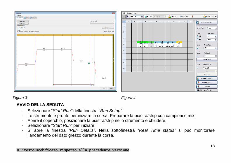
Figura 3 Figura 4
AVVIO DELLA SEDUTA
- Selezionare “Start Run” della finestra “Run Setup”. - Lo strumento è pronto per iniziare la corsa. Preparare la piastra/strip con campioni e mix. - Aprire il coperchio, posizionare la piastra/strip nello strumento e chiudere. - Selezionare “Start Run” per iniziare. - Si apre la finestra “Run Details”. Nella sottofinestra “Real Time status” si può monitorare
l’andamento del dato grezzo durante la corsa.

19 :testo modificato rispetto alla precedente versione
ANALISI DEI DATI
Alla fine della corsa selezionare “STOP” per uscire dalla modalità “10°C forever”. A questo punto si aprirà automaticamente la finestra “Data Analysis”. Per escludere un campione dall’analisi andare in “plate setup-view/edit plate” (in alto a destra nella schermata). Selezionare il pozzetto corrispondente e spuntare la casella “Exclude well in analysis” in basso a destra. Analizzare i dati di FAM e JOE separatamente.
a) FAM (figura 5): deselezionare Joe nella finestra di amplificazione. In alto a sinistra selezionare Settings e impostare:
Cq determination mode: single threshold
Baseline Setting: Baseline subtracted curve fit
Apply Fluorescence Drift Correction
Cycles to analyze: from 1 to 45
Baseline threshold: Single Threshold-User defined-introdurre il valore 25. Cliccare OK Considerare il Ct di FAM dei campioni validi ed interpretare il risultato come segue:
FAM Ct Interpretazione
Campione Presente HCV RNA positivo
Assente HCV RNA non rilevato

20 :testo modificato rispetto alla precedente versione
Figura 5
b) JOE: deselezionare FAM nella finestra di quantificazione. In alto a sinistra selezionare “Settings” e impostare:
Cq determination mode: single threshold
Baseline Setting: Baseline subtracted curve fit
Cycles to analyze: from 1 to 45
Baseline threshold: Single Threshold-User defined-introdurre il valore 100. Cliccare OK.

21 :testo modificato rispetto alla precedente versione
Considerare il Ct di JOE per i campioni ed interpretare il risultato come segue:
JOE Ct Interpretazione
Campione Presente Valido
Assente Non valido*
(*) In assenza del segnale del C.I. nei campioni il risultato non può essere confermato, quindi la seduta deve essere ripetuta
Se nella seduta vengono analizzati anche il controllo negativo ed il controllo positivo, interpretare i risultati come segue:
FAM Ct JOE Ct Interpretazione
Controllo Negativo
Assente Presente Valido
Assente Assente Non valido*
Presente Presente Non valido**
Controllo Positivo
Presente Assente Valido
Assente Assente Non valido*
(*) Ipotesi di inibizione della RT-PCR o degradazione dell’RNA in qualche passaggio della procedura. (**) Possibile contaminazione.

22 :testo modificato rispetto alla precedente versione
ATTENZIONE: per una corretta interpretazione dei risultati, prestare sempre attenzione al grafico della seduta.
Esempio 1 (figura 6a): il campione evidenziato in rosso nel grafico sembra mostrare un Ct positivo (35), ma dal dato grafico si evidenzia un artefatto strumentale. Il valore trovato di Ct non deve essere preso in considerazione ed il campione deve essere ripetuto.
Figura 6a
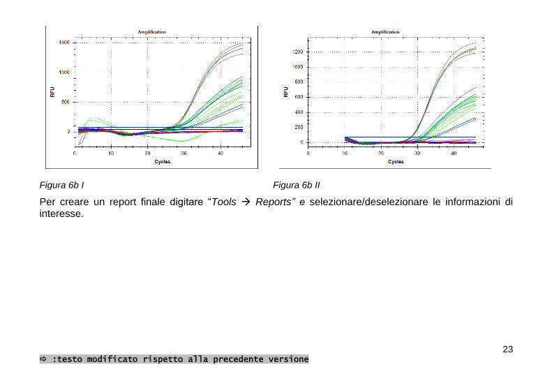
Esempio 2 (figura 6b I e II): se il rumore di fondo della fluorescenza all’inizio della curva risulta troppo disomogeneo, è possibile escludere i primi cicli selezionando “Settings” e impostando “Cycles to analyze: from 10 to 45”
23 :testo modificato rispetto alla precedente versione
Figura 6b I Figura 6b II
Per creare un report finale digitare “Tools Reports” e selezionare/deselezionare le informazioni di interesse.

24 :testo modificato rispetto alla precedente versione
AVVERTENZE
- In caso di assenza del segnale atteso di JOE è consigliabile ripetere la seduta. - Il controllo positivo non ha il C.I., quindi non ci si deve attendere il segnale di JOE. - In caso di contaminazione è importante risalire all’origine (fase di estrazione e/o RT-PCR). L’utilizzo
dei controlli forniti può essere d’aiuto per assicurare le corrette prestazioni del test e per l’identificazione dell’origine di tale problema.
25 :testo modificato rispetto alla precedente versione
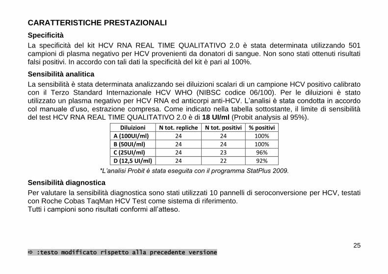
CARATTERISTICHE PRESTAZIONALI
Specificità
La specificità del kit HCV RNA REAL TIME QUALITATIVO 2.0 è stata determinata utilizzando 501 campioni di plasma negativo per HCV provenienti da donatori di sangue. Non sono stati ottenuti risultati falsi positivi. In accordo con tali dati la specificità del kit è pari al 100%.
Sensibilità analitica
La sensibilità è stata determinata analizzando sei diluizioni scalari di un campione HCV positivo calibrato con il Terzo Standard Internazionale HCV WHO (NIBSC codice 06/100). Per le diluizioni è stato utilizzato un plasma negativo per HCV RNA ed anticorpi anti-HCV. L’analisi è stata condotta in accordo col manuale d’uso, estrazione compresa. Come indicato nella tabella sottostante, il limite di sensibilità del test HCV RNA REAL TIME QUALITATIVO 2.0 è di 18 UI/ml (Probit analysis al 95%).
Diluizioni N tot. repliche N tot. positivi % positivi
A (100UI/ml) 24 24 100%
B (50UI/ml) 24 24 100%
C (25UI/ml) 24 23 96%
D (12,5 UI/ml) 24 22 92%
*L’analisi Probit è stata eseguita con il programma StatPlus 2009.
Sensibilità diagnostica
Per valutare la sensibilità diagnostica sono stati utilizzati 10 pannelli di seroconversione per HCV, testati con Roche Cobas TaqMan HCV Test come sistema di riferimento. Tutti i campioni sono risultati conformi all’atteso.

26 :testo modificato rispetto alla precedente versione
Genotipi
Le prestazioni del kit HCV RNA REAL TIME QUALITATIVO 2.0 rispetto ai diversi genotipi di HCV sono state valutate analizzando diversi punti di diluizioni di campioni clinici HCV positivi aventi differente genotipo (come indicato di seguito). La carica virale dei campioni clinici è stata determinata utilizzando come riferimento il Terzo Standard Internazionale HCV WHO (NIBSC codice 06/100). Genotipi testati: 1a, 1b, 2a, 2b, 2a/2c, 3a, 4c/4d, 5 e 6 (determinati col dispositivo NLM GEN-C cod. AC004 e sequenziamento). Punti di diluizione analizzati:
Gen 1a: da 1,5x10² a 2,8x10⁶ UI/ml Gen 1b: da 1,1x10² a 4,3x10⁶ UI/ml Gen 2: da 9,8x101 a 2,7x10⁶ UI/ml Gen 3: da 1,5x10² a 2,8x10⁶ UI/ml Gen 4: da 4,5x10² a 1,8x10⁶ UI/ml Gen 5: 3x10² Gen 6: 7x10²
Come mostrato nella figura sottostante (figura 7) la rivelazione dei diversi genotipi è confrontabile. L’efficienza di rilevazione del dispositivo HCV RNA REAL TIME QUALITATIVO 2.0 è pertanto indipendente dai genotipi analizzati.

27 :testo modificato rispetto alla precedente versione
Figura 7. Il genotipo è stato determinato mediante il dispositivo GEN-C cod. AC004
28 :testo modificato rispetto alla precedente versione
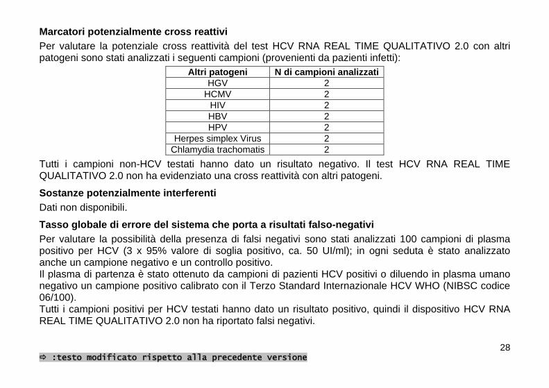
Marcatori potenzialmente cross reattivi
Per valutare la potenziale cross reattività del test HCV RNA REAL TIME QUALITATIVO 2.0 con altri patogeni sono stati analizzati i seguenti campioni (provenienti da pazienti infetti):
Altri patogeni N di campioni analizzati
HGV 2
HCMV 2
HIV 2
HBV 2
HPV 2
Herpes simplex Virus 2
Chlamydia trachomatis 2
Tutti i campioni non-HCV testati hanno dato un risultato negativo. Il test HCV RNA REAL TIME QUALITATIVO 2.0 non ha evidenziato una cross reattività con altri patogeni.
Sostanze potenzialmente interferenti
Dati non disponibili.
Tasso globale di errore del sistema che porta a risultati falso-negativi
Per valutare la possibilità della presenza di falsi negativi sono stati analizzati 100 campioni di plasma positivo per HCV (3 x 95% valore di soglia positivo, ca. 50 UI/ml); in ogni seduta è stato analizzato anche un campione negativo e un controllo positivo. Il plasma di partenza è stato ottenuto da campioni di pazienti HCV positivi o diluendo in plasma umano negativo un campione positivo calibrato con il Terzo Standard Internazionale HCV WHO (NIBSC codice 06/100). Tutti i campioni positivi per HCV testati hanno dato un risultato positivo, quindi il dispositivo HCV RNA REAL TIME QUALITATIVO 2.0 non ha riportato falsi negativi.

29 :testo modificato rispetto alla precedente versione
Confronto con il sistema ABBOTT REAL TIME HCV (Abbott)
Le prestazioni del dispositivo HCV RNA REAL TIME QUALITATIVO 2.0 sono state confrontate con quelle del sistema ABBOTT REAL TIME HCV (Abbott) mediante l’analisi di 102 campioni positivi e negativi per HCV RNA provenienti dalla routine di un laboratorio ospedaliero. I campioni positivi sono risultati tutti positivi, i campioni negativi hanno dato esito negativo. I risultati ottenuti mostrano una buona correlazione tra i due dispositivi (figura 8).
Figura 8
30 :testo modificato rispetto alla precedente versione
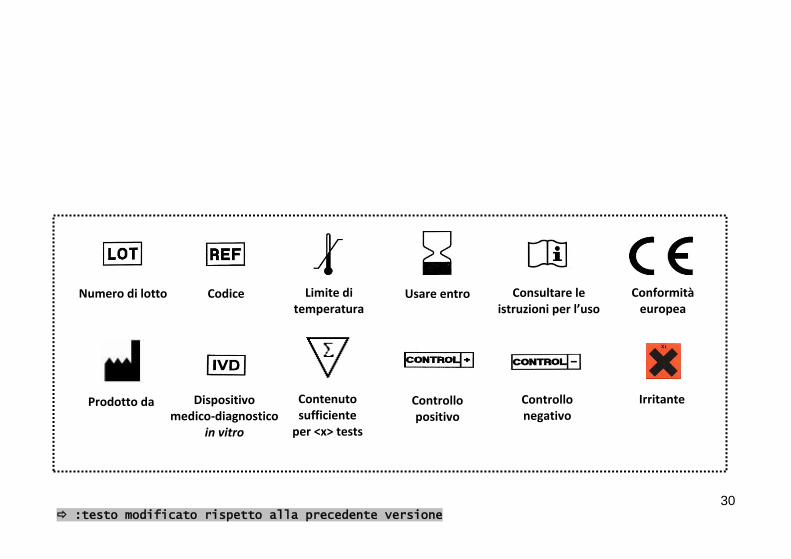
Prodotto da
Numero di lotto Codice Limite di temperatura
Usare entro Consultare le istruzioni per l’uso
Dispositivo medico-diagnostico
in vitro
Contenuto sufficiente
per <x> tests
Irritante
Conformità europea
Controllo positivo
Controllo negativo

AA896/48
0459
MOLECULAR BIOLOGY
EXTRACTION: AA1318 NOT INCLUDED
HCV RNA REAL TIME
QUALITATIVE 2.0
NUCLEAR LASER MEDICINE S.r.l.
HEAD OFFICE: Viale delle Industrie, 3 – 20090 SETTALA MI (Italy)
Tel (+39) 02/95. 24. 51 - Fax (+39) 02/95. 24. 52. 37
WEB: www.nlm.it – E-MAIL: [email protected]
NLM7185 VER 01/09/2014
48 TEST
30742 GMDN
32 : modified text compared with the previous version
INTENDED USE
HCV RNA REAL TIME QUALITATIVE 2.0 TEST provides reagents for the qualitative detection of C-hepatitis virus (HCV) RNA in human plasma samples by Real Time RT-PCR of the 5’-untranslated region (5’-UTR) of the viral RNA; the Core region is co-amplified to make the ampolicons compatible with NLM HCV genotyping kit GEN-C 2.0 code AC004/24. The Internal Control is endogenous (mRNA of the Housekeeping gene GAPDH) and is co-extracted and co-amplified in order to monitor the complete procedure. This device has to be used in association with viral RNA extraction kit NLM code AA1318 “Viral DNA/RNA Extraction”, to be ordered separately. Viral RNA is retrotranscribed and amplified in one step RT-PCR, using CFX (BioRad).
HCV RNA REAL TIME QUALITATIVE 2.0 TEST is intended for use in association with clinical presentation and other laboratory markers (HCV-Ab, ALT, etc) for the diagnosis of HCV infection in patients. The device is not intended for use as a screening test for the presence of HCV RNA in blood donors. The device is for professional use only.
INTRODUCTION
Hepatitis C virus is the main etiologic agent of acute viral hepatitis that can lead, in most patients, to liver cirrhosis and then to hepatocellular carcinoma1. HCV can also cause extrahepatic diseases2-4. HCV genome is a linear single strand RNA with one ORF (Open Reading Frame); in 5’ position there is an untranslated highly conserved region (5’-UTR), followed by the Core region, they both represent the target for viral molecular biology detection. The 5’-UTR and Core genetic heterogeneity among different HCV strains determines the classification into different genotypes5,7,8. HCV has a parenteral transmission through exposure to infected blood. This exposure can occur in the context of blood transfusion before 1992, injection drug use, solid organ transplantation from infected donors, birth from an infected mother, occupational exposure to infected blood, high-risk sexual practice6.

33 : modified text compared with the previous version
After initial exposure, HCV RNA can be detected in patients blood within one to three weeks; acute infection can be severe but rarely is fulminant6. Although generally asymptomatic, about 85% of the acute infections become chronic: persistence of HCV infection is diagnosed by the detection of HCV RNA in the blood for at least six months6. The recent development of directly acting antiviral (DAA)-based regimens and the approval of the first, boceprevir- and telaprevir-based triple-therapy regimen, have brought new hopes for higher virus eradication rate across different disease settings. This calls for even more accurate and sensitive HCV RNA tests able to monitor faster virus kinetics and promptly detect treatment failures.9 With new viral load tests acting as “companion diagnostic”, not only the conventional treatment but also the development and applications of the new generation DAA can be fully supported for a more effective management of chronic hepatitis C and its long-term complications9.
REFERENCES
1. Sarrazin C. Diagnosis of hepatitis C: update 2004. Journal of Gastroenterology and Hepatology (2004) 19, S88-S93
2. Johnson RJ, Gretch DR, Yamabe H et al. Membranoproliferative glomerulonephritis associated with hepatitis C virus infection. N Engl J med 1993; 328: 465-470.
3. Agnello V, Chung RT, Kaplan RM. A role for hepatitis C virus infection in Type II cryoglobulinemia. N Engl J Med 1992; 327: 1490-1495.
4. Andreone P, Zignego AL, Cursaro C et al. Prevalence of monoclonal gammopathies with hepatitis C virus infection . Ann Intern Med 1998; 129: 294-298
5. Zein NN. Clinical Significance of Hepatitis C Virus Genotypes. Clinical Microbiology Reviews, Apr. 2000, p. 223-235.
6. National Institutes of Health, Consensus Conference Statement. Management of hepatitis C: 2002. 7. P.T. Hraber et al. Comparative analysis of hepatitis C virus phylogenies from coding and non-coding regions:
the 5' untranslated region (UTR) fails to classify subtypes. Virology Journal 2006, 3:103 8. M. A. Ansari et al. HCV-Core Region: Its Significance in HCV-Genotyping and Type Dependent Genomic
Expression. Macedonian Journal of Medical Sciences. 2012 Mar 15; 5(1):30-39.

34 : modified text compared with the previous version
9. G. Colucci Molecular diagnostic and predictive tests in the evolution of chronic hepatitis C anti-viral therapies BMC Infect Dis. 2012; 12(Suppl 2): S8. Published online 2012 November 12
PRINCIPLES OF THE PROCEDURE
HCV RNA REAL TIME QUALITATIVE 2.0 TEST is based on two processes: 1. Viral RNA extraction 2. Reverse transcription, amplification and detection of target sequence using Real Time RT-PCR.
The Internal Control (GAPDH mRNA) is endogenous and therefore extracted together with the sample in order to monitor all the procedure.
1. Viral RNA extraction
HCV and IC RNA extraction from human plasma samples has to be performed using “Viral DNA/RNA Extraction” (NLM code AA1318).
2. RT-PCR and Detection
RT-PCR target
The HCV RNA 5’-UTR region was chosen as target to detect the HCV presence in human plasma samples because it is highly conserved among HCV genotypes. Co-amplification of Core region is included in order to discriminate genotypes 1a, 1b and 6 in case of samples that have to be analyzed with NLM AC004/24 Gen-C 2.0 kit. The Internal Control is the mRNA of the constitutively expressed GAPDH gene that is co-extracted and co-amplified together with target HCV; the IC amplicon is similar to HCV target sequence in terms of length and base composition.
Reverse transcription and Amplification
Following extraction step, RNA (HCV and IC) is retrotranscribed and amplified in one-step RT-PCR, using appropriate enzymes and buffer conditions.

35 : modified text compared with the previous version
Detection
Real Time PCR allows target sequences detection during amplification itself, using suitable probes labeled with fluorescent dyes. In the amplification mix there are two specific probes, respectively for the Internal Control and for HCV 5’-UTR region, each labeled with a reporter dye and a quencher dye. The fluorescent reporter dye is different for the two probes: FAM for HCV and JOE for IC. In presence of a light source, when the probes are intact and donor and quencher are close to each other, the fluorescence emitted by the donor fluorophore is absorbed by the proximal quencher. During the amplification each probe matches to its specific sequence and it is target of 5 '-> 3' nuclease activity of the DNA polymerase. When the donor and acceptor fluorophores are separated, the donor fluorescence can be detected at its specific wavelength: in this way, the amplification of the viral RNA and IC can be monitored as the reaction proceeds.
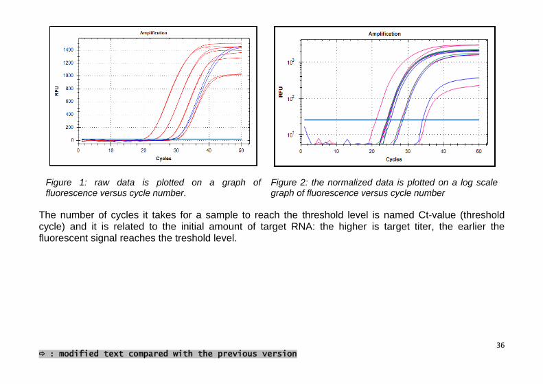
CFX (BioRad) comprises in a single instrument a thermal cycler for target amplification and a fluorometer for the detection of fluorescence during the cycling process. The computer connected with the Real Time machine collects fluorescent data that are displayed in a graph through a specific software (Figure. 1). Following raw data collection, analysis is carried out. Raw data is normalized to correct the background signal, then a threshold level can be set: this is the level at which fluorescence data are analyzed (Figure 2).
36 : modified text compared with the previous version
Figure 1: raw data is plotted on a graph of fluorescence versus cycle number.
Figure 2: the normalized data is plotted on a log scale graph of fluorescence versus cycle number
The number of cycles it takes for a sample to reach the threshold level is named Ct-value (threshold cycle) and it is related to the initial amount of target RNA: the higher is target titer, the earlier the fluorescent signal reaches the treshold level.

37 : modified text compared with the previous version
REAGENTS COMPOSITION (Store at -25/-15°C)
Mix A Probes Primers Tris-EDTA
1 x 47 l
Mix B Primers Tris-EDTA
1 x 110 l
Mix C (R36,37,38) RT-PCR enzymes MgCl2
dNTPs
1 x 330 l
Negative Control Negative Control is human plasma non reactive for HIV-Ab and RNA, HCV-Ab and RNA, HBS-Ag and DNA. Negative Control has to be extracted.
4 x 500 l
Positive Control Synthetic RNA Positive Control is provided in freeze-dried state. Resuspend it in14 µl of RNase, DNase and Protease free water before use in RT-PCR step.
4 provette
38 : modified text compared with the previous version
MODULARITY
Reagents are provided for 4 distinct runs; 12 samples + 1 Negative Control + 1 Positive Control can be analysed in each run. If modularity isn’t respected reagents could not be enough for the number of tests declared.
STABILITY AND STORAGE
- All the reagents are stable up to the expiry date indicated on the label when stored at -25/-15°C. - Thaw all the reagents on ice or at +2/+8°C. - Mixes are stable until expiration date if stored at -25/-15°C. These reagents can be refrozen and
thawed max 4 times. - Mix A contains FAM and JOE fluorophores that are photosensitive: avoid prolonged exposure to
light. - Master Mix has to be used immediately after preparation; once dispensed in PCR tubes, discard the
remaining reagent. Avoid prolonged exposure to light. - The Positive Control is a template RNA, supplied pre-aliquoted and lyophilized: it is recommended
to resuspend the reagent with RNase, DNase and Protease free water before use. Then add the master mix into the tube and transfer the entire volumein the amplification strip/plate; do this operation after the addition of the samples into the respective tubes to avoid contamination.

39 : modified text compared with the previous version
SPECIMEN COLLECTION AND HANDLING
Viral RNA is isolated and purified from human plasma specimens prepared within 6 hours from blood collection. - Collect blood observing universal precautions for venipuncture - Collect blood in sterile tubes with EDTA as anticoagulant and centrifuge at 1000-1500g for 10-15
minutes to separate plasma. Use only EDTA as anticoagulant, other types of anticoagulants may interfere with the performance of the test
- Aliquot 450 l of plasma for RNA extraction using biohazard vertical down-flow airbox and operate to avoid RNA degradation.
- Store samples at +2/+8°C until extraction step. If they are not processed immediately store them at -25/-15°C for up to 72 hours prior to freezing at ≤ -70°C.
- Avoid repeated freeze-thaw cycles of plasma samples - Perform the procedure using universal precautions and handle samples as if capable of transmitting
infection.
PRECAUTIONS
- Only professional and opportunely trained personnel should use this kit. Handle this product according to established good laboratory practices and universal precautions throughout the assay procedure.
- All disposable items (tips and tubes) must be DNase, RNase free. Use aerosol-resistant pipette tips to avoid pipettes contamination. Use a new tip every time a volume is dispensed.
- Discard all used material as biohazardous waste. - The negative control is a human plasma sample non reactive for HIV-Ab and RNA, HCV-Ab and
RNA, HBV HbsAg and DNA. However it should be handled as potentially infectious and should be treated with the necessary safety precautions.
- Mix C: Irritant (R36, 37, 38) for S phrases refer to MSDS.

40 : modified text compared with the previous version
- Do not eat, drink, smoke or apply cosmetics in areas where reagents or specimens are handled. - In case of contact with reagents rinse immediately with water and seek medical advice. - Do not use device after its expiration date. - Do not mix reagents from different lots. - Pay attention to device modularity (12 samples + 1 Negative Control + 1 Positive Control for 4 runs):
if the modularity is not respected the reagents could be insufficient for all the tests. - Due to the high sensitivity of the test, particular attention should be paid to cross-contaminations. It is
recommended to perform the assay in three different areas: - Area 1: pre-PCR (samples handling and extraction) - Area 2: Master Mix preparation. - Area 3: post-PCR (Real Time PCR)
It’s recommended to clean the workstation with chlorine bleach at the end of the procedure. - Do not use the device if the box is damaged; contact the supplier - It is advisable to have constant and uniform laboratory temperature, avoid to place the
instruments near heating/cooling sources that may compromise the right working.

41 : modified text compared with the previous version
MATERIAL REQUIRED BUT NOT PROVIDED
AREA 1
Vertical downflow airbox Dedicated adjustable volume pipettes set and aerosol barrier tips 1,5 and 2 ml DNase, RNase free tubes Magnetic separator Vortex mixer and Heat block Centrifuge RNase, DNase and Protease free water
AREA 2
Vertical downflow airbox Dedicated adjustable volume pipettes set and aerosol barrier tips DNase, RNase free 0,2 ml flat cap Real Time PCR, strips or plates DNase, RNase free DNase, RNase and Protease free water
AREA 3
CFX (Biorad) CFX (Biorad) software

42 : modified text compared with the previous version
ASSAY PROCEDURE
VIRAL RNA EXTRACTION
(Extraction method “Viral DA/RNA Extraction”, NLM code AA1318) Refer to the instructions for use for preparation, handling and disposal of reagents, taking into account 450 µl starting sample quantity. This protocol is for manual use and involves the use of a 2 ml reaction tubes and a suitable magnetic separator. For the automation of the procedure with the Janus workstation (Perkin Elmer) contact the company.
Prepare the necessary amount of 2 ml centrifuge tubes (samples and negative control). Before starting with the procedure set heat block to +55°C. Ensure that Proteinase K and Poly(A) RNA have been prepared as described.
1. Dispense in the following order 10 µl Proteinase K and 450 µl of sample into 2,0 ml centrifuge tubes. Add 450 µl Lysis Buffer Viral RNA and then 6 µl Poly(A) RNA; proceed pipetting up and down for at least 8 times and incubate at 55°C for 10 minutes, vortexing ones or twice during incubation. It is also possible to prepare a premix of Lysis Buffer Viral RNA and Poly(A) RNA, calculating the necessary amount for the number of samples + 1 to process. Use this mix within 15 minutes.
2. Following incubation, briefly centrifuge the samples to avoid contamination when opening the tubes. Add 35 µl of resuspended Magnetic Beads Viral DNA/RNA (the Magnetic Bead suspension should be mixed vigorously before dispensing) and 900 µl of Binding Buffer Viral DNA/RNA to each tube, mix by pipetting up and down for at least 6 times and incubate for 5 minutes at RT. It is possible to prepare a premix of beads and Binding Buffer Viral DNA/RNA.
3. Place the tubes in a magnetic separator for at least 2 minutes to let the beads be attracted by the magnets. Remove and discard supernatant by pipetting, being careful not to take any beads.

43 : modified text compared with the previous version
4. Remove the tubes from the magnetic separator. Add 500 μl Wash Buffer A Viral DNA/RNA to the tube. Resuspend the beads by pipetting until the beads are completely resuspended and incubate for 1 minute at RT.
5. Place the tube in the magnetic separator to draw the beads to the side of the tube (about 1 minute). Pipette off the supernatant and then remove the tube from the magnet.
6. Repeat the washing procedure (steps 4 and 5) with 500 μl Wash Buffer B Viral DNA/RNA. Discard Wash Buffer B Viral DNA/RNA repeating the aspiration with a fine-tipped pipette; be careful to remove all the residual fluid without disturbing the pellet.
7. Transfer the tubes to the thermo block and let them air-dry at 55°C for 20 minutes to remove any traces of ethanol. The beads-pellet should become light brown when dried, otherwise extend the incubation for further 5 minutes or until completely dry It is important that the beads are completely dried and that there aren’t ethanol drops on the tube walls before continuing with the elution step, as residual ethanol can prevent amplification.
8. Add 70 μl of Elution Buffer Viral DNA/RNA to the tubes and briefly resuspend the beads by pipetting. Close the tubes, gently vortex the tubes and incubate the suspension for 5 minutes at 55°C.
9. Separate the magnetic beads by placing the tubes in the magnetic separator for at least. 1 minute. Transfer the supernatant containing the purified viral RNA to the desired storage tubes.
Purified RNA can be stored at +2/+8°C if immediately used, otherwise keep it frozen at -25/-15°C or at ≤ 70°C for longer periods. It is recommended to thaw RNA at +2/+8°C.

44 : modified text compared with the previous version
REAL TIME RT-PCR
- Set Real Time PCR profile before preparing Master Mix.
Reagents Volume per sample
Mix A 0,6 µl
Mix B 0,4 µl
Mix C 5 µl
- Carefully resuspend the mix before use. - Prepare Master Mix for the number of purified samples (clinical specimens and negative control) +
positive control +2 volumes (to have enough Master Mix for all the samples). - Mix gently and dispense 6 µl of Master Mix in the plate/strip previously marked. - Add 14 µl of purified RNA to each well/tube and mix pipetting up and down. - At last (to avoid cross contaminations), resuspend the positive control with 14 µl RNase, DNase and
Protease free water, then add the mix and transfer all the volume in the specific well/tube . WARNING: dispense mix and samples very carefully, in order to avoid creating air bubbles. If possible, centrifuge the plate/strip before placing it into the Real Time PCR instrument.
45 : modified text compared with the previous version
RUN SETUP
Turn on the PC and CFX. Open the “Bio-Rad CFX Manager Software” and click on “Create new run”, select the model of instrument used and click “OK”; the “Run Setup” window will open. It is possible to create a new protocol, select/modify an existing one or repeat a run. To repeat a run with the same settings, open the desired run and click on “File - Repeat Run”.
Create a new protocol
- “Protocol create New” (figure 3) - “Protocol Editor – new” - Enter the reaction volume (20 µl) in the “Sample Volume” box - Add the steps by clicking on “Insert Step” on the left of the window (it is possible to modify
temperatures and times directly on the graph or in the test below with a double click): 1. 50°C for 15 min 2. 95° for 20 sec 3. 95°C for 15 sec 4. 60°C for 1 min; add “plate read” in this step 5. Insert GOTO 3, 44 more times 6. Optional: for downstream application with GEN-C 2.0 (cod. NLM AC004/24), add 10°C
forever (type 00:00 and press Enter) - Click OK. Save the new protocol.
46 : modified text compared with the previous version
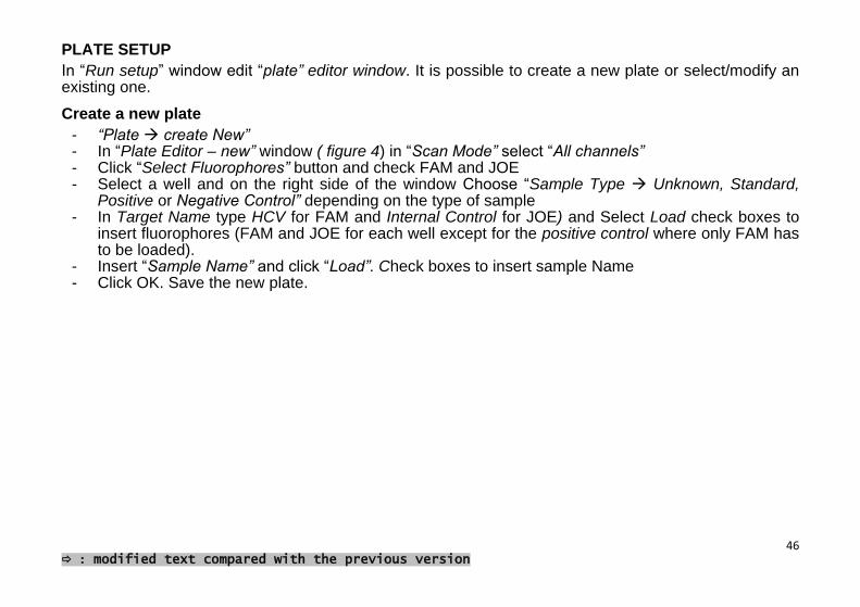
PLATE SETUP
In “Run setup” window edit “plate” editor window. It is possible to create a new plate or select/modify an existing one.
Create a new plate
- “Plate create New” - In “Plate Editor – new” window ( figure 4) in “Scan Mode” select “All channels” - Click “Select Fluorophores” button and check FAM and JOE - Select a well and on the right side of the window Choose “Sample Type Unknown, Standard,
Positive or Negative Control” depending on the type of sample - In Target Name type HCV for FAM and Internal Control for JOE) and Select Load check boxes to
insert fluorophores (FAM and JOE for each well except for the positive control where only FAM has to be loaded).
- Insert “Sample Name” and click “Load”. Check boxes to insert sample Name - Click OK. Save the new plate.

47 : modified text compared with the previous version
Figure 3 Figure 4:
Select/modify existing protocol or plate
- To import an existing protocol or plate click “Select Existing” in protocol or plate window. - To modify an existing plate click “Select Existing” “Edit Select”. - Proceed as explained above.

48 : modified text compared with the previous version
START RUN
Prepare the plate/strip with samples and mix.
In “Start Run” tab click “Open Lid”, place the plate/strip in CFX instrument and click “Close Lid”
Press “Start Run” to begin.
It’s possible to monitor the Raw data plots during the assay in “Run Details” window, in “Real Time status” page.
DATA ANALYSIS
At the end of the assay click “Stop” to exit from 10°C forever mode. The window “Data Analysis” will open automatically. To exclude a sample from the analysis go to “plate setup-view/edit plate” (in the upper right), click on the well to omit and then on “Exclude well in analysis” (in the lower right). Analyze data for FAM and JOE separately.
a) FAM (figure 5): deselect Joe in the amplification window.
Click on “Settings” and select:
Cq determination mode: single threshold
Baseline Setting: Baseline subtracted curve fit
Apply Fluorescence Drift Correction
Cycles to analyze: from 1 to 45
Baseline threshold: Single Threshold-User defined-introduce the value 25. Click OK.

49 : modified text compared with the previous version
Check samples FAM Ct and interpret results as follows:
. FAM Ct Interpretation
Sample Present positive HCV RNA
Absent HCV RNA not detected
Figure 5

50 : modified text compared with the previous version
b) JOE: deselect FAM in the amplification window. Click on “Settings” and select:
Cq determination mode: single threshold
Baseline Setting: Baseline subtracted curve fit
Cycles to analyze: from 1 to 45
Baseline threshold: Single Threshold-User defined-introduce the value 100. Click OK
Check samples JOE Ct and interpret results as follows:
JOE Ct Interpretation
Sample Present Valid
Absent Not Valid*
(*) In absence of IC signal sample result can’t be confirmed, therefore sample run should be repeated.
If negative and positive controls have been tested, interpret results as follows:
FAM Ct JOE Ct Interpretation
Negative Control
Absent Present Valid
Absent Absent Not Valid*
Present Present Not Valid**
Positive Control
Present Absent Valid
Absent Absent Not Valid*
(*) Hypothesis of RT-PCR inhibition or RNA degradation in one of the procedure steps (**) Possible contamination

51 : modified text compared with the previous version
WARNING: for a correct interpretation of results, always pay attention to the graph of the session.
Example 1 (figure 6a): the sample in red in the graph shows a positive Ct value (35), but the graph itself shows it is an instrumental artifact. The Ct value should not be taken into account and the sample has to be repeated.
Figure 6a

52 : modified text compared with the previous version
Example 2 (figure 6b I e II): If fluorescence signals at the beginning of the run are not homogeneous, it is possible to exclude the first cycles; select from “Settings” - Cycles to analyze: from 10 to 45
Figure 6b I Figure 6b II
Create a Report (“Tools Reports”). Select/Deselect useful information.
WARNING
- In case of absence of expected JOE signal it is advisable to repeat the assay. - CP has not an Internal Control, therefore JOE signal is not expected. - When contamination events occur it’s important to find out the source (extraction and/or RT-PCR
steps). Controls provided can be very useful to ensure the correct test performance and to find out contamination origin.

53 : modified text compared with the previous version
PERFORMANCE CHARACTERISTICS
Specificity
The specificity of HCV RNA REAL TIME QUALITATIVE 2.0 TEST was determined by analysing 501 HCV negative plasma samples from blood donors. No false positive results were obtained. According to the results the specificity of the assay is 100%.
Analytical sensitivity
Sensitivity was determined by analysing dilution levels of an HCV positive clinical specimen calibrated with the HCV WHO 3rd International Standard (NIBSC code 06/100). For the dilutions a plasma negative for HCV RNA and antibodies was used. The analysis was performed according to instructions for use, RNA extraction included. As shown in table below, the detection limit of HCV RNA REAL TIME QUALITATIVE 2.0 TEST is 18 IU/ml (Probit analysis at 95%).
Diluitions N tot. runs N tot. positives % positives
A (100 IU/ml) 24 24 100%
B (50 IU/ml) 24 24 100%
C (25 IU/ml) 24 23 96%
D (12,5 IU/ml) 24 22 92%
*Probit Analysis done with Statplus 2009 software.
Diagnostic sensitivity
Diagnostic sensitivity was evaluated by analising 10 HCV seroconversion panels, tested with Roche Cobas TaqMan HCV Test as reference system. All the samples gave the expected result.

54 : modified text compared with the previous version
HCV Genotypes
The performance of HCV RNA REAL TIME QUANTITATIVE 2.0 TEST on different HCV genotypes was determined by analysing four dilution levels of different HCV positive clinical specimens genotypes (as listed below). The viral load of the samples used was determined by comparing the signal of the original samples with the signal of the HCV WHO 3rd International Standard (NIBSC code 06/100).
Genotypes tested: 1a, 1b, 2a, 2b, 2a/2c, 3a, 4c/4d, 5 and 6 (determined with NLM GEN C cod AC004 and sequencing).
Dilutions analyzed: Gen 1a: from 1,5x10² to 2,8x10⁶ IU/ml Gen 1b: from 1,1x10² to 4,3x10⁶ IU/ml Gen 2: from 9,8x101 to 2,7x10⁶ IU/ml Gen 3: from 1,5x10² to 2,8x10⁶ IU/ml Gen 4: from 4,5x10² to 1,8x10⁶ IU/ml Gen 5: 3x10² Gen 6: 7x10²
As showed in the graphic below (figure 7) detection of HCV RNA is comparable for the different genotypes analysed. Every tested samples gave a positive result, as expected, therefore detection efficiency of HCV RNA REAL TIME QUALITATIVE 2.0 TEST is independent from the genotype.

55 : modified text compared with the previous version
Figure 7 Samples genotype was determined using NLM GEN-C cod. AC004
56 : modified text compared with the previous version
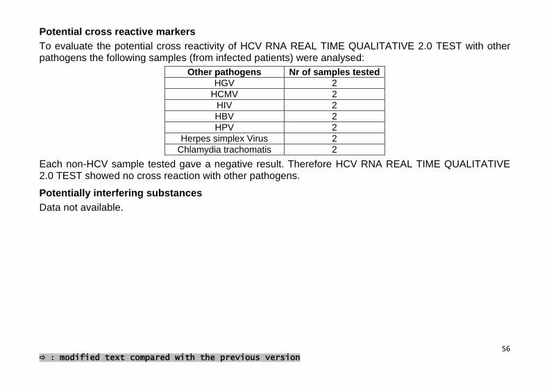
Potential cross reactive markers
To evaluate the potential cross reactivity of HCV RNA REAL TIME QUALITATIVE 2.0 TEST with other pathogens the following samples (from infected patients) were analysed:
Other pathogens Nr of samples tested
HGV 2
HCMV 2
HIV 2
HBV 2
HPV 2
Herpes simplex Virus 2
Chlamydia trachomatis 2
Each non-HCV sample tested gave a negative result. Therefore HCV RNA REAL TIME QUALITATIVE 2.0 TEST showed no cross reaction with other pathogens.
Potentially interfering substances
Data not available.

57 : modified text compared with the previous version
Whole system failure rate leading to false-negative results
To evaluate the whole system failure rate leading to false negative results 100 HCV positive plasma samples were analysed (3 x 95% positive cut-off concentration ~ 50 IU/ml); in each run one negative control was added. The starting plasma was obtained diluting in human negative plasma a positive clinical specimen calibrated with the HCV WHO 3rd International Standard (NIBSC code 06/100). All the HCV positive samples tested gave a positive result, therefore HCV RNA REAL TIME QUALITATIVE 2.0 TEST doesn’t lead to false-negative results.

58 : modified text compared with the previous version
Comparsion with ABBOTT REAL TIME HCV (Abbott)
The performance of the REAL TIME HCV QUALITATIVE TEST was compared to ABBOTT REAL TIME HCV (Abbott) by analysing 102 HCV positive and negative clinical samples. All the positive samples gave a positive result and the four negative were found negative. Results obtained show a good correlation between these two assays (figure 8).
Figure 8

59 : modified text compared with the previous version

European Conformity
Lot Number Catalogue number
Temperature Limitation
Use by Consult Instructions for use
In vitro diagnostic medical device
Sufficient for Irritant Manufacturer Positive control
Negative control