
Labeling Deoxyadenosine for the Preparation of Functional Conjugated Oligonucleotides
10
Labeling Deoxyadenosine for the Preparation of Functional Conjugated Oligonucleotides Massimo L. Capobianco,* ,§ Elena Marchesi, †,‡ Daniela Perrone, ‡ and Maria Luisa Navacchia § § Istituto per la Sintesi Organica e la Fotoreattivita ̀ del Consiglio Nazionale delle Ricerche (ISOF-CNR), via P. Gobetti 101, 40129 Bologna, Italy † Dipartimento di Scienze Mediche dell'Universita ̀ di Ferrara, corso Giudecca 203, 44121 Ferrara, Italy ‡ Dipartimento di Scienze Chimiche e Farmaceutiche dell'Universita ̀ di Ferrara, via L. Borsari 46, 44121 Ferrara, Italy * S Supporting Information ABSTRACT: Herein we present a versatile synthetic method for the 8-thioalkylation of (deoxy)adenosine with a short carbon linker having on the other side a variety of molecules (psoralen, acridine) and functional groups (alkyne). After conventional protections, the modified adenosine can be phosphytylated and inserted into an oligonucleotide without affecting the standard protocols for supported oligonucleotide synthesis. The hybridization properties of a generic oligonucleotide containing the above conjugated moieties toward both DNA and RNA are evaluated both in the case of a perfectly complementary strand and in the case of a single mismatch. This methodology is suitable for the preparation of several types of derivatives andthrough the alkynyl moietyprovides fast access to click-chemistry transformations. ■ INTRODUCTION In the past thirty years, oligonucleotides have been chemically bound (i.e., conjugated) to a variety of: fluorophores, 1,2 drugs, 3−5 or other active molecules such as alkylating 6 or cleaving groups 7,8 for biochemical and structural studies; they have been bound to a variety of materials, such as metals, 9 glass, 10 polymers, 11 and peptides, for the realization of biochemical probes and biosensors. Oligonucleotides have also been actively employed as down-regulators of genetic expression in cellular studies, where they have been conjugated to a plethora of derivatives to enhance the cellular uptake 12−16 or increase the binding affinity toward their RNA target 17−19 (antisense methodology) or to form more efficient triple helices on the double-stranded DNA 20,21 (antigene approach). More recently, oligonucleotides have also been used as molecular scaffolds to exploit their autoassembly properties for the realization of supramolecular aggregates with potential applications in the field of electronics devices. 22−26 For each goal it is important to be sure that the chosen methodology does not hamper the hybridization properties of the native oligonucleotide on which are based the forecasted applications. Since mid 1980′s almost all the synthetic oligonucleotides are prepared through the automated supported synthesis; hence, any functionalization must be introduced during this procedure, or performed as postsynthetic treatment on a suitable modified oligonucleotide carrying a reactive group, introduced during the automated synthesis. The most common derivatizations of the oligonucleotides are done at their 5′ or 3′ ends or both, as in the case of the so- called ″molecular beacons″; 27,28 however, for some purposes it is important to be able to link a tether in the middle of a sequence, by modifying a base, a sugar, a phosphate, or an entire internucleotidic region. 29 Derivatization of nucleobases enables the incorporation of one or more suitable function- alities to the desired position of the oligonucleotide, 2 leaving the 5′ and 3′ ends available for further modifications. The labeling of the base has been used for the preparation of several derivatives ranging from artificial endonucleases 7,30 to the control of the correct base pairing. 31−33 The functionalization of the nucleobase is usually performed linking the chosen moiety to the C-6 position of pyrimidines, and on the C-8 position of purine bases. 34−38 Received: May 16, 2013 Revised: July 15, 2013 Published: July 25, 2013 Article pubs.acs.org/bc © 2013 American Chemical Society 1398 dx.doi.org/10.1021/bc400243q | Bioconjugate Chem. 2013, 24, 1398−1407
-
Upload
maria-luisa -
Category
Documents
-
view
213 -
download
0
Transcript of Labeling Deoxyadenosine for the Preparation of Functional Conjugated Oligonucleotides

Labeling Deoxyadenosine for the Preparation of FunctionalConjugated OligonucleotidesMassimo L. Capobianco,*,§ Elena Marchesi,†,‡ Daniela Perrone,‡ and Maria Luisa Navacchia§
§Istituto per la Sintesi Organica e la Fotoreattivita ̀ del Consiglio Nazionale delle Ricerche (ISOF-CNR), via P. Gobetti 101, 40129Bologna, Italy†Dipartimento di Scienze Mediche dell'Universita ̀ di Ferrara, corso Giudecca 203, 44121 Ferrara, Italy‡Dipartimento di Scienze Chimiche e Farmaceutiche dell'Universita ̀ di Ferrara, via L. Borsari 46, 44121 Ferrara, Italy
*S Supporting Information
ABSTRACT: Herein we present a versatile synthetic method for the 8-thioalkylation of (deoxy)adenosine with a short carbonlinker having on the other side a variety of molecules (psoralen, acridine) and functional groups (alkyne). After conventionalprotections, the modified adenosine can be phosphytylated and inserted into an oligonucleotide without affecting the standardprotocols for supported oligonucleotide synthesis. The hybridization properties of a generic oligonucleotide containing the aboveconjugated moieties toward both DNA and RNA are evaluated both in the case of a perfectly complementary strand and in thecase of a single mismatch. This methodology is suitable for the preparation of several types of derivatives andthrough thealkynyl moietyprovides fast access to click-chemistry transformations.
■ INTRODUCTION
In the past thirty years, oligonucleotides have been chemicallybound (i.e., conjugated) to a variety of: fluorophores,1,2
drugs,3−5 or other active molecules such as alkylating6 orcleaving groups7,8 for biochemical and structural studies; theyhave been bound to a variety of materials, such as metals,9
glass,10 polymers,11 and peptides, for the realization ofbiochemical probes and biosensors. Oligonucleotides havealso been actively employed as down-regulators of geneticexpression in cellular studies, where they have been conjugatedto a plethora of derivatives to enhance the cellular uptake12−16
or increase the binding affinity toward their RNA target17−19
(antisense methodology) or to form more efficient triple heliceson the double-stranded DNA20,21 (antigene approach). Morerecently, oligonucleotides have also been used as molecularscaffolds to exploit their autoassembly properties for therealization of supramolecular aggregates with potentialapplications in the field of electronics devices.22−26 For eachgoal it is important to be sure that the chosen methodologydoes not hamper the hybridization properties of the nativeoligonucleotide on which are based the forecasted applications.Since mid 1980′s almost all the synthetic oligonucleotides are
prepared through the automated supported synthesis; hence,
any functionalization must be introduced during this procedure,or performed as postsynthetic treatment on a suitable modifiedoligonucleotide carrying a reactive group, introduced during theautomated synthesis.The most common derivatizations of the oligonucleotides are
done at their 5′ or 3′ ends or both, as in the case of the so-called ″molecular beacons″;27,28 however, for some purposes itis important to be able to link a tether in the middle of asequence, by modifying a base, a sugar, a phosphate, or anentire internucleotidic region.29 Derivatization of nucleobasesenables the incorporation of one or more suitable function-alities to the desired position of the oligonucleotide,2 leavingthe 5′ and 3′ ends available for further modifications. Thelabeling of the base has been used for the preparation of severalderivatives ranging from artificial endonucleases7,30 to thecontrol of the correct base pairing.31−33 The functionalizationof the nucleobase is usually performed linking the chosenmoiety to the C-6 position of pyrimidines, and on the C-8position of purine bases.34−38
Received: May 16, 2013Revised: July 15, 2013Published: July 25, 2013
Article
pubs.acs.org/bc
© 2013 American Chemical Society 1398 dx.doi.org/10.1021/bc400243q | Bioconjugate Chem. 2013, 24, 1398−1407
Here, we report a study on the preparation of thioderivativesbound to the C-8 position of the deoxyadenosine containingdifferent functional molecules, and we describe the synthesisand the hybridization properties of oligonucleotides containingthese modifications in the middle of a generic sequence.
■ RESULTSSynthesis of Psoralen-Deoxyadenosine Conjugate
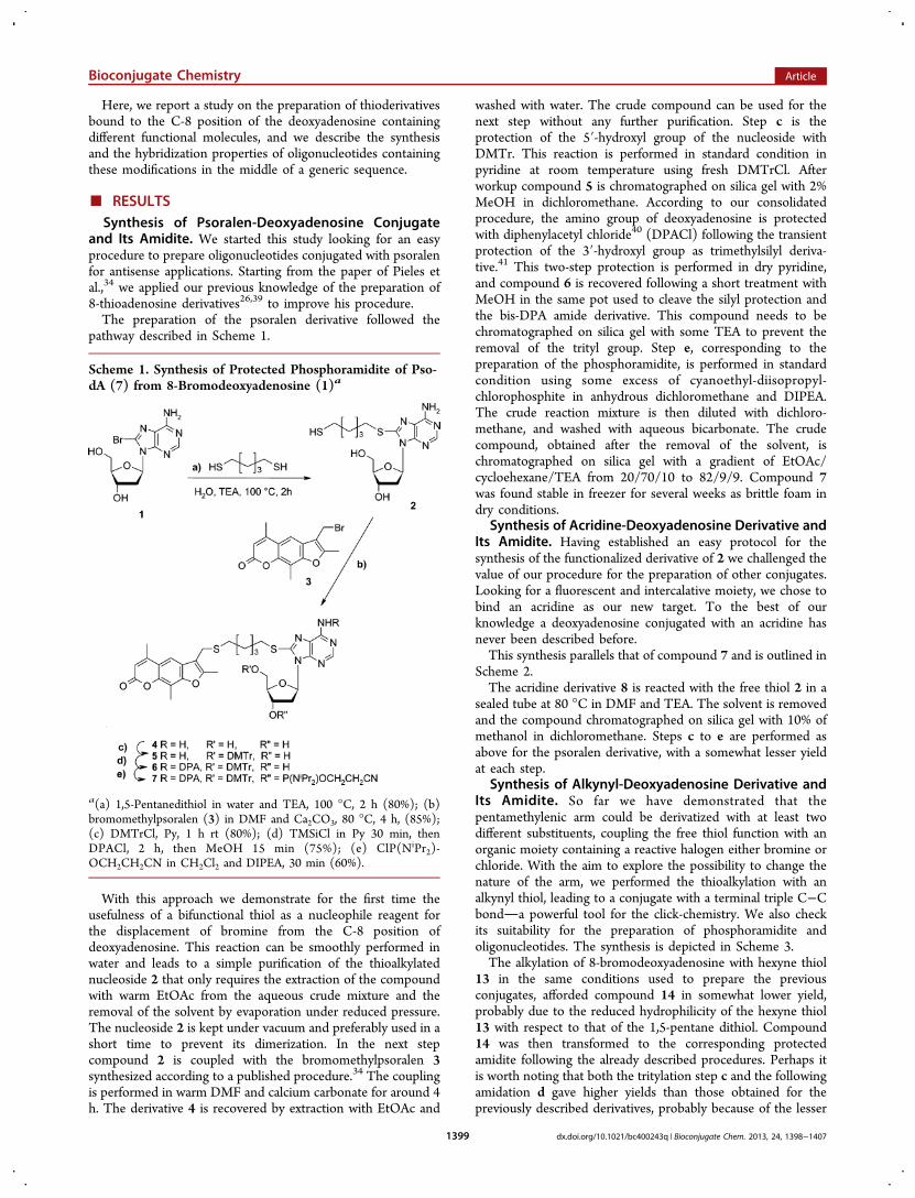
and Its Amidite. We started this study looking for an easyprocedure to prepare oligonucleotides conjugated with psoralenfor antisense applications. Starting from the paper of Pieles etal.,34 we applied our previous knowledge of the preparation of8-thioadenosine derivatives26,39 to improve his procedure.The preparation of the psoralen derivative followed the
pathway described in Scheme 1.
With this approach we demonstrate for the first time theusefulness of a bifunctional thiol as a nucleophile reagent forthe displacement of bromine from the C-8 position ofdeoxyadenosine. This reaction can be smoothly performed inwater and leads to a simple purification of the thioalkylatednucleoside 2 that only requires the extraction of the compoundwith warm EtOAc from the aqueous crude mixture and theremoval of the solvent by evaporation under reduced pressure.The nucleoside 2 is kept under vacuum and preferably used in ashort time to prevent its dimerization. In the next stepcompound 2 is coupled with the bromomethylpsoralen 3synthesized according to a published procedure.34 The couplingis performed in warm DMF and calcium carbonate for around 4h. The derivative 4 is recovered by extraction with EtOAc and
washed with water. The crude compound can be used for thenext step without any further purification. Step c is theprotection of the 5′-hydroxyl group of the nucleoside withDMTr. This reaction is performed in standard condition inpyridine at room temperature using fresh DMTrCl. Afterworkup compound 5 is chromatographed on silica gel with 2%MeOH in dichloromethane. According to our consolidatedprocedure, the amino group of deoxyadenosine is protectedwith diphenylacetyl chloride40 (DPACl) following the transientprotection of the 3′-hydroxyl group as trimethylsilyl deriva-tive.41 This two-step protection is performed in dry pyridine,and compound 6 is recovered following a short treatment withMeOH in the same pot used to cleave the silyl protection andthe bis-DPA amide derivative. This compound needs to bechromatographed on silica gel with some TEA to prevent theremoval of the trityl group. Step e, corresponding to thepreparation of the phosphoramidite, is performed in standardcondition using some excess of cyanoethyl-diisopropyl-chlorophosphite in anhydrous dichloromethane and DIPEA.The crude reaction mixture is then diluted with dichloro-methane, and washed with aqueous bicarbonate. The crudecompound, obtained after the removal of the solvent, ischromatographed on silica gel with a gradient of EtOAc/cycloehexane/TEA from 20/70/10 to 82/9/9. Compound 7was found stable in freezer for several weeks as brittle foam indry conditions.
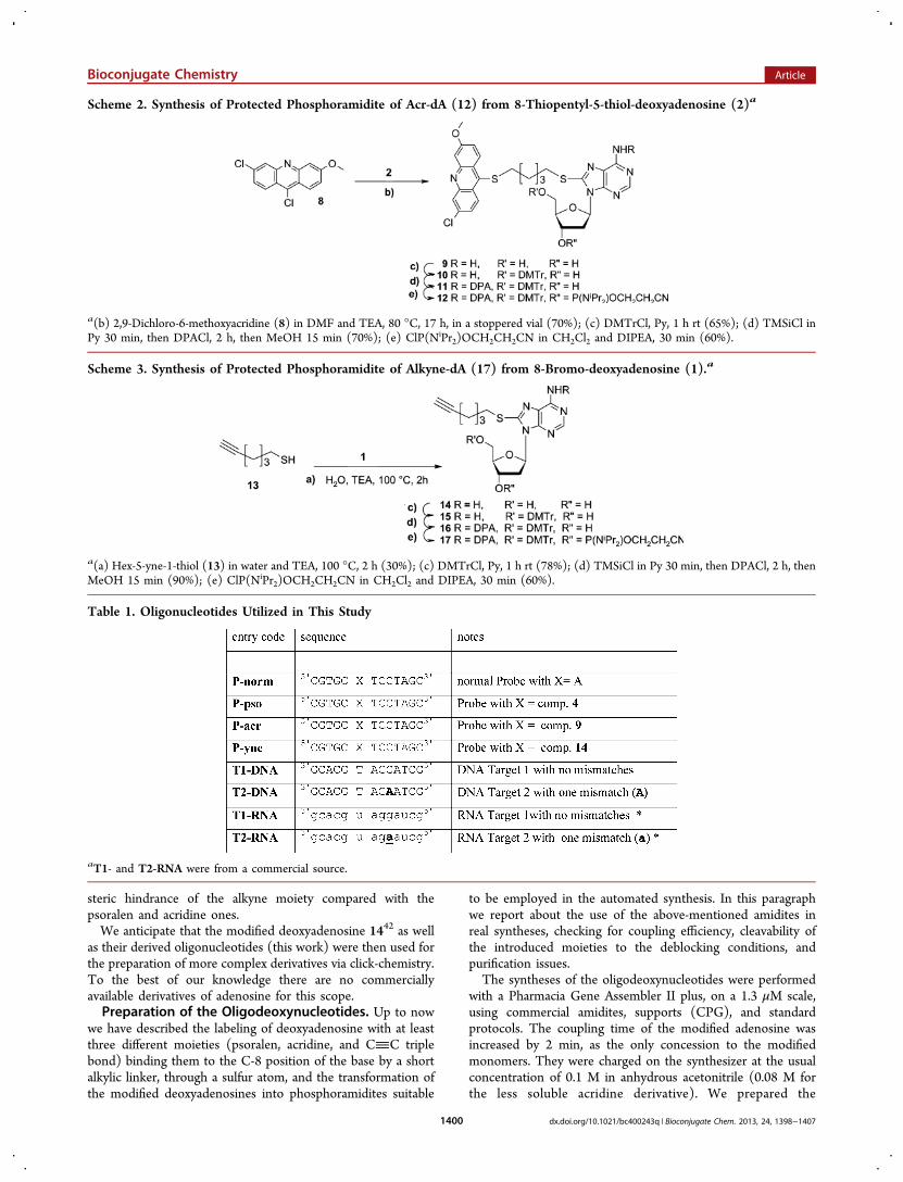
Synthesis of Acridine-Deoxyadenosine Derivative andIts Amidite. Having established an easy protocol for thesynthesis of the functionalized derivative of 2 we challenged thevalue of our procedure for the preparation of other conjugates.Looking for a fluorescent and intercalative moiety, we chose tobind an acridine as our new target. To the best of ourknowledge a deoxyadenosine conjugated with an acridine hasnever been described before.This synthesis parallels that of compound 7 and is outlined in
Scheme 2.The acridine derivative 8 is reacted with the free thiol 2 in a
sealed tube at 80 °C in DMF and TEA. The solvent is removedand the compound chromatographed on silica gel with 10% ofmethanol in dichloromethane. Steps c to e are performed asabove for the psoralen derivative, with a somewhat lesser yieldat each step.
Synthesis of Alkynyl-Deoxyadenosine Derivative andIts Amidite. So far we have demonstrated that thepentamethylenic arm could be derivatized with at least twodifferent substituents, coupling the free thiol function with anorganic moiety containing a reactive halogen either bromine orchloride. With the aim to explore the possibility to change thenature of the arm, we performed the thioalkylation with analkynyl thiol, leading to a conjugate with a terminal triple C−Cbonda powerful tool for the click-chemistry. We also checkits suitability for the preparation of phosphoramidite andoligonucleotides. The synthesis is depicted in Scheme 3.The alkylation of 8-bromodeoxyadenosine with hexyne thiol
13 in the same conditions used to prepare the previousconjugates, afforded compound 14 in somewhat lower yield,probably due to the reduced hydrophilicity of the hexyne thiol13 with respect to that of the 1,5-pentane dithiol. Compound14 was then transformed to the corresponding protectedamidite following the already described procedures. Perhaps itis worth noting that both the tritylation step c and the followingamidation d gave higher yields than those obtained for thepreviously described derivatives, probably because of the lesser
Scheme 1. Synthesis of Protected Phosphoramidite of Pso-dA (7) from 8-Bromodeoxyadenosine (1)a
a(a) 1,5-Pentanedithiol in water and TEA, 100 °C, 2 h (80%); (b)bromomethylpsoralen (3) in DMF and Ca2CO3, 80 °C, 4 h, (85%);(c) DMTrCl, Py, 1 h rt (80%); (d) TMSiCl in Py 30 min, thenDPACl, 2 h, then MeOH 15 min (75%); (e) ClP(NiPr2)-OCH2CH2CN in CH2Cl2 and DIPEA, 30 min (60%).
Bioconjugate Chemistry Article
dx.doi.org/10.1021/bc400243q | Bioconjugate Chem. 2013, 24, 1398−14071399
steric hindrance of the alkyne moiety compared with thepsoralen and acridine ones.We anticipate that the modified deoxyadenosine 1442 as well
as their derived oligonucleotides (this work) were then used forthe preparation of more complex derivatives via click-chemistry.To the best of our knowledge there are no commerciallyavailable derivatives of adenosine for this scope.Preparation of the Oligodeoxynucleotides. Up to now
we have described the labeling of deoxyadenosine with at leastthree different moieties (psoralen, acridine, and CC triplebond) binding them to the C-8 position of the base by a shortalkylic linker, through a sulfur atom, and the transformation ofthe modified deoxyadenosines into phosphoramidites suitable
to be employed in the automated synthesis. In this paragraphwe report about the use of the above-mentioned amidites inreal syntheses, checking for coupling efficiency, cleavability ofthe introduced moieties to the deblocking conditions, andpurification issues.The syntheses of the oligodeoxynucleotides were performed
with a Pharmacia Gene Assembler II plus, on a 1.3 μM scale,using commercial amidites, supports (CPG), and standardprotocols. The coupling time of the modified adenosine wasincreased by 2 min, as the only concession to the modifiedmonomers. They were charged on the synthesizer at the usualconcentration of 0.1 M in anhydrous acetonitrile (0.08 M forthe less soluble acridine derivative). We prepared the
Scheme 2. Synthesis of Protected Phosphoramidite of Acr-dA (12) from 8-Thiopentyl-5-thiol-deoxyadenosine (2)a
a(b) 2,9-Dichloro-6-methoxyacridine (8) in DMF and TEA, 80 °C, 17 h, in a stoppered vial (70%); (c) DMTrCl, Py, 1 h rt (65%); (d) TMSiCl inPy 30 min, then DPACl, 2 h, then MeOH 15 min (70%); (e) ClP(NiPr2)OCH2CH2CN in CH2Cl2 and DIPEA, 30 min (60%).
Scheme 3. Synthesis of Protected Phosphoramidite of Alkyne-dA (17) from 8-Bromo-deoxyadenosine (1).a
a(a) Hex-5-yne-1-thiol (13) in water and TEA, 100 °C, 2 h (30%); (c) DMTrCl, Py, 1 h rt (78%); (d) TMSiCl in Py 30 min, then DPACl, 2 h, thenMeOH 15 min (90%); (e) ClP(NiPr2)OCH2CH2CN in CH2Cl2 and DIPEA, 30 min (60%).
Table 1. Oligonucleotides Utilized in This Study
aT1- and T2-RNA were from a commercial source.
Bioconjugate Chemistry Article
dx.doi.org/10.1021/bc400243q | Bioconjugate Chem. 2013, 24, 1398−14071400
oligodeoxynucleotides listed in Table 1. The length of theprobe was chosen to maximize the effects of modifications onthe duplex stability, expecting at the same time a good degree ofduplex formation at room temperature, to facilitate HPLCanalysis, and to allow the formation of a full helix turn, in orderto extend our finding to real application cases. The arbitrarysequence was chosen to avoid secondary structures, with themodified adenosine (indicated with X) located in such a way toprovide a unique site for psoralen binding at the center of theduplex, and locating a transition mismatch in a sensitiveposition.The coupling efficiency of the synthesis was evaluated based
on spectroscopic readings of the DMTr cation, performed bythe synthesizer. In the same manner we also checked theefficiency of the consecutive coupling. (See Table in SI).In all the syntheses the efficiency figures fitted with the
normal range of functioning of the machine. Thus we can affirmthat the coupling efficiency of the modified amidites, with theprecautionary elongation of the coupling time, is identical tothat of a commercial one, and the introduction of a modifiedbase does not affect the coupling of the subsequent amidite.At the end of the syntheses (DMTr-off), the supports were
sealed in Eppendorf tubes filled with 30% aqueous ammonia,and heated at 50 °C for 16 h. After this time the tubes werecooled, and the supports were filtered off and washed with 1mL of water. The aqueous phases were combined andlyophilized. The crude products were then purified on apreparative RP-18 column in a 0.1 M aqueous solution ofTEAA with a gradient of acetonitrile from 0 to 30%. The yieldsin pure compounds (fractions with a purity of more than 97%from HPLC at 260 nm) are reported in the Table in SI. All theoligonucleotides showed the expected mass demonstrating thatno unexpected modifications were induced by the synthesis andpurification procedures.Hybridization Experiments. Having the pure compounds
in our hands, we were eager to see the possible differences ofthe hybridization properties. To this aim we envisaged to testthe probes against their complementary strand. As somemodifications can have different effects on the meltingtemperatures depending on the DNA or RNA nature of theopposite strand,43,44 we planned to measure the meltingtemperatures of our probes against both T1-DNA and -RNA,and also against targets containing a single mismatch: anadenosine instead of a guanosine in position 5: T2-DNA and-RNA.The results of these experiments are summarized in Table 2.These values will be discussed later on, together with the
results of following experiments. At this point we onlyanticipate that all compounds but P-acr and P-pso with T2-RNA do form duplexes stable enough to be studied at roomtemperature.Irradiation of P-pso with Their Targets. Having
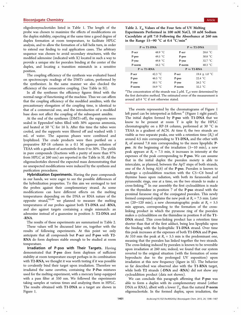
demonstrated that P-pso does form duplexes of sufficientstability at room temperature except perhaps in its combinationwith T2-RNA, we thought it was worth testing if it was possibleto covalently bind their target upon irradiation. Therefore weirradiated the same cuvettes, containing the P-Pso mixturesused for the melting experiment, with a mercury lamp equippedwith a pass filter at 350 nm. We followed the experimentstaking samples at various times and analyzing them in HPLC.The results obtained with T1-DNA as a target are shown inFigure 1.
The events represented by the chromatograms of Figure 1left panel can be interpreted as follows 6 (Figure 1 right panel).The initial duplex formed by P-pso with T1-DNA that weknow to be present at room T is split by the HPLCchromatography on a RP-18 column, performed at 30 °C inTEAA in a gradient of ACN. At time 0, the two strands arevisible as two separate peaks, one with a retention time (Rt) ofaround 4.5 min corresponding to T1-DNA, and the other withRt of around 7.8 min corresponding to the more lipophilic P-pso. At the beginning of the irradiation (5−10 min), a newpeak appears at Rt = 7.5 min, it increases in intensity at theexpenses of the peak corresponding to P-pso. We can assumethat in the initial duplex the psoralen moiety is able tointercalate, as planned, between the ApT steps of both strands,one of the A being A(6) of the P-pso. Psoralen is known toundergo a cycloaddition reaction with the C5−C6 bond ofthymine bases upon radiation, with both its furanosidic andpyranosidic rings, one at a time, on the two strands to make across-linking.45 In our assembly the first cycloaddition is madeon the thymidine in position 7 of the P-pso strand with theproximal furanose ring of the conjugated psoralen. This newlyformed compound explains the new peak at Rt = 7.5 min. Lateron (20−120 min), a new chromatographic peaks at Rt = 5.5min appears, corresponding to the formation of the cross-linking product in which the pyranose ring of the psoralenmakes a cycloaddition on the thimidine in position 8 of the T1-DNA strand. This cross-linking product has a retention timeshorter than that of the first adduct, being less lipophilic uponthe binding with the hydrophilic T1-DNA strand. Over timethis peak increases at the expenses of both T1-DNA and P-pso.At 310 min the peak at Rt = 5.5 min is the predominant one,meaning that the psoralen has linked together the two strands.The cross-linking induced by psoralen is known to be reversibleupon irradiation at 260 nm; indeed, we found that our systemreverted to the original situation (with the formation of somebyproducts due to the prolonged UV exposition) uponirradiation at this new frequency (figure in SI). The behaviorso far described was observed also with the T1-RNA target,while both T2 strands (-DNA and -RNA) did not show anycycloaddition product (data not shown).We can conclude this paragraph affirming that P-pso was
able to form a duplex with its complementary strand (eitherDNA or RNA), albeit with a lower Tm than the natural P-normoligonucleotide. In the formed duplex, upon irradiation, the
Table 2. Tm Values of the Four Sets of UV MeltingExperiments Performed in 100 mM NaCl, 10 mM SodiumCacodylate at pH 7.0 Following the Absorbance at 260 nmin the Range 15−90 °C at 0.4 °C/mina
P vs T1-DNA P vs T2-DNA
P-acr 48.9 °C P-acr 28.6 °CP-pso 49.5 °C P-pso 30.8 °CP-yne 49.8 °C P-yne 32.7 °CP-norm 56.2 °C P-norm 40.3 °C
P vs T1-RNA P vs T2-RNA
P-acr 42.3 °C P-acr 19.4 ± 1.0 °CP-pso 46.5 °C P-pso 22.4 °CP-yne 50.1 °C P-yne 26.2 °CP-norm 54.9 °C P-norm 32.2 °C
aThe concentration of the strands was 2 μM. Tms were determined bythe first derivative method. The extimated error of the measurement isaround ±0.4 °C if not otherwise stated.
Bioconjugate Chemistry Article
dx.doi.org/10.1021/bc400243q | Bioconjugate Chem. 2013, 24, 1398−14071401
psoralen moiety was able to make cycloaddition products withthe nearer thymidines on both the strands, as planned. Thisbehavior was suppressed by the presence of a mismatch in theproximity of the presumed intercalation site with both DNAand RNA targets. This finding means that the conjugate withthe modified adenosine is able to exert its programmedbiochemical activity, and also that in this kind of ″recognition″the activity of the P-pso is impaired by the presence of a near
mismatch. In other words, this conjugate shows a good degree
of selectivity toward its complementary strand.Fluorescence Variation of P-acr upon Hybridization.
Acridine can intercalate between DNA base-pairs in duplex and
triplex, contributing to stabilize the adducts.46,47 Following
intercalation, its emitted fluorescence changes. We set up a
simple titration experiment to check this point registering the
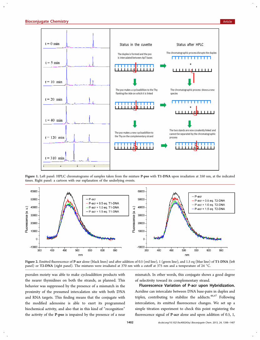
fluorescence signal of P-acr alone and upon addition of 0.5, 1,
Figure 1. Left panel: HPLC chromatograms of samples taken from the mixture P-pso with T1-DNA upon irradiation at 350 nm, at the indicatedtimes. Right panel: a cartoon with our explanation of the underlying events.
Figure 2. Emitted fluorescence of P-acr alone (black lines) and after addition of 0.5 (red line), 1 (green line), and 1.5 eq (blue line) of T1-DNA (leftpanel) or T2-DNA (right panel). The mixtures were irradiated at 370 nm with a cutoff at 375 nm and a temperature of 24 °C.
Bioconjugate Chemistry Article
dx.doi.org/10.1021/bc400243q | Bioconjugate Chem. 2013, 24, 1398−14071402
and 1.5 equivalents (eq) of T1-DNA (Figure 2 left panel) orT2-DNA (Figure 2 right panel).In the left panel (Figure 2) we observe that the fluorescence
signal of P-acr changes from around 5900 to 4200 a.u. uponaddition of 1 eq of the matching complementary strand T1-DNA; moreover, this signal does not change with the furtheraddition of another 0.5 eq of target. In the right panelexperiment, instead, the maximum of the fluorescence signaldecreases from 5900 to 4700 upon addition of 1 eq of T2-DNAand reaches the value of 4500 upon a further addition of 0.5 eqof the mismatched complementary strand. This means that theacridine moiety of the P-acr conjugate maintains its ability tointeract with the formed duplex as planned for these conjugates.Click Chemistry on P-yne Oligonucleotide. To show
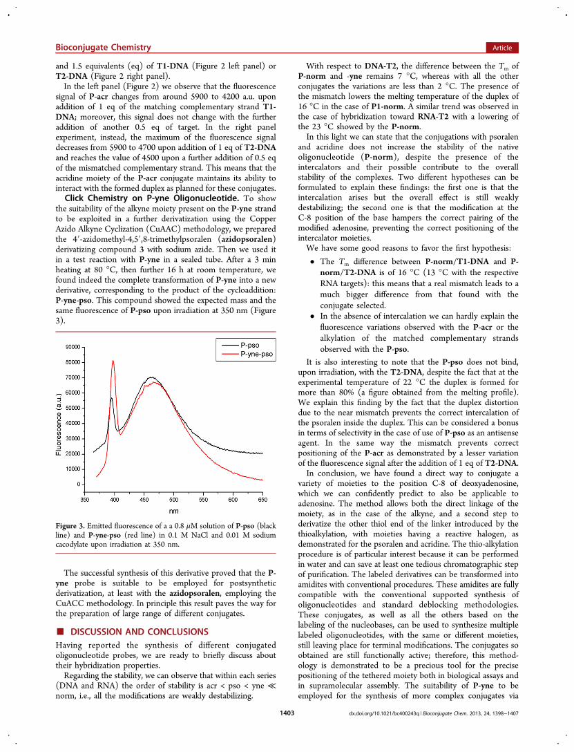
the suitability of the alkyne moiety present on the P-yne strandto be exploited in a further derivatization using the CopperAzido Alkyne Cyclization (CuAAC) methodology, we preparedthe 4′-azidomethyl-4,5′,8-trimethylpsoralen (azidopsoralen)derivatizing compound 3 with sodium azide. Then we used itin a test reaction with P-yne in a sealed tube. After a 3 minheating at 80 °C, then further 16 h at room temperature, wefound indeed the complete transformation of P-yne into a newderivative, corresponding to the product of the cycloaddition:P-yne-pso. This compound showed the expected mass and thesame fluorescence of P-pso upon irradiation at 350 nm (Figure3).
The successful synthesis of this derivative proved that the P-yne probe is suitable to be employed for postsyntheticderivatization, at least with the azidopsoralen, employing theCuACC methodology. In principle this result paves the way forthe preparation of large range of different conjugates.
■ DISCUSSION AND CONCLUSIONSHaving reported the synthesis of different conjugatedoligonucleotide probes, we are ready to briefly discuss abouttheir hybridization properties.Regarding the stability, we can observe that within each series
(DNA and RNA) the order of stability is acr < pso < yne ≪norm, i.e., all the modifications are weakly destabilizing.
With respect to DNA-T2, the difference between the Tm ofP-norm and -yne remains 7 °C, whereas with all the otherconjugates the variations are less than 2 °C. The presence ofthe mismatch lowers the melting temperature of the duplex of16 °C in the case of P1-norm. A similar trend was observed inthe case of hybridization toward RNA-T2 with a lowering ofthe 23 °C showed by the P-norm.In this light we can state that the conjugations with psoralen
and acridine does not increase the stability of the nativeoligonucleotide (P-norm), despite the presence of theintercalators and their possible contribute to the overallstability of the complexes. Two different hypotheses can beformulated to explain these findings: the first one is that theintercalation arises but the overall effect is still weaklydestabilizing; the second one is that the modification at theC-8 position of the base hampers the correct pairing of themodified adenosine, preventing the correct positioning of theintercalator moieties.We have some good reasons to favor the first hypothesis:
• The Tm difference between P-norm/T1-DNA and P-norm/T2-DNA is of 16 °C (13 °C with the respectiveRNA targets): this means that a real mismatch leads to amuch bigger difference from that found with theconjugate selected.
• In the absence of intercalation we can hardly explain thefluorescence variations observed with the P-acr or thealkylation of the matched complementary strandsobserved with the P-pso.
It is also interesting to note that the P-pso does not bind,upon irradiation, with the T2-DNA, despite the fact that at theexperimental temperature of 22 °C the duplex is formed formore than 80% (a figure obtained from the melting profile).We explain this finding by the fact that the duplex distortiondue to the near mismatch prevents the correct intercalation ofthe psoralen inside the duplex. This can be considered a bonusin terms of selectivity in the case of use of P-pso as an antisenseagent. In the same way the mismatch prevents correctpositioning of the P-acr as demonstrated by a lesser variationof the fluorescence signal after the addition of 1 eq of T2-DNA.In conclusion, we have found a direct way to conjugate a
variety of moieties to the position C-8 of deoxyadenosine,which we can confidently predict to also be applicable toadenosine. The method allows both the direct linkage of themoiety, as in the case of the alkyne, and a second step toderivatize the other thiol end of the linker introduced by thethioalkylation, with moieties having a reactive halogen, asdemonstrated for the psoralen and acridine. The thio-alkylationprocedure is of particular interest because it can be performedin water and can save at least one tedious chromatographic stepof purification. The labeled derivatives can be transformed intoamidites with conventional procedures. These amidites are fullycompatible with the conventional supported synthesis ofoligonucleotides and standard deblocking methodologies.These conjugates, as well as all the others based on thelabeling of the nucleobases, can be used to synthesize multiplelabeled oligonucleotides, with the same or different moieties,still leaving place for terminal modifications. The conjugates soobtained are still functionally active; therefore, this method-ology is demonstrated to be a precious tool for the precisepositioning of the tethered moiety both in biological assays andin supramolecular assembly. The suitability of P-yne to beemployed for the synthesis of more complex conjugates via
Figure 3. Emitted fluorescence of a a 0.8 μM solution of P-pso (blackline) and P-yne-pso (red line) in 0.1 M NaCl and 0.01 M sodiumcacodylate upon irradiation at 350 nm.
Bioconjugate Chemistry Article
dx.doi.org/10.1021/bc400243q | Bioconjugate Chem. 2013, 24, 1398−14071403
CuAAC, one of the most used click-chemistry procedures tosynthesize bioconjugate molecules, extends the applicable rangeof our methodology. Considering the enormous potential ofoligonucleotide−ligand conjugates for delivery of nucleic acidtherapeutics, we are evaluating click-chemistry for preparationof oligonucleotide conjugates that can be more easilyinternalized in cells.
■ EXPERIMENTAL PROCEDURESGeneral. HPLC-MS analyses were performed on a Agilent
1100 HPLC system and an Esquire 3000 Plus Bruker massspectrometer using a Zorbax C8 column (4.6 × 150 mm, 5 μm)(linear gradient water/CH3CN at a 0.5 mL/min flow rate,detection at λ 260 nm). HPLC analysis were performed on aAgilent 1260 with a diode array detector and a Varian Pro Star363 fluorescence detector. NMR spectra were recorded with aVarian Mercury 400 MHz instrument. The synthesis of theoligonucleotides was achieved with a Pharmacia GeneAssembler 2 instrument. UV melting studies were performedwith a Cary 100 spectrophotometer. A LOT-Oriel instrumentequipped with a 100 W mercury lamp and interference filterswas used to irradiate the mixture with the P-pso probe.Synthesis of 8-Thiopentanethiol-2′-deoxyadenosine
(Compound 2). In 500 mL flask equipped with a condenserwere added: a 1.6 mM suspension of commercial 8-bromo-2′-deoxyadenosine (220 mg, 0.67 mmol) in water, 1,5-pentanedithiol (276 mg, 2.2 mmol), and triethylamine (0.92mL, 6.7 mmol). The resulting solution was heated at 100 °C for2 h. The warm reaction mixture (40−50 °C) was extracted withethylacetate (2 × 100 mL), the solvent was evaporated underreduced pressure, and the residue used without purification(80% yield on HPLC).
1H NMR (400 MHz, CDCl3) δ: 8.02 (1H, s, H2), 6.20 (1H,dd, J1 = J2 = 6.4 Hz; collapsing to d upon irradiation at δ 2.10;H1′), 5.45 (1H, m; ex with D2O; C5′−OH), 5.31 (1H, d, J =4.0 Hz m; ex with D2O; C3′−OH), 4.43 (1H, br d; H4′), 3.82(1H, br m; H3′), 3.65 (1H, dd, part A of an ABX system, JAB =12 Hz, JAX = 4.4 Hz; collapsing to d, JAB = 4.4 Hz uponirradiation at δ 3.87; H5′), 3.50 (1H, dd, part B of an ABXsystem, JAB = 12 Hz, JBX = 4.4 Hz; collapsing to d, JAB = 4.4 Hzupon irradiation at δ 3.87; H5″), 3.10 (1H, m; collapsing to dd,J1 = 6.0 Hz, J2 = 13.0 Hz, upon irradiation at δ 6.20; collapsingto dd, J1 = 6.4 Hz, J2 = 13.0 Hz, upon irradiation at δ 2.10;H2′), 2.67 (2H, m), 2.45 (3H, m changing to 2H, t J = 4 Hzupon D2O shake, CH2, SH), 2.10 (1H, m; H2″), 1.80−1.60(6H, m).
13C NMR (100 MHz, DMSO-d6) δ: 24.1 (CH2), 27.4(CH2), 28.6 (CH2), 28.9 (CH2), 32.7 (CH2), 33.3 (CH2), 62.8(CH2), 71.8 (CH), 85.5 (CH), 88.8 (CH), 118.8 (q), 151.1(q), 151.9 (q), 154.9 (CH) 167.6 (q).ESI-MS (m/z): 386.1 [M+H]+ (calc. 385.1 for M =
C15H23N5O3S2).8-Thiopentanethiomethylpsoralensoralen-2′-deoxya-
denosine (Compound 4). 1H NMR (400 MHz, CDCl3) δ:8.02 (1H, s, H2), 7.55 (1H, s), 6.29 (1H, s), 6.25 (1H, dd, J1 =J2 = 6.0 Hz; H1′), 5.42 (1H, m, ex with D2O; C3′−OH), 5.30(2H, d, J = 4 Hz, ex with D2O; NH2), 4.47 (1H, br s, H3′), 3.88(1H, br s, H4′), 3.62 (1H, m, H5′), 3.50 (1H, m, H5″), 3.23(2H, m), 3.10 (1H, m, H2′), 2.50−2.30 (12H, m), 1.75−1.40(6H, m).
13C NMR (100 MHz, DMSO-d6): δ 9.0 (CH3), 12.7 (CH3),19.4 (CH3), 24.1 (CH2), 28.1 (CH2), 29.0 (CH2), 29.1 (CH2),31.3 (CH2), 32.7 (CH2), 38.0 (CH2), 62.9 (CH2), 72.0 (CH),
85.5 (CH), 88.9 (CH), 112.9 (CH), 113.2 (CH), 116.2 (q),120.2 (q), 123.6 (q), 125.5 (q), 148.8 (q), 151.1 (q), 151.9 (q),154.4 (CH), 155.1 (q), 160.1 (q), 160.7 (q).ESI-MS (m/z): 648.3 [M+Na]+ and 626.3 [M+H]+ (calc.
625.2 for M = C30H35N5O6S2).5′-Dimethoxytrityl-8-thiopentanethiomethylpsora-
lensoralen-2′-deoxyadenosine (Compound 5). 1H NMR(400 MHz, CDCl3): δ 8.02 (1H, s, H2), 7.65 (1H, s), 7.26 (9H,m), 6.75 (4H m), 6.29 (1H, dd, J1 = J2 = 6.4 Hz; H1′), 6.24(1H, s), 5.36 (1H, br s, ex with D2O; C3′−OH), 4.89 (1H, m,H3′), 4.06 (1H, m, H4′), 3.80−3.70 (7H, m; OCH3, H5′), 3.47(1H, m, H2′), 3.40 (1H, m, H5″), 3.23 (2H, m), 2.56 (3H, s;CH3), 2.49 (3H, s; CH3), 2.41 (5H, m; CH3, CH2), 2.28 (1H,m H2′), 1.75−1.40 (6H m).
13C NMR (100 MHz, CDCl3) δ: 8.8 (CH3), 12.4 (CH3),19.6 (CH3), 25.3 (CH2), 28.1 (CH2), 28.3 (CH2), 28.9 (CH2),31.4 (CH2), 32.5 (CH2), 37.2 (CH2), 55.4 (CH3), 64.0 (CH2),73.3 (CH), 84.4 (CH), 85.9 (CH), 86.4 (q), 109.5 (q), 111.6(q), 111.9 (CH), 113.2 (q), 125.1 (q), 127.0 (CH), 128.0(CH), 128.4 (CH), 130.2 (CH), 136.1 (q), 145.0 (q), 151.6(q), 151.8 (q), 153.6 (q), 154.0 (CH), 158.6 (q).ESI-MS (m/z): 950.5 [M+Na]+ and 928.5 [M+H]+ (calc.
927.3 for M = C51H53N5O8S2).5′-Dimethoxytrityl-8-thiopentanethiomethylpsora-
lensoralen-N6-diphenylacetyl-2′-deoxyadenosine (Com-pound 6). 1H NMR (400 MHz, CDCl3) δ: 8.35 (1H, s, H2),7.65 (1H, s), 7.50−7.10 (19H, m), 6.75 (4H m), 6.24 (1H, dd,J1 = J2 = 6.4 Hz; H1′), 6.20 (1H, s), 4.95 (1H s), 4.85 (1H, m,H3′), 4.10 (1H, m, H4′), 3.85−3.65 (7H, m; OCH3, H5′), 3.44(1H, m, H2′), 3.39 (1H, d, J = 4 Hz; H5″), 3.22 (2H, m), 2.56(3H, s; CH3), 2.49 (3H, s; CH3), 2.41 (5H, m; CH3, CH2),2.25 (1H, m H2′), 1.80−1.40 (6H m).
13C NMR (100 MHz, CDCl3) δ: 8.7 (CH3), 12.5 (CH3),19.6 (CH3), 25.3 (CH2), 28.2 (CH2), 28.8 (CH2), 31.5 (CH2),32.2 (CH2), 37.2 (CH2), 38.8 (CH2), 55.5 (CH3), 57.5 (CH),64.2 (CH2), 73.0 (CH), 84.7 (CH), 86.1 (CH), 86.5 (q), 109.4(q), 111.6 (q), 111.9 (CH), 113.0 (q), 113.2 (CH), 125.1 (q),127.0 (CH), 127.6 (CH), 127.9 (CH), 128.4 (CH), 128.9(CH), 129.1 (CH), 129.4 (CH), 130.2 (CH), 136.1 (q), 139.0(q), 145.9 (q), 146.1 (q), 153.4 (q), 153.7 (q), 154.0 (CH),154.7 (q), 154.9 (q), 158.6 (q), 161.9 (q).ESI-MS (m/z): 1122.5 [M+H]+ and 1144.5 [M+Na]+ (calc.
1121.4 for M = C65H63N5O9S2).5′-Dimethoxytrityl-8-thiopentanethiomethylpsora-
lensoralen-N6-diphenylacetyl-2′-deoxyadenosine-3′-cy-anoethyl diisopropylphosphoramidite (Compound 7).1H NMR (400 MHz, CDCl3): δ 8.50 (1H, br s), 8.35 (1H, s,H2), 7.65 (1H, s), 7.50−7.10 (19H, m), 6.75 (4H, m), 6.24(1H, dd, J1 = J2 = 6.4 Hz; H1′), 6.20 (1H, s), 4.95 (1H, s), 4.85(0.5H, m, H3′), 4.83 (0.5H, m, H3′), 4.10 (1H, m, H4′), 3.85−3.65 (9H, m), 3.44 (1H, m, H2′), 3.40 (3H, m), 3.22 (2H, m),2.65 (1H, m), 2.56 (3H, s; CH3), 2.52 (1H, m), 2.49 (3H, s;CH3), 2.41 (5H, m; CH3, CH2), 2.25 (0.5H, m H2′), 2.23(0.5H, m H2″), 1.80−1.40 (6H m), 1.21(3H, s), 1.19 (6H, s),1.17 (3H, s).
31P NMR (160 MHz, CDCl3) δ: 149.9, 149.6.Synthesis of 8-Thiopentanethiomethylacridine-2′-de-
oxyadenosine (Compound 9). A solution of 2 (385 mg, 1.0mmol), acridine 8 (600 mg, 2 mmol), and triethylamine (1.8mL, 10 mmol) was heated in dry dimethylformamide (10 mL)at 80 °C in a sealed tube for 17 h. The reaction mixture wasdiluted with 5 mL of CH2Cl2/CH3OH 80/20. The crudecompound was precipitated with 80 mL of petroleum ether/
Bioconjugate Chemistry Article
dx.doi.org/10.1021/bc400243q | Bioconjugate Chem. 2013, 24, 1398−14071404
ethyl ether 70/30. The precipitate was purified on silica gelcolumn. Elution with CH2Cl2/CH3OH 90:10 led to the targetproduct 9 (440 mg, 70% yield).
1H NMR (400 MHz, CDCl3) δ: 8.66 (1H, d, J = 9.2), 8.18(1H, d, J = 2.4), 8.13 (1H, s, H2), 8.09 (1H, d, J = 9.6 Hz), 7.93(1H, d, J = 2.8), 7.49 (2H, m), 6.28 (1H, dd, J1 = 5.6 Hz, J2 =9.6 Hz; collapsing to d J = 9.6 Hz upon irradiation at δ 2.2;H1′), 4.82 (2H, br s; ex with D2O; NH2), 4.70 (1H, d, J = 5.6;collapsing to s upon irradiation at δ 2.90; H3′), 4.17 (1H br s,H4′), 4.01 (3H, s), 3.90 (1H, dd, part A of an ABX system, JAB= 12.8 Hz, JAX = 1.6 Hz; H5′), 3.73 (1H, dd, part B of an ABXsystem, JAB = 12.8 Hz, JBX = 1.6 Hz; H5″), 3.10 (2H, m), 2.90(3H, m), 2.19 (2H, dd, J1 = 5.6 Hz, J2 = 13.6 Hz; H2″), 2.63(2H, m), 1.48 (4H, m).
13C NMR (100 MHz, CDCl3) δ: 27.8 (CH2), 28.6 (CH2),29.9 (CH2), 32.6 (CH2), 36.9 (CH2), 40.3 (CH2), 56.0 (CH3),63.6 (CH2), 73.6 (CH), 86.9 (CH), 89.9 (CH), 126.3 (CH),127.9 (q), 128.1 (CH), 128.2 (CH), 128.7 (CH), 130.6 (q),131.9 (CH), 146.6 (q), 147.2 (q), 151.2 (q), 153.9 (CH),158.5 (q), 160.2 (q).ESI-MS (m/z): 627.1 and 629.1 [M+H]+. (calc. 626,1 M =
C29H31ClN6O4S2).5′-Dimethoxytrityl-8-thiopentanethiomethylacridine-
2′-deoxyadenosine (Compound 10). 1H NMR (400 MHz,CDCl3) δ: 8.68 (1H, d, J = 8.7), 8.20 (1H, d, J = 2.2), 8.10 (1H,d, J = 9.5 Hz), 8.05 (1H, s, H2), 7.94 (1H, d, J = 2.9 Hz), 7.50(2H, m), 7.40−7.10 (9H, m), 6.75 (4H, m), 6.26 (1H, dd, J =6.9; H1′), 4.88 (1H, m; H3′), 4.05 (1H, m; H4′), 4.01 (3H, s),3.76 (3H, s), 3.75 (3H, s), 3.72 (1H, m; H5′), 3.47 (1H, m,H2′), 3.39 (1H, m; H5″), 3.14 (2H, m), 2.90 (2H, m), 2.27(1H, m; H2″), 1.63 (2H, m), 1.45 (4H, m).
13C NMR (100 MHz, CDCl3) δ: 27.8 (CH2), 28.9 (CH2),29.9 (CH2), 32.6 (CH2), 37.0 (CH2), 40.3 (CH2), 55.6 (CH3),56.1 (CH3), 63.6 (CH2), 73.8 (CH), 86.4 (q), 86.9 (CH), 89.9(CH), 120.0 (CH), 126.3 (CH), 127.2 (CH), 127.9 (q), 128.1(CH), 128.2 (CH), 128.5 (CH), 128.7 (CH), 130.0 (CH),130.6 (q), 131.9 (CH), 136.1 (q), 145.0 (q), 146.6 (q), 147.2(q), 151.5 (q), 153.9 (CH), 158.6 (q), 160.2 (q).ESI-MS (m/z): 951.1 and 953.1 [M+Na]+. (Calc. 928.3 for
M = C50H49ClN6O6S2).5′-Dimethoxytrityl-8-thiopentanethiomethylacridine-
N6-diphenylacetyl-2′-deoxyadenosine (Compound 11).1H NMR (400 MHz, CDCl3) δ: 8.68 (1H, d, J = 8.7), 8.35(1H, s, H2), 8.20 (1H, d, J = 2.2), 8.10 (1H, d, J = 9.2 Hz), 7.94(1H, d, J = 2.6 Hz), 7.49 (2H, m), 7.36−7.15 (19 H, m), 6.75(4H, m), 6.20 (1H, dd, J = 6.8; H1′), 4.95 (1H, s), 4.84 (1H,m; H3′), 4.74 (2H, br s; ex with D2O; NH2), 4.10 (1H, m;H4′), 4.01 (3H, s), 3.75 (3H, s), 3.74 (3H, s), 3.72 (1H, m;H5′), 3.40 (1H, m, H2′), 3.36 (1H, m; H5″), 3.12 (2H, m),2.90 (2H, m), 2.27 (1H, m; H2″), 1.61 (2H, m), 1.45 (4H, m).
13C NMR (100 MHz, CDCl3) δ: 27.9 (CH2), 29.0 (CH2),30.0 (CH2), 32.6 (CH2), 37.0 (CH2), 40.3 (CH2), 55.6 (CH3),56.1 (CH3), 57.5 (CH), 63.6 (CH2), 73.7 (CH), 86.4 (q), 86.9(CH), 89.9 (CH), 120.0 (CH), 126.3 (CH), 127.2 (CH), 127.6(CH), 128.0 (q), 128.2 (CH), 128.4 (CH), 128.5 (CH), 128.7(CH), 129.1 (CH), 129.6 (CH), 130.0 (CH), 130.6 (q), 131.9(CH), 136.1 (q), 139.0 (q), 145.0 (q), 146.6 (q), 147.2 (q),151.5 (q), 153.9 (CH), 158.6 (q), 160.2 (q), 162.0 (q).ESI-MS (m/z): 1123.3 [M+H]+, 1145.4 [M+Na]+. (Calc.
1122.4 for C64H59ClN6O7S2).5′-Dimethoxytrityl-8-thiopentanethiomethylacridine-
N6-diphenylacetyl-2′-deoxyadenosine-3′-cyanoethyldiisopropylphosphoramidite (Compound 12). 1H NMR
(400 MHz, CDCl3) δ: 8.68 (1H, d, J = 8.7), 8.45 (1H br s),8.36 (0.5H, s; H2), 8.35 (0.5H, s; H2), 8.20 (1H, br s), 8.10(1H, d, J = 9.2 Hz), 7.94 (1H, d, J = 2.6 Hz), 7.49 (2H, m),7.36−7.15 (19 H, m), 6.75 (4H, m), 6.20 (1H, dd, J = 6.8 Hz;H1′), 5.0 (1H s), 4.85 (0.5H, m; H3′), 4.82 (0.5H, m; H3′),4.21 (1H, m; H4′), 4.01 (3H s), 4.81 (1H, m; H5′), 3.75 (1.5H, s), 3.74 (3H, s), 3.73 (1.5H, s) 3.70 (1H, m; H5″), 3.60(2H, m), 3.50 (1H, m, H2′), 3.40 (2H, m), 3.30 (2H, m), 2.95(2H, m), 2.60 (1H, m), 2.45 (1H, m), 2.40 (0.5 H, m; H2″),2.35 (0.5 H, m; H2″), 1.40−1.10 (18 H, m).
31P NMR (160 MHz, CDCl3) δ: (147.4, 147.1).Synthesis of 8-Thiohex-5-yne-2′-deoxyadenosine
(Compound 14). Compound 1 (220 mg, 0.67 mmol) andthiol 13 (430 mg, 4 mmol) were reacted as reported above forthe preparation of 2. The target compound 14 (243 mg) wasobtained in 30% yield, and used without purification.
1H NMR (400 MHz, CD3OD) δ: 8.10 (1H, s, H2), 6.39(1H, dd, J1 = J2 = 6.0 Hz; collapsing to d upon irradiation at δ2.25; H1′), 4.60 (2H, m), 4.08 (1H, m; H4′), 3.86 (1H, dd,part A of an ABX system, JAB = 12.4 Hz, JAX = 2.4 Hz; H5′),3.75 (1H, dd, part B of an ABX system, JAB = 12.4 Hz, JBX = 2.4Hz; H5″), 3.36 (3H, m), 3.02 (1H, m; H2′), 2.25 (1H, m;H2″), 2.21 (2H, m), 1.90 (2H, m), 1.68 (2H, m).
13C NMR (100 MHz, CD3OD) δ: 21.3 (CH2), 31.3 (CH2),32.2 (CH2), 35.8 (CH2), 41.9 (CH2), 42.9 (CH2), 67.0 (CH),76.5 (CH), 87.2 (q), 90.4 (CH), 93.1(CH), 124.2 (q), 153.8(q), 154.4 (q), 154.8 (CH), 158.6 (q).ESI-MS (m/z): 364.0 [M+H]+. (Calc. 363.1 for M =
C16H21N5O3S).5′-Dimethoxytrityl-8-thiohex-5-yne-2′-deoxyadeno-
sine (Compound 15). 1H NMR (400 MHz, CD3OD) δ: 7.93(1H, s, H2), 7.35 (2H, m), 7.24−7.12 (7H, m), 6.73 (4H, m),6.40 (1H, dd, J1 = J2 = 6.4 Hz; collapsing to d upon irradiationat δ 2.26; H1′), 4.74 (1H, m, H3′), 4.08 (1H, m; collapsing to t,J = 4.8 Hz upon irradiation at δ 4.74 and to d, J = 4.0 Hz uponirradiation at δ 3.33; H4′), 3.75 (3H, s, OCH3), 3.74 (3H, s,OCH3), 3.45 (1H, m H2′), 3.37−3.29 (5H, m), 2.24 (1H, m;H2″), 2.16 (2H, m), 1.90 (2H, m), 1.63 (2H, m).
13C NMR (100 MHz, CD3OD) δ: 21.3 (CH2), 31.3 (CH2),32.2 (CH2), 35.8 (CH2), 40.7 (CH2), 49.8 (CH2), 58.5 (CH3),67.9 (CH), 75.7 (CH), 88.7 (CH), 90.2 (q), 90.5 (CH), 116.6(CH), 130.3 (CH), 131.3 (CH), 132.1 (CH), 133.9 (CH),140.2 (q), 149.3 (q), 154.7 (CH), 158.2 (q), 162.7 (q).ESI-MS (m/z): 666.4 [M+H]+ 668.4 [M+Na]+. (Calc. 665.3
for M = C37H39N5O5S).5′-Dimethoxytrityl-8-thiohex-5-yne-N6-diphenylace-
tyl-2′-deoxyadenosine (Compound 16). 1H NMR (400MHz, CD3OD) δ: 8.31 (1H, s, H2), 7.45−7.07 (19 H, m), 6.70(4H, m), 6.36 (1H, dd, J1 = J2 = 6.8 Hz; H1′), 5.45 (1H, s),4.76 (1H, m, H3′), 4.10 (1H, m; collapsing to d, J = 4.4 Hzupon irradiation at δ 3.27 and to t, J = 6.0 Hz upon irradiationat δ 4.76; H4′), 3.68 (3H, s, OCH3), 3.65 (3H, s, OCH3), 3.48(1H, m; H2′), 3.32−3.24 (5H, m), 2.28 (1H, m; H2″), 2.16(2H, m), 1.90 (2H, m), 1.63 (2H, m).
13C NMR (100 MHz, CD3OD) δ: 21.5 (CH2), 31.4 (CH2),32.1 (CH2), 35.6 (CH2), 40.7 (CH2), 49.8 (CH2), 58.4 (CH3),62.3 (CH), 67.9 (CH), 75.6 (CH), 88.9 (CH), 90.0 (q), 90.7(CH), 116.6 (CH), 128.2 (q), 131.0 (CH), 131.3 (CH), 132.0(CH), 132.3 (CH), 132.6 (CH), 132.9 (CH), 134.0 (CH),140.1 (q), 143.4 (q), 143.9 (q), 149.2 (q), 150.4 (q), 154.0(CH), 159.6 (q), 162.7 (q), 175.5 (q).ESI-MS (m/z): 860.4 [M+H]+. (Calc. 859.3 for M =
C51H49N5O6S).
Bioconjugate Chemistry Article
dx.doi.org/10.1021/bc400243q | Bioconjugate Chem. 2013, 24, 1398−14071405
5′-Dimethoxytrityl-8-thiohex-5-yne-N6-diphenylace-tyl-2′-deoxyadenosine-3′-cyanoethyl diisopropylphos-phoramidite (Compound 17). 1H NMR (400 MHz,CD3OD) δ: 8.43 (1H br s), 8.38 (0.5 H s), 8.37(0.5 H s),7.45−7.07 (19 H, m), 6.70 (4H, m), 6.27 (1H, m; H1′), 6.00(1H, s), 4.94 (0.5 H, m; H3′), 4.86 (0.5 H, m; H3′), 4.22 (1Hm; H4′) 3.85 (1H m; H5′), 3.74 (1.5 H; OCH3), 3.75 (3 H;OCH3), 3.76 (1.5 H; OCH3), 3.67 (1H m; H5″), 3.60 (2H m),3.50 (1H m; H2′), 3.45 (2H, m), 3.30 (3H m), 2.61 (1H m),2.47 (1H m), 2.40 (0.5H m; H2″), 2.35 (0.5 H, m; H2″), 1.90(2H m), 1.60 (2H m), 1.25 (2H m), 1.21 (3H s), 1.19 (6H s),1.17 (3H, s).
31P NMR (160 MHz, CDCl3) δ: (149.3, 149.0).Synthesis and Purification of Oligonucleotides. All the
oligonucleotides were synthesized on a Pharmacia GeneAssembler 2 on a 1.3 μM scale with standard protocols witha 2 min longer coupling time for the conjugated adenosines. Allthe oligonucleotides were deblocked by treatment with 30%aqueous ammonia at 50 °C for 16 h, then lyophilized. Thetargets were purified on a preparative C-18 reverse phasecolumn using 0.1 M aqueous solution of triethylammoniumacetate (TEAA) in a gradient of acetonitrile from 0 to 30%. Thecollected fractions were analized in HPLC to give final lots withpurity higher than 97% at 260 nm, and then lyophilized.Synthesis of P-yne-pso. To a solution of P-yne (18 OD
c.a. 0.15 μmol) in 310 μL of H2O/triethylammonium acetatebuffer solution (2 M, pH 7) /DMSO mixture 1/0.5/1.6 v/v, asolution of azidomethylpsoralen (0.3 μmol, 30 μL of a 10 mMDMSO solution) and ascorbic acid aq. solution freshlyprepared (40 μL of 5 mM) was added . The mixture wasbriefly stirred with a vortex mixer and deaerated by bubblingnitrogen. To the resulting preparation, 20 μL of a CuSO4·5H2O−TBTA complex (10 mM solution in H2O/DMSO 45/55 mixture) was added, and the tube was flushed with nitrogenand sealed. The mixture was heated for 3 min at 80 °C and keptat room temperature overnight. The reaction was diluted with4-fold volume of cold ethanol and kept at −20 °C for 20 min.The cold mixture was centrifuged for 10 min; then, thesupernatant was removed and the resulting pellet washed (2 ×1 mL) of cold ethanol each, and finally dried. 5 OD (c.a. 0.05μmol) of a compound with purity higher than 90% at 260 nmin HPLC analysis was recovered.ESI-MS Analysis. T1-DNA: 3998.9 (calc. 3999.7 for
C127H159N53O75P12). T2-DNA: 3983.1 (calc. 3983.6 forC127H159N53O74P12). P-norm: 3908.8 (calc. 3908.7 forC125H160N46O77P12). P-pso: 4283.0 (calc. 4282.8 forC145H182N46O80P12S2). P-acr: 4284.4 (calc. 4283.7 forC144H178ClN47O78P12S2). P-yne: 4020.7 (calc. 4020.7 forC131H168N46O77P12S). P-yne-pso: 4303.6 (calc. 4303.0 forC146H181N49O80P12S).The synthetic methodology described in this work is based
on the patent: M. L. Capoibianco and M. L. Navacchia PCTInt. Appl. WO 2012164484 A1 2012.
■ ASSOCIATED CONTENT
*S Supporting InformationDetailed synthetic procedures for the preparation of com-pounds: 1−7, 10−12, and 15−17; detailed syntheticprocedures and the full characterization of compond 13 andazidopsoralen; Table: efficiency of coupling for oligonucleo-tides and final recovery based on CPG loading. Figure: HPLCstudy of reversion of adducts formed upon cross-linking of P-
pso with T1-DNA. This material is available free of charge viathe Internet at http://pubs.acs.org.
■ AUTHOR INFORMATIONCorresponding Author*E-mail: [email protected] authors declare no competing financial interest.
■ REFERENCES(1) Wojczewski, C., Stolze, K., and Engels, J. W. (1999) Fluorescentoligonucleotides - Versatile tools as probes and primers for DNA andRNA analysis. Synlett. 10, 1667−1678.(2) Capobianco, M. L., Barbarella, G., and Manetto, A. (2012)Oligothiophenes as fluorescent markers for biological applications.Molecules 17, 910−933.(3) Garbesi, A., Bonazzi, S., Zanella, S., Capobianco, M. L., Giannini,G., and Arcamone, F. (1997) Synthesis and binding properties ofconjugates between oligodeoxynucleotides and daunorubicin deriva-tives. Nucleic Acids Res. 25, 2121−2128.(4) Arya, D. P., and Willis, B. (2003) Reaching into the major grooveof B-DNA: synthesis and nucleic acid binding of a Neomycin-Hoechst33258 conjugate. J. Am. Chem. Soc. 125, 12398−12399.(5) Duca, M., Oussedik, K., Ceccaldi, A., Halby, L., Guianvarc’h, D.,Dauzonne, D., Monneret, C., Sun, J. S., and Arimondo, P. B. (2005)Triple helix-forming oligonucleotides conjugated to new inhibitors oftopoisomerase II: Synthesis and binding properties. Bioconjugate Chem.16, 873−884.(6) Murakami, A., Yamayoshi, A., Iwase, R., Nishida, J., Yamaoka, T.,and Wake, N. (2001) Photodynamic antisense regulation of humancervical carcinoma cell growth using psoralen-conjugated oligo-(nucleoside phosphorothioate). Eur. J. Pharm. Sci. 13, 25−34.(7) Moser, H. E., and Dervan, P. B. (1987) Sequence-specificcleavage of double helical DNA by triple helix formation. Science 238,645−650.(8) Boutorine, A. S., Tokuyama, H., Takasugi, M., Isobe, H.,Nakamura, E., and Helene, C. (1995) Fullerene-oligonucleotideconjugates: Photoinduced sequence-specific DNA cleavage. Angew.Chem. 33, 2462−2465.(9) Du, H., Strohsahl, C. M., Camera, J., Miller, B. L., and Krauss, T.D. (2005) Sensitivity and specificity of metal surface-immobilized″molecular beacon″ biosensors. J. Am. Chem. Soc. 127, 7932−7940.(10) Jung, A., Stemmler, I., Brecht, A., and Gauglitz, G. (2001)Covalent strategy for immobilization of DNA-microspots suitable formicroarrays with label-free and time-resolved optical detection ofhybridization. Fresenius J. Anal. Chem. 371, 128−136.(11) Alemdaroglu, F. E., and Herrmann, A. (2007) DNA meetssynthetic polymers - highly versatile hybrid materials. Org. Biomol.Chem. 5, 1311−1320.(12) Bijsterbosch, M. K., Rump, E. T., De Vrueh, R. L. A., Dorland,R., van Veghel, R., Tivel, K. L., Biessen, E. A. L., van Berkel, T. J. C.,and Manoharan, M. (2000) Modulation of plasma protein binding andin vivo liver cell uptake of phosphorothioate oligodeoxynucleotides bycholesterol conjugation. Nucleic Acids Res. 28, 2717−2725.(13) Ballico, M., Drioli, S., Morvan, F., Xodo, L., and Bonora, G. M.(2001) Triple, MPEG-conjugated, helix-forming oligonucleotides(TRIPEGXs): Liquid-phase synthesis of natural and chimeric ″all-purine″ sequences linked to high molecular weight poly(ethyleneglycols). Bioconjugate Chem. 12, 719−725.(14) Weyermann, J., Lochmann, D., Georgens, C., and Zimmer, A.(2005) Albumin-protamine-oligonucleotide-nanoparticles as a newantisense delivery system. Part 2: cellular uptake and effect. Eur. J.Pharm. Biopharm. 59, 431−438.(15) Wu, R. P., Youngblood, D. S., Hassinger, J. N., Lovejoy, C. E.,Nelson, M. H., Iversen, P. L., and Moulton, H. M. (2007) Cell-penetrating peptides as transporters for morpholino oligomers: effectsof amino acid composition on intracellular delivery and cytotoxicity.Nucleic Acids Res. 35, 5182−5191.
Bioconjugate Chemistry Article
dx.doi.org/10.1021/bc400243q | Bioconjugate Chem. 2013, 24, 1398−14071406
(16) Youngblood, D. S., Hatlevig, S. A., Hassinger, J. N., Iversen, P.L., and Moulton, H. M. (2007) Stability of cell-penetrating peptide-morpholino oligomer conjugates in human serum and in cells.Bioconjugate Chem. 18, 50−60.(17) Chen, J. K., Carlson, D. V., Weith, H. L., Obrien, J. A., Goldman,M. E., and Cushman, M. (1992) Synthesis of an oligonucleotide-intercalator conjugate in which the linker chain is attached via thephenolic hydroxyl group of fagaron. Tetrahedron Lett. 17, 2275−2278.(18) Asseline, U., Thuong, N. T., and Helene, C. (1997) Synthesisand properties of oligonucleotides covalently linked to intercalatingagents. New J. Chem. 21, 5−17.(19) Byrn, S. R., Carlson, D. V., Chen, J. K., Cushman, M. S.,Goldman, M. E., Ma, W. P., Pidgeon, C. L., Ray, K. A., Stowell, J. G.,and Weith, H. L. (1991) Drug-oligonucleotide conjugates. Adv. DrugDelivery Rev. 6, 287−308.(20) Vinogradov, S., Roig, V., Sergueeva, Z., Nguyen, C. H.,Arimondo, P., Thuong, N. T., Bisagni, E., Sun, J. S., Helene, C., andAsseline, U. (2003) Synthesis and binding properties of oligo-2′-deoxyribonucleotides conjugated with triple-helix-specific intercala-tors: benzo[e] and benzo[g] pyridoindoles. Bioconjugate Chem. 14,120−135.(21) Stierle, V., Duca, M., Halby, L., Senamaud-Beaufort, C.,Capobianco, M. L., Laigle, A., Jolles, B., and Arimondo, P. B. (2008)Targeting MDR1 gene: Synthesis and cellular study of modifieddaunomycin-triplex-forming oligonucleotide conjugates able to inhibitgene expression in resistant cell lines. Mol. Pharmacol. 73, 1568−1577.(22) Rothemund, P. W. K. (2006) Folding DNA to create nanoscaleshapes and patterns. Nature 440, 297−302.(23) Gu, H. Z., Chao, J., Xiao, S. J., and Seeman, N. C. (2010) Aproximity-based programmable DNA nanoscale assembly line. Nature465, 202−U286.(24) Alesi, S., Brancolini, G., Melucci, M., Capobianco, M. L.,Venturini, A., Camaioni, N., and Barbarellala, G. (2008) Water-soluble,electroactive, and photoluminescent quaterthiophene-dinucleotideconjugates. Chem.Eur. J. 14, 513−521.(25) Alesi, S., Brancolini, G., Viola, I., Capobianco, M. L., Venturini,A., Camaioni, N., Gigli, G., Melucci, M., and Barbarella, G. (2009) Self-organization, optical, and electrical properties of alpha-quinquethio-phene-dinucleotide conjugates. Chem.Eur. J. 15, 1876−1885.(26) Navacchia, M. L., Favaretto, L., Treossi, E., Palermo, V., andBarbarella, G. (2010) Self-complementary nucleoside-thiophenehybrid systems: synthesis and supramolecular organization. Macromol.Rapid Commun. 31, 351−355.(27) Tyagi, S., and Kramer, F. R. (1996) Molecular beacons: Probesthat fluoresce upon hybridization. Nat. Biotechnol. 14, 303−308.(28) Cazzato, A., Capobianco, M. L., Zambianchi, M., Favaretto, L.,Bettini, C., and Barbarella, G. (2007) Oligothiophene molecularbeacons. Bioconjugate Chem. 18, 318−322.(29) Akiyama, T., and Hogan, M. E. (1997) Structural analysis ofDNA bending induced by tethered triple helix forming oligonucleo-tides. Biochemistry 36, 2307−2315.(30) Dreyer, G. B., and Dervan, P. B. (1985) Sequence-specificcleavage of single-stranded DNA: oligodeoxynucleotide- EDTA XFe(II). Proc. Natl. Acad. Sci. U. S. A. 82, 968−972.(31) Hurley, D. J., Seaman, S. E., Mazura, J. C., and Tor, Y. (2002)Fluorescent 1,10-phenanthroline-containing oligonucleotides distin-guish between perfect and mismatched base pairing. Org. Lett. 4,2305−2308.(32) Hwang, G. T., Seo, Y. J., and Kim, B. H. (2004) A highlydiscriminating quencher-free molecular beacon for probing DNA. J.Am. Chem. Soc. 126, 6528−6529.(33) Capobianco, M. L., Cazzato, A., Alesi, S., and Barbarella, G.(2008) Oligothiophene-5-labeled deoxyuridines for the detection ofsingle nucleotide polymorphisms. Bioconjugate Chem. 19, 171−177.(34) Pieles, U., Sproat, B. S., Neuner, P., and Cramer, F. (1989)Preparation of a novel psoralen containing deoxyadenosine buildingblock for the facile solid-phase synthesis of psoralen-modifiedoligonucleotides for a sequence specific crosslink to a given targetsequence. Nucleic Acids Res. 17, 8967−8978.
(35) Manfredini, S., Baraldi, P. G., Bazzanini, R., Marangoni, M.,Simoni, D., Balzarini, J., and Declercq, E. (1995) Synthesis andcytotoxic activity of 6-vinyl- and 6-ethynyluridine and 8-vinyl- and 8-ethynyladenosine. J. Med. Chem. 38, 199−203.(36) Sagi, G., Otvos, L., Ikeda, S., Andrei, G., Snoeck, R., andDeclercq, E. (1994) Synthesis and antiviral activities of 8-alkynyl-, 8-alkenyl-, and 8-alkyl-2′-deoxyadenosine analogues. J. Med. Chem. 37,1307−1311.(37) Tierney, M. T., and Grinstaff, M. W. (2000) Synthesis andstability of oligodeoxynucleotides containing C8-labeled 2 ′-deoxy-adenosine: Novel redox nucleobase probes for DNA-mediated charge-transfer studies. Org. Lett. 2, 3413−3416.(38) Hwang, G. T., Seo, Y. J., and Kim, B. H. (2005) Pyrene-labeleddeoxyuridine and deoxyadenosine: fluorescent discriminating phenom-ena in their oligonucleotides. Tetrahedron Lett. 46, 1475−1477.(39) Chatgilialoglu, C., Navacchia, M. L., and Postigo, A. (2006) Afacile one-pot synthesis of 8-oxo-7,8-dihydro-(2 ′-deoxy)adenosine inwater. Tetrahedron Lett. 47, 711−714.(40) Capobianco, M. L., Carcuro, A., Tondelli, L., Garbesi, A., andBonora, G. M. (1990) One pot solution synthesis of cyclicoligodeoxyribonucleotides. Nucleic Acid Res. 18, 2661−2669.(41) Ti, G. S., Gaffney, B. L., and Jones, R. A. (1982) Transientprotection: efficient one-flask synthesis of protected deoxynucleosides.J. Am. Chem. Soc. 104, 1316−1319.(42) Perrone, D., Bortolini, O., Fogagnolo, M., Marchersi, E., Mari,L., Massarenti, C., Navacchia, M. L., Sforza, F., Varani, K., andCapobianco, M. L. Synthesis and in vitro cytotoxicity ofdeoxyadenosine-bile acid conjugates linked with 1,2,3-triazole.Submitted.(43) Morvan, F., Porumb, H., Degols, G., Pompon, A., Sproat, B. S.,Rayner, B., Malvy, C., Lebleu, B., and Imbach, J. L. (1993)Comparative analysis of seven oligonucleotide analogs as potentialantisense agents. J. Med. Chem. 36, 280−287.(44) Pandolfi, D., Rauzi, F., and Capobianco, M. L. (1999)Evaluation of different types of end-capping modifications on thestability of oligonucleotides toward 3 ′- and 5 ′-exonucleases.Nucleosides Nucleotides 18, 2051−2069.(45) Kanne, D., Rapoport, H., and Hearst, J. E. (1984) 8-Methoxypsoralen-nucleic acid photoreaction - effect of methylsubstitution on pyrone vs furan photoaddition. J. Med. Chem. 27,531−534.(46) Asseline, U., Delarue, M., Lancelot, G., Toulme, F., Thuong, N.T., Montenaygarestier, T., and Helene, C. (1984) Nucleic-acidbinding-molecules with high-affinity and base sequence specificity -intercalating agents covalently linked to oligodeoxynucleotides. Proc.Natl. Acad. Sci. U.S.A. 81, 3297−3301.(47) Sun, J. S., Francois, J. C., Montenay-Garestier, T., Saison-Behmoaras, T., Roig, V., Thuong, N. T., and Helene, C. (1989)Sequence-specific intercalating agents: intercalation at specificsequences on duplex DNA via major groove recognition byoligonucleotide-intercalator conjugates. Proc. Natl. Acad. Sci. U.S.A.86, 9198−9202.
Bioconjugate Chemistry Article
dx.doi.org/10.1021/bc400243q | Bioconjugate Chem. 2013, 24, 1398−14071407