Antiproliferative properties of flavone acetic acid (NSC 347512) (LM 975), a new anticancer agent
7
EWJ Corr~r Ch Onroi, Val 23, No.IO, pp 1529-1535, 1987 Printed in Great Britain 0277-5379/87$3.00+0.00 fQ 1987 PergamonJournalsLtd. Antiproliferative Properties of Flavone Acetic Acid (NSC 347512) (LM 975), a New Anticancer Agent LAURA S. CAPOLONGO*, GIOVANNA BALCONI,* PAOLO UBEZIO,* RAFFAELLA GIAVAZZI,t GIULIA TARABOLETTI,t ANTONELLA REGONESI,? OMAR C. YODER: and MAURIZIO D’INCALCI* Wituto di Ricerche Farmacologiche ‘Mario Negri’ , Via Eritrea, 62, 20157 Milan, Italy, tlstituto Mario Negri Bergamo, Via Gavazfeni 11, 24100 Bergamo, Itab and fLiaison Offize, National Cancer Institute, Brussels, Belgium Abstract-The antiproliferative activiy of I&one acetic acid (LM 975) was investigated on human adenocarcinoma cell lines (HCCP2998, HCC-MI%, HCC-MI410, HT 29, LoVo), on a murine colon adenocarcinoma cell line (Colon 26), on murine pancreatic adenocarcinoma cells growing in primary culture (Pan 03) and on human normal fibroblasts (Nl). No cytotoxic effects were found against human notmaljbroblasts. LM 975 was active against murine adenocarcinoma Pan 03 and Colon 26, known to be sensitive in vivo too and, to variable extents, on human adenocarcinoma cell lines. LM 975 in vitro cytotoxic potency was relatively low. The high concentrations (1.c 1.4 mM) required to obtain a cytotoxic effect are, however, pharmacologically reasonable since they are comparable with drug plasma levels in mice or in patients treated with tolerable doses. After a relatively short LM 975 treatment (2 h) DNA, RNA and protein synthesis were inhibited in different proportions. In more sensitive cells LM 975 appeared to inhibit RNA synthesis more than DNA and protein synthesis. Inhibition of macromolecule synthesis after 2 h exposure was completely reversed in 24 h recovery. After 2 h treatment no detectable DNA breakage was found by the alkaline elution method, thus corroborating the idea that this compound does not act by causing DNA damage. INTRODUCTION FLAVONE ACETIC ACID (LM 975) is a new anticancer agent which has shown activity in some experimen- tal tumors such as the murine adenocarcinomas Colon 38 [ 11, Colon 07/A, Colon 51, Colon 10/A and the murine pancreatic ductal adenocarcinomas Pan 02 and Pan 03 [2, 31. It has been selected for clinical investigation in Europe and in the U.S.A. No information is yet available on the mechanism of action of this drug, which may act by completely different mechanisms from those described for the anticancer agents identified so far. This hypothesis is suggested by the following considerations: (i) the chemical structure of LM 975 does not resemble that of any known anticancer agent; (ii) LM 975 shows a peculiar pattern of antitumor activity in Accepted 15 May 1987. Reprint requests to: M. D’Incalci, Istituto ‘Mario Negri’ , Via Eritrea 62, 20157 Milan, Italy. This work was supported by the CNR (Italian National Research Council, Rome, Italy) Progetto Finalizzato ‘Oncologia’ , Contract No. 86.00653.44. The generous contribution of the Italian Association for Cancer Research Milan, Italy, is gratefully acknowledged. mice, solid, slow growing tumors being more suscep- tible than rapidly growing leukemias; (iii) the drug shows a pattern oftoxicity different from that ofmost anticancer agents (i.e. no bone marrow toxicity). As a first step to mechanistic studies on LM 975 we investigated its antiproliferative activity on human colon adenocarcinoma cell lines (HCC-P 2998, HCC-M 1544, HCC-M 1410, HT 29 and LoVo), on a murine colon adenocarcinoma cell line (Colon 26), on mouse pancreatic adenocarcinoma cells growing in primary culture (Pan 03) and on human normal fibroblasts (Nl). We present here the results of these studies and report on the effects of these drugs on DNA, RNA and protein synthesis, on the cell cycle and on DNA integrity. MATERIALS AND METHODS Cell cultures Five human colorectal adenocarcinoma cell lines (HCC-P 2998, HCC-M 1544, HCC-M 1410, HT 29 and LoVo); a murine adenocarcinoma cell line (Colon 26); cells in primary culture derived from a mouse pancreatic adenocarcinoma (Pan 03) and I529
Transcript of Antiproliferative properties of flavone acetic acid (NSC 347512) (LM 975), a new anticancer agent
EWJ Corr~r Ch Onroi, Val 23, No. IO, pp 1529-1535, 1987 Printed in Great Britain
0277-5379/87$3.00+0.00 fQ 1987 PergamonJournalsLtd.
Antiproliferative Properties of Flavone Acetic Acid (NSC 347512) (LM 975), a New Anticancer Agent
LAURA S. CAPOLONGO*, GIOVANNA BALCONI,* PAOLO UBEZIO,* RAFFAELLA GIAVAZZI,t GIULIA TARABOLETTI,t ANTONELLA REGONESI,? OMAR C. YODER: and MAURIZIO D’INCALCI*
Wituto di Ricerche Farmacologiche ‘Mario Negri’, Via Eritrea, 62, 20157 Milan, Italy, tlstituto Mario Negri Bergamo, Via Gavazfeni 11, 24100 Bergamo, Itab and fLiaison Offize, National Cancer Institute, Brussels, Belgium
Abstract-The antiproliferative activiy of I&one acetic acid (LM 975) was investigated on human adenocarcinoma cell lines (HCCP2998, HCC-MI%, HCC-MI410, HT 29, LoVo), on a murine colon adenocarcinoma cell line (Colon 26), on murine pancreatic adenocarcinoma cells growing in primary culture (Pan 03) and on human normal fibroblasts (Nl).
No cytotoxic effects were found against human notmaljbroblasts. LM 975 was active against murine adenocarcinoma Pan 03 and Colon 26, known to be sensitive in vivo too and, to variable extents, on human adenocarcinoma cell lines.
LM 975 in vitro cytotoxic potency was relatively low. The high concentrations (1.c
1.4 mM) required to obtain a cytotoxic effect are, however, pharmacologically reasonable since they are comparable with drug plasma levels in mice or in patients treated with tolerable doses.
After a relatively short LM 975 treatment (2 h) DNA, RNA and protein synthesis were inhibited in different proportions. In more sensitive cells LM 975 appeared to inhibit RNA synthesis more than DNA and protein synthesis. Inhibition of macromolecule synthesis after 2 h
exposure was completely reversed in 24 h recovery. After 2 h treatment no detectable DNA breakage was found by the alkaline elution method, thus corroborating the idea that this compound does not act by causing DNA damage.
INTRODUCTION FLAVONE ACETIC ACID (LM 975) is a new anticancer agent which has shown activity in some experimen- tal tumors such as the murine adenocarcinomas Colon 38 [ 11, Colon 07/A, Colon 51, Colon 10/A and the murine pancreatic ductal adenocarcinomas Pan 02 and Pan 03 [2, 31.
It has been selected for clinical investigation in Europe and in the U.S.A.
No information is yet available on the mechanism of action of this drug, which may act by completely different mechanisms from those described for the anticancer agents identified so far. This hypothesis is suggested by the following considerations: (i) the chemical structure of LM 975 does not resemble that of any known anticancer agent; (ii) LM 975 shows a peculiar pattern of antitumor activity in
Accepted 15 May 1987. Reprint requests to: M. D’Incalci, Istituto ‘Mario Negri’, Via Eritrea 62, 20157 Milan, Italy. This work was supported by the CNR (Italian National Research Council, Rome, Italy) Progetto Finalizzato ‘Oncologia’, Contract No. 86.00653.44. The generous contribution of the Italian Association for Cancer Research Milan, Italy, is gratefully acknowledged.
mice, solid, slow growing tumors being more suscep- tible than rapidly growing leukemias; (iii) the drug shows a pattern oftoxicity different from that ofmost anticancer agents (i.e. no bone marrow toxicity). As a first step to mechanistic studies on LM 975 we investigated its antiproliferative activity on human colon adenocarcinoma cell lines (HCC-P 2998, HCC-M 1544, HCC-M 1410, HT 29 and LoVo), on a murine colon adenocarcinoma cell line (Colon 26), on mouse pancreatic adenocarcinoma cells growing in primary culture (Pan 03) and on human normal fibroblasts (Nl).
We present here the results of these studies and report on the effects of these drugs on DNA, RNA and protein synthesis, on the cell cycle and on DNA integrity.
MATERIALS AND METHODS
Cell cultures Five human colorectal adenocarcinoma cell lines
(HCC-P 2998, HCC-M 1544, HCC-M 1410, HT 29 and LoVo); a murine adenocarcinoma cell line (Colon 26); cells in primary culture derived from a mouse pancreatic adenocarcinoma (Pan 03) and
I529
1530 L.S. Capolongo et al.
normal human fibroblasts (Nl), were studied. HCC-P 2998 was obtained from a primary col-
orectal carcinoma, and HCC-M 1410 and HCC-M 1544 from liver metastasis of two different patients with primary colorectal carcinoma. HCC-Ml410 and HCC-M 1544 were established in vitro after growth of the original tumor S.C. in nude mice [4]. The HCC lines were maintained in Ham’s F-12 medium (Flow Lab., Irvine, U.K.) supplemented with 10% fetal calf serum (FCS, Gibco Europe, Glasgow, U.K.), 5 pg/ml epidermal growth factor, 5 pg/ml insulin and 2 kg/ml transferrin (Sigma, St. Louis, U.S.A.).
The HT 29 colon adenocarcinoma cell line [5] was maintained in Eagle’s MEM with Hanks’ bal- anced salt solution with 2% MEM non essential amino acid solution, 1% MEM vitamin solution (Gibco) and 10% FCS; the LoVo line [6] was maintained in Ham’s F-12 with 10% FCS.
The murine Colon 26 cell line was obtained in our laboratory by excising the Colon 26 tumor 18 days after S.C. implantation in female BALB/c mice (Charles River, Italy). A tissue/cell suspension was obtained by enzymatic digestion with 0.3% colla- genase (Sigma Chemical Co. St Louis, MO) and seeded into tissue culture flasks. Culture medium used was RPM1 1640 (Gibco, Paisley, U.K.) plus 10% FCS. At the third passage cells were used.
The mouse pancreatic adenocarcinoma, Pan 03, was excised from female C57 B1/6 mice 28 days after S.C. implantation and processed as described for Colon 26 but colony forming cells were tested in primary culture.
Normal human fibroblasts N 1 (gift of Dr. L. Witte, University College of Physicians and Sur- geons, New York, U.S.A.) were grown in Dulbecco’s MEM (Gibco) supplemented with 10% FCS.
All cell types grew as monolayers in 75 cm2 tissue culture flasks (Falcon, Becton Dickinson and CO., Oxnard, CA) at 37°C in a humidified 5% CO:! atmosphere.
Cells were harvested using 0.125% trypsin (Euro- bio, Paris, France) in Dulbecco’s phosphate buffered saline (PBS) without Ca*+ and Mg2+ (Gibco), containing 0.5 mM EDTA. Penicillin (100 IU/ml) and streptomycin (100 pg/ml) were added during treatment.
Drugs LM 975 was kindly provided by Dr. P. Briet,
Lipha, Lyon, France. The drug was dissolved in 1% NaHCOs and diluted in culture medium (1 : 20) immediately before each experiment.
SGrowth inhibition Cells were plated at 1 X 10⁴/cm2 in 16-mm wells
of a 24-well plate (Falcon). Seventy-two hours later, cells were treated with a freshly prepared solution
of LM 975 for 2 or 24 h. At the end of treatment cultures were washed with prewarmed PBS and replenished with drug-free medium, After a 72 h recovery period, cells were harvested and the growth inhibition was evaluated by counting the cells in a Coulter Counter (Coulter Electronics Ltd, Har- penden, Herts, U.K.).
Data are the average of six replications for each experiment and are representative of at least two experiments.
Colony assay Cells (1 X 10³) were plated in each 60 X 15 mm
tissue culture dish 24 h before treatment. After 24 h of treatment the dishes were washed with 5 ml of PBS and replenished with fresh drug-free medium. Eight to ten days after the initial plating, colonies were stained with 1% crystal violet in 20% ethanol. Colony growth inhibition was evaluated by coun- ting colonies with more than 50 cells, using a dissecting microscope. Data were expressed as a percentage of controls.
Each experimental point was determined in quad- ruplicate. Experiments were repeated twice.
Inhibition of DNA, RNA andprotein synthesis Cells were seeded with 3 X lo4 cells/cm* in 35-
mm wells of a 6-multiwell tissue culture plate (Fal- con) and incubated in a humidified 5% CO2 atmos- phere at 37°C for about 20 h. The medium was then replaced with either 2 ml of fresh medium (controls) or 2 ml of medium containing 1.4 mM LM 975 for 2 h. DNA, RNA and protein synthesis was determined by adding radiolabeled precursors methyl-[3H]thymidine (sp. act. 70-90 Ci/mmol); 5-[3H]uridine (sp. act. 25-30 Ci/mmol); L-4,5- [3H]leucine (sp. act. 120-190 Ci/mmol), respect- ively, at three different times: between the 1st and 2nd hour of treatment, between the 7th and 8th hour of recovery in drug free medium and between the 23rd and 24th hour of recovery time. The final concentration of each radiolabeled precursor was 1 pCi/ml.
At the end of radioisotope incubation cells were washed once, harvested and the cell suspension (2 ml) was mixed with 2 ml of cold 10% (V/V) trichloroacetic acid, The precipitate was collected on a 2.5 cm diameter glass microfiber filter (What- man, GF/C) and washed three times with 2 ml of cold 5% trichloroacetic acid and twice with 2 ml of ethanol.
After drying, the filters were transferred to scintil- lation vials with 10 ml of Lipoluma (Lumac) Soluene (Packard) solution (10 : 1) and radioac- tivity was determined by a LS 5800 B-counter (Beckman Instrument, Irvine California).
Each point represents the average of six repli- cations.
Antiproliferative Properties of LM 975 1531
Alkaline e&ion The method of alkaline elution was recently
reviewed in detail [7]. Cells were labeled for 24 h using medium supplemented with 0.2 @i/ml [3H]thymidine (sp. act. 20 Ci/mmol, Amersham) and lo-6 M unlabeled thymidine. A post-labeling 18-24 h chasing in medium without [3H]thymidine was performed before drug treatment. Approxi- mately lo6 cells were resuspended in cold PBS and layered on polycarbonate filters, 0.8 pm pore size and 25 mm diameter (Nucleopore Corp., Pleasan- ton, CA). Cells were then lysed with a solution containing 2% sodium dodecyl sulfate-O.02 M Na EDTA-0.1 M glycine, pH 10.0 (lysis solution), which was allowed to flow through the filter by gravity.
After connecting the outlet of the filter holders to the pumping system, proteinase K, 2 ml of 0.5 mg/ ml (EM Laboratories, Darmstadt, F.R.G.), dis- solved in the lysis solution, were added to a reservoir over the polycarbonate filters and pumped for approximately 1 h at a rate of 0.35 ml/min.
DNA was eluted from the filters by pumping 0.02 M EDTA solution adjusted to pH 12.1 with tetrapropylammonium hydroxide (RSA Corp., Elmsford, NY) containing 0.1% sodium dodecyl sulfate through the filters at approx. 2 ml/h. Three- hour fractions were collected, and fractions and filters were processed as described previously [ 71.
Flow cytometry For cell cycle studies, aliquots of cells were centri-
fuged and the pellet was resuspended with 1 ml of staining solution composed of the double-stranded nucleic acid probe propidium iodide (Calbiochem Behring Co., S. Diego, U.S.A.), 50 pg/ml in 0.1% sodium citrate. 15 ~1 of 1% nonidet P40 (Sigma Chemical Co., St. Louis, U.S.A.) and 15 pl of RNAse (Calbiochem) 0.5 mg/ml stock solution were added to the cell suspension todisrupt the cytoplasm completely and eliminate any disturbances due to double-stranded RNA. After at least 30 min incubation at room temperature, samples were ana- lysed using an Ortho 30L Cytofluorograph (Ortho Instruments, Westwood, U.S.A.) and the DNA frequency histograms were recorded by a HP85 microcomputer fitted to the cytofluorograph.
RESULTS
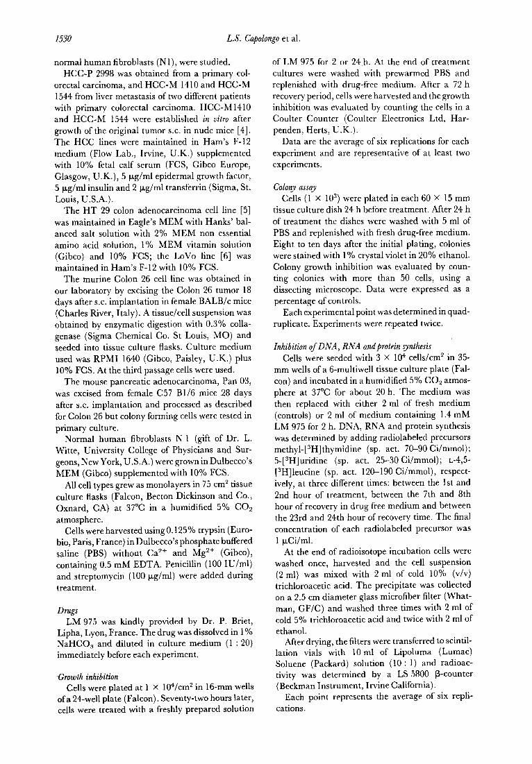
Studies of cytotoxicity Table 1 shows the growth inhibitory effects of
LM 975 in human colonic cell lines and on normal human fibroblasts observed after 2 or 24 h exposure to the drug and after 72 h of post-drug incubation at 37°C in drug-free medium. Two hours treatment only inhibited growth of the HCC-P 2998 cell line:
Table 1. Antiproliferative effect of LM 975 on different cell lines. Data represent the number of surviving cells after 2 or 24 h of treattnmt and 72 h of post-treatment incubation in dmg free
medium, expressed as percentages of controls
Treatment 0.5 mM 1.0 mM 1.4 mM Cell lines time LM 975 LM 975 LM 975
(h) co/o) W) (%I
HT 29 2 LOVO 2 HCC-P2998 2
Colon 26 2
Nl 2
HT 29 24 HCC-Ml410 24 LOVO 24 HCC-Ml544 24 HCC-P2998 24
Colon 26 24
Nl 24
1lOr 6 95t 9 go+- 5 128k 6 135 f 15
92k 17 75 k 21 42 2 ll**
- - 992 7
- 121 k 8
113 f 16 8lk 9 87k 11 89 t 31 75 k 14 65 k 14 93t 4 79 5 3* 77 ” 4* 60? 11 51 -c 2* 47 f 6* 702 11 46 k 10* 36 2 8*
- 59 f 2*
116k 7 89It 6 97k 6
*P < 0.01, Dunnet’s test.
0
{OH
:Hp 0 &P 0 Ol
0
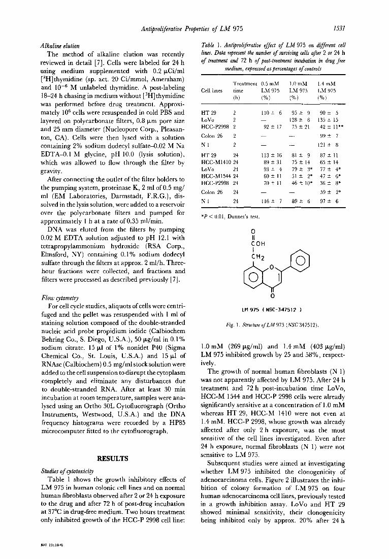
LM975 (NSC-347512 1
Fig. 1. Structure of LM 975 (NSC 347512).
1 .O mM (269 pg/ml) and 1.4 mM (403 pg/ml) LM 975 inhibited growth by 25 and 58%, respect- ively.
The growth of normal human fibroblasts (N 1) was not apparently affected by LM 975. After 24 h treatment and 72 h post-incubation time LoVo, HCC-M 1544 and HCC-P 2998 cells were already significantly sensitive at a concentration of 1 .O mM whereas HT 29, HCC-M 1410 were not even at 1.4 mM. HCC-P 2998, whose growth was already affected after only 2 h exposure, was the most sensitive of the cell lines investigated. Even after 24 h exposure, normal fibroblasts (N 1) were not sensitive to LM 975.
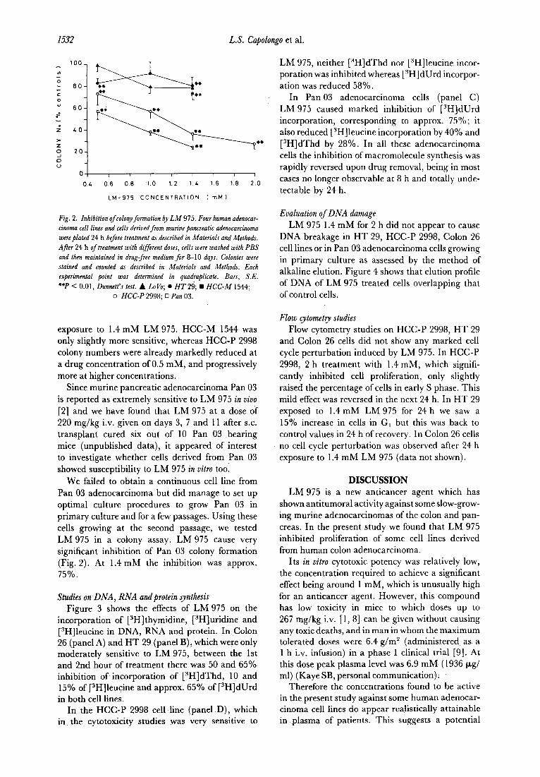
Subsequent studies were aimed at investigating whether LM 975 inhibited the clonogenicity of adenocarcinoma cells. Figure 2 illustrates the inhi- bition of colony formation of LM 975 on four human adenocarcinoma cell lines, previously tested in a growth inhibition assay. LoVo and HT 29 showed minimal sensitivity, their clonogenicity being inhibited only by approx. 20% after 24 h
L.S. Capolongo et al.
0.6 0.6 06 1.0 1.2 14 1.6 1.6 2.0
LM-975 CONCENTRATION ( mM 1
Fig. 2. Inhibition of colonyformation b LM 975. Four human adenocar- cinema cell lines and cells derived from murine pancreatic adenocarcinoma wereplated 24 h before treatment as described in Materials and Methods. After 24 h of treatment with diff eren t d oses, cells were washed with PBS and then maintained in drug-free medium for 8-10 days. Colonies were stained and counted as described in Materials and Methods. Each experimental point was determined in quadruplicate. Bars, S.E. **P < 0.01, Dunnctt’s test. A LoVo; ??HT 29; ??HCC-M 1544;
o HCC-P 2998; 0 Pan 03.
exposure to 1.4 mM LM 975. HCC-M 1544 was only slightly more sensitive, whereas HCC-P 2998 colony numbers were already markedly reduced at a drug concentration of 0.5 mM, and progressively more at higher concentrations,
Since murine pancreatic adenocarcinoma Pan 03 is reported as extremely sensitive to LM 975 in uiuo [2] and we have found that LM 975 at a dose of 220 mg/kg i.v. given on days 3, 7 and 11 after S.C. transplant cured six out of 10 Pan 03 bearing mice (unpublished data), it appeared of interest to investigate whether cells derived from Pan 03 showed susceptibility to LM 975 in vitro too.
We failed to obtain a continuous cell line from Pan 03 adenocarcinoma but did manage to set up optimal culture procedures to grow Pan 03 in primary culture and for a few passages. Using these cells growing at the second passage, we tested LM 975 in a colony assay. LM 975 cause very significant inhibition of Pan 03 colony formation (Fig. 2). At 1.4 mM the inhibition was approx. 75%.
Studies on DNA, RNA andprotein synthesis Figure 3 shows the effects of LM 975 on the
incorporation of [3H]thymidine, [3H]uridine and [3H]leucine in DNA, RNA and protein. InColon 26 (panel A) and HT 29 (panel B), which were only moderately sensitive to LM 975, between the 1st and 2nd hour of treatment there was 50 and 65% inhibition of incorporation of [3H]dThd, 10 and 15% of [3H]leucine and approx. 65% of [3H]dUrd in both cell lines.
In the HCC-P 2998 cell line (panel D), which in the cytotoxicity studies was very sensitive to
LM 975, neither [3H]dThd nor [3H]leucine incor- poration was inhibited whereas [‘H]dUrd incorpor- ation was reduced 58%.
In Pan 03 adenocarcinoma cells (panel C) LM 975 caused marked inhibition of [3H]dUrd incorporation, corresponding to approx. 75%; it also reduced [3H]leucine incorporation by 40% and [3H]dThd by 28%. I n all these adenocarcinoma cells the inhibition of macromolecule synthesis was rapidly reversed upon drug removal, being in most cases no longer observable at 8 h and totally unde- tectable by 24 h.
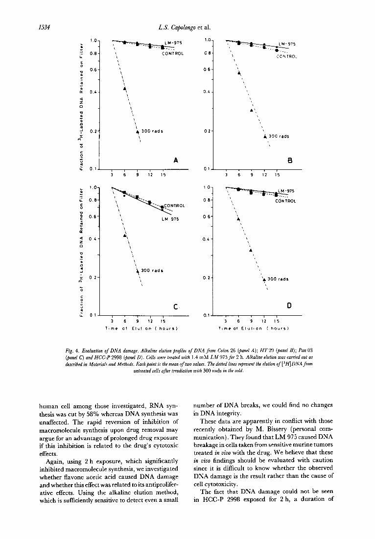
Evaluation of DNA damage LM 975 1.4 mM for 2 h did not appear to cause
DNA breakage in HT 29, HCC-P 2998, Colon 26 cell lines or in Pan 03 adenocarcinoma cells growing in primary culture as assessed by the method of alkaline elution. Figure 4 shows that elution profile of DNA of LM 975 treated cells overlapping that of control cells.
Flow cytometry studies Flow cytometry studies on HCC-P 2998, HT 29
and Colon 26 cells did not show any marked cell cycle perturbation induced by LM 975. In HCC-P 2998, 2 h treatment with 1.4 mM, which signifi- cantly inhibited cell proliferation, only slightly raised the percentage of cells in early S phase. This mild effect was reversed in the next 24 h. In HT 29 exposed to 1.4 mM LM 975 for 24 h we saw a 15% increase in cells in G, but this was back to control values in 24 h of recovery. In Colon 26 cells no cell cycle perturbation was observed after 24 h exposure to 1.4 mM LM 975 (data not shown).
DISCUSSION LM 975 is a new anticancer agent which has
shown antitumoral activity against some slow-grow- ing murine adenocarcinomas of the colon and pan- creas. In the present study we found that LM 975 inhibited proliferation of some cell lines derived from human colon adenocarcinoma.
Its in vitro cytotoxic potency was relatively low, the concentration required to achieve a significant effect being around 1 mM, which is unusually high for an anticancer agent. However, this compound has low toxicity in mice to which doses up to 267 mg/kg, i.v. [ 1, 81 can be given without causing any toxic deaths, and in man in whom the maximum tolerated doses were 6.4 g/m2 (administered as a 1 h i.v. infusion) in a phase I clinical trial [9]. At this dose peak plasma level was 6.9 mM (1936 kg/ ml) (Kaye SB, personal communication).
Therefore the concentrations found to be active in the present study against some human adenocar- cinema cell lines do appear realistically attainable in plasma of patients. This suggests a potential

Antiproliferative Properties of LM 975
6 b 2L
EN0 OF t TREATMENT
150
z 0 ;
i loo- $_/.j
z - s
50- ??* ,
/ .:
C 0 , I
B 0 ,
I 1
hours 0 8 2L
EN0 OFT TREATMENT
150
z 0
f 100
fz
: 0
if 50
r’
C 0 8 24 hours b ;, 2& hours
END OF 1 EN0 OFT TREATMENT TREATMENT
+=-A ,, ~_.__.__._.-.-.-‘-‘-“~
/I i
W
D
1533
hours
Fig. 3. Inhibition of DNA, RNA andprotein synthesis by LM 975 in Colon 26 (panel A); HT 29 (panel B); Pan 03 (panel C) and HCC-P 2998 (panel D). Cells were seeded and treated with 1.4 mM LM 975 for 2 h (1~ described in Materials and Methods. DNA, RNA and protein gnthesis was determined adding radiolabeled precursors met/#-[“Hlthymidine; 5-[3H] widine; ~-4,5-[~H] let&e, respectively, at three different times: between the 1st and 2nd hour of treatment, between the 7th and 8th hour of recovery time in drug-free medium and between the 23rd and 24th hour of recovery. The faral concentration of each radiolabeled precursor was 1 pJ2i/ml. At the end of radioisotope incubation cells were washed once, harvested and the cell suspension was mixed with cold 10% (v/v) trichloroacetic acid. The precipitate was collected on a glass microfiberjilter’and processed as described in Materials and Methods for the determination of macromolecule-bound radioactioip. At time 0 (end of treatment) and 8 or 24 h recovery time each point represents the percentage of controls of six replications. Symbols: (-) methyl-[gH]thymidine,
(---) 5-[3H]uridine; (-.-) ~-4,5-[~H]t eucine. Bars, S.E. **P < 0.01, Dunnett’s test.
therapeutic efficacy of LM 975 against human colon cancer.
The cytotoxicity studies showed variable sensi- tivity among the five human adenocarcinoma cell lines investigated. HCC-P 2998, recently isolated from a biopsy of a primary human colorectal carci- noma, was susceptible even when exposed to 1.0 mM LM 975 for only 2 h. The least sensitive cell line, HT 29, was only slightly affected when 1.4 mM drug concentrations were maintained for 24 h. The identification of some cell lines partially sensitive to this drug is a first step towards studies
aimed at elucidating the mode of action of LM 975. With this in mind, we chose a relatively short duration of treatment (2 h) to avoid unspecific effects due to cell damage induced by drug cytotoxic- ity. Indeed already between the 1st and 2nd hour of treatment DNA, RNA and protein synthesis was inhibited by LM 975. However, the synthesis of macromolecules was affected to a different extent and proportion in the different cell lines. In sensitive cell lines LM 975 caused stronger inhibition of RNA synthesis than DNA synthesis. For example, in HCC-P 2998, which was the most sensitive
1534 L.S. Capolongo et al.
1 CONTROL \
; 300 fads
t l.O-
.: 0.6-
:
P .E
0.6-
I ; (I
z 0 4- \ \ , \
‘4 300 fads
\ \
C ,
I I 1 1
3 6 9 12 15
T,mc of Elutlcn ( hours)
4 300 rdds
0 6-
! CONTROL
4
‘4 300 rads
D 0.1 ! I I
3 6 9 12 15
llmc ot Elutaon ( hours1
Fig. 4. Evaluation of DNA damage. Alkaline e&ion projiles of DNA f rom Colon 26 (panel A); HT 29 (panel B); Pan 03 (panel C) and HCC-P 2998 Qanel D). C 11 e s were treated with 1.4 mM LM 975 for 2 h. Alkaline e&ion was carried out as described in Materials und Methods. Each point is the mean of two values. The dotted lines represent the e&ion of L3H]DNA from
untreated cells after irradiation with 300 rads in the cold.
human cell among those investigated, RNA syn- thesis was cut by 58% whereas DNA synthesis was unaffected. The rapid reversion of inhibition of macromolecule synthesis upon drug removal may argue for an advantage of prolonged drug exposure if this inhibition is related to the drug’s cytotoxic effects.
Again, using 2 h exposure, which significantly inhibited macromolecule synthesis, we investigated whether flavone acetic acid caused DNA damage and whether this effect was related to its antiprolifer- ative effects. Using the alkaline elution method, which is sufficiently sensitive to detect even a small
number of DNA breaks, we could find no changes in DNA integrity.
These data are apparently in conflict with those recently obtained by M. Bissery (personal com- munication). They found that LM 975 caused DNA breakage in cells taken from sensitive murine tumors treated in uivo with the drug. We believe that these in vivo findings should be evaluated with caution since it is difficult to know whether the observed DNA damage is the result rather than the cause of cell cytotoxicity.
The fact that DNA damage could not be seen in HCC-P 2998 exposed for 2 h, a duration of
Antiproliferative Properties of LM 975 1535
treatment which in this cell line did inhibit cell growth, supports the idea that LM 975 does not act by causing DNA breakage. In addition, preliminary experiments indicate that LM 975 does not bind markedly to DNA as assessed by equilibrium dialysis.
To our knowledge all drugs which act by causing DNA damage produce an accumulation of cells in S phase and G2 [ 10, 111; the finding that LM 975 did not cause this cell cycle perturbation also argues against the hypothesis that the drug acts through DNA damage. A further argument against DNA damage being at the basis of LM 975 activity derives from observations that the drug does not cause bone marrow toxicity; this would be uncom- mon for a drug that acts by damaging DNA.
In conclusion the present study provides evidence that LM 975 has a moderate antiproliferative activity on some adenocarcinoma cells at concen- trations which are pharmacologically achievable in plasma of patients receiving tolerable doses.
The mechanism of action of this drug should be further investigated but studies so far suggest that it does not act by producing DNA damage. A finding that requires confirmation is whether RNA
synthesis inhibition by LM 975 is a general feature in all drug-sensitive cells and what is its molecular basis.
The cytotoxic effect of LM 975 is not very high. Even on Pan 03 cells derived from a mouse tumor which can be cured by LM 975 [2], less than a log cell kill was observed. The greater in vivo activity can be tentatively explained in two ways:
(i) LM 975 could be converted in vivo to more effective metabolites which are not formed when the drug is tested against cells growing in culture.
(ii) LM 975 could stimulate some immunological response which in turn may be partially responsible for the in vivo antitumoral activity.
We are inclined to exclude the former possibility since we could not identify any major LM 975 bioproduct in plasma, urine or tissues of mice treated with therapeutic doses of the drug or by incubating LM 975 with fortified mouse liver microsomes (Damia G et al., manuscript in prep- aration). The second possibility which is under investigation in this laboratory has recently been suggested (Wiltrout RH, personal communication).
1.
2.
3. 4.
5.
6.
7.
8.
9.
10.
11.
REFERENCES Plowman J, Narayanan VL, Dykes D et al. Flavone acetic acid: a novel agent with preclinical antitumor activity against colon adenocarcinoma 38 in mice. Cancer Treat Rep 1986, 70, 631-635. Corbett TH, Bissery M-C, Wozniak A et al. Activity of flavone acetic acid (NSC-347512) against solid tumors of mice. Invest New Drugs 1986,4, 207-220. National Cancer Institute. Flavone acetic acid NSC 347512. Clinical Brochure, July 1985. Giavazzi R, Campbell DE, Jessup JM, Cleary K, Fidler IJ. Metastatic behavior of tumor cells isolated from primary and metastatic human colorectal carcinomas implanted into different sites in nude mice. Cancer Res 1986,46, 1928-1933. Fogh J, Trempe G. New human tumor cell lines. In: Fogh J, ed. Human Tumor Cells in Vitro. New York, Plenum Press, 1975, 115-159. Drewinko B, Romsdahl MM, Yang LY, Ahearn MJ, Trujillo JM. Establishment of a human carcinoembryonic antigen-producing colon adenocarcinoma cell line. Cancer Res 1976, 36, 467-475. Kohn KW, Ewig RAG, Erickson LC, Zwelling LA. Measurement of strand breaks and cross-links by alkaline elution. In: Friedberg EC, Hanawalt PC, eds. DNA Repair, part B. New York, Marcel Dekker, 1981, Vol. 1, 379-401. Zaharko DS, Grieshaber CK, Plowman J, Cradock JC. Therapeutic and pharmacokinetic relationships offlavone acetic acid: an agent with activity against solid tumors. Cancer Treat Rep1986, 70, 1415-1421. Kerr DJ, Kaye SB, Setanoians A et al. Phase I and pharmacokinetic study of LM975 (flavone acetic acid). In: Fifth NC1 EORTC Symposium on New Drugs in Cancer Therapy, Amsterdam, The Netherlands, 22-24 October 1986, Abstr. 8.03. Tobey RA. Different drugs arrest cells at a number of distinct stages in GP. Nature 1975, 254, 245-247. Rao P, Rao PN. The cause of Gz-arrest in Chinese hamster ovary cells treated with anticancer drugs. J Nut1 Cancer Inst 1976, 57, 1139-l 143.


![[Objective-C] - 02 Properties e Costruttori](https://static.fdocumenti.com/doc/165x107/548229e4b4af9fc2488b465e/objective-c-02-properties-e-costruttori.jpg)