Acute right intrathoracic gastric volvulus. · Acute right intrathoracic gastric volvulus. ......
4
Acute right intrathoracic gastric volvulus. A rare surgical emergency Ann. Ital. Chir., 84, 2, 2013 205 Ann. Ital. Chir., 2013 84: 205-207 pii: S0003469X13019076 Pervenuto in Redazione Aprile 2012, Accettato per la pubblicazione Giugno 2012 Correspondence to: Arvin Dibra, MD, PhD, Catholic University of Tirana, Faculty of Medicine, Tirana, Albania (e-mail: [email protected]) Arvin Dibra*, Francesco Rulli***, Myzafer Kaçi*, Ejona Çeliku**, Xheladin Draçini* *University Hospital Centre of Tirana “Mother Theresa”, First Clinic of General Surgery, University of Tirana, Albania **Intern on Radiology, “Lady of Good Counseel” Catholic University of Tirana, Albania ***Department of Surgery, University of Rome Tor Vergata, Rome, Italy Acute right intrathoracic gastric volvulus. A rare surgical emergency The acute intrathoracic gastric volvulus is a rare condition, difficult to diagnose and treat. It consist in a abnormal organo-axial rotation over 180°, associated with gastric obstruction or strangulation. More uncommon condition is the gastric volvulus caused by a right sliding diaphragmatic hernia and dislocating the stomach, or part of it, on the right hemithorax. Gastric volvulus classic clinical presentation described by Borchardt, consist on a triad of severe epigastric pain, vomiting followed by retching without ability to vomit and difficulty or inability to pass a nasogastric tube. Imaging, beginning from a simple chest radiograph showing an elevated gastric air-fluid level in lower lung segments, can help to define diagnosis and to determine the immediate necessity to operate trying to avoid fatal complications as gastric ischemia, perforation or haemorrhage. We present the case of a 58 year-old man arrived at our Emergency Department with moderate acute epigastric pain and already vomiting from 4 hours. The patient underwent initially a chest radiograph, Computed Tomography, upper digestive endoscopy, upper digestive contrasted radiology and then was operated. Post operative situation of the patient on recovery and during the 3 months follow up didn’t experience any pain or difficulty in feeding. KEY WORDS: Borchardt’s triad, Diaphragmatic hernia, Gastric volvulus Introduction Mostly happening on the fifth decade of life, gastric volvulus consist in a rotation greater than 180° of the stomach, or parts of it. The most common type of rota- tion consist in the rotation along the longitudinal gas- tric axis. Acute intrathoracic gastric volvulus occurs when the stomach undergoes organo-axial torsion in the chest due to an enlargement of the hiatus or a diaphragmatic her- nia. This particular condition can occur also after sur- gical procedures on the upper abdomen, mainly after hiatal hernia repair. It is also found in literature described as iatrogenic diaphragmatic hernia associated with intrathoracic gastric volvulus after left nephrectomy 1 , esophagogastrectomy 2 and splenopancreatectomy 3 . The clinical situation of the intrathoracic stomach com- bined with gastric volvulus are epigastric and lower chest pain, post prandial discomfort and dysphagia, initial vomiting followed by retching without ability to vomit, hemorrhage, retrosternal sense of fullness and anemia associated with inability or difficulty to pass a nasogas- tric tube. This pathological condition is frequently diagnosed by imaging intrathoracic viscera and/or air-fluid levels in the chest radiograph; this can be followed by a upper oral contrast radiology or upper gastrointestinal endoscopy. Computed Tomography (CT) scan leads to immediate diagnosis with anatomical details. This condition is con-
Transcript of Acute right intrathoracic gastric volvulus. · Acute right intrathoracic gastric volvulus. ......
Acute right intrathoracic gastric volvulus.A rare surgical emergency
Ann. Ital. Chir., 84, 2, 2013 205
Ann. Ital. Chir., 2013 84: 205-207pii: S0003469X13019076
Pervenuto in Redazione Aprile 2012, Accettato per la pubblicazioneGiugno 2012Correspondence to: Arvin Dibra, MD, PhD, Catholic University of Tirana,Faculty of Medicine, Tirana, Albania (e-mail: [email protected])
Arvin Dibra*, Francesco Rulli***, Myzafer Kaçi*, Ejona Çeliku**, Xheladin Draçini*
*University Hospital Centre of Tirana “Mother Theresa”, First Clinic of General Surgery, University of Tirana, Albania**Intern on Radiology, “Lady of Good Counseel” Catholic University of Tirana, Albania***Department of Surgery, University of Rome Tor Vergata, Rome, Italy
Acute right intrathoracic gastric volvulus. A rare surgical emergency
The acute intrathoracic gastric volvulus is a rare condition, difficult to diagnose and treat. It consist in a abnormalorgano-axial rotation over 180°, associated with gastric obstruction or strangulation. More uncommon condition is thegastric volvulus caused by a right sliding diaphragmatic hernia and dislocating the stomach, or part of it, on the righthemithorax. Gastric volvulus classic clinical presentation described by Borchardt, consist on a triad of severe epigastricpain, vomiting followed by retching without ability to vomit and difficulty or inability to pass a nasogastric tube.Imaging, beginning from a simple chest radiograph showing an elevated gastric air-fluid level in lower lung segments,can help to define diagnosis and to determine the immediate necessity to operate trying to avoid fatal complications asgastric ischemia, perforation or haemorrhage. We present the case of a 58 year-old man arrived at our EmergencyDepartment with moderate acute epigastric pain and already vomiting from 4 hours. The patient underwent initially achest radiograph, Computed Tomography, upper digestive endoscopy, upper digestive contrasted radiology and then wasoperated. Post operative situation of the patient on recovery and during the 3 months follow up didn’t experience anypain or difficulty in feeding.
KEY WORDS: Borchardt’s triad, Diaphragmatic hernia, Gastric volvulus
Introduction
Mostly happening on the fifth decade of life, gastricvolvulus consist in a rotation greater than 180° of thestomach, or parts of it. The most common type of rota-tion consist in the rotation along the longitudinal gas-tric axis. Acute intrathoracic gastric volvulus occurs when thestomach undergoes organo-axial torsion in the chest dueto an enlargement of the hiatus or a diaphragmatic her-
nia. This particular condition can occur also after sur-gical procedures on the upper abdomen, mainly afterhiatal hernia repair. It is also found in literature describedas iatrogenic diaphragmatic hernia associated withintrathoracic gastric volvulus after left nephrectomy 1,esophagogastrectomy 2 and splenopancreatectomy 3.The clinical situation of the intrathoracic stomach com-bined with gastric volvulus are epigastric and lower chestpain, post prandial discomfort and dysphagia, initialvomiting followed by retching without ability to vomit,hemorrhage, retrosternal sense of fullness and anemiaassociated with inability or difficulty to pass a nasogas-tric tube.This pathological condition is frequently diagnosed byimaging intrathoracic viscera and/or air-fluid levels in thechest radiograph; this can be followed by a upper oralcontrast radiology or upper gastrointestinal endoscopy.Computed Tomography (CT) scan leads to immediatediagnosis with anatomical details. This condition is con-
sidered life-threatening because delayed treatment mayresult in perforation, infarction or other lethal outcomes4. We will describe a rare case of an acute intrathoracicgastric volvulus located on right chest, due to rightdiaphragmatic hernia, occurring in a middle-aged man.
Case report
A 58 year-old man arrived at the Emergency Departmentof UHC “Mother Theresa” of Tirana with a 12 hourshistory of acute moderate epigastric pain and sense ofretrosternal fullness. The patient reports about vomitingonce and then retching without being able to vomitagain. Physical examination showed diffuse tenderness,hyperactive bowel sounds and unventilated right lungbasal segments. Abdominal ultrasound and electrocar-diography were both unremarkable. It wasn’t possible topass a functional nasogastric tube.On patients arrival blood pressure was 125/90 mmHg,heart rate was 92 beats per minute, respiratory rate was15 times per minute, and body temperature was 36.8°C.Laboratory results were as follows: WBC 8,900/ l with43% segmented neutrophils, BUN 56 mg/dl, creatinine0.97 mg/dl, amylase 57 IU/l, lipase 12 IU/l, AST 45IU/l and ALT 41 IU/l. The chest radiograph demonstrated an elevated gastricair-fluid level in the right lower lung segments (Fig. 1).An urgent gastroduodenoscopy was performed demon-strating difficult passage over the gastro oesophageal junc-tion, massive fluid collection below that point and inabil-ity to reach the duodenum due to a structural abnor-mality. The explored gastric mucosa was strongly hyper-emic. A combined chest – abdominal CT scan demon-strated a distended stomach, located and rotated in theright lower hemithorax through a right diaphragmaticdefect (Fig. 2). The correct diagnosis of right intratho-racic gastric volvulus through diaphragmatic hernia wasconfirmed by performing an upper gastrointestinal oralcontrast radiograph (Fig. 3).
With this working diagnosis, emergent laparotomy wasperformed. The pyloric valve was rised in abdominal cav-ity up to the diaphragmatic level and the other parts ofthe stomach were found in the right lower thorax. We
A. Dibra, et. al.
206 Ann. Ital. Chir., 84, 2, 2013
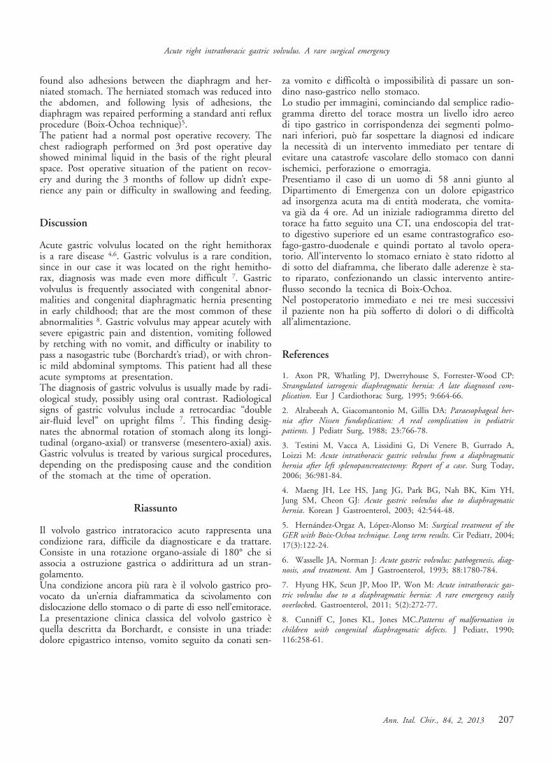
Fig. 1: Chest radiograph at admission shows a high gastric air-fluid lev-el on the right lower lung segments and an elevated gastric contour.
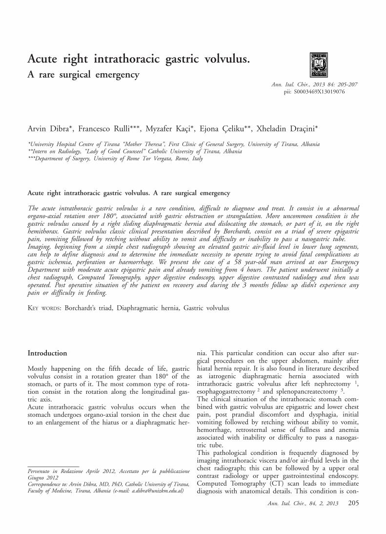
Fig. 2: Combined chest and abdominal CT scan demonstrated a dis-tended stomach, located and rotated in the right lower hemithoraxthrough a right diaphragmatic defect.
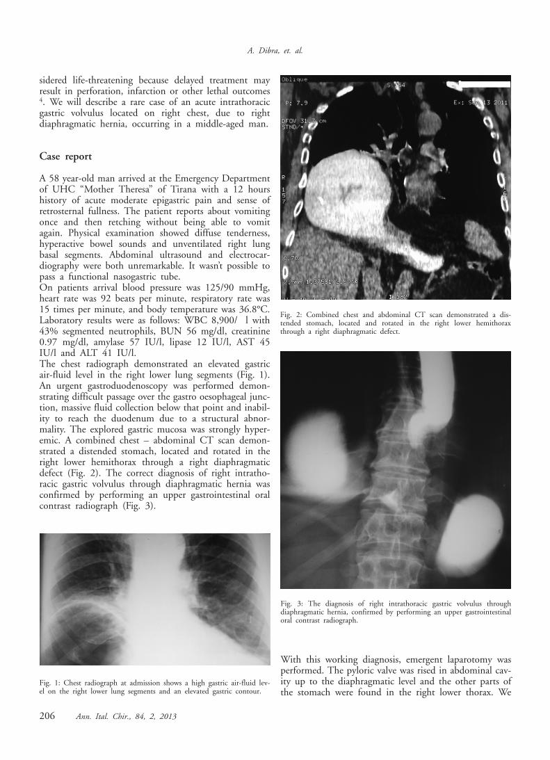
Fig. 3: The diagnosis of right intrathoracic gastric volvulus throughdiaphragmatic hernia, confirmed by performing an upper gastrointestinaloral contrast radiograph.
found also adhesions between the diaphragm and her-niated stomach. The herniated stomach was reduced intothe abdomen, and following lysis of adhesions, thediaphragm was repaired performing a standard anti refluxprocedure (Boix-Ochoa technique)5.The patient had a normal post operative recovery. Thechest radiograph performed on 3rd post operative dayshowed minimal liquid in the basis of the right pleuralspace. Post operative situation of the patient on recov-ery and during the 3 months of follow up didn’t expe-rience any pain or difficulty in swallowing and feeding.
Discussion
Acute gastric volvulus located on the right hemithoraxis a rare disease 4,6. Gastric volvulus is a rare condition,since in our case it was located on the right hemitho-rax, diagnosis was made even more difficult 7. Gastricvolvulus is frequently associated with congenital abnor-malities and congenital diaphragmatic hernia presentingin early childhood; that are the most common of theseabnormalities 8. Gastric volvulus may appear acutely withsevere epigastric pain and distention, vomiting followedby retching with no vomit, and difficulty or inability topass a nasogastric tube (Borchardt’s triad), or with chron-ic mild abdominal symptoms. This patient had all theseacute symptoms at presentation.The diagnosis of gastric volvulus is usually made by radi-ological study, possibly using oral contrast. Radiologicalsigns of gastric volvulus include a retrocardiac “doubleair-fluid level” on upright films 7. This finding desig-nates the abnormal rotation of stomach along its longi-tudinal (organo-axial) or transverse (mesentero-axial) axis.Gastric volvulus is treated by various surgical procedures,depending on the predisposing cause and the conditionof the stomach at the time of operation.
Riassunto
Il volvolo gastrico intratoracico acuto rappresenta unacondizione rara, difficile da diagnosticare e da trattare.Consiste in una rotazione organo-assiale di 180° che siassocia a ostruzione gastrica o addirittura ad un stran-golamento.Una condizione ancora più rara è il volvolo gastrico pro-vocato da un’ernia diaframmatica da scivolamento condislocazione dello stomaco o di parte di esso nell’emitorace. La presentazione clinica classica del volvolo gastrico èquella descritta da Borchardt, e consiste in una triade:dolore epigastrico intenso, vomito seguito da conati sen-
za vomito e difficoltà o impossibilità di passare un son-dino naso-gastrico nello stomaco.Lo studio per immagini, cominciando dal semplice radio-gramma diretto del torace mostra un livello idro aereodi tipo gastrico in corrispondenza dei segmenti polmo-nari inferiori, può far sospettare la diagnosi ed indicarela necessità di un intervento immediato per tentare dievitare una catastrofe vascolare dello stomaco con danniischemici, perforazione o emorragia. Presentiamo il caso di un uomo di 58 anni giunto alDipartimento di Emergenza con un dolore epigastricoad insorgenza acuta ma di entità moderata, che vomita-va già da 4 ore. Ad un iniziale radiogramma diretto deltorace ha fatto seguito una CT, una endoscopia del trat-to digestivo superiore ed un esame contrastografico eso-fago-gastro-duodenale e quindi portato al tavolo opera-torio. All’intervento lo stomaco erniato è stato ridotto aldi sotto del diaframma, che liberato dalle aderenze è sta-to riparato, confezionando un classic intervento antire-flusso secondo la tecnica di Boix-Ochoa.Nel postoperatorio immediato e nei tre mesi successiviil paziente non ha più sofferto di dolori o di difficoltàall’alimentazione.
References
1. Axon PR, Whatling PJ, Dwerryhouse S, Forrester-Wood CP:Strangulated iatrogenic diaphragmatic hernia: A late diagnosed com-plication. Eur J Cardiothorac Surg, 1995; 9:664-66.
2. Alrabeeah A, Giacomantonio M, Gillis DA: Paraesophageal her-nia after Nissen fundoplication: A real complication in pediatricpatients. J Pediatr Surg, 1988; 23:766-78.
3. Testini M, Vacca A, Lissidini G, Di Venere B, Gurrado A,Loizzi M: Acute intrathoracic gastric volvulus from a diaphragmatichernia after left splenopancreatectomy: Report of a case. Surg Today,2006; 36:981-84.
4. Maeng JH, Lee HS, Jang JG, Park BG, Nah BK, Kim YH,Jung SM, Cheon GJ: Acute gastric volvulus due to diaphragmatichernia. Korean J Gastroenterol, 2003; 42:544-48.
5. Hernández-Orgaz A, López-Alonso M: Surgical treatment of theGER with Boix-Ochoa technique. Long term results. Cir Pediatr, 2004;17(3):122-24.
6. Wasselle JA, Norman J: Acute gastric volvulus: pathogenesis, diag-nosis, and treatment. Am J Gastroenterol, 1993; 88:1780-784.
7. Hyung HK, Seun JP, Moo IP, Won M: Acute intrathoracic gas-tric volvulus due to a diaphragmatic hernia: A rare emergency easilyoverlocked. Gastroenterol, 2011; 5(2):272-77.
8. Cunniff C, Jones KL, Jones MC.Patterns of malformation inchildren with congenital diaphragmatic defects. J Pediatr, 1990;116:258-61.
Ann. Ital. Chir., 84, 2, 2013 207
Acute right intrathoracic gastric volvulus. A rare surgical emergency