Università degli Studi di Padova Dipartimento di Salute...
100
Università degli Studi di Padova Dipartimento di Salute della Donna e del Bambino - SDB DOTTORATO DI RICERCA IN MEDICINA DELLO SVILUPPO E SCIENZE DELLA PROGRAMMAZIONE INDIRIZZO DI EMATOONCOLOGIA, IMMUNOLOGIA E GENETICA Ciclo XXIV TITOLO ANALISI DI NUOVE STRATEGIE TERAPEUTICHE PER PAZIENTI IN ETÀ PEDIATRICA AFFETTI DA TUMORI SOLIDI REFRATTARI ALLA CHEMIOTERAPIA STANDARD Direttore della Scuola : Ch.mo Prof. Giuseppe Basso Coordinatore d’indirizzo: Ch.mo Prof. Giuseppe Basso Supervisori : Ch.mo Prof. Ottaviano Modesto Carli Dr Gianni Bisogno Dottorando: Dott.ssa Alessia Compostella
Transcript of Università degli Studi di Padova Dipartimento di Salute...
Università degli Studi di Padova
Dipartimento di Salute della Donna e del Bambino - SDB
DOTTORATO DI RICERCA IN MEDICINA DELLO SVILUPPO E SCIENZE DELLA PROGRAMMAZIONE
INDIRIZZO DI EMATOONCOLOGIA, IMMUNOLOGIA E GENETICA
Ciclo XXIV
TITOLO
ANALISI DI NUOVE STRATEGIE TERAPEUTICHE PER PAZIENTI IN ETÀ PEDIATRICA AFFETTI DA TUMORI SOLIDI REFRATTARI ALLA CHEMIOTERAPIA
STANDARD
Direttore della Scuola : Ch.mo Prof. Giuseppe Basso
Coordinatore d’indirizzo: Ch.mo Prof. Giuseppe Basso
Supervisori : Ch.mo Prof. Ottaviano Modesto Carli
Dr Gianni Bisogno
Dottorando: Dott.ssa Alessia Compostella


INDICE
OBIETTIVI E SOMMARIO pag. 1
1 GLI ADOLESCENTI pag. 3
1.1 Introduzione pag. 3
1.2 Rhabdomyosarcoma in Adolescents. A Report From the AIEOP Soft
Tissue Sarcoma Committee pag. 7
2 ANALISI DATI TOPOTECAN/CARBOPLATINO pag. 23
2.1 Introduzione pag. 23
2.2 A Topotecan carboplatin based strategy for children with refractory or
recurrent rhabdomyosarcoma pag. 27
3 I NUOVI FARMACI pag. 45
3.1 Introduzione pag. 45
3.2 Trials clinici pag. 49
3.3 Protocollo fase II pag. 55
3.3.1 Introduzione pag. 55
3.3.2 Phase II study of intrathecal topotecan in leptomeningeal
relapsed rhabdomyosarcoma and Ewing PNET children
and adolescents. pag. 57
PUBBLICAZIONI, POSTER/ABSTRACTS pag. 91
COMUNICAZIONI ORALI pag. 93
RINGRAZIAMENTI pag. 95


1
OBIETTIVI E SOMMARIO
L’attività di ricerca del Dottorato si è svolta presso la Clinica di Oncoematologia
Pediatrica del Dipartimento di Pediatria dell’Università degli Studi di Padova.
Il programma del dottorato si è sviluppato in un contesto di ricerca clinica ai fini dello
studio di “nuove” strategie terapeutiche per i pazienti pediatrici affetti da tumori solidi
recidivi/refrattari.
Il Nostro Centro costituisce uno dei maggiori Centri di Emato-Oncologia pediatrica in
Italia ed è il Centro Coordinatore per i sarcomi delle parti molli a livello Nazionale.
In Italia esiste, come è noto, una rete di rapporti tra i centri di Emato-Oncologia
Pediatrica che permette di seguire i bambini affetti da patologia neoplastica in modo
omogeneo e coordinato. La partecipazione attiva ai Protocolli terapeutici
dell’Associazione Italiana di Emato-Oncologia Pediatrica (AIEOP) e il contributo
costante nell’ elaborazione dei Protocolli stessi da parte del Nostro Centro, costituisce
uno dei punti fermi dell’attività clinica e scientifica. Per quanto attiene alla patologia in
discussione (tumori solidi dell’infanzia), il Nostro Centro ha inoltre funzioni di
coordinamento a livello europeo nell’ambito del protocollo per la cura dei sarcomi delle
parti molli EpSSG 2005.
Il Nostro Centro nell’attività scientifica e clinica quotidiana collabora, tra gli altri, con:
• AIEOP: Associazione Italiana di Emato-Oncologia Pediatrica
• EpSSG: European Protocol Soft Tissue Sarcoma Group
• EPOC: European Paediatric Oncology Off-patent Medicines Consortium
• INT Mi: Istituto Nazionale Tumori di Milano
• IOV: Istituto Oncologico Veneto
• ITCC: Innovative Therapies for Children with Cancer
Il nostro lavoro si è pertanto concentrato sui pazienti in età pediatrica, adolescenziale e
del giovane adulto affetti da tumore solido (in particolare Rabdomiosarcoma
recidivo/refrattario).
Una prima parte del lavoro si è focalizzata sullo studio della popolazione di pazienti
adolescenti affetti da rabdomiosarcoma (RMS) e trattati secondo i protocolli del soft
tissue sarcoma commitee (STSC) (1).
Una seconda parte del Dottorato è stata dedicata all’analisi dei risultati ottenuti dai
pazienti affetti da RMS trattati in seconda linea con il regime Topotecan/Carboplatino
(2).

2
Fin dall’inizio dell’attività di Dottorato, visti gli obiettivi dello stesso, è iniziata una
formazione specifica nell’ambito dei trials clinici, con partecipazione a corsi ad hoc,
formazione di un gruppo di lavoro sui nuovi farmaci e apertura del nostro Centro a
numerosi trials (3.2).
A “conclusione” di questo iter gli sforzi sono stati coordinati alla stesura di un
protocollo di fase II per pazienti affetti da recidiva meningea di RMS/PNET. Il
protocollo è in fase di stesura, verrà presentata l’attuale bozza (3.3).

3
1 GLI ADOLESCENTI
1.1 Introduzione
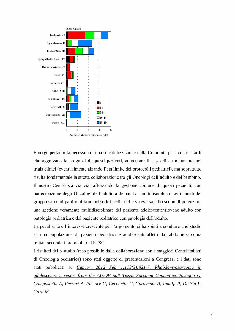
La Comunità Scientifica ha mostrato negli ultimi anni un crescente interesse per quella
che viene definita “Adolescent and Young Adult Oncology” (AYA) (1). Dati
epidemiologici delle ultime decadi mostrano per la fascia d’età 15-45 anni di pazienti
oncologici i peggiori risultati in termini di outcome/sopravvivenza. L’analisi del SEER
(Survival, Epidemiology and End Results) relativa ai dati di sopravvivenza da tumore in
base all’età (1975-1997), ha mostrato un miglioramento annuale del tasso di
sopravvivenza a 5 anni superiore all’1,5% per i pazienti di età <15 anni e >50 anni, a
fronte di un tasso <0,5% tra i 15-24 anni, assenza di miglioramento tra i 24-35 anni
(vedi grafico) (2, 3).
Tali risultati sono oggetto di ampio dibattito; uno dei principali fattori chiamati in causa
a motivare questi pessimi risultati è la scarsa partecipazione dei pazienti adolescenti e

4
giovani adulti nei trials clinici; in effetti il tasso di arruolamento nei trials clinici in base
all’età riflette l’andamento prognostico degli stessi pazienti (vedi grafico) (3, 4, 5, 6).
Altri fattori considerati importanti sono: una maggior aggressività biologica a parità di
patologia (7, 8); ritardi diagnostici dovuti sia al paziente che ai professionisti della
salute (il primo riluttante a esporre problematiche personali in una fase di maturazione
complessa caratterizzata da senso di autonomia e “invincibilità”, i secondi per scarsa
consapevolezza/conoscenza delle patologie oncologiche di questa fascia d’età) (3, 9).
Questo gruppo di pazienti così complessi sia dal punto di vista sociale e psicologico che
dal punto di vista dell’epidemiologia delle patologie oncologiche di cui sono affetti,
risiedono in quella che viene definita un’area “grigia”, “no-man’s land”, a metà strada
tra l’Oncologia Pediatrica e l’Oncologia dell’adulto. Infatti in questo gruppo emerge
una “transizione” epidemiologica: diminuiscono le patologie oncologiche pediatriche
(Wilms, medulloblastomi, rabdomiosarcomi,…) e aumenta l’incidenza di quelle tipiche
dell’età adulta (es. carcinomi), l’una di competenza pediatrica, l’altra dell’oncologo
dell’adulto (vedi grafico).
5
Emerge pertanto la necessità di una sensibilizzazione della Comunità per evitare ritardi
che aggravano la prognosi di questi pazienti, aumentare il tasso di arruolamento nei
trials clinici (eventualmente alzando l’età limite dei protocolli pediatrici), ma soprattutto
risulta fondamentale la stretta collaborazione tra gli Oncologi dell’adulto e del bambino.
Il nostro Centro sta via via rafforzando la gestione comune di questi pazienti, con
partecipazione degli Oncologi dell’adulto a demand ai multidisciplinari settimanali del
gruppo sarcomi parti molli/tumori solidi pediatrici e viceversa, allo scopo di potenziare
una gestione veramente multidisciplinare del paziente adolescente/giovane adulto con
patologia pediatrica e del paziente pediatrico con patologia dell’adulto.
La peculiarità e l’interesse crescente per l’argomento ci ha spinti a condurre uno studio
su una popolazione di pazienti pediatrici e adolescenti affetti da rabdomiosarcoma
trattati secondo i protocolli del STSC.
I risultati dello studio (reso possibile dalla collaborazione con i maggiori Centri italiani
di Oncologia pediatrica) sono stati oggetto di presentazioni a Congressi e i dati sono
stati pubblicati su Cancer. 2012 Feb 1;118(3):821-7. Rhabdomyosarcoma in
adolescents: a report from the AIEOP Soft Tissue Sarcoma Committee. Bisogno G,
Compostella A, Ferrari A, Pastore G, Cecchetto G, Garaventa A, Indolfi P, De Sio L,
Carli M.
6
BIBLIOGRAFIA
1 Bleyer A. Adolescent and young adult (AYA) oncology: the first A. Pediatr Hematol
Oncol. 2007 Jul-Aug;24(5):325-36.
2 Bleyer A, Viny A, Barr R. Cancer in 15- to 29-year-olds by primary site. Oncologist.
2006 Jun;11(6):590-601. Review.
3 Ferrari A, Bleyer A. Participation of adolescents with cancer in clinical trials. Cancer
Treat Rev. 2007 Nov;33(7):603-8.
4 Ferrari A, Dama E, Pession A, Rondelli R, Pascucci C, Locatelli F, Ferrari S,
Mascarin M, Merletti F, Masera G, Aricò M, Pastore G. Adolescents with cancer in
Italy: entry into the national cooperative paediatric oncology group AIEOP trials. Eur J
Cancer. 2009 Feb;45(3):328-34.
5 Ferrari A, Montello M, Budd T, Bleyer A. The challenges of clinical trials for
adolescents and young adults with cancer. Pediatr Blood Cancer. 2008 May;50(5
Suppl):1101-4.
6 Bleyer A, Montello M, Budd T, Saxman S. National survival trends of young adults
with sarcoma: lack of progress is associated with lack of clinical trial participation.
Cancer. 2005 May 1;103(9):1891-7.
7 Ferrari A, Dileo P, Casanova M, Bertulli R, Meazza C, Gandola L, Navarria P,Collini
P, Gronchi A, Olmi P, Fossati-Bellani F, Casali PG. Rhabdomyosarcoma in adults. A
retrospective analysis of 171 patients treated at a single institution. Cancer. 2003 Aug
1;98(3):571-80.
8 Joshi D, Anderson JR, Paidas C, Breneman J, Parham DM, Crist W; Soft Tissue
Sarcoma Committee of the Children's Oncology Group. Age is an independent
prognostic factor in rhabdomyosarcoma: a report from the Soft Tissue Sarcoma
Committee of the Children's Oncology Group. Pediatr Blood Cancer. 2004
Jan;42(1):64-73.
9 Ferrari A, Miceli R, Casanova M, Meazza C, Favini F, Luksch R, Catania S, Fiore M,
Morosi C, Mariani L. The symptom interval in children and adolescents with soft tissue
sarcomas. Cancer. 2010 Jan 1;116(1):177-83.

7
1.2 Rhabdomyosarcoma in Adolescents. A Report From the
AIEOP Soft Tissue Sarcoma Committee
Gianni Bisogno, MD, PhD1; Alessia Compostella, MD1; Andrea Ferrari, MD2; Guido
Pastore, MD3; Giovanni Cecchetto, MD4; Alberto Garaventa, MD5; Paolo Indolfi,
MD6; Luigi De Sio, MD7; and Modesto Carli, MD1.
BACKGROUND: In many types of cancer, the survival rates are reported to be less
favorable for adolescents compared with younger children. To investigate whether this
is true for adolescents with rhabdomyosarcoma (RMS), the results obtained in patients
enrolled in protocols run by the Italian Soft Tissue Sarcoma Committee (STSC) were
analyzed. METHODS: From 1988 through 2005, 643 patients were registered (567
children ages birth-14 years and 76 adolescents ages 15-19 years) and treated in 4 STSC
protocols. The number of patients enrolled was compared with the expected number
calculated from incidence rates derived from the Italian network of cancer registries.
RESULTS: Only 27% of the expected number of adolescents with RMS were enrolled
in the STSC trials. Compared with children, adolescents were found to have a longer
interval from initial symptoms to diagnosis (8 weeks vs 4.6 weeks), more alveolar RMS
(47.4% vs 32.6%), lymph node infiltration (39.1% vs 23.3%), and metastases at the
time of diagnosis (30.7% vs 17.8%). The 2 age groups received similar treatments. The
5-year overall survival (OS) rate was 68.9% in children versus 57.2% in adolescents (P:
0.006), and the progression-free survival (PFS) rate was 64.3% in children versus
48.1% in adolescents (P: 0.0237). On multivariate analysis, age, tumor site, lymph node
involvement, and metastases were found to be significant prognostic factors for OS and
PFS. CONCLUSIONS: Survival for adolescents with RMS enrolled in STSC protocols
appears to be satisfactory. The higher prevalence of unfavorable tumor characteristics
noted among adolescents seems to explain their worse outcome compared with children.
However, the limited number of adolescents enrolled in STSC studies is worrisome, and
cooperation with oncologists who treat adults needs to be improved.
Cancer 2011;000:000–000. VC 2011 American Cancer Society.
KEYWORDS: rhabdomyosarcoma, adolescents, soft tissue sarcoma, survival.

8
Rhabdomyosarcoma (RMS) is a rare tumor that typically affects children and
adolescents, with an annual incidence of 4.3 cases per 1 million population aged <20
years. Approximately 3 in 4 cases occur in children aged <10 years, with a peak
incidence between ages 3 and 5 years and a second, smaller peak in adolescence, after
which the incidence drops significantly with increasing age. Approximately 70% of
patients with localized RMS can now be cured, but their outcome is influenced by
various prognostic factors identified over the years and currently used for risk stratifica-
tion and risk-adapted treatment decisions (1). Along with other variables such as
histology, local and distant invasiveness, and tumor site and size, the patient’s age has
emerged as one of the most relevant factors, with older patients reported to have a
worse prognosis (2,3).
Among the various age groups, adolescents with cancer form a group with particular
features. Several studies have shown that improvements in the survival rates achieved in
recent years have been less satisfactory for adolescents and young adults compared with
younger children (4,5). Among the reasons suggested to explain this phenomenon are
the greater presence in adolescents of tumors with less favorable characteristics, delays
in the diagnosis, and a low accrual of adolescents in clinical trials (5,6).
Corresponding author: Gianni Bisogno, MD, PhD, Division of Hematology/Oncology,
Department of Pediatrics, Padova University Hospital, via Giustiniani 3-35128,
Padova, Italy; Fax: (011) 39-049-8213510; [email protected]
1Hematology/Oncology Division, Department of Pediatrics, Padova University
Hospital, Padova, Italy; 2Pediatric Oncology Unit, National Tumor Institute, Milan,
Italy; 3Childhood Cancer Registry, Piedmont, Italy; 4Pediatric Surgery Unit,
Department of Pediatrics, Padova University Hospital, Padova, Italy; 5Pediatric
Hematology/Oncology Division, G. Gaslini Children’s Hospital, Genova, Italy;
6Pediatric Oncology Service, Second University of Naples, Naples, Italy; 7Oncology
Division, Bambin Gesù Hospital, Rome, Italy
We thank Angela Scagnellato for data processing and Ilaria Zanetti and Angela De
Paoli for the statistical analysis.
DOI: 10.1002/cncr.26355, Received: February 15, 2011; Revised: April 18, 2011;
Accepted: April 28, 2011, Published online in Wiley Online Library
(wileyonlinelibrary.com).

9
To the best of our knowledge, no published studies to date have focused on adolescents
with RMS. Therefore, we analyzed the clinical and demographic characteristics,
treatment, and outcome for patients in this age group who were treated in the clinical
trials coordinated by the Associazione Italiana di Ematooncologia Pediatrica (AIEOP)
Soft Tissue Sarcoma Committee (STSC) between 1988 and 2005. Because age was not
considered to be a factor for treatment stratification purposes, children and adolescents
received the same treatment, making them an ideal population for evaluating the relative
contributions of the above-mentioned factors.
To our knowledge, no other multicenter or institutional protocols including adolescents,
or even adults, with RMS were being run in parallel in Italy during the same period of
time.
MATERIALS AND METHODS
Over the course of 18 years, patients were enrolled by AIEOP centers in 4 consecutive
protocols: RMS-88 and RMS-96 for children and adolescents with localized RMS, and
MMT4 and RMS4.99 for those with metastatic disease. Pretreated patients, patients
with RMS as a second malignancy, or though for which no data were available were not
considered eligible for the purpose of this analysis. Patients ages 15 years to 19 years
were classified as adolescents, and those aged <15 years were classified as children.
Details regarding surgery, radiotherapy, and chemotherapy had been collected
prospectively and were reviewed for the purpose of the current study. Informed consent
according to the local institutional guidelines was obtained at the time of a patient’s
enrollment in each protocol.
Disease was staged according to the TNM and Intergroup Rhabdomyosarcoma Study
(IRS) systems. In the TNM system, T1 indicates tumors confined to the organ or tissue
of origin and T2 lesions invade contiguous structures; T1 and T2 are further classified
as A or B according to whether the tumor diameter is < 5 cm or > 5 cm, respectively.
N1 indicates regional lymph node involvement. In the IRS system, group I defines
completely excised tumors, group II indicates macroscopically resected tumors with
microscopic residual disease and/or regional lymph node involvement, group III
indicates macroscopic residual disease after incomplete resection or biopsy, and group
IV is used to denote metastatic disease.

10
Despite differences in the chemotherapy regimens used in the various protocols, the
policy dictating the therapeutic decisions remained much the same over the years.
Treatment was based on: 1) conservative surgery or biopsy at the time of diagnosis; 2)
initial chemotherapy according to various regimens; 3) disease evaluation after the first
3 or 4 courses of chemotherapy; 4) second-look surgery in the event of residual disease;
and 5) adjuvant chemotherapy after initial or delayed radical surgery. Radiotherapy was
used for patients considered to be at risk of developing local recurrence (IRS groups II,
III, and IV).
Various chemotherapeutic regimens were adopted over the years, based on the different
protocols and the extent of disease. Briefly, in the RMS-88 study, vincristine and
actinomycin D (VA regimen) were administered to patients in IRS group I, ifosfamide
was added for patients in IRS group II (IVA), and doxorubicin (adriamycin) (VAIA)
was added for patients in IRS group III. In the RMS-96 protocols, low-risk patients
were treated with VA, standard-risk patients received IVA, and high-risk patients were
randomized to receive either the VAIA or CEVAIE (carboplatin, epirubicin, vincristine,
etoposide, ifosfamide, and actinomycin D) combinations; details of the chemotherapy
regimens have been published elsewhere (7).
Patients included in the MMT4 protocol received 4 cycles of the CEVAIE regimen. In
1991, the protocol was amended and the fourth cycle was replaced with high-dose
melphalan (200 mg/m2) with autologous peripheral blood stem cell rescue (8). Finally,
in the RMS4.99 protocol, after the initial CEVAIE regimen, 3 consecutive cycles of
high-dose chemotherapy were administered, followed by local treatment and
maintenance chemotherapy with vincristine, actinomycin D, and cyclophosphamide (9).
Response was formally evaluated after initial chemotherapy (week 9) and at the end of
treatment and was defined as complete response (CR; clinically or histologically
confirmed complete disappearance of disease); partial response (PR; at least a two-
thirds reduction in tumor volume); minor response (a greater than one-third but less than
two-thirds reduction in tumor volume); no response or stable disease, or a less than one-
third reduction in tumor volume; and progressive disease (an increase in tumor size or
the detection of new lesions).
Patient Accrual
The number of patients enrolled in the AIEOP protocols was compared with the number
of cases expected to be diagnosed in Italy during the same period, based on incidence

11
data from the well-established Italian network of population-based cancer registries
(AIRTUM), which pools data drawn from 22 general registries and 3 specialist
registries (2 regarding childhood and adolescent cancer and 1 pertaining to female
breast cancer) and covers 32.9% of children and 26.9% of adolescents residing in Italy
(10).
Statistical Analysis
Survival curves were calculated using the Kaplan-Meier method. Overall survival (OS)
was considered as the time from diagnosis to last follow-up or death because of any
cause and progression-free survival (PFS) was considered as the time from diagnosis to
first disease progression, recurrence, death because of any cause, or latest contact for
children who never experienced an event. The logrank test was used to compare
survival rates between different subgroups of patients by univariate analysis,
considering patient characteristics (age and gender) and tumor features (histological
subtype, site, size, invasiveness, lymph node involvement, and type and number of
metastases). The different sites were grouped by prognosis as favorable (orbit, head and
neck, and genitourinary non bladder/prostate) or unfavorable (parameningeal, extrem-
ities, bladder/prostate, and other sites). A P value <.05 was considered statistically
significant. A multivariate analysis was conducted with the Cox proportional hazards
regression method to determine the independent prognostic influence of pretreatment
factors on survival, using the variables found to correlate with OS and PFS on uni-
variate analysis. The study was approved by the Ethics Committees of all the centers
taking part and informed consent was obtained from all patients enrolled in the
protocols.
RESULTS
Patients
The clinical characteristics of the 643 patients considered for this analysis are shown in
Table 1.
A total of 567 patients were children (median age, 4.8 years) and 76 were adolescents
(median age, 16.5 years). The male/ female ratio was 1.5 in children and 2.3 in
adolescents.
12
A median of 4 adolescents (range, 1-8 adolescents) were registered each year during the
study period, whereas 15.4 adolescent cases per year were expected according to the
AIRTUM data (10). The observed-to-expected (O/E) ratio for adolescents with RMS
was 0.27, whereas that for children was 0.9 during the same period. The number of
adolescents registered for the STSC protocols increased progressively from 3.6 (1988-
1993) to 5.5 (2000-2005) cases per year.
Data regarding the time elapsed between the onset of symptoms and diagnosis were
available for 580 patients and ranged from 0 to 155 weeks (median, 5 weeks). The
median diagnostic delay for children was 4.6 weeks (range, 0 weeks-155 weeks), which
differed significantly (P < .0001) from the findings among adolescents, whose median
latency period was 8 weeks (range, 0 weeks-74 weeks).
The tumor characteristics differed in the 2 age groups. Adolescents had more cases of
genitourinary non bladder/prostate tumors (36.8% vs 12.9% in children; P < .0001),
alveolar histology (47.4% vs 32.6% in children; P = .01), lymph node involvement
(35.5% vs 21.7% in children; P = .004), and metastases at the time of diagnosis (30.3%
vs 17.8% in children; P = .008) (Table 1).
Treatment
Patients were treated with a combined approach including surgery, radiotherapy, and
chemotherapy. Overall, 358 patients underwent tumor resection at the time of diagnosis
or after chemotherapy (313 children and 45 adolescents). Surgery was complete in 45%
and 44.4% of cases, respectively. The high rate of complete resections performed at the
time of diagnosis (IRS group I) among the adolescents is explained by a large number
of patients with paratesticular tumors in this age group.
Data regarding radiotherapy were available for 598 patients. The percentage of patients
treated with radiation was similar in the 2 age groups (61.6% in children and 59.4% in
adolescents). There were no differences with regard to the doses administered, with the
median dosage being 44.8 gray (Gy) (range, 14.4-69.0 Gy) for children and adolescents
alike.
The response to initial chemotherapy was evaluable in 438 patients and was good (CR +
PR) in 74.3% of children and 81.1% of adolescents. Adolescents had a higher, although
not statistically significant (P = .4), rate of tumor progression during the course of
treatment (5.4% vs 2.9% in children).

13
Survival
With a median follow-up of 8.8 years (range, 3 years-20.5 years), the 5-year OS rate
was 68.9% for children and 57.2% for adolescents (P = 0.006), and the 5-year PFS rate
was 64.3% and 48.1%, respectively (P = .02) (Fig. 1).
Table 1. Clinical Characteristics of the Patients
Characteristic
Sex
Children
(N 5
567)
% Adolescent
s (N 5 76)
% Total
(N 5 643) Male 337 5
9
.
4
53 6
9
.
7
390
Female 230 4
0
.
6
23 3
0
.
3
253
Primary site
Orbit 56 9
.
9
2 2
.
6
58
Head and neck 57 1
0
.
1
9 1
1
.
8
66
Parameningeal 106 1
8
.
7
10 1
3
.
2
116
GU BP 57 1
0
.
1
5 6
.
6
62
GU NON-BP 73 1
2
.
9
28 3
6
.
8
101
Extremities 84 1
4
.
8
9 1
1
.
8
93
Other sites 134 2
3
.
6
13 1
7
.
1
147
Histology
Alveolar 185 3
2
.
6
36 4
7
.
4
221
Non alveolar 374 6
6
.
0
39 5
1
.
3
413
NOS 8 1
.
4
1 1
.
3
9
T
classification
T1 262 4
6
.
2
36 4
7
.
4
298
T2 296 5
2
.
2
36 4
7
.
4
332
Missing 9 1
.
6
4 5
.
3
13
N
classification
N0 405 7
1
.
4
42 5
5
.
3
447
N1 123 2
1
.
7
27 3
5
.
5
150
Missing 39 6
.
9
7 9
.
2
46
Tumor size
cm
<5 243 4
2
.
9
32 4
2
.
1
275
>5 305 5
3
.
8
39 5
1
.
3
344
Missing 19 3
.
3
5 6
.
6
24
IRS group
I 65 1
1
.
5
20 2
6
.
3
85
II 72 1
2
.
7
7 9
.
2
79
III 329 5
8
.
0
25 3
2
.
9
354
IV 101 1
7
.
8
23 3
0
.
3
124
Missing — 1 1
.
3
1
Abbreviations: GU BP, genitourinary bladder/prostate; GU NON-BP, genitourinary non-
bladder/prostate; IRS, Intergroup Rhabdomyosarcoma Study; NOS, not otherwise specified.
When patients with localized disease were considered alone, the results were similar in
children and adolescents, with 5-year OS rates of 76.6% versus 78.6% and 5-year PFS
14
rates of 72.5% versus 66.8%, respectively (P: 0.9162). Patients with metastatic disease
at the time of diagnosis fared much worse, with the OS and PFS rates dropping to
31.8% and 24.6%, respectively, for children, and 10.4% and 5.8%, respectively, for
adolescents (P: 0.01).
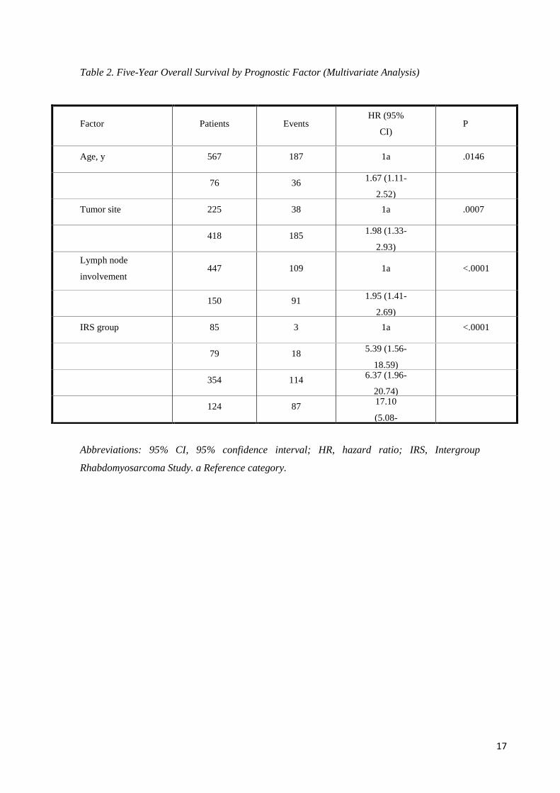
Multivariate analysis identified several factors that were independently and significantly
correlated with better survival: age < 15 years, favorable tumor sites, and no lymph
node or metastatic dissemination (Table 2). All these variables also were confirmed to
be independent prognostic factors for PFS.
Because previous studies found age to be significant using 1 year and 10 years of age as
the lower and upper cutoff values, we performed a further analysis for these 2 age
groups. The survival rate was much the same in children ages 10 years to 14 years and
adolescents, and was worse than in younger children (Fig. 2). The 5-year OS and PFS
rates were significantly higher in children ages 1 year to 9 years compared with children
ages 10 years to 19 years: 72% versus 56.8%, respectively, (P < .0001) and 64% versus
52%, respectively (P: 0.003).
A new multivariate analysis in which different age groups were taken into account (age
< 1 year, ages 1year-9 years, and ages 10 years-19 years) produced similar results, with
age (1 year-9 years), favorable tumor sites, and the absence of lymph node or metastatic
dissemination found to be independently associated with better OS and PFS.

15
Figure 1. (Top) Overall survival and (Bottom) progression free survival are shown in children
and adolescents with rhabdomyosarcoma. 95% CI indicates 95% confidence interval.
DISCUSSION
Despite several reports suggesting that the survival trends for adolescents with cancer
are not improving to the same degree as in children (4,5), to our knowledge there have
still been few studies published to date regarding this particular population, and none
focusing on RMS.
The STSC protocols did not differentiate treatment by age; in particular, there were no
differences with regard to the local treatment strategies and similar percentages of
children and adolescents underwent surgery and radiotherapy.
16
In our analysis, the survival results were better for children. Various reasons have been
suggested to explain why adolescents may fare less well than children with the same
disease, and one of the most important may be a higher incidence of adverse prognostic
factors in older patients. Our data confirm this aspect in patients with RMS; unfavorable
tumor characteristics (eg, alveolar subtype, lymph node involvement, and metastases at
the time of diagnosis) were more common in adolescents than in children. An
unexpectedly high number of adolescents with paratesticular tumors were registered in
the STSC protocols, possibly reflecting a more effective referral to AIEOP centers by
urologists and surgeons. The paratesticular site is highly favorable and explains why
adolescents had a higher percentage of complete tumor resections at the time of
diagnosis; this may also have contributed to raising the PFS and OS in the adolescent
age group.
Age per se has been indicated as a prognostic factor in various tumors, including RMS
(11,12). Joshi et al analyzed the clinical features and treatment outcome of patients aged
< 21 years in the IRS group protocols and concluded that a larger percentage of patients
aged > 10 years have an alveolar histology, unfavorable tumor sites, and a more
advanced tumor stage than noted in children aged < 10 years, but all these features were
not enough to justify their worse outcome, and age remained a strong independent risk
factor (2). More recently, age (> 10 years and < 1 year) proved to be an adverse
prognostic factor in a pooled analysis of 788 patients with metastatic RMS (3). In the
current study, which included patients with localized and metastatic RMS, unfavorable
tumor features and advanced stage in particular appeared to have a more important role,
with the role of age diminishing only when localized tumor was considered separately.
The outcome was very similar for the patients ages 10 years to 14 years and those ages
14 years to 19 years, suggesting that the age cutoff of 10 years may be more appropriate
for the purpose of attributing different risk factors. The results of the current study thus
indicate that adolescents should not be treated differently from younger children on the
basis of age alone.
Some authors have suggested that drug metabolism or treatment-related toxicity might
differ between adolescent and younger patients, potentially explaining the difference in
outcome (13). The limited number of major toxic events recorded in the population
analyzed in the current study prevented us from investigating this aspect.
17
Table 2. Five-Year Overall Survival by Prognostic Factor (Multivariate Analysis)
Abbreviations: 95% CI, 95% confidence interval; HR, hazard ratio; IRS, Intergroup
Rhabdomyosarcoma Study. a Reference category.
Factor Patients Events HR (95%
CI) P
Age, y 567 187 1a .0146
76 36
1.67 (1.11-
2.52)
Tumor site 225 38 1a .0007
418 185
1.98 (1.33-
2.93)
Lymph node
involvement 447 109 1a <.0001
150 91
1.95 (1.41-
2.69)
IRS group 85 3 1a <.0001
79 18
5.39 (1.56-
18.59)
354 114
6.37 (1.96-
20.74)
124 87
17.10
(5.08-
57.52)

18
Figure 2. Progression-free survival is shown by age group. 95% CI indicates 95% confidence
interval.
However, it is important to improve our knowledge in this area by planning clinical
pharmacology studies in these patients.
Another particular factor that is apparent in the adolescent population is the diagnostic
delay. Several authors have suggested that the time elapsing from the onset of
symptoms to diagnosis is longer for adolescents than for children (6,14). This was
confirmed in the population in the current study, in whom the median diagnostic delay
for adolescents was nearly twice as long as that for children (8.4 weeks vs 4.8 weeks),
and suggests that the more advanced stage of disease noted in adolescents, and the
consequently worse prognosis, may be partially explained by a late diagnosis. The
reasons for this diagnostic delay lie within the limited awareness of families and the
community that adolescents can develop cancer and in the fact that adolescents tend to
have a strong sense of independence and may be reluctant to ask for help or submit to a
medical examination, and therefore symptoms are often attributed to physical exertion,
fatigue, trauma, and stress.
An important issue that most likely interferes with any improvement being made in the
survival of adolescents concerns their limited participation in clinical trials (5,15).
When survival rates and accrual rates were compared using Surveillance,
Epidemiology, and End Results (SEER) data, an overlap became apparent: the lower the

19
accrual rate, the worse the results in terms of survival (16). To our knowledge, the
protocols coordinated by the STSC were the only national multicenter protocols avail-
able for children with RMS in Italy, and should be considered a type of standard of
treatment. Only 27% of the expected number of adolescents was recruited into the
STSC protocols, however, whereas > 90% of the expected numbers of children were
enrolled in these protocols during the same period. This poor recruitment of adolescents
in pediatric protocols has been highlighted by a recent analysis comparing the number
of cases registered at the AIEOP centers with the incidence rates obtained from the
AIRTUM population-based registries by cancer type. The O/E ratio for RMS was 0.33,
which is one of the highest among all cancer types in adolescents but grossly unsatis-
factory (10). This demonstrates that adolescents in Italy are often referred to adult
oncology units although their disease is a “pediatric” cancer. Programs dedicated to
adolescents and young adults are still limited and adolescents with RMS may
consequently receive treatment according not to current pediatric guidelines but to the
approach adopted for adult soft tissue sarcoma, which may make their survival rates less
satisfactory, as shown by the analysis of a series of adult patients with RMS (17).
CONCLUSIONS
The survival of children and adolescents enrolled in STSC protocols could be
considered to be satisfactory, especially in patients without metastases. The results of
the current study indicate that RMS presents with more aggressive features in
adolescents and this has a major impact on their survival. An additional factor concerns
the finding that only a small percentage of the adolescents affected are enrolled in
clinical trials, and this may prevent them from receiving the best possible care. A better
cooperation with oncologists who treat adults is mandatory to improve the treatment of
adolescents with RMS.
FUNDING SUPPORT
Supported in part by a grant from the Fondazione Cittàdella Speranza.
CONFLICT OF INTEREST DISCLOSURES
The authors made no disclosures.

20
REFERENCES
1. Breitfeld PP, Meyer WH. Rhabdomyosarcoma: new windows of opportunity.
Oncologist. 2005;10:518-527.
2. Joshi D, Anderson JR, Paidas C, Breneman J, Parham DM, Crist W; Soft Tissue
Sarcoma Committee of the Children’s Oncology Group. Age is an independent
prognostic factor in rhabdomyosarcoma: a report from the Soft Tissue Sarcoma
Committee of the Children’s Oncology Group. Pediatr Blood Cancer.
2004;42:64-73.
3. Oberlin O, Rey A, Lyden E, et al. Prognostic factors in metastatic
rhabdomyosarcomas: results of a pooled analysis from United States and
European cooperative groups. J Clin Oncol. 2008;26:2384-2389.
4. Smith MA, Seibel NL, Altekruse SF, et al. Outcomes for children and
adolescents with cancer: challenges for the twenty-first century. J Clin Oncol.
2010;28:2625-2634.
5. Bleyer A, Montello M, Budd T, Saxman S. National survival trends of young
adults with sarcoma: lack of progress is associated with lack of clinical trial
participation. Cancer. 2005;103:1891-1897.
6. Ferrari A, Miceli R, Casanova M, et al. The symptom interval in children and
adolescents with soft tissue sarcomas. Cancer. 2010;116:177-183.
7. Bisogno G, De Rossi C, Gamboa Y, et al. Improved survival for children with
parameningeal rhabdomyosarcoma: results from the AIEOP soft tissue sarcoma
committee. Pediatr Blood Cancer. 2008;50:1154-1158.
8. Carli M, Colombatti R, Oberlin O, et al. European intergroup studies (MMT4–
89 and MMT4–91) on childhood metastatic rhabdomyosarcoma: final results
and analysis of prognostic factors. J Clin Oncol. 2004;22:4787-4794.
9. Bisogno G, Ferrari A, Prete A, et al. Sequential high-dose chemotherapy for
children with metastatic rhabdomyosarcoma. Eur J Cancer. 2009;45:3035-3041.
10. Ferrari A, Dama E, Pession A, et al. Adolescents with cancer in Italy: entry into
the national cooperative paediatric oncology group AIEOP trials. Eur J Cancer.
2009;45:328-334.
11. Orbach D, Rey A, Oberlin O, et al. Soft tissue sarcoma or malignant
mesenchymal tumors in the first year of life: experience of the International

21
Society of Pediatric Oncology (SIOP) Malignant Mesenchymal Tumor
Committee. J Clin Oncol. 2005;23:4363-4371.
12. Meza JL, Anderson J, Pappo AS, Meyer WH;Children’s Oncology Group.
Analysis of prognostic factors in patients with nonmetastatic rhabdomyosarcoma
treated on intergroup rhabdomyosarcoma studies III and IV: the Children’s
Oncology Group. J Clin Oncol. 2006;24:3844-3851.
13. Veal GJ, Hartford CM, Stewart CF. Clinical pharmacology in the adolescent
oncology patient. J Clin Oncol. 2010;28:4790-4799.
14. Martin S, Ulrich C, Munsell M, Taylor S, Lange G, Bleyer A. Delays in cancer
diagnosis in underinsured young adults and older adolescents. Oncologist.
2007;12:816-824.
15. Ferrari A, Bleyer A. Participation of adolescents with cancer in clinical trials.
Cancer Treat Rev. 2007;33:603-608.
16. Bleyer WA, Tejeda H, Murphy SB, et al. National cancer clinical trials: children
have equal access; adolescents do not. J Adolesc Health. 1997;21:366-373.
17. Sultan I, Qaddoumi I, Yaser S, Rodriguez-Galindo C, Ferrari A. Comparing
adult and pediatric rhabdomyosarcoma in the surveillance, epidemiology and
end results program, 1973 to 2005: an analysis of 2,600 patients. J Clin Oncol.
2009;27:3391-3397.

22
23
2 ANALISI DATI TOPOTECAN/CARBOPLATINO
2.1 Introduzione
Il RMS è il più comune sarcoma delle parti molli nella popolazione pediatrica e
adolescente; nelle ultime decadi l’affinamento dell’approccio multimodale alla
patologia, costituito da chirurgia, radioterapia (RT) e polichemioterapia (CT), ha
permesso di migliorare moltissimo la prognosi dei pazienti pediatrici affetti da questa
neoplasia, passando da una sopravvivenza a 5 anni del 50% negli anni ’70 al 70% degli
anni ’90 (1). Circa il 90% dei pazienti con malattia localizzata all’esordio ottiene una
remissione completa; tuttavia 1/3 di pazienti ricade (2, 3). Per questi pazienti
(metastatici, refrattari, recidivi), la prognosi è ancora ad oggi infausta (4).
Sono quindi necessari nuovi farmaci e nuove strategie terapeutiche per migliorarne la
prognosi.
Risulta di fondamentale importanza identificare fattori prognostici utili a disegnare
protocolli “risk-based”. Un recente studio ha dimostrato che l’istologia, la sede, il tipo e
il timing della recidiva sono fattori correlati alla prognosi in modo significativo (5).
Tra i vari farmaci identificati come attivi in studi preclinici e di fase I vi sono i derivati
delle Camptotecine, Irinotecan e Topotecan. Si tratta di molecole che inibendo la
Topoisomerasi I interferiscono con la divisione cellulare e la replicazione del DNA.
Entrambe le molecole hanno dimostrato in studi preclinici attività su linee cellulari di
numerosi tumori pediatrici, un buon profilo di tossicità nonché efficacia in studi di fase
I (6, 7, 8, 9), anche sui RMS (10, 11).
Vista l’attività delle singole molecole, successivamente sono state studiate varie
combinazioni: Topotecan+ciclofosfamide (12, 13), Topotecan alternato allo schema
VAC (14), Topotecan+Vincristina e Doxorubicina (15, 16)
Da tali studi emerge che questi farmaci, pur non impattando in modo eclatante sulla
sopravvivenza, permettono di ottenere un discreto tasso di riposta in pazienti spesso
pesantemente pretrattati; vengono quindi attualmente considerati delle opzioni
terapeutiche potenzialmente valide.
L’attuale Protocollo per il trattamento del RMS (EpSSG 2005) propone in seconda linea
una strategia terapeutica basata su un regime con Topotecan e Carboplatino. Pazienti
che non rispondono in maniera soddisfacente ai primi 3 cicli di CT dimostrano una

24
cattiva prognosi e sono considerati “refrattari”; pertanto vengono shiftati ad un
trattamento che prevede l’utilizzo di farmaci non utilizzati fino ad allora (Topotecan,
Carboplatino, Ciclofosfamide ed Etoposide; l’antraciclina viene utilizzata nei pazienti in
cui non era prevista in prima linea). Stesso dicasi per i pazienti che ricadono dopo il
termine della CT di prima linea.
Del Topotecan si è detto sopra; il razionale per l’utilizzo del Carboplatino in questo
setting si basa sul precedente impiego dello stesso in regimi polichemioterapici
riconosciuti attivi nel RMS quali il CEVAIE. Carboplatino è stato poi usato da solo in
un “window study” dal UKCCSG nonchè in uno del CWS per i RMS metastatici. La
fattibilità della combinazione è stata provata in occasione di uno studio di fase II
eseguito al Bambin Gesu’ di Roma.
Un’analisi preliminare dei risultati ottenuti nei pazienti affetti da RMS recidivo o
refrattario trattati con Topotecan e Carboplatino, ha confermato la fattibilità della
combinazione: la tossicità è risultata lieve e prevalentemente ematologica; il tasso di
risposta discreto, comparabile con quello osservato con altre combinazioni.
Su tale base è stato condotto uno studio prospettico multicentrico su pazienti affetti da
RMS refrattario/recidivo trattati in seconda linea con il regime Topotecan/Carboplatino.
Lo scopo dello studio era analizzare le caratteristiche delle recidive, il profilo di
tossicità e l’efficacia della combinazione Topotecan/Carboplatino.
Segue l’articolo che a breve sarà sottomesso a rivista scientifica.

25
BIBLIOGRAFIA
1: Crist W, Gehan EA, Ragab AH, Dickman PS, Donaldson SS, Fryer C, Hammond D,
Hays DM, Herrmann J, Heyn R, et al. The Third Intergroup Rhabdomyosarcoma Study.
J Clin Oncol. 1995 Mar;13(3):610-30.
2 Crist WM, Anderson JR, Meza JL, Fryer C, Raney RB, Ruymann FB, Breneman J,
Qualman SJ, Wiener E, Wharam M, Lobe T, Webber B, Maurer HM, Donaldson SS.
Intergroup rhabdomyosarcoma study-IV: results for patients with nonmetastatic
disease. J Clin Oncol. 2001 Jun 15;19(12):3091-102.
3 Koscielniak E, Harms D, Henze G, Jürgens H, Gadner H, Herbst M, Klingebiel T,
Schmidt BF, Morgan M, Knietig R, Treuner J. Results of treatment for soft tissue
sarcoma in childhood and adolescence: a final report of the German Cooperative Soft
Tissue Sarcoma Study CWS-86. J Clin Oncol. 1999 Dec;17(12):3706-19.
4 Pappo AS, Anderson JR, Crist WM, Wharam MD, Breitfeld PP, Hawkins D, Raney
RB, Womer RB, Parham DM, Qualman SJ, Grier HE. Survival after relapse in children
and adolescents with rhabdomyosarcoma: A report from the Intergroup
Rhabdomyosarcoma Study Group. J Clin Oncol. 1999 Nov;17(11):3487-93.
5 Mazzoleni S, Bisogno G, Garaventa A, Cecchetto G, Ferrari A, Sotti G, Donfrancesco
A, Madon E, Casula L, Carli M; Associazione Italiana di Ematologia e Oncologia
Pediatrica Soft Tissue Sarcoma Committee. Outcomes and prognostic factors after
recurrence in children and adolescents with nonmetastatic rhabdomyosarcoma.
Cancer. 2005 Jul 1;104(1):183-90.
6 Stewart CF, Zamboni WC, Crom WR, Gajjar A, Heideman RL, Furman WL, Meyer
WH, Houghton PJ, Pratt CB. Topoisomerase I interactive drugs in children with cancer.
Invest New Drugs. 1996;14(1):37-47. Review.
7 Houghton PJ, Cheshire PJ, Myers L, Stewart CF, Synold TW, Houghton JA.
Evaluation of 9-dimethylaminomethyl-10-hydroxycamptothecin against xenografts
derived from adult and childhood solid tumors. Cancer Chemother Pharmacol.
1992;31(3):229-39.
8 Houghton PJ, Cheshire PJ, Hallman JD 2nd, Lutz L, Friedman HS, Danks MK,
Houghton JA. Efficacy of topoisomerase I inhibitors, topotecan and irinotecan,
administered at low dose levels in protracted schedules to mice bearing xenografts of
human tumors. Cancer Chemother Pharmacol. 1995;36(5):393-403.
9 Vassal G, Terrier-Lacombe MJ, Bissery MC, Vénuat AM, Gyergyay F, Bénard J,
Morizet J, Boland I, Ardouin P, Bressac-de-Paillerets B, Gouyette A. Therapeutic
26
activity of CPT-11, a DNA-topoisomerase I inhibitor, against peripheral primitive
neuroectodermal tumour and neuroblastoma xenografts. Br J Cancer. 1996
Aug;74(4):537-45.
10 Nitschke R, Parkhurst J, Sullivan J, Harris MB, Bernstein M, Pratt C. Topotecan in
pediatric patients with recurrent and progressive solid tumors: a Pediatric Oncology
Group phase II study. J Pediatr Hematol Oncol. 1998 Jul-Aug;20(4):315-8.
11 Pappo AS, Lyden E, Breneman J, Wiener E, Teot L, Meza J, Crist W, Vietti T. Up-
front window trial of topotecan in previously untreated children and adolescents with
metastatic rhabdomyosarcoma: an intergroup rhabdomyosarcoma study. J Clin Oncol.
2001 Jan 1;19(1):213-9.
12 Saylors RL 3rd, Stine KC, Sullivan J, Kepner JL, Wall DA, Bernstein ML, Harris
MB, Hayashi R, Vietti TJ; Pediatric Oncology Group. Cyclophosphamide plus
topotecan in children with recurrent or refractory solid tumors: a Pediatric Oncology
Group phase II study. J Clin Oncol. 2001 Aug 1;19(15):3463-9.
13 Walterhouse DO, Lyden ER, Breitfeld PP, Qualman SJ, Wharam MD, Meyer WH.
Efficacy of topotecan and cyclophosphamide given in a phase II window trial in
children with newly diagnosed metastatic rhabdomyosarcoma: a Children's Oncology
Group study. J Clin Oncol. 2004 Apr 15;22(8):1398-403. Epub 2004 Mar 8. Erratum in:
J Clin Oncol. 2004 Aug 1;22(15):3205.
14 Soft Tissue Sarcoma Committee of the Children's Oncology Group, Lager JJ, Lyden
ER, Anderson JR, Pappo AS, Meyer WH, Breitfeld PP. Pooled analysis of phase II
window studies in children with contemporary high-risk metastatic rhabdomyosarcoma:
a report from the Soft Tissue Sarcoma Committee of the Children's Oncology Group. J
Clin Oncol. 2006 Jul 20;24(21):3415-22.
15 Meazza C, Casanova M, Zaffignani E, Luksch R, Podda M, Favini F, Catania S,
Biassoni V, Morosi C, Ferrari A. Efficacy of topotecan plus vincristine and doxorubicin
in children with recurrent/refractory rhabdomyosarcoma. Med Oncol. 2009;26(1):67-
72.
16 Pappo AS, Lyden E, Breitfeld P, Donaldson SS, Wiener E, Parham D, Crews KR,
Houghton P, Meyer WH; Children's Oncology Group. Two consecutive phase II
window trials of irinotecan alone or in combination with vincristine for the treatment of
metastatic rhabdomyosarcoma: the Children's Oncology Group. J Clin Oncol. 2007 Feb
1;25(4):362-9.
27
2.2 A Topotecan/Carboplatin based strategy for children with
refractory or recurrent rhabdomyosarcoma
Alessia Compostella, MD1, Gianni Bisogno, MD, PhD1 et al.
ABSTRACT
The prognosis for children with resistant/relapsing Rhabdomyosarcoma (RMS) remains
poor and therefore there is a need to test new drugs combinations. Topotecan (T) and
Carboplatin (C) are known to have activity against a variety of pediatric tumors so a
T/C based chemotherapy has been proposed as second line chemotherapy for children
relapsed after being treated in the soft tissue sarcoma committee (STSC) protocols.
Methods:
38 patients with available data on response have been analyzed: 8 resistant to first line
treatment and 30 treated at relapse. Treatment: T: 2 mg/m2 days 1,2,3; C: 250 mg/m2
days 4,5 every 3 weeks. Tumor response has been evaluated after 2 cycles adopting
standard criteria: complete response (CR); partial response (PR= tumor size reduction
>50%); minor response (MR= reduction <50%); no response (NR= reduction <25%),
progressive disease (PD= increase of tumor size or detection of new lesions)
Results:
18 patients presented unfavorable histotype and 19 a favorable one (1 NOS). At
diagnosis IRS Group was: II: 3 patient; III: 25; IV: 10. Tumor site was unfavorable in
the great majority of children (30/38): 9 parameningeal (PM), 9 extremities, 9 other
sites, 3 genitorurinary bladder-prostate (GU-BP); among 8 favorable sites 4 were head
and neck non parameningeal, 3 genito-urinary non BP, only 1 orbit. 24 patients received
2 cycles, 3 only 1 due to early PD. Toxicity was predominantly hematologic with no
severe non-hematologic toxic events reported. Major response was evident in 9 patients
(CR+PR). The response rate was globally 28%; 15% in favorable histology and 33% in
unfavorable one.
Conclusions:
Our study shows that the T/C combination has a mild toxicity in pretreated patients. The
response rate is somewhat lower when compared to other combinations tested in phase
II studies but it is of interest for the population with alveolar subtype.

28
Corresponding author: Gianni Bisogno, MD, PhD, Division of Hematology/Oncology,
Department of Pediatrics, Padova University Hospital, via Giustiniani 3-35128,
Padova, Italy; Fax: (011) 39-049-8213510; [email protected]
1Hematology/Oncology Division, Department of Pediatrics, Padova University
Hospital, Padova, Italy.
We thank Angela Scagnellato for data processing and Ilaria Zanetti for the statistical
analysis.
29
INTRODUCTION
Despite the success of current multimodal therapy which has increased the survival of
patients with RMS over 70% there is still a substantial number of patients who relapse
and need effective salvage chemotherapy. Thus it is important to investigate novel
antineoplastic combinations for their potential incorporation into front line therapy.
In this study we tested a chemotherapy strategy based on the administration of regimens
including Topotecan (T) and Carboplatin (C) in a group of children and adolescents
with refractory RMS.
Topotecan, a campthotecine derivative, has demonstrated in pre-clinical studies high
activity against pediatric malignancies such as medulloblastoma, neuroblastoma and
rhabdomyosarcoma. Consequently several studies of T alone or in association with
other antiblastic drugs were initiated.
Carboplatin has been part of previously used regimens (CEVAIE) that proved to be
effective against RMS (1). It has also been used alone in a window study conducted by
the UKCCSG. A phase II trial has been performed at the Bambino Gesù Hospital in
Rome showing the feasibility of the proposed regimen. The T/C combination is also
used as window treatment in the current CWS protocol for metastatic RMS.
This two drugs have constituted the base of the second line strategy recommended for
children with RMS who relapsed after being treated in the STSC protocols.
MATERIAL AND METHODS
Between 2002 (only one patient was diagnosed in 1995) and 2011, 38 patients under 19
years old joined this study. They were registered from 12 centers belonging to the
AIEOP (Associazione Italiana di Ematologia e Oncologia Pediatrica) and taking part in
studies coordinated by the AIEOP STSC.
Eligible patients were required to have a histologically-confirmed diagnosis of RMS,
and to be refractory or relapsing after the inclusion in one of the protocols coordinated
by the STSC.
Other eligibility criteria were: a life expectancy of at least 8 weeks, a modified Lansky
score of > 50, recovery from the toxic effects of prior chemotherapy, a hemoglobin
level greater than 9 g/dl, an absolute neutrophil count greater than 1,500/mm3, a platelet
count higher than 100,000/mm3, adequate liver function (bilirubin level ≤ 1.5 mg/100

30
ml; ALT ≤ twice the normal value), adequate renal function (serum creatinine
concentration ≤ 1.5 mg/dL or creatinine clearance > 60 ml/min/1.73 m2) and normal
metabolic parameters (serum electrolytes, glucose, calcium, phosphorus). Patients with
an interval of less than 3 weeks since the administration of radiotherapy or
chemotherapy were excluded.
At the baseline, the tumor was reassessed, with computed tomography (CT) or a
magnetic resonance imaging (MRI) scans of disease sites and measurements of all
disease parameters, chest X-ray, chest CT scan, whole body technetium bone scan and
bone marrow aspirates and biopsies.
The study was approved by the Ethics Committees of each center taking part and
informed consent was obtained from patients or parents, as appropriate.
Treatment
Patients received 2 blocks of T\C, followed by alternating blocks of Topotecan \
Cyclophosphamide and Carboplatin \ Etoposide for a total of 6 courses with 3-week
interval (see figure 1).
Local treatment was scheduled after the two initial courses. Surgery and radiotherapy
had to be considered but the type and time of local treatment were left to the responsible
clinician according to the patient condition, relapsing tumor characteristics, and
previous treatment. The coordinating STSC Centre was available to discuss the strategy
for the most difficult cases.
The schedule for drugs administration is described in figure 1 and was as follow:
Topotecan: 2 mg/m2/day administered by 30 minutes intravenous infusion once daily
on day 1, 2 and 3 (total dose 6 mg/m2/course); Carboplatin: 250 mg/m2/day in 1 hour
intravenous infusion on day 4 and 5 (total dose 500 mg/m2 course).
Cycles were given every 21 days, with neutrophils >1.0 x 10⁹/l and platelets to >80 x
109/l and following resolution of non-hematopoietic toxicity. Use of colony-stimulating
factors were given according to Institutional policy.
Toxicity was graded using the National Cancer Institute Common Toxicity Criteria
version 2.0.
Response evaluation
After the initial two T/C courses and at the end of treatment, a formal assessment of the
primary tumor and all sites of metastases had to be performed.

31
Response criteria were as follows: complete response (CR) = resolution of all evidence
of disease; partial response (PR) = a tumor size reduction of more than 50% in the sum
of the products of the two maximum perpendicular diameters of all measurable lesions;
minor response (MR) = a reduction of less than 50% but more than 25% in the sum of
the products of the two maximum perpendicular diameters of all measurable lesions.
Stable disease or a reduction in size of less than 25% was recorded as no response (NR),
while an increase in tumor size or the detection of new lesions was considered as
progressive disease (PD). Responses had to last at least 4 weeks after the assessment of
the response.
Due to the difficulty in judging tumor response on bone marrow aspirates, we decided
not to consider the bone marrow in the assessment of tumor response unless there was
clear evidence of progressive disease or a new lesion.
Statistical method
Survival curves were calculated using the Kaplan-Meier method, considering: overall
survival (OS), from the dates of relapse to latest follow-up or death from any cause;
progression-free survival (PFS), from diagnosis to first progression, relapse, death from
any cause or latest contact for children who never experienced an event.
The log-rank test was used to compare survival rates between different subgroups of
patients in univariate analysis, considering patients’ characteristics (age and gender) and
tumor features (histological subtype, site, size, invasiveness, lymph node involvement,
type and number of metastases). The different sites were grouped according their
prognosis in favorable (orbit, head and neck, genitor-urinary non bladder prostate) and
unfavorable (parameningeal, extremities, bladder-prostate, other sites). A p-value of less
than 0.05 was considered statistically significant. A multivariate analysis was conducted
using Cox’s proportional hazards regression method to determine the independent
prognostic influence of pretreatment factors on survival, using the variables correlated
with OS and PFS at univariate analysis.
A phase II methodology using a Gehan 2-step design has been applied to evaluate the
response to the two initial T/C cycles. The expected effectiveness ( ) was considered as
20% for the whole group. If at least one response was recorded in the first 14 eligible
patients, recruitment was to continue to at least 25 patients so that the standard error of
the observed response rate would be 0.10.

32
The study was approved by the Ethics Committees of all centers taking part and
informed consent was obtained for all patients enrolled on the protocol.
RESULTS
Clinical features
A total of 38 patients joined the study, 32 of whom were evaluable for response to the
T/C response study. Patients characteristics are shown in table 1. The age range was 0.4
– 18.6 years (median 4.7; media 6.2). 16 were male, 22 were female. Histotypes were:
18 were unfavorable RMS, 20 favorable (18 embrional RMS, 1 spindle cell RMS, 1
NOS RMS). Tumors were mainly located in unfavorable sites. 10 patients were
metastatic at diagnosis. At the entry in the study 8 patients had persistent disease at the
end of first-line treatment, 20 had a loco-regional relapse (15 only local, 1 with
concomitant lymph node involvement, 4 only node involvement ), the others had only
distant relapse or local and distant relapse.
Treatment
Patients had been previously treated in 12 italian hemato-oncology units according to
different protocols named RMS88, RMS4.99, RMS96, EpSSG2005. Surgery has been
performed in all patients at diagnosis, nearly all (35) being diagnostic biopsies; 10
patients underwent surgery after initial chemotherapy. Radiotherapy was delivered to 23
patients during first line chemotherapy (CT); 15 did not (8 of them because of age).
First line CT included high-dose chemotherapy with stem cell rescue in 2 patients.
Local treatment was scheduled after the two initial T/C courses: 8 patients underwent
surgery; 2 of them being microscopically radical, 3 macroscopically radical, a patient
suffer a mutilating operation (exenteratio orbitae), 2 had no data about. Radiotherapy
was delivered to 20 patients, 12 of them had been irradiated during first line
chemotherapy, 8 were not (4 because of young age).
After T/C CT many patients were treated with alternating Topotecan/Cyclofosphamide-
Etoposide/Carboplatin courses till progression. Other drugs frequently used were low
dose Vinorelbine and Cyclophosphamide. Some patients have been treated with poliCT:
VAC, ICE, Gemox; drugs less used were Irinotecan, Vincristine, Temozolamide,
Caelix. A patients with recurrent RMS in the upper extremities underwent local

33
treatment with arterial Cisplatin. 2 patients were treated with high dose CT and
autologous transplantation.
Response and outcome
6 patient were not evaluable for response to the two initial T/C cycles: local treatment
(RT/surgery) was used at relapse before T/C administration in 4 patients; 2 other
patients were not evaluated according established criteria for response to T/C CT.
Overall in 32 evaluable patients, 2 CR and 7 PR were documented, for an overall
response rate of 28%. A minor response was recorded in 3 cases. 11 were PD, 9 SD.
When any type of tumor size reduction (complete, partial, minor) was considered, a
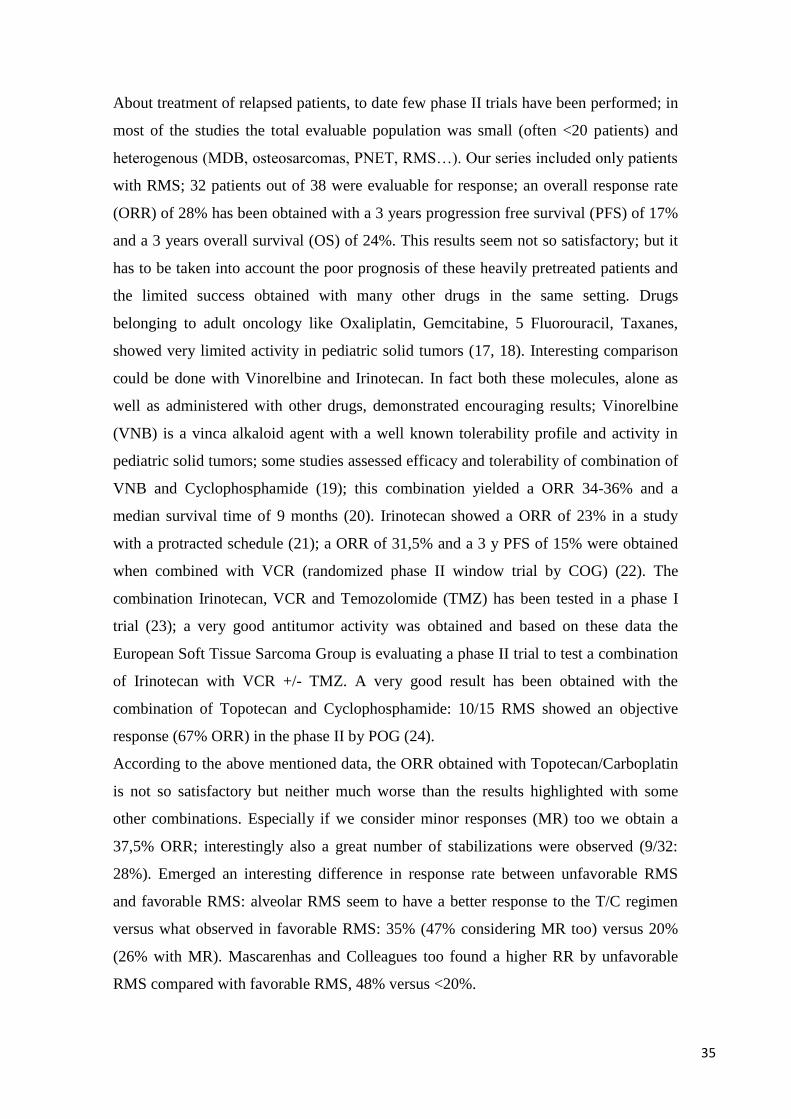
37.5% response rate was calculated (12/32). 5 years OS was nearly 17%, 5 years PFS
was 14%.
Alveolar RMS seem to have a better response to the T/C regimen with 6/17 objective
responses, then 35% (47% considering also MR: 8/17), rare stabilizations, a great
number of progressions (8/17: 47%); favorable RMS showed 3/15 (20%) responses
(26% considering MR); but many stable disease (8/15: 53%) and few progressions.
Among 5 evaluable patients relapsed on therapy 5 had no response (neither a minor 1)
and usually progressed to T/C. Among 24 evaluable patients who relapsed after
completing CT, 9 had a good response to second line CT and 3 had a minor response.
Toxicity
Toxicity of T/C based chemotherapy was mainly haematological: 24 out of 38 patients
experienced neutropenia or anemia or thrombocytopenia, some of them with
concomitant fever. One patient experienced cytopenia and tubulopathy, one patient
experienced cytopenia and mucositis, 1 suffered isolated nephrotoxicity. A heart disease
was discovered in a child after receiving the combination. 6 out of 33 had no toxicity. 4
patients had no data about chemotherapy toxicities. Overall 8 patients are currently
alive; 30 are dead.
DISCUSSION
The treatment of patients with refractory RMS is still problematic and patients
prognosis is still poor. It’s a habit to treat refractory patients with drugs not used during
first line treatment in attempt to overcome drug resistance. Campthotecin derivatives are

34
anticancer agents that inhibit Topoisomerase I activity; among of them are Irinotecan
and Topotecan; both have shown promising results in preclinical studies on human
tumor xenografts derived from pediatric tumors such as RMS and medulloblastoma
(MDB) (2, 3, 4). Phase I clinical trials confirmed the preclinical findings both in adults
(5, 6) and children (7, 8, 9). Phase II studies in children confirmed the achieved
improvement in neuroblastoma and RMS (10, 11, 12). Carboplatin has known activity
against a variety of pediatric solid tumors, either alone or in combination therapy, and is
less toxic than Cisplatin (CDDP) and than most other agents. The use of T after a DNA
damaging agents such as Carboplatin is appealing in that T may prevent repair of
Carboplatin-induced damage (13). For the above reasons, T and C seemed a rational
combination for clinical exploration in pediatric malignancies.
As a consequence we designed a T/C based protocol for patients relapsing after been
treated in one of the STSC protocols. As a first step we analyzed tumor and patients
characteristics to find out factors important in determining the first and second line
treatment response rate. Previous studies found that tumor histology, tumor primary
site, type of recurrence and temporal relation to therapy were associated significantly
with prognosis in patients with recurrent RMS (14). In fact OS of patients who suffers
distant relapse differs dramatically from who suffer only local recurrence (15). The
timing of recurrence also influence prognosis. OS for patients whose disease recurred
on therapy was significantly lower than patients with late failures (14). Similar results
were published by IRSG and CWS were the prognosis of relapsed patients was
significantly different between patients who developed recurrence after completing CT
compared with patients who had developed recurrence while receiving CT (19% vs.
2,7% P<0,05) (16). Our data do not permit to calculate time of recurrence but we can
say that patients whose disease progressed soon during first line CT or did not show a
good response to first courses showed no response even to second line CT and
discouraging outcome.
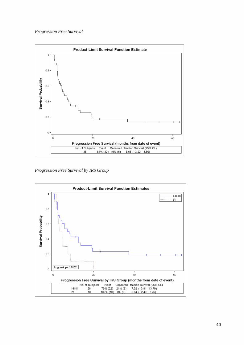
In our study IRS stage seems to affect prognosis (statistical significance for OS, a trend
was evident for PFS). Furthermore, primary site seem to be linked to response rate. In
fact, 7 out of 8 patients with no response to first line CT were located in an unfavorable
primary site (4 of them were PM). We found the same data in non-responders to second
line CT: the primary site of the tumor was unfavorable in 18 out of 20 patients who did
not show any sensitivity to T/C regimen.
35
About treatment of relapsed patients, to date few phase II trials have been performed; in
most of the studies the total evaluable population was small (often <20 patients) and
heterogenous (MDB, osteosarcomas, PNET, RMS…). Our series included only patients
with RMS; 32 patients out of 38 were evaluable for response; an overall response rate
(ORR) of 28% has been obtained with a 3 years progression free survival (PFS) of 17%
and a 3 years overall survival (OS) of 24%. This results seem not so satisfactory; but it
has to be taken into account the poor prognosis of these heavily pretreated patients and
the limited success obtained with many other drugs in the same setting. Drugs
belonging to adult oncology like Oxaliplatin, Gemcitabine, 5 Fluorouracil, Taxanes,
showed very limited activity in pediatric solid tumors (17, 18). Interesting comparison
could be done with Vinorelbine and Irinotecan. In fact both these molecules, alone as
well as administered with other drugs, demonstrated encouraging results; Vinorelbine
(VNB) is a vinca alkaloid agent with a well known tolerability profile and activity in
pediatric solid tumors; some studies assessed efficacy and tolerability of combination of
VNB and Cyclophosphamide (19); this combination yielded a ORR 34-36% and a
median survival time of 9 months (20). Irinotecan showed a ORR of 23% in a study
with a protracted schedule (21); a ORR of 31,5% and a 3 y PFS of 15% were obtained
when combined with VCR (randomized phase II window trial by COG) (22). The
combination Irinotecan, VCR and Temozolomide (TMZ) has been tested in a phase I
trial (23); a very good antitumor activity was obtained and based on these data the
European Soft Tissue Sarcoma Group is evaluating a phase II trial to test a combination
of Irinotecan with VCR +/- TMZ. A very good result has been obtained with the
combination of Topotecan and Cyclophosphamide: 10/15 RMS showed an objective
response (67% ORR) in the phase II by POG (24).
According to the above mentioned data, the ORR obtained with Topotecan/Carboplatin
is not so satisfactory but neither much worse than the results highlighted with some
other combinations. Especially if we consider minor responses (MR) too we obtain a
37,5% ORR; interestingly also a great number of stabilizations were observed (9/32:
28%). Emerged an interesting difference in response rate between unfavorable RMS
and favorable RMS: alveolar RMS seem to have a better response to the T/C regimen
versus what observed in favorable RMS: 35% (47% considering MR too) versus 20%
(26% with MR). Mascarenhas and Colleagues too found a higher RR by unfavorable
RMS compared with favorable RMS, 48% versus <20%.

36
Toxicity of T/C based chemotherapy was mainly haematological and mild, usually
reversible.
At relapse to T/C based CT no “standard” treatment is known to be effective. Than a
variety of strategies have been used in our series: RT and surgery if not performed
before; metronomic CT (low dose VNB, Cyclophosphamide, Etoposide), high dose CT,
Irinotecan, VAC, GEMOX (Gemcitabine+Oxaliplatin) and so on. Because of this
heterogeneity it is difficult to interpret data on OS in relation to T/C efficacy.
We conclude that T/C based CT is very well tolerated with similar results in terms of
RR. We consider it an option but these results are not so satisfactory. The rarity of the
disease complicate the researchers work; an effort has to be done to enroll patients in
clinical trials, eventually up to 21 years and join data and expertise. There is a strong
need to individualize treatment (e.g. alveolar RMS good responder to T/C versus non
alveolar?), to find out new molecular targets, new drugs or new schedules to improved
prognosis of these young patients.

37
FIG 1: Treatment schedule

38
TABLE 2 Tumor characteristics
Sex
- Male 16
- Female 22
Primary Site
- Orbit 1
- Head and Neck non Parameningeal 4
- Head and ne ckParameningeal 9
- Genito-Urinary Bladder Prostate 3
- Genito-Urinary non Bladder Prostate 3
- Extremities 9
- Other sites 9
Histology
Alveolar 18
Embrional 18
Spindle cell 1
Unknown 1
T stage
T1 15
T2 22
Unknown 1
N stage
N0 21
N1 16
Nx 1
Tumor size
< 5 cm 12
> 5 cm 25
Unknown 1
IRS group
I/II/III 28
IV 10

39
Overall Survival
Overall Survival by IRS Group
40
Progression Free Survival
Progression Free Survival by IRS Group
41
REFERENCES
1: Frascella E, Pritchard-Jones K, Modak S, Mancini AF, Carli M, Pinkerton CR.
Response of previously untreated metastatic rhabdomyosarcoma to combination
chemotherapy with carboplatin, epirubicin and vincristine. Eur J Cancer. 1996
May;32A(5):821-5.
2 Carol H, Houghton PJ, Morton CL, Kolb EA, Gorlick R, Reynolds CP, Kang MH,
Maris JM, Keir ST, Watkins A, Smith MA, Lock RB. Initial testing of topotecan by the
pediatric preclinical testing program. Pediatr Blood Cancer. 2010 May;54(5):707-15.
3 Pawlik CA, Houghton PJ, Stewart CF, Cheshire PJ, Richmond LB, Danks MK.
Effective schedules of exposure of medulloblastoma and rhabdomyosarcoma xenografts
to topotecan correlate with in vitro assays. Clin Cancer Res. 1998 Aug;4(8):1995-2002.
4 Houghton PJ, Cheshire PJ, Hallman JD 2nd, Lutz L, Friedman HS, Danks MK,
Houghton JA. Efficacy of topoisomerase I inhibitors, topotecan and irinotecan,
administered at low dose levels in protracted schedules to mice bearing xenografts of
human tumors. Cancer Chemother Pharmacol. 1995;36(5):393-403.
5 Rowinsky EK, Verweij J. Review of phase I clinical studies with topotecan. Semin
Oncol. 1997 Dec;24(6 Suppl 20):S20-3-S20-10. Review.
6 Dennis MJ, Beijnen JH, Grochow LB, van Warmerdam LJ. An overview of the
clinical pharmacology of topotecan. Semin Oncol. 1997 Feb;24(1 Suppl 5):S5-12-S5-
18. Review.
7 Tubergen DG, Stewart CF, Pratt CB, Zamboni WC, Winick N, Santana VM, Dryer
ZA, Kurtzberg J, Bell B, Grier H, Vietti TJ. Phase I trial and pharmacokinetic (PK) and
pharmacodynamics (PD) study of topotecan using a five-day course in children with
refractory solid tumors: a pediatric oncology group study. J Pediatr Hematol Oncol.
1996 Nov;18(4):352-61.
8 Furman WL, Baker SD, Pratt CB, Rivera GK, Evans WE, Stewart CF. Escalating
systemic exposure of continuous infusion topotecan in children with recurrent acute
leukemia. J Clin Oncol. 1996 May;14(5):1504-11.
9 Pratt CB, Stewart C, Santana VM, Bowman L, Furman W, Ochs J, Marina N,
Kuttesch JF, Heideman R, Sandlund JT, et al. Phase I study of topotecan for pediatric
patients with malignant solid tumors. J Clin Oncol. 1994 Mar;12(3):539-43.
10 Nitschke R, Parkhurst J, Sullivan J, Harris MB, Bernstein M, Pratt C. Topotecan in
pediatric patients with recurrent and progressive solid tumors: a Pediatric Oncology
Group phase II study. J Pediatr Hematol Oncol. 1998 Jul-Aug;20(4):315-8.

42
11 Kretschmar CS, Kletzel M, Murray K, Thorner P, Joshi V, Marcus R, Smith EI,
London WB, Castleberry R. Response to paclitaxel, topotecan, and topotecan-
cyclophosphamide in children with untreated disseminated neuroblastoma treated in an
upfront phase II investigational window: a pediatric oncology group study. J Clin
Oncol. 2004 Oct 15;22(20):4119-26.
12 Pappo AS, Lyden E, Breneman J, Wiener E, Teot L, Meza J, Crist W, Vietti T. Up-
front window trial of topotecan in previously untreated children and adolescents with
metastatic rhabdomyosarcoma: an intergroup rhabdomyosarcoma study. J Clin Oncol.
2001 Jan 1;19(1):213-9.
13 Athale UH, Stewart C, Kuttesch JF, Moghrabi A, Meyer W, Pratt C, Gajjar A,
Heideman RL. Phase I study of combination topotecan and carboplatin in pediatric
solid tumors. J Clin Oncol. 2002 Jan 1;20(1):88-95.
14 Mazzoleni S, Bisogno G, Garaventa A, Cecchetto G, Ferrari A, Sotti G,
Donfrancesco A, Madon E, Casula L, Carli M; Associazione Italiana di Ematologia e
Oncologia Pediatrica Soft Tissue Sarcoma Committee. Outcomes and prognostic
factors after recurrence in children and adolescents with nonmetastatic
rhabdomyosarcoma. Cancer. 2005 Jul 1;104(1):183-90.
15 Flamant F, Rodary C, Rey A, Praquin MT, Sommelet D, Quintana E, Theobald S,
Brunat-Mentigny M, Otten J, Voûte PA, Habrand JL, Martelli H, Barrett A, Terrier-
Lacombe MJ, Oberlin O. Treatment of non-metastatic rhabdomyosarcomas in
childhood and adolescence. Results of the second study of the International Society of
Paediatric Oncology: MMT84. Eur J Cancer. 1998 Jun;34(7):1050-62.
16 Klingebiel T, Pertl U, Hess CF, Jürgens H, Koscielniak E, Pötter R, van Heek-
Romanowski R, Rossi R, Schött C, Spaar HJ, Willnow U, Treuner J. Treatment of
children with relapsed soft tissue sarcoma: report of the German CESS/CWS REZ 91
trial. Med Pediatr Oncol. 1998 May;30(5):269-75.
17 Macy ME, Duncan T, Whitlock J, Hunger SP, Boklan J, Narendren A, Herzog C,
Arceci RJ, Bagatell R, Trippett T, Christians U, Rolla K, Ivy SP, Gore L; Pediatric
Oncology Experimental Therapeutics Investigators' Consortium (POETIC). A multi-
center phase Ib study of oxaliplatin (NSC#266046) in combination with fluorouracil
and leucovorin in pediatric patients with advanced solid tumors. Pediatr Blood Cancer.
2013 Feb;60(2):230-6.
18 Zwerdling T, Krailo M, Monteleone P, Byrd R, Sato J, Dunaway R, Seibel N, Chen
Z, Strain J, Reaman G; Children's Oncology Group. Phase II investigation of docetaxel

43
in pediatric patients with recurrent solid tumors: a report from the Children's Oncology
Group. Cancer. 2006 Apr 15;106(8):1821 8.
19 Casanova M, Ferrari A, Bisogno G, Merks JH, De Salvo GL, Meazza C, Tettoni K,
Provenzi M, Mazzarino I, Carli M. Vinorelbine and low-dose cyclophosphamide in the
treatment of pediatric sarcomas: pilot study for the upcoming European
Rhabdomyosarcoma Protocol. Cancer. 2004 Oct 1;101(7):1664-71.
20 Minard-Colin V, Ichante JL, Nguyen L, Paci A, Orbach D, Bergeron C, Defachelles
AS, André N, Corradini N, Schmitt C, Tabone MD, Blouin P, Sirvent N, Goma G,
Geoerger B, Oberlin O. Phase II study of vinorelbine and continuous low doses
cyclophosphamide in children and young adults with a relapsed or refractory malignant
solid tumour: good tolerance profile and efficacy in rhabdomyosarcoma--a report from
the Société Française des Cancers et leucémies de l'Enfant et de l'adolescent (SFCE).
Eur J Cancer. 2012 Oct;48(15):2409-16.
21 Bisogno G, Riccardi R, Ruggiero A, Arcamone G, Prete A, Surico G, Provenzi M,
Bertolini P, Paolucci P, Carli M. Phase II study of a protracted irinotecan schedule in
children with refractory or recurrent soft tissue sarcoma. Cancer. 2006 Feb
1;106(3):703-7.
22 Mascarenhas L, Lyden ER, Breitfeld PP, Walterhouse DO, Donaldson SS, Paidas
CN, Parham DM, Anderson JR, Meyer WH, Hawkins DS. Randomized phase II
window trial of two schedules of irinotecan with vincristine in patients with first relapse
or progression of rhabdomyosarcoma: a report from the Children's Oncology Group. J
Clin Oncol. 2010 Oct 20;28(30):4658-63.
23 McNall-Knapp RY, Williams CN, Reeves EN, Heideman RL, Meyer WH. Extended
phase I evaluation of vincristine, irinotecan, temozolomide, and antibiotic in children
with refractory solid tumors. Pediatr Blood Cancer. 2010 Jul 1;54(7):909-15
24 Saylors RL 3rd, Stine KC, Sullivan J, Kepner JL, Wall DA, Bernstein ML, Harris
MB, Hayashi R, Vietti TJ; Pediatric Oncology Group. Cyclophosphamide plus
topotecan in children with recurrent or refractory solid tumors: a Pediatric Oncology
Group phase II study. J Clin Oncol. 2001 Aug 1;19(15):3463-9.

44
45
3 I NUOVI FARMACI
3.1 Introduzione
Negli ultimi anni il miglioramento delle tecniche di diagnostica, la ricerca in ambito
farmacologico e l’approccio multimodale, hanno permesso un aumento della
sopravvivenza dei bambini e adolescenti affetti da tumore. Mentre negli anni ’50 la
quasi totalità dei casi esitava nel decesso, siamo giunti ora ad una sopravvivenza a 5
anni del 75% (1). I maggiori successi sono stati ottenuti nell’ambito delle patologie
ematologiche (leucemie e linfomi) mentre i risultati ottenuti nei tumori solidi sono stati
meno eclatanti.
La prognosi dei pazienti affetti da tumore dipende molto dallo stadio all’esordio: la
sopravvivenza dei pazienti pediatrici affetti da rabdomiosarcoma (RMS) arriva, per
esempio, al 70% quando la malattia è localizzata, ma scende al di sotto del 30% per i
pazienti con malattia metastatica (2). La prognosi è particolarmente severa anche per i
pazienti con malattia in recidiva e/o resistente al trattamento chemioterapico di prima
linea. Risulta quindi di fondamentale importanza ricercare nuove strategie terapeutiche
per i pazienti con malattia metastatica all’esordio o resistente al trattamento di prima
linea. A tale scopo numerose sono le vie percorribili: lo studio di nuovi farmaci, di
nuove schedule/combinazioni di farmaci noti, di formulazioni adatte alla
somministrazione a pazienti pediatrici.
L’ambito dei nuovi farmaci include sia le molecole/farmaci di ultima generazione che
tanto hanno segnato la ricerca più recente (inibitori delle tirosin kinasi, anticorpi
monoclonali), sia i farmaci non ancora utilizzati nel paziente pediatrico ma che ormai
fanno parte del trattamento standard negli adulti.
La ricerca ha via via identificato lo stretto legame esistente tra genetica e tumore.
L’utilizzo di tecniche sempre più sofisticate di analisi ha permesso di studiare alcune
delle basi molecolari del processo neoplastico e la ricerca sui farmaci antineoplastici si è
recentemente focalizzata sulla cosiddetta “target therapy”. Le basi molecolari e le
anomalie genetiche implicate nella malattia cancro, determinano uno squilibrio nella
produzione/attivazione di alcune molecole. La determinazione della struttura delle
molecole coinvolte ha permesso di “costruire” inibitori altamente selettivi in grado di
interferire/bloccare i pathways cellulari erroneamente up/down regolati nel processo
neoplastico. I successi ottenuti con alcune molecole hanno dato una spinta alla ricerca in

46
tale ambito: Imatinib, inibitore del recettore della tirosin-chinasi che blocca l’attività dei
recettori c-Abl, c-Kit e PDGF, costituisce ad esempio uno dei più eclatanti successi
della target therapy: il farmaco è divenuto rapidamente trattamento standard per i
pazienti affetti da tumori gastrointestinali stromali e leucemia linfatica cronica. Dati
preclinici promettenti riguardano anche molecole quali gli inibitori delle
metalloproteinasi e gli inibitori di alcuni fattori di crescita quali VEGF, PDGF, EGF e
dei loro recettori (3).
Interferire con i pathways cellulari sregolati nella cellula neoplastica è sicuramente una
via da percorrere. La ricerca deve pertanto perseguire tale obiettivo e gli studi clinici di
fase I, II e III sulle “nuove molecole” sono uno strumento di fondamentale importanza,
ancillare agli studi di laboratorio che ne costituiscono la base. Tuttavia, se l’inibizione
di pathways coinvolti può essere una strategia efficace, non va sottostimato il fatto che
vi sono molte alterazioni genetiche e più vie coinvolte in un tipo di tumore; molti
processi biologici inoltre hanno vie “alternative” che possono potenzialmente bypassare
il pathway con il quale il farmaco interferisce. Una spia di ciò è la non completa
concordanza di risultati tra sperimentazione preclinica e clinica, soprattutto quando le
molecole in studio vengono utilizzate da sole. Inoltre non vanno dimenticati i potenziali
effetti collaterali dovuti ai molteplici ruoli che la molecola target può possedere e va
considerata la possibilità dell’insorgenza di resistenze. Questo dimostra come
probabilmente sia necessario pensare a dei “pannelli” di farmaci che mirino a bloccare i
numerosi pathways coinvolti nel processo neoplastico. Tali premesse suggeriscono
inoltre che la target therapy è una delle vie, ma non può essere la sola arma contro una
patologia tanto complessa.
E’ quindi importante non tralasciare le altre vie possibili: in particolare considerare
schedule che prevedano la somministrazione di farmaci a bassi dosaggi ma in modo
continuativo (“terapia metronomica”): risultati di studi recenti suggeriscono che la
somministrazione di alcuni farmaci citotossici a basse dosi aumenta l’ effetto
antiangiogenico dei farmaci. Tale approccio potrebbe essere utile per minimizzare la
tossicità dei farmaci, soprattutto in pazienti che necessitano di trattamenti molto
prolungati. I farmaci citotossici possono essere in quest’ottica associati alle nuove
molecole, quali gli inibitori del VEGFR, per un reciproco potenziamento (4).
Anche l’utilizzo di chemioterapici mutuati dall’esperienza nell’adulto dei quali esistano
dati di efficacia su linee cellulari di tumori solidi pediatrici (es.: inibitori delle
Topoisomerasi, Taxani…) è una via percorribile (5, 6).

47
Altra possibilità che si prospetta è lo studio della farmacologia di farmaci in uso in
Oncologia Pediatrica (quali ad esempio le antracicline) volti ad ottimizzarne l’utilizzo
(7).
Da tali premesse emerge la complessità del problema e la molteplicità delle vie che
vanno percorse.
Le prospettive delineate rientrano nell’ottica di una visione “allargata” del problema:
come sottolineato recentemente (8), la ricerca di nuovi agenti antitumorali è
fondamentale e deve applicarsi su più fronti possibile, ma senza focalizzarsi su un’unica
strategia. Le molecole di nuova generazione (target therapy) sono promettenti ma
probabilmente non costituiranno, sole, la soluzione ad un problema tanto complesso. E’
necessario trovare le vie più adeguate per il miglior utilizzo dei farmaci nuovi e vecchi.

48
BIBLIOGRAFIA
1: Gatta G, Zigon G, Capocaccia R, Coebergh JW, Desandes E, Kaatsch P, Pastore G,
Peris-Bonet R, Stiller CA; EUROCARE Working Group. Survival of European children
and young adults with cancer diagnosed 1995-2002. Eur J Cancer. 2009 Apr;45(6):992-
1005.
2: Oberlin O, Rey A, Lyden E, Bisogno G, Stevens MC, Meyer WH, Carli M, Anderson
JR. Prognostic factors in metastatic rhabdomyosarcomas: results of a pooled analysis
from United States and European cooperative groups. J Clin Oncol. 2008 May
10;26(14):2384-9. PubMed PMID: 18467730.
3: Onisto M, Slongo ML, Gregnanin L, Gastaldi T, Carli M, Rosolen A. Expression and
activity of vascular endothelial growth factor and metalloproteinases in alveolar and
embryonal rhabdomyosarcoma cell lines. Int J Oncol. 2005 Sep;27(3):791-8.
4: Gasparini G. Metronomic scheduling: the future of chemotherapy? Lancet Oncol
2001 Dec;2(12):733-40.
5: Wagner LM, Crews KR, Iacono LC, Houghton PJ, Fuller CE, McCarville MB,
Goldsby RE et al. Phase I trial of temozolomide and protracted irinotecan in pediatric
patients with refractory solid tumors. Clin Cancer Res. 2004 Feb 1;10(3):840-8.
6: Athale UH, Stewart C, Kuttesch JF, Moghrabi A, Meyer W, Pratt C, Gajjar A,
Heideman RL. Phase I study of combination topotecan and carboplatin in pediatric
solid tumors. J Clin Oncol. 2002 Jan 1;20(1):88-95.
7: Evans WE, McLeod HL. Pharmacogenomics--drug disposition, drug targets, and
side effects. N Engl J Med. 2003 Feb 6;348(6):538-49.
8: Hambley TW, Hait WN. Is anticancer drug development heading in the right
direction? Cancer Res. 2009 Feb 15;69(4):1259-62.
49
3.2 Trials clinici
Il RMS è una neoplasia altamente chemiosensibile con ottimi tassi di risposta alla
terapia di prima linea. Tuttavia i tassi di risposta e la prognosi di pazienti refrattari alla
terapia di prima linea o che ricadono al termine della terapia di prima linea sono
deludenti.
La ricerca sui nuovi farmaci nell’ambito dell’Oncologia Pediatrica ha subito una
notevole spinta negli ultimi anni proprio sulla base della necessità di migliorare la
prognosi di questi pazienti. Le Aziende Farmaceutiche hanno manifestato un interesse
crescente e chi lavora con la patologia neoplastica pediatrica sta iniziando a conoscere
la complessità dell’iter che porta un farmaco dal laboratorio alla clinica passando per gli
studi clinici di fase I, II, III.
Gli studi clinici rendono possibile la raccolta di dati su tossicità, minima dose efficace,
efficacia del farmaco testato da solo e in combinazione. Costituiscono il punto di
passaggio obbligato e strettamente “controllato” per l’approvazione di un farmaco; per i
pazienti la partecipazione a tali trials rappresenta una chance terapeutica; infatti si tratta
di pazienti con patologia non più responsiva alla terapia standard, con poche prospettive
terapeutiche (o nulle) e cattiva prognosi; hanno accesso attraverso i protocolli a “nuovi”
farmaci potenzialmente efficaci (o più efficaci di altri) prima che questi siano sul
mercato e godono di una supervisione specialistica particolarmente attenta e stretta; a
tale proposito ricordo i dati già esposti in precedenza (capitolo 1) relativamente ai
pazienti adolescenti/giovani adulti il cui scarso tasso di arruolamento nei trials clinici
correla con la loro peggior prognosi (capitolo 1).
Il Centro che partecipa al trial offre una chance terapeutica in più al paziente, può di
conseguenza costituire “polo attrattivo”/di riferimento per altri centri minori a
fallimento della terapia standard; per il Centro partecipare ad un trial clinico comporta
la disponibilità di risorse e una adeguata expertise da parte del personale dedicato; a
questo scopo da alcuni anni il nostro gruppo ha iniziato un percorso formativo
nell’ambito dei trials clinici per figura medica, infermieristica e amministrativa (data
manager). In tal modo è aumentata l’expertise necessaria e il Centro ha via via avviato
la partecipazione a un numero sempre maggiore di protocolli di fase II e III nazionali e
internazionali focalizzati prevalentemente su pazienti affetti da tumori solidi pediatrici
refrattari/recidivi. Questo ha permesso al nostro Centro di diventare attualmente uno dei
Centri Italiani con maggior offerta di trials clinici.

50
Partecipare alla ricerca su nuovi farmaci significa collaborare con numerosi partners in
ricerca medica clinica, ricerca di base, aziende del settore del farmaco; per quanto
concerne i tumori solidi pediatrici (in particolare i sarcomi delle parti molli), tra questi
vi sono l’EpSSG (European Pediatric Soft Tissue Sarcoma Group), l’ ITCC (Innovative
Therapies for Children with Cancer), Conticanet (CONNective TIssue CAncer
NETwork), l’EMEA (European MEdicines Agency), ENCCA (European Network for
Cancer Research in Children and Adolescents); l’obiettivo comune di tale
collaborazione è trovare terapie innovative per la cura di pazienti pediatrici e
adolescenti con sarcoma e uniformare le strategie dei centri europei per garantire
standard di cura ottimali ai pazienti.
Segue l’elenco dei trials a cui il Gruppo/Centro ha partecipato negli ultimi anni e sta
partecipando, con lo stato dell’arte.

51
Indication of Trial Clinical
Phase of
Trial
(I-IV)
Year in which trial
was conducted
Open-Label trial of Glivec (imatinib mesylate)
in patients with refractory desmoplastic small
round cell tumors (DSRCT) expressing a
molecular target of Glivec (PDGF-R and/or C-
kit)
II Close 2010
GEMOX: Studio di fase 2 con gemcitabina in
combinazione con oxaliplatino nei tumori
solidi pediatrici refrattari recidivati.
II Close 2009
BERNIE: Open-label, multi-center,
randomized, two stage adaptive design study of
the combination of bevacizumab with standard
chemotherapy in minor patients with metastatic
high risk rhabdomyosarcoma, non-
rhabdomyosarcoma soft-tissue sarcoma or high
risk Ewing’s sarcoma/soft-tissue PNET
II 2009- still open
TOTEM 2: Phase 2-single arm studies of
Temozolomide in combination with Topotecan
refractory and relapsed neuroblastoma and
other pediatric solid tumours
II 2009- still open
"Studio di fase III sull'efficacia
dell'intensificazione della dose in pazienti con
sarcoma di Ewing non metastatico
(ISG/AIEOP EW1)"
II 2010-still open
Protocollo terapeutico con chemioterapia ad
alte dosi, radioterapia, terapia di mantenimento
con ciclofosfamide a basse dosi e anti-COX 2
per sarcoma di Ewing metastatico: studio
ISG/AIEOP (ISG/AIEOP EW-2)”
II 2009-still open
A Study to Determine the Activity of SCH
717454 in Subjects With Osteosarcoma or
II Close July 2010

52
Ewing’s Sarcoma That Has Relapsed After
Standard Systemic Therapy (Protocol
No. P04720)
Phase II, open label, non-randomized study of
second or third line treatment with sorafenib
(BAY 43-9006) in patients affected by relapsed
high-grade osteosarcoma.
II 2009- still open
A Phase 1/2 Combined Dose Ranging and
Randomised, Open-label, Comparative Study
of the Efficacy and Safety of Plerixafor
(Mozaic) in Addition to Standard Regimens for
Mobilisation of Haematopoietic Stem Cells
into Peripheral Blood, and Subsequent
Collection by Apheresis, Versus Standard
Mobilisation Regimens Alone in Paediatric
Patients, Aged 2 to <18 Years, with Solid
Tumours Eligible for Autologous Transplants
II 2010- still open
Epoc-Doxo: Phase II pharmacokinetic study to
assess the age dependency
in the clearance of doxorubicin in paediatric
patients with solid tumours and leukaemia
II 2010- still open
APREPITANT: MK0869-208 “A Phase III,
Randomized, Double-Blind, Active
Comparator-Controlled Clinical Trial,
Conducted Under In-House Blinding
Conditions, to Examine the Efficacy and Safety
of Aprepitant for the Prevention of
Chemotherapy-Induced Nausea and Vomiting
(CINV) in Pediatric Patients”
III 2011- still open
HERBY: A phase II open-label, randomized,
multi-centre comparative study of
bevacizumab-based therapy in paediatric
patients with newly diagnosed supratentorial
high-grade glioma.
II 2011- still open
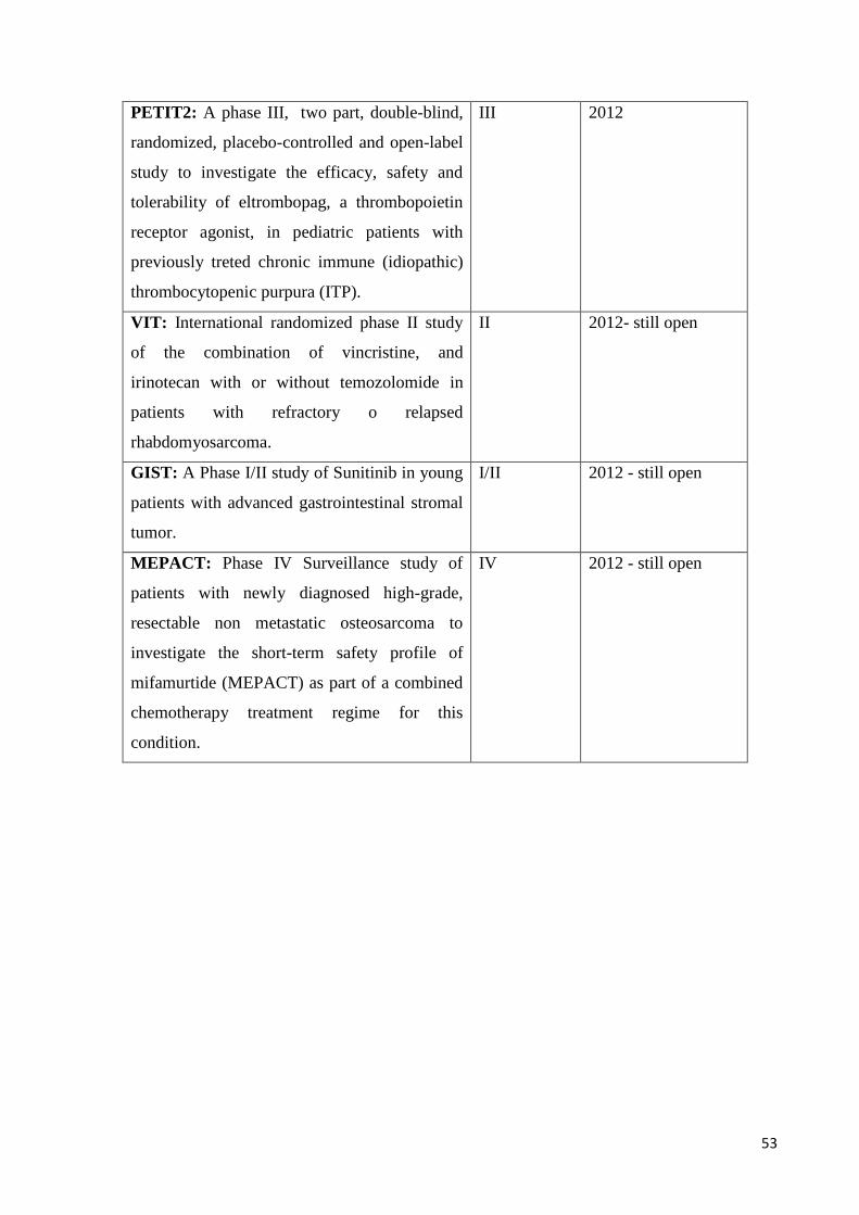
53
PETIT2: A phase III, two part, double-blind,
randomized, placebo-controlled and open-label
study to investigate the efficacy, safety and
tolerability of eltrombopag, a thrombopoietin
receptor agonist, in pediatric patients with
previously treted chronic immune (idiopathic)
thrombocytopenic purpura (ITP).
III 2012
VIT: International randomized phase II study
of the combination of vincristine, and
irinotecan with or without temozolomide in
patients with refractory o relapsed
rhabdomyosarcoma.
II 2012- still open
GIST: A Phase I/II study of Sunitinib in young
patients with advanced gastrointestinal stromal
tumor.
I/II 2012 - still open
MEPACT: Phase IV Surveillance study of
patients with newly diagnosed high-grade,
resectable non metastatic osteosarcoma to
investigate the short-term safety profile of
mifamurtide (MEPACT) as part of a combined
chemotherapy treatment regime for this
condition.
IV 2012 - still open

54

55
3.3 Protocollo di fase II
3.3.1 Introduzione
Parte integrante del progetto era il raggiungimento di una adeguata expertise per la
stesura di Protocolli di fase II-III. Tale obiettivo è stato raggiunto grazie alla formazione
in itinere attraverso partecipazione a corsi specifici come:
“ITCC Training Days”, corso sullo sviluppo di nuovi farmaci in oncologia pediatrica,
tenutosi a Roma dal 22 al 24 ottobre 2009.
“3rd ESO-SIOP Europe Masterclass in paediatric oncology”, tenutosi a Castelgandolfo,
Roma, dal 12 al 18/06/2010.
Inoltre la partecipazione attiva e via via crescente come subinvestigator a trials clinici di
fase II e III, nazionali e internazionali, ha contribuito ad una progressiva conoscenza del
percorso che porta alla stesura dei protocolli e della loro gestione; la formazione di un
Gruppo dedicato ai “Nuovi Farmaci” è uno dei risultati di questo lavoro e costituisce
senza dubbio un valore aggiunto per il Nostro Centro.
Il gruppo è formato da:
Data Managers per la raccolta dati, la compilazione delle CRF (case reporting
form) con supervisione medica, le relazioni con i centri coordinatori, i Monitors, le
aziende sponsor, i comitati etici.
Infermiere di Ricerca dedicate a seguire i pazienti arruolati nei protocolli
(contabilità e conservazione del farmaco, preparazione e somministrazione della
chemioterapia, relazione col paziente e i famigliari negli aspetti di loro competenza,
feedback a medici e data managers).
Medici dedicati alla gestione superspecialistica dei pazienti in protocollo, con
valutazione dell’eligibilità dei pazienti al protocollo, arruolamento degli stessi,
consegna, spiegazione, firma dei consensi, prescrizione della chemioterapia, controllo e
gestione degli eventi avversi (in particolare i “SAE”, serious adverse events),
supervisione delle CRF.
Il coinvolgimento come subinvestigator con la fondamentale partecipazione a
Investigators’ Meeting (es: Windsor il 22-23.05.2008 per il Protocollo BO20924,
Heathrow il 24.03.2009 per il Protocollo BO20924, Madrid il 19-20.01.2010 per il
Protocollo BO20924) ed a visite di apertura del Centro ai protocolli, ha permesso

56
l’acquisizione di una certa expertise in questo ambito, frontiera relativamente “nuova”
per il mondo pediatrico.
A “conclusione” di tale iter formativo gli sforzi sono stati coordinati alla stesura di un
protocollo di fase II per le recidive di tumori solidi pediatrici.
L’oggetto del protocollo è il trattamento delle meningosi da RMS/EPNET con
topotecan intratecale. La stesura del protocollo è in corso, la versione definitiva verrà
probabilmente presentata alla riunione annuale sui tumori solidi pediatrici che si tiene a
Padova verso la fine dell’anno.
Segue quanto finora scritto del protocollo di fase II.
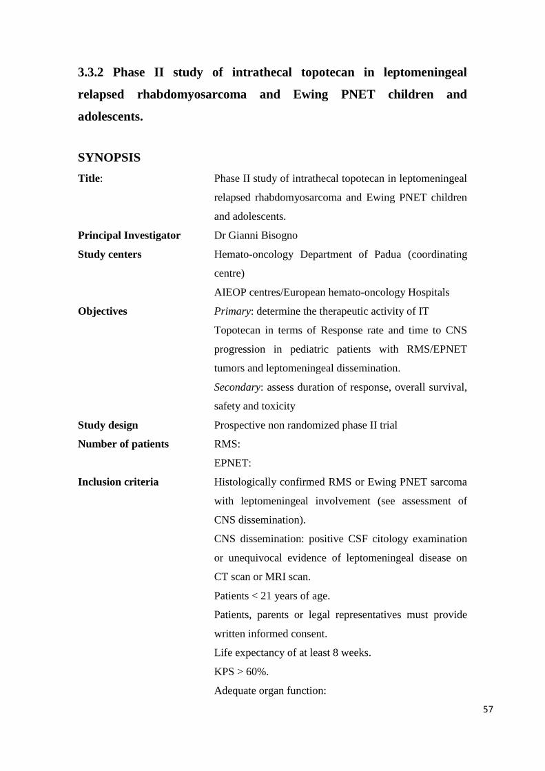
57
3.3.2 Phase II study of intrathecal topotecan in leptomeningeal
relapsed rhabdomyosarcoma and Ewing PNET children and
adolescents.
SYNOPSIS
Title Title: Phase II study of intrathecal topotecan in leptomeningeal
relapsed rhabdomyosarcoma and Ewing PNET children
and adolescents.
Principal Investigator Dr Gianni Bisogno
Study centers Hemato-oncology Department of Padua (coordinating
centre)
AIEOP centres/European hemato-oncology Hospitals
Objectives Primary: determine the therapeutic activity of IT
Topotecan in terms of Response rate and time to CNS
progression in pediatric patients with RMS/EPNET
tumors and leptomeningeal dissemination.
Secondary: assess duration of response, overall survival,
safety and toxicity
Study design Prospective non randomized phase II trial
Number of patients RMS:
EPNET:
Inclusion criteria Histologically confirmed RMS or Ewing PNET sarcoma
with leptomeningeal involvement (see assessment of
CNS dissemination).
CNS dissemination: positive CSF citology examination
or unequivocal evidence of leptomeningeal disease on
CT scan or MRI scan.
Patients < 21 years of age.
Patients, parents or legal representatives must provide
written informed consent.
Life expectancy of at least 8 weeks.
KPS > 60%.
Adequate organ function:
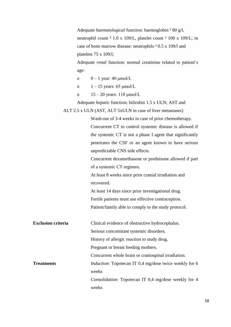
58
Adequate haematological function: haemoglobin ³ 80 g/l,
neutrophil count ³ 1.0 x 10⁹/L, platelet count ³ 100 x 109/L; in
case of bone marrow disease: neutrophils ³ 0.5 x 10⁹/l and
platelets 75 x 109/l;
Adequate renal function: normal creatinine related to patient’s
age:
o 0 – 1 year: 40 μmol/L
o 1 – 15 years: 65 μmol/L
o 15 – 20 years: 110 μmol/L
Adequate hepatic function: bilirubin 1.5 x ULN; AST and
ALT 2.5 x ULN (AST, ALT 5xULN in case of liver metastases)
Wash-out of 3-4 weeks in case of prior chemotherapy.
Concurrent CT to control systemic disease is allowed if
the systemic CT is not a phase I agent that significantly
penetrates the CSF or an agent known to have serious
unpredictable CNS side effects.
Concurrent dexamethasone or prednisone allowed if part
of a systemic CT regimen.
At least 8 weeks since prior cranial irradiation and
recovered.
At least 14 days since prior investigational drug.
Fertile patients must use effective contraception.
Patient/family able to comply to the study protocol.
Exclusion criteria Clinical evidence of obstructive hydrocephalus.
Serious concomitant systemic disorders.
History of allergic reaction to study drug.
Pregnant or breast feeding mothers.
Concurrent whole brain or craniospinal irradiation.
Treatments Induction: Topotecan IT 0,4 mg/dose twice weekly for 6
weeks
Consolidation: Topotecan IT 0,4 mg/dose weekly for 4
weeks

59
Maintenance: Topotecan IT 0,4 mg/dose twice monthly
for 4 months then monthly through year 1
Assessment and
Criteria for evaluation Efficacy: MRI/CT scans
CSF cytology (LP/intraventricular reservoir)
Safety: Safety profile will be evaluated. Clinical and
laboratory toxicities/symptomatology will be graded
according to NCI-Common toxicity criteria AE v3.0.
The adverse events which are not reported in the NCI-
Common toxicity criteria will be graded as mild,
moderate, severe, life-threatening
Statistical method
Post study follow up Every 2 months up to death or study cut-off.

60
TABLE OF CONTENTS
BACKGROUND
Rhabdomyosarcoma/Ewing PNET
Incidence and Epidemiology
Prognostic Factors
Cellular and molecular classification
Stage Information
Treatment Option Overview
Leptomeningeal dissemination of cancer
Intrathecal chemotherapy
Intrathecal Topotecan
RMS/EPNET with leptomeningeal dissemination enrolled in STSC protocols
OBJECTIVES AND ENDPOINTS
PRIMARY
SECONDARY
OVERALL STUDY PLAN
STUDY DESIGN
STUDY CENTERS
EXPECTED NUMBER OF PATIENTS
PATIENT REGISTRATION
STUDY PERIOD
SUBJECT POPULATION AND SELECTION
INCLUSION/EXCLUSION CRITERIA
EFFICACY AND SAFETY ASSESSMENTS
CLINICAL ASSESSMENTS
LABORATORY ASSESSMENTS
TUMOUR ASSESSMENTS
TREATMENTS
DOSING AND ADMINISTRATION
61
TREATMENT DURATION
TREATMENT DISCONTINUATION
CONCOMITANT THERAPIES
FOLLOW UP
ADVERSE EVENTS
ADVERSE EVENTS AE definition
Recording of Adverse Events
SERIOUS ADVERSE EVENTS SAE definition
SAEs reporting
ADVERSE EVENTS FOLLOW UP
ANNUAL SAFETY REPORT
STATISTICAL CONSIDERATIONS
TRIAL PLAN AND SAMPLE SIZE
ANALYSIS
DATA MANAGMENT
DATA COLLECTION, QUALITY ASSURANCE, MANAGEMENT
APPENDIXES
APPENDIX 1: PERFORMANCE SCALE AND LASKY PLAY SCALE
REFERENCES

62
BACKGROUND
Rhabdomyosarcoma/PNET
Incidence and Epidemiology
Childhood rhabdomyosarcoma, a soft tissue malignant tumor of mesenchymal origin,
accounts for approximately 3.5% of the cases of cancer among children aged 0 to 14
years and 2% of the cases among adolescents and young adults aged 15 to 19 years (2,
3). The incidence is 4.5 per 1 million children and 50% of cases are seen in the first
decade of life (4). Most cases of rhabdomyosarcoma occur sporadically, with no
recognized predisposing factor or risk factor (8). Genetic conditions associated with
rhabdomyosarcoma include Li-Fraumeni cancer susceptibility syndrome (with germline
p53 mutations), (9, 10, 11) pleuropulmonary blastoma (with DICER1 mutations), (12,
13) neurofibromatosis type I, (14) Costello syndrome (with germline HRAS mutations),
(15-18) Beckwith-Wiedemann syndrome (with which Wilms tumor and hepatoblastoma
are more commonly associated), (19, 20) and Noonan syndrome. (18, 21, 22)
Dramatic improvements in survival have been achieved for children and adolescents
with cancer (1). Between 1975 and 2002, childhood cancer mortality has decreased by
more than 50%. For rhabdomyosarcoma, the 5-year survival rate has increased over the
same time from 53% to 65% for children younger than 15 years and from 30% to 47%
for adolescents aged 15 to 19 years (1).
RMS may arise everywhere in the body. The most common primary sites for
rhabdomyosarcoma are the head, the genitourinary tract, and the extremities (5, 6).
Within extremity tumors, tumors of the hand and foot occur more often in older patients
and have an alveolar histology; these tumors also have a higher rate of metastatic spread
(7). Other less common primary sites include the trunk, chest wall, perineal/anal region,
and abdomen including the retroperitoneum and biliary tract. Symptoms depend on the
site of origin.
Prognostic Factors
The prognosis for a child or adolescent with rhabdomyosarcoma is related to the age of
the patient, site of origin, tumor size (widest diameter), resectability, presence of
metastases, number of metastatic sites or tissues involved, presence or absence of

63
regional lymph node involvement, histopathologic subtype (alveolar vs. embryonal),
and delivery of radiation therapy in selected cases (5, 6, 23-30) as well as unique
biological characteristics of rhabdomyosarcoma tumor cells (31). It is unclear whether
response to induction chemotherapy, as judged by anatomic imaging, correlates with the
likelihood of survival in patients with rhabdomyosarcoma, as one study found an
association and another study did not (32, 33).
Rhabdomyosarcoma is usually curable in most children with localized disease who
receive combined-modality therapy, with more than 70% surviving 5 years after
diagnosis (5, 6, 34). Local relapses are more frequent than metastatic ones. Relapses are
uncommon after 5 years of disease-free survival, with a 9% late-event rate at 10 years.
Relapses, however, are more common for patients who have gross residual disease in
unfavorable sites following initial surgery and those who have metastatic disease at
diagnosis (35).
Cellular and molecular classification
Rhabdomyosarcoma can be divided into several histologic subsets: embryonal
rhabdomyosarcoma, which has embryonal, botryoid, and spindle cell subtypes; alveolar
rhabdomyosarcoma; and pleomorphic rhabdomyosarcoma (36,37).
Embryonal Rhabdomyosarcoma: the embryonal subtype is the most frequently observed
subtype in children, accounting for approximately 60% to 70% of rhabdomyosarcomas
of childhood (36).Tumors with embryonal histology typically arise in the head and neck
region or in the genitourinary tract, although they may occur at any primary site.
Embryonal tumors often show loss of specific genomic material from the short arm of
chromosome 11 (44,45, 46). The consistent loss of genomic material at the chromosome
11p15 region in embryonal tumors suggests the presence of a tumor suppressor gene,
although no such gene has yet been identified.
Botryoid and spindle cell subtypes: botryoid tumors represent about 10% of all
rhabdomyosarcoma cases and are embryonal tumors that arise under the mucosal
surface of body orifices such as the vagina, bladder, nasopharynx, and biliary tract.
The spindle cell variant of embryonal rhabdomyosarcoma is most frequently observed
at the paratesticular site (38). Both the botryoid and the spindle cell subtypes are
associated with very favorable outcomes (37).
64
Alveolar Rhabdomyosarcoma: approximately 20% of children with rhabdomyosarcoma
have the alveolar subtype. An increased frequency of this subtype is noted in
adolescents and in patients with primary sites involving the extremities, trunk, and
perineum/perianal region (36).
Unique translocations between the FOXO1 (previously called FKHR) gene on
chromosome 13 and either the PAX3 gene on chromosome 2 (t(2;13)(q35;q14)) or the
PAX7 gene on chromosome 1 (t(1;13)(p36;q14)) are found in 70% to 80% of patients
with alveolar histology tumors (42, 43, 44). Translocations involving the PAX3 gene
occur in approximately 59% of alveolar rhabdomyosarcoma cases, while the PAX7
gene appears to be involved in about 19% of cases (42).
Pleomorphic (Anaplastic) Rhabdomyosarcoma: pleomorphic rhabdomyosarcoma occurs
predominantly in adults aged 30 to 50 years and is rarely seen in children (39). In
adults, pleomorphic rhabdomyosarcoma is associated with a worse prognosis. In
children, the term anaplasia is preferred (40). In a retrospective review of 546 pediatric
patients, the presence of anaplasia was only associated in univariate analysis with
inferior clinical outcome in patients with intermediate-risk rhabdomyosarcoma (41).
Stage Information
Before a biopsy of a suspected tumor mass is performed, imaging studies of the mass
and baseline laboratory studies should be obtained. After the diagnosis of
rhabdomyosarcoma has been made, an extensive evaluation to determine the extent of
the disease should be done prior to instituting therapy. This evaluation should include a
chest x-ray, computed tomography (CT) scan of the chest, bilateral bone marrow
aspirates and biopsies, bone scan, magnetic resonance imaging (MRI) of the base of the
skull and brain (for parameningeal primary tumors only), and CT scan of the abdomen
and pelvis (for lower extremity or genitourinary primary tumors).
A CT or MRI scan of regional lymph nodes should be considered. Abnormal-appearing
lymph nodes should be biopsied when possible. One study has demonstrated that
sentinel lymph node biopsies can be safely performed in children with
rhabdomyosarcoma, and tumor-positive biopsies may alter the treatment plan (47).
Positron emission tomography (PET) with fluorine-18-fluorodeoxyglucose (FDG) scans
can identify areas of possible metastatic disease not seen by other imaging modalities
(48-50). However, the efficacy of these two procedures for identifying involved lymph

65
nodes or other sites is currently under investigation, and these procedures are not
required by current treatment protocols.
As noted previously, prognosis for children with rhabdomyosarcoma depends
predominantly on the primary site, tumor size, Group, and histologic subtype. Favorable
prognostic groups were identified in previous Intergroup Rhabdomyosarcoma Study
Group (IRSG) studies, and treatment plans were designed on the basis of assignment of
patients to different treatment groups according to prognosis. Several years ago, the
IRSG merged with the National Wilms Tumor Study Group and two large cooperative
pediatric cancer treatment groups to form the Children's Oncology Group (COG). New
protocols for children with soft tissue sarcoma are developed by the Soft Tissue
Sarcoma Committee of the COG (COG-STS). Current COG-STS protocols for
rhabdomyosarcoma use the TNM-based pretreatment staging system that incorporates
the primary tumor site, presence or absence of tumor invasion of surrounding tissues,
tumor size, regional lymph node status, and the presence or absence of metastases (51,
52). After patients are categorized by Stage and Surgical-pathologic Group, a Risk
Group is assigned. This takes into account Stage, Group, and histology. Patients are
classified for protocol purposes as having a low risk, intermediate risk, or high risk of
disease recurrence (53, 54). Treatment assignment is based on Risk Group.
Treatment Option Overview
All children with rhabdomyosarcoma should receive chemotherapy. The intensity and
duration of the chemotherapy are dependent on the Risk Group assignment. All children
with rhabdomyosarcoma require multimodality therapy with systemic chemotherapy, in
conjunction with either surgery, radiation therapy (RT), or both modalities to maximize
local tumor control (55-57).
Surgical resection may be performed prior to chemotherapy if it will not result in
disfigurement, substantial functional compromise, or organ dysfunction. In most cases,
this is not possible, and therefore, only an initial biopsy is performed. The majority of
patients have Group III (gross residual) disease.
After initial chemotherapy, Group III patients receive definitive RT for control of the
primary tumor. Some patients with initially unresected tumors may undergo second-
look surgery (delayed primary excision) to remove residual tumor. This is most
appropriate if the delayed excision is deemed feasible with acceptable
66
functional/cosmetic outcome, and if a modest reduction in radiation dose is expected to
significantly reduce the risk of long-term adverse effects. RT is given to clinically
suspicious lymph nodes (detected by palpation or imaging) unless the suspicious lymph
nodes are biopsied and shown to be free of rhabdomyosarcoma.
The treatment of rhabdomyosarcoma by the Children's Oncology Group (COG) and in
Europe (as exemplified by trials from the Intergroup Rhabdomyosarcoma Study Group
[IRSG], the Soft Tissue Sarcoma Committee of the COG [COG-STS], and the
International Society of Pediatric Oncology Malignant Mesenchymal Tumor [MMT]
Group) differs in management and overall treatment philosophy (56). Children are
treated with a more or less intense CT according to risk group. Main drugs are
Ifosfamide, Actinomicine, Vincristine and Doxorubicine.
Leptomeningeal dissemination of cancer
Neoplastic meningitis results from the spread of malignant cells to leptomeninges and
subarachnoid space and their dissemination within the cerebrospinal fluid (CSF)
compartment. Malignant cells may reach the subarachnoid space through the blood
(arterial or venous), by growing along nerve and vascular sheeths, by migration from a
tumor adjacent to CSF or by iatrogenic spread of tumor cells following resection of
metastasis (58, 59).
Neoplastic meningitis is a devasting complication of both solid and hematologic tumors
and is estimated to occur in 5-8% of cancer patients. Among adults the most common
cancers that metastasize to the leptomeninges are breast cancer, lung cancer, melanoma,
lymphomas and leukemias; in the pediatric population leukemia is the most common
cancer with predilection for leptomeningeal dissemination; however primary central
nervous system tumors (medulloblastoma and glioma), other cancers like
neuroblastoma, rhabdomyosarcoma and retinoblastoma may also disseminate to the
leptomeninges (60).
Despite treatment the median survival duration for patients with neoplastic meningitis is
in the range of 8-16 weeks. The impact of neoplastic meningitis is likely to increase in
the future as advances in systemic treatment have improved survival but leave the
leptomeninges and CSF a sanctuary site (61). The meninges are a sanctuary site because
protected by the blood brain barrier (BBB) from the cytotoxic effects of systemic

67
anticancer therapy. The BBB often prevents efficient penetration of many drugs into the
CSF space so that only few systemic chemotherapy agents can produce clinically
relevant CSF concentrations and only at high doses; this could produce significant
systemic toxicity. Regional delivery of drugs directly into the CSF is pharmacologically
advantageous, with small doses producing high CSF concentration with minimal
systemic exposure (62). Unfortunately only a limited number of drugs have been found
to be safe and efficacious when administered by the intrathecal (IT) route.
Therapeutic approaches for leptomeningeal dissemination of cancer are:
- Radiotherapy (RT)
- Systemic CT
- I.T. CT
Focal RT is performed in the treatment of bulky disease and in patients with CSF flow
blocks. In addition focal RT should also be administered to symptomatic areas with a
short palliative schedule. Another use of RT is in the treatment of cauda equina
syndrome and cranial neuropathies from neoplastic meningitis, whereas craniospinal
irradiation is rarely used because of significant systemic toxicity (63).
Systemic CT efficacy is not affected by CSF flow obstruction if compared to IT CT.
However systemic CT can be limited by systemic toxicity because it’s necessary to
administer high doses to reach a clinically relevant CSF concentrations; another
difficulty is using an effective treatment for neoplastic meningitis as well as for the
underlying disease causing the meningeal spread. High doses methotrexate (MTX) have
favorable CSF penetration but considerable systemic toxicity too; moreover MTX is not
typically part of standard regimens used to treat many of the underlying tumors, making
its incorporation into systemic treatment difficult. Ifosfamide and topotecan are both
active but toxic, like MTX. Oral temozolomide and capecitabine have shown interesting
results against leptomeningeal dissemination from breast and lung cancer (63).
Intrathecal chemotherapy
CT administration can be undertaken either I.T. via a lumbar puncture (LP) or via an
intraventricular device with a catheter into the lateral ventricle by Ommaya reservoir.
68
IT CT has been used in the treatment of leptomeningeal dissemination even though the
extent of its benefit has not been proven in randomized controlled trials and some
studies showed discordant evidence. IT treatment offers local therapy with minimum
systemic toxicity, and avoiding the BBB drugs are distributed throughout the entire
subarachnoid space; although high drug concentrations could be achieved in the CSF,
IT CT is not effective for bulky disease in the meninges because intra-CSF agents
penetrate only 2-3 mm into such lesions (63).
IT administration of anticancer drugs has been an effective strategy for the primary
treatment and prevention of leptomeningeal leukemias and lymphomas, but it has not
been effective in patients with neoplastic meningitis from solid tumors or in patients
with refractory leptomeningeal leukemias (this results in part from the limited number
of agents available for IT administration) (64).
Only a small number of anticancer agents are regularly used: methotrexate, cytarabine,
liposomal cytarabine, thiotepa. None of these have resulted in significantly prolonged
survival and combination of intra-CSF drugs have not improved outcomes over single
agents (61). MTX and liposomal cytarabine are the IT drugs most commonly used for
leptomeningeal dissemination in solid tumors. However in the few randomized trials the
data were discordant when comparing treatments (65). No one have a significant impact
on survival, in the studies available “time to neurological progression” is the most
frequently used parameter to evaluate response to treatment; the most common adverse
event during IT CT is arachnoiditis/chemical meningitis (66, 67).
Then neoplastic meningitis is still a relevant clinical problem, therefore it is essential to
develop new IT agents with novel mechanism of action. In the last few years various
experimental IT drugs have been reported upon from small clinical trials, a few case
reports, and preclinical studies.
Among them Topotecan showed interesting results.
Intrathecal Topotecan
Topotecan is a topoisomerase I poison that has anticancer activity against a variety of
adult and childhood solid tumors. Preclinical studies demonstrated a good CSF
penetration capability; 0,1 mg intraventricular dose (equivalent to 1 mg in humans) was
defined as a well tolerated dose (68, 69).

69
The results of these preclinical studies served as the basis for phase I studies of IT
topotecan in patients with neoplastic meningitis. The phase I study conducted by Blaney
and colleagues (64) proved the feasibility of administering topotecan at 0,4 mg as
maximum tolerated dose (MTD) to children and adults with neoplastic meningitis (17
assessable patients, heterogenous histotypes); arachnoiditis was the dose limiting
toxicity (DLT). A second phase I study (60) aimed to re-evaluate the dosing schedule
for IT topotecan administration based upon preclinical evidence that the antitumor
activity of topoisomerase I inhibitors is schedule dependent (70, 69). 19 patients <22
years with leukemias and central nervous system cancer were enrolled. The study
demonstrated that intensified dosing of topotecan is feasible (with chemical
arachnoiditis being the DLT) but whether shorter or longer duration of exposure to the
drug would be superior or not is unknown. Because of many different tumor types
enrolled, this study, as well as the first one, cannot make definitive conclusions
regarding antitumor activity.
A phase II study by Groves and colleagues (61) analyzed the results obtained with IT
topotecan classic schedule administration in 62 adult patients with leptomeningeal
dissemination from solid tumors. 40 patients were evaluable; treatment was well
tolerated (arachnoiditis being the most common adverse event) but outcomes were no
better than those reported in trials that employed other IV CT. Better results have been
obtained by IT topotecan administered to 20 children with leptomeningeal leukemia
(71): among 16 evaluable patients a 37,5% CR have been achieved with a confirmed
mild toxicity. A phase II trial of IT topotecan was performed in children with
dissemination to the meninges by medulloblastoma and other solid tumors (72); the
treatment was well tolerated, there were no objective responses; however the authors
observed a benefit in terms of disease stabilization greater than 5 months in 4 children.
Data available show that:
leptomeningeal dissemination of tumors is a devasting problem because outcome is very
bad and treatments available are very few;
data about IT topotecan come out studies with heterogeneous and small population of
patients;
results in terms of RR/PFS/OS are disappointing but clinical benefit and stabilizations
have been observed;
70
at our knowledge no phase II studies have been performed with IT topotecan in patients
with rhabdomyosarcoma-EPNET tumors and leptomeningeal spread.
For this reason with designed a phase II study with IT topotecan in patients with
rhabdomyosarcoma-EPNET tumors and leptomeningeal spread.
RMS/EPNET with leptomeningeal dissemination enrolled in STSC protocols
We have found 13 patients with leptomeningeal dissemination from RMS or Ewing
PNET tumors enrolled in STSC clinical trials from 1979 to 2013.
Analysis about clinical characteristics and outcome are on going.
OBJECTIVES AND ENDPOINTS
The primary objective is to determine the therapeutic activity of IT Topotecan in terms
of Response rate and time to CNS progression in pediatric patients with RMS/EPNET
tumors.
The secondary endpoint is to assess duration of response, overall survival, safety and
toxicity.
OVERALL STUDY PLAN
STUDY DESIGN
Multicenter, prospective, non randomized phase II trial.
STUDY CENTERS
Italy: Padova, INT Milano, Genova, Roma, Napoli, Bergamo, Torino...
Europe: …
EXPECTED NUMBER OF PATIENTS
PATIENT REGISTRATION
71
STUDY PERIOD
SUBJECT POPULATION AND SELECTION
INCLUSION CRITERIA
Histologically confirmed RMS or Ewing PNET sarcoma with leptomeningeal
involvement (see assessment of CNS dissemination)
CNS dissemination: positive CSF citology examination or unequivocal evidence of
leptomeningeal disease on CT scan or MRI scan.
Patients < 21 years of age
Patients, parents or legal representatives must provide written informed consent
Life expectancy of at least 8 weeks
KPS > 60%
Adequate organ function:
Adequate haematological function: haemoglobin ³ 80 g/l, neutrophil count ³ 1.0 x 10⁹/L,
platelet count ³ 100 x 109/L; in case of bone marrow disease: neutrophils ³ 0.5 x 10⁹/l
and platelets 75 x 109/l.
Adequate renal function: normal creatinine related to patient’s age:
o 0 – 1 year: 40 μmol/L
o 1 – 15 years: 65 μmol/L
o 15 – 20 years: 110 μmol/L
Adequate hepatic function: bilirubin <1.5 x ULN; AST and ALT <2.5 x ULN (AST,
ALT 5xULN in case of liver metastases)
Wash-out of 3-4 weeks in case of prior chemotherapy
Concurrent CT to control systemic disease is allowed if the systemic CT is not a phase I
agent that significantly penetrates the CSF or an agent known to have serious
unpredictable CNS side effects
Concurrent dexamethasone or prednisone allowed if part of a systemic CT regimen
At least 8 weeks since prior cranial irradiation and recovered
At least 14 days since prior investigational drug
Fertile patients must use effective contraception

72
Patient/family able to comply to the study protocol
EXCLUSION CRITERIA
• Clinical evidence of obstructive hydrocephalus
• Serious concomitant systemic disorders
• History of allergic reaction to study drug
• Pregnant or breast feeding mothers
• Concurrent whole brain or craniospinal irradiation
TREATMENTS
DOSING AND ADMINISTRATION
Topotecan is supplied in 4 mg vials; the content of each vial is diluted in 4 ml of sterile
water then further diluted with preservative free, pyrogen free saline to a final volume
of 10 ml. Administration of the drug is performed at a constant rate of 2,0 ml/minute
(total 5 minutes) through an intraventricular reservoir or lumbar puncture (LP).
The volume of CSF equivalent to the volume of drug to be administered is removed
prior to drug administration.
Patients who receive topotecan via LP have to remain prone, flat or in the
Trendelemburg position for 1 hour following drug administration. After drug
administration via an intraventricular reservoir, the reservoir has to be flushed slowly
for 1-2 minutes with approximately 2 ml of either CSF or preservative free normal
saline then pumped 4-6 times.
Treatment:
• Induction: Topotecan IT 0,4 mg/dose twice weekly for 6 weeks
• Consolidation: Topotecan IT 0,4 mg/dose weekly for 4 weeks
• Maintenance: Topotecan IT 0,4 mg/dose twice monthly for 4 months then monthly
through year 1
TREATMENT DURATION
The maximum planned treatment duration in absence of toxicity or progression is up to
2 years.

73
FOLLOW-UP
The follow-up period begins when the patient discontinues from study treatment.
If feasible, one post-discontinuation visit will be performed 30 days after treatment
discontinuation and follow-up in all patients must be pursued every 2 months until the
patient's death or up to at least one year until study cut-off.
Patients with adverse events at the end of the study related to treatment must be
followed until recovery.
During post-therapy follow-up, information will be collected in the CRF regarding date
of disease progression, further second line treatment (chemo, radiotherapy, surgery) and
death. The date of first documented disease progression must be recorded on the CRF
even if it occurs after the patient has started a new therapy. All deaths will be recorded.
TREATMENT DISCONTINUATION
Treatment should be discontinued if this is considered to be in the best interest of the
patient. Treatment could be discontinued for the following reasons:
Investigator’s decision: if this decision is made because of toxicity, a serious adverse
event, or a clinically significant laboratory value, appropriate measures will be taken
and IGR will be notified immediately.
The patient, parents or legal representative’s refusal, withdrawal of patient consent.
The investigator or sponsor, for significant safety or efficacy reason, stops the study or
stops the patient's participation in the study.
Evidence of progressive disease exists.
The patient becomes pregnant or fails to use adequate birth control (for those patients
who are able to conceive).
The patient is non compliant with study procedures.
Life threatening toxicity.
Unmanageable or unacceptable toxicity, including the need for more than 2 dose
reductions, except in cases of obvious patient benefit in continuing the treatment.
Treatment delay of more than 3 weeks for any reason except in cases of obvious
patient benefit in continuing the treatment.

74
Study discontinuation, must be reported to IGR as soon as possible and immediately in
case of discontinuation related to a serious adverse event. The primary reason and date
of removal for all patients will be documented on the case report form (e.g. lost to
follow-up, withdrawal of consent, patients wrongly included, adverse events, etc.). The
final evaluation required by the protocol will be performed at the time of study
discontinuation. Further follow-up should be reported. The investigator will attempt to
complete all discharge procedures at the time a patient is removed from the treatment.
CONCOMITANT THERAPIES
Patients could receive chemotherapy to control systemic disease provided the systemic
CT is not a phase I agent, doesn’t significantly penetrate the CSF, or is not known to
have serious unpredictable CNS side effects.
Patients are allowed to receive full supportive care therapies concomitantly during the
study.
If during the study patient develops a need for palliative radiotherapy, it should be
ensured that this is not a manifestation of progressive disease (patients with progressive
disease must discontinue study therapy). Palliative radiotherapy may be given for
control of pain or for other reasons with no curative intent. The irradiated area cannot be
used as a parameter for response assessment.
EFFICACY AND SAFETY ASSESSMENTS
The following exams will performed according to the schedule of assessments after
signature of written informed consent by patients or parents or legal representative.
CLINICAL ASSESSMENTS
Medical History
Relevant past medical history, and current medical conditions not related to the current
indication or disease for which patient entered into the study
Information related to diagnosis of the disease under study
Previous surgery, radiotherapy, systemic therapy, investigational therapy

75
Any disease related symptoms present at baseline.
Physical examination
Physical and Neurological Examination
ECOG Performance Status or Lansky-Play score
Vital signs (pulse, blood pressure, temperature)
Height, Weight and Body Surface
Will be done within 7 days before study enrolment, then before each administration
LABORATORY AND TUMOR ASSESSMENTS
CSF studies: cell count, differential, protein, glucose. CSF citology: at baseline and at
each CT administration
CT or MRI scans: at baseline, every 2 months and at the first post-discontinuation visit.
MRI of the spine if clinically indicated.
Bone marrow aspirates: at baseline
Pregnancy test (urine or serum) in females of childbearing potential within 7 days
before study enrolment.
Complete Blood Count: leukocyte, neutrophil, platelets and haemoglobin within 7 days
before study enrolment, then once a week during each cycle and every 2 days in case of
neutro- or thrombocytopenia, and if possible at the first post-discontinuation visit
Serum Biochemistry: sodium, potassium, calcium, total protein, creatinine, urea, AST
(SGOT), ALT (SGPT), total bilirubin, albumin within 7 days before study enrolment,
then once a week, and if possible at the first post-discontinuation visit.
SAFETY ASSESSMENTS
Adverse events are evaluated according to NCI common toxicity criteria (vers 3.0).
Treatment has to be stopped if there is >grade 3 non hematologic toxicity considered to
be at least possibly related to topotecan with the following exception: >grade 3
headache prevented after subsequent doses using premedication or >grade 3 nausea or
vomiting that is well controlled or prevented after subsequent doses with antiemetics.
After the routine use of dexamethasone patients with arachnoiditis could not receive
further topotecan.
76
Any non hematologic toxicity experienced during a cycle must resolve to Grade 1 or
lower before the next cycle may be administered.
CRITERIA FOR ASSESSMENT OF RESPONSE
Response is classified as complete response (CR), stable disease (SD) or progressive
disease (PD).
For CR patients need to have complete clearing of malignant cells from lumbar or
ventricular CSF on two consecutive citologic studies at least 4 weeks apart, with no
worsening of physical or neurological findings clearly attributable to neoplastic
meningitis; complete clearing of disease on two consecutive MRI scans >4 weeks apart.
PD is defined as the occurence of new malignant cells in the CSF on two consecutive
occasions at least 1 week apart from after at least 3 previous consecutive negative CSF
cytologist obtained at least 1 week apart or an increase of >25% in the size of
measurable lesions on MRI or new lesions on MRI after a CR.
Patients are considered to have stable disease (SD) if they don’t meet the criteria for
either a CR or PD and without worsening of physical findings clearly attributable to
disease.
ADVERSE EVENTS
It is of the utmost importance that all staff involved in the study are familiar with the
content of this section. The principal investigator is responsible for ensuring this.
ADVERSE EVENT
Definition
An adverse event (AE) is the development of an undesirable medical condition or the
worsening of a preexisting medical condition in a clinical investigation subject. The
event need not necessarily have a causal relationship with study drug and can occur at
any time, including run-in or wash-out periods, even if no study treatment has been
administered. An undesirable medical condition can be symptoms (eg, nausea, chest
pain), signs (eg, tachycardia, enlarged liver) or the abnormal results of an investigation
(eg, laboratory findings, x-ray, ECG).
77
Recording of Adverse Events
When to collect AEs: any AE that occur from the time consent is given, during the
study and in the 30 days following the last administration of study treatment should be
recorded.
What AE to collect: all observed AEs regardless of treatment group or suspected causal
relationship to study drug will be assessed following NCI-CTC Criteria and recorded on
the AE page(s) of the CRF, and in case of serious adverse event in a SAE form too.
Worsening/exacerbation of sign and symptoms (in terms of severity and/or frequency,
or the appearance of new manifestations/complications) of the malignancy under study
or of a pre-existing illness should be reported as AE in the appropriate section of the
CRF.
Lack of or insufficient clinical response, benefit, efficacy, therapeutic effect, or
pharmacological action, should not be recorded as an AE.
The investigator must make the distinction between exacerbation of preexisting illness
and lack of therapeutic efficacy. In addition, clinically significant changes in physical
examination findings and abnormal objective test findings (e.g., x-ray, ECG) should
also be recorded as AE.
For all AEs, the investigator must pursue and obtain adequate information both to
determine the outcome of the AE and to assess whether it meets the criteria for
classification as a SAE requiring immediate notification to IGR. For all AEs, sufficient
information should be obtained by the investigator to determine the causality of the AE
(i.e., study drug or other illness). The investigator is required to assess causality and
indicate that assessment on the CRF. All AEs and specially those that are serious,
suspected to be related to study drug or considered significant by the investigator or
clinical monitor must be followed after the time of therapy discontinuation until the
event or its sequelae resolve or stabilize at a level acceptable to the investigator and the
clinical monitor or his/her designated representative.
All AEs will be recorded in the Case Reporting Form (CRF).
SERIOUS ADVERSE EVENT
SAE definition
A Serious Adverse Event (SAE) is any adverse event occurring at any dose that:

78
· Is fatal (results in death).
· Is life-threatening.
· Requires or prolongs inpatient hospitalization.
· Results in persistent or significant disability/incapacity.
· Is a congenital anomaly/birth defect.
· Is medically significant or requires intervention to prevent one of the outcomes listed
above.
Any clinical event or laboratory result considered serious by the investigator and not
corresponding to the criteria of seriousness defined above is nevertheless considered to
be medically significant. Such an event/result can carry a risk for the patient and can
require medical intervention to prevent one of the outcomes listed above (i.e. overdoses,
second cancer and pregnancies can be considered medically significant). Medical and
scientific judgment should be exercised in deciding whether other situations such as
important medical events that may not be immediately life-threatening or result in
hospitalization but may jeopardize the safety of the patient or may require intervention
to prevent one of the outcomes listed in the definition above.
A life-threatening AE is any adverse drug experience that places the patient/subject at
immediate risk of death from the reaction as it occurred, i.e., it does not include a
reaction that, had it occurred in a more severe form, might have caused death.
Hospitalization is defined as in-patient hospital admission associated with an AE which
occurs or worsens after the patient has been included in study. Thus attendance/
treatment at an emergency room/outpatient department does not meet hospitalization
SAE criteria. However, an event which results in attendance / treatment at such a
facility is an SAE if it is considered medically significant or required intervention to
prevent one of the other seriousness criteria.
Note: The SAE is the diagnosis or sign /symptom, NOT the procedure or test defined as
any inpatient admission.
Inpatient admission in the absence of a precipitating, treatment-emergent, clinical AE
may meet criteria for “seriousness” but is not an adverse experience and thus is not
subject to immediate reporting to IGR.
Prolongation of hospitalization is defined as any extension of an inpatient
hospitalization beyond the stay anticipated/required in relation to the original reason for
the initial admission, as determined by the investigator or treating physician. For
79
protocol-specified hospitalizations in clinical trials, prolongation is defined as any
extension beyond the length of stay described in the protocol. Prolongation in the
absence of a precipitating, treatment-emergent, clinical AE (i.e., not associated with the
development of a new AE or worsening of a pre-existing condition) may meet criteria
for "seriousness" but is not an adverse experience and thus is not subject to immediate
reporting to IGR. Pre-planned treatments or surgical procedures should be noted in the
baseline documentation for the entire protocol and/or for the individual patient/subject.
Disability is a substantial disruption of a person’s ability to conduct normal life
functions.
SAEs reporting
Any SAE or SUSAR as defined above which occurs or comes to the attention of the
investigator at any time during the study and through 30 days after the last
administration of study drug, independent of the circumstances or suspected cause, must
be reported immediately, within 24 hours of knowledge by fax via a SAE form.
The Pharmacovigilance Unit at IGR will assess the adverse events in terms of
seriousness, expectedness (IB), severity (NCI-CTCAE v3.0) and relationship to the
study drug. All SAEs will be coded using medDRA.
Assessment of causality of SAEs may be reviewed during the study by the study
coordinator. Information collected in the SAE form is crucial to assess the case and for
this reason diligence in collecting as much verifiable and reliable information: BOTH
QUALITY and TIMELINES are key factors.
All SAEs should be reported immediately (within 24 hours of knowledge of the event),
regardless of time elapsed since last study drug dose (until 30 days after the last
administration of study drug). The investigator must provide any relevant information
for the required 8 days follow up report for any SAE which is fatal or life threatening.
As far as possible, for each event, the following should be noted:
1. As clear as possible a description in medical terminology to allow for a complete
medical assessment of the case and independent determination of possible causality
2. Its duration (start and end dates)
3. Action taken and the necessity for corrective treatment or not, stoppage of study
drug(s) or not, and so on
80
4. Its intensity (grade 1-5), according to the NCI/NIH Common Terminology Criteria
AE version 3.0 (a copy can be downloaded from the CTEP home page:
http://ctep.info.nih.gov).
5. Its relationship to the study drug or treatment, the pathology treated, another
pathology or another treatment, or to a constraint linked to the research (period without
treatment, further tests required for the research, and so on). If causality is unknown and
the investigator does not know whether or not study drug caused the event, it should be
attributed to study drug. If the investigator's causality assessment is "unknown but not
related to study drug", this should be clearly documented on study records.
6. Documentation of all co-medication and/or therapies
7. Documentation of all relevant medical history and/or co-existing diseases
8. The outcome (where applicable).
For non fatal events, developments should be followed up until either recovery or
recovery of a previous state of health or until the stabilization of possible aftereffects.
The investigator must also attach the following to the serious adverse event report form,
wherever possible:
· A copy of the summary of hospitalization or prolongation of hospitalization
· A copy of the post-mortem report
· A copy of all laboratory examinations and the dates on which these examinations were
carried out, including relevant negative results, as well as normal laboratory ranges.
· All other document that he judges useful and relevant.
All these documents will remain anonymous.
Further information can be requested (by fax, telephone or when visiting) by the
monitor and/or the safety manager. All SAEs will be recorded in the Case Reporting
Form (CRF) too.
Adverse events follow-up
The investigator is responsible for the appropriate medical follow-up of patients until
resolution or stabilization of the adverse event or until the patient's death. This may
mean that follow-up should continue once the patient has left the trial. Follow up
information about a previously reported serious adverse event must be reported by the
investigator to the pharmacovigilance unit within 48 hours of receiving it.
81
The investigator also transmits the final report at the time of resolution or stabilization
of the SAE. He retains the documents concerning the supposed adverse event so that
previously transmitted information can be completed if necessary.
Annual safety report
The pharmacovigilance unit at IGR will issue once a year throughout the clinical trial,
or on request, the annual safety report (ASR) of the study, in accordance with the
detailed guidance issued by the European Commission on the collection, verification
and presentation of adverse reaction reports arising from clinical trials on medicinal
products for human use of April 2006 and the applicable revisions thereof. The
pharmacovigilance unit will send a copy of the ASR to the investigators and national
sponsors. Each national sponsor should submit the ASR within 60 days of the data lock
point (date of the first authorisation of the concerned clinical trial by a competent
authority in a member state) to the national competent authority and the national Ethic
Committee of the concerned Member State, according to national legislation.
STATISTICAL CONSIDERATIONS
TRIAL PLAN AND SAMPLE SIZE
ANALYSIS
DATA MANAGEMENT
DATA COLLECTION, QUALITY ASSURANCE, MANAGEMENT
82
REFERENCES
1. Smith MA, Seibel NL, Altekruse SF, et al.: Outcomes for children and
adolescents with cancer: challenges for the twenty-first century. J Clin Oncol 28 (15):
2625-34, 2010.
2. Gurney JG, Severson RK, Davis S, et al.: Incidence of cancer in children in the
United States. Sex-, race-, and 1-year age-specific rates by histologic type. Cancer 75
(8): 2186-95, 1995.
3. Ries LA, Kosary CL, Hankey BF, et al., eds.: SEER Cancer Statistics Review,
1973-1996. Bethesda, Md: National Cancer Institute, 1999. Also available online. Last
accessed April 01, 2013.
4. Ognjanovic S, Linabery AM, Charbonneau B, et al.: Trends in childhood
rhabdomyosarcoma incidence and survival in the United States, 1975-2005. Cancer 115
(18): 4218-26, 2009.
5. Crist W, Gehan EA, Ragab AH, et al.: The Third Intergroup
Rhabdomyosarcoma Study. J Clin Oncol 13 (3): 610-30, 1995.
6. Maurer HM, Gehan EA, Beltangady M, et al.: The Intergroup
Rhabdomyosarcoma Study-II. Cancer 71 (5): 1904-22, 1993.
7. Casanova M, Meazza C, Favini F, et al.: Rhabdomyosarcoma of the
extremities: a focus on tumors arising in the hand and foot. Pediatr Hematol Oncol 26
(5): 321-31, 2009 Jul-Aug.
8. Gurney JG, Young JL Jr, Roffers SD, et al.: Soft tissue sarcomas. In: Ries LA,
Smith MA, Gurney JG, et al., eds.: Cancer incidence and survival among children and
adolescents: United States SEER Program 1975-1995. Bethesda, Md: National Cancer
Institute, SEER Program, 1999. NIH Pub.No. 99-4649., pp 111-123. Also available
online. Last accessed April 01, 2013.
9. Li FP, Fraumeni JF Jr: Rhabdomyosarcoma in children: epidemiologic study
and identification of a familial cancer syndrome. J Natl Cancer Inst 43 (6): 1365-73,
1969.
10. Diller L, Sexsmith E, Gottlieb A, et al.: Germline p53 mutations are frequently
detected in young children with rhabdomyosarcoma. J Clin Invest 95 (4): 1606-11,
1995.

83
11. Trahair T, Andrews L, Cohn RJ: Recognition of Li Fraumeni syndrome at
diagnosis of a locally advanced extremity rhabdomyosarcoma. Pediatr Blood Cancer 48
(3): 345-8, 2007.
12. Dehner LP, Jarzembowski JA, Hill DA: Embryonal rhabdomyosarcoma of the
uterine cervix: a report of 14 cases and a discussion of its unusual clinicopathological
associations. Mod Pathol 25 (4): 602-14, 2012.
13. Doros L, Yang J, Dehner L, et al.: DICER1 mutations in embryonal
rhabdomyosarcomas from children with and without familial PPB-tumor predisposition
syndrome. Pediatr Blood Cancer 59 (3): 558-60, 2012.
14. Ferrari A, Bisogno G, Macaluso A, et al.: Soft-tissue sarcomas in children and
adolescents with neurofibromatosis type 1. Cancer 109 (7): 1406-12, 2007.
15. Gripp KW, Lin AE, Stabley DL, et al.: HRAS mutation analysis in Costello
syndrome: genotype and phenotype correlation. Am J Med Genet A 140 (1): 1-7, 2006.
16. Aoki Y, Niihori T, Kawame H, et al.: Germline mutations in HRAS proto-
oncogene cause Costello syndrome. Nat Genet 37 (10): 1038-40, 2005.
17. Gripp KW: Tumor predisposition in Costello syndrome. Am J Med Genet C
Semin Med Genet 137 (1): 72-7, 2005.
18. Kratz CP, Rapisuwon S, Reed H, et al.: Cancer in Noonan, Costello,
cardiofaciocutaneous and LEOPARD syndromes. Am J Med Genet C Semin Med
Genet 157 (2): 83-9, 2011.
19. Samuel DP, Tsokos M, DeBaun MR: Hemihypertrophy and a poorly
differentiated embryonal rhabdomyosarcoma of the pelvis. Med Pediatr Oncol 32 (1):
38-43, 1999.
20. DeBaun MR, Tucker MA: Risk of cancer during the first four years of life in
children from The Beckwith-Wiedemann Syndrome Registry. J Pediatr 132 (3 Pt 1):
398-400, 1998.
21. Moschovi M, Touliatou V, Vassiliki T, et al.: Rhabdomyosarcoma in a patient
with Noonan syndrome phenotype and review of the literature. J Pediatr Hematol Oncol
29 (5): 341-4, 2007.
22. Hasle H: Malignant diseases in Noonan syndrome and related disorders. Horm
Res 72 (Suppl 2): 8-14, 2009.
23. Smith LM, Anderson JR, Qualman SJ, et al.: Which patients with microscopic
disease and rhabdomyosarcoma experience relapse after therapy? A report from the soft

84
tissue sarcoma committee of the children's oncology group. J Clin Oncol 19 (20): 4058-
64, 2001.
24. Joshi D, Anderson JR, Paidas C, et al.: Age is an independent prognostic factor
in rhabdomyosarcoma: a report from the Soft Tissue Sarcoma Committee of the
Children's Oncology Group. Pediatr Blood Cancer 42 (1): 64-73, 2004.
25. Breneman JC, Lyden E, Pappo AS, et al.: Prognostic factors and clinical
outcomes in children and adolescents with metastatic rhabdomyosarcoma--a report from
the Intergroup Rhabdomyosarcoma Study IV. J Clin Oncol 21 (1): 78-84, 2003.
26. La Quaglia MP, Heller G, Ghavimi F, et al.: The effect of age at diagnosis on
outcome in rhabdomyosarcoma. Cancer 73 (1): 109-17, 1994.
27. Punyko JA, Mertens AC, Baker KS, et al.: Long-term survival probabilities for
childhood rhabdomyosarcoma. A population-based evaluation. Cancer 103 (7): 1475-
83, 2005.
28. Lawrence W Jr, Hays DM, Heyn R, et al.: Lymphatic metastases with
childhood rhabdomyosarcoma. A report from the Intergroup Rhabdomyosarcoma
Study. Cancer 60 (4): 910-5, 1987.
29. Mandell L, Ghavimi F, LaQuaglia M, et al.: Prognostic significance of regional
lymph node involvement in childhood extremity rhabdomyosarcoma. Med Pediatr
Oncol 18 (6): 466-71, 1990.
30. Dantonello TM, Int-Veen C, Winkler P, et al.: Initial patient characteristics can
predict pattern and risk of relapse in localized rhabdomyosarcoma. J Clin Oncol 26 (3):
406-13, 2008.
31. Sorensen PH, Lynch JC, Qualman SJ, et al.: PAX3-FKHR and PAX7-FKHR
gene fusions are prognostic indicators in alveolar rhabdomyosarcoma: a report from the
children's oncology group. J Clin Oncol 20 (11): 2672-9, 2002.
32. Burke M, Anderson JR, Kao SC, et al.: Assessment of response to induction
therapy and its influence on 5-year failure-free survival in group III rhabdomyosarcoma:
the Intergroup Rhabdomyosarcoma Study-IV experience--a report from the Soft Tissue
Sarcoma Committee of the Children's Oncology Group. J Clin Oncol 25 (31): 4909-13,
2007.
33. Ferrari A, Miceli R, Meazza C, et al.: Comparison of the prognostic value of
assessing tumor diameter versus tumor volume at diagnosis or in response to initial
chemotherapy in rhabdomyosarcoma. J Clin Oncol 28 (8): 1322-8, 2010.

85
34. Crist WM, Anderson JR, Meza JL, et al.: Intergroup rhabdomyosarcoma study-
IV: results for patients with nonmetastatic disease. J Clin Oncol 19 (12): 3091-102,
2001.
35. Sung L, Anderson JR, Donaldson SS, et al.: Late events occurring five years or
more after successful therapy for childhood rhabdomyosarcoma: a report from the Soft
Tissue Sarcoma Committee of the Children's Oncology Group. Eur J Cancer 40 (12):
1878-85, 2004.
36 Parham DM, Ellison DA: Rhabdomyosarcomas in adults and children: an
update. Arch Pathol Lab Med 130 (10): 1454-65, 2006.
37 Newton WA Jr, Gehan EA, Webber BL, et al.: Classification of
rhabdomyosarcomas and related sarcomas. Pathologic aspects and proposal for a new
classification--an Intergroup Rhabdomyosarcoma Study. Cancer 76 (6): 1073-85, 1995.
38 Leuschner I: Spindle cell rhabdomyosarcoma: histologic variant of embryonal
rhabdomyosarcoma with association to favorable prognosis. Curr Top Pathol 89: 261-
72, 1995.
39 Sultan I, Qaddoumi I, Yaser S, et al.: Comparing adult and pediatric
rhabdomyosarcoma in the surveillance, epidemiology and end results program, 1973 to
2005: an analysis of 2,600 patients. J Clin Oncol 27 (20): 3391-7, 2009.
40 Kodet R, Newton WA Jr, Hamoudi AB, et al.: Childhood rhabdomyosarcoma
with anaplastic (pleomorphic) features. A report of the Intergroup Rhabdomyosarcoma
Study. Am J Surg Pathol 17 (5): 443-53, 1993.
41 Qualman S, Lynch J, Bridge J, et al.: Prevalence and clinical impact of
anaplasia in childhood rhabdomyosarcoma : a report from the Soft Tissue Sarcoma
Committee of the Children's Oncology Group. Cancer 113 (11): 3242-7, 2008.
42 Barr FG, Smith LM, Lynch JC, et al.: Examination of gene fusion status in
archival samples of alveolar rhabdomyosarcoma entered on the Intergroup
Rhabdomyosarcoma Study-III trial: a report from the Children's Oncology Group. J Mol
Diagn 8 (2): 202-8, 2006.
43 Dumont SN, Lazar AJ, Bridge JA, et al.: PAX3/7-FOXO1 fusion status in
older rhabdomyosarcoma patient population by fluorescent in situ hybridization. J
Cancer Res Clin Oncol 138 (2): 213-20, 2012.
44 Merlino G, Helman LJ: Rhabdomyosarcoma--working out the pathways.
Oncogene 18 (38): 5340-8, 1999.

86
45 Koufos A, Hansen MF, Copeland NG, et al.: Loss of heterozygosity in three
embryonal tumours suggests a common pathogenetic mechanism. Nature 316 (6026):
330-4, 1985 Jul 25-31.
46 Scrable H, Witte D, Shimada H, et al.: Molecular differential pathology of
rhabdomyosarcoma. Genes Chromosomes Cancer 1 (1): 23-35, 1989.
47 Kayton ML, Delgado R, Busam K, et al.: Experience with 31 sentinel lymph
node biopsies for sarcomas and carcinomas in pediatric patients. Cancer 112 (9): 2052-
9, 2008.
48 Völker T, Denecke T, Steffen I, et al.: Positron emission tomography for
staging of pediatric sarcoma patients: results of a prospective multicenter trial. J Clin
Oncol 25 (34): 5435-41, 2007.
49 Tateishi U, Hosono A, Makimoto A, et al.: Comparative study of FDG
PET/CT and conventional imaging in the staging of rhabdomyosarcoma. Ann Nucl Med
23 (2): 155-61, 2009.
50 Baum SH, Frühwald M, Rahbar K, et al.: Contribution of PET/CT to prediction
of outcome in children and young adults with rhabdomyosarcoma. J Nucl Med 52 (10):
1535-40, 2011.
51 Lawrence W Jr, Gehan EA, Hays DM, et al.: Prognostic significance of staging
factors of the UICC staging system in childhood rhabdomyosarcoma: a report from the
Intergroup Rhabdomyosarcoma Study (IRS-II). J Clin Oncol 5 (1): 46-54, 1987.
52 Lawrence W Jr, Anderson JR, Gehan EA, et al.: Pretreatment TNM staging of
childhood rhabdomyosarcoma: a report of the Intergroup Rhabdomyosarcoma Study
Group. Children's Cancer Study Group. Pediatric Oncology Group. Cancer 80 (6):
1165-70, 1997.
53 Raney RB, Anderson JR, Barr FG, et al.: Rhabdomyosarcoma and
undifferentiated sarcoma in the first two decades of life: a selective review of intergroup
rhabdomyosarcoma study group experience and rationale for Intergroup
Rhabdomyosarcoma Study V. J Pediatr Hematol Oncol 23 (4): 215-20, 2001.
54 Breneman JC, Lyden E, Pappo AS, et al.: Prognostic factors and clinical
outcomes in children and adolescents with metastatic rhabdomyosarcoma--a report from
the Intergroup Rhabdomyosarcoma Study IV. J Clin Oncol 21 (1): 78-84, 2003.
87
55 Donaldson SS, Meza J, Breneman JC, et al.: Results from the IRS-IV
randomized trial of hyperfractionated radiotherapy in children with rhabdomyosarcoma-
-a report from the IRSG. Int J Radiat Oncol Biol Phys 51 (3): 718-28, 2001.
56 Stevens MC, Rey A, Bouvet N, et al.: Treatment of nonmetastatic
rhabdomyosarcoma in childhood and adolescence: third study of the International
Society of Paediatric Oncology--SIOP Malignant Mesenchymal Tumor 89. J Clin Oncol
23 (12): 2618-28, 2005.
57 Donaldson SS, Anderson JR: Rhabdomyosarcoma: many similarities, a few
philosophical differences. J Clin Oncol 23 (12): 2586-7, 2005.
58 Gleissner B, Chamberlain MC. Neoplastic meningitis. Lancet Neurol. 2006
May;5(5):443-52. Review.
59 Van der Ree TC, Dippel DW, Avezaat CJ, Sillevis Smitt PA, Vecht CJ, van
denBent MJ. Le ptomeningeal metastasis after surgical resection of brain metastases. J
Neurol Neurosurg Psychiatry. 1999 Feb;66(2):225-7.
60 Blaney SM, Tagen M, Onar-Thomas A, Berg SL, Gururangan S, Scorsone K,
Su J, Goldman S, Kieran MW, Kun L, Boyett J, Stewart C. A phase-1 pharmacokinetic
optimal dosing study of intraventricular topotecan for children with neoplastic
meningitis: a Pediatric Brain Tumor Consortium study. Pediatr Blood Cancer. 2013
Apr;60(4):627-32.
61 Groves MD, Glantz MJ, Chamberlain MC, Baumgartner KE, Conrad CA, Hsu
S, Wefel JS, Gilbert MR, Ictech S, Hunter KU, Forman AD, Puduvalli VK, Colman H,
Hess KR, Yung WK. A multicenter phase II trial of intrathecal topotecan in patients
withmeningeal malignancies. Neuro Oncol. 2008 Apr;10(2):208-15.
62 Blaney SM, Poplack DG. Neoplastic meningitis: diagnosis and treatment
considerations. Med Oncol. 2000 Aug;17(3):151-62. Review. PubMed PMID:
10962524.
63 Lombardi G, Zustovich F, Farina P, Della Puppa A, Manara R, Cecchin D,
Brunello A, Cappetta A, Zagonel V. Neoplastic meningitis from solid tumors: new
diagnostic and therapeutic approaches. Oncologist. 2011;16(8):1175-88.
64 Blaney SM, Heideman R, Berg S, Adamson P, Gillespie A, Geyer JR, Packer
R, Matthay K, Jaeckle K, Cole D, Kuttesch N, Poplack DG, Balis FM. Phase I clinical
trial of intrathecal topotecan in patients with neoplastic meningitis. J Clin Oncol. 2003
Jan 1;21(1):143-7.
88
65 Glantz MJ, Jaeckle KA, Chamberlain MC, Phuphanich S, Recht L, Swinnen
LJ, Maria B, LaFollette S, Schumann GB, Cole BF, Howell SB. A randomized
controlled trial comparing intrathecal sustained-release cytarabine (DepoCyt) to
intrathecal methotrexate in patients with neoplastic meningitis from solid tumors. Clin
Cancer Res. 1999 Nov;5(11):3394-402.
66 Rudnicka H, Niwińska A, Murawska M. Breast cancer leptomeningeal
metastasis--the role of multimodality treatment. J Neurooncol. 2007 Aug;84(1):57-62.
67 Bokstein F, Lossos A, Siegal T. Leptomeningeal metastases from solid tumors:
a comparison of two prospective series treated with and without intra-cerebrospinal
fluid chemotherapy. Cancer. 1998 May 1;82(9):1756-63.
68 Blaney SM, Cole DE, Godwin K, Sung C, Poplack DG, Balis FM. Intrathecal
administration of topotecan in nonhuman primates. Cancer Chemother Pharmacol.
1995;36(2):121-4.
69 Sung C, Blaney SM, Cole DE, Balis FM, Dedrick RL. A pharmacokinetic
model of topotecan clearance from plasma and cerebrospinal fluid. Cancer Res. 1994
Oct 1;54(19):5118-22.
70 Houghton PJ, Cheshire PJ, Myers L, Stewart CF, Synold TW, Houghton JA.
Evaluation of 9-dimethylaminomethyl-10-hydroxycamptothecin against xenografts
derived from adult and childhood solid tumors. Cancer Chemother Pharmacol.
1992;31(3):229-39.
71 Potter SL, Berg S, Ingle AM, Krailo M, Adamson PC, Blaney SM. Phase 2
clinical trial of intrathecal topotecan in children with refractory leptomeningeal
leukemia: a Children's Oncology Group trial (P9962). Pediatr Blood Cancer. 2012
Mar;58(3):362-5.
72 Abstract 128 Neurooncology 2007, phase II clinical trial of IT topotecan in
children with leptomeningeal dissemination from medulloblastoma or an underlying
solid or CNS tumor: a children’s oncology group study.);
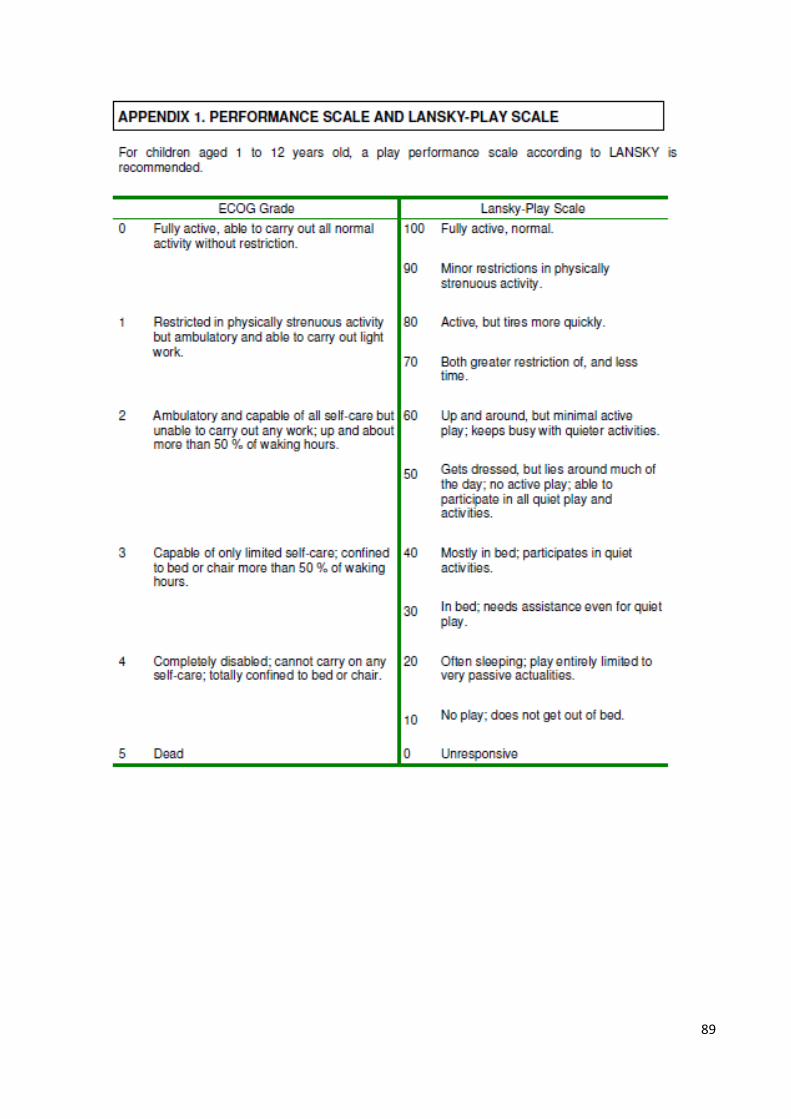
89

90
91
PUBBLICAZIONI
1. Cancer. 2012 Feb 1;118(3):821-7. Rhabdomyosarcoma in adolescents: a report from
the AIEOP Soft Tissue Sarcoma Committee. Bisogno G, Compostella A, Ferrari A,
Pastore G, Cecchetto G, Garaventa A, Indolfi P, De Sio L, Carli M.
2. J Pediatr Surg. 2010 Feb;45(2):e5-8. Multiple synchronous tumors in a child with
Fanconi anemia. Compostella A, Toffolutti T, Soloni P, Dall'Igna P, Carli M, Bisogno
G.
3. Pediatr Blood Cancer. 2009 Jul;52(7):902-3. A case of oxaliplatin overdose. Soloni P,
Compostella A, Carli M, Bisogno G.
4. In stesura: Compostella A, Bisogno G et al. A Topotecan carboplatin based strategy
for children with refractory or recurrent rhabdomyosarcoma
POSTERS/ABSTRACTS
• “SOVRADOSAGGIO DI OXALIPLATINO: UN CASO CLINICO”, Soloni P,
Compostella A, Carli M, Bisogno G Haematologica 2008, S4, XXXV congresso
AIEOP, Ancona 26-28.10.2008, abstract book e Poster.
• THE TOPOTECAN/CARBOPLATIN COMBINATION IN THE TREATMENT OF
RESISTANT RHABDOMYOSARCOMA. A REPORT FROM THE AIEOP SOFT
TISSUE SARCOMA COMMITTEE. Compostella A, Bisogno G, Soloni P,
Scagnellato A, Carli M.O., accepted for poster presentation at the 42nd Congress of the
International Society of Paediatric Oncology, and for publication in the abstract book.
• RHABDOMYOSARCOMA IN ADOLESCENTS. A REPORT FROM THE AIEOP
SOFT TISSUE SARCOMA COMMITTEE (STSC), Compostella A et al., accepted for
an oral presentation at the 42nd Congress of the International Society of Paediatric
Oncology, and for publication in the abstract book.
• “TOPOTECAN/CARBOPLATINO NEL RABDOMIOSARCOMA (RMS)
RESISTENTE O RECIDIVATO”. Compostella A. et al., XXXVI congresso AIEOP
tenutosi a Pisa dal 6 all’8.06.2010.
• “SARCOMA INDIFFERENZIATO DEL FEGATO: È REALMENTE CAMBIATA
LA PROGNOSI DI QUESTI PAZIENTI NEL CORSO DEGLI ANNI?
L’ESPERIENZA DEL GRUPPO DI LAVORO SUI SARCOMI DELLE PARTI
MOLLI (SPM) DELL’AIEOP”. Soloni P, Compostella A. et al., XXXVI congresso
AIEOP tenutosi a Pisa dal 6 all’8.06.2010.

92
93
COMUNICAZIONI ORALI
Padova, 27-30.11.2009: “Rabdomiosarcoma e adolescenti”, comunicazione orale al
65° congresso nazionale SIP.
Padova, 3-4.12.2009: “Il Rabdomiosarcoma negli adolescenti”, comunicazione orale
durante la riunione annuale sui tumori solidi pediatrici, CSS di neuroblastoma,
sarcomi delle parti molli, tumori rari, tumore di Wilms dell’AIEOP.
Padova, 20.04.2010: “Caso clinico sui tumori solidi in età pediatrica, i sarcomi delle
parti molli”, tumor board del seminario sui tumori solidi in età pediatrica, i sarcomi
delle parti molli, rivolto alla Scuola di Specializzazione in Pediatria dell’Università di
Padova.
Castelgandolfo, giugno 2010: “A “shady” soft tissue hematoma”. Case report
presentato al 3rd ESO-SIOP Europe Masterclass in Pediatric Oncology.
Padova, 17.11.2010: “Tumefazione parti molli: cosa si fa e cosa non si fa”, riunione
tumori solidi pediatrici PD, il corso, rabdomiosarcoma.
Padova, 18-19.11.2010: “RMS: ripensare la stadiazione. L’idea del COG”,
comunicazione orale durante la “Riunione annuale dei gruppi di lavoro:
Neuroblastoma, Sarcomi delle parti molli, Tumori rari e Tumore di Wilms”.
Padova, 21.06.2013: “Il Rabdomiosarcoma come esempio di tumore pediatrico
nell’adulto”, CONVEGNO AIOM GIOVANI VENETO. TUMORI RARI TRA
EMPIRISMO ED EVIDENZA. Sessione “Novità e punti fermi nella gestione delle
neoplasie rare”.

94

95
RINGRAZIAMENTI
I ringraziamenti dopo gli anni trascorsi presso l’Oncoematologia Pediatrica di Padova
vanno inevitabilmente a tutti i Colleghi che hanno condiviso un pezzetto della mia
strada: in particolare a Pietro Soloni, la mitica Cri Pizzato, Barbara Buldini e gli altri
“colleghi giovani”, tutti i Medici, le Infermiere, le OS del Reparto e del Day Hospital.
Ai Colleghi della Chirurgia Pediatrica, dell’Anatomia Patologica, della radiologia. Al
Prof. Rosolen e ai ragazzi del suo Laboratorio.
Al Prof. Basso, che mi dice ogni volta di aver perso la grandissima occasione di
lavorare in Oncoematologia pediatrica (e forse ha ragione).
Un grazie speciale è per il gruppo tumori solidi, a tutte le ragazze, Gloria, Elisa,
Caroline. Grazie al Prof Carli, instancabile ed entusiastico “capocordata”.
Grazie a Ilaria, stressata fino all’ultimo del mio impegno pediatrico! Grazie davvero per
la disponibilità immensa. Martina e Carlotta, difficile lasciarvi! Grazie per come portate
avanti il vostro lavoro, grande nonostante le poche gratifiche, cercando il massimo nel
poco tempo che vi diamo…grazie perché siete proprio delle belle persone e lavorare con
voi mi ha fatto crescere.
Angela…dovrei dedicarti una tesi piena di grazie! Il feeling nato così rapidamente è
stato un bellissimo regalo; sei stata preziosa per tutto, sempre in questi anni. Grazie
perché la tua ironia porta gioia, la tua dedizione e capacità lavorativa va oltre quello che
chi guarda da fuori può vedere, grazie per la pazienza, la disponibilità, la sincerità.
A Gianni (Dr Bisogno): difficile sintetizzare…è stato bello lavorare con te, sai
equilibrare stimoli, incoraggiamenti, rimproveri, sempre con rispetto; sai uscire dal
ruolo di capo e anche così “fare gruppo”. Grazie per l’esempio, la pazienza (scusa il mio
disordine perenne!), l’entusiasmo. Devo dire che faccio fatica a pensare di non lavorare
più con te, chissà…
Lavorare (che era anche un po’ “vivere”) in Oncoematologia pediatrica è stato un
momento voluto e prezioso, in tutti i suoi aspetti. Preziosi e impressi a fuoco nel cuore e
nella memoria sono tanti piccoli pazienti e famiglie. A loro va un grazie immenso che
va oltre ciò che la penna può scrivere.
Grazie a chi da casa, ha tollerato la mia mancanza, i miei umori, gli orari dei treni, la
mia testardaggine nel restare dentro un involucro che la carta sembrava non dire
mio…quindi grazie alla mia famiglia (mamma, papà, Marco e Chiara), e ad amici come
Ambra che non si stancano di costruire e sostenere e camminare.

96
Grazie infinite a Roberto che in tutte le mie scelte mi è stato ed è roccia e sostegno, ora
marito e papà di incommensurabile Bellezza. A Linda che ha vissuto l’ultima parte di
questa bellissima avventura e la difficoltà di “lasciarla”, e a Pietro, che tollera lo sforzo
di questi mesi per raggiungere questo altro traguardo di una strada contorta ma
bellissima.
Grazie!