STUDI DI PERMEAZIONE CUTANEA IN VITRO DI POLVERI E...
154
UNIVERSITA’ DEGLI STUDI DI PARMA DOTTORATO DI RICERCA IN SCIENZE DELLA PREVENZIONE CICLO XXII STUDI DI PERMEAZIONE CUTANEA IN VITRO DI POLVERI E NANOPARTICELLE METALLICHE Coordinatore: Chiar.mo Prof. ANTONIO MUTTI Tutor: Dott.ssa FRANCESCA FILON LARESE Dottorando: MATTEO CROSERA
Transcript of STUDI DI PERMEAZIONE CUTANEA IN VITRO DI POLVERI E...
UNIVERSITA’ DEGLI STUDI DI PARMA
DOTTORATO DI RICERCA IN SCIENZE DELLA PREVENZIONE
CICLO XXII
STUDI DI PERMEAZIONE CUTANEA IN VITRO DI POLVERI E
NANOPARTICELLE METALLICHE
Coordinatore:
Chiar.mo Prof. ANTONIO MUTTI
Tutor:
Dott.ssa FRANCESCA FILON LARESE
Dottorando:
MATTEO CROSERA

RIASSUNTO
Il ruolo della cute è stato a lungo sottostimato, soprattutto in ambito professionale: essa
era considerata una barriera impermeabile alle sostanze chimiche. Oggi è noto che molti
agenti tossici sono in grado di penetrare attraverso la pelle e l’assorbimento cutaneo è un
fenomeno molto studiato nell’ambito di una completa valutazione dei fattori di rischio sia in
ambito professionale che ambientale. Gli studi di assorbimento percutaneo in vitro possono
essere usati per definire le caratteristiche di diffusione dei composti chimici in differenti
settori industriali, per la valutazione del rischio da esposizione cutanea a sostanze tossiche,
ma anche come metodo per documentare la loro biodisponibilità.
L’assorbimento attraverso la pelle di metalli in polvere (cobalto, nichel, cromo) è stato
evidenziato in lavoratori esposti e in esperimenti di laboratorio con volontari, ma in generale
ci sono ancora pochi dati sull’argomento.
Questo lavoro di dottorato ha l’obiettivo di studiare l’effetto di alcune variabili, quali il
pH del sudore sintetico, l’utilizzo di detergenti e la presenza di lesioni cutanee,
sull’assorbimento cutaneo di questi metalli in un sistema di diffusione in-vitro.
Inoltre si è scelto di applicare l’esperienza nell’utilizzo delle celle di diffusione di Franz ai
nuovi nanomateriali ed, in particolare, alle nanoparticelle metalliche dato che lo sviluppo
delle nanotecnologie è molto veloce, ma l’impatto dei nanomateriali sulla salute umana è
ancora da studiare.
I risultati dei nostri esperimenti hanno mostrato che, in vitro, ioni di cobalto, nichel e
cromo rilasciati da polveri metalliche, ma anche nanoparticelle di argento e oro, sono in grado
di permeare attraverso la cute, e che la presenza di lesioni cutanee, l’uso di detergenti e il pH
della fase donatrice, sono in grado di volta in volta di modificare tale processo.
PAROLE CHIAVE: Assorbimento cutaneo; in vitro; Celle di Franz; Polveri metalliche;
Nanoparticelle
II
ABSTRACT
The role of the skin, in occupational and public hygiene, has been underestimated for a
long time: until the mid-1960s it was considered as an almost impermeable barrier for
chemicals. Actually is known that many hazardous substances can permeate through the skin
and dermal absorption is studied from a complete risk assessment point of view.
In vitro percutaneous permeation studies can be used to define the diffusion characteristic
of chemicals in different industrial settings for the dermal exposure risk assessment, but also
as one way of documenting their bioavailability.
Skin absorption of metal powders (cobalt, nickel and chromium) has been found in
exposed workers and in laboratory experiments with volunteers, but in general there are few
available data on this argument.
This doctoral work aims to study the skin absorption of metal powders and the effect of
some variables, like the pH of the synthetic sweat, the decontamination with detergents, or the
presence of lesions on the skin in an in vitro diffusion system.
Moreover, it has been decided to apply our experience with Franz cells use to the study of
new nanomaterials, and, in particular, to metal nanoparticles, since nanotechnology
development is very fast, but the impact of nanomaterial on human health is less clear.
Our results shown that ions of cobalt, nickel and chromium released from metal powders,
but also silver and gold nanoparticles, can permeate through the skin in in vitro experiments.
This process can be modified by the presence of skin lesions, by the decontamination with
detergents and by the pH of the donor phase.
KEYWORDS: Dermal absorption; in vitro; Franz Cells; Metal powders; Nanoparticles

III
INDICE
RIASSUNTO............................................................................................................................................................ I
ABSTRACT ............................................................................................................................................................ II
INDICE...................................................................................................................................................................III
1. SCOPO DELLO STUDIO ......................................................................................................................................1
2. INTRODUZIONE ...................................................................................................................................................4
2.1. LA CUTE...........................................................................................................................................................5
2.1.1. L’epidermide..............................................................................................................................................6
2.1.2. Il derma......................................................................................................................................................7
2.1.3. L’ipoderma.................................................................................................................................................8
2.1.4. Appendici cutanee......................................................................................................................................8
2.2. L’ASSORBIMENTO CUTANEO.....................................................................................................................9
2.3. ASSORBIMENTO CUTANEO DI COBALTO, NICHEL E CROMO ...........................................................12
2.4. ASSORBIMENTO CUTANEO DI NANOPARTICELLE METALLICHE ...................................................14
2.4.1. Nanoscienze, nanotecnologie e nanomateriali.........................................................................................14
2.4.2. Nanotossicologia .....................................................................................................................................14
2.4.3. Assorbimento cutaneo di nanoparticelle metalliche ................................................................................15
3. MATERIALI E METODI ....................................................................................................................................18
3.1. LE CELLE DI DIFFUSIONE DI FRANZ ......................................................................................................19
3.2. PROCEDURA SPERIMENTALE GENERALE.............................................................................................22
3.2.1. Preparazione della cute ..........................................................................................................................22
3.2.2. Integrità della cute..................................................................................................................................23
3.2.3. Test di permeazione .................................................................................................................................23
3.2.4. Mineralizzazione della cute dopo l’esperimento......................................................................................24
3.2.5. Preparazione dei campioni di cute per le analisi di microscopia elettronica..........................................24
3.2.6. Misure analitiche strumentali ..................................................................................................................25
3.3. PROCEDURE SPERIMENTALI SPECIFICHE.............................................................................................27
3.3.1. Assorbimento cutaneo in vitro di polvere di cromo ed effetti della detersione ........................................27
3.3.2. Assorbimento cutaneo in vitro di polveri di cobalto, nichel e cromo attraverso cute umana integra e
lesa .........................................................................................................................................................28
3.3.3. Permeazione cutanea in vitro di nanoparticelle d’argento attraverso cute umana integra e lesa ..........29
3.3.4. Permeazione cutanea in vitro di nanoparticelle d’oro attraverso cute umana integra e lesa .................29
4. RISULTATI E DISCUSSIONI.............................................................................................................................31
4.1. ASSORBIMENTO CUTANEO IN VITRO DI POLVERE DI CROMO ED EFFETTI DELLA
DETERSIONE ............................................................................................................................................32
4.2. ASSORBIMENTO CUTANEO IN VITRO DI POLVERI DI COBALTO, NICHEL E CROMO
ATTRAVERSO CUTE UMANA INTEGRA E LESA................................................................................33

IV
4.3. PERMEAZIONE CUTANEA IN VITRO DI NANOPARTICELLE DI ARGENTO ATTRAVERSO
CUTE UMANA INTEGRA E LESA..........................................................................................................35
4.4. PERMEAZIONE CUTANEA IN VITRO DI NANOPARTICELLE D’ORO ATTRAVERSO CUTE
UMANA INTEGRA E LESA .....................................................................................................................37
5. CONCLUSIONI ....................................................................................................................................................39
6. BIBLIOGRAFIA ...................................................................................................................................................42
ALLEGATI................................................................................................................................................................49
ALLEGATO I.........................................................................................................................................................50
ALLEGATO II .......................................................................................................................................................67
ALLEGATO III ......................................................................................................................................................84
ALLEGATO IV....................................................................................................................................................111
ALLEGATO V .....................................................................................................................................................127
APPENDICE............................................................................................................................................................144
V
Allegato I
Francesca Larese Filon, Flavia D’Agostin, Matteo Crosera, Gianpiero Adami, Massimo
Bovenzi, Giovanni Maina. In vitro percutaneous absorption of chromium powder and the
effect of skin cleanser. Toxicology in vitro 2008 (22) 1562-1567.
Allegato II
Francesca Larese Filon, Flavia D’Agostin, Matteo Crosera, Gianpiero Adami, Massimo
Bovenzi, Giovanni Maina. In vitro absorption of metal powders through intact and damaged
human skin. Toxicology in vitro 2009 (23) 574-579.
Allegato III
Matteo Crosera, Massimo Bovenzi, Giovanni Maina, Gianpiero Adami, Caterina Zanette,
Chiara Florio, Francesca Larese Filon. Nanoparticle dermal absorption and toxicity: a review
of the literature. International Archives of Occupational and Environmenal Health 2009 (82)
1043-1055.
Allegato IV
Francesca Larese Filon, Flavia D’Agostin, Matteo Crosera, Gianpiero Adami, Nadia
Renzi, Massimo Bovenzi, Giovanni Maina. Human skin penetration of silver nanoparticles
through intact and damaged skin. Toxicology 2009 (255) 33-37.
Allegato V
Francesca Larese Filon, Matteo Crosera, Gianpiero Adami, Massimo Bovenzi, Federica
Rossi, Giovanni Maina. Human skin penetration of gold nanoparticles through intact and
damaged skin. Manuscript in preparation.

1. SCOPO DELLO STUDIO
Scopo dello studio
2
Il progetto di ricerca nel quale si inserisce questo lavoro di dottorato nasce dalla
collaborazione tra il gruppo di ricerca di Chimica Analitica e Ambientale e l’Unità Clinica
Operativa di Medicina del Lavoro dell’Università di Trieste e si propone di studiare la cute
come barriera semipermeabile bidirezionale con l'obiettivo di comprendere il suo ruolo
nell'assorbimento, nell'escrezione e nella sensibilizzazione allergica da contatto e gli aspetti
molecolari a questi correlati.
In generale, nel campo della tutela della salute occupazionale e ambientale, infatti,
l’assorbimento percutaneo sta riscuotendo negli ultimi anni sempre maggior interesse, al pari
delle vie classiche di assorbimento quali inalazione e ingestione, nella logica di una
valutazione completa dei rischi dovuti all’esposizione a sostanze tossiche.
Gli studi di assorbimento percutaneo in vitro possono essere usati per definire le
caratteristiche di diffusione dei composti chimici in differenti settori industriali, dalla
cosmetica alla farmaceutica, dalla produzione di detergenti all’agrochimica, per la valutazione
del rischio da esposizione cutanea a sostanze tossiche, ma anche come metodo per verificare
la loro biodisponibilità. Per questo in alcuni paesi i test in vitro sono inclusi tra i criteri per
assegnare le skin notation ai composti chimici (Drexler, 1998).
In questo progetto di ricerca, dopo i primi lavori riguardati l’assorbimento percutaneo in
vitro di composti organici, quali benzine, glicoleteri, testosterone, acido benzoico e caffeina, e
alla partecipazione al progetto europeo EDETOX (2000) per la standardizzazione dei metodi
degli studi in vitro (van der Sandt et al., 2004), si è passati allo studio di polveri di metalli,
come cobalto, nichel e cromo (Larese et al., 2004; 2007), e di ossidi metallici, quale il
biossido di piombo (Larese et al., 2006).
L’assorbimento attraverso la pelle di metalli in polvere (cobalto, nichel, cromo, rame,
ecc.) è stato evidenziato in lavoratori esposti e in esperimenti di laboratorio con volontari, ma
in generale ci sono ancora pochi dati sull’argomento.
Cobalto, nichel e cromo sono dei comuni sensibilizzanti e la loro presenza in oggetti
metallici di uso quotidiano può aumentare il rischio di dermatiti allergiche da contatto in
soggetti sensibilizzati. La valutazione dell’assorbimento attraverso la pelle di tali metalli in un
sistema in vitro, come le celle a diffusione di Franz, può contribuire a definire un quadro più
completo del rischio da esposizione cutanea, sia in ambito professionale che domestico.
Questo di dottorato si inserisce a questo livello, con l’obiettivo di studiare l’effetto
sull’assorbimento cutaneo di questi metalli, di alcune variabili, quali il pH del sudore
sintetico, l’utilizzo di detergenti, l’entità dell’esposizione e la presenza di lesioni cutanee, in
un sistema in vitro.
Scopo dello studio
3
Inoltre si è scelto di applicare l’esperienza maturata nel corso degli anni nell’utilizzo delle
celle di diffusione di Franz ai nuovi nanomateriali ed, in particolare, alle nanoparticelle
metalliche. La penetrazione cutanea di nanoparticelle deve essere ancora studiata e i protocolli
classici di valutazione devono essere adattati e ristandardizzati per i nuovi nanocomposti.
Le colture cellulari (Bernstein and Vaughan, 1999), le celle di diffusione di Franz (Franz,
1975), il “tape stripping” (Escobar-Chávez et al, 2008) e gli impianti di cute umana su
animali, rimangono strumenti importanti per studiare le interazioni tra le nanoparticelle e il
tessuto cutaneo umano, ma devono anche essere sviluppati nuovi metodi e nuove applicazioni
tecniche (Moger et al., 2008, Monteiro-Riviere and Inman, 2006; SCCP, 2007).
La valutazione della tossicità delle nanoparticelle metalliche richiede innanzitutto la
determinazione delle loro caratteristiche quali forma, dimensioni, concentrazione, purezza e
tendenza all’aggregazione. Le caratteristiche chimico-fisiche delle nanoparticelle, infatti, sono
intrinsecamente collegate alla loro reattività e quindi alle loro possibili applicazioni, ma anche
alla loro tossicità (Auffan et al., 2009).
Una differenza molto importante nello studio di polveri metalliche nanometriche rispetto
alle micropolveri risulta essere il meccanismo con cui il metallo permea la cute. Nel caso di
particelle grossolane, esse non sono in grado di penetrare la cute, ma in sudore sintetico il
metallo si ossida e gli ioni possono eventualmente attraversare la barriera cutanea. Nel caso
delle nanoparticelle, invece, essere potrebbero penetrare la cute anche in quanto tali; inoltre
l’alto rapporto superficie/volume delle nanoparticelle fa sì che anche la ionizzazione del
metallo possa avvenire in maniera più estesa rispetto alle micropolveri.
La scelta dei metodi analitici più adatti alla tipologia del composto ed alle sue
concentrazioni nelle varie fasi e la ricerca di nuove applicazioni strumentali necessarie
all’attuale campo di indagine, rivestono quindi un ruolo molto importante nel progetto in
corso.

2. INTRODUZIONE
Introduzione
5
2.1. LA CUTE
La cute è l’organo più grande del corpo umano e, oltre alla sua primaria funzione di
barriera verso l’ambiente esterno, ricopre diversi ruoli nel mantenimento dell’omeostasi
fisiologica. Essa, per esempio, previene la disidratazione minimizzando la perdita di acqua,
partecipa alla termoregolazione, fornisce supporto per gli organi interni, protegge il corpo
dalle radiazioni UV e partecipa alla sintesi di vitamina D.
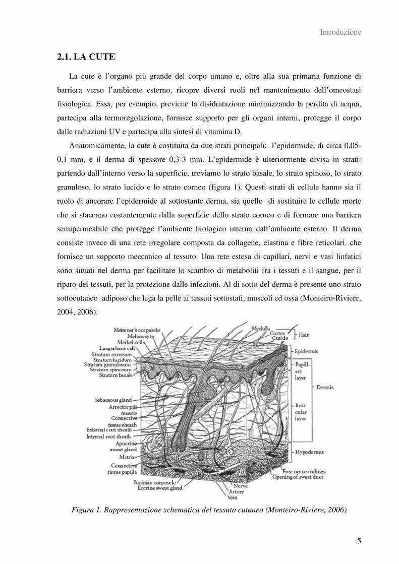
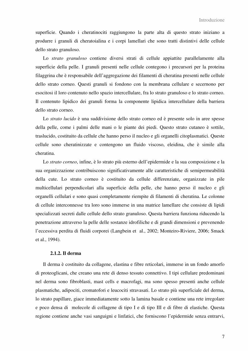
Anatomicamente, la cute è costituita da due strati principali: l’epidermide, di circa 0,05-
0,1 mm, e il derma di spessore 0,3-3 mm. L’epidermide è ulteriormente divisa in strati:
partendo dall’interno verso la superficie, troviamo lo strato basale, lo strato spinoso, lo strato
granuloso, lo strato lucido e lo strato corneo (figura 1). Questi strati di cellule hanno sia il
ruolo di ancorare l’epidermide al sottostante derma, sia quello di sostituire le cellule morte
che si staccano costantemente dalla superficie dello strato corneo e di formare una barriera
semipermeabile che protegge l’ambiente biologico interno dall’ambiente esterno. Il derma
consiste invece di una rete irregolare composta da collagene, elastina e fibre reticolari. che
fornisce un supporto meccanico al tessuto. Una rete estesa di capillari, nervi e vasi linfatici
sono situati nel derma per facilitare lo scambio di metaboliti fra i tessuti e il sangue, per il
riparo dei tessuti, per la protezione dalle infezioni. Al di sotto del derma è presente uno strato
sottocutaneo adiposo che lega la pelle ai tessuti sottostati, muscoli ed ossa (Monteiro-Riviere,
2004, 2006).
Figura 1. Rappresentazione schematica del tessuto cutaneo (Monteiro-Riviere, 2006)
Introduzione
6
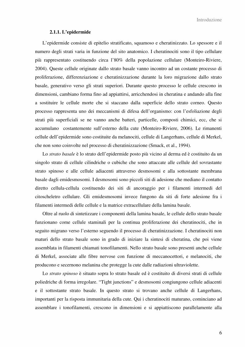
2.1.1. L’epidermide
L’epidermide consiste di epitelio stratificato, squamoso e cheratinizzato. Lo spessore e il
numero degli strati varia in funzione del sito anatomico. I cheratinociti sono il tipo cellulare
più rappresentato costituendo circa l’80% della popolazione cellulare (Monteiro-Riviere,
2004). Queste cellule originate dallo strato basale vanno incontro ad un costante processo di
proliferazione, differenziazione e cheratinizzazione durante la loro migrazione dallo strato
basale, generativo verso gli strati superiori. Durante questo processo le cellule crescono in
dimensioni, cambiano forma fino ad appiattirsi, arricchendosi in cheratina e andando alla fine
a sostituire le cellule morte che si staccano dalla superficie dello strato corneo. Questo
processo rappresenta uno dei meccanismi di difesa dell’organismo: con l’esfoliazione degli
strati più superficiali se ne vanno anche batteri, particelle, composti chimici, ecc, che si
accumulano costantemente sull’esterno della cute (Monteiro-Riviere, 2006). Le rimanenti
cellule dell’epidermide sono costituite da melanociti, cellule di Langerhans, cellule di Merkel,
che non sono coinvolte nel processo di cheratinizzazione (Smack, et al., 1994).
Lo strato basale è lo strato dell’epidermide posto più vicino al derma ed è costituito da un
singolo strato di cellule cilindriche o cubiche che sono attaccate alle cellule del sovrastante
strato spinoso e alle cellule adiacenti attraverso desmosomi e alla sottostante membrana
basale dagli emidesmosomi. I desmosomi sono piccoli siti di adesione che mediano il contatto
diretto cellula-cellula costituendo dei siti di ancoraggio per i filamenti intermedi del
citoscheletro cellulare. Gli emidesmosomi invece fungono da siti di forte adesione fra i
filamenti intermedi delle cellule e la matrice extracellulare della lamina basale.
Oltre al ruolo di sintetizzare i componenti della lamina basale, le cellule dello strato basale
funzionano come cellule staminali per la continua proliferazione dei cheratinociti, che in
seguito migrano verso l’esterno seguendo il processo di cheratinizzazione. I cheratinociti non
maturi dello strato basale sono in grado di iniziare la sintesi di cheratina, che poi viene
assemblata in filamenti chiamati tonofilamenti. Nello strato basale sono presenti anche cellule
di Merkel, associate alle fibre nervose con funzione di meccanocettori, e melanociti, che
producono e secernono melanina che protegge la cute dalle radiazioni ultraviolette.
Lo strato spinoso è situato sopra lo strato basale ed è costituito di diversi strati di cellule
poliedriche di forma irregolare. “Tight junctions” e desmosomi congiungono cellule adiacenti
e il sottostante strato basale. In questo strato si trovano anche cellule di Langerhans,
importanti per la risposta immunitaria della cute. Qui i cheratinociti maturano, cominciano ad
assemblare i tonofilamenti, crescono in dimensioni e si appiattiscono parallelamente alla
Introduzione
7
superficie. Quando i cheratinociti raggiungono la parte alta di questo strato iniziano a
produrre i granuli di cheratoialina e i corpi lamellari che sono tratti distintivi delle cellule
dello strato granuloso.
Lo strato granuloso contiene diversi strati di cellule appiattite parallelamente alla
superficie della pelle. I granuli presenti nelle cellule contegono i precursori per la proteina
filaggrina che è responsabile dell’aggregazione dei filamenti di cheratina presenti nelle cellule
dello strato corneo. Questi granuli si fondono con la membrana cellulare e secernono per
esocitosi il loro contenuto nello spazio intercellulare, fra lo strato granuloso e lo strato corneo.
Il contenuto lipidico dei granuli forma la componente lipidica intercellulare della barriera
dello strato corneo.
Lo strato lucido è una suddivisione dello strato corneo ed è presente solo in aree spesse
della pelle, come i palmi delle mani o le piante dei piedi. Questo strato cutaneo è sottile,
traslucido, costituito da cellule che hanno perso il nucleo e gli organelli citoplasmatici. Queste
cellule sono cheratinizzate e contengono un fluido viscoso, eleidina, che è simile alla
cheratina.
Lo strato corneo, infine, è lo strato più esterno dell’epidermide e la sua composizione e la
sua organizzazione contribuiscono significativamente alle caratteristiche di semipermeabilità
della cute. Lo strato corneo è costituito da cellule differenziate, organizzate in pile
multicellulari perpendicolari alla superficie della pelle, che hanno perso il nucleo e gli
organelli cellulari e sono quasi completamente riempite di filamenti di cheratina. Le colonne
di cellule interconnesse tra loro sono immerse in una matrice lamellare che consiste di lipidi
specializzati secreti dalle cellule dello strato granuloso. Questa barriera funziona riducendo la
penetrazione attraverso la pelle delle sostanze idrofiliche e di grandi dimensioni e prevenendo
l’eccessiva perdita di fluidi corporei (Langbein et al., 2002; Monteiro-Riviere, 2006; Smack
et al., 1994).
2.1.2. Il derma
Il derma è costituito da collagene, elastina e fibre reticolari, immerse in un fondo amorfo
di proteoglicani, che creano una rete di denso tessuto connettivo. I tipi cellulare predominani
nel derma sono fibroblasti, mast cells e macrofagi, ma sono spesso presenti anche cellule
plasmatiche, adipociti, cromatofori e leucociti stravasati. Lo strato più superficiale del derma,
lo strato papillare, giace immediatamente sotto la lamina basale e contiene una rete irregolare
e poco densa di molecole di collagene di tipo I e di tipo III e di fibre di elastiche. Questa
regione contiene anche vasi sanguigni e linfatici, che forniscono l’epidermide senza entrarvi,
Introduzione
8
e terminazioni nervose che possono fermarsi nel derma o entrare nell’epidermide. Le
protrusioni del tessuto connettivo nell’epidermide sono chiamate papille cutanee. Lo strato
reticolare del derma giace sotto lo strato papillare. Questo strato è sostanzialmente più spesso
di quello superficiale ed è caratterizzato da collagene di tipo I, fibre elastiche e poche cellule
(Monteiro-Riviere, 2006).
2.1.3. L’ipoderma
L’ipoderma è la fascia di natura prevalentemente adiposa che giace sotto la pelle con
funzioni di isolante, di riserva di calorie, di ancoraggio della pelle ai muscoli e alle ossa. Esso
è costituito prevalentemente di adipociti, cellule piene di lipidi, e di tessuto connettivo
contenente fibre di collagene ed elastina che danno flessibilità e libero movimento alla pelle
rispetto alle strutture più profonde (Monteiro-Riviere, 2006).
2.1.4. Appendici cutanee
I follicoli piliferi, le ghiandole sebacee associate, i muscoli erettori dei peli e le ghiandole
sudoripare sono le appendici comunemente associate alla cute. I peli sono prodotti dai
follicoli pilieri e sono strutture cheratinizzate che derivano da invaginazioni dell’epidermide
che attraversano il derma e possono estendersi fino all’ipoderma. Sebbene la permeazione
cutanea attraverso i follicoli piliferi richieda comunque l’attraversamento dello strato corneo, i
follicoli rappresentano le regioni di maggior area e possono quindi contribuire in maniera
significativa all’aumento dell’assorbimento transcutaneo (Monteiro-Riviere, 2004). Il tessuto
connettivo alla base dei follicoli piliferi costituisce un sito di ancoraggio per i muscoli erettori
dei peli che, in seguito alla contrazione, non solo danno luogo all’erezione dei peli, ma
aiutano anche lo svuotamento delle ghiandole sebacee che secernono il loro prodotto di
secrezione, il sebo, nel canale del follicolo pilifero.
Introduzione
9
2.2. L’ASSORBIMENTO CUTANEO
Il ruolo della cute è stato a lungo sottostimato, soprattutto in ambito professionale: fino
agli anni ’60, essa era considerata una barriera impermeabile alle sostanze chimiche. L’igiene
occupazionale si è tradizionalmente focalizzata sull’inalazione in quanto era considerata la più
importate via di espozione alle sostanze tossiche e la percezione del rischio era più alta. Oggi
è noto che molti agenti tossici sono in grado di penetrare anche attraverso la pelle e
l’assorbimento cutaneo è ormai un fenomeno studiato nell’ambito di una completa
valutazione dei fattori di rischio sia professionali che ambientali. Sono stati sviluppati diversi
metodi pratici per valutare l’esposizione cutanea e si è aperto il dibattito per lo sviluppo di
limiti per l’esposizione cutanea al pari di quelli per la via inalatoria. In generale, però, rispetto
all’inalazione, nell’esposizione cutanea sono stati fatti meno progressi riguardo la
standardizzazione di metodi di valutazione condivisi e solo negli ultimi anni si è sviluppata
una terminologia di base e dei modelli teorici che consentano il confronto dei dati dei vari
studi (Cherrie and Robertson, 1995).
Uno dei modelli concettuali più accettati si basa sull’osservazione che il processo di
trasporto dei composti chimici attraverso la cute è spinto dal gradiente di concentrazione tra la
superficie cutanea e il tessuto vascolarizzato sottostante che rimuove costantemente le
sostanze che penetrano e quindi il fattore determinante risulta essere la concentrazione del
composto sulla superficie cutanea (Schneider et al., 1999).
Le sostanze possono arrivare alla cute in diversi modi: possono depositarsi direttamente
dall’aria, trasferirsi per contatto della cute con superfici contaminate o per immersione di una
parte del corpo nella sostanza stessa. Allo stesso modo i contaminanti possono lasciare la
pelle attraverso altri meccanismi: essi possono evaporare o essere rimossi tramite abrasione o
lavaggio prima di venire assorbiti dal tessuto cutaneo. Infine, un ruolo molto importante è
svolto dall’uso dell’equipaggiamento di protezione che può modificare la quantità di sostanza
che arriva a contatto con la pelle.
Nel modello multicompartimentale di Schneider et al, (1999) vengono identificati sei
compartimenti e otto processi di trasferimento del contaminante. I compartimenti sono
rappresentati dalla sorgente del contaminante, dall’aria circostante, dal contaminate depositato
sulle superfici, dagli strati di contaminante all’esterno e all’interno dei vestiti e allo strato di
contaminante sulla superficie della cute. Tra questi compartimenti vi è un continuo scambio di
contaminante per emissione, deposizione, risospensione o evaporazione, trasferimento,
rimozione, ridistribuzione, decontaminazione e penetrazione.
Introduzione
10
L’entità dei vari processi di scambio fra un compartimento e l’altro determina la quantità
e la concentrazione del contaminante sulla superficie della pelle e, quindi, l’eventuale
assorbimento nel tessuto e nel sistema circolatorio.
Per quanto riguarda il fenomeno dell’assorbimento cutaneo, esso è un termine generale
che descrive il trasporto di sostanze chimiche dalla superficie esterna della cute fino a
raggiungere il sistema circolatorio (EHC 235, 2006). Questo processo viene spesso diviso in:
- penetrazione: l’ingresso di una sostanza all’interno di un particolare strato o struttura;
- permeazione: la penetrazione di un composto in un secondo strato che sia
funzionalmente e strutturalmente differente dal primo strato attraversato;
- assorbimento: l’assorbimento del composto nel sistema vascolare locale e nel sistema
linfatico della pelle. Questo generalmente porta ad un assorbimento nella circolazione
sistemica (assorbimento sistemico).
L’assorbimento cutaneo non viene studiato solo in funzione di un’esposizione a sostanze
tossiche con effettti nocivi, ma viene anche sfruttato in farmacologia per la somministrazione
di alcuni farmaci che, una volta assorbiti nel circolo locale della pelle, passano alla
circolazione sistemica e vengono distribuiti in tutto l’organismo.
Lo strato che controlla l’assorbimento attraverso la cute è sostanzialmente lo strato corneo
che, nonostante sia di spessore molto ridotto, offre con i suoi strati di corneociti immersi nella
matrice lipidica un’efficiente barriera contro la penetrazione di xenobiotici.
Sono state identificate tre vie di penetrazione dei composti attraverso la cute: (i)
intercellulare, (ii) transcellulare e (iii) attraverso gli annessi cutanei, quali follicoli piliferi e
ghiandole sudoripare e sebacee: tutte e tre le vie possono dare un contributo alla diffusione
dei composti chimici attraverso la barriera cutanea.
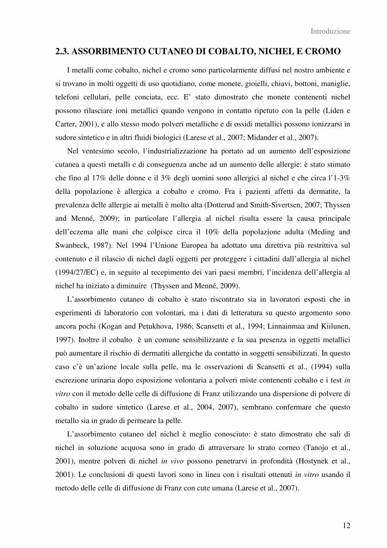
L’assorbimento cutaneo può essere influenzato da una grande varietà di fattori (tabella 1),
quali l’integrità della cute, il sito anatomico, la presenza di patologie (dermatiti da contatto,
eczema atopico, psoriasi) la presenza di veicolanti, la flessione meccanica (Rouse et al., 2007)
e l’uso di detergenti o di altri composti chimici (Larese et al., 2006).
Questi fattori possono essere raggruppati in tre classi: fattori intrinseci della cute, fattori
chimico-fisici del composto e fattori dipendenti dal tipo di esposizione.
I composti chimici possono attraversare la cute per diffusione attiva, legandosi a lipidi o
proteine con funzione di carrier: tuttavia il processo di assorbimento cutaneo è descrivibile
fondamentalmente come una diffusone passiva.
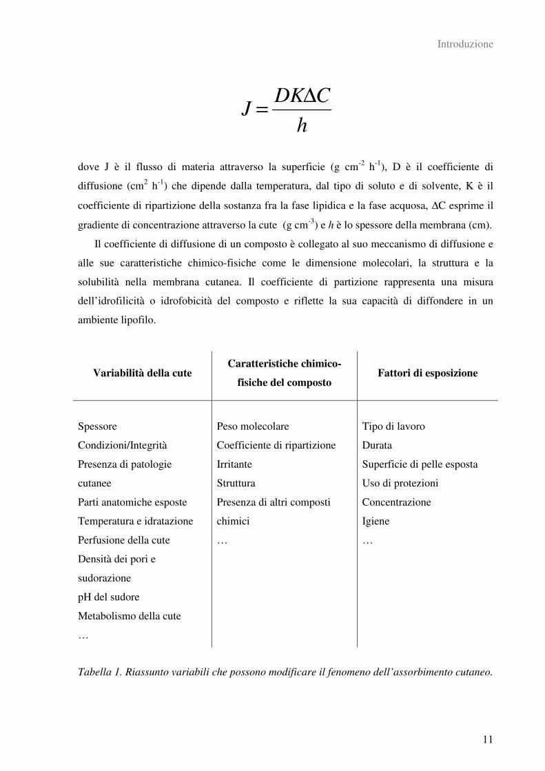
Il flusso di un composto chimico allo steady-state può essere predetto attraverso la 1a
legge di Fick per la diffusione in condizioni stazionarie:
Introduzione
11
J =DK∆C
h
dove J è il flusso di materia attraverso la superficie (g cm-2
h-1
), D è il coefficiente di
diffusione (cm2 h
-1) che dipende dalla temperatura, dal tipo di soluto e di solvente, K è il
coefficiente di ripartizione della sostanza fra la fase lipidica e la fase acquosa, ∆C esprime il
gradiente di concentrazione attraverso la cute (g cm-3
) e h è lo spessore della membrana (cm).
Il coefficiente di diffusione di un composto è collegato al suo meccanismo di diffusione e
alle sue caratteristiche chimico-fisiche come le dimensione molecolari, la struttura e la
solubilità nella membrana cutanea. Il coefficiente di partizione rappresenta una misura
dell’idrofilicità o idrofobicità del composto e riflette la sua capacità di diffondere in un
ambiente lipofilo.
Variabilità della cute Caratteristiche chimico-
fisiche del composto Fattori di esposizione
Spessore
Condizioni/Integrità
Presenza di patologie
cutanee
Parti anatomiche esposte
Temperatura e idratazione
Perfusione della cute
Densità dei pori e
sudorazione
pH del sudore
Metabolismo della cute
…
Peso molecolare
Coefficiente di ripartizione
Irritante
Struttura
Presenza di altri composti
chimici
…
Tipo di lavoro
Durata
Superficie di pelle esposta
Uso di protezioni
Concentrazione
Igiene
…
Tabella 1. Riassunto variabili che possono modificare il fenomeno dell’assorbimento cutaneo.
Introduzione
12
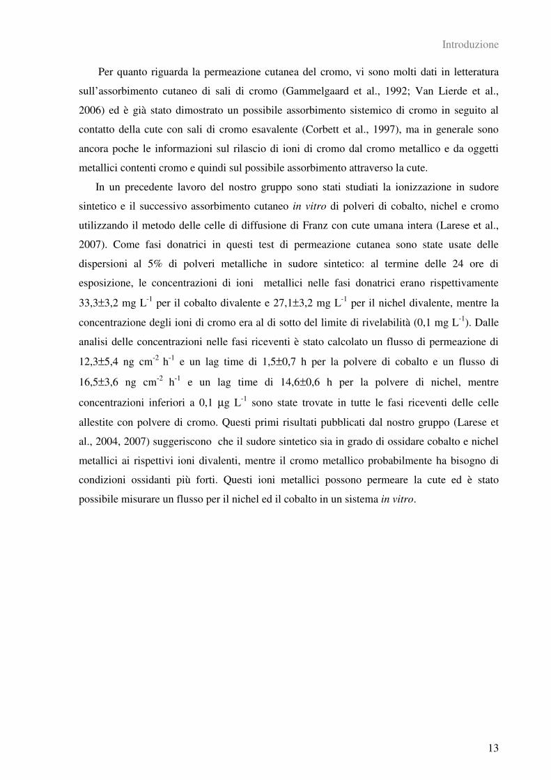
2.3. ASSORBIMENTO CUTANEO DI COBALTO, NICHEL E CROMO
I metalli come cobalto, nichel e cromo sono particolarmente diffusi nel nostro ambiente e
si trovano in molti oggetti di uso quotidiano, come monete, gioielli, chiavi, bottoni, maniglie,
telefoni cellulari, pelle conciata, ecc. E’ stato dimostrato che monete contenenti nichel
possono rilasciare ioni metallici quando vengono in contatto ripetuto con la pelle (Liden e
Carter, 2001), e allo stesso modo polveri metalliche e di ossidi metallici possono ionizzarsi in
sudore sintetico e in altri fluidi biologici (Larese et al., 2007; Midander et al., 2007).
Nel ventesimo secolo, l’industrializzazione ha portato ad un aumento dell’esposizione
cutanea a questi metalli e di conseguenza anche ad un aumento delle allergie: è stato stimato
che fino al 17% delle donne e il 3% degli uomini sono allergici al nichel e che circa l’1-3%
della popolazione è allergica a cobalto e cromo. Fra i pazienti affetti da dermatite, la
prevalenza delle allergie ai metalli è molto alta (Dotterud and Smith-Sivertsen, 2007; Thyssen
and Menné, 2009); in particolare l’allergia al nichel risulta essere la causa principale
dell’eczema alle mani che colpisce circa il 10% della popolazione adulta (Meding and
Swanbeck, 1987). Nel 1994 l’Unione Europea ha adottato una direttiva più restrittiva sul
contenuto e il rilascio di nichel dagli oggetti per proteggere i cittadini dall’allergia al nichel
(1994/27/EC) e, in seguito al recepimento dei vari paesi membri, l’incidenza dell’allergia al
nichel ha iniziato a diminuire (Thyssen and Menné, 2009).
L’assorbimento cutaneo di cobalto è stato riscontrato sia in lavoratori esposti che in
esperimenti di laboratorio con volontari, ma i dati di letteratura su questo argomento sono
ancora pochi (Kogan and Petukhova, 1986; Scansetti et al., 1994; Linnainmaa and Kiilunen,
1997). Inoltre il cobalto è un comune sensibilizzante e la sua presenza in oggetti metallici
può aumentare il rischio di dermatiti allergiche da contatto in soggetti sensibilizzati. In questo
caso c’è un’azione locale sulla pelle, ma le osservazioni di Scansetti et al., (1994) sulla
escrezione urinaria dopo esposizione volontaria a polveri miste contenenti cobalto e i test in
vitro con il metodo delle celle di diffusione di Franz utilizzando una dispersione di polvere di
cobalto in sudore sintetico (Larese et al., 2004, 2007), sembrano confermare che questo
metallo sia in grado di permeare la pelle.
L’assorbimento cutaneo del nichel è meglio conosciuto: è stato dimostrato che sali di
nichel in soluzione acquosa sono in grado di attraversare lo strato corneo (Tanojo et al.,
2001), mentre polveri di nichel in vivo possono penetrarvi in profondità (Hostynek et al.,
2001). Le conclusioni di questi lavori sono in linea con i risultati ottenuti in vitro usando il
metodo delle celle di diffusione di Franz con cute umana (Larese et al., 2007).
Introduzione
13
Per quanto riguarda la permeazione cutanea del cromo, vi sono molti dati in letteratura
sull’assorbimento cutaneo di sali di cromo (Gammelgaard et al., 1992; Van Lierde et al.,
2006) ed è già stato dimostrato un possibile assorbimento sistemico di cromo in seguito al
contatto della cute con sali di cromo esavalente (Corbett et al., 1997), ma in generale sono
ancora poche le informazioni sul rilascio di ioni di cromo dal cromo metallico e da oggetti
metallici contenti cromo e quindi sul possibile assorbimento attraverso la cute.
In un precedente lavoro del nostro gruppo sono stati studiati la ionizzazione in sudore
sintetico e il successivo assorbimento cutaneo in vitro di polveri di cobalto, nichel e cromo
utilizzando il metodo delle celle di diffusione di Franz con cute umana intera (Larese et al.,
2007). Come fasi donatrici in questi test di permeazione cutanea sono state usate delle
dispersioni al 5% di polveri metalliche in sudore sintetico: al termine delle 24 ore di
esposizione, le concentrazioni di ioni metallici nelle fasi donatrici erano rispettivamente
33,3±3,2 mg L-1
per il cobalto divalente e 27,1±3,2 mg L-1
per il nichel divalente, mentre la
concentrazione degli ioni di cromo era al di sotto del limite di rivelabilità (0,1 mg L-1
). Dalle
analisi delle concentrazioni nelle fasi riceventi è stato calcolato un flusso di permeazione di
12,3±5,4 ng cm-2
h-1
e un lag time di 1,5±0,7 h per la polvere di cobalto e un flusso di
16,5±3,6 ng cm-2
h-1
e un lag time di 14,6±0,6 h per la polvere di nichel, mentre
concentrazioni inferiori a 0,1 µg L-1
sono state trovate in tutte le fasi riceventi delle celle
allestite con polvere di cromo. Questi primi risultati pubblicati dal nostro gruppo (Larese et
al., 2004, 2007) suggeriscono che il sudore sintetico sia in grado di ossidare cobalto e nichel
metallici ai rispettivi ioni divalenti, mentre il cromo metallico probabilmente ha bisogno di
condizioni ossidanti più forti. Questi ioni metallici possono permeare la cute ed è stato
possibile misurare un flusso per il nichel ed il cobalto in un sistema in vitro.
Introduzione
14
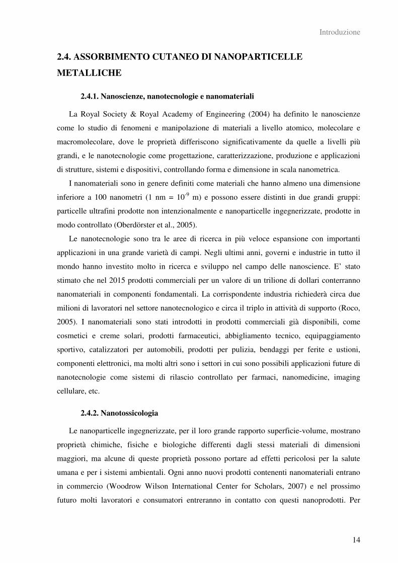
2.4. ASSORBIMENTO CUTANEO DI NANOPARTICELLE
METALLICHE
2.4.1. Nanoscienze, nanotecnologie e nanomateriali
La Royal Society & Royal Academy of Engineering (2004) ha definito le nanoscienze
come lo studio di fenomeni e manipolazione di materiali a livello atomico, molecolare e
macromolecolare, dove le proprietà differiscono significativamente da quelle a livelli più
grandi, e le nanotecnologie come progettazione, caratterizzazione, produzione e applicazioni
di strutture, sistemi e dispositivi, controllando forma e dimensione in scala nanometrica.
I nanomateriali sono in genere definiti come materiali che hanno almeno una dimensione
inferiore a 100 nanometri (1 nm = 10-9
m) e possono essere distinti in due grandi gruppi:
particelle ultrafini prodotte non intenzionalmente e nanoparticelle ingegnerizzate, prodotte in
modo controllato (Oberdörster et al., 2005).
Le nanotecnologie sono tra le aree di ricerca in più veloce espansione con importanti
applicazioni in una grande varietà di campi. Negli ultimi anni, governi e industrie in tutto il
mondo hanno investito molto in ricerca e sviluppo nel campo delle nanoscience. E’ stato
stimato che nel 2015 prodotti commerciali per un valore di un trilione di dollari conterranno
nanomateriali in componenti fondamentali. La corrispondente industria richiederà circa due
milioni di lavoratori nel settore nanotecnologico e circa il triplo in attività di supporto (Roco,
2005). I nanomateriali sono stati introdotti in prodotti commerciali già disponibili, come
cosmetici e creme solari, prodotti farmaceutici, abbigliamento tecnico, equipaggiamento
sportivo, catalizzatori per automobili, prodotti per pulizia, bendaggi per ferite e ustioni,
componenti elettronici, ma molti altri sono i settori in cui sono possibili applicazioni future di
nanotecnologie come sistemi di rilascio controllato per farmaci, nanomedicine, imaging
cellulare, etc.
2.4.2. Nanotossicologia
Le nanoparticelle ingegnerizzate, per il loro grande rapporto superficie-volume, mostrano
proprietà chimiche, fisiche e biologiche differenti dagli stessi materiali di dimensioni
maggiori, ma alcune di queste proprietà possono portare ad effetti pericolosi per la salute
umana e per i sistemi ambientali. Ogni anno nuovi prodotti contenenti nanomateriali entrano
in commercio (Woodrow Wilson International Center for Scholars, 2007) e nel prossimo
futuro molti lavoratori e consumatori entreranno in contatto con questi nanoprodotti. Per
Introduzione
15
questo motivo vi è la necessità di capire a fondo gli effetti tossicologici e ambientali di questi
prodotti, il loro ciclo vitale, le vie di esposizione per l’uomo, i rischi per i lavoratori, il
comportamento delle nanoparticelle una volta che sono all’interno del corpo, ecc, in modo da
poter usare questi nuovi materiali con sicurezza (EPA, 2007; NIOSH, 2007). La
nanotossicologia è una disciplina emergente (Oberdörster et al., 2005) e c’è un gap tra la
valutazione della sicurezza dei nanomateriali e lo sviluppo nanotecnologico che ogni giorno
produce nuovi materiali, nuove vie di sintesi, nuove applicazioni e, infine, nuovi prodotti
pronti per il commercio. Come suggerito nel report della Royal Society & Royal Academy of
Engineering (2004), le nanoparticelle dovranno essere trattate come nuovi composti chimici
dal punto di vista del rischio per la salute perché grazie alle loro dimensioni possono
attraversare le normali barriere protettive del corpo (Schulte et al., 2008). Inoltre, particelle
ultrafini, differenti in sorgenti e composizione sono una componente del particolato
atmosferico e il loro assorbimento per via polmonare e cutanea in popolazioni che vivono in
aree inquinate devono essere meglio studiate (Ayres et al., 2008).
2.4.3. Assorbimento cutaneo di nanoparticelle metalliche (Allegato III)
Ad oggi in letteratura sono disponibili pochi studi sulla permeazione e penetrazione
cutanea di nanoparticelle con risultati non omogenei probabilmente dovuti a differenze nei
metodi e nelle tecniche impiegati, alle condizioni di laboratorio e soprattutto all’assenza di
protocolli di valutazione standardizzati. Inoltre, mentre la via di esposizione respiratoria è
sempre materia di attenzione e preoccupazione per i lavoratori e per la popolazione in
generale (Donaldson et al., 2006; Oberdörster et al., 2005; Rotoli et al., 2008), la cute è
considerata impermeabile e la percezione del rischio è molto bassa.
Nella letteratura disponibile, diversi studi suggeriscono che la cute sia un’importante via
d’ingresso per alcuni tipi di nanoparticelle sia in campo occupazionale che per i consumatori.
Alvarez-Roman et al. (2004) hanno usato il microscopio confocale a scansione laser per
visualizzare la distribuzione di due tipi di nanoparticelle di polistirene, fluorescenti e non
biodegradabili, di diametro di 20 e 200 nm, nella cute di maiale dopo 0,5, 1 e 2 ore di
esposizione in celle di diffusione verticale. Le immagini della superficie hanno mostrato che
le nanoparticelle di polistirene si accumulavano preferenzialmente nelle aperture dei follicoli
piliferi, aumentando in modo tempo-dipendente, e la localizzazione nei follicoli era maggiore
per le dimensioni particellari più piccole. Tinkle et al. (2003) hanno studiato gli effetti dei
movimenti di flessione sull’assorbimento cutaneo con cute integra di particelle micrometriche
fluorescenti di destrano: al termine degli esperimenti le particelle sono state osservate anche

Introduzione
16
negli strati cutanei più profondi. Kim et al. (2004) hanno scoperto che nanoparticelle
introdotte nel derma migravano verso i nodi linfatici, probabilmente trasportate da macrofagi
e cellule di Langerhans, sollevando preoccupazione per l’immunomodulazione.
In generale l’attenzione dei ricercatori in queste prime fasi sì è concentrata
sull’assorbimento cutaneo delle più diffuse categorie di nanoparticelle (nanotubi di
carbonio, fullereni, quantum dots e il biossido di titanio). Nonostante alcuni studi abbiano
evidenziato uno scarso o nullo assorbimento attraverso la cute (NANODERM, 2007), molti
altri lavori sollevano dei dubbi sulla loro effettiva sicurezza e molte delle variabili in gioco
non sono state prese ancora in debita considerazione.
Molto meno studiate sono invece le nanoparticelle metalliche e di ossidi metallici. I
ricercatori stanno sviluppando soluzioni tecniche per applicazioni industriali, farmacologiche,
mediche, ecc, per nanoparticelle metalliche, leghe e ossidi metallici. E’ però già stato
dimostrato che polveri metalliche più grossolane di ossidi metallici poste in media biologici
possono rilasciare ioni metallici (Midander et al., 2006) che eventualmente possono permeare
attraverso la cute (Larese et al., 2007) e questo fenomeno sembra essere più consistente con
particelle nanometriche considerata la loro grande reattività superficiale (Auffan et al., 2009).
Per quanto riguarda l’eventuale permeazione cutanea e la tossicità cellulare di nanopolveri
metalliche si sa ancora molto poco: Baroli et al. (2007) hanno dimostrato, in un esperimento
in vitro, che nanoparticelle di derivati del ferro (<10 nm) sono in grado di penetrare
passivamente nella cute attraverso la matrice lipidica dello strato corneo e i follicoli piliferi,
fino allo strato granuloso. In alcuni casi le nanoparticelle sono state trovate anche negli strati
vitali dell’epidermide. Papageorgiou et al. (2007), invece, hanno comparato la citotossicità e
la genotossicità di nanoparticelle e microparticelle di una lega di cromo e cobalto in
fibroblasti umani in coltura riscontrando un maggiore danno cellulare da parte delle particelle
più piccole. Berry et al. (2004) hanno investigato la tossicità di nanoparticelle di ossido di
ferro di 8 e 15 nm su fibroblasti cutanei in coltura: le nanoparticelle venivano rapidamente
internalizzate per endocitosi causando la distruzione del citoscheletro e diminuendo la
proliferazione cellulare.
Le nanoparticelle d’argento vengono largamente usate per la loro attività antibatterica in
molti prodotti commerciali come tessuti, dispositivi medici, contraccettivi, potabilizzanti
(Woodrow Wilson International Center for Scholars, 2007). Inoltre il nano-argento è
utilizzato nel trattamento di ferite e scottature e come rivestimento nelle protesi ortopediche.
L’argento, tradizionalmente poco tossico per i mammiferi, può causare argiria o argirosi in
soggetti occupazionalmente esposti in modo cronico. Quindi, a causa di un esteso impiego di
Introduzione
17
nanoparticelle d’argento in prodotti che vanno a diretto contatto con la cute, quali tessuti,
abbigliamento sportivo, garze per ferite ed ustioni, l’assorbimento cutaneo deve essere
valutato attentamente. Sebbene alcuni studi abbiano suggerito la biocompatibilità di questi
prodotti (Leaper, 2006; Supp et al., 2005; Muangman et al., 2006; Wright et al., 2002), test
tossicologici con cheratinociti e fibroblasti in coltura hanno mostrato che alcuni tipi di
nanoparticelle d’argento rilasciate da tessuti commerciali risultavano tossiche per le cellule,
inibendo la proliferazione e modificando la morfologia cellulare (Lam et al., 2004; Paddle-
Ledinek et al., 2006; Poon and Burd, 2004). Altri Autori hanno suggerito un incremento della
permeazione cutanea di argento in seguito ad esposizione in vitro a nano-argento in situazione
di cute lesa (Larese et al., 2009) o in seguito all’uso di bendaggi al nano-argento per il
trattamento di ustioni estese (Trop et al., 2006).
Il nano-oro invece riveste un particolare interesse per applicazioni nell’imaging cellulare e
in medicina, ma alcuni studi hanno sollevato alcuni dubbi sulla sua sicurezza per l’uptake di
cluster d’oro (1,4 nm) e di nanoparticelle d’oro/citrato (13 nm) da differenti tipi di cellule,
compresi fibroblasti cutanei umani, e le conseguenti interazioni con il DNA (Connor et al.,
2005; Tsoli et al., 2005) e alterazioni di attività cellulari quali adesione, crescita, sintesi
proteica, formazione della matrice extracellulare (Pernodet et al., 2006). Inoltre Sonavane et
al. (2008) hanno usato le celle di diffusione di Franz per dimostrare una permeazione
dimensione-dipendente di nano-oro attraverso cute di ratto.
3. MATERIALI E METODI
Materiali e Metodi
19
3.1. LE CELLE DI DIFFUSIONE DI FRANZ
Il processo di assorbimento attraverso la pelle può essere studiato sia in vivo che in vitro,
sia sull’uomo che sugli animali. La ricerca in vivo si serve di volontari, di animali vivi, di
impianti di cute umana su animali, ecc, mentre negli esperimenti in vitro è possibile utilizzare
lembi di cute animale o umana, proveniente da autopsie o da interventi di chirurgia plastica.
I metodi in vitro sono particolarmente usati per gli studi di assorbimento cutaneo di
composti chimici in differenti settori industriali, dalla cosmetica alla farmaceutica, dalla
produzione di detergenti all’agrochimica, ma anche per la valutazione del rischio da
esposizione cutanea a xenobiotici e sostanze tossiche. Per questo in alcuni paesi i test in vitro
sono inclusi tra i criteri per assegnare le “skin notation” ai composti chimici (Drexler et al,
1998).
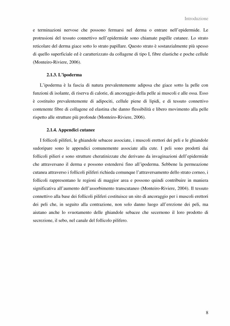
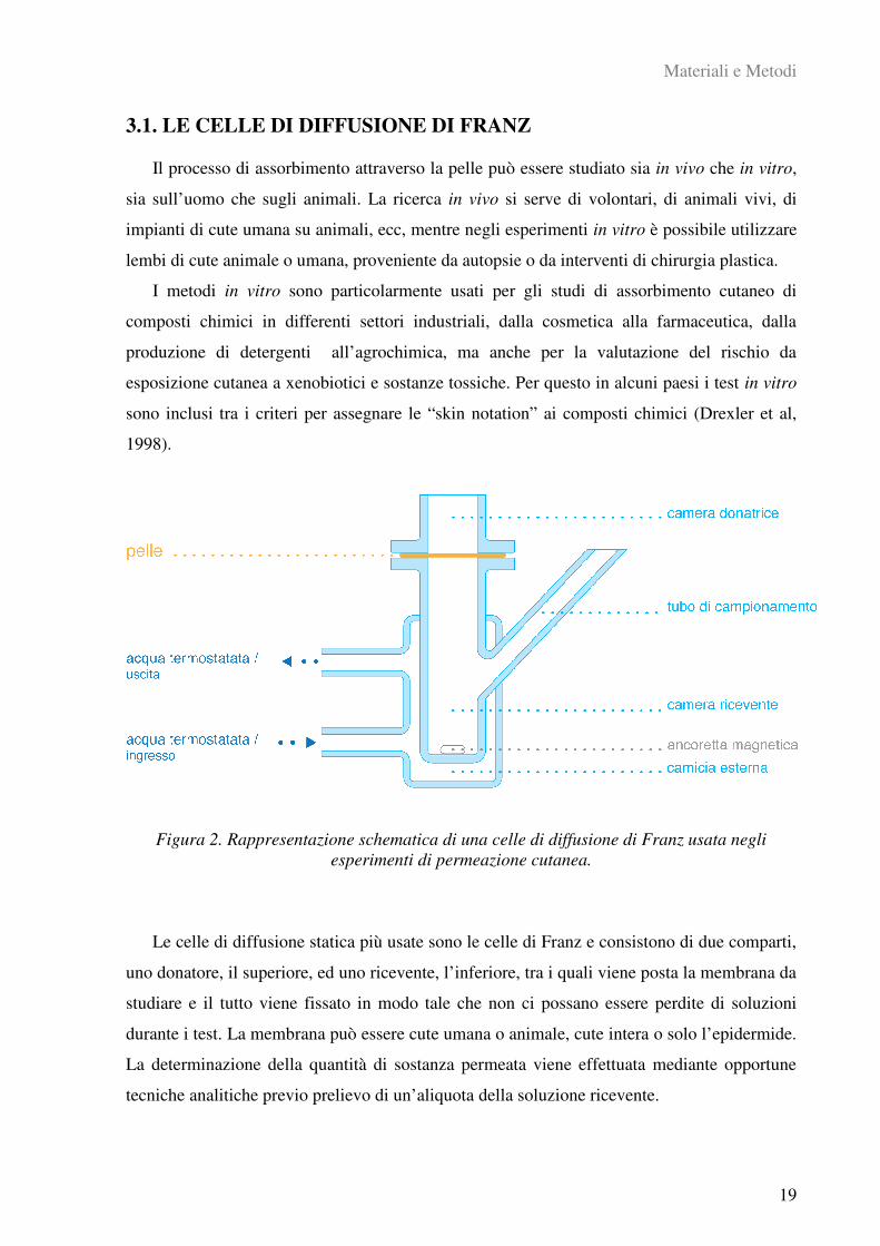
Figura 2. Rappresentazione schematica di una celle di diffusione di Franz usata negli
esperimenti di permeazione cutanea.
Le celle di diffusione statica più usate sono le celle di Franz e consistono di due comparti,
uno donatore, il superiore, ed uno ricevente, l’inferiore, tra i quali viene posta la membrana da
studiare e il tutto viene fissato in modo tale che non ci possano essere perdite di soluzioni
durante i test. La membrana può essere cute umana o animale, cute intera o solo l’epidermide.
La determinazione della quantità di sostanza permeata viene effettuata mediante opportune
tecniche analitiche previo prelievo di un’aliquota della soluzione ricevente.
Materiali e Metodi
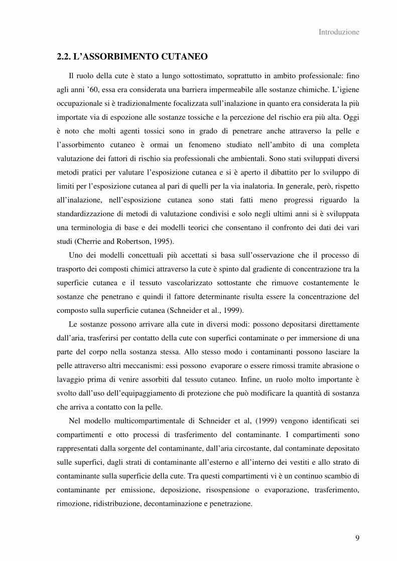
20
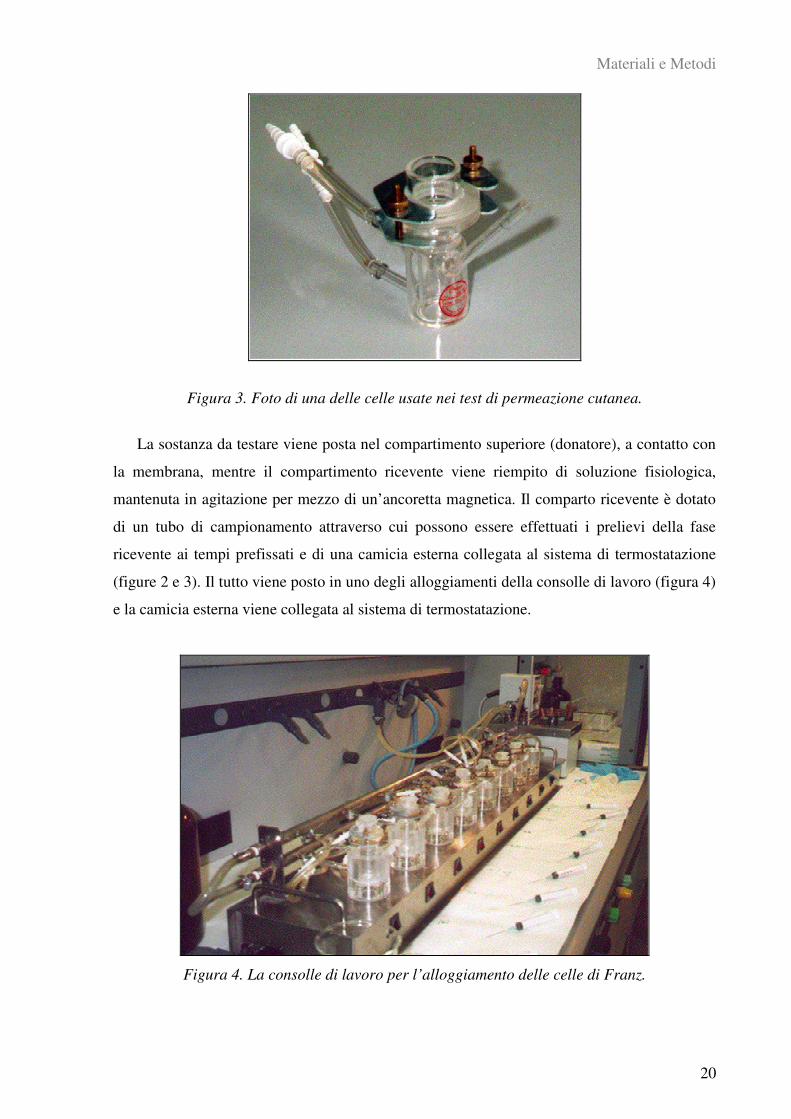
Figura 3. Foto di una delle celle usate nei test di permeazione cutanea.
La sostanza da testare viene posta nel compartimento superiore (donatore), a contatto con
la membrana, mentre il compartimento ricevente viene riempito di soluzione fisiologica,
mantenuta in agitazione per mezzo di un’ancoretta magnetica. Il comparto ricevente è dotato
di un tubo di campionamento attraverso cui possono essere effettuati i prelievi della fase
ricevente ai tempi prefissati e di una camicia esterna collegata al sistema di termostatazione
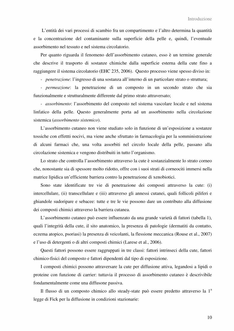
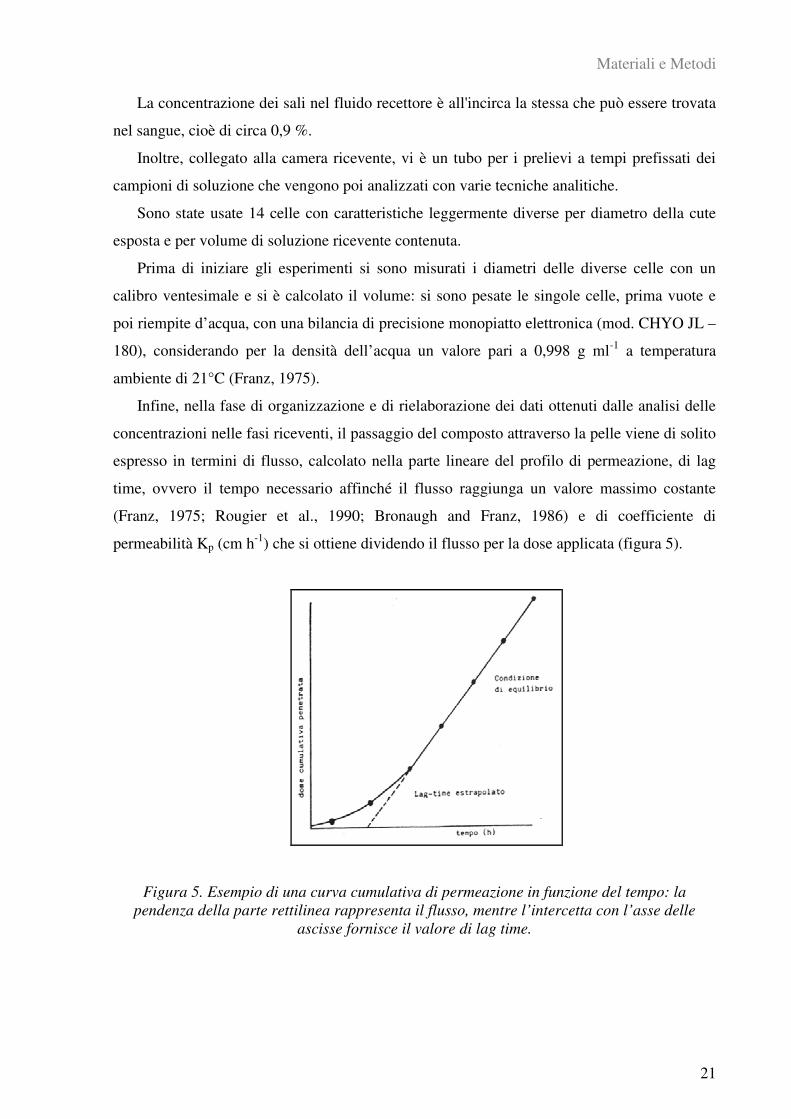
(figure 2 e 3). Il tutto viene posto in uno degli alloggiamenti della consolle di lavoro (figura 4)
e la camicia esterna viene collegata al sistema di termostatazione.
Figura 4. La consolle di lavoro per l’alloggiamento delle celle di Franz.
Materiali e Metodi
21
La concentrazione dei sali nel fluido recettore è all'incirca la stessa che può essere trovata
nel sangue, cioè di circa 0,9 %.
Inoltre, collegato alla camera ricevente, vi è un tubo per i prelievi a tempi prefissati dei
campioni di soluzione che vengono poi analizzati con varie tecniche analitiche.
Sono state usate 14 celle con caratteristiche leggermente diverse per diametro della cute
esposta e per volume di soluzione ricevente contenuta.
Prima di iniziare gli esperimenti si sono misurati i diametri delle diverse celle con un
calibro ventesimale e si è calcolato il volume: si sono pesate le singole celle, prima vuote e
poi riempite d’acqua, con una bilancia di precisione monopiatto elettronica (mod. CHYO JL –
180), considerando per la densità dell’acqua un valore pari a 0,998 g ml-1
a temperatura
ambiente di 21°C (Franz, 1975).
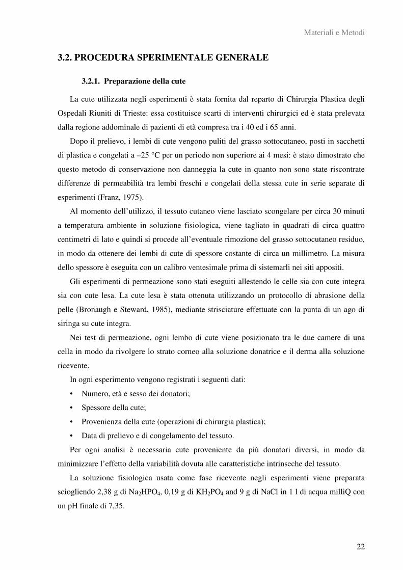
Infine, nella fase di organizzazione e di rielaborazione dei dati ottenuti dalle analisi delle
concentrazioni nelle fasi riceventi, il passaggio del composto attraverso la pelle viene di solito
espresso in termini di flusso, calcolato nella parte lineare del profilo di permeazione, di lag
time, ovvero il tempo necessario affinché il flusso raggiunga un valore massimo costante
(Franz, 1975; Rougier et al., 1990; Bronaugh and Franz, 1986) e di coefficiente di
permeabilità Kp (cm h-1
) che si ottiene dividendo il flusso per la dose applicata (figura 5).
Figura 5. Esempio di una curva cumulativa di permeazione in funzione del tempo: la
pendenza della parte rettilinea rappresenta il flusso, mentre l’intercetta con l’asse delle
ascisse fornisce il valore di lag time.
Materiali e Metodi
22
3.2. PROCEDURA SPERIMENTALE GENERALE
3.2.1. Preparazione della cute
La cute utilizzata negli esperimenti è stata fornita dal reparto di Chirurgia Plastica degli
Ospedali Riuniti di Trieste: essa costituisce scarti di interventi chirurgici ed è stata prelevata
dalla regione addominale di pazienti di età compresa tra i 40 ed i 65 anni.
Dopo il prelievo, i lembi di cute vengono puliti del grasso sottocutaneo, posti in sacchetti
di plastica e congelati a –25 °C per un periodo non superiore ai 4 mesi: è stato dimostrato che
questo metodo di conservazione non danneggia la cute in quanto non sono state riscontrate
differenze di permeabilità tra lembi freschi e congelati della stessa cute in serie separate di
esperimenti (Franz, 1975).
Al momento dell’utilizzo, il tessuto cutaneo viene lasciato scongelare per circa 30 minuti
a temperatura ambiente in soluzione fisiologica, viene tagliato in quadrati di circa quattro
centimetri di lato e quindi si procede all’eventuale rimozione del grasso sottocutaneo residuo,
in modo da ottenere dei lembi di cute di spessore costante di circa un millimetro. La misura
dello spessore è eseguita con un calibro ventesimale prima di sistemarli nei siti appositi.
Gli esperimenti di permeazione sono stati eseguiti allestendo le celle sia con cute integra
sia con cute lesa. La cute lesa è stata ottenuta utilizzando un protocollo di abrasione della
pelle (Bronaugh e Steward, 1985), mediante strisciature effettuate con la punta di un ago di
siringa su cute integra.
Nei test di permeazione, ogni lembo di cute viene posizionato tra le due camere di una
cella in modo da rivolgere lo strato corneo alla soluzione donatrice e il derma alla soluzione
ricevente.
In ogni esperimento vengono registrati i seguenti dati:
• Numero, età e sesso dei donatori;
• Spessore della cute;
• Provenienza della cute (operazioni di chirurgia plastica);
• Data di prelievo e di congelamento del tessuto.
Per ogni analisi è necessaria cute proveniente da più donatori diversi, in modo da
minimizzare l’effetto della variabilità dovuta alle caratteristiche intrinseche del tessuto.
La soluzione fisiologica usata come fase ricevente negli esperimenti viene preparata
sciogliendo 2,38 g di Na2HPO4, 0,19 g di KH2PO4 and 9 g di NaCl in 1 l di acqua milliQ con
un pH finale di 7,35.

Materiali e Metodi
23
Il sudore sintetico usato per disperdere le polveri metalliche è una soluzione allo 0,5% di
NaCl, 0,1% di urea e 0,1% di acido lattico. Il pH viene portato al valore finale con
ammoniaca.
3.2.2. Integrità della cute
Prima e dopo ogni esperimento è stata testata l'integrità della cute attraverso misure di
conducibilità (o resistenza: R = 1/C) elettrica utilizzando un conduttimetro (Metrohm, 660,
Metrohm AG Oberdorfstr. 68 CH-9100 Herisau) operante a 300 Hz collegato a due elettrodi
in acciaio inox (Fasano et al., 2002). Dopo aver riempito la camera ricevente della cella di
soluzione fisiologica, viene posta la cute e quindi anche la camera donatrice viene riempita di
soluzione fisiologica. Si attendono circa 30 minuti affinché si instauri l’equilibrio termico e a
questo punto si effettua la misura di conducibilità con i due elettrodi immersi nella soluzione
ricevente.
I dati di conducibilità, ottenuti in S, sono stati convertiti in K cm-2
.
I lembi di pelle che avevano una resistenza inferiore a 3,95±0,27 K cm-2
sono stati
ritenuti danneggiati e scartati e le eventuali soluzioni sono state eliminate dall’esperimento
(Davies et al., 2004).
3.2.3. Test di permeazione
Prima di ogni esperimento le celle di Franz vengono lavate con acido nitrico diluito (6%
v/v) e risciacquate con acqua milliQ: una volta asciutte, esse vengono sistemate nella consolle
di lavoro e collegate al sistema di termostatazione. Al termine dei test invece le celle di Franz
vengono lavate con acqua e detersivo, quindi con acqua regia, poi con acido nitrico diluito
(6% v/v) ed infine risciacquate più volte con acqua milliQ.
Ogni compartimento inferiore viene completamente riempito di soluzione ricevente e
lasciato termostatare per circa mezz’ora, fino al raggiungimento dell’equilibrio termico: in
generale i nostri esperimenti sono stati condotti ad una temperatura di 32°C.
Il campione di cute viene poi posizionato con il derma a contatto con la soluzione
ricevente, facendo attenzione che all’interfaccia non rimangano bolle d’aria che ridurrebbero
la superficie di contatto. Al di sopra della cute è posta la camera donatrice e tutto il sistema
viene fissato con pinze di polietilene, in modo che non ci siano perdite delle varie fasi. La
camera donatrice infine viene riempita con un volume di sostanza da testare sufficiente a
ricoprire omogeneamente tutta la superficie della pelle. Una volta riempita la camera
donatrice, le aperture della cella, apertura superiore e tubo laterale, vengono chiuse con tappi
Materiali e Metodi
24
in plastica o parafilm per evitare fenomeni di evaporazione delle soluzioni (Franz, 1975).
Il tempo di esposizione è stato di 24 ore in tutti i test; a intervalli prefissati vengono
eseguiti i prelievi per il monitoraggio e la definizione dei profili di permeazione. Ad ogni
campionamento vengono prelevati 1,5 ml di soluzione ricevente da ogni cella attraverso il
tubo laterale utilizzando una siringa in polietilene da 2,5 ml. Dopo il prelievo, viene
ripristinato il volume iniziale all’interno della cella aggiungendo soluzione fisiologica fresca
sempre facendo attenzione alle bolle d’aria che si possono formare.
I campioni di fase ricevente prelevati durante le 24 ore, vengono posti in provette da 1,5
ml e conservati in congelatore ad una temperatura di –25 °C fino al momento dell’analisi.
Al termine delle 24 ore la fase donatrice delle varie celle viene recuperata e posta in
provette da 15 ml. La camera donatrice e l’epidermide esposta vengono lavate con alcuni ml
di acqua milliQ che vengono poi addizionati alla soluzione già recuperata.
La pinza di plastica viene infine rimossa e il lembo di cute e le varie fasi vengono
congelate per le successive analisi.
3.2.4. Mineralizzazione della cute dopo l’esperimento
Al momento delle analisi, i lembi di cute vengono scongelati ed asciugati a temperatura
ambiente per due ore, quindi le aree esposte sono tagliate, pesate e messe in becher con 10 ml
di HNO3 al 69 % v/v per la mineralizzazione (le quantità di pelle in generale sono comprese
tra 0,6 e 1,2 g). Le soluzioni vengono riscaldate a 150°C per 10 ore. Dopo circa due ore,
quando il tessuto è completamente disgregato, viene aggiunta, goccia a goccia, H2O2 al 30%
v/v fino ad un totale di 2 ml; quindi si continua ad evaporare fino ad ottenere circa 2 ml di
soluzione.
Infine le soluzioni vengono portate ad un volume di 10 ml con acqua milliQ e analizzate.
3.2.5. Preparazione dei campioni di cute per le analisi di microscopia elettronica
In alcuni test di permeazione cutanea di nanoparticelle metalliche, alcuni lembi di cute
esposta sono stati analizzati al microscopio elettronico a trasmissione (TEM) al fine di
visualizzare le nanoparticelle eventualmente penetrate nei vari strati della pelle.
I campioni scelti vengono tagliati in sezioni più piccole e fissati per 3 ore in una soluzione
al 3 % di glutaraldeide in tampone di cacodilato allo 0,1 M (pH 7,3). Le sezioni di pelle
fissate vengono lavate due volte (per 10 minuti ciascuna) con il tampone allo 0,1 M di
cacodilato e sono state trattate con tetrossido di osmio all’1 % per 1 ora a 4 °C.
A questo punto i campioni vengono disidratati con una serie di aggiunte crescenti di
Materiali e Metodi
25
etanolo e poi immersi in una resina epossidica Dow (DER332; Unione Chimica Europea,
Milan, Italy): l’ultima inclusione in resina è stata fatta sotto vuoto.
Le sezioni semi-fini e le ultra sottili vengono tagliate con un ultramicrotomo (Ultracut
UCT - Leica Microsystems, Milan, Italy) dotato di una lama di diamante lunga 3 mm.
In seguito, le sezioni semi-fini vengono osservate con un microscopio ottico Leitz Dialux
20 EB (Leica Microsystems, Milan, Italy), mentre le sezioni ultra sottili vengono colorate due
volte con citrato di piombo e acetato di uranile e vengono osservate con un microscopio
elettronico a trasmissione (EM208; Philips, Eindhoven, The Netherlands) con un sistema di
acquisizione ad alta definizione SIS Morada e un sistema di acquisizione dell’immagine
digitale iTEM (FEI Italia, Milan, Italy).
3.2.6. Misure analitiche strumentali
Per le misurazioni analitiche delle concentrazioni dei metalli studiati in questo progetto,
sono state utilizzate le seguenti tecniche analitiche strumentali:
• Spettroscopia di Assorbimento Atomico Elettro-Termica con Fornetto di Grafite
(GF-AAS);
• Spettroscopia di Emissione Atomica con sorgente al Plasma Induttivamente
Accoppiato (ICP-AES);
• Spettrometria di Massa con sorgente al Plasma Induttivamente Accoppiato (ICP-
MS).
La scelta della tecnica analitica più opportuna è stata fatta in funzione delle concentrazioni
attese nelle varie soluzioni da analizzare e quindi dei limiti di rilevabilità degli strumenti per i
vari elementi studiati.
3.2.6.1. Spettroscopia di Assorbimento Atomico Elettro-Termica con Fornetto di
Grafite
Lo strumento utilizzato per le analisi di cobalto, cromo e nichel è uno spettrometro
Thermo M series GF95Z (UK) dotato di fornetto di grafite e di autocampionatore FS95,
presso il Dipartimento di Scienze Chimiche, Laboratorio di Chimica Analitica Ambientale e
Strumentale, Università di Trieste.
Lo strumento utilizzato per le analisi dell’argento è uno spettrometro Perkin Elmer 4100
ZL (USA) equipaggiato con fornetto di grafite HGA e autocampionatore AS/71, presso il
Dipartimento di Traumatologia, Ortopedia e Medicina del Lavoro, Laboratorio di
Tossicologia Industriale, Università di Torino.

Materiali e Metodi
26
3.2.6.2. Spettroscopia di Emissione Atomica con sorgente al Plasma Induttivamente
Accoppiato
Lo strumento utilizzato per le analisi di cobalto, cromo e nichel è uno Spettrometro ottico
al Plasma assiale Spectroflame Modula-E (SPECTRO, Germany) presso il Dipartimento di
Scienze Chimiche, Laboratorio di Chimica Analitica Ambientale e Strumentale, Università di
Trieste.
3.2.6.3. Spettrometria di Massa con sorgente al Plasma Induttivamente Accoppiato
Lo strumento utilizzato per le analisi dell’argento è uno spettrometro ICP-MS Agilent
7500ce (USA) equipaggiato con una cella di collisione per l’abbattimento delle interferenze,
presso il Dipartimento di Traumatologia, Ortopedia e Medicina del Lavoro, Laboratorio di
Tossicologia Industriale, Università di Torino.
Materiali e Metodi
27
3.3. PROCEDURE SPERIMENTALI SPECIFICHE
3.3.1. Assorbimento cutaneo in vitro di polvere di cromo ed effetti della detersione
(Allegato I)
In questo lavoro è stato studiato l’assorbimento percutaneo del cromo metallico in vitro,
utilizzando sudore sintetico a pH 4,5, e l’effetto di un comune detergente (contenente sodio
laurilsolfato e sodio lauriletere solfato) sul processo di assorbimento del metallo stesso. Negli
ambienti di lavoro la pulizia con il detergente della cute contaminata è una pratica abituale:
essa dovrebbe rimuovere la maggior parte delle sostanze tossiche depositatesi, ma numerosi
studi hanno dimostrato che essa può determinare un aumento dell’assorbimento percutaneo
delle stesse. In particolare, il sodio laurilsolfato può facilitare la penetrazione di alcuni agenti
tossici, ad esempio nichel e piombo, alterando il mantello idrolipidico e la normale funzione
“barriera” della cute (Frankild et al., 1995, Larese et al., 2006).
Prima degli esperimenti di permeazione cutanea, sono stati svolti alcuni test propedeutici
per studiare gli effetti del pH del sudore sintetico e della concentrazione della polvere sulla
ionizzazione della metallo, processo necessario per una eventuale permeazione cutanea
(Larese et al., 2007).
I test di permeazione sono stati svolti col metodo delle celle di diffusione di Franz
utilizzando cute umana integra. Come fase donatrice è stato usato 1 ml di una sospensione al
5 % w/v di polvere di cromo, con una dimensione media delle particelle (APS) inferiore ai 10
µm, in sudore sintetico fresco a pH 4,5. Inoltre in 4 celle, a 30 minuti dall’inizio
dell’esperimento, è stato eseguito il lavaggio della cute con un comune detergente contenente
sodio laurilsolfato.
La permeazione del cromo è stata monitorata nelle 24 ore attraverso il prelievo dopo 1, 2,
4, 8, 16, 18, 20, 24 ore di 1,5 ml di fase ricevente rimpiazzata con soluzione fisiologica fresca.
Alla fine degli esperimenti la cute è stata mineralizzata con acido nitrico concentrato e acqua
ossigenata a caldo, per l’analisi del cromo rimasto all’interno della cute.
I valori di concentrazione del cromo nelle varie soluzioni sono stati determinati tramite
spettroscopia di emissione atomica al plasma induttivamente accoppiato (ICP-AES) e
spettroscopia di assorbimento atomico con fornetto di grafite (GF-AAS) con sistema di
correzione del fondo Zeeman.
Il limite di rilevabilità per il cromo in ICP-AES era di 10 µg L-1
alla lunghezza d’onda di
267,716 nm. La precisione espressa come deviazione standard relativa percentuale (RSD%)

Materiali e Metodi
28
delle misure è stata sempre inferiore al 5%.
Il limite di rilevabilità per il cromo in GF-AAS era di 0,2 µg L-1
alla lunghezza d’onda di
357,9 nm. Come modificante di matrice è stata usata una soluzione di Mg(NO3)2 allo 0,5%
w/v. La precisione espressa come deviazione standard relativa percentuale (RSD%) delle
misure è stata sempre inferiore al 10%.
3.3.2. Assorbimento cutaneo in vitro di polveri di cobalto, nichel e cromo
attraverso cute umana integra e lesa (Allegato II)
In questo studio è stata valutata l’influenza che le lesioni cutanee possono avere sulla
permeazione cutanea in vitro di polveri metalliche di cobalto, nichel e cromo, utilizzando il
metodo delle celle di diffusione di Franz e un protocollo di abrasione cutanea proposto da
Bronaugh e Steward (1985).
Come fasi donatrici sono stati utilizzati 1,5 ml di dispersioni al 5% w/v di polveri di
cobalto (APS < 2 m), di nichel (APS =2,2 m) e di cromo (APS < 10 m) in sudore sintetico
a pH 4,5, mentre come fase ricevente è stata usata soluzione fisiologica a pH 7,3.
Dato che i profili di permeazione nelle 24 ore di esposizione sono già stati ricavati in
precedenti lavori del nostro gruppo (Larese et al., 2004, 2007), è stata studiata solo la
permeazione totale alle 24 ore, senza campionamenti intermedi. Per ognuno dei tre metalli
sono state allestite 2 celle come bianco, 6 celle con cute integra e 6 celle con cute lesa.
Alla fine degli esperimenti la cute è stata mineralizzata con acido nitrico concentrato e
acqua ossigenata a caldo, per l’analisi del contenuto dei metalli all’interno della cute.
Le fasi donatrici sono state centrifugate per 15 minuti a 3000 giri/minuto e il surnatante è
stato filtrato due volte con filtri per siringa da 0,45 m prima delle analisi per determinare la
percentuale di metallo presente in forma ionizzata.
I valori di concentrazione del cromo nelle varie soluzioni sono stati determinati tramite
spettroscopia di assorbimento atomico con fornetto di grafite (GF-AAS) con sistema di
correzione del fondo Zeeman. Il limite di rilevabilità per il cobalto era di 0,4 µg L-1
alla
lunghezza d’onda di 240,7 nm; il limite di rilevabilità per il nichel era di 0,2 µg L-1
alla
lunghezza d’onda di 232,0; il limite di rilevabilità per il cromo era di 0,2 µg L-1
alla lunghezza
d’onda di 357,9 nm. Una soluzione di Mg(NO3)2 allo 0,5% w/v è stata usata come
modificante di matrice per le analisi di cobalto e cromo. La precisione espressa come
deviazione standard relativa percentuale (RSD%) delle misure è stata sempre inferiore al
10%.

Materiali e Metodi
29
3.3.3. Permeazione cutanea in vitro di nanoparticelle d’argento attraverso cute
umana integra e lesa (Allegato IV)
In questo studio abbiamo valutato con il metodo in vitro delle Franz cells il passaggio di
nanoparticelle di argento, rivestite con polivinilpirrolidone, attraverso la cute sia integra che
lesa.
I test sono stati ripetuti due volte. Nel primo esperimento, sono state allestite 8 celle con
cute integra e 8 celle con cute lesa, secondo il protocollo proposto da Bronaugh e Steward
(1985). Nel compartimento donatore delle celle è stata collocata una quantità complessiva pari
a 70 µg cm-2
di nanoparticelle di argento, rivestite in polivinilpirrolidone con un diametro
medio di 25 nm misurato al TEM (transmission electron microscopy), disperse in etanolo 0.14
% w/v ed infine diluite 1:10 con sudore sintetico a pH 4,5. Come fase ricevente è stata usata
soluzione fisiologica a pH 7,3. Al termine delle 24 ore le soluzioni riceventi sono state
recuperate per le successive analisi.
I test sono stati ripetuti una seconda volta utilizzando la stessa quantità di fase donatrice,
ma allestendo 5 celle con cute integra e 5 con cute lesa. Durante le 24 ore di esposizione sono
stati eseguiti i prelievi alle 4, 8, 20, 24 ore. Per ogni cella sono stati prelevati 1,5 ml di
soluzione ricevente e sostituiti con soluzione fisiologica fresca.
Nei due test, 4 celle sono state allestite come bianco, quindi trattate allo stesso modo delle
altre, ma come fase donatrice è stato utilizzato solo sudore sintetico, senza nanoparticelle.
Al termine delle 24 ore alcuni campioni di cute esposta sono stati utilizzati per le
investigazioni al TEM.
I valori di concentrazione del cromo nelle varie soluzioni sono stati determinati tramite
spettroscopia di assorbimento atomico con fornetto di grafite (GF-AAS) con sistema di
correzione del fondo Zeeman. Il limite di rilevabilità per l’argento era di 0,1 µg L-1
alla
lunghezza d’onda di 328,1 nm. Come modificanti di matrice sono state usate una soluzione di
Mg(NO3)2 allo 0,1% w/v e una di Pd(NO3)2 allo 0,1% w/v. La precisione espressa come
deviazione standard relativa percentuale (RSD%) delle misure è stata sempre inferiore al 5%.
3.3.4. Permeazione cutanea in vitro di nanoparticelle d’oro attraverso cute umana
integra e lesa (allegato V)
In questo studio è stata studiata la permeazione in vitro di nanoparticelle d’oro, con un
diametro medio misurato al TEM di 12,6±0,9 nm, attraverso cute umana integra e lesa, con il
metodo delle celle di diffusione di Franz.
Materiali e Metodi
30
I test sono stati ripetuti due volte. Nel primo test propedeutico, sono state allestite 8 celle
con cute integra e 8 celle con cut lesa, secondo il protocollo proposto da Bronaugh e Steward
(1985). Come fase donatrice sono stati usati 0,5 ml di una soluzione contenente 100 mg L-1
di
nanoparticelle d’oro, diluiti 1:3 con sudore sintetico a pH 4,5 per un volume totale di 1,5 ml e
una quantità media di 15 µg cm-2
. Come fase ricevente è stata usata soluzione fisiologica a pH
7,3. Al termine delle 24 ore di esposizione le fasi riceventi sono state recuperate per le
successive analisi delle concentrazione.
Nel secondo test, sono state allestite 8 celle con cute integra e 8 celle con cute lesa. Come
fase donatrice sono stati usati 1,5 ml di una soluzione contenente 100 mg L-1
di nanoparticelle
d’oro, diluiti 1:1 con acqua milliQ per un volume totale di 3 ml e una quantità media di 45 µg
cm-2
. Agli intervalli prestabiliti (4, 8, 16, 24 ore) 1,5 ml di soluzione ricevente sono stati
prelevati da ogni cella per le analisi delle concentrazioni e sostituiti con lo stesso volume di
soluzione fisiologica fresca. Al termine delle 24 ore di esposizione le varie fasi sono state
recuperate per le successive analisi.
Per ogni esperimento, 4 celle sono state trattate come bianchi, cioè trattate come le altre
celle, ma senza nanoparticelle d’oro nella fase donatrice.
I campioni di cute integra, dopo essere stati rimossi dalle celle di Franz, sono stati divisi in
epidermide e derma tramite shock termico in acqua a 60°C per un minuto; per tre campioni lo
strato corneo è stato rimosso tramite stripping con un nastro adesivo di VC. I vari strati sono
stati mineralizzati separatamente con acido nitrico e acqua ossigenata a caldo. Alcuni
campioni di cute, sia integra che lesa, sono invece stati utilizzati per le analisi al TEM.
I valori di concentrazione del cromo nelle varie soluzioni sono stati determinati tramite
spettroscopia massa al plasma induttivamente accoppiato (ICP-MS). Il limite di rilevabilità
per l’oro era di 0,001 µg L-1
. La precisione espressa come deviazione standard relativa
percentuale (RSD%) delle misure è stata sempre inferiore al 3%.

4. RISULTATI E DISCUSSIONI
Risultati e Discussioni
32
4.1. ASSORBIMENTO CUTANEO IN VITRO DI POLVERE DI CROMO
ED EFFETTI DELLA DETERSIONE
(Allegato I)
4.1.1. Risultati
Il nostro studio ha evidenziato un incremento progressivo nel passaggio del metallo. Alle
24 ore, la quantità di cromo permeata nella fase ricevente delle celle non trattate con il
detergente è pari a 0,016±0,005 g cm-2
con un flusso medio di 0,84±0,25 ng cm-2
h-1
e un lag
time di 1,1± 0,7 ore. Nelle celle trattate con il detergente, le concentrazioni di cromo sono
molto basse e dello stesso ordine di grandezza di quelle osservate nei bianchi di controllo.
L’analisi della cute ha rivelato invece una quantità di cromo dopo 24 ore di esposizione di
3,19±1,48 g cm-2
nelle celle non lavate con il detergente ed una concentrazione di 5,46±1,09
g cm-2
in quelle trattate con il lavaggio.
4.1.2. Discussione
Il nostro studio ha confermato, analogamente ai dati disponibili in letteratura, un basso
flusso di assorbimento percutaneo per il cromo metallico, rispetto ad altri metalli: il sudore
sintetico a pH 4,5 era in grado di ossidare la polvere di Cr a Cr (III) che, a differenza del Cr
(VI), attraversa la cute meno facilmente e presenta una maggiore affinità per le proteine
cutanee.
Il lavaggio della cute con il detergente e la rimozione della fase donatrice dopo 30 minuti
di esposizione, hanno determinato una riduzione del passaggio del metallo nel liquido
ricevente e un significativo incremento della concentrazione di cromo nella cute: questo può
essere attribuito da un lato alla elevata affinità del Cr (III) per i costituenti della cute e
dall’altro all’azione del sodio laurilsolfato che, alterando l’integrità della membrana, facilita
l’assorbimento del metallo nella cute. Questo dato deve essere tenuto in considerazione, in
particolare in ambito professionale, al fine di predisporre un’adeguata protezione della cute
dal contatto con sostanze tossiche, poiché anche una breve esposizione, seguita da accurata
pulizia, potrebbe rappresentare un potenziale fattore di rischio per l’assorbimento. Va inoltre
sottolineata l’importanza della scelta del detergente: utilizzando un prodotto comune per la
decontaminazione è possibile determinare un aumento non trascurabile della penetrazione
dell’agente tossico nella cute.
Risultati e Discussioni
33
4.2. ASSORBIMENTO CUTANEO IN VITRO DI POLVERI DI
COBALTO, NICHEL E CROMO ATTRAVERSO CUTE UMANA
INTEGRA E LESA
(allegato II)
4.2.1. Risultati
I dati di permeazione di questo studio sono stati riassunti come mediane, valori massimi,
valori minimi e 25-75esimo percentile. Le quantità medie di cobalto e nichel trovate nella fase
ricevente sono significativamente più alte quando le polveri metalliche sono applicate su cute
lesa rispetto alla cute integra: le mediane sono rispettivamente 0,0084 vs 3,566 µg cm-2
per il
cobalto e 0,0072 vs 2,631 µg cm-2
per il nichel, mentre non sono state riscontrate differenze
significative nella permeazione del cromo.
Le quantità dei tre metalli rimaste nella cute dopo le 24 ore di esposizione sono
significativamente più alte nella cute lesa rispetto alla cute integra: le mediane sono
rispettivamente 48,7 vs 29,6 µg cm-2
per il cobalto, 131 vs 82,3 µg cm-2
per il nichel e 62,1 vs
14,4 µg cm-2
per il cromo.
Le analisi delle fasi donatrici, dopo rimozione della polvere per centrifugazione e
filtrazione, hanno confermato la presenza di ioni metallici in soluzione: la ionizzazione di
cobalto e nichel risulta significativamente maggiore quando la sospensione è applicata alla
cute lesa rispetto alla cute integra (55,6 e 129 mg L-1
vs. 21,3 e 57,5 mg L-1
), mentre il Cr non
mostra differenze significative tra i due gruppi di celle (0,18 vs 0,28 mg L-1
).
4.2.2. Conclusioni
Questo studio ha dimostrato che polveri di cobalto, nichel e cromo, con diametro medio
nell’ordine del micron, possono essere ossidate a contatto con il sudore sintetico e rilasciare
ioni metallici in soluzione: tali ioni sono poi in grado di attraversare la cute, in modo variabile
e dipendente sia dalla percentuale ionizzata sia dalle condizioni della cute. Le lesioni cutanee
aumentano significativamente il passaggio in vitro di cobalto e nichel, mentre per quanto
riguarda il cromo non ci sono differenze significative tra i due gruppi di celle e tra le celle
trattate e i bianchi.
Per quanto riguarda invece la quantità di metallo che rimane nella pelle, la differenza tra
le celle con cute integra e le celle con cute lesa è significativa per tutti i tre metalli studiati: in
particolare, le quantità penetrate nella cute trattata con il cromo sono dello stesso ordine di
Risultati e Discussioni
34
grandezza delle celle trattate con gli altri metalli. Questo suggerisce che la cute abbia un
tropismo particolare per gli ioni trivalenti del cromo intrappolandoli nei vari strati
probabilmente attraverso il legame con proteine della pelle ed evitando che permeino nella
fase ricevente (Hostynek, 2003; Samitz et al., 1969).
La fase donatrice infine sembra essere influenzata dalla presenza di lesioni cutanee nel
caso di cobalto e nichel: la ionizzazione dei due metalli è significativamente superiore nelle
celle allestite con cute integra rispetto a quelle con cute lesa, suggerendo che il contatto della
polvere a componenti cutanei esposti in seguito alla lesione possa contribuire alla
ionizzazione stessa. Questa differenza non è stata notata negli esperimenti con polvere di
cromo.
Questi risultati insieme ai dati già presenti in letteratura confermano che è necessario
prevenire il contatto dei metalli e dei loro sali con la cute, soprattutto in presenza di lesioni
cutanee anche piccole, situazione molto comune nell’ambiente di lavoro: nei nostri test
piccole lesioni hanno portato ad un significativo aumento dell’assorbimento cutaneo.
Risultati e Discussioni
35
4.3. PERMEAZIONE CUTANEA IN VITRO DI NANOPARTICELLE DI
ARGENTO ATTRAVERSO CUTE UMANA INTEGRA E LESA
(Allegato IV)
4.3.1. Risultati
I dati di permeazione di questo studio sono stati riassunti come mediane, valori massimi,
valori minimi e 25-75esimo percentile: essi mostrano che l’argento può passare attraverso la
cute umana integra con un valore di mediana alle 24 ore di 0,46 ng cm-2
, mentre la
permeazione attraverso la cute danneggiata è circa cinque volte maggiore con un valore di
mediana di 2,32 ng cm-2
. Le concentrazioni dei bianchi sono tutte al di sotto del limite di
rilevabilità.
Per il gruppo di celle con cute lesa è stato possibile calcolare un flusso nelle 24 ore di
0,6±0,2 ng cm-2
h-1
con un lag time inferiore a 1 ora, mentre per le celle con cute integra non è
stato possibile ricavare un range di linearità date le basse concentrazioni misurate nelle fasi
riceventi.
Le analisi al TEM dei campioni di cute esposti per 24 ore hanno mostrato che le
nanoparticelle di argento studiate in questi test sono in grado di penetrare la barriera dello
strato corneo e di raggiungere gli strati vitali più superficiali dell’epidermide.
4.3.2. Discussione
Lo studio della tossicità e delle vie di penetrazione delle nanoparticelle è iniziato solo
recentemente e i dati relativi alla loro capacità di penetrare la cute sono ancora pochi. In
particolare lo studio delle nanoparticelle di argento risulta interessante perché si tratta della
tipologia di nanoparticelle maggiormente prodotta dall’industria con ampie applicazioni in
prodotti di uso comune quali tessuti tecnici, disinfettanti ambientali, pesticidi, ecc.
I nostri dati sperimentali hanno mostrato che l’assorbimento cutaneo in vitro di argento,
applicato in forma di nanoparticelle, attraverso la cute integra risulta molto basso, ma
misurabile, mentre le lesioni cutanee aumentano la permeazione di cinque volte. Le analisi di
microscopia elettronica inoltre hanno mostrato che le nanoparticelle sono in grado di
raggiungere gli strati vitali dell’epidermide confermando quindi la penetrazione delle
nanoparticelle in quanto tali e non solo l’assorbimento dell’argento che ionizzabile in
soluzione.
Tali osservazioni sono in accordo con altri studi sull’assorbimento cutaneo di differenti

Risultati e Discussioni
36
tipologie di nanoparticelle (vedi allegato III) e pongono l’attenzione sulla esigenza di valutare
molto attentamente l’esposizione cutanea alle nanoparticelle, in particolare nanoparticelle
metalliche, di cui conosce ancora poco. Inoltre, da un punto di vista occupazionale questi test
confermano la necessità di utilizzare i dispositivi di protezione individuale per la prevenzione
del contatto cutaneo con le nanoparticelle, soprattutto in presenza di abrasioni e lesioni
cutanee, molto frequenti nelle normali condizioni di lavoro, che possono portare ad un
aumento nell’assorbimento percutaneo.
Risultati e Discussioni
37
4.4. PERMEAZIONE CUTANEA IN VITRO DI NANOPARTICELLE
D’ORO ATTRAVERSO CUTE UMANA INTEGRA E LESA
(Allegato V)
4.4.1. Risultati
Le nanoparticelle d’oro applicate in sudore sintetico a pH 4,5 hanno portato ad un
assorbimento di oro in vitro dopo 24 ore di esposizione pari a 60,8±25,3 ng cm-2
attraverso la
cute integra e 55,2± 27,3 ng cm-2
attraverso la cute lesa.
Le nanoparticelle d’oro applicate in acqua milliQ, invece, hanno portato ad un
assorbimento di oro più consistente e cioè di 214±44 ng cm-2
attraverso la cute integra e di
188±50 ng cm-2
attraverso la cute lesa.
Nel secondo esperimento è stato possibile calcolare un flusso nelle 24 ore di esposizione
di 7,8±2,0 ng cm-2
h-1
e di 7,1±2,5 ng cm-2
h-1
rispettivamente per la cute integra e per la cute
lesa, con un lag time per entrambe inferiore a 1 ora
La quantità di oro rimasta all’interno della cute dopo 24 ore è significativamente più alta
nella cute lesa rispetto alla cute integra (rispettivamente 7,9±4,5 vs 1,8±0,7 µg cm-2
).
Le analisi sulle concentrazioni di oro all’interno dello strato corneo, dell’epidermide e del
derma della cute integra hanno mostrato un contenuto di oro decrescente dall’esterno verso
l’interno: negli “strip” di nastro adesivo è stata trovata una quantità di oro decrescente dagli
0,40±0,05 µg cm-2
del primo “strip” più esterno agli 0,17±0,01 µg cm-2
del sesto e più
profondo “strip”. Il contenuto di metallo nell’epidermide è di 1,78±0,68 µg cm-2
mentre nel
derma raggiunge gli 0,05±0,02 µg cm-2
.
Le analisi di microscopia elettronica hanno evidenziato la presenza di nanoparticelle d’oro
sia nell’epidermide che nel derma sia in campioni di cute integra che in campioni di cute lesa.
4.4.2. Discussione
I risultati di questi esperimenti in vitro con cute umana intera non hanno evidenziato
differenze significative tra cute integra e cute lesa per quanto riguarda l’assorbimento dell’oro
applicato in forma di nanoparticelle sia con il sudore sintetico che con l’acqua milliQ, mentre
un aumento della concentrazione della fase donatrice ha portato ad un aumento della quantità
di oro permeata nella fase ricevente. L’analisi dei vari strati della cute ha ben evidenziato un
gradiente di concentrazione dall’esterno verso l’interno e una differenza significativa della
Risultati e Discussioni
38
quantità di oro rimasta all’interno della cute lesa rispetto alla cute integra.
L’indagine microscopica ha confermato che anche con cute integra le nanoparticelle d’oro
studiate in questi esperimenti sono in grado di attraversare lo spessore cutaneo e raggiungere
gli strati vitali più profondi fino a raggiungere il derma. Questo risultato conferma che le
concentrazioni d’oro ritrovate nella cute dopo mineralizzazione non derivano solo da metallo
in forma ionica, ma anche da nanoparticelle in quanto tali allo stato di ossidazione (0).
Infine, i risultati di questi test suggeriscono la possibilità di usare le nanoparticelle d’oro
come carrier di farmaci in formulazioni a rilascio transcutaneo e in altre applicazioni
biomediche che richiedono una diffusione delle nanoparticelle nei tessuti.
La carenza di dati in letteratura e i pochi risultati ottenuti in test in vitro richiedono
comunque una più approfondita valutazione del fenomeno dell’assorbimento cutaneo
utilizzando altri tipi di nanoparticelle differenti in forma, dimensioni e rivestimento, e la
valutazione accurata di tutte le variabili che possono influenzare la permeazione cutanea in
funzione dell’obiettivo che si vuole raggiungere.

5. CONCLUSIONI
Conclusioni
40
In conclusione, i risultati dei nostri esperimenti hanno mostrato che, in vitro, cobalto,
nichel e cromo rilasciati da polveri metalliche e nanoparticelle di argento e oro sono in grado
di permeare attraverso la cute fino alla fase ricevente, e che la presenza di lesioni cutanee,
l’uso di detergenti, il pH della fase donatrice o l’entità dell’esposizione, sono in grado di volta
in volta di modificare tale processo.
In particolare le lesioni cutanee determinano nei nostri esperimenti un notevole aumento
nella permeazione di cobalto e nichel, applicati in forma di polveri metalliche disperse in
sudore sintetico, e di argento applicato come nanoparticelle rivestite, mentre non vi sono
differenze significative per quanto riguarda la permeazione del cromo da polvere metallica e
dell’oro in forma di nanoparticelle. In tutti i test, però, le lesioni cutanee hanno determinato
un notevole aumento della concentrazione di metallo nel tessuto cutaneo misurato dopo
mineralizzazione della cute esposta.
Anche l’utilizzo di un comune detergente contenente sodio laurilsolfato e sodio lauriletere
solfato nei test di assorbimento cutaneo del cromo metallico ha causato un aumento
significativo della quantità di metallo misurato nella cute, nonostante la fase donatrice fosse
stata rimossa dopo 30 minuti di esposizione. Questo risultato conferma l’effetto di
potenziamento della permeazione che hanno questi tipi di tensioattivi (in particolare del sodio
laurilsolfato) che causano modificazioni della composizione dei lipidi dello strato corneo,
estraendo la matrice lipidica dello spazio intercellulare e diminuendo così la funzione di
barriera della cute.
Le analisi della cute al TEM hanno inoltre dimostrato che le nanoparticelle di oro e di
argento in un sistema in vitro sono in grado di penetrare all’interno dei vari strati cutanei.
Questo significa che l’assorbimento del nichel può avvenire sia con un meccanismo di
ionizzazione seguito da permeazione, già dimostrato in altri lavori (Larese et al., 2007;
2009b), sia con un meccanismo di penetrazione diretta.
I risultati di questi primi lavori sulla permeazione cutanea di nanoparticelle e l’interesse
suscitato, ci hanno spinto a proseguire su questa strada e sono attualmente in corso le analisi
sui test di permeazione cutanea di nanoparticelle di nichel e di cobalto.
Una delle problematiche che si incrontrano in questo tipo di studi riguarda la
quantificazione delle nanoparticelle direttamente nei tessuti. Le analisi di microscopia
elettronica non sono quantitative e la laboriosa preparazione dei campioni può dare luogo ad
artefatti. Le analisi spettrofotometriche dopo mineralizzazione della cute, invece, forniscono
solo la concentrazione totale del metallo. Con queste tecniche quindi non è possibile
distinguere la percentuale di metallo penetrato in forma di nanoparticella e in forma ionica. A
Conclusioni
41
questo proposito si stanno cercando soluzioni strumentali e metodologiche in grado di
distinguere gli effetti diretti delle nanoparticelle da quelli indiretti dovuti al rilascio di ioni e
di residui del processo di sintesi come possono essere catalizzatori o solventi.
Quindi, al fine di implementare le nostre possibilità di analisi con l’utilizzo di metodiche
diverse e strumentazioni all’avanguardia, sia per quanto riguarda l’analisi microscopica dei
tessuti esposti sia per studiare le formulazioni contenenti nanoparticelle, sono state avviate
delle collaborazioni con laboratori di altre strutture di ricerca.
Attualmente è in corso un progetto per l’ottimizzazione dell’analisi di campioni di cute
esposta a nanoparticelle metalliche tramite microtomografia (micro-CT) alla SYRMEP
“beamline” (Elettra-Sincrotrone; Trieste). Questo studio ha come obiettivo la visualizzazione
della distribuzione del metallo nei vari strati cutanei con tecniche d’indagine che richiedano
una preparazione del campione meno invasiva rispetto alla microscopia elettronica.
Queste tecniche di visualizzazione insieme all’indagine microscopica a trasmissione
(TEM) e a scansione (SEM) eseguite presso il Centro di Coordinamento e Sviluppo Progetti e
Apparecchiature dell’Università di Trieste e all’analisi della concentrazione totale eseguita
dopo mineralizzazione della cute con tecniche di emissione atomica (ICP-AES) e
assorbimento atomico (GF-AAS) hanno lo scopo di fornire un quadro più completo
dell’eventuale penetrazione e distribuzione delle nanoparticelle all’interno della cute.
Un’altra collaborazione è stata avviata con un gruppo di ricerca dell’Istituto di Fisica
Applicata, Consiglio Nazionale delle Ricerche, Sesto Fiorentino (FI) per la sintesi,
caratterizzazione e distribuzione nei tessuti di nanorods d’oro e della possibilità di aumentare
tale penetrazione attraverso l’utilizzo del laser.
Per quanto riguarda invece la tossicità cellulare è in corso una collaborazione con un
gruppo di ricerca del Dipartimento di Scienze della Vita dell’Università di Trieste per la
valutazione della tossicità cellulare di nanoparticelle di argento sulla linea cellulare HaCaT,
derivata da cheratinociti umani spontaneamente immortalizzati per poi proseguire con le altre
nanoparticelle metalliche.
In questo tipo di studi in vitro ci sono ovviamente delle limitazioni dovute soprattutto alla
difficoltà di ricreare tutte le condizioni di un’esposizione in vivo. L’utilizzo di cute espiantata,
l’eccessiva idratazione nelle 24 ore, la staticità delle celle di diffusione e l’impossibilità di
controllare contemporaneamente tutte le variabili, possono portare a differenze con l’effettiva
situazione in un’esposizione cutanea reale. Anche questi test, però, risultanto essere molto
importanti e, insieme ai dati dei test su volontari e agli studi epidemiologici, contribuiscono a
definire un quadro completo della valutazione del rischio.

6. BIBLIOGRAFIA
Bibliografia
43
Alvarez-Roman R, Naik A, Kalia YN, Guy RH, Fessi H (2004) Skin penetration and
distribution of polymeric nanoparticles. J. Control. Release. 99: 53-62.
Auffan M, Rose J, Wiesner MR, Bottero JY (2009) Chemical stability of metallic
nanoparticles: A parameter controlling their potential cellular toxicity in vitro. Environ.
Pollution 157: 1127-1133.
Ayres JG, Borm P, Cissee FR, Castranova V, Donaldson K, Ghio A, Harrison RM, Hider
R, Kelly F, Kooter IM, Marano F, Maynard RL, Mudway I, Nel A, Sioutas C, Smith S,
Baeza-Squiban A, Cho A, Duggan S, Froines J (2008) Evaluating the toxicity of airborne
particulate matter and nanoparticles by measuring oxidative stress potential – A workshop
report an consensus statement. Inhal. toxicol.. 20: 75-99.
Baroli B, Ennas MG, Loffredo F, Isola M, Pinna R and Lopez-Quintela A (2007)
Penetration of Metallic Nanoparticles in Human Full-Thickness Skin. J. Invest. Dermatol.
127: 1701-1712.
Bernstein IA and Vaughan FL, (1999) Cultured keratinocytes in in vitro
dermatotoxicological investigation: a review. J. Toxicol. Environ. Health B, 2: 1-30.
Berry CC, Charles S, Wells S, Dalby MJ, Curtis ASG (2004) The influence of transferrin
stabilised magnetic nanoparticles on human dermal fibroblasts in culture. Int. J. Pharmac.
269: 211-225.
Bronough RL and Steward R (1985) Methods for in vitro percutaneous absorption studies.
V: permeation through damaged skin. J. Pharm. Sci. 15: 1062-1066.
Bronaugh RL and Franz T J (1986) Vehicle effects on percutaneous absorption : in vivo
and in vitro comparisons with human skin. Br. J. Dermatol. 115: 1-11.
Cherrie JW and Robertson A (1995) Biologically relevant assessment of dermal exposure.
Ann. Occup. Hyg. 39: 387–92.
Connor EE, Mwamuka J, Gole A, Murphy CJ, Wyatt MD (2005) Gold Nanoparticles Are
Taken Up by Human Cells but Do Not Cause Acute Cytotoxicity. Small, 1: 325-327.
Corbett GE, Finley BL, Paustenbach DJ, Kerge BD (1997) Systemic uptake of chromium
in human volunteers following dermal contact with hexavalent chromium (22 mg/dL). J.
Exposure Anal. Environ. Epidemiol. 7: 179-189.
Davies DJ, Ward RJ, Heylings JR (2004). Multi-species assessment of electrical
Bibliografia
44
resistance as a skin integrity marker for in vivo percutaneous absorption studies. Toxicol. In
vitro 18:351-358.
Dotterud LK and Smith-Sivertsen T (2007) Allergic contact sensitization in the general
adult population: A population-based study from Northern Norway. Cont. Dermat. 56: 10-15.
Donaldson K, Aitken R, Tran L, Stone V, Duffin R, Forrest G, Alexander A, (2006).
Carbon nanotubes: a review of their properties in relation to pulmonary toxicology and
workplace safety. Toxicol. Sci. 92: 5-22.
Drexler H, 1998. Assignment of skin notation for MAK values and its legal consequences
in Germany. Int. Arch. Occup. Environ. Health 71, 503-505.
EDETOX, 2000. Evaluations and Predictions of Dermal Absorption of Toxic Chemicals,
EU Framework V: Quality of Life, Environment and Health Key Action Funding (Project
Number: QLKA-2000-00196).
EPA (2007) Nanotechnology White Paper. Prepared for the U.S. Environmental
Protection Agency by members of the Nanotechnology Workgroup, a group of EPA’s Science
Policy Council Science Policy Council U.S. Environmental Protection Agency Washington,
DC.
EHC (Environmental Health Criteria) 235 (2006) Dermal Absorption. World Health
Organization (WHO),
Escobar-Chávez JJ, Merino-Sanjuán V, López-Cervantes M, Urban-Morlan Z, Piñón-
Segundo E, Quintanar-Guerrero D, Ganem-Quintanar A (2008) The tape-stripping technique
as a method for drug quantification in skin. J. Pharm. Pharmaceut. Sci. 11: 104-130.
Fasano W, Manning L, Green J (2002). Rapid assessment of rat and human epidermal
membranes for in vitro dermal regulatory testing: correlation of electrical resistance with
tritiated water permeability. Toxicol. In vitro 16: 731-740.
Frankild S, Andersen K, Nielsen G. (1995) Effect of sodium lauryl sulphate (SLS) on in
vitro percutaneous penetration of water, hydrocortisone and nickel. Contact Dermatitis, 32:
338-345.
Franz TJ (1975) On the relevance of in vitro data. J. Invest. Dermatol. 93: 633–640.
Gammelgaard B, Fullerton A, Avnstorp C, Menné T (1992) Permeation of chromium salts
through human skin in vitro. Contact Dermatitis 27: 302-310.
Bibliografia
45
Hostynek JJ, Dreher F, Pelosi A, Anigbogu A, Maibach HI (2001). Human stratum
corneum penetration by nickel: in vivo study of depth distribution after occlusive application
of the metal as powder. Acta Derm. Venereol. Suppl. 212: 5-10.
Hostynek JJ (2003) Factors determining percutaneous metal absorption. Food Chem.
Toxicol. 41: 327-345.
Kim S, Lim YT, Soltesz EG, De Grand AM, Lee J, Nakayama A, Parker JA, Mihaljevic
T, Laurence RG, Dor DM, Cohn LH, Bawendi MG, Frangioni JV (2004) Near-infrared
fluorescent type II quantum dots for sentinel lymph node mapping. Nat. Biotechnol. 22: 93-
97.
Kogan VY and Petukhova NE (1986) Cobalt. UNEP, Moscow.
Lam PK, Chan ES, Ho WS, Liew CT (2004) In vitro cytotoxicity testing of a
nanocrystalline silver dressing (Acticoat) on cultured keratinocytes. Br. J. Biomed. Sci. 61:
125-127.
Langbein L, Grund C, Kuhn C, Praetzel S, Kartenbeck J, Brandner JM, Moll I, Franke
WW (2002) Tight junctions and compositionally related junctional structures in mammalian
stratified epithelia and cell cultures derived therefrom. Eur J Cell Biol. 81: 419-435.
Larese Filon F, Maina G, Adami G, Venier M, Coceani N, Bussani R, Massiccio M,
Barbieri P, Spinelli P (2004) In vitro percutaneous absorption of cobalt. Int. Arch. Environ.
Health 77: 85-89.
Larese Filon F, Boeninger M, Maina G, Adami G, Spinelli P, Damian A (2006) Skin
absorption of inorganic lead and the effects of skin cleansers. J. Occup. Environ. Med. 48:
692-699.
Larese Filon F, Adami G, Venier M, Maina G, Renzi N (2007) In vitro percutaneous
absorption of metal compounds. Toxicol. Lett. 170: 49-56.
Larese Filon F, D’Agostin F, Crosera M, Adami G, Renzi N, Bovenzi M, Maina G (2009)
Human skin penetration of silver nanoparticles through intact and damaged skin. Toxicol.
255: 33-37.
Leaper DJ (2006). Silver dressing: their role in wound management. Int Wound J, 3: 282–
294.
Liden C and Carter S (2001) Nickel release from coins. Cont. Dermat.44: 160-165.

Bibliografia
46
Linnainmaa M and Kiilunen M (1997) Urinary cobalt as a measure of exposure in the wet
sharpen of hard metal and stellite blades. Int. Arch. Occup. Environ. Health 69: 193-200.
Meding B and Swanbeck G (1987) Prevalence of hand eczema in an industrial city. Br. J.
Dermatol. 116: 627-631.
Midander K, Wallinder IO, Leygraf C (2007) In vitro studies of copper release from
powder particles in synthetic biological media. Environ. Pollution 145: 51-59.
Moger J, Johnston BD, Tyler CR (2008) Imaging metal oxide nanoparticles in biological
structures with CARS microscopy. Optic express.16: 3408-3419.
Monteiro-Riviere NA (2004) Anatomical factors affecting barrier function. In:
Dermatotoxicology. Eds. H. Zhai and H.I. Maibach, CRC Press. pp. 43-70.
Monteiro-Riviere NA (2006) Structure and function of skin. In: Dermal Absorption
Models in Toxicology and Pharmacology. Ed. J.E. Riviere, Taylor and Francis. pp. 1-19.
Monteiro-Riviere NA and Inman AO (2006) Challenges for assessing carbon
nanomaterial toxicity to the skin. Carbon 44: 1070-1078.
Muangman P, Chuntrasakul C, Silthram S, Suvanchote S, Benjathanung R, Kittidacha S,
Rueksomtawin S (2006) Comparison of efficacy of 1% silver sulfadiazine and Acticoat for
treatment of partial-thickness burn wounds. J. Med. Assoc. Thai. 89: 953–958.
NANODERM (2007) Quality of skin as a barrier to ultra-fine particles. Final Report.
(Project Number: QLK4-CT-2002-02678) www.uni-leipzig.de/~nanoderm/
NIOSH, National Institute for Occupational Safety and Health (2007) Progress Toward
Safe Nanotechnology in the Workplace - A Report from the NIOSH Nanotechnology
Research Center,. DHHS (NIOSH) Pubblication No 2007-123, available on line:
www.cdc.gov/niosh.
Oberdörster G, Oberdörsters E, Oberdörster J (2005) Nanotoxicology: An Emerging
Discipline Evolving from Studies of Ultrafine Particles. Environ. Health Perspect. 113:823–-
839.
Paddle-Ledinek JE, Nasa Z, Cleland HJ (2006) Effect of different wound dressings on cell
viability and proliferation. Plast. Reconstr. Surg. 117: 110S-118S.
Papageorgiou I, Brown C, Schins R, Singh S, Newson R, Davis S, Fisher J, Ingham E,
Case CP (2007) The effect of nano- and micron-sized particles of cobalt–chromium alloy on

Bibliografia
47
human fibroblasts in vitro. Biomaterials, 28: 2946-2958.
Pernodet N, Fang X, Sun Y, Bakhtina A, Ramakrishnan A, Sokolov J, Ulman A,
Rafailovich M (2006) Adverse Effects of Citrate/Gold Nanoparticles on Human Dermal
Fibroblasts. Small, 2: 766-773.
Poon VK, Burd A (2004) In vitro cytotoxity of silver: implication for clinical wound care.
Burns 30: 140-147.
Roco MC (2005) Environmentally responsible development of nanotechnology. Environ.
Sci. Technol. 39: 106A-112A.
Rotoli BM, Bussolati O, Bianchi MG, Barilli A, Balasubramanian C, Bellucci S,
Bergamaschi E (2008) Non-functionalized multi-walled carbon nanotubes alter the
paracellular permeability of human airway epithelial cells. Toxicol. Lett. 178: 95–102.
Rougier A, Rallis M, Krien P, Lotte C (1990). In vivo percitaneous absorption: a key role
for stratum corneum/ vehicle partitioning. Arch. Dermatol. Res. 282: 498 - 505.
Rouse JG, Yang J, Ryman-Rasmussen JP, Barron AR, Monteiro-Riviere NA (2007)
Effects of mechanical flexion on the penetration of fullerene amino acid-derivatized peptide
nanoparticles through skin. Nano Lett. 7: 155-160.
Scansetti G, Botta GC, Spinelli P, Reviglione L, Ponzetti C (1994) Absorption and
excretion of cobalt in the hard metal industry. Sci. Total Environ. 150: 141-144.
SCCP – Scientific Committee on Consumer Products (2007) Preliminary opinion on
safety of nanomaterials in cosmetic products. www.ec.europa.eu/health/ph_risk/risk_en.htm
Schneider T, Vermeulen R, Brouwer DH, Cherrie JW, Kromhout H, Fogh CL (1999)
Conceptual model for assessmentof dermal exposure. Occup. Environ. Med. 56: 765-773.
Schulte PA, Geraci C, Zumwalde R, Hoover M and Kuempel E (2008) Occupational Risk
Management of Engineered Nanoparticles, J. Occup. Environ. Hyg, 5: 239-249.
Smack DP, Korge MD, James WD (1994) Keratin and keratinization. J. Am. Acad.
Dermatol, 30: 85-102.
Sonavane G, Tomoda K, Sano A, Ohshima H, Terada H, Makino K (2008) In vitro
permeation of gold nanoparticles through rat skin and rat intestine: Effect of particle size.
Colloids Surf. B Biointerfaces. 65: 1-10.
Supp AP, Neely AN, Supp DM, Warden GD, Boyce ST (2005) Evaluation of cytotoxicity

Bibliografia
48
and antimicrobial activity of Acticoat burn dressing for management of microbial
contamination in cultured skin substitutes grafted to athymic mice. J. Burn Care Rehabil. 26:
238-246.
Tanojo H, Hostynek JJ, Mountford H, Maibach HI (2001) In vitro permeation of nickel
salts through human stratum corneum. Acta Derm. Venereol. Suppl. 212: 19-23.
Tinkle SS, Antonini JM, Rich BA, Roberts JR, Salmen R, DePree K, Adkins EJ (2003)
Skin as a route of exposure and sensitization in chronic beryllium disease. Environ. Health
Perspect. 111: 1202-1208.
The Royal Society & the Royal Academy of Engineering, (2004) Nanoscience and
nanotechnologies: opportunities and uncertainties. 29 July.
Thyssen PJ and Menné T (2009) Metal Allergy - A Review on Exposures, Penetration,
Genetics, Prevalence, and Clinical Implications Chem. Res. Toxicol.DOI: 10.1021/tx9002726
Trop M, Novak M, Rodl S, Hellbom B, Kroell W, Goessler W (2006) Silver-Coated
Dressing Acticoat Caused Raised Liver Enzymes and Argyria-like Symptoms in Burn Patient.
Journal of Trauma. 60:648-652.
Tsoli M, Kuhn H, Brandau W, Esche H, Schmid G (2005) Cellular Uptake and Toxicity of
Au55 Clusters. Small, 1: 841-844.
van der Sandt JJM, Maas WJM, van Burgsteden JA, Sartorelli P, Montomoli L, Larese F
Payan JP, Limaset JC, Carmichael P, Kenyon S, Robinson E, Dick I , Nilsen JB, Schaller K-
H, Korinth G, Cage S, Wilkinson SC, Williams FM (20049 In vitro prediction of skin
absorption of caffeine, testosterone and benzoic acid : multicentre comparison study. Regul.
Pharmacol. Toxicol. 39: 271-281.
Van Lierde V, Chery C, Roche N, Monstrey S, Moens L, Vanhaecke F (2006) In vitro
permeation of cheromium species through porcine and human skin as determined by capillare
electrophoresis-inductively coupled plasma-sector field mass spectrometry. Anal. Bioanal.
Chem. 384: 378-384.
Woodrow Wilson International Center for Scholars. Nanotechnology Consumer Products
Inventory. (2007) Available: http://www.nanotechproject.org/consumerproducts
Wright JB. Lam K, Buret AG, Olson ME, Burrell RE (2002) Early healing events in a
porcine model of contaminated wounds: effects of nanocrystalline silver on matrix
metalloproteinases, cell apoptosis, and healing. Wound Repair Regen. 10, 141–151.

ALLEGATI

Allegati
50
ALLEGATO I
IN VITRO PERCUTANEOUS ABSORPTION OF CHROMIUM POWDER AND THE
EFFECT OF SKIN CLEANSER
Francesca Larese Filon, Flavia D’Agostin, Matteo Crosera, Gianpiero Adami, Massimo
Bovenzi, Giovanni Maina
Pubblicato in: Toxicology in vitro, 22 (2008) 1562–1567.

Allegati
51
ABSTRACT
The present study tried to investigate, using a synthetic sweat at pH 4.5, whether metallic
chromium can pass through the skin (in vitro) and the effect of rapid skin decontamination
with a common detergent.
A suspension of chromium powder in synthetic sweat at pH 4.5 was prepared and shaken
with a stirring plate at room temperature for 30 minutes. Human skin membranes were set up
in Franz-diffusion cells and 1 ml of the freshly made suspension was applied to the outer
surface of the skin for 24 hours. The tests were performed without and with decontamination
using the cleanser 30 minutes after the start of exposure. The appearance of metal ions in the
aqueous receptor phase was quantified by Electro Thermal Atomic Absorption Spectroscopy
(ETAAS) and Inductively Coupled Plasma Atomic Emission Spectroscopy (ICP-AES).
Speciation analysis and measurements of chromium skin content were also performed.
Chromium skin permeation was demonstrated in in vitro experiments using the Franz cell
system, giving a permeation flux of 0.84±0.25 ng cm-2
h-1
and a lag time of 1.1±0.7 h. The
cleaning procedure stop Cr permeation but its concentration into the skin significantly
increased (Mann-Witney U test P<0.03).
The results revealed that chromium applied as powder can pass through the skin and that
decontamination, done after 30 minutes of exposure, prevent Cr skin permeation but increase
Cr content into the skin.
KEYWORDS: Powders; In vitro; Percutaneous absorption; Chromium; Cleanser; Skin
decontamination
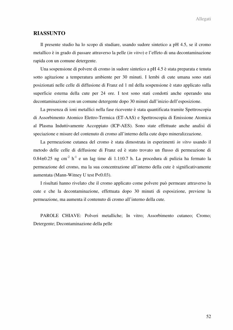
Allegati
52
RIASSUNTO
Il presente studio ha lo scopo di studiare, usando sudore sintetico a pH 4.5, se il cromo
metallico è in grado di passare attraverso la pelle (in vitro) e l’effeto di una decontaminazione
rapida con un comune detergente.
Una sospensione di polvere di cromo in sudore sintetico a pH 4.5 è stata preparata e tenuta
sotto agitazione a temperatura ambiente per 30 minuti. I lembi di cute umana sono stati
posizionati nelle celle di diffusione di Franz ed 1 ml della sospensione è stato applicato sulla
superficie esterna della cute per 24 ore. I test sono stati condotti anche operando una
decontaminazione con un comune detergente dopo 30 minuti dall’inizio dell’esposizione.
La presenza di ioni metallici nella fase ricevente è stata quantificata tramite Spettroscopia
di Assorbimento Atomico Elettro-Termica (ET-AAS) e Spettroscopia di Emissione Atomica
al Plasma Induttivamente Accoppiato (ICP-AES). Sono state effettuate anche analisi di
speciazione e misure del contenuto di cromo all’interno della cute dopo mineralizzazione.
La permeazione cutanea del cromo è stata dimostrata in esperimenti in vitro usando il
metodo delle celle di diffusione di Franz ed è stato trovato un flusso di permeazione di
0.84±0.25 ng cm-2
h-1
e un lag time di 1.1±0.7 h. La procedura di pulizia ha fermato la
permeazione del cromo, ma la sua concentrazione all’interno della cute è significativamente
aumentata (Mann-Witney U test P<0.03).
I risultati hanno rivelato che il cromo applicato come polvere può permeare attraverso la
cute e che la decontaminazione, effettuata dopo 30 minuti di esposizione, previene la
permeazione, ma aumenta il contenuto di cromo all’interno della cute.
PAROLE CHIAVE: Polveri metalliche; In vitro; Assorbimento cutaneo; Cromo;
Detergente; Decontaminazione della pelle
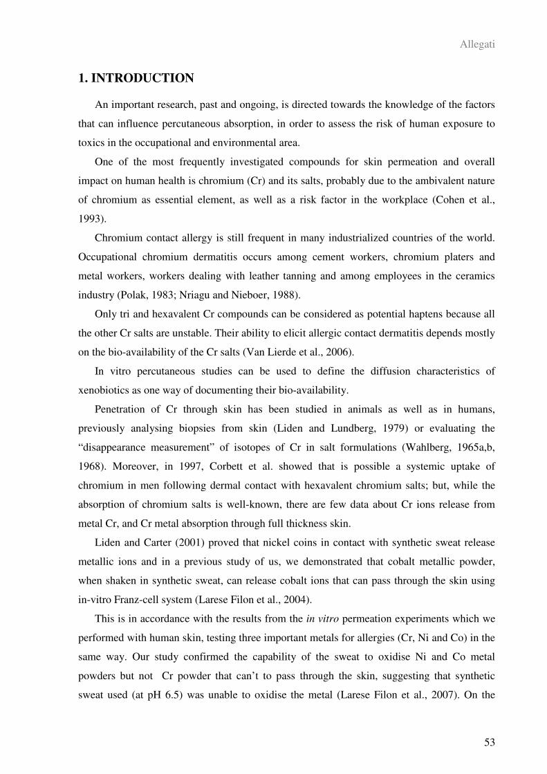
Allegati
53
1. INTRODUCTION
An important research, past and ongoing, is directed towards the knowledge of the factors
that can influence percutaneous absorption, in order to assess the risk of human exposure to
toxics in the occupational and environmental area.
One of the most frequently investigated compounds for skin permeation and overall
impact on human health is chromium (Cr) and its salts, probably due to the ambivalent nature
of chromium as essential element, as well as a risk factor in the workplace (Cohen et al.,
1993).
Chromium contact allergy is still frequent in many industrialized countries of the world.
Occupational chromium dermatitis occurs among cement workers, chromium platers and
metal workers, workers dealing with leather tanning and among employees in the ceramics
industry (Polak, 1983; Nriagu and Nieboer, 1988).
Only tri and hexavalent Cr compounds can be considered as potential haptens because all
the other Cr salts are unstable. Their ability to elicit allergic contact dermatitis depends mostly
on the bio-availability of the Cr salts (Van Lierde et al., 2006).
In vitro percutaneous studies can be used to define the diffusion characteristics of
xenobiotics as one way of documenting their bio-availability.
Penetration of Cr through skin has been studied in animals as well as in humans,
previously analysing biopsies from skin (Liden and Lundberg, 1979) or evaluating the
“disappearance measurement” of isotopes of Cr in salt formulations (Wahlberg, 1965a,b,
1968). Moreover, in 1997, Corbett et al. showed that is possible a systemic uptake of
chromium in men following dermal contact with hexavalent chromium salts; but, while the
absorption of chromium salts is well-known, there are few data about Cr ions release from
metal Cr, and Cr metal absorption through full thickness skin.
Liden and Carter (2001) proved that nickel coins in contact with synthetic sweat release
metallic ions and in a previous study of us, we demonstrated that cobalt metallic powder,
when shaken in synthetic sweat, can release cobalt ions that can pass through the skin using
in-vitro Franz-cell system (Larese Filon et al., 2004).
This is in accordance with the results from the in vitro permeation experiments which we
performed with human skin, testing three important metals for allergies (Cr, Ni and Co) in the
same way. Our study confirmed the capability of the sweat to oxidise Ni and Co metal
powders but not Cr powder that can’t to pass through the skin, suggesting that synthetic
sweat used (at pH 6.5) was unable to oxidise the metal (Larese Filon et al., 2007). On the
Allegati
54
contrary, after exposure of the skin to a donor solution containing Cr salts, Cr permeation was
very high (Gammelgaard et al., 1992; Corbett et al., 1997; Hostynek, 2003; Van Lierde et al.,
2006; Larese Filon et al., 2007)
The aims of this study were twofold: first to investigate, using a synthetic sweat at a lower
pH (4.5), whether metallic Cr can pass through full thickness skin and, second to evaluate the
effect of rapid skin decontamination using a common cleanser containing sodium lauryl
sulphate (SLS).
In occupational settings skin contamination is fairly common, and the cleaning procedure
not only may visibly remove most of the toxic agent, but also increase skin uptake by the
penetration-enhancing effect of the surfactants. For example, sodium lauryl sulphate can
increase the skin penetration of some toxic substances (Frankild et al., 1995) because it has a
destructive action on stratum corneum by changing its lipid composition (Ashton et al., 1986),
extracting lipids from the intercellular spaces in the stratum corneum (Imokava et al., 1989;
Froebe et al., 1990), and changing alpha–keratin into beta-keratin that causes swelling and
hydration of the stratum corneum (Scheuplein and Ross, 1970; Putterman et al., 1977). Larese
(2006) found that a decontamination procedure using sodium lauryl sulphate after 30 minutes
of application of lead oxide cause an increase of lead skin absorption.
The lack of data on skin absorption of Cr metal and the need for a better understanding of
the effect of decontamination agents led us to test this metal. To do so, experiments were
performed in in vitro Franz-cell system using human skin.
For our experiments we used the experience and the protocols employed during the
European project EDETOX (Evaluations and predictions of DErmal absorption of TOXic
chemicals), a three-year (2001-2004) research program funded by European Union
(EDETOX, 2000).
2. MATERIALS AND METHODS
2.1. Chemicals
All chemicals were analytical grade. Urea, magnesium nitrate hexahydrate, sodium
hydrogenphosphate, potassium dihydrogenphosphate, diphenylcarbazide, potassium
bichromate, hydrogen peroxide (30% v/v) were purchased from Carlo Erba (Milan, Italy);
ammonium hydroxide (25% w/v) was bought from J. T. Baker (Deventer, Holland); lactic
acid (90% v/v) from Acros Organics (Geel, Belgium); acetone, nitric acid (69% v/v) and
Allegati
55
sulfuric acid (95% v/v) from Sigma Aldrich (Milan, Italy).
Water reagent grade was produced with a Millipore purification pack system (milliQ
water).
Cr powder come from Alfa Aesar (Karlsruhe, Germany) and consisted of chromium
powder, APS (Average Particle Size) <10 micron, 99.8% purity (metals basis). The
physiological solution used as the receptor fluid was prepared by dissolving 2.38 g of
Na2HP04, 0.19 g of KH2P04 and 9 g of NaCl into 1 liter of milliQ water (final pH = 7.35).
The synthetic sweat solution used as the donor fluid consisted in 0.5% w/v sodium chloride,
0.1% w/v urea and 0.1% w/v lactic acid in milliQ water; pH was adjusted with ammonia (at
pH 3.5 or 4.5 or 5.5).
2.2. Dissolution tests in synthetic sweat
As preliminary test at the study of in vitro percutaneous absorption of chromium powder,
a research about the dissolution of chromium powder in synthetic sweat at three different pH
(3.5, 4.5 e 5.5) and at two different concentrations (0.5% w/v and 5% w/v) has been
performed in order to investigate the extent of ionization of this metal. To do so, 9 samples (3
for every different pH) of suspension of chromium powder in synthetic sweat for both
concentrations and other 3 samples with only synthetic sweat at pH 5.5 as blank has been
prepared. The volume of each suspension was 40 ml. Each suspension was put into a baker
and left under moderate agitation for 24 hours at room-temperature. After 8 and 24 hours, 10
ml were removed, centrifuged for 15 minutes at 3000 rpm and the surnatant filtered two times
through 0.45 m GHP Acrodisc syringe filters (Pall Life Sciences, Ann Arbor, MI) and
analysed by ICP-AES (see section 2.6.3) for the total chromium concentration and with UV-
vis spectrophotometry (see section 2.6.2) for the determination of Cr(VI).
2.3. Preparation of skin membranes
Human abdominal full thickness skin was obtained as surgical waste from at least 2
donors for each experiments. Donors were man and women within a wide range of age
(45÷71 years). Prior to freezing, subcutaneous fat was removed and hair shaved. All the
pieces of full thickness skin were stored in freezer at -25°C for a period up to, but not
exceeding, 4 months. It has been shown that this method of storage does not damage the skin
since no difference in permeability was observed between fresh and frozen segments of the
same skin in a separate series of experiments (Franz, 1975). Skin integrity was tested before
and after each experiment using electrical conductibility by means of a conductometer
Allegati
56
(Metrohm, 660 Conductometer, AG Oberdorfstr. 68 CH-9100 Herisau) operating at 300 Hz
connected to two stainless steel electrodes (Fasano et al., 2002). The electrical conductance
data, obtained in µS, were converted in K cm-2
. Cells with a resistance lower than 3.95±0.27
K cm-2
, were considered to be damaged as suggested by Davies et al. (2004).
2.4. In vitro diffusion system
Percutaneous absorption studies were performed using static diffusion cells following the
Franz Method (Franz, 1975). The receptor compartment has a mean volume of 14.0 ml and
was maintained at 32°C by means of circulation of termostated water in the jacket
surrounding the cell. This temperature value has been chosen in order to reproduce the hand’s
physiological temperature at normal conditions. The receptor fluid consisted of phosphate
buffered saline solution. The concentration of the salt in the receptor fluid is approximately
the same that can be found in the blood. The solution in each cell was continuously stirred
using a Teflon coated magnetic stirrer.
Each piece of skin was clamped between the donor and the receptor compartment; the
mean exposed skin area was 3.29 cm² and the membranes thickness was 1 mm.
The two experiments were performed as follows.
Experiment 1
At time 0, the exposure chambers of Franz diffusion cells were each filled with 1 ml of a
freshly made suspension of 2.5 g of Cr powder in 50 ml synthetic sweat at pH 4.5. Before
application, the suspension was shaken with a stirring plate at room temperature for 30
minutes. The metal was studied at infinite dose (as Cr powder).
At selected intervals (1, 2, 4, 8, 16, 18, 20 and 24 hours) 1.5 ml of the dermal bathing
solution was removed and analysed by means of ETAAS. Each receptor sample was
immediately replaced with an equal volume of fresh physiological solution.
At the end of the experiments, the dermal bathing solution were removed and total
chromium concentration was determined in each receiving chamber solution by means of
ETAAS, while the donor solutions were collected and the skin was washed with 3 ml of fresh
physiological solution for three times. The wash solutions were added to the donor solution
for the analysis.
The experiments were performed with 6 cells at the 8 selected times for a total of 48
samples. Two additional cells were considered as blank. In the experiments we used skin from
2 different donors.

Allegati
57
Experiment 2
At time 0, the exposure chambers of Franz diffusion cells were each filled with 1 ml of a
freshly made suspension of 2.5 g of Cr powder in 50 ml synthetic sweat at pH 4.5. Before
application, the suspension was shaken with a stirring plate at room temperature for 30
minutes. The metal was studied at infinite dose (as Cr powder). After 30 minutes, the donor
solution was removed using a syring and three cotton balls. The skin pieces were next
carefully cleaned with three cotton balls wetted with a cleanser containing sodium lauryl
sulphate and sodium laureth sulphate (Ivory soap, Procter & Gamble, Cincinnati, OHIO).
After the cleaning procedure the exposure chambers were filled with 1 ml of synthetic sweat
at pH 4.5. At selected intervals (1, 2, 4, 8, 16, 18, 20 and 24 hours) 1.5 ml of the dermal
bathing solution was removed and analysed. Each sample was immediately replaced with an
equal volume of fresh physiological solution. At the end of the experiments, the dermal
bathing solutions were removed and total chromium concentration was determined in each
receiving chamber solution by means of ETAAS, while the donor solutions were collected
and the skin was washed with 3 ml of fresh physiological solution for three times. The wash
solutions were added to the donor solution for the analysis.
The experiments were performed with 4 cells at the 8 selected times for a total of 32
samples.
2.5. Skin digestion after the experiment
After the experiments, the skin was stored in freezer at –25°C. At the time of the analysis,
the skin membranes were dried for 24 hours at room-temperature and then the exposed area
was cut into sections and put into bakers with 25 ml of HNO3 70% for digestion (amounts of
skin were between 0.5 and 0.8 g). Afterward solutions were agitated for 20 hours at 100°C
(after 2 hours they were added, drop by drop, of 5 ml of H2O2 till the remained solutions were
of few millilitres. Then solutions were diluted to 25 ml with milliQ water for the analysis by
ICP-AES.
2.6. Quantitative analysis
2.6.1. Donor and Receptor solutions
The donor solutions were collected at the end of the experiment, centrifuged for 15
minutes at 3000 rpm and the surnatant was filtrated two times through 0.45 m GHP Acrodisc
syringe filters (Pall Life Sciences, Ann Arbor, MI) and then analyzed.
The donor and the receptor solutions measurements were performed using electro-thermal
Allegati
58
atomic absorption spectrometry (ETAAS) with Zeeman background correction. A Thermo M
series AA spectrometer equipped with a GF95Z Zeeman Furnace and a FS95 Furnace
Autosampler (Thermo Electron Corporation, Cambridge, UK) were used for analyses.
Cr detection limit at the analytical wavelength of 357.9 nm was 0.4 µg L-1
. Calibration
curves were obtained using three different standards at 0.6, 3 and 6 µg L-1
. A 0.5% w/v
solution of Mg(NO3)2 was used as modifying matrix for Cr. The samples were analyzed
measuring against standard solutions for instrumental calibration. The precision of the
measurements as repeatability (RSD%) for the analysis was always less than 10%.
2.6.2. Analysis of Cr(VI) in dissolution tests
The speciation analysis of Cr in the dissolution tests was performed by UV-
spectrophotometry (Spectrophotometer Varian model DMS300) by means of
diphenylcarbazide method: 5 ml of the sample were added of 1 ml of 1.2 M sulphuric acid
and 50 µl of a solution obtained dissolving 0.25 g of 1,5-diphenylcarbazide into 50 ml of
acetone, waiting for the development of the colour for 10 minutes and then measuring the
absorbance at 540 nm. The determination was performed by calibration curve obtained with
six standard solutions of Cr(VI) at known concentrations (0, 0.05, 0.25, 0.50, 1, 2 mg L-1
)
obtained by dilution of a solution of K2Cr2O7 at 1000 mg L-1
prepared dissolving 2.828 g of
pure K2Cr2O7 (oven-dried) in one liter of 1% v/v HNO3 using a calibrated flask. The limit of
detection (LOD) was 0.03 mg L-1
of Cr(VI).
2.6.3. Analysis of skin and of dissolution test solutions
Solutions obtained from the skin digestion procedure and from the dissolution tests were
analysed by ICP-AES using a Spectroflame Modula E optical plasma interface (OPI)
instrument (by SPECTRO, Germany) for determine the total Cr content using calibration
curves (range: 0-10 mg L-1
) obtained by dilution of Spectrascan Chromium standard solution
for ICP-AES analyses (by Teknolab A/S, Norway). The limit of detection (LOD) at the
operative wavelength of 267.716 was 0.010 mg L-1
. The precision of the measurements as
repeatability (RSD%) for the analysis was always less than 5%.
2.7. Data analysis
The concentration data ( µg cm-3
) of metals in the receptor solution were converted to the
total amount that penetrated (µg cm-2
), with a correction for dilution due to sample removal.
To evaluate better the increase of Cr in donor solution the value of the cell at time 1 h was
subtracted from the values obtained at the other times and then plotted against time. The slope
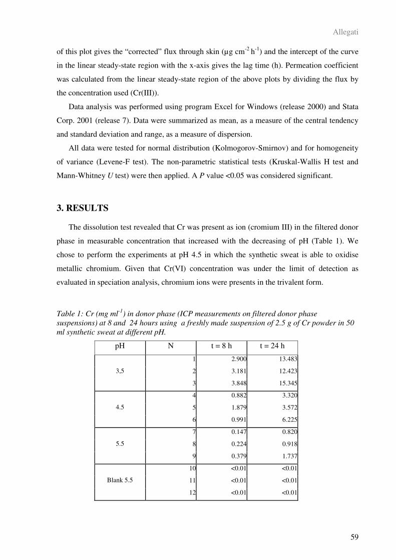
Allegati
59
of this plot gives the “corrected” flux through skin (µg cm-2
h-1
) and the intercept of the curve
in the linear steady-state region with the x-axis gives the lag time (h). Permeation coefficient
was calculated from the linear steady-state region of the above plots by dividing the flux by
the concentration used (Cr(III)).
Data analysis was performed using program Excel for Windows (release 2000) and Stata
Corp. 2001 (release 7). Data were summarized as mean, as a measure of the central tendency
and standard deviation and range, as a measure of dispersion.
All data were tested for normal distribution (Kolmogorov-Smirnov) and for homogeneity
of variance (Levene-F test). The non-parametric statistical tests (Kruskal-Wallis H test and
Mann-Whitney U test) were then applied. A P value <0.05 was considered significant.
3. RESULTS
The dissolution test revealed that Cr was present as ion (cromium III) in the filtered donor
phase in measurable concentration that increased with the decreasing of pH (Table 1). We
chose to perform the experiments at pH 4.5 in which the synthetic sweat is able to oxidise
metallic chromium. Given that Cr(VI) concentration was under the limit of detection as
evaluated in speciation analysis, chromium ions were presents in the trivalent form.
Table 1: Cr (mg ml-1
) in donor phase (ICP measurements on filtered donor phase
suspensions) at 8 and 24 hours using a freshly made suspension of 2.5 g of Cr powder in 50
ml synthetic sweat at different pH.
pH N t = 8 h t = 24 h
1 2.900 13.483
2 3.181 12.4233,5
3 3.848 15.345
4 0.882 3.320
5 1.879 3.5724.5
6 0.991 6.225
7 0.147 0.820
8 0.224 0.9185.5
9 0.379 1.737
10 <0.01 <0.01
11 <0.01 <0.01Blank 5.5
12 <0.01 <0.01
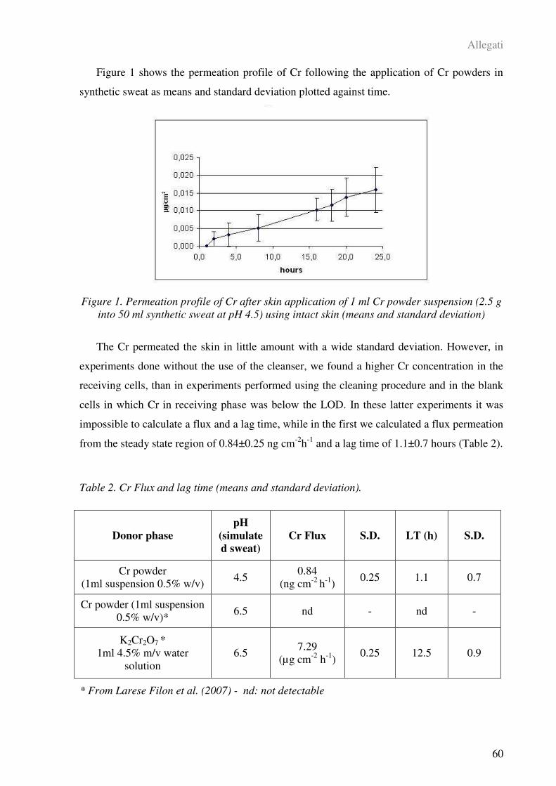
Allegati
60
Figure 1 shows the permeation profile of Cr following the application of Cr powders in
synthetic sweat as means and standard deviation plotted against time.
Figure 1. Permeation profile of Cr after skin application of 1 ml Cr powder suspension (2.5 g
into 50 ml synthetic sweat at pH 4.5) using intact skin (means and standard deviation)
The Cr permeated the skin in little amount with a wide standard deviation. However, in
experiments done without the use of the cleanser, we found a higher Cr concentration in the
receiving cells, than in experiments performed using the cleaning procedure and in the blank
cells in which Cr in receiving phase was below the LOD. In these latter experiments it was
impossible to calculate a flux and a lag time, while in the first we calculated a flux permeation
from the steady state region of 0.84±0.25 ng cm-2
h-1
and a lag time of 1.1±0.7 hours (Table 2).
Table 2. Cr Flux and lag time (means and standard deviation).
* From Larese Filon et al. (2007) - nd: not detectable
Donor phase
pH
(simulate
d sweat)
Cr Flux S.D. LT (h) S.D.
Cr powder
(1ml suspension 0.5% w/v) 4.5
0.84
(ng cm-2
h-1
) 0.25 1.1 0.7
Cr powder (1ml suspension
0.5% w/v)* 6.5 nd - nd -
K2Cr2O7 *
1ml 4.5% m/v water
solution
6.5 7.29
(µg cm-2
h-1
) 0.25 12.5 0.9
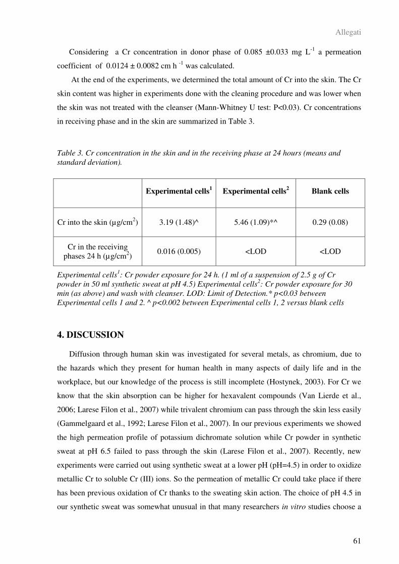
Allegati
61
Considering a Cr concentration in donor phase of 0.085 ±0.033 mg L-1
a permeation
coefficient of 0.0124 ± 0.0082 cm h -1
was calculated.
At the end of the experiments, we determined the total amount of Cr into the skin. The Cr
skin content was higher in experiments done with the cleaning procedure and was lower when
the skin was not treated with the cleanser (Mann-Whitney U test: P<0.03). Cr concentrations
in receiving phase and in the skin are summarized in Table 3.
Table 3. Cr concentration in the skin and in the receiving phase at 24 hours (means and
standard deviation).
Experimental cells1: Cr powder exposure for 24 h. (1 ml of a suspension of 2.5 g of Cr
powder in 50 ml synthetic sweat at pH 4.5) Experimental cells2: Cr powder exposure for 30
min (as above) and wash with cleanser. LOD: Limit of Detection.* p<0.03 between
Experimental cells 1 and 2. ^ p<0.002 between Experimental cells 1, 2 versus blank cells
4. DISCUSSION
Diffusion through human skin was investigated for several metals, as chromium, due to
the hazards which they present for human health in many aspects of daily life and in the
workplace, but our knowledge of the process is still incomplete (Hostynek, 2003). For Cr we
know that the skin absorption can be higher for hexavalent compounds (Van Lierde et al.,
2006; Larese Filon et al., 2007) while trivalent chromium can pass through the skin less easily
(Gammelgaard et al., 1992; Larese Filon et al., 2007). In our previous experiments we showed
the high permeation profile of potassium dichromate solution while Cr powder in synthetic
sweat at pH 6.5 failed to pass through the skin (Larese Filon et al., 2007). Recently, new
experiments were carried out using synthetic sweat at a lower pH (pH=4.5) in order to oxidize
metallic Cr to soluble Cr (III) ions. So the permeation of metallic Cr could take place if there
has been previous oxidation of Cr thanks to the sweating skin action. The choice of pH 4.5 in
our synthetic sweat was somewhat unusual in that many researchers in vitro studies choose a
Experimental cells1 Experimental cells
2 Blank cells
Cr into the skin (µg/cm2) 3.19 (1.48)^ 5.46 (1.09)*^ 0.29 (0.08)
Cr in the receiving
phases 24 h (µg/cm2)
0.016 (0.005) <LOD <LOD
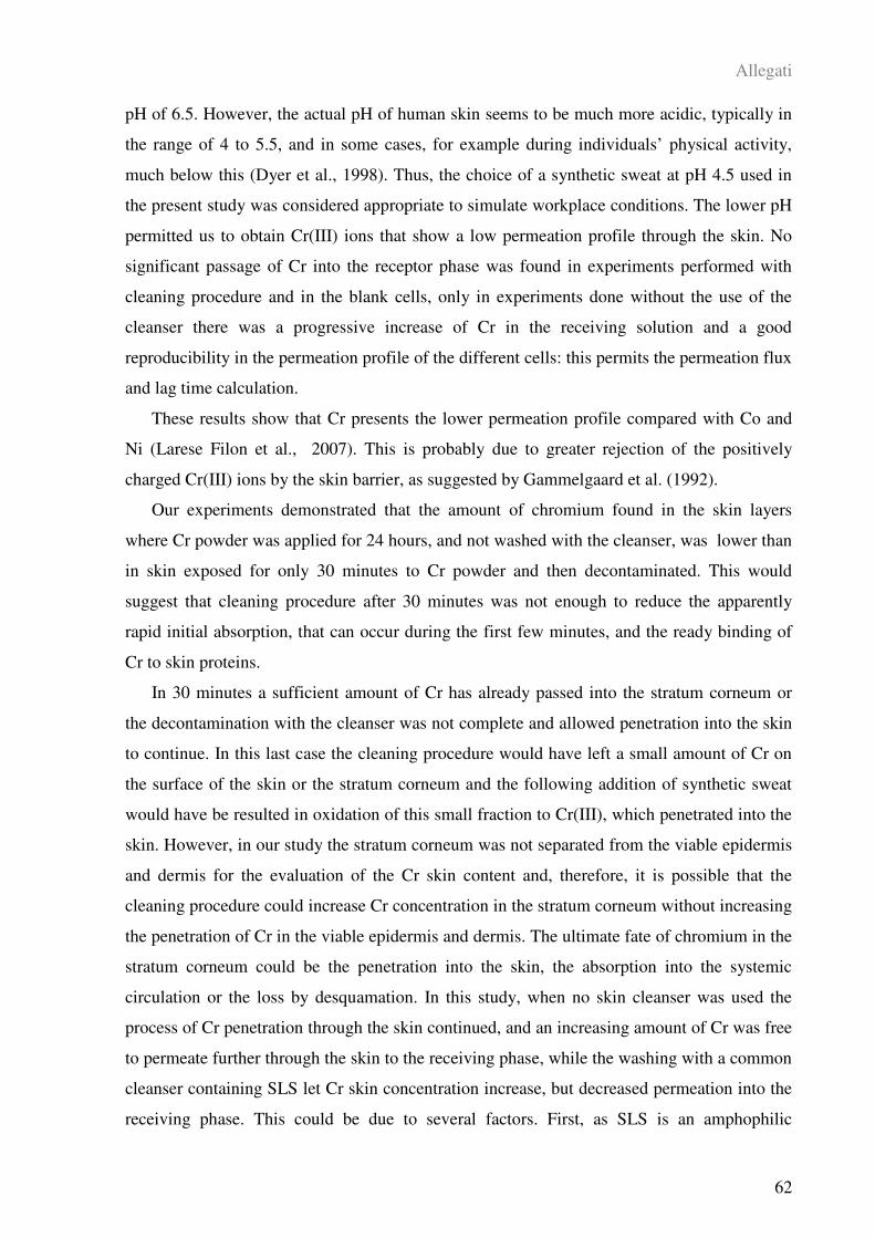
Allegati
62
pH of 6.5. However, the actual pH of human skin seems to be much more acidic, typically in
the range of 4 to 5.5, and in some cases, for example during individuals’ physical activity,
much below this (Dyer et al., 1998). Thus, the choice of a synthetic sweat at pH 4.5 used in
the present study was considered appropriate to simulate workplace conditions. The lower pH
permitted us to obtain Cr(III) ions that show a low permeation profile through the skin. No
significant passage of Cr into the receptor phase was found in experiments performed with
cleaning procedure and in the blank cells, only in experiments done without the use of the
cleanser there was a progressive increase of Cr in the receiving solution and a good
reproducibility in the permeation profile of the different cells: this permits the permeation flux
and lag time calculation.
These results show that Cr presents the lower permeation profile compared with Co and
Ni (Larese Filon et al., 2007). This is probably due to greater rejection of the positively
charged Cr(III) ions by the skin barrier, as suggested by Gammelgaard et al. (1992).
Our experiments demonstrated that the amount of chromium found in the skin layers
where Cr powder was applied for 24 hours, and not washed with the cleanser, was lower than
in skin exposed for only 30 minutes to Cr powder and then decontaminated. This would
suggest that cleaning procedure after 30 minutes was not enough to reduce the apparently
rapid initial absorption, that can occur during the first few minutes, and the ready binding of
Cr to skin proteins.
In 30 minutes a sufficient amount of Cr has already passed into the stratum corneum or
the decontamination with the cleanser was not complete and allowed penetration into the skin
to continue. In this last case the cleaning procedure would have left a small amount of Cr on
the surface of the skin or the stratum corneum and the following addition of synthetic sweat
would have be resulted in oxidation of this small fraction to Cr(III), which penetrated into the
skin. However, in our study the stratum corneum was not separated from the viable epidermis
and dermis for the evaluation of the Cr skin content and, therefore, it is possible that the
cleaning procedure could increase Cr concentration in the stratum corneum without increasing
the penetration of Cr in the viable epidermis and dermis. The ultimate fate of chromium in the
stratum corneum could be the penetration into the skin, the absorption into the systemic
circulation or the loss by desquamation. In this study, when no skin cleanser was used the
process of Cr penetration through the skin continued, and an increasing amount of Cr was free
to permeate further through the skin to the receiving phase, while the washing with a common
cleanser containing SLS let Cr skin concentration increase, but decreased permeation into the
receiving phase. This could be due to several factors. First, as SLS is an amphophilic
Allegati
63
molecule and forms aggregates known as anionic micelles that do not diffuse through the skin
membranes, it is possible that Cr is retained in the skin in association with SLS micelles.
Second, SLS, which is an alkaline detergent, could have led to a change of the pH of the
stratum corneum and in that way would have influenced the permeation of Cr through the
skin. Samitz et al. (1969) found a pH dependent diffusion of Cr(III), with the largest diffusion
at pH 7 and decreased diffusion at pH 5 and 9. At last, SLS could damage the barrier
properties of the skin and increase binding of Cr to skin proteins by direct action on the skin.
The detergent properties of SLS could have been responsible for extracting some components
of the stratum corneum, most likely proteins of the corneocyte envelope, disorganizing the
lipidic matrix that cements them, opening routes of penetration within the stratum corneum
and thus increasing the amount of Cr bound to skin proteins, due to the affinity of Cr for
proteins. It has been reported that sodium lauryl sulphate can increase nickel penetration by
reducing the barrier integrity of the skin (Frankield et al., 1995) and, in our previous work, we
demonstrated that lead skin penetration increased significantly when a cleanser, containing
sodium lauryl sulphate, was tested (Larese Filon et al., 2006).
Moreover, it is well known that the electrophilic nature of many metals, for example
chromium (III), determines their protein reactivity, which can result in depot formation in the
stratum corneum (Samitz and Katz, 1976). Such protein-metal binding can take place in all
strata of the skin to the extent of building up a secondary barrier and inhibiting further
diffusion (Hostynek, 2003).
In vitro experiments demonstrated that Cr(III) ions show a strong affinity for epithelial
and dermal tissues, forming stable complexes which slow the rate of diffusion (Samitz et al.,
1969). It has also been speculated that Cr(III) is the actual hapten and that Cr(VI), after
entering the skin cells, is reduced to Cr(III) which in its turn binds onto intracellular proteins,
thereby forming immunogenic complexes (Van Lierde et al., 2006).
In conclusion, although Cr metal presents a low risk of absorption, these data suggest that
is necessary to prevent skin contamination and to choose carefully the decontamination
measures that are provided to workers. This may be important for chromium sensitive
workers exposed to detergents in daily-cleaning situations.
However, it is an in vitro skin model with its inherent limitations. On the one hand,
several assumptions and conditions used in this in vitro protocol that might contribute to an
upward bias in percutaneous penetration, might include a prolonged hydration of the stratum
corneum with sweat and the Cr amount applied. On the other hand, workers are likely to wash
more than once per day and often have mechanically damaged which the evidence suggests
Allegati
64
would make penetration worse. Our risk analysis did not include these latter factors that
would therefore lead to an underestimate of risk (Larese Filon et al., 2006).
5. REFERENCES
Ashton P, Hadgraft J, Walters K (1986) Effect of surfactants in percutaneous absorption.
Pharm. Acta Helv. 8: 228-234.
Cohen MD, Kargacin B, Klein CB, Costa M (1993) Mechanisms of chromium
carcinogenicity and toxicity. Crit. Rev. Toxicol. 23: 255-281.
Corbett GE, Finley BL, Paustenbach DJ, Kerge BD (1997) Systemic uptake of chromium
in human volunteers following dermal contact with hexavalent chromium (22mg/dL). J.
Exposure Anal. Environ. Epidemiol. 7: 179-189.
EDETOX, 2000. Evaluations and Predictions of Dermal Absorption of Toxic Chemicals,
EU Framework V: Quality of Life, Environment and Health Key Action Funding (Project
Number: QLKA-2000-00196).
EEC. European Committee for Standardization, European Standard EN 1811:1998
Reference method for release of nickel from products. Central Secretariat, Brussels.
Davies DJ, Ward RJ, Heylings JR (2004) Multi-species assessment of electrical resistence
as a skin integrity marker for in vivo percutaneous absorption studies. Toxicol. in vitro 18:
351-358.
Dyer J, Scrivner N, Dentel S (1998) A pratical guide for determining the solubility of
metal hydroxides and oxides in water. Environ. Progr. 17: 1-8.
Fasano W, Manning L, Green J (2002) Rapid assessment of rat and human epidermal
membranes for in vitro dermal regulatory testing: correlation of electrical resistence with
tritiated water permeability. Toxicol. in vitro 16: 731-740.
Frankild S, Andersen K, Nielsen G (1995) Effect of sodium lauryl sulphate (SL) on in
vitro percutaneous penetration of water, hydrocortisone and nickel. Cont. Dermat. 32: 338-
345.
Franz TJ (1975) On the relevance of in vitro data. J. Invest. Dermatol. 93: 633-640.
Froebe C, Simion F, Rhein L, Cagan R, Kligman A (1990) Stratum corneum lipid removal
by surfactants: relation to in vivo irritation. Dermatologica 181: 277-283.
Allegati
65
Gammelgaard B, Fullerton A, Avastorp C, Menné T (1992) Permeation of chromium salts
through human skin in vitro. Cont. dermat. 27: 302-310.
Hostynek JJ (2003) Factors determining percutaneous metal absorption. Food
Chem.Toxicol. 41: 327-345.
Imokawa G, Akasaki S, Minematsu Y (1989) Importance of intercellular lipids in water-
retention properties of the stratum corneum: induction and recovery of surfactants dry skin.
Arch. Dermatol. Res. 281: 45-51.
Larese Filon F, Maina G, Adami G, Venier M, Coceani N, Bussani R, Massiccio M,
Barbieri P, Spinelli P (2004) In vitro percutaneous absorption of cobalt. Int. Arch. Environ.
Health 77: 85-89.
Larese Filon F, Boeninger M, Maina G, Adami G, Spinelli P, Damian A (2006) Skin
absorption of inorganic lead and the effects of skin cleansers. J. Occup. Environ. Med. 48:
692-699.
Larese Filon F, Adami G, Venier M, Maina G, Renzi N (2007) In vitro percutaneous
absorption of metal compounds. Toxicol. Lett. 170: 49-56.
Liden S and Lundberg E (1979) Penetration of chromium in intact human skin in vivo. J.
Invest. Dermatol. 72: 42-45.
Liden C and Carter S (2001) Nickel release from coins. Contact dermatitis 44: 160-165.
Nriagu JO and Nieboer E (1988) Chromium in the natural and human environment.
Wiley, New York.
Polak L (1983) Immunology of chromium. In : Burrows D (ed). Chromium : metabolism
and toxicity. CRC, Boca Raton , FL.
Putterman G, Wolejsza N, Wolfram M (1977) The effect of detergents on swelling in
stratum corneum. J. Soc. Cosmet. Chem. 9: 521-532.
Samitz MH, Katz SA, Schreiner DM, Gross PR (1969) Chromium-protein interactions.
Acta Dermato-Venereol. 49: 142-146.
Samitz MH and Katz SA (1976) Nickel-epidermal interactions: diffusion and binding.
Environ. Research 11: 34-39.
Scheuplein R and Ross L (1970) Effects of surfactants and solvents on the permeability of
epidermidis. J. Soc. Cosmet. Chem. 21: 853-857.
Allegati
66
Van Lierde V, Chery C, Roche N, Monstrey S, Moens L, Vanhaecke F (2006) In vitro
permeation of chromium species through porcine and human skin as determined by capillary
electrophoresis-inductively coupled plasma-sector field mass spectrometry. Anal. Bioanal.
Chem. 384: 378-384.
Wahlberg JE (1965a) Disappearance measurements, a method for studying percutaneous
absorption of istotote-labelled compounds emitting gamma-rays. Acta Dermato-Venereol. 45:
397-414.
Wahlberg JE (1965b) Percutaneous absorption of sodium chromate (51Cr), cobaltous (58
Co) and mercuric (203Hg) chlorides through excised human and guinea pig skin. Acta
Dermato-Venereol. 45: 415-426.
Wahlberg JE (1968) Percutaneous absorption from chromium (51Cr) solutions of different
pH, 1.4-12.8. An experimental study in the guinea pig. Dermatol. 137: 17-25.
Allegati
67
ALLEGATO II
IN VITRO ABSORPTION OF METAL POWDERS THROUGH INTACT AND
DAMAGED HUMAN SKIN
Francesca Larese Filon, Flavia D’Agostin, Matteo Crosera, Gianpiero Adami, Massimo
Bovenzi, Giovanni Maina
Pubblicato in: Toxicology in vitro, 23 (2009) 574–579
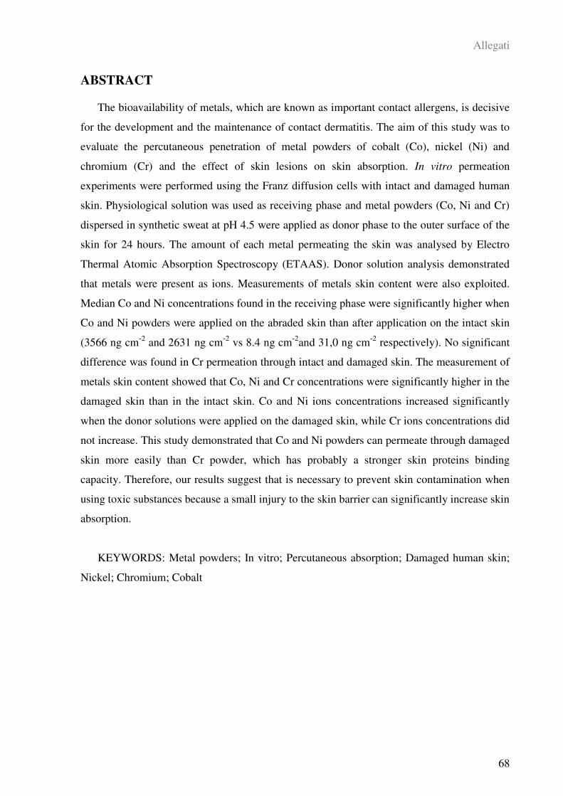
Allegati
68
ABSTRACT
The bioavailability of metals, which are known as important contact allergens, is decisive
for the development and the maintenance of contact dermatitis. The aim of this study was to
evaluate the percutaneous penetration of metal powders of cobalt (Co), nickel (Ni) and
chromium (Cr) and the effect of skin lesions on skin absorption. In vitro permeation
experiments were performed using the Franz diffusion cells with intact and damaged human
skin. Physiological solution was used as receiving phase and metal powders (Co, Ni and Cr)
dispersed in synthetic sweat at pH 4.5 were applied as donor phase to the outer surface of the
skin for 24 hours. The amount of each metal permeating the skin was analysed by Electro
Thermal Atomic Absorption Spectroscopy (ETAAS). Donor solution analysis demonstrated
that metals were present as ions. Measurements of metals skin content were also exploited.
Median Co and Ni concentrations found in the receiving phase were significantly higher when
Co and Ni powders were applied on the abraded skin than after application on the intact skin
(3566 ng cm-2
and 2631 ng cm-2
vs 8.4 ng cm-2
and 31,0 ng cm-2
respectively). No significant
difference was found in Cr permeation through intact and damaged skin. The measurement of
metals skin content showed that Co, Ni and Cr concentrations were significantly higher in the
damaged skin than in the intact skin. Co and Ni ions concentrations increased significantly
when the donor solutions were applied on the damaged skin, while Cr ions concentrations did
not increase. This study demonstrated that Co and Ni powders can permeate through damaged
skin more easily than Cr powder, which has probably a stronger skin proteins binding
capacity. Therefore, our results suggest that is necessary to prevent skin contamination when
using toxic substances because a small injury to the skin barrier can significantly increase skin
absorption.
KEYWORDS: Metal powders; In vitro; Percutaneous absorption; Damaged human skin;
Nickel; Chromium; Cobalt

Allegati
69
RIASSUNTO
La biodisponibilità dei metalli, riconosciuti come importanti allergeni da contatto, è
decisiva per lo sviluppo e il mantenimento delle dermatiti da contatto. Lo scopo di questo
studio era la valutazione della permeazione cutanea di polveri metalliche di cobalto (Co),
nichel (Ni) e cromo (Cr) e l’effetto delle lesioni della pelle sull’assorbibimento cutaneo. Gli
esperimenti di permeazione in vitro sono stati condotti usando le celle di diffusione di Franz
con cute umana intera, integra e lesa. Come soluzione ricevente è stata usata soluzione
fisiologica e come fasi donatrici sono state applicate alla superficie esterna della cute delle
polveri di metalli (Co, Ni e Cr) disperse in sudore sintetico a pH 4,5 per 24 ore. Le quantità di
ogni metallo che hanno permeato la pelle sono state analizzate tramite Spettroscopia di
Assorbimento Atomico Elettro-Termica (ETAAS).
Le analisi delle soluzioni donatrici hanno dimostrato che i metalli erano presenti come
ioni. Inoltre è stato analizzato, dopo mineralizzazione, il contenuto di metalli anche all’interno
della cute. Le concentrazioni mediane di Co e Ni trovate nelle fasi riceventi erano
significativamente più alte quando le polveri di Co e Ni sono state applicate nella cute abrasa
rispetto alla cute integra (rispettivamente 3566 ng cm-2
e 2631 ng cm-2
vs 8.4 ng cm-2
e 31 ng
cm-2
). Nessuna differenza significativa tra cute integra e cute lesa, invece, è stata trovata nella
permeazione del Cr. Le analisi dei metalli rimasti nella pelle ha evidenziato che le
concentrazioni di Co, Ni e Cr erano significativamente più alte nella cute danneggiata rispetto
alla cute integra.
Infine, la concentrazione di ioni di Co e Ni aumentavano significativamente quando le fasi
donatrici erano applicate alla cute danneggiata rispetto alla cute integra, mentre non vi erano
differenze per quanto riguarda la ionizzazione del Cr.
Questo studio ha mostrato che le polveri di Co e di Ni possono permeare la pelle più
facilmente rispetto alla polvere di Cr, il quale probabilmente viene legato in maniera più forte
dalle proteine della cute. Quindi, i nostri risultati suggeriscono che è necessario prevenire la
contaminazione della cute quando si usano sostanze tossiche poiché anche piccole lesioni
della pelle possono aumentare significativamente l’assorbimento cutaneo.
PAROLE CHIAVE: Polveri metalliche; In vitro; Assorbimento cutaneo; Cute umana lesa;
Nichel; Cromo; Cobalto
Allegati
70
1. INTRODUCTION
In industrialized countries, skin is at high risk of exposure to chemicals and other
contaminants which can be found in the environment or at a workplace. Allergic contact
dermatitis affects approximately 1-10% of the general population (Guin, 1995). Nickel (Ni),
cobalt (Co) and chromium (Cr) are recognized as the most common cause of contact allergy
affecting 10-15% of women and a few % of men in the general population. Nickel allergy, in
particular, is the most important background factor for hand eczema, which occurs in 10% of
adult people (Meding and Swanbeck, 1987). A wide variety of metal objects that come into
repetitive contact with the skin can release metallic ions that can diffuse through the skin and
cause allergy. A limited number of experimental studies suggest that, under physiologically
relevant conditions, metals may ionize and so permeate the skin. Liden and Carter (2001)
found that euro coins in contact with synthetic sweat release nickel ions. Larese Filon et al.
(2004, 2007), using in vitro Franz cell system, demonstrated that cobalt and nickel powders,
when stirred in synthetic sweat, can release metallic ions that can pass through the skin.
Skin absorption of Co has been found in exposed workers and in laboratory experiments
with volunteers, but there were few in vitro experimental data on skin absorption of this metal
(Suzuki-Yasumoto and Inaba, 1976; Carson et al., 1986; Kogan and Petukhova, 1986;
Scansetti et al., 1994; Linnainmaa and Kiilunen, 1997; Larese Filon et al., 2004). It is known
that Co is a common sensitizer and its presence in metal objects can increase the risk of
allergic contact dermatitis in sensitised people. In this latter there is a local action on the skin,
but the findings by Scansetti et al. (1994) and Linnainmaa and Kiilunen (1997) suggest that
this metal can permeate the skin. Studying hard metal industry workers, no correlation
between Co in ambient air and Co urine concentrations was found by Scansetti et al. (1994).
This result was attributed to skin contact as proved by an experiment involving voluntary skin
exposure to freshly mixed or waste powder containing 5-15% of Co that identified a ten fold
increase of urinary cobalt in the post exposure samples. This finding is consistent with the
experiment performed by Linnainmaa and Kiilunen (1997) who observed an increase of
urinary Co 4 to 6 hours following skin exposure to Co powder in 5 volunteers.
More extensive studies of nickel skin absorption have been undertaken. It was found that
in vitro water solution of nickel salts can pass through the stratum corneum (Tanojo et al.,
2001), and in vivo nickel powder can penetrate in depth profiles of the stratum corneum after
occlusion (Hostynek, 2001). These results are consistent with earlier experiments with full
Allegati
71
thickness human skin in vitro, suggesting that in vivo nickel ions may permeate (Tanojo et al.,
2001). There are, however, a shortage of experimental data using nickel powder (Larese Filon
et al., 2007).
Penetration of Cr through skin has been investigated in animals as well as in human skin
biopsy specimens (Liden and Lundberg, 1979) for evaluating the “disappearance
measurement” of isotopes of Cr in salt formulations (Wahlberg 1965a, 1965b, 1968).
Moreover, Corbett et al. (1997) observed a systemic uptake of chromium in men following
dermal contact with hexavalent chromium salts. While the absorption of chromium salts in
vitro experiments has been widely demonstrated (Gammlgaard et al., 1992; Van Lierde et al.,
2006; Larese Filon et al., 2007), little is known about Cr ions release from metal Cr and skin
absorption of chromium in metal form. Our recent study performed with full thickness human
skin in diffusion cells, showed that Cr powder in synthetic sweat at a low pH (pH 4.5) is able
to permeate the skin (D’Agostin et al., 2007).
In order to better understand bioavailability of metals involved in skin sensitisation (Ni,
Cr, Co), in this study we evaluated their skin permeation in an in vitro diffusion cell system.
In vitro methods are particularly useful for measuring dermal absorption of chemicals in
industrial settings (e.g. cosmetic, detergent and agrochemical). Therefore, in some countries
they are included among the criteria for assignment of a skin notation to chemicals (Drexler,
1998). The technical standardisation procedure has been reached on, although there is still
some conflict over which guidelines to adopt (e.g., Organisation for Economic Co-operation
and Development, 2000 vs ACGIH, American Conference of Governmental Industrial
Hygienists, 1995). On the contrary, in vivo animal studies pose a lot of problems when
extrapolating the results to humans (Poet, 2000), and theoretical models (Fiserova-Bergerova
et al., 1990) tend to overestimate potential dermal exposure in occupational workplaces.
In this study, experiments were performed using intact skin as well as damaged skin to
estimate the effect of skin lesions on skin absorption. Metallic powders were tested in the
same way with the purpose of comparing the different ability of the three metals to permeate
through human skin. To carry out the experiments, we used the experience and the protocols
employed during the European project EDETOX (Evaluations and predictions of DErmal
absorption of TOXic chemicals), a three-year research program (2001-2004) funded by
European Union (EDETOX, 2000).
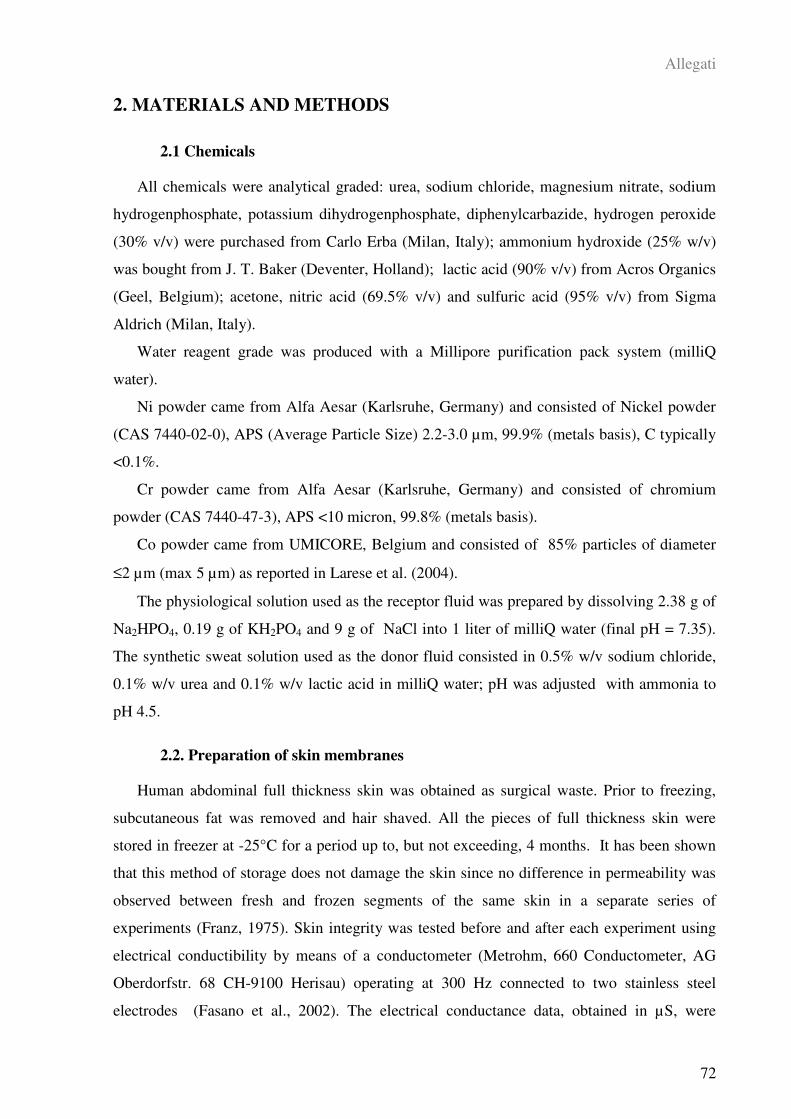
Allegati
72
2. MATERIALS AND METHODS
2.1 Chemicals
All chemicals were analytical graded: urea, sodium chloride, magnesium nitrate, sodium
hydrogenphosphate, potassium dihydrogenphosphate, diphenylcarbazide, hydrogen peroxide
(30% v/v) were purchased from Carlo Erba (Milan, Italy); ammonium hydroxide (25% w/v)
was bought from J. T. Baker (Deventer, Holland); lactic acid (90% v/v) from Acros Organics
(Geel, Belgium); acetone, nitric acid (69.5% v/v) and sulfuric acid (95% v/v) from Sigma
Aldrich (Milan, Italy).
Water reagent grade was produced with a Millipore purification pack system (milliQ
water).
Ni powder came from Alfa Aesar (Karlsruhe, Germany) and consisted of Nickel powder
(CAS 7440-02-0), APS (Average Particle Size) 2.2-3.0 µm, 99.9% (metals basis), C typically
<0.1%.
Cr powder came from Alfa Aesar (Karlsruhe, Germany) and consisted of chromium
powder (CAS 7440-47-3), APS <10 micron, 99.8% (metals basis).
Co powder came from UMICORE, Belgium and consisted of 85% particles of diameter
≤2 µm (max 5 µm) as reported in Larese et al. (2004).
The physiological solution used as the receptor fluid was prepared by dissolving 2.38 g of
Na2HPO4, 0.19 g of KH2PO4 and 9 g of NaCl into 1 liter of milliQ water (final pH = 7.35).
The synthetic sweat solution used as the donor fluid consisted in 0.5% w/v sodium chloride,
0.1% w/v urea and 0.1% w/v lactic acid in milliQ water; pH was adjusted with ammonia to
pH 4.5.
2.2. Preparation of skin membranes
Human abdominal full thickness skin was obtained as surgical waste. Prior to freezing,
subcutaneous fat was removed and hair shaved. All the pieces of full thickness skin were
stored in freezer at -25°C for a period up to, but not exceeding, 4 months. It has been shown
that this method of storage does not damage the skin since no difference in permeability was
observed between fresh and frozen segments of the same skin in a separate series of
experiments (Franz, 1975). Skin integrity was tested before and after each experiment using
electrical conductibility by means of a conductometer (Metrohm, 660 Conductometer, AG
Oberdorfstr. 68 CH-9100 Herisau) operating at 300 Hz connected to two stainless steel
electrodes (Fasano et al., 2002). The electrical conductance data, obtained in µS, were
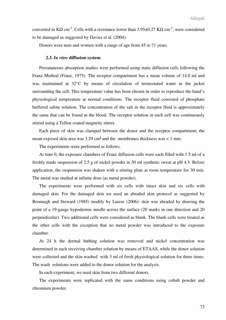
Allegati
73
converted in K cm-2
. Cells with a resistance lower than 3.95±0.27 K cm-2
, were considered
to be damaged as suggested by Davies et al. (2004).
Donors were men and women with a range of age from 45 to 71 years.
2.3. In vitro diffusion system
Percutaneous absorption studies were performed using static diffusion cells following the
Franz Method (Franz, 1975). The receptor compartment has a mean volume of 14.0 ml and
was maintained at 32°C by means of circulation of termostated water in the jacket
surrounding the cell. This temperature value has been chosen in order to reproduce the hand’s
physiological temperature at normal conditions. The receptor fluid consisted of phosphate
buffered saline solution. The concentration of the salt in the receptor fluid is approximately
the same that can be found in the blood. The receptor solution in each cell was continuously
stirred using a Teflon coated magnetic stirrer.
Each piece of skin was clamped between the donor and the receptor compartment; the
mean exposed skin area was 3.29 cm² and the membranes thickness was < 1 mm.
The experiments were performed as follows.
At time 0, the exposure chambers of Franz diffusion cells were each filled with 1.5 ml of a
freshly made suspension of 2.5 g of nickel powder in 50 ml synthetic sweat at pH 4.5. Before
application, the suspension was shaken with a stirring plate at room temperature for 30 min.
The metal was studied at infinite dose (as metal powder).
The experiments were performed with six cells with intact skin and six cells with
damaged skin. For the damaged skin we used an abraded skin protocol as suggested by
Bronaugh and Steward (1985) modify by Larese (2006): skin was abraded by drawing the
point of a 19-gauge hypodermic needle across the surface (20 marks in one direction and 20
perpendicular). Two additional cells were considered as blank. The blank cells were treated as
the other cells with the exception that no metal powder was introduced to the exposure
chamber.
At 24 h the dermal bathing solution was removed and nickel concentration was
determined in each receiving chamber solution by means of ETAAS, while the donor solution
were collected and the skin washed with 3 ml of fresh physiological solution for three times.
The wash solutions were added to the donor solution for the analysis.
In each experiment, we used skin from two different donors.
The experiments were replicated with the same conditions using cobalt powder and
chromium powder.
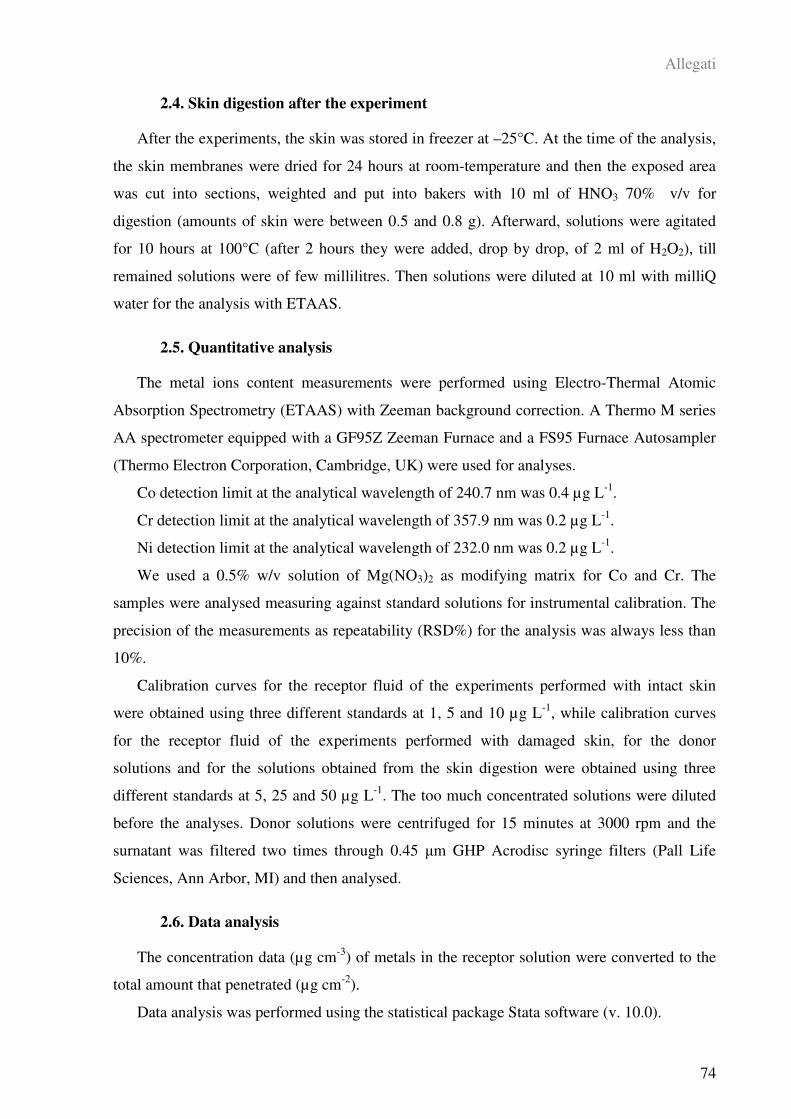
Allegati
74
2.4. Skin digestion after the experiment
After the experiments, the skin was stored in freezer at –25°C. At the time of the analysis,
the skin membranes were dried for 24 hours at room-temperature and then the exposed area
was cut into sections, weighted and put into bakers with 10 ml of HNO3 70% v/v for
digestion (amounts of skin were between 0.5 and 0.8 g). Afterward, solutions were agitated
for 10 hours at 100°C (after 2 hours they were added, drop by drop, of 2 ml of H2O2), till
remained solutions were of few millilitres. Then solutions were diluted at 10 ml with milliQ
water for the analysis with ETAAS.
2.5. Quantitative analysis
The metal ions content measurements were performed using Electro-Thermal Atomic
Absorption Spectrometry (ETAAS) with Zeeman background correction. A Thermo M series
AA spectrometer equipped with a GF95Z Zeeman Furnace and a FS95 Furnace Autosampler
(Thermo Electron Corporation, Cambridge, UK) were used for analyses.
Co detection limit at the analytical wavelength of 240.7 nm was 0.4 µg L-1
.
Cr detection limit at the analytical wavelength of 357.9 nm was 0.2 µg L-1
.
Ni detection limit at the analytical wavelength of 232.0 nm was 0.2 µg L-1
.
We used a 0.5% w/v solution of Mg(NO3)2 as modifying matrix for Co and Cr. The
samples were analysed measuring against standard solutions for instrumental calibration. The
precision of the measurements as repeatability (RSD%) for the analysis was always less than
10%.
Calibration curves for the receptor fluid of the experiments performed with intact skin
were obtained using three different standards at 1, 5 and 10 µg L-1
, while calibration curves
for the receptor fluid of the experiments performed with damaged skin, for the donor
solutions and for the solutions obtained from the skin digestion were obtained using three
different standards at 5, 25 and 50 µg L-1
. The too much concentrated solutions were diluted
before the analyses. Donor solutions were centrifuged for 15 minutes at 3000 rpm and the
surnatant was filtered two times through 0.45 m GHP Acrodisc syringe filters (Pall Life
Sciences, Ann Arbor, MI) and then analysed.
2.6. Data analysis
The concentration data (µg cm-3
) of metals in the receptor solution were converted to the
total amount that penetrated (µg cm-2
).
Data analysis was performed using the statistical package Stata software (v. 10.0).
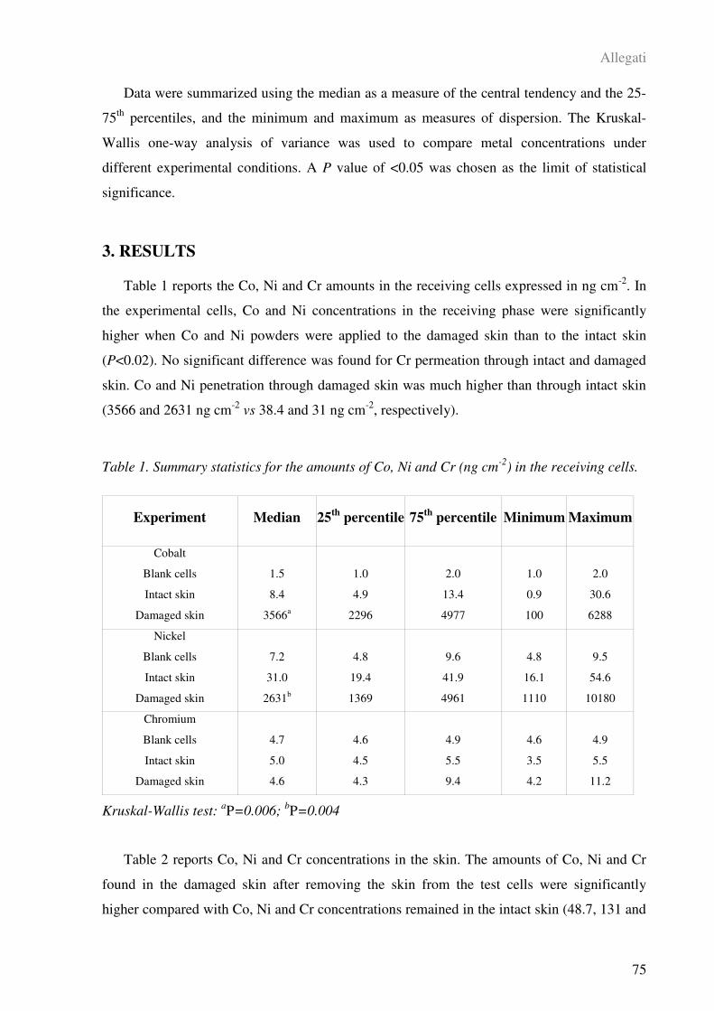
Allegati
75
Data were summarized using the median as a measure of the central tendency and the 25-
75th
percentiles, and the minimum and maximum as measures of dispersion. The Kruskal-
Wallis one-way analysis of variance was used to compare metal concentrations under
different experimental conditions. A P value of <0.05 was chosen as the limit of statistical
significance.
3. RESULTS
Table 1 reports the Co, Ni and Cr amounts in the receiving cells expressed in ng cm-2
. In
the experimental cells, Co and Ni concentrations in the receiving phase were significantly
higher when Co and Ni powders were applied to the damaged skin than to the intact skin
(P<0.02). No significant difference was found for Cr permeation through intact and damaged
skin. Co and Ni penetration through damaged skin was much higher than through intact skin
(3566 and 2631 ng cm-2
vs 38.4 and 31 ng cm-2
, respectively).
Table 1. Summary statistics for the amounts of Co, Ni and Cr (ng cm-2
) in the receiving cells.
Kruskal-Wallis test: aP=0.006;
bP=0.004
Table 2 reports Co, Ni and Cr concentrations in the skin. The amounts of Co, Ni and Cr
found in the damaged skin after removing the skin from the test cells were significantly
higher compared with Co, Ni and Cr concentrations remained in the intact skin (48.7, 131 and
Experiment Median 25th
percentile 75th
percentile Minimum Maximum
Cobalt
Blank cells
Intact skin
Damaged skin
1.5
8.4
3566a
1.0
4.9
2296
2.0
13.4
4977
1.0
0.9
100
2.0
30.6
6288
Nickel
Blank cells
Intact skin
Damaged skin
7.2
31.0
2631b
4.8
19.4
1369
9.6
41.9
4961
4.8
16.1
1110
9.5
54.6
10180
Chromium
Blank cells
Intact skin
Damaged skin
4.7
5.0
4.6
4.6
4.5
4.3
4.9
5.5
9.4
4.6
3.5
4.2
4.9
5.5
11.2
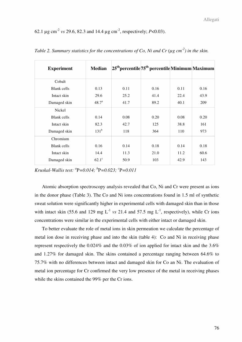
Allegati
76
62.1 µg cm-2
vs 29.6, 82.3 and 14.4 µg cm-2
, respectively; P<0.03).
Table 2. Summary statistics for the concentrations of Co, Ni and Cr (µg cm-2
) in the skin.
Kruskal-Wallis test: aP=0.014;
bP=0.023;
cP=0.011
Atomic absorption spectroscopy analysis revealed that Co, Ni and Cr were present as ions
in the donor phase (Table 3). The Co and Ni ions concentrations found in 1.5 ml of synthetic
sweat solution were significantly higher in experimental cells with damaged skin than in those
with intact skin (55.6 and 129 mg L-1
vs 21.4 and 57.5 mg L-1
, respectively), while Cr ions
concentrations were similar in the experimental cells with either intact or damaged skin.
To better evaluate the role of metal ions in skin permeation we calculate the percentage of
metal ion dose in receiving phase and into the skin (table 4): Co and Ni in receiving phase
represent respectively the 0.024% and the 0.03% of ion applied for intact skin and the 3.6%
and 1.27% for damaged skin. The skins contained a percentage ranging between 64.6% to
75.7% with no differences between intact and damaged skin for Co an Ni. The evaluation of
metal ion percentage for Cr confirmed the very low presence of the metal in receiving phases
while the skins contained the 99% per the Cr ions.
Experiment Median 25th
percentile 75th
percentile Minimum Maximum
Cobalt
Blank cells
Intact skin
Damaged skin
0.13
29.6
48.7a
0.11
25.2
41.7
0.16
41.4
89.2
0.11
22.4
40.1
0.16
43.9
209
Nickel
Blank cells
Intact skin
Damaged skin
0.14
82.3
131b
0.08
42.7
118
0.20
125
364
0.08
38.8
110
0.20
161
973
Chromium
Blank cells
Intact skin
Damaged skin
0.16
14.4
62.1c
0.14
11.3
50.9
0.18
21.0
103
0.14
11.2
42.9
0.18
60.6
143
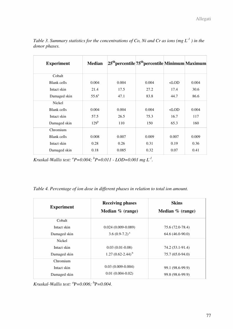
Allegati
77
Table 3. Summary statistics for the concentrations of Co, Ni and Cr as ions (mg L-1
) in the
donor phases.
Kruskal-Wallis test: aP=0.004;
bP=0.011 - LOD=0.001 mg L
-1.
Table 4. Percentage of ion dose in different phases in relation to total ion amount.
Kruskal-Wallis test: aP=0.006;
bP=0.004.
Experiment Median 25th
percentile 75th
percentile Minimum Maximum
Cobalt
Blank cells
Intact skin
Damaged skin
0.004
21.4
55.6a
0.004
17.5
47.1
0.004
27.2
83.8
<LOD
17.4
44.7
0.004
30.6
86.6
Nickel
Blank cells
Intact skin
Damaged skin
0.004
57.5
129b
0.004
26.5
110
0.004
75.3
150
<LOD
16.7
65.3
0.004
117
160
Chromium
Blank cells
Intact skin
Damaged skin
0.008
0.28
0.18
0.007
0.26
0.085
0.009
0.31
0.32
0.007
0.19
0.07
0.009
0.36
0.41
Experiment Receiving phases
Median % (range)
Skins
Median % (range)
Cobalt
Intact skin
Damaged skin
0.024 (0.009-0.089)
3.6 (0.9-7.2) a
75.6 (72.0-78.4)
64.6 (46.0-90.0)
Nickel
Intact skin
Damaged skin
0.03 (0.01-0.08)
1.27 (0.62-2.44) b
74.2 (53.1-91.4)
75.7 (65.0-94.0)
Chromium
Intact skin
Damaged skin
0.03 (0.009-0.004)
0.01 (0.004-0.02)
99.1 (98.6-99.9)
99.8 (98.6-99.9)
Allegati
78
4. DISCUSSION
Even though it is known that polar organic compounds and some metals can appreciably
penetrate damaged skin (Ilyin et al., 1975; Larese Filon et al., 2006), there is limited
knowledge about the behaviour of Co, Ni and Cr metal powders. This study was focused on
the percutaneous absorption of these metal powders through full thickness human skin
(epidermis and dermis) with intact as well as abraded skin barrier in an in vitro diffusion cell
system. In vitro percutaneous studies can be used to define the diffusion characteristics of
xenobiotics as one way of documenting their bioavailability.
Skin absorption of metals is closely related to the capacity of the sweat to oxidise metals:
our previous in vitro permeation experiments showed that Co powder can permeate the skin
when applied using a dispersion in synthetic sweat that oxidises metallic cobalt into ions,
while no absorption occurred using a dispersion in water (Larese Filon et al., 2004).
The metal oxidation has been shown in experiments treating metal objects with synthetic
sweat. This method was used to determine nickel release in objects such as coins (Liden and
Carter, 2001), suggesting a route of skin permeation which was until recently unknown. In
our study, experiments were carried out using synthetic sweat at a low pH (pH 4.5) with the
aim to oxidize metallic powders to soluble ions, so that the permeation of metallic powders
could take place if there was previous oxidation of metals by means of the sweating skin
action. This hypothesis was confirmed because the experiments performed by polarographic
analysis revealed that metals were present as ions in the donor phase. (Larese 2004, 2007)
The choice of pH 4.5 in our synthetic sweat was somewhat unusual that many researchers
choose a pH of 6.5 for their simulate sweat (EEC 1998). However, the actual pH of human
skin seems to be much more acidic, typically in the range of 4 to 5.5 and in some cases, for
example during individuals’ physical activity, much lower (Dyer et al., 1998). This is an
important feature, because most chemical elements, such as chromium, become appreciably
more ionized as acidity increases, approximately 10 to 100 fold per each one pH unit decrease
(Zlotogorski, 1987). Thus, the choice of a simulate sweat at pH 4.5, as used in the present
study, was considered appropriate to reproduce workplace conditions.
Our experiments demonstrated that metal powders are able to permeate through intact
human skin in variable amounts: higher for Co and Ni, lower for Cr. However, the values
obtained with intact human skin can be considered low compared to other substances which
diffuse through the skin (Larese Filon et al., 1999). When the abraded skin procedure was
used (Larese Filon et al., 2006), Co and Ni permeation increased significantly. Substantially
Allegati
79
increased penetration of other metals through damaged skin has been previously reported
(Ilyin et al., 1975; Larese Filon et al., 2006). In opposite, there was no significant difference
in the permeation through intact and damaged skin barrier for Cr. Moreover, the amount of Cr
found in the receiving phase, after exposure of the abraded skin to Cr powder for 24 hours,
was considerably lower than that detected after exposure to Co and Ni powders. These
findings could be explained by the different concentrations of Co, Ni and Cr ions found in the
donor phases applied on intact and damaged skin. Polarographic analysis revealed that Co and
Ni ions were present in the donor solutions in higher concentrations than Cr ions (Larese,
2007). The Co and Ni ion concentrations increased significantly when the donor phase was
applied on the damaged skin: this finding might be explained by the increasing surface in
contact with the metal and the action of some skin protein released with the damaging
procedure. In our knowledge there aren’t other investigation on that topic to substantiate our
results.
In contrast with Co and Ni experiments, no increase in Cr ions concentration was found
when Cr powder was used. This could mean that the synthetic sweat used in this study was
able to oxidise only a small fraction of metallic Cr to soluble Cr ions.
A further interesting finding of this experimental investigation was the metal skin content:
the amounts of Co, Ni and Cr found in the damaged skin were significantly higher than in the
intact skin. Ni concentrations in intact and damaged skin were considerably higher than Co
and Cr concentrations. However, the high Cr concentration which was found in the damaged
skin suggests that binding of Cr in the skin interferes with diffusion of Cr through the skin for
the stronger binding of Cr to skin proteins. This is confirmed by the calculation of the
percentage of Cr ions into the skin that reached the 99% of the total ion amount.
It is well known that the electrophilic nature of many metals, such as chromium and
nickel, determines their protein reactivity, which can result in depot formation in the stratum
corneum (Samitz and Katz, 1976). Such protein-metal binding can take place in all strata of
the skin to the extent of building up a secondary barrier and inhibiting further diffusion
(Hostynek, 2003). The results of our study are broadly consistent with those reported in other
in vitro experimental investigations which suggested that Cr (III) ions show a strong affinity
for epithelial and dermal tissues, forming stable complexes which slow the rate of diffusion
(Samitz et al. 1969).
In conclusion, the finding of the present confirmed that an injury to the skin barrier could
significantly enhance Co and Ni permeation. In contrast, Cr permeation is lower both in
intact than in damaged skin for the low ionisation of this metal and for his high protein

Allegati
80
binding property. More that the 99% of the total Cr ions was into the skin: this might explain
the extremely low flux of Cr through damaged skin.
In the experiments carried out with intact skin the amounts of Co, Ni and Cr found in the
receiving phase were lower than those detected in our previous studies. It should be noted,
however, that our previous permeation experiments were performed using a different pH for
synthetic sweat and different procedures were use to remove and analyse the dermal bathing
solution at selected times during a 24 hours period (Larese Filon et al., 2007; D’Agostin et al.,
2007).
How accurately these in vitro results relate to in vivo workers’ skin is not known. A case
of intoxication after a chemical burn with increased Co in serum has been reported by Neligan
(1966). In that case the cutaneous barrier was damaged and the absorption of Co was
substantial. We recognise that the present in vitro study suggests only a simple way to
simulate what occurs during real-life situations. In occupational settings workers may have
their skin mechanically damaged because of cuts and abrasions which would make metal
penetration substantially greater than that shown in our investigation. Moreover, it is well
known that nickel allergic hypersensitivity develops more readily on exposure of damaged
skin than from exposure on intact skin. The findings of our study underline the need to
prevent contact with potential toxic substances and suggest that it is advisable for workers to
use protective clothing, such as gloves and disposable overalls, since the granulometry of
metal particles found in the work environment is similar to that used in our experiments.
5. REFERENCES
ACGIH, American Conference of Governmental Industrial Hygienists (1995) Threshold
Limit Values (TLVs) for Chemical Substances and Physical Agents and Biological Exposure
Indices (BEIs). Cincinnati, USA.
Carson BL, Ellis HV, McCann JL (1986) Toxicology and biological monitoring of metals
in humans. Lewis, Chelsea, pp.75-92.
Corbett GE, Finley BL, Paustenbach DJ, Kerge BD (1997) Systemic uptake of chromium
in human volunteers following dermal contact with hexavalent chromium (22mg/dL). J.
Exposure Anal. Environ. Epidemiol. 7: 179-189.
D’Agostin F, Crosera M, Adami G, Malvestito A, Rosani R, Bovenzi M, Maina G, Larese
Filon F (2007) Assorbimento percutaneo di polvere di cromo ed effetti della detersione:
Allegati
81
risultati di una indagine in vitro. G. Ital. Med. Lav. Erg. 3: 452-454.
Davies DJ, Ward RJ, Heylings JR (2004) Multi-species assessment of electrical resistence
as a skin integrity marker for in vivo percutaneous absorption studies. Toxicol. in Vitro 18:
351-358.
Drexler H (1998) Assignment of skin notation for MAK values and its legal consequences
in Germany. Int. Arch. Occup. Environ. Health 71: 503-505.
Dyer J, Scrivner N, Dentel S (1998) A practical guide for determining the solubility of
metal hydroxides and oxides in water. Environ. Progr. 17: 1-8.
EDETOX, 2000. Evaluations and Predictions of Dermal Absorption of Toxic Chemicals,
EU Framework V: Quality of Life, Environment and Health Key Action Funding (Project
Number: QLKA-2000-00196).
EEC. European Committee for Standardization, European Standard EN 1811:1998
Reference method for release of nickel from products. Central Secretariat, Brussels.
Fasano W, Manning L, Green J (2002) Rapid assessment of rat and human epidermal
membranes for in vitro dermal regulatory testing: correlation of electrical resistence with
tritiated water permeability. Toxicol. in Vitro 16: 731-740.
Fiserova-Bergerova V, Pierche TJ, Droz P (1990) Dermal absorption potential of
industrial chemicals: criteria for skin notation. Am. J. Ind. Med. 17: 617-635.
Flora CJ and Nioboer E (1980) Determination of nickel by differential pulse polarography
with dropping mercury electrode. Anal. Chem. 52: 1013-1020.
Franz TJ (1975) On the relevance of in vitro data. J. Invest. Dermatol. 93: 633-640.
Fullerton A, Andersen JR, Hoelgaard A, Menne T (1986) Permeation of nickel salts
through human skin in vitro. Contact dermatitis 15: 173-177.
Fullerton A, Andersen JR, Hoelgaard A (1988) Permeation of nickel through human skin
in vitro – effect of vehicles. Br. J. Dermatol. 118: 509-516.
Gammelgaard B, Fullerton A, Avastorp C, Menné T (1992) Permeation of chromium salts
through human skin in vitro. Contact Dermatitis 27: 302-310.
Guin JD (1995) Practical contact dermatitis. McGraw-Hill, Inc. New York.
Hostynek JJ, Dreher F, Pelosi A, Anigbogu A, Maibach HI (2001) Human stratum
corneum penetration by nickel: in vivo study of depth distribution after occlusive application
Allegati
82
of the metal as powder. Acta Derm. Venereol. Suppl. 212: 5-10.
Hostynek JJ (2003) Factors determining percutaneous metal absorption. Food Chem.
Toxicol. 41: 327-345.
Ilyin L, Ivannikav A, Parfenov Y, Stolyarov V (1975) Strontium absorption through
damaged and undamaged human skin. Health Physics 29: 75-80.
Kogan VY and Petukhova NE (1986) Cobalt. UNEP, Moscow.
Larese Filon F, Fiorito A, Adami G, Barbieri P, Coceani N, Bussani R; Reisenhofer E
(1999) Skin absorption in vitro of glycol ethers. Int. Arch. Occup. Environ. Health 72: 480-
484.
Larese Filon F, Maina G, Adami G, Venier M, Coceani N, Bussani R, Massiccio M,
Barbieri P, Spinelli P (2004) In vitro percutaneous absorption of cobalt. Int. Arch. Environ.
Health 77: 85-89.
Larese Filon F, Boeninger M, Maina G, Adami G, Spinelli P, Damian A (2006) Skin
absorption of inorganic lead and the effects of skin cleansers. J. Occup. Environ. Med. 48:
692-699.
Larese Filon F, Adami G, Venier M, Maina G, Renzi N (2007) In vitro percutaneous
absorption of metal compounds. Toxicol. Lett. 170: 49-56.
Liden S and Lundberg E (1979) Penetration of chromium in intact human skin in vivo. J.
Invest. Dermatol. 72: 42-45.
Liden C and Carter S (2001) Nickel release from coins. Contact Dermatitis 44: 160-165.
Linnainmaa M and Kiilunen M (1997) Urinary cobalt as a measure of exposure in the wet
sharpen of hard metal and stellite blades. Int. Arch. Occup. Environ. Health 69: 193-200.
Meding B and Swanbeck G (1987) Prevalence of hand eczema in an industrial city. Br. J.
Dermatol. 116: 627-631.
Neligan PC (1996) Transcutaneous metal absorption following chemical burn injury.
Burns 22: 232-233.
OECD, Organization for Economic Co-operation and Development (2000) Draft
Guidance Document for the Conduct of Skin Absorption Studies. Paris, 2000.
Poet TS (2000) Assessing dermal absorption. Toxicol. Sci. 58: 1-2.

Allegati
83
Samitz MH, Katz SA, Schreiner DM, Gross PR (1969) Chromium-protein interactions.
Acta Dermato-Venereol. 49: 142-146.
Samitz MH and Katz SA (1976) Nickel-epidermal interactions: diffusion and binding.
Environ Research 11: 34-39.
Scansetti G, Botta GC, Spinelli P, Reviglione L, Ponzetti C (1994) Absorption and
excretion of cobalt in the hard metal industry. Sc. Total Environ. 150: 141-144.
Suzuki-Yasumoto M and Inaba J (1976) Absorption and metabolism of radioactive cobalt
compounds through normal and wounded skin. Diag. Treat Inc. Radionuclides Proc. Int. Sem.
12: 119-136.
Tanojo H, Hostynek JJ, Mountford H, Maibach HI (2001) In vitro permeation of nickel
salts through human stratum corneum. Acta Derm. Venereol. Suppl. 212: 19-23.
Van Lierde V, Chery C, Roche N, Monstrey S, Moens L, Vanhaecke F (2006) In vitro
permeation of chromium species through porcine and human skin as determined by capillary
electrophoresis-inductively coupled plasma-sector field mass spectrometry. Anal. Bioanal.
Chem. 384: 378-384.
Wahlberg JE (1965a) Disappearance measurements, a method for studying percutaneous
absorption of istotote-labelled compounds emitting gamma-rays. Acta Dermato-Venerol. 45:
397-414.
Wahlberg JE (1965b) Percutaneous absorption of sodium chromate (51Cr), cobaltous (58
Co) and mercuric (203 Hg) chlorides through excised human and guinea pig skin. Acta
Dermato-Venereol. 45: 415-426.
Wahlberg JE (1968) Percutaneous absorption from chromium (51Cr) solutions of different
pH, 1.4-12.8. An experimental study in the guinea pig. Dermatologica 137: 17-25.
Zlotogorski A (1987) Distribution of skin surface pH on the forehead and cheek of adults.
Arch. Dermatol. Res. 279: 398-401.

Allegati
84
ALLEGATO III
NANOPARTICLE DERMAL ABSORPTION AND TOXICITY: A REVIEW OF THE
LITERATURE
Matteo Crosera, Massimo Bovenzi, Giovanni Maina, Gianpiero Adami, Caterina Zanette,
Chiara Florio, Francesca Larese Filon
Pubblicato in: International Archives of Occupational and Environmenal Health, (2009)
82: 1043–1055
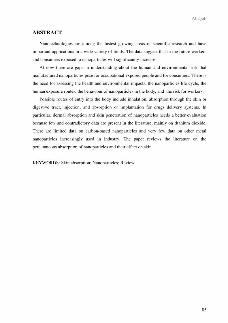
Allegati
85
ABSTRACT
Nanotechnologies are among the fastest growing areas of scientific research and have
important applications in a wide variety of fields. The data suggest that in the future workers
and consumers exposed to nanoparticles will significantly increase .
At now there are gaps in understanding about the human and environmental risk that
manufactured nanoparticles pose for occupational exposed people and for consumers. There is
the need for assessing the health and environmental impacts, the nanoparticles life cycle, the
human exposure routes, the behaviour of nanoparticles in the body, and the risk for workers.
Possible routes of entry into the body include inhalation, absorption through the skin or
digestive tract, injection, and absorption or implantation for drugs delivery systems. In
particular, dermal absorption and skin penetration of nanoparticles needs a better evaluation
because few and contradictory data are present in the literature, mainly on titanium dioxide.
There are limited data on carbon-based nanoparticles and very few data on other metal
nanoparticles increasingly used in industry. The paper reviews the literature on the
percutaneous absorption of nanoparticles and their effect on skin.
KEYWORDS: Skin absorption; Nanoparticles; Review
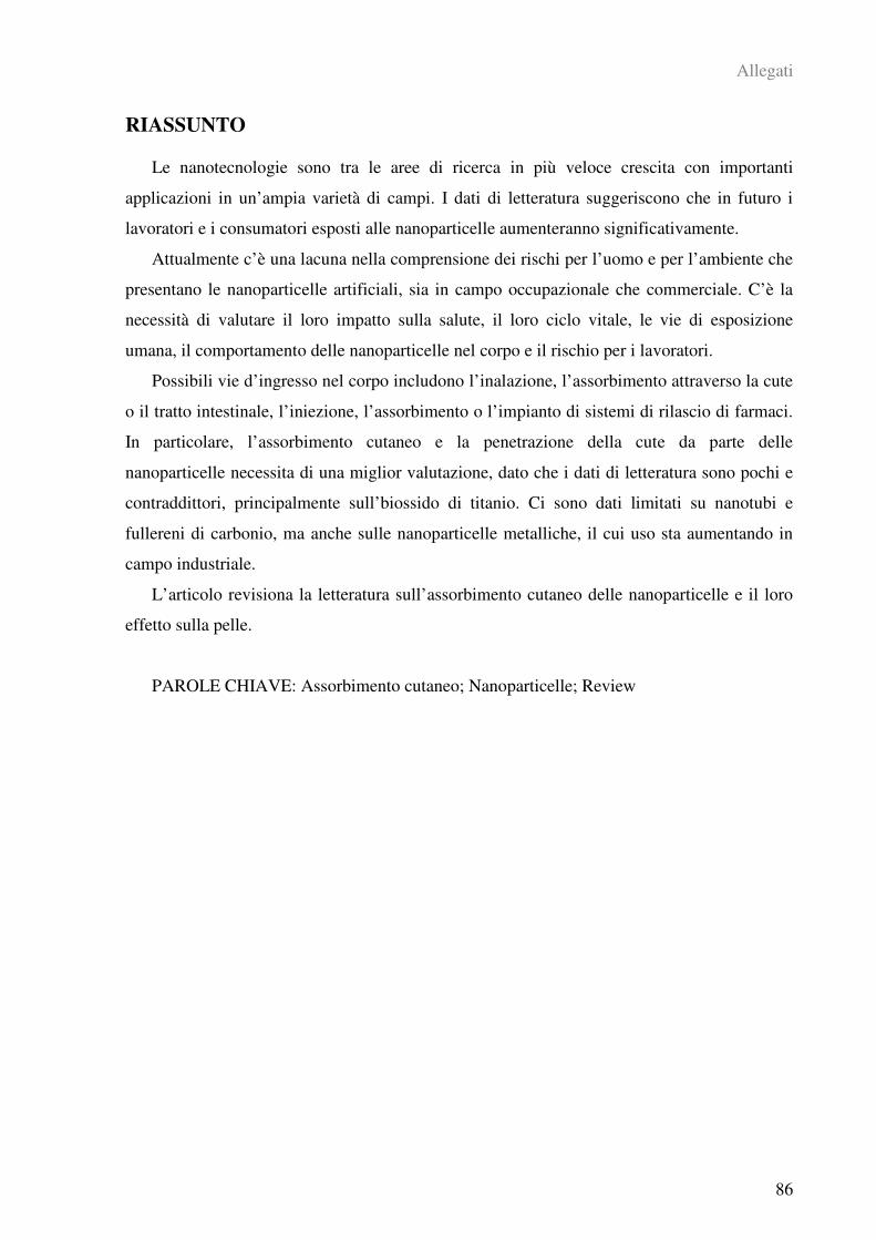
Allegati
86
RIASSUNTO
Le nanotecnologie sono tra le aree di ricerca in più veloce crescita con importanti
applicazioni in un’ampia varietà di campi. I dati di letteratura suggeriscono che in futuro i
lavoratori e i consumatori esposti alle nanoparticelle aumenteranno significativamente.
Attualmente c’è una lacuna nella comprensione dei rischi per l’uomo e per l’ambiente che
presentano le nanoparticelle artificiali, sia in campo occupazionale che commerciale. C’è la
necessità di valutare il loro impatto sulla salute, il loro ciclo vitale, le vie di esposizione
umana, il comportamento delle nanoparticelle nel corpo e il rischio per i lavoratori.
Possibili vie d’ingresso nel corpo includono l’inalazione, l’assorbimento attraverso la cute
o il tratto intestinale, l’iniezione, l’assorbimento o l’impianto di sistemi di rilascio di farmaci.
In particolare, l’assorbimento cutaneo e la penetrazione della cute da parte delle
nanoparticelle necessita di una miglior valutazione, dato che i dati di letteratura sono pochi e
contraddittori, principalmente sull’biossido di titanio. Ci sono dati limitati su nanotubi e
fullereni di carbonio, ma anche sulle nanoparticelle metalliche, il cui uso sta aumentando in
campo industriale.
L’articolo revisiona la letteratura sull’assorbimento cutaneo delle nanoparticelle e il loro
effetto sulla pelle.
PAROLE CHIAVE: Assorbimento cutaneo; Nanoparticelle; Review

Allegati
87
1. INTRODUCTION
Nanomaterials are defined as materials that have at least one dimension <100 nanometers
(1 nm = 10-9
m) and they can be divided into two large groups: ultrafine nanosized particles
not intentionally produced and engineered nanoparticles produced in a controlled, engineered
way (Oberdörster et al., 2005a).
Nanotechnologies are among the fastest growing areas of scientific research and have
important applications in a wide variety of fields. The corresponding industries would require
about two million workers in nanotechnology, and about three times as many jobs in
supporting activities (Roco, 2005). Nanoscale materials are already being introduced for use
in many commercially available products like cosmetics and sunscreens, pharmaceuticals,
stain resistant clothing, sports equipment, automobile catalytic converters, dental bonding,
cleanings products, dressings for specific wound care strategies, but many are the fields of
possible future applications of nanotechnologies as drug delivery systems, nanomedicine,
environmental remediation and cell imaging.
Engineered nanoparticles, because of their big surface-to-volume ratios, exhibit chemical,
physical, and biological properties distinctly different from the same materials in the bulk
form, but such properties may lead to adverse effects on human health and environmental
systems.
Every year new products containing nanomaterials enter in the market (Woodrow Wilson
International Center for Scholars, 2007) and in the next future more workers and costumers
will come in contact with nanoproducts. Since the toxicological and environmental effects of
these compounds are not fully known, there is the need to understand better the health and
environmental impacts, the nanoparticles life cycle, the human exposure routes, the behaviour
of nanoparticles into the body, and the risk for workers in order to use these new materials in
a safe way (Dreher KL, 2004; EPA, 2007; Gwinn and Vallyathan, 2006; Hoet et al., 2004;
Nasterlack et al., 2008; Nel et al., 2006; NIOSH, 2007; Oberdörster et al., 2005a; Oberdörster
et al., 2005b).
Nanotoxicology is an emerging discipline (Oberdörster et al., 2005a) and there is a gap
between the nanomaterials safety evaluation and the nanotechnology development that daily
produce new materials, new synthesis, new applications, and new products ready for the
market. According to the Royal Society & the Royal Academy of Engineering report (2004),
nanoparticles should be treated as new chemicals from a risk-point of view because they can
overcome the body’s normal protective barrier given their size (Nasterlack et al., 2007;
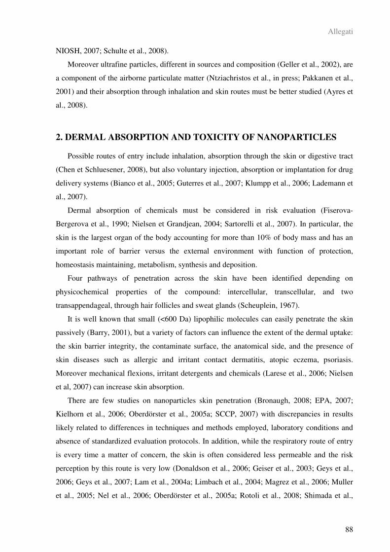
Allegati
88
NIOSH, 2007; Schulte et al., 2008).
Moreover ultrafine particles, different in sources and composition (Geller et al., 2002), are
a component of the airborne particulate matter (Ntziachristos et al., in press; Pakkanen et al.,
2001) and their absorption through inhalation and skin routes must be better studied (Ayres et
al., 2008).
2. DERMAL ABSORPTION AND TOXICITY OF NANOPARTICLES
Possible routes of entry include inhalation, absorption through the skin or digestive tract
(Chen et Schluesener, 2008), but also voluntary injection, absorption or implantation for drug
delivery systems (Bianco et al., 2005; Guterres et al., 2007; Klumpp et al., 2006; Lademann et
al., 2007).
Dermal absorption of chemicals must be considered in risk evaluation (Fiserova-
Bergerova et al., 1990; Nielsen et Grandjean, 2004; Sartorelli et al., 2007). In particular, the
skin is the largest organ of the body accounting for more than 10% of body mass and has an
important role of barrier versus the external environment with function of protection,
homeostasis maintaining, metabolism, synthesis and deposition.
Four pathways of penetration across the skin have been identified depending on
physicochemical properties of the compound: intercellular, transcellular, and two
transappendageal, through hair follicles and sweat glands (Scheuplein, 1967).
It is well known that small (<600 Da) lipophilic molecules can easily penetrate the skin
passively (Barry, 2001), but a variety of factors can influence the extent of the dermal uptake:
the skin barrier integrity, the contaminate surface, the anatomical side, and the presence of
skin diseases such as allergic and irritant contact dermatitis, atopic eczema, psoriasis.
Moreover mechanical flexions, irritant detergents and chemicals (Larese et al., 2006; Nielsen
et al, 2007) can increase skin absorption.
There are few studies on nanoparticles skin penetration (Bronaugh, 2008; EPA, 2007;
Kielhorn et al., 2006; Oberdörster et al., 2005a; SCCP, 2007) with discrepancies in results
likely related to differences in techniques and methods employed, laboratory conditions and
absence of standardized evaluation protocols. In addition, while the respiratory route of entry
is every time a matter of concern, the skin is often considered less permeable and the risk
perception by this route is very low (Donaldson et al., 2006; Geiser et al., 2003; Geys et al.,
2006; Geys et al., 2007; Lam et al., 2004a; Limbach et al., 2004; Magrez et al., 2006; Muller
et al., 2005; Nel et al., 2006; Oberdörster et al., 2005a; Rotoli et al., 2008; Shimada et al.,
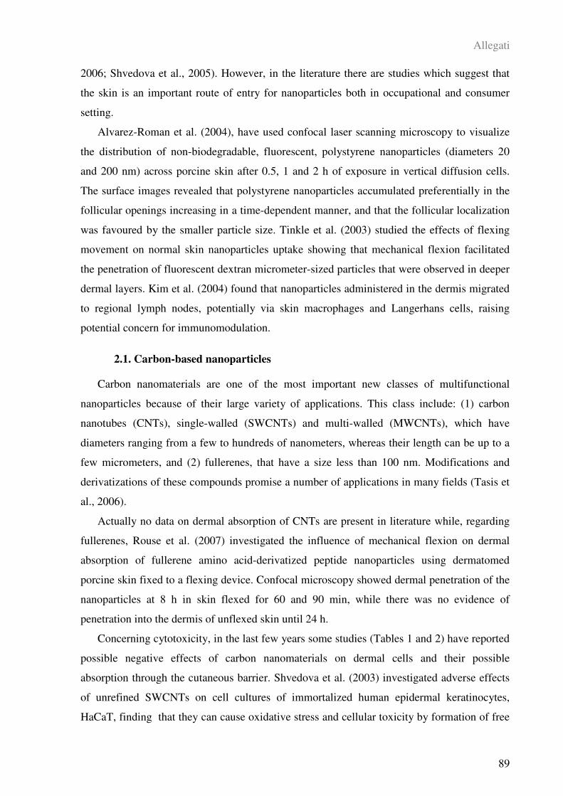
Allegati
89
2006; Shvedova et al., 2005). However, in the literature there are studies which suggest that
the skin is an important route of entry for nanoparticles both in occupational and consumer
setting.
Alvarez-Roman et al. (2004), have used confocal laser scanning microscopy to visualize
the distribution of non-biodegradable, fluorescent, polystyrene nanoparticles (diameters 20
and 200 nm) across porcine skin after 0.5, 1 and 2 h of exposure in vertical diffusion cells.
The surface images revealed that polystyrene nanoparticles accumulated preferentially in the
follicular openings increasing in a time-dependent manner, and that the follicular localization
was favoured by the smaller particle size. Tinkle et al. (2003) studied the effects of flexing
movement on normal skin nanoparticles uptake showing that mechanical flexion facilitated
the penetration of fluorescent dextran micrometer-sized particles that were observed in deeper
dermal layers. Kim et al. (2004) found that nanoparticles administered in the dermis migrated
to regional lymph nodes, potentially via skin macrophages and Langerhans cells, raising
potential concern for immunomodulation.
2.1. Carbon-based nanoparticles
Carbon nanomaterials are one of the most important new classes of multifunctional
nanoparticles because of their large variety of applications. This class include: (1) carbon
nanotubes (CNTs), single-walled (SWCNTs) and multi-walled (MWCNTs), which have
diameters ranging from a few to hundreds of nanometers, whereas their length can be up to a
few micrometers, and (2) fullerenes, that have a size less than 100 nm. Modifications and
derivatizations of these compounds promise a number of applications in many fields (Tasis et
al., 2006).
Actually no data on dermal absorption of CNTs are present in literature while, regarding
fullerenes, Rouse et al. (2007) investigated the influence of mechanical flexion on dermal
absorption of fullerene amino acid-derivatized peptide nanoparticles using dermatomed
porcine skin fixed to a flexing device. Confocal microscopy showed dermal penetration of the
nanoparticles at 8 h in skin flexed for 60 and 90 min, while there was no evidence of
penetration into the dermis of unflexed skin until 24 h.
Concerning cytotoxicity, in the last few years some studies (Tables 1 and 2) have reported
possible negative effects of carbon nanomaterials on dermal cells and their possible
absorption through the cutaneous barrier. Shvedova et al. (2003) investigated adverse effects
of unrefined SWCNTs on cell cultures of immortalized human epidermal keratinocytes,
HaCaT, finding that they can cause oxidative stress and cellular toxicity by formation of free
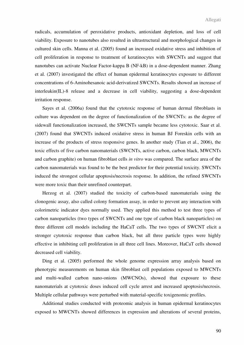
Allegati
90
radicals, accumulation of peroxidative products, antioxidant depletion, and loss of cell
viability. Exposure to nanotubes also resulted in ultrastructural and morphological changes in
cultured skin cells. Manna et al. (2005) found an increased oxidative stress and inhibition of
cell proliferation in response to treatment of keratinocytes with SWCNTs and suggest that
nanotubes can activate Nuclear Factor-kappa B (NF-kB) in a dose-dependent manner. Zhang
et al. (2007) investigated the effect of human epidermal keratinocytes exposure to different
concentrations of 6-Aminohesanoic acid-derivatized SWCNTs. Results showed an increase of
interleukin(IL)-8 release and a decrease in cell viability, suggesting a dose-dependent
irritation response.
Sayes et al. (2006a) found that the cytotoxic response of human dermal fibroblasts in
culture was dependent on the degree of functionalization of the SWCNTs: as the degree of
sidewall functionalization increased, the SWCNTs sample became less cytotoxic. Saar et al.
(2007) found that SWCNTs induced oxidative stress in human BJ Foreskin cells with an
increase of the products of stress responsive genes. In another study (Tian et al., 2006), the
toxic effects of five carbon nanomaterials (SWCNTs, active carbon, carbon black, MWCNTs
and carbon graphite) on human fibroblast cells in vitro was compared. The surface area of the
carbon nanomaterials was found to be the best predictor for their potential toxicity. SWCNTs
induced the strongest cellular apoptosis/necrosis response. In addition, the refined SWCNTs
were more toxic than their unrefined counterpart.
Herzog et al. (2007) studied the toxicity of carbon-based nanomaterials using the
clonogenic assay, also called colony formation assay, in order to prevent any interaction with
colorimetric indicator dyes normally used. They applied this method to test three types of
carbon nanoparticles (two types of SWCNTs and one type of carbon black nanoparticles) on
three different cell models including the HaCaT cells. The two types of SWCNT elicit a
stronger cytotoxic response than carbon black, but all three particle types were highly
effective in inhibiting cell proliferation in all three cell lines. Moreover, HaCaT cells showed
decreased cell viability.
Ding et al. (2005) performed the whole genome expression array analysis based on
phenotypic measurements on human skin fibroblast cell populations exposed to MWCNTs
and multi-walled carbon nano-onions (MWCNOs), showed that exposure to these
nanomaterials at cytotoxic doses induced cell cycle arrest and increased apoptosis/necrosis.
Multiple cellular pathways were perturbed with material-specific toxigenomic profiles.
Additional studies conducted with proteomic analysis in human epidermal keratinocytes
exposed to MWCNTs showed differences in expression and alterations of several proteins,
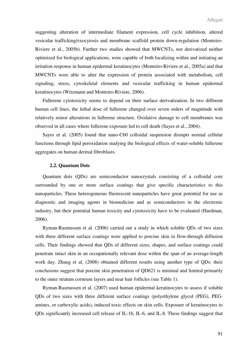
Allegati
91
suggesting alteration of intermediate filament expression, cell cycle inhibition, altered
vesicular trafficking/exocytosis and membrane scaffold protein down-regulation (Monteiro-
Riviere et al., 2005b). Further two studies showed that MWCNTs, nor derivatized neither
optimized for biological applications, were capable of both localizing within and initiating an
irritation response in human epidermal keratinocytes (Monteiro-Riviere et al., 2005a) and that
MWCNTs were able to alter the expression of protein associated with metabolism, cell
signaling, stress, cytoskeletal elements and vesicular trafficking in human epidermal
keratinocytes (Witzmann and Monteiro-Riviere, 2006).
Fullerene cytotoxicity seems to depend on their surface derivatization. In two different
human cell lines, the lethal dose of fullerene changed over seven orders of magnitude with
relatively minor alterations in fullerene structure. Oxidative damage to cell membranes was
observed in all cases where fullerene exposure led to cell death (Sayes et al., 2004).
Sayes et al. (2005) found that nano-C60 colloidal suspension disrupts normal cellular
functions through lipid peroxidation studying the biological effects of water-soluble fullerene
aggregates on human dermal fibroblasts.
2.2. Quantum Dots
Quantum dots (QDs) are semiconductor nanocrystals consisting of a colloidal core
surrounded by one or more surface coatings that give specific characteristics to this
nanoparticles. These heterogeneous fluorescent nanoparticles have great potential for use as
diagnostic and imaging agents in biomedicine and as semiconductors in the electronic
industry, but their potential human toxicity and cytotoxicity have to be evaluated (Hardman,
2006).
Ryman-Rasmussen et al. (2006) carried out a study in which soluble QDs of two sizes
with three different surface coatings were applied to porcine skin in flow-through diffusion
cells. Their findings showed that QDs of different sizes, shapes, and surface coatings could
penetrate intact skin in an occupationally relevant dose within the span of an average-length
work day. Zhang et al. (2008) obtained different results using another type of QDs: their
conclusions suggest that porcine skin penetration of QD621 is minimal and limited primarily
to the outer stratum corneum layers and near hair follicles (see Table 1).
Ryman-Rasmussen et al. (2007) used human epidermal keratinocytes to assess if soluble
QDs of two sizes with three different surface coatings (polyethylene glycol (PEG), PEG-
amines, or carboxylic acids), induced toxic effects on skin cells. Exposure of keratinocytes to
QDs significantly increased cell release of IL-1b, IL-6, and IL-8. These findings suggest that

Allegati
92
surface coating of QDs does not influence the uptake by keratinocytes but is a primary
determinant of cytotoxicity and immunotoxicity. Similar results were found by Zhang et al.
(2008) using a different type of water-soluble QDs with a cadmium/selenide core and a
cadmium sulfide shell coated with PEG. Another study carried out by Rouse et al. (2008)
investigated the effects of applied strain on QDs uptake by human keratinocytes. Their data
indicated that addition of strain resulted in an increase in cytokine production and QDs
uptake, with irritation and reduction of cell viability. These data suggest that application of
physiological load conditions can increase cell membrane permeability, thereby increasing
nanoparticle concentration in cells.
2.3. Titanium Dioxide and Zinc Oxide
Titanium dioxide (TiO2) and zinc oxide (ZnO) are largely present in many sunscreens
formulations to protect against UV-induced skin damage. When exposed to UV radiation,
TiO2 and ZnO do not undergo any chemical decomposition and for that reason they represent
an alternative to chemical agents. Moreover, they offer a wider range of protection compared
to other organic compounds. Actually, in many formulations TiO2 and ZnO are included as
nanosized particles because in this form they are transparent and more aesthetically
acceptable to the consumers. Furthermore, TiO2 nanoparticles are used in other several
products (sport clothes, surface cleaning agents, computer devices) of the everyday life and
the exposure occasions are increasing day by day.
Tan et al. (1996) performed a pilot study on percutaneous absorption of microfine TiO2
from sunscreens applying the formulation to the skin for 2-6 weeks to 13 selected volunteers
scheduled to have skin surgery for a total of 16 skin biopsies. After excision, the stratum
corneum was removed from the skin samples by tape stripping and the concentration of
titanium after digestion of the skin was evaluated. The titanium concentration on the skin
biopsies of the subjects exposed to the microfine TiO2 was higher than the controls.
These findings were not confirmed by other researchers who did not observe penetration
of nanoparticles in the viable layers of the epidermis using different TiO2 nanoparticle
formulations and different investigative techniques. A small amount of metal oxide was only
detected into the hair follicles following application to volunteers (Lademann et al, 1999).
Mavon et al. (2007) used the tape stripping method in in vivo experiments on volunteers
after exposure to a sunscreen formulation containing TiO2 nanoparticles. Transmission
electron microscopy (TEM) and particle-induced X-ray emission (PIXE) techniques were
used to localize the TiO2 in skin sections in in vitro experiments by the same Authors. In this

Allegati
93
in vivo and in vitro permeation study no TiO2 was detected in the follicle, viable epidermis or
dermis, and more than 90% of the applied sunscreen was recovered in the first 15 tape
strippings while the remaining 10% was localized in the furrows and in the opened
infundibulum.
Schulz et al. (2002), using optical and electron microscopy, proved that neither surface
characteristics and particle size nor shape of the micronised pigments result in any dermal
absorption of this substance. Micronised TiO2 was solely deposited on the outermost surface
of the stratum corneum, but not in deeper stratum corneum layers, the human epidermis and
the dermis.
The three-year European project NANODERM involved a great number of research
groups in the evaluation of skin permeation of different TiO2 based sunscreens. The project
provided many data from in vivo and in vitro experiments with human and porcine skin, with
human foreskin transplanted to immunodeficient mice, and with dermal cells in culture.
Some Authors involve in the project detected a little amount of titanium dioxide in the
deeper viable epidermal layers using spatially resolved ion beam analysis (PIXE, RBS, STIM
and secondary electron imaging) on freeze-dried cross-sections of pig skin biopsies (Menzel
et al., 2004), while other Author reported that TiO2 nanoparticles do not penetrate through the
stratum corneum of human skin transplants (Kertész et al, 2005; Kiss et al., 2008).
The project report confirmed the safety of the sunscreens formulation containing TiO2
nanoparticles, reporting no evidence of nanoparticle transcutaneous penetration
(NANODERM, 2007).
Instead, Bennat and Müler-Goymann (2000) found that different formulations had
different penetration: according with their experiments, microfine TiO2 penetrated deeper
into human skin from an oily dispersion than from an aqueous one, and encapsulation of the
pigments into liposomes caused a higher penetration into the skin. Furthermore, penetration
was greater when applied to hairy skin, suggesting a surface penetration through hair follicles
or pores.
Also Gamer et al. (2006) investigated the in vitro absorption of microfine ZnO and TiO2
in cosmetic formulations through porcine skin. One ZnO formulation and two TiO2
formulations were tested in modified Franz static dermal penetration cells. In their
conclusions Authors observed that neither microfine ZnO nor micrfine TiO2 was able to
penetrate through porcine skin, and that most of the applied material was recovered in the first
five tape strips, indicating that the material did not penetrate into the deeper layers of the skin.
ZnO skin absorption was investigated by Cross et al. (2007) using Franz-type diffusion
Allegati
94
cells. Two different formulations of 26-30 nm ZnO particles and one placebo cream base
formulation containing no ZnO nanoparticles were compared. Authors found that less than
0.03% of the applied zinc content was detected in the receptor phase (not significantly
different following application of a placebo formulation). No particles could be detected by in
the lower stratum corneum or viable epidermis by electron microscopy, suggesting that
minimal nanoparticle penetration occurs through the human epidermis (see Table 1).
Some Authors suggested that nanoparticles can elicit a photocatalytic activity into the
dermal layers causing formation of free radicals in skin cells, damaging DNA (Cai et al.,
1992; Dunford et al., 1997; Wamer et al., 1997, Serpone et al., 2001), disrupting normal cell
functions and cell viability (Sayes et al., 2006b). So the debate is just open about their safety
use.
2.4. Silver and Gold Nanoparticles
Owing to their strong antibacterial activity, silver nanoparticles are largely used as a
component of various commercially available products such as textiles, medical devices,
contraceptives, water disinfectants, and room spray (Woodrow Wilson International Center
for Scholars, 2007). Moreover, nanosilver is used for treatment of wounds and burns, as well
as for coating on implants.
Some Authors suggested an increased dermal penetration of nanosilver associated with
damaged skin in in vitro experiments (Larese et al., 2009) or following the use of nanosilver
coated dressings in case of extensive burns (Trop et al, 2006).
Traditionally, silver is relatively non-toxic to mammalian but can cause argyria or
argyrosis in subjects with chronic occupational exposure. Because of the extensive presence
of nanosilver in textiles, wound dressing, sport clothes and other products which come in
direct contact with the skin, dermal exposure must be carefully evaluated.
Keratinocytes and fibroblasts in culture were used to assess the cytotoxic effects of
nanosilver released from several types of silver containing dressings (Table 2) although some
laboratory and clinical studies suggested their dermal biocompatibility (Leaper, 2006; Supp et
al., 2005; Muangman et al., 2006; Wright et al., 2002). The results of these studies showed
that keratinocytes proliferation was significantly inhibited and cell morphology affected after
exposure to extracts of nanocrystalline coated dressings (Paddle-Ledinek et al., 2006; Lam et
al., 2004b). Poon and Burd (2004) found that nanosilver crystallines were toxic to both
keratinocytes and fibroblasts, and that fibroblasts appeared to be more sensitive to silver than
keratinocytes.
Allegati
95
Nanogold is also an interesting nanomaterial for its applications in cell imaging, cancer
therapy, tissue welding and nanomedicine. Sonavane et al. (2008) investigated the in vitro
cutaneous penetration of three types of gold nanoparticles differing in size (15 nm, 102 nm
and 198 nm) using the Franz diffusion cell method with rat skin. Gold nanoparticles showed
size dependent permeation through rat skin. 15 nm gold nanoparticles showed higher
permeation compared to 102 nm and 198 nm gold nanoparticles. TEM study of rat skin
revealed accumulation of smaller size gold nanoparticles in deeper region of skin whereas
larger particles were observed mainly in epidermis and dermis.
Regarding the cytotoxicity of gold nanoparticles, a number of studies have argued its safe
use because the uptake of gold clusters (1.4 nm) by different types of cells and their
interaction with DNA have been demonstrated (Connor et al., 2005; Liu et al., 2003; Tsoli et
al., 2005).
Pernodet et al. (2006) investigated the effects of citrate/gold nanoparticles at different
concentrations and exposure times on human dermal fibroblasts. They found that, as a result
of the intracellular nanoparticle presence, actin stress fibres disappeared, thereby inducing
major adverse effects on cell viability. Properties such as cell spreading and adhesion, cell
growth, and protein synthesis to form the extracellular matrix were altered dramatically,
suggesting that the internal cell activities were damaged.
2.5. Other Metals and Metal Oxides
Researchers are developing a number of metal and alloy nanoparticles for either various
applications in industrial processes, such as catalyst, fillers, semiconductors, or for systemic
drug administration, but very few data regarding their toxicity are available in literature.
It is well known that metal and metal oxide powders once placed in biologic media can
release metal ions (Midander et al., 2006) that can subsequently pass through the skin (Larese
et al., 2007) but little is known about skin penetration of metal nanoparticles.
Berry et al. (2004) found that underivatised iron oxide nanoparticles (8-15 nm) were
rapidly endocytosed into cultured human dermal fibroblasts causing disruption to the cell
cytoskeleton and a decrease in proliferation. The same nanoparticles, transferrin derivatised,
stimulated cell proliferation and were not internalised, but appeared to attach to the outside of
the cell membrane, most likely to cell expressed transferring receptors.
Baroli et al. (2007) demonstrated that iron-based rigid nanoparticles smaller than 10 nm
were able to passively penetrate the skin through the SC lipidic matrix and hair follicle
orifices, reaching the deepest layers of the SC, the stratum granulosum, and hair follicles. In
Allegati
96
rare cases, nanoparticles were also found in the viable epidermis.
Papageorgiou et al. (2007) compared the cytotoxic and genotoxic effects of nanoparticles
and micron-sized particles of cobalt-chrome alloy in cultured human fibroblasts.
Nanoparticles, which caused more free radicals in an acellular environment, induced more
DNA damage than micron-sized particles using the alkaline comet assay. Nanoparticles
appeared to disintegrate within the cells faster than microparticles with the creation of
electron dense deposits, which were enriched in cobalt. The mechanism of cell damage
appeared to be different after exposure to nanoparticles and microparticles.
3. DISCUSSION AND CONCLUSIONS
Experimental findings on skin absorption and skin toxicity of nanoparticles are
contradictory. More data are needed to better define and understand if skin represents a route
of entry of nanoparticles into the body or a target tissue. In the final report of the project
NANODERM it is stated that adverse health effects for the topical application of sunscreens
containing TiO2 nanoparticles (especially when coated) are not expected for healthy skin but
several other studies on carbon-based nanoparticles and quantum dots confirm an interaction
between human dermal cells and nanosized particles. The shortage of data about many types
of new compounds, such as metals and metal oxides, calls for more studies to improve
understanding of nanoparticle skin absorption. Quantitative data are needed because there is
evidence that some nanoparticles can pass through the skin in particular conditions such as
wounds, flexures sites and lesions.
Moreover, nanoparticles characterization should be essential in the future studies on
dermal penetration and toxicity. Size, shape, coating, purity, presence of catalysts, extent of
agglomeration and agglutination of the nanoparticles could influence the amount permeating
the skin and the toxicity of the nanomaterials.
Finally, to investigate the interaction between new nanocompounds and the human skin
the researchers have to take into consideration several exposure variables, such as anatomical
exposure sites, extension of the exposition area, time of exposition, chronic and repeated
exposure, presence of skin diseases, and the role of cleanser and penetration enhancer.
The classic investigation protocols must be adapted and re-standardized to the new
nanosized compounds. Cell cultures (Bernstein and Vaughan, 1999), Franz diffusion cells,
tape stripping (Escobar-Chávez et al, 2008), human skin implantations on animals, remain
powerful tools to study particle interaction with human dermal tissue. Furthermore new
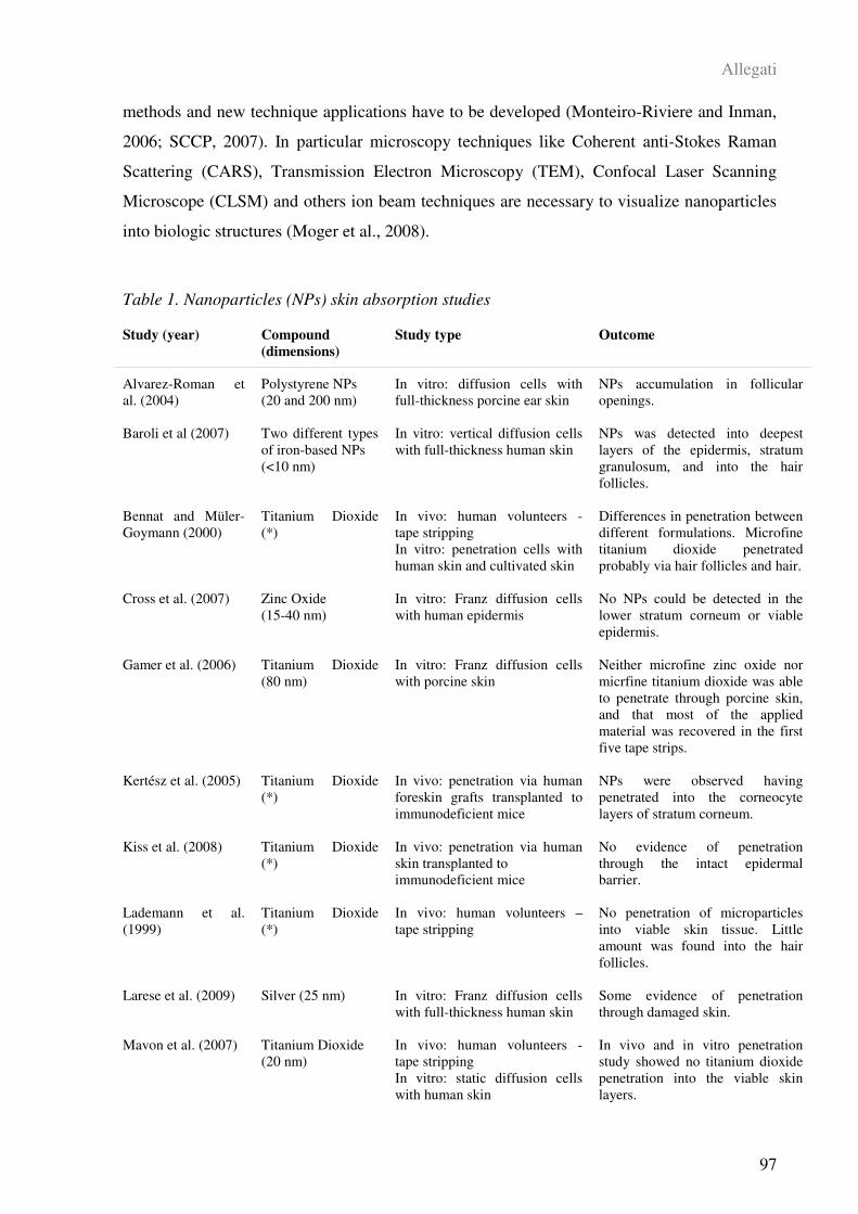
Allegati
97
methods and new technique applications have to be developed (Monteiro-Riviere and Inman,
2006; SCCP, 2007). In particular microscopy techniques like Coherent anti-Stokes Raman
Scattering (CARS), Transmission Electron Microscopy (TEM), Confocal Laser Scanning
Microscope (CLSM) and others ion beam techniques are necessary to visualize nanoparticles
into biologic structures (Moger et al., 2008).
Table 1. Nanoparticles (NPs) skin absorption studies
Study (year)
Compound
(dimensions)
Study type Outcome
Alvarez-Roman et
al. (2004)
Polystyrene NPs
(20 and 200 nm)
In vitro: diffusion cells with
full-thickness porcine ear skin
NPs accumulation in follicular
openings.
Baroli et al (2007) Two different types
of iron-based NPs
(<10 nm)
In vitro: vertical diffusion cells
with full-thickness human skin
NPs was detected into deepest
layers of the epidermis, stratum
granulosum, and into the hair
follicles.
Bennat and Müler-
Goymann (2000)
Titanium Dioxide
(*)
In vivo: human volunteers -
tape stripping
In vitro: penetration cells with
human skin and cultivated skin
Differences in penetration between
different formulations. Microfine
titanium dioxide penetrated
probably via hair follicles and hair.
Cross et al. (2007) Zinc Oxide
(15-40 nm)
In vitro: Franz diffusion cells
with human epidermis
No NPs could be detected in the
lower stratum corneum or viable
epidermis.
Gamer et al. (2006) Titanium Dioxide
(80 nm)
In vitro: Franz diffusion cells
with porcine skin
Neither microfine zinc oxide nor
micrfine titanium dioxide was able
to penetrate through porcine skin,
and that most of the applied
material was recovered in the first
five tape strips.
Kertész et al. (2005) Titanium Dioxide
(*)
In vivo: penetration via human
foreskin grafts transplanted to
immunodeficient mice
NPs were observed having
penetrated into the corneocyte
layers of stratum corneum.
Kiss et al. (2008) Titanium Dioxide
(*)
In vivo: penetration via human
skin transplanted to
immunodeficient mice
No evidence of penetration
through the intact epidermal
barrier.
Lademann et al.
(1999)
Titanium Dioxide
(*)
In vivo: human volunteers –
tape stripping
No penetration of microparticles
into viable skin tissue. Little
amount was found into the hair
follicles.
Larese et al. (2009) Silver (25 nm) In vitro: Franz diffusion cells
with full-thickness human skin
Some evidence of penetration
through damaged skin.
Mavon et al. (2007) Titanium Dioxide
(20 nm)
In vivo: human volunteers -
tape stripping
In vitro: static diffusion cells
with human skin
In vivo and in vitro penetration
study showed no titanium dioxide
penetration into the viable skin
layers.
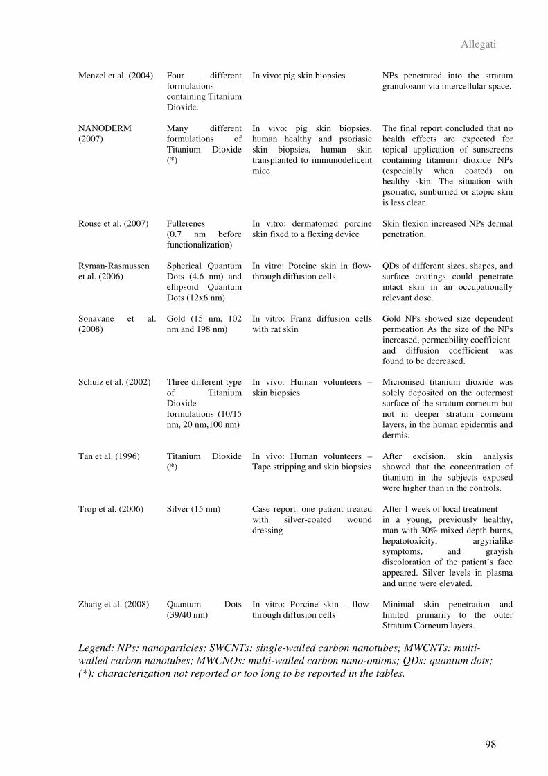
Allegati
98
Menzel et al. (2004). Four different
formulations
containing Titanium
Dioxide.
In vivo: pig skin biopsies NPs penetrated into the stratum
granulosum via intercellular space.
NANODERM
(2007)
Many different
formulations of
Titanium Dioxide
(*)
In vivo: pig skin biopsies,
human healthy and psoriasic
skin biopsies, human skin
transplanted to immunodeficent
mice
The final report concluded that no
health effects are expected for
topical application of sunscreens
containing titanium dioxide NPs
(especially when coated) on
healthy skin. The situation with
psoriatic, sunburned or atopic skin
is less clear.
Rouse et al. (2007) Fullerenes
(0.7 nm before
functionalization)
In vitro: dermatomed porcine
skin fixed to a flexing device
Skin flexion increased NPs dermal
penetration.
Ryman-Rasmussen
et al. (2006)
Spherical Quantum
Dots (4.6 nm) and
ellipsoid Quantum
Dots (12x6 nm)
In vitro: Porcine skin in flow-
through diffusion cells
QDs of different sizes, shapes, and
surface coatings could penetrate
intact skin in an occupationally
relevant dose.
Sonavane et al.
(2008)
Gold (15 nm, 102
nm and 198 nm)
In vitro: Franz diffusion cells
with rat skin
Gold NPs showed size dependent
permeation As the size of the NPs
increased, permeability coefficient
and diffusion coefficient was
found to be decreased.
Schulz et al. (2002) Three different type
of Titanium
Dioxide
formulations (10/15
nm, 20 nm,100 nm)
In vivo: Human volunteers –
skin biopsies
Micronised titanium dioxide was
solely deposited on the outermost
surface of the stratum corneum but
not in deeper stratum corneum
layers, in the human epidermis and
dermis.
Tan et al. (1996) Titanium Dioxide
(*)
In vivo: Human volunteers –
Tape stripping and skin biopsies
After excision, skin analysis
showed that the concentration of
titanium in the subjects exposed
were higher than in the controls.
Trop et al. (2006) Silver (15 nm) Case report: one patient treated
with silver-coated wound
dressing
After 1 week of local treatment
in a young, previously healthy,
man with 30% mixed depth burns,
hepatotoxicity, argyrialike
symptoms, and grayish
discoloration of the patient’s face
appeared. Silver levels in plasma
and urine were elevated.
Zhang et al. (2008) Quantum Dots
(39/40 nm)
In vitro: Porcine skin - flow-
through diffusion cells
Minimal skin penetration and
limited primarily to the outer
Stratum Corneum layers.
Legend: NPs: nanoparticles; SWCNTs: single-walled carbon nanotubes; MWCNTs: multi-
walled carbon nanotubes; MWCNOs: multi-walled carbon nano-onions; QDs: quantum dots;
(*): characterization not reported or too long to be reported in the tables.
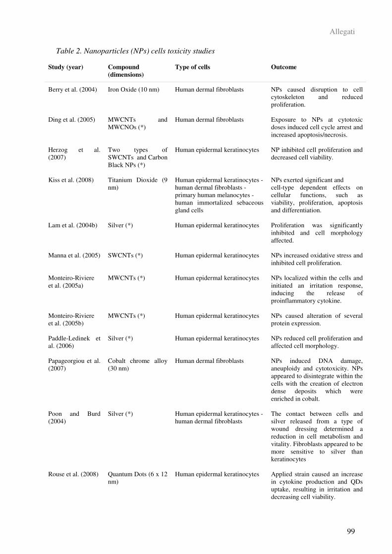
Allegati
99
Table 2. Nanoparticles (NPs) cells toxicity studies
Study (year)
Compound
(dimensions)
Type of cells Outcome
Berry et al. (2004) Iron Oxide (10 nm) Human dermal fibroblasts NPs caused disruption to cell
cytoskeleton and reduced
proliferation.
Ding et al. (2005) MWCNTs and
MWCNOs (*)
Human dermal fibroblasts Exposure to NPs at cytotoxic
doses induced cell cycle arrest and
increased apoptosis/necrosis.
Herzog et al.
(2007)
Two types of
SWCNTs and Carbon
Black NPs (*)
Human epidermal keratinocytes NP inhibited cell proliferation and
decreased cell viability.
Kiss et al. (2008) Titanium Dioxide (9
nm)
Human epidermal keratinocytes -
human dermal fibroblasts -
primary human melanocytes -
human immortalized sebaceous
gland cells
NPs exerted significant and
cell-type dependent effects on
cellular functions, such as
viability, proliferation, apoptosis
and differentiation.
Lam et al. (2004b) Silver (*) Human epidermal keratinocytes Proliferation was significantly
inhibited and cell morphology
affected.
Manna et al. (2005) SWCNTs (*) Human epidermal keratinocytes NPs increased oxidative stress and
inhibited cell proliferation.
Monteiro-Riviere
et al. (2005a)
MWCNTs (*) Human epidermal keratinocytes NPs localized within the cells and
initiated an irritation response,
inducing the release of
proinflammatory cytokine.
Monteiro-Riviere
et al. (2005b)
MWCNTs (*) Human epidermal keratinocytes NPs caused alteration of several
protein expression.
Paddle-Ledinek et
al. (2006)
Silver (*) Human epidermal keratinocytes NPs reduced cell proliferation and
affected cell morphology.
Papageorgiou et al.
(2007)
Cobalt chrome alloy
(30 nm)
Human dermal fibroblasts NPs induced DNA damage,
aneuploidy and cytotoxicity. NPs
appeared to disintegrate within the
cells with the creation of electron
dense deposits which were
enriched in cobalt.
Poon and Burd
(2004)
Silver (*) Human epidermal keratinocytes -
human dermal fibroblasts
The contact between cells and
silver released from a type of
wound dressing determined a
reduction in cell metabolism and
vitality. Fibroblasts appeared to be
more sensitive to silver than
keratinocytes
Rouse et al. (2008) Quantum Dots (6 x 12
nm)
Human epidermal keratinocytes Applied strain caused an increase
in cytokine production and QDs
uptake, resulting in irritation and
decreasing cell viability.
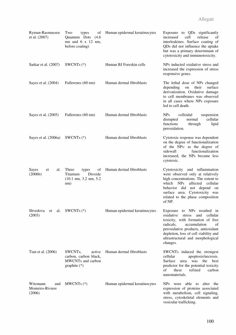
Allegati
100
Ryman-Rasmussen
et al. (2007)
Two types of
Quantum Dots (4.6
nm and 6 x 12 nm,
before coating)
Human epidermal keratinocytes Exposure to QDs significantly
increased cell release of
interleukines. Surface coating of
QDs did not influence the uptake
but was a primary determinant of
cytotoxicity and immunotoxicity.
Sarkar et al. (2007) SWCNTs (*) Human BJ Foreskin cells NPs inducted oxidative stress and
increased the expression of stress
responsive genes.
Sayes et al. (2004) Fullerenes (60 nm) Human dermal fibroblasts The lethal dose of NPs changed
depending on their surface
derivatization. Oxidative damage
to cell membranes was observed
in all cases where NPs exposure
led to cell death.
Sayes et al. (2005) Fullerenes (60 nm) Human dermal fibroblasts NPs colloidal suspension
disrupted normal cellular
functions through lipid
peroxidation.
Sayes et al. (2006a) SWCNTs (*) Human dermal fibroblasts Cytotoxic response was dependent
on the degree of functionalization
of the NPs: as the degree of
sidewall functionalization
increased, the NPs became less
cytotoxic.
Sayes et al.
(2006b)
Three types of
Titanium Dioxide
(10.1 nm, 3.2 nm, 5.2
nm)
Human dermal fibroblasts Cytotoxicity and inflammation
were observed only at relatively
high concentrations. The extent to
which NPs affected cellular
behavior did not depend on
surface area. Cytotoxicity was
related to the phase composition
of NP.
Shvedova et al.
(2003)
SWCNTs (*) Human epidermal keratinocytes Exposure to NPs resulted in
oxidative stress and cellular
toxicity, with formation of free
radicals, accumulation of
peroxidative products, antioxidant
depletion, loss of cell viability and
ultrastructural and morphological
changes.
Tian et al. (2006) SWCNTs, active
carbon, carbon black,
MWCNTs and carbon
graphite (*)
Human dermal fibroblasts SWCNTs induced the strongest
cellular apoptosis/necrosis.
Surface area was the best
predictor for the potential toxicity
of these refined carbon
nanomaterials.
Witzmann and
Monteiro-Riviere
(2006)
MWCNTs (*) Human epidermal keratinocytes NPs were able to alter the
expression of proteins associated
with metabolism, cell signaling,
stress, cytoskeletal elements and
vesicular trafficking.

Allegati
101
Zhang et al. (2007) SWCNTs (*) Human epidermal keratinocytes Exposure resulted in a dose-
dependent irritation response with
an increase in IL-8 release and a
decrease in cell viability.
Zhang et al. (2008) Quantum Dots (39/40
nm)
Human epidermal keratinocytes Cell viability decreased ,and IL-6
and IL-8 release increased, both
significantly. NPs were found in
cytoplasmic vacuoles and at the
periphery of the cell membranes.
Legend: NPs: nanoparticles; SWCNTs: single-walled carbon nanotubes; MWCNTs: multi-
walled carbon nanotubes; MWCNOs: multi-walled carbon nano-onions; QDs: quantum dots;
(*): characterization not reported or too long to be reported in the tables.
4. REFERENCES
Alvarez-Roman R, Naik A, Kalia YN, Guy RH, Fessi H (2004) Skin penetration and
distribution of polymeric nanoparticles. J. Control. Release. 99: 53-62.
Ayres JG, Borm P, Cassee FR, Castranova V, Donaldson K, Ghio A, Harrison RM, Hider
R, Kelly F, Kooter IM, Marano F, Maynard RL, Mudway I, Nel A, Sioutas C, Smith S,
Baeza-Squiban A, Cho A, Duggan S, Froines J (2008) Evaluating the toxicity of airborne
particulate matter and nanoparticles by measuring oxidative stress potential – A workshop
report an consensus statement. Inhalation toxicology. 20: 75-99.
Baroli B, Ennas MG, Loffredo F, Isola M, Pinna R and Lopez-Quintela A (2007)
Penetration of Metallic Nanoparticles in Human Full-Thickness Skin. J. Invest. Dermatol.
127: 1701–1712.
Barry BW (2001) Novel mechanisms and devices to enable successful transdermal drug
delivery. Eur J Pharm Sci. 14: 101-114.
Bennat C and Müller-Goymann CC (2000) Skin penetration and stabilization of
formulations containing microfine titanium dioxide as physical UV filter. Int. J. Cosmet. Sci.
22: 271–283.
Bernstein IA and Vaughan FL (1999) Cultured keratinocytes in in vitro
dermatotoxicological investigation: a review. J. Toxicol. Environm. Health, Part B, 2: 1-30.
Berry CC, Charles S, Wells S, Dalby MJ, Curtis ASG (2004) The influence of transferrin
stabilised magnetic nanoparticles on human dermal fibroblasts in culture. Int. J. Pharmac.
269: 211-225.
Allegati
102
Bianco A, Kostarelos K and Prato M (2005) Applications of carbon nanotubes in drug
delivery. Current Opinion in Chemical Biology. 9: 674-679
Bronaugh RL (2008) Skin penetration of Nanoparticles. Proceeding of PPP2008,
Perspectives in percutaneous penetration, La Grande Motte 25-29th of March 2008, pg. 1.
Cai R, Kubota Y, Shuin T, Sakai H, Hashimoto K, Fujishima A (1992) Induction of
cytotoxicity by photoexcited TiO2 particles. Cancer Res. 52: 2346–2348.
Chen X and Schluesener HJ (2008) Nanosilver: A nanoproduct in medical application.
Toxicol. Lett. 176: 1-12.
Connor EE, Mwamuka J, Gole A, Murphy CJ, Wyatt MD (2005) Gold Nanoparticles Are
Taken Up by Human Cells but Do Not Cause Acute Cytotoxicity. Small, 1: 325-327.
Cross SE, Innes B, Roberts M, Tsuzuki T, Robertson TA, McCormick P (2007) Human
skin penetration of sunscreen nanoparticles: In-vitro assessment of a novel micronized Zinc
Oxide formulation. Skin Pharmacol. Physiol. 20: 148-154.
Ding L, Stilwell J, Zhang T, Elboudwarej O, Jiang H, Selegue JP, Cooke PA, Gray JW,
Chen FF (2005) Molecular characterization of the cytotoxic mechanism of multiwall carbon
nanotubes and nano-onions on human skin fibroblast. Nano Lett. 5: 2448–2464.
Donaldson K, Aitken R, Tran L, Stone V, Duffin R, Forrest G, Alexander A (2006).
Carbon nanotubes: a review of their properties in relation to pulmonary toxicology and
workplace safety. Toxicol. Sci. 92: 5–22.
Dreher KL (2004) Health and Environmental Impact of Nanotechnology: Toxicological
Assessment of Manufactured Nanoparticles. Toxicol. Sci, 77: 3-5.
Dunford R, Salinaro A, Cai L, Serpone N, Horikoshi S, Hidaka H, Knowland J (1997)
Chemical oxidation and DNA damage catalysed by inorganic sunscreen ingredients. FEBS
Lett. 418: 87-90.
EPA (2007) Nanotechnology White Paper. Prepared for the U.S. Environmental
Protection Agency by members of the Nanotechnology Workgroup, a group of EPA’s Science
Policy Council Science Policy Council U.S. Environmental Protection Agency Washington,
DC.
Escobar-Chávez JJ, Merino-Sanjuán V, López-Cervantes M, Urban-Morlan Z, Piñón-
Segundo E, Quintanar-Guerrero D, Ganem-Quintanar A (2008) The tape-stripping technique
Allegati
103
as a method for drug quantification in skin. J. Pharm. Pharmaceut. Sci. 11: 104-130.
Fiserova-Bergerova V, Pierce JT, Droz PO (1990) Dermal absorption potential of
industrial chemicals: criteria for skin notation. Am. J. Ind. Med. 17: 617-635.
Franz TJ (1975) On the relevance of in vitro data. J. Invest. Dermatol. 93: 633–640.
Gamer AO, Leibold E, van Ravenzwaay B (2006) The in vitro absorption of microfine
zinc oxide and titanium dioxide through porcine skin. Toxicol. in Vitro 20:301–307.
Geiser M, Schurch S, Gehr P (2003). Influence of surface chemistry and topography of
particles on their immersion into the lung’s surface-lining layer. J. Appl. Physiol. 94:1793–
1801.
Geller MD, Kim S, Misra C, Sioutas C, Olson BA, Marple VA (2002). A methodology for
measuring size-dependent chemical composition of ultrafine particles. Aerosol Science and
Technology. 36: 748-762.
Geys J, Coenegrachts L, Vercammen J, Engelborghs Y, Nemmar A, Nemery B, Hoet PH
(2006). In vitro study of the pulmonary translocation of nanoparticles: a preliminary study.
Toxicol. Lett. 160: 218–226.
Geys J, Nemery B, Hoet PH (2007). Optimisation of culture conditions to develop an in
vitro pulmonary permeability model. Toxicol. In Vitro 21: 1215–1219.
Guterres SS, Alves MP, Pohlmann AR (2007) Polymeric Nanoparticles, Nanospheres and
Nanocapsules, for Cutaneous Applications. Drug Target Insights. 2: 147-157.
Gwinn MR and Vallyathan V (2006) Nanoparticles: Health Effects—Pros and Cons.
Environ. Health. Perspect. 114: 1818-1825.
Hardman R (2006) A toxicologic review of quantum dots: toxicity depends on
physicochemical and environmental factors. . Environ. Health. Perspect.114: 165-172.
Herzog E, Casey A, Lyng FM, Chambers G, Byrne HJ, Davoren M (2007) A new
approach to the toxicity testing of carbon-based nanomaterials - The clonogenic assay.
Toxicol. Lett. 174: 49-60.
Hoet PHM, Brüske-Hohfeld I, Salata OV (2004) Nanoparticles – known and unknown
health risks. Journal of Nanobiotechnology. 2: 12.
Kertész Zs, Szikszai Z, Gontier E, Moretto P, Surlève-Bazeille JE, Kiss B, Juhász I,
Hunyadi J, Kiss AZ (2005) Nuclear microprobe study of TiO2-penetration in the epidermis of
Allegati
104
human skin xenografts. Nucl. Instr. And Meth. In Phys. Res. B, 231: 280-285.
Kielhorn J, Melching-Kollmuß S, Mangelsdorf I (2006) World Health Organization
(WHO), Environmental Health Criteria 235 DERMAL ABSORPTION
Kim S, Lim YT, Soltesz EG, De Grand AM, Lee J, Nakayama A, Parker JA, Mihaljevic
T, Laurence RG, Dor DM, Cohn LH, Bawendi MG, Frangioni JV (2004) Near-infrared
fluorescent type II quantum dots for sentinel lymph node mapping. Nat. Biotechnol. 22: 93-
97.
Kiss B, Biró T, Czifra G, Tóth BI, Kertész Zs, Szikszai Z, Kiss AZ, Juhász I, Zouboulis
CC, Hunyadi J (2008) Investigation of micronizided titanium dioxide penetration in human
skin xenografts and its effect on cellular functions of human skin-derived cells. Experiment.
Dermatol. 17: 659-667.
Klumpp C, Kostarelos K, Prato M, Bianco A (2006) Functionalized carbon nanotubes as
emerging nanovectors for the delivery of therapeutics. Biochimica et Biophysica Acta, 1758:
404-412.
Lademann J, Weigmann HJ, Rickmeyer C, Barthelmes H, Schaefer H, Mueller G, Sterry
W (1999) Penetration of Titanium Dioxide in sunscreen formulation into the horny layer and
the follicular orifice. Skin Pharmacol. Appl. Skin Physiol. 12: 247-256.
Lademann J, Richter H, Teichmann A, Otberg N, Blume-Peytavi U, Luengo J, Weiß B,
Schaefer UF, Lehr CM, Wepf R, Sterry W (2007) Nanoparticles – An efficient carrier for
drug delivery into the hair follicles. European Journal of Pharmaceutics and
Biopharmaceutics. 66: 59-164.
Lam CW, James JT, McCluskey R, Hunter RL (2004a) Pulmonary Toxicity of Single-
Wall Carbon Nanotubes in Mice 7 and 90 Days After Intratracheal Instillation. Toxicol. Sci.
77: 126-134.
Lam PK, Chan ES, Ho WS, Liew CT (2004b) In vitro cytotoxicity testing of a
nanocrystalline silver dressing (Acticoat) on cultured keratinocytes. Br. J. Biomed. Sci. 61:
125-127.
Larese Filon F, Boeninger M, Maina G, Adami G, Spinelli P, Damian A (2006) Skin
absorption of inorganic lead and the effects of skin cleansers. J. Occup. Environ. Med. 48:
692-699.
Larese Filon F, Gianpietro G, Venier M, Maina G, Renzi N (2007) In vitro percutaneous
Allegati
105
absorption of metal compounds. Toxicol. Lett. 170: 49-56.
Larese Filon F, D’Agostin F, Bovenzi M, Crosera M, Adami G, Romano C, Maina G
(2009) Human skin penetration of silver nanoparticles through intact and damaged skin.
Toxicol. 255: 33-37.
Leaper DJ (2006). Silver dressing: their role in wound management. Int Wound J, 3: 282–
294.
Limbach LK, Wick P, Manser P, Grass RN, Bruinink A, Stark WJ (2007). Exposure of
engineered nanoparticles to human lung epithelial cells: influence of chemical composition
and catalytic activity on oxidative stress. Environ. Sci. Technol. 41: 4158–4163.
Liu Y, Meyer-Zaika W, Franzka S, Schmid G, Tsoli M, Kuhn H (2003) Gold-Cluster
Degradation by the Transition of B-DNA into A-DNA and the Formation of Nanowires.
Angew. Chem. Int. Ed. 42: 2853-2857.
Magrez A, Kasas S, Salicio V, Pasquier N, Seo JW, Celio M, Catsicas S, Schwaller B,
Forro L (2006) Cellular Toxicity of Carbon-Based Nanomaterials. Nano Lett. 6: 1121-1125.
Manna SK, Sarkar S, Barr J, Wise K, Barrera EV, Jejelowo O, Rice-Ficht AC, Ramesh
GT (2005) Single-walled carbon nanotube induces oxidative stress and activates nuclear
transcription factor-kB in Human keratinocytes. Nano Lett. 5: 1676-1684.
Mavon A, Miquel C, Lejeune O, Payre B, Moretto P (2007) In vitro percutaneous
absorption and in vivo stratum corneum distribution of an organic and mineral sunscreen. Skin
Pharmacol. Physiol. 20: 10-20.
Menzel F, Reinert T, Vogt J. Butz T (2004) Investigations of percutaneous uptake of
ultrafine TiO2 particles at the high energy ion nanoprobe LIPSION. Nucl. Instrum. Meth. B.
219-220: 82-86.
Midander K, Wallinder IO, Leygraf C (2007) In vitro studies of copper release from
powder particles in synthetic biological media. Environ. Pollution 145: 51-59.
Moger J, Johnston BD, Tyler CR (2008) Imaging metal oxide nanoparticles in biological
structures with CARS microscopy. Optic express.16: 3408-3419.
Monteiro-Riviere NA, Nemanich RJ, Inman AO, Wang YY, Riviere JE (2005a) Multi-
walled carbon nanotube interactions with human epidermal keratinocytes. Toxicol. Lett, 155:
377–384.
Allegati
106
Monteiro-Riviere NA, Wang YY, Hong SM, Inman AO, Nemanich RJ, Tan J (2005b)
Proteomic analysis of nanoparticle exposure in human keratinocyte cell culture. The Toxicol
84: 2183.
Monteiro-Riviere NA and Inman AO (2006) Challenges for assessing carbon
nanomaterial toxicity to the skin. Carbon 44: 1070-1078.
Muangman P, Chuntrasakul C, Silthram S, Suvanchote S, Benjathanung R, Kittidacha S,
Rueksomtawin S (2006) Comparison of efficacy of 1% silver sulfadiazine and Acticoat for
treatment of partial-thickness burn wounds. J. Med. Assoc. Thai. 89: 953–958.
Muller J, Huaux F, Moreau N, Misson P, Heilier JF, Delos M, Arras M, Fonseca A, Nagy
JB, Lison D (2005) Respiratory toxicity of multi-wall carbon nanotubes. Toxicol. Appl.
Pharmacol. 207: 221- 231.
NANODERM (2007) Quality of skin as a barrier to ultra-fine particles. Final Report.
(Project Number: QLK4-CT-2002-02678) www.uni-leipzig.de/~nanoderm/
Nasterlack M, Zober A, Oberlinner C (2008) Considerations on occupational medical
surveillance in employees handling nanoparticles. Int. Arch. Occup. Environ. Health. 81: 721-
726.
Nel A, Xia T, Mädler L, Li N (2006) Toxic Potential of Materials at the Nanolevel.
Science. 311: 622-627.
Nielsen JB and Grandjean P (2004) Criteria for skin notation in different countries. Am. J.
Ind. Med. 45: 275-280.
Nielsen JB, Nielsen F, Sørensen JA (2007) Defense against dermal exposures is only skin
deep: significantly increased penetration through slightly damaged skin. Arch. Dermatol. Res.
299: 423-431.
NIOSH, National Institute for Occupational Safety and Health (2007) Progress Toward
Safe Nanotechnology in the Workplace - A Report from the NIOSH Nanotechnology
Research Center,. DHHS (NIOSH) Pubblication No 2007-123, available on line:
www.cdc.gov/niosh.
Ntziachristos L, Ning Z, Geller MD, Sheesley RJ, Schauer JJ, Sioutas C (2007) Fine,
ultrafine and nanoparticle trace element compositions near a major freeway with a high
heavy-duty diesel fraction. Atmos. Environ. 41: 5684-5696.
Allegati
107
Oberdörster G, Oberdörsters E, Oberdörster J (2005a) Nanotoxicology: An Emerging
Discipline Evolving from Studies of Ultrafine Particles. Environ. Health Perspect. 113: 823-
839.
Oberdörster G, Maynard A, Donaldson K, Castranova V, Fitzpatrick J, Ausman K, Carter
J, Kreyling W, Lai D, Olin S, Monteiro-Riviere N, Warheit D, Yang H and A report from the
ILSI Research Foundation/Risk Science Institute Nanomaterial Toxicity Screening Working
Group (2005b) Principles for characterizing the potential human health effects from exposure
to nanomaterials: elements of a screening strategy. Particle and Fibre Toxicol. 2:8.
Paddle-Ledinek JE, Nasa Z, Cleland HJ (2006) Effect of different wound dressings on cell
viability and proliferation. Plast. Reconstr. Surg. 117: 110S–118S.
Pakkanen TA, Kerminen VM, Korhonen CH, Hillamo RE, Aarnio P, Koskentalo T,
Maenhaut W (2001) Urban and rural ultrafine (PM0.1) particles in the Helsinki area.
Atmospheric Environment 35: 4593-4607.
Papageorgiou I, Brown C, Schins R, Singh S, Newson R, Davis S, Fisher J, Ingham E,
Case CP (2007) The effect of nano- and micron-sized particles of cobalt–chromium alloy on
human fibroblasts in vitro. Biomaterials, 28: 2946-2958.
Pernodet N, Fang X, Sun Y, Bakhtina A, Ramakrishnan A, Sokolov J, Ulman A,
Rafailovich M (2006) Adverse Effects of Citrate/Gold Nanoparticles on Human Dermal
Fibroblasts. Small, 2: 766-773.
Poon VK and Burd A (2004) In vitro cytotoxity of silver: implication for clinical wound
care. Burns 30: 140-147.
Roco MC (2005) Environmentally responsible development of nanotechnology. Environ.
Sci. Technol. 39: 106A-112A.
Rotoli BM, Bussolati O, Bianchi MG, Barilli A, Balasubramanian C, Bellucci S,
Bergamaschi E (2008) Non-functionalized multi-walled carbon nanotubes alter the
paracellular permeability of human airway epithelial cells. Toxicol. Lett. 178: 95–102.
Rouse JG, Yang J, Ryman-Rasmussen JP, Barron AR, Monteiro-Riviere NA (2007)
Effects of mechanical flexion on the penetration of fullerene amino acid-derivatized peptide
nanoparticles through skin. Nano Lett. 7: 155-160.
Rouse JG, Haslauer CM, Loboa EG, Monteiro-Riviere NA (2008) Cyclic tensile strain
increases interactions between human epidermal keratinocytes and quantum dot nanoparticles.
Allegati
108
Toxicol. in vitro. 22: 491-497.
Ryman-Rasmussen JP, Riviere JE, Monteiro-Riviere NA (2006) Penetration of intact skin
by quantum dots with diverse physicochemical properties. Toxicol. Sci. 91: 159–165.
Ryman-Rasmussen JP, Riviere JE, Monteiro-Riviere NA (2007) Surface coatings
determine cytotoxicity and irritation potential of quantum dot nanoparticles in epidermal
keratinocytes. J. Invest. Dermatol. 127: 143-153.
Sarkar S, Sharma C, Yog R, Periakaruppan A, Jejelowo O,Thomas R, Barrera EV, Rice-
Ficht AC,Wilson BL, Ramesh GT (2007) Analysis of stress responsive genes induced by
single-walled carbon nanotubes in BJ foreskin cells. J. Nanosci. Nanotechnol. 7: 584–592.
Sartorelli P, Ahlers HW, Alanko K, Chen-Peng C, Cherrie JW, Drexler H, Kezic S,
Johanson G, Larese Filon F, Maina G, Montomoli L, Nielsen JB (2007) How to improve skin
notation. Position paper from a workshop. Regul. Toxicol. Pharmacol. 49: 301-307.
Sayes CM, Fortner JD, Guo W, Lyon D, Boyd AM, Ausman KD, Tao YJ, Sitharaman B,
Wilson LJ, Hughes JB, West JL, Colvin VL (2004) The Differential Cytotoxicity of Water-
Soluble Fullerenes. Nano Lett. 4: 1881-1887.
Sayes CM, Gobin AM, Ausman KD, Mendez J, West JL, Colvin VL (2005) Nano-C60
cytotoxicity is due to lipid peroxidation. Biomaterials 26: 7587–7595.
Sayes CM, Liang F, Hudson JL, Mendez J, Guo W, Beach JM, MooreVC, Doyle CD,
West JL, Billups WE, Ausman KD, Colvin VL (2006a) Functionalization density dependence
of single-walled carbon nanotubes cytotoxicity in vitro. Toxicol. Lett. 161: 135-142.
Sayes CM, Wahi R, Preetha AK, Liu Y, Jennifer LW, Kevin DA, David BW, Vicki LC
(2006b) Correlating nanoscale titania structure with toxicity: a cytotoxicity and inflammatory
response study with human dermal fibroblasts and human lung epithelial cells. Toxicol. Sci.
92: 174–185.
SCCP – Scientific Committee on Consumer Products (2007) Preliminary opinion on
safety of nanomaterials in cosmetic products. www.ec.europa.eu/health/ph_risk/risk_en.htm
Scheuplein RJ (1967) Mechanisms of percutaneous absorption, II. Transient diffusion and
the relative importance of various routes of skin penetration. J. Invest. Dermatol. 48: 79-88.
Schulte PA, Geraci C, Zumwalde R, Hoover M, Kuempel E (2008) Occupational Risk
Management of Engineered Nanoparticles, Journal of Occupational and Environmental
Allegati
109
Hygiene, 5: 239-249.
Schulz J, Hohenberg H, Pflücker F, Gartner E, Will, T, PfeiVer S, Wepf R, Wendel V,
Gers-Barlag H, Wittern KP (2002) Distribution of sunscreens on skin. Adv. Drug Deliv. Rev.
54 (Suppl. 1), S157–S163.
Serpone N, Salinaro A, Emeline A (2001) Deleterious Effects of Sunscreen Titanium
Dioxide Nanoparticles on DNA. Efforts to Limit DNA Damage by Particle Surface
Modification, Nanoparticles & Nanostructured Surfaces - Novel Reporters with Biological
Applications, (SPIE - C.J. Murphy, Ed.), 2: 86-98.
Shimada A, Kawamura N, Okajima M, Kaewamatawong T, Inoue H, Morita T (2006)
Translocation pathway of the intratracheally instilled ultrafine particles from the lung into the
blood circulation in the mouse. Toxicol. Pathol. 34: 949-957.
Shvedova AA, Castranova V, Kisin E, Schwegler-Berry D, Murray A, Gandlelsman V,
Maynard A, Baron P (2003) Exposure to carbon nanotube material: assessment of
cytotoxicity using human keratinocytes cells. J. Toxicol. Environ. Health. 66: 1909-1926.
Shvedova AA, Kisin ER, Mercer R, Murray AR, Johnson VJ, Potapovich AI, Tyurina
YY, Gorelik O, Arepalli S, Schwegler-Berry D (2005) Unusual inflammatory and fibrogenic
pulmonary responses to single-walled carbon nanotubes in mice. Am. J. Physiol. Lung Cell
Mol. Physiol. 289: L698-L708.
Sonavane G, Tomoda K, Sano A, Ohshima H, Terada H, Makino K (2008) In vitro
permeation of gold nanoparticles through rat skin and rat intestine: Effect of particle size.
Colloids Surfaice B 65: 1-10.
Supp AP, Neely AN, Supp DM, Warden GD, Boyce ST (2005) Evaluation of cytotoxicity
and antimicrobial activity of Acticoat burn dressing for management of microbial
contamination in cultured skin substitutes grafted to athymic mice. J. Burn Care Rehabil. 26:
238-246.
Tan MH, Commens CA, Burnett L, Snitch PJ (1996) A pilot sudy on the percutaneous
absorption of microfine titanium dioxide from sunscreens. Australas. J. Dermatol. 37: 185-
187.
Tasis D, Tagmatarchis N, Bianco A, Prato M (2006) Chemistry of carbon nanotubes.
Chem. Rev. 106: 1105-1136.
Tian F, Cui D, Schwarz H, Estrada GG, Kobayashi H (2006) Cytotoxicity of single-wall
Allegati
110
carbon nanotubes on human fibroblasts. Toxicol. In Vitro. 20: 1202-1212.
Tinkle SS, Antonini JM, Rich BA, Roberts JR, Salmen R, DePree K, Adkins EJ (2003)
Skin as a route of exposure and sensitization in chronic beryllium disease. Environ. Health
Perspect. 111: 1202-1208.
The Royal Society & the Royal Academy of Engineering, (2004) Nanoscience and
nanotechnologies: opportunities and uncertainties. 29 July.
Trop M, Novak M, Rodl S, Hellbom B, Kroell W, Goessler W (2006) Silver-Coated
Dressing Acticoat Caused Raised Liver Enzymes and Argyria-like Symptoms in Burn Patient.
Journal of Trauma. 60: 648-652.
Tsoli M, Kuhn H, Brandau W, Esche H, Schmid G (2005) Cellular Uptake and Toxicity of
Au55 Clusters. Small, 1: 841-844.
Wamer WG, Yin JJ, Wei RR (1997) Oxidative damage to nucleic acids photosensitized by
titanium dioxide. Free Radic. Biol. Med. 23: 851–858.
Witzmann FA and Monteiro-Riviere NA (2006) Multi-walled carbon nanotube exposure
alters protein expression in human keratinocytes. Nanomedicine. 2: 158-168.
Woodrow Wilson International Center for Scholars. Nanotechnology Consumer Products
Inventory. (2007) Available: http://www.nanotechproject.org/consumerproducts
Wright JB. Lam K, Buret AG, Olson ME, Burrell RE (2002) Early healing events in a
porcine model of contaminated wounds: effects of nanocrystalline silver on matrix
metalloproteinases, cell apoptosis, and healing. Wound Repair Regen. 10: 141–151.
Zhang LW, Zeng L, Barron AR, Monteiro-Riviere NA (2007) Biological interactions of
functionalized single-wall carbon nanotubes in human epidermal keratinocytes. Int. J.
Toxicol. 26: 103-113.
Zhang LW, Yu WW, Colvin VL, Monteiro-Riviere NA, (2008). Biological interactions of
quantum dot nanoparticles in skin and in human epidermal keratinocytes. Toxicol. Appl.
Pharmacol. 228: 200-211.

Allegati
111
ALLEGATO IV
HUMAN SKIN PENETRATION OF SILVER NANOPARTICLES THROUGH
INTACT AND DAMAGED SKIN
Francesca Larese Filon, Flavia D’Agostin, Matteo Crosera, Gianpiero Adami, Nadia
Renzi, Massimo Bovenzi, Giovanni Maina
Pubblicato in: Toxicology 255 (2009) 33–37
Allegati
112
ABSTRACT
There is a growing interest on nanoparticle safety for topical use. The benefits of
nanoparticles have been shown in several scientific fields, but little is known about their
potential to penetrate the skin. This study aims at evaluating in vitro skin penetration of silver
nanoparticles. Experiments were performed using the Franz diffusion cell method with intact
and damaged human skin. Physiological solution was used as receiving phase and 70 µg cm-2
of silver nanoparticles coated with polyvinylpirrolidone dispersed in synthetic sweat were
applied as donor phase to the outer surface of the skin for 24h. The receptor fluid
measurements were performed by Electro Thermal Atomic Absorption Spectroscopy
(ETAAS). Human skin penetration was also determined by using Transmission Electron
Microscope (TEM) to verify the location of silver nanoparticles in exposed membranes.
Median silver concentrations of 0.46 ng cm-2
(range <LOD - 2.23) and 2.32 ng cm-2
(range 0.43-11.6) were found in the receiving solutions of cells where the nanoparticles
solution was applied on intact skin (8 cells) and on damaged skin (8 cells) respectively.
Twenty-four hours silver flux permeation in damaged skin was 0.6±0.2 ng cm-2
with a lag
time < 1 hour.
Our experimental data showed that silver nanoparticles absorption through intact and
damaged skin was very low but detectable, and that in case of damaged skin it was possible
an increasing permeation of silver applied as nanoparticles. Moreover, silver nanoparticles
could be detected in the stratum corneum and the outermost surface of the epidermis by
electron microscopy.
We demonstrated for the first time that silver applied as nanoparticles coated with
polyvinylpirrolidone is able to permeate the damaged skin in an in-vitro diffusion cell system.
KEYWORDS: Nanoparticles; Silver; Skin absorption; In vitro; Damaged skin

Allegati
113
RIASSUNTO
Nella comunità scientifica si sta diffondendo un crescente interesse sulla sicurezza delle
nanoparticelle per uso topico. I vantaggi delle nanoparticelle sono stati dimostrati in diversi
campi scientifici, ma poco si sa circa il loro potenziale di penetrare la cute. Questo studio ha
lo scopo di valutare la penetrazione cutanea in vitro di nanoparticelle d’argento. Gli
esperimenti sono stati condotti con il metodo delle celle di diffusione di Franz con cute umana
intera, integra e lesa. Come fase ricevente è stata usata soluzione fisiologica, mentre 70 µg
cm-2
di nanoparticelle d’argento rivestite con polivinilpirrolidone e disperse in sudore
sintetico sono state applicate, come fase donatrice, sulla superficie esterna della cute per 24
ore. Le analisi delle fasi riceventi sono state effettuate con Spettroscopia di Assorbimento
Atomico Elettro-Termica. La penetrazione delle nanoparticelle nella cute è stata inoltre
investigata tramite Microscopia Elettronica a Trasmissione (TEM) per verificare la
localizzazione delle nanoparticelle d’argento nella membrana esposta.
Per quanto riguarda le quantità d’argento permeate, il valore mediano trovato è stato di
0,46 ng cm-2
(range <LOD – 2,23) and 2,32 ng cm-2
(range 0,43-11,6) rispettivamente nelle
celle allestite con cute integra e nelle celle con cute lesa. Inoltre è stato calcolato un flusso di
permeazione attraverso la cute lesa di 0,6±0,2 ng cm-2
con un lag time inferiore ad 1 ora.
I nostri dati sperimentali hanno mostrato che l’assorbimento delle nanoparticelle d’argento
attraverso la cute integra e danneggiata era molto basso, ma misurabile con un aumento
significativo nel caso della cute lesa.
Inoltre, le nanoparticelle d’argento sono state individuate tramite microscopia elettronica
nello strato corneo e negli strati più superficiali dell’epidermide.
Per la prima volta, quindi, è stato dimostrato che l’argento applicato in foma di
nanoparticelle rivestite con polivinilpirrolidone sono in grado di permeare attraverso la cute
danneggiata in un sistema di diffusione in vitro.
PAROLE CHIAVE: Nanoparticelle; Argento; Assorbimento cutaneo; In vitro; Cute
danneggiata
Allegati
114
1. INTRODUCTION
The potential of solid nanoparticles to penetrate the stratum corneum and to diffuse into
underlying structures raises a considerable health and safety issue for their topical use. The
benefits of nanoparticles have been shown recently in several scientific fields, but little is
known about the potential risk of contamination with nanomaterials through the cutaneous
route during production or use. Nanoparticles may, just by virtue of their dimension (100 nm
or less), exert biological effects: it has been demonstrated that materials at nanometric scale
gain new properties that can be exploited in biotechnology, bioengineering and nanomedicine.
Moreover, their surface can be changed by bioactive molecules or imaging probes that can be
adsorbed, coated, conjugated or linked to them. These modified systems have been proposed
for (i) cell labeling and targeting, (ii) tissue engineering, (iii) drug delivery or targeting, (iv)
magnetic resonance imaging, (v) hyperthermia, (vi) magnetofection and (vii) analysis of
biomolecules (Penn et al., 2003; Gupta and Gupta, 2005; Neuberger et al., 2005).
Despite a technology with great potentialities, extensive studies on nanotoxicology and
nanomaterial safeness have only recently been undertaken (Hoet et al., 2004; Holsapple and
Lehamn-McKeeman, 2005; Holsapple et al., 2005; Oberdoster et al., 2005; Thomas and
Sayre, 2005).
Previous studies have reported that zinc oxide and titanium dioxide particles do not
permeate the skin (Pflucker et al., 2001; Schultz et al., 2002), but the evidence of non-
permeation was gained only indirectly using tape stripping and electron microscopy. These
investigations showed that micronized titanium dioxide and zinc oxide were deposited on the
external surface of the stratum corneum and could not be detected in deeper stratum corneum
layers, the human epidermis and dermis. These data are consistent with the findings of
Lademann et al. (1999), who revealed 17- nm titanium dioxide particles only in the upper
stratum corneum and hair follicles following repeated application of an o/w emulsion to
volunteers. Similar findings were reported by Dussert et al. (1997), who found no evidence of
penetration of zinc oxide and titanium dioxide nanoparticles into human epidermis using
electron microscopy. More recent in vitro studies, using pig skin exposed to microfine zinc
oxide and titanium dioxide sunscreen formulations, suggested that no particles were able to
penetrate porcine stratum corneum (Gamer et al., 2006). Similar results were obtained by
Cross et al. (2007), who demonstrated in vitro that human skin penetration of zinc oxide
nanoparticles was negligible.
Because of their antiseptic properties, silver nanoparticles are widely used in creams,
Allegati
115
textiles, topical products and surgical prosthesis, but until now we have no information about
their capability to penetrate or permeate the skin. Therefore, in order to assess the potential
silver nanoparticles skin absorption, in vitro permeation studies with human skin were
performed using a Franz (1975) static diffusion cell. Using this technique, we investigated the
total amount of silver permeating through human skin during a 24-hours period. Experiments
were run with intact skin as well as abraded skin following the Bronaugh and Steward (1985)
protocol to estimate the effect of skin lesions on the permeation rate. Trasmission Electron
Microscopy (TEM) was used to determine the location of silver nanoparticles in the skin
layers. In the study we applied the experience and the protocol employed during the European
project EDETOX (Evaluations and predictions of DErmal absorption of TOXic chemicals), a
three-years (2001-2004) research program founded by European Commission (EDETOX,
2000).
2. MATERIALS AND METHODS
2.1. Chemicals
All chemicals were analytical grade. Urea, magnesium nitrate hexahydrate, palladium
nitrate, sodium hydrogenphosphate, potassium dihydrogenphosphate, hydrogen peroxide
(30%) were purchased from Carlo Erba (Milan, Italy); ammonium hydroxide (25%) was
bought from J. T. Baker (Deventer, Holland); lactic acid (90%) from Acros Organics (Geel,
Belgium); nitric acid from Sigma Aldrich (Milan, Italy). Water reagent grade was produced
with a Millipore purification pack system (milliQ water).
The physiological solution used as the receptor fluid was prepared by dissolving 2.38 g of
Na2HP04, 0.19 g of KH2P04 and 9 g of NaCl into 1 L of milliQ water (final pH = 7.35).
The synthetic sweat solution used as the donor fluid consisted in 0.5% sodium chloride,
0.1% urea and 0.1% lactic acid in milliQ water; pH 4.5 was adjusted with ammonia.
2.2. Nanoparticle synthesis and characterization
Silver nanoparticles synthesis was carried out according to the method described by Graf
et al. (2003). Synthesis included a stabilization step to prevent irreversible particle
aggregation on dispersion in an aqueous medium. Stabilization was achieved by coating the
nanoparticle core with polyvinylpirrolidone. A 0.14% wt silver coated nanoparticles solution
in ethanol absolute 99% was used. TEM was used to characterized the nanoparticles: a drop
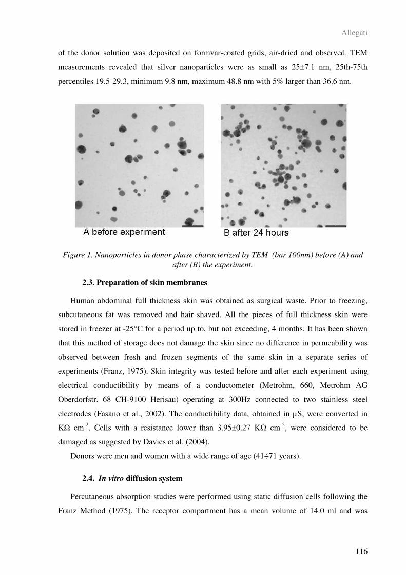
Allegati
116
of the donor solution was deposited on formvar-coated grids, air-dried and observed. TEM
measurements revealed that silver nanoparticles were as small as 25±7.1 nm, 25th-75th
percentiles 19.5-29.3, minimum 9.8 nm, maximum 48.8 nm with 5% larger than 36.6 nm.
Figure 1. Nanoparticles in donor phase characterized by TEM (bar 100nm) before (A) and
after (B) the experiment.
2.3. Preparation of skin membranes
Human abdominal full thickness skin was obtained as surgical waste. Prior to freezing,
subcutaneous fat was removed and hair shaved. All the pieces of full thickness skin were
stored in freezer at -25°C for a period up to, but not exceeding, 4 months. It has been shown
that this method of storage does not damage the skin since no difference in permeability was
observed between fresh and frozen segments of the same skin in a separate series of
experiments (Franz, 1975). Skin integrity was tested before and after each experiment using
electrical conductibility by means of a conductometer (Metrohm, 660, Metrohm AG
Oberdorfstr. 68 CH-9100 Herisau) operating at 300Hz connected to two stainless steel
electrodes (Fasano et al., 2002). The conductibility data, obtained in µS, were converted in
K cm-2
. Cells with a resistance lower than 3.95±0.27 K cm-2
, were considered to be
damaged as suggested by Davies et al. (2004).
Donors were men and women with a wide range of age (41÷71 years).
2.4. In vitro diffusion system
Percutaneous absorption studies were performed using static diffusion cells following the
Franz Method (1975). The receptor compartment has a mean volume of 14.0 ml and was
Allegati
117
maintained at 32°C by means of circulation of termostated water in the jacket surrounding the
cell. This temperature value has been chosen in order to reproduce the hand’s physiological
temperature at normal conditions. The concentration of the salt in the receptor fluid is
approximately the same that can be found in the blood. The solution in each cell was
continuously stirred using a Teflon coated magnetic stirrer.
Each piece of skin was clamped between the donor and the receptor compartment; the
mean exposed skin area was 3.29 cm² and the average membranes thickness was 1 mm.
The experiments were performed as follows:
Experiment 1
At time 0, the donor chambers of eight Franz diffusion cells were filled with 70 µg cm-2
of silver nanoparticles dispersed in ethanol absolute 0.14 wt % and diluited 1:10 with
synthetic sweat at pH 4.5 to reproduce in vivo condition.
At 24 h the dermal bathing solution was removed and silver concentration was determined
in each receiving chamber solution. Some skin tissue pieces were analyzed with TEM.
Experiment 2
Experiment 1 was repeated using an abraded skin protocol as suggested by Bronaugh and
Steward (1975): skin was abraded by drawing the point of a 19-gauge hypodermic needle
across the surface (20 marks in one direction and 20 perpendicular).
Blanks. For each experiment, four cells were added as blank. The blank cells were treated
as the other cells with the exception that no silver nanoparticle was introduced to the exposure
chamber.
Experiment 3
At time 0, the exposure chambers of 5 Franz diffusion cells were filled with 70 µg cm-2
of
nanoparticles solution dispersed in ethanol absolute 0.14 wt % and diluited 1:10 with
synthetic sweat at pH 4.5 to reproduce in vivo condition (1.5 ml solution). At selected
intervals (4, 8, 20, 24 h) 1.5 ml of the dermal bathing solution was removed and analyzed.
Each receptor sample was immediately replaced with an equal volume of fresh physiological
solution.
Experiment 4
Experiment 1 was repeated using an abraded skin protocol as described above. At selected
intervals (4, 8, 20, 24 h) 1.5 ml of the dermal bathing solution was removed and analyzed.
Each receptor sample was immediately replaced with an equal volume of fresh physiological
solution.
Allegati
118
2.5. Analytic Measurements
An electro-thermal atomic absorption spectrometry (GFAAS) with Zeeman background
correction was used to measure the Ag concentration in receiving phase. The instrument used
for analysis was a Perkin Elmer 4100ZL spectrophotometer equipped with an HGA graphite
furnace and autosampler AS/71. The analytical wavelenght was of 328.1 nm. The limit of
detection (LOD) for silver was 0.1 µg L-1
. The calibration curves were obtained using
standard at 5, 10 e 20 g L-1
and every sample was added up with a solution of 0.1%
Mg(NO3)2 and 1g L-1
of Pd(NO3)2 as matrix modifier.
A 5-point standard curve was used for the analytical measurements. The correlation
coefficient of the standard curve was: 0.9995 (r)
The samples were analyzed measuring against standard solutions for instrumental
calibration. The CV for the analysis of each metal was always less than 5%.
2.6. Skin fixation protocol for TEM analysis
After removal, some skin samples were cut into smaller sections and fixed for 3 h in a
solution of 3% glutaraldehyde (Serva, Heidelberg, Germany) in 0.1 M cacodylate buffer (pH
7.3). The fixed skin sections were washed twice (10 min each) with 0.1 M cacodylate buffer
and then post fixed with 1% osmium tetroxide for 1 h at 4°C. Post fixed samples were
dehydrated with an ascending ethanol series ending with 100% ethanol and then embedded in
Dow epoxy resin (DER332; Unione Chimica Europea, Milan, Italy) and DER732 (Serva).
The last resin embedding was made under vacuum. Semi fine and ultra thin sections were
prepared with an Ultramicrotome Leica Ultracut UCT (Leica Microsystems, Milan, Italy)
equipped with a diamond blade Drukker 3mm (Emme3, Milan Italy). Semi fine sections were
observed with an optical microscope Leitz Dialux 20 EB (Leica Microsystems, Milan, Italy)
instead ultra thin sections were double stained with lead citrate and uranyl acetate and
observed with a Transmission Electron Microscope (EM208; Philips, Eindhoven, The
Netherlands) with an high definition acquisition system SIS Morada and a digital image
acquisition system iTEM (FEI Italia, Milan, Italy).
2.7. Data analysis
Silver concentration data (µg cm-3
) in the receptor solution were converted to the total
amount that penetrated (µg cm-2
), with a correction for diluition due to sample removal.
Data analysis was performed using Excel for Windows release 2000 and the statistical
software SPSS for Windows. All data were tested for normal distribution (Kolmogorov-
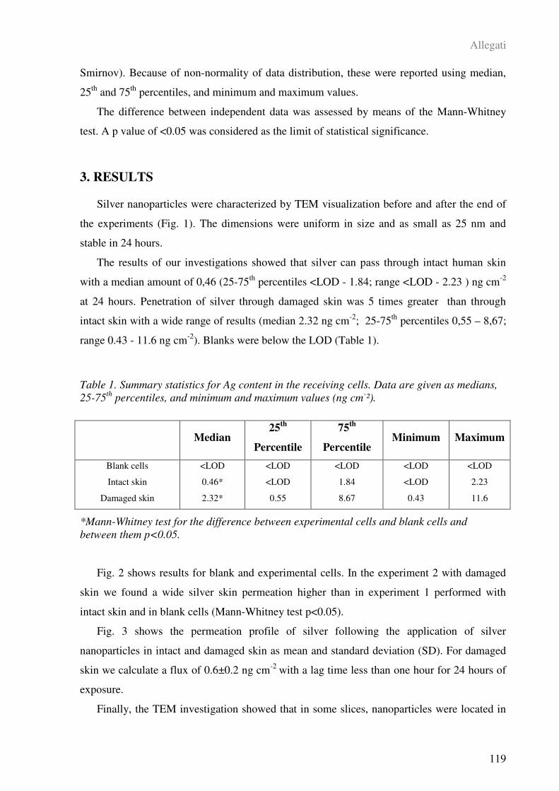
Allegati
119
Smirnov). Because of non-normality of data distribution, these were reported using median,
25th
and 75th
percentiles, and minimum and maximum values.
The difference between independent data was assessed by means of the Mann-Whitney
test. A p value of <0.05 was considered as the limit of statistical significance.
3. RESULTS
Silver nanoparticles were characterized by TEM visualization before and after the end of
the experiments (Fig. 1). The dimensions were uniform in size and as small as 25 nm and
stable in 24 hours.
The results of our investigations showed that silver can pass through intact human skin
with a median amount of 0,46 (25-75th
percentiles <LOD - 1.84; range <LOD - 2.23 ) ng cm-2
at 24 hours. Penetration of silver through damaged skin was 5 times greater than through
intact skin with a wide range of results (median 2.32 ng cm-2
; 25-75th
percentiles 0,55 – 8,67;
range 0.43 - 11.6 ng cm-2
). Blanks were below the LOD (Table 1).
Table 1. Summary statistics for Ag content in the receiving cells. Data are given as medians,
25-75th
percentiles, and minimum and maximum values (ng cm-²).
*Mann-Whitney test for the difference between experimental cells and blank cells and
between them p<0.05.
Fig. 2 shows results for blank and experimental cells. In the experiment 2 with damaged
skin we found a wide silver skin permeation higher than in experiment 1 performed with
intact skin and in blank cells (Mann-Whitney test p<0.05).
Fig. 3 shows the permeation profile of silver following the application of silver
nanoparticles in intact and damaged skin as mean and standard deviation (SD). For damaged
skin we calculate a flux of 0.6±0.2 ng cm-2
with a lag time less than one hour for 24 hours of
exposure.
Finally, the TEM investigation showed that in some slices, nanoparticles were located in
Median 25
th
Percentile
75th
Percentile Minimum Maximum
Blank cells
Intact skin
Damaged skin
<LOD
0.46*
2.32*
<LOD
<LOD
0.55
<LOD
1.84
8.67
<LOD
<LOD
0.43
<LOD
2.23
11.6

Allegati
120
the stratum corneum and in the upper layers of the epidermis (Figure 4).
Figure 2. Silver skin penetration at 24 hours (the lines in the box plots represent the median
values, the 25° and 75° percentiles).
Figure 3. Silver skin penetration cumulative curve (means and SD) in the 24 hours.
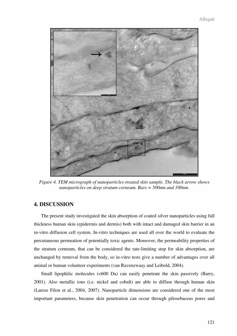
Allegati
121
Figure 4. TEM micrograph of nanoparticles-treated skin sample. The black arrow shows
nanoparticles on deep stratum corneum. Bars = 500nm and 100nm.
4. DISCUSSION
The present study investigated the skin absorption of coated silver nanoparticles using full
thickness human skin (epidermis and dermis) both with intact and damaged skin barrier in an
in-vitro diffusion cell system. In-vitro techniques are used all over the world to evaluate the
percutaneous permeation of potentially toxic agents. Moreover, the permeability properties of
the stratum corneum, that can be considered the rate-limiting step for skin absorption, are
unchanged by removal from the body, so in-vitro tests give a number of advantages over all
animal or human volunteer experiments (van Ravenzwaay and Leibold, 2004).
Small lipophilic molecules (<600 Da) can easily penetrate the skin passively (Barry,
2001). Also metallic ions (i.e. nickel and cobalt) are able to diffuse through human skin
(Larese Filon et al., 2004, 2007). Nanoparticle dimensions are considered one of the most
important parameters, because skin penetration can occur through pilosebaceus pores and

Allegati
122
sweat gland pores: particles with diameter ranging between 7-20 nm were almost exclusively
detected in the hair follicle infundibulum and below (Schaefer et al., 1990; Lauer et al., 2004;
Toll et al., 2004; Meidan et al., 2005). Alvarez-Roman et al. (2004) found that 20-200 nm
polystyrene nanoparticles were preferentially accumulated in follicle openings. Vogt et al.
(2006) showed that 40-nm particles were able to penetrate the perifollicular dermis through
the hair follicle. In contrast, for particle penetration through the intercellular transcutaneous
pathway (through the stratum corneum lipidic matrix) van den Bergh et al. (1999)
demonstrated that elastic particles of 100-150 nm could penetrate the stratum corneum matrix
through channel-like structures remaining confined in it. Honeywell-Nguyen et al. (2004)
found that elastic particles were able to reach the stratum corneum-viable epidermis junction
whereas rigid particles were recovered only in the superficial layers of stratum corneum.
A recent study performed by Baroli et al. (2007) showed that metallic (iron) nanoparticles
smaller than 10 nm are able to penetrate the skin through the stratum corneum lipidic matrix
and hair follicle orifices, reaching the deepest layers of the stratum corneum and less often the
uppermost strata of the viable epidermis, but do not permeate the skin.
To date, there has never been, to our knowledge, an in-vitro study of silver nanoparticles
permeation through human skin, and none of previous researchers studied the effect of skin
lesions on skin absorption. In fact, the amounts of silver permeating the skin found in the
experiments performed with intact skin and in the blank cells were just above or below the
analytical determination limit, while higher silver concentrations were found in the
experiments with damaged skin. Data from this study suggest that human skin absorption of
silver nanoparticles was relatively low compared with previously published absorption rates
for metal powders (Larese Filon et al., 2004, 2007): silver flux was 25 times less than nickel
powder flux and 200 time less than cobalt powder but only in condition of damaged skin. To
note that the concentration of silver nanopaticles in donor phase was 70µg cm-2
while Co and
Ni ions in donor phases were respectively 9 and 10 µg cm-2
.
However, our experiments showed not only that it is possible a low flux of silver after
nanoparticles application on donor phase, but also that rigid nanoparticles smaller than 30 nm
are able to penetrate passively the skin, reaching the deepest layers of the stratum corneum
and the outermost surface of the epidermis. These results may suggest that a small fraction of
the particles were dissolved and diffused through skin membranes as elemental silver. In
accordance with this hypothesis, the solubilization and oxidation of nickel particles on the
surface of the skin has been reported as the mechanism by which stratum corneum-diffusible
compounds are generated and able to penetrate stratum corneum, probably by the intercellular
Allegati
123
route (Hostynek et al., 2001).
Several factors and assumptions inherent to our in-vitro protocol may not represent real-
life situations. On one hand, a limitation of this study is that it has been performed in static
Franz cells where skin was static. On the other hand, flexing human epidermis has been
suggested to lead to the increased translocation of small particles into the membrane (Tinkle
et al., 2003).
Another aspect of our in-vitro experiments, which may not be realistic, is that the stratum
corneum was exposed to simulate sweat for 24 hours. Excessive hydration is known to
increase penetration of many compounds. Nevertheless, workers have often mechanically
damaged skin that includes cuts and abrasions, which would make penetration substantially
higher, as confirmed by this study.
In summary, the findings of this study provide the following new information on
nanoparticle skin absorption: (i) there is experimental evidence for in-vitro nanoparticle skin
permeation, and (ii) there is an appreciable increase in permeation using damaged skin.
This is the first time silver nanoparticles skin penetration was demonstrated: we need
more data on nanoparticles skin absorption and their potential risk through the cutaneous
route for occupational exposed people but also for consumers that can be repeated exposed to
nanoparticles applied on the skin.
5. REFERENCES
Alvarez-Romàn R, Naik A, Kalia YN, Guy RH, Fess H (2004) Skin penetration and
distribution of polymeric nanoparticles. J. Control Release 99: 53–62.
Barry BW (2001) Novel mechanisms and devices to enable successful transdermal drug
delivery. Eur. J. Pharm. Sci. 14: 101–114.
Baroli B, Ennas MG, Loffredo F, Isola M, Pinna R, Lopez-Quintela MA (2007)
Penetration of metallic nanoparticles in human full-thickness skin. J. Invest. Dermatol. 127:
1701–1712.
Bronough RL and Steward R (1985) Methods for in vitro percutaneous absorption studies.
V: permeation through damaged skin. J. Pharm. Sci. 15: 1062–1066.
Cross SE, Brian I, Roberts MS (2007) Human skin penetration of sunscreen nanoparticles:
in- vitro assessment of a novel micronized zinc oxide formulation. Skin. Pharmacol. Physiol.
20: 148–154.
Allegati
124
Davies DJ, Ward RJ, Heylings JR (2004) Multi-species assessment of electrical resistence
as a skin integrity marker for in vivo percutaneous absorption studies. Toxicol. in Vitro 18:
351–358.
Dussert AS, Gooris E, Hemmerle J (1997) Characterisation of the mineral content of a
physical sunscreen emulsion and its distribution onto human stratum corneum. Int. J. Cosmet.
Sci. 19: 185–187.
EDETOX (2000) Evaluations and Predictions of Dermal Absorption of Toxic Chemicals,
EU Framework V: Quality of Life, Environment and Health Key Action Funding (Project
Number: QLKA-2000-00196).
Fasano W, Manning L, Green J (2002) Rapid assessment of rat and human epidermal
membranes for in vitro dermal regulatory testing: correlation of electrical resistence with
tritiated water permeability. Toxicol. in Vitro 16: 731–740.
Franz TJ (1975) On the relevance of in vitro data. J. Invest. Dermatol. 93: 633–640.
Gamer AO, Leibold E, van Ravenzwaay B (2006) The in vitro absorption of microfine
zinc oxide and titanium dioxide through porcine skin. Toxicol. in Vitro 20: 301–307.
Graf C, Vossen DLJ, Imhof A, van Blaaderen A (2003) A general method to coat
colloidal particles with silica. Langmuir 19: 6693–6700.
Gupta AK and Gupta M (2005) Synthesis and surface engineering of iron oxide
nanoparticles for biomedical applications. Biomaterials 26: 3995–4021.
Hoet PHM, Bruske-Hohlfield I, Salata OV (2004) Nanoparticles - known and unknown
health risks. J. Nanobiotechnol. 2: 12.
Holsapple MP, Farland WH, Landry TD (2005) Research strategies for safety evaluation
of nanomaterials, part II: toxicological and safety evaluation of nanomaterials , current
challenges and data needs. Toxicol. Sci. 88: 12–17.
Holsapple MP nad Lehman-McKeeman LD (2005) Forum series: research strategies for
safety evaluation of nanomaterials. Toxicol. Sci. 88: 315–318.
Honeywell-Nguyen PL, Gooris GS, Bouwstra JA (2004) Quantitative assessment of the
transport of elastic and rigid vesicle components and a model drug from these vesicle
formulations into human skin in vivo. J. Invest. Dermatol. 123: 902–910.
Hostynek JJ, Dreher F, Pelosi A, Anigbogu A, Maibach HI (2001) Human stratum

Allegati
125
corneum penetration by nickel. In vivo study of depth distribution after occlusive application
of the metal as a powder. Acta Derm. Venereol. Suppl. 212: 5–10.
Lademann J, Weigmann HJ, Rickmeyer C, Barthelmes H, Schaefer H, Mueller G, Sterry
W (1999) Penetration of titanium dioxide microparticles in a sunscreen formulation into the
horny layer and the follicular orifice. Skin Pharmacol. Appl. Physiol. 12: 247–256.
Larese Filon F, Maina G, Adami G, Venier M, Coceani N, Bussani R, Massiccio M,
Barbieri P, Spinelli P (2004) In vitro percutaneous absorption of cobalt. Int. Arch. Occup.
Environ. Health 77: 85–89.
Larese Filon F, Adami G, Venier M, Maina G, Renzi N (2007) In vitro percutaneous
absorption of metal compounds. Toxicol. Lett. 170: 49–56.
Lauer AC, Ramachandran C, Lieb LM, Niemiec S, Weiner ND (1996) Targeted delivery
to the pilosebaceous unit via liposomes. Adv. Drug Deliv. Rev. 18: 311–324.
Meidan VM, Bonner MC, Michniak BB (2005) Transfollicular drug delivary – is it a
reality? Int. J. Pharm. 306: 1–14.
Neuberger T, Schopf B, Hofmann H, Hofmann M, von Rechenberg B (2005)
Superparamagnetic nanoparticles for biomedical applications: possibilities and limitations of a
new drug delivery system. J. Magn. Magn. Mat. 293: 483–496.
Oberdörster G, Oberdörster E, Oberdörster J (2005) Nanotoxicology: an emerging
discipline evolving from studies of ultrafine particles. Environ. Health Perspect. 113: 823–
839.
Penn SG, He L, Natan MJ (2003) Nanoparticles for bioanalysis. Curr. Opin. Chem. Biol.
7: 609–615.
Pflucker F, Wendel V, Hohenberg H, Gartner E, Will T, Pfeiffer S, Wepf R, Gers-Barlag
H (2001) The human stratum corneum layer: an effective barrier against dermal uptake of
different forms of topically applied micronised titanium dioxide. Skin Pharmacol. Appl. Skin
Physiol. 14 (Suppl. 1): 92–97.
Schaefer H, Watts F, Brod J, Illel B (1990) Follicular penetration. In: Prediction of
percutaneous penetration. Methods, measurements, modelling (Scott, R.C., Guy, R.H.,
Hadgraft, J., eds), London: IBC Technical Services, 163–732.
Schulz J, Hohenberg H, Pflucker F, Gartner E, Will T, Pfeiffer S, Wepf R, Wendel V,
Allegati
126
Gers-Barlag H, Wittern KP (2002) Distribution of sunscreens on skin. Adv. Drug Deliv. Rev.
54(S1): 157–163.
Thomas K and Sayre P (2005) Research strategies for safety evaluation of nanomaterials,
part I: evaluating the human health implications of exposure to nanoscale materials. Toxicol.
Sci. 87: 316–321.
Tinkle SS, Antonini JM, Rich BA, Roberts JR, Salmen R, De Pree K, Adkins EJ (2003)
Skin as a route of exposure and sensitization in chronic beryllium disease. Environ. Health
Perspect. 111: 1202–1208.
Toll R, Jacobi U, Richter H, Laedmann J, Shaefer H, Blume-Peytavi U (2004) Penetration
profile of microspheres in follicular targeting of terminal hair follicles. J. Invest. Dermatol.
123: 168–176.
van den Bergh BA, Bouwstra JA, Junginger HE, Wertz PW (1999a) Elasticity of veicles
affects hairless mouse skin structure and permeability. J. Control. Release 62: 367–379.
van den Bergh BA, Vroom J, Gerritsen H, Junginger HE, Bouwstra JA (1999b)
Interactions of elastic and rigid vesicles with human skin in vitro: electron microscopy and
two-photon excitation microscopy. Biochim. Biophys. Acta 1461: 155–173.
van Ravenzwaay B and Leibold E (2004) The significance of in vitro rat skin absorption
studies to human risk assessment. Toxicol. in Vitro 18: 219–225.
Vogt A, Combadiere B, Hadam S, Stieler KM, Lademann J, Schaefer H, Autran B,
Sterry1 W, Blume-Peytavi U (2006) 40 nm, but not 750 or 1500 nm, nanoparticles enter
epidermal CD1a + cells after transcutaneous application on human skin. J. Invest. Dermatol.
126: 1316–1322.
Allegati
127
ALLEGATO V
HUMAN SKIN PENETRATION OF GOLD NANOPARTICLES THROUGH INTACT
AND DAMAGED SKIN
Francesca Larese Filon, Matteo Crosera, Gianpiero Adami, Massimo Bovenzi, Federica
Rossi, Giovanni Maina
Manoscritto in preparazione
Allegati
128
ABSTRACT
The potential for solid nanoparticles to penetrate the skin lies at the centre of the debate
concerning the safety for their use. Gold nanoparticles are produced for many applications in
photocatalysts, photoelectrochemical materials, in medical fields as chronophores for
immunoreactions. The benefits of nanoparticles have been shown in several scientific fields,
but little is known about their potential to diffuse into tissues. Currently there is a lack of
available data demonstrating whether manufactured nanoparticles can gain access to the
epidermis and derma after skin contact even though it is known that dermally administered
nanoparticles can transfer to regional lymph nodes via skin macrophages and Langerhans
cells.
This study aims at evaluating in vitro skin penetration of gold nanoparticles. Experiments
were performed using the Franz diffusion cell method with intact and damaged human skin.
Physiological solution was used as receiving phase and 3ml of a solution containing 100 ppm
of gold nanoparticles diluited 1:1 in water was applied as donor phase to the outer surface of
the skin for 24 h. The receptor fluid measurements were performed by Inductively Coupled
Plasma Mass Spectroscopy (ICP-MS). Human skin penetration was also determined by using
Transmission Electron Microscope (TEM) to verify the location of gold nanoparticles in
exposed membranes.
Mean gold content of 214±43.7 ng cm-2
and 187.7±50.2 ng cm-2
were found in the
receiving solutions of cells where the nanoparticles solution was applied on intact skin (8
cells) and on damaged skin (8 cells) respectively. 24 hours gold flux permeation was 7.8±2.0
ng cm-2
h-1
in intact skin and 7.1±2.5 ng cm-2
h-1
in damaged skin, with a lag time < 1 hour.
Moreover, gold nanoparticles could be detected in the stratum corneum, in the deeper layers
of the epidermis and, finally in the dermis by electron microscopy.
This study showed that gold applied as nanoparticles is able to permeate the human skin
in an in-vitro diffusion cell system.
KEYWORDS: Nanoparticles; Gold; Skin absorption; In vitro; Damaged skin
Allegati
129
RIASSUNTO
La possibilità per nanoparticelle solide di penetrare all’interno della cute è uno dei punti al
centro del dibattito sulla sicurezza del loro uso. Le nanoparticelle d’oro vengono prodotte per
molte applicazioni, per esempio, in fotocatalisi, come cromofori per immunoreazioni o come
carrier per farmaci, ecc. I vantaggi dell’uso delle nanoparticelle sono stati dimostrati in molti
campi, ma poco si sa circa il loro potenziale di penetrazione e diffusione nei tessuti.
Attualmente vi è una lacuna nei dati disponibili sulla capacità di nanoparticelle artificiali di
raggiungere l’epidermide e il derma dopo essere venute in contatto con la cute, anche se è
stato dimostrato che nanoparticelle somministrate nel derma sono in grado di migrare ai nodi
linfatici trasportate dai macrofagi e dalle cellule di Langerhans.
Questo studio ha l’obiettivo di studiare la penetrazione di nanoparticelle d’oro in un
sistema in vitro. Gli esperimenti sono stati condotti usando il metodo delle celle di diffusione
di Franz con cute umana intera, integra e lesa. Come fase ricevente è stata usata soluzione
fisiologica e come fase donatrice sono stati applicati, sulla superficie esterna della cute, 3 ml
di una soluzione contente 100 ppm di nanoparticelle d’oro diluite in acqua milliQ, per 24 ore.
Le analisi delle varie soluzioni sono state effettuate tramite Spettroscopia di Massa al Plasma
Induttivamente Accoppiato (ICP-MS). Sul tessuto cutaneo sono state effettuate anche analisi
di microsopia elettronica a trasmissione (TEM).
Nelle fasi riceventi è stato trovato un contenuto medio di oro di 214±44 ng cm-2
e di
188±50 ng cm-2
rispettivamente nelle celle con cute integra e nelle celle con cute lesa. Nelle
24 ore è stato calcolato un flusso di 7,8±2,0 ng cm-2
h-1
nella cute integra e 7,1±2,5 ng cm-2
h-1
nella cute lesa, con un lag time inferiore ad 1 ora. Le analisi al TEM hanno evidenziato
nanoparticelle d’oro sia nell’epidermide che nel derma.
Questo studio ha mostrato che in un sistema di diffusione in vitro le nanoparticelle d’oro
sono in grado di penetrare nello spessore cutaneo e di essere ritrovate nei vari strati della
pelle.
PAROLE CHIAVE: Nanoparticelle; Oro; Assorbimento cutaneo; In vitro; Cute
danneggiata
Allegati
130
1. INTRODUCTION
Over the past decades an enormous interest in nanotechnologies has grown all over the
world for their possible applications in a wide range of fields because of their small size (100
nm or less), comparable to that of biological molecules. In recent years the use of gold
nanoparticles and nanorods is increasing since their valuable size and shape dependent
properties are easy to control and modify and this offers powerful tools for the researchers
activity (Penn et al., 2003; Gupta and Gupta, 2005; Neuberger et al., 2005; Chen et al., 2008).
In particular, gold nanomaterials are rising as a lead candidate in the transport of payloads
inside tissues and cells for drug delivery, gene therapy and cell imaging. Rosi et al. (2005)
described the use of gold nanoparticle-oligonucleotide complexes as intracellular gene
regulation agents for the control of protein expression in cells. These particles were stable
against enzymatic digestion and carried oligonucleotides at a higher effective concentration
than conventional transfection agents. Kawano et al. (2006) investigated the benefits of
combining the use of PEG-modified cationic gold nanoparticles with electroporation for in-
vivo gene delivery. The authors concluded that these modified nanoparticles maintained DNA
more stably in the blood flow than in the case of naked DNA and electroporation assisted in
restricted gene expression of circulating DNA in limited areas of the liver. Several other
studies have been carried out on the use of modified gold nanomaterials in the imaging and
therapy of tumors (Bhattacharya and P. Mukherjee, 2008; El-Sayed et al., 2006), in the direct
welding of connective tissues [Gobin et al., 2005; Ratto et al., 2009), as immunostaining
marker particles for electron microscopy, chromophores for immunoreactions (Mirkin et al.,
1996), photocatalyst (Kamat, 2002) and Raman sensors (Tian et al., 2002).
Gold nanoparticles are stable in the body (Niidome et al., 2006), but it is datable whether
gold nanoparticles are biocompatible and more data are needed to better understand their fate
into the body (Lewinski et al., 2008, Takahashi et al., 2006).
Therefore, among the great number of applications, gold nanoparticles would be an
interesting carrier for transdermal delivery (Chen et al., 2008), but to our knowledge, there are
no data on their skin permeation in in-vitro system using human skin.
Larese et al. (2009a) demonstrated the permeation of silver nanoparticles sized 25 nm
through intact and damaged human skin (0.46 ng cm−2
and 2.32 ng cm−2
respectively) using
the Franz cell method (Franz., 1975), while Sonavane et al. (2008) revealed higher skin and
intestine in-vitro permeation of gold nanoparticles with smaller size (15 nm) in rats. Diffusion
data would permit to increase knowledge on gold nanoparticles.

Allegati
131
To investigate the potential gold nanoparticles skin absorption in-vitro, we carried out a
series of permeation experiments with human skin using a Franz static diffusion cell. Using
this technique, we investigated the total amount of gold permeating through human skin
during a 24-hour period. Experiments were run with intact skin as well as abraded skin
following the Bronaugh and Steward (1985) protocol to estimate the effect of skin lesions on
the permeation rate. In this study we applied the protocol adopted in the European project
EDETOX (Evaluations and predictions of DErmal absorption of TOXic chemicals), a three-
year research program (2001-2004) financially supported by the European Commission
(EDETOX, 2000).
2. MATERIALS AND METHODS
2.1. Chemicals
All chemicals were analytical grade Urea, magnesium nitrate hexahydrate, palladium
nitrate, sodium hydrogenphosphate, potassium dihydrogenphosphate, and hydrogen peroxide
(30%) were purchased from Carlo Erba (Milan, Italy); ammonium hydroxide (25%) from J. T.
Baker (Deventer, Holland); lactic acid (90%) from Acros Organics (Geel, Belgium); sodium
tetrachloroauric acid, trisodium citrate dihydrate, nitric acid (>69%) and hydrochloric acid
(36.5-38%) from Sigma Aldrich (Milan, Italy). Water reagent grade was produced with a
Millipore purification pack system (milliQ water).
The physiological solution used as the receptor fluid was prepared by dissolving 2.38 g of
Na2HP04, 0.19 g of KH2P04 and 9 g of NaCl into 1 L of milliQ water (final pH = 7.35).
The synthetic sweat solution used as the donor fluid included 0.5% sodium chloride, 0.1%
urea and 0.1% lactic acid in milliQ water; pH 4.5 was adjusted with ammonia.
2.2. Nanoparticle synthesis and characterization
Gold nanoparticles were synthesized by reducing tetrachloroauric acid with sodium citrate
as described by Grabar et al. 1995. Trasmission Electron Microscopy (TEM) was used to
evaluate nanoparticles size and stability before and after experimentsll as 12.6 ± 0.9 nm
(number of nanoparticles measured: 345).
Total gold concentration was analysed by Inductively Coupled Plasma - Atomic Emission
Spectroscopy (ICP-AES): 1 ml of nanoparticle suspension was treated with 1 ml of Aqua
regia (a solution having concentrated nitric acid and concentrated hydrochloric acid mixed at

Allegati
132
a volume ratio of 1:3) at room temperature and the volume was adjusted to 10 ml with milliQ
water. The gold solution was measured at a wavelength of 267.595 nm. A five-point standard
curve was used for the analytical measurements. The correlation coefficient of the standard
curve was 0.9989. The samples were analysed by measuring against standard solutions for
instrumental calibration. The coefficient of variation (CV) for the analysis of each metal was
less than 3% and the limit of detection was 0.030 mg L-1
. The determined concentration of
gold in the initial nanoparticle suspension was 100±5 mg L-1
(n = 3).
In order to evaluate the possible presence of gold ions in solution, 10 ml of gold
nanoparticle suspension was added with 10 mg of sodium chloride to aggregate the
nanoparticles and then centrifuged; the surfactant was then removed and analysed by mean of
ICP-AES. Concentration of gold was under the limit of detection (0.030 mg L-1
).
2.3. Preparation of skin membranes
Human abdominal full thickness skin was obtained as surgical waste after the
authorization of the local Ethical Committee. Prior to freezing, subcutaneous fat was
removed and hair shaved by a razor. All the pieces of full thickness skin were stored in
freezer at -25°C for a period up to, but not exceeding, 4 months. It has been shown that this
method of storage does not damage the skin since no difference in permeability was observed
between fresh and frozen segments of the same skin in a separate series of experiments
(Franz, 1975). Skin integrity was tested before and after each experiment using electrical
conductibility by means of a conductometer (Metrohm, 660, Metrohm AG Oberdorfstr. 68
CH-9100 Herisau) operating at 300Hz and connected to two stainless steel electrodes (Fasano
et al., 2002). The conductibility data in µS were converted into K cm-2
. Cells with a
resistance lower than 3.95±0.27 K cm-2
were considered to be damaged as suggested by
Davies et al. (2004)
Donors were men and women with a wide range of age (41-71 years).
2.4. In vitro diffusion system
Percutaneous absorption studies were performed using static diffusion cells following the
Franz method. The receptor compartment had a mean volume of 14 ml and was maintained at
32°C by means of circulation of thermostated water in the jacket surrounding the cell. This
temperature value was chosen in order to reproduce the hand physiological temperature at
normal conditions. The concentration of the salt in the receptor fluid was approximately the
same that can be found in the blood. The solution in each cell was continuously stirred using a
Allegati
133
Teflon coated magnetic stirrer.
Each piece of skin was clamped between the donor and the receptor compartment; the
mean exposed skin area was 3.29 cm² and the average membranes thickness was 1 mm.
The experiments were carried out as follows:
Experiment 1
At time 0, the exposure chambers of 8 Franz diffusion cells were filled with 0.5 ml
solution containing 100 mg L-1
of nanoparticles solution diluted 1:3 (15 µg cm-2
) with
synthetic sweat at pH 4.5 to reproduce in vivo condition.
At 24 h the dermal bathing solution was removed and gold concentration was determined
in each receiving chamber solution.
Experiment 2
Experiment 1 was repeated using an abraded skin protocol as suggested by Bronaugh and
Steward (1985): skin was abraded by drawing the point of a 19-gauge hypodermic needle
across the surface (20 marks in one direction and 20 perpendicular).
Experiment 3
At time 0, the exposure chambers of 8 Franz diffusion cells were filled with 1.5 ml
solution containing 100 mg L-1
of nanoparticles solution diluted 1:1 with milliQ water (45 µg
cm-2
). At selected intervals (4, 8, 16, 24 h) 1.5 ml of the dermal bathing solution was removed
and analyzed. Each receptor sample was immediately replaced with an equal volume of fresh
physiological solution.
Experiment 4
Experiment 3 was repeated using an abraded skin protocol as described above.
Blanks: for each experiment, four cells were added as blank. The blank cells were treated
as the other cells with the exception that no gold nanoparticles was introduced to the exposure
chamber.
Experiments 1 and 2 were preliminary tests to evaluate nanoparticles permeation using the
same protocol previously reported (Larese et al., 2009a). Experiments 3 and 4 were performed
using a higher gold concentration in milliQ water to avoid interference of the saline on gold
nanoparticles .
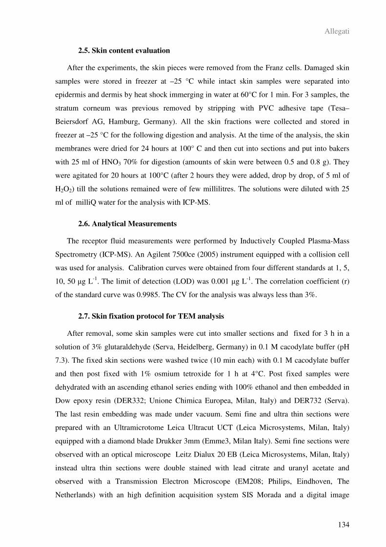
Allegati
134
2.5. Skin content evaluation
After the experiments, the skin pieces were removed from the Franz cells. Damaged skin
samples were stored in freezer at –25 °C while intact skin samples were separated into
epidermis and dermis by heat shock immerging in water at 60°C for 1 min. For 3 samples, the
stratum corneum was previous removed by stripping with PVC adhesive tape (Tesa–
Beiersdorf AG, Hamburg, Germany). All the skin fractions were collected and stored in
freezer at –25 °C for the following digestion and analysis. At the time of the analysis, the skin
membranes were dried for 24 hours at 100° C and then cut into sections and put into bakers
with 25 ml of HNO3 70% for digestion (amounts of skin were between 0.5 and 0.8 g). They
were agitated for 20 hours at 100°C (after 2 hours they were added, drop by drop, of 5 ml of
H2O2) till the solutions remained were of few millilitres. The solutions were diluted with 25
ml of milliQ water for the analysis with ICP-MS.
2.6. Analytical Measurements
The receptor fluid measurements were performed by Inductively Coupled Plasma-Mass
Spectrometry (ICP-MS). An Agilent 7500ce (2005) instrument equipped with a collision cell
was used for analysis. Calibration curves were obtained from four different standards at 1, 5,
10, 50 g L-1
. The limit of detection (LOD) was 0.001 g L-1
. The correlation coefficient (r)
of the standard curve was 0.9985. The CV for the analysis was always less than 3%.
2.7. Skin fixation protocol for TEM analysis
After removal, some skin samples were cut into smaller sections and fixed for 3 h in a
solution of 3% glutaraldehyde (Serva, Heidelberg, Germany) in 0.1 M cacodylate buffer (pH
7.3). The fixed skin sections were washed twice (10 min each) with 0.1 M cacodylate buffer
and then post fixed with 1% osmium tetroxide for 1 h at 4°C. Post fixed samples were
dehydrated with an ascending ethanol series ending with 100% ethanol and then embedded in
Dow epoxy resin (DER332; Unione Chimica Europea, Milan, Italy) and DER732 (Serva).
The last resin embedding was made under vacuum. Semi fine and ultra thin sections were
prepared with an Ultramicrotome Leica Ultracut UCT (Leica Microsystems, Milan, Italy)
equipped with a diamond blade Drukker 3mm (Emme3, Milan Italy). Semi fine sections were
observed with an optical microscope Leitz Dialux 20 EB (Leica Microsystems, Milan, Italy)
instead ultra thin sections were double stained with lead citrate and uranyl acetate and
observed with a Transmission Electron Microscope (EM208; Philips, Eindhoven, The
Netherlands) with an high definition acquisition system SIS Morada and a digital image
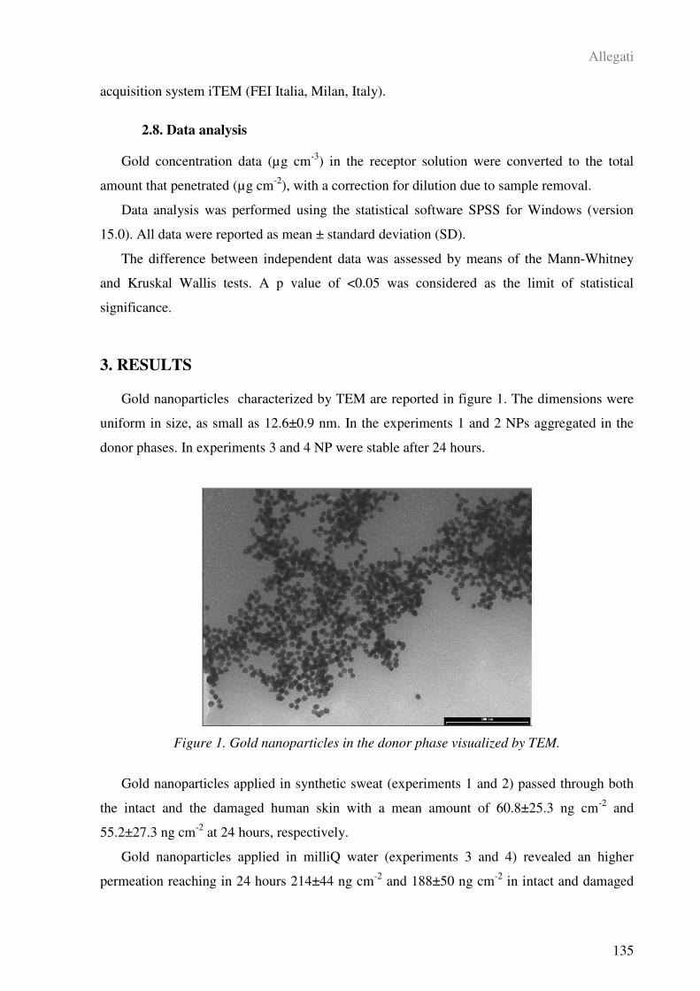
Allegati
135
acquisition system iTEM (FEI Italia, Milan, Italy).
2.8. Data analysis
Gold concentration data (µg cm-3
) in the receptor solution were converted to the total
amount that penetrated (µg cm-2
), with a correction for dilution due to sample removal.
Data analysis was performed using the statistical software SPSS for Windows (version
15.0). All data were reported as mean ± standard deviation (SD).
The difference between independent data was assessed by means of the Mann-Whitney
and Kruskal Wallis tests. A p value of <0.05 was considered as the limit of statistical
significance.
3. RESULTS
Gold nanoparticles characterized by TEM are reported in figure 1. The dimensions were
uniform in size, as small as 12.6±0.9 nm. In the experiments 1 and 2 NPs aggregated in the
donor phases. In experiments 3 and 4 NP were stable after 24 hours.
Figure 1. Gold nanoparticles in the donor phase visualized by TEM.
Gold nanoparticles applied in synthetic sweat (experiments 1 and 2) passed through both
the intact and the damaged human skin with a mean amount of 60.8±25.3 ng cm-2
and
55.2±27.3 ng cm-2
at 24 hours, respectively.
Gold nanoparticles applied in milliQ water (experiments 3 and 4) revealed an higher
permeation reaching in 24 hours 214±44 ng cm-2
and 188±50 ng cm-2
in intact and damaged
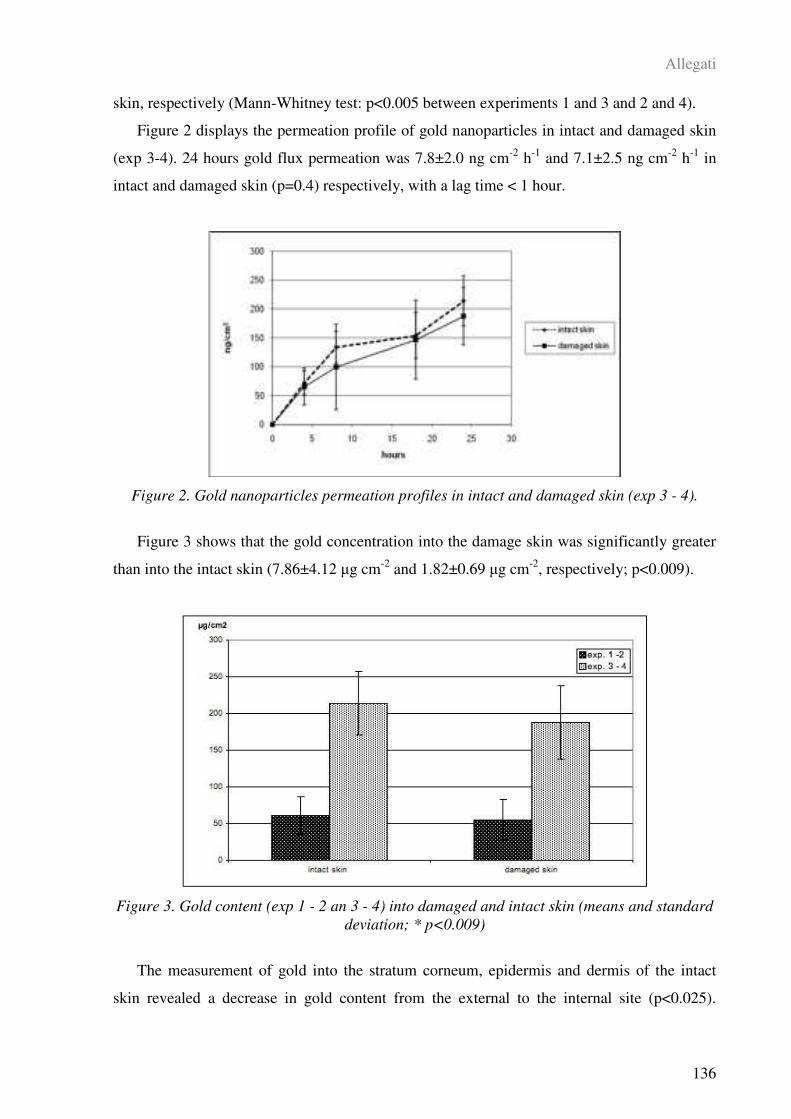
Allegati
136
skin, respectively (Mann-Whitney test: p<0.005 between experiments 1 and 3 and 2 and 4).
Figure 2 displays the permeation profile of gold nanoparticles in intact and damaged skin
(exp 3-4). 24 hours gold flux permeation was 7.8±2.0 ng cm-2
h-1
and 7.1±2.5 ng cm-2
h-1
in
intact and damaged skin (p=0.4) respectively, with a lag time < 1 hour.
Figure 2. Gold nanoparticles permeation profiles in intact and damaged skin (exp 3 - 4).
Figure 3 shows that the gold concentration into the damage skin was significantly greater
than into the intact skin (7.86±4.12 g cm-2
and 1.82±0.69 g cm-2
, respectively; p<0.009).
Figure 3. Gold content (exp 1 - 2 an 3 - 4) into damaged and intact skin (means and standard
deviation; * p<0.009)
The measurement of gold into the stratum corneum, epidermis and dermis of the intact
skin revealed a decrease in gold content from the external to the internal site (p<0.025).

Allegati
137
Figure 4 shows the gold content into strip numbers ranging from 0.40±0.05 g cm-2
in the
superficial strip (1st) to 0.17±0.01 g cm
-2 into the deepest strip (6
th) (p<0.025).
Figure 4. Gold content (exp 3) into stratum corneum tape stripping (means and standard
deviation; *p<0.025). From 1st more superficial to 6th more deep.
The gold content into the epidermis (1.74±0.68 g cm-2
) and into the derma (0.05±0.02 g
cm-2
) revealed a significant decrease in gold content in the deepest layers of the skin
(p<0.005, Figure 5).
Figure 5. Gold content (exp 3) into epidermis and dermis of intact skin samples (means and
standard deviation; * p<0.005)
The TEM investigation showed that in some slices of both intact and damaged skin, gold
nanoparticles were located in the stratum corneum and in the epidermis, but also in the derma
(Figure 6).

Allegati
138
Figure 6. TEM micrographs of nanoparticles-treated intact skin sample. The black arrow
shows nanoparticles on different dermal leyers. Bars: a = 10 µm; b,c,d = 200nm.
4. DISCUSSION
The present study investigated the skin absorption of gold nanoparticles using full
thickness human skin (epidermis and dermis) with both intact and damaged skin barrier in an
in-vitro diffusion cell system.
Nanoparticles skin permeation is something new and there are very few data available in
the literature. For TiO2 the results of the three-year European project NANODERM (2007)
reported no evidence of nanoparticle transcutaneous penetration (NANODERM, 2007; Kiss et
al., 2008). In opposite, Baroli et al. (2007) showed that metallic (iron) nanoparticles smaller
than 10 nm are able to penetrate the skin through the stratum corneum lipidic matrix and hair
follicle orifices, reaching the deepest layers of the stratum corneum and less often the
uppermost strata of the viable epidermis. The authors, however, found that the nanoparticles
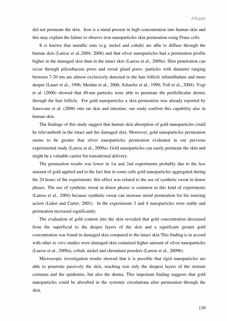
Allegati
139
did not permeate the skin. Iron is a metal present in high concentration into human skin and
this may explain the failure to observe iron nanoparticles skin permeation using Franz cells.
It is known that metallic ions (e.g. nickel and cobalt) are able to diffuse through the
human skin (Larese et al.,2004; 2006) and that silver nanoparticles had a permeation profile
higher in the damaged skin than in the intact skin (Larese et al., 2009a). Skin penetration can
occur through pilosebaceus pores and sweat gland pores: particles with diameter ranging
between 7-20 nm are almost exclusively detected in the hair follicle infundibulum and more
deeper (Lauer et al., 1996; Meidan et al., 2006; Schaefer et al., 1990; Toll et al., 2004). Vogt
et al. (2006) showed that 40-nm particles were able to penetrate the perifollicular dermis
through the hair follicle. For gold nanoparticles a skin permeation was already reported by
Sanovane et al. (2008) into rat skin and intestine; our study confirm this capability also in
human skin.
The findings of this study suggest that human skin absorption of gold nanoparticles could
be relevantboth in the intact and the damaged skin. Moreover, gold nanoparticles permeation
seems to be greater that silver nanoparticles permeation evaluated in our previous
experimental study (Larese et al., 2009a). Gold nanoparticles can easily permeate the skin and
might be a valuable carrier for transdermal delivery.
The permeation results was lower in 1st and 2nd experiments probably due to the less
amount of gold applied and to the fact that in some cells gold nanoparticles aggregated during
the 24 hours of the experiments: this effect was related to the use of synthetic sweat in donor
phases. The use of synthetic sweat in donor phases is common in this kind of experiments
(Larese et al., 2004) because synthetic sweat can increase metal permeation for his ionizing
action (Liden and Carter, 2001). In the experiments 3 and 4 nanoparticles were stable and
permeation increased significantly.
The evaluation of gold content into the skin revealed that gold concentration decreased
from the superficial to the deeper layers of the skin and a significant greater gold
concentration was found in damaged skin compared to the intact skin This finding is in accord
with other in vitro studies were damaged skin contained higher amount of silver nanoparticles
(Larese et al., 2009a), cobalt, nickel and chromium powders (Larese et al., 2009b).
Microscopic investigation results showed that it is possible that rigid nanoparticles are
able to penetrate passively the skin, reaching non only the deepest layers of the stratum
corneum and the epidermis, but also the derma. This important finding suggests that gold
nanoparticles could be absorbed in the systemic circulationa after permeation through the
skin.
Allegati
140
There are some limitations of this study that deserve attention. A limitation is that the
experiments were carried out with Franz cells in which skin was static. Flexing human
epidermis has been suggested to lead to the increased translocation of small particles into the
membrane (Rouse et al., 2007; Tinkle et al., 2003).
Another limitation of our in-vitro experiments is that the stratum corneum was exposed
for 24 hours. Excessive hydration is known to increase penetration of many compounds.
Nevertheless, people have often mechanically damaged skin such as cuts and abrasions,
which would make penetration substantially higher.
In conclusion, the findings of this experimental study showed that gold nanoparticles
could be useful for transdermal delivery applications and that this kind of nanoparticles can
permeate the skin in greater amount than other nanoparticles such us silver nanoparticles
(Larese et al., 2009a). More studied are needed to evaluate skin absorption using different size
and shape of gold nanoparticles.
5. REFERENCES
Baroli B, Ennas MG, Loffredo F, Isola M, Pinna R, Lopez-Quintela MA (2007)
Penetration of metallic nanoparticles in human full-thickness skin. J. Investig. Dermatol. 127:
1701- 712.
Bhattacharya R and Mukherjee P (2008) Biological properties of “naked” metal
nanoparticles. Adv. Drug. Deliv. Rev. 60: 1289-1306.
Bronough R and Steward R (1985) Methods for in vitro percutaneous absorption studies.
V: permeation through damaged skin. J. Pharm. Sci. 15: 1062-1066.
Chen PC, Mwakwari SC, Oyelere AK (2008). Gold nanoparticles: From nanomedicine to
nanosensing. Nanotechnol, Science Appl.. 1: 45-66.
Davies DJ, Ward RJ, Heylings JR (2004) Multi-species assessment of electrical resistence
as a skin integrity marker for in vivo percutaneous absorption studies. Toxicol. in Vitro 18:
351-358.
EDETOX (2000) Evaluations and Predictions of Dermal Absorption of Toxic Chemicals,
EU Framework V: Quality of Life, Environment and Health Key Action Funding (Project
Number: QLKA-2000-00196).
El-Sayed IH, Huang X, El-Sayed MA (2006) Selective laser photo-thermal therapy of
Allegati
141
epithelial carcinoma using anti-EGFR antibody conjugated gold nanoparticles. Cancer Lett.
239: 129-135.
Fasano W, Manning L, Green J (2002). Rapid assessment of rat and human epidermal
membranes for in vitro dermal regulatory testing: correlation of electrical resistance with
tritiated water permeability. Toxicol. In vitro 16: 731-740.
Franz TJ (1975). On the relevance of in vitro data. J. Invest. Dermatol. 93, 633-640.
Gobin AM, O’Neal DP, Watkins DM, Halas NJ, Drezek RA, West JL (2005) Near
Infrared Laser-Tissue Welding Using Nanoshells as an Exogenous Absorber. Laser Surg.
Med. 37: 123-129.
Grabar CK, Griffith Freeman R, Hommer MB, Natam MJ (1995) Preparation and
characterization of Au Colloid Monolayers. Anal. Chem. 67: 735-743.
Gupta AK, Gupta M (2005) Synthesis and surface engineering of iron oxide nanoparticles
for biomedical applications. Biomaterials 26: 3995-4021.
Kamat PV (2002) Photophysical, Photochemical and Photocatalytic Aspects of Metal
Nanoparticles. J. Phys. Chem. B 106: 7729-7744.
Kawano T, Yamagata M, Takahashi H, Niidome Y, Yamada S, Katayama Y, Niidome T,
(2006) Stabilizing of plasmid DNA in vivo by PEG-modified cationic gold nanoparticles and
the gene expression assisted with electrical pulses. J. Control. Release. 111: 382-389.
Kiss B, Biró T, Czifra G, Tóth BI, Kertész Zs, Szikszai Z, Kiss AZ, Juhász I, Zouboulis
CC, Hunyadi J (2008) Investigation of micronizided titanium dioxide penetration in human
skin xenografts and its effect on cellular functions of human skin-derived cells. Experiment.
Dermatol. 17: 659-67.
Larese Filon F, Maina G, Adami G, Venier M, Coceani N, Bussani R, Massiccio M,
Barbieri P, Spinelli P (2004) In vitro percutaneous absorption of cobalt. Int. Arch. Environ.
Health 77: 85-89.
Larese Filon F, Adami G, Venier M, Maina G, Renzi N (2007) In vitro percutaneous
absorption of metal compounds. Toxicol. Lett. 170: 49-56.
Larese Filon F, D’Agostin F, Crosera M, Adami G, Renzi N, Bovenzi M, Maina G
(2009a) Human skin penetration of silver nanoparticles through intact and damaged skin.
Toxicol. 255: 33-37.
Allegati
142
Larese Filon F, D’Agostin F, Crosera M, Adami G, Bovenzi M, Maina G (2009b) In vitro
absorption of metal powders through intact and damaged human skin. Toxicol. in vitro 23:
574-579.
Lauer AC, Ramachandran C, Lieb LM, Niemiec S, Weiner ND (1996) Targeted delivery
to the pilosebaceous unit via liposomes. Adv. Drug Deliv. Rev. 18: 311-324.
Lewinski N, Colvin V, Drezek R (2008) Cytotoxicity of Nanoparticles. Small 4: 26-49.
Meidan VM, Bonner MC, Michniak BB (2005) Transfollicular drug delivary – is it a
reality? Int. J. Pharm. 306: 1-14.
Mirkin CA, Letsinger RL, Mucic RC, Storhoff JJ (1996) A DNA-based method for
rationally assembling nanoparticles into macroscopic materials. Nature 382: 607-609.
NANODERM (2007) Quality of skin as a barrier to ultra-fine particles. Final Report.
(Project Number: QLK4-CT-2002-02678) www.uni-leipzig.de/~nanoderm
Neuberger T, Schopf B, Hofmann H, Hofmann M, von Rechenberg B (2005)
Superparamagnetic nanoparticles for biomedical applications: possibilities and limitations of a
new drug delivery system. J. Magn. Magn. Mat. 293: 483–496.
Niidome T, Yamagata M, Okamoto Y, Akiyama Y, Takahashi H, Kawano T, Katayama
Y, Niidome Y (2006) PEG-modified gold nanorods with a stealth character for in vivo
applications. J. Control. Release 114: 343-347.
Penn SG, He L, Natan MJ (2003) Nanoparticles for bioanalysis. Curr. Opin. Chem. Biol.
7: 609–615.
Ratto F, Matteini P, Rossi F, Menabuoni L, Tiwari N, Kulkarni SK, Pini R (2009)
Photothermal effects in connective tissues mediated by laser-activated gold nanorods.
Nanomed.: Nanotechnol. Biol. Med. 5: 143-151.
Rosi NL, Giljohann DA, Shad Thaxton C, Lytton-Jean AKR, Han MS, Mirkin CA (2006)
Oligonucleotide-Modified Gold Nanoparticles for Intracellular Gene Regulation. Science,
312: 1027-1030.
Rouse JG, Yang J, Ryman-Rasmussen JP, Barron AR, Monteiro-Riviere NA (2007)
Effects of mechanical flexion on the penetration of fullerene amino acid-derivatized peptide
nanoparticles through skin. Nano Lett. 7: 155-160.
Schaefer H, Watts F, Brod J, Illel B (1990) Follicular penetration. In: Prediction of
Allegati
143
percutaneous penetration. Methods, measurements, modelling (Scott, R.C., Guy, R.H.,
Hadgraft, J., eds), London: IBC Technical Services, 163 732.
Sonavane G, Tomoda K, Sano A, Ohshima H, Terada H, Makino K (2008) In vitro
permeation of gold nanoparticles through rat skin and rat intestine: Effect of particle size.
Colloids Surf. B Biointerfaces. 65: 1-10.
Takahashi H, Niidome Y, Niidome T, Kaneko K, Kawasaki H, Yamada S (2006)
Modification of Gold Nanorods Using Phosphatidylcholine to Reduce Cytotoxicity. Langmuir
22: 2-5.
Tian ZQ, Ren B, Wu DY (2002) Surface-Enhanced Raman Scattering: From Noble to
Transition Metals and from Rough Surfaces to Ordered Nanostructures. J. Phys. Chem. B
106: 9463-9483.
Tinkle SS, Antonini JM, Rich BA, Roberts JR, Salmen R, De Pree K (2003) Skin as a
route of exposure and sensitization in chronic beryllium disease. Environ. Health Perspect.
111: 1202-1208.
Toll R, Jacobi U, Richter H, Lademann J, Schaefer H, Blume-Peytavi U (2004)
Penetration profile of microspheres in follicular targeting of terminal hair follicles. J. Invest.
Dermatol. 123: 168-176.
Vogt A, Combadiere B, Hadam S, Stieler KM, Lademann J, Schaefer H (2006) 40 nm, but
not 750 or 1500 nm, nanoparticles enter epidermal CD1a + cells after transcutaneous
application on human skin. J. Invest. Dermatol. 126: 1316-1322.

APPENDICE
Appendice
145
PUBBLICAZIONI SCIENTIFICHE
D'Agostin F., Crosera Matteo, Adami G., Malvestio A., Rosani R., Bovenzi M., Maina
G., Larese Filon F. Abstract: In vitro percutaneous absorption of chromium powder and the
effect of skin cleanser. Giornale Italiano di Medicina del Lavoro ed Ergonomia 2007, 29 (3
Suppl): 452-4.
Larese Filon F., D'Agostin F., Crosera Matteo, Adami G., Rosani R., Romano C.,
Bovenzi M., Maina G.. Abstract: In vitro percutaneous absorption of silver nanoparticles.
Giornale Italiano di Medicina del Lavoro ed Ergonomia 2007, 29 (3 Suppl): 451-2.
Larese Filon F., D’Agostin F., Crosera Matteo, Adami G., Bovenzi M., Maina M. In
vitro percutaneous absorption of chromium powder and the effect of skin cleanser.
Toxicology in vitro 2008 (22) 1562-1567.
Larese Filon F., D’Agostin F., Crosera Matteo, Adami G., Renzi N., Bovenzi M., Maina
M. Human skin penetration of silver nanoparticles through intact and damaged skin.
Toxicology 2009 (255) 33-37.
Larese Filon F., D’Agostin F., Crosera Matteo, Adami G., Bovenzi M., Maina M. In
vitro absorption of metal powders through intact and damaged human skin. Toxicology in
vitro 2009 (23) 574-579.
Crosera Matteo, Bovenzi M., Maina G., Adami G., Zanette C., Florio C., Larese Filon F.
Nanoparticle dermal absorption and toxicity: a review of the literature. International Archives
of Occupational and Environmenal Health 2009 (82) 1043-1055.
Appendice
146
PARTECIPAZIONE A CONGRESSI NAZIONALI ED
INTERNAZIONALI
Durante il Corso di Dottorato in Scienze della Prevenzione, si è cercato di diffondere i
risultati della nostra ricerca attraverso la partecipazione a congressi nazionali ed internazionali
con poster, abstract e comunicazioni orali, riportate di seguito:
M. Crosera, G. Adami, F.Larese, E. Reisenhofer e M. Bovenzi “Assorbimento cutaneo in
vitro di polvere ultrafine di cromo” XX Congresso Nazionale di Chimica Analitica, S.
Martino al Cimino (VT), 16-20 Settembre 2007.
G. Adami, S. Cozzi, M. Crosera, F. Lo Coco e E. Reisenhofer “Determinazione mediante
titolazione amperometrica con europio(III) di specie complessanti nell’acqua di mare del
Golfo di Trieste” XX Congresso Nazionale di Chimica Analitica, S. Martino al Cimino (VT),
16-20 Settembre 2007.
F. D’Agostin, M. Crosera, G. Adami, A. Malvestio, R. Rosani, M. Bovenzi, G. Maina, F.
Larese Filon. “Assorbimento percutaneo di polvere di cromo ed effetti della detersione:
risultati di una indagine in vitro” 70° Congresso Nazionale SIMLII (Società Italiana di
Medicina del Lavoro e Igiene Industriale). Roma, 12-15 dicembre 2007.
F. Larese Filon, F. D’Agostin, M. Crosera, G. Adami, R. Rosani, C. Romano, M.
Bovenzi, G. Maina. “Assorbimento percutaneo di nanoparticelle di argento in un sistema in
vitro” 70° Congresso Nazionale SIMLII (Società Italiana di Medicina del Lavoro e Igiene
Industriale). Roma, 12-15 dicembre 2007.
F. Larese Filon, F. D’Agostin, M. Bovenzi, M. Crosera, G. Adami, C. Romano, G.
Maina. “Human skin penetration of silver nanoparticles: in-vitro assessment” Perspective in
Percutaneous Penetration, PPP2008, Eleventh International Conference, La Grande-Motte,
France, 25-29 marzo 2008.
F. Larese Filon, F. D’Agostin, M. Crosera, M. Bovenzi, G. Adami, G. Maina. “Human
skin penetration of silver nanoparticles” Nanotox 2008, 2nd International Conference on
Nanotoxicology, Zurigo, 7-10 Settembre 2008.
Appendice
147
M. Crosera, F. Larese Filon, F. D’Agostin, M. Bovenzi, G. Maina, G. Adami
“Assorbimento cutaneo in vitro di polveri di cobalto, nichel e cromo attraverso cute umana
integra e lesa” XXI Congresso Nazionale di Chimica Analitica, Arcavacata di Rende (CS),
21-25 Settembre 2008.
C. Zanette, C. Florio, M. Crosera, G. Adami, M. Bovenzi, F. Filon Larese. “Effetto
citotossico di nanoparticelle d’argento su cheratinociti umani HaCaT in coltura” XV
Congresso Nazionale della Società Italiana di Tossicologia, Verona, 19-22 gennaio 2009.
C. Zanette, C. Florio, M. Crosera, G. Adami, M. Bovenzi, F. Filon Larese. “Cytotoxicity
of silver nanoparticles on human HaCaT keratinocytes” OEESC2009, Occupational and
Environmental Exposure of the Skin to Chemicals, Edimburgo, 14-17 giugno 2009.
F. Larese Filon, M. Crosera, G. Adami, F. D’Agostin, M. Bovenzi, G. Maina. “Human
skin penetration of gold nanoparticles: an in-vitro assessment” OEESC2009, Occupational
and Environmental Exposure of the Skin to Chemicals, Edimburgo, 14-17 giugno 2009.
M. Crosera, F. Larese Filon, M. Bovenzi, G. Maina, E. Reisenhofer, G. Adami. “Human
skin penetration of gold nanoparticles through intact and damaged skin” XXIII Congresso
Nazionale della Società Chimica Italiana, Sorrento, 5-10 Luglio 2009.
Appendice
148
RINGRAZIAMENTI
Al termine di questo percorso desidero ringraziare il prof. Antonio Mutti, la dott.ssa
Francesca Filon Larese, il prof. Massimo Bovenzi, il prof. Gianpiero Adami, il prof. Edoardo
Reisenhofer, Francesca e la mia famiglia per avermi dato questa nuova opportunità,
sostenendomi, consigliandomi, incoraggiandomi e, talvolta, sopportandomi durante questi tre
anni: il Corso di Dottorato è così diventato, oltre ad un’occasione di crescita professionale,
anche un momento importante di arricchimento personale ed umano.