Signals in a Storm
2
Author Bio Text until an ‘end nested style character’ (Com- mand+3) xxxx xxxx xx xxxxx xxxxx xxxx xxxxxx xxxxxx xxx xx xxxxxxxxxx xxxx xxxxxxxxxx xxxxxx xxxxxxxx xxxxxxxx. 46 Scientific American, March 2011 Photograph/Illustration by Artist Name 46 Scientific American, March 2011 IMAGE GENERATED BY TOM BARTOL Salk Institute for Biological Studies IN COLLABORATION WITH JUSTIN KINNEY, DAN KELLER, CHANDRA BAJAJ, MARY KENNEDY, JOEL STILES, KRISTEN HARRIS AND TERRY SEJNOWSKI IMAGING SIGNALS IN A STORM A new computer imaging technique shows researchers how brain cells communicate—one molecule at a time By Carl Schoonover IF YOU COULD PAUSE TIME FOR AN INSTANT AND MAKE yourself small enough to discern individual molecules, the far right of this image is what you might see when one brain cell communicates with another across a synapse—the point of contact between two nerve cells. How the brain senses, thinks, learns and emotes depends on how all its nerve cells, or neurons, communicate with one another. And as a result, many labora- tories are working feverishly to understand how synapses function—and how psychiatric drugs, which target them, improve patients’ lives. Yet neuroscientists are hobbled by the fact that synapses are extremely complex, vanish- ingly small and extraordinarily fast. Thanks to the coordinated efforts of over 1,400 types of molecules, one neuron communicates with an- other by spitting out chemical neurotransmit- ters that carry its message across a thin gap to a receptive surface on its partner. The only way to provide a full account of what goes on at the synapse is to build a computer model that is as realistic as possible. The hope is that running a moment-by-moment, mol- ecule-by-molecule simulation will yield novel insights that could then be tested experimentally. The computer-generated image here, creat- ed by Tom Bartol of the Salk Institute for Biolog- ical Studies and his colleagues, is a start. It rep- resents a small portion of a three-dimensional reconstruction, four years in the making, of a mi- nuscule cube of nervous tissue in a rat brain. Aside from showing structure, it captures a sin- gle dispatch, at the right, from one neuron to another. Individual molecules of the chemical neurotransmitter (yellow) explode out of the synapse formed at the point of contact between an axon (gray) extending from the signaling cell and a dendrite (blue) on the receiver. (The blue- green structure is a nonneuronal cell that aids neurons in their normal function.) One important observation made possible by Bartol’s simulation is that fully one fifth of the volume in this region of the brain is nothing but the space between neighboring cells— space through which neurotransmitters can ap- parently spread fairly widely. This broad diffu- sion contradicts the standard picture of the synapse as a site of communication between only two neurons and could potentially alter our understanding of how information spreads through the brain. Carl Schoonover is a neuroscience Ph.D. candidate at Columbia University and author of Portraits of the Mind: Visualizing the Brain from Antiquity to the 21st Century (Abrams, 2010). VIEW A SLIDE SHOW AND FURTHER READING ScientificAmerican.com/ mar2011/brain
Transcript of Signals in a Storm
Author Bio Text until an ‘end nested style character’ (Com-mand+3) xxxx xxxx xx xxxxx xxxxx xxxx xxxxxx xxxxxx xxx xx xxxxxxxxxx xxxx xxxxxxxxxx xxxxxx xxxxxxxx xxxxxxxx.
46 Scientifi c American, March 2011 Photograph/Illustration by Artist Name46 Scientifi c American, March 2011
IMAG
E GE
NER
ATED
BY
TOM
BAR
TOL
Salk
Inst
itute
for B
iolo
gica
l Stu
dies
IN C
OLL
ABO
RATI
ON
WIT
H JU
STIN
KIN
NEY
, DA
N K
ELLE
R, C
HAN
DRA
BAJ
AJ, M
ARY
KEN
NED
Y, JO
EL S
TILE
S, K
RIST
EN H
ARRI
S AN
D T
ERRY
SEJ
NO
WSK
I
IMAGING
SIGNALSIN A STORMA new computer imaging technique shows researchers how brain cells communicate—one molecule at a time
By Carl Schoonover
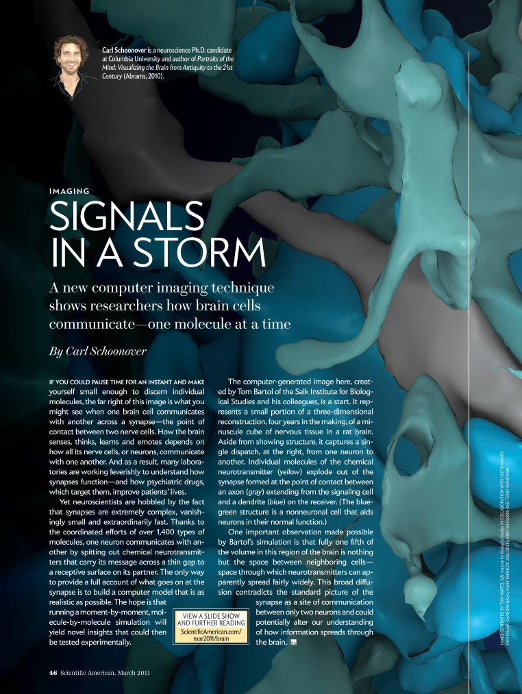
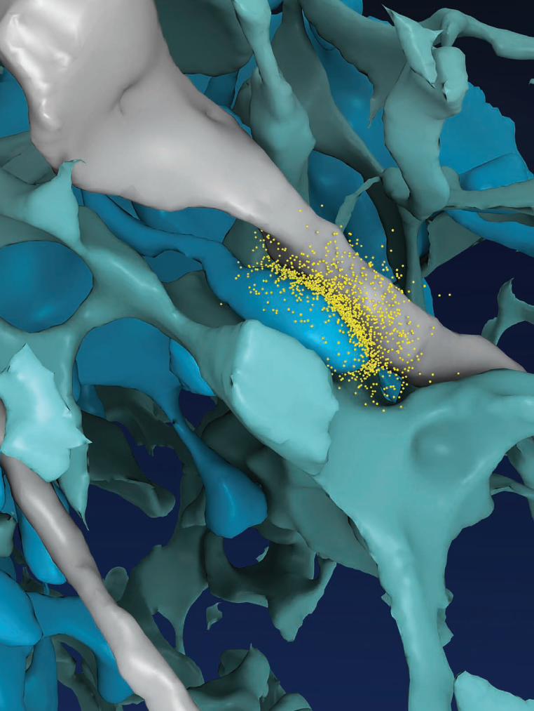
IF YOU COULD PAUSE TIME FOR AN INSTANT AND MAKE yourself small enough to discern individual molecules, the far right of this image is what you might see when one brain cell communicates with another across a synapse—the point of contact between two nerve cells. How the brain senses, thinks, learns and emotes depends on how all its nerve cells, or neurons, communicate with one another. And as a result, many labora-tories are working feverishly to understand how synapses function—and how psychiatric drugs, which target them, improve patients’ lives.
Yet neuroscientists are hobbled by the fact that synapses are extremely complex, vanish-ingly small and extraordinarily fast. Thanks to the coordinated eff orts of over 1,400 types of molecules, one neuron communicates with an-other by spitting out chem ical neurotransmit-ters that carry its message across a thin gap to a receptive surface on its partner. The only way to provide a full account of what goes on at the synapse is to build a computer model that is as realistic as possible. The hope is that running a moment-by-moment, mol-e cule- by-mol e cule simulation will yield novel insights that could then be tested experimentally.
The computer-generated image here, creat-ed by Tom Bartol of the Salk Institute for Biolog-ical Studies and his colleagues, is a start. It rep-resents a small portion of a three-dimensional reconstruction, four years in the making, of a mi-nuscule cube of nervous tissue in a rat brain. Aside from showing structure, it captures a sin-gle dispatch, at the right, from one neuron to another. Individual molecules of the chemical neurotransmitter (yellow) explode out of the synapse formed at the point of contact between an axon (gray) extending from the signaling cell and a dendrite (blue) on the receiver. (The blue-green structure is a nonneuronal cell that aids neurons in their normal function.)
One important observation made possible by Bartol’s simulation is that fully one fi fth of the volume in this region of the brain is nothing but the space between neighboring cells—space through which neurotransmitters can ap-parently spread fairly widely. This broad diff u-sion contradicts the standard picture of the
synapse as a site of communication between only two neurons and could potentially alter our under stand ing of how information spreads through the brain.
Carl Schoonover is a neuroscience Ph.D. candidate at Columbia University and author of Portraits of the Mind: Visualizing the Brain from Antiquity to the 21st Century (Abrams, 2010).
VIEW A SLIDE SHOW AND FURTHER READING
Scientifi cAmerican.com/mar2011/brain
sad0311Scho4p.indd 46 1/13/11 5:27:47 PM
Author Bio Text until an ‘end nested style character’ (Com-mand+3) xxxx xxxx xx xxxxx xxxxx xxxx xxxxxx xxxxxx xxx xx xxxxxxxxxx xxxx xxxxxxxxxx xxxxxx xxxxxxxx xxxxxxxx.
March 2011, ScientificAmerican.com 47Photograph/Illustration by Artist Name Month 20xx, Scientific American.com 47
sad0311Scho4p.indd 47 1/13/11 5:27:55 PM