
Reproductive Sciencesroyanaward.com/files12/articolo 2 lista pubblicazioni...The local derangement...
12
http://rsx.sagepub.com/ Reproductive Sciences http://rsx.sagepub.com/content/17/4/320 The online version of this article can be found at: DOI: 10.1177/1933719109353205 2010 17: 320 Reproductive Sciences Boy Torres, Giuseppe Rago, Pietro Litta and Felice Petraglia Pasquale Florio, Massimo Gabbanini, Lavinia E. Borges, Lorella Bonaccorsi, Serena Pinzauti, Fernando M. Reis, Paulo Reviews: Activins and Related Proteins in the Establishment of Pregnancy Published by: http://www.sagepublications.com On behalf of: Society for Gynecologic Investigation can be found at: Reproductive Sciences Additional services and information for http://rsx.sagepub.com/cgi/alerts Email Alerts: http://rsx.sagepub.com/subscriptions Subscriptions: http://www.sagepub.com/journalsReprints.nav Reprints: http://www.sagepub.com/journalsPermissions.nav Permissions: http://rsx.sagepub.com/content/17/4/320.refs.html Citations: at Univ di Siena on December 20, 2010 rsx.sagepub.com Downloaded from
Transcript of Reproductive Sciencesroyanaward.com/files12/articolo 2 lista pubblicazioni...The local derangement...

http://rsx.sagepub.com/Reproductive Sciences
http://rsx.sagepub.com/content/17/4/320The online version of this article can be found at:
DOI: 10.1177/1933719109353205
2010 17: 320Reproductive SciencesBoy Torres, Giuseppe Rago, Pietro Litta and Felice Petraglia
Pasquale Florio, Massimo Gabbanini, Lavinia E. Borges, Lorella Bonaccorsi, Serena Pinzauti, Fernando M. Reis, PauloReviews: Activins and Related Proteins in the Establishment of Pregnancy
Published by:
http://www.sagepublications.com
On behalf of:
Society for Gynecologic Investigation
can be found at:Reproductive SciencesAdditional services and information for
http://rsx.sagepub.com/cgi/alertsEmail Alerts:
http://rsx.sagepub.com/subscriptionsSubscriptions:
http://www.sagepub.com/journalsReprints.navReprints:
http://www.sagepub.com/journalsPermissions.navPermissions:
http://rsx.sagepub.com/content/17/4/320.refs.htmlCitations:
at Univ di Siena on December 20, 2010rsx.sagepub.comDownloaded from
REVIEWS
Activins and Related Proteins in the Establishment of PregnancyPasquale Florio, MD, PhD, Massimo Gabbanini, MD,
Lavinia E. Borges, MD, PhD, Lorella Bonaccorsi, PhD,Serena Pinzauti, MD, Fernando M. Reis, MD, PhD,
Paulo Boy Torres, MD, PhD, Giuseppe Rago, MD, PhD Student,Pietro Litta, MD, and Felice Petraglia, MD
Activin A and related proteins (inhibins, follistatin [FS], follistatin-related gene [FLRG], endometrial
bleeding associated factors [ebaf]) are involved in the complex mechanisms allowing the establishment and
the maintenance of pregnancy. As a consequence of ovarian progesterone stimuli, activin A is expressed
and secreted by the stromal endometrial cells, which locally induces the decidualization process, a
prerequisite for implantation. Moreover, activin A does influence the implantation phase, also enhancing
cytotrophoblast differentiation, indirectly, by increasing the expression of other molecules involved in
embryo implantation, such as matrix metalloproteinases (MMPs) and leukemia inhibitory factor (LIF).
The local derangement of activin A pathway in some pregnancy disorders (incomplete and complete
miscarriages, recurrent abortion, and ectopic pregnancy [EP]) further sustains the hypothesis that
activin A and its related proteins play a relevant role in the establishment of pregnancy.
KEY WORDS: activin A, endometrium, decidualization, embryo implantation, pregnancy,
miscarriage, ectopic pregnancy, abortion.
INTRODUCTION
Human endometrium is a highly specialized tissue,
providing an optimal environment for implantation of the
semiallogeneic embryo. Following the estrogen-induced
proliferation, progesterone involves the cyclic adenosine
monophosphate (cAMP) signaling pathway and induces
differentiative events within all compartments of the endo-
metrium, creating a receptive environment for blastocyst
attachment and invasion.1,2 Endometrial epithelial glands
undergo morphological and functional differentiation and
commence active secretion of a complex nutritive and
growth factor–rich media, contributing to uterine fluid and
providing support to blastocyst growth and development
before endometrial attachment.3,4 The endometrium repre-
sents not only a target for hormones but also an endocrine
organ itself, synthesizing several substances that influence the
local environment in an autocrine and paracrine manner.5
A growing number of data are currently pointing to
the role of activins and related proteins in the physiology
of the human endometrium.6 Activins are expressed and
secreted by the human endometrium and locally modu-
late stromal decidualization, a prerequisite for implanta-
tion; this effect results from a complex network of
interactions between activins and other locally regulating
processes.7,8 This complex scenario suggests a pivotal role
of activins in endometrial physiological changes, allowing
the establishment of pregnancy.
BIOCHEMISTRY
Activins are members of the transforming growth factor b(TGF-b) superfamily, which are structurally related
From the Department of Pediatrics, Obstetrics and Reproductive Medicine,
Section of Obstetrics and Gynecology, University of Siena, Siena, Italy (PF,
MG, LEB, LB, SP, FMR, PBT, GR, FP); and Department of Gynecological
Science and Human Reproduction, University of Padua, School of Medicine,
Padua, Italy (PL).
Address correspondence to: Pasquale Florio, PhD, MD, Department of
Pediatrics, Obstetrics and Reproductive Medicine, Section of Obstetrics and
Gynecology, University of Siena, Policlinico ‘‘Le Scotte,’’ Viale Bracci, 53100
Siena, Italy. E-mail: [email protected].
Reproductive Sciences Vol. 17 No. 4 April 2010 320-330DOI. 10.1177/1933719109353205# 2010 The Author(s)
320 at Univ di Siena on December 20, 2010rsx.sagepub.comDownloaded from
proteins involved in the control of cell proliferation,
differentiation, apoptosis, metabolism, homeostasis, differ-
entiation, immune response, and endocrine function.9
Activins possess a cysteine knot scaffold and are secreted as
homodimers or heterodimers of inhibin b-subunits.
Although 4 b-subunit genes (bA, bB, bC, and bE) have
been described in humans, only dimers composed of bA/
bA (activin A), bB/bB (activin B), and bA/bB (activin
AB) subunits have been shown to bebiologically active.10,11
Activins signal to their targets by interacting with 2
transmembrane serine/threonine receptor kinases (type
I and type II receptors). Activin binds first to its type II
receptor, ActRII or ActRIIB, leading to the recruitment,
phosphorylation, and subsequent activation of the type I
receptor (activin-like kinase 4, ALK4).12 On activation,
ALK4 phosphorylates a subset of cytoplasmic Smad pro-
teins (Smad2 and Smad3), which form part of the postre-
ceptor signal transduction system.13 Then, they undergo
homotrimerization and form heterometric complexes
with Smad 4; these activated complexes translocate into
the nucleus and, in conjunction with other nuclear cofac-
tors, regulate the transcription of target genes (Figure 1).
Access of activins to the cell is regulated by various
extracellular binding proteins.14 Follistatin (FS) binds
activins with high affinity (50-500 pmol/L) to form
biologically inactive complexes (Figure 1). It consists of
a 63-residue amino (N)-terminal segment, followed by
3 successive FS domains of 73 to 75 amino acids contain-
ing 10 cysteine residues termed FS-1, FS-2, and FS-3.
Alternative splicing generates 2 FS isoforms: FS288,
ebafAlk4
ActrRII
FLRG
βA βA
Activin AFS
Alk4
ActrRII
inhibitsbinding to receptor
inhibitsbinding to receptor
βA
Activin A
ActrRII
Alk4
P Smad 2/3 Smad 4
Smad 2/3 Smad 4
P
Genes expression
Inhibin A
β-glycan
βA βA
Activin A
ActrRII
A
B
DC
βA
Figure 1. Graphic representation of activin A signaling and interactions with its related peptides. A, Activin dimer first binds to the type II
(ActRII) receptor, which then recruits and phosphorylates type I (Alk4) receptor. These in turn phosphorylate receptor-activated Smads 2/3 that
subsequently form a complex with Smad 4 and is translocated to the nucleus, where it regulates the transcription of target genes. B, Activin
antagonists (follistatin-related gene [FLRG] and follistatin [FS]) bind activin A in the extracellular space, blocking its access to receptors. C,
Endometrial bleeding associated factor binds the activin receptor type II (ActRII), preventing the phosphorylation of Smad proteins and the
subsequent signaling pathway. D, Inhibin a subunit interacts with b-glycans promoting the binding of the inhibin b subunit to the ActRII,
which prevents activin A access.
Activin A is Implicated in Human Reproduction Reproductive Sciences Vol. 17, No. 4, April 2010 321
at Univ di Siena on December 20, 2010rsx.sagepub.comDownloaded from
which binds heparan sulfate proteoglycans with high affi-
nity via residues in FS-1 and is considered a local regulator
of activin actions, and FS315, which does not bind cell-
surface proteoglycans and is the predominant circulating
form of the protein.15,16
Follistatin-related gene (FLRG), also known as
FS-related protein or FS-like 3, is a recently characterized
member of the FS family, which binds activin with relatively
high affinity.17 Follistatin-related gene differs from FS by
lacking the third FS domain and a consensus heparin-
binding sequence. In this regard, FLRG is thought to act
as a circulating activin-binding protein, much like FS315.
Outside the 26 conserved cysteine residues, murine FLRG
and FS share about 40% amino acid identity.18 Because both
proteins bind and neutralize activin (Figure 1), it seems likely
that the residues that are important for binding activins are
located in these conserved regions. Intriguingly, expression
and regulation studies suggest that FS and FLRG might not
be complete functional homologs.18
Other proteins are relevant in counteracting activins by
displacing it from its own receptors. This is the case of
inhibins and endometrial bleeding associated factors (ebaf;
Figure 1). Like activins, inhibins are members of the TGF-bsuperfamily consisting of ana subunit that can dimerize with
either bA or bB subunit to form inhibin A (a-bA) or B
(a-bB).19 They operate as potent selective antagonists of
activins and their function is tightly related to the presence
of b-glycans, membrane-anchored proteoglycans that act
as inhibin coreceptors. The interaction of b-glycans with
inhibina subunit promotes the binding of the inhibinb sub-
unit to the ActRII, preventing activin access (Figure 1).20,21
Endometrial bleeding associated factor (ebaf) is
another member of the TGF-b family, recently renamed
left-right determination, factor A (LEFTY-A).22 It is a
protein of 370 amino acids, which presents a 65% similar-
ity with activin A structure. It binds to the activin recep-
tor type II B (ActRIIB), preventing the phosphorylation
of Smad proteins and the subsequent signaling pathway
(Figure 1).23
THE HUMAN ENDOMETRIUM AS A SOURCE OF ACTIVIN ANDRELATED PROTEINS
Activins were originally isolated in the ovary and
identified as a gonadal protein due to its action on pitui-
tary follicle-stimulating hormone (FSH) synthesis and
secretion.24 Subsequently, it was detected in several other
organs, but the main source of circulating protein levels is
still unknown.8
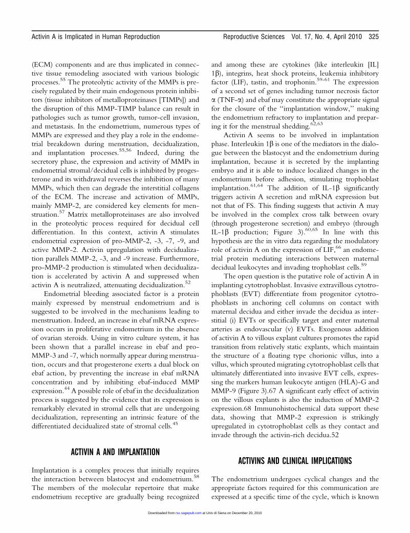
ActivinbA messenger RNA (mRNA) is expressed both
in endometrial glands and in stromal cells25-27; the immunos-
taining along the menstrual cycle is widely different
(Figure 2).28 A specific and predominant bA immunostain-
ing in the cytoplasm of luminal and glandular epithelial cells
with an inconsistent staining pattern alongside the glandular
cells during the proliferative phase and a more intense and
uniform staining pattern in the secretory phase have been
described. The glandular immunostaining progressively
increases across the secretory phase, reaching a maximum
in the late secretory phase.29 The staining also tended to be
more intense in the glandular cells near the luminal
epithelium.30 However, the stromal cells are weakly immu-
noreactive from the menstrual to the midsecretory phase and
become stronger in the late secretory phase. Additional
immunostaining is present in the vascular endometrium and
in subsets of infiltrating leukocytes.28,31,32
Activins B and AB are also endometrial products.
Indeed, normal endometrium also expresses the activin
bB subunit, with the staining intensity being strongest
in the late secretory phase compared to proliferative and
early secretory phase secretions (Figure 3).31,32
Inhibin a subunit is detected in normal human endo-
metrium, predominantly localized in the glandular and
luminal epithelium, thus suggesting an endometrial pro-
duction of inhibins A and B. Inhibin a subunit showed
no or minimal expression during the proliferative phase,
increasing during the secretory phase.28,33,34 Indeed, the
expression of inhibin a subunit evaluated by immunohis-
tochemistry was significantly higher during the early
and late secretory phase than in the proliferative phase
(Figure 2).33,35,36 b-glycan, fundamental for inhibin’s
action, is localized in stromal and epithelial cells and the
staining is particularly abundant in the basal and apical
borders of the cell.37
Follistatin expression is also detected in endometrial
stromal and epithelial cells,38,39 even if both its mRNA
and protein expression is low across the menstrual cycle,
with a peak only in early pregnant decidua (Figure 2).40
The pattern of activin A and FS expression are resembled
by secretion of the proteins into the uterine lumen.
Indeed, activin A, but not FS, is secreted in higher
amounts by the endometrium in the secretive than in the
proliferative phase of the menstrual cycle. Furthermore,
activin A levels in washing uterine fluids significantly cor-
relate with endometrial thickness and with the day of the
menstrual cycle.25 There are no assays available for acti-
vins B and AB.
Follistatin-related gene is strongly localized in epithe-
lial cells of glands and vessel walls, both in cytoplasma and
322 Reproductive Sciences Vol. 17, No. 4, April 2010 Florio et al
at Univ di Siena on December 20, 2010rsx.sagepub.comDownloaded from
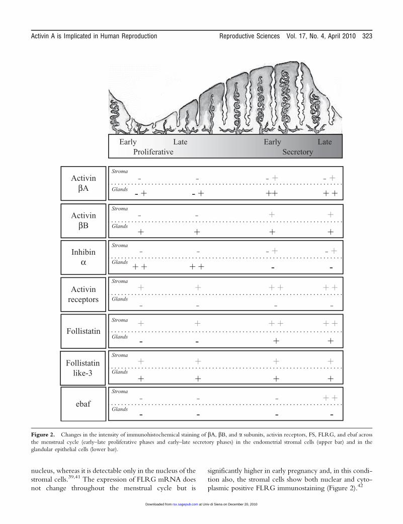
nucleus, whereas it is detectable only in the nucleus of the
stromal cells.39,41 The expression of FLRG mRNA does
not change throughout the menstrual cycle but is
significantly higher in early pregnancy and, in this condi-
tion also, the stromal cells show both nuclear and cyto-
plasmic positive FLRG immunostaining (Figure 2).42
Early Late Early LateProliferative Secretory
ActivinβA
- -
+ +
- + - +
++- + - +
Stroma
Glands
Inhibinα
Activinreceptors
Follistatin
-
+
-
+ ++
+ +
+ +
-
+ +
-
-
- +
-
- +
+
- -
+
-
+ + + +
-
Stroma
Glands
Stroma
Glands
Stroma
Glands
Follistatinlike-3
+
-
+ ++ ++
++-
Stroma
Glands
ebaf-
- -
-
- + +
- -
Stroma
Glands
+
++
+ ++
++
Stroma
Glands
ActivinβB
Figure 2. Changes in the intensity of immunohistochemical staining of bA, bB, and a subunits, activin receptors, FS, FLRG, and ebaf across
the menstrual cycle (early–late proliferative phases and early–late secretory phases) in the endometrial stromal cells (upper bar) and in the
glandular epithelial cells (lower bar).
Activin A is Implicated in Human Reproduction Reproductive Sciences Vol. 17, No. 4, April 2010 323
at Univ di Siena on December 20, 2010rsx.sagepub.comDownloaded from
Endometrial bleeding associated factor is expressed
differently during the menstrual cycle. Immunohisto-
chemical stainings show a low level of immunoreactivity
in the early, mid- and late proliferative and early and mid-
secretory endometrium. A strong immunoreactivity for
ebaf is reported in the stroma and to lesser extent in the
endometrial glands in late secretory and menstrual
endometrium (Figure 2).43,44 A similar increase in ebaf
mRNA occurs in explants from proliferative endome-
trium cultured in the absence of ovarian steroids.45
The various activin receptor subtypes have been
detected in the endometrial stromal cells (ESCs) but not
in the surface of glandular epithelium at any stage of the
cycle.40,46 Protein expression of the respective receptors
is maintained throughout the secretory phase with a
strong staining in decidual cells, during mid- to late secre-
tory phase and in early pregnancy (Figure 2).39 Therefore,
expression of activin receptors increases in the prolifera-
tive phase of endometrial cycle and this increase contin-
ues throughout the secretive phase, whereas FS remains
constant during the menstrual cycle.
LOCAL EFFECTS OF ACTIVIN A IN THE HUMANENDOMETRIUM
Exquisite mechanisms control the processes that drive
endometrium through proliferative and secretive phases,
thus preparing this tissue for implantation. In the late
secretory phase of the menstrual cycle, decidual
transformation of ESCs is a prerequisite for human
implantation.1-4 It commonly occurs in stromal cells
surrounding the spiral arteries about 10 days after the
postovulatory rise in progesterone levels and is character-
ized by the transformation of the ESCs in elongated
fibroblast-like cells, the decidualized ESCs (human ESCs
[HESCs]). Decidualization is independent of the presence
of an implanting blastocyst but occurs in response to pro-
gesterone and involves the cAMP signaling pathway.47
However, in the absence of conception, falling circulat-
ing progesterone levels in the late secretory phase of the
cycle elicit sloughing of the decidualized superficial
endometrial layer and menstruation. When pregnancy
occurs, the decidual reaction extends to the basal endo-
metrial layer and it is critical for trophoblast invasion and
placenta formation. Activin A has been recognized as a
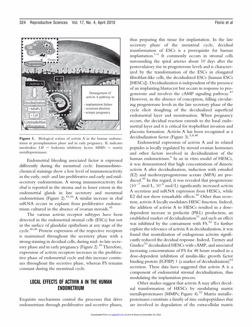
decidualization factor (Figure 3).5,8,48
Endometrial expression of activin A and its related
peptides is locally regulated by steroid ovarian hormones
and other factors involved in decidualization of the
human endometrium.6 In an in vitro model of HESCs,
it was demonstrated that high concentrations of dimeric
activin A after decidualization, induction with estradiol
(E2) and medroxyprogesterone acetate (MPA) are pro-
duced.49 In this regard, it was revealed that progesterone
(10�7 mol/L, 10–6 mol/L) significantly increased activin
A secretion and mRNA expression from HESCs, while
E2 did not show remarkable effects.50 Other than secre-
tion, activin A locally modulates HESC function. Indeed,
the addition of activin A to HESCs resulted in a dose-
dependent increase in prolactin (PRL) production, an
established marker of decidualization51 and such an effect
was inhibited by the cotreatment with FS.52 To further
explore the relevance of activin A in decidualization, it was
found that neutralization of endogenous activins signifi-
cantly reduced the decidual response. Indeed, Tierney and
Giudice53 decidualized HESCs with cAMP, and associated
increasing concentrations of FS for 48 hours resulted in a
dose-dependent inhibition of insulin-like growth factor
binding protein (IGFBP) 1 (a marker of decidualization)54
secretion. These data have suggested that activin A is a
component of endometrial stromal decidualization, thus
modulating the implantation process.
Other studies suggest that activin A may affect decid-
ual transformation of HESCs by modulating matrix
metalloproteinases (MMPs; Figure 4).52 Matrix metallo-
proteinases constitute a family of zinc endopeptidases that
are involved in degradation of the extracellular matrix
IL-β+ Activin A
Decidualization
+MMPs
Cytotrophoblastdifferentiation
Embryo
Decidua
Derangement ofactivin A pathway in
- implantation failure- recurrent abortion- ectopic pregnancy
+ LIF
OvaryProgesterone
Figure 3. Biological actions of activin A in the human endome-
trium in preimplantation phase and in early pregnancy. IL indicates
interleukin; LIF ¼ leukemia inhibitory factor; MMPs ¼ matrix
metalloproteinases.
324 Reproductive Sciences Vol. 17, No. 4, April 2010 Florio et al
at Univ di Siena on December 20, 2010rsx.sagepub.comDownloaded from
(ECM) components and are thus implicated in connec-
tive tissue remodeling associated with various biologic
processes.55 The proteolytic activity of the MMPs is pre-
cisely regulated by their main endogenous protein inhibi-
tors (tissue inhibitors of metalloproteinases [TIMPs]) and
the disruption of this MMP-TIMP balance can result in
pathologies such as tumor growth, tumor-cell invasion,
and metastasis. In the endometrium, numerous types of
MMPs are expressed and they play a role in the endome-
trial breakdown during menstruation, decidualization,
and implantation processes.55,56 Indeed, during the
secretory phase, the expression and activity of MMPs in
endometrial stromal/decidual cells is inhibited by proges-
terone and its withdrawal reverses the inhibition of many
MMPs, which then can degrade the interstitial collagens
of the ECM. The increase and activation of MMPs,
mainly MMP-2, are considered key elements for men-
struation.57 Matrix metalloproteinases are also involved
in the proteolytic process required for decidual cell
differentiation. In this context, activin A stimulates
endometrial expression of pro-MMP-2, -3, -7, -9, and
active MMP-2. Activin upregulation with decidualiza-
tion parallels MMP-2, -3, and -9 increase. Furthermore,
pro-MMP-2 production is stimulated when decidualiza-
tion is accelerated by activin A and suppressed when
activin A is neutralized, attenuating decidualization.52
Endometrial bleeding associated factor is a protein
mainly expressed by menstrual endometrium and is
suggested to be involved in the mechanisms leading to
menstruation. Indeed, an increase in ebaf mRNA expres-
sion occurs in proliferative endometrium in the absence
of ovarian steroids. Using in vitro culture system, it has
been shown that a parallel increase in ebaf and pro-
MMP-3 and -7, which normally appear during menstrua-
tion, occurs and that progesterone exerts a dual block on
ebaf action, by preventing the increase in ebaf mRNA
concentration and by inhibiting ebaf-induced MMP
expression.44 A possible role of ebaf in the decidualization
process is suggested by the evidence that its expression is
remarkably elevated in stromal cells that are undergoing
decidualization, representing an intrinsic feature of the
differentiated decidualized state of stromal cells.45
ACTIVIN A AND IMPLANTATION
Implantation is a complex process that initially requires
the interaction between blastocyst and endometrium.58
The members of the molecular repertoire that make
endometrium receptive are gradually being recognized
and among these are cytokines (like interleukin [IL]
1b), integrins, heat shock proteins, leukemia inhibitory
factor (LIF), tastin, and trophonin.59-61 The expression
of a second set of genes including tumor necrosis factor
a (TNF-a) and ebaf may constitute the appropriate signal
for the closure of the ‘‘implantation window,’’ making
the endometrium refractory to implantation and prepar-
ing it for the menstrual shedding.62,63
Activin A seems to be involved in implantation
phase. Interleukin 1b is one of the mediators in the dialo-
gue between the blastocyst and the endometrium during
implantation, because it is secreted by the implanting
embryo and it is able to induce localized changes in the
endometrium before adhesion, stimulating trophoblast
implantation.61,64 The addition of IL-1b significantly
triggers activin A secretion and mRNA expression but
not that of FS. This finding suggests that activin A may
be involved in the complex cross talk between ovary
(through progesterone secretion) and embryo (through
IL-1b production; Figure 3).60,65 In line with this
hypothesis are the in vitro data regarding the modulatory
role of activin A on the expression of LIF,66 an endome-
trial protein mediating interactions between maternal
decidual leukocytes and invading trophoblast cells.59
The open question is the putative role of activin A in
implanting cytotrophoblast. Invasive extravillous cytotro-
phoblasts (EVT) differentiate from progenitor cytotro-
phoblasts in anchoring cell columns on contact with
maternal decidua and either invade the decidua as inter-
stitial (i) EVTs or specifically target and enter maternal
arteries as endovascular (v) EVTs. Exogenous addition
of activin A to villous explant cultures promotes the rapid
transition from relatively static explants, which maintain
the structure of a floating type chorionic villus, into a
villus, which sprouted migrating cytotrophoblast cells that
ultimately differentiated into invasive EVT cells, expres-
sing the markers human leukocyte antigen (HLA)-G and
MMP-9 (Figure 3).67 A significant early effect of activin
on the villous explants is also the induction of MMP-2
expression.68 Immunohistochemical data support these
data, showing that MMP-2 expression is strikingly
upregulated in cytotrophoblast cells as they contact and
invade through the activin-rich decidua.52
ACTIVINS AND CLINICAL IMPLICATIONS
The endometrium undergoes cyclical changes and the
appropriate factors required for this communication are
expressed at a specific time of the cycle, which is known
Activin A is Implicated in Human Reproduction Reproductive Sciences Vol. 17, No. 4, April 2010 325
at Univ di Siena on December 20, 2010rsx.sagepub.comDownloaded from
as the ‘‘implantation window.’’ In light of the fact that
activin A is a well-known regulator of the differentiation
of proliferative cytotrophoblast into extravillous invasive
trophoblast cells of the anchoring villi, we and others
hypothesized that interactions between activin A and its
related proteins in the endometrium may have a funda-
mental role at implantation.5,8,69 The derangement of
activin A machinery has, therefore, the potential to be
correlated with impairments of endometrial receptivity
needed for implantation to occur, suggesting that some
types of infertility may be associated with dysregulated
endometrial expression of activin A (Figure 3).
In addition, a dysregulated expression of ebaf in the
endometrium of a subset of women with infertility during
the receptive phase of the menstrual cycle has been
shown.70 Indeed, abundantly secreted protein is present
in the endometrium of these women during the
implantation window and during the critical period of
endometrial receptivity; ebaf is more abundant in patients
with endometriosis, who did not conceive than in
patients who became pregnant.70 In this regard, it was
evaluated whether activin A may be an in vivo marker
of endometrial receptivity. Therefore, it was examined
whether activin A concentrations in uterine washing fluid
differ between women who become pregnant after
intrauterine insemination (IUI) and those who did not.
Activin A levels were significantly higher in the endome-
trial wash fluid collected from women who later become
pregnant than in those who did not and correlated with
endometrial thickness.25 Furthermore, activin A repre-
sented a marker to predict pregnancy, because in patients
submitted to IUI, the probability of pregnancy was near
to 100% in patients with high activin A level whereas this
percentage decreased to 20% in patients with low activin
A level.71
In agreement with the hypothesis that activin A is
involved in implantation, data derived from the evalua-
tion of the expression of activin bA and bB subunits and
FS in the endometrium of women with implantation
failure was compared with a fertile control group. It was
demonstrated that the endometrium of women with
implantation failure shares an altered activin A–FS
interaction pathway (Figure 3), because FS expression
was significantly reduced in the endometrial epithelial
glandular cells in women with implantation failure
compared with control group of healthy fertile women.
However, this reduction did not appear to translate into
an expected increase in endometrial activin expression,
because activin subunits’ staining was not significantly dif-
ferent.72 In any case, this finding may represent an altered
activin FS interactive pathway, probably involved in the
mechanism related to implantation failure.
The impairment of endometrial activin pathway
seems to be involved in ectopic pregnancy (EP; Figure 3).
Ectopic pregnancy is an important cause of maternal
deaths in early pregnancy and its diagnosis is based on the
positive visualization of an extrauterine pregnancy out-
side the uterus. Factors affecting decidualization and/or
embryo transport inside the uterus have the potential to
cause EP. The intensity of expression of bA and bB
subunits, type IIA and type IIB receptors, and FS was
stronger in the EP group when compared with the
pseudopregnant group; however, quantitative reverse
transcriptase polymerase chain reaction (RT-PCR)
showed a significant decrease in mRNA levels of these
proteins, with the exception of bB-subunit with an
apparent paradox of high concentration of protein but
low mRNA expression.73 The hypothesis of a reduced
local protein secretion was proposed on the basis of the
findings reporting low serum concentrations of activin
A in women with EP.74 Indeed, activin A measurement
was performed at the initial visit in early pregnancies of
unknown location (PULs; n ¼ 536), in whom there was
no clear ultrasound evidence of an intrauterine preg-
nancy. The diagnosis of a normal intrauterine pregnancy
was made on follow-up by the demonstration of an
intrauterine gestational sac on the scan, while EP was
diagnosed later at laparoscopy and on histological exam-
ination of the surgical specimens. Serum activin A levels
were lowest in women with PUL, later diagnosed as hav-
ing an EP and, at the cutoff of 0.37 ng/mL, activin A
combined a sensitivity and a specificity of 100% and
99.6%, respectively, for prediction of EP.74
Likewise, the analysis of activin B expression in
decidualized endometrium from ectopic pregnancies
showed low levels when compared with that of ongoing
pregnancies or miscarriages. However, activin B was
lower in the serum of women with ectopic pregnancies
than in viable intrauterine gestations, with a clear correla-
tion between serum activin B and the degree of
decidualization.75
Because activin A and inhibin A are also produced by
the human placenta,76 their serum concentrations were
measured in women with incomplete and complete mis-
carriage, with the aim of evaluating their putative clinical
usefulness as marker of early pregnancy viable placenta-
tion.77 It was found that activin A levels significantly
decreased in complete miscarriage, whereas inhibin A
concentrations were significantly lower both in incom-
plete and complete miscarriages, with the lowest
326 Reproductive Sciences Vol. 17, No. 4, April 2010 Florio et al
at Univ di Siena on December 20, 2010rsx.sagepub.comDownloaded from
concentrations in complete miscarriage.77 This study
prompted to evaluate whether the serum activin A and
inhibin A measurement may provide additional tools for
the prediction of pregnancy viability in women with
vaginal bleeding due to threatened abortion and failing
pregnancies.78 It was found that (1) serum activin A and
inhibin A were significantly lower in nonviable pregnan-
cies than in women with threatened abortion but
ongoing pregnancy; (2) low levels of inhibin A and acti-
vin A occur in patients with threatened abortion whose
pregnancy failed after 3 weeks; (3) inhibin A measure-
ment was identified as the best marker in detection of
early pregnancy viability. Indeed, inhibin A at the best
cutoff detected by the receiver operating curve analysis
achieved the best accuracy for prediction of failing preg-
nancy, showing a sensitivity of 90.6% and a specificity of
99.5%.77
On the basis of the above-mentioned data, a putative
role for activin A has also been supposed in the mechan-
ism related to recurrent miscarriage, whose etiology is in
large part unknown, because causes vary from chromoso-
mal anomalies to luteal phase defects.79 In this regard, it
was demonstrated that the endometrial expression of FS
and activin bA subunit in the ESCs collected from
women with recurrent miscarriage was significantly
lower than in control women, and this evidence has
reinforced the idea that the alterations of endometrial
functions are locally accompanied by a dysregulation of
activin A pathway (Figure 3).80,81
CONCLUSIONS
Activins and related regulatory proteins (inhibin, ebaf, FS,
FLRG, b-glycan) are endometrial factors whose expres-
sion is dynamically and tightly regulated according to
endometrial cycle. In particular, activin A is a decidualiz-
ing growth factor and its mRNA and protein expression
is part of the complex series of events allowing
implantation.
The derangement of endometrial activin pathway
may be part of the complex repertoire of local molecular
events and therefore involved both in infertility and dis-
orders of early pregnancy, such as miscarriage and EP.
REFERENCES
1. Jabbour HN, Kelly RW, Fraser HM, Critchley HO. Endocrine
regulation of menstruation. Endocr Rev. 2006;27(1):17-46.
2. Gargett CE, Chan RW, Schwab KE. Hormone and growth
factor signaling in endometrial renewal: role of stem/progeni-
tor cells. Mol Cell Endocrinol. 2008;288(1-2):22-29.
3. Guzeloglu-Kayisli O, Basar M, Arici A. Basic aspects of
implantation. Reprod Biomed Online. 2007;15(6):728-739.
4. Gellersen B, Brosens IA, Brosens JJ. Decidualization of the
human endometrium: mechanisms, functions, and clinical
perspectives. Semin Reprod Med. 2007;25(6):445-453.
5. Reis FM, Cobellis L,Luisi S, et al. Paracrine/autocrinecontrol of
female reproduction. Gynecol Endocrinol. 2000;14(6):464-475.
6. Florio P, Rossi M, Sigurdardottir M, et al. Paracrine regula-
tion of endometrial function: interaction between progester-
one and corticotropin-releasing factor (CRF) and activin A.
Steroids. 2003;68(10-13):801-807.
7. Salamonsen LA, Dimitriadis E, Jones RL, Nie G. Complex
regulation of decidualization: a role for cytokines and pro-
teases—a review. Placenta. 2003;24(suppl A):S76-S85.
8. Luisi S, Florio P, Reis FM, Petraglia F. Expression and secre-
tion of activin A: possible physiological and clinical implica-
tions. Eur J Endocrinol. 2001;145(3):225-236.
9. Guo X, Wang XF. Signaling cross-talk between TGF-beta/
BMP and other pathways. Cell Res. 2008;19(1):71-88.
10. Lin SJ, Lerch TF, Cook RW, Jardetzky TS, Woodruff TK.
The structural basis of TGF-beta, bone morphogenetic pro-
tein, and activin ligand binding. Reproduction. 2006;132(2):
179-190.
11. Chang H, Brown CW, Matzuk MM. Genetic analysis of the
mammalian transforming growth factor-beta superfamily.
Endocr Rev. 2002;23(6):787-823.
12. Welt C, Sidis Y, Keutmann H, Schneyer A. Activins, inhibins,
and follistatins: from endocrinology to signaling. A paradigm
for the new millennium. Exp Biol Med. 2002;227(9):724-752.
13. Derynck R, Zhang Y, Feng XH. Smads: transcriptional acti-
vators of TGF-b responses. Cell. 1998;95(6):737-740.
14. Harrison CA, Gray PC, Vale WW, Robertson DM. Antago-
nists of activin signaling: mechanisms and potential biological
applications. Trends Endocrinol Metab. 2005;16(2):73-78.
15. Schneyer AL, Wang Q, Sidis Y, Sluss PM. Differential distribu-
tion of follistatin isoforms: application of a new FS315-specific
immunoassay. J Clin Endocrinol Metab. 2004;89(10):5067-5075.
16. Shimonaka M, Inouye S, Shimasaki S, Ling N. Follistatin
binds to both activin and inhibin through the common subu-
nit. Endocrinology. 1991;128(6):3313-3315.
17. Tsuchida K, Arai KY, Kuramoto Y, Yamakawa N,
Hasegawa Y, Sugino H. Identification and characterization
of a novel follistatin-like protein as a binding protein for the
TGF-beta family. J Biol Chem. 2000;275(52):40788-40796.
18. Schneyer A, Schoen A, Quigg A, Sidis Y. Differential binding
and neutralization of activins A and B by follistatin and follis-
tatin like-3 (FSTL-3/FSRP/FLRG). Endocrinology. 2003;
144(5):1671-1674.
19. Bilezikjian LM, Blount AL, Donaldson CJ, Vale WW. Pitui-
tary actions of ligands of the TGF-beta family: activins and
inhibins. Reproduction. 2006;132(2):207-215.
Activin A is Implicated in Human Reproduction Reproductive Sciences Vol. 17, No. 4, April 2010 327
at Univ di Siena on December 20, 2010rsx.sagepub.comDownloaded from
20. Chapman SC, Bernard DJ, Jelen J, Woodruff TK. Properties
of inhibin binding to betaglycan, InhBP/p120 and the activin
type II receptors. Mol Cell Endocrinol. 2002;196(1-2):79-93.
21. Lewis KA, Gray PC, Blount AL, et al. Betaglycan binds
inhibin and can mediate functional antagonism of activin
signalling. Nature. 2000;404(6776):411-414.
22. Kosaki K, Bassi MT, Kosaki R, et al. Characterization and
mutation analysis of human LEFTY A and LEFTY B,
homologues of murine genes implicated in left-right axis
development. Am J Hum Genet. 1999;64(3):712-721.
23. Ulloa L, Tabibzadeh SJ. Lefty inhibits receptor-regulated
Smad phosphorylation induced by the activated transforming
growth factor-beta receptor. Biol Chem. 2001;276(24):
21397-21404.
24. Ling N, Ying SY, Ueno N, et al. Pituitary FSH is released by a
heterodimer of the beta-subunits from the two forms of inhi-
bin. Nature. 1986;321(6072):779-782.
25. Florio P, Severi FM, Luisi S, et al. Endometrial expression and
secretion of activin A, but not follistatin, increase in the secre-
tory phase of the menstrual cycle. J Soc Gynecol Investig. 2003;
10(4):237-243.
26. Petraglia F, Florio P, Luisi S, et al. Expression and secretion of
inhibin and activin in normal and neoplastic uterine tissues.
High levels of serum activin A in women with endometrial
and cervical carcinoma. J Clin Endocrinol Metab. 1998;83(4):
1194-1200.
27. Reis FM, Di Blasio AM, Florio P, Ambrosini G, Di Loreto C,
Petraglia F. Evidence for local production of inhibin A and
activin A in patients with ovarian endometriosis. Fertil Steril.
2001;75(2):367-373.
28. Leung PH, Salamonsen LA, Findlay JK. Immunolocalization
of inhibin and activin subunits in human endometrium across
the menstrual cycle. Hum Reprod. 1998;13(12):3469-3477.
29. Otani T, Minami S, Kokawa K, Shikone T, Yamoto M,
Nakano R. Immunohistochemical localization of activin A
in human endometrial tissues during the menstrual cycle and
in early pregnancy. Obstet Gynecol. 1998;91(5 pt 1):685-692.
30. Morrish DW, Dakour J, Li H. Inhibin/activin subunits beta-A
(-betaA) and beta-B (-betaB) are differentially localised in nor-
mal, hyperplastic and malignant human endometrial tissue.
Functional regulation of human trophoblast differentiation.
J Reprod Immunol. 1998;39(1-2):179-195.
31. Mylonas I, Makovitzky J, Hoeing A, et al. Inhibin/activin
subunits beta-A (-betaA) and beta-B (-betaB) are differentially
localised in normal, hyperplastic and malignant human endo-
metrial tissue. Acta Histochem. 2006;108(1):1-11.
32. Mylonas I, Jeschke U, Wiest I, et al. Inhibin/activin subunits
alpha, beta-A and beta-B are differentially expressed in normal
human endometrium throughout the menstrual cycle. Histo-
chem Cell Biol. 2004;122(5):461-471.
33. Teni TR, Sampat MB, Sheth NA. Inhibin (10.7 kD prostatic
peptide) in normal, hyperplastic, and malignant human endo-
metria: an immunohistochemical study. J Pathol. 1992;168(1):
35-40.
34. Petraglia F, Calza L, Garuti GC, et al. Presence and synthesis
of inhibin subunits in human decidua. J Clin Endocrinol Metab.
1990;71(2):487-492.
35. Worbs S, Shabani N, Mayr D, et al. Expression of the inhibin/
activin subunits (-alpha, -betaA and -betaB) in normal and
carcinogenic endometrial tissue: possible immunohistochem-
ical differentiation markers. Oncol Rep. 2007;17(1):97-104.
36. Mylonas I, Jeschke U, Winkler L, et al. Immunohistochemical
expression of inhibin-alpha in human endometrium and the in
vitro secretion of inhibin, estradiol and cortisol in cultured
human endometrial glandular cells. Arch Gynecol Obstet.
2003;268(3):142-150.
37. Florio P, Ciarmela P, Reis FM, et al. Inhibin alpha-subunit
and the inhibin coreceptor betaglycan are downregulated
in endometrial carcinoma. Eur J Endocrinol. 2005;152(2):
277-284.
38. Torres PB, Florio P, Ferreira MC, Torricelli M, Reis FM,
Petraglia F. Deranged expression of follistatin and follistatin-
like protein in women with ovarian endometriosis. Fertil Steril.
2007;88(1):200-205.
39. Jones RL, Salamonsen LA, Zhao YC, Ethier JF,
Drummond AE, Findlay JK. Expression of activin receptors,
follistatin and betaglycan by human endometrial stromal cells;
consistent with a role for activins during decidualization. Mol
Hum Reprod. 2002;8(4):363-374.
40. Petraglia F, Gallinelli A, Grande A, et al. Local production and
action of follistatin in human placenta. J Clin Endocrinol Metab.
1994;78(1):205-210.
41. Wang HQ, Takebayashi K, Tsuchida K, Nishimura M,
Noda Y. Follistatin-related gene (FLRG) expression in human
endometrium: sex steroid hormones regulate the expression of
FLRG in cultured human endometrial stromal cells. J Clin
Endocrinol Metab. 2003;88(9):4432-4439.
42. Florio P, Ciarmela P, Toti P, et al. Human endometrium and
decidua express follistatin-related gene (FLRG) mRNA and
peptide. Mol Cell Endocrinol. 2004;218(1-2):129-135.
43. Tabibzadeh S, Lessey B, Satyaswaroop PG. Temporal and site-
specific expression of transforming growth factor-b4 in human
endometrium. Mol Hum Reprod. 1998;4(6):595-602.
44. Cornet PB, Picquet C, Lemoine P, et al. Regulation and
function of LEFTY-A/EBAF in the human endometrium.
mRNA expression during the menstrual cycle, control by
progesterone, and effect on matrix metalloprotineases. Biol
Chem. 2002;277(45):42496-42504.
45. Tang M, Mikhailik A, Pauli I, et al. Decidual differentiation of
stromal cells promotes Proprotein Convertase 5/6 expression
and lefty processing. Endocrinology. 2005;146(12):5313-5320.
46. Piestrzeniewicz-Ulanska D, Brys M, Semczuk A,
Jakowicki JA, Krajewska WM. Expression of TGF-beta type
I and II receptors in normal and cancerous human endome-
trium. Cancer Lett. 2002;186(2):231-239.
47. Gellersen B, Brosens J. Cyclic AMP and progesterone recep-
tor cross-talk in human endometrium: a decidualizing affair.
J Endocrinol. 2003;178(3):357-372.
328 Reproductive Sciences Vol. 17, No. 4, April 2010 Florio et al
at Univ di Siena on December 20, 2010rsx.sagepub.comDownloaded from
48. Jones RL, Findlay JK, Salamonsen LA. The role of activins
during decidualisation of human endometrium. Aust N Z J
Obstet Gynaecol. 2006;46(3):245-249.
49. Jones RL, Salamonsen LA, Findlay JK. Activin A promotes
human endometrial stromal cell decidualization in vitro. J Clin
Endocrinol Metab. 2002;87(8):4001-4004.
50. Florio P, Rossi M, Vigano P, et al. Interleukin 1beta and pro-
gesterone stimulate activin a expression and secretion from
cultured human endometrial stromal cells. Reprod Sci. 2007;
14(1):29-36.
51. Telgmann R, Gellersen B. Marker genes of decidualization:
activation of the decidual prolactin gene. Hum Reprod Update.
1998;4(5):472-479.
52. Jones RL, Findlay JK, Farnworth PG, Robertson DM,
Wallace E, Salamonsen LA. Activin A and inhibin A differen-
tially regulate human uterine matrix metalloproteinases:
potential interactions during decidualization and trophoblast
invasion. Endocrinology. 2006;147(2):724-732.
53. Tierney EP, Giudice LC. Role of activin A as a mediator of in
vitro endometrial stromal cell decidualization via the cyclic
adenosine monophosphate pathway. Fertil Steril. 2004;
81(suppl 1):899-903.
54. Harada M, Osuga Y, Takemura Y, et al. Mechanical stretch
upregulates IGFBP-1 secretion from decidualized endometrial
stromal cells. Am J Physiol Endocrinol Metab. 2006;290(2):
E268-E272.
55. Curry TE, Osteen KG. The matrix metalloproteinase system:
changes, regulation, and impact throughout the ovarian and
uterine reproductive cycle. Endocr Rev. 2003;24(4):428-465.
56. Zhang X, Nothnick WB. The role and regulation of the uter-
ine matrix metalloproteinase system in menstruating and non-
menstruating species. Front Biosci. 2005;10:353-366.
57. Salamonsen LA, Woolley DE. Matrix metalloproteinases in
normal menstruation. Hum Reprod. 1996;11(suppl 2):124-133.
58. Paria BC, Song H, Dey SK. Implantation: molecular basis of
embryo-uterine dialogue. Int J Dev Biol. 2001;45(3):597-605.
59. Dimitriadis E, White CA, Jones RL, Salamonsen LA. Cyto-
kines, chemokines and growth factors in endometrium related
to implantation. Hum Reprod Update. 2005;11(6):613-630.
60. Domınguez F, Remohı J, Pellicer A, Simon C. Paracrine
interactions during human implantation. Rev Endocr Metab
Disord. 2002;3(2):97-105.
61. Rossi M, Sharkey AM, Vigano P, et al. Identification of genes
regulated by interleukin-1beta in human endometrial stromal
cells. Reproduction. 2005;130(5):721-729.
62. Tabibzadeh S. Molecular control of the implantation window.
Hum Reprod Update. 1998;4(5):465-471.
63. Kashiwagi A, DiGirolamo CM, Kanda Y, et al. The postim-
plantation embryo differentially regulates endometrial gene
expression and decidualization. Endocrinology. 2007;148(9):
4173-4184.
64. Sunder S, Lenton EA. Endocrinology of the peri-implantation
period. Baillieres Best Pract Res Clin Obstet Gynaecol. 2000;
14(5):789-800.
65. Keelan JA, Groome NP, Mitchell MD. Regulation of
activin-A production by human amnion, decidua and placenta
in vitro by pro-inflammatory cytokines. Placenta. 1998;
19(5-6):429-434.
66. Perrier d’Hauterive S, Charlet-Renard C, Dubois M, et al.
Human endometrial leukemia inhibitory factor and
interleukin-6: control of secretion by transforming growth
factor-beta-related members. Neuroimmunomodulation. 2005;
12(3):157-163.
67. Bearfield C, Jauniaux E, Groome N, Sargent IL,
Muttukrishna S. The secretion and effect of inhibin A, activin
A and follistatin on first-trimester trophoblasts in vitro. Eur J
Endocrinol. 2005;152(6):909-916.
68. Caniggia I, Lye SJ, Cross JC. Activin is a local regulator of
human cytotrophoblast cell differentiation. Endocrinology.
1997;138(9):3976-3986.
69. Jones RL, Salamonsen LA, Findlay JK. Potential roles for
endometrial inhibins, activins and follistatin during human
embryo implantation and early pregnancy. Trends Endocrinol
Metab. 2002;13(4):144-150.
70. Tabibzadeh S, Mason JM, Shea W, Cai Y, Murray MJ,
Lessey B. Dysregulated expression of ebaf, a novel molecular
defect in the endometria of patients with infertility. J Clin
Endocrinol Metab. 2000;85(7):2526-2536.
71. Florio P, Bruni L, Galleri L, et al. Evaluation of endometrial
activin A secretion for prediction of pregnancy after intrauterine
insemination. Fertil Steril. 2009 Feb 5. [Epub ahead of print].
72. Prakash A, Tuckerman E, Laird S, Ola B, Li TC, Ledger WC.
A preliminary study comparing the endometrial expression of
inhibin, activin and follistatin in women with a history of
implantation failure after IVF treatment and a control group.
BJOG. 2008;115(4):532-537.
73. Refaat B, Amer S, Ola B, Chapman N, Ledge W. The expres-
sion of activin—ßA- and–ßB- subunits, follistatin, and activin
type II receptors in fallopian tubes bearing an ectopic preg-
nancy. J Clin Endocrinol Metab. 2008;93(1):293-299.
74. Florio P, Severi FM, Bocchi C, et al. Single serum activin a
testing to predict ectopic pregnancy. J Clin Endocrinol Metab.
2007;92(5):1748-1753.
75. Horne AW, van den Driesche S, King AE. Endometrial inhi-
bin/activin beta-B subunit expression is related to decidualiza-
tion and is reduced in tubal ectopic pregnancy. J Clin
Endocrinol Metab. 2008;93(5):2375-2382.
76. Florio P, Luisi S, Ciarmela P, Severi FM, Bocchi C,
Petraglia F. Inhibins and activins in pregnancy. Mol Cell Endo-
crinol. 2004;225(1-2):93-110.
77. Luisi S, Florio P, D’Antona D, et al. Maternal serum inhibin
A levels are a marker of a viable trophoblast in incomplete
and complete miscarriage. Eur J Endocrinol. 2003;148(2):
233-236.
78. Florio P, Luisi S, D’Antona D, Severi FM, Rago G,
Petraglia F. Maternal serum inhibin A levels may predict preg-
nancy outcome in women with threatened abortion. Fertil
Steril. 2004;81(2):468-470.
Activin A is Implicated in Human Reproduction Reproductive Sciences Vol. 17, No. 4, April 2010 329
at Univ di Siena on December 20, 2010rsx.sagepub.comDownloaded from

79. Rai R, Regan L. Recurrent miscarriage. Lancet. 2006;
368(9535):601-611.
80. Prakash A, Laird S, Tuckerman E, Li TC, Ledger WL. Inhibin A
andactivinAmaybeused topredictpregnancyoutcome inwomen
with recurrent miscarriage. Fertil Steril. 2005;83(6):1758-1763.
81. Prakash A, Li TC, Tuckerman E, Laird S, Wells M,
Ledger WL. A study of luteal phase expression of inhibin,
activin, and follistatin subunits in the endometrium of
women with recurrent miscarriage. Fertil Steril. 2006;86(6):
1723-1730.
For reprints and permissions queries, please visit SAGE’s Web site at http://www.sagepub.com/journalsPermissions.nav
330 Reproductive Sciences Vol. 17, No. 4, April 2010 Florio et al
at Univ di Siena on December 20, 2010rsx.sagepub.comDownloaded from