ISSN: 1824 - 0739 Argomenti di Otorinolaringoiatria Moderna - Anno 13 - Gennaio... · AOM -...
36
Argomenti di Otorinolaringoiatria Moderna Organo ufficiale della Associazione Italiana Otorinolaringoiatri Libero-Professionisti A.I.O.L.P. Anno 13 / n. 24 – Gennaio-Giugno 2013 Indirizzo internet: www.aiolp.it Dir. Resp. Elisabetta Sartarelli - Registr. presso il Tribunale di Velletri (Roma) n. 19 del 02/08/2001 - Poste Italiane s.p.a. - Sped. in Abb. post. - 70% - DCB Roma ISSN: 1824 - 0739
Transcript of ISSN: 1824 - 0739 Argomenti di Otorinolaringoiatria Moderna - Anno 13 - Gennaio... · AOM -...
Argomenti diOtorinolaringoiatriaModerna
Organo ufficiale della Associazione Italiana Otorinolaringoiatri Libero-Professionisti
A.I.O.L.P.
Anno 13 / n. 24 – Gennaio-Giugno 2013Indirizzo internet: www.aiolp.it
Dir.
Resp
. Elis
abet
ta S
arta
relli
- Re
gist
r. pr
esso
il T
ribun
ale
di V
elle
tri (R
oma)
n. 1
9 de
l 02/
08/2
001
- Pos
te It
alia
ne s
.p.a
. - S
ped.
in A
bb. p
ost.
- 70
% -
DCB
Rom
a
ISSN: 1824 - 0739
AOM - Argomenti di Otorinolaringoiatria Moderna
Il modo più semplice di essere aggiornati
Quota Sociale per l’anno 2013 (da saldare entro il31 Dicembre 2013):
n Socio ordinario . . . . . . . € 50,00
n Sostenitore . . . . . . . . . . . € 50,00
n Specialista dal 2013 . . . € 0,00
n ” dal 2012 . . . € 25,00
n ” dal 2011 . . . € 25,00
n mora per ogni anno 50% della quota
Argomenti di Otorinolaringoiatria Moderna
Organo ufficiale della Associazione Italiana Otorinolaringoiatri Libero-Professionisti
A.I.O.L.P.
Direttore: Elisabetta Sartarelli
CollaboratoriF. Bergamo, Simona Cittadini, D. Martino,
R. Paroni Sterbini, G. Petrillo
Comitato Scientifico: C. Berardi, M. E. Berioli, U. Cecchini,
D. Celestino, Stefano Cittadini, M. Poerio, D. Tarsitani
Segreteria A.I.O.L.P.Casella Postale n. 54, 00040 Castel Gandolfo (RM)
Tel. +39 339 6224303E-mail: [email protected]
Redazione:Casella Postale n. 5400040 Castel Gandolfo (RM)Tel. 06/93273374 - 06/93273655Fax 06/97257974; Mobile 333/6961682E-mail: [email protected]
© Copyright: A.I.O.L.P. Editore: A.I.O.L.P.
Stampa: Arti Grafiche s.a.s.P.zza S. Fagiolo, 1/2 - 00041 Albano LazialeTel./Fax 06 9320046 - E-mail: [email protected] presso il Tribunale di Velletri (Roma) n. 19 del 02/08/2001 Indirizzo internet: www.aiolp.it
Pubblicazione semestrale riservata ai Soci AIOLP
XII CONGRESSO NAZIONALE A.I.O.L.P.“La tecnologia al servizio dell’ambulatorio O.R.L.”
Milano27 – 28 settembre 2013
Segreteria Scientifica: Dott. Marco Capelli 333.3753103Segreteria Organizzativa: Sig.ra Erika Monese 3392168235
ABSTRACT BOOKof
First Bulgarian - Italian Rhinology Friendship Meeting - Plovdiv, Bulgaria
17th - 19th May 2012
2012
A.I.O.L.P.Associazione Italiana
Otorinolaringoiatri Libero-Professionisti
4
Argomenti di Otorinolaringoiatria Moderna Anno 13 / n. 24; gennaio-giugno 2013
Presentazione
È con piacere ed affetto che ho accolto l’offerta della Prof.ssa Dilyana Vicheva, otorinolaringoiatrapresso l’Università di Plovdiv (Bulgaria), di pubblicare gli Atti del Primo Convegno Bulgaro - Italiano diRinologia svoltosi nel maggio 2012 a Plovdiv.
Ma con rammarico la Redazione è stata obbligata a modificare gli scritti originali, togliendo iltesto in lingua bulgara ed italiana, che affiancavano gli articoli, lasciando così solo il testo in inglese.
Nella speranza che comunque il testo in inglese, lingua ormai abituale nella cultura medica, nonporti svantaggio a nessuno, auguro a tutti i nostri lettori di aprire questa nuova frontiera con un oriente anoi poco conosciuto, ma che rivela preparazione, efficacia e serietà professionale.
Il Direttore
Introduction
I have accepted, with pleasure and affection, the offer of M.D. Dilyana Vicheva, ENT doctorPlovdiv Medical University (Bulgaria) to publish the Abstracts of the First Bulgarian-Italian RhinologyFriendship Meeting, occurred in may 2012, in Plovdiv.
To my regret, the editorial team had to adapt the original text of articles and in that process theBulgarian and Italian translation had to be removed so that only the English version could be printed . I do feel, however that the English language, nowadays so frequently used in medicine, would safely beunderstood by all our Readers. I wish for all of you to go beyond this Eastern new frontier, really unknownto many people, and deeply appreciate their skill, talent and cleverness in the work.
Editor

5
Anno 13 / n. 24; gennaio-giugno 2013 Argomenti di Otorinolaringoiatria Moderna
Future perspectives of the bulgarianFIRST BULGARIAN - ITALIAN RHINOLOGY FRIENDSHIP MEETING
Dilyana VichevaDepartment of Otorhinolaryngology, Medical University, Plovdiv, Bulgaria
The Bulgarian Rhinologic Society has had a long standing tradition of producing leaders in Bulgarianotorhinolaryngology. Originally started in 2004 under the leadership of Ognyan Despotov. Every year theBulgarian Rhinologic Society provides the highest level of dedicated rhinologic subspecialty trainingprogrammes, courses and education of the residents.
In Europe we will have to collaborate more intensively with the other rhinologic societies. TheBulgarian Rhinologic Society has a wonderful collaboration with the Italian rhinologists. The FirstBulgarian-Italian Rhinology Friendship Meeting will start with our future rhinology activities includingexchange programmes for residents and specialists, clinical and research experience, rhinology researchlaboratory experiments etc.
The future of rhinology is in the international friendships, fellowships and grant programmes. Bulgariaand Italy will be among the leaders in the European Rhinologic Society and in the International RhinologicSocieties.
БЪДЕЩИ ПЕРСПЕКТИВИ ПРЕД БЪЛГАРО-ИТАЛИАНСКОТО РИНОЛОГИЧНО ПРИЯТЕЛСТВО
Диляна Вичева
Катедра по Оториноларингология, Медицински Университет, Пловдив, България
Българското Ринологично Сдружение има дълга традиция на изгражда лидери в Българскатаоториноларингология. То се сформира през 2004 г. под ръководството на Проф. Д-р ОгнянДеспотов. Всяка година Българското Ринологично Сдружение провежда на най-високо ниворинологични специализирани обучителни програми, курсове и обучение на специализанти.
В Европа ние ще трябва да си сътрудничам по-интензивно с други ринологични асоциации. Отдълго време, Българското Ринологично Сдружение има едно прекрасно сътрудничество с итали-анските изявени ринолози. Първа Българо-Италианската Ринологична приятелска среща щезапочне с нашите общи бъдещи ринологични дейности, включително програми за обмен наспециализанти и специалисти, клинични и изследователски проучвания, ринологични лабора-торни експерименти и др.Бъдещето на Ринологията е в международните приятелства, стипендии и програми за отпусканена безвъзмездни средства за обучение и практика. България и Италия ще бъде сред лидерите вЕвропейскита РинологичнаАсоциация и в Международните Ринологични Общества
6
Argomenti di Otorinolaringoiatria Moderna Anno 13 / n. 24; gennaio-giugno 2013
CONTENT
Presentazione............................................................................................................................ p. 14E. Sartarelli
Future perspectives of the bulgarian - italian Rhinology friendship .......................................... p. 15Dilyana Vicheva
Functional rhinoseptoplasty & revision rhinoplasty .................................................................. p. 19I. Tasca, G. Ceroni Compadretti
Impaired nasal breathing after rhinoplasty................................................................................ p. 10Rumen Benchev
Hmgb1 - A novel mediator of nasal inflammation .................................................................... p. 10L. Cavone
Transnasal endoscopic control of epistaxis .............................................................................. p. 11Nicolas Busaba
Glicerretic acid nasal spray in rhinosinusal pathologiesin adults: rationale and result .................................................................................................... p. 12
V. Damiani, C. Viti, M. Simone, A. Campioni
Allergic rhinitis in children: preliminary results after corticosteroids or glycyrrhetic acid nasal treatment ................................................................ p. 13
C. Cuppari, S. Manti, M. Sturiale et al
Glicerretic acid nasal spray in rhinosinusal pathologiesin adults: preliminary experiences in Bulgaria .......................................................................... p. 14
Dilyana Vicheva
Treatment of habitual snoring and mild forms of sleep apnoeby palatoplasty using cartilage implants from the nasal septum .............................................. p. 15
Rumen Benchev, Svetla Vasileva
Wedge resection in rhinoplasty ................................................................................................. p. 15Petko Kabakchiev
Bacterio-therapy in human rhinopharynx: an update ................................................................ p. 16M. Santagati, M. Scillato, I. La Mantia et al

7
Anno 13 / n. 24; gennaio-giugno 2013 Argomenti di Otorinolaringoiatria Moderna
Endoscopic frontal sinusotomy ................................................................................................ p. 17A. Varini
Fess in the treatment of the diseases of the nose and sinuses – 5 years experience, 450 cases .................................................................................................. p. 17
Hristo Zlatanov, Ventzislav Tzvetkov, Samuil Milev
Etiopathogenesis of the nasal deformities................................................................................. p. 18Petko Kabakchiev, Dimitrina Todorova – Edreva
Fungal rhinosinusitis ................................................................................................................. p. 19M. Renco
Computed tomography in chronic rhinosinuitis ........................................................................ p. 19F. Nicastro , L. Nicastro, A. Varini
Endoscopic surgery for malignant tumours involving the nose, paranasal sinuses & skull base ................................................................................................. p. 20
P. Castelnuovo
External approaches to maxillo-facial malignancies .................................................................. p. 21A. Camaioni
Comparison of sinus surgery for nasal polyposis with conventional instrumentation and microdebrider........................................................................................... p. 22
G. Tirelli, B. Cutrera, A. Gatto et al
Parallelism in otosurgery and sinus surgery ............................................................................ p. 23G. N. Frau
Endoscopic dacryocystorhinostomy ........................................................................................ p. 23A. Barbieri, S. Garofolo, G. Pane et al
Corrective rhinoplasty in congenital unilateral and bilateral ...................................................... p. 25cleft lip and palate
Ventzislav Tzvetkov, Nadya Georgieva
Pre-and post-operative therapy in endoscopic sinus surgeryfor chronic rhinosinusitis .......................................................................................................... p. 25
M. Spanio, A. Varini
Own method for strengthening the nose tip and columella ..................................................... p. 26Ventzislav Tzvetkov, Nadya Georgieva

8
Argomenti di Otorinolaringoiatria Moderna Anno 13 / n. 24; gennaio-giugno 2013
Nasal tip surgery ....................................................................................................................... p. 27Plamen Nedev
Managing of the deviated nose In the rhinoseptoplasty ........................................................... p. 27Frodita Jakimovska
Fess in the treatment of maxillary sinusitis of dental origin ..................................................... p. 28Hristo Zlatanov, Samuil Milev
Our experience with sinonasal inverted papilloma. Presentation of three clinical cases........................................................................................... p. 28
Julian Rangachev, Todor Popov
Norme per la pubblicazione....................................................................................................... p. 30
Editorial standars ...................................................................................................................... p. 31
Organigramma A.I.O.L.P. 2013-15 .......................................................................................... p. 32
9
Anno 13 / n. 24; gennaio-giugno 2013 Argomenti di Otorinolaringoiatria Moderna
Rhinoplasty is a surgical procedure thatmodifies the external aspect and the functionalcharacteristics of the nose through manipulationof the skin, cartilage, and underlying bone. Thetechniques of modern rhinoplasty are based on theconcept of preserving and reorienting tissues asan alternative to surgery, which involves thesacrifice of large segments of cartilage and bone,thus creating unnecessary tissue voids thatrecover and scar in an unpredictable way.Therefore, conservative surgery increases thesurgeon’s control over the outcome, since itfacilitates an appropriate balance between thecorrected support structures and the tissuecoating. The concept of function in rhinoplastyderives from the recognition, and, therefore, thepreservation of some generic and specificanatomical segments. The purpose of thispresentation is to describe these segments andunderline their importance with regards to nasalfunction.
Secondary rhinoplasty is a consequence ofinadequate or incorrectly performed primarysurgery and it is one of the most challenging
plastic surgery procedures. Even expert surgeonsreport an average revision rate varying from 8% to15%. Revision rhinoplasty can range from minorcorrection to complex reconstructive proceduresand for this reason rhinologist must consider bothaesthetic and functional principles and the skilledused of different surgical weapons. Infact, thissurgery requires extensive knowledge of differenttechniques which must be modified according tothe specific defects. Preoperatively, clear anddetailed analysis of the deformities is essential forchoosing the most appropriate operation and forthe planning of the different surgical steps.Preoperative photographic documentation of thedeformities should be taken carefully and, in thisview, informed consent should take intoconsideration all the potentials variables which caninfluence the surgical outcome. In thispresentation, the Authors will show theirexperience in secondary rhinoplasty, underlyingsome anatomic and functional key-points whichhave to be respected in order to lower the causesfor revision surgery.
FUNCTIONAL RHINOSEPTOPLASTY & REVISION RHINOPLASTY
I. Tasca, G. Ceroni CompadrettiDepartment of Otorhinolaryngology, Imola Hospital, ITALY
10
Argomenti di Otorinolaringoiatria Moderna Anno 13 / n. 24; gennaio-giugno 2013
Thirty four patients with post rhinoplastynasal obstruction were examined. Clinical andfunctional methods for assessment of nasalbreathing were applied. Decrease of minimal crosssectional areas and nasal volumes and increase ofnasal resistance were found.
The evaluation of nasal obstruction withvisual analog scale showed 23.5% with severenasal obstruction and 50% with moderateobstruction. The following pathological changes
were found: 18 cases with deviated nasal septum;collapsed and deformed upper lateral cartilages in15 cases; collapsed alar cartilages in 13 cases;narrowing of the pyriform aperture in 4 cases;scars and adhesions in 11 cases and hypertrophyof the inferior turbinate in 6 cases.
Based on the performed observations,recommendations for functional rhinoplasty andrevision surgery are made.
IMPAIRED NASAL BREATHING AFTER RHINOPLASTY
Rumen BenchevPresident of the Bulgarian Otorhinolaryngologic Society, Head and Neck Surgery
High mobility group box 1 (HMGB1), anubiquitous protein present in the nuclei andcytoplasm of almost all cell types, emerged as keymediator of inflammation during sterile andinfection-associated responses. When released byactivated immune cells, HMGB1, acts as a proinflammatory cytokine able to establish a viciouscycle of inflammation, promoting the recruitment,survival and activation of many different immune-cell types. Increasing evidence underlining theimportance of HMGB1 in many differentinflammatory conditions suggest an important role
for the protein in ENT pathologies too. Manydifferent strategies have been proposed to preventthe release or to block the action of HMGB1 in orderto reduce inflammation but, unfortunately, nonesucceeded in the clinical arena. Recentlyglycyrrhizin, a natural compound obtained fromlicorice root, demonstrated its ability to block thepro-inflammatory effects of HMGB1. On this basiswe investigated the effects of a potent glycyrrhizinderivate, 18 -glycyrrhetic acid, on HMGB1-inducedinflammation in different cells playing a pivotal rolein ENT pathologies.
HMGB1 - A NOVEL MEDIATOR OF NASAL INFLAMMATION
L. Cavone Department of Preclinical and Clinical Pharmacology,University of Florence, Italy

11
Anno 13 / n. 24; gennaio-giugno 2013 Argomenti di Otorinolaringoiatria Moderna
Introduction: Epistaxis is a common clinicaldisorder. Treatment is dictated by its frequency,severity, and location. The majority of anteriorepistaxis cases can be controlled with conservativemeasures. Posterior epistaxis is typically moresevere and harder to control. The traditional modesfor controlling posterior epistaxis include variousforms of packing, arterial embolization, andsurgery. Transantral internal maxillary and ethmoidartery ligation can be associated with significantmorbidity.
Objective: Describe our experience withendoscopic transnasal control of epistaxis.
Material and Methods: This is a retrospectivereview of 35 consecutive patients who presentedwith posterior epistaxis and failed nasal packing.Clinical data reviewed included patientdemographics, sinus CT imaging, nasal endoscopyfindings, surgical technique, operativecomplications, and length of hospital stay.
Results: The study group consisted of 24males and 11 females with a median age of 57
years. CT and CT angiogram aided in surgicalplanning. The source of bleeding was superiornasal septum in 12 patients, posterior nasalseptum in 6 patients, and lateral nasal wall in 17patients. Hemostasis was achieved by endoscopiccauterization in 22 patients and sphenopalatineartery (SPA) ligation in the remaining 13 patients.Average surgical time was 50 minutes, averageblood loss was 50 ml, and average hospital staywas one day. There were no operativecomplications. The surgical technique of SPAligation will be described with a videodemonstration.
Conclusion: Transnasal endoscopic controlof epistaxis is effective in the treatment of posteriorepistaxis. The surgery allows for an accuratediagnosis of the source of bleeding and targetedhemostasis. In addition, it has low morbidity, shortoperative time, and short hospital stay. Themajority of cases does not require formal SPAligation, and hence may be staffed by anyotolaryngologist familiar with endoscopictechniques.
TRANSNASAL ENDOSCOPIC CONTROL OF EPISTAXIS
Nicolas Busaba
Department of Otology and Laryngology, Harvard Medical School and Department of Otolaryngology,Massachusetts Eye and Ear Infirmary, Boston, Massachusetts, USA

12
Argomenti di Otorinolaringoiatria Moderna Anno 13 / n. 24; gennaio-giugno 2013
Introduction: Emerging and increasingevidences showed that HMGB1 (H igh Mobility GroupBox1) protein can be considered as an extremelypotent flogistic mediator able to activates specificmembrane receptors such as TLR-2, -4, -9 and RAGEon a large number of cells including eosinophils,macrophages, granulocytes, dendritic cells,fibroblasts and endothelial cells, with release ofvarious cytokines, chemokines, prostanoids andHMGB1 itself. Specifically, extracellular HMGB1 isstrongly involved in the formation and maintenance ofvicious inflammatory cycles that lead to thedevelopment of acute and chronic rhinosinusalpathologies.
High levels of HMGB1 had been alreadydemonstrated in nasal secretions of patients affectedby rhinosinusitis, allergic rhinitis, turbinatehypertrophy and nasal polyps. Glicerretic acid, anHMGB1 scavenger, resulted able, in “in vitro” studies,to inhibit extracellular flogistic activities of HMGB1.
A recently developed medical device, based onthe topical use of dipotassium glicirizzinate andmannitol opens interesting prospects in the treatmentof rhinosinusal pathologies.
Aim of the study: To evaluate efficacy andtolerability of dipotassium glicirizzinate and mannitolnasal spray in acute and chronic inflammatoryrhinosinusal pathologies.
Material and methods: 34 patients with acuteinflammatory rhinosinusal pathologies and 56patients with chronic inflammatory rhinosinusal wereenrolled. Rhinosinusal specific signs and symptomson a 0-3 severity scale and nasal obstruction on a 0-
10 VAS were evaluated before and after treatment inall patients. Turbinate hypertrophy and nasal polypsdimensions (chronic group) pre- and post-therapywere also analyzed. Treatment consisted in 2puffs/nostril of spray administered 2 times/day for 7days in the acute setting and for 30 days in patientswith chronic diseases.
Results: Nasal congestion, rhinorrea, post-nasal drip and headache significantly improved aftertherapy (p<0.001) both in patients with acute andchronic rhinosinusal pathologies. VAS for nasalobstruction decreased, in the acute patients group,from a pre-treatment mean value of 7,6 0,71 to2,9 1,30 after therapy (p<0.001) and in the chronicgroup from 7,4 0,45 to 3,0 1,10 (p<0.001). Turbinatehypertrophy significantly decreased in both acute andchronic patients (p<0.001). 12 patients with chronicrhinosinusitis had nasal polyps; specifically 8 patientshad grade III polyps and 4 patients had a grade IIpolyps before treatment. After 1 month of spray, 9patients had grade I polyps, 4 had grade II, and 1 hadgrade III polyps (p<0.001).
No serious adverse effects were recorded.Concerning spray palatability, 28/34 acute patientsand 38/56 chronic ones judged it as good, 4/34 acutepatients and 14/56 chronic patients considered it asacceptable and only 1/34 and 3/56 defined it as notacceptable.
Conclusions: Further studies are needed toconfirm these preliminary data, but a new interestingtopical treatment for both acute and chronicinflammatory rhinosinusal pathologies had certainlybeen found.
GLICERRETIC ACID NASAL SPRAY IN RHINOSINUSAL PATHOLOGIES IN ADULTS: RATIONALE AND RESULT
V. Damiani, C. Viti, M. Simone, A. CamaioniENT Department, San Giovanni Addolorata Hospital, Rome, Italy

13
Anno 13 / n. 24; gennaio-giugno 2013 Argomenti di Otorinolaringoiatria Moderna
Glycyrrhizin (GL), a major component oflicorice, could be considered as a new effectivedrug candidate to treatment of allergy, based on itsanti-inflammatory activity. This study aimed atverifying whether GL nasal treatment could changenasal mucus levels of high mobility group box 1(HMGB1) in children with allergic rhinitis. TheHMGB1 is secreted by activated cells of the innateimmune system and/or released by injured tissuesand necrotic cells; HMGB1 up-regulatesproinflammatory cytokines in several inflammatorydiseases. Globally, 35 children (19 3.7 years), withallergic rhinitis ± males and 16 females, medianage 9.3 and monosensitized to parietaria, wereevaluated. The control group consisted of 24healthy children (11 males and 13 females, medianage 4.1 years). Allergic children were randomlyassigned to receive, ± 9.1 for seven days, nasal GLtreatment (n=12), nasal corticosteroids treatment(n=12) or placebo (n=11).
Nasal mucus HMGB1 levels were measured atbaseline (T0), after seven days (T1) of treatment.
At baseline, HMGB1 levels in nasal mucuswere higher in children with allergic rhinitis than incontrol group (96.9±19.3 ng/ml vs 9.27±4.01ng/ml; p<0.001). At T1, HMGB1 levels in nasalmucus were significantly diminished (p<0.001) inchildren treated with nasal GL and CS (23.5±6.3ng/ml and 28.14±7.2 ng/ml respectively)compared to the placebo group (72.6±12.7 ng/ml).Moreover, the symptom scores were significantlydecreased (p<0.001) in children treated comparedto the placebo group, whereas no significantdifference was observed between the twotreatment groups.The present study providesevidence that: 1) nasal mucus HMGB1 levels couldbe a suitable markers of inflammation in allergicrhinitis; 2) GL nasal treatment could haveglucocorticoid-like anti-inflammatory effects.
ALLERGIC RHINITIS IN CHILDREN: PRELIMINARY RESULTS AFTER CORTICOSTEROIDS OR GLYCYRRHETIC ACID NASAL TREATMENT
C. Cuppari, S. Manti, M. Sturiale, I. Loddo, L. Grasso, C. Salpietro Department of Pediatrics, Genetics and Immunology Unit, University of Messina, Italy

14
Argomenti di Otorinolaringoiatria Moderna Anno 13 / n. 24; gennaio-giugno 2013
The diagnosis of rhinitis and rhinosinusalpathologies in clinical practice is based on thepatient’s history and symptoms, and the clinicalfindings. We investigated 32 patients. There were19 women and 13 men, the average age was 37.6years (range: 18-59) in the by means of acousticrhinometry in the Department ofOtorhinolaryngology of Medical University,Plovdiv, Bulgaria. All of them had history andclinical symptoms for different rhinitis andrhinosinuitis. Their basic complaint was nasalobstruction. In order to evaluate objectively thereaction of their nasal mucosa we used acousticrhinometry.
The investigation was performed on the first
and 6 days after treatment with glicerretic acidnasal spray. We analyzed the results of the crosssectional areas (MCA) up to 8 centimeters from thetip of the nose. During the first visit we registerednasal obstruction in the region of the nasal valve(MCA=2.58 cm2). Six days after treatment withglicerretic acid nasal spray the average value of theMCA was 2.82 cm2.
It is concluded that intranasal glicerretic acidnasal spray is quite effective with minor sideeffects and could be used successfully as a firstline treatment in different rhinosinusal pathologiesin adults. Acoustic rhinometry as an accurateobjective method for measuring of intranasalgeometry is a reliable means for monitoring of thetreatment results.
GLICERRETIC ACID NASAL SPRAY IN RHINOSINUSAL PATHOLOGIES INADULTS: PRELIMINARY EXPERIENCES IN BULGARIA
Dilyana VichevaDepartment of torhinolaryngology, Medical University, Plovdiv, Bulgaria
15
Anno 13 / n. 24; gennaio-giugno 2013 Argomenti di Otorinolaringoiatria Moderna
The goal of the report is to share our resultsin the treatment of habitual snoring and mild formsof sleep apnea by implanting cartilages from thenasal septum in the soft palate.
The following methodology was used: incases with nasal obstruction due to septaldeviation and snoring, the cartilages left after theinferior and posterior chondrotomy were implantedinto the palate. The idea for stabilization of the softpalate with autological cartilage was taken fromThe Pillar Procedure, where alogenic implants areinserted in the middle of the soft palate by specialpistol. 27 patients from 27 to 64 years of age wereoperated upon. 70% of them were males.
The indications for surgery were set afterendoscopic and functional assessment of theupper respiratory tract, üller’s test, questionnaires,visual-analogue scale /VAS/ and polisomnographyin patients suspected for obstructive sleep apnea.
The results of the surgical treatment weremeasured by functional assessment of nasalbreathing-rhinomanometry, AHI, snoring index andVAS. Improvement of snoring was found in 82%and of nasal breathing in 86% of the patients 6month after the operation.
It is concluded that the described methodcould be used successfully in well selectedpatients.
TREATMENT OF HABITUAL SNORING AND MILD FORMS OF SLEEP APNOE BY PALATOPLASTY USING CARTILAGE IMPLANTS FROM THE NASAL SEPTUM
Rumen Benchev, Svetla VasilevaPresident of the Bulgarian Otorhinolaryngologic Society, Head and Neck Surgery
The technique of wedge resection of thelateral bony wall of the nose was first described byJoseph in 1907. Despite of the logic of theprocedure and the easy to do, wedge resection isnot often practiced. In our presentation we wouldlike to promote this easy to do procedure onceagain, in severe asymmetry of the bony pyramid.
To allow repositioning of the deformed nose,the bony pyramid is detached by osteotomies fromthe frontal bone and the maxillary bones. This is
accomplishing through a combination of differentinterconnecting osteotomies. The rhinoseptoplastyis usually standard surgical procedure, this wayroutinely bilateral paramedian (median), lateral andtransverse osteotomies are performed. Generallymobilization and repositioning of the pyramid alsorequire mobilization and correction of the septum,septal osteotomies as well.
There may be exception to this standardprocedure, depending upon the patient’s pathology
WEDGE RESECTION IN RHINOPLASTY
Petko KabakchievDepartment of otorhinolaryngology, University Hospital Lozenets, Sofia
16
Argomenti di Otorinolaringoiatria Moderna Anno 13 / n. 24; gennaio-giugno 2013
and surgical goals to be achieved. If a limitedamount of narrowing of the pyramid is required, itis sufficient to perform two oblique osteotomies. Inseverely deformed broad nasal bony pyramid onthe other hand extra osteotomies-intermediatemay be added.
After bony pyramid and septum have beenmobilized, to narrow the external nose the surgeonhas to do bilateral infraction. In some cases one ofthe sides of the bony pyramid is long, and plainlateral osteotomies are not sufficient to producesymmetry with the infracture. The technique of
choice is the wedge resection of the lateral bonywall of the nose. The technique requires somechanges in all stages of the surgery, includingSeptoplasty where a wedge could be removed too.Mobilization and lowering of the septum with ahorizontal (or vertical) wedge resection is anessential part of the technique.
Unilateral wedge resection allows rotation ofthe pyramid; bilateral wedge resection (introducedmuch later in 1970) is an effective method oflowering a prominent nasal pyramid.
In recent years, probiotics have been used as atreatment to promote oral health; they have afundamental role in keeping the balance of themicrobial ecology associated with the ability of thesebacterial species to interfere during surfacecolonization. In the oral cavity, Streptococcussalivarius, a non-pathogenic and predominant oralspecies, is able to coexist in the same environment andreduce the frequency of colonization of the mainpathogens involved in upper respiratory tractinfections. The aim of the present study was to test thesafety, tolerability and the rhinopharynx colonization ofS. salivarius 24 SMB already characterized as notvirulent, not antibiotic resistant, possessing the abilityto inhibit S. pneumoniae and S. pyogenes, andadhering ex vivo. Indeed showing promisingproprieties as oral therapy, the clinical trial protocol ofa nasal spray formulation of S. salivarius 24 SMB wasconducted on healthy volunteers to evaluate the safetyand the ability to colonize and persist in the upper
respiratory tract. The study, still ongoing, enrolled 8patients: the formulation was given for 3 days afterazithromycin treatment and the presence of S.salivarius 24 SMB was determined after 2 h, 4 h, 24 h,2 days and 7 days from nasal spray administrationplating nasal swabs for each time onto Mitis Salivariusagar (MSA). Our results demonstrated: i) the absenceof adverse effects for all subjects enrolled, and ii) thecapability of S. salivarius 24 SMB to persist inrhinopharynx tissue in 6 subjects after 6 days from thelast dose of formulation with a density of approximally105 CFU/ mL. These results were obtained by analysisof each -haemolitic streptococci isolated on MSAusing molecular identification for correct ID,antagonism tests to evaluate BLIS production andRAPD-PCR to distinguish S. salivarius 24 SMB’sgenotype from other S.salivarius strains. The nasalspray was well tolerated in all these patients. Thesepreliminary results confirm that S. salivarius 24 SMBis safe and could be used for bacterial-therapy.
BACTERIO-THERAPY IN HUMAN RHINOPHARYNX: AN UPDATE
M. Santagatia, M. Scillato a, I. La Mantia b, L. Pintaldi b , S. Stefani a
Departiment of Bio-Medical Science sect. Microbiology, University of Catania, Italy a; Otolaryngology Section, S. Marta and S. Venera Hospital, Acireale, Catania , Italy b
17
Anno 13 / n. 24; gennaio-giugno 2013 Argomenti di Otorinolaringoiatria Moderna
Our personal philosophy follows theprinciples of the limited removal of the so calledprechambers in the anterior ethmoid (according toStammberger). The first step is commonly thepartial resection of the uncinate process, thatopens the infundibulum. Removal of theuppermost attachment of the uncinate, laterallybent, the cap of the terminal recess, blind end ofthe infundibulum, in the majority of cases, clearsthe frontal recess and the frontal ostium(“uncapping the egg”). Less frequently, it is alsonecessary to remove the dome of one or morefrontal cells above and behind, in order to provideenough view of the frontal ostium. Partial removalof cells in the frontal recess area may result instenosis. The bony bridge between the frontalostium and the supraorbital ethmoid cell should be
completely removed. If present, an interfrontalsinus septal cell must not be mistaken for the truefrontal sinus. The basal lamella of the ethmoidbulla has resulted to reach and to be frequentlyattached onto the skull base and, then, it definesthe posterior boundary of the frontal recess.Removal of the bulla lamella, when it reaches theskull base, exposes the suprabullar and retrobullarrecesses and the anterior ethmoid artery, locatedon the roof. Stepwise and precise identification andremoval of the anterior ethmoid cells encroachingon the frontal recess and initial preservation of thebulla lamella (intact bulla technique) allow to followthe natural frontal sinus outflow tract during thedissection, reducing the risk of disorientation,incomplete removal of the obstruction, bleedingand skull base injury.
ENDOSCOPIC FRONTAL SINUSOTOMY
A. Varini Department of Otorhinolaryngology, Casa di Cura Salus, Trieste, Italy
Objectives: with the technical developmentand the evolution of the pathological concepts forthe diseases of the nose and sinuses, thefunctional sinus surgery is increasingly going intopractice and is proving to be a safe and effectivemethod of treatment by replacing the open surgicalprocedures. We present our five-year exprience intreating diseases of the nose and sinuses with theuse of functional endoscopic sinus surgery.
Materials and methods: retrospectiveanalysis of 450 patients with diseases of the noseand sinuses that have undergone FESS proceduresin the Department of Otorhinolaryngology-MMA-Sofia.
Results: excellent relief of the symptoms hasbeen achieved with minimal invasiveness, rapidrecovery and with extremely rare and mildoperative complications.
FESS IN THE TREATMENT OF THE DISEASES OF THE NOSE AND SINUSES - 5 YEARS EXPERIENCE, 450 CASES
Hristo Zlatanov, Ventzislav Tzvetkov, Samuil Milev Department of Otorhinolaryngology, Military Medical Academy, Sofia, Bulgaria
18
Argomenti di Otorinolaringoiatria Moderna Anno 13 / n. 24; gennaio-giugno 2013
The nasal septum has caused much debatein ENT, much of it focussing on its significance incontributing to nasal obstruction, with thesubsequent need for large proportion of ENTsurgical intervention.
The object of our presentation is to presentand discuss our opinion concerning the etiologyand pathogenesis of the deformities of the nasalseptum and external nose. The idea was todetermine the reasons for the deviations, as wellmost of the literature on the topic underline traumain a different stages of the life as a main cause.
The method was careful evaluation of theavailable CT and MR images of the head of patients(all age groups) visited the Department ofRadiology of our University Hospital with differentcomplains, diagnosis and pathology findings.
The growth phase of the nose may be dividedinto the following more or less arbitrary periods:prenatal, neonatal, childhood and pubertal.
The developmental phase is the period of
embryonic life (from the third week to the thirdmonth after conception) in which the nose andrelated structures are formed maxillary andmandibulary processes begin to develop, at aboutthe third month formation of the nose, maxilla andmandibula is completed. The septum developsfrom the medial wall of the nasal capsule, betweenthe seventh to eight week and second to thirdmonth of embryonic life. At the birth the vomer,maxillary crest and palatine crest are bony. Afterbirth the posterior part of the septum graduallyossifies from cranial to caudal and from caudal tocranial. The septum in particular its anterior partdemonstrates rapid growth in neonatal life andearly childhood; its growth then gradually slowsdown. Growth of the perpendicular plate continuesat a fast pace until age 10 years. At puberty theprocess of ossification has reached the vomer.
Our discussion is related with theembryology, growing process of the nose and thedifferent radiographic findings in patients.
ETIOPATHOGENESIS OF THE NASAL DEFORMITIES
Petko Kabakchiev, Dimitrina Todorova - EdrevaDepartment of Otorhinolaryngology, University Hospital Lozenets, Sofia, Bulgaria
19
Anno 13 / n. 24; gennaio-giugno 2013 Argomenti di Otorinolaringoiatria Moderna
The interaction between fungi and humansshows in paranasal sinuses quite different aspects.Dramatic clinical manifestations have beendescribed, but we know also mild andpaucisintomatic clinical entities, and “saprophytic”colonization too. The pathogenesis of diseases asallergic fungal rhinosinusitis and eosinophilicfungal rhinosinusitis are yet occasion ofcontroversies, but also that particular entity that
the fungus ball is. The fungi are ubiquitous: so whyonly relatively few subjects, immunocompetent,develop a fungal disease?
Our retrospective study, take in account therhinosinusitis cases of our ENT Department in theyears 2007-2011: we focused mainly on patientspresenting fungus balls a quite well defined clinicalentity, on epidemiology, clinics, imaging, and postsurgical follow up.
FUNGAL RHINOSINUSITIS
M. RencoENT Department San Polo Hospital, Monfalcone-Gorizia, Italy
Chronic rhinosinusitis (CRS) with or withoutnasal polyps (NP) has shown high prevalence andsignificant impact on the health care economy.Computed tomography (CT) is the imagingmodality of choice. Magnetic resonance (MR) isbetter reserved for differentiation of mucosalthickening from retained secretions and tumorsfrom inflammatory diseases. Diagnosis of crs isclinical, based on symptomatology, duration ofdisease (history), physical examination andendoscopic signs, and CT should be evaluated onlyin conjunction with the clinical findings forconfirming and corroborating the inflammation. CTcontributes especially to diagnosis of bonechanges/involvement (thinning, erosion in np;thickening, sclerosis, sign of osteitis: localized at a
tumor origin as in inverted papilloma, or diffuse asin crs or previous surgery)and to diagnosis ofassociated coexistent diseases such as mucocele,pyocele, fungus ball (iron like), tumors,meningocele, meningoencephalocele, csf leak.From the international literature, there arediscrepancies and lack of statistically significantcorrelation between symptoms severity and CTscan severity. Patients with a diagnosis of crsbased on symptoms may have negative CT scanand viceversa, patients with negative symptombased diagnosis of crs may have positive CT scan.An higher CT score with completely opacifiedsinuses do not correlate with more severesymptoms. No correlation exists betweenopacification of specific regions on CT and areas
COMPUTED TOMOGRAPHY IN CHRONIC RHINOSINUSITIS
F. Nicastro(1), L. Nicastro(2), A. Varini(3)
(1)Director of Department of Otorhinolaryngology, Ospedale dell’Angelo Mestre(Venice), Italy; (2) ENT University Clinic, Trieste, Italy; (3)Department of Otorhinolaryngology, Cas a di Cura Salus Trieste, Italy

20
Argomenti di Otorinolaringoiatria Moderna Anno 13 / n. 24; gennaio-giugno 2013
of facial pain or pressure. CT should be consideredonly if medical treatment fails and prior to decidesurgery. The usage of triplanar information of highresolution CT scans is mandatory in evaluating thecomplex ethmoid anatomy, especially the frontalrecess area. CT can provide reconstruction of threedimensional picture of the anatomy and drainagepathways of the sinuses (“building block concept”by Wormald). The future is 3D reconstruction and
analysis of sinonasal anatomy. In conclusion CT isa poor diagnostic tool for crs and it should berequested after failure of maximum medicaltreatment for confirmation and staging the extentof disease. CT provides a surgical map showingthe details of the bony anatomy. Close cooperationbetween radiologist and otorhinolaryngologist iscrucial for a correct method of reading, properinterpretation and productive decision.
Malignant sino-nasal neoplasms are rarepathologies, accounting for less than 1% of allhead and neck cancers. Treatment of these tumorsis based on surgery, traditionally performedthrough trans-facial approaches or anteriorcraniofacial resection (CFR), combined inadvanced stage lesions with adjuvant radiotherapy(RT). During the last 15 years, trans-nasalendoscopic surgery has been extensively used tomanage small/intermediate stage tumors. Morerecently, the possibility to achieve endoscopicresection of the anterior skull base (ASB) and overlying dura, with subsequent repair of thedefects, has further advanced the adoption ofendoscopic technique. The aim of this lecture is toexplain the main surgical steps in endoscopictumour removal and to review our experience inthe management of sino-nasal and skull basetumors during a 15-year period in our Institution,with special emphasis on the evolution of surgicalstrategies toward minimally invasive approaches.Our study included a retrospective analysis ofpatients treated for sino-nasal and skull base
malignancies from 1996 to 2010, managed by thesurgical team at the Departments ofOtorhinolaryngology of the University of Insubria-Varese and Brescia. Data was retrieved from adatabase dedicated to neoplasms of the sino-nasaltract. There were 320 patients considered eligiblefor the study. An exclusively endoscopic approachwas performed in 260 cases: 137 patientsunderwent endoscopic resection (ER) and 123underwent ER with trans-nasal craniectomy(ERTC). The remaining 60 cases underwent acombined cranio-endoscopic resection (CER). Thecommonest histological diagnosis includedadenocarcinoma (42.9%), squamous-cellcarcinoma (10.5%), olfactory neuroblastoma(10.5%), mucosal melanoma (8%), and adenoidcystic carcinoma (5.2%). Overall, 158 (48.7%)patients received some form of adjuvant treatment.The follow-up ranged from 1 to 168 months. The5-year disease-specific survival in this cohort wasalmost comparable to external approaches. To thebest of our knowledge, this is the largest seriesreported to date of malignant tumors of the
ENDOSCOPIC SURGERY FOR MALIGNANT TUMOURS INVOLVING THE NOSE,PARANASAL SINUSES & SKULL BASE
P. CastelnuovoDepartment of Otorhinolaryngology University of Insubria, Varese Italy
21
Anno 13 / n. 24; gennaio-giugno 2013 Argomenti di Otorinolaringoiatria Moderna
sinonasal tract and adjacent skull base treated withpure endoscopic or cranio-endoscopic techniques.These results combined with short hospitalization,due to a very limited morbidity typical of mini-invasive procedures, seem to indicate that
endoscopic surgery, when properly planned and inexpert hands, may be a valid alternative to standardexternal surgical approaches for the managementof malignancies of the sinonasal tract.
The aim of our study is compare the externaland intranasal approaches to the maxillo-facialcomplex for treatment of malignancies, showingthe limits of intranasal approach and pointing theadvantages of external one.
Through the last decades the intranasalapproaches, originally developed for treatment ofbenign pathology of nose and paranasal sinuses,has more great useful also in treatment ofmalignant lesions of maxillo-facial complex.
This approach has his best advantages in alimited morbidity and in a fewer days ofhospitalization. On the other hands this approachhas many limits.
Most of all the less surgical exposure thanopen surgery, but also more difficult in access ofparticularly region of maxillary complex(especially the lateral and posterior wall ofmaxillary sinus as well as the lateral portion of
frontal sinus). Moreover most of authors inliterature agree in considering the invasion oforbit, palate, dura and the extra nasal/sinusalinvasion as a contraindication for intranasalapproach.
For this considerations the externalapproachesis still largely used to treatmalignanciesof maxillo-facialcomplex.
With this approaches the survival rate at 5years vary in literature from 25% at 65%. Themost common of these are paralateronasal (alsoextended) approach and midfacial deglovingapproach. The last one is more effective when anopen wide field is necessary especially when it’sneeded a combined ENT-neurosurgical approachand it give more acceptable aesthetic results. Ifthe malignant extended to the lateral space ofmaxillo-facial complex it can be useful to use aninfratemporal way (type C) to achieve the tumor.
EXTERNAL APPROACHES TO MAXILLO-FACIAL MALIGNANCIES
A. CamaioniENT Department, San Giovanni Addolorata Hospital, Rome, Italy

22
Argomenti di Otorinolaringoiatria Moderna Anno 13 / n. 24; gennaio-giugno 2013
Nasal polyposis (NP) is a chronic inflammatorydisease considered as a subgroup of chronicrhinosinusitis according to the classification ofFokkens et al (2007).
Nasal polyps arise from the osteo-meatalcomplex and have a centrifugal and multidirectionalgrowth that may involve every nasal sinus (Wigand& Hosemann 1989).
Functional Endoscopic Nasal Surgery (FESS)is the therapeutic gold-standard when thepharmacological therapy fails (Levine 1990,Kennedy 1992, Smith et al. 1993).
The surgical aim is to eradicate polyps, torestore nasal ventilation and to preventrecurrences preserving as much as possible theintegrity of the respiratory mucosal layer (Senioret al 1998, Holmstrom et al 2002, Watelet et al2004, Fokkens et al 2007, Sauer et al 2007).
The endoscopic surgical technique firstdescribed by Messerklinger and Stammbergerpromote the use of manual instruments as thecutting forceps to perform a controlled mucosalresection (Messerklinger 1978, Stammberger1986); thereafter Setliff and Parsons appliedpowered instruments as the Microdebriders in
sinonasal surgery (Setliff & Parsons 1994, Setliff1995, Ferguson 1999).
Powered instruments perform a continoussuction/irrigation, improve visualisation of thesurgical field facilitating controlled resections andreduce mucosal damage, allowing a quickermucosal recovery and less tissue trauma(Bernstein et al 1998, Setliff & Parsons 1994,Krouse & Christmas 1996).
At the opposite, conventional surgeryperformed using Blakesley forceps work strippingthe mucosa with more radicality but, at the sametime, increasing the risk of bone exposition;consequently the riepitelization occurs slowly andis more frequently associated with formation ofsinechiae (Bernstein et al 1998, Selivanova et al2003).
The introduction of a new instrumentation inFESS emphasizes the question of which approachis the best in terms of therapeutic effectiveness.
The aim of the present perspective single-blind randomized study is to compare therecurrence rate in function of the surgicalinstrumentation used for the surgical treatment ofNP: Microdebrider vs Blakesley forceps.
COMPARISON OF SINUS SURGERY FOR NASAL POLYPOSIS WITHCONVENTIONAL INSTRUMENTATION AND MICRODEBRIDER
G. Tirelli, B. Cutrera, A. Gatto, M. Tofanelli, P. BoviniENT Department, University Hospital “Ospedali Riuniti” Trieste, Italy
23
Anno 13 / n. 24; gennaio-giugno 2013 Argomenti di Otorinolaringoiatria Moderna
Ear surgery is mainly done with fineinstruments with the help of the microscope andhas been recognized as one of the most delicatesurgeries. This finesse has permitted the passagefrom otosurgical to otoneurosurgical and skullbase procedures and many ENT’s are among thebest surgeons worldwide in this field. The samefinesse is necessary for nasal surgery usingFunctional Endoscopic Sinus Surgery (FESS); infact, there is so much of similarity between themiddle ear cleft and paranasal sinuses. There is anerve to take care of, facial nerve and optic nerve.The periorbit as a landmark may be compared withthe lateral sinus and the giugular bulb. Dura materand carotid artery are present at the limits of bothstructures. The ear and the sinuses are well
pneumataized and blocking of aeration may be acause of disease. The trans-labyrinthine approachto the cerebello-pontine angle for the removal ofan acoustic tumor may be compared to the nasalroute to hypophysectomy or removal of tumors ofthe clinoid. Orbital decompression has affinity withfacial nerve decompression while endolymphaticsac shunt may remind marsupialisation of thelacrimal sac for dacryocystitis secondary toblockage of naso-lacrimal duct. Both otologic andendoscopic sinus surgery are better done onlyafter diligent study of anatomy and hands-oncadaver dissection. The parallelisms present in thistwo surgical fields makes them so interesting to bemastered by the same ENT surgeon.
PARALLELISM IN OTOSURGERY AND SINUS SURGERY
G. N. Frau U.O. ORL Ospedale Santa Maria del Carmine Rovereto TN, Italy
Dacryocystorhinostomy is indicated inpatients with chronic obstruction or stenosis of thesac or of the nasolacrimal duct when pathologiessuch as epiphora or recurrent dacryocystitis causesevere discomfort to the patient.
All patients had been assessed by anophthalmologist and had had repeated sacwashouts, which failed to improve their syntoms.
If the obstruction is in the puncta or thecanalicolus endonasal DCR will not help.
External DCR is the conventional techniquebut with high morbidity, endoscopic DCR reducedmorbidity and has the same success rate.
The success rate of EDCR is comparable tothe external approach and it has many advantages:minimally invasive procedure, better aestetic resultwith no cutaneous scars, it preserves the lacrimalpump system, faster resumption of normal dailyactivities, any intranasal pathology that might havecaused obstruction can be addressed, active
ENDOSCOPIC DACRYOCYSTORHINOSTOMY
A. Barbieri, S. Garofolo, G. Pane, V. Roustan, M. BarbieriDepartment of Otorhinolaryngology, Medical University, Genoa, Italy

24
Argomenti di Otorinolaringoiatria Moderna Anno 13 / n. 24; gennaio-giugno 2013
infection of the lacrimal system is not acontroindication to surgery, it is much less bloodythan the external approach, the perioperative timeis shorter. The disadvantages are: possibility oforbital hemorrhage from the anterior ethmoid arteryduring the endoscopic procedure, possibility oforbital fat herniation and subcutaneous enphysema,2 operators (ENT and ophthalmologist)
Before the operation the instrumentation andthe patient have to be prepared and the surgerytimes organised (ophthalmologist and ENT).
We use endoscopes 4 mm 0° and 45°,pediathric endoscopic instruments, high resolutionvideocamera, microdrill, microdebryder, fibreopticlight probe (0,9-0,5 mm).
The head of the patient is elevated in the anti-Trendelemburg position (15-20 °). The nose ispacked with a solution containing epinephrine andxylocaine to induce local haemostasis; the packingis left there for 8-10 minutes. Then we inject thearea of the anterior attachment of the middleturbinate using a solution containing carbocaineand adrenaline 1:20000; we use a spinal needle nr.22. If the procedure is done under local anesthesiawe put a coanal packing. The ophthalmologist thendilates the punctum of the inferior canaliculus anda fibreoptic light probe is used to cannulate andilluminate the lacrimal sac.
A mucosal flap is elevated, exposing thelacrimal fossa (often we drill directly on themucosa). The bone is drilled out, exposing themedial wall of the lacrimal sac. In this faseanathomical differences are possible: if we have aprotrusion of agger nasi cells , we have first toopen them or if we have a medial displacement ofthe uncinate process, we have first to remove itbefore drilling. The ophthalmologist nextintroduces a probe tenting the medial wall of thelacrimal sac. The sac is incised, as anteriorly as
possible, to create a neo-ostium so that tears candrain directly into the nose. The ostium is keptopen with a tube stent placed through the punctainto the sac and out of the nose. Then we washwith a saline solution at 2°C and we don’t needpacking.
The tubes are kept in place from four to sixmonths.
The techniques, in our experience, have avery satisfactory surgical result that is comparableif the mucosa covers all exposed bone and wemake a large epithelialized fistula to avoidgranulation tessue or synechia that are the mostcommon causes of surgical failure in DCR. Wemake a large ostium by removing a piece of boneabout 6-8 mm in diameter, because after thesurgical bony opening, it has been shown that thehealed ostium does not remain as large as theinitial bony opening. It is necessary to preserve thenasal mucosa and to put the flaps on the bare boneto create an anastomosis between the nasal andthe sac mucosa. It is very important to make theostium at the level of the common canaliculum ,so tears then flow directly into the nose; if thefistula is too low it may not bypass an upperobstruction; if the fistula is too high tears may flowagainst gravity and stagnation may induce localinfections. The success rate is similar to thesuccess of external DCR (90%). The endonasallaser DCR procedure using the KTP has the samesuccess rate (90%). Intraoperative topicalMitomycin-C had also been used by manysurgeons to prevent granulations but its efficacy isnot conclusive.
We use always stents to keep the ostiumopen but when the mucosal sac flap is evertedthere is a possibility for obtaining wide ostiumwithout tube stenting.
25
Anno 13 / n. 24; gennaio-giugno 2013 Argomenti di Otorinolaringoiatria Moderna
Objectives: Cleft lip and cleft palate are the mostcommon birth defects. Patients with congenitalunilateral and bilateral cleft palate and lip aresubject to a number of planned surgery. Despitethe significant optimization of the operational ageand improvement of operational methods with ageoccur several secondary disturbances. These arethe residual deformation of the external nose andinner structures. The nose is one of the mostaffected organs in congenital unilateral andbilateral cleft palate and lip and the question of itscorrection is essential. Secondary nasal correction
in a different volume is almost always necessary. Materials and methods: Retrospective
congenital unilateral and bilateral cleft palate andlip between the ages of 16 and 37 years.
Results and conclusions: The correctiverhinoplasty of residual nasal deformities inunilateral and bilateral congenital cleft palate andlip significantly improved respiratory function andthe external symmetry of the nose in the patientsstudied. This surgery is usually the final of themultiannual program for the treatment of patientswith congenital defects.
CORRECTIVE RHINOPLASTY IN CONGENITAL UNILATERAL AND BILATERAL CLEFT LIP AND PALATE
Ventzislav Tzvetkov, Nadya GeorgievaDepartment of Otorhinolaryngology, Military Medical Academy, Sofia, Bulgaria
The common pre-op and peri-operative systemicmedications are basically antibiotics andcorticosteroids. Antibiotic treatment is consideredimperative in case of purulent discharge and evenwhen nasal packs or stents are used. However, anantibiotic preoperative therapy, more thanprophylaxis, is almost always recommended,because this surgery is contaminated and, mainly,in order to improve the conditions of the surgicalfield and thus reducing inflammation and bleeding
and risk of complications. The prophylaxis maystart 1-2 weeks before and should be based on theresults of sinus cultures, even if it is routinely andmostly empirical. Systemic corticosteroids reduceinflammation, risk of bleeding, oedema and thesize of the polyps and, therefore reducing surgicaltime and risk of injuries. Short course of systemicsteroids before the operation are recommended,especially in severe nasal polyposis, such as inASA triad. Postoperative protocols vary widely
PRE-AND POST-OPERATIVE THERAPY IN ENDOSCOPIC SINUS SURGERY FOR CHRONIC RHINOSINUSITIS
M. Spanio (1), A. Varini (2)
Director of Dep rtment of Otorhinolaryngology, Casa di Cura Salus Trieste, Italy (1); Department of Otorhinolaryngology, Casa di Cura Salus Trieste, Italy (2)
26
Argomenti di Otorinolaringoiatria Moderna Anno 13 / n. 24; gennaio-giugno 2013
among surgeons and studies. Surely, it has to bediversified according to the nature and extent ofpathology. The same day of the operation or afterremoving the packing, the operated nasal cavitieswould need to be daily cleaned by nasal watery andsaline solutions, sprays, douches or irrigations, formoistening until the clots and crusts subside.Patients are advised to repeat it twice or moretimes a day for weeks. It is essential to givepatients information, instructions and counselabout late complications and nasal hygiene. Post-operative wound care is a matter of debate. Localsteroids are usually given, even starting soon afterthe operation. Since the probable role ofimmunopathological factors, many studies have
described benefit from post-operative topicalsteroids on nasal symptoms and on polyprecurrence. Atopic patients with nasal polypsshould take antihistamines for several months,though in chronic rhinosinusitis has been found noevidence of benefit. In the absence of pus, the useof antibiotics is questionable. Diffuse polyposisrequires often systemic steroids at intervals. InASA triad patients, additional therapy withantileukotrienes is suggested. Symptoms do nothelp in follow-up, and asymptomatic postoperativecomplications may be discovered only byendoscopy. Postoperative CT is recommendedonly in case of need of revision.
Objective: Most of the methods described inthe literature to enhance tip of the nose andcolumella give insufficient satisfactory functionaland aesthetic results. The proposed and used byus own surgical method aims to simultaneouslystrengthen columellar stability, lift nose tip andwing and improve nostril symmetry.
Materials and methods: Retrospectiveanalysis of surgical treatment in 100 patients, 41 of
whom had congenital unilateral or bilateral cleftpalate and lip. Operative technique is based on theuse of bone - cartilage avtotransplantantrepresenting a fragment of nasal back, which isimplanted in the nasal tip.
Results and conclusions: Our methodologygives excellent aesthetic and functionalpostoperative results and allowing us to use itroutinely in practice.
OWN METHOD FOR STRENGTHENING THE NOSE TIP AND COLUMELLA
Ventzislav Tzvetkov, Nadya GeorgievaDepartment of Otorhinolaryngology, Military Medical Academy, Sofia, Bulgaria
27
Anno 13 / n. 24; gennaio-giugno 2013 Argomenti di Otorinolaringoiatria Moderna
Control of nasal tip contour has always been akey component of a successful rhinoseptoplasty.The nasal tip surgery is an integral part ofrhinoseptoplasty. The goal of the nasal tip plasticsurgery is to reduce or increase, change the shapeor and projection, rotation and contour the tip. Wepresent a new technique for the reduction andlifting of the nasal tip with a proportional reductionof the alar cartilage and suture technique - cranio-caudal transdomal sutures. The purpose of thepresentation is to show our experience in this fieldand to describe our own version of suture
technique. Using suture techniques along withexcision of the alar cartilages permits a moreradical change of the nasal tip. The cranio-caudaltransdomal sutures give the chance not only tonarrow the nasal tip, but also to rotate it. Therotation is considerable and controlled in 70% ofthe cases with this suture technique. Thecontrolled positioning of the nasal tip depends onthe position of the cranio-caudal transdomalsutures and their tightening. Cranio-caudaltransdomal sutures are highly effective and easyto be performed.
NASAL TIP SURGERY
Plamen NedevDepartment of Neurosurgery and Otorhinolaryngology - Medical University Varna;
Clinic of Otorhinolaringology - University Hospital “Saint Marina” - Varna
Correction of the deviated nose is one of themost challenging areas in rhinoplasty. The deviatednose represents a complex of aesthetic andfunctional problem.
The nose can be thought of as being divided intothree parts including: top third, middle third andbottom third. The top third is bony part of the nose,the middle third is bridge-made of cartilage, andthe bottom third is the tip of the nose. Deviatednoses are often due to nasal bones being pushedin one direction or another. Generally, this is due totrauma. Osteotomies or nasal bone fractures areoften needed to straighten the deviated portion ofthe nasal bones. Although many patients seeksurgical repair for deviated noses because they are
self-conscientious, a twisted nasal bridge canimpact breathing and airflow through the nose.Septal surgery plays a central role in the successfulmanagement of the externally deviated nose. Adeviated nasal septum should be corrected at thesame time as a deviated nose for better long termaestetic and functional breathing improvement.Cartilage grafting is often necessary in thecollapsed areas of the cartilaginous portion of thenose to give a straighter looking nose.
Structural principles and surgical anatomy willserve as the foundation, emphasizing the areas inthe nose in which the intersection of form andfunction are most important.
MANAGING OF THE DEVIATED NOSE IN THE RHINOSEPTOPLASTY
Frodita Jakimovska University Clinic of Otorhinolaryngology, Skopje, Macedonia

28
Argomenti di Otorinolaringoiatria Moderna Anno 13 / n. 24; gennaio-giugno 2013
Objectives: 12% of the maxillary sinusitis isof dental origin. It often remains undiagnosed andits improper treatment leads to recurrence andcomplications. They have a polymicrobial mixedaerobic and anaerobic etiology and are oftencomplicated by oroantral fistulas, material fromendodontal treatment or dental implants. Wepresent our experience with 12 patients with
maxillary sinusitis of dental origin. Matherials and methods: retrospective
analysis of treatment with functional endoscopicsinus surgery of 12 patients with maxillarysinusitis of odontogenic origin. Treatment was incolaboration with a dentist.
Results: treatment achieved disease cure withminimal invasiveness and quick recovery.
FESS IN THE TREATMENT OF MAXILLARY SINUSITIS OF DENTAL ORIGIN
Hristo Zlatanov, Samuil MilevDepartment of Otorhinolaryngology, Military Medical Academy, Sofia, Bulgaria
Sinonasal papillomas are defined as“benign epithelial tumours of the sinonasalmucosa, composed of an exophytic orendophytic proliferation of columnar and/orsquamous cell, with mild to moderate atypiaand without stromal invasion”. They areuncommon clinical presentations, representingonly 0.4-4.7% of all sinonasal tumours. Onanatomic basis Batsakis in 1979 divided them intofour varieties- inverted papillomas arising from thelateral nasal wall (62%), fungi form papillomasarising from the nasal septum (32%), cylindricalpapillomas arising from the paranasal sinuses(6%) and keratinising squamous papilloma arising
from the nasal vestibule (frequency unknown).Papillomas are very common presenting in adultsbetween 30-50 years of age. Men are affectedtwice as often than women. Nasal obstructions themost common symptom and is usually unilateral.Local excision can often lead to local recurrence in50-70% of cases, usually with in 1-2years.The length of the periods between relapses are not correlated with the risk of subsequenttransformation into malignant disease-cancer.Human papilloma virus (HPV) has been identifiedin inverted papillomas and fungi form papillomas.We present three clinical cases from our practice.
OUR EXPERIENCE WITH SINONASAL INVERTED PAPILLOMA.PRESENTATION OF THREE CLINICAL CASES
Julian Rangachev, Todor Popov Department of Otorhinolaryngology, Medical University, Sofia, Bulgaria
Anno 13 / n. 14 - gennaio / giugno 2013 Argomenti di Otorinolaringoiatria Moderna
A.I.O.L.P.Associazione Italiana
Otorinolaringoiatri Libero-Professionisti
30
Argomenti di Otorinolaringoiatria Moderna Anno 13 / n. 24; gennaio-giugno 2013
Norme per la pubblicazione
Argomenti di Otorinolaringoiatria Moderna è un periodico semestrale inviato gratuitamente a tutti i Soci inregola con la quota annuale.
Pubblica lavori originali di interesse otorinolaringoiatrico, note di attualità, recensioni, rubriche redazionali, noti-zie associative ed abstracts di lavori scientifici considerati significativi.
I lavori devono essere originali e non possono essere presentati contemporaneamente ad altre riviste, il lorocontenuto deve essere conforme alla legislazione vigente in materia di etica della ricerca. Gli articoli pubblicati impe-gnano unicamente la responsabilità degli Autori.
La proprietà letteraria degli articoli è riservata alla rivista.
Il contributo deve essere inviato alla Redazione della rivista “Argomenti di Otorinolaringoiatria Moderna” CasellaPostale n. 54 - 00040 Castel Gandolfo (RM), corredato di:11. Titolo del lavoro in italiano ed in inglese12. Nome e cognome per intero degli Autori13. Sede od istituto presso il quale viene svolta l’attività lavorativa degli Autori14. Riassunto in italiano ed in inglese
Non saranno accettati gli articoli sprovvisti della traduzione in inglese del riassunto15. Parole chiave in italiano ed in inglese16. Lettera di cessione del copyright alla A.I.O.L.P.17. In quattro copie dattiloscritte, con interlinea: 1,5 e di numero non superiore ad otto18. Una pagina fuori testo deve indicare: nome, indirizzo, telefono ed e-mail dell’Autore, cui vanno indirizzate la corri-
spondenza relativa al lavoro e le bozze. In assenza di tale indicazione le bozze saranno inviate al primo Autore.19. Trascrizione del testo su CD, con programma Microsoft, Word per Windows
Non saranno accettati gli articoli sprovvisti di CD, su cui sia stata registrata l’ultima versione corretta deltesto, né quelli trascritti con programma diverso da quello indicato
10. La bibliografia va limitata alle voci essenziali identificate nel testo con numeri arabi ed elencate in ordine alfabe-tico al termine del manoscritto. Dovrà riportare: cognome ed iniziale del nome degli Autori, titolo dell’articolo inlingua originale, titolo della rivista secondo l’abbreviazione dell’Index Medicus, anno di pubblicazione, volume,prima ed ultima pagina.Non saranno accettati gli articoli sprovvisti di bibliografia correttamente trascritta
11. Una copia completa del contributo (titolo, Autori, Istituto di appartenenza, indirizzo dell’Autore di riferimento,riassunto, parole chiave, testo, bibliografia) dovrà essere inviata all’indirizzo e-mail: [email protected].
12. Agli Autori è riservata la correzione ed il rinvio entro tre giorni dal ricevimento delle prime bozze dell’articolo.13. I dattiloscritti, il CD e le illustrazioni dei lavori non saranno restituiti, bensì distrutti dopo la pubblicazione. 14. La pubblicazione del testo è a carico della rivista, mentre le foto, le tabelle e gli estratti sono a carico degli autori.15. Le foto e le tabelle devono essere inviate alla tipografia: Arti Grafiche – P.zza S. Fagiolo, 1-2 - 00041 Albano -
Tel. / Fax: 06 9320046; e-mail: [email protected]
In caso di mancato recapito della rivista, per ricevere numeri arretrati e per segnalare ilcambiamento dell’indirizzo, rivolgersi a:
• Redazione della rivista “Argomenti di Otorinolaringoiatria Moderna”: [email protected]• RAOM - Redazione Argomenti di Otorinolaringoiatria Moderna - Casella postale n. 54 - 00040
Castel Gandolfo (RM)
31
Anno 13 / n. 24; gennaio-giugno 2013 Argomenti di Otorinolaringoiatria Moderna
Editorial standards
Argomenti di Otorinolaringoiatria Moderna is a semestral periodic journal, free of charge to all paid-up members ofthe A.I.O.L.P.It publishes original ENT works, notes of actuality, critiques, editorial columns, associative news and abstracts ofmeaningful scientific articles.The articles have to be original and cannot contemporarily be introduced to other journals, their content has to beconforming to the laws in force in subject of ethics of the search. The published articles entirely hock the responsibility of the Authors.The literary ownership of the articles is reserved to the journal.
The manuscripts must be sent to: Redazione della rivista Argomenti di Otorinolaringoiatria Moderna - Post Box n.54 - 00040 Castel Gandolfo (RM) - Italy, supplied with:11. Title and text of the work in English language12. Name and surname in full of the Authors13. Name and Headquarters of the institute near which the working activity of the Authors is developed14. Abstract in English language15. Key words in English language16. Letter of transfer of the copyright to the A.I.O.L.P.17. The manuscript must be sent in one typed copy with line spacing: 1,5, with numbered pages (no more than
eight)18. A page out text has to point out: name, address, telephone and e-mail of the author, which the correspondence
and the drafts must be addressed. In absence of such indications, the drafts will be sent to the first Author.19. Transcript of the text on a CD, (Microsoft, Word for Windows)10. Every CD has to bring a label with: title of the work and the name of the first author
The Journal will not accept articles without CD version or those transcribed with different program.11. The bibliography must be limited to the essential voices, in alphabetical order and identified in the
text with Arabic numbers and listed at the end of the manuscript. The citation will have to bring:surname and initial letter of the name of the Authors, title of the article in original language, title ofthe magazine according to the abbreviation of the Index Medicus, year of publication, volume, firstand last page number.
12. A complete copy of the work (title, Authors, Institute of affiliation, address of the author of reference,abstract, keywords, text, bibliography) must be sent by e-mail to: [email protected]
13. It’s Author’s duty the correction of the first drafts of the article within three days. The typescripts,the CD and the illustrations of the work won’t be returned, but destroyed after the publication.
14. The publication of the text is charged to the journal, while the photos, the charts and the extracts areto be paid by the authors.
15. The photos and the charts must be sent to: Arti Grafiche - P.zza S. Fagiolo, 1-2 - 00041 Albano Laziale (RM) Tel./Fax: +39 06.9320046, e-mail: [email protected]
32
Argomenti di Otorinolaringoiatria Moderna Anno 13 / n. 24; gennaio-giugno 2013
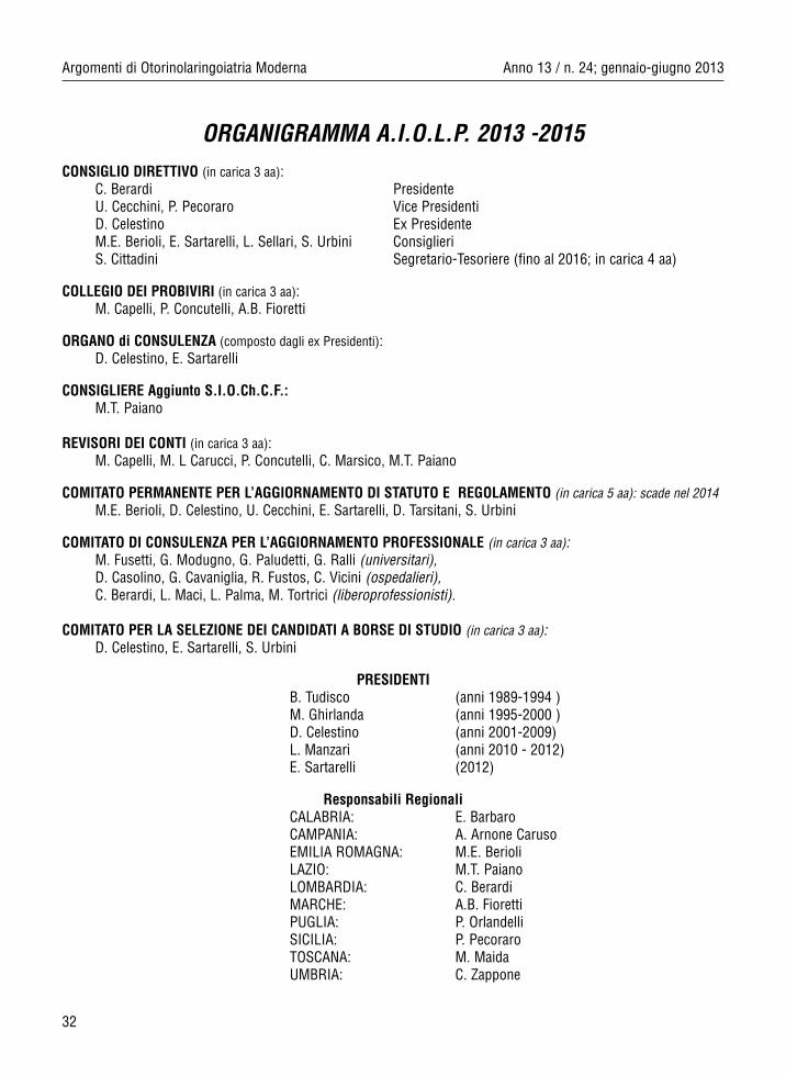
ORGANIGRAMMA A.I.O.L.P. 2013 -2015CONSIGLIO DIRETTIVO (in carica 3 aa):
C. Berardi PresidenteU. Cecchini, P. Pecoraro Vice PresidentiD. Celestino Ex PresidenteM.E. Berioli, E. Sartarelli, L. Sellari, S. Urbini ConsiglieriS. Cittadini Segretario-Tesoriere (fino al 2016; in carica 4 aa)
COLLEGIO DEI PROBIVIRI (in carica 3 aa):M. Capelli, P. Concutelli, A.B. Fioretti
ORGANO di CONSULENZA (composto dagli ex Presidenti):D. Celestino, E. Sartarelli
CONSIGLIERE Aggiunto S.I.O.Ch.C.F.:M.T. Paiano
REVISORI DEI CONTI (in carica 3 aa):M. Capelli, M. L Carucci, P. Concutelli, C. Marsico, M.T. Paiano
COMITATO PERMANENTE PER L’AGGIORNAMENTO DI STATUTO E REGOLAMENTO (in carica 5 aa): scade nel 2014M.E. Berioli, D. Celestino, U. Cecchini, E. Sartarelli, D. Tarsitani, S. Urbini
COMITATO DI CONSULENZA PER L’AGGIORNAMENTO PROFESSIONALE (in carica 3 aa):M. Fusetti, G. Modugno, G. Paludetti, G. Ralli (universitari),D. Casolino, G. Cavaniglia, R. Fustos, C. Vicini (ospedalieri),C. Berardi, L. Maci, L. Palma, M. Tortrici (liberoprofessionisti).
COMITATO PER LA SELEZIONE DEI CANDIDATI A BORSE DI STUDIO (in carica 3 aa):D. Celestino, E. Sartarelli, S. Urbini
PRESIDENTIB. Tudisco (anni 1989-1994 )M. Ghirlanda (anni 1995-2000 )D. Celestino (anni 2001-2009)L. Manzari (anni 2010 - 2012)E. Sartarelli (2012)
Responsabili RegionaliCALABRIA: E. BarbaroCAMPANIA: A. Arnone CarusoEMILIA ROMAGNA: M.E. BerioliLAZIO: M.T. PaianoLOMBARDIA: C. Berar diMARCHE: A.B. FiorettiPUGLIA: P. OrlandelliSICILIA: P. PecoraroTOSCANA: M. MaidaUMBRIA: C. Zappone
Anno 13 / n. 24; gennaio-giugno 2013 Argomenti di Otorinolaringoiatria Moderna
Storia dell’Associazione
Il 18 Maggio 1989 è stata costituita l’Associazione ItalianaOtorinolaringoiatri Libero Professionisti (A.I.O.L.P.) affiliata, dall’anno succes-sivo, alla Società Italiana di Otorinolaringoiatria e Chirurgia Cervico-Facciale(S.I.O. e Ch.C.F.). L’A.I.O.L.P. ha l’obbiettivo di riunire ed organizzare tutti gliSpecialisti in Otorinolaringoiatria liberi professionisti; come tali sono conside-rati i Colleghi che non hanno in essere rapporti di dipendenza con Università odEnti Ospedalieri, cioè libero professionisti puri, convenzionati esterni con ilServizio Sanitario Nazionale od altri Enti, specialisti ambulatoriali, consulentiospedalieri, termalisti, medici militari, specialisti O.R.L. di fabbrica, convenzio-nati o dipendenti di A.S.L. (Azienda Sanitaria Locale) e di Case di Cura, exuniversitari ed ex ospedalieri. Coloro che pur non possedendo i requisiti diSocio desiderino partecipare alla vita associativa, possono iscriversi come“Sostenitori A.I.O.L.P.” senza diritto di voto all’Assemblea dei Soci né eleggibi-lità alle cariche sociali.
L’A.I.O.L.P. mira a tutelare il prestigio della figura dell’Otoiatra LiberoProfessionista, a valorizzarne la qualificazione ed a promuoverne e sostenernein modo permanente la formazione.
Story of the Association
The Italian Association of the free-lance professional Otologists(A.I.O.L.P.) was constituted on the 18th of May 1989. The very next year it wasaffiliated to the Italian Society of Otorhinolaryngology and Cervical-FacialSurgery (S.I.O. and Ch.C.F.). The purpose of A.I.O.L.P. is to assemble and organize every free-lance E.N.T. Specialists. For free-lance we mean thoseCollegues who are not Hospitals or University’s employees but just free-lancepanel professional Specialists of the National Health Service, Boards, out-patients department Specialists, Hospital’s Consultings. Specialists whowork in the Baths, Medical Officers, E.N.T. Specialists working in factories,Hospital’s panel Doctors, A.S.L.’s (Local Health’s Business) employees,Specialists who work in Nursing Homes, former University and Hospital’sspecialists. Should somebody have not the necessary requirements to becomea Member but still desires to take part in the social life of the Association canbe enroled as “A.I.O.L.P. founder member” but with no right to vote during theMembers’ meeting or to be eligible dignitary.
The aim of A.I.O.L.P. is to safeguard the role of free-lance Otologists,to enhance their qualification as well as to back or permanently promote theirtraining.