Invasive salivary duct carcinoma ex pleomorphic adenoma of the
7
CASE REPORT Open Access Invasive salivary duct carcinoma ex pleomorphic adenoma of the parotid gland: a teaching case giving rise to the genuine diagnostic difficulty on an inadequate cytology specimen Sohsuke Yamada 1,3* , Atsunori Nabeshima 1 , Takahisa Tabata 2 , Xin Guo 1 , Takashi Tasaki 1 , Ke-Yong Wang 1 , Shohei Shimajiri 1 and Yasuyuki Sasaguri 1 Abstract: A history of a recent rapid increase in long-standing swelling mass was presented in the right parotid gland of an 85-year-old male. The inadequate cytologic specimens contained few small clusters of three- dimensional malignant epithelial cells having hyperchromatic pleomorphic nuclei and prominent nucleoli, adjacent to a cluster of benign monomorphic myoepithelial cells. We first interpreted it merely as an adenocarcinoma, not otherwise specified. A radical parotidectomy was performed, and gross examination revealed an encapsulated and firm tumor lesion, looking grayish-blue to yellowish-white, focally associated with extracapsular invasion. On microscopic examination, the tumor was predominantly composed of a proliferation of highly atypical epithelial cells having abundant eosinophilic cytoplasm, often arranged in a Roman-bridge appearance with foci of comedo necrosis, alternating with extensive infiltration to adjacent stroma in a trabecular or alveolar fashion with severe vessel permeation. Within the background of pleomorphic adenoma, the carcinoma cells sometimes replaced ductal luminal cells while retaining an intact-like myoepithelial layer. Therefore, we finally made a diagnosis of invasive salivary duct carcinoma ex pleomorphic adenoma. We should be aware that owing to its characteristic features, cytopathologists might be able to determine correct diagnosis, based on multiple and adequate samplings. Virtual slides: The virtual slide(s) for this article can be found here: http://www.diagnosticpathology.diagnomx.eu/ vs/2126158270695815 Keywords: Salivary duct carcinoma, Carcinoma ex pleomorphic adenoma, Salivary gland, Cytology Background Among all salivary gland neoplasms, carcinoma ex pleo- morphic adenoma (Ca ex PA) accounts for approximately 3.6% [1], whereas constitutes 6.2% of all PA and 11.6% of all malignant salivary gland neoplasms [1,2]. Despite of that, Ca ex PA is uncommon and has a prevalence rate of 5.6 cases per 100,000 malignant neoplasms and a yearly incidence rate of 0.17 tumors per 1 million people [1,2]. This neoplasm is defined as an epithelial malignant transformation within a primary (de novo) or recurrent PA, and often poses a diagnostic challenge to clinicians and cytopathologists, since its entity is difficult to diagnose pre-operatively [3,4]. In fact, Ca ex PA can be asymptomatic as most of them are not widely invasive on gross find- ings and often have similar clinical presentations as PA, however, patients with Ca ex PA have a poor prognosis due to infiltrative and destructive behavior, and thus, early and accurate diagnosis and aggressive surgical treatment (i.e., total or radical parotidectomy) can in- crease their survival rates [1-5]. Although any form of carcinoma can be observed and also be a mixture of subtypes, the malignant component of Ca ex PA is most often adenocarcinoma, not otherwise specified (NOS), and sometimes, may be salivary duct carcinoma (SDC), * Correspondence: [email protected] 1 Departments of Pathology and Cell Biology School of Medicine, University of Occupational and Environmental Health, Kitakyushu, Japan 3 Department of Pathology and Cell Biology, School of Medicine, University of Occupational and Environmental Health, 1-1 Iseigaoka, Yahatanishi-ku, Kitakyushu 807-8555, Japan Full list of author information is available at the end of the article © 2012 Yamada et al.; licensee BioMed Central Ltd. This is an Open Access article distributed under the terms of the Creative Commons Attribution License (http://creativecommons.org/licenses/by/2.0), which permits unrestricted use, distribution, and reproduction in any medium, provided the original work is properly cited. Yamada et al. Diagnostic Pathology 2012, 7:61 http://www.diagnosticpathology.org/content/7/1/61
Transcript of Invasive salivary duct carcinoma ex pleomorphic adenoma of the
Yamada et al. Diagnostic Pathology 2012, 7:61http://www.diagnosticpathology.org/content/7/1/61
CASE REPORT Open Access
Invasive salivary duct carcinoma ex pleomorphicadenoma of the parotid gland: a teaching casegiving rise to the genuine diagnostic difficulty onan inadequate cytology specimenSohsuke Yamada1,3*, Atsunori Nabeshima1, Takahisa Tabata2, Xin Guo1, Takashi Tasaki1, Ke-Yong Wang1,Shohei Shimajiri1 and Yasuyuki Sasaguri1
Abstract: A history of a recent rapid increase in long-standing swelling mass was presented in the right parotidgland of an 85-year-old male. The inadequate cytologic specimens contained few small clusters of three-dimensional malignant epithelial cells having hyperchromatic pleomorphic nuclei and prominent nucleoli, adjacentto a cluster of benign monomorphic myoepithelial cells. We first interpreted it merely as an adenocarcinoma, nototherwise specified. A radical parotidectomy was performed, and gross examination revealed an encapsulated andfirm tumor lesion, looking grayish-blue to yellowish-white, focally associated with extracapsular invasion. Onmicroscopic examination, the tumor was predominantly composed of a proliferation of highly atypical epithelialcells having abundant eosinophilic cytoplasm, often arranged in a Roman-bridge appearance with foci of comedonecrosis, alternating with extensive infiltration to adjacent stroma in a trabecular or alveolar fashion with severevessel permeation. Within the background of pleomorphic adenoma, the carcinoma cells sometimes replacedductal luminal cells while retaining an intact-like myoepithelial layer. Therefore, we finally made a diagnosis ofinvasive salivary duct carcinoma ex pleomorphic adenoma. We should be aware that owing to its characteristicfeatures, cytopathologists might be able to determine correct diagnosis, based on multiple and adequatesamplings.
Virtual slides: The virtual slide(s) for this article can be found here: http://www.diagnosticpathology.diagnomx.eu/vs/2126158270695815
Keywords: Salivary duct carcinoma, Carcinoma ex pleomorphic adenoma, Salivary gland, Cytology
BackgroundAmong all salivary gland neoplasms, carcinoma ex pleo-morphic adenoma (Ca ex PA) accounts for approximately3.6% [1], whereas constitutes 6.2% of all PA and 11.6% ofall malignant salivary gland neoplasms [1,2]. Despite ofthat, Ca ex PA is uncommon and has a prevalence rateof 5.6 cases per 100,000 malignant neoplasms and ayearly incidence rate of 0.17 tumors per 1 million people[1,2]. This neoplasm is defined as an epithelial malignant
* Correspondence: [email protected] of Pathology and Cell Biology School of Medicine, Universityof Occupational and Environmental Health, Kitakyushu, Japan3Department of Pathology and Cell Biology, School of Medicine, University ofOccupational and Environmental Health, 1-1 Iseigaoka, Yahatanishi-ku,Kitakyushu 807-8555, JapanFull list of author information is available at the end of the article
© 2012 Yamada et al.; licensee BioMed CentraCommons Attribution License (http://creativecreproduction in any medium, provided the or
transformation within a primary (de novo) or recurrentPA, and often poses a diagnostic challenge to cliniciansand cytopathologists, since its entity is difficult to diagnosepre-operatively [3,4]. In fact, Ca ex PA can be asymptomaticas most of them are not widely invasive on gross find-ings and often have similar clinical presentations as PA,however, patients with Ca ex PA have a poor prognosisdue to infiltrative and destructive behavior, and thus,early and accurate diagnosis and aggressive surgicaltreatment (i.e., total or radical parotidectomy) can in-crease their survival rates [1-5]. Although any form ofcarcinoma can be observed and also be a mixture ofsubtypes, the malignant component of Ca ex PA is mostoften adenocarcinoma, not otherwise specified (NOS),and sometimes, may be salivary duct carcinoma (SDC),
l Ltd. This is an Open Access article distributed under the terms of the Creativeommons.org/licenses/by/2.0), which permits unrestricted use, distribution, andiginal work is properly cited.
Yamada et al. Diagnostic Pathology 2012, 7:61 Page 2 of 7http://www.diagnosticpathology.org/content/7/1/61
undifferentiated carcinoma, adenoid cystic carcinoma, ormucoepidermoid carcinoma [1,2,4,6]. While, WHO statesthat its component is most frequently a poorly differen-tiated carcinoma, e.g., SDC or adenocarcinoma, NOS, oran undifferentiated carcinoma [3].On the other hand, SDC was first described by
Kleinsasser et al in 1968 [7], and to date, more than 100cases have been reported and account for approximately9% among all salivary gland neoplasms [8]. SDC is a dis-tinctive but relatively common high grade adenocarcin-oma arising from the excretory ductal epithelium of themajor salivary glands, especially the parotid gland [8-10].Clinically, these tumors are characterized by aggressive(i.e., infiltrative and destructive) behavior with localrecurrence, early and distant metastasis, invasion ofthe facial nerve, and/or significant mortality [8-11].Histopathologically, SDCs have a striking resemblanceto ductal carcinoma of the breast, exhibiting intraduc-tal and infiltrating components [8,10,11]. In addition,very intriguingly, they should show a broader clinico-pathological spectrum and many cases of them maydevelop within PA as a result of malignant transform-ation of ductal epithelial cells [9,12]. It has been actuallyreported that multifocal origin of SDC from major ex-cretory ducts surrounding a PA was found [8]. By con-trast, Simpson RHW et al. described that the majorityof them arise de novo (as in the breast) probably from apure in situ carcinoma [13]. Similar to Ca ex PA, aggres-sive clinical management in the early stage of SDCappears to be the only hope for good prognosis[5,6,8-11]. Thus, it is critical to establish an accuratepreoperative diagnosis by fine-needle aspiration cy-tology, however, previous studies have indicated the dif-ficulty of correct characterization of Ca ex PA and/orSDC due to sampling errors or inadequateness and mis-interpretation [14,15].In fact, invasive SDC ex PA could be relatively com-
mon disease, as compared with some recently publishedcase reports of very rare tumor cell types in the salivarygland [16-18]. Despite of that, we report a case of SDCex PA, which originated from the parotid gland and par-tially involved the surrounding soft tissue and lymphnodes as a rapidly increased but long-standing swellingmass. Based on the relatively inadequate cytology speci-mens, we preoperatively interpreted it merely as an adeno-carcinoma, NOS.
Materials and methodsThe patient was an 85-year-old Japanese man. A fine-needle aspiration from the parotid gland mass was per-formed, followed by a radical parotidectomy. The tumorspecimens after fixation in 10% neutral buffered formalinwere embedded in paraffin for histological or immuno-histochemical examinations. All immunohistochemical
stainings were carried out using Dako Envision kit (DakoCytomation Co., Glostrup, Denmark) according to themanufacturer’s instructions. For transmission electronmicroscopy (JEM-1200EX, JEOL Ltd., Tokyo, Japan), thedehydrated tissue specimens, after fixed with 2.5% glu-taraldehyde and immersed in 2% osmium tetroxide, wereembedded in epoxy resin.
Case presentationThe patient had a history of benign prostatic hyperplasia2 years ago. There was no history of malignancy, im-munosuppressive disorders, use of immunosuppressivemedications, or unusual infections.He noticed a long-standing swelling mass in the right
parotid gland and an enlargement of the neck lymphnode 5 years before the resection. Following that, a recentrapid increase of them was presented. Laboratory data, in-cluding blood cell count, chemistry and tumor markers,were within normal limits. A neck CT scan revealed a het-erogeneously enhanced and poorly-demarcated mass,measuring approximately 3 × 3 cm, in the right parotidgland. CT scans of the chest and abdomen disclosed nodefinite evidence of metastasis in the lymph nodes orother organs. The patient was alive and well at 8 monthsafter the operation.
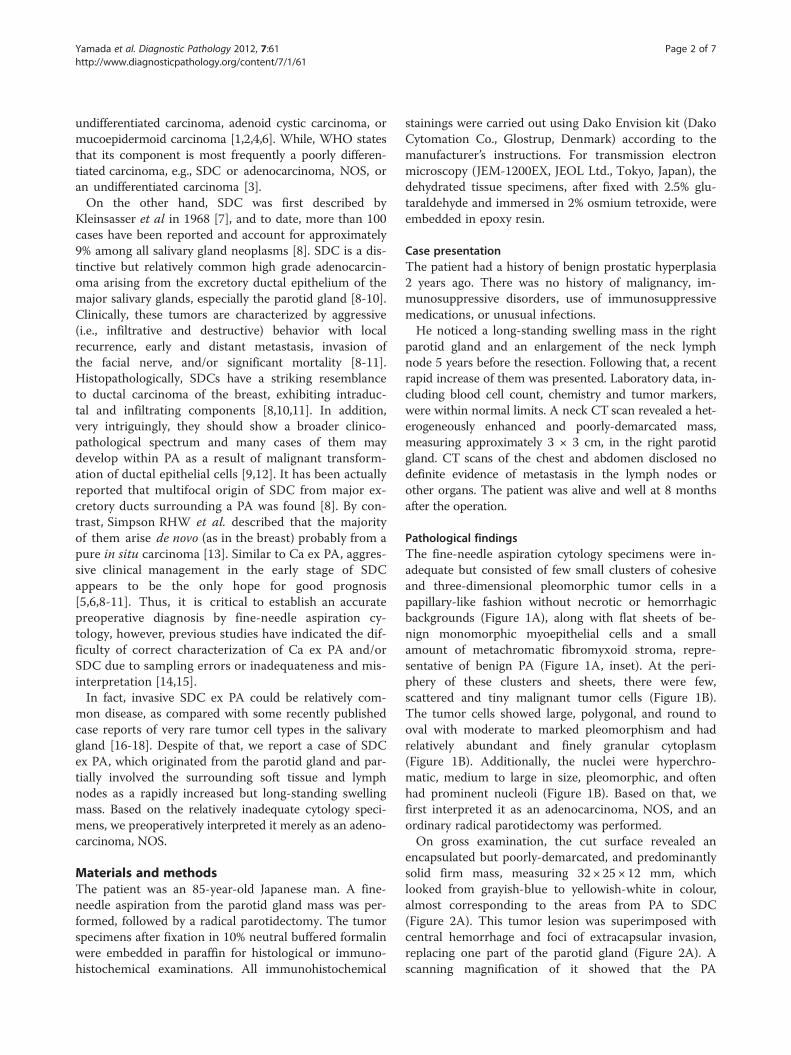
Pathological findingsThe fine-needle aspiration cytology specimens were in-adequate but consisted of few small clusters of cohesiveand three-dimensional pleomorphic tumor cells in apapillary-like fashion without necrotic or hemorrhagicbackgrounds (Figure 1A), along with flat sheets of be-nign monomorphic myoepithelial cells and a smallamount of metachromatic fibromyxoid stroma, repre-sentative of benign PA (Figure 1A, inset). At the peri-phery of these clusters and sheets, there were few,scattered and tiny malignant tumor cells (Figure 1B).The tumor cells showed large, polygonal, and round tooval with moderate to marked pleomorphism and hadrelatively abundant and finely granular cytoplasm(Figure 1B). Additionally, the nuclei were hyperchro-matic, medium to large in size, pleomorphic, and oftenhad prominent nucleoli (Figure 1B). Based on that, wefirst interpreted it as an adenocarcinoma, NOS, and anordinary radical parotidectomy was performed.On gross examination, the cut surface revealed an
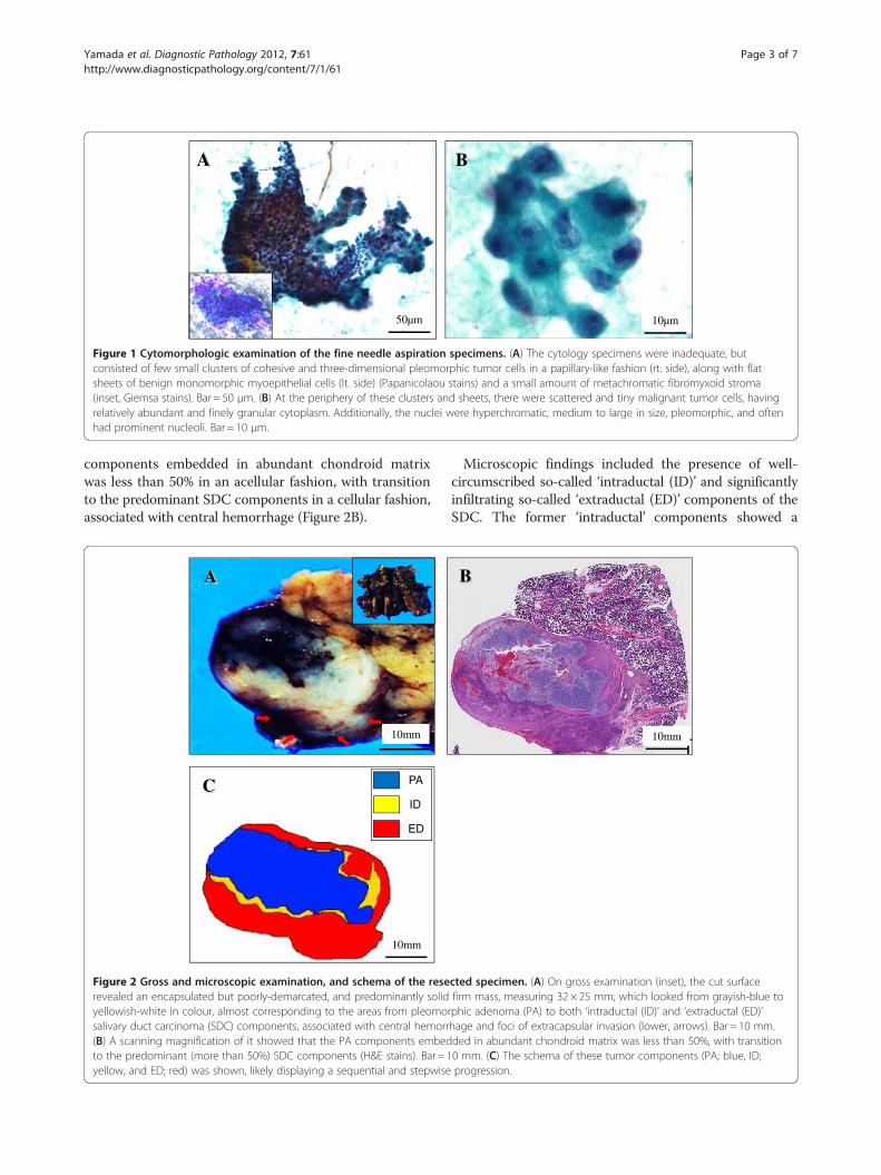
encapsulated but poorly-demarcated, and predominantlysolid firm mass, measuring 32 × 25 × 12 mm, whichlooked from grayish-blue to yellowish-white in colour,almost corresponding to the areas from PA to SDC(Figure 2A). This tumor lesion was superimposed withcentral hemorrhage and foci of extracapsular invasion,replacing one part of the parotid gland (Figure 2A). Ascanning magnification of it showed that the PA
A
50µm
B
10µm
Figure 1 Cytomorphologic examination of the fine needle aspiration specimens. (A) The cytology specimens were inadequate, butconsisted of few small clusters of cohesive and three-dimensional pleomorphic tumor cells in a papillary-like fashion (rt. side), along with flatsheets of benign monomorphic myoepithelial cells (lt. side) (Papanicolaou stains) and a small amount of metachromatic fibromyxoid stroma(inset, Giemsa stains). Bar = 50 μm. (B) At the periphery of these clusters and sheets, there were scattered and tiny malignant tumor cells, havingrelatively abundant and finely granular cytoplasm. Additionally, the nuclei were hyperchromatic, medium to large in size, pleomorphic, and oftenhad prominent nucleoli. Bar = 10 μm.
Yamada et al. Diagnostic Pathology 2012, 7:61 Page 3 of 7http://www.diagnosticpathology.org/content/7/1/61
components embedded in abundant chondroid matrixwas less than 50% in an acellular fashion, with transitionto the predominant SDC components in a cellular fashion,associated with central hemorrhage (Figure 2B).
A
10mm
C
10mm
PA
ID
ED
Figure 2 Gross and microscopic examination, and schema of the reserevealed an encapsulated but poorly-demarcated, and predominantly solidyellowish-white in colour, almost corresponding to the areas from pleomosalivary duct carcinoma (SDC) components, associated with central hemorr(B) A scanning magnification of it showed that the PA components embedto the predominant (more than 50%) SDC components (H&E stains). Bar = 1yellow, and ED; red) was shown, likely displaying a sequential and stepwise
Microscopic findings included the presence of well-circumscribed so-called ‘intraductal (ID)’ and significantlyinfiltrating so-called ‘extraductal (ED)’ components of theSDC. The former ‘intraductal’ components showed a
B
10mm
cted specimen. (A) On gross examination (inset), the cut surfacefirm mass, measuring 32 × 25 mm, which looked from grayish-blue torphic adenoma (PA) to both ‘intraductal (ID)’ and ‘extraductal (ED)’hage and foci of extracapsular invasion (lower, arrows). Bar = 10 mm.ded in abundant chondroid matrix was less than 50%, with transition0 mm. (C) The schema of these tumor components (PA; blue, ID;progression.
Yamada et al. Diagnostic Pathology 2012, 7:61 Page 4 of 7http://www.diagnosticpathology.org/content/7/1/61
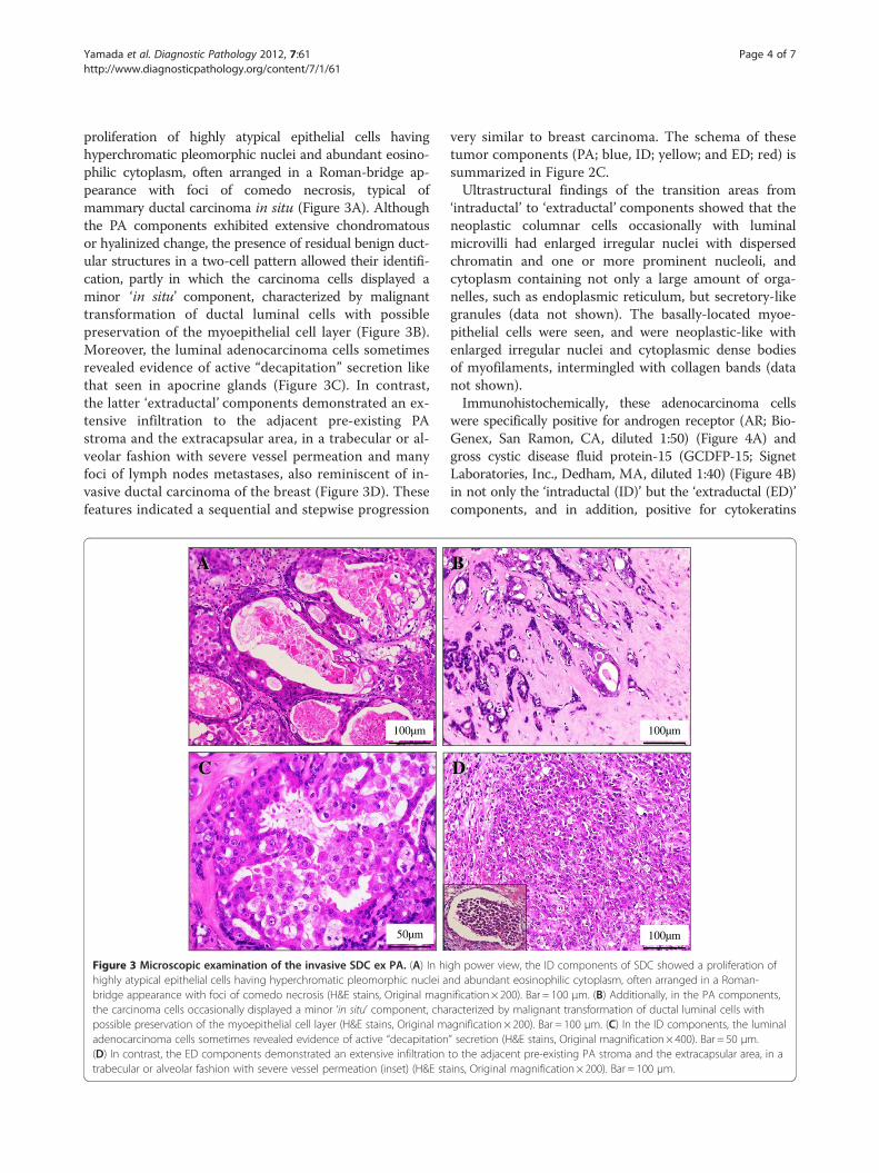
proliferation of highly atypical epithelial cells havinghyperchromatic pleomorphic nuclei and abundant eosino-philic cytoplasm, often arranged in a Roman-bridge ap-pearance with foci of comedo necrosis, typical ofmammary ductal carcinoma in situ (Figure 3A). Althoughthe PA components exhibited extensive chondromatousor hyalinized change, the presence of residual benign duct-ular structures in a two-cell pattern allowed their identifi-cation, partly in which the carcinoma cells displayed aminor ‘in situ’ component, characterized by malignanttransformation of ductal luminal cells with possiblepreservation of the myoepithelial cell layer (Figure 3B).Moreover, the luminal adenocarcinoma cells sometimesrevealed evidence of active “decapitation” secretion likethat seen in apocrine glands (Figure 3C). In contrast,the latter ‘extraductal’ components demonstrated an ex-tensive infiltration to the adjacent pre-existing PAstroma and the extracapsular area, in a trabecular or al-veolar fashion with severe vessel permeation and manyfoci of lymph nodes metastases, also reminiscent of in-vasive ductal carcinoma of the breast (Figure 3D). Thesefeatures indicated a sequential and stepwise progression
100µm
50µm
A
C
Figure 3 Microscopic examination of the invasive SDC ex PA. (A) In hihighly atypical epithelial cells having hyperchromatic pleomorphic nuclei abridge appearance with foci of comedo necrosis (H&E stains, Original magnthe carcinoma cells occasionally displayed a minor ‘in situ’ component, chapossible preservation of the myoepithelial cell layer (H&E stains, Original madenocarcinoma cells sometimes revealed evidence of active “decapitation(D) In contrast, the ED components demonstrated an extensive infiltrationtrabecular or alveolar fashion with severe vessel permeation (inset) (H&E sta
very similar to breast carcinoma. The schema of thesetumor components (PA; blue, ID; yellow; and ED; red) issummarized in Figure 2C.Ultrastructural findings of the transition areas from
‘intraductal’ to ‘extraductal’ components showed that theneoplastic columnar cells occasionally with luminalmicrovilli had enlarged irregular nuclei with dispersedchromatin and one or more prominent nucleoli, andcytoplasm containing not only a large amount of orga-nelles, such as endoplasmic reticulum, but secretory-likegranules (data not shown). The basally-located myoe-pithelial cells were seen, and were neoplastic-like withenlarged irregular nuclei and cytoplasmic dense bodiesof myofilaments, intermingled with collagen bands (datanot shown).Immunohistochemically, these adenocarcinoma cells
were specifically positive for androgen receptor (AR; Bio-Genex, San Ramon, CA, diluted 1:50) (Figure 4A) andgross cystic disease fluid protein-15 (GCDFP-15; SignetLaboratories, Inc., Dedham, MA, diluted 1:40) (Figure 4B)in not only the ‘intraductal (ID)’ but the ‘extraductal (ED)’components, and in addition, positive for cytokeratins
100µm
100µm
B
D
gh power view, the ID components of SDC showed a proliferation ofnd abundant eosinophilic cytoplasm, often arranged in a Roman-ification× 200). Bar = 100 μm. (B) Additionally, in the PA components,racterized by malignant transformation of ductal luminal cells withagnification× 200). Bar = 100 μm. (C) In the ID components, the luminal” secretion (H&E stains, Original magnification× 400). Bar = 50 μm.to the adjacent pre-existing PA stroma and the extracapsular area, in ains, Original magnification × 200). Bar = 100 μm.
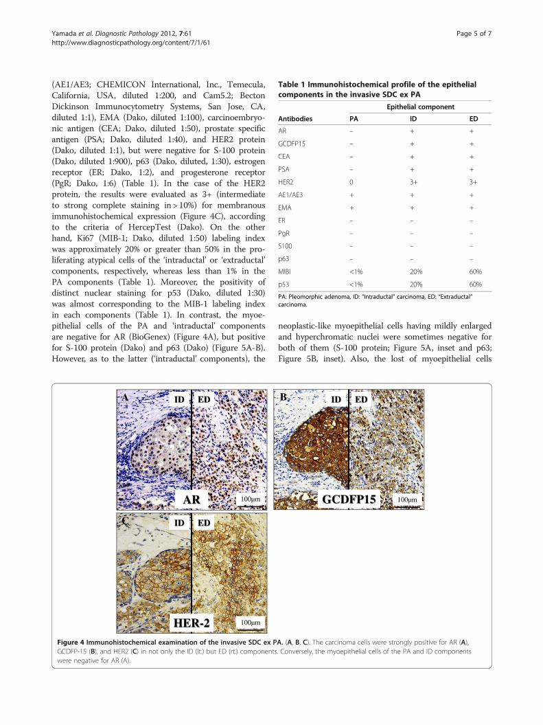
Table 1 Immunohistochemical profile of the epithelialcomponents in the invasive SDC ex PA
Epithelial component
Antibodies PA ID ED
AR – + +
GCDFP15 – + +
CEA – + +
PSA – + +
HER2 0 3+ 3+
AE1/AE3 + + +
EMA + + +
ER – – –
PgR – – –
S100 – – –
p63 – – –
MIBI <1% 20% 60%
p53 <1% 20% 60%
PA: Pleomorphic adenoma, ID: “Intraductal” carcinoma, ED: “Extraductal”carcinoma.
Yamada et al. Diagnostic Pathology 2012, 7:61 Page 5 of 7http://www.diagnosticpathology.org/content/7/1/61
(AE1/AE3; CHEMICON International, Inc., Temecula,California, USA, diluted 1:200, and Cam5.2; BectonDickinson Immunocytometry Systems, San Jose, CA,diluted 1:1), EMA (Dako, diluted 1:100), carcinoembryo-nic antigen (CEA; Dako, diluted 1:50), prostate specificantigen (PSA; Dako, diluted 1:40), and HER2 protein(Dako, diluted 1:1), but were negative for S-100 protein(Dako, diluted 1:900), p63 (Dako, diluted, 1:30), estrogenreceptor (ER; Dako, 1:2), and progesterone receptor(PgR; Dako, 1:6) (Table 1). In the case of the HER2protein, the results were evaluated as 3+ (intermediateto strong complete staining in > 10%) for membranousimmunohistochemical expression (Figure 4C), accordingto the criteria of HercepTest (Dako). On the otherhand, Ki67 (MIB-1; Dako, diluted 1:50) labeling indexwas approximately 20% or greater than 50% in the pro-liferating atypical cells of the ‘intraductal’ or ‘extraductal’components, respectively, whereas less than 1% in thePA components (Table 1). Moreover, the positivity ofdistinct nuclear staining for p53 (Dako, diluted 1:30)was almost corresponding to the MIB-1 labeling indexin each components (Table 1). In contrast, the myoe-pithelial cells of the PA and ‘intraductal’ componentsare negative for AR (BioGenex) (Figure 4A), but positivefor S-100 protein (Dako) and p63 (Dako) (Figure 5A-B).However, as to the latter (‘intraductal’ components), the
100µm AR
ID ED
100µm HER-2
ID ED
A
C
Figure 4 Immunohistochemical examination of the invasive SDC ex PGCDFP-15 (B), and HER2 (C) in not only the ID (lt.) but ED (rt.) componentswere negative for AR (A).
neoplastic-like myoepithelial cells having mildly enlargedand hyperchromatic nuclei were sometimes negative forboth of them (S-100 protein; Figure 5A, inset and p63;Figure 5B, inset). Also, the lost of myoepithelial cells
100µm GCDFP15
ID ED B
A. (A, B, C). The carcinoma cells were strongly positive for AR (A),. Conversely, the myoepithelial cells of the PA and ID components

Yamada et al. Diagnostic Pathology 2012, 7:61 Page 6 of 7http://www.diagnosticpathology.org/content/7/1/61
was focally apparent in the ‘intraductal’ components(Figure 5A-B).Based on all these features, we suggested that these
proliferating carcinoma cells were characteristic of apo-crine differentiation, and finally made a diagnosis of in-vasive SDC ex PA of the parotid gland.
DiscussionAggressive clinical treatment in the early stage for inva-sive SDC is the only hope for better prognosis, due to ahigh grade, malignant tumor, based on the infiltrativeand destructive behavior [8-11]. Thus, it is critical to es-tablish an accurate preoperative diagnosis by fine-needleaspiration cytology, the clinical utility of which in diag-nosing salivary gland tumors has been generalized. Thecytologic findings of invasive SDC reflect the histopatho-logical ones resembling invasive ductal carcinoma of thebreast, showing cohesive, three-dimensional clusters andflat sheets of large and polygonal atypical cells havingabundant and finely granular cytoplasm, and prominentnucleoli, arranged in an irregular branching, cribriform,or papillary growth pattern as well as single cells forma-tion in the background of frequent necrosis [15,19]. Add-itionally, smears from Ca ex PA show moderate to highcellularity with pleomorphic malignant cells in the back-ground of biphasic components of PA, displaying sheetsof benign monomorphic epithelial cells along with meta-chromatic, fibrillary and myxoid or chondroid stroma[14]. As in the current case, although the specimenswere inadequate, the cytologic features were almostsimilar to those as described above, even while neithernecrotic backgrounds nor any cribriform formations wereobserved. Despite that, a confident and accurate diagnosisof SDC ex PA was impossible on cytology, suggestivelydue to sampling errors, lack of experience, cytomorpholo-gic variety, and misinterpretation. Nevertheless, in cases
A
100µm S-100 protein Figure 5 Myoepithelial markers analysis of the invasive SDC ex PA inS-100 protein (A) and p63 (B). By contrast, the myoepithelial cells of the PAside) (Original magnification × 200, Bar = 100 μm). However, as to the ID comildly enlarged and hyperchromatic nuclei were sometimes negative for bmagnification × 400, Bar = 50 μm). Also, the lost of myoepithelial cells was f
with a strong clinical suspicion of Ca ex PA, such as ours,multiple fine needle aspiration should be performed andits suspicion should be raised to alert the pathologist, atthe very least.An immunohistochemical analysis indicates that AR
and GCDFP-15 are highly and specifically expressed inmore than 80% and 90% of patients with SDC, respect-ively, whereas ER and PgR, well known as commonbreast carcinoma markers, are very weakly or negativelystained [11,20-22]. Actually, one paper proposed that,when the cytologic features of high grade adenocarcinomawith a variety of cell morphology are difficult to make anaccurate diagnosis, immunostaining for AR and ER oncytologic smears would be very useful for the diagnosis ofSDC [19].On the other hand, the luminal SDC carcinoma cells
rarely show immunohistochemical expression of S-100protein and p63, whereas the ‘reminiscent’ benign myoe-pithelial cells reveal those strong expression in the ‘intra-ductal’ and PA components [9-11,20-22], even while thelost of myoepithelial cells is occasionally seen in thetransition areas from ‘intraductal’ to ‘extraductal’ compo-nents [20], as in our case. Indeed, myoepithelial cells canbe considered as natural tumor suppressors, distinguish-ing between early and advanced malignant tumors in thetransition from in situ to invasive carcinomas, and canrarely undergo malignancy. However, in the present case,the neoplastic-like enlarged myoepithelial cells in thetransition areas were sometimes negative for the abovemyoepithelial markers. These features indicate that thosecells would not have apparent myoepithelial phenotypes,but potentially neoplastic character, probably confirmedby the ultrastructural findings. We could provide theevidence for the first time that invasive SDC ex PA mayarise within or from PA as a result of neoplastic trans-formation of outer supporting myoepithelial cells, as well
B
100µm p63 immunohistochemistry. (A, B) The carcinoma cells were negative forand ID components were positive for S-100 protein and p63 (A-B, rt.mponents, the neoplastic-like myoepithelial cells (arrowheads) havingoth of S-100 protein (A, inset) and p63 (B, inset) (Originalocally apparent in the ID components (A-B, lt. side).
Yamada et al. Diagnostic Pathology 2012, 7:61 Page 7 of 7http://www.diagnosticpathology.org/content/7/1/61
as inner ductal epithelial cells. Despite of that, futurestudies will be further required to determine whetherour suggestion is significant after collecting and examininga larger number of its cases.
ConclusionWe herein reported a case of an invasive SDC ex PA. Thepresent case was tentatively diagnosed as adenocarcinoma,NOS on the examination of the cytology, because itssmears showed the inadequate and few components ofmalignant cells. All cytopathologists should be aware thatits clinically and immunohistochemically characteristicfeatures, as well as multiple and adequate fine needle as-piration, could lead to a correct diagnosis.
ConsentWritten informed consent was obtained from the patientfor publication of this case report and any accompanyingimages. A copy of the written consent is available for re-view by the Editor-in-Chief of this journal.
Competing interestsThe authors declare that they have no competing interests.
Authors’ contributionsSY and AN participated in conception of the idea and writing of themanuscript. SY, AN, TT, XG, TT, KYW, SS and YS performed thecytohistological and ultrastructural interpretation of the tumor tissue. Allauthors have read and approved the final manuscript.
Author details1Departments of Pathology and Cell Biology School of Medicine, Universityof Occupational and Environmental Health, Kitakyushu, Japan. 2Departmentsof Otorhinolaryngology, School of Medicine, University of Occupational andEnvironmental Health, Kitakyushu, Japan. 3Department of Pathology and CellBiology, School of Medicine, University of Occupational and EnvironmentalHealth, 1-1 Iseigaoka, Yahatanishi-ku, Kitakyushu 807-8555, Japan.
Received: 15 March 2012 Accepted: 23 April 2012Published: 30 May 2012
References1. Gnepp DR: Malignant mixed tumours of the salivary glands: a review.
Pathol Annu 1993, 28:279–328.2. Antony J, Gopalan V, Smith RA, Lam AK: Carcinoma ex pleomorphic
adenoma: a comprehensive review of clinical, pathological andmolecular data. Head Neck Pathol 2011 Jul 09, doi:[Epub ahead of print].
3. Gnepp DR, Brandwein-Gensler MS, El-Naggar AK, Nagao T: Carcinoma expleomorphic adenoma. In World Health Organization Classification ofTumours: Pathology and Genetics of Head and Neck Tumours. Edited byBarnes L, Eveson JW, Reichart P, Sidransky D. Lyon, France: IARC Press;2005:242–243.
4. Ellis GL, Auclair PL: Carcinoma ex pleomorphic adenoma, In: Tumors of theSalivary Glands. Atlas of tumor pathology. Washington, D.C: Armed ForcesInstitute of Pathology; 2008:259–269.
5. Altemani A, Martins MT, Freitas L, Soares F, Araújo NS, Araújo VC: Carcinomaex pleomorphic adenoma (CXPA): immunoprofile of the cells involved incarcinomatous progression. Histopathology 2005, 46:635–641.
6. Lewis JE, Olsen KD, Sebo TJ: Carcinoma ex pleomorphic adenoma:pathologic analysis of 73 cases. Hum Pathol 2001, 32:596–604.
7. Kleinsasser O, Klein HJ, Hubner G: Speinchelgangcarcinome, ein denMilchegangcarcinomen der Brustdruse, analoge Gruppe vonSpeicheldrusentumoren. Archiv klein exper. Ohren-, Nasen- und Kehlkopfheilk1968, 192:100–115.
8. Brandwein-Gensler MS, Skalova A, Nagao T: Salivary duct carcinoma. InWorld Health Organization Classification of Tumours: Pathology and Geneticsof Head and Neck Tumours. Edited by Barnes L, Eveson JW, Reichart P,Sidransky D. Lyon, France: IARC Press; 2005:236–237.
9. Delgado R, Vuitch F, Albores-Saavedra J: Salivary duct carcinoma. Cancer1993, 72:1503–1512.
10. Lewis JE, McKinney BC, Weiland LH, Ferreiro JA, Olsen KD: Salivary ductcarcinoma: clinicopathologic and immunohistochemical review of 26cases. Cancer 1996, 77:223–230.
11. Ellis GL, Auclair PL: Salivary duct carcinoma, In: Tumors of the SalivaryGlands. Atlas of tumor pathology. Washington, D.C: Armed Forces Institute ofPathology; 2008:322–332.
12. Brandwein MS, Jagirder J, Patil J, Biller H, Kaneko M: Salivary ductcarcinoma (cribriform salivary carcinoma of excretory ducts): aclinicopathologic and immunohistochemical study of 12 cases. Cancer1990, 65:2307–2314.
13. Simpson RHW, Desai S, Palma SD: Salivary duct carcinoma in situ of theparotid gland. Histopathology 2008, 53:416–425.
14. Nigam S, Kumar N, Jain S: Cytomorphologic spectrum of carcinoma expleomorphic adenoma. Acta Cytol 2004, 48:309–314.
15. Garcia-Bonafe M, Catala I, Tarragona J, Tallada N: Cytologic diagnosis ofsalivary duct carcinoma: a review of seven cases. Diagn Cytopathol 1998,19:120–123.
16. Shin-ichi Nakatsuka, Hiroshi Harada, Hiroshi Fujiyama, Koji Takeda, KojiKitamura, Hayato Kimura, Teruaki Nagano, Mahito Ito, Yuji Asada: Aninvasive adenocarcinoma of the accessory parotid gland: a rare exampledeveloping from a low-grade cribriform cystadenocarcinoma? DiagnPathol 2011, 6:122.
17. Schraven Sebastian P, Plontke Stefan K, Roland Syha, Falko Fend, HartwigWolburg, Patrick Adam: Dendritic cell tumor in a salivary gland lymphnode: a rare differential diagnosis of salivary gland neoplasms. DiagnPathol 2011, 6:94.
18. Bersch C, Back W: Primary sebaceous adenoma of the salivary glands? arare differential diagnosis: report of a case. Diagn Pathol 2007,2(Suppl 1):S16.
19. Moriki T, Ueta S, Takahashi T, Mitani M, Ichien M: Salivary duct carcinoma:cytologic characteristics and application of androgen receptorimmunostaining for diagnosis. Cancer (Cancer Cytopathology) 2001,93:344–350.
20. Kapadia SB, Barnes L: Expression of androgen receptor, gross cysticdisease fluid protein, and CD44 in salivary duct carcinoma. Mod Pathol1998, 11:1033–1038.
21. Fan CY, Wang J, Barnes EL: Expression of androgen receptor and prostaticspecific markers in salivary duct carcinoma: an immunohistochemicalanalysis of 13 cases and review of the literature. Am J Surg Pathol 2000,24:579–586.
22. Williams MD, Roberts D, Blumenschein GR Jr, Temam S, Kies MS, Rosenthal DI,Weber RS, El-Naggar AK: Differential expression of hormonal and growthfactor receptors in salivary duct carcinomas: biologic significance andpotential role in therapeutic stratification of patients. Am J Surg Pathol 2007,31:1645–1652.
doi:10.1186/1746-1596-7-61Cite this article as: Yamada et al.: Invasive salivary duct carcinoma expleomorphic adenoma of the parotid gland: a teaching case giving riseto the genuine diagnostic difficulty on an inadequate cytologyspecimen. Diagnostic Pathology 2012 7:61.