Il Sistema Venoso Cerebrale Extracranico: un … · Il Sistema Venoso Cerebrale Extracranico: un...
49
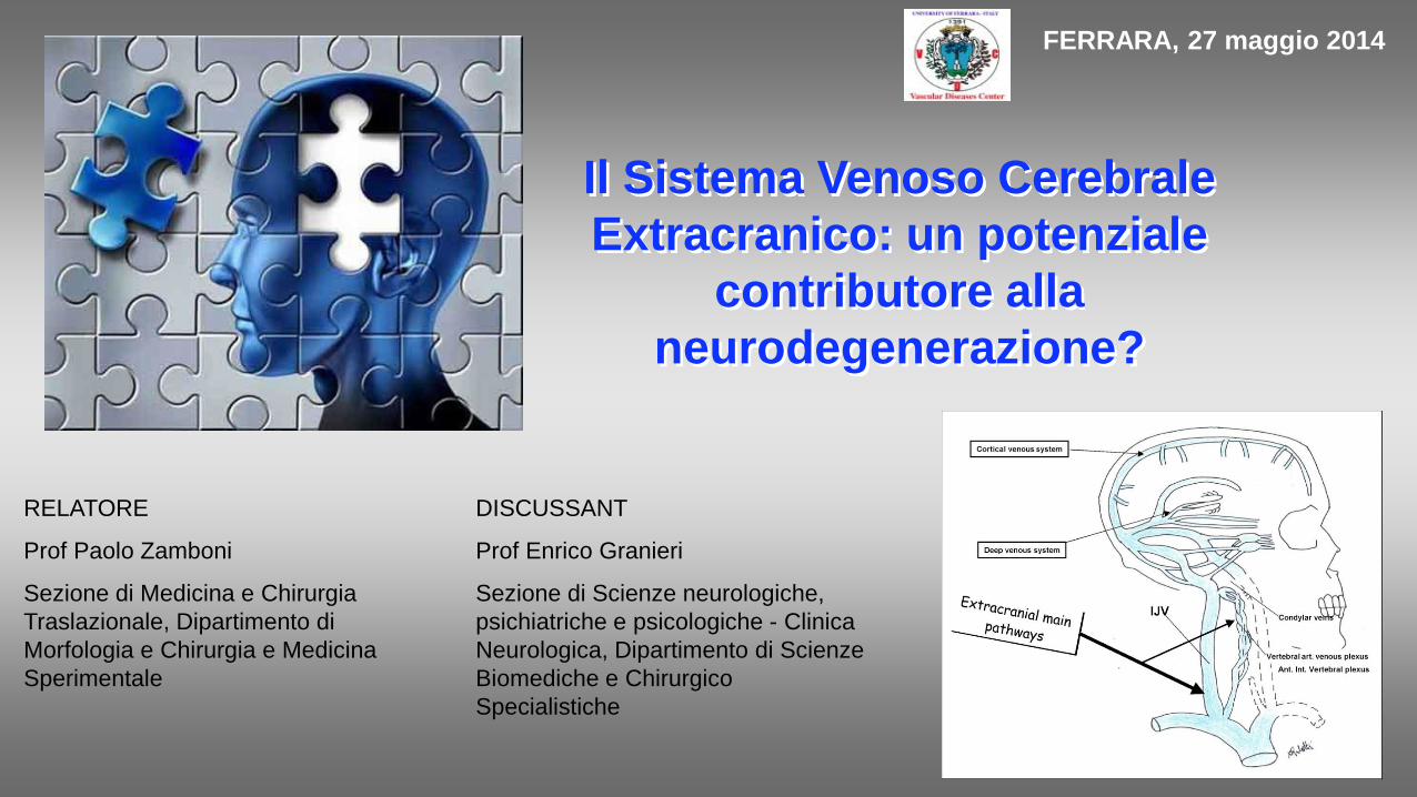
Il Sistema Venoso Cerebrale Extracranico: un potenziale contributore alla neurodegenerazione? FERRARA, 27 maggio 2014 RELATORE Prof Paolo Zamboni Sezione di Medicina e Chirurgia Traslazionale, Dipartimento di Morfologia e Chirurgia e Medicina Sperimentale DISCUSSANT Prof Enrico Granieri Sezione di Scienze neurologiche, psichiatriche e psicologiche - Clinica Neurologica, Dipartimento di Scienze Biomediche e Chirurgico Specialistiche
Transcript of Il Sistema Venoso Cerebrale Extracranico: un … · Il Sistema Venoso Cerebrale Extracranico: un...
Il Sistema Venoso Cerebrale
Extracranico: un potenziale
contributore alla
neurodegenerazione?
FERRARA, 27 maggio 2014
RELATORE
Prof Paolo Zamboni
Sezione di Medicina e Chirurgia
Traslazionale, Dipartimento di
Morfologia e Chirurgia e Medicina
Sperimentale
DISCUSSANT
Prof Enrico Granieri
Sezione di Scienze neurologiche,
psichiatriche e psicologiche - Clinica
Neurologica, Dipartimento di Scienze
Biomediche e Chirurgico
Specialistiche
PR
IMA
RY
Vascular Disease Center
University of Ferrara
Chief: Prof. Paolo Zamboni
email: [email protected]

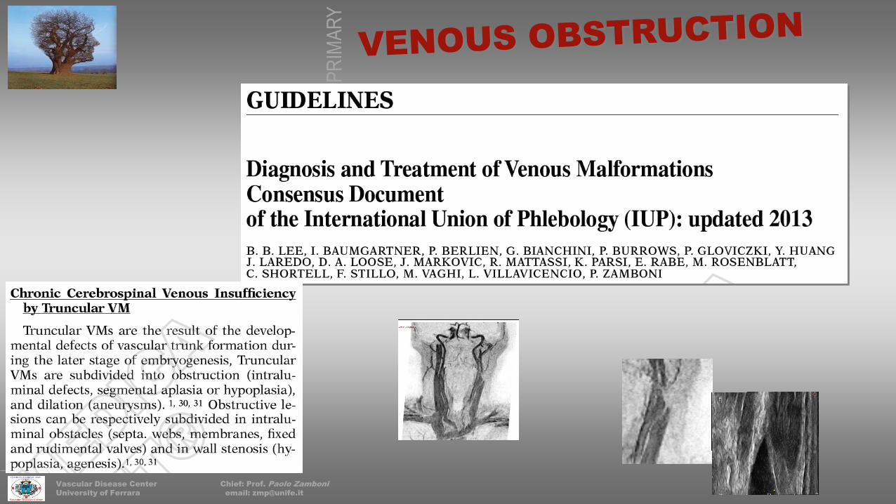
PRIMARY VENOUS OSTRUCTION
INTRALUMINAL OBSTACLES

Muscular entrapment-external compression
PR
IMA
RY
Vascular Disease Center
University of Ferrara
Chief: Prof. Paolo Zamboni
email: [email protected]
Multimodality imaging techniques comparison
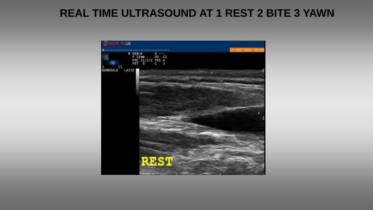
REAL TIME ULTRASOUND AT 1 REST 2 BITE 3 YAWN

WALL STENOSIS TWISTING
SVC Azygos Arch
Twisting
SVC
Azygos Arch
Twisting
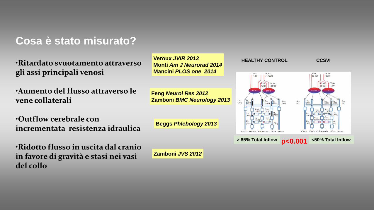
Cosa è stato misurato? •Ritardato svuotamento attraverso gli assi principali venosi •Aumento del flusso attraverso le vene collaterali •Outflow cerebrale con incrementata resistenza idraulica •Ridotto flusso in uscita dal cranio in favore di gravità e stasi nei vasi del collo
Veroux JVIR 2013
Monti Am J Neurorad 2014
Mancini PLOS one 2014
Feng Neurol Res 2012
Zamboni BMC Neurology 2013
Beggs Phlebology 2013
Zamboni JVS 2012
HEALTHY CONTROL CCSVI
p<0.001 <50% Total Inflow > 85% Total Inflow


Patologie venose
extracraniche e
neurodegenerazione •AD and brain aging • Parkinson • Meniere • Normotensive hydrocephalus • SM
Chung et al J Alzheimers Dis. 2014
Lanzillo et al BMC Neurol. 2013
Zamboni et al J Neur Neurosurg Psichiatry 2009
Zivadinov et al Neurology 2010
Liu et al J Vasc Surg 2014
Filipo et al Eur Arch Otorhinolaryngol. 2013
Di Berardino et al Phlebology. 2014
Beggs et al BMC Med. 2013

Sono note conseguenze
fisiopatologiche? •Riduzione della portata e della velocità del flusso liquorale
•Ridotta perfusione cerebrale
Zamboni Funct Neur 2009
Beggs J Magn Reson Imaging 2013
Beggs BMC Neur 2013
Zivadinov JVIR 2013
Magnano J Magn Reson Imaging 2012
Zamboni BMC Medicine 2011
D'haeseleer Lancet Neurol 2011
Utriainen Neurol Res 2012
Garaci Radiology 2012
Guttmann J Neuroimaging 2012
Zivadinov BMC Neurology 2011
VL CP
V3
V4
Brain Superficial Veins
Deep Veins
CSF
Carotid/Basal
Arteries IJVs
SSS
SAS
STS
Spinal
Column
AoS
FM
AV
AV
SR
AV
CSF Bulk Flow Legend AV Arachnoid villi
AoS Aqueduct of
Sylvius
CP Choroid plexus
CSF Cerebrospinal
fluid
FM Foramen magnum
IJV Internal jugular vein
SAS Sub-arachnoid
space
SR Starling resistor
SSS Superior sagittal
sinus
STS Straight sinus
VL Lateral ventricle
V3 Third ventricle
V4 Fourth ventricle
Venous hypertension here will
tend to inhibit absorption of
CSF by the SSS.
P
1 2

CSF Absorption into SSS
CSF absorption vs CSF pressure
• CSF absorption is driven by pressure difference between SAS and SSS at a rate of approximately 0.1031 mL/min/mmHg.
• Minimum of 5 mmHg CSF pressure required to permit CSF absorption through the arachnoid villi into the superior sagittal sinus.
Source: Cutler RWP, et al. Brain 1968, 91(4):707-720
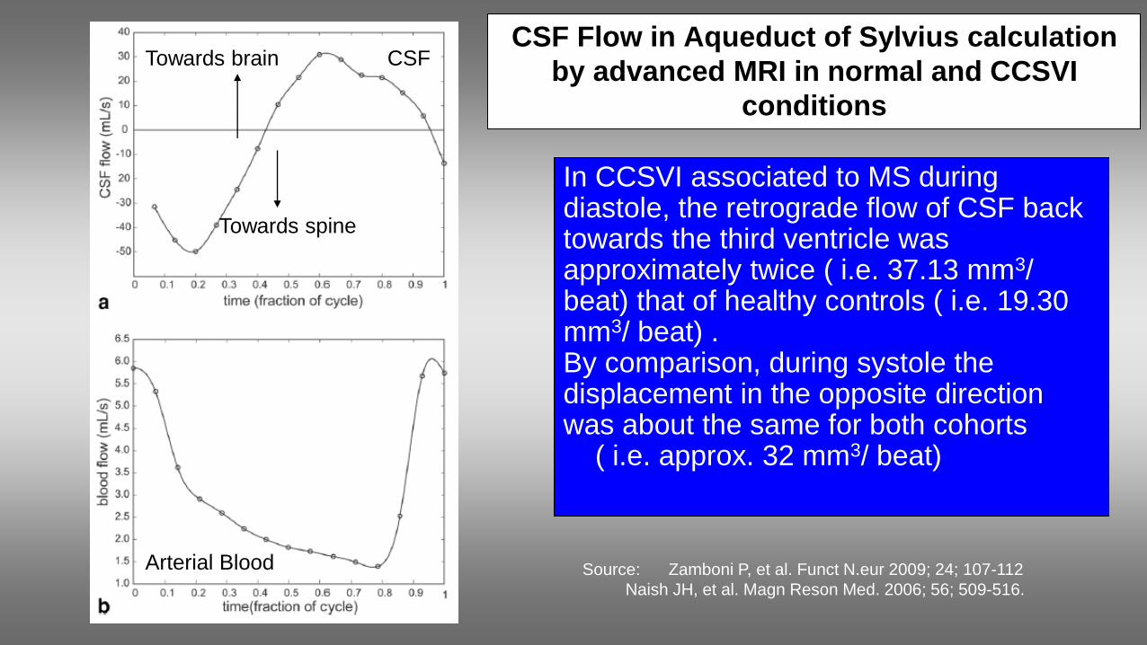
CSF Flow in Aqueduct of Sylvius calculation
by advanced MRI in normal and CCSVI
conditions
In CCSVI associated to MS during diastole, the retrograde flow of CSF back towards the third ventricle was approximately twice ( i.e. 37.13 mm3/ beat) that of healthy controls ( i.e. 19.30 mm3/ beat) . By comparison, during systole the displacement in the opposite direction was about the same for both cohorts ( i.e. approx. 32 mm3/ beat)
CSF
Arterial Blood
Towards spine
Towards brain
Source: Zamboni P, et al. Funct N.eur 2009; 24; 107-112
Naish JH, et al. Magn Reson Med. 2006; 56; 509-516.
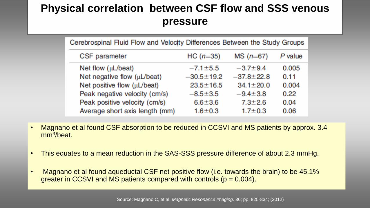
Physical correlation between CSF flow and SSS venous
pressure
• Magnano et al found CSF absorption to be reduced in CCSVI and MS patients by approx. 3.4 mm3/beat.
• This equates to a mean reduction in the SAS-SSS pressure difference of about 2.3 mmHg.
• Magnano et al found aqueductal CSF net positive flow (i.e. towards the brain) to be 45.1% greater in CCSVI and MS patients compared with controls (p = 0.004).
Source: Magnano C, et al. Magnetic Resonance Imaging. 36; pp. 825-834; (2012)
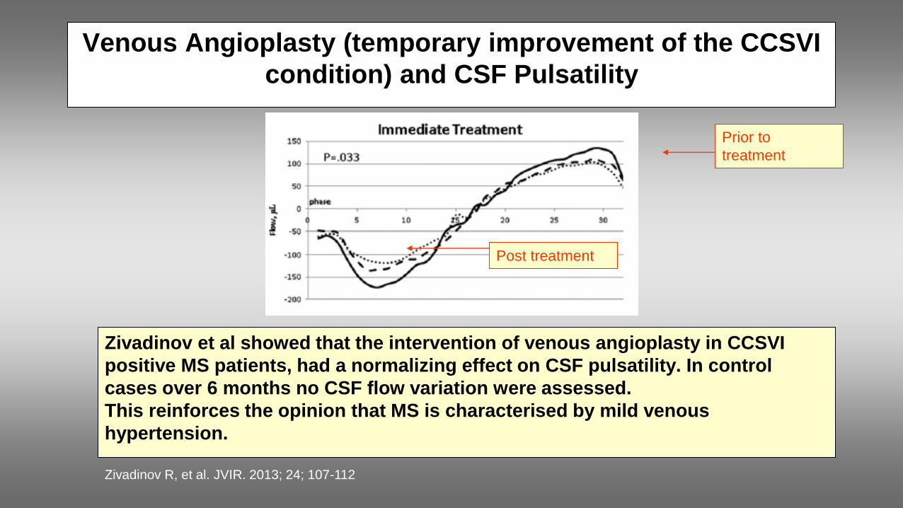
Venous Angioplasty (temporary improvement of the CCSVI
condition) and CSF Pulsatility
Zivadinov et al showed that the intervention of venous angioplasty in CCSVI
positive MS patients, had a normalizing effect on CSF pulsatility. In control
cases over 6 months no CSF flow variation were assessed.
This reinforces the opinion that MS is characterised by mild venous
hypertension.
Prior to
treatment
Post treatment
Zivadinov R, et al. JVIR. 2013; 24; 107-112
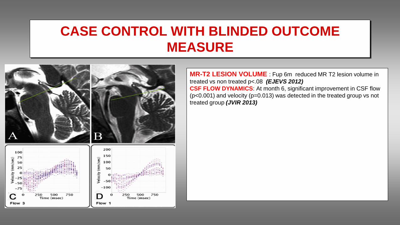
CASE CONTROL WITH BLINDED OUTCOME
MEASURE
MR-T2 LESION VOLUME : Fup 6m reduced MR T2 lesion volume in
treated vs non treated p<.08 (EJEVS 2012)
CSF FLOW DYNAMICS: At month 6, significant improvement in CSF flow
(p<0.001) and velocity (p=0.013) was detected in the treated group vs not
treated group (JVIR 2013)
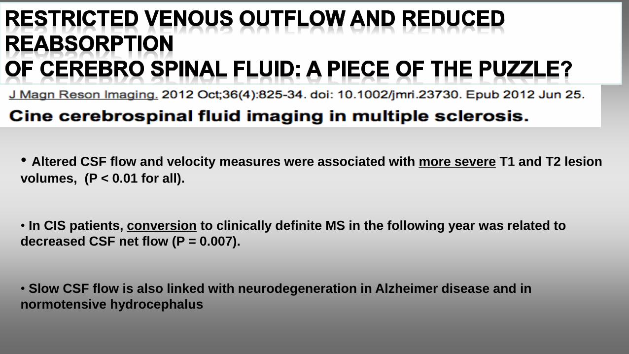
• Altered CSF flow and velocity measures were associated with more severe T1 and T2 lesion
volumes, (P < 0.01 for all).
• In CIS patients, conversion to clinically definite MS in the following year was related to
decreased CSF net flow (P = 0.007).
• Slow CSF flow is also linked with neurodegeneration in Alzheimer disease and in
normotensive hydrocephalus

1: Daouk J, et al Relationship between cerebrospinal fluid
flow, ventricles morphology, and DTI properties in internal
capsules: differences between Alzheimer's disease and
normal-pressure hydrocephalus. Acta Radiol. 2013 Oct 17.
[Epub ahead of print]
2: Hosseinzadeh S, et al. Elevated CSF and plasma
microparticles in a rat model of streptozotocin-induced
cognitive impairment. Behav Brain Res. 2013 1;256:503-11.
3: Erickson MA, et al. Lipopolysaccharide impairs amyloid
β efflux from brain: altered vascular sequestration,
cerebrospinal fluid reabsorption, peripheral clearance and
transporter function at the blood-brain barrier. J
Neuroinflammation. 2012,29;9:150.
4: Stomrud E, et al. CSF biomarkers correlate with cerebral
blood flow on SPECT in healthy elderly. Dement Geriatr
Cogn Disord. 2012;33(2-3):156-63.
5: Santos AN, et al. Amyloid-β oligomers in cerebrospinal
fluid are associated with cognitive decline in patients with
Alzheimer's disease. J Alzheimers Dis. 2012;29(1):171-6.
6: Banks WA, et al. Impairments in brain-to-blood transport
of amyloid-β and reabsorption of cerebrospinal fluid in an
animal model of Alzheimer's disease are reversed by
antisense directed against amyloid-β protein precursor. J
Alzheimers Dis. 2011;23(4):599-605.
Jugular venous reflux
reduced CSF re-absorption
more MRI lesion in AD
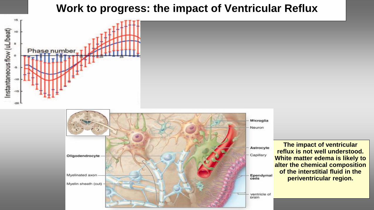
Work to progress: the impact of Ventricular Reflux
The impact of ventricular reflux is not well understood.
White matter edema is likely to alter the chemical composition
of the interstitial fluid in the periventricular region.
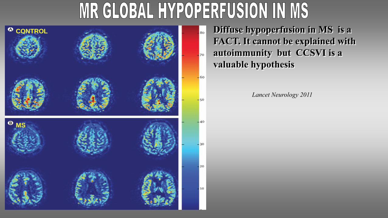
CONTROL
MS
Lancet Neurology 2011
Diffuse hypoperfusion in MS is a
FACT. It cannot be explained with
autoimmunity but CCSVI is a
valuable hypothesis

International Consortium for
Brain Mapping.
ICBM Atlas of normality
SPECT Atlas of normality
generated from 47 healthy
subjects.
Harvard Medical School
Variability of perfusion of the
white matter in normal cases.
Guttmann, J Neuroimaging 2012
• 1294 SPECT-MRI in MS
cases, early and late RR, SP
respectively.
• Comparison with normal
cerebral perfusion patterns
provided by SPECT atlas of
normal healthy individuals

Guttmann, J Neuroimaging 2012
Chronic plaques were more prevalent in WM regions with lower relative
perfusion. Lesions in more highly perfused regions were more commonly
observed in early RR MS and therefore, may be more likely to successfully
remyelinate and resolve.
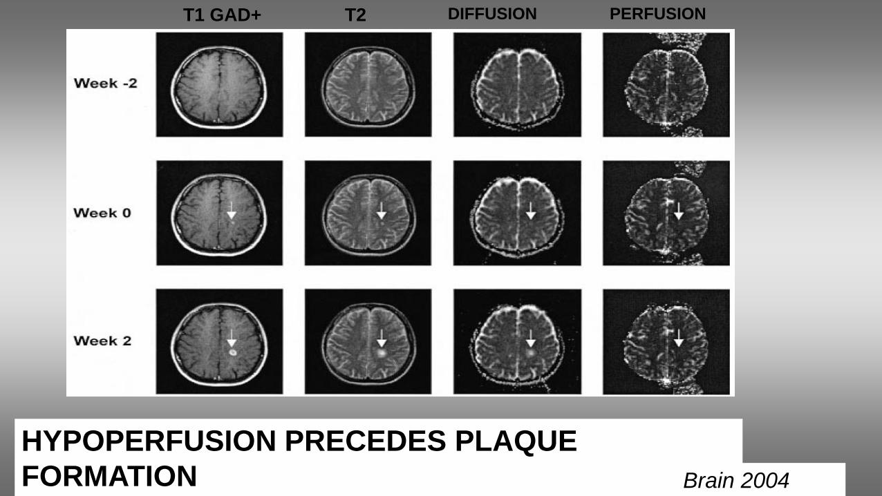
T1 GAD+ T2 DIFFUSION PERFUSION
HYPOPERFUSION PRECEDES PLAQUE
FORMATION Brain 2004
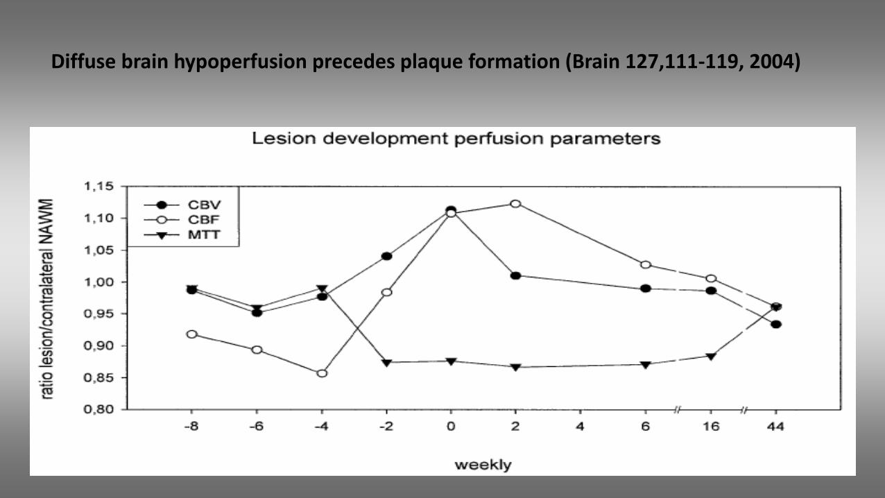
Diffuse brain hypoperfusion precedes plaque formation (Brain 127,111-119, 2004)

RELATIONSHIP BETWEEN BRAIN PERFUSION AND JUGULAR FLOW: THE SIGNIFICANCE OF
STENOSIS
•Evidence already exists for reduced perfusion in
patients with MS but there has been no attempt to
correlate this with obstructed venous outflow.
• 2D magnetic resonance imaging (MRI) flow
techniques demonstrate that flow in the internal jugular
veins in humans is linearly related to global brain
perfusion
Controls + non stenotic
Stenotic
Neurol Res. 2012 Oct;34(8):780-92

The data support a role of CCSVI in cerebral hemody- namic changes, such as a
decrease of CBV and CBF, re- gardless of the presence of MS.
Garaci et al, Radiology: Volume 265: October 2012

A: CBF in a 33 yo, relapsing remitting,
CCSVI-MS patient with a VHISS 5.
B: CBF in a 38 yo, relapsing remitting,
CCSVI-MS patient with a VHISS 12.
The dark areas indicate lower CBF in the
patient with higher VHISS.
Cerebral blood flow (CBF) at MRI perfusional study
Robust correlation between VHISS and MR perfusional parameters (r= -0.70 to -0.71,
p<0.002).
CCSVI is related to brain hypoperfusion
BMC Medicine 2011

MS & Loss of Cerebral Veins
HC SP RR
• MS is associated with a marked deduction in cerebral vein volume (VV).
• VV (for all vein diameters): HC = 82.9 mL; MS = 66.9 mL;
Reduction = 19.3%; p<0.0001
• VV (for veins <0.3 mm): HC = 53.8 mL; MS = 45.0 mL;
Reduction = 16.4%; p<0.0001 [Strongly correlated with CCSVI (p<0.003)]
Source: Zivadinov R, et al. BMC Neurology 2011, 11:128

SWI Venography
A1 A2
B1 B2
C1 C2
Do vessels degrade because flow is shunted
away from them?
Slide courtesy of Yulin Ge, NYU
normal control (A) and two MS patients (B, C) demonstrate a
significantly reduced number of veins in perviventricular
NAWM in patients compared to controls. MS patient with
more lesions (C) has less venous structures depicted on
SWI mIP image than MS patient with fewer lesions (B).
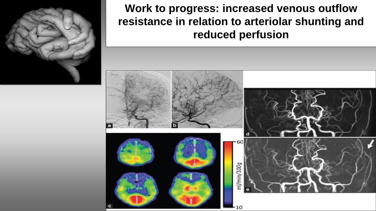
Work to progress: increased venous outflow
resistance in relation to arteriolar shunting and
reduced perfusion

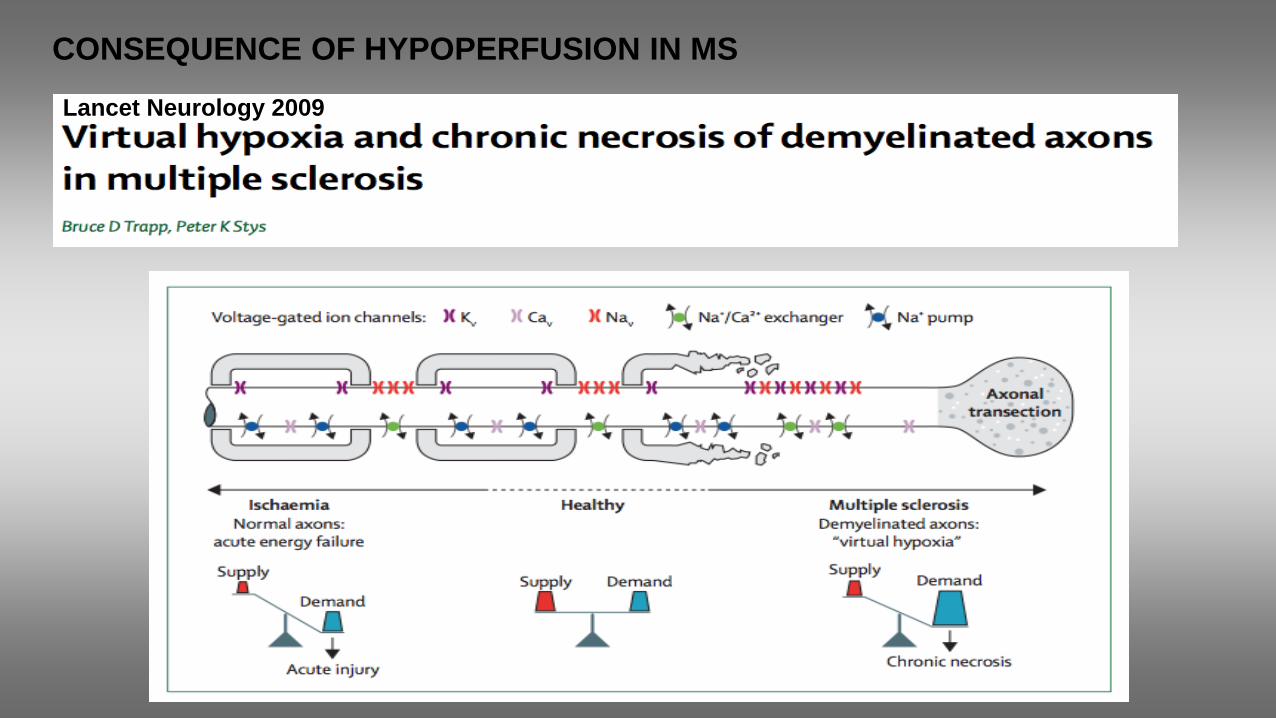
CONSEQUENCE OF HYPOPERFUSION IN MS
Lancet Neurology 2009

Oligodendrocyte susceptibility to hypoperfusion
• ROS (increased i-NOS in macrophages and microglia)
• Mitochondria impairment (defective phosphorilation)
• Hypoxia associated molecules (expression HIF 1 alfa)

NEW MS LESIONS
1. Loss of oligodendrocytes
2. Dead myelin
3. Myelin-laden macrophages
4. Only subsequently T and B cells infiltration

• Damage to axon can occur without the presence of inflammation (Int MS J. 2009 Jun;16(2):57-6; Ann Neurol. 2009 Dec;66(6):739-53
• Axon demyelination was seen in early lesion without inflammatory cells (NEJM 2011).
DEMYELINATION AND AXONAL DAMAGE PRECEDE T-CELL
- INFLAMMATORY CELL INFILTRATION
ANN NEUR 2009; NEJM 2011

The loop between
hypoxia and NF-kB
1. Hypoxia induces HIF alfa and
beta translocating to the
nucleus
2. Binding with hypoxia
response promoter HRE and
gene interaction
3. Genes of the NF-kB
4. In addition, in hypoxia the
p50 and p65 subunits of NF-
kB are no more inhibited,
translocate to nucleous, and
in turn activate inflammatory
genes
NORMOXIA HYPOXIA
Source: N Engl J Med 2012
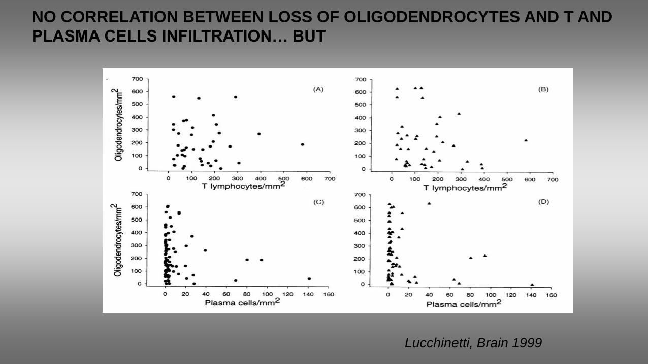
NO CORRELATION BETWEEN LOSS OF OLIGODENDROCYTES AND T AND
PLASMA CELLS INFILTRATION… BUT
Lucchinetti, Brain 1999
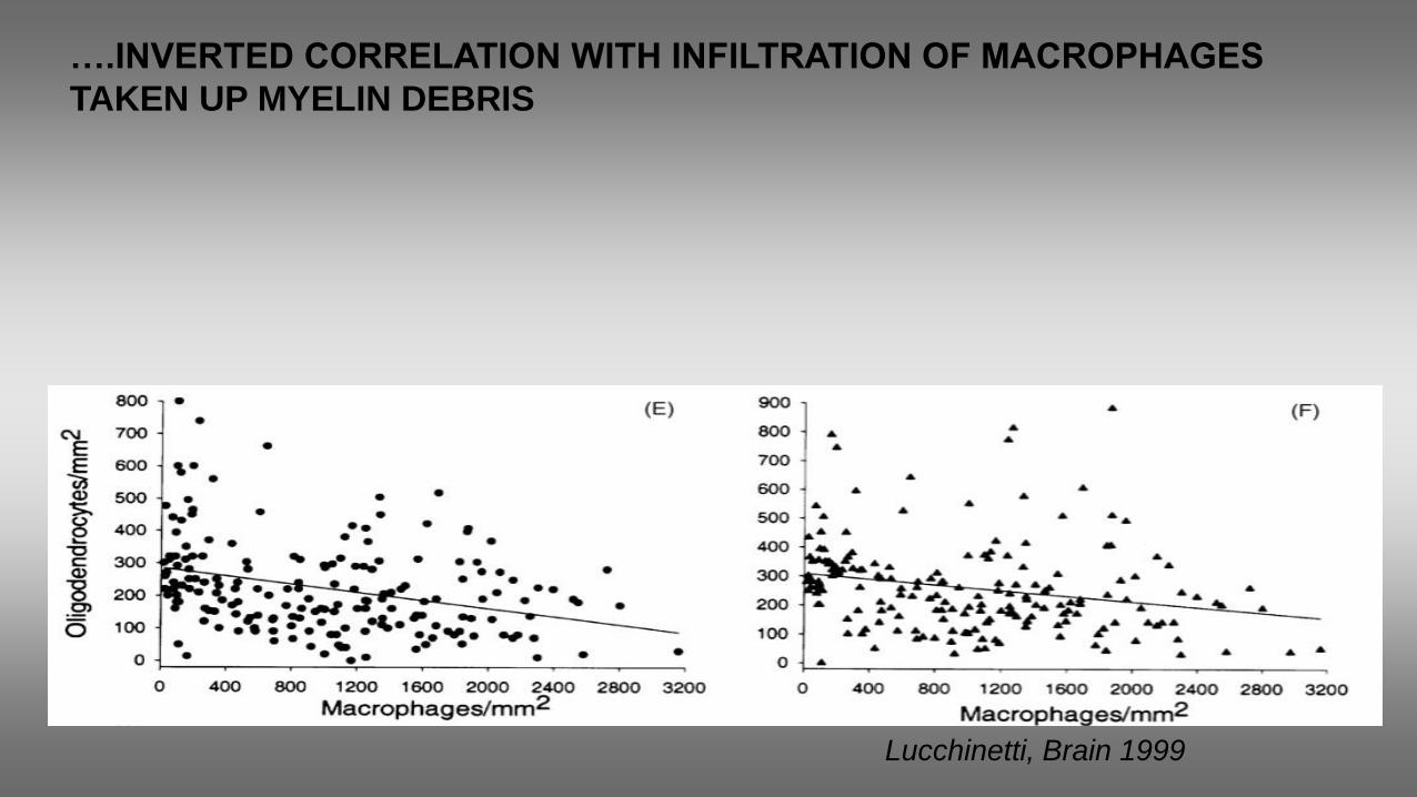
….INVERTED CORRELATION WITH INFILTRATION OF MACROPHAGES
TAKEN UP MYELIN DEBRIS
Lucchinetti, Brain 1999

PATHOLOGIC PATTERNS OF MS
I. Massive tissue distruction mediated by CD8+T cells infiltrates and
macrophages.
II. Massive deposition of immunoglobulins and component of activated
complement
III. Oligodendrocytes apoptosis, “dying back” oligodendrogliopathy
IV. Neurodegeneration and oligodendrocytes death also in the periplaque
WM
Are chronologically late events?
Are effects of the hypoperfusion?

Tsamopoulos et al. J Mult Scler 2014
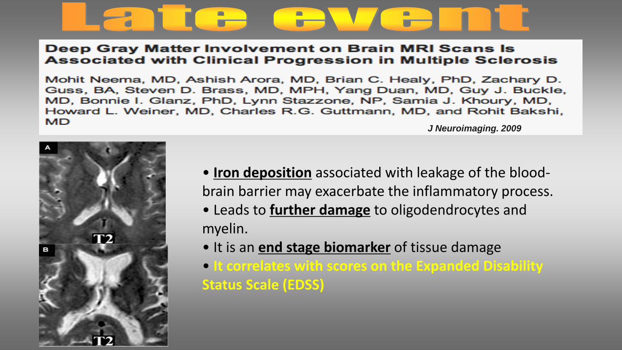
J Neuroimaging. 2009
• Iron deposition associated with leakage of the blood-brain barrier may exacerbate the inflammatory process. • Leads to further damage to oligodendrocytes and myelin. • It is an end stage biomarker of tissue damage • It correlates with scores on the Expanded Disability Status Scale (EDSS)

•Venous wall and perivenous tissue both show
typical histology of chronic venous stasis
• Major evidence in the subcortical gray matter
•Neglected part of MS pathology
Fibrin cuff 26%
Microbleeding
47%
V

Perivenous iron deposition. Is Heme iron the iron in MS?
RBC EXTRAVASATION IN
CHRONIC VENOUS
INSUFFICIENCY.
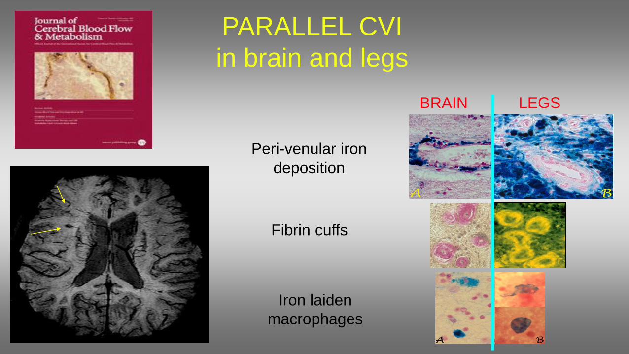
PARALLEL CVI
in brain and legs
LEGS BRAIN
Peri-venular iron
deposition
Fibrin cuffs
Iron laiden
macrophages

CCSVI and IRON DEPOSITS on SWI
Normal Control Multiple Sclerosis High iron
Low iron
Zivadinov et al. 2010, Haackee M 2010
• Significant correlations
between extracranial blockages and iron
loading in the pulvinar nucleus of thalamus,
thalamus, globus pallidus, and hippocampus
and in T2-LV, T1-LV
• Increased iron stores
correlates with the disability (EDSS)
Bakshi 2008
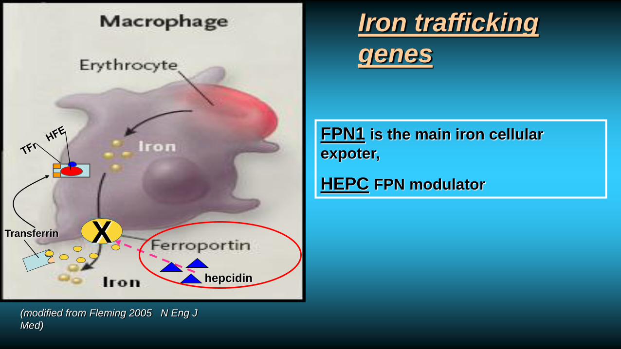
FPN1 is the main iron cellular
expoter,
HEPC FPN modulator
(modified from Fleming 2005 N Eng J
Med)
X Transferrin
hepcidin
Iron trafficking
genes
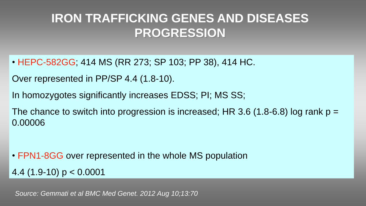
IRON TRAFFICKING GENES AND DISEASES
PROGRESSION
• HEPC-582GG; 414 MS (RR 273; SP 103; PP 38), 414 HC.
Over represented in PP/SP 4.4 (1.8-10).
In homozygotes significantly increases EDSS; PI; MS SS;
The chance to switch into progression is increased; HR 3.6 (1.8-6.8) log rank p =
0.00006
• FPN1-8GG over represented in the whole MS population
4.4 (1.9-10) p < 0.0001
Source: Gemmati et al BMC Med Genet. 2012 Aug 10;13:70
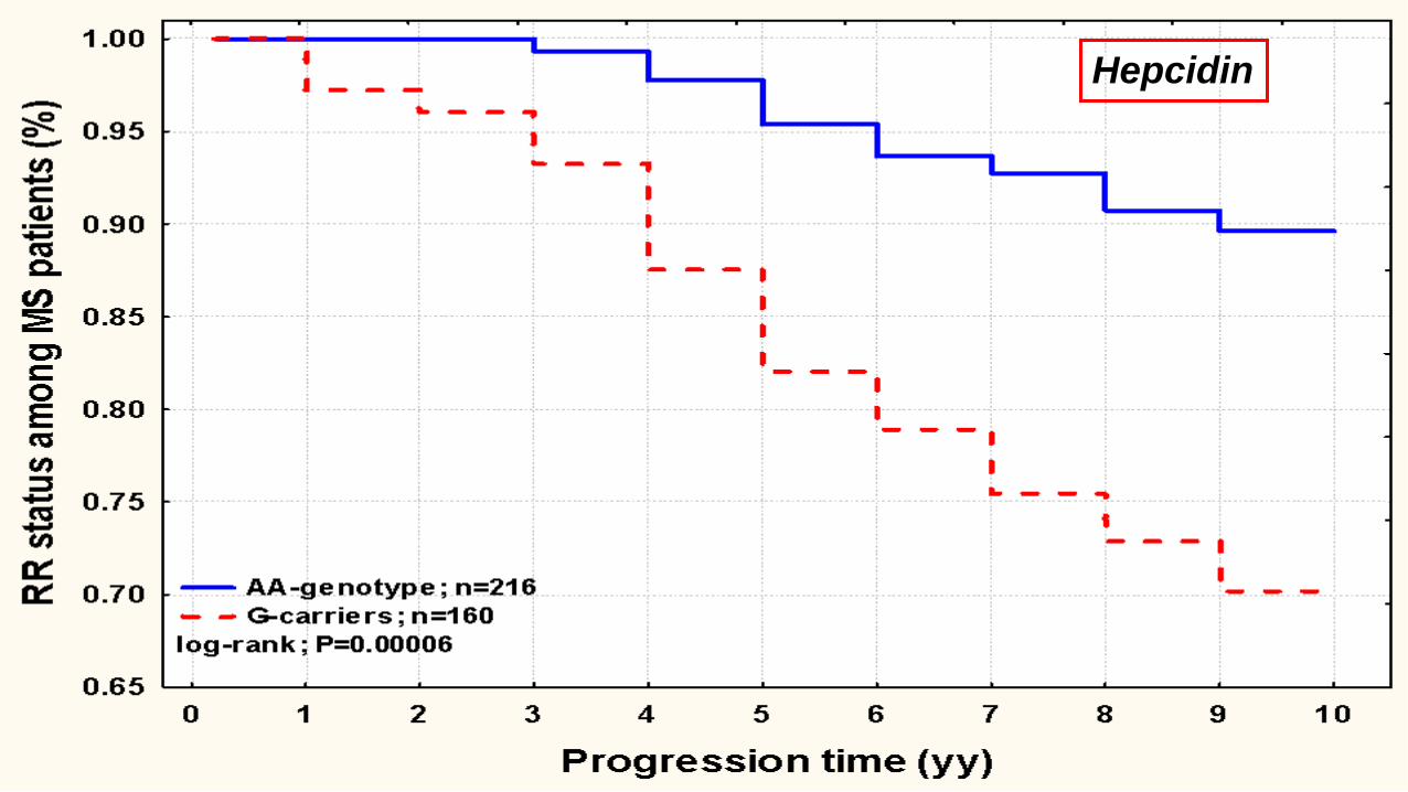
Hepcidin

Università degli Studi di Ferrara
Dottorato in Scienze Biomediche
e Biotecnologiche Curricula in Fisiopatologia del Sistema Nervoso
Endocrino e Vascolare
Dottorato in Fisica