Guida all’esame morfologico dello striscio ematico ... · referto completo ed esaustivo Come...
2
Risposta rigenerativa Policromasia lieve Guida all’esame morfologico dello striscio ematico Policromasia marcata Colorazione rapida - policromasia NBM - Reticolociti di cane NBM - Reticolociti di gatto Anemia Emolitica Immuno Mediata Sferociti senza policromasia Sferociti con policromasia Ghost cells Agglutinazione Rouleaux Altre poichilocitosi Colorazione rapida - gatto 3 corpi di heinz poco visibili (frecce) e 2 evidenti NBM - Corpi di Heinz Eccentrociti* Blister cell e cheratociti Morfologie eritrocitarie varie Emazie crenate Acantociti Cellule di Burr Schistocita Punteggiature basofile Agenti infettivi* Mycoplasma haemofelis Mycoplasma haemocanis Anaplasma phagocytophilum Babesia gibsoni Babesia canis Leucociti Neutrofilo normale Neutrofilo ‘a banda’ Neutrofilo - tossicità lieve Neutrofilo - tossicità moderata Neutrofilo - tossicità marcata** Monocita normale Eosinofilo normale di cane Eosinofilo normale di gatto Basofilo normale di cane Basofilo normale di gatto Piastrine Linfocita normale Conta piastrinica normale (50x) Linfocita - reattività lieve Conta piastrinica diminuita (50x) Linfocita - reattività moderata Aggregati piastrinici (50x) Linfocita - reattività moderata Piastrine normali e grandi Linfocita - reattività marcata/blastolinfoide Macropiastrina Cane - 2 corpi di Heinz Tutte le immagini, se non altrimenti indicato, sono state osservate con un microscopio ad alta risoluzione (obiettivo 100x) Le immagini e le didascalie sono state fornite da: Dennis B. DeNicola, DVM, PhD, DACVP Rick L. Cowell, DVM, MS, MRCVS, DACVP Michelle Frye, MS, DVM Nikola Pantchev, DVM *In questa guida non sono incluse immagini relative ad agenti infettivi, come la Leishmania, facilmente ritrovabili nelle cellule del midollo. **Illustration reproduced with permission from Reagan WJ, Rovira AI, DeNicola DB, eds. Veterinary Hematology: Atlas of Common Domestic and Non-Domestic Species. 2nd ed. Ames, IA: Wiley-Blackwell; 2008. Copyright 2008 Wiley-Blackwell. Striscio ematico normale di gatto Striscio ematico normale di cane
Transcript of Guida all’esame morfologico dello striscio ematico ... · referto completo ed esaustivo Come...
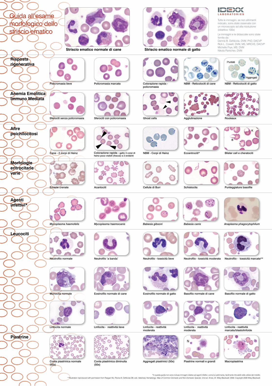
Risposta rigenerativa
Policromasia lieve
Guida all’esame morfologico dello striscio ematico
Policromasia marcata Colorazione rapida - policromasia
NBM - Reticolociti di cane NBM - Reticolociti di gatto
Anemia Emolitica Immuno Mediata
Sferociti senza policromasia Sferociti con policromasia Ghost cells Agglutinazione Rouleaux
Altre poichilocitosi
Colorazione rapida - gatto 3 corpi di heinz poco visibili (frecce) e 2 evidenti
NBM - Corpi di Heinz Eccentrociti* Blister cell e cheratociti
Morfologie eritrocitarie varie
Emazie crenate Acantociti Cellule di Burr Schistocita Punteggiature basofile
Agenti infettivi*
Mycoplasma haemofelis Mycoplasma haemocanis Anaplasma phagocytophilumBabesia gibsoni Babesia canis
Leucociti
Neutrofilo normale Neutrofilo ‘a banda’ Neutrofilo - tossicità lieve Neutrofilo - tossicità moderata Neutrofilo - tossicità marcata**
Monocita normale Eosinofilo normale di cane Eosinofilo normale di gatto Basofilo normale di cane Basofilo normale di gatto
Piastrine
Linfocita normale
Conta piastrinica normale (50x)
Linfocita - reattività lieve
Conta piastrinica diminuita (50x)
Linfocita - reattività moderata
Aggregati piastrinici (50x)
Linfocita - reattività moderata
Piastrine normali e grandi
Linfocita - reattività marcata/blastolinfoide
Macropiastrina
Cane - 2 corpi di Heinz
Tutte le immagini, se non altrimenti indicato, sono state osservate con un microscopio ad alta risoluzione (obiettivo 100x)
Le immagini e le didascalie sono state fornite da: Dennis B. DeNicola, DVM, PhD, DACVP Rick L. Cowell, DVM, MS, MRCVS, DACVP Michelle Frye, MS, DVM Nikola Pantchev, DVM
*In questa guida non sono incluse immagini relative ad agenti infettivi, come la Leishmania, facilmente ritrovabili nelle cellule del midollo. **Illustration reproduced with permission from Reagan WJ, Rovira AI, DeNicola DB, eds. Veterinary Hematology: Atlas of Common Domestic and Non-Domestic Species. 2nd ed. Ames, IA: Wiley-Blackwell; 2008. Copyright 2008 Wiley-Blackwell.
Striscio ematico normale di gattoStriscio ematico normale di cane

Un esame ematologico accurato è dato da un sistema performante e dall’analisi dello striscio ematico.
© 2009 IDEXX Laboratories, Inc. All rights reserved. • 09-68105-00 EN All ®/TM marks are owned by IDEXX Laboratories, Inc. or its affiliates in the United States and/or other countries.
Analizzatore ematologico LaserCyte® Citofluorimetro a raggio laser › profilo ematico completo › formula leucocitaria a cinque popolazioni › numero assoluto dei reticolociti › sistema rapido e di facile utilizzo › referto completo ed esaustivo
Come preparare un striscio ematico correttoCompletate il vostro esame ematologico realizzando uno striscio di ottima qualità
* Per campioni con ematocrito basso (anemia), si deve aumentare l’angolo tra i vetrini per avere un film di sangue più spesso. Per campioni con ematocrito alto (disidratazione, policitemia ecc.), l’angolo tra i vetrini deve essere diminuito per preparare un film più sottile.
† Assicurarsi che lo striscio di sangue sia completamente asciutto prima di passare alla colorazione. Se l’umidità è alta, asciugare il vetrino con un ventilatore a bassa velocità o semplicemente agitare il vetrino all’aria tenendolo in mano. Non usare l’asciugacapelli ne altre apparecchiature.
1. Depositare una piccola goccia di sangue fresco e/o correttamente miscelato con l’anticoagulante su di un vetrino nuovo e pulito a circa 2 cm dal bordo.
2. Disporre un secondo vetrino ”diffusore” nuovo e pulito di fronte alla goccia di sangue cercando di formare un angolo di 30° rispetto al primo vetrino.*
3. Far scivolare il vetrino ‘diffusore’ in modo che venga a contatto con la goccia di sangue.
4. Aspettare che il sangue diffonda per capillarità sul margine del secondo vetrino.
5. Con un movimento fluido ma saldo, muovere il vetrino ‘diffusore’ verso la restante parte del primo vetrino mantenendo fermo l’angolo di 30 gradi e senza sollevarlo. Il sangue dalla goccia seguirà il vetrino e si diffonderà lasciando un sottile film sul vetrino di base. Lo striscio di sangue dovrebbe essere 3- 4 cm di lunghezza.
6. Lasciare asciugare lo striscio all’aria.†
Formazione ed Eventi
....per imparare in modo pratico!
IDEXX ha deciso di offrirvi le novità in materia di diagnostica e clinica veterinaria, con una serie di incontri con noti esperti nazionali ed internazionali che hanno collaborato per realizzare delle presentazioni interattive e molto pratiche.
Il programma di formazione continua prevede, inoltre, una serie di conferenze web gratuite con noti relatori italiani che potrete comodamente seguire dalla vostra postazione internet.
Corsi on-line
Seminari
Diagnostic Updates
Visitate il nostro sito idexx.it alla pagina Formazione ed Eventi oppure scrivete una mail a [email protected] IDEXX Reference Laboratory Wetherby
Grange House, Sandbeck WayWetherby, West Yorkshire LS22 7DN
Tel: 01937 544000, Fax: 01937 544001
http://www.idexx.co.uk
IDEXX Reference Laboratory Southwater
4 Oakhurst, Business Park,Southwater, Horsham, West Sussex RH13 9RT
Tel: 01403 730176, Fax: 01403 732784
email: [email protected]
FHM is a common cause of severe haemolytic ane-
mia in cats. Cats with FHM present with depression, lethargy,
anorexia or inappetence, weakness, weight loss and dehydra-
tion. On physical examination, mucous membranes are pale and
sometimes icteric. Tachypnea, tachycardia and a heart murmur
may be present. Fever and splenomegaly are not uncommon.
Subclinical carriers of feline haemotropic mycoplasmas show no
clinical sign of the disease.
The CBC in cats presenting with clinical signs of illness reveals
a haematocrit that is often 50% of normal. The anaemia is usually
regenerative, but may be nonregenerative if there is concurrent
illness or infection with FeLV. In cats with subclinical infections, the
CBC may be normal or reveal only a mild anaemia. A biochemical
profile is usually normal, but increases in ALT from hypoxia and
hyperbilirubinaemia from haemolysis may be present.
Haemotropic mycoplasma in red blood cells (1000x), Picture Nikola Pantchev
UK028-0908
Diagnostic Update
Feline Haemotropic Mycoplasmosis
Feline haemotropic mycoplasmas are parasites that attach to
the outside of erythrocytes and result in anaemia. This organism
was formerly known as Haemobartonella but has been reclassi-
fied as a mycoplasma based on recent RNA sequence
analysis. Feline haemotropic mycoplasmas are small (0.3–0.8
μm) gram-negative bacteria that lack a cell wall and infect a variety
of mammalian species, including people. Damage caused by
parasite attachment and immune response by the host results in
increased red blood cell (RBC) destruction and anaemia. There
are three haemotropic mycoplasmas that have been identified in
cats: Mycoplasma haemofelis, Candidatus Mycoplasma haemo-
minutum and most recently Candidatus Mycoplasma turicensis.
(Candidatus designation is given to incompletely characterised
species.)
Recent studies revealed that 12.7% of healthy blood donor
cats, 14.5% of healthy client-owned cats having routine blood
work performed, and 28% of sick cats where FHM was suspect-
ed were positive for infection.
Risk factors associated with haemotropic mycoplasma infection
include: access to the outdoors, fleas, male gender, age of less
than 4 to 6 years, presentation during summer months, positive
FeLV status, history of cat bite abscesses and absence of current
vaccination.
Transmission can occur through fleas and possibly ticks and
lice. Kittens can be infected from the queen. Biting and aggres-
sive behaviour have been associated with transmission. Blood
transfusion with infected blood can result in infection of the blood
recipient.
Feline HaeMoTRopiC MyCoplasMa (FHM - FORMeRLy HaeMobartonella)
Diagnostic Update
October 08
Diagnosing the cause of anaemia in feline patients can be frustrating and difficult at best. It is not uncommon
to rule out obvious causes such as bleeding and renal failure and be left with a list of differential diagnoses
that can be a challenge to work through. Often, feline haemotropic mycoplasmosis (FHM), formerly known as
haemobartonellosis or feline infectious anaemia, remains a possibility. Traditionally, diagnosis of this infection
has relied on microscopically identifying the organism on the patient’s blood smear, which is an insensitive
method and can result in misidentification. Response to treatment is a common means of trying to confirm
this diagnosis. A positive response does not actually confirm the diagnosis, and if the cat does not respond,
precious time is lost trying to identify the true cause of anaemia.
References
1. Messick JB. Hemotrophic mycoplasmas (hemoplasmas): A review and new insights into pathogenic potential. Vet Clin Pathol. 2004;33:2-13.
2. Harvey JW. Hemotrophic mycoplasmosis (hemobartonellosis). In Green Ce, ed. Infectious Diseases of the Dog and Cat. Philadelphia: Saunders elsevier; 2006:252-260.
3. Sykes Je. Feline hemotropic mycoplasmosis (feline hemobartonellosis). Vet Clin Small anim. 2003;33:773-789.
4. Jensen WA, Lappin MR, Reagan W, et al. Use of a polymerase chain reaction assay to detect and differentiate two strains of Haemobartonella felis in naturally infected cats. am J Vet res 2001;62:604-608.
5. Willi B, Tasker S, Boretti FS, et al. Phylogenetic Analysis of “Candidatus Mycoplasma turicensis” Isolates from Pet Cats in the United Kingdom, Australia, and South Africa, with Analysis of Risk Factors for Infection. J Clin Microbiol. 2006; 44(12):4430-5.
6. Hackett TB, Jensen WA, Lehman TL, et al. Prevalence of DNA of Mycoplasma haemofelis, ‘Candidatus Mycoplasma haemominutum’, anaplasma phagocytophilum, and species of bartonella, neorickettsia, and ehrlichia in cats used as blood donors in the United States. J am Vet Med assoc. 2006:229;700-7005.
7. Dowers KL, Olver C, Radecki SV, Lappin MR. enrofloxacin for treatment of cats experimentally infected with large form Haemobartonella felis. J am Vet Med assoc. 2002:221;250-253.
8. George JW, Rideout BA, Griffey SM, Pedersen NC. effect of preexisting FeLV infection or FeLV and feline immunodeficiency virus coinfection on pathogenicity of the small variant of Haemobartonella felis in cats. am J Vet res. 2002;63:1172-1178.
ordering the iDeXX RealpCR™ FHM Test is easy –
just add it to your iDeXX Reference laboratories test order form.
test code name and contents specimen requirements list price
MPCR IDeXX RealPCR™ Feline Hemotropic Mycoplasma (FHM) 1 mL eDTA whole blood £35.50
(formerly Haemobartonella) Test
Mycoplasma haemofelis, Candidatus Mycoplasma
haemominutum and Candidatus Mycoplasma turicensis
Results in 2 – 7 days
Contacting iDeXXIf you have questions about the IDeXX RealPCR™ FHM Test, call our pathology team on 01937 544 000
IDEXXL’esame dello striscio ematico
P/N: 09-80031-00
I DE
XX
Vet
Stat®
Analizzatore emogas ed elettroliti
ID
EXX VetLab® Station
IDEXX C
oag Dx™ Analizzatore
IDE
XX
Lase
rCyte®
Analizzatore ematologico
IDE
XX
Cata
lyst D
x® Analizzatore biochim
ico
IDE
XX S
NAPshot Dx® Analizzatore
IDE
XX
Vet
Lab® UA™ Analizzatore per urin
e