
Focal Point: Diagnostica Corneale - oopi.it · Focal Point: Diagnostica Corneale ... Ambrosio R Jr,...
46
Fabrizio Camesasca, MD Paolo Vinciguerra, MD Eye Center – Humanitas Research Hospital Rozzano - Milano Responsabile: Dott. P. Vinciguerra Focal Point: Diagnostica Corneale Tomografia Corneale: Cosa Aggiunge Financial interests: Zeiss, SIFI www.oopi.it
Transcript of Focal Point: Diagnostica Corneale - oopi.it · Focal Point: Diagnostica Corneale ... Ambrosio R Jr,...
Fabrizio Camesasca, MD
Paolo Vinciguerra, MD
Eye Center – Humanitas Research Hospital Rozzano - Milano
Responsabile: Dott. P. Vinciguerra
Focal Point: Diagnostica Corneale
Tomografia Corneale: Cosa Aggiunge
Financial interests: Zeiss, SIFI
www.o
opi.it
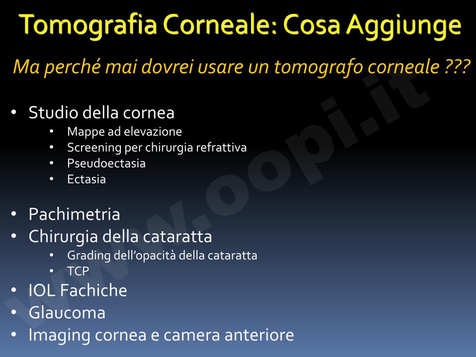
Tomografia Corneale: Cosa Aggiunge Ma perché mai dovrei usare un tomografo corneale ???
• Studio della cornea
• Mappe ad elevazione • Screening per chirurgia refrattiva • Pseudoectasia • Ectasia
• Pachimetria • Chirurgia della cataratta
• Grading dell’opacità della cataratta • TCP
• IOL Fachiche • Glaucoma • Imaging cornea e camera anteriore
ww
w.oopi.it
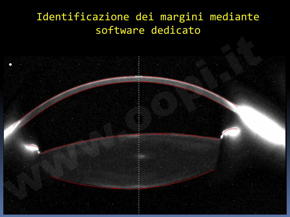
Identificazione dei margini mediante software dedicato
www.o
opi.it
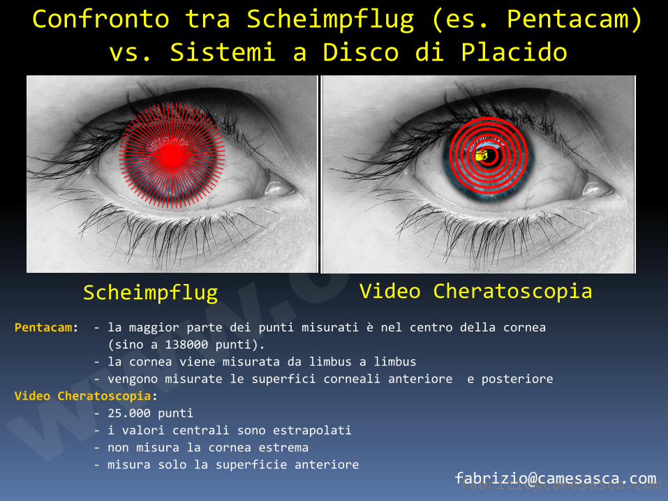
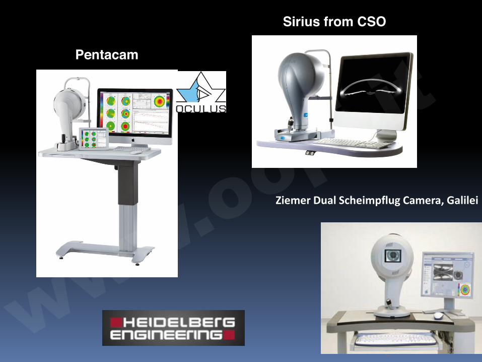
Scheimpflug Video Cheratoscopia
Confronto tra Scheimpflug (es. Pentacam) vs. Sistemi a Disco di Placido
Pentacam: - la maggior parte dei punti misurati è nel centro della cornea (sino a 138000 punti). - la cornea viene misurata da limbus a limbus - vengono misurate le superfici corneali anteriore e posteriore Video Cheratoscopia: - 25.000 punti - i valori centrali sono estrapolati - non misura la cornea estrema - misura solo la superficie anteriore
�
www.o
opi.it
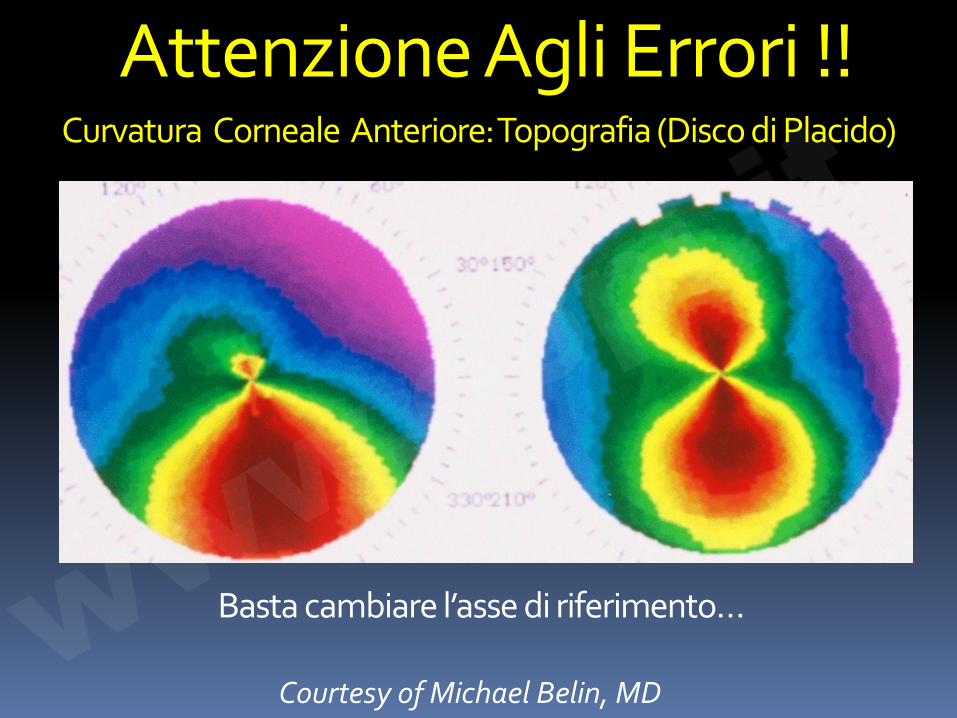
Curvatura Corneale Anteriore: Topografia (Disco di Placido)
Courtesy of Michael Belin, MD
Attenzione Agli Errori !!
Basta cambiare l’asse di riferimento… www.o
opi.it
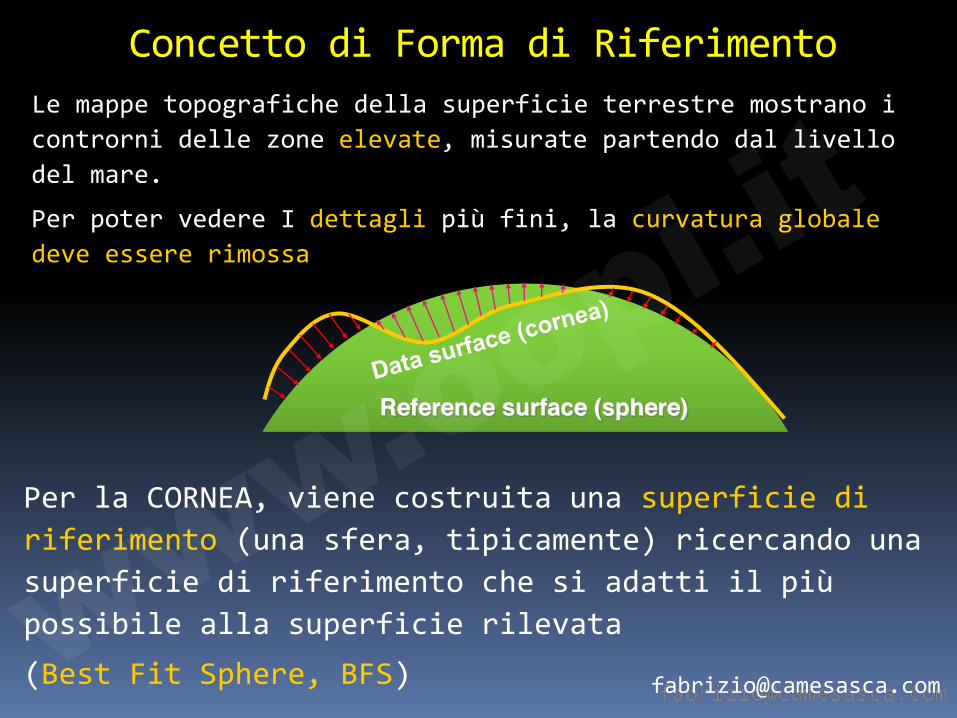
Concetto di Forma di Riferimento Le mappe topografiche della superficie terrestre mostrano i controrni delle zone elevate, misurate partendo dal livello del mare.
Per poter vedere I dettagli più fini, la curvatura globale deve essere rimossa
Reference surface (sphere)
Per la CORNEA, viene costruita una superficie di riferimento (una sfera, tipicamente) ricercando una superficie di riferimento che si adatti il più possibile alla superficie rilevata (Best Fit Sphere, BFS) ww
w.oopi.it
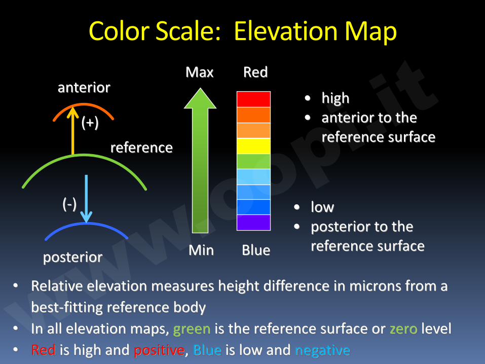
• Relative elevation measures height difference in microns from a best-fitting reference body
• In all elevation maps, green is the reference surface or zero level • Red is high and positive, Blue is low and negative
Color Scale: Elevation Map
• high • anterior to the
reference surface (+)
anterior
• low • posterior to the
reference surface
(-)
posterior
reference
Red
Blue Min
Max
www.o
opi.it
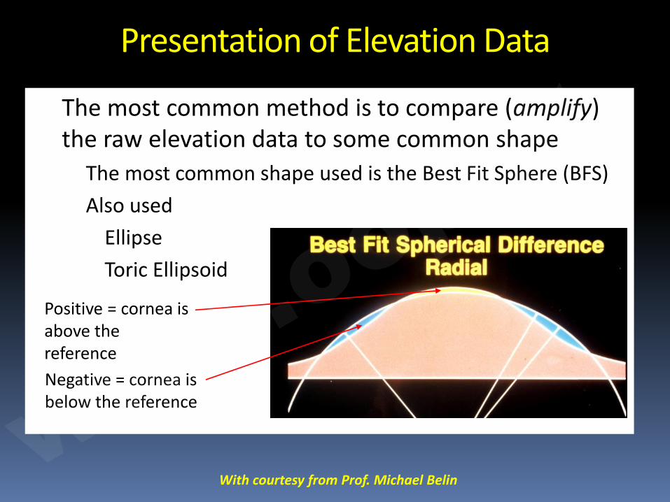
Presentation of Elevation Data
� The most common method is to compare (amplify) the raw elevation data to some common shape à The most common shape used is the Best Fit Sphere (BFS) à Also used
� Ellipse � Toric Ellipsoid
Positive = cornea is above the reference Negative = cornea is below the reference
With courtesy from Prof. Michael Belin ww
w.oopi.it
[email protected] With courtesy from Prof. Michael Belin
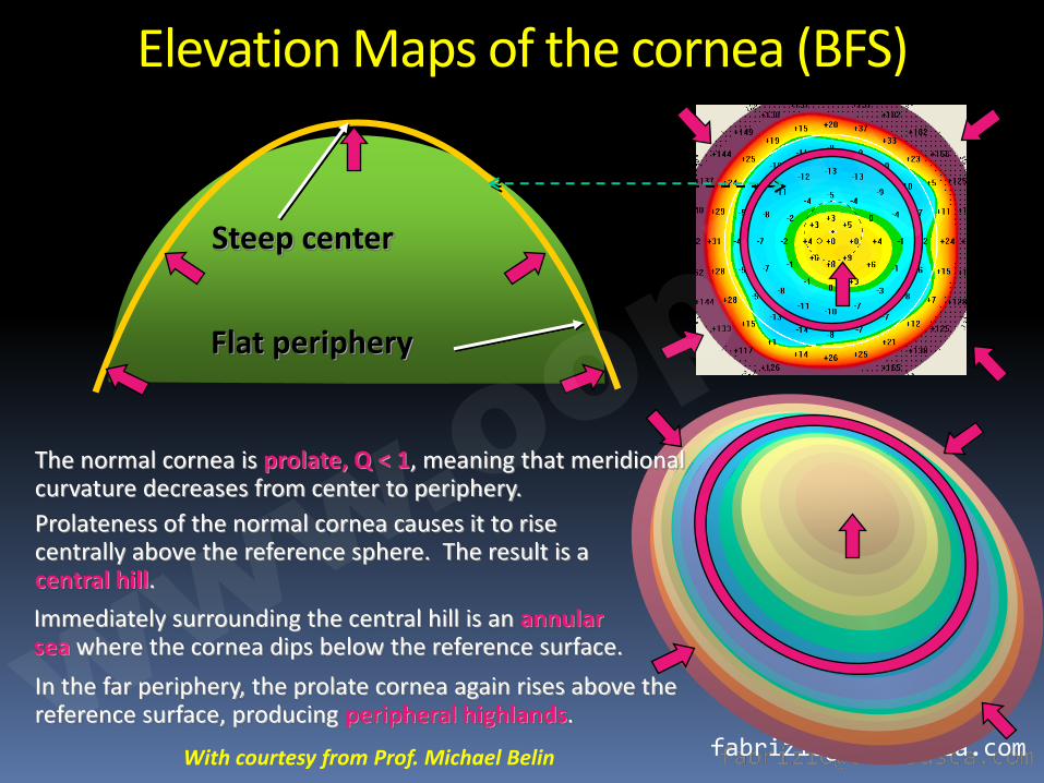
Elevation Maps of the cornea (BFS)
Steep center
Flat periphery
The normal cornea is prolate, Q < 1, meaning that meridional curvature decreases from center to periphery. Prolateness of the normal cornea causes it to rise centrally above the reference sphere. The result is a central hill. Immediately surrounding the central hill is an annular sea where the cornea dips below the reference surface. In the far periphery, the prolate cornea again rises above the reference surface, producing peripheral highlands. ww
w.oopi.it
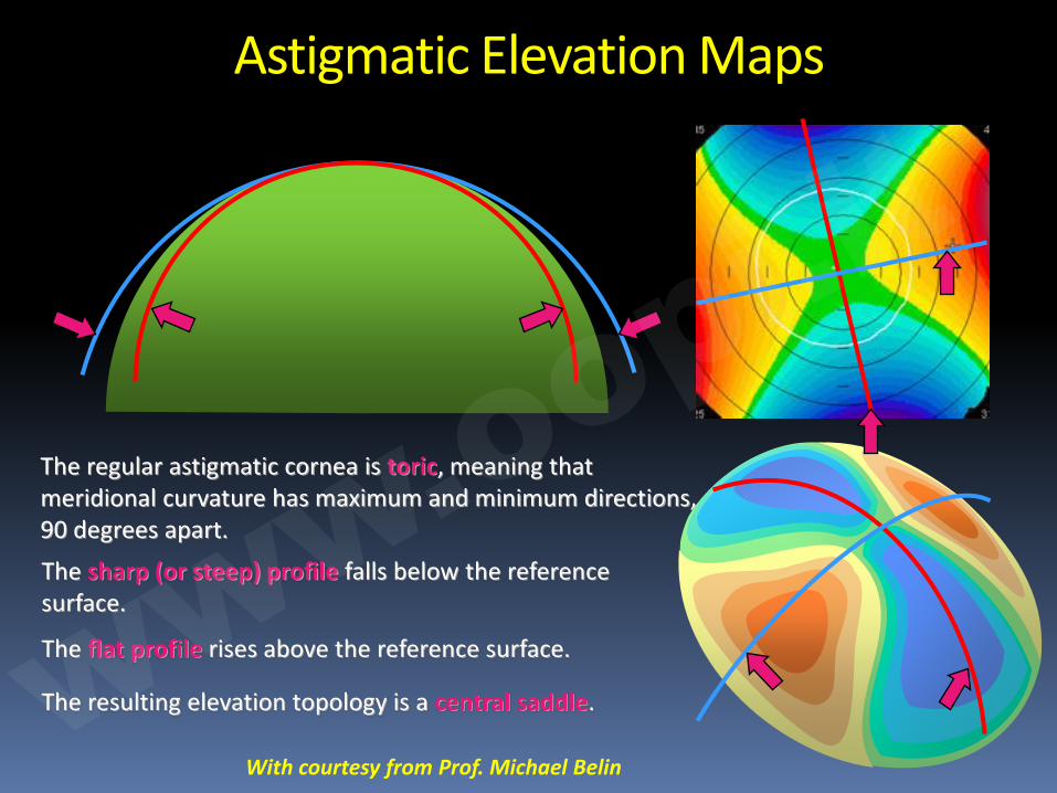
Astigmatic Elevation Maps
The regular astigmatic cornea is toric, meaning that meridional curvature has maximum and minimum directions, 90 degrees apart. The sharp (or steep) profile falls below the reference surface.
The flat profile rises above the reference surface.
The resulting elevation topology is a central saddle.
With courtesy from Prof. Michael Belin ww
w.oopi.it
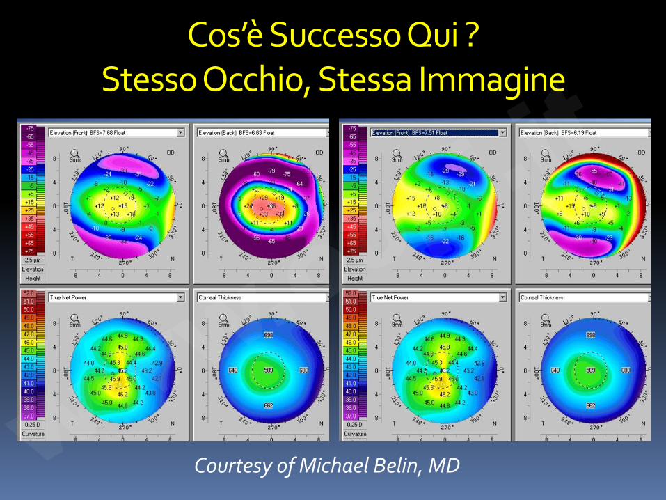
Cos’è Successo Qui ? Stesso Occhio, Stessa Immagine
Courtesy of Michael Belin, MD www.o
opi.it
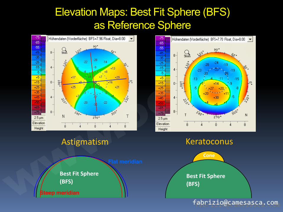
Elevation Maps: Best Fit Sphere (BFS) as Reference Sphere
Astigmatism Keratoconus
Flat meridian
Steep meridian Best Fit Sphere
(BFS) Best Fit Sphere
(BFS)
Cone
www.o
opi.it
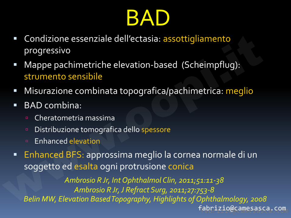
BAD � Condizione essenziale dell’ectasia: assottigliamento
progressivo
� Mappe pachimetriche elevation-based (Scheimpflug): strumento sensibile
� Misurazione combinata topografica/pachimetrica: meglio
� BAD combina: à Cheratometria massima à Distribuzione tomografica dello spessore à Enhanced elevation
� Enhanced BFS: approssima meglio la cornea normale di un soggetto ed esalta ogni protrusione conica
Belin MW, Elevation Based Topography, Highlights of Ophthalmology, 2008
Ambrosio R Jr, Int Ophthalmol Clin, 2011;51:11-38 Ambrosio R Jr, J Refract Surg, 2011;27:753-8 www.o
opi.it
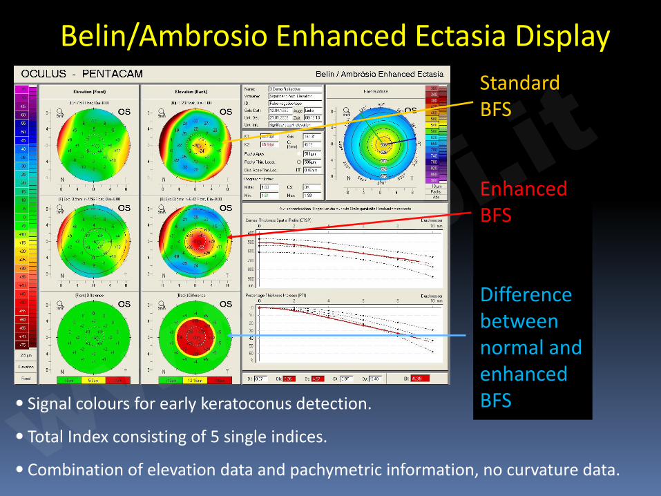
• Signal colours for early keratoconus detection.
• Total Index consisting of 5 single indices.
• Combination of elevation data and pachymetric information, no curvature data.
Belin/Ambrosio Enhanced Ectasia Display Standard BFS
Enhanced BFS
Difference between normal and enhanced BFS ww
w.oopi.it
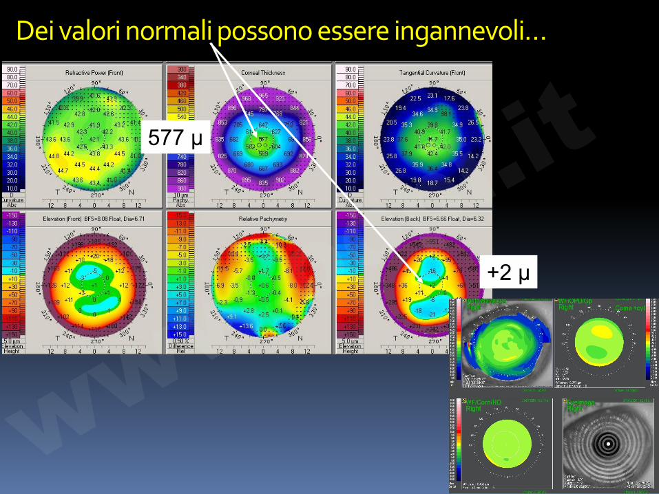
Dei valori normali possono essere ingannevoli…
577 µ
+2 µ
www.o
opi.it
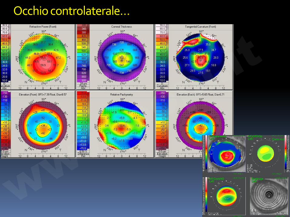
Occhio controlaterale…
www.o
opi.it
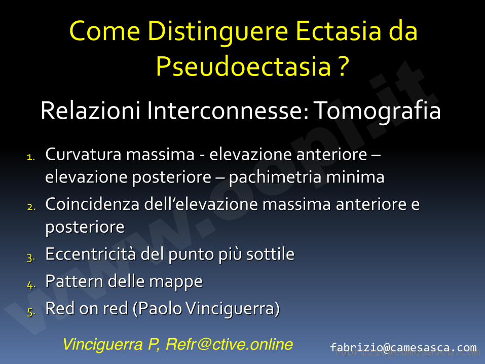
Come Distinguere Ectasia da Pseudoectasia ?
1. Curvatura massima - elevazione anteriore – elevazione posteriore – pachimetria minima
2. Coincidenza dell’elevazione massima anteriore e posteriore
3. Eccentricità del punto più sottile 4. Pattern delle mappe 5. Red on red (Paolo Vinciguerra)
Relazioni Interconnesse: Tomografia
Vinciguerra P, [email protected] www.o
opi.it
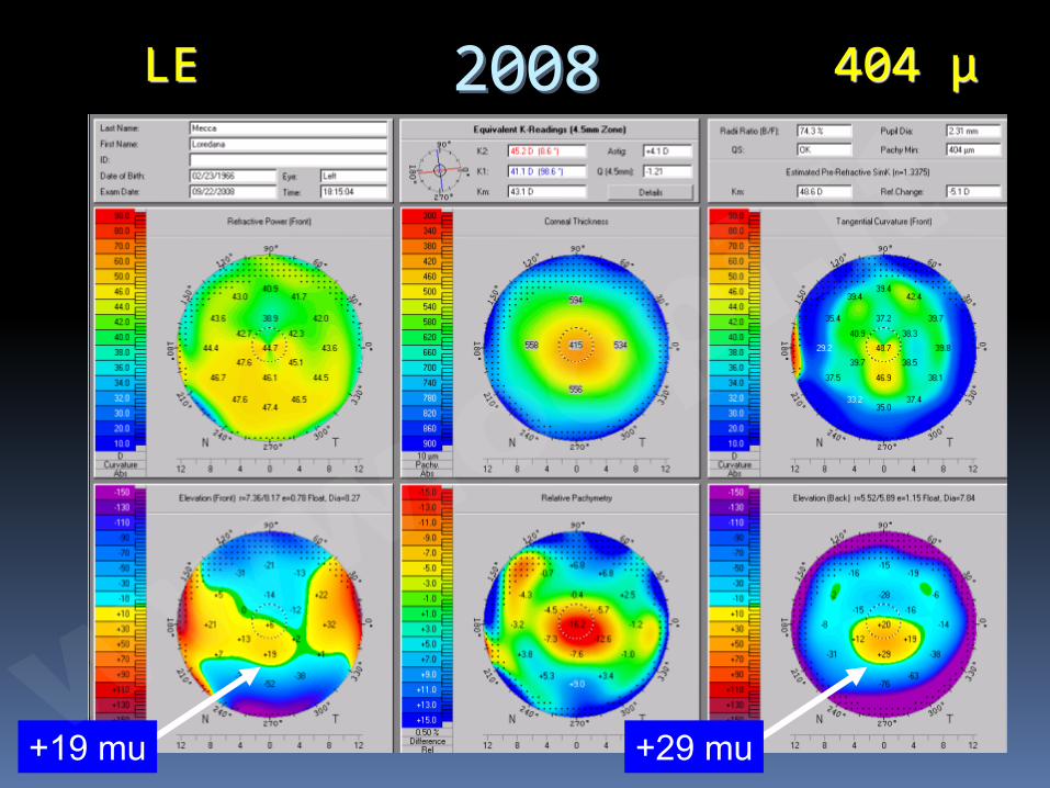
2008 LE 404 μ
+19 mu +29 mu www.o
opi.it
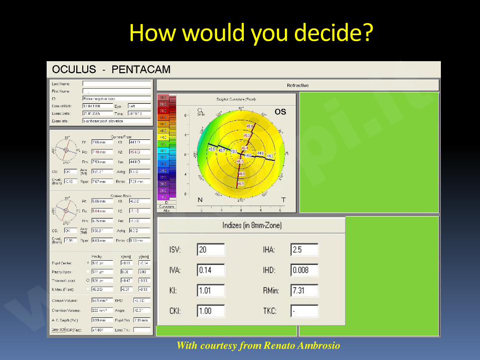
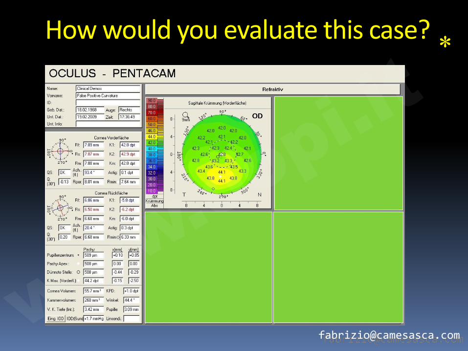
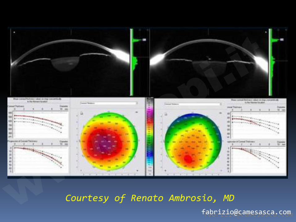
How would you decide?
With courtesy from Renato Ambrosio ww
w.oopi.it
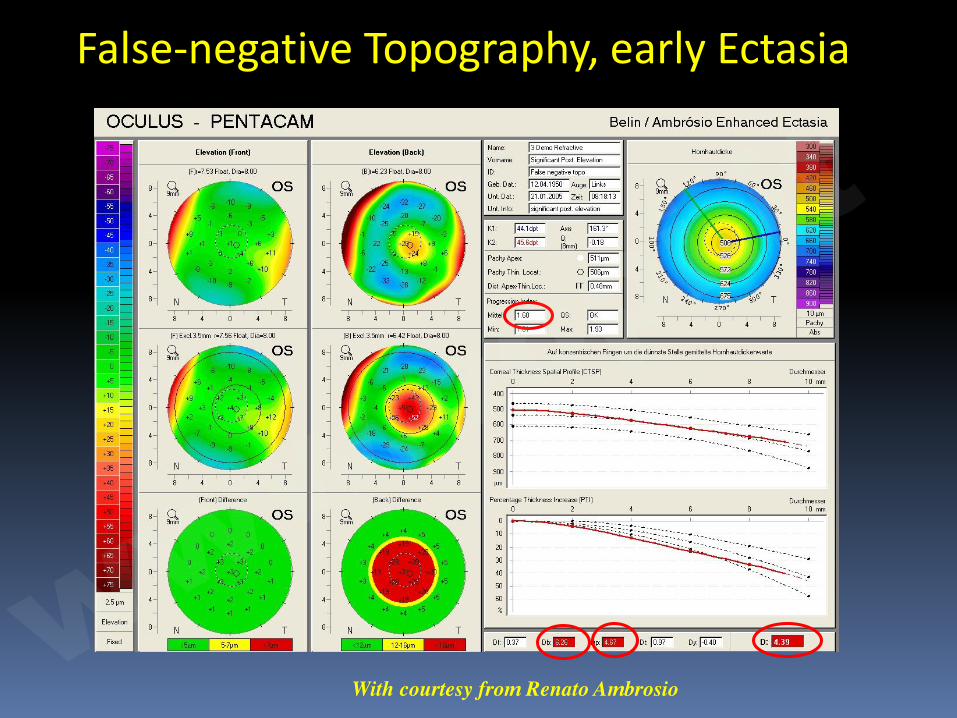
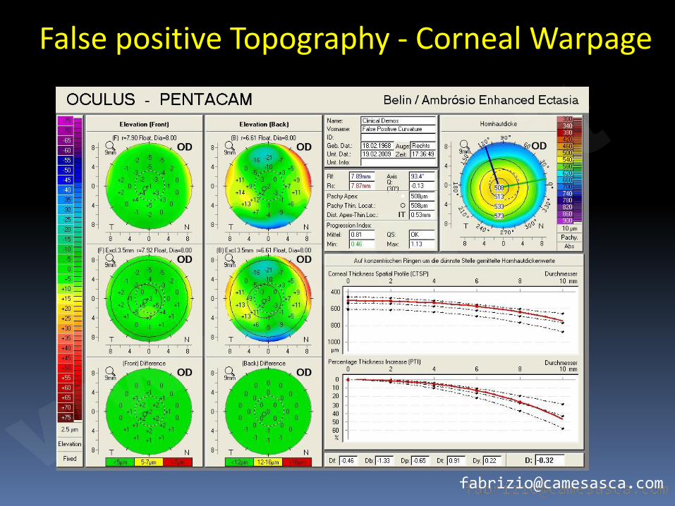
False-negative Topography, early Ectasia
With courtesy from Renato Ambrosio ww
w.oopi.it
Pachimetria
www.o
opi.it
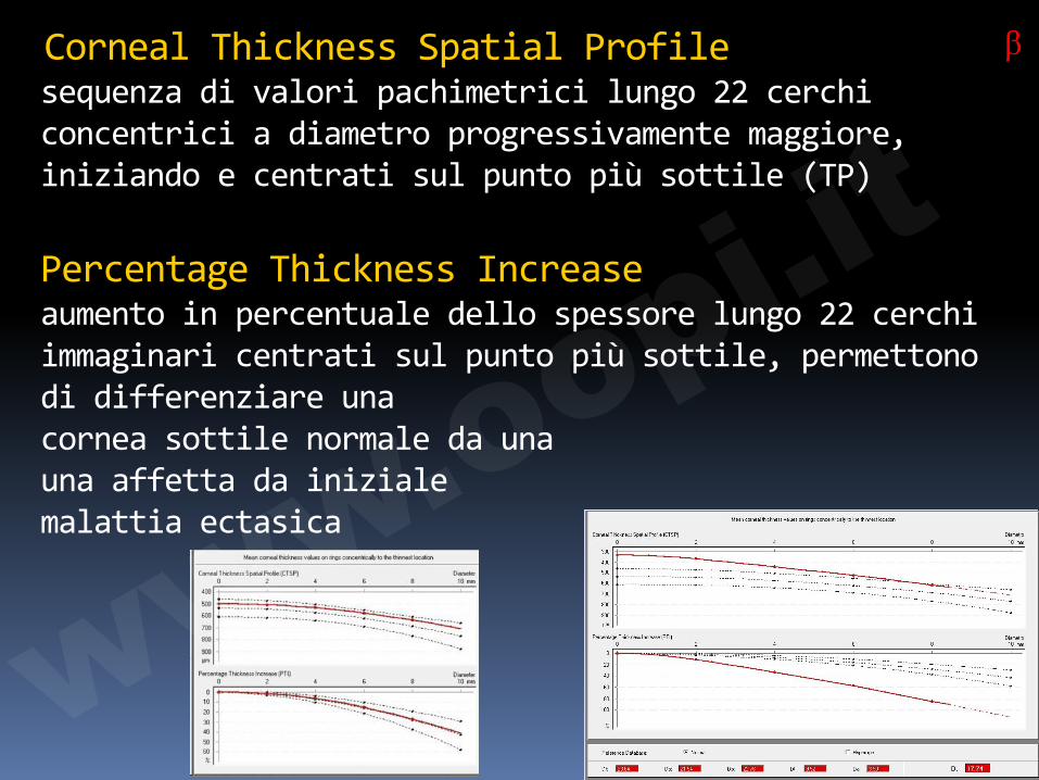
Corneal Thickness Spatial Profile sequenza di valori pachimetrici lungo 22 cerchi concentrici a diametro progressivamente maggiore, iniziando e centrati sul punto più sottile (TP) Percentage Thickness Increase aumento in percentuale dello spessore lungo 22 cerchi immaginari centrati sul punto più sottile, permettono di differenziare una cornea sottile normale da una una affetta da iniziale malattia ectasica
β
www.o
opi.it
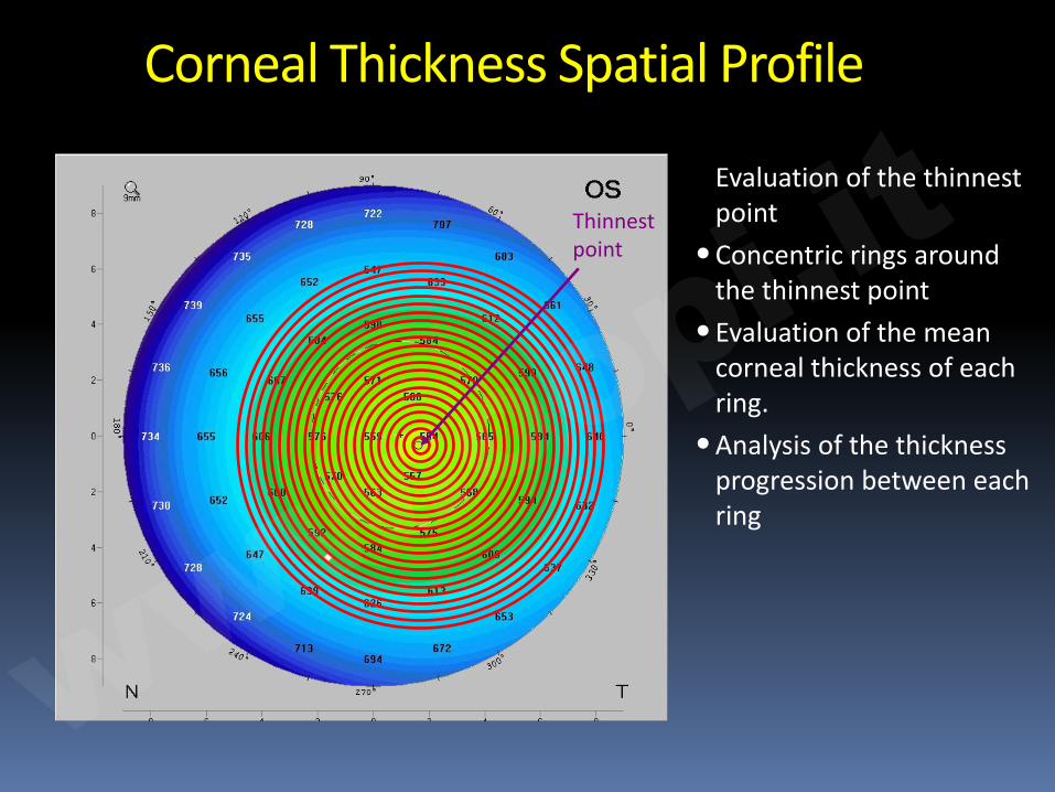
Corneal Thickness Spatial Profile
Evaluation of the thinnest point
•Concentric rings around the thinnest point
•Evaluation of the mean corneal thickness of each ring.
•Analysis of the thickness progression between each ring
Thinnest point
www.o
opi.it
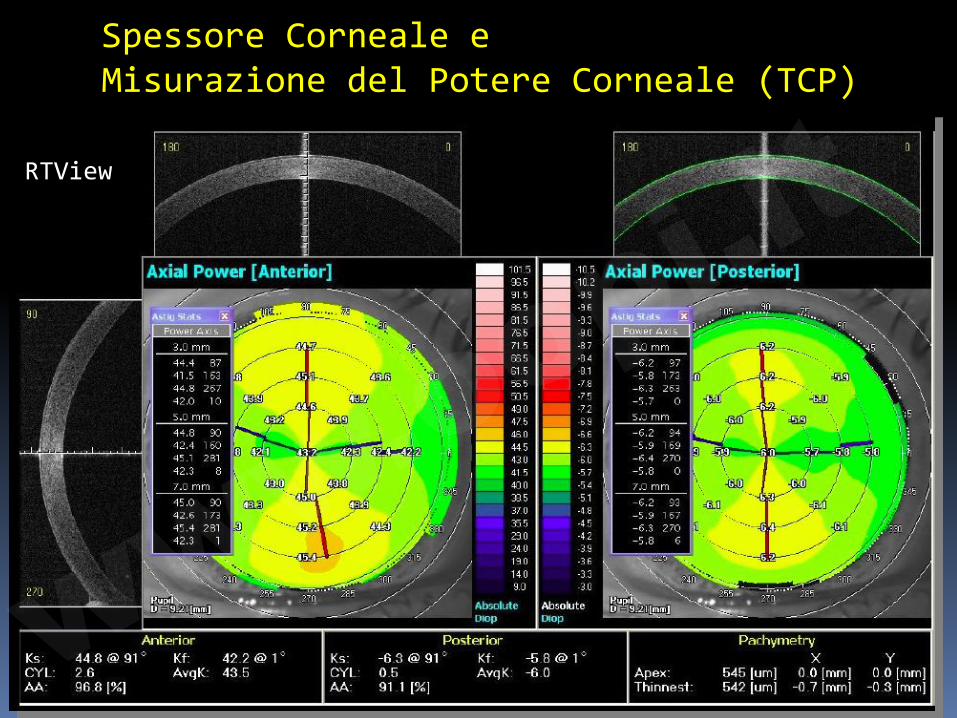
Spessore Corneale e Misurazione del Potere Corneale (TCP)
RTView
www.o
opi.it
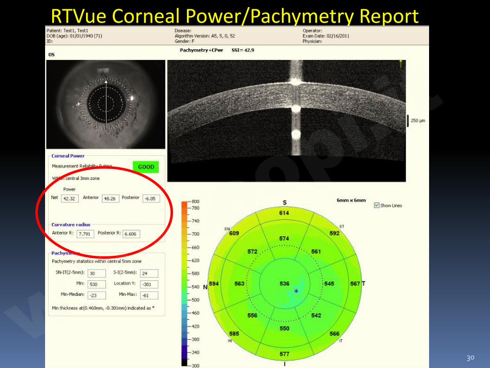
RTVue Corneal Power/Pachymetry Report
30
www.o
opi.it
TCP � I cheratometri ed i topografi misurano la
superficie corneale anteriore
� Estrapolano la superficie posteriore
� Il cambiamento di curvatura e spessore della cornea post-chirurgia refrattiva porta ad una variazione in curvatura – e quindi anche in potere – della cornea posteriore
� Rischio di errore aumentato in pazienti sottoposti a chirurgia refrattiva.
� TCP misura direttamente le superfici anteriore e posteriore fornendo un potere corneale più accurato utile, ad es., nel calcolo delle IOL
31 SOI Internazionale -2013 ww
w.oopi.it
Total corneal astigmatism, important for toric IOL‘s?
Anterior surface only? Is the posterior surface really important?
“Accuracy of Corneal Astigmatism Estimation by Neglecting the Posterior Corneal Surface Measurement”:
10% of eyes with more than 1 D of astigmatism: - difference in magnitude > 0.5 D - or difference in angle > 10° (30 % remaining astigmastism) between anterior and total astigmatism.
JAU-DER HO, CHING-YAO TSAI, AND SHIOW-WEN LIOU; © 2009 BY ELSEVIER INC. ALL RIGHTS RESERVED. 0002-9394/09/$36.00; doi:10.1016/j.ajo.2008.12.020
Conclusion, the posterior surface should be considered in terms of angle and amount of the astigmatism ww
w.oopi.it
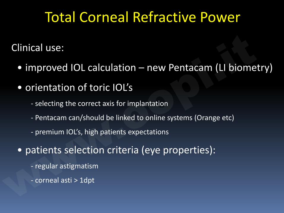
Total Corneal Refractive Power
Clinical use:
• improved IOL calculation – new Pentacam (LI biometry)
• orientation of toric IOL’s - selecting the correct axis for implantation
- Pentacam can/should be linked to online systems (Orange etc)
- premium IOL’s, high patients expectations
• patients selection criteria (eye properties): - regular astigmatism
- corneal asti > 1dpt www.o
opi.it
Chirurgia della Cataratta
• Valutazione oggettiva dell’opacità del cristallino
• Ruolo della superficie corneale post. nel calcolo delle IOL toriche
• IOL fachiche
www.o
opi.it
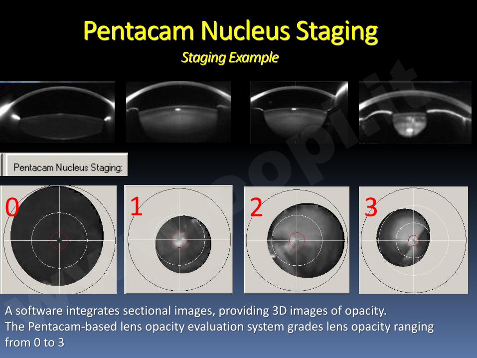
1 3 2
Pentacam Nucleus Staging Staging Example
0
A software integrates sectional images, providing 3D images of opacity. The Pentacam-based lens opacity evaluation system grades lens opacity ranging from 0 to 3
www.o
opi.it
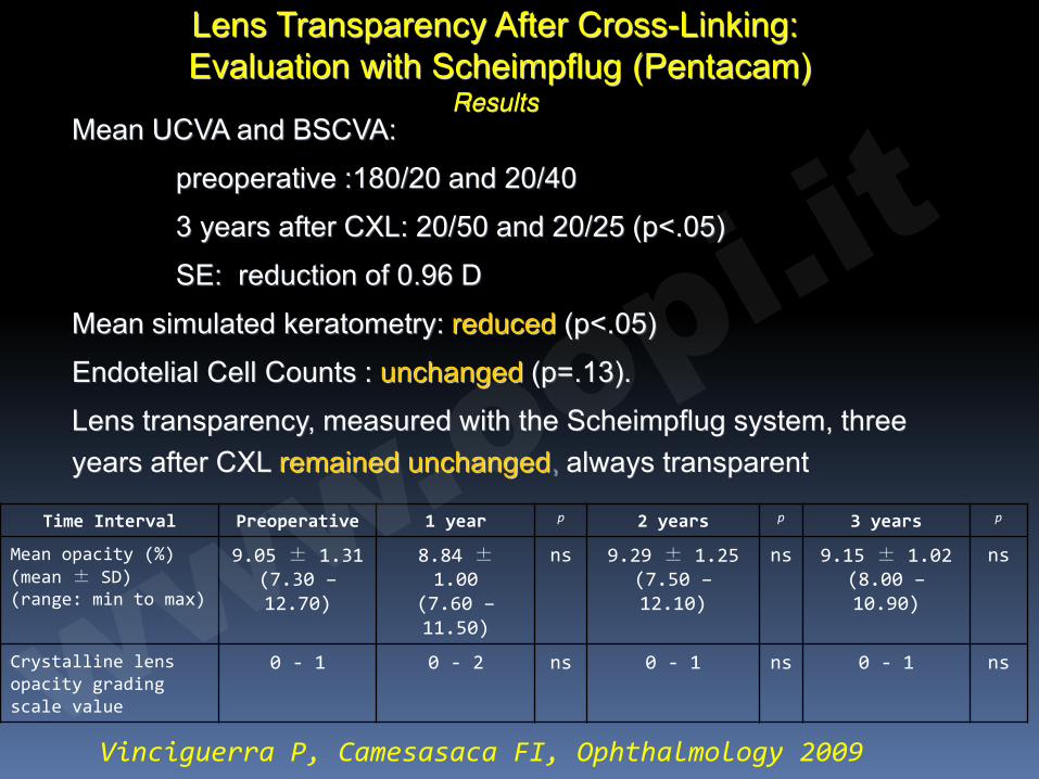
Mean UCVA and BSCVA:
preoperative :180/20 and 20/40
3 years after CXL: 20/50 and 20/25 (p<.05)
SE: reduction of 0.96 D
Mean simulated keratometry: reduced (p<.05)
Endotelial Cell Counts : unchanged (p=.13).
Lens transparency, measured with the Scheimpflug system, three years after CXL remained unchanged, always transparent
Lens Transparency After Cross-Linking: Evaluation with Scheimpflug (Pentacam)
Results
Time Interval Preoperative 1 year p 2 years p 3 years p
Mean opacity (%) (mean ± SD) (range: min to max)
9.05 ± 1.31 (7.30 – 12.70)
8.84 ± 1.00
(7.60 – 11.50)
ns 9.29 ± 1.25 (7.50 – 12.10)
ns 9.15 ± 1.02 (8.00 – 10.90)
ns
Crystalline lens opacity grading scale value
0 - 1 0 - 2 ns 0 - 1 ns 0 - 1 ns
Vinciguerra P, Camesasaca FI, Ophthalmology 2009 ww
w.oopi.it
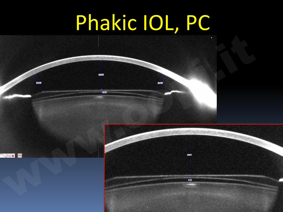
� angle supported (NuVita MA 20 B&L) � iris fixated (Verisys, Artisan), spheric and
toric � angle fixated (Cachet, ALCON) � ICL (behind the iris in front of the
crystalline lens): Staar, spheric and toric
Phakic IOLs
www.o
opi.it
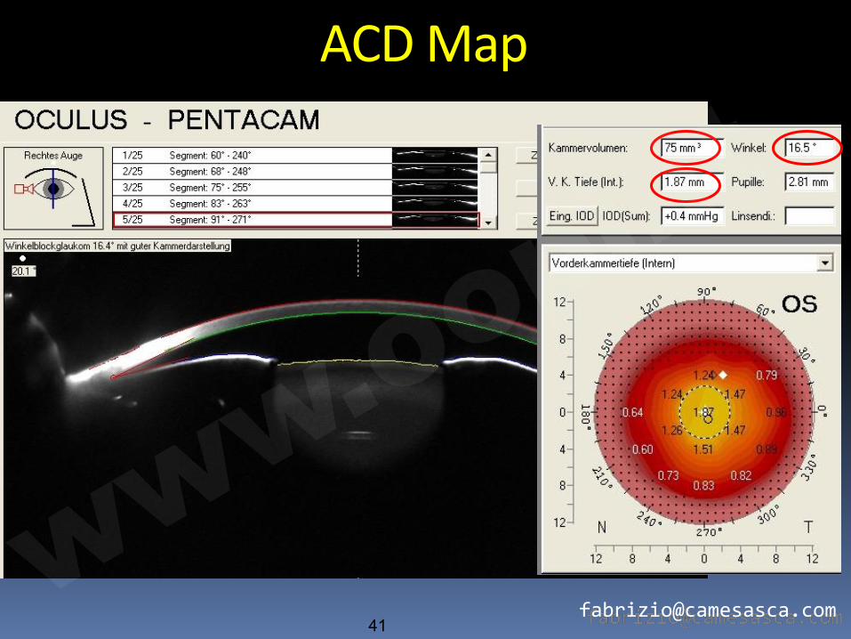
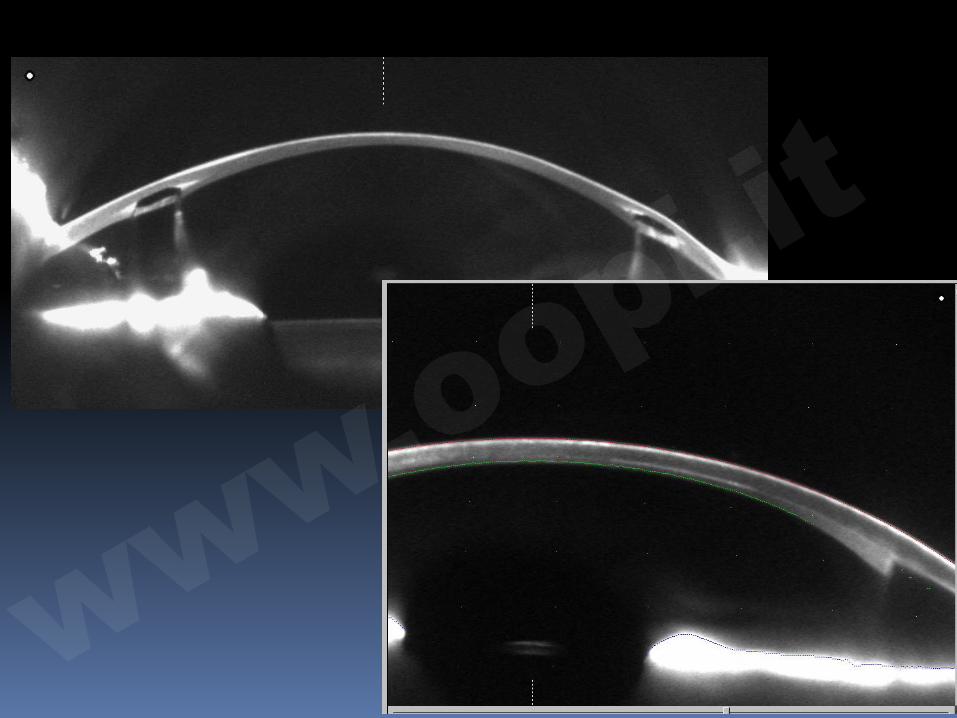
Glaucoma • anterior eye segment • ACD, ACD-map, ACV,
ACA, IOP correction, pre-post iridectomy
• IOP correction according to corneal thickness
www.o
opi.it
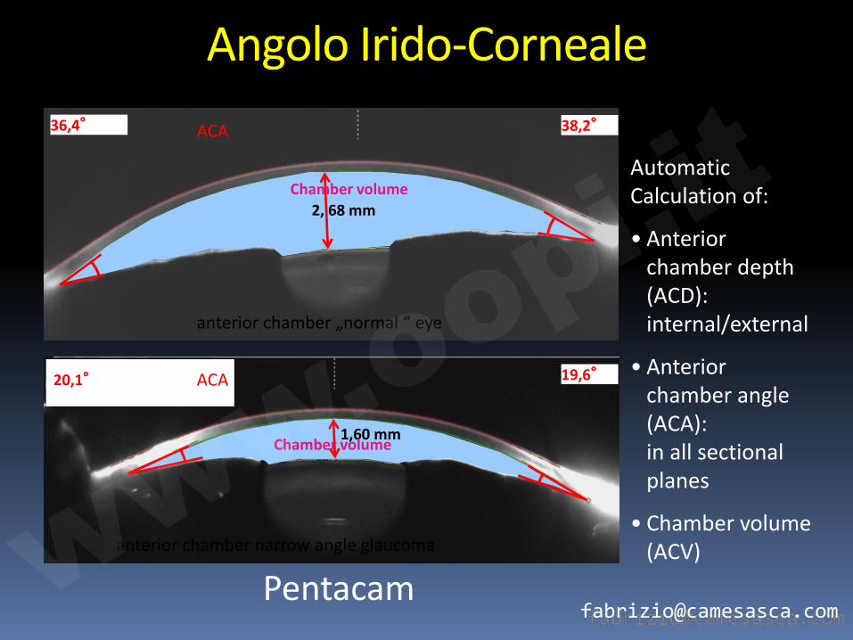
Kammervolumen
anterior chamber „normal “ eye
anterior chamber narrow angle glaucoma
2, 68 mm
Automatic Calculation of:
• Anterior chamber depth (ACD): internal/external
• Anterior chamber angle (ACA): in all sectional planes
• Chamber volume (ACV)
1,60 mm
36,4° 38,2°
20,1°
19,6°
ACA
ACA
Chamber volume
Chamber volume
Angolo Irido-Corneale
Pentacam www.o
opi.it
Immagini della Cornea e della Camera Anteriore
www.o
opi.it
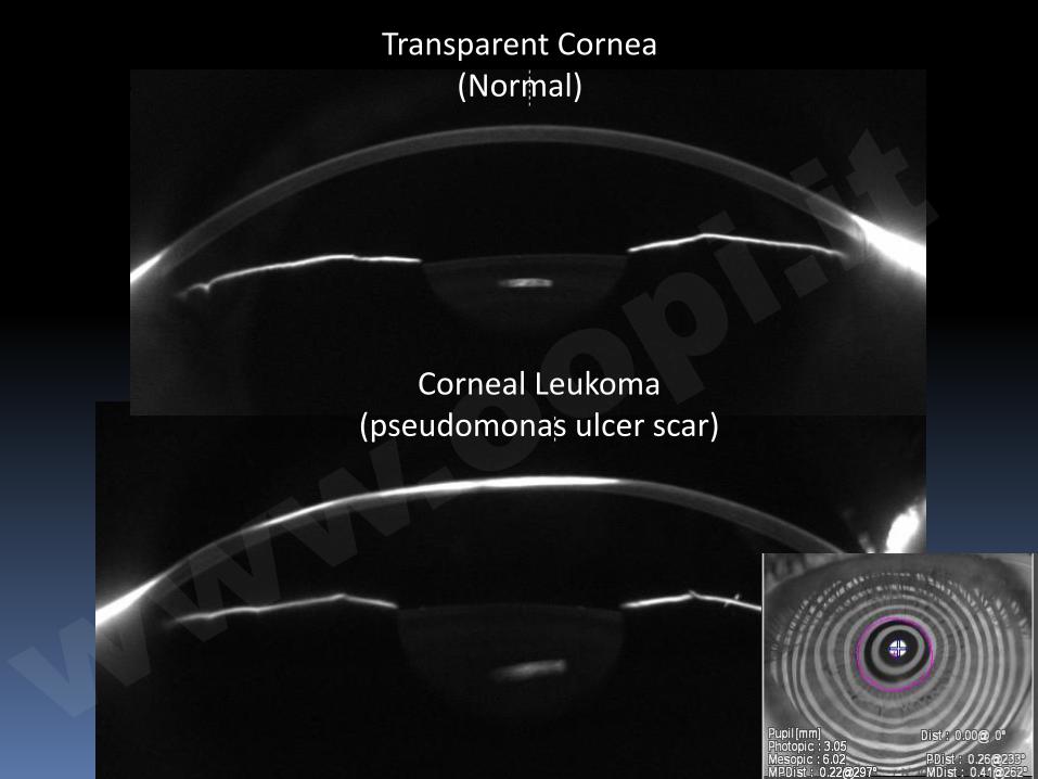
Transparent Cornea (Normal)
Corneal Leukoma (pseudomonas ulcer scar)
www.o
opi.it
Conclusioni � Perché usare un tomografo corneale ?
à Miglioramento della comprensione delle
caratteristiche della cornea
à Selezione del pz per chirurgia refrattiva
à Diagnosi di pseudoectasia vs. ectasia
à Valutazione della camera anteriore
à Valutazione del pz glaucomatoso
à Documentazione medico-legale
www.o
opi.it