Ciclo Cellulare e Apoptosi - 193.205.144.19193.205.144.19/scienze/ciclo/1.pdf · Ciclo Cellulare e...
54
1 Programma del corso di Ciclo Cellulare e Apoptosi Ciclo Cellulare e Apoptosi (aa 2005/2006) Titolari: Prof.ssa Carla Caruso Dott.ssa Maria Saveria Gilardini Montani
-
Upload
dinhnguyet -
Category
Documents
-
view
222 -
download
0
Transcript of Ciclo Cellulare e Apoptosi - 193.205.144.19193.205.144.19/scienze/ciclo/1.pdf · Ciclo Cellulare e...

1
Programma del corso di
Ciclo Cellulare e ApoptosiCiclo Cellulare e Apoptosi
(aa 2005/2006)
Titolari: Prof.ssa Carla CarusoDott.ssa Maria Saveria Gilardini Montani

2
Il Ciclo Cellulare: Fasi, Controllo e Regolazione. Strategia generale e fasi del ciclo cellulare. Sistemi sperimentali per lo studio del ciclo cellulare: uova
di anfibi e lieviti. Studio sui mutanti cdc e wee in S. pombe. Regolazione di
MPF mediante fosforilazione e defosforilazione. Studio sui mutanti cdc in S. cerevisiae. Cicline G1 e SPF. Cdks e cicline nel ciclo cellulare dei mammiferi. Punti di controllo del ciclo e ruolo del punto di restrizione. Ruolo di Rb nella regolazione del ciclo cellulare. DNA danneggiato da UV e ruolo di p53. Oncogeni e oncoproteine.

3
Le basi genetiche del cancro. Cancerogeni, mutageni, virus tumorali.
Tipi di morte cellulare: apoptosi e necrosi. Caratteristiche e significato biologico
I recettori di morte e i ligandi La via delle caspasi Il ruolo dei mitocondri nei processi apoptotici Il sistema Fas/FasL La regolazione dell’apoptosi: le proteine della
famiglia di Bcl-2; IAP e FLIP L’apoptosi caspasi-indipendente Tecniche di laboratorio per lo studio dell’apoptosi
4
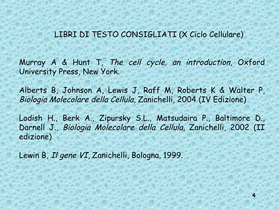
LIBRI DI TESTO CONSIGLIATI (X Ciclo Cellulare)
Murray A & Hunt T, The cell cycle, an introduction, Oxford University Press, New York.
Alberts B, Johnson A, Lewis J, Raff M, Roberts K & Walter P, Biologia Molecolare della Cellula, Zanichelli, 2004 (IV Edizione)
Lodish H., Berk A., Zipursky S.L., Matsudaira P., Baltimore D., Darnell J., Biologia Molecolare della Cellula, Zanichelli, 2002 (II edizione)
Lewin B, Il gene VI, Zanichelli, Bologna, 1999.

5
Prokaryotic cellProkaryotic cell

6
ComponentsComponents• Cytoplasm• Ribosomes• Nuclear Zone• DNA• Plasmid• Cell Membrane• Cell Wall• Capsule (or slime layer)• Flagellum

7
Eukaryotic cellEukaryotic cell
8
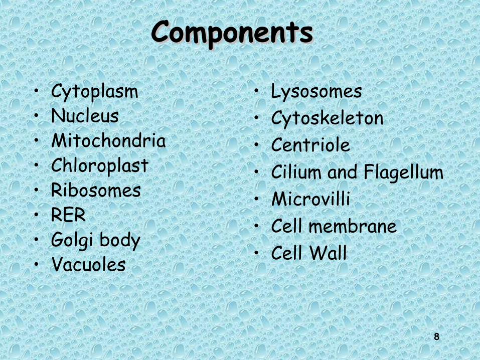
ComponentsComponents
• Cytoplasm• Nucleus• Mitochondria• Chloroplast• Ribosomes• RER• Golgi body• Vacuoles
• Lysosomes• Cytoskeleton• Centriole• Cilium and Flagellum• Microvilli• Cell membrane• Cell Wall

9
10
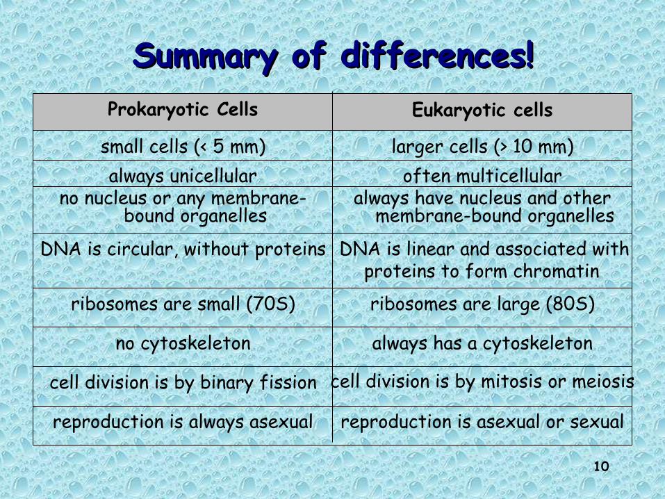
Summary of differences!Summary of differences!
reproduction is asexual or sexualreproduction is always asexual
cell division is by mitosis or meiosiscell division is by binary fission
always has a cytoskeletonno cytoskeleton
ribosomes are large (80S)ribosomes are small (70S)
DNA is linear and associated with proteins to form chromatin
DNA is circular, without proteins
always have nucleus and other membrane-bound organelles
no nucleus or any membrane-bound organelles
often multicellularalways unicellularlarger cells (> 10 mm)small cells (< 5 mm)
Eukaryotic cellsProkaryotic Cells

11
Characteristic Features of Bacteria and Characteristic Features of Bacteria and Archaea: Comparison to EukaryotesArchaea: Comparison to Eukaryotes
• Bacteria and Archaea lack membrane-bound nuclei and organelles, and have a single circular chromosome.
• Archaea and Eukaryotes have multiple complex RNA polymerases and begin translation with methionine; bacteria begin translation with formyl-methionine.
12
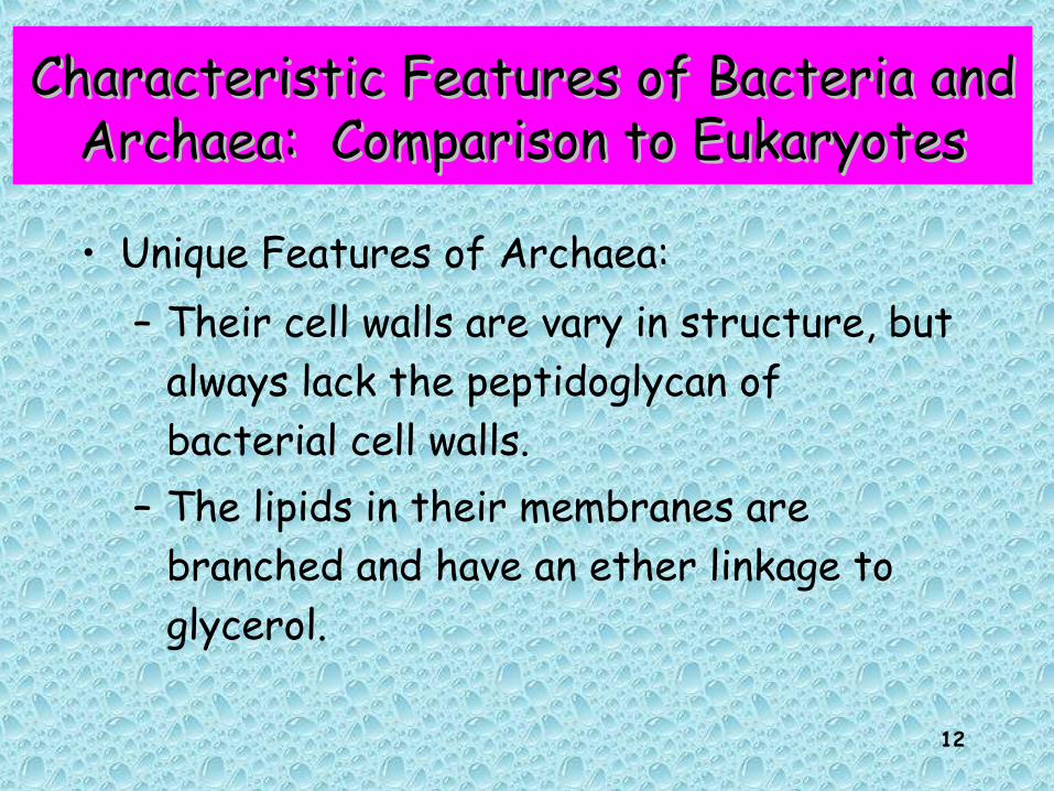
Characteristic Features of Bacteria and Characteristic Features of Bacteria and Archaea: Comparison to EukaryotesArchaea: Comparison to Eukaryotes
• Unique Features of Archaea:– Their cell walls are vary in structure, but
always lack the peptidoglycan of bacterial cell walls.
– The lipids in their membranes are branched and have an ether linkage to glycerol.

13
EUBACTERIA-LIKESmallCell wallNo nucleusNo internal membranes or organellesNo eukaryotic cytoskeletal elementsCell division by splittingMany transporters for ions and small molecules
EUKARYOTE-LIKEMachinery for DNA replication, RNA transcription and protein translationRibosomal proteinsFive histone genes are like those in eukaryotic chromatin
Features of archaea (=archaebacteria) based on Features of archaea (=archaebacteria) based on complete genome sequencescomplete genome sequences

14
Archaea and Eukarya Archaea and Eukarya share a more recent share a more recent common ancestorcommon ancestor

15

16
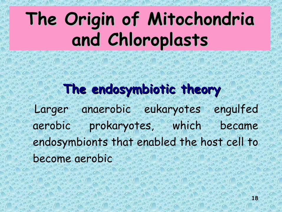
The Origin of Mitochondria The Origin of Mitochondria and Chloroplastsand Chloroplasts
• The endosymbiotic theoryThe endosymbiotic theory
– Evidence that supports the theory of endosymbiosis:• Physical similarities exist between
mitochondria, chloroplasts and prokaryotes• Molecular data indicates mitochondria and
chloroplasts are of prokaryotic origin

17
Same size and shape as bacteria Double membrane 70 S Ribosomes Circular chromosomes Replicate on their own
18
The Origin of Mitochondria The Origin of Mitochondria and Chloroplastsand Chloroplasts
The endosymbiotic theoryThe endosymbiotic theoryLarger anaerobic eukaryotes engulfed aerobic prokaryotes, which became endosymbionts that enabled the host cell to become aerobic

19

20
THE ENDOSYMBIOTIC THEORYTHE ENDOSYMBIOTIC THEORY
Reduced carboncompounds + O2
High ATPyield
Electron transport
Reduced carboncompounds
Low ATPyield
Fermentation
Aerobic bacterium
Anaerobic eukaryote
Pyruvateand O2
ATP
chain
1. Eukaryotic cellsurrounds andengulfs bacterium.
2. Bacterium liveswithin eukaryotecell.
3. Eukaryote suppliesbacterium withreduced carboncompounds;bacterium supplieseukaryote with ATP.
21
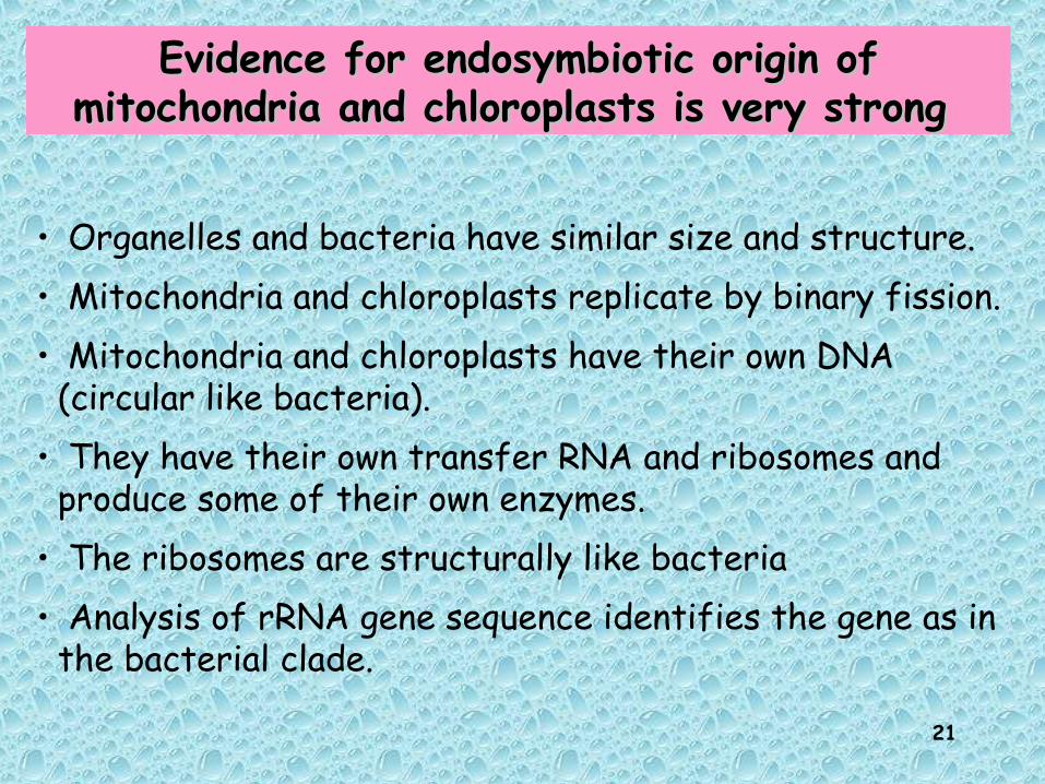
Evidence for endosymbiotic origin of Evidence for endosymbiotic origin of mitochondria and chloroplasts is very strong mitochondria and chloroplasts is very strong
• Organelles and bacteria have similar size and structure.• Mitochondria and chloroplasts replicate by binary fission. • Mitochondria and chloroplasts have their own DNA
(circular like bacteria).• They have their own transfer RNA and ribosomes and
produce some of their own enzymes. • The ribosomes are structurally like bacteria • Analysis of rRNA gene sequence identifies the gene as in
the bacterial clade.

22
Where did the Features of Eukaryote cell come from?Where did the Features of Eukaryote cell come from?
23
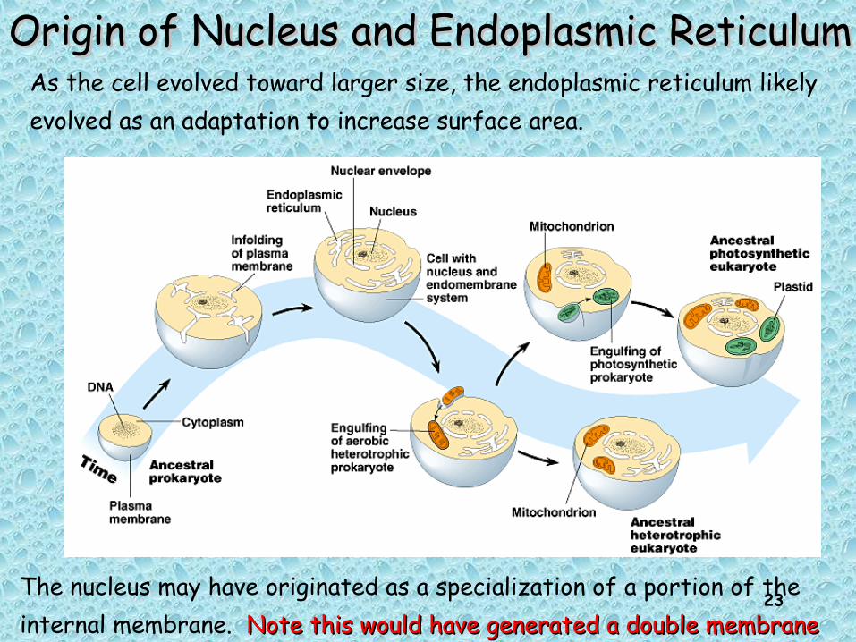
Origin of Nucleus and Endoplasmic ReticulumOrigin of Nucleus and Endoplasmic Reticulum
The nucleus may have originated as a specialization of a portion of the internal membrane. Note this would have generated a double membraneNote this would have generated a double membrane
As the cell evolved toward larger size, the endoplasmic reticulum likely evolved as an adaptation to increase surface area.

24Copyright © 2002 Pearson Education, Inc., publishing as Benjamin Cummings
Endosymbiosis likely happened while the oxygen was “poisoning” anaerobic archaea-eukarya ancestor. Aerobic respiration and photosynthesis evolved once in bacteria and were then imported into the eukarya lineage

25
The three domains seem to have genomes that are chimeric mixes of DNA that was transferred across the boundaries of the domains
This has lead some researchers to suggest replacing the classical tree with a web-like phylogeny

26
La divisione cellulareLa divisione cellulare
27
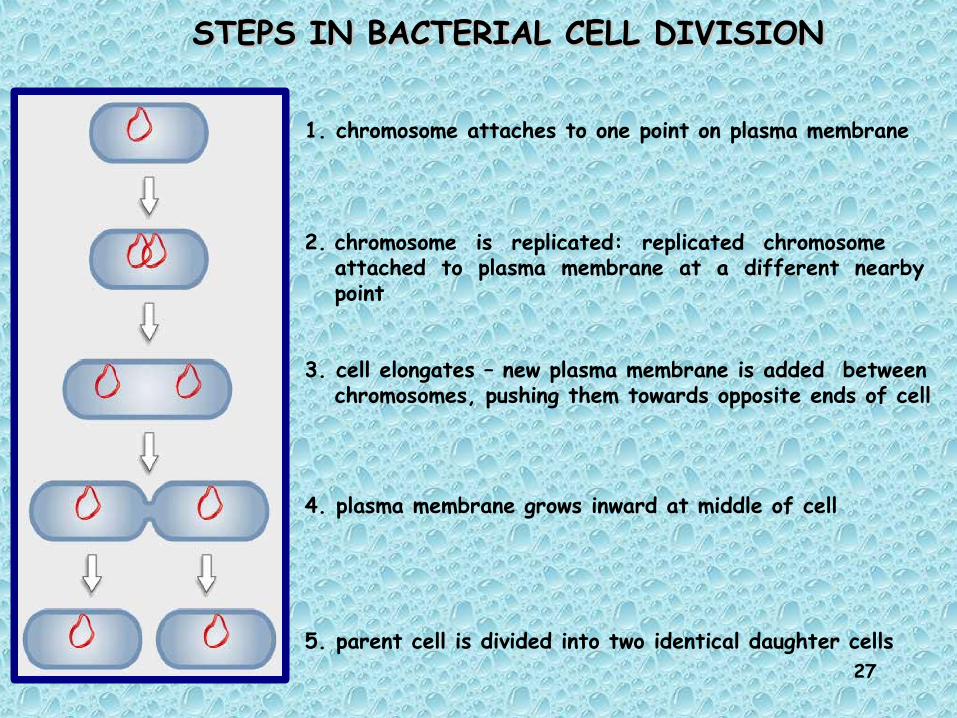
STEPS IN BACTERIAL CELL DIVISIONSTEPS IN BACTERIAL CELL DIVISION
1. chromosome attaches to one point on plasma membrane
2. chromosome is replicated: replicated chromosome attached to plasma membrane at a different nearby point
3. cell elongates – new plasma membrane is added between chromosomes, pushing them towards opposite ends of cell
4. plasma membrane grows inward at middle of cell
5. parent cell is divided into two identical daughter cells

28
Bacterial Cell DivisionBacterial Cell Division DNA replication produces two copies of the DNA replication produces two copies of the
genomegenome The cell grows to approximately double in sizeThe cell grows to approximately double in size The two chromosomes separate as the cell The two chromosomes separate as the cell
growsgrows A new cell wall is formed between the two A new cell wall is formed between the two
chromosomes is a process called binary fissionchromosomes is a process called binary fission Under optimal conditions, the entire process Under optimal conditions, the entire process
can occur in 20 minutes can occur in 20 minutes

29
Why does a cell divide?Why does a cell divide?
-As a cell absorbs nutrients and gets larger, the volume of the cell increases faster than the surface area.
-Therefore, the demands of the cell (the volume) exceed the ability of the cell to bring in nutrients and export wastes.
Solution?Solution? Divide into two smaller cells Divide into two smaller cells

30
When is cell division occurring?When is cell division occurring?
Different kinds of cells divide at different rates:Yeast cell – 2 hoursAmoeba – a few daysHuman embryo cell – 15-20 minutesHuman adult cell – 8 hours to 100 days
GROWTH -increase number of cellsREPAIR -replace lost cells due to injury, diseaseCANCER – Abnormally high rates of cell division due to mutation

31
AgingAging
All cells die after a certain number of divisions (programmed cell death). At any given time some cells are dividing and some cells are dying.
Childhood Cell division > cell death
Adulthood Cell division = cell death
Aging Cell division < cell death
32
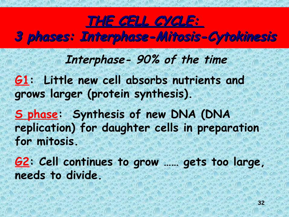
THE CELL CYCLE: THE CELL CYCLE: 3 phases: Interphase-Mitosis-Cytokinesis3 phases: Interphase-Mitosis-Cytokinesis
Interphase- 90% of the time
G1: Little new cell absorbs nutrients and grows larger (protein synthesis).
S phase: Synthesis of new DNA (DNA replication) for daughter cells in preparation for mitosis.
G2: Cell continues to grow …… gets too large, needs to divide.
33
Cell cycle movieCell cycle movie

34
Cell cycle schemeCell cycle scheme
35
How long is one cell cycle? Depends. Eg. Skin cells every 24 hours. Some bacteria every 1-2 hours. Some cells every 3 months. Nerve cells, never. Cancer cells very short.
Programmed cell death: Each cell type will only do so many cell cycles then die. (Apoptosis)

36

37
Mammalian Cell CycleG1:G1: Highly variable,
Absent in rapidly dividing cells, long in slow-growing cells
S:S: 6-8 hours G2:G2: 3-6 hours
M:M: 1-2 hours

38
Determinazione della Determinazione della durata delle fasi del ciclo durata delle fasi del ciclo
cellularecellulare
Marcatura per brevi periodi con 3H-timidina
Osservazione delle cellule mitotiche marcate

39
Mitotic Cell DivisionMitotic Cell Division2 major processes:
• mitosis – nuclear division => preserves diploid number of chromosomes
• cytokinesis – cytoplasmic division => cell divides into two daughter cells

40
MitosisMitosis4 phases:
1st – Prophase (3 major events)
2nd – Metaphase
3rd – Anaphase
4th – Telophase and Cytokinesis

41
1. Prophase1. Prophase
i) chromosomes condense
• 3 major events
ii) spindle fibers form
iii) nuclear membrane breaks down

42
Mitotic Spindle FormsMitotic Spindle Forms
• spindle fibers are specialized microtubules• spindle fibers radiate out from centrioles,
forming the “aster”• centrioles occur in pairs, and are duplicated
during interphase• one pair of centrioles migrates to one pole of
cell, the other pair migrates to opposite pole of cell
43
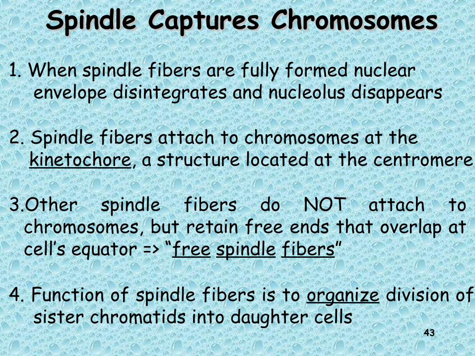
Spindle Captures ChromosomesSpindle Captures Chromosomes
1. When spindle fibers are fully formed nuclear envelope disintegrates and nucleolus disappears
2. Spindle fibers attach to chromosomes at the kinetochore, a structure located at the centromere
3.Other spindle fibers do NOT attach to chromosomes, but retain free ends that overlap at cell’s equator => “free spindle fibers”
4. Function of spindle fibers is to organize division of sister chromatids into daughter cells

44
• Prophase – Inside nucleus
• Chromosomes condense• Nucleoli begin to break down and disappear
– Outside nucleus• Centrosomes move apart and migrate to opposite ends
of the cell• Interphase microtubules disappear and are replaced by
microtubules that grow from the MTOC

45
• Prometaphase– Nuclear envelope breaks down– Microtubules invade nuclear area– Chromosomes attach to microtubules through
kinetochore– Mitotic spindle includes other microtubules that
are involved in the process
46
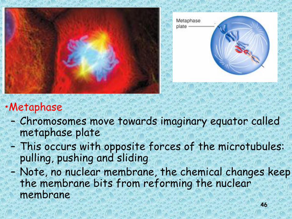
•Metaphase – Chromosomes move towards imaginary equator called
metaphase plate– This occurs with opposite forces of the microtubules:
pulling, pushing and sliding– Note, no nuclear membrane, the chemical changes keep
the membrane bits from reforming the nuclear membrane

47
• Anaphase– Separation of sister chromatids allows each
chromatid to be pulled towards spindle pole connected to by kinetochore microtubule

48
3. Anaphase3. Anaphase
• spindle fibers attached to kinetochores shorten and pull chromatids poleward
• free spindle fibers lengthen and push poles of cell apart
AnaphaseAnaphase AA
AnaphaseAnaphase BB
49
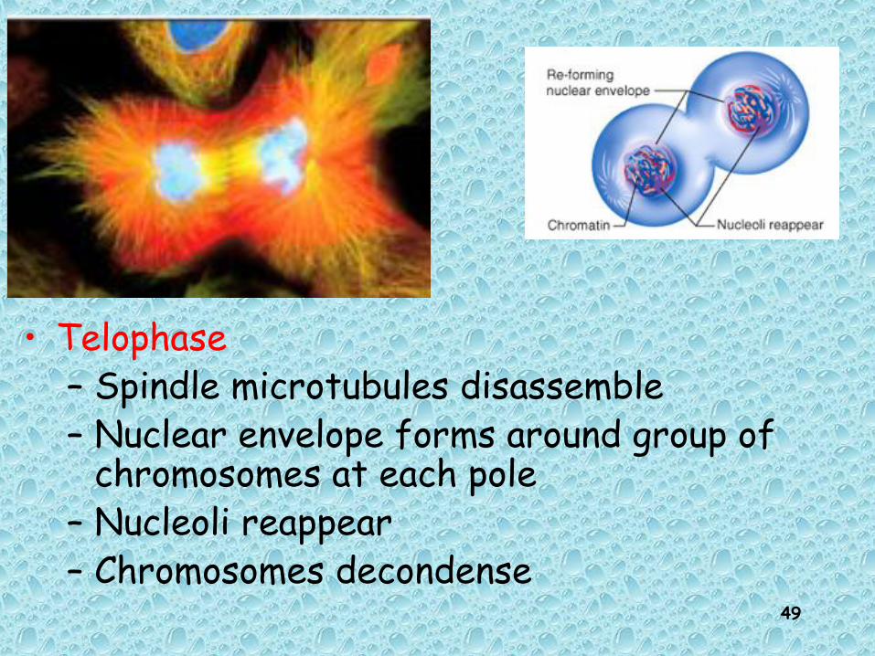
• Telophase– Spindle microtubules disassemble– Nuclear envelope forms around group of
chromosomes at each pole– Nucleoli reappear– Chromosomes decondense

50
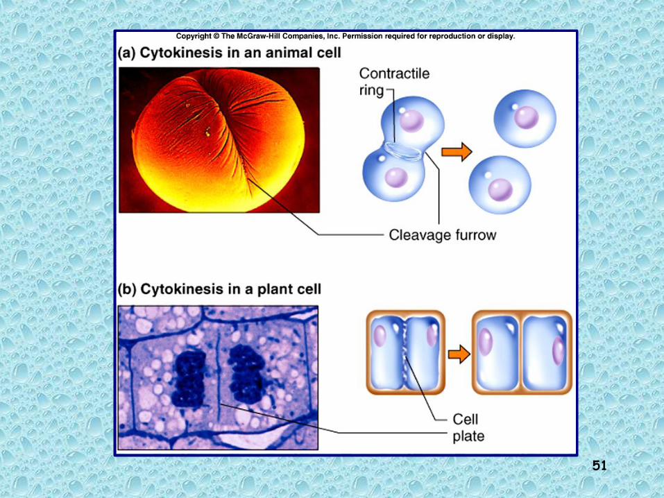
CytokinesisCytokinesis Cytokinesis occurs, enclosing each daughter nucleus
into a separate cell Starts during anaphase and ends in telophase Animal cells: contractile ring pinches cells into two
halves Plant cells: cell plate forms dividing cell into two
halves
51

52
Animal cells: – microfilaments attached to plasma membrane
form a ring around equator of cell – ring contracts, like a drawstring, dividing the
cytoplasm
Plant cells:- stiff cell wall makes pinching impossible- Golgi complex buds off vesicles filled with
carbohydrate- vesicles line up at equator and fuse, producing a
structure called the cell plate- cell plate becomes new cell wall between the two
cells

53
Mitosis: an overviewMitosis: an overview

54
Mitosis: an overviewMitosis: an overview