CARATTERIZZAZIONE AVANZATA DEL COMPLESSO DI LIEVITO KEOPS...
106
Sede Amministrativa: Università degli Studi di Padova Dipartimento di Chimica Biologica SCUOLA DI DOTTORATO DI RICERCA IN BIOCHIMICA E BIOTECNOLOGIE INDIRIZZO DI BIOCHIMICA E BIOFISICA CICLO XXII CARATTERIZZAZIONE AVANZATA DEL COMPLESSO DI LIEVITO KEOPS/EKC ADVANCED CHARACTERIZATION OF THE YEAST KEOPS/EKC COMPLEX Direttore della Scuola: Ch.mo Prof. Giuseppe Zanotti Coordinatore d’indirizzo: Ch.mo Prof. Maria Catia Sorgato Supervisore: Dott. Geppo Sartori Dottorando: Elena Casanova
Transcript of CARATTERIZZAZIONE AVANZATA DEL COMPLESSO DI LIEVITO KEOPS...

Sede Amministrativa: Università degli Studi di Padova
Dipartimento di Chimica Biologica
SCUOLA DI DOTTORATO DI RICERCA IN BIOCHIMICA E BIOTECNOLOGIE
INDIRIZZO DI BIOCHIMICA E BIOFISICA
CICLO XXII
CARATTERIZZAZIONE AVANZATA DEL
COMPLESSO DI LIEVITO KEOPS/EKC
ADVANCED CHARACTERIZATION OF THE YEAST
KEOPS/EKC COMPLEX
Direttore della Scuola: Ch.mo Prof. Giuseppe Zanotti
Coordinatore d’indirizzo: Ch.mo Prof. Maria Catia Sorgato
Supervisore: Dott. Geppo Sartori
Dottorando: Elena Casanova


«Tutti sanno che una cosa è impossibile da realizzare,
finché arriva uno sprovveduto che non lo sa e la inventa.»
Albert Einstein


I
Index
Abstract .................................................................................................................................. 1
Riassunto ............................................................................................................................... 3
Introduction ........................................................................................................................... 5
YEAST AS A MODEL EUKARYOTE ............................................................................ 5
THE KEOPS/EKC COMPLEX ......................................................................................... 7
The Kae1 subunit ........................................................................................................... 9
The Bud32 protein kinase ............................................................................................ 13
Atomic structure of the KEOPS complex.................................................................... 17
Bud32 INTERACTS WITH OTHER PROTEINS, BESIDES THOSE OF THE KEOPS
COMPLEX ...................................................................................................................... 18
The Grx4 glutaredoxin................................................................................................. 18
The Sch9 protein kinase .............................................................................................. 20
Sch9 is the yeast homolog of mammalian Akt/PKB ................................................... 22
THE Sch9-Bud32-Grx4 SIGNALING PATHWAY ....................................................... 23
AIM OF THE THESIS .................................................................................................... 25
Results and discussion ......................................................................................................... 27
PHOSPHORYLATION OF THE S. cerevisiae Grx4 GLUTAREDOXIN BY THE
Bud32 KINASE UNVEILS A NOVEL SIGNALING PATHWAY INVOLVING Sch9,
A YEAST MEMBER OF THE Akt/PKB SUBFAMILY ............................................... 27
Phosphorylation at Ser134 of Grx4 by Bud32 contributes to the functionality of the
glutaredoxin in yeast cells ........................................................................................... 27
Phosphorylation of Bud32 at Ser258 is unrelated to its functions within the KEOPS
complex ....................................................................................................................... 32
Does the Sch9-Bud32-Grx4 phosphorylation cascade affect any function of Grx4?.. 34
ANALYSIS OF THE PHYSIOLOGICAL ROLE OF Kae1 ........................................... 37
Analysis of the phenotype of KAE1 mutants ............................................................... 37
The KAEE147A
mutant shows a strong defect in the regulation of GAL1 transcription 38
The KAE1 mutations impair telomere homeostasis ..................................................... 39
Mutations of Kae1 alter its cellular localization .......................................................... 40
Is yeast Kae1 endowed with nuclease activity?........................................................... 42

II
Kae1 IS AN IN VITRO SUBSTRATE OF Bud32 .......................................................... 48
MS analysis of phosphorylated Kae1 .......................................................................... 52
Conclusions .......................................................................................................................... 55
ROLE OF Bud32 IN A NEW SIGNALING PATHWAY IN YEAST ........................... 55
ANALYSIS OF Kae1 ACTIVITY .................................................................................. 56
BIOCHEMICAL RELATIONSHIP BETWEEN Bud32 AND Kae1 ............................. 57
Materials and methods ......................................................................................................... 59
STRAINS......................................................................................................................... 59
E. coli strains ............................................................................................................... 59
S. cerevisiae strains ..................................................................................................... 59
MEDIA ............................................................................................................................ 60
MEDIA FOR E. coli .................................................................................................... 60
MEDIA FOR S. cerevisiae .......................................................................................... 60
VECTORS ....................................................................................................................... 61
pFA6a-kanMX4 .......................................................................................................... 61
pYeDP1/8.2 ................................................................................................................. 62
pET-20 (Novagen) ....................................................................................................... 63
pET-28 (Novagen) ....................................................................................................... 64
pYM2 .......................................................................................................................... 64
ONE-STEP TRANSFORMATION OF YEAST ............................................................ 65
HIGH EFFICIENCY LIAC TRANSFORMATION OF YEAST ................................... 66
PREPARATION OF YEAST DNA ................................................................................ 67
POLYMERASE CHAIN REACTION (PCR) ................................................................ 67
MUTAGENESIS OF GRX4, BUD32 AND KAE1 .......................................................... 69
CREATION OF THE STRAINS USED FOR THE PHENOTYPIC ANALYSIS OF
THE EFFECT OF THE PHOSPHORYLATION ON Grx4 BY Bud32 ......................... 71
TETRAD DISSECTION ................................................................................................. 73
EPITOPE-TAGGING OF KAE1 AND AFT1 ................................................................. 73
PREPARATION OF YEAST RNA BY EXTRACTION WITH HOT ACID PHENOL 75
ANALYSIS OF GAL1 TRANSCRIPTION .................................................................... 76
ANALYSIS OF FET3/FIT3 TRANSCRIPTION............................................................ 76
REAL-TIME PCR ........................................................................................................... 77
III
NORTHERN BLOT ........................................................................................................ 78
DETECTION USING RADIOLABELED PROBES: ................................................ 79
DETECTION USING NON-RADIOACTIVE PROBES: .......................................... 80
PREPARATION OF RADIOLABELED PROBES FOR NORTHERN AND
SOUTHERN BLOTTING ............................................................................................... 81
PREPARATION OF NON-RADIOACTIVE PROBES FOR NORTHERN BLOTTING
......................................................................................................................................... 81
SOUTHERN BLOT ANALYSIS OF TELOMERE LENGTH ...................................... 82
PREPARATION OF THE PROBE FOR NUCLEASE ASSAY .................................... 83
NUCLEASE ASSAY ...................................................................................................... 83
PURIFICATION OF PROTEINS FROM E. coli CELLS .............................................. 84
SODIUM DODECYL SULFATE POLYACRYLAMIDE GEL ELECTROPHORESIS
(SDS-PAGE) .................................................................................................................... 85
WESTERN BLOT ........................................................................................................... 86
PHOSPHOTRANSFERASE ASSAY ............................................................................. 87
PROTEIN EXTRACTION FROM YEAST .................................................................... 88
PROTEIN IMMUNOPRECIPITATION ......................................................................... 89
ANTI-HA IMMUNOCYTOLOGY ................................................................................. 89
References ........................................................................................................................... 91

IV
Abstract/Riassunto
1
Abstract
During my PhD I have studied the properties of two yeast proteins, the protein kinase
Bud32 and the putative protease Kae1, that take part in a nuclear complex named
KEOPS/EKC. Actually, while Kae1 is associated uniquely to the proteins of the complex,
Bud32 has many other partners in the cell; in fact, I have also studied its strong
relationship with the Grx4 glutaredoxin.
The yeast KEOPS/EKC complex has been isolated in 2006 by two different groups and has
been shown to be involved in both telomere homeostasis and transcription regulation. The
complex is evolutionarily conserved and is composed of five proteins: the protein kinase
Bud32, the hypothetical metallo-protease Kae1, the still uncharacterized Cgi121, and two
other proteins of small size, Pcc1 and Pcc2/Gon7. For many years, our attention has been
focused on the atypical protein kinase Bud32, which interacts with many other yeast
proteins, suggesting that it may play several roles in the cell. Among these Bud32 partners,
we demonstrated that the glutaredoxin Grx4 is a substrate of the protein kinase, being
readily phosphorylated by Bud32 mainly at Ser 134. Also, this modification is upregulated
by the previous phosphorylation of Bud32 at its Ser258 residue by the Sch9 protein kinase
(the yeast homologue of mammalian Akt/PKB). During the first part of my PhD I
deepened the study of the physiological significance of this new phosphorylation cascade.
By the phenotypic analysis of yeast strains expressing mutagenized forms of Grx4, I
demonstrated that the phosphorylation of Grx4 by Bud32 is important for Grx4
functionality in vivo. However I could not identify a specific effect of the Bud32-mediated
phosphorylation of Grx4 on the known activities of the glutaredoxin, wich is involved in
iron cellular homeostasis and in the survival under oxidative stress conditions. This result
suggests that the Bud32-mediated phosphorylation of Grx4 play a role in different,
uncharacterized functions of the glutaredoxin. I also checked if the phosphorylation of
Bud32 by Sch9 could modulate the activity of the whole KEOPS complex, but the analysis
of telomeres length and of the activation rate of the galactose-inducible GAL1 gene (one of
the main transcriptional targets of KEOPS) showed that these functions are unaffected in a
Bud32 unphosphorylatable mutant (S258A). These results suggest that the phosphorylation
of Bud32 at Ser258 is unrelated to its function within the KEOPS complex.
Abstract/Riassunto
2
I then addressed my attention to the Kae1 subunit of the complex. By using two strains
expressing mutagenized forms of Kae1, I could demonstrate that the activity of this protein
is essential for the complex, both at the telomere and at the transcriptional level. The
biochemical function of Kae1 is however still unknown. It was initially classified as a
protease, and, in effect, in 2006 an endopeptidase activity was indirectly demonstrated for
the human homologue of Kae1, OSGEP. On the contrary, in 2007 Hecker et al.
demonstrated that an archaeal orthologue of Kae1 is an AP-endonuclease . During my PhD
I tried to define the activity of yeast Kae1, but the results obtained are not sufficient to
clarify this point.
Finally, I decided to verify the hypothesis, coming from a recent work that describes the
atomic structure of an archaeal-derived KEOPS complex, that Kae1 could be a substrate of
Bud32. Using the yeast Bud32 and Kae1 proteins, co-expressed and purified from E.coli, I
observed that the also the recombinant proteins are tightly associated, forming a kind of
catalytic KEOPS subcomplex. Using several mutagenized forms of these proteins I
demonstrated, by in vitro phosphotranspherase assays, that Bud32 is able to phosporylate
Kae1 and that the binding of Kae1 has an inhibitory effect on the catalytic activity of the
kinase. An important confirmation comes from the MS analysis of phosphorylated Kae1,
that identified Ser 367 as a target of Bud32. However this might not be the only
phosphorylated residue.
Altogether these results indicate that, at least in vitro, a regulatory relationship exists
between Bud32 and Kae1. This is interesting as the two proteins are liable to carefully
modulate the functions of the entire KEOPS complex.
Abstract/Riassunto
3
Riassunto
Durante il Dottorato di ricerca, mi sono occupata dello studio di due proteine di lievito, la
proteinchinasi Bud32 e l‟ipotetica proteasi Kae1, che fanno parte di un complesso nucleare
denominato KEOPS/EKC. Mentre Kae1 è associata unicamente alle proteine del
complesso, Bud32 ha molti altri partner all‟interno della cellula; oggetto del mio studio è
stata infatti anche la sua forte relazione con la glutaredoxina Grx4.
Il complesso di lievito KEOPS/EKC, isolato nel 2006 da due diversi gruppi di ricerca, è
coinvolto nell‟omeostasi telomerica e nella regolazione della trascrizione. Il complesso è
evolutivamente conservato ed è composto da cinque proteine: la chinasi Bud32, l‟ipotetica
proteasi Kae1, Cgi121, proteina non ancora caratterizzata, e altre due piccole subunità,
Pcc1 e Pcc2/Gon7. Per molti anni la nostra attenzione è stata rivolta alla proteinchinasi
atipica Bud32, che interagisce con molte altre proteine di lievito, suggerendo come essa
possa avere altri ruoli all‟interno della cellula. Tra i vari partner di Bud32, abbiamo
dimostrato che la glutaredoxina Grx4 risulta essere un substrato della chinasi sia in vivo
che in vitro, ed è infatti fosforilata da Bud32 principalmente nella Ser 134. Questa
relazione è inoltre a sua volta modulata dalla precedente fosforilazione di Bud32 nella
Ser258 da parte della chinasi Sch9 (l‟omologa in lievito della chinasi di mammifero
Akt/PKB). Durante la prima parte del mio dottorato mi sono concentrata sulla ricerca di un
possibile significato fisiologico di questa nuova cascata di fosforilazioni. Analizzando
ceppi di lievito che esprimono forme mutagenizzate di Grx4, ho dimostrato come la
fosforilazione di Grx4 da parte di Bud32 sia importante per la funzionalità della proteina in
vivo. Sfortunatamente, non ho potuto identificare un effetto di questa fosforilazione sulle
attività della glutaredoxina, coinvolta nella regolazione dell‟omeostasi cellulare del ferro e
nella sopravvivenza in condizioni di stress ossidativo. Questi risultati portano all‟ipotesi
che la fosforilazione di Grx4 da parte di Bud32 giochi un ruolo in qualche funzione diversa
e ancora non caratterizzata della glutaredoxina.
Ho inoltre verificato se la fosforilazione nella Serina 258 di Bud32 da parte di Sch9
potesse modulare l‟attività dell‟intero complesso KEOPS, ma l‟analisi della lunghezza dei
telomeri e del livello di attivazione del gene inducibile GAL1 (uno dei maggiori target
trascrizionali di KEOPS) ha rivelato che queste funzioni non erano colpite nel mutante
Abstract/Riassunto
4
BUDS258A
. Questi risultati suggeriscono che la fosforilazione della serina 258 di Bud32 non
sia collegata alle funzioni della chinasi nel complesso KEOPS.
Ho successivamente indirizzato la mia attenzione alla proteina Kae1. Attraverso l‟utilizzo
di due ceppi, esprimenti forme mutagenizzate di Kae1, ho potuto dimostrare come l‟attività
di questa proteina sia essenziale per l‟intero complesso, sia a livello dei telomeri che della
trascrizione. La funzione biochimica di Kae1 è tuttavia ancora sconosciuta. Inizialmente la
proteina è stata classificata come una proteasi, e in effetti nel 2006 è stata indirettamente
dimostrata un'attività endopeptidasica per l'omologa umana di Kae1, OSGEP. Al contrario,
nel 2007 Hecker et al. hanno dimostrato che l‟omologa di Kae1 in Archea è un‟AP-
endonucleasi. Durante il mio dottorato ho provato a definire l‟attività della proteina Kae1
di lievito, ma i risultati ottenuti non sono stati sufficienti per chiarire questo punto.
Infine, ho deciso di verificare l‟ipotesi, derivante da un recente lavoro in cui viene descritta
la struttura atomica del complesso KEOPS negli Archaea, che Kae1 possa essere un
substrato di Bud32. Utilizzando le proteine di lievito Bud32 e Kae1, espresse in E.coli, ho
osservato che, come accade nelle cellule di lievito, le proteine ricombinanti sono
strettamente associate e formano quindi una sorta di sub-complesso catalitico di KEOPS.
Utilizzando diverse forme mutagenizzate delle due proteine, in test chinasici in vitro, ho
dimostrato che Bud32 è in grado di fosforilare Kae1 e che il legame di Kae1 ha un effetto
inibitorio sull‟attività catalitica della chinasi. Un‟importante conferma è derivata
dall‟analisi di spettrometria di massa su Kae1 fosforilata, in cui la Ser 367 è stata
identificata come target di Bud32. Tuttavia questo potrebbe non essere l‟unico residuo
fosforilato.
Nel complesso, questi dati indicano che, perlomeno in vitro Bud32 e Kae1 vengono
reciprocamente regolate. Questo dato è interessante, dal momento che le due proteine
potrebbero modulare le funzioni dell‟intero complesso KEOPS.
Introduction
5
Introduction
YEAST AS A MODEL EUKARYOTE
The yeast Saccharomyces cerevisiae is an eukaryotic micro-organism classified in the
kingdom of Fungi. It is perhaps the most useful yeast, owing to its use since ancient times
in baking and brewing. It is also one of the most intensively studied eukaryotic model
organisms in molecular and cell biology, much like Escherichia coli as the model
prokaryote. S.cerevisiae cells are round to ovoid, 5–10 m in diameter. It reproduces by a
division process known as budding. Yeasts have asexual and sexual reproductive cycles,
however the most common mode of vegetative growth in yeast is asexual reproduction by
budding or fission. In the budding yeast S.cerevisiae a small bud, or daughter cell, is
formed on the parent cell. The nucleus of the parent cell splits into a daughter nucleus and
migrates into the daughter cell. The bud continues to grow until it separates from the parent
cell, forming a new cell. Under high stress conditions haploid cells will generally die,
however under the same conditions diploid cells can undergo sporulation, entering sexual
reproduction (meiosis) and producing four haploid spores, which can go on to mate
(conjugate), reforming the diploid. Yeast has two mating types, a and α, which show
primitive aspects of sex differentiation. Unlike most other microorganisms, in S.cerevisiae
both the haploid and diploid state are stable. Thus, recessive mutations can be conveniently
isolated and manifested in haploid strains, and complementation tests can be carried out
through the formation of diploid strains. Although yeasts have greater genetic complexity
than bacteria, containing 3.5 times more DNA than E.coli cells, they share many of the
technical advantages that permitted rapid progress in the molecular genetics of prokaryotes
and their viruses. Some of the properties that make yeast particularly suitable for biological
studies include rapid growth, dispersed cells, the ease of replica plating and mutant
isolation, a well-defined genetic system, and most important, a highly versatile DNA
transformation system. Unlike many other microorganisms, numerous mutations in genes
for biosynthetic pathways are available in S.cerevisiae and are conveniently used as
selectable growth markers on synthetic media. Being nonpathogenic, yeast can be handled
Introduction
6
with little precautions. Large quantities of normal bakers‟ yeast are commercially available
and can provide a cheap source for biochemical studies. The development of DNA
transformation has made yeast particularly accessible to gene cloning and genetic
engineering techniques. Genes corresponding to virtually any genetic trait can be identified
by complementation of mutants with plasmids from genome libraries. Plasmids can be
introduced into yeast cells either as replicating molecules or by integration into the
genome. In contrast to most other organisms, integrative recombination of transforming
DNA in yeast proceeds exclusively via homologous recombination. Exogenous DNA with
at least partial homologous segments can therefore be directed to specific locations in the
genome. Also, homologous recombination, coupled with yeasts‟ high levels of gene
conversion, has led to the development of techniques for the direct replacement of
genetically engineered DNA sequences into their normal chromosome locations. Thus,
normal wild-type genes, even those having no previously known mutations, can be
conveniently replaced with altered and disrupted alleles. The phenotypes arising after
disruption of yeast genes has contributed significantly toward the understanding of the
function of certain proteins in vivo. Also unique to yeast, transformation can be carried out
directly with synthetic oligonucleotides, permitting the convenient productions of
numerous altered forms of proteins. These techniques have been extensively exploited in
the analysis of gene regulation, structure-function relationships of proteins, chromosome
structure, and other general questions in cell biology. The overriding virtues of yeast are
illustrated by the fact that mammalian genes are being introduced into yeast for systematic
analyses of the functions of the corresponding gene products. In addition, yeast has proved
to be valuable for studies of other organisms genes, by the use of the two-hybrid screening
system (for protein-protein interactions), the use of YACs for cloning large fragments of
DNA, and the expression systems for the laboratory and commercial preparation of
heterologous proteins. During the last two decades, an ever-increasing number of
molecular biologists have taken up yeast as their primary research system, resulting in a
virtually autocatalytic stimulus for continuing investigations of all aspects of molecular
and cell biology. Most significantly, the knowledge of the DNA sequence of the complete
genome, which was completed in 1996, has altered the way molecular and cell biologist
approach and carry out their studies 1. In addition, genome sequencing allowed the start-up
Introduction
7
of several projects of systematic investigation of genes functions by the in-depth study of
strains carrying disrupted genes.
Among the genes sequenced and disrupted in our laboratory, a gene, encoding an atypical
Ser/Thr protein kinase (afterwards named Bud32), aroused our interest and was intensively
studied. An important trait of this protein was it property of interacting with several
proteins, in particular we identified two strong interactors of the protein kinase: the Grx4
glutaredoxin and a putative metallo-endoprotease, that we named Kae1 (kinase-associated
endopeptidase 1)
THE KEOPS/EKC COMPLEX
In 2006, two groups have independently discovered in Saccharomyces cerevisiae a protein
complex called either KEOPS (for Kinase, Endopeptidase and Other Proteins of Small
size) or EKC (for Endopeptidase-like Kinase Chromatin-associated). The first group 2
demonstrated the involvement of the complex in telomere uncapping and elongation,
whereas the second one demonstrated its involvement in transcription of essential
eukaryotic genes. This complex consists of five subunits: the Ser/Thr protein kinase
Bud32, the kinase-associated endopeptidase 1 (Kae1), and the small subunits Pcc1,
Pcc2/Gon7 and Cgi121 2-4
.
During a genome-wide screen in S.cerevisiae, Downey and colleagues isolated CGI121, a
component of the KEOPS complex, as a suppressor of cdc13-1, an allele of the telomere-
capping protein Cdc13.
The telomere is a nucleoproteic structure at the ends of chromosomes, which protects them
from destruction 5, 6
. In addition to that, the telomere organizes chromosome-end
replication, in part by regulating the recruitment of telomerase, an enzyme consisting of an
RNA component and a reverse-transcriptase enzyme 7. Telomerase catalyzes the addition
of tandem G-rich repeats at the 3′ end of linear chromosomes (TG1–3 in S.cerevisiae). In
yeast, telomerase recruitment is mediated by the Cdc13-Ten1-Stn1 complex and the
Yku70/80 heterodimer, both of which help to recruit telomerase via direct physical
interactions with its components 8, 9
. Cdc13 directly binds TG-rich telomeric single-
Introduction
8
stranded (ss) DNA and, together with Yku70/Yku80, also plays a critical role in
chromosome end protection. At restrictive temperatures, strains carrying capping-defective
alleles of these genes generate large amounts of ssDNA at telomeres and initiate a robust
DNA-damage checkpoint response that is dependent on MEC1 and RAD9 10, 11
. In addition
to inducing checkpoint arrest and ensuing replicative senescence 12
, defects in telomere
capping are a potent threat to genome stability 13, 14
. It has been shown that deletion of
either genes encoding DNA-damage checkpoint components (RAD9, MEC1, RAD24) or
the gene encoding the 5′-3′ exonuclease Exo1, suppress the checkpoint arrest imparted by
telomere-capping defects 6. Telomere-capping defects associated with the cdc13-1 allele
can also be partially suppressed by overexpression of the Stn1 protein, which interacts
directly with Cdc13 15
.
Using a genome-wide functional genomics screen the authors were able to isolate genetic
suppressors of the thermosensitive cdc13-1 allele; one of these was the YML036W gene,
successively named CGI121.
CGI121 is highly conserved in eukaryotes, with the exception of Drosophila. Human
CGI121 had already been identified in a two-hybrid screen using the human homolog of
BUD32, the p53-related protein kinase PRPK as bait 16
. In yeast this relationship is
conserved and it has been shown that indeed Cgi121 interacts physically with Bud32.
Using tandem affinity purification (TAP) experiments, Bud32 and Cgi121 were found to
be part of a complex containing also Gon7 and Kae1. Downey et al. proposed that the
KEOPS complex acts as a critical regulator of telomere elongation at native telomeres and
at double strand breaks (DSBs), and that it can promote telomere uncapping in cdc13-1
strains. This dual role of the KEOPS complex appears unique among telomeric proteins
and suggests that this conserved protein kinase-containing complex links the processes of
telomere protection and telomere elongation.
As mentioned before, the same complex was isolated in a parallel study, during a screen to
isolate suppressors of a splicing defect which leads to a cold-sensitive phenotype. During
the screen, the authors identified a new ORF and they called the gene PCC1 (for Polarized
growth Chromatin-associated Controller 1); this gene encodes a small protein of about
10kDa. PCC1 is not essential, but pcc1 null cells grow very slowly and are thermosensitive
at 34°C. The construction of a thermosensitive mutant allele indicated that Pcc1 is required
for normal cell cycle progression and mating projection formation. In fact Pcc1 is involved

Introduction
9
in the expression of some of the genes induced by the mating pheromone -factor.
Mutation of Pcc1 also affects the expression of the GAL genes (that are induced by the
presence of galactose in the medium) by impairing the recruitment of the SAGA and
Mediator co-activators. By the analysis of proteins interacting with Pcc1, the authors
isolated the EKC complex, that contains the Bud32 kinase, the putative endopeptidase
Kae1 and two additional proteins, Pcc2/Gon7 and Cgi121. Genetic and physical
interactions among these proteins strongly suggested that this complex is a functional unit.
Chromatin immunoprecipitation experiments and multiple genetic interactions of PCC1
mutants with mutants of the transcription apparatus and chromatin-modifying enzymes
underscore the direct function of the complex in transcription. Sequence analyses and
functional complementation experiments 3, 17
have shown that subunits of KEOPS are
conserved among species and suggest that the function of the complex in transcription
and/or telomere maintenance (and possibly other processes) is also conserved in other
species.
The Kae1 subunit
A universally conserved protein
The KAE1 gene of S.cerevisiae encodes a 368 aminoacids protein of a prevalent nuclear
localization; this gene is essential for yeast cells, as its inactivation is lethal 18
. Kae1 is the
most highly conserved member of the KEOPS complex, with sequence identity of roughly
60% between yeast and humans; the human homolog of KAE1 is called OSGEP. The Kae1
protein and its homologues belong to the small set of about 60 universal proteins present in
all members of the three domains of life 19
. This protein was placed in 2004 by Galperin
and Koonin at the top of their list of 10 „known-unknown‟ proteins „that should be priority
targets for experimental study‟. Indeed, this putative endopeptidase (of „known‟
biochemical function; even if until now its function is still not really defined) was the only
protein of „unknown‟ biological role present in all the 70 genomes then available. The
endopeptidase activity of Kae1/OSGEP proteins was suggested by the homology with an
O-sialoglycoprotein endopeptidase (Gcp) previously purified from Pasteurella
Introduction
10
haemolytica by Mellors and colleagues in 1991; this similarity permitted to include Kae1
in the M22 family of metallo-proteases (MEROPS database,20
).
A putative metallo-protease
In general, metallo-proteases are enzymes that have critical roles in processes like gene
expression, regulation of cell cycle, intracellular targeting of proteins and apoptosis. In
these enzymes, a divalent cation, usually Zn2+
(but also Co2+
or Mn2+
) activates the water
molecule. The metal ion is held in place by amino acid ligands, usually three in number.
The known metal ligands are His, Glu, Asp or Lys; and at least one other residue, which
may play an electrophilic role, is required for catalysis. Of the known metalloproteases,
around half contain an HEXXH motif, which has been shown, by crystallographic studies,
to form part of the metal-binding.
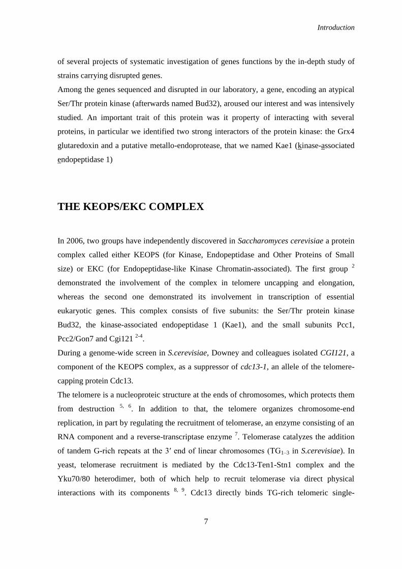
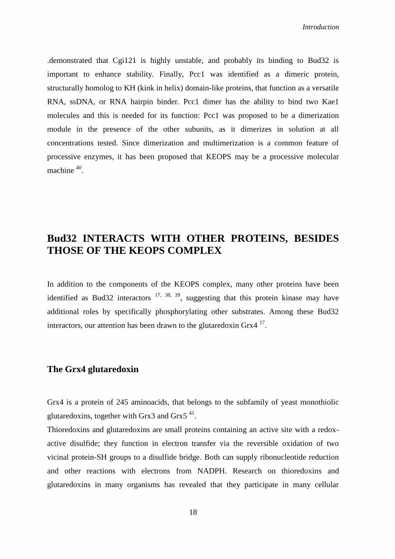
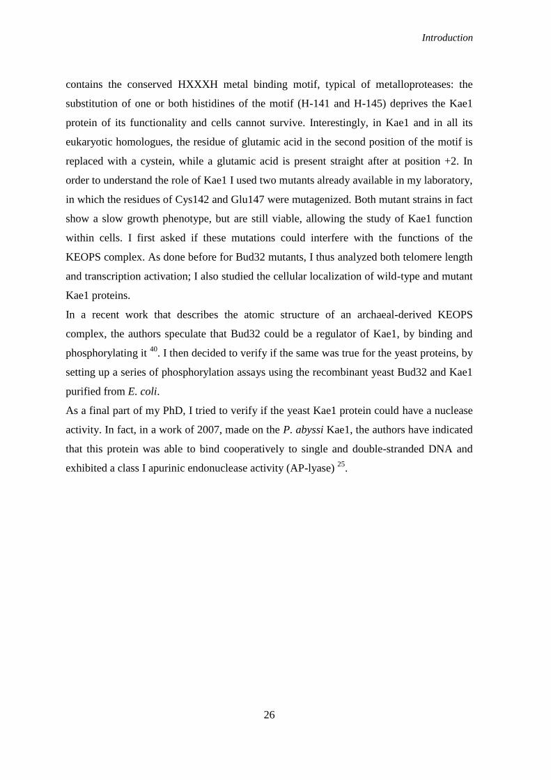
An HXXXH motif is present also in Kae1: this is essential as the mutagenesis of either of
the two histidines (H-141 and H-145) deprives the protein of its functionality (data not
published from our laboratory and 3). It is interesting to notice, however, that in Kae1 and
in all its eukaryotic homologues, the residue of Glutamic acid in the second position is
replaced by a Cystein (fig. 1). Cystein and Glutamic acid have very different properties and
the presence of that residue may create a great steric effect in correspondence of the site of
metal binding. This characteristic suggests that Kae1 may act with a different catalytic
mechanism with respect to the other metallo-proteases.
Fig. 1-Metal ion
binding domain in
S. cerevisiae Kae1
and in its
homologs (from
C.Peggion, PhD
thesis, 2006)

Introduction
11
In 1999, Koonin and co-workers identified an HSP70-actin-like fold (HALF) in Kae1-
related proteins and suggested that they were ATP-dependent proteases with chaperone
activity 21
. Therefore, it was proposed that yeast Kae1 could modulate the activity of the
KEOPS complexes via its proteolytic activity.
In favor of the protease hypothesis, a work of 2006 by Ng et al. 22
, indirectly demonstrated
that a proteasic activity is associated to the mammalian homolog of Kae1, OSGEP.
OSGEP is a protein of 335 amino acids, encoded by a gene located on chromosome 14. It
lies immediately adjacent to the APEX gene (which encodes the APEX nuclease, a
multifunctional DNA repair enzyme) in a 5'-to-5' orientation; transcription of both the
OSGEP and APEX genes is regulated by a bidirectional promoter containing a CCAAT
box. Northern blot analysis showed ubiquitous expression of the OSGEP gene in several
tissues, and similarities in expression patterns between OSGEP and APEX 22
.
Ng et al. observed that OSGEP is over-expressed in acute promyelocytic leukemia (APL)
cells. In normal cells, accumulation of misfolded nuclear hormone receptor corepressor (N-
CoR) as insoluble protein aggregates, induces endoplasmic reticulum stress and activates
unfolded protein response (UPR). In contrast, APL cells are resistant to UPR-induced
apoptosis. By purification and spectrometric analysis from an APL cell line, it was
revealed that the glycoprotease activity of OSGEP is responsible for the resistance of APL
cells to the UPR-induced apoptosis, through processing misfolded N-CoR protein.
Furthermore, the cleavage of N-CoR in APL cells could be blocked by the broad-spectrum
protease inhibitor AEBSF and by RNA interference-mediated down-regulation of OSGEP
expression. The protease activity of OSGEP could therefore represent a way exploited by
tumor cells to survive the toxic insult of misfolded protein(s) 23
.
A putative endonuclease
Recently, the structure of the protein YeaZ from Salmonella typhimurium (a bacterial
homolog of yeast Kae1) has been solved, showing that structurally this protein contains a
HALF fold and belongs to the ASKHA (Acetate and Sugar Kinases/Hsp70/Actin)
superfamily of phosphotransferases. However, purified YeaZ does not bind ATP and has
no glycoprotease activity 24
.
Introduction
12
In 2007 the analysis of the crystal structure and the biochemical function of the orthologue
of Kae1 in the archaeon Pyrococcus abyssi (PaKae1, 25
) showed that indeed this protein
belongs to the ASKHA protein family. PaKae1 was shown to bind the ATP analogue
AMPPNP in a canonical manner. Surprisingly, the nucleotide was found to be directly
bound through its phosphate groups to a non-heme iron. Mutation of residues coordinating
this iron in the yeast Kae1 (the two histidines of the HXXXH motif) causes lethality,
demonstrating the importance of the metal binding for the function of this protein 3.
Although yeast Kae1 was proposed to possess endopeptidase activity, the crystal structure
of PaKae1 as well as biochemical activity tests did not support this hypothesis. Rather, it
was shown that PaKae1 binds cooperatively to single and double-stranded DNA and
induces DNA conformational changes without significant DNA lengthening or shortening.
PaKae1 also exhibits a class I apurinic endonuclease activity (AP-lyase), suggesting an
important function of this protein in the maintenance of genome integrity 25
.
Since the two functions of protease and nuclease are dramatically different, the conserved
function of Kae1 remains an open question.
Genome analysis has revealed that homologs of yeast Kae1 and Bud32 are present in all
eukaryotes and archaea. The genes encoding the archaeal orthologues of Kae1 and Bud32
are juxtaposed in nearly all archaeal genomes, and they are even fused in several archaea,
encoding a single bifunctional protein; this strongly suggests that, even when their genes
are separated, Kae1 and Bud32 are physically and functionally linked 26
. To get further
insights on the interactions between Kae1 and Bud32, in 2008 the archaeal “fusion” protein
MjBud32 of M. jannaschii was expressed in E.coli and its crystal structure solved 27
. The
structure of MJ1130 revealed the strict association of Kae1 and Bud32: the intramolecular
interactions identified between the two moieties of MJ1130 suggested that the two yeast
proteins might interact similarly. In fact the same interaction between Bud32 and Kae1 has
been clearly demonstrated in yeast by mutagenesis of the corresponding residues involved
in the surface interaction. The analysis of these mutants demonstrated that they strongly
affect the function of the yeast KEOPS complex, as they elicit major phenotypic effects
both on transcription and telomere maintenance, thus linking the function of the complex
to its structural integrity.
Introduction
13
The Bud32 protein kinase
As mentioned before, the yeast BUD32 gene was identified and analyzed in our laboratory,
after the completion of the S.cerevisiae genome sequence 1, in the frame of the EUROFAN
project 28
. The deletion of BUD32 is responsible for different phenotypic effects; among
these, the drastic slowdown of cell growth, the inability of homozygous diploid cells to
enter sporulation, alterations in the cell wall and random budding 29-32
.
BUD32 encodes a protein of 261 amino acids, initially named piD261, prevalently located
in the nucleus. Biochemical data showed that Bud32 is an atypical protein kinase 33-35
.
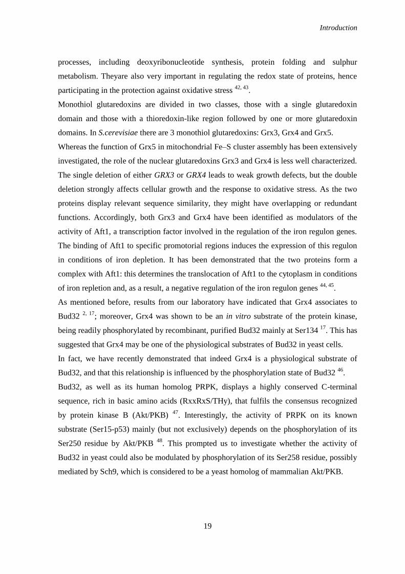
General characteristics of protein kinases
The phosphorylation event is one of the most frequent and general mechanisms by which
nearly all cellular functions are regulated in higher organisms. The enzymes responsible
for this kind of reaction are protein-kinases. These enzymes use the -phosphate of ATP
(or GTP) to generate phosphate monoesters using protein alcohol groups (on Ser and Thr)
and/or protein phenolic groups (on Tyr) as phosphate acceptors. Protein-kinases make up a
superfamily of homologous proteins in Eukarya, being related by virtue of their catalytic
domain. The kinase domain consists of about 250-300 amino acid residues and contains 12
conserved subdomains that fold into a common catalytic core structure. In the type of
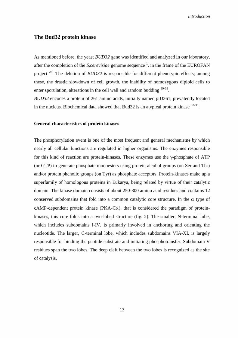
cAMP-dependent protein kinase (PKA-C), that is considered the paradigm of protein-
kinases, this core folds into a two-lobed structure (fig. 2). The smaller, N-terminal lobe,
which includes subdomains I-IV, is primarly involved in anchoring and orienting the
nucleotide. The larger, C-terminal lobe, which includes subdomains VIA-XI, is largely
responsible for binding the peptide substrate and initiating phosphotransfer. Subdomain V
residues span the two lobes. The deep cleft between the two lobes is recognized as the site
of catalysis.
Introduction
14
Twelve aminoacid residues are invariant or nearly invariant throughout the superfamily,
and hence are strongly implicated as playing essential roles in enzymes function. In PKA-
C, these correspond to Gly50 and Gly52 in subdomain I, Lys52 in subdomain II, Glu91
in subdomain III, Asp 166 and Asn171 in subdomain VIB, Asp184 and Gly 186 in
subdomain VII, Glu208 in subdomain VIII, Asp220 and Gly225 in subdomain IX, and
Arg280 in subdomain XI.
Subdomain I contains the consensus motif GxGxxGxV (“glycinic loop”), starting with
Gly50 in PKA-C. It acts as a flexible clamp that covers and anchors the non-transferable
phosphates of ATP. Subdomain II contains the invariant Lys (Lys 72 in PKA-C), which
is essential for maximal enzyme activity: it helps anchor and orient ATP, toghether with
the nearly invariant Glu91 (in subdomain III). Subdomain IV contains no invariant
residues and does not appear to be directly involved in catalysis or substrate recognition.
Subdomain V links the small and large lobes of the catalytic subunit. Subdomain VIA
folds into the large hydrophobic -helix that extends through the large lobe; this part of the
molecule appears to act mainly as a support structure. Subdomain VIB contains two
invariant residues (Asp 166 and Asn171 in PKA-C), that lie within the consensus motif
HRDLKxxN, known as the “catalytic loop”; within the loop, Asp166 is the catalytic base.
Subdomain VII contains the highly conserved DFG triplet (corresponding to Asp184-
Phe185-Gly186 in PKA-C), where Asp184 helps to orient the -phosphate for transfer.
Fig. 2-The Catalytic Core of PKA.
The N-terminal lobe is colored white,
and the C-terminal lobe is pink. The
hinge region between the lobes is
magenta. An inhibitor peptide is
shown in green with the position of
the serine that is the target for
phosphorylation shown as P0. ATP is
in ball and stick representation. The
glycine-rich region that is important
for localization of the phosphates of
ATP is shown colored light purple. α
Helices and β strands are labeled.
(From Johnson L.N. et al., 1996)
Introduction
15
Subdomain VIII includes the highly conserved APE motif (residues 206-208 in PKA-C).
This subdomain, named “activation loop”, appears to play a major role in recognition of
peptide substates; indeed, many protein-kinases are known to be activated by
phosphorylation of residues in this domain. In PKA-C, maximal kinase activity requires
phosphorylation of Thr197, probably occurring through an intermolecular
autophosphorylation mechanism. Subdomain IX contains a nearly invariant Asp (Asp220
in PKA-C) that acts to stabilize the catalytic loop. Subdomain X is the most poorly
conserved subdomain and its function is obscure. Subdomain XI extends to the C-terminal
end of the kinase domain; it contains the nearly invariant Arg corresponding to Arg280 in
PKA-C. 36
.
Characteristics of the Bud32 protein kinase
Bud32 belongs to the piD261 family of Ser/Thr protein kinases, which has representatives
in virtually all eukaryotic and archaeal organisms. Unlike the majority of eukaryotic
protein kinases, Bud32 preferentially phosphorylates acidic substrates, like casein and
osteopontin 33, 35
.
Bud32 is the shortest protein kinase known to date, entirely lacking the C-terminal
subdomain XI. If it is considered moreover that the C-terminal 44 residues sequence,
including a unique stretch of basic residues, does not display any significant homology
with the members of the protein kinase family, it should be concluded that also subdomain
X and part of subdomain IX are lacking or deeply altered in Bud32. Bud32 also displays
some unique features, such a single glycine left in the glycine loop, proline replaced by
leucine in the APE motif and the lysil residue within the DLKPEN sequence, which is
diagnostic of Ser/Thr kinases, substituted by a threonyl residue 33
. Despite its small size
and low overall similarity with the other members of the protein kinase family, Bud32
displays all the main signatures of a protein kinase catalytic domain, with special reference
to the conservation of invariant residues, whose relevance to its catalytic activity has been
confirmed by mutational analysis. Starting from the N-terminus, the suspected functional
equivalence of Gly-25 with the invariant second glycine of the phosphate anchor motif,
GXGXXG (equivalent to PKA Gly-52), was confirmed by mutating it to Val, which gives
a fully inactive. Lys-52 must be the functional equivalent of PKA Lys-72, since its

Introduction
16
replacement fully suppressed catalytic activity. Another important residue for the kinasic
activity is Glu-76, which matches well the invariant glutamyl residue (PKA Glu-91) that
defines subdomain III; the mutation of this glutamic acid to alanine fully suppressed
catalytic activity. Finally, domain VIB includes the highly conserved catalytic loop, with
its invariant motif DXXXXN, whose actual identification with the 161-166 segment of
Bud32 (DLTSSN) was validated by showing that the replacement of Asp-161 by Ala
nearly abolishes activity. The activation loop of Bud32 is abnormally short; however, it
includes two phosphorylatable residues, Ser-187 and Ser-189. Thanks to a mutant in which
both Ser-187 and Ser-189 had been replaced by Ala (SS187,189AA) it was seen that those
residues do indeed undergo autophosphorylation. All these features suggest that in several
respects the properties of Bud32 are unique 34
.
The human homolog of BUD32 is PRPK, even if the two proteins share only 30% of
identity. This gene was cloned from an interleukin-2 activated cytotoxic T-cell subtraction
library and shown to up regulate transcriptional activity of p53 once transfected in COS-7
cells. PRPK binds to and phosphorylates p53 at Ser15 37
, whence the acronym PRPK (p53
related protein kinase). It was shown that also Bud32 is able to interact with human p53
and to phosphorylate its amino-terminal domain, at Ser15 and Ser37; conversely, PRPK
partially restores the normal growth phenotype of yeast, when overexpressed in strains
where the gene encoding Bud32 has been disrupted. This indicates that the two genes are
functional and not only structural homologues.
Several different approaches have shown that Bud32 is able to interact with many yeast
proteins 17, 38, 39
. Particularly remarkable is its tight association with Kae1, within the
KEOPS complex. Although the kinase activity of Bud32 is relevant for the functions of the
KEOPS complex in yeast, it is actually unknown whether Bud32-dependent
phosphorylation of other subunits of the complex could directly regulate its activity.
Introduction
17
Atomic structure of the KEOPS complex
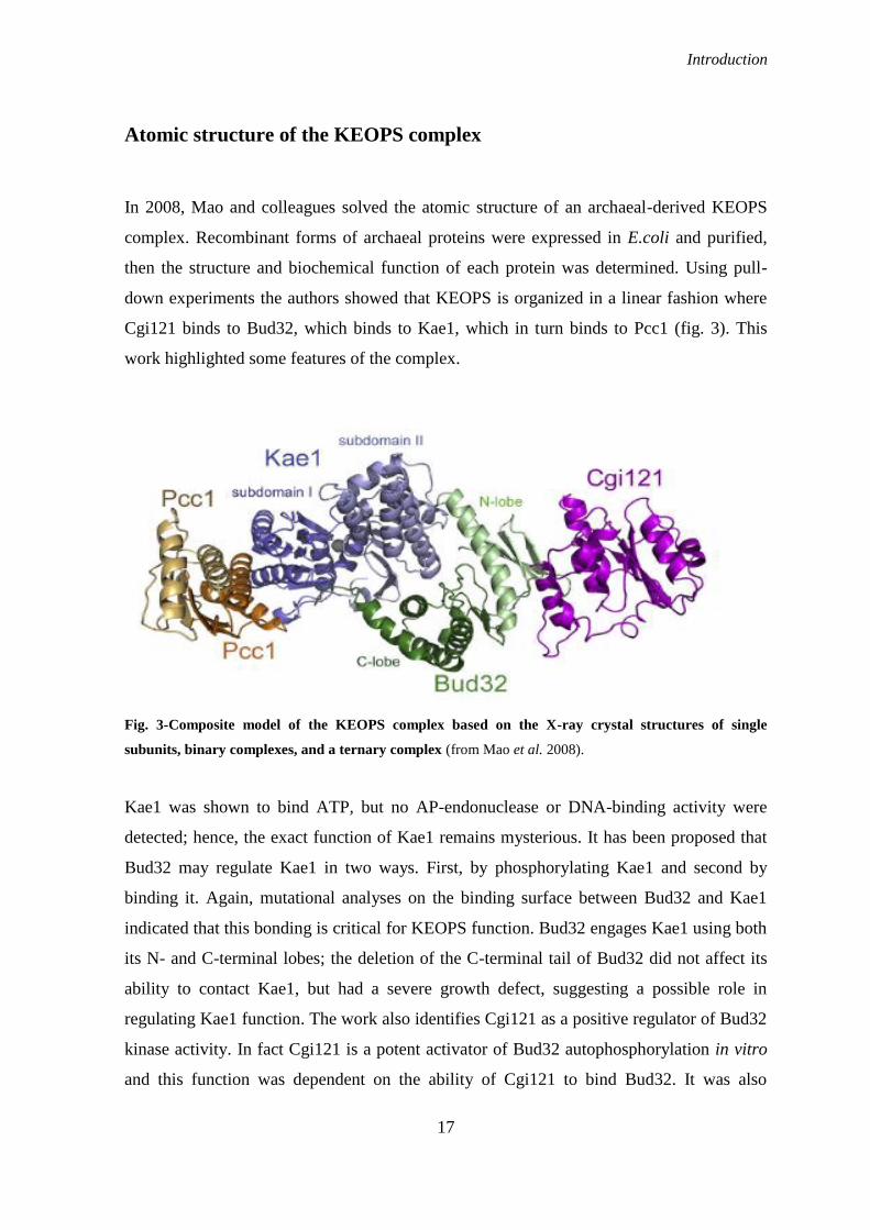
In 2008, Mao and colleagues solved the atomic structure of an archaeal-derived KEOPS
complex. Recombinant forms of archaeal proteins were expressed in E.coli and purified,
then the structure and biochemical function of each protein was determined. Using pull-
down experiments the authors showed that KEOPS is organized in a linear fashion where
Cgi121 binds to Bud32, which binds to Kae1, which in turn binds to Pcc1 (fig. 3). This
work highlighted some features of the complex.
Fig. 3-Composite model of the KEOPS complex based on the X-ray crystal structures of single
subunits, binary complexes, and a ternary complex (from Mao et al. 2008).
Kae1 was shown to bind ATP, but no AP-endonuclease or DNA-binding activity were
detected; hence, the exact function of Kae1 remains mysterious. It has been proposed that
Bud32 may regulate Kae1 in two ways. First, by phosphorylating Kae1 and second by
binding it. Again, mutational analyses on the binding surface between Bud32 and Kae1
indicated that this bonding is critical for KEOPS function. Bud32 engages Kae1 using both
its N- and C-terminal lobes; the deletion of the C-terminal tail of Bud32 did not affect its
ability to contact Kae1, but had a severe growth defect, suggesting a possible role in
regulating Kae1 function. The work also identifies Cgi121 as a positive regulator of Bud32
kinase activity. In fact Cgi121 is a potent activator of Bud32 autophosphorylation in vitro
and this function was dependent on the ability of Cgi121 to bind Bud32. It was also
Introduction
18
.demonstrated that Cgi121 is highly unstable, and probably its binding to Bud32 is
important to enhance stability. Finally, Pcc1 was identified as a dimeric protein,
structurally homolog to KH (kink in helix) domain-like proteins, that function as a versatile
RNA, ssDNA, or RNA hairpin binder. Pcc1 dimer has the ability to bind two Kae1
molecules and this is needed for its function: Pcc1 was proposed to be a dimerization
module in the presence of the other subunits, as it dimerizes in solution at all
concentrations tested. Since dimerization and multimerization is a common feature of
processive enzymes, it has been proposed that KEOPS may be a processive molecular
machine 40
.
Bud32 INTERACTS WITH OTHER PROTEINS, BESIDES
THOSE OF THE KEOPS COMPLEX
In addition to the components of the KEOPS complex, many other proteins have been
identified as Bud32 interactors 17, 38, 39
, suggesting that this protein kinase may have
additional roles by specifically phosphorylating other substrates. Among these Bud32
interactors, our attention has been drawn to the glutaredoxin Grx4 17
.
The Grx4 glutaredoxin
Grx4 is a protein of 245 aminoacids, that belongs to the subfamily of yeast monothiolic
glutaredoxins, together with Grx3 and Grx5 41
.
Thioredoxins and glutaredoxins are small proteins containing an active site with a redox-
active disulfide; they function in electron transfer via the reversible oxidation of two
vicinal protein-SH groups to a disulfide bridge. Both can supply ribonucleotide reduction
and other reactions with electrons from NADPH. Research on thioredoxins and
glutaredoxins in many organisms has revealed that they participate in many cellular
Introduction
19
processes, including deoxyribonucleotide synthesis, protein folding and sulphur
metabolism. Theyare also very important in regulating the redox state of proteins, hence
participating in the protection against oxidative stress 42, 43
.
Monothiol glutaredoxins are divided in two classes, those with a single glutaredoxin
domain and those with a thioredoxin-like region followed by one or more glutaredoxin
domains. In S.cerevisiae there are 3 monothiol glutaredoxins: Grx3, Grx4 and Grx5.
Whereas the function of Grx5 in mitochondrial Fe–S cluster assembly has been extensively
investigated, the role of the nuclear glutaredoxins Grx3 and Grx4 is less well characterized.
The single deletion of either GRX3 or GRX4 leads to weak growth defects, but the double
deletion strongly affects cellular growth and the response to oxidative stress. As the two
proteins display relevant sequence similarity, they might have overlapping or redundant
functions. Accordingly, both Grx3 and Grx4 have been identified as modulators of the
activity of Aft1, a transcription factor involved in the regulation of the iron regulon genes.
The binding of Aft1 to specific promotorial regions induces the expression of this regulon
in conditions of iron depletion. It has been demonstrated that the two proteins form a
complex with Aft1: this determines the translocation of Aft1 to the cytoplasm in conditions
of iron repletion and, as a result, a negative regulation of the iron regulon genes 44, 45
.
As mentioned before, results from our laboratory have indicated that Grx4 associates to
Bud32 2, 17
; moreover, Grx4 was shown to be an in vitro substrate of the protein kinase,
being readily phosphorylated by recombinant, purified Bud32 mainly at Ser134 17
. This has
suggested that Grx4 may be one of the physiological substrates of Bud32 in yeast cells.
In fact, we have recently demonstrated that indeed Grx4 is a physiological substrate of
Bud32, and that this relationship is influenced by the phosphorylation state of Bud32 46
.
Bud32, as well as its human homolog PRPK, displays a highly conserved C-terminal
sequence, rich in basic amino acids (RxxRxS/THy), that fulfils the consensus recognized
by protein kinase B (Akt/PKB) 47
. Interestingly, the activity of PRPK on its known
substrate (Ser15-p53) mainly (but not exclusively) depends on the phosphorylation of its
Ser250 residue by Akt/PKB 48
. This prompted us to investigate whether the activity of
Bud32 in yeast could also be modulated by phosphorylation of its Ser258 residue, possibly
mediated by Sch9, which is considered to be a yeast homolog of mammalian Akt/PKB.
Introduction
20
The Sch9 protein kinase
Sch9 is an AGC kinase of 90KDa, initially identified as a high-copy suppressor of the cdc-
25 temperature-sensitive allele and of cAMP-PKA signaling defective mutants. Like cells
overexpressing components of the cAMP pathway, cells overexpressing SCH9 are
sensitive to heat shock. SCH9 is not itself an essential gene, but SCH9 cells grow slowly
and this phenotype is readily suppressed by activation of the cAMP pathway. This
suppression may therefore be explained by the functional overlap in the activities of Sch9,
PKA and TOR (the Target Of Rapamycin protein) 49
.
AGC kinases regulate various signaling events that orchestrate growth and morphogenesis
and are readily activated by nutrients availability. Members of the AGC family of Ser/Thr
protein kinases share considerable homology in their kinase domains. The catalytic core
has a bilobal composition: the smaller N-terminal lobe binds nucleotides whereas the large
C-terminal lobe participates in substrate binding and catalysis 50
. This family includes
PKA, PKG, PKC and also the phosphoinositide-dependent kinase (PDK), PKB, and the
ribosomal protein S6 kinase (S6K).
Sch9 is required for longevity and cell size in budding yeast. Strains that are deficient in
SCH9 form smaller colonies with respect to the wild-type, with a fewer number of cells
and cells of smaller cell size (Whi phenotype). They also grow at a slower rate; sch9 null
strains, in fact, are characterized by a prolonged G1 phase of the cell cycle, such that their
doubling time is greater than that of wild-type cells 49, 51
. The finding of a role of Sch9 in
cell cycle progression is reminiscent of the functions of AGC kinases in other organisms.
In Drosophila, PKB/Akt controls cell cycle progression and also decreased abundance of
PDK or S6K lead to an increase in the proportion of small cells in G1 phase. These
similarities between yeast Sch9 and different animal AGC kinases suggest that Sch9 may
serve multiple roles that are performed by specific isoforms in higher organisms 52
.
Sch9 is also involved in replicative aging, as suggested by the identification of Sch9-
regulated genes in a screen for yeast cells with extended replicative life span (defined as
the number of daughter cells produced by a single mother cell before its death). It has been
shown that sch9 null strains have also a threefold extension in chronological life span
(defined as the time cells survive in stationary phase), compared to wild-type yeast 53
.
Longevity in yeast is tightly correlated with multiple stress resistance. Interestingly, cells
Introduction
21
lacking SCH9 showed an increase in the resistance to stress 53, 54
. Recently, Sch9 has been
identified as a transcriptional activator that is recruited, only in stress conditions, to the
chromatin of genes induced by osmotic stress; moreover, the ability of Sch9 to induce the
expression of osmostress genes is directly due to its kinase function. 55
.
Analysis of the localization of Sch9 suggests that it has a role as a nutrient sensor. During
log phase, Sch9 localizes throughout the cell, but is enriched at the vacuolar membrane;
this enrichment disappears in response to carbon starvation. As the vacuole is an important
reservoir of amino acids, phosphate and other metabolites, Sch9 may communicate to the
cell the status of these internal nutrient pools, thus regulating the initiation of the cell cycle
progression (Start). Start is the short interval during late G1 phase after which cells are
committed to division; passage through Start requires a critical cell size, nutrient
sufficiency, a critical translation rate and absence of mating pheromone. Sch9 is a potent
negative regulator of Start and an activator of the ribosomal protein (RP) and ribosome
biogenesis (Ribi) regulon, the transcriptional programs that dictate ribosome synthesis rate
in accord with environmental and intracellular conditions 51
.
Sch9 is regulated by TORC1, the Target Of Rapamycin-Complex1, which phosphorylates
six amino acids situated in its C-terminal sequence. TORC1-dependent phosphorylation is
required for Sch9 activity and Sch9 is required for TORC1 to properly regulate ribosome
biogenesis and translation initiation. Sch9 mediates TORC1 signals to a number of distal
readouts: it blocks the induction of genes required for entry into G0 by directly
phosphorylating the Ser/Thr kinase Rim15, and thereby antagonizing its nuclear
accumulation 57, 58
. Sch9 is critical for TORC1 ability to antagonize eIF2a phosphorylation
and thus maintain efficient translation initiation56
; also, Sch9 plays important roles in the
regulated expression of RNA polymerase II (Pol II)-dependent genes required for ribosome
biogenesis51, 56
. Except for Rim15, the substrates of Sch9 involved in these processes are
not known. A very recent work took advantage of a mass spectrometry technique to define
the TORC1-regulated phosphoproteome. These studies led to the observation that the
repressor of RNA Pol III, Maf1, is directly phosphorylated by Sch9, and that Sch9
regulates both Maf1 localization and binding to RNA PolIII. In addition to RNA Pol IIII,
Sch9 was found to regulate also the synthesis of RNA Pol I transcripts. Thus, Sch9 appears
to play a central role in the regulation of the protein synthesis machinery59
.
Introduction
22
Sch9 is the yeast homolog of mammalian Akt/PKB
The AKT gene is the cellular homolog of the v-akt oncogene transduced by AKT8, an acute
transforming retrovirus in mice that was originally described in 1977 60
. Akt is a Ser/Thr
protein kinase, composed of an N-terminal pleckstrin homology (PH) domain, followed by
a catalytic domain and a short C-terminal tail. The catalytic domain is most similar to
cyclic AMP-dependent protein kinase A (PKA; 65% similarity) and to protein kinase C
(PKC; 75% similarity); thus, Akt is also frequently referred to as protein kinase B (PKB)61,
62. The optimal consensus sequence for phosphorylation by Akt was found to be R-X-R-X-
X-S/T 63
; this consensus motif is a common feature of known substrates of Akt, and its
presence is useful to predict if a given protein may be phosphorylated by the kinase in
vitro.
Akt binds to membrane phosphatydilinositol (3,4,5)-trisphosphate (PIP3) thanks to its PH
domain. The enzyme responsible for the conversion from phosphatydilinositol (3,4)-
bisphosphate (PIP2) to (PIP3) is phosphatydilinositol 3-kinase (PI3K), that is triggered by
many growth factors, such as PDGF, EGF or FGF. It has been shown that the interaction of
PIP3 with Akt may initiate the activation process, perhaps by recruiting the protein to the
plasma membrane 61
. In addition to phosphoinositide binding, Akt needs to be
phosphorylated at two key residues to be fully active: Thr308 of the activation loop, by
PDK, and Ser473 (in the hydrophobic motif of the C-terminal tail), by mTORC2 (although
other molecules can also phosphorylate the latter residue). Phosphorylation by mTORC2
stimulates the subsequent phosphorylation of Akt by PDK1. Activated Akt can then
activate or deactivate many substrates via its kinase activity 64
.
Consequences of Akt activation include diverse biological responses, ranging from
primarily metabolic functions such as glucose transport, glycolysis, glycogen synthesis and
the suppression of gluconeogenesis, to protein synthesis and increased cell size. One of the
targets of Akt is the protein kinase GSK3: Akt inhibits GSK3 activity, in an insulin-
stimulated and PI3K-dependent manner, by direct phosphorylation of an N-terminal
regulatory serine residue of GSK3 62
. Inhibition of GSK3 is thought to contribute to the
stimulation of glycogen synthesis and translation of certain mRNAs by insulin 61
.
Akt has also important functions in cell-cycle progression and apoptosis suppression.
Another target of Akt is in fact the proapoptotic BCL2-antagonist of death (BAD) protein:
Introduction
23
once phosphorylated, this protein is retained in the cytosol by the 14-3-3 proteins and its
pro-apoptotic activity is neutralized. Moreover, PI3K-dependent Akt activation can be
regulated through the tumor suppressor PTEN, which works essentially as the opposite of
PI3K: it acts as a phosphatase to dephosphorylate PIP3 back to PIP2. This removes Akt
from the membrane and this delocalization significantly decreases the rate of Akt
activation. Interestingly, PTEN is often mutated or even silenced in human cancer.
Conversely, Akt iperactivation is frequently found in poorly differentiated tumors (and
hence considered a negative prognostic marker for disease outcome) (reviewed in 62
).
THE Sch9-Bud32-Grx4 SIGNALING PATHWAY
As mentioned before, in human cells, phosphorylation at Ser250 of PRPK (Bud32
homolog) by Akt ⁄PKB positively regulates in vivo the activity of PRPK on its
physiological substrate p5348
. We then asked if a similar regulation could occur also in
yeast cells. We indeed noticed that also Bud32 displays at his C-terminus the consensus
sequence for Akt/PKB and that the Bud32 residue, corresponding to the Serine 250 in
mammalian PRPK, is the Serine 258. We first looked for a genetic interaction between the
two genes and observed that the combination of SCH9 deletion and Bud32 mutations that
alter its catalytic activity affects the growth of yeast cells more severely than each of the
two single mutations. We then looked at the phosphorylation status of Bud32: using an
antibody anti-pSer258, which recognizes the phosphorylated target site for Akt at the C-
terminus of Bud32, we could see that Serine 258 is phosphorylated in vivo. Consequently,
the recognition is virtually absent when the Ser258 is replaced with an Alanine. A similar
result could be seen when we analyzed a mutant strain in which SCH9 was deleted,
supporting the idea that Sch9 is implicated in the phosphorylation of Bud32 at Ser258, in
vivo.
It is known that the abundance of Sch9, that is a nutrient-sensitive kinase, is modulated by
the presence of nutrients in the medium. We then wanted to verify if the amount of Sch9
had any effect on the phosphorylation of Bud32. The amount of Sch9 in cells grown in
glucose was indeed higher than in cells grown in glycerol; interestingly, we observed that
Introduction
24
also the phosphorylation of Bud32 at Ser258 was higher in cells grown in glucose than in
cells grown in glycerol. These results suggest that Bud32 might be a physiological target of
Sch9, representing one of the effectors of this protein kinase known to be involved in
multiple cellular processes. We then demonstrated that Sch9 and Bud32 are able to interact
and, thank to an in vitro reaction using the immunoprecipitated Sch9 and recombinant
Bud32 as a substrate, that Sch9 phosphorylates Bud32 at Ser258.
In order to check if this regulation could have some effects on Bud32 catalytic activity, we
compared the two forms of Bud32 (phosphorylated, or not, at Ser258) for their ability to
phosphorylate recombinant, purified Grx4. We could see that when Bud32 was not Ser258-
phosphorylated, i.e. in the SCH9 strain, or when it carried the S258A mutation,
phosphorylation of Grx4 was reduced by up to 40% of the wild-type activity, similarly to
what was seen with the catalytic (K52A and D161A) mutants. Nevertheless, the catalytic
activity of Bud32 was unaffected in the BUD32S258A
strain, as results from its ability to still
autophosphorylate and phosphorylate casein. These results may therefore indicate that
Ser258 modification could modulate the ability of Bud32 to recognize the Grx4 substrate;
in support of this hypothesis, we noticed that wild type Bud32 associated with Grx4, while
the mutant Bud32 S258A
did not.
We could therefore describe a novel S.cerevisiae signaling pathway that implicates Bud32
and Sch9 in modulating the phosphorylation state of Grx4 in yeast cells, with possible
implications for the regulation of its activity. Analysis of the growth of the S258A mutant
revealed that cells were almost unaffected when compared to catalytically inactive or null
mutants of Bud32. Our hypothesis is that the S258 phosphorylation could affect growth
only in specific environmental conditions. 46
.The search for a physiological role of this
newly described signaling pathway was a matter of my research PhD project.
Introduction
25
AIM OF THE THESIS
During the first year of my PhD I collaborated in the description of a novel S. cerevisiae
signaling pathway that implicates the protein kinase Bud32, Sch9 (the yeast homolog of
mammalian Akt), and the glutaredoxin Grx4. We demonstrated that Grx4 is not only
phosphorylated in vitro by Bud32, but that it is also a physiological substrate of the kinase,
and that this relationship is influenced by the phosphorylation status of Bud32. In fact
Bud32 is phosphorylated at Ser258 by the Sch9 kinase and this has the effect of
upregulating the ability of Bud32 to interact with Grx4 and to phosphorylate it 46
.
The aim of my subsequent work was to study the physiological significance of this new
phosphorylation cascade. To do this, I analyzed the phenotype of mutant cells in which
Grx4 was not phosphorylatable by Bud32, and evaluated a series of parameters, first of all
cell growth. I then asked if this cascade could be able to modulate the activity of the whole
KEOPS complex, via the Bud32 subunit. I thus analyzed both the levels of GAL1
transcription (one of the main transcriptional targets of the complex) and telomere length
in strains in which the cascade was impaired.
As described in the introduction, Grx4 is important in the maintenance of the redox state of
proteins, and thus in the protection against oxidative stress. Moreover Grx4, together with
Grx3, participates in iron homeostasis, through the control of the cellular localization of
the Aft1 transcription factor 44, 45
. Therefore I evaluated if these known functions of Grx4
are affected when the Sch9-Bud32-Grx4 cascade is impaired. The hypothesis was that
Bud32 might be indirectly involved in iron homeostasis, through its capacity of
phosphorylating the glutaredoxin. This modification, in fact, might activate or inhibit the
ability of Grx4 to mediate the cytoplasmic translocation of Aft1, and consequently the
transcription of the iron regulon. To verify this hypothesis, I looked at the cellular
localization of Aft1 in strains in which Bud32 was wild-type or mutagenized in the key
residue D161A and also measured the levels of transcription of two iron regulon genes in
different conditions (iron-repletion or depletion) and strains (wild-type, BUDD161A
,
BUDS258A
).
I then devoted my study to the KEOPS subunit Kae1. Kae1 has been classified as a
metalloprotease, due to its similarity with a glycoprotein of P. haemolytica. Indeed, Kae1
Introduction
26
contains the conserved HXXXH metal binding motif, typical of metalloproteases: the
substitution of one or both histidines of the motif (H-141 and H-145) deprives the Kae1
protein of its functionality and cells cannot survive. Interestingly, in Kae1 and in all its
eukaryotic homologues, the residue of glutamic acid in the second position of the motif is
replaced with a cystein, while a glutamic acid is present straight after at position +2. In
order to understand the role of Kae1 I used two mutants already available in my laboratory,
in which the residues of Cys142 and Glu147 were mutagenized. Both mutant strains in fact
show a slow growth phenotype, but are still viable, allowing the study of Kae1 function
within cells. I first asked if these mutations could interfere with the functions of the
KEOPS complex. As done before for Bud32 mutants, I thus analyzed both telomere length
and transcription activation; I also studied the cellular localization of wild-type and mutant
Kae1 proteins.
In a recent work that describes the atomic structure of an archaeal-derived KEOPS
complex, the authors speculate that Bud32 could be a regulator of Kae1, by binding and
phosphorylating it 40
. I then decided to verify if the same was true for the yeast proteins, by
setting up a series of phosphorylation assays using the recombinant yeast Bud32 and Kae1
purified from E. coli.
As a final part of my PhD, I tried to verify if the yeast Kae1 protein could have a nuclease
activity. In fact, in a work of 2007, made on the P. abyssi Kae1, the authors have indicated
that this protein was able to bind cooperatively to single and double-stranded DNA and
exhibited a class I apurinic endonuclease activity (AP-lyase) 25
.
Results and Discussion
27
Results and discussion
PHOSPHORYLATION OF THE S. cerevisiae Grx4
GLUTAREDOXIN BY THE Bud32 KINASE UNVEILS A
NOVEL SIGNALING PATHWAY INVOLVING Sch9, A
YEAST MEMBER OF THE Akt/PKB SUBFAMILY
Phosphorylation at Ser134 of Grx4 by Bud32 contributes to the
functionality of the glutaredoxin in yeast cells
As described in the introduction, the yeast KEOPS/EKC complex is conserved throughout
evolution and is involved in transcription regulation and telomere maintenance 2, 3
. The
complex is composed of five proteins: the hypothetical endonuclease Kae1, the protein
kinase Bud32, the still uncharacterized Cgi121, and two other proteins of small size, Pcc1
and Pcc2/Gon7. For many years, our attention has been focused on the Bud32 protein. It
came to light that this atypical protein kinase interacts with many other proteins, in
addition to the components of the EKC⁄KEOPS complex 17
, suggesting that it may have
additional roles by specifically phosphorylating other substrates. Among these Bud32
interactors, our attention has been drawn to the glutaredoxin Grx4, which is an in vitro
substrate of the protein kinase, being readily phosphorylated by recombinant, purified
Bud32 at Ser134 17
. Several data collected in our laboratory and described by C. Peggion
in her PhD thesis (2006), confirmed that Grx4 is able to interact with Bud32 in yeast cells,
by co-Immunoprecipitation data, and that it is also an in vivo substrate of the kinase. We
also noticed that this relationship is influenced by the phosphorylation status of Bud32
itself 46
. In fact, Bud32, which displays a highly conserved C-terminal sequence that fulfils
the consensus recognized by the mammalian Akt/PKB protein kinase, is phosphorylated at
its Ser258 residue by Sch9, the yeast homologue of Akt/PKB. We proved that
phosphorylation of Grx4 by Bud32 is also activated by Sch9 46
. We thus identified a novel
phosphorylation cascade, implicating Sch9, Bud32 and Grx4.
Results and Discussion
28
Starting from these data, my PhD project focused on the in vivo effects of the
phosphorylation of Grx4 by Bud32 and on the possible interactions between this new
phosphorylation cascade and the functions of the KEOPS complex. As indicated by the in
vitro data, phosphorylation of Grx4 by recombinant Bud32 would occur mainly at the
Ser134 residue and, more weakly, at Ser133, two residues embedded in a highly acidic
stretch of the protein 17
. This sequence is situated in the linker region between the
thioredoxin-like and the glutaredoxin domains of Grx4 41
, and its modification would be
likely to influence, directly or indirectly, the activity of the enzyme. To evaluate the
contribution of the phosphorylation at these residues to the biological competence of Grx4
in yeast cells, I constructed a series of yeast plasmids expressing the unphosphorylatable
mutants of Grx4 S134A, S133A, and SS133-134AA, as well as the phospho-mimic mutant
S134D, in order to test their capacity to complement the GRX4 deletion, in comparison
with that of the wild-type GRX4 sequence.
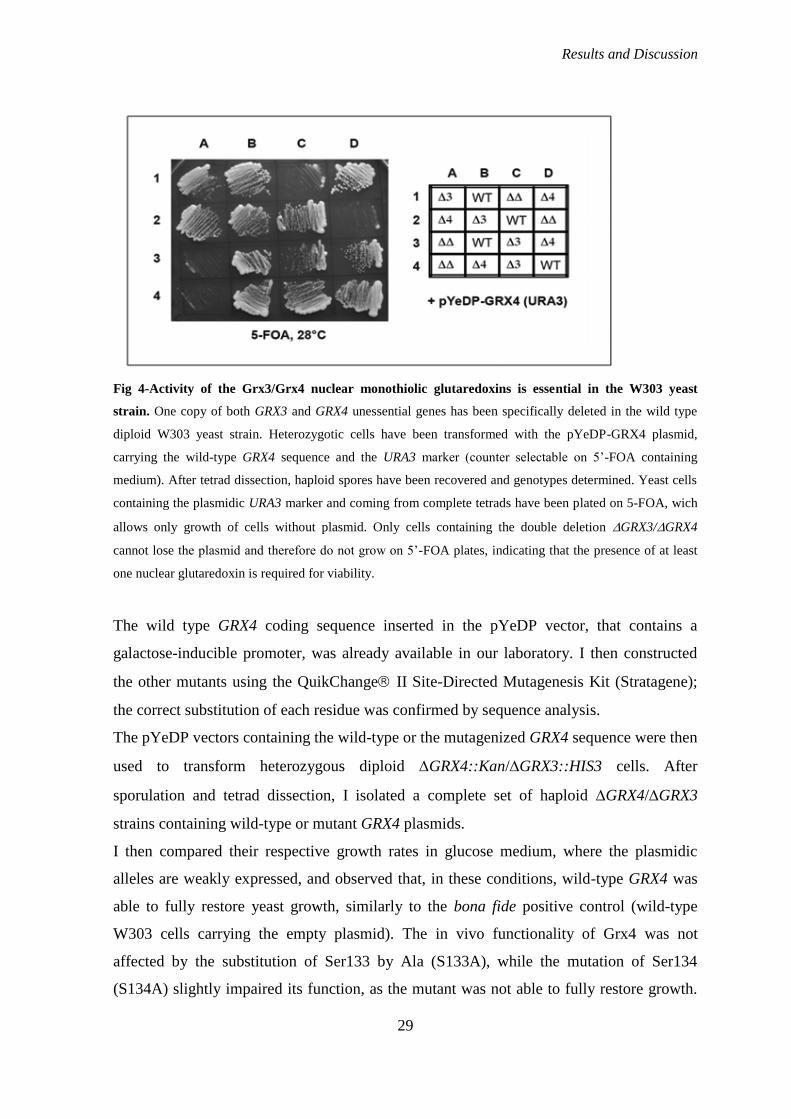
S. cerevisiae, however, possesses another nuclear monothiolic glutaredoxin, Grx3, which is
very similar to Grx4; the two proteins cooperate and show interchangeable roles, e.g. in the
transcriptional regulation of iron-dependent genes 44, 45
. Therefore, to specifically
investigate in vivo the effect of mutating Grx4, we created the double null strain
GRX3⁄GRX4 (for more details see Material and methods). Surprisingly, we noticed that,
unlike what was observed with other commonly used yeast strains (such as BY4742 and
CML128), cells containing the double mutation are nonviable in the W303 genetic
background (Fig. 4), indicating that in the W303 strain the functions of nuclear
monothiolic glutaredoxins are essential. This may reflect the subtle differences existing
between yeast laboratory strains, in particular with regard to the responses to
environmental changes or stresses involving these oxidoreductases.
Results and Discussion
29
Fig 4-Activity of the Grx3/Grx4 nuclear monothiolic glutaredoxins is essential in the W303 yeast
strain. One copy of both GRX3 and GRX4 unessential genes has been specifically deleted in the wild type
diploid W303 yeast strain. Heterozygotic cells have been transformed with the pYeDP-GRX4 plasmid,
carrying the wild-type GRX4 sequence and the URA3 marker (counter selectable on 5‟-FOA containing
medium). After tetrad dissection, haploid spores have been recovered and genotypes determined. Yeast cells
containing the plasmidic URA3 marker and coming from complete tetrads have been plated on 5-FOA, wich
allows only growth of cells without plasmid. Only cells containing the double deletion GRX3/GRX4
cannot lose the plasmid and therefore do not grow on 5‟-FOA plates, indicating that the presence of at least
one nuclear glutaredoxin is required for viability.
The wild type GRX4 coding sequence inserted in the pYeDP vector, that contains a
galactose-inducible promoter, was already available in our laboratory. I then constructed
the other mutants using the QuikChange II Site-Directed Mutagenesis Kit (Stratagene);
the correct substitution of each residue was confirmed by sequence analysis.
The pYeDP vectors containing the wild-type or the mutagenized GRX4 sequence were then
used to transform heterozygous diploid GRX4::Kan/GRX3::HIS3 cells. After
sporulation and tetrad dissection, I isolated a complete set of haploid GRX4/GRX3
strains containing wild-type or mutant GRX4 plasmids.
I then compared their respective growth rates in glucose medium, where the plasmidic
alleles are weakly expressed, and observed that, in these conditions, wild-type GRX4 was
able to fully restore yeast growth, similarly to the bona fide positive control (wild-type
W303 cells carrying the empty plasmid). The in vivo functionality of Grx4 was not
affected by the substitution of Ser133 by Ala (S133A), while the mutation of Ser134
(S134A) slightly impaired its function, as the mutant was not able to fully restore growth.
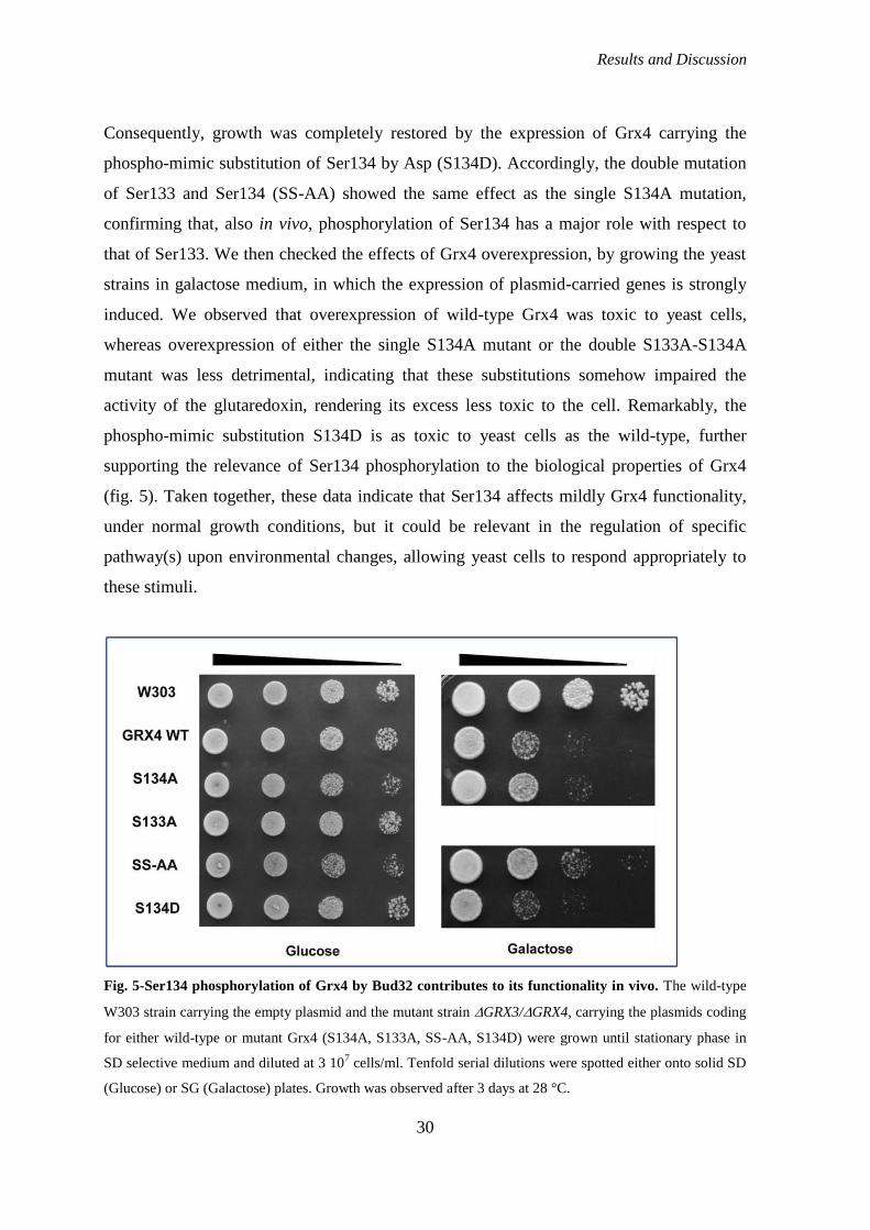
Results and Discussion
30
Consequently, growth was completely restored by the expression of Grx4 carrying the
phospho-mimic substitution of Ser134 by Asp (S134D). Accordingly, the double mutation
of Ser133 and Ser134 (SS-AA) showed the same effect as the single S134A mutation,
confirming that, also in vivo, phosphorylation of Ser134 has a major role with respect to
that of Ser133. We then checked the effects of Grx4 overexpression, by growing the yeast
strains in galactose medium, in which the expression of plasmid-carried genes is strongly
induced. We observed that overexpression of wild-type Grx4 was toxic to yeast cells,
whereas overexpression of either the single S134A mutant or the double S133A-S134A
mutant was less detrimental, indicating that these substitutions somehow impaired the
activity of the glutaredoxin, rendering its excess less toxic to the cell. Remarkably, the
phospho-mimic substitution S134D is as toxic to yeast cells as the wild-type, further
supporting the relevance of Ser134 phosphorylation to the biological properties of Grx4
(fig. 5). Taken together, these data indicate that Ser134 affects mildly Grx4 functionality,
under normal growth conditions, but it could be relevant in the regulation of specific
pathway(s) upon environmental changes, allowing yeast cells to respond appropriately to
these stimuli.
Fig. 5-Ser134 phosphorylation of Grx4 by Bud32 contributes to its functionality in vivo. The wild-type
W303 strain carrying the empty plasmid and the mutant strain GRX3/GRX4, carrying the plasmids coding
for either wild-type or mutant Grx4 (S134A, S133A, SS-AA, S134D) were grown until stationary phase in
SD selective medium and diluted at 3 107 cells/ml. Tenfold serial dilutions were spotted either onto solid SD
(Glucose) or SG (Galactose) plates. Growth was observed after 3 days at 28 °C.

Results and Discussion
31
Finally, to evaluate the specific contribution of Bud32 to Grx4 phosphorylation at Ser134
and Ser133, I constructed a mutant version of recombinant, His-tagged Grx4 (S133A-
S134A), and, after expression in E.coli, used it as substrate for an in vitro phosphorylation
reaction by native Bud32 immunoprecitated from yeast cells. Despite several purification
attempts, the recovery of wild-type and mutant Grx4 was low; however, as shown in fig. 6,
the results demonstrated that Bud32 was able to phosphorylate the low amount of wild-
type Grx4 present in the reaction (left, upper panel), whereas the mutant Grx4 was not
phosphorylated at all (right, upper panel), despite the presence of a higher amount of
recombinant protein, as revealed by the western blot (lower panels).
Fig. 6-Total protein lysates (500 g) of yeast cells expressing wild-type, HA-tagged, Bud32 (Bud32–HA)
were used to immunoprecipitate Bud32. Immune complexes were subjected to an in vitro phosphorylation
reaction in the presence of [-33
P]ATP and 25 ng of recombinant wild-type Grx4p (WT) or 50 ng of the
Grx4p double mutant S133A/S134A (SS-AA). After SDS/PAGE and blotting, filters were autoradiographed
(upper panels), and then visualized with antibodies against the HA-tag (for Bud32) or the HIS-tag (for Grx4)
(lower panels). The marked radiolabeled bands (*) are produced by an unidentified contaminant of the
recombinant Grx4p proteins. The strong signals in western blots (**) correspond to the IgG light chains
released by the resin used for Bud32–HA precipitation. As a negative control, the immunoprecipitation with
anti-HA antibodies was performed also from a wild-type strain (lanes NoTag).
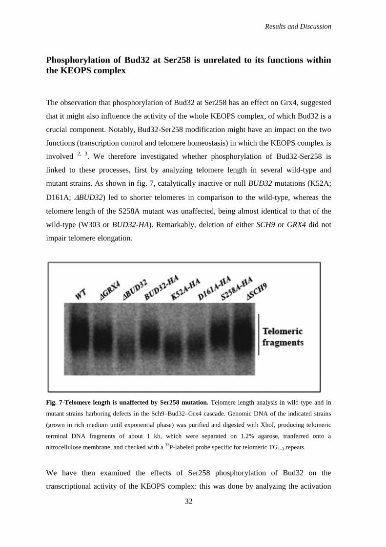
Results and Discussion
32
Phosphorylation of Bud32 at Ser258 is unrelated to its functions within
the KEOPS complex
The observation that phosphorylation of Bud32 at Ser258 has an effect on Grx4, suggested
that it might also influence the activity of the whole KEOPS complex, of which Bud32 is a
crucial component. Notably, Bud32-Ser258 modification might have an impact on the two
functions (transcription control and telomere homeostasis) in which the KEOPS complex is
involved 2, 3
. We therefore investigated whether phosphorylation of Bud32-Ser258 is
linked to these processes, first by analyzing telomere length in several wild-type and
mutant strains. As shown in fig. 7, catalytically inactive or null BUD32 mutations (K52A;
D161A; BUD32) led to shorter telomeres in comparison to the wild-type, whereas the
telomere length of the S258A mutant was unaffected, being almost identical to that of the
wild-type (W303 or BUD32-HA). Remarkably, deletion of either SCH9 or GRX4 did not
impair telomere elongation.
Fig. 7-Telomere length is unaffected by Ser258 mutation. Telomere length analysis in wild-type and in
mutant strains harboring defects in the Sch9–Bud32–Grx4 cascade. Genomic DNA of the indicated strains
(grown in rich medium until exponential phase) was purified and digested with XhoI, producing telomeric
terminal DNA fragments of about 1 kb, which were separated on 1.2% agarose, tranferred onto a
nitrocellulose membrane, and checked with a 33
P-labeled probe specific for telomeric TG1–3 repeats.
We have then examined the effects of Ser258 phosphorylation of Bud32 on the
transcriptional activity of the KEOPS complex: this was done by analyzing the activation
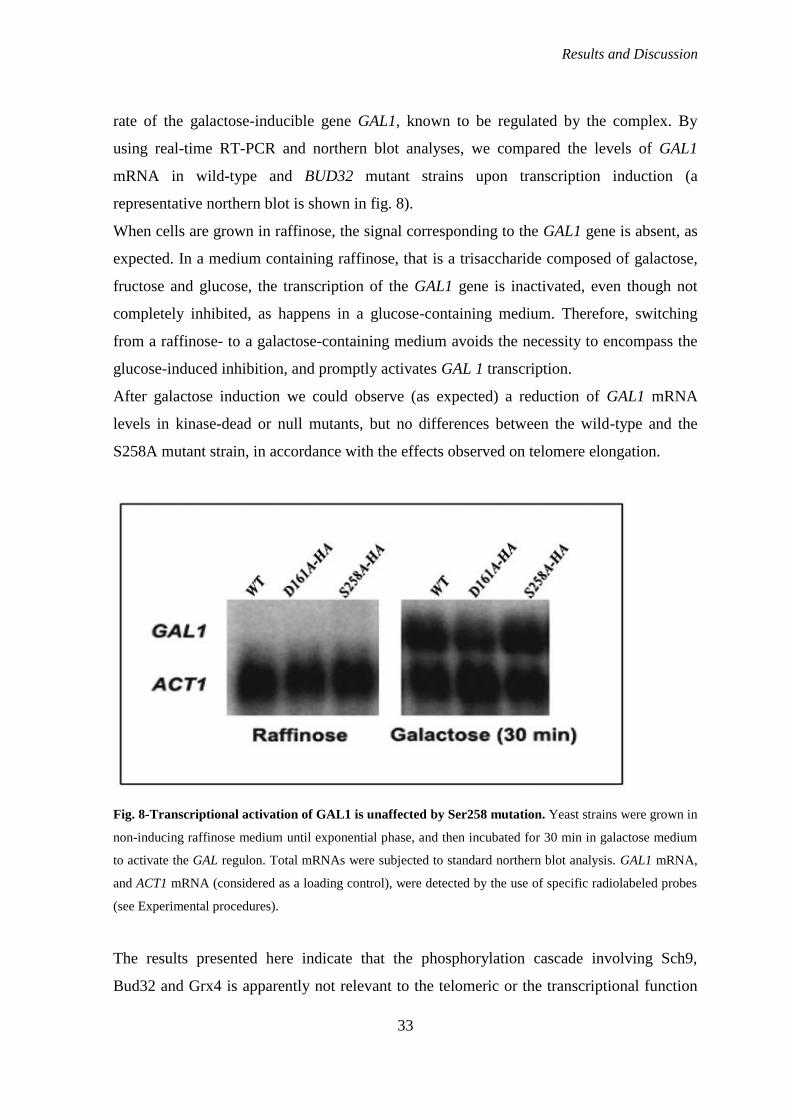
Results and Discussion
33
rate of the galactose-inducible gene GAL1, known to be regulated by the complex. By
using real-time RT-PCR and northern blot analyses, we compared the levels of GAL1
mRNA in wild-type and BUD32 mutant strains upon transcription induction (a
representative northern blot is shown in fig. 8).
When cells are grown in raffinose, the signal corresponding to the GAL1 gene is absent, as
expected. In a medium containing raffinose, that is a trisaccharide composed of galactose,
fructose and glucose, the transcription of the GAL1 gene is inactivated, even though not
completely inhibited, as happens in a glucose-containing medium. Therefore, switching
from a raffinose- to a galactose-containing medium avoids the necessity to encompass the
glucose-induced inhibition, and promptly activates GAL 1 transcription.
After galactose induction we could observe (as expected) a reduction of GAL1 mRNA
levels in kinase-dead or null mutants, but no differences between the wild-type and the
S258A mutant strain, in accordance with the effects observed on telomere elongation.
Fig. 8-Transcriptional activation of GAL1 is unaffected by Ser258 mutation. Yeast strains were grown in
non-inducing raffinose medium until exponential phase, and then incubated for 30 min in galactose medium
to activate the GAL regulon. Total mRNAs were subjected to standard northern blot analysis. GAL1 mRNA,
and ACT1 mRNA (considered as a loading control), were detected by the use of specific radiolabeled probes
(see Experimental procedures).
The results presented here indicate that the phosphorylation cascade involving Sch9,
Bud32 and Grx4 is apparently not relevant to the telomeric or the transcriptional function
Results and Discussion
34
of the Bud32-containing KEOPS complex. Accordingly, Grx4 has never been isolated as a
component of the KEOPS complex, indicating that the phosphorylation of Grx4 by Bud32
is independent from the known activities of the complex and must therefore be involved in
different functions and possibly unrelated pathways. Furthermore, we noticed that growth
of BUD32 mutants lacking the highly conserved C-terminal tail was not affected, similarly
to what has been observed in the case of the single S258A substitution, indicating that
these mutations do not impair the main biological properties of the kinase. Moreover,
recent data from the 3D structure of an archeal Bud32 homolog (Mj1130p) 27
indicate that
the Bud32 C-terminal tail is located far from the catalytic site, suggesting that its alteration
should not be detrimental to the overall structure. Finally, our data are consistent with a
cellular role of Bud32 (via Grx4 phosphorylation) which would be unrelated to the
functions of Bud32 as a component of KEOPS, but could be involved in the response to
environmental stimuli or endogenous stresses. Whether Bud32 could achieve the two tasks
while being simultaneously associated with the KEOPS complex and the Grx4 substrate or,
alternatively, whether only a cellular fraction of Bud32, not included in KEOPS, could
associate with the glutaredoxin, will be a matter for future investigation.
Does the Sch9-Bud32-Grx4 phosphorylation cascade affect any function
of Grx4?
As briefly explained in the Introduction, Grx4, together with Grx3, participates in iron
homeostasis, through the control of the cellular localization of the Aft1 transcription factor.
We hypothesized that Bud32 might be indirectly involved in this pathway, by
phosphorylating the glutaredoxin. This kind of modification, in fact, might be able to
activate or inhibit the ability of Grx4 to mediate the cytoplasmic translocation of Aft1. If
this is the case, the activity of Bud32 would influence the transcription of the iron regulon.
To verify this hypothesis, I first looked at the cellular localization of Aft1 in strains in
which Bud32 was wild-type or mutagenized in the key residue D161A. I thus created two
strains, espressing Aft1 3HA-tagged, combined to wild-type or mutagenized Bud32
(AFT1HA
/BUDWT
, AFT1HA
/BUDD161A
) and then observed the signal at the fluorescence
microscope (see Materials and Methods for details). Fig. 9 shows that the cellular
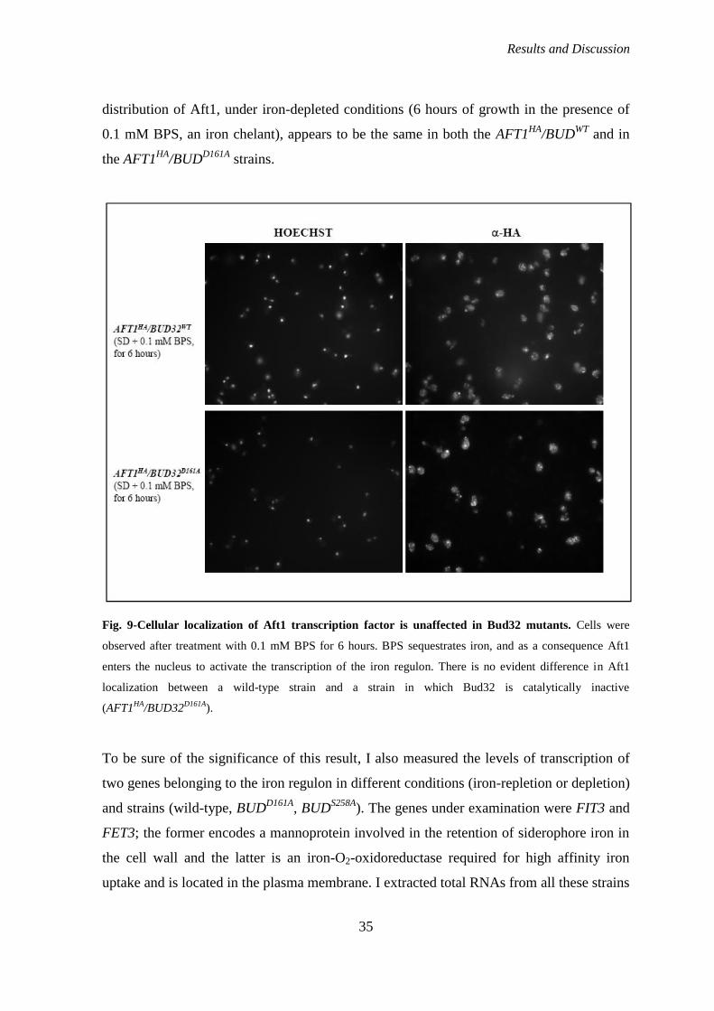
Results and Discussion
35
distribution of Aft1, under iron-depleted conditions (6 hours of growth in the presence of
0.1 mM BPS, an iron chelant), appears to be the same in both the AFT1HA
/BUDWT
and in
the AFT1HA
/BUDD161A
strains.
Fig. 9-Cellular localization of Aft1 transcription factor is unaffected in Bud32 mutants. Cells were
observed after treatment with 0.1 mM BPS for 6 hours. BPS sequestrates iron, and as a consequence Aft1
enters the nucleus to activate the transcription of the iron regulon. There is no evident difference in Aft1
localization between a wild-type strain and a strain in which Bud32 is catalytically inactive
(AFT1HA
/BUD32D161A
).
To be sure of the significance of this result, I also measured the levels of transcription of
two genes belonging to the iron regulon in different conditions (iron-repletion or depletion)
and strains (wild-type, BUDD161A
, BUDS258A
). The genes under examination were FIT3 and
FET3; the former encodes a mannoprotein involved in the retention of siderophore iron in
the cell wall and the latter is an iron-O2-oxidoreductase required for high affinity iron
uptake and is located in the plasma membrane. I extracted total RNAs from all these strains
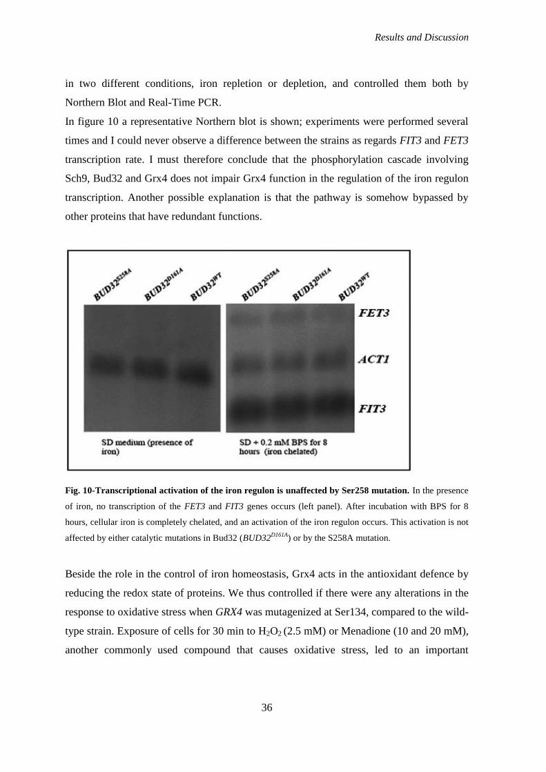
Results and Discussion
36
in two different conditions, iron repletion or depletion, and controlled them both by
Northern Blot and Real-Time PCR.
In figure 10 a representative Northern blot is shown; experiments were performed several
times and I could never observe a difference between the strains as regards FIT3 and FET3
transcription rate. I must therefore conclude that the phosphorylation cascade involving
Sch9, Bud32 and Grx4 does not impair Grx4 function in the regulation of the iron regulon
transcription. Another possible explanation is that the pathway is somehow bypassed by
other proteins that have redundant functions.
Fig. 10-Transcriptional activation of the iron regulon is unaffected by Ser258 mutation. In the presence
of iron, no transcription of the FET3 and FIT3 genes occurs (left panel). After incubation with BPS for 8
hours, cellular iron is completely chelated, and an activation of the iron regulon occurs. This activation is not
affected by either catalytic mutations in Bud32 (BUD32D161A
) or by the S258A mutation.
Beside the role in the control of iron homeostasis, Grx4 acts in the antioxidant defence by
reducing the redox state of proteins. We thus controlled if there were any alterations in the
response to oxidative stress when GRX4 was mutagenized at Ser134, compared to the wild-
type strain. Exposure of cells for 30 min to H2O2 (2.5 mM) or Menadione (10 and 20 mM),
another commonly used compound that causes oxidative stress, led to an important
Results and Discussion
37
decrease in cell viability, as expected. However, no statistically significant differences
existed between the wild-type and the mutants as regarded cell survival (data not shown).
The results here described do not enable us to define a role for the Sch9-Bud32-Grx4
phosphorylation cascade within yeast cells. The possibility that the phosphorylation at the
Ser134 residue of Grx4 alters the interaction between the glutaredoxin and its partners
(numerous, as judged by a two-hybrid screening) will be matter of further investigation.
ANALYSIS OF THE PHYSIOLOGICAL ROLE OF Kae1
Analysis of the phenotype of KAE1 mutants
The study of the Kae1 protein was forbidden by the unavailability of viable mutants. In
fact, the mutagenesis of one or both histidines of the conserved HXXXH motif (H-141 and
H-145), typical of metalloproteases, deprives the Kae1 protein of its functionality and cells
cannot survive (data not published from our laboratory; 3). In order to obtain a viable
mutant strain with a clear phenotype, we observed that in Kae1 and in its eukaryotic
homologues, the residue of Glutamic Acid in the second position of the motif is replaced
by a Cystein. We therefore decided to substitute the residue of Cystein with a Glutamic
Acid (C142E) and to mutagenize the Glutamic Acid present straight after the motif at
position +2 in an Alanine (E147A). We hypothesized that this residue (E147) could
function as the Glutamic Acid situated inside the HXXXH motif in the majority of
metalloproteases (C.Peggion, PhD thesis, 2006). As it can be seen in figure 11, at the
optimal temperature of 28 °C, both mutant strains show a slow growth phenotype, in
comparision to the wild-type strain. The most affected is the KAE1E147A
mutant; its
phenotype is exacerbated when cells are grown at 37 °C, indicating a thermosensitive
mutant. Instead, the KAE1C142E
mutant can be defined as crio-sensitive, as its phenotype is
more evident at the temperature of 22°C.
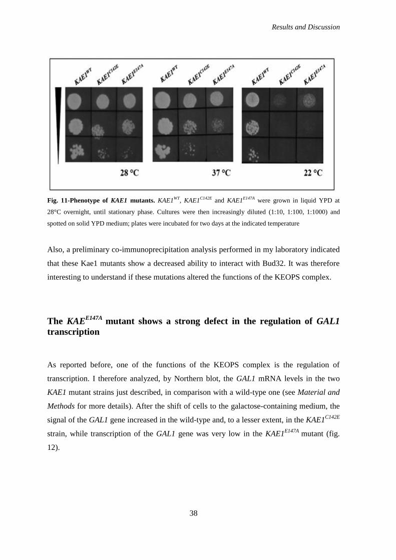
Results and Discussion
38
Fig. 11-Phenotype of KAE1 mutants. KAE1WT
, KAE1C142E
and KAE1E147A
were grown in liquid YPD at
28°C overnight, until stationary phase. Cultures were then increasingly diluted (1:10, 1:100, 1:1000) and
spotted on solid YPD medium; plates were incubated for two days at the indicated temperature
Also, a preliminary co-immunoprecipitation analysis performed in my laboratory indicated
that these Kae1 mutants show a decreased ability to interact with Bud32. It was therefore
interesting to understand if these mutations altered the functions of the KEOPS complex.
The KAEE147A
mutant shows a strong defect in the regulation of GAL1
transcription
As reported before, one of the functions of the KEOPS complex is the regulation of
transcription. I therefore analyzed, by Northern blot, the GAL1 mRNA levels in the two
KAE1 mutant strains just described, in comparison with a wild-type one (see Material and
Methods for more details). After the shift of cells to the galactose-containing medium, the
signal of the GAL1 gene increased in the wild-type and, to a lesser extent, in the KAE1C142E
strain, while transcription of the GAL1 gene was very low in the KAE1E147A
mutant (fig.
12).

Results and Discussion
39
Fig. 12-Northern blot showing the levels of GAL1 transcription in KAE1WT
, KAE1C142E
and KAE1E147A
strains. Cells were grown in raffinose-containing medium until OD600=0.4 and then were shifted in
galactose-containing medium for 30 min, to activate the transcription of the GAL1 gene. The transcription of
the ACT1 gene serves as an internal control to assure that the basal level of transcription is the same in all
strains.
This result suggests that the biochemical activity of the Kae1 protein is important for the
functions of the entire complex, as the substitution of one residue probably important for
the metallic-ion binding impairs the regulation of transcription.
The KAE1 mutations impair telomere homeostasis
After the analysis of transcription regulation, I observed (by Southern blot) telomere
lengths in KAE1C142E
and in KAE1E147A
mutant cells. The probe used corresponded to the
Y‟ region, that is present in almost half of yeast telomeres. As we can see in figure 13, the
wild type strain telomeres are longer than those of the two mutant strains. Moreover,
telomeres of KAE1C142E
are even shorter than telomeres of the BUD32 strain.
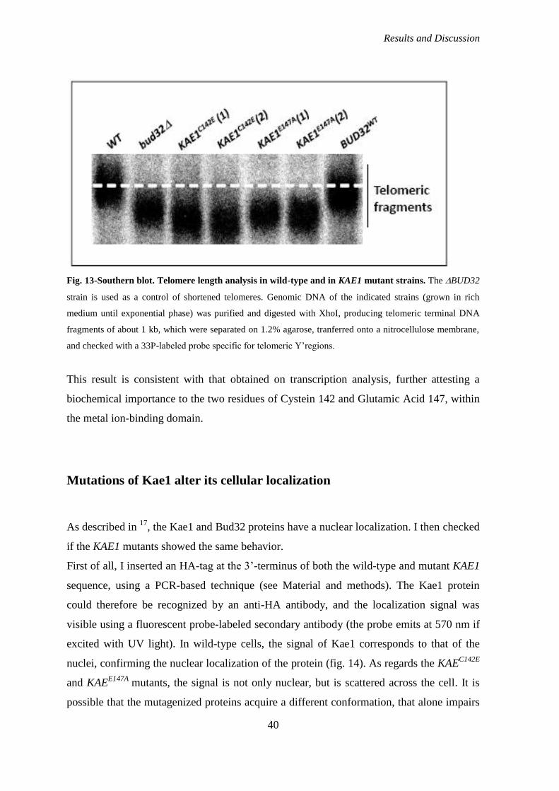
Results and Discussion
40
Fig. 13-Southern blot. Telomere length analysis in wild-type and in KAE1 mutant strains. The BUD32
strain is used as a control of shortened telomeres. Genomic DNA of the indicated strains (grown in rich
medium until exponential phase) was purified and digested with XhoI, producing telomeric terminal DNA
fragments of about 1 kb, which were separated on 1.2% agarose, tranferred onto a nitrocellulose membrane,
and checked with a 33P-labeled probe specific for telomeric Y‟regions.
This result is consistent with that obtained on transcription analysis, further attesting a
biochemical importance to the two residues of Cystein 142 and Glutamic Acid 147, within
the metal ion-binding domain.
Mutations of Kae1 alter its cellular localization
As described in 17
, the Kae1 and Bud32 proteins have a nuclear localization. I then checked
if the KAE1 mutants showed the same behavior.
First of all, I inserted an HA-tag at the 3‟-terminus of both the wild-type and mutant KAE1
sequence, using a PCR-based technique (see Material and methods). The Kae1 protein
could therefore be recognized by an anti-HA antibody, and the localization signal was
visible using a fluorescent probe-labeled secondary antibody (the probe emits at 570 nm if
excited with UV light). In wild-type cells, the signal of Kae1 corresponds to that of the
nuclei, confirming the nuclear localization of the protein (fig. 14). As regards the KAEC142E
and KAEE147A
mutants, the signal is not only nuclear, but is scattered across the cell. It is
possible that the mutagenized proteins acquire a different conformation, that alone impairs
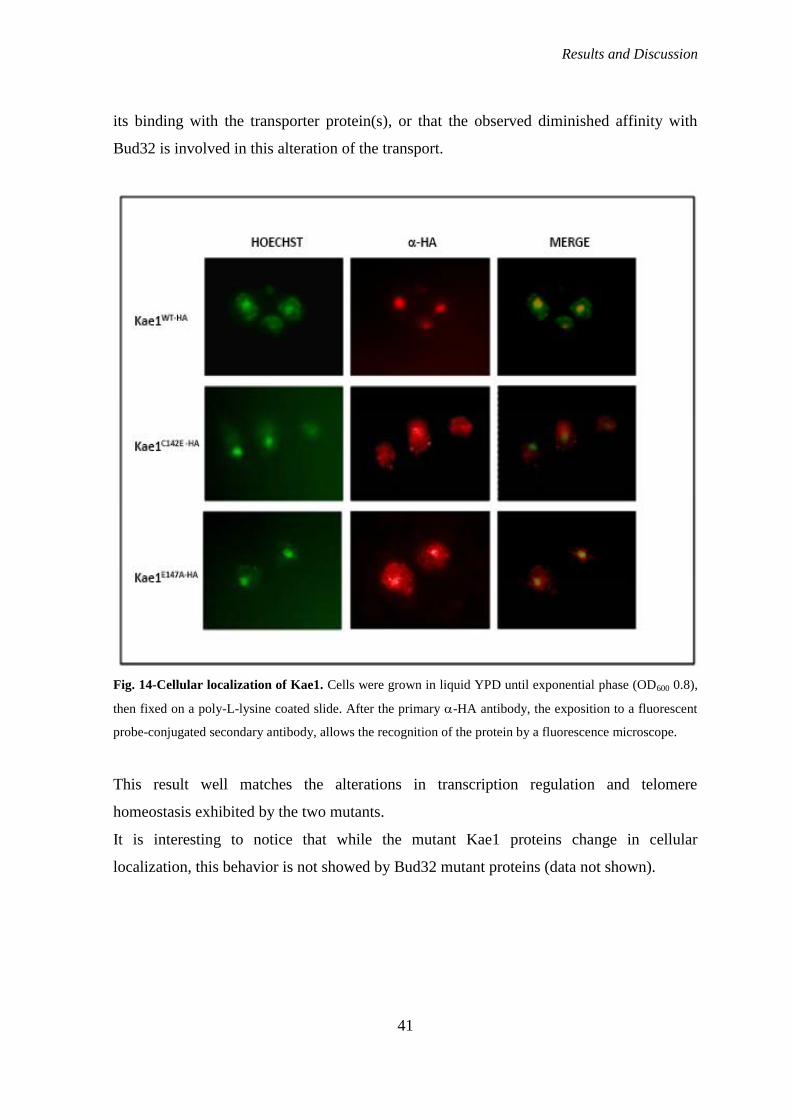
Results and Discussion
41
its binding with the transporter protein(s), or that the observed diminished affinity with
Bud32 is involved in this alteration of the transport.
Fig. 14-Cellular localization of Kae1. Cells were grown in liquid YPD until exponential phase (OD600 0.8),
then fixed on a poly-L-lysine coated slide. After the primary -HA antibody, the exposition to a fluorescent
probe-conjugated secondary antibody, allows the recognition of the protein by a fluorescence microscope.
This result well matches the alterations in transcription regulation and telomere
homeostasis exhibited by the two mutants.
It is interesting to notice that while the mutant Kae1 proteins change in cellular
localization, this behavior is not showed by Bud32 mutant proteins (data not shown).
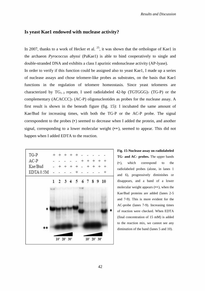
Results and Discussion
42
Is yeast Kae1 endowed with nuclease activity?
In 2007, thanks to a work of Hecker et al. 25
, it was shown that the orthologue of Kae1 in
the archaeon Pyrococcus abyssi (PaKae1) is able to bind cooperatively to single and
double-stranded DNA and exhibits a class I apurinic endonuclease activity (AP-lyase).
In order to verify if this function could be assigned also to yeast Kae1, I made up a series
of nuclease assays and chose telomere-like probes as substrates, on the basis that Kae1
functions in the regulation of telomere homeostasis. Since yeast telomeres are
characterized by TG1–3 repeats, I used radiolabeled 42-bp (TGTGGG)7 (TG-P) or the
complementary (ACACCC)7 (AC-P) oligonucleotides as probes for the nuclease assay. A
first result is shown in the beneath figure (fig. 15): I incubated the same amount of
Kae/Bud for increasing times, with both the TG-P or the AC-P probe. The signal
correspondent to the probes () seemed to decrease when I added the protein, and another
signal, corresponding to a lower molecular weight (), seemed to appear. This did not
happen when I added EDTA to the reaction.
Fig. 15-Nuclease assay on radiolabeled
TG- and AC- probes. The upper bands
(), which correspond to the
radiolabeled probes (alone, in lanes 1
and 6), progressively diminishes or
disappears, and a band of a lower
molecular weight appears (), when the
Kae/Bud proteins are added (lanes 2-5
and 7-9). This is more evident for the
AC-probe (lanes 7-9). Increasing times
of reaction were checked. When EDTA
(final concentration of 15 mM) is added
to the reaction mix, we cannot see any
diminution of the band (lanes 5 and 10).
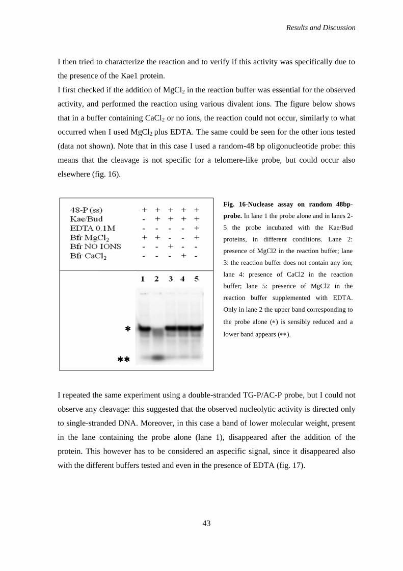
Results and Discussion
43
I then tried to characterize the reaction and to verify if this activity was specifically due to
the presence of the Kae1 protein.
I first checked if the addition of MgCl2 in the reaction buffer was essential for the observed
activity, and performed the reaction using various divalent ions. The figure below shows
that in a buffer containing CaCl2 or no ions, the reaction could not occur, similarly to what
occurred when I used MgCl2 plus EDTA. The same could be seen for the other ions tested
(data not shown). Note that in this case I used a random-48 bp oligonucleotide probe: this
means that the cleavage is not specific for a telomere-like probe, but could occur also
elsewhere (fig. 16).
I repeated the same experiment using a double-stranded TG-P/AC-P probe, but I could not
observe any cleavage: this suggested that the observed nucleolytic activity is directed only
to single-stranded DNA. Moreover, in this case a band of lower molecular weight, present
in the lane containing the probe alone (lane 1), disappeared after the addition of the
protein. This however has to be considered an aspecific signal, since it disappeared also
with the different buffers tested and even in the presence of EDTA (fig. 17).
Fig. 16-Nuclease assay on random 48bp-
probe. In lane 1 the probe alone and in lanes 2-
5 the probe incubated with the Kae/Bud
proteins, in different conditions. Lane 2:
presence of MgCl2 in the reaction buffer; lane
3: the reaction buffer does not contain any ion;
lane 4: presence of CaCl2 in the reaction
buffer; lane 5: presence of MgCl2 in the
reaction buffer supplemented with EDTA.
Only in lane 2 the upper band corresponding to
the probe alone () is sensibly reduced and a
lower band appears ().

Results and Discussion
44
To further evaluate the specificity of this biochemical reaction, I purified a mutagenized
version of the Kae/Bud coexpressed proteins, in which the histidines 141 and 145 of Kae1,
responsible for the coordination of the metal ion, and thus of the ATP moiety, were
substituted with Alanine residues (Kae1HH141-145AA
/Bud32). The integrity of those
histidines is essential in yeast cells and so I expected to observe no activity when these are
substituted with alanine.
In order to work in the same conditions for the wild-type and the mutagenized protein, I
purified again the Kae1WT
/Bud32, obtaining a cleaner enzyme as that used so far (compare
fig. 22, page 48-49 and fig. 18, below).
Fig. 17-Nuclease assay on a double stranded
probe, obtained by mixing the two single-
stranded (radiolabeled) TG- and AC-probes.
Even if in the presence of MgCl2 and CaCl2
(lanes 2 and 4) the probe seems partially
degraded, it is not possible to detect the presence
of the lower band as in previous experiments.

Results and Discussion
45
Fig. 18-New purification of coexpressed Kae/Bud. Panel A: the flow-through of the resin-containing
column. Panel B: four fractions of the first column-washing (Tris/HCl pH 7.5, 0.3 M NaCl, 10 mM
imidazole). Panel C: five fractions of the second column-washing, containing 20 mM imidazole. Panel D:
five fractions of the eluition of the protein from the column with a buffer containing 100 mM imidazole.
Below in figure 19, a nuclease test using both the wild-type and the mutated protein is
shown. The result is not very clear although the disappearance of the upper band seems to
be quantitatively more important in the presence of the wild-type protein, compared to the
mutated one. However, a lot of radioactive signal is retained in the wells of the gel,
especially when the Kae1WT
/Bud32 protein is used, and that signal may be part of the
cleaved or the uncleaved moiety.
As the result was not clear, I made other attempts, testing different reaction conditions.
Unfortunately, in the following experiments I was not able to see any cleavage, as it can be
highlighted in the two figures below (and in many other experiments not shown).
In figure 20, the band corresponding to the probe disappears in the presence of both the
wild-type and the mutated proteins. However it cannot be explained why a part of signal
seems to disappear after the addition of the protein; in fact, we cannot see a corresponding
Fig. 19-Nuclease assay on the AC-
probe, in the presence of the wild-
type (Kae1WT/Bud32) or the
mutagenized (Kae1HHAA/Bud32)
protein. After addition of the wt
protein (lanes 2-4), the upper band,
corresponding to the protein alone,
nearly disappears, even if a lot of
radioactivity is retained in the gel
wells, thus making the result difficult
to interpret. When the mutated protein
is added (lanes 6-8), the cleavage
seems to be less efficient. In lanes 5
and 9 EDTA is added to the raction.

Results and Discussion
46
increasing of the lower band (but a lot of radioactivity still remains in the gel wells).
Unexpectedly, when Bud32 alone was used (lane 10), the upper signal corresponding to
the probe is nearly completely vanished. As this could not be observed in the subsequent
experiments (see for example fig. 21), we conclude that in our hands these experiments are
not enough reproducible and that probably also the results previously obtained were not
specifically due to an activity of the Kae1 protein alone. What I had observed might be due
to the presence of some contaminants in the protein eluate.
As a last experiment (fig.21), I compared the activity of the previous, less clean,
purification of Kae/Bud (Kae1WT
/Bud OLD, see figure 15) with those of the newly purified
wild-type and mutated proteins (see fig. 18).
Fig. 20-Nuclease assay on the AC-
probe, in the presence of the wild-
type (Kae1WT/Bud32) or the
mutagenized (Kae1HHAA/Bud32)
protein. Unfortunately, both the wt
and the mutated protein seem to
cleave the probe (lanes 2-3 and 6-7),
while the reaction is abolished by
EDTA (lanes 5 and 9) or the absence
of divalent ions (lanes 4 and 8).
Surprisingly, also Bud32 alone seem
to be able to completely cleave the
probe (lane 10). Notice that no
differences occur if ATP is added to
the reaction (lanes 2-3 and 6-7).
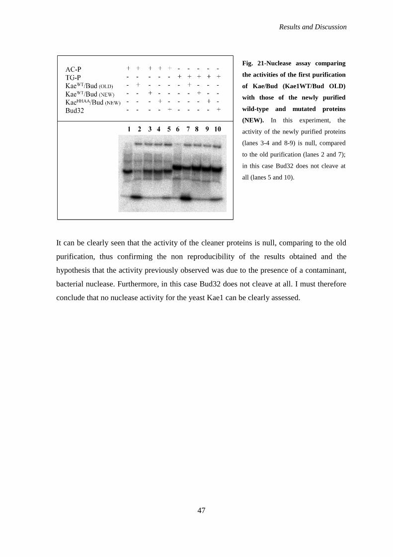
Results and Discussion
47
It can be clearly seen that the activity of the cleaner proteins is null, comparing to the old
purification, thus confirming the non reproducibility of the results obtained and the
hypothesis that the activity previously observed was due to the presence of a contaminant,
bacterial nuclease. Furthermore, in this case Bud32 does not cleave at all. I must therefore
conclude that no nuclease activity for the yeast Kae1 can be clearly assessed.
Fig. 21-Nuclease assay comparing
the activities of the first purification
of Kae/Bud (Kae1WT/Bud OLD)
with those of the newly purified
wild-type and mutated proteins
(NEW). In this experiment, the
activity of the newly purified proteins
(lanes 3-4 and 8-9) is null, compared
to the old purification (lanes 2 and 7);
in this case Bud32 does not cleave at
all (lanes 5 and 10).
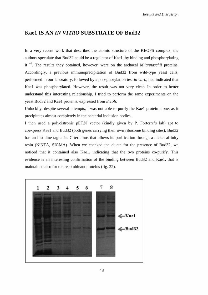
Results and Discussion
48
Kae1 IS AN IN VITRO SUBSTRATE OF Bud32
In a very recent work that describes the atomic structure of the KEOPS complex, the
authors speculate that Bud32 could be a regulator of Kae1, by binding and phosphorylating
it 40
. The results they obtained, however, were on the archaeal M.jannaschii proteins.
Accordingly, a previous immunoprecipitation of Bud32 from wild-type yeast cells,
performed in our laboratory, followed by a phosphorylation test in vitro, had indicated that
Kae1 was phosphorylated. However, the result was not very clear. In order to better
understand this interesting relationship, I tried to perform the same experiments on the
yeast Bud32 and Kae1 proteins, expressed from E.coli.
Unluckily, despite several attempts, I was not able to purify the Kae1 protein alone, as it
precipitates almost completely in the bacterial inclusion bodies.
I then used a polycistronic pET28 vector (kindly given by P. Forterre‟s lab) apt to
coexpress Kae1 and Bud32 (both genes carrying their own ribosome binding sites). Bud32
has an histidine tag at its C-terminus that allows its purification through a nickel affinity
resin (NiNTA, SIGMA). When we checked the eluate for the presence of Bud32, we
noticed that it contained also Kae1, indicating that the two proteins co-purify. This
evidence is an interesting confirmation of the binding between Bud32 and Kae1, that is
maintained also for the recombinant proteins (fig. 22).
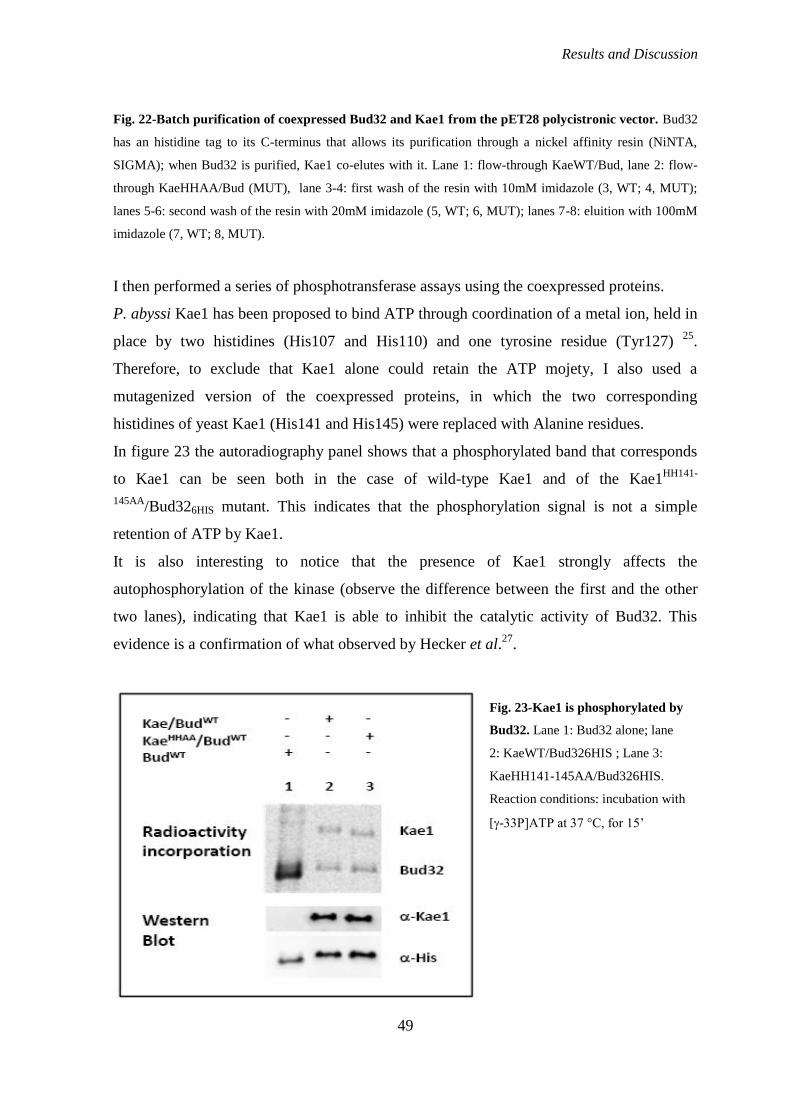
Results and Discussion
49
Fig. 22-Batch purification of coexpressed Bud32 and Kae1 from the pET28 polycistronic vector. Bud32
has an histidine tag to its C-terminus that allows its purification through a nickel affinity resin (NiNTA,
SIGMA); when Bud32 is purified, Kae1 co-elutes with it. Lane 1: flow-through KaeWT/Bud, lane 2: flow-
through KaeHHAA/Bud (MUT), lane 3-4: first wash of the resin with 10mM imidazole (3, WT; 4, MUT);
lanes 5-6: second wash of the resin with 20mM imidazole (5, WT; 6, MUT); lanes 7-8: eluition with 100mM
imidazole (7, WT; 8, MUT).
I then performed a series of phosphotransferase assays using the coexpressed proteins.
P. abyssi Kae1 has been proposed to bind ATP through coordination of a metal ion, held in
place by two histidines (His107 and His110) and one tyrosine residue (Tyr127) 25
.
Therefore, to exclude that Kae1 alone could retain the ATP mojety, I also used a
mutagenized version of the coexpressed proteins, in which the two corresponding
histidines of yeast Kae1 (His141 and His145) were replaced with Alanine residues.
In figure 23 the autoradiography panel shows that a phosphorylated band that corresponds
to Kae1 can be seen both in the case of wild-type Kae1 and of the Kae1HH141-
145AA/Bud326HIS mutant. This indicates that the phosphorylation signal is not a simple
retention of ATP by Kae1.
It is also interesting to notice that the presence of Kae1 strongly affects the
autophosphorylation of the kinase (observe the difference between the first and the other
two lanes), indicating that Kae1 is able to inhibit the catalytic activity of Bud32. This
evidence is a confirmation of what observed by Hecker et al.27
.
Fig. 23-Kae1 is phosphorylated by
Bud32. Lane 1: Bud32 alone; lane
2: KaeWT/Bud326HIS ; Lane 3:
KaeHH141-145AA/Bud326HIS.
Reaction conditions: incubation with
[-33P]ATP at 37 °C, for 15‟
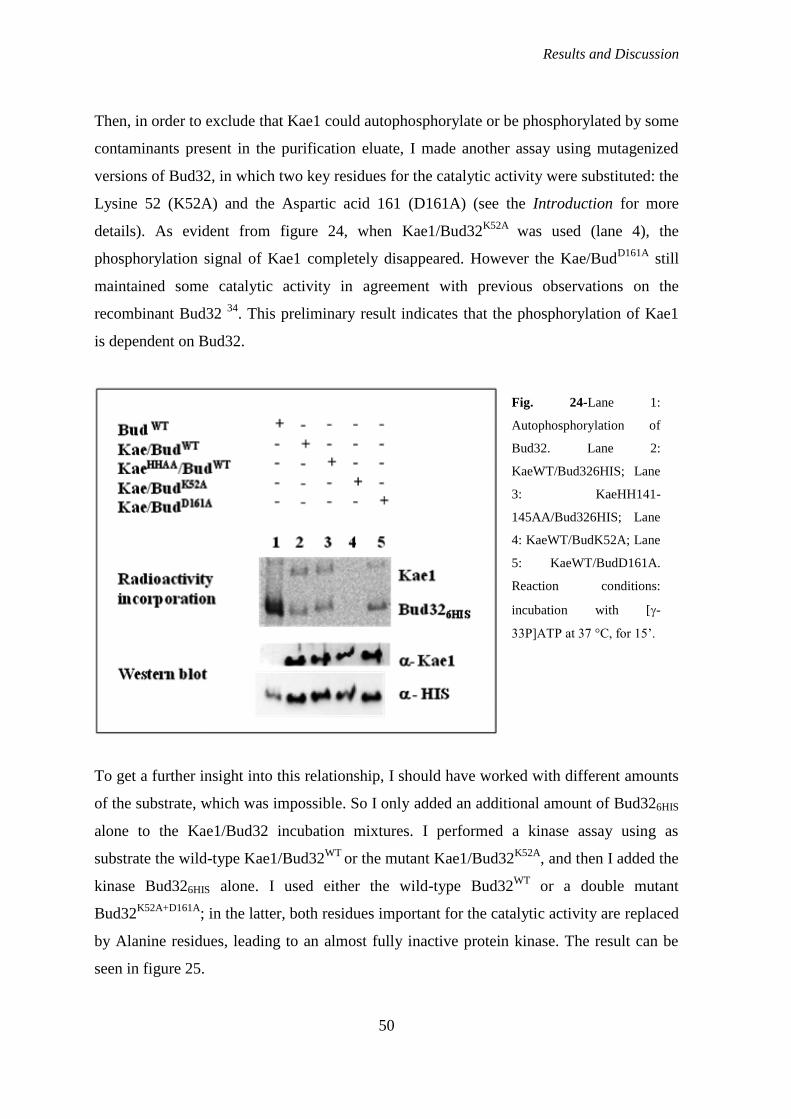
Results and Discussion
50
Then, in order to exclude that Kae1 could autophosphorylate or be phosphorylated by some
contaminants present in the purification eluate, I made another assay using mutagenized
versions of Bud32, in which two key residues for the catalytic activity were substituted: the
Lysine 52 (K52A) and the Aspartic acid 161 (D161A) (see the Introduction for more
details). As evident from figure 24, when Kae1/Bud32K52A
was used (lane 4), the
phosphorylation signal of Kae1 completely disappeared. However the Kae/BudD161A
still
maintained some catalytic activity in agreement with previous observations on the
recombinant Bud32 34
. This preliminary result indicates that the phosphorylation of Kae1
is dependent on Bud32.
To get a further insight into this relationship, I should have worked with different amounts
of the substrate, which was impossible. So I only added an additional amount of Bud326HIS
alone to the Kae1/Bud32 incubation mixtures. I performed a kinase assay using as
substrate the wild-type Kae1/Bud32WT
or the mutant Kae1/Bud32K52A
, and then I added the
kinase Bud326HIS alone. I used either the wild-type Bud32WT
or a double mutant
Bud32K52A+D161A
; in the latter, both residues important for the catalytic activity are replaced
by Alanine residues, leading to an almost fully inactive protein kinase. The result can be
seen in figure 25.
Fig. 24-Lane 1:
Autophosphorylation of
Bud32. Lane 2:
KaeWT/Bud326HIS; Lane
3: KaeHH141-
145AA/Bud326HIS; Lane
4: KaeWT/BudK52A; Lane
5: KaeWT/BudD161A.
Reaction conditions:
incubation with [-
33P]ATP at 37 °C, for 15‟.

Results and Discussion
51
The phosphorylation signal on Kae1 was immediately evident when the wild-type
Kae1/Bud32 coexpressed proteins were used and did not increase when wild-type Bud32
was added; only an increase of the autophosphorylation of Bud32 can be noticed (lane 2)
and this further confirms that Kae1 exerts a quantitative inhibition on Bud32 activity.
When the double mutant Bud32K52A+D161A
was added (lane 3), both signals
(phosphorylation of Kae1 and autophosphorylation of Bud32) seem to slightly diminish,
but this result is not sufficient to give rise to speculations.
More interesting, when I used the mutant Kae1/Bud32K52A
proteins, the basal signal of
Kae1 phosphorylation was not visible in autoradiography (lane 4) and so we could
appreciate the appearance of a phosphorylation band when the wild-type Bud32 was added
(lane 5). Instead, when the double mutant Bud32K52A+D161A
protein was added, no signal of
phosphorylation could be detected (lane 6). This result is a further confirmation of the
relevance of Bud32 activity for Kae1 phosphorylation.
As mentioned before, I could not obtain a soluble purified Kae1 protein. I then tried to
purify Kae1 in denaturing conditions (with 8M urea) and to carry out a refolding of the
protein directly on the Ni-NTA resin; immobilization of the protein on the matrix, in fact,
should theoretically circumvent protein-protein aggregation due to the spatial separation of
the bound proteins. However the attempt failed because all the protein was retained by the
resin. I then used this resin-immobilized protein as a substrate for another
Fig. 25. Incubation of the
wild-type or mutagenized
Kae/Bud32 proteins with
either the wt or the
catalytically inactive double
mutant Bud32. Lane 1:
Kae1/Bud32WT
alone, the
same plus Bud32WT
(lane 2)
or Bud32K+D
(lane 3). Lane
4: Kae1/Bud32K52A
alone, the
same plus Bud32WT
(lane 5)
or Bud32K+D
(lane 6).

Results and Discussion
52
phosphotransferase assay. Increasing amounts of wild-type Bud32 or double mutant
Bud32K52A+D161A
where added to the same amount of Kae1 bound to the resin. The result is
shown in figure 26: a not well defined, although visible, band is present when a sufficient
amount of the wild-type Bud32 is added to the resin (lanes 1-5). The signal raised with the
quantity of protein kinase until saturation. Conversely, this could not be seen when the
mutagenized Bud32K52A+D161A
was added (lanes 6-9).
Altogether these results indicate that, at least in vitro, a regulatory relationship exists
between Bud32 and Kae1; in fact, Bud32 phosphorylates Kae1, which conversely inhibits
the catalytic activity of the kinase. This is very interesting as the two proteins are liable to
carefully modulate the functions of the entire KEOPS complex.
MS analysis of phosphorylated Kae1
I then tried to identify the specific residue(s) of Kae1 phosphorylated by Bud32. First of all
I measured the level of phosphorylated Kae1, in an in vitro assay on Kae1/Bu32WT
, and
found that 2.6 % of the total protein is phosphorylated. Thanks to Dott. G. Arrigoni
Fig. 26-Phosphorylation
assay on the resin-
immobilized Kae1. Lane
1: resin-bound Kae1
alone. Lane 2-5:
increasing amounts of
Bud32WT
are added to the
resin suspension(100,
200, 300, 400 ng). Lane
6-9: increasing amounts
of Bud32K+D
are added to
the resin suspension(100,
200, 300, 400 ng).

Results and Discussion
53
(University of Padova), we made a MS analysis on recombinant Kae1, after incubation
with additional Bud32 for 30 minutes, in the presence of 62.5 M ATP (see Materials and
Methods for more details). The analysis revealed that the Kae1 peptide DFS*ETVVTQK is
phosphorylated at the Serine indicated; other three peptides are phosphorylated, but they
have not yet been sequenced: IPNEPSPGYNIEQLAK, DYYIALGLEGSANK,
AQNPVVLYVSGGNTQVIAYSEK (fig. 27)
Fig. 27-Aminoacid sequence of the Kae1 protein, with the sequenced peptide and the site of
phosphorylation, and the three other peptides that are supposed to be phosphorylated.
These new data encourage to perform further experiments in order to confirm the
biochemical relationship between Bud32 and Kae1. A first step will be the mutagenesis of
Ser367 of recombinant Kae1 protein to an Alanine residue. If this is the main residue
phosphorylated by Bud32, the mutant Kae1 will show a decreased, or even a null signal, in
an in vitro kinase assay. However, also the three other peptides may be substrates of
Bud32. If this was confirmed, we will mutagenize other residues, to locate the main targets
of the kinase.
Results and Discussion
54
The other important step will be the identification of a physiological role of this
phosphorylation of Kae1 by Bud32, through the analysis of the phenotype of Kae1
mutants, including the analysis of transcription regulation and telomere homeostasis, in
order to know if this phosphorylation is involved in the functions of the KEOPS complex.
It is interesting to notice that, in the structure described by Mao et al., 40
, the Ser367
residue lies within insert 2, a very conserved sequence, specific of the Kae1 family, that is
exposed in the protein structure and hence accessible to the active site of Bud32. In this
work, Mao and colleagues write that “partial (373-end) or full deletion (365-end) of the C-
terminal tail of yeast Kae1 caused a severe growth defect”. In particular, the authors
identified a residue within this region, T369, which leads to the same growth defect if
mutagenized to Alanine. They thus assumed that T369 might be the phosphoregulatory
site. However, our identification of Ser367, that is only two residues from Thr369 allows
the hypothesis that the T369A phenotype may be due to the close proximity of S367, either
because the T369A substitution alters the conformation of the local sequence, and thus the
accessibility of Bud32, or because the previous phosphorylation of T369 is essential to
S367 phosphorylation.
Conclusions
55
Conclusions
ROLE OF Bud32 IN A NEW SIGNALING PATHWAY IN
YEAST
In the first part of my PhD thesis, I have described my contributions to the unveiling of a
novel signaling pathway in S.cerevisiae, that implicates Sch9, the yeast homolog of
mammalian Akt, Bud32 and the glutaredoxin Grx4. We showed that Grx4, previously
described as an in vitro substrate of Bud32 17
, is also a physiological target of the kinase,
that phosphorylates it in the Ser 133 and, mainly, Ser134 residues. This phosphorylation
contributes to the functionality of the glutaredoxin in yeast cells; in fact the growth of
mutant cells in which Grx4 is unphosphorylatable by Bud32 is affected, even if in a mild
way.
Moreover, this relationship is influenced by the phosphorylation status of Bud32. Bud32 is
phosphorylated at the Ser258 residue by the Sch9 kinase, and this phosphorylation
upregulates the ability of Bud32 to bind and phosphorylate Grx4 46
.
When searching for a physiological role for this new phosphorylation cascade, however,
we noticed that phosphorylation of Bud32 at Ser258 is unrelated to the role of KEOPS
complex in telomere homeostasis and transcription regulation, suggesting that Bud32
partecipates in multiple pathways in yeast cells.
Also, the cascade does not seem to affect some known functions of Grx4, like iron
homeostasis and oxidative stress protection. We may then hypothesize that it plays a role
in different and still uncharacterized functions of Grx4.
It is important to highlight that this regulatory system is evolutionarily conserved. In fact, it
has been demonstrated that in human cells Akt (Sch9 homolog) phosphorylates PRPK
(Bud32 homolog) at the Ser250 residue, and that this phosphorylation positively regulates
in vivo the activity of PRPK on its physiological substrate, p53 48
.

Conclusions
56
ANALYSIS OF Kae1 ACTIVITY
A part of my PhD study was devoted to the subunit of the KEOPS complex named Kae1.
As described in the introduction, Kae1 was previously classified as a protease, and indeed
it contains a HXXXH motif, highly conserved in metalloproteases. The motif is essential in
Kae1, as the mutagenesis of one or both histidines (H-141 and H-145) deprives the protein
of its functionality and is lethal to the cell. Interestingly, in Kae1 and in all its eukaryotic
homologues, the residue of Glutamic Acid in the second position (HEXXH), normally
present in metallo proteases, is replaced with a Cystein, while a Glutamic Acid is present
after the second histidine, in position + 2 (HCGIHIE). These characteristics suggest that
Kae1 may act with a different catalytic mechanism compared with the other metallo-
proteases. In order to understand the role of Kae1, I used two mutants already available in
my laboratory, in which these two residues were substituted (KAEC142E
and KAEE147A
).
Both mutants, in fact, showed a slow growth phenotype, but were still viable. I first asked
if those mutations could interfere with the functions of KEOPS. Indeed, I observed that
both transcription regulation and telomere homeostasis were affected in these mutant cells.
I thus concluded that the biochemical activity of the Kae1 protein must be important for
the functions of the entire complex, as the mutation in two residues within the metal ion
binding domain impairs both telomere homeostasis and transcription regulation
Then I verified if the mutations of Kae1 could alter the cellular localization of the protein:
in fact I could observe that, in contrast with the wild-type, the mutant proteins are not
exclusively nuclear, but are scattered across the whole cell. This result well matches with
the alterations in the KEOPS functions exhibited by the two mutants, as both telomere
homeostasis and transcription regulation functions are performed within the nucleus. As
the two mutations are not part of a localization sequence, we may hypothesize that they
could somehow interfere with the binding to other proteins, like cellular transporters. It is
also interesting to notice that the catalytic mutants of Bud32 do not show alterations in
Bud32 cell localization with respect to the wild-type, i.e. the protein is always nuclear.
In order to attribute a biochemical activity to Kae1, I wanted to verify if the yeast Kae1
protein could be a nuclease. In fact, in a work of 2007 it was shown that the orthologue of
Kae1 in the archaeon Pyrococcus abyssi (PaKae1;) is able to bind cooperatively to single
and double-stranded DNA and exhibits a class I apurinic endonuclease activity (AP-lyase)
Conclusions
57
25. Moreover, a nuclease function is hypothesized to be essential for telomerase, as the
latter needs 3‟ssDNA overhangs to add new telomeres at the level of double-strand breaks
(DSBs). The identification of a nuclease activity for Kae1 would then be very important to
clarify the molecular mechanism by which KEOPS exerts its function in telomere
maintenance. Hovewer, from our experiments, no nuclease activity for the yeast Kae1 can
be clearly assessed. This is in agreement with what observed by Mao et al. in 2008, when
the authors assessed that the archaeal MjKae1 has an ATPase, but no AP-endonuclease or
DNA-binding activity 40
.
BIOCHEMICAL RELATIONSHIP BETWEEN Bud32 AND
Kae1
In the work describing the atomic structure of an archaeal-derived KEOPS complex 40
,
Mao and colleagues suggested that MjBud32 phosphorylates MjKae1, and that this
phosphorylation has a regulatory function. We then decided to verify if the same was true
for the yeast proteins. As, despite several attempts, I was not able to purify the Kae1
protein alone, due to its precipitation in the bacterial inclusion bodies, I used a
polycistronic vector, allowing the coexpression of yeast Kae1 and Bud32. I then performed
a series of kinase assays using the coexpressed proteins and demonstrated that, at least in
vitro, a regulatory relationship exists between Bud32 and Kae1. In fact, I showed that
Bud32 is able to phosphorylate Kae1 and that Kae1 inhibits the catalytic activity of the
kinase. These results confirm those obtained with the archaeal proteins, suggesting a
conserved mechanism within the complex. An ulterior important confirmation comes from
the MS analysis of phosphorylated Kae1, that identified Ser 367 as a target of Bud32.
However this might not be the only phosphorylated residue.
Therefore the relationship between Bud32 and Kae1 is liable to carefully modulate the
functions of the entire KEOPS complex.

Conclusions
58
Materials and Methods
59
Materials and methods
STRAINS
E. coli strains
STRAIN GENOTYPE
InvF‟ endA1, recA1, hsdR17(r-k
, m+k
) supE44, -, thi-1, gyrA, relA1,
φ80lacZM15 (lacZYA-argF),deoR+, F
BL21 F-, ompT, hsdS(r-m-), dcm, gal, (DE3) tonA
S. cerevisiae strains
STRAIN GENOTYPE
W303-1B Mat a/ ; ade2-1 ; hys3-11,15 ; leu2-3,112 ; trp1-1 ; ura3-1 ; canr
W303-1B 2n a/α ade2-1/ade2-1; hys3-11,15/hys3-11,15; leu2-3,112/leu2-3,112; trp1-
1/trp1-1; ura3-1/ura3-1; can1-100/can1-100
W303-1B/BUD32WT
-HA3
W303-1B/BUD32D161A
-HA3
W303-1B/BUD32S258A
-HA3
STRAINS CONSTRUCTED IN THIS WORK:
W303-1B 2n/GRX4::KanMX4/GRX3::HIS3
W303-1B /GRX4::KanMX4/GRX3::HIS3+pYeDP GRX4 WT
W303-1B /GRX4::KanMX4/GRX3::HIS3+pYeDP GRX4 SS133-134AA
W303-1B /GRX4::KanMX4/GRX3::HIS3+pYeDP GRX4 S134A
W303-1B /GRX4::KanMX4/GRX3::HIS3+pYeDP GRX4 S133A
W303-1B /GRX4::KanMX4/GRX3::HIS3+pYeDP GRX4 S134D
W303-1B /KAE1WT
-HA3
W303-1B /KAE1E147A
-HA3
W303-1B /KAE1C142E
-HA3
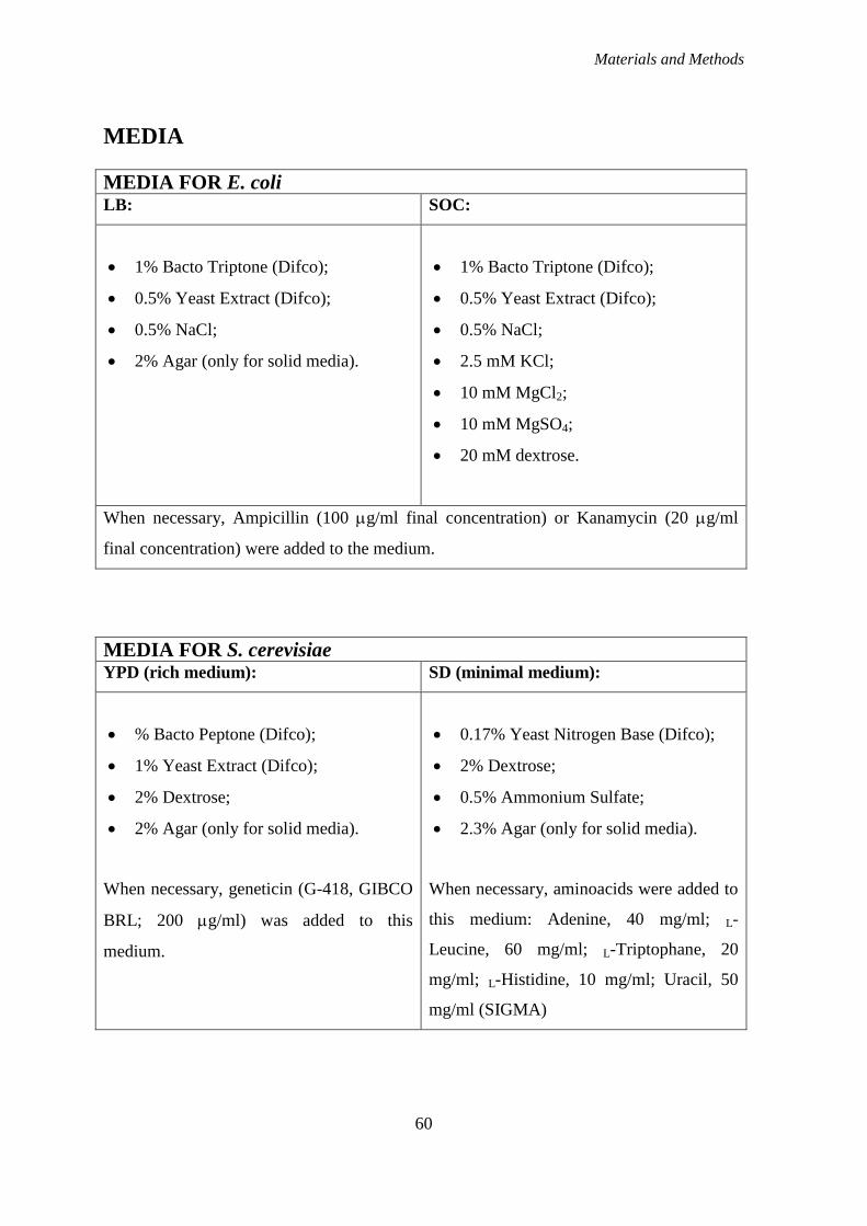
Materials and Methods
60
MEDIA
MEDIA FOR E. coli LB: SOC:
1% Bacto Triptone (Difco);
0.5% Yeast Extract (Difco);
0.5% NaCl;
2% Agar (only for solid media).
1% Bacto Triptone (Difco);
0.5% Yeast Extract (Difco);
0.5% NaCl;
2.5 mM KCl;
10 mM MgCl2;
10 mM MgSO4;
20 mM dextrose.
When necessary, Ampicillin (100 g/ml final concentration) or Kanamycin (20 g/ml
final concentration) were added to the medium.
MEDIA FOR S. cerevisiae YPD (rich medium): SD (minimal medium):
% Bacto Peptone (Difco);
1% Yeast Extract (Difco);
2% Dextrose;
2% Agar (only for solid media).
When necessary, geneticin (G-418, GIBCO
BRL; 200 g/ml) was added to this
medium.
0.17% Yeast Nitrogen Base (Difco);
2% Dextrose;
0.5% Ammonium Sulfate;
2.3% Agar (only for solid media).
When necessary, aminoacids were added to
this medium: Adenine, 40 mg/ml; L-
Leucine, 60 mg/ml; L-Triptophane, 20
mg/ml; L-Histidine, 10 mg/ml; Uracil, 50
mg/ml (SIGMA)

Materials and Methods
61
SPORULATION MEDIUM: FOA:
1% KAc;
2% Agar.
0.17% Yeast Nitrogen Base (Difco);
2% Dextrose;
0.5% Ammonium Sulfate;
2% Agar.
Aminoacids (Ade, 40 mg/ml; Leu, 60
mg/ml; Trp, 20 mg/ml; His, 10 mg/ml;
Ura, 50 mg/ml) and the 5‟-Fluoroorotic
acid (1 g/l) are filtered-sterilized and
added after the medium has been
autoclaved
VECTORS
pFA6a-kanMX4
(Achim W. et al., 1994)
This vector of 3938 bp carries a selectable marker for both E.coli and yeast cells. It is used
as a template for PCR with oligonucleotides that contain regions complementary to the
target gene to be deleted (GRX4 in our case). PCR products are then used for high
efficiency transformation of yeast cells. We also used a modified vector in which the KAN
cassette was substituted with an HIS3 cassette, cloned in the BglII site. This latter plasmid
was used for the deletion of the GRX3 gene.
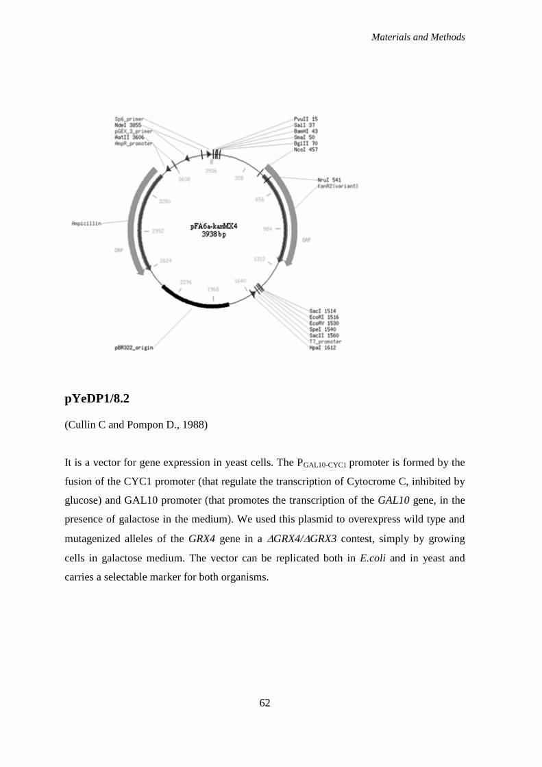
Materials and Methods
62
pYeDP1/8.2
(Cullin C and Pompon D., 1988)
It is a vector for gene expression in yeast cells. The PGAL10-CYC1 promoter is formed by the
fusion of the CYC1 promoter (that regulate the transcription of Cytocrome C, inhibited by
glucose) and GAL10 promoter (that promotes the transcription of the GAL10 gene, in the
presence of galactose in the medium). We used this plasmid to overexpress wild type and
mutagenized alleles of the GRX4 gene in a GRX4/GRX3 contest, simply by growing
cells in galactose medium. The vector can be replicated both in E.coli and in yeast and
carries a selectable marker for both organisms.
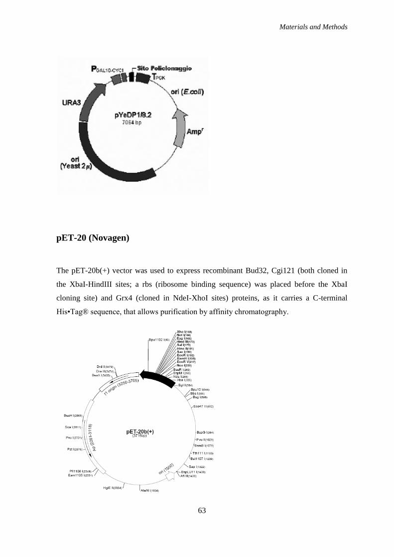
Materials and Methods
63
pET-20 (Novagen)
The pET-20b(+) vector was used to express recombinant Bud32, Cgi121 (both cloned in
the XbaI-HindIII sites; a rbs (ribosome binding sequence) was placed before the XbaI
cloning site) and Grx4 (cloned in NdeI-XhoI sites) proteins, as it carries a C-terminal
His•Tag® sequence, that allows purification by affinity chromatography.
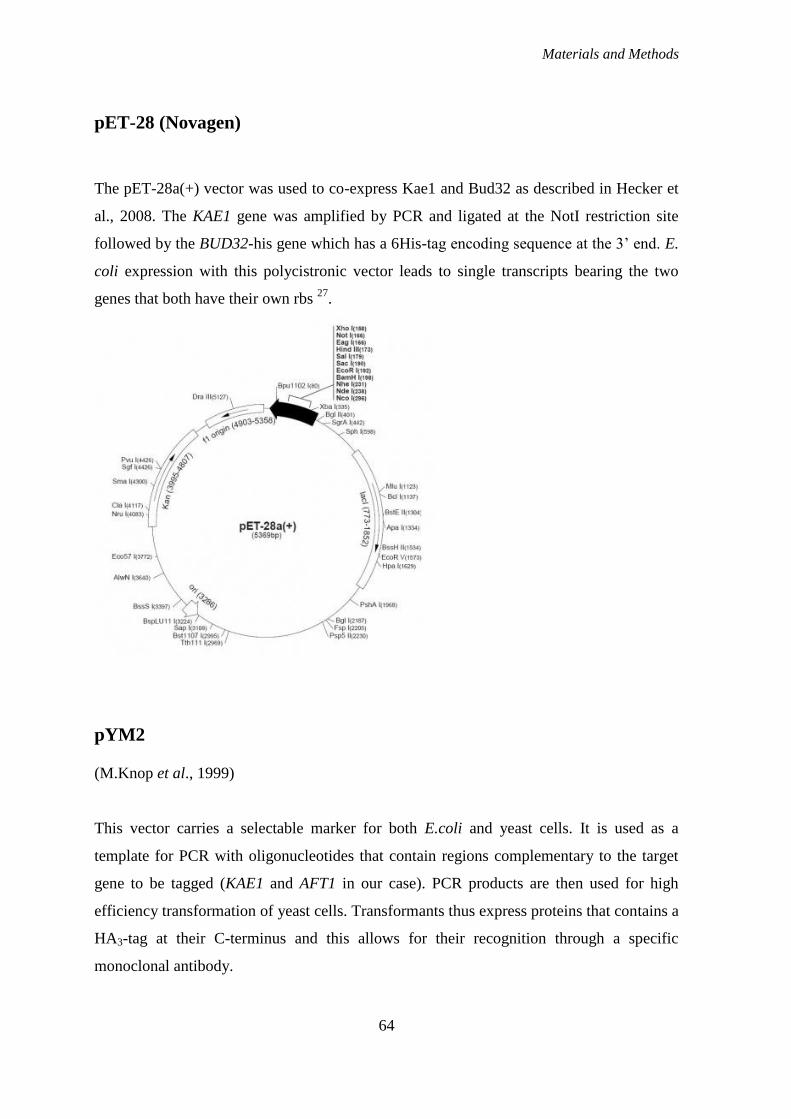
Materials and Methods
64
pET-28 (Novagen)
The pET-28a(+) vector was used to co-express Kae1 and Bud32 as described in Hecker et
al., 2008. The KAE1 gene was amplified by PCR and ligated at the NotI restriction site
followed by the BUD32-his gene which has a 6His-tag encoding sequence at the 3‟ end. E.
coli expression with this polycistronic vector leads to single transcripts bearing the two
genes that both have their own rbs 27
.
pYM2
(M.Knop et al., 1999)
This vector carries a selectable marker for both E.coli and yeast cells. It is used as a
template for PCR with oligonucleotides that contain regions complementary to the target
gene to be tagged (KAE1 and AFT1 in our case). PCR products are then used for high
efficiency transformation of yeast cells. Transformants thus express proteins that contains a
HA3-tag at their C-terminus and this allows for their recognition through a specific
monoclonal antibody.
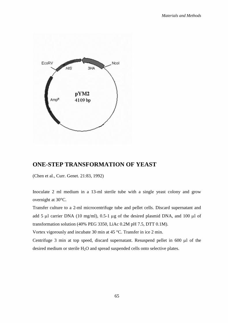
Materials and Methods
65
ONE-STEP TRANSFORMATION OF YEAST
(Chen et al., Curr. Genet. 21:83, 1992)
Inoculate 2 ml medium in a 13-ml sterile tube with a single yeast colony and grow
overnight at 30°C.
Transfer culture to a 2-ml microcentrifuge tube and pellet cells. Discard supernatant and
add 5 l carrier DNA (10 mg/ml), 0.5-1 g of the desired plasmid DNA, and 100 l of
transformation solution (40% PEG 3350, LiAc 0.2M pH 7.5, DTT 0.1M).
Vortex vigorously and incubate 30 min at 45 °C. Transfer in ice 2 min.
Centrifuge 3 min at top speed, discard supernatant. Resuspend pellet in 600 l of the
desired medium or sterile H2O and spread suspended cells onto selective plates.

Materials and Methods
66
HIGH EFFICIENCY LIAC TRANSFORMATION OF YEAST
(Gietz et al., YEAST 11:355-360, 1995)
Inoculate 2-5 ml of liquid YPD and incubate with shaking overnight at 30°C.
Count o/n culture and inoculate 50 ml of warm YPD to an OD600=0.2.
Incubate the culture at 30°C on a shaker at 200 rpm until the absorbance reaches
OD600=0.7-0.8. This will take 3 to 5 hours. This culture will give sufficient cells for 10
transformations.
Harvest the culture in a sterile 50 ml centrifuge tube at 1000 x g for 5 min.
Pour off the medium, resuspend the cells in 25 ml of sterile water and centrifuge again.
Pour off the water, resuspend the cells in 1 ml 100 mM LiAc and transfer the suspension to
a 1.5 ml microfuge tube.
Pellet the cells at top speed for 15 sec and remove the LiAc with a micropipette.
Resuspend the cells to a final volume of 500 µl in 100 mM LiAc.
Boil SS-DNA for 5 min. and quickly chill in ice water.
Vortex the cell suspension and pipette 50 µl samples into microfuge tubes. Pellet the cells
and remove the LiAc with a micropipette.
Add the "transformation mix”: 240 µl PEG (50% w/v), 36 µl 1.0 M LiAc, 5 µl SS-DNA
(10 mg/ml), X µl DNA*, 70-X µl Sterile ddH2O. Carefully add these ingredients in the
order listed. The PEG should go in first, which shields the cells from the detrimental
effects of the high concentration of LiAc.
Vortex each tube vigorously until the cell pellet has been completely mixed. Usually takes
about 1 min.
Incubate (w/o agitation) at 30 °C for 30 min.
Heat shock in a water bath at 42°C for 20 min.
Microfuge at 6-8000 rpm for 15 sec and remove the transformation mix with a
micropipette.**
Pipette 600 µl of sterile water into each tube and resuspend the pellet by pipetting it up and
down gently. Spread suspended cells onto selective plates.
*For gene disruption via PCR, you can use an entire reaction of 50 µl without any further
purification.
Materials and Methods
67
**When you are attempting to disrupt a gene with a PCR-amplified KAN::MX4 cassette,
you have to resuspend yeast cells in YPD (about 20 ml), after step 15, and let them grow 2
to 3 hours before plating on YPD+G-418.
PREPARATION OF YEAST DNA
Inoculate 2 ml medium in a 13-ml sterile tube with a single yeast colony and grow
overnight.
Transfer culture to a 2-ml microcentrifuge tube (proper for the MagNA Lyser Instrument,
Roche) and pellet cells. Discard supernatant and add an equal amount of glass beads, 200
l TE buffer, 0.1 M NaCl, 2% TRITON X-100, 1% SDS, 100 l basic phenol and 100 l
chloroform:isoamyl alcool (24:1).
Vortex vigorously in the MagNA Lyser three time for 30 sec at 6000 rpm, with 1 min in
ice, and add 200 l TE. Centrifuge at 13000 rpm for 10 min, 4°C.
Transfer aqueous (top) phase to a clean 1.5-ml microcentrifuge tube, add 2.5 volumes of
ice-cold 100% ethanol and precipitate 30 min (or overnight), -80°C. Microcentrifuge 15
min at 13000 rpm, 4°C.
Dry the pellet and resuspend in 50 l sterile H2O and add 1 l RNAse A; let 15 min at 37
°C. Determine the concentration spectrophotometrically and run electrophoresis gel.
POLYMERASE CHAIN REACTION (PCR)
(Sambrook K.J. et al., 1989; Ausubel F.M. et al., 1989)
The polymerase chain reaction (PCR) is a technique used to amplify a single or few copies
of a piece of DNA across several orders of magnitude, generating thousands to millions of
copies of a particular DNA sequence. The method relies on thermal cycling, consisting of
cycles of repeated heating and cooling of the reaction for DNA melting and enzymatic
replication of the DNA. Primers (short DNA fragments) containing sequences
Materials and Methods
68
complementary to the target region along with a DNA polymerase (after which the method
is named) are key components to enable selective and repeated amplification. As PCR
progresses, the DNA generated is itself used as a template for replication, setting in motion
a chain reaction in which the DNA template is exponentially amplified. PCR can be
extensively modified to perform a wide array of genetic manipulations.
Almost all PCR applications employ a heat-stable DNA polymerase, such as Taq
polymerase, an enzyme originally isolated from the bacterium Thermus aquaticus.
Reactions were performed in these conditions:
5-10 ng DNA template (that contains the DNA region (target) to be amplified);
200 M deoxynucleoside triphosphates (dNTPs);
1 M of each primer (that are complementary to the 3' ends of each of the sense
and anti-sense strand of the DNA target);
1X buffer solution, providing a suitable chemical environment for optimum activity
and stability of the DNA polymerase and containing a monovalent cation (KCl)
(Finnzymes);
1.5 mM MgCl2;
1U Phusion or DyNAzyme™ II DNA polymerase (Finnzymes);
H2O to reach volume.
Common volumes are 20–50 μl and reactions were carried out in a GeneAmp System
9700 (PE Applied Biosystem) thermal cycle.
Reaction conditions varies depending on the type of DNA polymerase used, but are usually
the following:
Initialization step: 5 min at 94 °C (for DyNAzyme™ II DNA polymerase ), or 30
sec at 98 °C (for Phusion).
Then 25-35 cycles of:
Denaturation step: 1 min at 94 °C (for DyNAzyme™ II DNA polymerase ), or 10
sec at 98 °C (for Phusion).for 20–30 seconds.
Annealing step: The reaction temperature is lowered to 50–65 °C for 20–40
seconds allowing annealing of the primers to the single-stranded DNA template.
Typically the annealing temperature is about 3-5 °C below the Tm of the primers
used.
Materials and Methods
69
Extension/elongation step: The extension time depends both on the DNA
polymerase used and on the length of the DNA fragment to be amplified. We used
a temperature of 72 C for 1min/Kb DNA target (for DyNAzyme™ II DNA
polymerase ), or 15-30 sec/Kb DNA (for Phusion).
Then a final elongation step at 72 °C for 5–10 minutes after the last PCR cycle to
ensure that any remaining single-stranded DNA is fully extended.
Final hold: This step at 4 °C for an indefinite time may be employed for short-term storage
of the reaction.
MUTAGENESIS OF GRX4, BUD32 AND KAE1
Mutagenesis of GRX4, BUD32 and KAE1 coding sequences, inserted in different vectors,
were performed using the QuikChange™ Site-Directed Mutagenesis Kit (Stratagene). In
the table below are listed the oligonucleotides used, designed according to the instruction
manual. With small letters are indicated the substituted bases. Controls of the correct
mutagenesis were performed both by PCR, when allele-specific primers were available
(see below), and by sequencing (BMR genomics service).
OLIGOs FOR GRX4
OLIGONUCLEOTIDE 5’-3’ SEQUENCE DESCRIPTION
GRX4-S133-134A-S GAAAGCAGCGGGgCTgCCGATGATG
AAGAG
Mutagenesis of both
residues of Grx4 (Ser-
134 and Ser-133) that
are phosphorylated by
Bud32, in pYeDP and
pET20 vectors
GRX4-S133-134A-AS CTCTTCATCATCGGcAGcCCCGCTGC
TTTC
GRX4-S134A-S AGCAGCGGGTCTaCCGATGATGAAG
AGGAC
Substitution of the main
serine phosphorylated by
Bud32 with an
unphosphorylatable
residue, in the pYeDP
vector
GRX4-S134A-AS GTCCTCTTCATCATCGGtAGACCCG
CTGCT
Materials and Methods
70
GRX4-S133A-S GAAAGCAGCGGGaCTTCCGATGATG
AAGAG
GRX4-S133A-AS CTCTTCATCATCGGAAGtCCCGCTG
CTTTC
GRX4-S134D-S AGCAGCGGGTCTgaCGATGATGAAG
AGGAC
Substitution of the Ser-
134 with a residue that
mimic a constitutively
phosphorylated
condition, in the pYeDP
vector
GRX4-S134D-AS GTGCTCTTCATCATCGtcAGACCCGC
TGCT
OLIGOs FOR BUD32
OLIGONUCLEOTIDE 5’-3’ SEQUENCE DESCRIPTION
K52A-S GTATATTATCgcATATAGGCCACC Mutagenesis of two
residues important for
the catalytic activity of
Bud32 (see introduction),
in the pET-28 vector,
containing KAE1/BUD32
coding sequences and in
the pET-20 vector,
containing only the
BUD32 coding sequence.
K52A-AS GATAATATACgcTTGATGAGAATCC
D161A-S GTCATGGTGcgTTGACAAGTTC
D161A-AS GAACTTGTCAAcgCACCATGAC
OLIGO USED FOR THE CONTROL OF THE MUTAGENESIS:
BUD K52 ASP CATCAAAAGTATATTATCgc Control of K52 mutation:
it anneals with the
mutagenized sequence
BUD wt ASP CATCAAAAGTATATTATCaa Control of K52 mutation:
it anneals with the wt
sequence
BUD32 D161 ASP ATGACTACTGTCATGGTGcg Control of D161
mutation: it anneals with
the mutagenized
sequence
BUD32 D161 ASP WT ATGACTACTGTCATGGTGat Control of D161
mutation: it anneals with
Materials and Methods
71
the wt sequence
BUD32 S258A-AS TCCTAGCATAGCTCTCTTACGACC Antisense oligo used
with the other 5 on top
OLIGOs FOR KAE1
OLIGONUCLEOTID
E
5’-3’ SEQUENCE DESCRIPTION
HHKAE AA-S GGAGTAAACgcCTGCATTGGTgcCATC
GAAATGG
Mutagenesis of the two
histidines of Kae1
(H141 and H145),
important for the metal
ion binding (see
introduction), in the
pET-28 vector,
containing
KAE1/BUD32 coding
sequences.
HHKAE AA-AS CCATTTCGATGgcACCAATGCAGgcGT
TTACTCC
CREATION OF THE STRAINS USED FOR THE
PHENOTYPIC ANALYSIS OF THE EFFECT OF THE
PHOSPHORYLATION ON Grx4 BY Bud32
For this analysis, we deleted both the GRX4 and the GRX3 coding sequences on yeast
genome, as the two genes share overlapping functions. We then transformed the
heterozygous diploid strain with galactose-inducible pYeDP plasmids, carrying a wild-type
or a mutagenized version of GRX4 (see on top).
Both genes were deleted in a PCR-mediated fashion, by amplification of either the
KAN::MX4 cassette (for the GRX4 gene) or the HIS3 cassette (for the GRX3 gene) with
specific primers that contained a sequence complementary to the plasmid (pFA6A, in
which the cassette is inserted), and two protruding sequences complementary to the region
in which the cassette has to be inserted in the genome (see explanatory figure for the GRX3
gene, but the same occurred for the GRX4 gene).
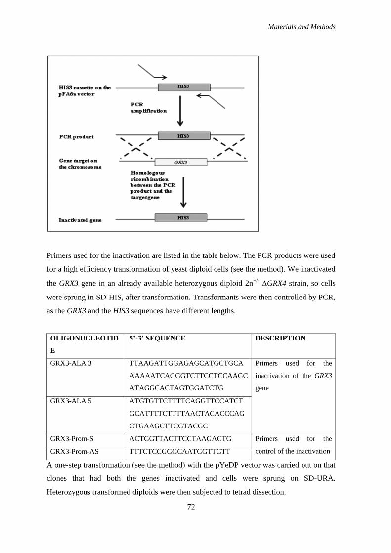
Materials and Methods
72
Primers used for the inactivation are listed in the table below. The PCR products were used
for a high efficiency transformation of yeast diploid cells (see the method). We inactivated
the GRX3 gene in an already available heterozygous diploid 2n+/-
GRX4 strain, so cells
were sprung in SD-HIS, after transformation. Transformants were then controlled by PCR,
as the GRX3 and the HIS3 sequences have different lengths.
OLIGONUCLEOTID
E
5’-3’ SEQUENCE DESCRIPTION
GRX3-ALA 3 TTAAGATTGGAGAGCATGCTGCA
AAAAATCAGGGTCTTCCTCCAAGC
ATAGGCACTAGTGGATCTG
Primers used for the
inactivation of the GRX3
gene
GRX3-ALA 5 ATGTGTTCTTTTCAGGTTCCATCT
GCATTTTCTTTTAACTACACCCAG
CTGAAGCTTCGTACGC
GRX3-Prom-S ACTGGTTACTTCCTAAGACTG Primers used for the
control of the inactivation GRX3-Prom-AS TTTCTCCGGGCAATGGTTGTT
A one-step transformation (see the method) with the pYeDP vector was carried out on that
clones that had both the genes inactivated and cells were sprung on SD-URA.
Heterozygous transformed diploids were then subjected to tetrad dissection.

Materials and Methods
73
TETRAD DISSECTION
Heterozygous diploid strains were allowed to sporulate on 1% potassium acetate solid
medium for 4-6 days at 28 °C. After the addiction of zymoliase, about 10 tetrads/diploid
strains were dissected and the spores allowed to germinate on YPD medium at 28 °C. For
each spore, the presence of a determined cassette/phenotype was controlled by growth on
selective media.
The deletion of GRX4 gene was thus controlled by growth on YPD+ G-418, the deletion of
GRX3 gene was controlled by growth on SD-HIS and the presence of the pYeDP vector
was controlled by growth on SD-URA.
EPITOPE-TAGGING OF KAE1 AND AFT1
(Knop et al., Yeast, 1999)
To add a C-terminal tag on these two genes, we used a protocol described in Knop et al.
(1999). Amplification of the tag from the pYM2 vector is carried out using specific
primers that contain a sequence complementary to the plasmid (and that is the same for all
pYM vectors, allowing the insertion of different tags at one locus, using a unique pair of
primers), and two protruding sequences complementary to the region in which the tag has
to be inserted in the genome. The oligonucleotides used for KAE1 and AFT1 are listed in
the table below: bases that are corresponding to a sequence on the vector are underlined.
OLIGONUCLEOTIDE 5’-3’ SEQUENCE DESCRIPTION
YKR9MYC-S ACGCAGAAATTCAGAACCGATGAA
GTATACGCAGCCTGGCGTGGCGTG
ATCGTACGCTGCAGGTCGAC
Primers used for
insertion of the HA
epitope on the KAE1
gene YKR9MYC-AS TATATATTCGTAGTGTGAACATTAG
GCTAACTGTAACAAAAATCGATCG

Materials and Methods
74
ATGAATTCGAGCTCG
AFT1-pYM12-S ATTAAAATGGTGAACGGCGAGTTG
AAGTATGTGAAGCCAGAAGATCGT
ACGCTGCAGGTCGAC
Primers used for
insertion of the HA
epitope on the AFT1
gene AFT1-pYM12-AS TGAAAATGGACGAGAGATACGTCT
AAGTTTGATTTCATCTATATGATCG
ATGAATTCGAGCTCG
The PCR products were used for high efficiency transformations of yeast diploid cells. In
the figure below the method is explained: the PCR products contains flanking homologous
sequences that allows their targeted integration into the genome of yeast cells.
Transformants were then controlled for the presence of the epitope by PCR, using the
primers listed below.
Materials and Methods
75
OLIGO USED FOR THE CONTROL OF THE EPITOPE-TAGGING:
OLIGONUCLEOTIDE 5’-3’ SEQUENCE DESCRIPTION
KAN&HISPRIMER TGGGCCTCCATGTCGCTGG PRIMER REV USED
FOR BOTH KAE1 AND
AFT1; IT ANNEALS AT
+427 BP FROM THE 5‟
POSITION OF
INTEGRATION
YKR PROM-S GGATCCGATAATGGTTGATTCTT
ATTTAGAC
Primer fwd for the control
of the integration in
KAE1. It anneals at 366
bp upstream of ATG
AFT1-1814-S TGTTGACCATCGTCATCTCA Primer fwd for the control
of the integration in
AFT1. It anneals at 1841
position in the ORF of
AFT1
PREPARATION OF YEAST RNA BY EXTRACTION WITH
HOT ACID PHENOL
(John Wiley & Sons, Current Protocols in Molecular Biology, 1995)
Grow yeast cells in 10 ml of desired medium to mid-exponential phase (OD600=1.0).
Transfer culture to 50-ml centrifuge tube and centrifuge cells 3 min at 7000 RPM, 4°C.
Discard supernatant, resuspend pellet in 1 ml ice-cold water. Transfer to a clean 1.5-ml
microcentrifuge tube. Microcentrifuge 10 sec at 4°C, and remove supernatant. Proceed to
step 4 or if desired immediately freeze pellet by placing tube in dry ice. The frozen pellet
can be stored for months at -80°C.
Resuspend cell pellet in 400 l TES solution (10 mM Tris/HCl, pH 7.5; 10 mM EDTA pH
8; 0.5% SDS). Add 400 l acid phenol (pH 5) and vortex vigorously 10 sec. Incubate 20 to
60 min at 65 °C with occasional, brief vortexing.
Place on ice 5 min. Microcentrifuge 10 min at 13000 rpm, 4°C.
Materials and Methods
76
Transfer aqueous (top) phase to a clean 1.5-ml microcentrifuge tube, add 400 l acid
phenol and vortex vigorously. Incubate 15 min at 65 °C with occasional, brief vortexing.
Repeat step 5.
Transfer aqueous phase to a clean 1.5-ml microcentrifuge tube and add 400 l
chloroform:isoamyl alcool (24:1). Vortex vigorously and microcentrifuge 15 min at 13000
rpm, 4°C.
Transfer aqueous phase to a new tube, add 40 l of 3M sodium acetate, pH 5.3, and 1 ml of
ice-cold 100% ethanol and precipitate 1 hr (or overnight), -80°C. Microcentrifuge 15 min
at 13000 rpm, 4°C. Wash RNA pellet by vortexing briefly in ice-cold 70% ethanol.
Microcentrifuge as before to pellet RNA.
Dry the pellet and resuspend in 25-40 l DEPC-treated H2O. Determine the concentration
spectrophotometrically by measuring the A260 and A280. Store at -80 °C, or at -20 °C if it is
to be used within 1 year.
ANALYSIS OF GAL1 TRANSCRIPTION
Yeast cells were grown to OD600 0.4 in Raffinose-containing medium and then shifted to
galactose-containing medium to activate the GAL regulon. Samples were taken after 30
min of induction and RNAs were prepared as previously described and subjected to
Northern Blotting.
ANALYSIS OF FET3/FIT3 TRANSCRIPTION
Yeast cells were grown in SD medium until OD600 0.6 and then Bathophenanthroline
disulfonic acid (BPS, Sigma) was added, at the final concentration of 0.2 mM or 0.1 mM,
for 30 minutes to 1 hour. BPS is a chelating reagent, that forms complexes with various
metal ions, particularly, Fe(II). As a result, levels of iron within cell decrease and starts the
activation of iron-sensing genes.

Materials and Methods
77
REAL-TIME PCR
Reverse transcription (RT) followed by the polymerase chain reaction (PCR) is the
technique of choice to analyze mRNA expression derived from various sources. Real-time
RT–PCR is highly sensitive and allows quantification of rare transcripts and small changes
in gene expression. Moreover, it is easy to perform, provides the necessary accuracy and
produces reliable as well as rapid quantification results. The simplest detection technique
for newly synthesized PCR products in real-time PCR uses SYBR Green I fluorescence
dye that binds specifically to the minor groove double-stranded DNA. The quantification
method of choice depends on the target sequence, the expected range of mRNA amount
present in the tissue, the degree of accuracy required and whether quantification needs to
be relative or absolute. Two different methods of analyzing data from real-time,
quantitative PCR exist: absolute quantification and relative quantification. Absolute
quantification determines the input copy number of the transcript of interest, usually by
relating the PCR signal to a standard curve. Relative quantification describes the change in
expression of the target gene relative to some reference group such as an untreated control
or a sample at time zero in a time-course study. Furthermore, a normalization of the target
gene with an endogenous standard (mainly non-regulated reference genes or housekeeping
genes like G3PDH or GAPDH, albumin, actins, tubulins, cyclophilin, 18S rRNA or 28S
rRNA) is recommended. In our study, we used a mathematical method for relative
quantification, as described from Pfaffl in 2001 65
. It is necessary to determine the cycle
threshold (Ct) for each transcript, that is defined as the point at which the fluorescence
rises appreciably above the background fluorescence. The relative expression ratio (R) of a
target gene is calculated based on E and the Ct deviation of an unknown sample versus a
control, and expressed in comparison to a reference gene:
𝑅𝑎𝑡𝑖𝑜 = 𝐸𝑡𝑎𝑟𝑔𝑒𝑡
∆𝐶𝑡 𝐶𝑜𝑛𝑡𝑟𝑜𝑙 −𝑠𝑎𝑚𝑝𝑙𝑒
𝐸𝑟𝑒𝑓𝑒𝑟𝑒𝑛𝑐𝑒 ∆𝐶𝑡 𝐶𝑜𝑛𝑡𝑟𝑜𝑙 −𝑠𝑎𝑚𝑝𝑙𝑒
Where Etarget is the real-time PCR efficiency of target gene transcript; Ereference is the real-
time PCR efficiency of a reference gene transcript; ΔCttarget is the Ct deviation of control –
Materials and Methods
78
sample of the target gene transcript; ΔCtref = Ct deviation of control – sample of reference
gene transcript.
As a reference gene we used actin.
For mRNA-based PCR, the RNA sample is first reverse transcribed to cDNA with reverse
transcriptase. To do retro-transcription, we use a commercial kit (SuperScript™ III First-
Strand Synthesis System for RT-PCR, Invitrogen) and oligo-dT to amplify total mRNAs.
For the real-time reaction, we used a Corbett Research Rotor-Gene™ instrument and the
following parameters: 95°C for 2 min, then 40 cycles of: 95°C for 15 sec and 60°C for 30
sec.
Every reaction contains:
25 l Platinum® SYBR
® Green qPCR SuperMix-UDG (Invitrogen)
1 l Forward primer 10 M
1 l Reverse primer 10 M
≤ 10 l Template (cDNA generated from 10 pg to 1 g of total RNA)
DEPC-treated H2O up to 50 l
NORTHERN BLOT
The northern blot is a technique used to study gene expression by detection of RNA (or
isolated mRNA) in a sample.
Total RNA extracted from yeast cells are separated by gel electrophoresis. The gel contains
1% agarose, 1X MOPS buffer (0.2 M MOPS, pH 7; 80 mM NaAc; 10 mM EDTA, pH 8)
and 6% formaldehyde as a denaturing agent for the RNA to limit secondary structure.
Sample are prepared as following:
RNA (10 g) and DEPC-treated H2O to 5 l
15 l MIX (50% formamide, that lowers the annealing temperature of the probe-
RNA interaction preventing RNA degradation by high temperatures; 6%
formaldehyde; 1X MOPS)

Materials and Methods
79
Samples are then denatured at 65 °C for 10 min and held in ice for 2 min. Before loading
gel, 2 l of 10X loading gel solution are added. Gel electrophoresis is carried out overnight
at 20 V.
The gel can be stained with ethidium bromide (EtBr) and viewed under UV light to
observe the quality and quantity of RNA before blotting. Since the large ribosomal subunit
is 28S (approximately 5kb) and the small ribosomal subunit is 18S (approximately 2kb)
two prominent bands will appear on the gel, the larger at close to twice the intensity of the
smaller. After that, the gel is washed twice in sterile H2O, for 10 min, to eliminate
formaldehyde.
The RNA samples, now separated by size, are transferred to a nylon membrane through a
capillary blotting system. We use a nylon membrane with a positive charge (Hybond™-N+,
Amersham), and SSC 20X (3 M NaCl, 0.3 M Sodium citrate, pH 7) as blotting buffer. The
capillary transfer takes about 20 h (overnight).
Once the RNA has been transferred to the membrane it is immobilized through covalent
linkage to the membrane by UV light, for 5 min.
At this point the membrane can be hybridized with the probe or stored at 4 °C.
DETECTION USING RADIOLABELED PROBES:
The membrane is first pre-hybridated at 42 °C for 30 min in the ULTRAhyb hybridization
buffer (Ambion). The labeled probe is then denatured at 100 °C for 3 min and then added
to the tube and let overnight (14-24 h) in hybridization at 42 °C.
After that, the hybridization solution can then be recovered (if reused within few days) and
the membrane is washed twice for 5 min in SSC 2X, SDS 0.1% and twice for 15 min in
SSC 0.1X, SDS 0.1%. The membrane is left to dry and exposed to autoradiography for
about 1 h, then scanned using the Cyclone apparatus (Packard).
Materials and Methods
80
DETECTION USING NON-RADIOACTIVE PROBES:
When we used non-radioactive probes, the transfer of the gel was performed on
nitrocellulose membrane.
The membrane is first pre-hybridated at 50 °C for 30 min in 30 ml of pre-hybridation
solution (5X SSC, 0.5% SDS, 1 mM EDTA pH 8). Discard the pre-hybridation solution
and add 10 ml (or more) of hybridation solution (0.75 M NaCl, 0.15 M Tris/HCl pH8, 10
mM EDTA, 0.1 M phosphate buffer pH 6.8, 0.1% Sodium pyrophosphate, 0.1% SDS,
0.2% PVP, 0.2% Ficoll 400, 0.2% BSA) plus 50 l of boiling-denatured ss-DNA (10
mg/ml) and incubate at 65 °C for at least 2 hours. At that time, denature the probe for 10
min at 100 °C and add it to the membrane. Let it incubate overnight at 65 °C.
After that, wash the membrane twice for 5 min in washing buffer A (SSC 2X, SDS 0.1%)
and twice for 5 min in washing buffer B (SSC 0.5X, SDS 0.1%, at 65 °C). A final wash
with washing buffer C (100 mM maleic acid, 150 mM NaCl, 0.3% tween 20) for 5 min at
room temperature. Saturate the membrane is with saturation buffer (100 mM maleic acid
pH 7.5, 150 mM NaCl, 3% BSA) for 30 min, and after that add the anti-digoxigenin
antibody (1:10000) and let 30 min or more. Then wash twice for 5 min in washing buffer
C. At this point, equilibrate the membrane for 2 min with the alkaline phosphatase buffer
(AP) (0.1 M Tris/HCl pH9.5, 0.1 M NaCl, 5 mM MgCl2) and incubate in the dark without
shaking with the revelation solution: 10 ml of AP buffer with 1 tablet of NBT/BCIP (Nitro
blue tetrazolium chloride and 5-Bromo-4-chloro-3-indolyl phosphate, Roche) until some
signal appears. BCIP is the AP substrate, which after dephosphorylation is oxidized by
NBT to yield a dark-blue indigo precipitating dye. NBT is thereby reduced to a dark-blue
precipitating dye and serves to intensify the color reaction making the detection more
sensitive.
Materials and Methods
81
PREPARATION OF RADIOLABELED PROBES FOR
NORTHERN AND SOUTHERN BLOTTING
As a detecting system, we use radiolabeled probes, made of PCR-amplified target DNA.
The probe is prepared accordingly to the DECAprime™ II Random Primed DNA Labeling
Kit (Ambion). The reaction is performed using random decamer oligonucleotide primers
and exonuclease-free klenow enzyme.
The probe is then purified using NucAway™ Spin Columns (Ambion), that are useful to
recover the labeled DNA while removing salts and unincorporated nucleotides.
In the table below are listed the oligonucleotides used to amplify the target DNA, from its
chromosomal locus.
OLIGONUCLEOTIDE 5’-3’ SEQUENCE DESCRIPTION
GAL1-3 S CTGCAAATGTTTTAGCTGCCACGTA Primers used to
amplify the GAL1
gene
GAL1-3 AS CATCTTTGTTAACCGTTCGATGCC
ACT1-cDNA-S ACCATGTTCCCAGGTATTGCC Primers used to
amplify the ACT1
gene
ACT1-cDNA-AS ACTTGTGGTGAACGATAGATG
PREPARATION OF NON-RADIOACTIVE PROBES FOR
NORTHERN BLOTTING
The analysis of the iron regulon transcription by northern blotting were performed using
random-priming labeled probes. The Klenow enzyme copies DNA template in the presence
of hexameric primers and alkali-labile Digoxigenin-11-2‟-deoxy-uridine-5‟triphosphate.
On average, the enzyme inserts one DIG moiety in every stretch of 20-25 nucleotides. The
resulting labeled product is a homogeneously labeled, sensitive hybridization probe.
FIT3, FET3 and ACT1 genes were first PCR-amplified with oligonucleotides listed in the
previous and following tables.
Materials and Methods
82
FIT3-cDNA-S TGTCTGGACTGGTGAAGGCAG Primers used to amplify
the FIT3 gene FIT3-cDNA-AS ATCAAACCAGTACCAGCGGTG
FET3-cDNA-S ATGACATTCTCCTGCTTTGCC Primers used to amplify
the FET3 gene FET3-cDNA-AS TCAGTGGAATGACGATCTTCG
About 1 g of template DNA (in sterile water, for a final volume of 15 l) was boiled for 2
min and then put in ice for 2 min. Then we added:
2 l of Hexanucleotide Mix 10 X(Roche), 2 l of DIG DNA Labeling Mix 10 X(Roche),
and 1 l of DNA polymerase I large (Klenow) fragment; then the reaction was left for 2
hr/overnight at 37 °C. To stop the reaction, the day after we added 2 l EDTA 0.5 M pH 8
and 2 l LiAc 4 M.
The DNA was then precipitated, by adding 70 l 100% Ethanol and left at -80 °C for about
30 min. We then centrifuged for 20 min at 13000 rpm, at 4 °C, discarded flow-through and
repeated with70% ethanol. The DNA pellet was then left to dry and resuspended with 50
l of sterile H2O.
SOUTHERN BLOT ANALYSIS OF TELOMERE LENGTH
Yeast strains were grown in YPDA to exponential phase (OD600 0.8-1.0) and cells were
collected. Genomic DNAs have been purified from yeast cells as previously described and
approximately 50 g were digested with XhoI and separated on 1.2% agarose gel. After
transfer onto Hybond™-N+ membrane (Amersham), filters have been hybridized with a
specific radiolabeled probe for telomeric Y‟-TG1-3 repeats.
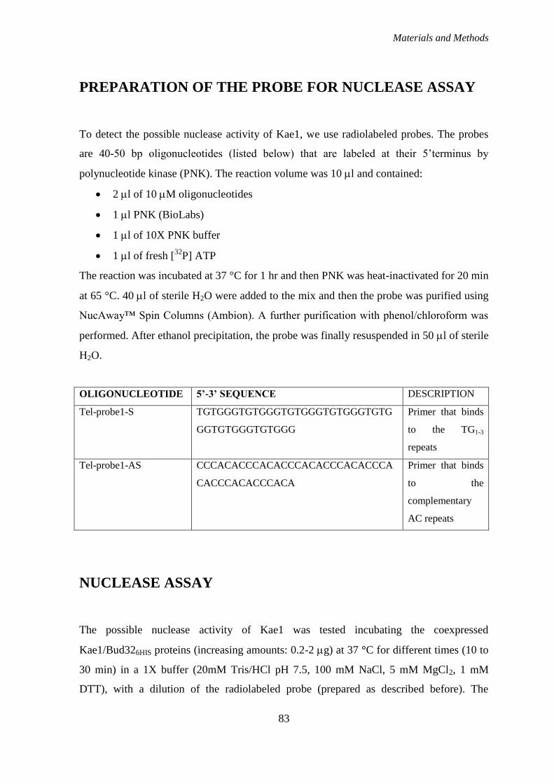
Materials and Methods
83
PREPARATION OF THE PROBE FOR NUCLEASE ASSAY
To detect the possible nuclease activity of Kae1, we use radiolabeled probes. The probes
are 40-50 bp oligonucleotides (listed below) that are labeled at their 5‟terminus by
polynucleotide kinase (PNK). The reaction volume was 10 l and contained:
2 l of 10 M oligonucleotides
1 l PNK (BioLabs)
1 l of 10X PNK buffer
1 l of fresh [32
P] ATP
The reaction was incubated at 37 °C for 1 hr and then PNK was heat-inactivated for 20 min
at 65 °C. 40 l of sterile H2O were added to the mix and then the probe was purified using
NucAway™ Spin Columns (Ambion). A further purification with phenol/chloroform was
performed. After ethanol precipitation, the probe was finally resuspended in 50 l of sterile
H2O.
OLIGONUCLEOTIDE 5’-3’ SEQUENCE DESCRIPTION
Tel-probe1-S TGTGGGTGTGGGTGTGGGTGTGGGTGTG
GGTGTGGGTGTGGG
Primer that binds
to the TG1-3
repeats
Tel-probe1-AS CCCACACCCACACCCACACCCACACCCA
CACCCACACCCACA
Primer that binds
to the
complementary
AC repeats
NUCLEASE ASSAY
The possible nuclease activity of Kae1 was tested incubating the coexpressed
Kae1/Bud326HIS proteins (increasing amounts: 0.2-2 g) at 37 °C for different times (10 to
30 min) in a 1X buffer (20mM Tris/HCl pH 7.5, 100 mM NaCl, 5 mM MgCl2, 1 mM
DTT), with a dilution of the radiolabeled probe (prepared as described before). The
Materials and Methods
84
reaction contained also 10 mM cold ATP. The presence or not of cold ATP, different
divalent cations (MgCl2, ZnSO4, FeSO4, CaCl2 or no ions) and EDTA (to chelate them)
was tested.
PURIFICATION OF PROTEINS FROM E. coli CELLS
To overexpress wild-type and mutant forms of Bud32, Grx4, Kae1/Bud32 we used the
pET-20 or the pET-28 vectors, described before. BL21 E.coli strains were transformed
with the plasmid that contained the desired sequence and clones were grown in LBA
medium until cellular density reached an OD600 of about 0.5. At that point, the expression
of the protein was induced by adding isopropil β-D-1-tiogalattopiranoside (IPTG) to the
medium, at the final concentration of 0.5 mM, and cells were left to grow overnight at 16
°C.
We usually do a trial scale purification, on 1 ml of culture, that is left in IPTG at 37 °C for
about 3 hr before attempting a large-scale purification, to determine if the standard
operating conditions will work for the recombinant protein of interest.
Bacteria were then harvested and resuspended in 5 ml/g of pellet of purification buffer (50
mM Tris/HCl pH 7.5, 0.3 M NaCl, 10% glycerol, 0.2 mM PMSF, 5 mM -
mercaptoethanol, 0.1% TRITON X-100 and 2 mM imidazole). Purification was performed
according to the protocol of the manufacturer by an affinity column containing HIS-
Select™ Nickel Affinity Gel (SIGMA). The proteins were eluted with 100 mM imidazole.
All the step of purification are controlled in SDS-PAGE: 16 l of each sample are mixed
with 4 l of 5X Laemmli Buffer (that reaches the 1X concentration: 60 mM Tris/HCl pH
6.8, 2% SDS, 5% -mercaptoethanol or 100 mM DTT, 10% glycerol, bromophenol blue),
boiled for 5 min and loaded on 12% SDS-PAGE. The gel is then stained with coomassie
blue or blotted to a nitrocellulose membrane.
All protein concentrations were determined both by coomassie-gel staining and by the
Bradford method using BSA as a standard.
Materials and Methods
85
SODIUM DODECYL SULFATE POLYACRYLAMIDE GEL
ELECTROPHORESIS (SDS-PAGE)
SDS-PAGE is a technique to separate proteins according to their electrophoretic mobility
(a function of length of polypeptide chain or molecular weight). The solution of proteins to
be analyzed is mixed with SDS, an anionic detergent which denatures secondary and non–
disulfide–linked tertiary structures, and applies a negative charge to each protein in
proportion to its mass. Heating the samples to at least 60 degrees C shakes up the
molecules, helping SDS to bind.
A tracking dye may be added to the protein solution (of a size smaller than protein) to
allow the experimenter to track the progress of the protein solution through the gel during
the electrophoretic run.
The gels generally consist of acrylamide, bisacrylamide, SDS, and a Tris-Cl buffer with
adjusted pH. Ammonium persulfate (an initiator for gel formation) and TEMED (that
catalyze the polymerization of acrylamide) are added when the gel is ready to be
polymerized. Gels are formed by 2 parts: the separating or resolving gel and the stacking or
loadinggel. The former is usually more basic (is prepared with Tris/HCl at pH 8.8) and has
a higher polyacrylamide content than the loading gel. The stacking gel is a large pore and
is prepared with Tris/HCl buffer pH 6.8 of about 2 pH units lower than that of
electrophoresis buffer (Tris/Glycine).
The gel is actually formed because the acrylamide solution contains a small amount,
generally about 1 part in 35 of bisacrylamide, which can form cross-links between two
polyacrylamide molecules. The ratio of acrylamide to bisacrylamide can be varied for
special purposes. The acrylamide concentration of the gel can also be varied, generally in
the range from 5% to 25%. Lower percentage gels are better for resolving very high
molecular weight proteins, while much higher percentages are needed to resolve smaller
proteins.
Gels are polymerized in a gel caster. First the separating gel is poured and allowed to
polymerize. Next a thin layer of isopropanol or 0.1% SDS is added. Next the loading gel is
poured and a comb is placed to create the wells. After the loading gel is polymerized the
comb can be removed and the gel is ready for electrophoresis.
Materials and Methods
86
The denatured sample proteins are added to the wells one end of the gel with a syringe or
pipette. Finally, the apparatus is hooked up to a power source under appropriate running
conditions to separate the protein bands.
An electric field is applied across the gel, causing the negatively-charged proteins to
migrate across the gel towards the anode. Depending on their size, each protein will move
differently through the gel matrix: short proteins will more easily fit through the pores in
the gel, while larger ones will have more difficulty (they encounter more resistance). After
a set amount of time (usually 1 hour at 120 V), the proteins will have differentially
migrated based on their size; smaller proteins will have traveled farther down the gel,
while larger ones will have remained closer to the point of origin. Following
electrophoresis, the gel may be stained with Coomassie Brilliant Blue R-250, allowing
visualization of the separated proteins, or processed further (e.g. Western blot). After
staining, different proteins will appear as distinct bands within the gel.
For coomassie brilliant blue R-250 staining, proteins are simultaneously fixed with
methanol:glacial acetic acid (45% H2O, 45% methanol and 10% glacial acetic acid) and
stained with coomassie brilliant blue R-250 (0.25 g for 100 ml solution). The gel is
immersed for several hours in the dye, and excess dye is then allowed to diffuse from the
gel during a prolonged period of destaining (in methanol:glacial acetic acid solution,
without the dye).
WESTERN BLOT
In order to make the proteins accessible to antibody detection, after SDS-PAGE, they are
moved from within the gel onto a membrane made of nitrocellulose or polyvinylidene
difluoride (PVDF). The blotting buffer is composed of 25 mM Tris, 192 mM glycine, 20%
methanol and 0.1% SDS. Proteins are transferred through electroblotting, that uses an
electric current to pull proteins from the gel into the nitrocellulose membrane. Blotting
takes about 1 hr at the constant current of 350 mA.
The proteins move while maintaining the organization they had within the gel. As a result
of this "blotting" process, the proteins are exposed on a thin surface layer for detection.
Materials and Methods
87
Protein binding is based upon hydrophobic interactions, as well as charged interactions
between the membrane and protein.
Since the membrane has been chosen for its ability to bind protein, and both antibodies and
the target are proteins, steps must be taken to prevent interactions between the membrane
and the antibody used for detection of the target protein (since the antibody is a protein
itself). Blocking of non-specific binding is achieved by placing the membrane for 1 hr in a
solution of 3% Bovine serum albumin (BSA) in Tris-buffered saline (TBS, 1X: 50 mM
Tris/HCl pH 7.5, 150 mM NaCl) with a minute percentage of detergent such as Tween 20,
0.1%. The protein in the dilute solution attaches to the membrane in all places where the
target proteins have not attached.
After blocking, a dilute solution of primary antibody (in TBST/BSA 1%) is incubated with
the membrane under gentle agitation, for 1-2 hr at room temperature, or overnight at 4 °C.
The membrane is then washed three times for 10 min with TBS+Tween 0.1% and then
exposed to another antibody, directed at a species-specific portion of the primary antibody
(secondary antibody). Most commonly, a horseradish peroxidase-linked secondary is used
to cleave a chemiluminescent agent, and the reaction product produces luminescence in
proportion to the amount of protein. After the secondary antibody, the membrane is
washed three more times, as described before and then revealed using the Kodak (NEN)
instrument, after treatment with the development solution that contains H2O2. Exposition
time varies according to the substrate, from 30 sec to 5 min about.
PHOSPHOTRANSFERASE ASSAY
The protein kinase activity of Bud32 was assayed by incubating the recombinant His-
tagged protein (about 100 ng) at 37 °C for 15 min in 20 μl of medium containing 50 mM
Tris/HCl, pH 7.5, 10 mM MnCl2, 62.5 μM “cold” ATP and 25 μM [γ-33
P]ATP (Amersham
Biosciences; specific radioactivity, 2000–3000 c.p.m./pmol) and the Grx4 (about 400 ng)
or the Kae1/Bud32 coexpressed proteins as phosphorylatable substrates. The reaction was
stopped by adding 5 μl of the gel electrophoresis loading buffer 5X (Laemmli buffer, Tris-
HCl 50 mM pH 6.8, 2% SDS, 1% -mercaptoethanol and coomassie blue staining) and
Materials and Methods
88
samples were subjected to SDS/PAGE (12% gels). Gel was then blotted and the membrane
scanned using the Cyclone apparatus (Packard), after an overnight exposition. The scanned
membranes were further analyzed by revelation with specific antibodies to verify presence
and amount of the proteins.
To measure the level of Kae1 phosphorylation, I performed the kinase assay incubating the
Kae1/Bud32 coexpressed protein with an additional amount of the kinase, for 30 minutes
(that proved to be the optimal condition for Kae1 phosphorylation). Samples were then
subjected to SDS/PAGE and the gel stained with Coomassie blue. The band corresponding
to Kae1 was cut out and radioactivity measured using the Liquid Scintillation Analyzer
(Packard). Then, for MS analysis, I repeated the same procedure, using only cold ATP. In
order to have a sufficient amount of Kae1 protein, 10 gel lanes band were cut. MS analysis
was performed by Dott.G.Arrigoni (Univ. of Padua).
PROTEIN EXTRACTION FROM YEAST
Inoculate 2-5 ml of liquid YPD and incubate with shaking overnight at 30°C.
Count o/n culture and inoculate 100 ml of warm YPD to an OD600=0.2.
Incubate the culture at 30°C on a shaker at 200 rpm until the absorbance reaches
OD600=0.7-0.8. Pellet cells at 4000 rpm for 3min, at 4 ºC. Dump supernatant.
Resuspend in 1ml sterile H2O. Transfer to a 2-ml microcentrifuge tube (proper for the
MagNA Lyser Instrument, Roche) and pellet cells; dump supernatant.
Add an equal amount of glass beads and 1 ml of ice-cold buffer: 50 mM Tris/HCl, pH 8,
150 mM NaCl, 1 mM PMSF, 0.2 mM NaOV, 10 mM NaF, 0.1% Nonidet. Shake each tube
in the MagNA Lyser for 30 sec at 6000 rpm and repeat 4 time, with 1 min in ice.
Centrifuge at 13000 rpm for 15 min at 4 ºC. Save the supernatant and centrifuge again.
Do protein concentration assay (Bradford; Bradford M.M., 1976).
Materials and Methods
89
PROTEIN IMMUNOPRECIPITATION
For the immunoprecipitation (IP) use about 500 ng of total proteins and incubated them for
2 hr in rotation at 4 °C with 10 l of a resin, covalently linked to the anti-HA antibody
(HA.11 Monoclonal Antibody, Affinity Matrix, COVANCE).
Centrifuge for 3 min at 2000 rpm at 4 °C, discard supernatant and wash twice with 900 l
of 50 mM Tris/HCl pH 7.5, 500 mM NaCl. Centrifuge again, discard supernatant and wash
with 900 l of 50 mM Tris/HCl pH 7.5 (without salt).
You can use the IP directly to do an in vitro phosphorylation assay (see method before). In
this case, reaction volume is higher (40 l, as the resin itself is 10 l), and we used MgCl2,
instead of Mn Cl2.
ANTI-HA IMMUNOCYTOLOGY
Cells are grown in 5 ml of YPD to an O.D.600 of 0.75 to 1.
To fix, 1/10 volume of 37% formaldehyde is added to the culture, which is shaken for a
further 40 minutes at room temperature. Cells are then recovered by centrifugation at 1400
X g for 2 minutes, and washed (i.e. gently resuspended then recovered and the supernatant
discarded) twice with Solution A (1.2 M sorbitol, 50 mM KPO4, pH 7).
To spheroplast, cells are resupended in 500 ul of Solution A containing 0.1% -
mercaptoethanol and 10 g/ml zymolyase. The suspension is incubated at 37oC without
shaking and checked periodically. As soon as the settled cell pellet loses its creamy, yellow
colour and becomes translucent (30 to 45 min), cells should be recovered and washed
twice with Solution A.
Cells are resuspended in 200 ul of solution A. A drop is placed on a poly-L-lysine coated
slide and allowed to sit for 10 minutes. Excess solution is aspirated. The adhered cells are
covered with PBS (150 mM NaCl, 50 mM NaPO4, pH 7.4) plus 0.1% BSA. The solution is
allowed to sit for 5 minutes before aspiration. This wash is repeated twice with PBS/0.1%
BSA containing 0.1% NP40.
Materials and Methods
90
The cells are covered with PBS/0.1% BSA containing the primary antibody (anti-HA-
mouse monoclonal Sigma, 1:1000). The slide is set on wet paper towels in a sealed
chamber and incubated at 4oC overnight.
Excess solution is aspirated. The cells are covered with PBS/0.1% BSA, which is allowed
to sit for 5 minutes before aspiration. This wash is repeated with PBS/0.1% BSA
containing 0.1% NP40, then again with PBS/0.1% BSA.
The cells are covered with PBS/0.1% BSA containing the secondary antibody (Cy3-
conjugated anti-mouse IgG, Sigma). The slide is set on wet paper towels in a sealed
chamber and incubated at room temperature for 2 hours.
Excess solution is aspirated. The cells are covered with PBS/0.1% BSA, which is let sit for
5 minutes before aspiration. This wash is repeated with PBS/0.1% BSA containing 0.1%
NP40, then again with PBS/0.1% BSA.
Mount solution (70% glycerol containing 2% n-propyl gallate and 0.25 ug/ml Hoechst) is
placed on the cells. A coverslip is placed and sealed with nail polish. Slides are stored at -
20oC until examination.
Samples are visualized with a fluorescence microscope (Leica) and images are processed
with Adobe Photoshop.

References
91
References
1. Goffeau, A. et al. Life with 6000 genes. Science 274, 546, 563-7 (1996).
2. Downey, M. et al. A genome-wide screen identifies the evolutionarily conserved
KEOPS complex as a telomere regulator. Cell 124, 1155-1168 (2006).
3. Kisseleva-Romanova, E. et al. Yeast homolog of a cancer-testis antigen defines a new
transcription complex. EMBO J. 25, 3576-3585 (2006).
4. Gavin, A. C. et al. Proteome survey reveals modularity of the yeast cell machinery.
Nature 440, 631-636 (2006).
5. d'Adda di Fagagna, F., Teo, S. H. & Jackson, S. P. Functional links between telomeres
and proteins of the DNA-damage response. Genes Dev. 18, 1781-1799 (2004).
6. Lydall, D. Hiding at the ends of yeast chromosomes: telomeres, nucleases and
checkpoint pathways. J. Cell. Sci. 116, 4057-4065 (2003).
7. Taggart, A. K. & Zakian, V. A. Telomerase: what are the Est proteins doing? Curr.
Opin. Cell Biol. 15, 275-280 (2003).
8. Pennock, E., Buckley, K. & Lundblad, V. Cdc13 delivers separate complexes to the
telomere for end protection and replication. Cell 104, 387-396 (2001).
9. Stellwagen, A. E., Haimberger, Z. W., Veatch, J. R. & Gottschling, D. E. Ku interacts
with telomerase RNA to promote telomere addition at native and broken chromosome
ends. Genes Dev. 17, 2384-2395 (2003).
10. Garvik, B., Carson, M. & Hartwell, L. Single-stranded DNA arising at telomeres in
cdc13 mutants may constitute a specific signal for the RAD9 checkpoint. Mol. Cell. Biol.
15, 6128-6138 (1995).
11. Maringele, L. & Lydall, D. EXO1-dependent single-stranded DNA at telomeres
activates subsets of DNA damage and spindle checkpoint pathways in budding yeast
yku70Delta mutants. Genes Dev. 16, 1919-1933 (2002).
12. d'Adda di Fagagna, F. et al. A DNA damage checkpoint response in telomere-initiated
senescence. Nature 426, 194-198 (2003).
13. DuBois, M. L., Haimberger, Z. W., McIntosh, M. W. & Gottschling, D. E. A
quantitative assay for telomere protection in Saccharomyces cerevisiae. Genetics 161, 995-
1013 (2002).

References
92
14. Hackett, J. A., Feldser, D. M. & Greider, C. W. Telomere dysfunction increases
mutation rate and genomic instability. Cell 106, 275-286 (2001).
15. Grandin, N., Reed, S. I. & Charbonneau, M. Stn1, a new Saccharomyces cerevisiae
protein, is implicated in telomere size regulation in association with Cdc13. Genes Dev. 11,
512-527 (1997).
16. Miyoshi, A. et al. Identification of CGI-121, a novel PRPK (p53-related protein
kinase)-binding protein. Biochem. Biophys. Res. Commun. 303, 399-405 (2003).
17. Lopreiato, R. et al. Analysis of the interaction between piD261/Bud32, an
evolutionarily conserved protein kinase of Saccharomyces cerevisiae, and the Grx4
glutaredoxin. Biochem. J. 377, 395-405 (2004).
18. Winzeler, E. A. et al. Functional characterization of the S. cerevisiae genome by gene
deletion and parallel analysis. Science 285, 901-906 (1999).
19. Koonin, E. V. Comparative genomics, minimal gene-sets and the last universal
common ancestor. Nat. Rev. Microbiol. 1, 127-136 (2003).
20. Rawlings, N. D., Morton, F. R. & Barrett, A. J. MEROPS: the peptidase database.
Nucleic Acids Res. 34, D270-2 (2006).
21. Aravind, L. & Koonin, E. V. Gleaning non-trivial structural, functional and
evolutionary information about proteins by iterative database searches. J. Mol. Biol. 287,
1023-1040 (1999).
22. Seki, Y. et al. Sequencing analysis of a putative human O-sialoglycoprotein
endopeptidase gene (OSGEP) and analysis of a bidirectional promoter between the OSGEP
and APEX genes. Gene 285, 101-108 (2002).
23. Ng, A. P. et al. Cleavage of misfolded nuclear receptor corepressor confers resistance
to unfolded protein response-induced apoptosis. Cancer Res. 66, 9903-9912 (2006).
24. Nichols, C. E. et al. Structural characterization of Salmonella typhimurium YeaZ, an
M22 O-sialoglycoprotein endopeptidase homolog. Proteins 64, 111-123 (2006).
25. Hecker, A. et al. An archaeal orthologue of the universal protein Kae1 is an iron
metalloprotein which exhibits atypical DNA-binding properties and apurinic-endonuclease
activity in vitro. Nucleic Acids Res. 35, 6042-6051 (2007).
26. Marcotte, E. M. et al. Detecting protein function and protein-protein interactions from
genome sequences. Science 285, 751-753 (1999).
27. Hecker, A. et al. Structure of the archaeal Kae1/Bud32 fusion protein MJ1130: a model
for the eukaryotic EKC/KEOPS subcomplex. EMBO J. (2008).
References
93
28. Clemente, M. L., Sartori, G., Cardazzo, B. & Carignani, G. Analysis of an 11.6 kb
region from the right arm of chromosome VII of Saccharomyces cerevisiae between the
RAD2 and the MES1 genes reveals the presence of three new genes. Yeast 13, 287-290
(1997).
29. Sartori, G., Mazzotta, G., Stocchetto, S., Pavanello, A. & Carignani, G. Inactivation of
six genes from chromosomes VII and XIV of Saccharomyces cerevisiae and basic
phenotypic analysis of the mutant strains. Yeast 16, 255-265 (2000).
30. de Groot, P. W. et al. A genomic approach for the identification and classification of
genes involved in cell wall formation and its regulation in Saccharomyces cerevisiae.
Comp. Funct. Genomics 2, 124-142 (2001).
31. Briza, P. et al. Systematic analysis of sporulation phenotypes in 624 non-lethal
homozygous deletion strains of Saccharomyces cerevisiae. Yeast 19, 403-422 (2002).
32. Ni, L. & Snyder, M. A genomic study of the bipolar bud site selection pattern in
Saccharomyces cerevisiae. Mol. Biol. Cell 12, 2147-2170 (2001).
33. Stocchetto, S., Marin, O., Carignani, G. & Pinna, L. A. Biochemical evidence that
Saccharomyces cerevisiae YGR262c gene, required for normal growth, encodes a novel
Ser/Thr-specific protein kinase. FEBS Lett. 414, 171-175 (1997).
34. Facchin, S. et al. Structure-function analysis of yeast piD261/Bud32, an atypical
protein kinase essential for normal cell life. Biochem. J. 364, 457-463 (2002).
35. Facchin, S. et al. Acidophilic character of yeast PID261/BUD32, a putative ancestor of
eukaryotic protein kinases. Biochem. Biophys. Res. Commun. 296, 1366-1371 (2002).
36. Hanks, S. K. & Hunter, T. Protein kinases 6. The eukaryotic protein kinase
superfamily: kinase (catalytic) domain structure and classification. FASEB J. 9, 576-596
(1995).
37. Abe, Y. et al. Cloning and characterization of a p53-related protein kinase expressed in
interleukin-2-activated cytotoxic T-cells, epithelial tumor cell lines, and the testes. J. Biol.
Chem. 276, 44003-44011 (2001).
38. Krogan, N. J. et al. Global landscape of protein complexes in the yeast Saccharomyces
cerevisiae. Nature 440, 637-643 (2006).
39. Ho, Y. et al. Systematic identification of protein complexes in Saccharomyces
cerevisiae by mass spectrometry. Nature 415, 180-183 (2002).
40. Mao, D. Y. et al. Atomic structure of the KEOPS complex: an ancient protein kinase-
containing molecular machine. Mol. Cell 32, 259-275 (2008).
41. Herrero, E. & de la Torre-Ruiz, M. A. Monothiol glutaredoxins: a common domain for
multiple functions. Cell Mol. Life Sci. 64, 1518-1530 (2007).

References
94
42. Holmgren, A. Thioredoxin and glutaredoxin systems. J. Biol. Chem. 264, 13963-13966
(1989).
43. Grant, C. M. Role of the glutathione/glutaredoxin and thioredoxin systems in yeast
growth and response to stress conditions. Mol. Microbiol. 39, 533-541 (2001).
44. Pujol-Carrion, N., Belli, G., Herrero, E., Nogues, A. & de la Torre-Ruiz, M. A.
Glutaredoxins Grx3 and Grx4 regulate nuclear localisation of Aft1 and the oxidative stress
response in Saccharomyces cerevisiae. J. Cell. Sci. 119, 4554-4564 (2006).
45. Ojeda, L. et al. Role of glutaredoxin-3 and glutaredoxin-4 in the iron regulation of the
Aft1 transcriptional activator in Saccharomyces cerevisiae. J. Biol. Chem. 281, 17661-
17669 (2006).
46. Peggion, C. et al. Phosphorylation of the Saccharomyces cerevisiae Grx4p
glutaredoxin by the Bud32p kinase unveils a novel signaling pathway involving Sch9p, a
yeast member of the Akt / PKB subfamily. FEBS J. 275, 5919-5933 (2008).
47. Obata, T. et al. Peptide and protein library screening defines optimal substrate motifs
for AKT/PKB. J. Biol. Chem. 275, 36108-36115 (2000).
48. Facchin, S. et al. Phosphorylation and activation of the atypical kinase p53-related
protein kinase (PRPK) by Akt/PKB. Cell Mol. Life Sci. 64, 2680-2689 (2007).
49. Toda, T., Cameron, S., Sass, P. & Wigler, M. SCH9, a gene of Saccharomyces
cerevisiae that encodes a protein distinct from, but functionally and structurally related to,
cAMP-dependent protein kinase catalytic subunits. Genes Dev. 2, 517-527 (1988).
50. Jacinto, E. & Lorberg, A. TOR regulation of AGC kinases in yeast and mammals.
Biochem. J. 410, 19-37 (2008).
51. Jorgensen, P. et al. A dynamic transcriptional network communicates growth potential
to ribosome synthesis and critical cell size. Genes Dev. 18, 2491-2505 (2004).
52. Sobko, A. Systems biology of AGC kinases in fungi. Sci. STKE 2006, re9 (2006).
53. Fabrizio, P., Pozza, F., Pletcher, S. D., Gendron, C. M. & Longo, V. D. Regulation of
longevity and stress resistance by Sch9 in yeast. Science 292, 288-290 (2001).
54. Fabrizio, P. & Longo, V. D. The chronological life span of Saccharomyces cerevisiae.
Aging Cell. 2, 73-81 (2003).
55. Pascual-Ahuir, A. & Proft, M. The Sch9 kinase is a chromatin-associated
transcriptional activator of osmostress-responsive genes. EMBO J. 26, 3098-3108 (2007).
56. Urban, J. et al. Sch9 is a major target of TORC1 in Saccharomyces cerevisiae. Mol.
Cell 26, 663-674 (2007).

References
95
57. Wanke, V. et al. Caffeine extends yeast lifespan by targeting TORC1. Mol. Microbiol.
69, 277-285 (2008).
58. Wei, M. et al. Life span extension by calorie restriction depends on Rim15 and
transcription factors downstream of Ras/PKA, Tor, and Sch9. PLoS Genet. 4, e13 (2008).
59. Huber, A. et al. Characterization of the rapamycin-sensitive phosphoproteome reveals
that Sch9 is a central coordinator of protein synthesis. Genes Dev. 23, 1929-1943 (2009).
60. Staal, S. P., Hartley, J. W. & Rowe, W. P. Isolation of transforming murine leukemia
viruses from mice with a high incidence of spontaneous lymphoma. Proc. Natl. Acad. Sci.
U. S. A. 74, 3065-3067 (1977).
61. Alessi, D. R. et al. Mechanism of activation of protein kinase B by insulin and IGF-1.
EMBO J. 15, 6541-6551 (1996).
62. Franke, T. F. PI3K/Akt: getting it right matters. Oncogene 27, 6473-6488 (2008).
63. Alessi, D. R., Caudwell, F. B., Andjelkovic, M., Hemmings, B. A. & Cohen, P.
Molecular basis for the substrate specificity of protein kinase B; comparison with
MAPKAP kinase-1 and p70 S6 kinase. FEBS Lett. 399, 333-338 (1996).
64. Sarbassov, D. D., Guertin, D. A., Ali, S. M. & Sabatini, D. M. Phosphorylation and
regulation of Akt/PKB by the rictor-mTOR complex. Science 307, 1098-1101 (2005).
65. Pfaffl, M. W. A new mathematical model for relative quantification in real-time RT-
PCR. Nucleic Acids Res. 29, e45 (2001).

References
96
Ringraziamenti
Ringraziamenti
Desidero ringraziare il Dott. Geppo Sartori per avermi dato l‟opportunità di svolgere in
questo laboratorio la tesi e il dottorato.
Un particolare ringraziamento va alla Prof.ssa Giovanna Carignani, mia supervisore per i
primi due anni, per i preziosi insegnamenti, la gentilezza e per avermi insegnato la bellezza
della ricerca.
Il Dott. Raffaele Lopreiato, per le lunghe chiacchierate e per avermi più volte indirizzata
negli esperimenti.
La Dott.ssa Caterina Peggion, per avermi saputo trasmettere le sue conoscenze, senza
gelosia, per la sua simpatia e la battuta sempre pronta.
La Dott.ssa Martina Frizzarin, compagna paziente in quest‟ultimo anno, per la sua
gentilezza e disponibilità.
I tesisti che ho conosciuto durante il dottorato, in particolare Erika, Denis, Pietro,
Valentina e Cristina.
I miei colleghi di dottorato, che hanno saputo riempire di allegria i corridoi e i laboratori e
hanno reso questi tre anni davvero speciali. In particolare ringrazio la Dott.ssa Martina
Semenzato, per la sua vitalità e per avermi saputa spesso ascoltare e consigliare..
Tutti i miei amici di Padova, in particolare Betta e Sara, grazie alle quali mi sono sentita
davvero a casa.
Un ringraziamento particolare va infine ai miei genitori, per avermi sempre sostenuta e
incoraggiata, i miei fratelli Annalisa e Ivano e le loro bellissime famiglie e, naturalmente,
Alberto.


![Liquidazione Pozzoli [modalità compatibilità] · Disciplina comune alle società di capitali artt. 2484 –2496 c.c. Causediscioglimento ... aumento del CS Liquidazione societaria](https://static.fdocumenti.com/doc/165x107/5ff9ebfeb131e411dd023246/liquidazione-pozzoli-modalit-compatibilit-disciplina-comune-alle-societ.jpg)