ACarotenoidExtractfromaSouthernItalianCultivarofPumpkin...
16
Research Article A Carotenoid Extract from a Southern Italian Cultivar of Pumpkin Triggers Nonprotective Autophagy in Malignant Cells Maria Russo, 1 Stefania Moccia, 1 Stefania Bilotto, 1 Carmela Spagnuolo, 1 Miriana Durante, 2 Marcello Salvatore Lenucci, 3 Giovanni Mita, 2 Maria Grazia Volpe, 1 Rita Patrizia Aquino, 4 and Gian Luigi Russo 1 1 Istituto di Scienze dell’Alimentazione, Consiglio Nazionale delle Ricerche, 83100 Avellino, Italy 2 Istituto di Scienze delle Produzioni Alimentari, Consiglio Nazionale delle Ricerche, 73100 Lecce, Italy 3 Dipartimento di Scienze e Tecnologie Biologiche ed Ambientali, Università del Salento, 73100 Lecce, Italy 4 Dipartimento di Farmacia, Università di Salerno, Fisciano, Italy Correspondence should be addressed to Gian Luigi Russo; [email protected] Maria Russo, Stefania Moccia, and Stefania Bilotto equally contributed to the data presented. Received 11 June 2017; Revised 10 October 2017; Accepted 24 October 2017; Published 21 December 2017 Academic Editor: Kota V. Ramana Copyright © 2017 Maria Russo et al. This is an open access article distributed under the Creative Commons Attribution License, which permits unrestricted use, distribution, and reproduction in any medium, provided the original work is properly cited. Carotenoids, including β-carotene, lycopene, and derivatives, such as retinoic acid, have been studied for their significant antiproliferative and differentiating activity on cancer cells in experimental models and in clinics. We are presenting here data on the mechanism of action of a carotenoid-enriched extract obtained from the pumpkin Cucurbita moschata, variety “long of Naples,” on two malignant human cell lines, Caco-2 and SAOs, derived from a colon adenocarcinoma and an osteosarcoma, respectively. The carotenoid extract has been obtained from pumpkin pulp and seeds by supercritical CO 2 extraction and employed to prepare oil-in-water nanoemulsions. The nanoemulsions, applied at a final carotenoid concentration of 200–400 μg/ml, were not cytotoxic, but induced a delay in cell growth of about 40% in both SAOs and Caco-2 cell lines. This effect was associated with the activation of a “nonprotective” form of autophagy and, in SAOs cells, to the induction of cell differentiation via a mechanism that involved AMPK activation. Our data suggest the presence of a pool of bioactive compounds in the carotenoid-enriched extract, acting additively, or synergistically, to delay cell growth in cancer cells. 1. Introduction In the last two decades, the anticancer properties of phyto- chemicals raised the interest of many scientists, although pre- clinical and clinical studies often generated contradictory results. Excellent reviews have been published on this topic [1–4]. Among these, a recent meta-analysis of prospective studies suggested a positive correlation between dietary intake of phytochemicals (800–600 mg/die) and reduced risk of can- cer, cardiovascular diseases, and all causes of mortality [2]. Carotenoids represent one of the most largely studied class of phytochemicals, since their biological activities have been asso- ciated with positive outcomes in terms of human health [5]. Carotenoids are yellow-orange pigments widespread in nature, responsible for the coloration of different plant organs (e.g., roots, fruits, and flowers), but they are also abundantly present in marine invertebrates and microorganisms [6]. Carotenoids include about 600 isoprenoid compounds con- taining up to 15 conjugated double bonds divided into the two major groups of carotenes and xanthophylls; the former are pure hydrocarbons such as β-carotene, while the latter include their oxygenated derivatives, such as zeaxanthin. About 50 carotenoids are present in the human diet, but only 20 have been identified in the human plasma, among these, the most represented are β-carotene and α-carotene, lyco- pene, and cryptoxanthin [7, 8]. In human tissues, carotenoids Hindawi Oxidative Medicine and Cellular Longevity Volume 2017, Article ID 7468538, 15 pages https://doi.org/10.1155/2017/7468538
-
Upload
truonghanh -
Category
Documents
-
view
213 -
download
0
Transcript of ACarotenoidExtractfromaSouthernItalianCultivarofPumpkin...
Research ArticleA Carotenoid Extract from a Southern Italian Cultivar of PumpkinTriggers Nonprotective Autophagy in Malignant Cells
Maria Russo,1 Stefania Moccia,1 Stefania Bilotto,1 Carmela Spagnuolo,1 Miriana Durante,2
Marcello Salvatore Lenucci,3 Giovanni Mita,2 Maria Grazia Volpe,1 Rita Patrizia Aquino,4
and Gian Luigi Russo1
1Istituto di Scienze dell’Alimentazione, Consiglio Nazionale delle Ricerche, 83100 Avellino, Italy2Istituto di Scienze delle Produzioni Alimentari, Consiglio Nazionale delle Ricerche, 73100 Lecce, Italy3Dipartimento di Scienze e Tecnologie Biologiche ed Ambientali, Università del Salento, 73100 Lecce, Italy4Dipartimento di Farmacia, Università di Salerno, Fisciano, Italy
Correspondence should be addressed to Gian Luigi Russo; [email protected]
Maria Russo, Stefania Moccia, and Stefania Bilotto equally contributed to the data presented.
Received 11 June 2017; Revised 10 October 2017; Accepted 24 October 2017; Published 21 December 2017
Academic Editor: Kota V. Ramana
Copyright © 2017 Maria Russo et al. This is an open access article distributed under the Creative Commons Attribution License,which permits unrestricted use, distribution, and reproduction in any medium, provided the original work is properly cited.
Carotenoids, including β-carotene, lycopene, and derivatives, such as retinoic acid, have been studied for their significantantiproliferative and differentiating activity on cancer cells in experimental models and in clinics. We are presenting here dataon the mechanism of action of a carotenoid-enriched extract obtained from the pumpkin Cucurbita moschata, variety “longof Naples,” on two malignant human cell lines, Caco-2 and SAOs, derived from a colon adenocarcinoma and an osteosarcoma,respectively. The carotenoid extract has been obtained from pumpkin pulp and seeds by supercritical CO2 extractionand employed to prepare oil-in-water nanoemulsions. The nanoemulsions, applied at a final carotenoid concentration of200–400 μg/ml, were not cytotoxic, but induced a delay in cell growth of about 40% in both SAOs and Caco-2 cell lines. Thiseffect was associated with the activation of a “nonprotective” form of autophagy and, in SAOs cells, to the induction of celldifferentiation via a mechanism that involved AMPK activation. Our data suggest the presence of a pool of bioactivecompounds in the carotenoid-enriched extract, acting additively, or synergistically, to delay cell growth in cancer cells.
1. Introduction
In the last two decades, the anticancer properties of phyto-chemicals raised the interest of many scientists, although pre-clinical and clinical studies often generated contradictoryresults. Excellent reviews have been published on this topic[1–4]. Among these, a recent meta-analysis of prospectivestudies suggested a positive correlation between dietary intakeof phytochemicals (800–600mg/die) and reduced risk of can-cer, cardiovascular diseases, and all causes of mortality [2].Carotenoids represent one of the most largely studied class ofphytochemicals, since their biological activities have been asso-ciated with positive outcomes in terms of human health [5].
Carotenoids are yellow-orange pigments widespread innature, responsible for the coloration of different plant organs(e.g., roots, fruits, and flowers), but they are also abundantlypresent in marine invertebrates and microorganisms [6].Carotenoids include about 600 isoprenoid compounds con-taining up to 15 conjugated double bonds divided into thetwo major groups of carotenes and xanthophylls; the formerare pure hydrocarbons such as β-carotene, while the latterinclude their oxygenated derivatives, such as zeaxanthin.About 50 carotenoids are present in the human diet, but only20 have been identified in the human plasma, among these,the most represented are β-carotene and α-carotene, lyco-pene, and cryptoxanthin [7, 8]. In human tissues, carotenoids
HindawiOxidative Medicine and Cellular LongevityVolume 2017, Article ID 7468538, 15 pageshttps://doi.org/10.1155/2017/7468538

undergo enzymatic transformation leading to apocarote-noids, such as retinoids, which encompass all natural andsynthetic derivatives of vitamin A (all-trans-retinol) [8].
The capacity of carotenoids to act as free radical scaven-gers has been indicated for a long time as the molecularmechanism responsible for their protective effects againstoxidative stress occurring in several degenerative diseases[6, 9]. In fact, their chemical structure, characterized by along system of conjugated double bonds with π-electronsdelocalized over the length of the polyene chain, resultseffectively in scavenging reactive oxygen species (ROS)[10]. However, while the in vitro activity of carotenoids asROS quenchers is well-known, their antioxidant capabilityin vivo is still controversial and debated. In particular, thepotential prooxidant properties of β-carotene have beenwidely discussed and related to the alarming results ofimportant chemopreventive studies [11]. In fact, in the β-Carotene and Retinol Efficacy Trial (CARET) and the ATBC(α-tocopherol β-carotene) studies, the daily administrationof β-carotene (30mg β-carotene and 25,000 IU retinyl pal-mitate in the CARET; 20mg β-carotene plus 50mg α-tocopherol in the ABTC) was associated with an increasedrisk of lung and prostate cancer development [12, 13]. Theresults of these studies are examples of the unexpected out-comes of clinical studies based on the administration of phy-tochemicals in cancer prevention [4]. It has been suggestedthat dietary dosage of carotenoids may promote health, butsupplementation with high doses may be associated withadverse effects in smokers or subjects exposed to environ-mental pollutants [14]. However, the promising results of arandomized controlled clinical trials emphasize the thera-peutic potential of lycopene to lower PSA (prostate-specificantigen) serum levels in prostate cancer [15]. Similarly, ina more recent trial, it has been reported that high concentra-tions of plasma β-carotene and α-carotene were associatedwith lower breast cancer risk in oestrogen-receptor negativetumours [16].
The conflicting results deriving from clinical studieshighlight the need to reconsider both preclinical and clinicalapproaches in the study of the potential beneficial effects ofcarotenoids and to explore new and unknown mechanismsof actions. Several research papers associate the anticancereffects of specific carotenoids (e.g., β-carotene, lycopene,and the retinol metabolite all-trans retinoic acid [ATRA])and not-retinoid precursors of carotenoids (e.g., astaxanthinand fucoxanthin), with a variety of molecular mechanisms:(1) ability to modulate hormone and growth factor signal-ling; (2) regulation of cell cycle machinery; (3) cell differen-tiation; (4) apoptosis (type I cell death); and (5) autophagy(type II cell death) [14, 17–21]. Autophagy is a complexprocess which can modulate cell death or cell survival,depending on the specific physiological conditions; there-fore, autophagy has been implicated in the physiopathogen-esis of cancer and other degenerative conditions [22, 23].Cells activate autophagic pathways under stress conditionsinducing distinct morphological changes, such as the forma-tion of double-membraned vacuoles, which incorporateparts of the cytoplasm subsequently digested by lysosomalhydrolases. This event is important to maintain cellular
integrity through the regeneration of metabolic precursorsand the elimination of damaged organelles [22, 24].Autophagy is essential for tissue homeostasis and embryo-nal development of multicellular organisms; it is governedby about 30 genes (autophagy-related gene, ATG), firstdiscovered in yeast and, subsequently, in higher vertebrates[25]. The biochemical pathway involved in membraneformation requires two ubiquitin-like conjugation systems:ATG5-ATG12 and LC3-ATG8 (microtubule-associatedprotein light chain 3). The LC3 system is present in twoforms: the inactive, free cytosolic form (LC3-I) and theactive form conjugated to phosphatidylethanolamine (LC3-II). The function of ATG5-ATG12 and LC3 system is thecomplexation to the autophagosome membrane during theextension phase; as a result, the autophagosomes contributeto the downstream events: formation of mature vesicles,their fusion to lysosomes, and, finally, degradation of thecargo [26]. New and more complex roles have been identi-fied for autophagy in cancer cells, where this process canexert opposite effects depending on the cellular contextand tumour progression. In particular, four functionally dif-ferent forms of autophagy can be induced by drug treat-ment, defined as cytoprotective, nonprotective, cytotoxic,and cytostatic autophagy [27, 28]. Cytoprotective autophagyresults in enhancing cancer cell survival since it confersresistance to chemotherapy and increases apoptosis whenblocked. However, chemotherapy can also promote a “non-protective” form of autophagy, which may contrast uncon-trolled cell growth and can be associated with cell cyclearrest (cytostatic autophagy) and/or the activation of cellu-lar differentiation. It is important to underline for thecomprehension of the present work that inhibition of non-protective autophagy does not influence drug sensitivity[28]. The role of nonprotective autophagy in cellular differen-tiation has also been demonstrated in colon adenocarcinomacell lines (Caco-2 and HT-29), where the heterotrimeric Gi3protein regulates autophagy and cell state of differentiation[29]. In addition, inhibition of autophagy suppresses mesen-chymal stem cell differentiation to osteoblasts [30–33]. Morerecently, it has been shown that the activation of differenti-ation in acute promyelocytic leukemia (APL) blasts andosteosarcoma cell lines by ATRA triggers the autophagicprocess [30, 34, 35].
In recent years, very few papers have been published onthe role of selected carotenoids, such as astaxanthin andfucoxanthin, in the regulation of autophagy in precancerousand cancer cells [17, 18, 21]. The present study investigatesthe capacity of a supercritical CO2 (SC-CO2) extractenriched in carotenoids obtained from Cucurbita moschatasp. to regulate cell growth in human malignant cells. Wetested oil-in-water (o/w) nanoemulsions prepared from thecarotenoid-containing extract in two human cancer celllines: SAOs and Caco-2, derived from a human osteosar-coma and colon adenocarcinoma, respectively. We con-cluded that the carotenoid-enriched extract, administeredto cancer cell lines by nanoemulsions, triggers a “nonprotec-tive” form of autophagy, which, in turns, is associated with adelay in cell growth, and induction of differentiation via amechanism, which involves AMPK activation.
2 Oxidative Medicine and Cellular Longevity

2. Materials and Methods
2.1. Sample Origin and Preparation. In the present study,carotenoid-enriched extract was prepared from a typicalproduct of Campania Region (Italy), the pumpkin variety“long Neapolitan pumpkin,” also known as pumpkin “full”of Naples. Long Neapolitan pumpkins were peeled, and fleshof fully ripe fruits was chopped into small pieces and dehy-drated, at 60°C, by a SalvisLab IC40 vacuum-drying oven(Bio Instruments S.r.l., Firenze, Italy). Simultaneously,seeds were recovered and dehydrated in the vacuum-dryingoven. Dried pumpkin flesh and seeds were ground in a labo-ratory ultra centrifugal mill (ZM200, Retsch GmbH, Haan,Germany) through 70 mesh (210 lm) or 35 mesh (500 lm)sieves, respectively. After grinding, the oven-dried fleshmatrix plus milled seeds (1 : 1, w/w) were extracted by SC-CO2 and the obtained oil was characterized for the main lipo-philic molecules (carotenoids, tocochromanols, and fattyacids) as reported [36–38]. Shortly, the total amount of carot-enoids was of 49.2mg/100 g, with α-carotene and β-carotenerepresenting 38.5 and 46.7% of the total, respectively.
2.2. Preparation and Analysis of Nanoemulsions.Oil-in-water(o/w) nanoemulsions containing carotenoid-enriched extract(CEN: carotenoid extract nanoemulsion) were preparedusing a high-energy emulsification-evaporation technique[39]. Briefly, the carotenoid-enriched extract was mixed ina 1 : 3 ratio (v/v) with tetrahydrofuran (THF) containing0.0025% (v/w) butylated hydroxytoluene (BHT) and addedto an aqueous solution containing 0.3% Tween 80. Organic/aqueous phase volume ratio was 1 : 9. The emulsions werehomogenized using an Ultra-Turrax homogenizer (T8, Ika-Werke, Germany) performing three cycles at 10000×g for2min. To reduce particle size, samples were sonicated(Sonicator, ultrasonic processor XL, Misonix), according toKentish et al. [40]. The solvent was removed from nanoemul-sions under nitrogen vapours, and samples were sterilizedthroughout a 0.2μm membrane. Control nanoemulsions(NE) were prepared replacing carotenoid extract with thesame quantity of THF/BHT 0.0025% v/w in the aqueoussolution. Particle size (expressed as “derived diameter”) dis-tributions were measured by LALLS (low angle laser lightscattering technique), and the concentration of carotenoidsincorporated into nanoemulsions was determined by extract-ing 0.5ml of nanoemulsions with hexane and absoluteethanol (2 : 1 v/v) as reported [41].
2.3. Cell Culture and Cell Proliferation. Two cell lines, Caco-2and SAOs, derived from human colorectal adenocarcinoma[42] and osteosarcoma [43, 44], respectively, were employed.For the proliferation assays, cells were maintained at lowconfluence in polystyrene Petri plates (Corning, Milan, Italy)treated to ensure an optimal adhesion. The culture mediumused was Dulbecco’s modified Eagle medium (DMEM)enriched to 10% foetal bovine serum (FBS) (Life Technolo-gies, Milan, Italy), 1% L-glutamine, 1% not-essential aminoacids, and 1% penicillin/streptomycin solution (Life Tech-nologies). Cells were treated as indicated and incubated at atemperature of 37°C in a humidified atmosphere containing
5% CO2. Caco-2 and SAOs cells were incubated at differenttimes (96–168h and 48–168 h, resp.) with CEN corre-sponding to a concentration of 400 and 200μg/ml (w/v)carotenoid-enriched extract, respectively. At the end ofincubation, cell suspension was stained using Trypan bluesolution (0.04% v/v), diluted in the culture medium (1 : 1).The cell countwas performed using EVEAutomatic cell coun-ter (Eve™, NanoEnTek, Seoul, South Korea) and expressed ascell number/ml. CyQuant viability assay was performed toquantify the number of living cells using a nuclear dye thatselectively binds to nucleic acids, emitting fluorescence.CyQuant assay was employed to the following treatments:(1) Caco-2 and SAOs cells, treated with CEN at the timesand concentration indicated above; (2) SAOs cells added with0.1mM AICAR (5-aminoimidazole-4-carboxamide 1-β-D-ribofuranoside, acadesine, N1-(β-D-ribofuranosyl)-5-ami-noimidazole-4-carboxamide; Sigma-Aldrich, Milan, Italy),an AMPK (5′ adenosine monophosphate-activated proteinkinase) activator [45]; and (3) SAOs and Caco-2 cells treatedwith 20μM chloroquine during the last 24 h of incubation,or pretreated with 50 nM bafilomycin A1 (Sigma-Aldrich) toassess the activation of a nonprotective autophagy, beforetreatment with CEN at concentration indicated above for96 h. Briefly, CyQuant mixture, containing the nuclear dye(CyQuant nuclear stain) and the suppressor of basal fluores-cence (background suppressor), was added to the culturemedium and incubated for 1 h at 37°C. Fluorescence wasmea-sured at the excitation wavelength of 485nm and 530nmemission, and the results were expressed as percentage of fluo-rescence of the untreated control using a microplate reader(Synergy HT BioTek, Milan, Italy).
2.4. Colony Forming Assay. Colony formation assay was per-formed as previously described [46]. Briefly, SAOs-2 cellswere seeded at low density (1000 cells/well) into a 12-wellplate, untreated or treated with CEN 200μg/ml (w/v), andcultured at 37°C until the large number of clones presentedmore than 50 cells (12 days). Cells were then washed inphosphate-buffered saline (PBS), fixed using 10% formalde-hyde in PBS, and stained with crystal violet (Sigma-Aldrich).Results were expressed as percentage of absorbance at595 nm after solubilization with acetic acid with respect tountreated controls using a microplate reader (Synergy HTBioTek). Images of representative colonies were captured inbright field using an inverted microscopy (Zeiss Axiovert200, Milan, Italy).
2.5. Measurement of Autophagy and Autophagic Flux.Autophagy was monitored using the Cyto-ID AutophagyDetection Kit (ENZO Life Science, Milan, Italy) as described[24, 47, 48]. Caco-2 and SAOs cells were incubated for 168 hwith CEN (400 and 200μg/ml (w/v), resp.) and with 20μMchloroquine or 2μM ATRA as positive controls. In particu-lar, we estimated the impaired autophagy flux in SAOs cellsby monitoring the accumulation of autophagic compart-ments induced by chloroquine, a lysosome inhibitor thatimpedes the fusion of autophagosomes and lysosomes and/or the activity of autolysosomes, adding it the last 4 h ofincubation together with different treatments as described
3Oxidative Medicine and Cellular Longevity

[49, 50]. After incubation, cells were washed and incubatedwith the autophagy detection marker (Cyto-ID) and thenuclear dye (Hoechst 33342). Subsequently, cells were rinsedwith assay buffer and photographed using a fluorescencemicroscope (Zeiss Axiovert 200) and a confocal microscopy(Leica SP8; Leica Microsystems, Milan, Italy). Finally, autop-hagosomes were quantified by normalizing green (Cyto-ID)and blue (Hoechst) fluorescences using a microplate fluores-cence reader (Synergy HT BioTek).
2.6. Knockdown of Beclin-1 in SAOs Cell Line. SAOs cells weretransfected with small interfering RNA (siRNA) targetingspecifically at human Beclin-1 gene (BECN1) (SignalSilence®Beclin-1 siRNA I from Cell Signaling Technology, Milan,Italy) or a nontarget siRNA-FITC (SignalSilence controlsiRNA fluorescein conjugate from Cell Signaling Technol-ogy). The oligonucleotide concentrations (20–40nM) andtime of transfection (72 h) were carefully selected to obtainan efficient protein knockdown with negligible toxicity[51, 52]. si-BECN1 or its negative control (si-FITC) wastransfected into SAOs cells using Interferin® (Polyplus-trans-fection® Strasbourg, France) following the manufacturerinstructions. To assess protein knockdown and autophagyfunctional effects, BECN1 and LC3-I/II expressions weredetected by immunoblotting on SAOs cellular lysates pre-pared as described above. Cells without transfection wereused as normal control and compared to si-FITC and si-BECN1 samples untreated or treated with NE or CEN(200μg/ml w/v) for 72h. CyQuant assay was employed tomeasure cell viability at the end of the experiment.
2.7. Immunoblotting. Caco-2 and SAOs cells were incubatedwith CEN at the same concentrations reported above, in thepresence of 2μM ATRA, as positive control. At the end ofincubation, cells were lysed using a lysis buffer containingprotease and phosphatase inhibitors, as reported [53]. Afterthe measurement of protein concentration [54], the totalprotein lysates were loaded on a 4%–12% precast gel (NovexBis-Tris precast gel 4%–12%; Life Technologies) using MES(2-(Nmorpholino) ethanesulfonic acid) or MOPS [(3-(N-morpholino) propanesulfonic acid)], (50mMMOPS, 50mMTris, 1% SDS, 1mM EDTA; pH7) buffer. The immunoblotswere performed following standard procedures, using as pri-mary antibodies: anti-LC3, anti-BECN1, anti-pAMPKThr172,anti p27KIP1 (Cell Signalling Technology), and anti-α-tubulin(Sigma-Aldrich) antibodies. PVDF membranes were finallyincubated with horseradish peroxidase-linked secondaryantibody raised against mouse or rabbit and immunoblotsdeveloped using the ECL Plus Western blotting detectionsystem kit (GE Healthcare, Milan, Italy). Band intensitieswere quantified measuring optical density on a Gel Doc2000 Apparatus (Bio-Rad Laboratories, Milan, Italy) andmultianalyst software (Bio-Rad Laboratories).
2.8. Alkaline Phosphatase Activity. Alkaline phosphataseactivity (ALP) was measured in total cell extracts using assubstrate p-nitrophenyl phosphate (p-NPP; Sigma-Aldrich)[55]. Briefly, SAOs cell extract (5μg) was incubated at 37°Cfor 10min in a reaction buffer containing 10mM p-NPP
and 5mM MgCl2 in 100mM Tris/HCl pH9.5. The reactionwas terminated by adding 1N NaOH. The extent of hydroly-sis was measured spectrophotometrically at 405nm, andresults were expressed as O.D. values/min/μg protein.
2.9. Intracellular ATP Levels. To measure intracellular ATPlevels, we used the ViaLight plus kit (Lonza; Euroclone,Milan, Italy) based on the bioluminescence measurement ofATP in metabolically active cells. According to the manufac-turer instructions, SAOs cells (2500/well) were seeded in 96-well microtitre tissue culture plate (Costar). At the end ofincubation with CEN (200μg/ml, w/v) and control nanone-mulsions, in the presence of 2μM ATRA, medium wasremoved and cells were treated with 50μl of lysis buffer for10min at room temperature. Subsequently, the kit reactionbuffer was added and samples incubated for 2min at roomtemperature. Finally, the plate was placed in a microplateluminometer (Synergy HT) and results expressed as nmolof ATP after extrapolation of luminescence values using alinear calibration curve made using standard ATP [56].
2.10. Statistical Analysis. Data are presented as mean values± standard deviation (SD), and the significance between thetreated group (indicated as CEN) and the control groups(vehicle or untreated) was measured using the Student’s testof at least five determinations.
3. Results and Discussion
3.1. Antiproliferative Effect of CEN on Caco-2 and SAOs CellLines. The two cell lines employed in the present study havebeen selected after a careful screening of different cell lines(data not shown) for the following reasons: (1) high resis-tance to different cell death stimuli (e.g., γ-radiations andtreatment with death ligands); (2) ability to differentiatein vitro when treated with bioactive compounds; and (3)low toxicity of the vehicle (NE) employed to prepare thecarotenoid-enriched nanoemulsions [34, 57].
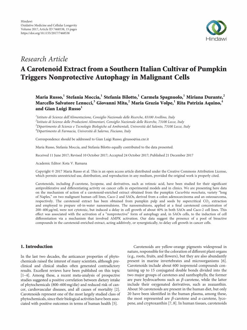
Caco-2 and SAOs cells were incubated with CEN, corre-sponding to 400 and 200μg/ml of carotenoid-enrichedextract (w/v), respectively, at different times. The effect oncell growth is reported in Figure 1 which shows that the treat-ment of Caco-2 with CEN significantly delayed cell prolifer-ation of about 50%, starting from 96h of incubation,compared to controls (untreated and NE; Figure 1(a)). Thesame effect was observed in SAOs cells where the decreasedrate of cell growth (40%) was significant starting from 120h(Figure 1(b)). The two different CEN concentrations applied,for example, 400 and 200μg/ml (w/v) for Caco-2 and SAOs,respectively, were established in preliminary dose-responseexperiments and selected to minimize the toxicity of the vehi-cle (NE; data not shown).
The different dose- and time-response observed in thetwo cell lines employed can be bona fide attributed to the dif-ferences existing in Caco-2 versus SAOs cells in terms of (1)duplication time, slower in the latter; (2) different uptake andmetabolism of CEN; and (3) differences in signalling path-ways sensitive to CEN components. Finally, it is worthwhileto note that the effect on the rate of cell growth, following
4 Oxidative Medicine and Cellular Longevity
CEN treatment, cannot be attributed solely to β-carotene, themajor carotenoid in CEN [36, 38], since when β-carotenewas assayed at the same concentration present in CEN, wedid not measure any significant effect on cell growth (datanot shown).
The delay in cell growth was also confirmed by CyQuantviability assay. The representative micrographs of the cellnuclei, performed by fluorescence microscopy, show thereduced number of viable cells in Caco-2 (Figure 2(c)) andSAOs (Figure 2(f)) after treatment with CEN at 168h, com-pared to NE (Figures 2(b) and 2(e)) and untreated cells(Figures 2(a) and 2(d)). Figures 2(g) and 2(h) show the quan-tification of data represented in Figures 2(a)–2(f), measuredas percentage of emitted fluorescence. CEN significantly slo-wed down the cell growth of 57% in Caco-2 and of 45% inSAOs cell lines.
It is worthwhile to note that we did not detect any sign ofcytotoxicity (e.g., apoptosis and necrosis) following treat-ment of both cell lines with CEN (data not shown). To fur-ther confirm the capacity of CEN to delay cell proliferationwithout affecting cell viability and inducing apoptosis, weperformed a colony formation assay, which gave an indica-tion of CEN effect of CEN after prolonged treatment. Asreported in Figure 3, the number of SAOs colonies formedafter treatment with CEN was reduced of about 60%.
3.2. Activation of Autophagy by the Carotenoid-EnrichedExtract.We hypothesized that the reduced rate of cell prolif-eration induced by CEN, in the absence of any “signature” ofcell death, could be related to the activation of an autophagicprocess. This hypothesis was also comforted by the micro-scopically observation of CEN-treated cells, which evidencedthe presence of intracellular vacuoles. This hypothesis wasinvestigated using multiple assays to detect autophagy andexclude false-positive results.
Caco-2 and SAOs cells were treated with CEN, at a con-centration of 400 and 200μg/ml (w/v), respectively, and
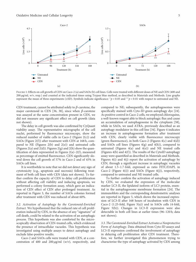
compared to NE; subsequently, the autophagosomes werespecifically stained with Cyto-ID green autophagy dye [24].As positive control in Caco-2 cells, we employed chloroquine,a well-known reagent able to block autophagic flux and causean accumulation of autophagosome in the cytoplasm [58],while in SAOs, we used ATRA, previously described as anautophagy modulator in this cell line [34]. Figure 4 indicatesan increase in autophagosome formation after treatmentwith CEN, clearly visible with fluorescence microscopy(green fluorescence), in both Caco-2 (Figures 4(c) and 4(d))and SAOs cell lines (Figures 4(g) and 4(h)), compared tountreated (Figures 4(a) and 4(e)) and NE treated cells(Figures 4(b) and 4(f)). The results of the CytoID autophagyassay were quantified as described in Materials and Methods.Figures 4(i) and 4(j) report the activation of autophagy byCEN, through a significant increase in autophagic vacuolesof about 1.5–1.7-fold, expressed as ratio FITC/DAPI, inCaco-2 (Figure 4(i)) and SAOs (Figure 4(j)), respectively,compared to untreated and NE treated cells.
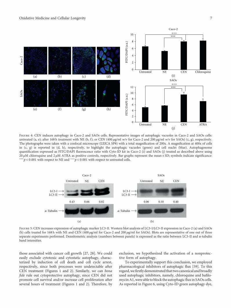
To further confirm the activation of autophagy inducedby CEN, we evaluated the expression of the molecularmarker LC3-II, the lipidated isoform of LC3 protein, essen-tial in the autophagosome membrane formation [24]. Theimmunoblots and the corresponding densitometric analysesare reported in Figure 5, which shows the increased expres-sion of LC3-II after 168 hours of incubation with CEN inCaco-2 (1.25-fold, Figure 5(a)) and in SAOs cells (4-fold,Figure 5(b)). Changes in LC3-II expression were alsodetectable in both cell lines at earlier times (96–120 h; datanot shown).
3.3. The Carotenoid-Enriched Extract Activates a NonprotectiveForm of Autophagy. Data obtained from Cyto-ID assays andLC3-II expression confirmed the involvement of autophagyin delaying cell proliferation after CEN treatment. There-fore, we further investigated this phenomenon trying tocharacterize the type of autophagy activated by CEN among
4.5
3
1.5
0
Live
cells
(×10
6 )
0 96 120 144 168Treatment (h)
UntreatedNECEN
Caco-2
⁎⁎ ⁎⁎
⁎⁎
⁎
(a)
1.5
1
0.5
0
Live
cells
(×10
6 )
0 48 72 120 168Treatment (h)
UntreatedNECEN
SAOs
⁎⁎
⁎⁎
(b)
Figure 1: Effects on cell growth of CEN on Caco-2 (a) and SAOs (b) cell lines. Cells were treated with different doses of NE and CEN (400 and200μg/ml, w/v, resp.) and counted at the indicated times using Trypan blue method, as described in Materials and Methods. Line graphsrepresent the mean of three experiments (±SD). Symbols indicate significance: ∗p < 0 05 and ∗∗p < 0 01 with respect to untreated and NE.
5Oxidative Medicine and Cellular Longevity

Figure 2: CEN delays cell proliferation in Caco-2 and SAOs cells. Micrographs of cell nuclei stained with CyQuant fluorescent dye of Caco-2(a, b, c) and SAOs (d, e, f) after 168 h of treatment with NE and CEN (400 and 200 μg/ml, w/v) (microscope Axiovert 200 Zeiss; FITCfluorescence filter, 200x magnification). Representative images of untreated Caco-2 and SAOs cells: untreated (a, d), incubated with NE(b, e) or CEN (c, f), respectively. CyQuant assay for nuclei quantification is shown in (g) for of Caco-2 and (h) for SAOs cells. CyQuantassay was performed after 168 h of treatment, as described in Materials and Methods. Bar graphs represent the mean± SD; symbolsindicate significance: ∗∗∗p < 0 001 with respect to NE; +++p < 0 001; and ++p < 0 01 with respect to untreated cells.
(a) (b)
Figure 3: CEN slowdowns cell proliferation. SAOs cells were treated with NE and CEN (200 μg/ml, w/v) for 12 days. Colony forming assaywas performed at the end of incubation as described in Materials and Methods and quantitated in the graph shown in (a). Bar graphsrepresent the mean± SD; symbols indicate significance with ∗∗∗p < 0 001 with respect to NE and +++p < 0 001 with respect to untreatedcells. (b) Representative photographs are reported of single colony (top) and total colonies (bottom) of untreated (A) cells and thosetreated with NE (B) and CEN (C) (200 μg/ml, w/v).
6 Oxidative Medicine and Cellular Longevity
those associated with cancer cell growth [27, 28]. We couldeasily exclude cytotoxic and cytostatic autophagy, charac-terized by induction of cell death and cell cycle arrest,respectively, since both processes were undetectable afterCEN treatment (Figures 1 and 2). Similarly, we can bonafide rule out cytoprotective autophagy, since CEN did notpromote cell survival and/or increase cell proliferation afterseveral hours of treatment (Figures 1 and 2). Therefore, by
exclusion, we hypothesized the activation of a nonprotec-tive form of autophagy.
To experimentally support this conclusion, we employedpharmacological inhibitors of autophagic flux [59]. To thisregard,wefirstlydemonstrated that twocanonical andbroadlyused autophagic inhibitors, namely, chloroquine and bafilo-mycinA1,were able to block the autophagicflux in SAOs cells.As reported in Figure 6, using Cyto-ID green autophagy dye,
Figure 4: CEN induces autophagy in Caco-2 and SAOs cells. Representative images of autophagic vacuoles in Caco-2 and SAOs cells:untreated (a, e); after 168 h treatment with NE (b, f); or CEN (400 μg/ml w/v for Caco-2 and 200 μg/ml w/v for SAOs) (c, g), respectively.The photographs were taken with a confocal microscope (LEICA SP8) with a total magnification of 200x. A magnification at 400x of cellsin (c, g) is reported in (d, h), respectively, to highlight the autophagic vacuoles (green) and cell nuclei (blue). Autophagosomequantification expressed as FITC/DAPI fluorescence ratio with Cyto-ID kit in Caco-2 (i) and SAOs (j) treated as described above using20μM chloroquine and 2 μM ATRA as positive controls, respectively. Bar graphs represent the mean± SD; symbols indicate significance:∗∗∗p < 0 001 with respect to NE and +++p < 0 001 with respect to untreated cells.
Caco-2
Untreated NE CEN
0.43 0.66 0.82
LC3-ILC3-II
�훼-Tubulin
(a)
SAOs
Untreated NE CEN
0.06 0.10 0.40
LC3-ILC3-II
�훼-Tubulin
(b)
Figure 5: CEN increases expression of autophagic marker LC3-II. Western blot analysis of LC3-I/LC3-II expression in Caco-2 (a) and SAOs(b) cells treated for 168 h with NE and CEN (400 μg/ml for Caco-2 and 200 μg/ml for SAOs). Blots are representative of one out of threeseparate experiments performed. Densitometric analysis (numbers between panels) is expressed as the ratio between LC3-II and α-tubulinband intensities.
7Oxidative Medicine and Cellular Longevity

both compounds significantly inhibited autophagy demon-strated by the accumulation of autophagosomes in cells. Inthe case of the treatment with bafilomycin A1, we alsodetected an increased expression of LC3-II, the lipidated iso-form of LC3 protein, as expected when a pharmacologicalinhibitor of autophagic flux is used, [24].
To evaluate if the autophagy induced by CEN was protec-tive or nonprotective, we designed the following experiment,based on the assumption that the inhibition of nonprotectiveautophagy does not affect drug sensitivity: in the presence ofa nonprotective autophagy, after chloroquine inhibition, weshould not detect any significant effect on cell growth com-paring CEN+chloroquine versus CEN monotreatment. Onthe opposite, if “cytoprotective” autophagy was triggered byCEN, treating cells with CEN+chloroquine, the inhibitionof the autophagic flux caused by chloroquine should resultin an increased cytotoxicity (apoptosis). As shown inFigure 7(a), the cotreatment chloroquine+CEN failed tosignificantly modify the number of viable cells compared toCEN monotreatment. Therefore, we concluded that theactivation of nonprotective autophagy in SAOs cells by CENwas responsible for the reduced rateof cell growth. Similardatawith chloroquine were obtained in Caco-2 cells (Figure 7(b)).We repeated the same experiment using a different autopha-gic inhibitor, bafilomycin A1. As reported in Figure 7(c), alsoin this case, the cotreatment bafilomycin A1 plus CEN didnot significantly modify the number of viable cells comparedto CEN monotreatment, confirming the activation of a non-protective form of autophagy in SAOs cells.
To understand how CEN interferes with autophagicmachinery, we used an approach different from the treat-ment with pharmacological autophagic inhibitors, that is,the genetic silencing of autophagy regulatory genes. To thisaim, we selected BECN1/Beclin 1 (beclin 1, autophagyrelated), the mammalian homolog of yeast Vps30/Atg6involved in the activation of macroautophagy [24]. Asreported in Figure 8, we demonstrated that a si-RNA againstBECN1 (si-BECN1) was able to almost abolish the expres-sion of BECN1 dose dependently. When we repeated thetreatment with CEN in SAOs cells in the presence of si-BECN1/si-FITC, we obtained a result superimposable to thatdescribed in Figure 7, where we used the pharmacologicalinhibitors. In fact, as reported in Figure 9, treating cells withCEN (Figure 9(a)), no significant differences in cell numbercomparing the si-BECN1 group of treatments versus therespective controls (si-FITC; compare the two orange barsin Figure 9(a)) were detected. These data were also confirmedby micrographs in Figure 9(b), where no significant differ-ences in cell numbers are present comparing Figure 9(b), Fwith Figure 9(b), I.
3.4. CEN Blocks Autophagic Flux in SAOs. To estimate thecapacity of CEN to modulate the autophagic flux, we applieda well-described method consisting in adding the inhibitorchloroquine the last few hours before the end of the treat-ment of a supposed autophagic effector, that is, in our case,CEN and its respective control, NE. Subsequently, we mea-sured autophagosome accumulation using Cyto-ID dye. As
Untreated Chloroquine
+++8
6
4
2
0
FIT/
DA
PI (a
.u.)
(a)
Untreated Bafilomycin A1
10
8
6
4
2
0
++
FIT/
DA
PI (a
.u.)
(b)
Untreated Bafilomycin A1
0.07 1.85
LC3-ILC3-II
�훼-Tubulin
0.07 1.85
(c)
Figure 6: Chloroquine and bafilomycin A1 inhibit autophagy. Autophagosome quantification expressed as FITC/DAPI fluorescence ratiowith Cyto-ID kit in SAOs treated with 20 μM chloroquine (a) or 50 nM bafilomycin A1 (b) for 48 h. Bar graphs represent the mean± SDwith symbols indicating significance: ++p < 0 01 and +++p < 0 001 with respect to untreated cells. (c) Immunoblotting showing LC3-I/LC3-IIexpression in SAOs cells treated with bafilomycin A1 as indicated above. Densitometric analysis values (numbers between panels) wereexpressed by the ratio LC3-II/α-tubulin.
8 Oxidative Medicine and Cellular Longevity
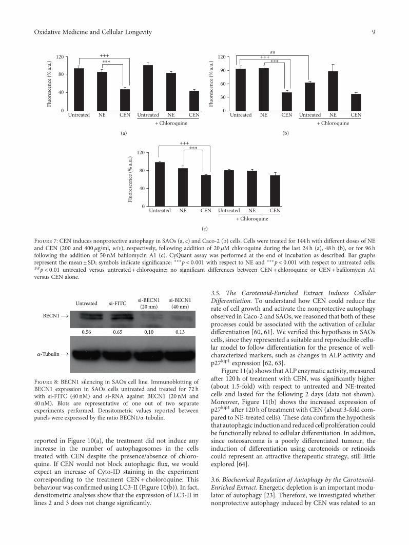
reported in Figure 10(a), the treatment did not induce anyincrease in the number of autophagosomes in the cellstreated with CEN despite the presence/absence of chloro-quine. If CEN would not block autophagic flux, we wouldexpect an increase of Cyto-ID staining in the experimentcorresponding to the treatment CEN+ choloroquine. Thisbehaviour was confirmed using LC3-II (Figure 10(b)). In fact,densitometric analyses show that the expression of LC3-II inlines 2 and 3 does not change significantly.
3.5. The Carotenoid-Enriched Extract Induces CellularDifferentiation. To understand how CEN could reduce therate of cell growth and activate the nonprotective autophagyobserved in Caco-2 and SAOs, we reasoned that both of theseprocesses could be associated with the activation of cellulardifferentiation [60, 61]. We verified this hypothesis in SAOscells, since they represented a suitable and reproducible cellu-lar model to follow differentiation for the presence of well-characterized markers, such as changes in ALP activity andp27kip1 expression [62, 63].
Figure 11(a) shows that ALP enzymatic activity,measuredafter 120 h of treatment with CEN, was significantly higher(about 1.5-fold) with respect to untreated and NE-treatedcells and lasted for the following 2 days (data not shown).Moreover, Figure 11(b) shows the increased expression ofp27kip1 after 120 h of treatment with CEN (about 3-fold com-pared to NE-treated cells). These data confirm the hypothesisthat autophagic induction and reduced cell proliferation couldbe functionally related to cellular differentiation. In addition,since osteosarcoma is a poorly differentiated tumour, theinduction of differentiation using carotenoids or retinoidscould represent an attractive therapeutic strategy, still littleexplored [64].
3.6. Biochemical Regulation of Autophagy by the Carotenoid-Enriched Extract. Energetic depletion is an important modu-lator of autophagy [23]. Therefore, we investigated whethernonprotective autophagy induced by CEN was related to an
120
80
40
0
Fluo
resc
ence
(% a
.u.)
Untreated NE CEN Untreated NE CEN+ Chloroquine
+++⁎⁎⁎
(a)
120
90
60
30
0
Fluo
resc
ence
(% a
.u.)
+++##⁎⁎⁎
Untreated NE CEN Untreated NE CEN+ Chloroquine
(b)
120
80
40
0
Fluo
resc
ence
(% a
.u.)
+++⁎⁎⁎
Untreated NE CEN Untreated NE CEN+ Chloroquine
(c)
Figure 7: CEN induces nonprotective autophagy in SAOs (a, c) and Caco-2 (b) cells. Cells were treated for 144 h with different doses of NEand CEN (200 and 400μg/ml, w/v), respectively, following addition of 20 μM chloroquine during the last 24 h (a), 48 h (b), or for 96 hfollowing the addition of 50 nM bafilomycin A1 (c). CyQuant assay was performed at the end of incubation as described. Bar graphsrepresent the mean± SD; symbols indicate significance: ∗∗∗p < 0 001 with respect to NE and +++p < 0 001 with respect to untreated cells;##p < 0 01 untreated versus untreated + chloroquine; no significant differences between CEN+ chloroquine or CEN+ bafilomycin A1versus CEN alone.
BECN1
�훼-Tubulin
Untreated si-FITC si-BECN1(20 nm)
si-BECN1(40 nm)
0.56 0.65 0.10 0.13
Figure 8: BECN1 silencing in SAOs cell line. Immunoblotting ofBECN1 expression in SAOs cells untreated and treated for 72 hwith si-FITC (40 nM) and si-RNA against BECN1 (20 nM and40 nM). Blots are representative of one out of two separateexperiments performed. Densitometric values reported betweenpanels were expressed by the ratio BECN1/α-tubulin.
9Oxidative Medicine and Cellular Longevity

initial alteration of energetic metabolism. To this aim, intra-cellular ATP was measured as an index of functional integrityof metabolically active cells. As reported in Figure 12, theATP intracellular levels, expressed as chemiluminescencevalue, significantly decreased after 24 hours of incubationwith CEN in SAOs cells. It is worthwhile to note that thereduced ATP level was in the same range of the ATRA treat-ment, employed as positive control [65, 66].
Experimental evidence indicates that lowering ATPintracellular concentration leads to the activation of AMPK,a metabolic modulator of glucose and lipid metabolismwhich acts in response to the alterations of nutrients andATP levels [45, 67]. In addition, AMPK is required forautophagic induction and is involved in the initial stage ofautophagosome nucleation [68, 69]. For these reasons, wehypothesized that, in SAOs cells, autophagy could be
(a) (b)
Figure 9: CEN induces nonprotective autophagy in SAOs cells. SAOs cells were treated with NE and CEN (200 μg/ml, w/v) in the presence ofa si-RNA against BECN1 (si-BECN1) or its corresponding control si-RNA (si-FITC, fluorescein conjugate) for 72 h. CyQuant assay wasperformed at the end of incubation as described in Materials and Methods. (a) Bar graphs represent the mean± SD; symbols indicatesignificance: ∗∗∗p < 0 001, ∗∗p < 0 01, and ∗p < 0 05 versus NE and +++p < 0 001 and +p < 0 05 versus untreated cells; no significantdifferences were detected between si-BECN1 versus si-FITC. (b) Representative micrographs (microscope Axiovert 200 Zeiss; FITCfluorescence filter, 200x magnification) of cell nuclei stained with CyQuant fluorescent dye of SAOs untreated (A), (D), (G), after 72 htreatment with NE (B), (E), (H) and CEN (200mg/ml, w/v) (C), (F), (I) (legend on top). The treatments with si-BECN1 and si-FITCdescribed referred to (G), (H), (I) and (D), (E), (F), respectively (legend on the left).
1210
86420
FITC
/DA
PI (a
.u.)
+++
Untreated NE CEN Untreated NE CEN+ Chloroquine
⁎⁎⁎
(a)
3−NE + Chloroquine4−CEN + Chloroquine
1 2 3 4
0.33 0.66 1.00 1.01
1−NE1−CEN
LC3-ILC3-II
�훼-Tubulin
(b)
Figure 10: CEN inhibits SAOs autophagic flux. (a) Cells were treated with NE and CEN (200 μg/ml, w/v) for 168 h with or without 20 μMchloroquine added in the last 4 h of incubation. Autophagosome quantification was expressed as FITC/DAPI fluorescence ratio and obtainedusing Cyto-ID staining, as described. Bar graphs represent the mean± SD; symbols indicate significance: ∗∗∗p < 0 001 with respect to NE and+++p < 0 001 with respect to untreated cells; no significant differences between CEN+ chloroquine versus CEN alone. (b) Immunoblotting ofLC3-I/LC3-II in SAOs cells treated as in (a). Bands are representative of one out of three separate experiments performed. Densitometricanalysis values (between panels) were expressed by the ratio LC3-II/α-tubulin.
10 Oxidative Medicine and Cellular Longevity

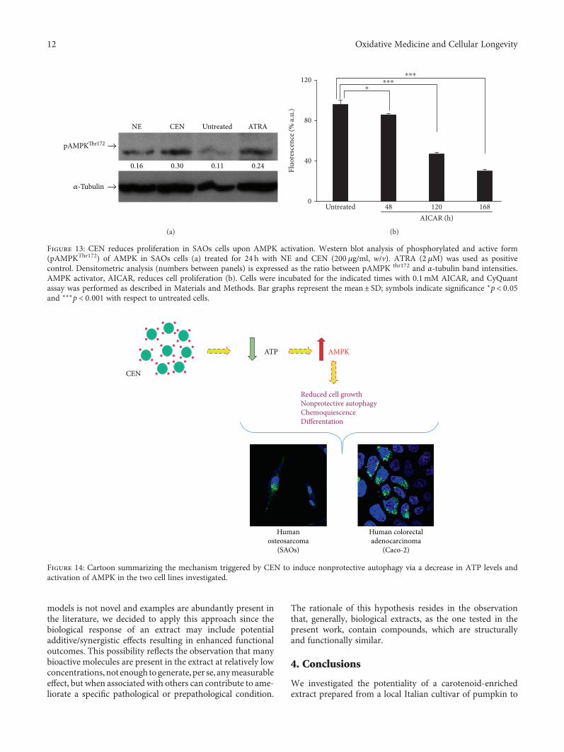
triggered by a reduction of ATP induced by CEN, throughthe activation of AMPK. To this aim, we evaluated the expres-sion levels of the phosphorylated and active form of theAMPK kinase, pAMPKThr172. As reported in Figure 13(a),the phosphorylated protein increased twice after CENtreatment compared to controls. To further confirm theinvolvement of AMPK to explain CEN effects, we employedAICAR, a synthetic AMPK activator [45, 70]. Figure 13(b)shows that AICAR delayed cell growth time dependently inSAOs cells. This effect was already significant at 48 h, butbecame stronger at 120 and 168 h,mimicking CEN treatment.
The possibility that autophagy induced by CEN could befunctionally related to cellular differentiation is supported byrecent data reporting that AMPK is activated during osteo-blast differentiation [60]. Moreover, it is well known thatthe energy sensing LK/AMPK pathway regulates p27kip1
phosphorylation and stabilization mediating the decision ofcells to enter autophagy or apoptosis [71]. In particular, ithas been suggested that early catabolic changes are important
to provide the energy source for osteoblasts; furthermore, theactivation of AMPK is a significant event for osteoblasts toprogress until the final stage of differentiation [60].
An important novelty of the present work regards thepotential impact of the nanoemulsion technology to improvesolubility of the carotenoid-enriched extract, enhancing itsstability and preventing degradation. Further studies are inprogress to improve this technique.
The results, summarized in Figure 14, show how CENcan delay cancer cell growth, leading to a cellular staterecently defined “chemoquiescence” and associated with dif-ferentiation. This effect is always related to an alteration inenergy metabolism and induction of nonprotective form ofautophagy through the activation of AMPK or inhibition ofmTOR/AKT-related signalling [72]. To our knowledge, datareported in this study represent one of the very few examplespresent in the literature where a carotenoid-derived prepara-tion interferes with the autophagic process in malignant celllines. Long time ago, in a short communication and in atotally different contest, before autophagy became known, itwas described the capacity of carotenoids to slow the growthof small cell lung cancer cells [73].
This study suggests that a carotenoid-enriched extract frompumpkin C. moschata could be tested as therapeutic adjuvantin association with conventional drugs [58]. Data from previ-ous studies on ATRA and vitamin D3 reinforce this hypoth-esis [34, 74]. In the next future, suitable animal model will beemployed to test the chemopreventive and/or chemothera-peutic efficacy of the carotenoid-enriched nanoemulsions.
An interesting conclusion emerging from the presentwork is the observation that β-carotene from pumpkin,assayed as an individual compound, did not show any effectson cell proliferation at the concentration present in thenanoemulsions, suggesting, perhaps, a synergism amongdifferent components, or the presence of not yet identifiedbioactive molecules. Future work will shed light on this issue.Although the strategy to test vegetable extracts enriched witha specific class of biological compounds in preclinical cellular
0.12
0.08
0.04
0
ALP
spec
ific a
ctiv
ity
Untreated NE CEN
+++⁎⁎
(a)
Untreated NE CEN
0.04 0.05 0.19
p27kip1
�훼-Tubulin
(b)
Figure 11: CEN induces differentiation in SAOs cells. ALP activity, used as differentiation marker, was assayed and expressed as specificactivity in SAOs cells (a) after 120 h of treatment with NE and CEN (200 μg/ml, w/v). ALP was determined as reported in Materials andMethods, and specific activity was expressed as O.D. values/min/μg protein. Bar graphs represent the mean± SD; symbols indicatesignificance: ∗∗p < 0 01 with respect to NE and +++p < 0 001 with respect to untreated cells. Western blot analysis of p27kip1 proteinexpression in SAOs cells (b) treated as above. Densitometric analysis (numbers between panels) is expressed as the ratio between p27kip1
and α-tubulin band intensities.
25
20
15
10
5
0
ATP
(nM
)
++
Untreated NE CEN ATRA
⁎
Figure 12: CEN modifies ATP intracellular levels in SAOs cells.Cells were treated for 24 h with NE, CEN (200 μg/ml, w/v), and2μM ATRA (positive control). ATP intracellular concentrationswere measured as reported in Materials and Methods andexpressed as nM ATP. Bar graphs represent the mean± SD;symbols indicate significance ∗p < 0 05 with respect to NE and++p < 0 01 with respect to untreated cells.
11Oxidative Medicine and Cellular Longevity
models is not novel and examples are abundantly present inthe literature, we decided to apply this approach since thebiological response of an extract may include potentialadditive/synergistic effects resulting in enhanced functionaloutcomes. This possibility reflects the observation that manybioactive molecules are present in the extract at relatively lowconcentrations, not enough togenerate, per se, anymeasurableeffect, but when associated with others can contribute to ame-liorate a specific pathological or prepathological condition.
The rationale of this hypothesis resides in the observationthat, generally, biological extracts, as the one tested in thepresent work, contain compounds, which are structurallyand functionally similar.
4. Conclusions
We investigated the potentiality of a carotenoid-enrichedextract prepared from a local Italian cultivar of pumpkin to
pAMPKThr172
�훼-Tubulin
NE CEN Untreated ATRA
0.16 0.30 0.11 0.24
(a)
Untreated
Fluo
resc
ence
(% a
.u.)
48 120 168AICAR (h)
120
80
40
0
⁎⁎⁎
⁎⁎⁎
⁎
(b)
Figure 13: CEN reduces proliferation in SAOs cells upon AMPK activation. Western blot analysis of phosphorylated and active form(pAMPKThr172) of AMPK in SAOs cells (a) treated for 24 h with NE and CEN (200 μg/ml, w/v). ATRA (2 μM) was used as positivecontrol. Densitometric analysis (numbers between panels) is expressed as the ratio between pAMPK thr172 and α-tubulin band intensities.AMPK activator, AICAR, reduces cell proliferation (b). Cells were incubated for the indicated times with 0.1mM AICAR, and CyQuantassay was performed as described in Materials and Methods. Bar graphs represent the mean± SD; symbols indicate significance ∗p < 0 05and ∗∗∗p < 0 001 with respect to untreated cells.
AMPKATP
CEN
Reduced cell growthNonprotective autophagyChemoquiescenceDi�erentation
Humanosteosarcoma
(SAOs)
Human colorectaladenocarcinoma
(Caco-2)
Figure 14: Cartoon summarizing the mechanism triggered by CEN to induce nonprotective autophagy via a decrease in ATP levels andactivation of AMPK in the two cell lines investigated.
12 Oxidative Medicine and Cellular Longevity
find applicative outcomes in the biomedical area usingpreclinical models. This possibility is supported by largebody of scientific evidence that fruits and vegetables con-tain dietary chemopreventive agents, including vitamin Aderivatives. We ended up with the observation that thecarotenoid-containing preparation does not directly killcancer cells, but delays their proliferation. In two differentcell lines, Caco-2 and SAOs, the carotenoid-enriched extract,administered in the form of nanoemulsions, induced analteration of the energetic metabolism which resulted in adecreased intracellular concentration of ATP and the activa-tion of an autophagic process (Figure 14). We present evi-dence that the “nonprotective” form of autophagy, triggeredby carotenoid-enriched nanoemulsions, directly or indirectlyinduces cellular differentiation in malignant cells, reducingtheir proliferating capacity.
Conflicts of Interest
The authors declare no conflict of interest.
Acknowledgments
The authors thank Dr. Giuseppe Mazzarella for the kindassistance in using confocal microscopy. This work wassupported by the following: C.I.S.I.A. project “Innovazionee Sviluppo del Mezzogiorno—Conoscenze Integrate perSostenibilità ed Innovazione del Made in Italy Agroalimen-tare—Legge 191/2009” from the Italian Ministry of Economyand Finance to the National Research Council.
References
[1] T. Norat, C. Scoccianti, M. C. Boutron-Ruault et al., “Europeancode against cancer 4th edition: diet and cancer,” Cancer Epi-demiology, vol. 39, Supplement 1, pp. S56–S66, 2015.
[2] D. Aune, E. Giovannucci, P. Boffetta et al., “Fruit and vegetableintake and the risk of cardiovascular disease, total cancer andall-cause mortality-a systematic review and dose-responsemeta-analysis of prospective studies,” International Journalof Epidemiology, vol. 46, no. 3, pp. 1029–1056, 2017.
[3] K. I. Block, C. Gyllenhaal, L. Lowe et al., “Designing a broad-spectrum integrative approach for cancer prevention andtreatment,” Seminars in Cancer Biology, vol. 35, pp. S276–S304, 2015.
[4] G. L. Russo, I. Tedesco, C. Spagnuolo, and M. Russo, “Antiox-idant polyphenols in cancer treatment: friend, foe or foil?,”Seminars in Cancer Biology, vol. 46, pp. 1–13, 2017.
[5] R. K. Saini, S. H. Nile, and S. W. Park, “Carotenoids from fruitsand vegetables: chemistry, analysis, occurrence, bioavailabilityand biological activities,” Food Research International, vol. 76,Part 3, pp. 735–750, 2015.
[6] K. K. Namitha and P. S. Negi, “Chemistry and biotechnologyof carotenoids,” Critical Reviews in Food Science and Nutrition,vol. 50, no. 8, pp. 728–760, 2010.
[7] A. Milani, M. Basirnejad, S. Shahbazi, and A. Bolhassani,“Carotenoids: biochemistry, pharmacology and treatment,”British Journal of Pharmacology, vol. 174, no. 11, pp. 1290–1324, 2017.
[8] J. von Lintig, “Colors with functions: elucidating the biochem-ical and molecular basis of carotenoid metabolism,” AnnualReview of Nutrition, vol. 30, no. 1, pp. 35–56, 2010.
[9] B. Demmig-Adams and W. W. Adams 3rd, “Antioxidants inphotosynthesis and human nutrition,” Science, vol. 298,no. 5601, pp. 2149–2153, 2002.
[10] N. E. Polyakov, T. V. Leshina, T. A. Konovalova, and L. D.Kispert, “Carotenoids as scavengers of free radicals in aFenton reaction: antioxidants or pro-oxidants?,” Free Radi-cal Biology & Medicine, vol. 31, no. 3, pp. 398–404, 2001.
[11] S. Eghbaliferiz and M. Iranshahi, “Prooxidant activity of poly-phenols, flavonoids, anthocyanins and carotenoids: updatedreview of mechanisms and catalyzing metals,” PhytotherapyResearch, vol. 30, no. 9, pp. 1379–1391, 2016.
[12] The ATBC Cancer Prevention Study Group, “The alpha-tocopherol, beta-carotene lung cancer prevention study:design, methods, participant characteristics, and compliance,”Annals of epidemiology, vol. 4, no. 1, pp. 1–10, 1994.
[13] G. S. Omenn, G. E. Goodman, M. D. Thornquist et al.,“Risk factors for lung cancer and for intervention effectsin CARET, the beta-carotene and retinol efficacy trial,” Journalof the National Cancer Institute, vol. 88, no. 21, pp. 1550–1559,1996.
[14] T. Tanaka,M. Shnimizu, andH.Moriwaki, “Cancer chemopre-vention by carotenoids,” Molecules, vol. 17, no. 12, pp. 3202–3242, 2012.
[15] N.Holzapfel, B.Holzapfel, S.Champ, J. Feldthusen, J.Clements,andD. Hutmacher, “The potential role of lycopene for the pre-vention and therapy of prostate cancer: from molecular mech-anisms to clinical evidence,” International Journal ofMolecularSciences, vol. 14, no. 7, pp. 14620–14646, 2013.
[16] M. F. Bakker, P. H. Peeters, V. M. Klaasen et al., “Plasmacarotenoids, vitamin C, tocopherols, and retinol and the riskof breast cancer in the European prospective investigation intocancer and nutrition cohort,” The American Journal of ClinicalNutrition, vol. 103, no. 2, pp. 454–464, 2016.
[17] M. Shen, K. Chen, J. Lu et al., “Protective effect of astaxanthinon liver fibrosis through modulation of TGF-β1 expressionand autophagy,”Mediators of Inflammation, vol. 2014, ArticleID 954502, 14 pages, 2014.
[18] J. Anguiano, T. P. Garner, M. Mahalingam, B. C. Das,E. Gavathiotis, and A. M. Cuervo, “Chemical modulation ofchaperone-mediated autophagy by retinoic acid derivatives,”Nature Chemical Biology, vol. 9, no. 6, pp. 374–382, 2013.
[19] K. N. Kim, S. J. Heo, S. M. Kang, G. Ahn, and Y. J. Jeon,“Fucoxanthin induces apoptosis in human leukemia HL-60cells through a ROS-mediated Bcl-xL pathway,” ToxicologyIn Vitro, vol. 24, no. 6, pp. 1648–1654, 2010.
[20] T. Schenk, S. Stengel, and A. Zelent, “Unlocking the potentialof retinoic acid in anticancer therapy,” British Journal ofCancer, vol. 111, no. 11, pp. 2039–2045, 2014.
[21] L. Hou, C. Gao, L. Chen, G. Hu, and S. Xie, “Essential role ofautophagy in fucoxanthin-induced cytotoxicity to human epi-thelial cervical cancer HeLa cells,” Acta Pharmacologica Sinica,vol. 34, no. 11, pp. 1403–1410, 2013.
[22] L. Galluzzi, M. C. Maiuri, I. Vitale et al., “Cell death modalities:classification and pathophysiological implications,” Cell Death& Differentiation, vol. 14, no. 7, pp. 1237–1243, 2007.
[23] A. M. K. Choi, S. W. Ryter, and B. Levine, “Autophagy inhuman health and disease,” The New England Journal ofMedicine, vol. 368, no. 7, pp. 651–662, 2013.
13Oxidative Medicine and Cellular Longevity

[24] D. J. Klionsky, F. C. Abdalla, H. Abeliovich et al., “Guidelinesfor the use and interpretation of assays for monitoring autoph-agy,” Autophagy, vol. 8, no. 4, pp. 445–544, 2012.
[25] J. M. Gump and A. Thorburn, “Autophagy and apoptosis:what is the connection?,” Trends in Cell Biology, vol. 21,no. 7, pp. 387–392, 2011.
[26] D. J. Klionsky, K. Abdelmohsen, A. Abe et al., “Guidelines forthe use and interpretation of assays for monitoring autophagy(3rd edition),” Autophagy, vol. 12, no. 1, pp. 1–222, 2016.
[27] D. A. Gewirtz, “The four faces of autophagy: implications forcancer therapy,” Cancer Research, vol. 74, no. 3, pp. 647–651,2014.
[28] D. A. Gewirtz, “The challenge of developing autophagy inhibi-tion as a therapeutic strategy,” Cancer Research, vol. 76, no. 19,pp. 5610–5614, 2016.
[29] S. Pattingre, L. De Vries, C. Bauvy et al., “The G-protein regu-lator AGS3 controls an early event during macroautophagy inhuman intestinal HT-29 cells,” The Journal of BiologicalChemistry, vol. 278, no. 23, pp. 20995–21002, 2003.
[30] M. Nollet, S. Santucci-Darmanin, V. Breuil et al., “Autophagyin osteoblasts is involved in mineralization and bone homeo-stasis,” Autophagy, vol. 10, no. 11, pp. 1965–1977, 2014.
[31] L. J. Hocking, C. Whitehouse, and M. H. Helfrich, “Autoph-agy: a new player in skeletal maintenance?,” Journal of Boneand Mineral Research, vol. 27, no. 7, pp. 1439–1447, 2012.
[32] A. T. Vessoni, A. R. Muotri, and O. K. Okamoto, “Autophagyin stem cell maintenance and differentiation,” Stem Cells andDevelopment, vol. 21, no. 4, pp. 513–520, 2012.
[33] A. Pantovic, A. Krstic, K. Janjetovic et al., “Coordinatedtime-dependent modulation of AMPK/Akt/mTOR signalingand autophagy controls osteogenic differentiation of humanmesenchymal stem cells,” Bone, vol. 52, no. 1, pp. 524–531,2013.
[34] M. Russo, F. Messano, M. Basile et al., “Radio-sensitizingeffects of all trans retinoic acid (ATRA) on human chroniclymphocytic leukemia and osteosarcoma cell lines,” EuropeanJournal of Cancer, vol. 61, no. 1, article S163, 2016.
[35] N. Orfali, T. R. O'Donovan, M. J. Nyhan et al., “Induction ofautophagy is a key component of all-trans-retinoic acid-induced differentiation in leukemia cells and a potential targetfor pharmacologic modulation,” Experimental Hematology,vol. 43, no. 9, pp. 781–793.e2, 2015.
[36] M. Durante, M. S. Lenucci, L. D’Amico, G. Piro, and G. Mita,“Effect of drying and co-matrix addition on the yield andquality of supercritical CO2 extracted pumpkin (Cucurbitamoschata Duch.) oil,” Food chemistry, vol. 148, pp. 314–320,2014.
[37] M. Durante, M. S. Lenucci, P. P. Marrese et al., “α-Cyclodex-trin encapsulation of supercritical CO2 extracted oleoresinsfrom different plant matrices: a stability study,” Food Chemis-try, vol. 199, pp. 684–693, 2016.
[38] M. Durante, M. Lenucci, and G. Mita, “Supercritical carbondioxide extraction of carotenoids from pumpkin (Cucurbitaspp.): a review,” International Journal of Molecular Sciences,vol. 15, no. 4, pp. 6725–6740, 2014.
[39] H. D. Silva, M. A. Cerqueira, B. W. S. Souza et al., “Nanoemul-sions of β-carotene using a high-energy emulsification–evapo-ration technique,” Journal of Food Engineering, vol. 102, no. 2,pp. 130–135, 2011.
[40] S. Kentish, T. J. Wooster, M. Ashokkumar, S. Balachandran,R. Mawson, and L. Simons, “The use of ultrasonics for
nanoemulsion preparation,” Innovative Food Science &Emerging Technologies, vol. 9, no. 2, pp. 170–175, 2008.
[41] Y. Yuan, Y. Gao, J. Zhao, and L. Mao, “Characterization andstability evaluation of β-carotene nanoemulsions prepared byhigh pressure homogenization under various emulsifyingconditions,” Food Research International, vol. 41, no. 1,pp. 61–68, 2008.
[42] M. Rousset, M. Laburthe, M. Pinto et al., “Enterocytic differen-tiation and glucose utilization in the human colon tumor cellline Caco-2: modulation by forskolin,” Journal of CellularPhysiology, vol. 123, no. 3, pp. 377–385, 1985.
[43] E. Murray, D. Provvedini, D. Curran, B. Catherwood,H. Sussman, and S. Manolagas, “Characterization of a humanosteoblastic osteosarcoma cell line (SAOS-2) with high bonealkaline phosphatase activity,” Journal of Bone and MineralResearch, vol. 2, no. 3, pp. 231–238, 1987.
[44] F. de Nigris, F. P. Mancini, C. Schiano et al., “Osteosarcomacells induce endothelial cell proliferation during neo-angiogen-esis,” Journal of Cellular Physiology, vol. 228, no. 4, pp. 846–852,2013.
[45] G. L. Russo, M. Russo, and P. Ungaro, “AMP-activated proteinkinase: a target for old drugs against diabetes and cancer,” Bio-chemical Pharmacology, vol. 86, no. 3, pp. 339–350, 2013.
[46] N. A. P. Franken, H. M. Rodermond, J. Stap, J. Haveman, andC. van Bree, “Clonogenic assay of cells in vitro,” Nature Proto-cols, vol. 1, no. 5, pp. 2315–2319, 2006.
[47] G. Russo, M. Russo, I. Castellano, A. Napolitano, andA. Palumbo, “Ovothiol isolated from sea urchin oocytesinduces autophagy in the Hep-G2 cell line,” Marine Drugs,vol. 12, no. 7, pp. 4069–4085, 2014.
[48] I. Tedesco, M. Russo, S. Bilotto et al., “Dealcoholated red wineinduces autophagic and apoptotic cell death in an osteosar-coma cell line,” Food and Chemical Toxicology, vol. 60,pp. 377–384, 2013.
[49] S. Guo, K. J. Pridham, and Z. Sheng, “Detecting autophagy andautophagy flux in chronic myeloid leukemia cells using a cyto-ID fluorescence spectrophotometric assay,”Methods in Molec-ular Biology, vol. 1465, pp. 95–109, 2016.
[50] S. Guo, Y. Liang, S. F. Murphy et al., “A rapid and high contentassay that measures cyto-ID-stained autophagic compart-ments and estimates autophagy flux with potential clinicalapplications,” Autophagy, vol. 11, no. 3, pp. 560–572, 2015.
[51] R. Nilsson, M. Jain, N. Madhusudhan et al., “Metabolicenzyme expression highlights a key role for MTHFD2 andthe mitochondrial folate pathway in cancer,” Nature Commu-nications, vol. 5, p. 3128, 2014.
[52] W. Zhang, Q. Li, C. Song, and L. Lao, “Knockdown ofautophagy-related protein 6, Beclin-1, decreases cell growth,invasion, and metastasis and has a positive effect onchemotherapy-induced cytotoxicity in osteosarcoma cells,”Tumour Biology, vol. 36, no. 4, pp. 2531–2539, 2015.
[53] M. Russo, C. Spagnuolo, S. Volpe, I. Tedesco, S. Bilotto, andG. L. Russo, “ABT-737 resistance in B-cells isolated fromchronic lymphocytic leukemia patients and leukemia cell linesis overcome by the pleiotropic kinase inhibitor quercetinthrough Mcl-1 down-regulation,” Biochemical Pharmacology,vol. 85, no. 7, pp. 927–936, 2013.
[54] M. M. Bradford, “A rapid and sensitive method for the quan-titation of microgram quantities of protein utilizing the princi-ple of protein-dye binding,” Analytical Biochemistry, vol. 72,no. 1-2, pp. 248–254, 1976.
14 Oxidative Medicine and Cellular Longevity

[55] J. R. Gum,W. K. Kam, J. C. Byrd, J. W. Hicks, M. H. Sleisenger,and Y. S. Kim, “Effects of sodium butyrate on human colonicadenocarcinoma cells. Induction of placental-like alkalinephosphatase,” The Journal of Biological Chemistry, vol. 262,no. 3, pp. 1092–1097, 1987.
[56] S. P. M. Crouch, R. Kozlowski, K. J. Slater, and J. Fletcher, “Theuse of ATP bioluminescence as a measure of cell proliferationand cytotoxicity,” Journal of Immunological Methods, vol. 160,no. 1, pp. 81–88, 1993.
[57] H. Orimo and T. Shimada, “Regulation of the human tissue-nonspecific alkaline phosphatase gene expression by all-trans-retinoic acid in SaOS-2 osteosarcoma cell line,” Bone,vol. 36, no. 5, pp. 866–876, 2005.
[58] R. K. Amaravadi, J. Lippincott-Schwartz, X. M. Yin et al.,“Principles and current strategies for targeting autophagy forcancer treatment,” Clinical Cancer Research, vol. 17, no. 4,pp. 654–666, 2011.
[59] D. C. Rubinsztein, P. Codogno, and B. Levine, “Autophagymodulation as a potential therapeutic target for diversediseases,” Nature Reviews Drug Discovery, vol. 11, no. 9,pp. 709–730, 2012.
[60] G. Xi, C. J. Rosen, and D. R. Clemmons, “IGF-I and IGFBP-2stimulate AMPK activation and autophagy, which are requiredfor osteoblast differentiation,” Endocrinology, vol. 157, no. 1,pp. 268–281, 2016.
[61] M. C. Gómez-Puerto, L. P. Verhagen, A. K. Braat, E. W. F.Lam, P. J. Coffer, and M. J. Lorenowicz, “Activation of autoph-agy by FOXO3 regulates redox homeostasis during osteogenicdifferentiation,” Autophagy, vol. 12, no. 10, pp. 1804–1816,2016.
[62] S. B. Rodan, Y. Imai, M. A. Thiede et al., “Characterization of ahuman osteosarcoma cell line (Saos-2) with osteoblastic prop-erties,” Cancer Research, vol. 47, no. 18, pp. 4961–4966, 1987.
[63] H. Drissi, D. Hushka, F. Aslam et al., “The cell cycle regulatorp27kip1 contributes to growth and differentiation of osteo-blasts,” Cancer Research, vol. 59, no. 15, pp. 3705–3711, 1999.
[64] P. Luo, X. Yang, M. Ying et al., “Retinoid-suppressed phos-phorylation of RARα mediates the differentiation pathway ofosteosarcoma cells,” Oncogene, vol. 29, no. 19, pp. 2772–2783, 2010.
[65] A. Chakrabarti, K. Gupta, J. P. Sharma et al., “ATP depletiontriggers acute myeloid leukemia differentiation through anATR/Chk1 protein-dependent and p53 protein-independentpathway,” The Journal of Biological Chemistry, vol. 287,no. 28, pp. 23635–23643, 2012.
[66] F. Papa, R. Lippolis, N. Sardaro, A. Gnoni, and S. Scacco, “Alltrans retinoic acid depresses the content and activity of themitochondrial ATP synthase in human keratinocytes,” Bio-chemical and Biophysical Research Communications, vol. 482,no. 2, pp. 301–304, 2017.
[67] M. M. Mihaylova and R. J. Shaw, “The AMPK signalling path-way coordinates cell growth, autophagy and metabolism,”Nature Cell Biology, vol. 13, no. 9, pp. 1016–1023, 2011.
[68] D. F. Egan, D. B. Shackelford, M. M. Mihaylova et al., “Phos-phorylation of ULK1 (hATG1) by AMP-activated proteinkinase connects energy sensing to mitophagy,” Science,vol. 331, no. 6016, pp. 456–461, 2011.
[69] D. Zhang, W.Wang, X. Sun et al., “AMPK regulates autophagyby phosphorylating BECN1 at threonine 388,” Autophagy,vol. 12, no. 9, pp. 1447–1459, 2016.
[70] J. E. Sullivan, K. J. Brocklehurst, A. E. Marley, F. Carey,D. Carling, and R. K. Beri, “Inhibition of lipolysis and lipogen-esis in isolated rat adipocytes with AICAR, a cell-permeableactivator of AMP-activated protein kinase,” FEBS Letters,vol. 353, no. 1, pp. 33–36, 1994.
[71] J. Liang, S. H. Shao, Z. X. Xu et al., “The energy sensing LKB1–AMPK pathway regulates p27kip1 phosphorylation mediatingthe decision to enter autophagy or apoptosis,” Nature CellBiology, vol. 9, no. 2, pp. 218–224, 2007.
[72] N. Kangwan, J. M. Park, E. H. Kim, and K. B. Hahm, “Chemo-quiescence for ideal cancer treatment and prevention: whereare we now?,” Journal of Cancer Prevention, vol. 19, no. 2,pp. 89–96, 2014.
[73] L. J. Galligan, C. L. Jackson, and L. E. Gerber, “Carotenoidsslow the growth of small cell lung cancer cells,” Annals of theNew York Academy of Sciences, vol. 691, pp. 267–269, 1993.
[74] M. L. Bristol, X. Di, M. J. Beckman et al., “Dual functions ofautophagy in the response of breast tumor cells to radiation:cytoprotective autophagy with radiation alone and cytotoxicautophagy in radiosensitization by vitamin D3,” Autophagy,vol. 8, no. 5, pp. 739–753, 2012.
15Oxidative Medicine and Cellular Longevity

Submit your manuscripts athttps://www.hindawi.com
Stem CellsInternational
Hindawi Publishing Corporationhttp://www.hindawi.com Volume 2014
Hindawi Publishing Corporationhttp://www.hindawi.com Volume 2014
MEDIATORSINFLAMMATION
of
Hindawi Publishing Corporationhttp://www.hindawi.com Volume 2014
Behavioural Neurology
EndocrinologyInternational Journal of
Hindawi Publishing Corporationhttp://www.hindawi.com Volume 2014
Hindawi Publishing Corporationhttp://www.hindawi.com Volume 2014
Disease Markers
Hindawi Publishing Corporationhttp://www.hindawi.com Volume 2014
BioMed Research International
OncologyJournal of
Hindawi Publishing Corporationhttp://www.hindawi.com Volume 2014
Hindawi Publishing Corporationhttp://www.hindawi.com Volume 2014
Oxidative Medicine and Cellular Longevity
Hindawi Publishing Corporationhttp://www.hindawi.com Volume 2014
PPAR Research
The Scientific World JournalHindawi Publishing Corporation http://www.hindawi.com Volume 2014
Immunology ResearchHindawi Publishing Corporationhttp://www.hindawi.com Volume 2014
Journal of
ObesityJournal of
Hindawi Publishing Corporationhttp://www.hindawi.com Volume 2014
Hindawi Publishing Corporationhttp://www.hindawi.com Volume 2014
Computational and Mathematical Methods in Medicine
OphthalmologyJournal of
Hindawi Publishing Corporationhttp://www.hindawi.com Volume 2014
Diabetes ResearchJournal of
Hindawi Publishing Corporationhttp://www.hindawi.com Volume 2014
Hindawi Publishing Corporationhttp://www.hindawi.com Volume 2014
Research and TreatmentAIDS
Hindawi Publishing Corporationhttp://www.hindawi.com Volume 2014
Gastroenterology Research and Practice
Hindawi Publishing Corporationhttp://www.hindawi.com Volume 2014
Parkinson’s Disease
Evidence-Based Complementary and Alternative Medicine
Volume 2014Hindawi Publishing Corporationhttp://www.hindawi.com
![Prodotto di solubilità - Home - people.unica.it · Dato che da ogni mole di CaCO 3 che si dissocia si formano una mole di ioni Ca2+ ed una di ioni CO 3 2-avrò: K ps 2+= [Ca ] [CO](https://static.fdocumenti.com/doc/165x107/5f4ee20697ce9c654407b226/prodotto-di-solubilit-home-dato-che-da-ogni-mole-di-caco-3-che-si-dissocia.jpg)


![ACaseofMultipleMyelomaMisdiagnosedasSeronegative ...downloads.hindawi.com/journals/crirh/2018/9746241.pdf · [10] C.Jorgensen,B.Guerin,V.Ferrazzi,C.Bologna,andJ.Sany, “Arthritis](https://static.fdocumenti.com/doc/165x107/5f60bd003abe4448394b1300/acaseofmultiplemyelomamisdiagnosedasseronegative-10-cjorgensenbguerinvferrazzicbolognaandjsany.jpg)