XXIV Congresso Nazionale SISET Abano 9-12 Novembre … · XXIV Congresso Nazionale SISET Abano 9-12...
45
Trombosi venose superficiali e trombosi venose distali Gualtiero Palareti / Benilde Cosmi Università di Bologna XXIV Congresso Nazionale SISET Abano 9-12 Novembre 2016
-
Upload
truongtuyen -
Category
Documents
-
view
226 -
download
0
Transcript of XXIV Congresso Nazionale SISET Abano 9-12 Novembre … · XXIV Congresso Nazionale SISET Abano 9-12...

Trombosi venose superficiali e
trombosi venose distali
Gualtiero Palareti / Benilde CosmiUniversità di Bologna
XXIV Congresso Nazionale SISET
Abano 9-12 Novembre 2016

2
thrombosis of the superficial vein system (suprafascial veins)
several terms:
superficial phlebitis or superficial thrombophlebitis
varicose vein thrombosis
Mondor’s disease
Trousseau’s syndrome
Most frequent sites: lower limbs
Also upper limbs, neck, thorax
Superficial vein thrombosis (SVT):
definition
3
SVT: traditionally considered to be a benign, self-limiting condition, distinct from thrombosis of the deep veins andrequiring only clinical diagnosis and symptomatic relief
(compression and NSAIDS)
Limited number of methodologically adequate studies for diagnosis and treatment

TVS: patologia sempre benigna?

5Frappe P, et al STEPH study; J Thromb Haemost 2014; 12: 831
Epidemiology of lower limb SVT
in a primary care community of 265 687 people in France,
0.64 per 1000/year lower incidence than that of venous thromboembolism (VTE), which is estimated to be 1/1000/year
Concomitant deep vein thrombosis (DVT) in 24.6% and
pulmonary embolism (PE) in 4.7% of pts
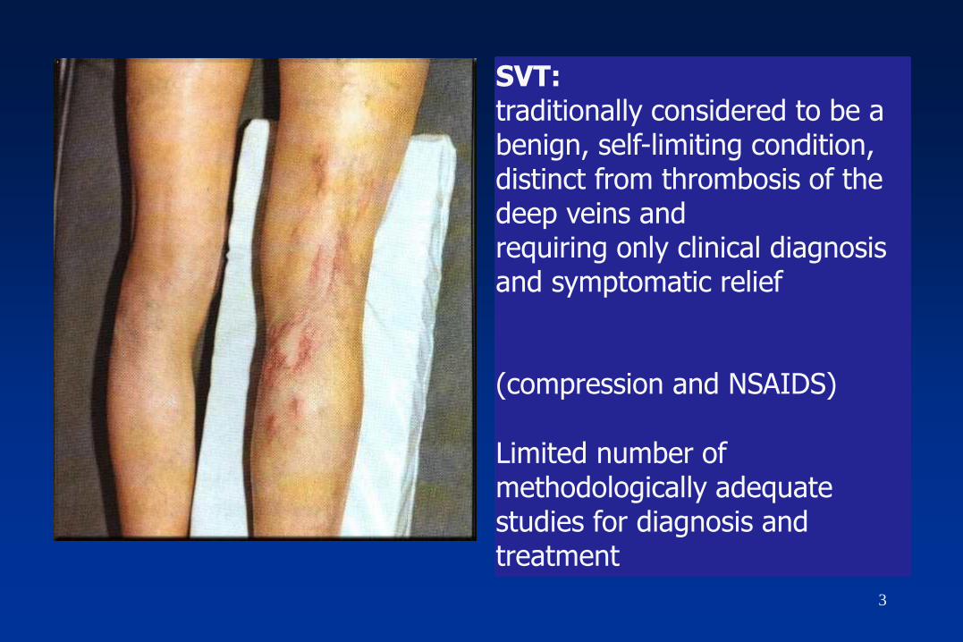
Di Minno et al J Thromb Haemost. 2016 May;14:964-726
Twenty-one studies (4358 patients) evaluated DVT prevalence
11 studies (2484 patients) evaluated PE prevalence in patients with
SVT.
At SVT diagnosis:
DVT weighted mean prevalence 18.1% (95% CI: 13.9- 23.3%)
PE weighted mean prevalence 6.9% (95% CI: 3.9-11.8%)
Screening for a major thromboembolic event may be worthwhile in
some SVT patients, in order to allow adequate anticoagulant
treatment
Other high-quality studies are warranted to confirm these findings
Prevalence of deep vein thrombosis and pulmonary embolism in patients with superficial vein thrombosis: a systematic review
and meta-analysis

7
common risk factors with DVT:
advanced age
surgery
active cancer
pregnancy
hormonal therapy
obesity
autoimmune diseases (particularly Behcet’s and Buerger’s diseases)
varicose veins main risk factor:
80–90% of cases (unlike in DVT)
Overall 3-month mortality:
• < 1% in SVT
• 5% in DVT
• 9–17% in PE , for the lower burden of comorbidities
Pathogenesis and prognosis of lower limb SVT

Thromb Haemost 2014
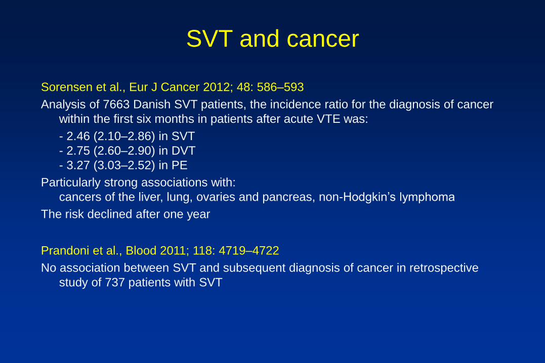
SVT and cancer
Sorensen et al., Eur J Cancer 2012; 48: 586–593
Analysis of 7663 Danish SVT patients, the incidence ratio for the diagnosis of cancer
within the first six months in patients after acute VTE was:
- 2.46 (2.10–2.86) in SVT
- 2.75 (2.60–2.90) in DVT
- 3.27 (3.03–2.52) in PE
Particularly strong associations with:
cancers of the liver, lung, ovaries and pancreas, non-Hodgkin’s lymphoma
The risk declined after one year
Prandoni et al., Blood 2011; 118: 4719–4722
No association between SVT and subsequent diagnosis of cancer in retrospective
study of 737 patients with SVT

Kitchens C. How I treat superficial venous thrombosis Blood 2011;117:39
Reasons Comments
Risk factors 1. Not an entirely benign disease DVT, PE, fatality not rare
2. Both SVT and VTE associated with similar clinical hypercoagulability (eg, trauma, surgery,pregnancy, immobility, obesity, advancing age, malignancy)
3. Incidence of thrombophilia enriched in both SVT and VTE patients
Natural history
1. Coexistence of VTE at time of diagnosis of SVT
Averages 25%
2. Progression of SVT to VTE Averages 10%-20%/y
3. Prior VTE a risk factor for future SVT
4. Prior SVT a risk factor for future VTE
5. No current plausible putative theory that justifies segregation of (local) SVT apart, different,and unique from (systemic) VTE 10
SVT: superficial venous manifestation of a systemic process that is more commonly called VTE
11
Diagnosis of SVT
No diagnostic gold standard
straightforward clinical manifestations :
pain, tenderness, swelling, warmth, and erythema, with a
palpable cord along the course of a superficial vein
objective testing may not be considered
mandatory for diagnosis (unlike for DVT).

12
Clinical diagnosis of lower limb SVT
clinical assessment alone frequently underestimates the true
extent of thrombosis, which may propagate from superficial into
deep veins
- no data on sensitivity and specificity of SVT clinical diagnosis
- no scoring system based on those clinical features
that make SVT more likely
- no validated diagnostic algorithms available

13
Objective diagnosis of SVT
Ultrasonography:objective test of choice for confirming SVT clinical suspicion superficial veins can be easily explored
same principles of DVT diagnosis: compression ultrasonography (CUS):
lack of compressibility of a superficial vein segment, and impairment of blood flow
Also evaluation of SVT true extent and
exclusion of concomitant DVT

14
Cosmi B, JTH 2015
scarcity of methodologically sound studiesvariable approach in clinical routine depending on local resources
unlike for DVT diagnosis, need for testing unclear and it may still be considered to be neither mandatory nor urgent.
Clinically suspected SVT (any site)
(pain, erythema, warmth hardness along the course of superficial vein)
↓
Ultrasonography (CUS) within 24-48 hours (bilateral if lower limbs involved)
↓
(if high clinical suspicion for lower or upper limb SVT:
therapeutic doses LMWH while waiting for
ultrasonography to exclude concomitant DVT)
Algorithm for the diagnosis of SVT

15
Aims of treatment of lower limb SVT
1-symptom relief
2-prevention of VTE, in relation to the thrombotic burden with different risks of thromboembolic complications
No consensus on the optimal management of SVT (without DVT or PE) in relation to the thrombotic burden
a small thrombus (< 4–5cm in length on ultrasonography):
minor, benign, and self-limiting, requiring only symptom relief
significant thrombus burden (> 4–5 cm in length):
more aggressive treatment, for its higher risk of extension

16
Aims of treatment of lower limb SVT
RCTs included patients with the most frequent locations of SVT,
i.e. long and short saphenous veins
higher risk of extension into the deep vein system through the
saphenofemoral (SFJ)/saphenopopliteal junction
SVT of a long saphenous vein with the thrombus head within 3 cm
of the SFJ excluded from interventional studies
equivalent to a DVT with regard to its high risk of progression (10–
70%) and therapeutic anticoagulation is indicated

Multicenter, randomized, double-blind, controlled vs placebo on efficacy and safety of
Fondaparinux (Arixtra) for the treatment of SVT
Patients enroled : 3.002
Inclusion: SVT confermed with CUS, > 5 cm length
Exclusion: SVT < 3 cm from saphenous-femoral cross, thrombotic events < previous 6 months,
active cancer, warfarin, NSAIDs, recent bleeds, platelets <100.000 plt/dl), Cr Cl< 30 ml/min
Treatments: Fondaparinux 2,5 mg or Placebo
Duration: 45 d
Follow-up: 1 month
2010;363:1222-32.

No difference for bleeding between treatment and placebo


Prospective, randomized, double-blind placebo controlled multicenter study (16 centres)

Consecutive outpatients were randomly assigned to receive in a
double-blind fashion one of the following subcutaneous
treatments with an allocation ratio of 1:1:1:
A Parnaparin 4250 UI aXa o.d. for 30 days (prophylactic dose of LMWH
for 30 days).
B Parnaparin 8500 UI aXa o.d. for 10 days followed by
6400 UI aXa once daily for 20 days (intermediate dose of LMWH for 30
days).
C Parnaparin 8500 UI aXa o.d. for 10 days followed by placebo for 20
days (intermediate dose of LMWH for 10 days).
TRATTAMENTI

Cumulative incidence of events in the 33 days of treatment + 60 days of follow-up
in the 3 groups Logrank test for trend P=0.0117
Log-rank (Mantel-Cox) Test p<0.0001
A VS B P<0.0001; B VS C P= 0.06; A VS C P=0.019
Cosmi et al.
JTH 2012

CONCLUSIONS
SVTs require anticoagulant treatment for at least 30 days
LMWH = relatively high dose
Fondaparinux = prophylactic dose
Class II elastic stockings
Some SVT still have late complications after 30 d of treatment
How to identify these patients?
Evaluation of individual risk factors

9th ACCP Consensus(Kearon et al. Chest 2012)
8.1.1. In patients with SVT ….of at least 5 cm in length,
we suggest the use of a prophylactic dose of
fondaparinux or LMWH for 45 days over no
anticoagulation (Grade 2B) .
8.1.2. In patients with SVT who are treated with
anticoagulation, we suggest fondaparinux 2.5 mg
daily over a prophylactic dose of LMWH (Grade 2C)

Comments on SVT
•Complications are frequent, associated with or occurring after SVT
•Complications can be clinically important
•Risk factors are similar to those of DVT/PE
•Thrombophilic alterations are frequent and similar to those found in patients with DVT
•A limited course of AC is mandatory but does not always prevent complications

Trombosi venose distali

Schematic representation of leg veins1, External iliac vein;
2, common femoral vein;
3, greater saphenous vein;
4, profound femoral vein;
5, (superficial) femoral vein;
6, popliteal vein;
7, anterior tibial confluent segment;
8, posterior tibial confluent segment;
9, peroneal confluent segment;
10, anterior tibial veins;
11, posterior tibial veins;
12, peroneal veins;
13, gastrocnemius muscle veins (medial head);
14, soleus muscle veins.

The OPTIMEV study: a French, multicenter, prospective,
observational study of inpatients and outpatients
referred to vascular medicine physicians for clinically suspected
VTE and followed for 3 years
(Galanaud et al., JTH 2014)
DVT at baseline = 1643
- Proximal = 43.2%
- Distal = 56.8%

Clinical evolution at 3 month follow-up
Simplified
CUS
417
Calf DVT
64
(15.1%)
No calf DVT
359
p
Outcomes at 3 mo:
1 PE; 2 Prox. DVT;
2 Calf DVT
5
(1.2%)
5 (7.8%)
[3 (4.7%)]*
3 (0.8%) 0.003
* excluding the 2 subjects in whom DVT was picked at the 2nd CUS

2016
The rationale for not routinely examining the distal veins:
(1) other assessment (e.g. low clinical probability; negative D-dimer);
(2) a repeat US of the proximal veins can be done after a week
(3) false-positive findings for DVT occur

2016
In patients with acute IDDVT of the leg and
(i)without severe symptoms or risk factors for extension, we suggest
serial imaging of the deep veins for 2 weeks over AC (Grade 2C),
(ii)with severe symptoms or risk factors for extension, we suggest AC
over serial imaging of the deep veins (Grade 2C).

2016
If the calf veins are imaged and IDDVT is diagnosed,
two management options:
1)treat patients with AC therapy;
2)do not treat patients with AC therapy unless extension of their DVT
is detected on a follow-up US examination (e.g. after one and two weeks)

2016
In patients with an IDDVT provoked by surgery or by a nonsurgical
transient risk factor,
- we suggest treatment with AC for 3 months
over treatment of a shorter period (Grade 2C),
- we recommend treatment with AC for 3 months over treatment of a
longer time-limited period (e.g. 6, 12 or 24 months) (Grade 1B),
- we recommend treatment with AC for 3 months over extended therapy
(no scheduled stop date) (Grade1B).

EVENTS DURING TREATMENT:
10/171 PTS (5.8%) had complications:
5 (2.9%) proximal DVT (all unprovoked IDDVT
the remaining extension of IDDVT
No major bleeding; 1.7% minor bleeding
EVENTS DURING 3 MO. FOLLOW-UP
5 complications: (3 developed cancer)
4 proximal DVT (3 had an unprovoked IDDVT)
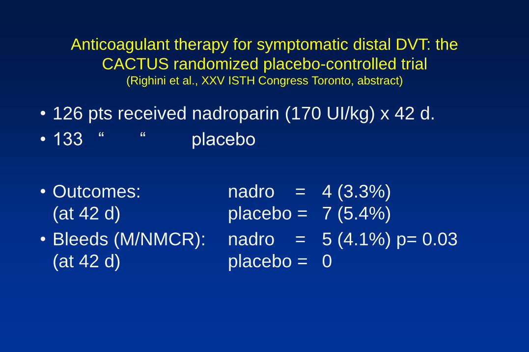
Anticoagulant therapy for symptomatic distal DVT: the
CACTUS randomized placebo-controlled trial(Righini et al., XXV ISTH Congress Toronto, abstract)
• 126 pts received nadroparin (170 UI/kg) x 42 d.
• 133 “ “ placebo
• Outcomes: nadro = 4 (3.3%)
(at 42 d) placebo = 7 (5.4%)
• Bleeds (M/NMCR): nadro = 5 (4.1%) p= 0.03
(at 42 d) placebo = 0

IDDVT treatment: current management in symptomatic pts
• Idiopathic IDDVT
Therapeutic LMWH followed by oral
anticoagulation (2.0-3.0 INR) for 3 months,
elastic stocking
• Secondary IDDVT
Therapeutic IDDVT for 1 week, half dose for 3
weeks, elastic stocking

The risk of recurrence after
treatment of IDDVT

Galanaud
JTH 2014

Results: 90 patients (male 48.9%) enrolled.
At follow-up (24±2 months) = 17 events (18.9%)
3 PE (two in cancer), 4 proximal DVTs (one in cancer) and 10 IDDVT.
Associated with a higher risk of complications
• male sex (HR 4.73 CI95%: 1.55-14.5; p = 0.006)
• cancer (HR 5.47 CI95%: 1.76-17.6; p = 0.003)

Conditions or risk factors for complications
after a first IDDVT (1)
Higher risk•(Axial vs Muscular IDDVT)
•Previous VTE events
•Males
•Age >50 years
•Cancer
•Unprovoked IDDVT
•Secondary IDDVT with persistently hampered mobilisation
•IDDVT involving the popliteal trifurcation
•IDDVT involving >1 calf vein
•IDDVT present in both legs
•Presence of predisposing diseases (e.g. inflammatory bowel diseases)
•Known thrombophilic alterations
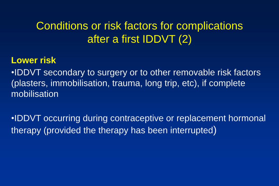
Conditions or risk factors for complications
after a first IDDVT (2)
Lower risk
•IDDVT secondary to surgery or to other removable risk factors
(plasters, immobilisation, trauma, long trip, etc), if complete
mobilisation
•IDDVT occurring during contraceptive or replacement hormonal
therapy (provided the therapy has been interrupted)
Rivaroxaban for the treatment of
symptomatic IDDVT (RIDTS study)
Proposed by: Walter Ageno &
Gualtiero Palareti

RIDTS study: Study design
All patients receive rivaroxaban, 15 mg BID for 3 weeks followed by open label rivaroxaban 20 mg OD for 3 weeks.
At the end of the first 6 weeks of treatment, all patients will be randomized to
A) Long treatment: rivaroxaban 20 mg OD for further 6 weeks B) Short treatment: placebo for further 6 weeks
Randomization: using an IVRS system implemented to guarantee the balanced and blinded fashion of the two groups
Follow-up = 2 years
RIDTS study
Più di 30 centri hanno dichiarato la loro
disponibilità allo studio

Grazie