Sede Amministrativa: Università degli Studi di Padova … · CURRICOLO: Biologia Cellulare CICLO...
104
1 Sede Amministrativa: Università degli Studi di Padova Dipartimento di Biologia Cellulare CORSO DI DOTTORATO DI RICERCA IN: Bioscienze e Biotecnologie CURRICOLO: Biologia Cellulare CICLO XXIX EMILIN1 role in tumor growth after enzymatic degradation Tesi redatta con il contributo finanziario del Centro di Riferimento Oncologico di Aviano Coordinatore: Ch.mo Prof. Paolo Bernardi Supervisore: Ch.mo Prof. Paolo Bernardi Co-Supervisore: Dr.ssa Paola Spessotto Dottorando : Orlando Maiorani
Transcript of Sede Amministrativa: Università degli Studi di Padova … · CURRICOLO: Biologia Cellulare CICLO...

1
Sede Amministrativa: Università degli Studi di Padova
Dipartimento di Biologia Cellulare
CORSO DI DOTTORATO DI RICERCA IN: Bioscienze e Biotecnologie
CURRICOLO: Biologia Cellulare
CICLO XXIX
EMILIN1 role in tumor growth after enzymatic degradation
Tesi redatta con il contributo finanziario del Centro di Riferimento Oncologico di Aviano
Coordinatore: Ch.mo Prof. Paolo Bernardi
Supervisore: Ch.mo Prof. Paolo Bernardi
Co-Supervisore: Dr.ssa Paola Spessotto
Dottorando : Orlando Maiorani

2
A mio padre
“Possa la mia anima rifiorire innamorata per tutta
l’esistenza”
(Rudolf Steiner)
“È più importante impedire a un animale di soffrire,
piuttosto che restare seduti a contemplare i mali
dell'Universo pregando in compagnia dei sacerdoti.”
(Buddha)

3
Table of Contents
Riassunto ............................................................................................................................... 6
Summary ................................................................................................................................ 8
Abbreviations ..................................................................................................................... 10
Chapter 1 ............................................................................................................................. 12
1 Elastic fibers ..................................................................................................................................... 12
1.2 Fibrillar components ................................................................................................................. 13
1.2.1 Collagens .................................................................................................................................................................... 13
1.2.2 Collagen type I, fibrils and fibers....................................................................................................................... 13
1.3 Reticular fibers ............................................................................................................................. 14
1.4 Elastic fibers and microfibrils ................................................................................................. 14
1.5 EMILINs family ............................................................................................................................. 15
1.5.1 EMILIN1 localization ............................................................................................................................................... 16
1.5.2 EMILIN1 structure .................................................................................................................................................... 16
1.5.3 Functions of EMILIN1 ............................................................................................................................................. 18
1.5.4 EMI domain function ............................................................................................................................................. 18
1.5.5 EMILIN1 and lymphatic system ......................................................................................................................... 19
1.6 gC1q: the EMILIN1 functional domain ............................................................................... 19
1.6.1 Integrins α4β1/α9β1 ............................................................................................................................................... 21
1.6.2 Structural aspects of gC1q domain/ integrin ligation .............................................................................. 21
1.6.3 The functional role of gC1q domain: adhesion, migration and proliferation ................................. 22
Chapter 2 ............................................................................................................................. 24
2.1Tumor Microenvironment ........................................................................................................ 24
4
2.2 Neutrophils ................................................................................................................................... 24
2.3 Neutrophil recruitment ............................................................................................................ 25
2.4 ECM molecules favoring cancer and its progression .................................................... 26
2.4.1 ECM Molecules with Anti-Tumor Activity ...................................................................................................... 28
2.5 EMILIN1 and cancer .................................................................................................................. 29
Chapter 3 ............................................................................................................................. 31
3.1 Neutrophil Elastase .................................................................................................................... 31
3.2 Biology of NE ............................................................................................................................... 31
3.2.1 NE Structure and Function................................................................................................................................... 31
3.2.2 Role of Extracellular NE ......................................................................................................................................... 32
3.2.3 Imbalance between NE and its Inhibitors in Inflammatory Disease .................................................... 33
3.3 The role of NE in cancer ........................................................................................................... 34
3.4 MMPs .............................................................................................................................................. 34
3.4.1 Matrix metallo-proteinase-3 (MMP-3) ............................................................................ 35
3.4.2 MMP-9 ........................................................................................................................................ 35
3.4.3 MT1-MMP.................................................................................................................................. 36
AIM ......................................................................................................................................... 38
Chapter 4 ............................................................................................................................. 39
Material and methods .................................................................................................... 39
4.1 Cell cultures .................................................................................................................................. 39
4.2 EMILIN1 expression and purification .................................................................................. 39
4.3 gC1q WT PRODUCTION, PURIFICATION AND SDS-PAGE ANALYSIS ..................... 40
4.4 Enzymatic digestion .................................................................................................................. 41
4.5 Immunoblotting .......................................................................................................................... 41
4.6 Transfection of HEK 293 cell line .......................................................................................... 42
5
4.7 MALDI TOF analysis ................................................................................................................... 42
4.8 SITE DIRECTED MUTAGENESIS AND CLONING INTO pQE30 VECTOR FOR THE
PRODUCTION OF gC1q MUTANTS ............................................................................................. 42
4.9 Gelatin zymography .................................................................................................................. 43
4.10 Adhesion assay: quantitative cell adhesion assay: “CAFCA ...................................... 44
4.11 Adhesion assay and proliferation by Xcelligence technology ................................. 45
4.12 Statistical analysis .................................................................................................................... 45
Chapter 5 ............................................................................................................................. 46
Results ................................................................................................................................... 46
5.1 The MMPs weak proteolytic activity on EMILIN1 ........................................................... 46
5.2 MMPs degradation in physiological conditions.............................................................. 51
5.3 EMILIN1 adhesive and antiproliferative properties are conserved after MT1-
MMP proteolytic action .................................................................................................................. 55
5.4 NE degrades the functional domain gC1q........................................................................ 56
5.5 Identification of NE cleavage site on the functional domain gC1q ......................... 60
5.6 The mutant R914W gC1q is resistant to NE ..................................................................... 63
5.7 R914W preserves its features after NE treatment .......................................................... 65
Chapter 6 ............................................................................................................................. 70
6.1Discussion ....................................................................................................................................... 70
References ........................................................................................................................... 75
Neutrophil elastase cleavage of the gC1q domain impairs the EMILIN1-
α4β1 integrin interaction, cell adhesion and anti-proliferative activity…..92
6
Riassunto EMILIN1 è una glicoproteina della matrice extracellulare coinvolta in molti processi
cellulari. Nella sua interezza è in grado di governare processi di elastogenesi dei tessuti
e ha un ruolo importante nella regolazione della struttura dei vasi linfatici. Inoltre, è
una proteina multi dominio, e, grazie ai suoi diversi domini funzionali, regola numerosi
altri processi. Ad esempio, EMILIN1 regola l’omeostasi della pressione sanguigna
tramite il suo dominio N-terminale, chiamato EMI domain; la regolazione dell’adesione
e della proliferazione cellulare, invece, avviene tramite l’interazione tra il suo dominio
C-terminale, chiamato gC1q, e l’integrina α4β1. L’effetto dell’interazione del dominio
gC1q con l’integrina α4β1 è del tutto peculiare. Generalmente l’interazione delle
molecole della matrice extracellulare con i recettori integrinici determina un aumento
della proliferazione piuttosto una diminuizione come nel caso dell’interazione gC1q
l’integrina α4β1. Studi condotti su topi EMILIN1-/- in cui non è possibile l’interazione
gC1q-integrina α4β1, hanno messo in evidenza l’attivazione del pathway delle MAP
chinasi, che induce un aumento della proliferazione cellulare. Dati precedentemente
pubblicati nel nostro laboratorio hanno dimostrato che nel microambiente tumorale
EMILIN1 viene degradata dalla elastasi rilasciata dai neutrofili, perdendo così le sue
proprietà funzionali. Altri autori hanno ipotizzato, mediante l’uso di un approccio
proteomico, che EMILIN1 può essere un substrato di alcune metalloproteasi, in
particolare le metalloproteasi 3, 9 e 14. Nel presente lavoro si dimostra che queste tre
metalloproteasi non sono in grado di svolgere un’ azione proteolitica rilevante. Tra
queste tre metalloproteasi, infatti, soltanto la metalloproteasi 14 sembra esercitare
un’azione proteolitica, anche se minima, su EMILIN1. Questa attività proteolitica
comunque non è paragonabile a quella esercitata dall’elastasi neutrofila, ed inoltre,
cosa ancora più importante, le proprietà funzionali di EMILIN1 non vengono
compromesse dopo il trattamento della proteina con la metalloproteasi 14, come è
stato dimostrato mediante saggi di adesione e proliferazione cellulare. Al contrario, il
trattamento con l’elastasi neutrofila determina la perdita delle proprietà regolatorie di
EMILIN1, causando una diminuzione dell’adesione ed un aumento della proliferazione
cellulare e suggerendo che la degradazione avviene nel dominio gC1q. Tra gli enzimi

7
testati, infatti, solo l’elastasi neutrofila è in grado di degradare e, quindi,
compromettere le funzioni regolatorie del dominio funzionale gC1q. E’ nata, quindi,
l’esigenza di identificare il sito/i di taglio dell’elastasi neutrofila sul dominio funzionale
gC1, per poi, costruire un mutante resistente all’azione proteolitica dell’elastasi
neutrofila. Consultando vari database riportanti i siti di taglio predetti
sperimentalmente dell’elastasi neutrofila e mediante un approccio di mutagenesi sito-
specifica, abbiamo creato vari mutanti. Tra questi un mutante che presentava la
sostituzione dell’aminoacido arginina con l’aminoacido triptofano, il mutante R914W,
si è dimostrato resistente all’azione proteolitica dell’elastasi neutrofila. Saggi funzionali
di adesione e proliferazione hanno confermato la capacità, da parte di questo mutante,
di preservare le sue proprietà regolatorie.

8
Summary EMILIN1 is a ECM glycoprotein involved in several cellular processes. In particular,
EMILIN1 is involved in elastogenesis processes and in the maintenance of lymphatic
vessels structures. EMILIN1 presents several regulatory properties exercised through
its EMI domain that is located at N-terminal region and is able to regulate homeostasis
of blood pressure, and its gC1q domain located at C-terminal region. In particular,
gC1q domain is involved in the regulation of both cell adhesion and cell proliferation
through the binding with α4β1 integrin. EMILIN1-/- mice present an increase of tumoral
cell proliferation; this is due to the loss of the interaction between gC1q domain-α4β1
integrin, that determines the activation of the MAPK pathway, resulting in upregulation
of cell proliferation. Studies previously published in our laboratory demonstrated that
neutrophil elastase, released by neutrophils present in tumor microenvironment, is able
to degrade EMILIN1 resulting in the loss of its regulatory functions.
Other authors have proposed EMILIN1 a possible substrate of several matrix
metalloproteinases: matrix metalloproteinase-3 (MMP-3), matrix metalloproteinase-9
(MMP-9) and matrix metalloproteinase 14 (MT1-MMP). We demonstrated that among
these MMPs only MT1-MMP shows a weak proteolytic activity on EMILIN1. Moreover,
we observed that MT1-MMP was not able to impair EMILIN1 functions. On the contrary
we observe that the digestion of EMILIN1 with neutrophil elastase was able to impair
EMILIN1 tumor suppressor role. At this step, we wanted to analyze the capability of
neutrophil elastase to degrade the gC1q domain. We digested the gC1q domain with
several proteases and we observed that among the tested proteases only neutrophil
elastase was able to degrade the gC1q domain and to impair its functionality. Thus, we
wanted to pinpoint the neutrophil elastase cleavage site on gC1q domain, in order to
generate a mutant of gC1q domain resistant to neutrophil elastase cleavage. We
consulted several peptidase database that contained predicted neutrophile elastase
cleavage sites, on these basis we generated several gC1q mutants. Among these
mutants that generated, we found that the mutant R914W, in which aminoacid arginine
was substituted with aminoacid tryptophan, was resistant to neutrophil elastase

9
cleavage. Functional adhesion and proliferation assays confirmed the capability of R914
mutant to maintain its properties after neutrophil elastase treatment.

10
Abbreviations
ACRP-30 Adipocyte Complement-Related Protein of 30kDa
BSA Bovine Serum Albumine
CAFCA Centrifugal Assay for Fluorescent based Cell Adhesion
DMEM Dulbecco’s Modified medium
ECM Extracellular Matrix
EDTA Ethylen Dynamine Tetra-Acetate
FBS Fetal Bovine Serum
FN Fibronectin
GAG Glycosaminoglican
gC1q globular C1q domain
HIS-tag Histidine tag
IPTG Isopropyl β-D-1-thiogalactopyranoside
LB Luria Bertani
LECs Lymphatic endothelial cells
mAb monoclonal antibody
MMPs Matrix metalloproteinases
NE Neutrophil elastase
NETs Neutrophil extracellular traps
NF-kB Nuclear Factor kappa-light-chain-Enhancerof Activated B cells
NMR Nuclear Magnetic Resonance
NSPs Neutrophil Serine Proteases
PAGE PolyAcrilamide Gel Electrophoresis
PBS Phosphate Buffer Saline
PCR Polymerase Chain Reaction
PG Proteoglycan
PR3 Proteinase 3
ROS Radical Oxygen Species

11
SDS Sodium Dodecyl Sulphate
TBT-T Tris Buffered Saline Tween-20
TGF-β Transforming Growth Factor- beta
TIMPs Tissue Inhibitors of Metallo Proteases
TNF Tumor Necrosis Factor
VEGF Vascular Endothelial Growth Factor
WB Western Blot
WT Wild type
12
Chapter 1
1 Elastic fibers
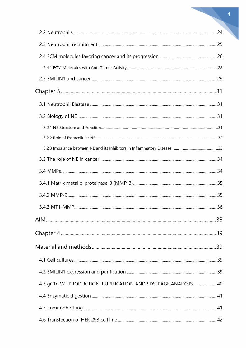
The extracellular matrix (ECM) is an intricate network of the non-cellular component
secreted by surrounding cells and involved in structural and biochemical functions that
are fundamental for tissue morphogenesis, differentiation and homeostasis [1]. ECM is
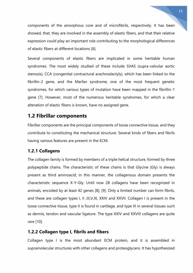
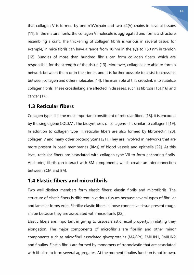
formed mainly by collagens, fibronectin, laminin, and proteoglycans (FIG 1).
The proportions of these components can vary greatly depending on tissue type [2].
Elastic fibers are major constituents of the ECM. They appear with highly variable
morphology in different connective tissues and confer the properties of resilience and
elastic recoil [3]. Analysis by electron microscopy allowed a common structure to be
recognized in all elastic fibers: they are formed by an amorphous core surrounded by
a coat of fibrillar elements, the microfibrils, variously represented in different tissues. In
spite of this simple basic organization, elastic fibers are now known to be highly
complex structures [4]. They are composed of several specific structural proteins,
including elastin, fibrillin 1 and 2, MFAP (Micro-Fibril Associated Protein) [5], LTBP
(Latent TGF-β Binding Protein), fibulin and EMILINs. Elastin and fibrillin are the main
FIGURE 1: Schematic view of the Extracellular Matrix organization. Adapted from: Karp, Cell and
Molecular Biology Concepts and Experiments, 2010
13
components of the amorphous core and of microfibrils, respectively. It has been
showed, that, they are involved in the assembly of elastic fibers, and that their relative
expression could play an important role contributing to the morphological differences
of elastic fibers at different locations [6].
Several components of elastic fibers are implicated in some heritable human
syndromes. The most widely studied of these include SVAS (supra-valvular aortic
stenosis), CCA (congenital contractural arachnodactyly), which has been linked to the
fibrillin-2 gene, and the Marfan syndrome, one of the most frequent genetic
syndromes, for which various types of mutation have been mapped in the fibrillin-1
gene [7]. However, most of the numerous heritable syndromes, for which a clear
alteration of elastic fibers is known, have no assigned gene.
1.2 Fibrillar components
Fibrillar components are the principal components of loose connective tissue, and they
contribute to constituting the mechanical structure. Several kinds of fibers and fibrils
having various features are present in the ECM.
1.2.1 Collagens
The collagen family is formed by members of a triple helical structure, formed by three
polypeptide chains. The characteristic of these chains is that Glycine (Gly) is always
present as third aminoacid; in this manner, the collagenous domain presents the
characteristic sequence X-Y-Gly. Until now 28 collagens have been recognized in
animals, encoded by at least 42 genes [8], [9]. Only a limited number can form fibrils,
and these are collagen types I, II ,III,V,XI, XXIV and XXVII. Collagen I is present in the
loose connective tissue, type II is found in cartilage, and type III in several tissues such
as dermis, tendon and vascular ligature. The type XXIV and XXVIII collagens are quite
rare [10].
1.2.2 Collagen type I, fibrils and fibers
Collagen type I is the most abundant ECM protein, and it is assembled in
supramolecular structures with other collagens and proteoglycans. It has hypothesized
14
that collagen V is formed by one α1(V)chain and two α2(V) chains in several tissues
[11]. In the mature fibrils, the collagen V molecule is aggregated and forms a structure
resembling a craft. The thickening of collagen fibrils is various in several tissue; for
example, in mice fibrils can have a range from 10 nm in the eye to 150 nm in tendon
[12]. Bundles of more than hundred fibrils can form collagen fibers, which are
responsible for the strength of the tissue [13]. Moreover, collagens are able to form a
network between them or in their inner, and it is further possible to assist to crosslink
between collagen and other molecules [14]. The main role of this crosslink is to stabilize
collagen fibrils. These crosslinking are affected in diseases, such as fibrosis [15],[16] and
cancer [17].
1.3 Reticular fibers
Collagen type III is the most important constituent of reticular fibers [18], it is encoded
by the single gene COL3A1. The biosynthesis of collagens III is similar to collagen I [19].
In addition to collagen type III, reticular fibers are also formed by fibronectin [20],
collagen V and many other proteoglycans [21]. They are involved in networks that are
more present in basal membranes (BMs) of blood vessels and epithelia [22]. At this
level, reticular fibers are associated with collagen type VII to form anchoring fibrils.
Anchoring fibrils can interact with BM components, which create an interconnection
between ECM and BM.
1.4 Elastic fibers and microfibrils
Two well distinct members form elastic fibers: elastin fibrils and microfibrils. The
structure of elastic fibers is different in various tissues because several types of fibrillar
and lamellar forms exist. Fibrillar elastic fibers in loose connective tissue present rough
shape because they are associated with microfibrils [22].
Elastic fibers are important in giving to tissues elastic recoil property, inhibiting they
elongation. The major components of microfibrils are fibrillin and other minor
components such as microfibril associated glycoproteins (MAGPs), EMILIN1, EMILIN2
and fibulins. Elastin fibrils are formed by monomers of tropoelastin that are associated
with fibulins to form several aggregates. At the moment fibulins function is not known,
15
but the deficiency of fibulins expression can determine a structural abnormality of
microfibrils and elastic fibers [23]. An hypothetic role of fibulin may be represented by
the regulation of the enzymatic activity of Lysyl oxidase (LOX) on tropoelastin [24].
Moreover, it has been illustrated, that the association between tropo-elastin, fibulins
and other proteins that are secreted by cells on microfibrils could serve as skeleton for
new elastic fibers [25].
1.5 EMILINs family
EMILINs are glycoproteins belonging to the superfamily of proteins characterized by
the presence of an EMI domain, located at N-terminal region and a gC1q domain,
located at C-terminal region [26],[27],[28]; gC1q domain is highly conserved and it is
present in 32 humans proteins [29][30].
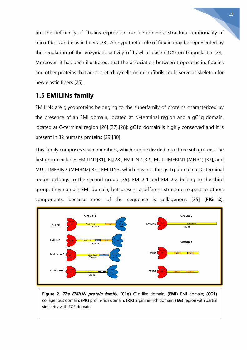
This family comprises seven members, which can be divided into three sub groups. The
first group includes EMILIN1[31],[6],[28], EMILIN2 [32], MULTIMERIN1 (MNR1) [33], and
MULTIMERIN2 (MMRN2)[34]. EMILIN3, which has not the gC1q domain at C-terminal
region belongs to the second group [35]. EMID-1 and EMID-2 belong to the third
group; they contain EMI domain, but present a different structure respect to others
components, because most of the sequence is collagenous [35] (FIG 2).
Figure 2. The EMILIN protein family. (C1q) C1q-like domain; (EMI) EMI domain; (COL)
collagenous domain; (PR) prolin-rich domain, (RR) arginine-rich domain; (EG) region with partial
similarity with EGF domain.

16
1.5.1 EMILIN1 localization
EMILIN1, initially called gp115, was isolated by Bressan et al. in 1983 from chick aorta
[36] associated with elastic fibers. In particular, EMILIN1 is commonly present in regions
where elastin and microfibrils are in close contact (that is where the name “Elastin
Microfibril Interface Located proteIN 1” comes from). Blood vessels present the highest
concentration of EMILIN1, but the protein has also been found in all examined connective
tissues (heart, lung, skin, intestine, cornea, kidney, skeletal muscle) and always in
association with elastic fibers [6]. The observation that EMILIN1 co-localizes with elastin in
vitro cultures of aorta cells, also indicates that the protein is produced in situ rather than
being of blood origin. After synthesis in aortic smooth muscle cells, the protein is
deposited in the ECM as a fine network. Soon after secretion, EMILIN undergoes
intermolecular cross-linking via disulphide bond formation, giving rise to high molecular
weight aggregates [37].
The protein could be detected during the early stages of aorta development in
association with a network of fibrils that were smaller in diameter than microfibrils and
probably represent maturing microfibrils. EMILIN1 deposition clearly precedes the
appearance of elastin and simultaneously with that of fibrillin-1 [38]. Thus, the appearance
of EMILIN1 can be considered an early event in elastogenesis, suggesting a specific
function of the protein in this process. This conclusion is enforced by the observation that
anti-EMILIN1 antibodies considerably alter the normal process of elastic fiber formation
in vitro and elastin deposition [6]. A structural role for EMILIN has also been hypothesized
in studies performed on chick embryo digit formation, where the arrangement of the
protein was compatible with the possible role for the anchoring elastin fibrils to other
matrix constituents [39].
1.5.2 EMILIN1 structure
The EMILIN1 gene maps to chromosome 2 in position p23.2 – p23.3 and it is organized
in eight exons interrupted by seven introns. EMILIN1 presents a multi-modular structure.
The EMI domain [40] is situated at N-terminus region. It is a cysteine-rich repeat sequence
and presents an elevated homology between the family members. The EMI domain

17
displays peculiar characteristics; indeed, despite of the majority of EMI domains known to
date, the EMI domain of EMILIN1 presents seven cysteine residues. Moreover, the spatial
organization of this domain is very different compared to other cysteine rich region such
as the EGF domain [41]. Furthermore, EMI domain presents at the C-terminal of the
domain a well conserved sequence (WRCCPG(Y/F )xGxxC) that appears to be unique to
the EMI domain [41]. EMILIN1, as few ECM proteins, at the central region, presents
domains in which the 3-4-3-4 spatial structure of hydrophobic residues shows α-helical
coiled-coils which results in a rigid rod [41],[42],[43]. Between the coiled coil region and
C-terminal region, EMILIN1 has a region of 91 amino acids that contains two sequences
very similar to structures called “leucine zippers”. The presence of this “leucine zipper
sequence” is rather strange for ECM proteins. There are very few cases reported in
literature in which this region is present: pollux, an ECM protein in Drosophila [44], and
dystrophin, a cytoplasmic protein that is able to interact with troponin [45]. The leucine
zipper functions are not well defined. It could be hypothesized that this structure is
involved in the association of the C-terminal with N-terminal domains to direct the
correct assembly of the EMILIN1 trimer [41]. Close to the leucine zipper there is a short
collagenic portion composed of 17 GXY triplets organized in a trypsin-resistant triple
helix [37]. The gC1q domain of EMILIN1 is encoded by exon 7 and exon 8 and it is
constituted by 151 amino acids, mostly hydrophobic, organized in β-sheet secondary
structure. This domain is highly conserved and it has a high homology with the C1q
complement. The presence of this domain is fundamental for the interaction between
EMILIN1 and the α4β1/α9β1 integrins [46].

18
1.5.3 Functions of EMILIN1
Several studies using a mouse models in which Emilin1 gene is inactivated have
evidenced multiple phenotypes: systemic hypertension [47], a reduction of anchoring
filaments in lymphatic
vessels and increase of
lymphatic vessels diameter
[48], dermal and epidermal
hyperproliferation [49].
In general EMILIN1 is
involved in several
functions associated to its
whole structure such as
elastogenesis, and the
maintenance of blood
vascular and cell
morphology (FIG 3). Moreover, EMILIN1, through its gC1q domain, is involved in other
functions such as regulation of its homotrimerization processes (coiled coil region),
skin homeostasis, skin carcinogenesis, cell adhesion and migration by interaction with
α4β1 and α9β1 integrins. Through the EMI domain, EMILIN1 regulates blood
hypertension.
1.5.4 EMI domain function
The correct structure and functionality such as elasticity of vessels is regulated by the
presence of smooth muscle cells and endothelial cells that cover vascular walls. For the
maintenance of the appropriate blood pressure it is important to maintain the correct
interconnections with ECM. Given that EMILIN1 is closely associated with elastic fibers
and microfibrils present in blood vessels [31],[41] and that it is involved both in
elastogenesis processes and regulation of blood vessel morphology [50] it has been
hypothesized that EMILIN1 can exert a mediator role in the regulation of blood
pressure. Indeed, it has been demonstrated that Emilin1-/- mice show an increase of
FIGURE 3. Schematic view of EMILIN1 function.
19
blood pressure respect to WT littermates. This increase of blood pressure may be due
to a peripheral vascular resistance as well as to a decrease in size of blood vessels. It
has been demonstrated that EMILIN1 through its EMI domain can bind pro TGF-β,
inhibiting its maturation to TGF-β by furin convertases. In this manner EMILIN1 exerts
a TGF-β antagonist role resulting in a decrease of blood pressure level [47].
1.5.5 EMILIN1 and lymphatic system
Lymphatic endothelial cells show interconnections with surrounding ECM that are
mediated by AFs [51]. Structural defects of AFs could decrease adsorption from
interstitium and can promote lymphedema. Abnormal AFs can render lymphatic vessels
less responsive to oncotic change [52]. EMILIN1 is secreted by Lymphatic Endothelial
cells line (LECs) and is closely associated at the abluminal side of lymphatic vessels. The
presence of EMILIN1 close to lymphatic vessels suggests an important role of EMILIN1
in the regulation of the functions and structure of lymphatic system [48]. Indeed,
Emilin1-/- mice show hyperplasia and increase of lymphatic vessels diameter respect to
WT mice. Among abnormalities, the decrease of AFs number and the presence of
multiple overlapping junctions of LECs are most frequent, in EMILIN1-/- mice, where
LECs lose interconnections with ECM: in this case it is possible to observe the formation
of intraluminal flaps showing structural and number of abnormalities [48].
1.6 gC1q: the EMILIN1 functional domain
Most of the EMILIN1 functions are due to the interaction between integrin α4β1 or
α9β1 and gC1q domain [49], [46], [53].
The C-terminal C1q domain exhibits a high homology with the gC1q domain of a lot
of members of the C1q/TNF superfamily. The aminoacidic sequence of the C1q-like
domain of EMILIN1 shows similar features to the other domains. This indicates that
aminoacidic sequence presents a high level of conservation of several hydrophobic and
aromatic residues. To obtain the best sequence alignment of EMILIN1 with the other
members of the superfamily, it was necessary to allow for the peculiar insertion of a 9-
residue sequence that is unique for EMILIN1 and 2 and is missing in all the other

20
members. In this manner, the structure prediction of C1q domain was obtained by
comparing the molecule with other members of the superfamily [28].
The gC1q domain exists in solution as a stable trimeric protein formed by three
identical polypeptide chains of 162 amino acids (150 from the natural domain
sequence, 7 from N-terminal fused histidine tag and initial Met residue, and 5 derived
from the cloning strategy) with an overall molecular mass of 51.624 KDa. Verdone et
al. determined the three-dimensional solution structure of the gC1q domain using a
hybrid approach that involved homology modeling and structure refinement with
experimental restraints obtained by NMR spectroscopy [54] (FIG 4).
The most relevant changes that occur between gC1q
domain and other gC1q domain (crystal) structures
solved to date are the reduction to nine (instead of ten)
of the number of antiparallel strands (A, A’, B’, B, C, D,
E, G, H) and the presence of a peculiar unstructured
loop spanning from Y927 to G945. This unstructured
loop, resulting from the insertion unique to EMILIN1
and EMILIN2 family members cited above, is located
upstream the missing strand F at the trimer apex and
possibly available for interactions with
ligands/receptors of gC1q domain. The disordered loop is followed by the so called
“molten strand F” (I953-L956), an interfacial sequence with no secondary classification
( and Ψ angles no longer define a β strand). Despite the loss of the local secondary
structure, region F is expected to be rigid (the spatial location of region F remains
invariant) and it is involved in inter subunit interactions. The self-interaction
(trimerization) of the gC1q domain was first investigated with the use of two hybrid
system; successive biochemical analyses exclude disulfide bond involvement in the
trimeric organization [37]. The association between monomers mainly occurs through
the buried strands A, A′, H, C belonging to sheet 1, and the unstructured region
spanning residues L947–P955, whereas strands B, B′, D, E, and G constitute the external
FIGURE 4. Structure of the
gC1q domain. Ribbon
representation of the assembly,
as top view
21
second sheet. The interface contacts between subunits are mainly hydrophobic and
involve the side chains of most conserved residues in gC1q family located at the base
of the trimeric assembly [41]. The top is slightly open (~150 Å) to form a cavity ~16 Å
deep. Among the 30 residues that establish inter subunit interactions, only two are
charged (R870 and R893) and located at opposite ends of an extended hydrophobic
surface. The presence of hydrophobic contacts near the base of the trimer has been
highlighted for other members of the C1q family and, apparently, it is a common
feature of these molecules (collagen X, ACRP30, and collagen VIII). In contrast, the
occurrence of hydrophilic interactions at the apex of the trimeric adduct, seen in
collagen X, ACRP30, and collagen VIII, cannot be confirmed in human gC1q domain
structure, which is relatively loose in that area [55].
1.6.1 Integrins α4β1/α9β1
Integrins are receptors involved in cell adhesion that present a well conserved structure
[56]. Only in recent years has been well characterized at the molecular level. The
difficulties encountered in trying to characterize this type of molecules are due to the
capability of integrins to bind several multi-adhesive ECM molecules, which are able to
bind other proteins such as cytokines, growth factors and matrix-degrading proteases
[57]. It is possible to classify integrins into subgroups based on ligand-binding features
or of their subunit composition. One of the principal function of integrins is due to
their capability to function as links between the ECM and the actin cytoskeleton [60].
In the last few years it has been proposed a mechanosensor role for integrins; indeed
they can exert this role generating signals that are able to affect cell physiology [61],
[62]. α4β1/α9β1 integrins interact with a variety of ECM proteins: for example, FN,
osteopontin, TSP-1 [63]. Differently from many other integrins, α4 and α9 integrins are
able to bind the ligands in a RGD independent manner [64].
1.6.2 Structural aspects of gC1q domain/ integrin ligation
All polypeptides that are able to interact with integrins show aspartic or glutamic acid
residues localized in loops that protrudes from core of the ligand [72]. Both aspartic
and glutamic amino acid residues are essential for integrin recognition. Sequences that
22
are present on ligands, containing aspartic residues, are able to recognize the majority
of integrins [73], [74], [75]. [76]. The interaction of the gC1q domain-α4β1 integrin is
more complicated than in other cases. The substitution of the glutamic acid in position
933 (E933) with alanine residue generates gC1q domain unable to recognize α4β1
integrin suggesting that glutamic acid in position 933 plays a fundamental role in this
interaction [55]. Several gC1q domain presenting mutation around the E933 region
have been tested to verify their ability to bind integrin α4β1. All the resulting gC1q
domain mutants were able to recognize the integrin confirming that the lack of
interaction between gC1q domain and integrin α4β1 is due only and exclusively to
glutamic residue present in region E933 [55]. Furthermore, it has been demonstrated
that the presence of the E993 amino acid residue in each of three monomers is
necessary to allow the interaction gC1q domain α4β1 integrin [55]. α4β1 integrin
dependent interactions, differently to α5β1- or αvβ1-dependent interactions with FN
and vitronectin that give to a cytoskeletal remodeling [77], [78], [79], [80], govern only
initial and intermediate steps of cell adhesion (attachment and spreading); on the
contrary it does not seem to regulate focal adhesion and stress fiber production [67].
1.6.3 The functional role of gC1q domain: adhesion, migration and
proliferation
Studies conducted by using blocking antibodies directed against different domains of
the protein, indicate that many EMILIN1 properties are fully accounted by its functional
domain gC1q; in particular, these studies indicate that cell adhesion and
antiproliferative functions of EMILIN1 rely on the interaction with integrin α4β1. Also
integrin α9β1, which is highly homologous with integrin integrin α4β1, is able to
interact with the gC1q domain. The interaction gC1q domain-integrin α9β1 can
promote several physiological processes such as skin homeostasis, cell adhesion and
cell proliferation. Normally, the binding of ECM components to integrin receptors
determines an increase of cell proliferation. Danussi et al, illustrated that EMILIN1
through the interaction of the gC1q domain with integrin α4β1 or integrin α9β1 can
negatively regulate cell proliferation [49],[82], [83]. In fact, mice Emilin1 -/-, show dermis

23
and epidermis hyperproliferation [49]. The antiproliferative role of EMILIN1 has been
demonstrated also in vitro on fibroblast cultures in which Emilin1-/- cells showed an
elevated level of proliferation compared to Emilin1+/+ cells.
The interaction gC1q domain- integrin α4β1/ α9β1 activates a signaling pathway that
is able to regulate cell proliferation through the involvement of several molecules such
as PI3K, PTEN, Erk 1/2 and SMAD.
24
Chapter 2
2.1Tumor Microenvironment
A link between inflammation and cancer was proposed by Rudolf Virchow in the 19th
century following the observation of leucocytes in several tumor tissues. Only during
the last decades this hypothesis has been clearly established. Some authors have
showed that inflammatory microenvironment is a ubiquitous component of all tumors
[84]. Moreover, epidemiological studies have illustrated that up to 30% of all cancer
may be due to inflammation [85]. Thus, it is possible to affirm that inflammation could
be one of the major causes of cancer. In inflammatory context, several cells are
recruited, such as neutrophils, macrophages, dendritic cells. Neutrophils, the first cells
arriving in an inflammatory microenvironment, are able to release cytokines, growth
factors and various proteases such as neutrophil elastase (NE) which are able to affect
the behavior of epithelial, mesenchymal and endothelial cells [87].
2.2 Neutrophils
Neutrophils exert an important role as first responders to pathogen attack. Neutrophils
are the major components in number of white blood cells and fulfill two fundamental
functions: the first is an immune surveillance function, and the second is to destroy
micro-organism. Neutrophils belong to granulocytes family, together with eosinophils
and basophils [88]. Under non-pathological conditions, the number of neutrophils
circulating is very low. On the contrary, during inflammatory conditions, the number of
released neutrophils is increased. To exert their role in immune system, they move
quickly; indeed among the cells present in the body, they are the quickest cells [91]
and are able to moving directionally (chemotaxis) to go towards their “prey”. In order
to do this, they expose several receptors that can be classified in: I) receptors that are
able to detect soluble chemo attractants, II) receptors for recognizing an inflammatory
microenvironment and III) receptors important for the activation of the adaptive
immune system [92]. To avoid aspecific activation, neutrophils usually remain in a
quiescent condition, but also in this condition they can present a more aggressive

25
phenotypes through a process called “priming” [93]. During this process a lot of
inflammatory molecules such as GM-CSF [94], platelet activating factor (PAF) [95],
lipopolysaccharide (LPS) [96] and TNF-α [97] are able to promote a state of pre-
activation. Moreover, “priming” can determine cell polarization, expression of integrin
[98], increase of oxidative burst [96], release of leukotriene B4 and acid arachidonic
[99],[100] and degranulation responses [101]. It has been hypothesized that neutrophil
“priming” is an early stage of neutrophil recruitment and comes before extravasation
[102].
2.3 Neutrophil recruitment
As already mentioned, neutrophils are the first cells to occur during inflammation or
infection conditions as a result of stimulus emitted by pathogens and host cells. When
neutrophils are present in transient form, they roll over along endothelial vessels in
“reconnaissance mission” to pick up damage or infection signal.
When the immune system is under the attack by microorganism, there is a condition
in which a lot of receptors, present on cell surface, are employed in neutrophil
recruitment resulting in upregulation. In this scenario, the levels of P-selectin are
increased in a few minutes following stimulation by reactive oxygen species (ROS)
[105], while the level of E-selectin are increased after 1 or 2 h thanks to stimulation by
IL-1, TNF-α or LPS. If the expression level of selectins allows to engage enough ligands,
neutrophils are immobilized. At this point neutrophils must adhere perfectly to walls of
blood vessels. In this neutrophil immobilization processes β2-integrins specifically
CD11a/CD18 (LFA-1) and CD11b/CD18 [103] are involved and after activation,
neutrophils acquire a polarized morphology, in which it is possible to distinguish an
attack region. The creation of this region is regulated by G-protein coupled receptors
by a signaling in which phosphoinositide 3-kinase (PI3K) catalyze the formation of the
second messenger phosphatidylinositol (3,4,5)-trisphosphate (PIP3) from
phosphatidylinositol (4,5)-bisphosphate (PIP2); PIP3 allows the activation of Rho
GTPase with consequently polarization of F-actin [106],[107].

26
At this point neutrophils are ready to migrate in proximity of the inflammation sites.
The extravasation is favored by the CD11a/CD18 and CD11b/CD18 integrins present
on neutrophil surface and the ligands present on endothelial cell [108]. Neutrophils can
release several other proteases in addition to NE such as cathepsin G, proteinase 3 (PR)
and matrix metalloproteinases (MMPs), in particular matrix metalloproteinase-9 (MMP-
9), that is able to break several component of the basal membrane such as laminins
and collagens [109]. NE is able to bind ECM components in a manner that are
inaccessible for tissue inhibitors [110]. Moreover, thanks to the presence of protective
film on ECM components, NE can resist to proteases inhibitors action and, in this
manner, determines ECM components breakdown [111], [112].
2.4 ECM molecules favoring cancer and its progression
ECM plays an important role in several cellular functions such as cell shape, cell
adhesion, proliferation, migration, differentiation and polarity [113],[127],[115]. Under
normal conditions the ECM composition is highly regulated through a delicate
equilibrium in which synthesis and degradation processes are balanced. This delicate
equilibrium is corrupted in pathological conditions, such as cancer. During cancer
insurgency, the increase of ECM components breakdown leads to the formation of
fragments that can favor tumor growth and progression [116],[117]. Several data
present in the literature reinforce the idea that ECM can play an important role in tumor
progression. Moreover, many of its components can enhance the development and the
spread of tumor cells. Only exiguous number of ECM components are able to carry out
a tumor suppressor function. There are many examples of how ECM molecules can
0exert strategies to promote tumor initiation and progression. In several types of
cancer, collagens present several structural alterations due to the linearization, as a
result of modification at post-translational level. This modifications, in ECM structure
could give various abnormalities regard to cellular differentiation, proliferation,
migration and survival [17],[123].
Fibronectin (FN) is another glaring case of ECM molecule that, following qualitative and
quantitative modifications, covers a role as mediator of the malignant process. Among
27
ECM molecules FN holds a principal role to understand the interaction between
“integrin- ECM” on cell survival and proliferation. In particular, the adhesion process
due to interaction between fibronectin and α5β1 integrin, can stimulate cell cycle
progression [124],[125]. In particular, endothelial and epithelial cells are an example of
cell types that need the presence of α5β1 integrin to adhere to ECM. During the
adhesion process several pathways are activated. Among these the molecular pathway
of FAK exerts an important role. Indeed, FAK signaling allows mechanical coupling
between ECM and cytoskeleton: when FAK is activated Rac-mediated cyclin D1 gene
and cyclin D1-dependent Rb phosphorylation are activated [126].
During fibrillogenesis, there is an increase of FN rigidity, with consequent
strengthening of binding between FN and integrin α5β1 [127]. Given that FN controls
collagen fibril organization, and viceversa, it is clear that both FN and collagens
modifications in size, density and rigidity can regulate the functions each other. This
scenario plays a fundamental role in tumor progression; indeed, high deposition of FN
is closely associated with tumor development [128],[129]. Moreover the engagement
of α5β1 to FN determines an increase of MMP-1, MMP-3 and MMP-9 release causing
tumor invasion [130],[131]. It is clear how not only qualitative but also quantitative
changes in the ECM composition can generate a microenvironment propitious to the
cancer onset. It is known that proteinases participate busily to ECM remodeling. The
degradation process of matrix proteins is called proteolysis, and it is facilitated by the
presence of proteases. The protease action allows to destroy the basement membrane,
that acts as a barrier for epithelial cells, and subsequently to form pathways for the cell
to migrate through [132]. Collagen IV is an abundant molecule present in ECM. Several
studies illustrated that in pancreatic model this ECM component is expressed close to
the cancer cells, contributing to form basement membrane like structures on the tumor
and it allows tumor cells to survive by autocrine mechanism [133]. Moreover, collagen
I degradation can determine angiogenesis: the cleavage of the triple helical of type I
collagen, is necessary to permit the formation of new blood vessels [134],[135]. On the
contrary, the proteolytic process of collagens can give also fragments able to contrast
angiogenesis. For example, endostatin, a fragment originated by the degradation of

28
type XVIII collagen, is able to inhibit the formation of new vessels [136]. Moreover,
tumstatin, a fragment of type IV collagen, by modulation of αvβ3 and αvβ5 integrin
signaling [137],[138], exerts a role similar to endostatin in endothelial cells, as well as
arresten and canstatin, other type IV collagen fragments, that show antiangiogenic
effect [139],[140]. FN fragments play a double role: they are able both to promote cell
growth [141] and to inhibit cell invasion. An example thereof is given by FN13 amino
acid peptide which can modulate αvβ3 integrin organization and is involved in the
inactivation of ILK pathway [142]. Laminin is another ECM component after MMPs
cleavage, that allows the exposure of hidden site, can promote cell migration and
invasion[143],[144].
2.4.1 ECM Molecules with Anti-Tumor Activity
An exiguous number of ECM proteins are able to exert a tumor suppressor role, some
of them through an indirect mechanism and others, through a direct mechanism.
Thrombospondin-1 (TSP-1) is an ECM protein involved in suppression of angiogenesis.
TSP-1, such as thrombospondin-2, plays a suppressor role in angiogenesis by the
interaction with vascular endothelial cell growth factor (VEGF), inhibition of endothelial
cell migration and inhibition of MMP-9 [145],[146]. Also fibulin-5 (FBLN-5) exerts tumor
suppressor function, by inhibiting angiogenesis [147],[148]. In order to regulate its
angiogenic control, FBLN-5 can control several endothelial cell activities by direct and
indirect mechanism. FBLN-5 can directly reduce endothelial cell migration and invasion
counteracting VEGF stimulation of ERK1/ERK2 and p38 MAPK [148]. Moreover, FBLN-5
increases TSP-1 expression in endothelial cells [148] causing a reduction of
angiogenesis by enhancing TSP-1 mediated apoptosis and also by the inhibition of
MMP-9 expression. Several studies illustrated that in endothelial cells the
overexpression of FBLN-5 during tubuligenesis can give a decrease of MMP-2 [149].
Furthermore, FBLN-5 blocks ROS production by interfering with the interaction present
between FN and β1 integrins. This mechanism allows FBLN-5, going to bind to β1
integrin, in place of FN, and then to inhibit cell spreading, proliferation and migration.
The FBLN-5 tumor suppressor role is illustrated in several studies where it is elucidated
29
that FBLN-5 mRNA level is drastically decreased in prostate, breast, kidney, ovary and
colon cancer [150],[151],[152]. Among the fibulin family, also for FBLN-3 a tumor
suppressor role has been recognized, by inhibiting angiogenesis and by the reduction
of tumor growth [149].
There are other examples of ECM molecules that may directly inhibit tumor
progression. Cysteine rich angiogenic inducer (CCN1) can be considered one of these.
High levels of CCN1 favor fibroblast apoptosis by binding to α6β1 integrin and
syndecan-4. This binding determines Bax activation that causes cytochrome c release
and activation of caspases -9 and -3 [153]. Decorin is another molecule that plays tumor
suppressor role in direct manner. Many tumor cells show high level of EGFR. The
presence of Decorin allows the delay the EGFR endocytosis, that leads to EGFR
degradation, whit the consequence to attenuate its signaling pathway.
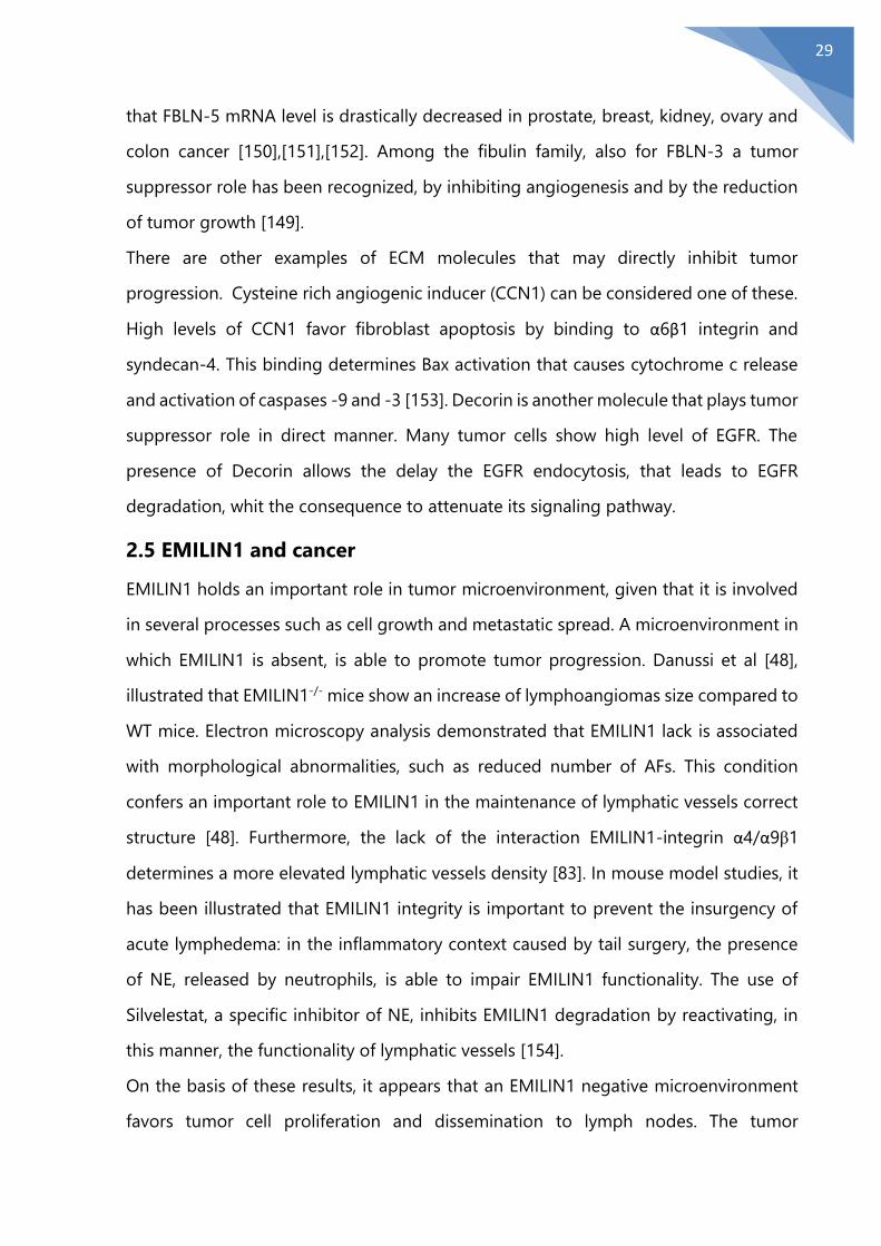
2.5 EMILIN1 and cancer
EMILIN1 holds an important role in tumor microenvironment, given that it is involved
in several processes such as cell growth and metastatic spread. A microenvironment in
which EMILIN1 is absent, is able to promote tumor progression. Danussi et al [48],
illustrated that EMILIN1-/- mice show an increase of lymphoangiomas size compared to
WT mice. Electron microscopy analysis demonstrated that EMILIN1 lack is associated
with morphological abnormalities, such as reduced number of AFs. This condition
confers an important role to EMILIN1 in the maintenance of lymphatic vessels correct
structure [48]. Furthermore, the lack of the interaction EMILIN1-integrin α4/α9β1
determines a more elevated lymphatic vessels density [83]. In mouse model studies, it
has been illustrated that EMILIN1 integrity is important to prevent the insurgency of
acute lymphedema: in the inflammatory context caused by tail surgery, the presence
of NE, released by neutrophils, is able to impair EMILIN1 functionality. The use of
Silvelestat, a specific inhibitor of NE, inhibits EMILIN1 degradation by reactivating, in
this manner, the functionality of lymphatic vessels [154].
On the basis of these results, it appears that an EMILIN1 negative microenvironment
favors tumor cell proliferation and dissemination to lymph nodes. The tumor

30
suppressor role played by EMILIN1 seems to be questioned by two different studies in
which the authors, by analyzing gene expression and by using proteomic approaches,
demonstrated an upregulation of EMILIN1 in ovarian carcinomas and osteosarcomas
[160][161]. Edlung et al., have shown that the expression of EMILIN1 in non-small lung
cancer is upregulated [159]. This could be explained by the fact that EMILIN1 plays a
tumor suppressor role in a context dependent manner. Another hypothesis may be
that EMILIN1 gene expression is elevated, but the environment present in tumoral
context is able to impair EMILIN1 functionality. A scenario in which EMILIN1 is unable
to exert its tumor suppressor role may be due to proteolytic enzymes released by cells
present in tumor microenvironment that may be able to degrade EMILIN1. A condition
in which EMILIN1 is lacking, as mentioned before, can be considered very similar to
that of EMILIN1-/- mice and resulting in the upregulation of cell proliferation. A question
which may arise is: are there conditions, in humans, in which EMILIN1 may loose its
functions? Pivetta et al, have demonstrated that by incubating EMILIN1 with several
supernatants of immune cell, the degradation of the protein was observed only when
EMILIN1 was incubated with neutrophils supernatants [162]. Furthermore, they
demonstrated that among the enzymes released by neutrophils only NE was able to
degrade EMILIN1 [162]. In this scenario, in which EMILIN1 is degraded, it may lack its
functionality, in particular regarding its tumor suppressor role. Moreover other authors
by a proteomic approach hypothesize that EMILIN1 can be degraded also by same
MMPs such as MMP-3, MMP-9 and MT1-MMP [164].

31
Chapter 3
3.1 Neutrophil Elastase (NE)
NE, with cathepsin G and proteinase 3 (PR3), belongs to serine proteinase family, stored
in the azurophilic granula of neutrophil granulocytes [165]. NE is able to degrade
several molecules such as elastin, collagen, cadherins, proteoglycan, fibronectin,
complement receptors, thrombomodulin, lung surfactant, and growth factors
(granulocyte-colony-stimulating factor, stromal cell-derived factor-1, and their
respective receptors, G-CSFR and CXCR4) [166]. Among the inhibitors of NE, an
important role is exerted by α1-antitrypsin. An imbalance of NE and α1-antitrypsin is
associated with the insurgency of chronic liver disease, rheumatoid arthritis, aneurysm
and lung emphysema [166]. Moreover it is evident that the imbalance between NE and
its inhibitors can give cancer progression of several tumors such as lung, liver and
colorectal cancer [167].
3.2 Biology of NE
NE has a high grade of homology with Proteinase3 (PR3), Cathepsin G (CG). NE, such
as all serine proteases, contains a conserved catalytic triad formed by: histidine, aspartic
acid and serine residues [168]. The gene encoding for NE is found on chromosome 10
[169] in mice and on chromosome 19 in humans [170]. It consists of five exons and four
introns [171]. The synthesis of NE is highly regulated at the transcriptional level before
being transported in the neutrophil azurophilic granules in their active mature form
[170]. NE is synthesized in an inactive form that presents: a signal peptide, an amino-
terminal pre-dipeptide and a C-terminal pro-peptide [172],[173].
3.2.1 NE Structure and Function
NE is generally able to cleave in proximity to small hydrophobic amino acids, in
particular, valine, alanine and isoleucine. However, the NE consensus sequences is very
similar to the other neutrophil serine proteases. Serine proteases show their own
specificity to recognize their consensus sequencing and very often this capability is due

32
to the distribution of charged amino acids [180]. NE has the highest activity at pH 7. As
other serine proteases, NE is able to cleave substrates by its catalytic triad formed by
serine, histidine and aspartate.
The structure of NE shows β-barrels interconnected by a segment and an α-helix
present at C-terminal region. Moreover, its structure is stabilized by the presence of
four disulfide bonds. When NE exerts its proteolytic activities, the -OH group of serine
provides to nucleophilic reactions forming a covalent bond with the substrate. The
nitrogen present on histidine amino acid residue accepts hydrogen from serine,
forming a tetrahedral complex. Electrons present in the peptide bond that occurs
between the amino groups and the carbonyl of substrate can interact with the
hydrogen present on histidine, with consequent bond breaking and release of N-
terminal portion. The electrons that are still present on nitrogen of the histidine
reforming the bond, resulting in an acyl-enzyme intermediate. The N-terminal region
of the cleaved peptide is replaced by molecule of water, forming an another tetrahedral
intermediate. At this point electrons arrange a bond between serine and hydrogen
present on the histidine with consequently resolution of tetrahedral intermediate, with
release of the C-terminal portion of the cleaved peptide, and restoring the active site
of the proteases [174].
3.2.2 Role of Extracellular NE
Tumor progression is strongly supported by conditions in which the presence of
inflammatory cells and continuous inflammation are not resolved [182],[183]. NE is
secreted in microenvironment by azurophilic granules [184]. The release of NE is
mediated by several type of chemokines, citokines, IL-8, TNF-α, C5α and LPS [185].
Given that neutrophils are the first cells to respond during microorganism attack,
proteases released by these cells, such as NE, are involved in early defensive mechanism
to face inflammatory responses. NE exerts an important role in inflammation
regulation. In order to do it, NE must evade the control of endogenous inhibitors. How
NE is able to do it is not clear, but several possible mechanism are been proposed [186].
One possible mechanism shows that a cavity is created among ECM and neutrophils,
33
in which NE is released and unassailable to large inhibitors [186]. The second
mechanism could be that the elevated molecules of NE form as a shield closed of to
inhibitors [110]. Given that NE exerts an important role in inflammation, several
experiments are conducted on animals in order to correlate a reduction of
inflammation with inhibition of NE. Using a hamster model, some authors illustrated,
an increase of NE expression during acute lung injury [187]. The treatment with
neutrophil inhibitors in dose dependent manner causes a decrease of tissue
inflammation.
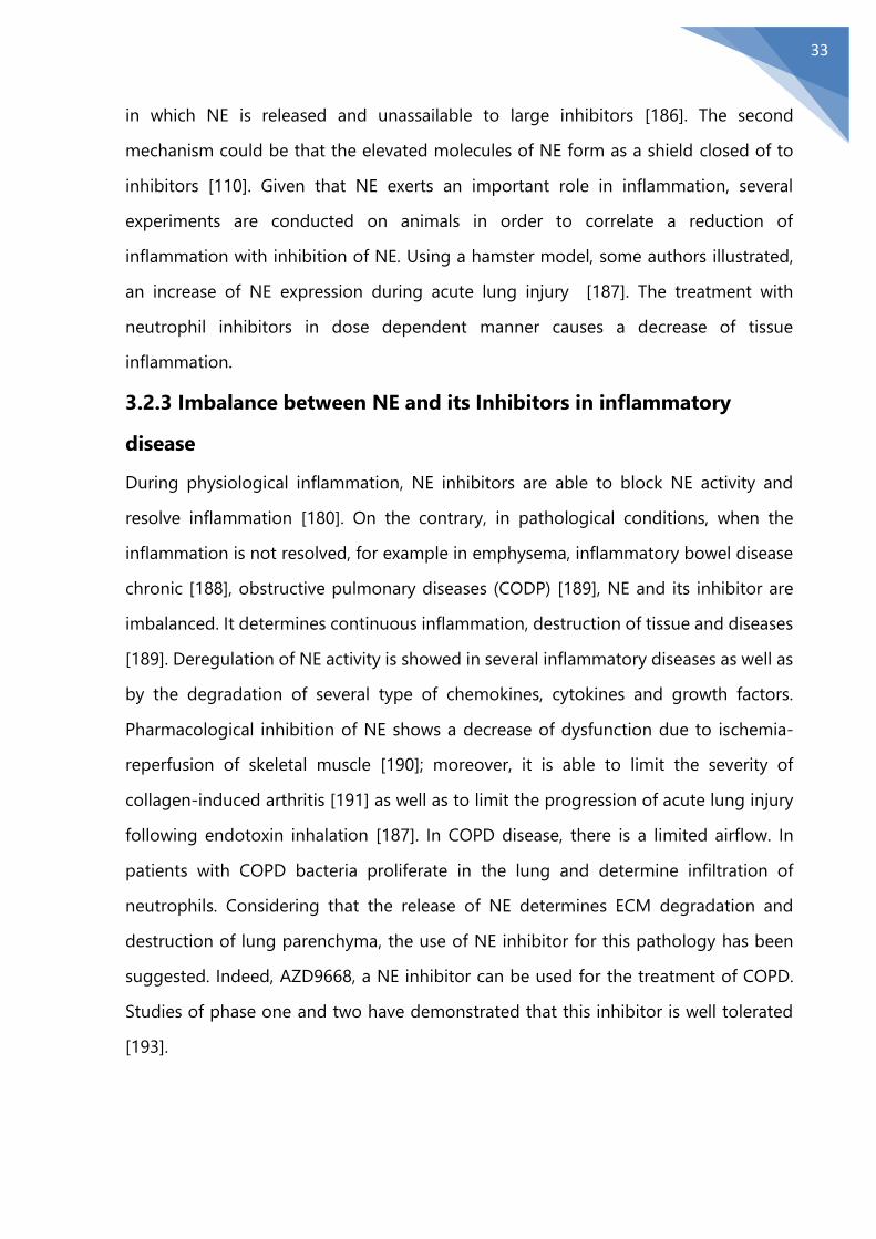
3.2.3 Imbalance between NE and its Inhibitors in inflammatory
disease
During physiological inflammation, NE inhibitors are able to block NE activity and
resolve inflammation [180]. On the contrary, in pathological conditions, when the
inflammation is not resolved, for example in emphysema, inflammatory bowel disease
chronic [188], obstructive pulmonary diseases (CODP) [189], NE and its inhibitor are
imbalanced. It determines continuous inflammation, destruction of tissue and diseases
[189]. Deregulation of NE activity is showed in several inflammatory diseases as well as
by the degradation of several type of chemokines, cytokines and growth factors.
Pharmacological inhibition of NE shows a decrease of dysfunction due to ischemia-
reperfusion of skeletal muscle [190]; moreover, it is able to limit the severity of
collagen-induced arthritis [191] as well as to limit the progression of acute lung injury
following endotoxin inhalation [187]. In COPD disease, there is a limited airflow. In
patients with COPD bacteria proliferate in the lung and determine infiltration of
neutrophils. Considering that the release of NE determines ECM degradation and
destruction of lung parenchyma, the use of NE inhibitor for this pathology has been
suggested. Indeed, AZD9668, a NE inhibitor can be used for the treatment of COPD.
Studies of phase one and two have demonstrated that this inhibitor is well tolerated
[193].
34
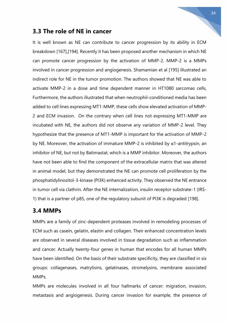
3.3 The role of NE in cancer
It is well known as NE can contribute to cancer progression by its ability in ECM
breakdown [167],[194]. Recently it has been proposed another mechanism in which NE
can promote cancer progression by the activation of MMP-2. MMP-2 is a MMPs
involved in cancer progression and angiogenesis. Shamamian et al [195] illustrated an
indirect role for NE in the tumor promotion. The authors showed that NE was able to
activate MMP-2 in a dose and time dependent manner in HT1080 sarcomas cells,
Furthermore, the authors illustrated that when neutrophil-conditioned media has been
added to cell lines expressing MT1-MMP, these cells show elevated activation of MMP-
2 and ECM invasion. On the contrary when cell lines not expressing MT1-MMP are
incubated with NE, the authors did not observe any variation of MMP-2 level. They
hypothesize that the presence of MT1-MMP is important for the activation of MMP-2
by NE. Moreover, the activation of immature MMP-2 is inhibited by α1-antitrypsin, an
inhibitor of NE, but not by Batimastat, which is a MMP inhibitor. Moreover, the authors
have not been able to find the component of the extracellular matrix that was altered
in animal model, but they demonstrated the NE can promote cell proliferation by the
phosphatidylinositol-3-kinase (PI3K) enhanced activity. They observed the NE entrance
in tumor cell via clathrin. After the NE internalization, insulin receptor substrate-1 (IRS-
1) that is a partner of p85, one of the regulatory subunit of PI3K is degraded [198].
3.4 MMPs
MMPs are a family of zinc-dependent proteases involved in remodeling processes of
ECM such as casein, gelatin, elastin and collagen. Their enhanced concentration levels
are observed in several diseases involved in tissue degradation such as inflammation
and cancer. Actually twenty-four genes in human that encodes for all human MMPs
have been identified. On the basis of their substrate specificity, they are classified in six
groups: collagenases, matrylisins, gelatinases, stromelysins, membrane associated
MMPs.
MMPs are molecules involved in all four hallmarks of cancer: migration, invasion,
metastasis and angiogenesis. During cancer invasion for example, the presence of
35
MMPs in specialized cell surface structures, called invadopodia, is important to enhance
their capacity to favor invasion [202]. Invadopodia are present in site where ECM
degradation occurs. In invadopodia, in order to degrade several ECM components,
several kind of matrix metalloproteinases such as MMP-14, a large number of ADAM,
MMP-2 and matrix metalloproteinase (MMP-9) are involved [203].
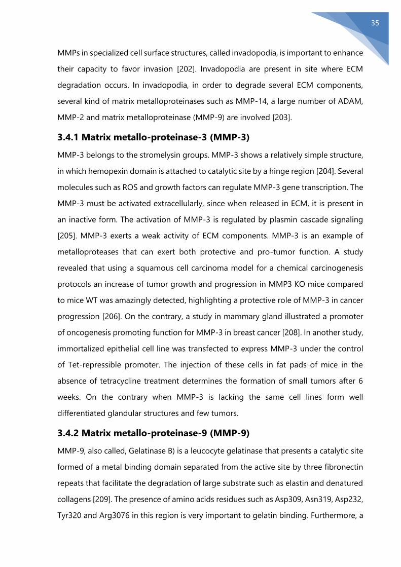
3.4.1 Matrix metallo-proteinase-3 (MMP-3)
MMP-3 belongs to the stromelysin groups. MMP-3 shows a relatively simple structure,
in which hemopexin domain is attached to catalytic site by a hinge region [204]. Several
molecules such as ROS and growth factors can regulate MMP-3 gene transcription. The
MMP-3 must be activated extracellularly, since when released in ECM, it is present in
an inactive form. The activation of MMP-3 is regulated by plasmin cascade signaling
[205]. MMP-3 exerts a weak activity of ECM components. MMP-3 is an example of
metalloproteases that can exert both protective and pro-tumor function. A study
revealed that using a squamous cell carcinoma model for a chemical carcinogenesis
protocols an increase of tumor growth and progression in MMP3 KO mice compared
to mice WT was amazingly detected, highlighting a protective role of MMP-3 in cancer
progression [206]. On the contrary, a study in mammary gland illustrated a promoter
of oncogenesis promoting function for MMP-3 in breast cancer [208]. In another study,
immortalized epithelial cell line was transfected to express MMP-3 under the control
of Tet-repressible promoter. The injection of these cells in fat pads of mice in the
absence of tetracycline treatment determines the formation of small tumors after 6
weeks. On the contrary when MMP-3 is lacking the same cell lines form well
differentiated glandular structures and few tumors.
3.4.2 Matrix metallo-proteinase-9 (MMP-9)
MMP-9, also called, Gelatinase B) is a leucocyte gelatinase that presents a catalytic site
formed of a metal binding domain separated from the active site by three fibronectin
repeats that facilitate the degradation of large substrate such as elastin and denatured
collagens [209]. The presence of amino acids residues such as Asp309, Asn319, Asp232,
Tyr320 and Arg3076 in this region is very important to gelatin binding. Furthermore, a

36
central O-glycosylated domain is involved in molecular flexibility, in regulation of
substrate specificity and MMP-9-dependent invasion [209].
Nonetheless, the proteolytic activity of MMP-9 on triple helical domains of collagen, is
controversial. Indeed, much it has been discussed about the susceptibility [213] or
resistance to MMP-9 cleavage of the triple helical domains of collagen [212]. One of
the main functions of MMP-9 in oncologic field is to regulate the formation of new
blood vessels participating actively to angiogenesis. Despite normal conditions, vessels
formed in tumor microenvironment are immature and present abnormalities [215].
MMP-9 plays a critical role as pro-angiogenic molecule [216] and it is able to regulate
angiogenic switch [217],[218]. Moreover, MMP-9 is able to control several processes
during neo angiogenesis such as pericyte apoptosis, recruitment and proliferation
[219].
3.4.3 Matrix metalloproteinase-14 (MT1-MMP)
MT1-MMP is secreted in inactive form and then it is activated by a furin-like convertase
by the cleavage of Arg108-Arg-Lys-Arg motif situated between the pro-peptide and
catalytic domain. The active form of MT1-MMP is translocated to membrane to exert
its cleavage function. Moreover, once the active MT1-MMP is translocated on plasma
membrane, it is subjected to autocatalytic process resulting in formation of an inactive
fragment and the release of CAT domain. MT1-MMP is able to hydrolyze several ECM
components such as type I collagen, fibronectin, vitronectin, laminin-1, and
extracellular matrix metalloproteinase inducer (EMMPRIN), and cytokines, chemokines
and growth factors [226],[227]. Some substrates of MT1-MMP, such as pericentrin and
breast cancer type 2 susceptibility gene (BRCA2) have also identified [228],[229].
Several cellular functions such as invasion, growth, motility and apoptosis are altered
by MT1-MMP through the digestion of the substrates mentioned above. It is found
that MT1-MMP is highly expressed in several tumors [231], in particular in breast, small
cell lung , bladder and ovarian cancer [232],[233]. Elevated expression levels of MT1-
MMP have been correlated with an increase of invasion, tumor growth and metastasis
[234]. Several studies have illustrated high expression of MT1-MMP in prostate and
37
squamous cell cancer, where it is able to promote the epithelial to mesenchymal
transition [235],[227]. Moreover, MT1-MMP, in pancreatic ductal adenocarcinoma, can
increase the activation of growth factors promoting chemo resistance [236].
38
AIM
EMILIN1 is an extracellular matrix glycoprotein and it is able to exert a wide range of
functions mainly associated with its gC1q domain. The interaction “gC1q domain- α4β1
integrin” is able to promote cell adhesion and migration but, more interestingly, it can
regulate proliferation, with “suppressive” effects. Moreover, EMILIN1 plays an
important role in ECM both in normal and pathological conditions. This hypothesis is
formulated on the basis of previous studies, in which the authors, Pivetta et al [162],
demonstrated that proteolytic enzymes, such as NE, released in the tumor
microenvironment by neutrophils, is able to impair the EMILIN1 features disabling its
antiproliferative functions. The EMILIN1 degradation by NE has been shown in
leiomyosarcomas, ovarian cancer and undifferentiated soft tissue sarcomas [162]. In a
recent study, it was illustrated in a non tumoral contest, the cruciality of EMILIN1
integrity to exert its functions. In this study the authors showed that EMILIN1
degradation by NE is able to promote surgically induced lymphedema [154].
Stegemann et al [164] proposed, by a proteomic approach, EMILIN1 a possible
substrate of some MMPs (MMP-3,-9 and MT1-MMP). The aim of this thesis was to
investigate the capability of this MMPs in to degrade EMILIN1 but above all if these
MMPs are able to impair EMILIN1 features. Moreover, we wanted to analyze if the
degradation of EMILIN1 by proteolytic enzymes impairs the function of gC1q domain
is affected by putative EMILIN1 fragmentation. Moreover, we wanted assess if whether
the tumor suppressor functions of EMILIN1 are due to proteolytic degradation, and in
case, in particular, we want to assess whether this loss depends on the degradation of
the whole protein or only of its functional domain gC1q.
39
Chapter 4
Material and methods
4.1 Cell cultures
In this study we used three types of cell line: the human leyomiosarcoma SKLMS-1 cell
line, the immortalized human T lymphocyte Jurkat cell line and Human Embrionyc
Kidney 293 cell line. All cells were obtained from the American Type Culture Collection
(ATCC, Manassas, VA, USA). SKLMS1 cell line and Human Embryonic Kidney 293 cell
line were maintained in DMEM High Glucose containing 10% fetal bovine serum (FBS)
(GIBCO BRL), 1% penicilline streptomycin (Sigma), 4% glutamine (Sigma). Jurkat cell
line was maintained in RPMI containing 10% fetal bovine serum (FBS) (GIBCO BRL). All
cell lines were maintained at 37°C, under 5% CO2 in humidified incubators.
4.2 EMILIN1 expression and purification
The human embryonic kidney cell line 293-EBNA, constitutively expressing the EBNA-
1 protein from Epstein-Barr virus (Stratagene), was transfected by electroporation with
the EMILIN construct [37]. Just before the transfection, a half-million cells were
collected and resuspended in serum-free culture medium (Dulbecco's modified Eagle's
medium) containing 25 mM NaCl and incubated for 5 min in the presence of 10 μg of
DNA. The cells were electroporated and plated in Dulbecco's modified Eagle's medium,
10% fetal calf serum. 24 h later 500 ng/ml puromycin were added to the medium for
the selection. Puromycin-resistant cells were selected and assayed for recombinant
protein expression by precipitating the spent (10 min at 13,000 rpm) serum-free culture
medium with 50% (w/v) trichloroacetic acid and 1% Triton X-100 and analyzing the
precipitate by SDS-PAGE.
EBNA-293 cells were expanded to mass culture, and the cells were maintained for 2
days in serum-free medium. Partial purification was achieved by dialysis of the
conditioned medium at 4 °C against 0.1 M NaCl, 20 mM Tris-HCl, pH 6.8 (buffer A).
Purification to near-homogeneity was achieved by chromatography on a DEAE-
40
cellulose (Amersham Pharmacia Biotech) column, equilibrated in the same buffer. The
bound material was eluted from the DEAE-cellulose column with a NaCl gradient in 20
mM Tris-HCl, pH 6.8. The peak fractions were pooled, dialyzed against 50 mM TrisHCl,
pH 8.8, 1.2 M ammonium sulfate (buffer B), and loaded onto a column of phenyl-
Sepharose CL-4B (Amersham Pharmacia Biotech). Bound material was eluted with a
linear gradient of ammonium sulfate in 50 mM Tris-HCl, pH 8.8 (buffer C). Given the
very large size of the EMILIN aggregate, the hydrophobic chromatography was
substituted by a size exclusion chromatography purification step using Sepharose CL-
4B (1.0 3 90.0 cm column).
4.3 gC1q WT PRODUCTION, PURIFICATION AND SDS-PAGE
ANALYSIS
The gC1q domain as recombinant protein was expressed as His6-tagged protein and
extracted under native conditions as previously described [37]. Briefly, 500 ml of liquid
culture grown at 0.6 A600 nm was induced with 1 mM isopropyl-1-thio-D-
galactopyranoside for 4 h at 37 °C. The culture was then centrifuged at 4000 g for 20
min, and the cell pellet was resuspended in sonication buffer (50 mM sodium
phosphate, pH 8.0, 0.3 M NaCl) at 5 volumes/g of wet weight. The samples were frozen
in a dry ice/ethanol bath, thawed in hot H2O, incubated 1h with 1 mg/ml lysozyme, and
sonicated on ice (1-min bursts/1-min cooling/200–300 watts). The cell lysate was
centrifuged at 10,000 x g for 20 min, the supernatant was collected and purification of
the His6- tagged recombinant fragment was performed by affinity chromatography on
nickel-nitrilotriacetic acid resin (Qiagen GmbH) under native conditions. The
recombinant protein was eluted from the affinity column in sonication buffer, pH 6.0,
containing 10% glycerol and 0.2 M imidazole. When resistance to denaturation by
heating in the presence of 1% SDS was assayed, the samples were incubated in sample
buffer (20mM Tris-HCl, pH 7.6, 100 mM NaCl, 25 mM EDTA, 1% SDS, 2 mM
phenylmethanesulfonyl fluoride, 5mM N-ethylmaleimide) for 1h at different
temperatures just before gel loading.
41
4.4 Enzymatic digestion
Plasma FN (Sigma Aldrich), Collagen type I (BD Bioscience), EMILIN1 or gC1q WT and
its mutated variants were incubated with the MMPs catalytic domain (Giotto Biotech
Srl), full length recombinant MMP-9 (Calbiochem), NE (Calbiochem), Proteinase 3 (Enzo
LifeScience, Inc) and Collagenase type I-A (Sigma-Aldrich) at different molar
enzyme:substrate ratios for different times as indicated at 37°C. The digestion with
recombinant catalytic domain of MMP-3, MMP-9, MT1-MMP and collagenase was
carried out in a buffer containing 10 mM CaCl2, 120 mM NaCl, 50 mM Tris, pH 7.5. The
solution for NE digestion contained 10 mM CaCl2, whereas for proteinase 3 (PR3) a 150
mM NaCl, 50 mM Tris, pH 7.5 buffer was used. Controls were incubated in the
appropriate buffer without enzymes. After the indicated times, reactions were stopped
by adding 5× Laemmli buffer, loaded onto 4%−20% polyacrylamide gel, and stained
in a 0.05% wt/vol Coomassie G-250, 5% v/v glacial acetic acid solution to monitor
substrate degradation. In some experiments, we analyzed fragmentation pattern by
immunoblotting. Sivelestat sodium salt hydrate was purchased from Sigma (S7198) and
suspended in a 20 mg/ml stock solution.
4.5 Immunoblotting
The enzymatic digestions were separated using 4-20% SDS-PAGE Criterion-TGX-stain
free Precast Gels (Biorad) and transferred to Nitrocellulose membranes (Biorad).
Membranes were blocked with 5% non-fat dried milk in TBS-0.1% Tween20, and
incubated overnight with the following primary antibodies: AS556 and AP556
(produced in our laboratory) at a diluition of 1:2000. The proteins were then decorated
using horseradish peroxidase-conjugated anti-rabbit antibodies (GE Healthcare) at
diluition of 1:3000 and autoradigraphed by Immobilion Western Chemoluminecsent
HRP Substrate (Millipore, Darmstadt, Germany) or analyzed on Biorad Chemidoc Touch
Imaging System.

42
4.6 Transfection of HEK 293 cell line
HEK 293 cells were transiently transfected with expression vectors pCMV2-MMP3-Flag
and pCMV-SP-MT1-MMP-flag (Sino Biological, Thermo Fisher Scientific) for MMP-3
and MT1-MMP respectively, using the Fugene HD reagent method (Roche), according
to the manufacturer's instructions. Cells were seeded into six well plates and grown to
80% confluence. 2 μg of the pCDNA vectors were mixed with 100 μl Dulbecco's
modified Eagle's medium and 7μl Fugene HD reagent. The mixture was incubated for
15 min at room temperature. The lipofection mixture was added onto the cells and
incubated at 37 °C for 4 hours before replacement with culture medium. After 24h has
added 0.3 µg of recombinant EMILIN1. Conditioned medium and cell lysates are been
collected after 24h, 48h and 72h.
4.7 MALDI TOF analysis
To determine the molecular weights of the gC1q NE cleaved fragments, the digestion
mixture was separated by SDS-PAGE using a precast, gradient, 4-20% gel (Biorad). After
Comassie G-250 staining, selected digestion bands were excised from the gel and
submitted to the in-house proteomic facility for the MALDI TOF analyses.
4.8 Site directed mutagenesis and cloning into pQE30 vector
for the production of the gC1q mutants
The gC1q mutants were generated by site-directed mutagenesis using the overlapping
PCR approach. In a first PCR the primer carrying the desired mutation was used in
combination with 5’- and 3’- flanking primers to generate two overlapping fragments
using, as template, the gC1q WT domain coding sequence inserted in the expression
vector pQE-30 (Qiagen) between the BamHI and KpnI restriction sites. The overlapping
fragments were gel-purified and used as templates in a two-step. PCR consisting of 12
elongation cycles in which the overlapping region work as primers and addition of the

43
5’- and 3’- flanking primers carrying BamHI and KpnI restriction sites, respectively,
followed by 25 amplification cycles. The used primers are listed in Table 1:
Mutant Primers
R914W
Forward: 5’-gaggccgtgctgtcctggtccaaccagggcgtggcccgc-3’
Reverse: 5’-gcgggccacgccctggttggaccaggacagcacggcctc-3’
R914V
Forward: 5’-gaggccgtgctgtccgtgtccaaccagggcgtggcccgc-3’
Reverse: 5’-gcgggccacgccctggttggacacggacagcacggcctc-3’
R914P
Forward: 5’-gaggccgtgctgtcccgctccaaccagggcgtggcccgc-3’
Reverse: 5’-gcgggccacgccctggttggagcgggacagacggcctc-3’
R914E
Forward: 5’-gaggccgtgctgtccgagtccaaccagggcgtggcccgc-3’
Reverse: 5’-gcgggccacgccctggttggactcggacagcacggcctc-3’
R914D
Forward: 5’-gaggccgtgctgtccgagctccaaccagggcgtggcccgc-3’
Reverse: 5’-gcgggccacgccctggttggaagctcggacagcacggctc-3’
S913A
Forward: 5’-gaggccgtgctggcccgctccaaccagggcgtggcccgc-3’
Reverse: 5’-gcgggccacgccctggttggagcgggccagcacggcctc-3’
S913G
Forward: 5’-gaggccgtgctgtcccgctccaaccagggcgtggcccgc-3’
Reverse: 5’-gcgggccacgccctggttggagcgggacagcacggcctc-3’
S913T
Forward:5’-gcgggccacgccctggttggagcgggtccagcacggcctc-3’
Reverse: 5’-gaggccgtgctggacccgctccaaccagggcgtggcccgc-3’
The final products were purified by Wizard SV columns (Promega), cut with BamHI and
KpnI (Promega), and ligated in pQE-30 cut with the same enzymes (T4 DNA Ligase,
Biolabs). Ligation products were transformed in MI5 bacterial strain. The expression
constructs containing mutant sequences were verified by DNA sequencing. The
oligonucleotides were purchased from Sigma Genosys.
4.9 Gelatin zymography
The proteolytic activity of MMP-9 has been evaluated by zymography assay. Briefly,
supernatants of osteoclast were analyzed by gelatin zymography in 0.1% gelatin
(Sigma Aldrich srl) -8% acrylamide gels without reduction. After electrophoresis, gel
was washed with a solution of Tris-HCl 50 mM pH 7.5 containing 2.5% Triton X-100
(Sigma Aldrich srl). Then the gels were incubated in a solution of developing buffer a
solution of Tris-HCl 50mM pH7.5 containing NaCl 150mM, CaCl2 10mM, e Brij 0.02%
(Sigma Aldrich srl), overnight to induce gelatin lysis. Then the gel was washed in

44
distilled water, and stained in a 0.05% wt/vol Coomassie G-250, 5% v/v glacial acetic
acid solution and decolorated with a mixture of methanol-glacial acetic acid (15%-5%)
to monitor substrate degradation.
4.10 Adhesion assay: quantitative cell adhesion assay:
“CAFCA”
The cell adhesion capability was tested by the use of the assay denoted CAFCA
(Centrifugal Assay for Fluorescence-based Cell Adhesion) [238], which allows for both
qualitative and quantitative parameters of cell-substratum adhesion to be established.
CAFCA is based on two centrifugation steps: the first one to allow a synchronized cell-
substratum contact and the second one (in the reverse direction) to allow for removal
of the unbound-weakly bound cells under controlled condition. The “coating
solutions”, composed of the gC1q WT and mutant gC1q R914W digested and not
digested with NE (10 µg/ml), were prepared dissolving several protein concentrations
in the 0.05 M bicarbonate buffer pH 9.6, and aliquoting 50 μl in each well of the bottom
CAFCA miniplates. The miniplates were incubated at 4°C for 8-16 h. After removing the
coating solution from the wells, they were fill with 200 μl of 1% (w/v) BSA blocking
solution and incubated at room temperature for at least 2 h. Human Jurkat T Cell lines
to be assayed for their binding capability to molecular substrates, were collected,
centrifugated and re-suspended in DMEM (or another preferred medium). The cells
were then incubated at 37°C for 10-20 minutes in the presence of 1-10 μM calcein (AM)
(Molecular Probes Inc., Eugene, PO) to allow metabolization and subsequently
fluorescent labelling. The incubation time with calcein AM may need to be extended;
the optimal labelling is achieved when the cell pellet attains a yellowish color. The
blocking agent were then removed from the wells, which were washed at least twice
with 200 μl of the cell-adhesion medium containing polyvinylpyrrolidone (PVP). The
wells were filled with 200 μl of the cell-adhesion medium with 2% (v/v) India Ink. 50 μl
aliquots of the labelled cells were added in each well. The bottom CAFCA miniplates
were placed in the opposite bottom black holder and centrifuged at 142g for 5 min,
followed by incubation at 37°C for 20 min. The top CAFCA miniplate wells were filled
45
with the same PVP-and India ink-containing medium as used for the bottom CAFCA
miniplates. After the CAFCA miniplates unit assembled, the fluorescence signal emitted
by cells in wells of the top (unbound cells) and bottom (substrate-bound cells) sides
were measured independently, using a microplate fluorimeter (Infinite M1000, Tecan,
Italia). The percentage of bound cells, out of the total amount of cells introduced into
the system, can be calculated as: bottom fluorescence value/bottom fluorescence +
top fluorescence values.
4.11 Adhesion assay and proliferation by Xcelligence
technology To quantitatively monitor cell adhesion and cell proliferation in real time, we adopted
the xCELLigence technology provided by the Real-Time Cell Analyzer dual plate
instrument (Roche). The cell index, an arbitrary measurement and a reflection of overall
cell number, attachment quality, and cell morphology can change as a function of time.
For adhesion assays, the E-Plates 96 (Roche) were precoated with gC1q WT or mutant
gC1q R914W not digested or digested with NE (20 μg/ml) (4°C, overnight). Cells were
then seeded at 5 x 10³ cells/well. Cells were monitored every 15’ for 1 h for cell adhesion
and every 1h for 48h for cell proliferation. Data analysis was performed using Real-
Time Cell Analyzer software (version 1.2) supplied with the instrument. Experiments
were performed in quintuplicate.
4.12 Statistical analysis
Statistical significance of the results was determined by using the two-tailed unpaired
Student’s t test to determine whether two datasets were significantly different. A value
of P < 0.05 was considered significant.

46
Chapter 5
Results
5.1 The MMPs exert a weak proteolytic activity on EMILIN1
Stegemann et al [164], by a proteomic approach, have proposed EMILIN1 as an
hypothetical substrate for MMPs, in particular MMP-3, MMP-9 and MT1-MMP. Pivetta
et al have illustrated that NE, released by neutrophils, is able to impair EMILIN1
functions and its tumor suppressor role [162]. We wanted to analyze if also MMP-3,
MMP-9 and MT1-MMP were able to impair EMILIN1 functions. In order to answer this
question EMILIN1 was incubated with recombinant catalytic domains of MMP-3, MMP-
9, and MT1-MMP at several time points and high molar ratio (1:4) E(Enzyme):
S(Substrate) and recombinant EMILIN1 (blue arrow) with recombinant MMP-9 full
length form at molar ratio of 1:20 (E:S). Coomassie staining evidenced a weak
proteolytic activity of MMPs, among which MT1-MMP seemed to show a higher
proteolytic activity than MMP-3 and MMP-9 (recombinant and full length) after 24h
FIGURE 5.1A. MMPs digestion on EMILIN1. EMILIN1 was incubated with MMP-3, MMP-
9, MT1-MMP for the times indicated. MT1-MMP shows an higher proteolytic activity
compared other MMPs.
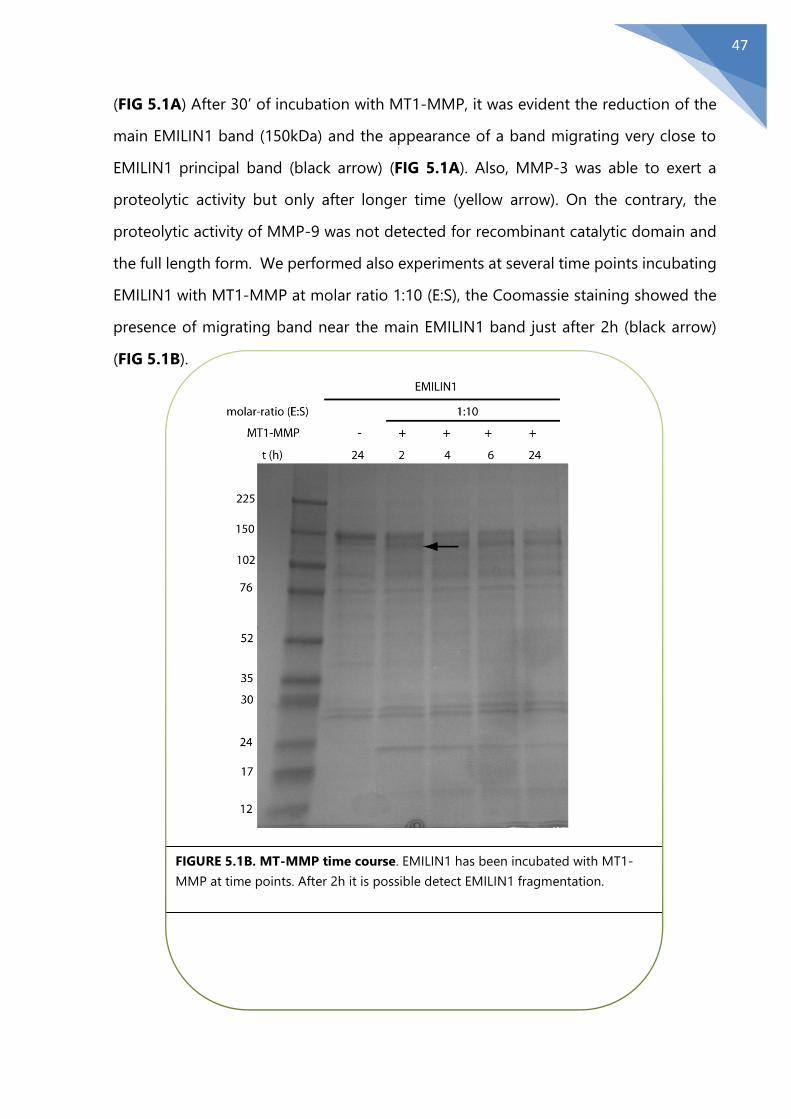
47
(FIG 5.1A) After 30’ of incubation with MT1-MMP, it was evident the reduction of the
main EMILIN1 band (150kDa) and the appearance of a band migrating very close to
EMILIN1 principal band (black arrow) (FIG 5.1A). Also, MMP-3 was able to exert a
proteolytic activity but only after longer time (yellow arrow). On the contrary, the
proteolytic activity of MMP-9 was not detected for recombinant catalytic domain and
the full length form. We performed also experiments at several time points incubating
EMILIN1 with MT1-MMP at molar ratio 1:10 (E:S), the Coomassie staining showed the
presence of migrating band near the main EMILIN1 band just after 2h (black arrow)
(FIG 5.1B).
FIGURE 5.1B. MT-MMP time course. EMILIN1 has been incubated with MT1-
MMP at time points. After 2h it is possible detect EMILIN1 fragmentation.
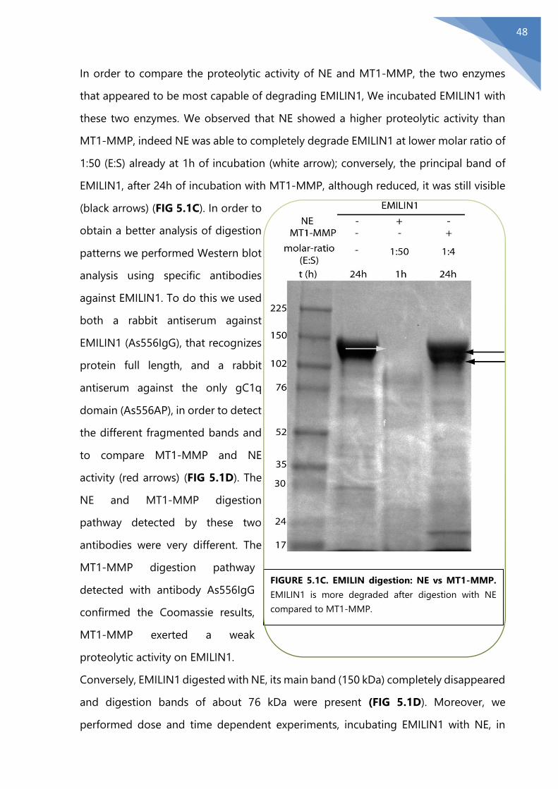
48
In order to compare the proteolytic activity of NE and MT1-MMP, the two enzymes
that appeared to be most capable of degrading EMILIN1, We incubated EMILIN1 with
these two enzymes. We observed that NE showed a higher proteolytic activity than
MT1-MMP, indeed NE was able to completely degrade EMILIN1 at lower molar ratio of
1:50 (E:S) already at 1h of incubation (white arrow); conversely, the principal band of
EMILIN1, after 24h of incubation with MT1-MMP, although reduced, it was still visible
(black arrows) (FIG 5.1C). In order to
obtain a better analysis of digestion
patterns we performed Western blot
analysis using specific antibodies
against EMILIN1. To do this we used
both a rabbit antiserum against
EMILIN1 (As556IgG), that recognizes
protein full length, and a rabbit
antiserum against the only gC1q
domain (As556AP), in order to detect
the different fragmented bands and
to compare MT1-MMP and NE
activity (red arrows) (FIG 5.1D). The
NE and MT1-MMP digestion
pathway detected by these two
antibodies were very different. The
MT1-MMP digestion pathway
detected with antibody As556IgG
confirmed the Coomassie results,
MT1-MMP exerted a weak
proteolytic activity on EMILIN1.
Conversely, EMILIN1 digested with NE, its main band (150 kDa) completely disappeared
and digestion bands of about 76 kDa were present (FIG 5.1D). Moreover, we
performed dose and time dependent experiments, incubating EMILIN1 with NE, in
FIGURE 5.1C. EMILIN digestion: NE vs MT1-MMP.
EMILIN1 is more degraded after digestion with NE
compared to MT1-MMP.
f

49
order to show the effectiveness proteolytic activity of NE. Western blotting analysis
revealed that the principal band of EMILIN1, digested by NE, was apparently greatly
reduced after 2 h at molar ratio 1:50 (E:S) (white arrow). These results have been
confirmed also by using the antibody, AS556AP, that is able to recognize the gC1q
domain. We observed a reduction of the main band of EMILIN1 both at molar ratio
1:100 (E:S) and (1:500) (white arrow) (FIG 5.1 E).
Conversely, at higher molar ratio 1:50 (E:S) this band completely disappeared (FIG
5.1E). It can be speculated that the activity of the tested MMPs on EMILIN1 substrate
is very low, or even absent for MMP-9, given that only at high enzyme concentrations
FIGURE 5.1D. EMILIN digestion: NE vs MT1-MMP. Western blotting analysis of
EMILIN1 samples incubated with MT1-MMP and NE at times and doses as indicated.
Membranes were probed with polyclonal antibodies against EMILIN1 As556IgG and
AS556AP.
-
-
-

50
and long incubation times EMILIN1 was partially digested; on the contrary, NE was able
to completely digest EMILIN1 also at low concentrations and short incubation times.
11111
FIGURE 5.1E. Western Blot analysis NE dose and time dependent on EMILIN1. (A) EMILIN1 is
incubated at several molar ratio with NE, membrane has been incubated with AS556IgG antibody
that recognizes several epitopes of the protein. (B) EMILIN1 is incubated at several molar ratio with
NE, membrane has been incubated with AS556AP antibody, that recognizes only the gC1q domain.
A
B

51
5.2 MMPs degradation in physiological conditions
The recombinant MMPs need saline buffer to reach the optimal enzymatic activity.
These buffer saline, determine pH values very different compared to the physiological
conditions and for this reason are unsuitable for studies with cell lines. To perform the
EMILIN1 digestion in a more physiological condition, we transfected HEK 293 cell line
with expression vectors for MMP-3 and MT1-MMP. To assess if MMPs enzymes was
expressed successfully we performed a Western Blot on cell lysates (to evaluate MT1-
MMP expression on membrane) and sovranatants (to evaluate the presence of secreted
MMP-3) (FIG 5.2A).
FIGURE 5.2A Western Blot analyses: MMP-3 and MT1-MMP expression. Membranes were
incubated with antibody α-Flag

52
EMILIN1 was added after 24h from transfection to the cell cultures and the conditioned
media used for different incubation times: 24h, 48h, 72h after transfection (FIG 5.2B).
FIGURE 5.2B Western Blot analyses: MMP-3 and MT1-MMP activities. Cells have been
transfected with pCMV2-MMP-3-Flag or pCMV-SP-MT1-MMP14-Flag. EMILIN1 was added
after 24h. The sovranatants have been collected after 24, 48 and 72h. The yellow arrows indicate
MMP-3 digestion bands. Black arrows indicate MT1-MMP digestion bands.
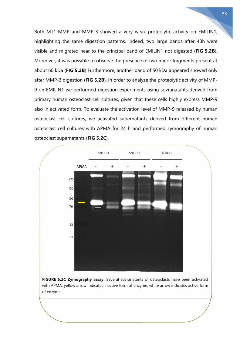
53
Both MT1-MMP and MMP-3 showed a very weak proteolytic activity on EMILIN1,
highlighting the same digestion patterns. Indeed, two large bands after 48h were
visible and migrated near to the principal band of EMILIN1 not digested (FIG 5.2B).
Moreover, it was possible to observe the presence of two minor fragments present at
about 60 kDa (FIG 5.2B) Furthermore, another band of 50 kDa appeared showed only
after MMP-3 digestion (FIG 5.2B). In order to analyze the proteolytic activity of MMP-
9 on EMILIN1 we performed digestion experiments using sovranatants derived from
primary human osteoclast cell cultures, given that these cells highly express MMP-9
also in activated form. To evaluate the activation level of MMP-9 released by human
osteoclast cell cultures, we activated supernatants derived from different human
osteoclast cell cultures with APMA for 24 h and performed zymography of human
osteoclast supernatants (FIG 5.2C).
FIGURE 5.2C Zymography assay. Several sovranatants of osteoclasts have been activated
with APMA. yellow arrow indicates inactive form of enzyme, white arrow indicates active form
of enzyme.

54
We then incubated EMILIN1 with sovranatants of human osteoclasts containing
activated MMP-9 for 1h, 6h, and 24h (FIG 5.2D).
Also with this approach we did not observe fragmentation bands of EMILIN1 after 24
h of incubation, confirming in this manner the results obtained with the recombinant
MMP-9.
FIGURE 5.2D Western Blotting analysis. EMILIN1 has been incubated with sovranatants of
osteoclasts activated, expressing MMP-9, for 1,6 and 24 h. MMP-9 is not able to degrade
EMILIN1

55
5.3 EMILIN1 adhesive and antiproliferative properties were
conserved after MT1-MMP proteolytic action
In order to verify if MT1-MMP is able to affect the functional properties of EMILIN1, we
performed adhesion assay using cells that express EMILIN1 ligand α4β1integrin [162].
To address this part we used SKLMS1 cell line that express this integrin and we
prepared coating of EMILIN1 not digested or digested with MT1-MMP. We did not
observe any reduction of cell adhesion of SKLMS1 on EMILIN1 digested by MT1-MMP.
Given that MT1-MMP partially degrade EMILIN1, the proteolytic activity of MT1-MMP
probably did not impair the functional role of the interaction between gC1q domain
and α4β1 integrin (FIG 5.3A). Moreover, in order to assess variations on proliferation
of SKLMS1 on EMILIN1 digested with MT1-MMP, we plated SKLMS1 on plates coated
with EMILIN1 alone and EMILIN1 digested with MT1-MMP for 24 h. We did not observe
FIGURE 5.3A. MT1-MMP treatment does not impair EMILIN1 functional properties. Dynamic
monitoring of SKLMS1 cell attachment in response to the effect of MT1-MMP on EMILIN1 cleavage
measured with the XCELLigence instrument and expressed as the cell index. A 1:25 (E:S) molar ratio
was used for a 24h incubation time. The wells were finally coated with 10 μg/ml of EMILIN1 (digested
or not by MT1-MMP). The data shown is the mean ± SD from n = 3 experiments with n = 6 replicates.
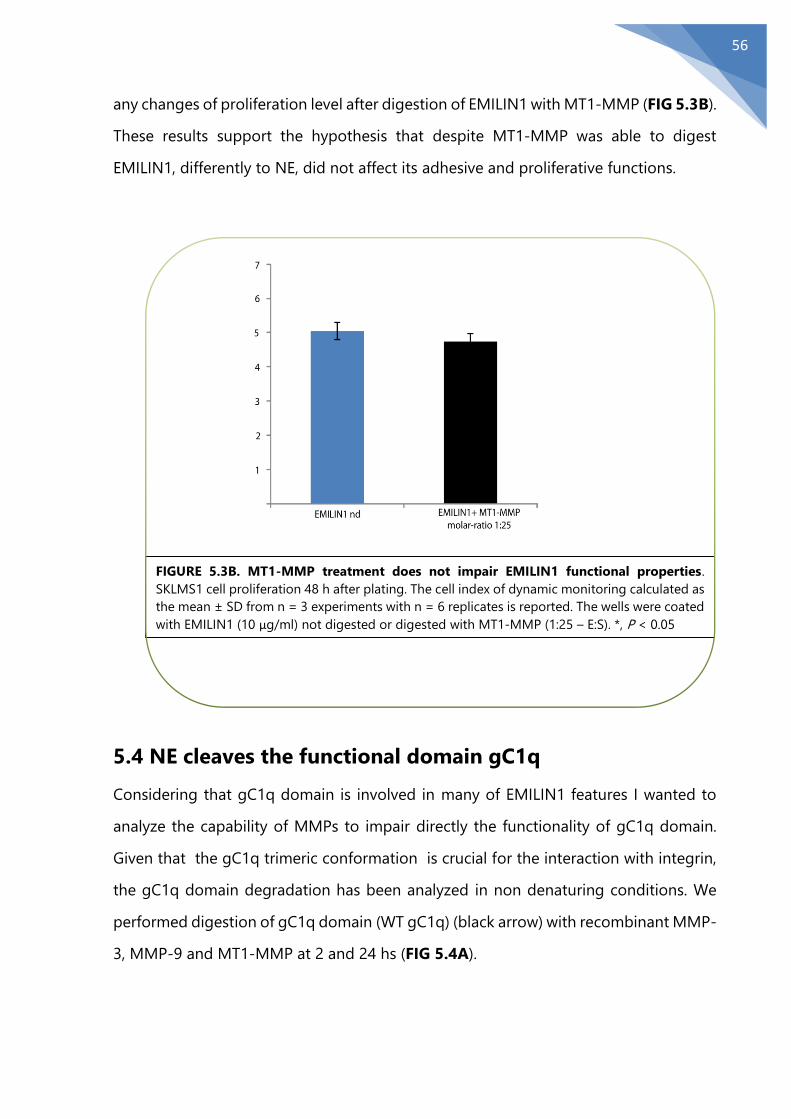
56
any changes of proliferation level after digestion of EMILIN1 with MT1-MMP (FIG 5.3B).
These results support the hypothesis that despite MT1-MMP was able to digest
EMILIN1, differently to NE, did not affect its adhesive and proliferative functions.
5.4 NE cleaves the functional domain gC1q
Considering that gC1q domain is involved in many of EMILIN1 features I wanted to
analyze the capability of MMPs to impair directly the functionality of gC1q domain.
Given that the gC1q trimeric conformation is crucial for the interaction with integrin,
the gC1q domain degradation has been analyzed in non denaturing conditions. We
performed digestion of gC1q domain (WT gC1q) (black arrow) with recombinant MMP-
3, MMP-9 and MT1-MMP at 2 and 24 hs (FIG 5.4A).
FIGURE 5.3B. MT1-MMP treatment does not impair EMILIN1 functional properties.
SKLMS1 cell proliferation 48 h after plating. The cell index of dynamic monitoring calculated as
the mean ± SD from n = 3 experiments with n = 6 replicates is reported. The wells were coated
with EMILIN1 (10 μg/ml) not digested or digested with MT1-MMP (1:25 – E:S). *, P < 0.05
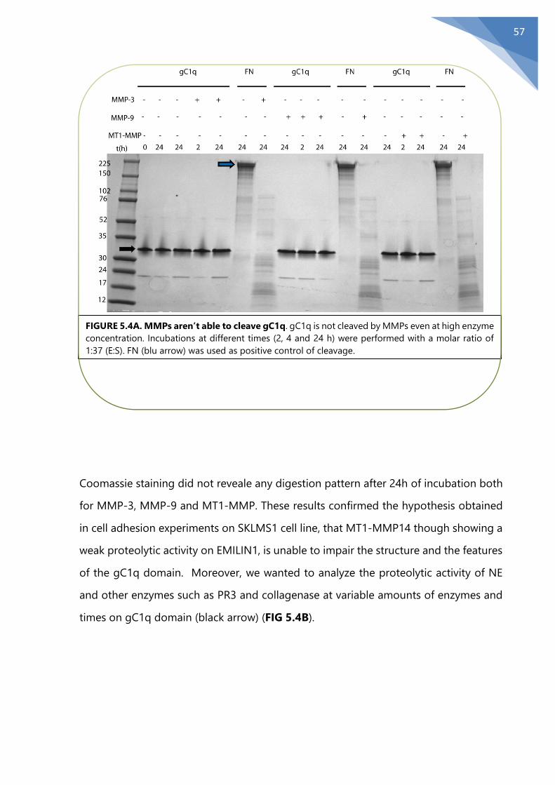
57
Coomassie staining did not reveale any digestion pattern after 24h of incubation both
for MMP-3, MMP-9 and MT1-MMP. These results confirmed the hypothesis obtained
in cell adhesion experiments on SKLMS1 cell line, that MT1-MMP14 though showing a
weak proteolytic activity on EMILIN1, is unable to impair the structure and the features
of the gC1q domain. Moreover, we wanted to analyze the proteolytic activity of NE
and other enzymes such as PR3 and collagenase at variable amounts of enzymes and
times on gC1q domain (black arrow) (FIG 5.4B).
FIGURE 5.4A. MMPs aren’t able to cleave gC1q. gC1q is not cleaved by MMPs even at high enzyme
concentration. Incubations at different times (2, 4 and 24 h) were performed with a molar ratio of
1:37 (E:S). FN (blu arrow) was used as positive control of cleavage.
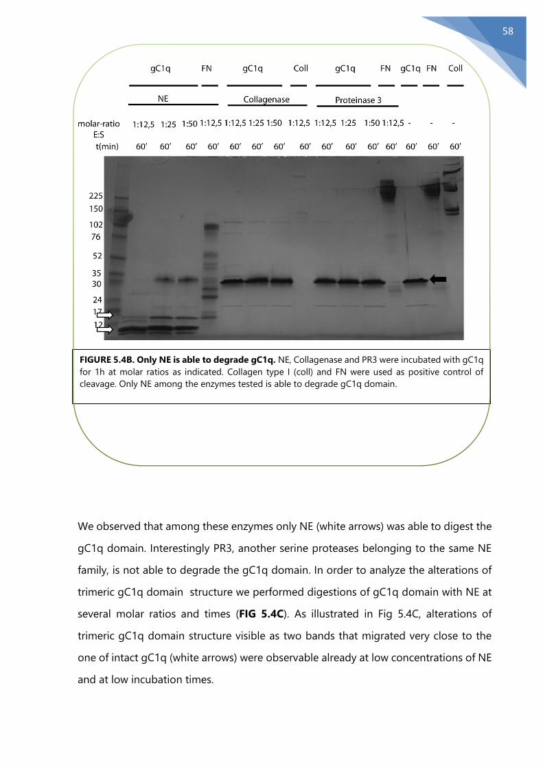
58
We observed that among these enzymes only NE (white arrows) was able to digest the
gC1q domain. Interestingly PR3, another serine proteases belonging to the same NE
family, is not able to degrade the gC1q domain. In order to analyze the alterations of
trimeric gC1q domain structure we performed digestions of gC1q domain with NE at
several molar ratios and times (FIG 5.4C). As illustrated in Fig 5.4C, alterations of
trimeric gC1q domain structure visible as two bands that migrated very close to the
one of intact gC1q (white arrows) were observable already at low concentrations of NE
and at low incubation times.
FIGURE 5.4B. Only NE is able to degrade gC1q. NE, Collagenase and PR3 were incubated with gC1q
for 1h at molar ratios as indicated. Collagen type I (coll) and FN were used as positive control of
cleavage. Only NE among the enzymes tested is able to degrade gC1q domain.
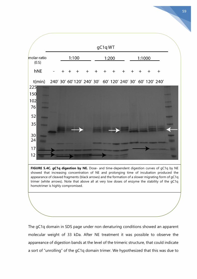
59
The gC1q domain in SDS page under non denaturing conditions showed an apparent
molecular weight of 33 kDa. After NE treatment it was possible to observe the
appareance of digestion bands at the level of the trimeric structure, that could indicate
a sort of “unrolling” of the gC1q domain trimer. We hypothesized that this was due to
FIGURE 5.4C. gC1q digestion by NE. Dose- and time-dependent digestion curves of gC1q by NE
showed that increasing concentration of NE and prolonging time of incubation produced the
appearance of cleaved fragments (black arrows) and the formation of a slower migrating form of gC1q
trimer (white arrows). Note that above all at very low doses of enzyme the stability of the gC1q
homotrimer is highly compromised.

60
NE proteolytic activity, that may determine in
this manner a loosener effect of the gC1q
domain structure. This treatment showed a
gC1q domain no longer formed by a single band
but as a whole of three bands that are able to
migrate more slowly than the not digested gC1q
domain. Moreover, Coomassie staining revealed
that at lower molar ratio E:S 1:1000 after 1 h of
incubation, the gC1q domain is completely
“unrolled” and the appearance of digestion
bands is absent (FIG 5.4C). Moreover, it was
evident the presence of two digestion bands of
about 16 and 13 kDa, increasing in a dose
dependent manner following NE incubation.
These results confirmed the very high specificity
of NE for the gC1q domain. In order to confirm
that the digestion pattern was due exclusively to
NE and not, for example, to contaminant, we
incubated recombinant form of gC1q WT (black
arrow) with NE (white arrows) and Silvelestat, a
specific inhibitor of NE, as illustrated (FIG 5.4D). When gC1q was incubated with NE
and Silvelestat, we didn’t observe digestion bands demostrating that is NE to
specifically cleave the EMILIN1 gC1q domain. In summary this results indicated that NE,
among the enzymes tested, was the only one able to abolish the functional gC1q
domain.
5.5 Identification of NE cleavage site on the functional
domain gC1q
Given that only NE, among the enzymes tested, was able to digest and impair the gC1q
domain functionality, we tried to pinpoint the precise cleavage site. First of all, we
FIGURE 5.4D. Silvelestat inhibits NE
activity. EMILIN1 has been incubated
with NE at molar ratio E:S 1:100) for 30’
and 6o’, and with Silvelestat, an inhibitor
of NE for 60’.

61
restrict the cleavage region by mass spectrometry analysis of the gc1q fragments
obtained after incubation with NE in different conditions. In order to do this, I
performed dose dependent experiments in which gC1q domain has been incubated
with NE for 1h to obtain digestion bands for mass spectrometry analysis with MALDI
TOF. At the highest concentration of NE, I observed two migrating bands of apparent
molecular weights ranging from 12 to 17 kDa, that were extracted from the gel, purified
and analyzed by MALDI TOF. The MALDI TOF analysis has detected two main peaks
with a molecular weight of 8650,7 and 8646,3 Da (FIG 5.5A); these peaks matched
almost perfectly to virtual fragments with a calculated mass of 8651 Da for cleavage
between serine in position 913 (S913) and arginine in position 914 (R914), or 8647 for
cleavage between lysine in position 912 (L912) and serine in position 913 (S913). These
proposed cleavage sites were particularly interesting because are properly exposed to
solvent as shown by NMR model (FIG 5.5B). As the NE cleavage sites are not well
defined, we took a trial and error mutagenesis approach to identify the exact cleavage
site.
A
B
FIGURE 5.5 (A-B) Scheme of gC1q primary structure with the possible cleavage sites emerged from MALDI TOF analysis, one placed between S913 and R914
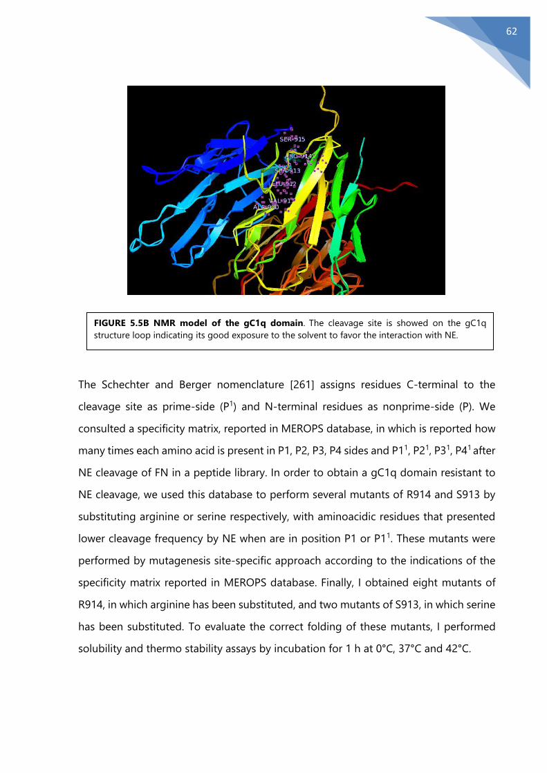
62
The Schechter and Berger nomenclature [261] assigns residues C-terminal to the
cleavage site as prime-side (P1) and N-terminal residues as nonprime-side (P). We
consulted a specificity matrix, reported in MEROPS database, in which is reported how
many times each amino acid is present in P1, P2, P3, P4 sides and P11, P21, P31, P41 after
NE cleavage of FN in a peptide library. In order to obtain a gC1q domain resistant to
NE cleavage, we used this database to perform several mutants of R914 and S913 by
substituting arginine or serine respectively, with aminoacidic residues that presented
lower cleavage frequency by NE when are in position P1 or P11. These mutants were
performed by mutagenesis site-specific approach according to the indications of the
specificity matrix reported in MEROPS database. Finally, I obtained eight mutants of
R914, in which arginine has been substituted, and two mutants of S913, in which serine
has been substituted. To evaluate the correct folding of these mutants, I performed
solubility and thermo stability assays by incubation for 1 h at 0°C, 37°C and 42°C.
FIGURE 5.5B NMR model of the gC1q domain. The cleavage site is showed on the gC1q
structure loop indicating its good exposure to the solvent to favor the interaction with NE.

63
5.6 The mutant R914W gC1q is resistant to NE
The mutants showing a correct trimeric structure were digested, with NE for 1h at molar
ratio 1:200 (E:S). Among the mutants obtained, only the mutant R914W gC1q (R914W),
in which arginine aminoacid residue is substituted with tryptophan aminoacid residue,
was more resistant to NE treatment: more than 90% of the trimeric peptide was intact
after 1h of NE digestion compared to gC1q domain (WT gC1q) (FIG 5.6a). Among the
mutants obtained by the substitution of the arginine aminoacid residue in position 914,
the mutant R914K gC1q (R914K) was more sensible to NE digestion compared to WT
gC1q, indicating a positive gain when lysine in position P11. The mutant R914H gC1q
(R914H), in which arginine is substituted with histidine, showed a digestion pattern
(black arrows) like WT gC1q (FIG 5.6b). Among the mutants obtained by the
substitution of the serine aminoacid residue in position 913, only the mutant S913T
gC1q (S913T) presented a correct folding, but it was completely digested by NE
treatment (black arrows) (FIG 5.6c). The mutants, R914G, R914E and R914D in which
arginine is substituted with glycine, glutamic acid, aspartic acid respectively were
digested even more efficiently (black arrows) (FIG 5.6d). Moreover, when we digested
with NE, a gC1q mutant that showed a mutations distant from the hypothesized
cleavage site, such as E933A gC1q (E933A), involved in the binding with α4β1 integrins,
this mutant showed a NE digestion (black arrows) pattern very similar to WT gC1q (FIG
5.6e). This data indicates that NE cleavage impairs the functionality of WT gC1q but at
the same time that the NE consensus sequence on WT gC1q is outside of region that
is able to recognize α4β1 integrin.
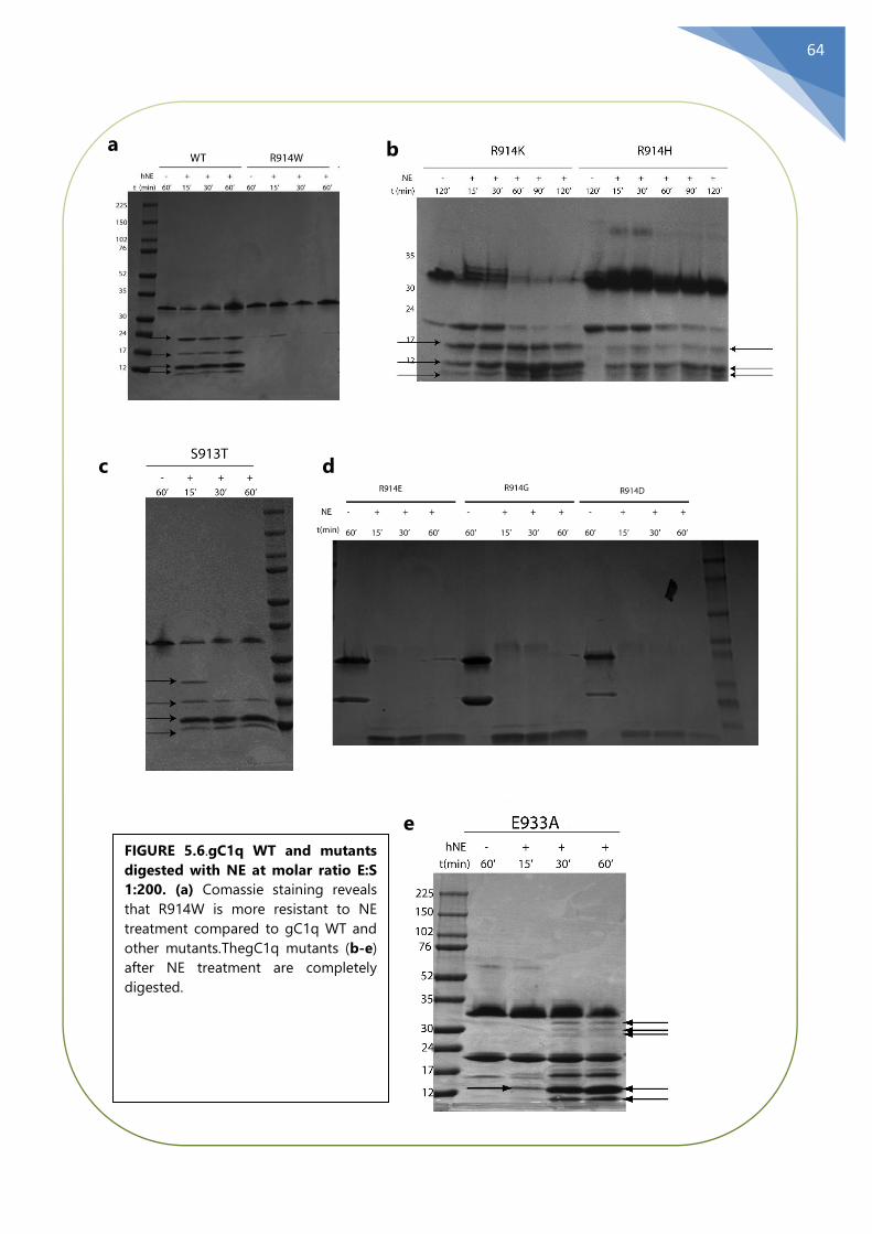
64
FIGURE 5.6.gC1q WT and mutants
digested with NE at molar ratio E:S
1:200. (a) Comassie staining reveals
that R914W is more resistant to NE
treatment compared to gC1q WT and
other mutants.ThegC1q mutants (b-e)
after NE treatment are completely
digested.
a b
c d
e

65
5.7 R914W preserves its features after NE treatment
The EMILIN1 through its domain gC1q is able to determine the cellular adhesion with
α4β1 integrin [46]. To demonstrate the R914W capability to maintain its function after
NE treatment, we performed an adhesion assay (CAFCA assay) of Jurkat line, that
expresses great amounts of α4β1 integrin. Both WT gC1q and the R914W mutant were
digested with NE at high molar ratio 1:25 (E:S). We plated Jurkat cells on plates coated
with WT gC1q or the R914W mutant alone or digested by NE. Applying standard assay
conditions (Spin at 1000 RPM, Spin off at 500 RPM), we observed as expected that cells
adhered on plates coated not digested WT gC1q but not on WT gC1q digested with
NE. On the contrary, Jurkat cells adhered at the same level on plates coated with the
R914W both digested and not digested with NE (FIG 5.7A).
These results demonstrated that gC1q WT after treatment with NE was not able to bind
α4β1 integrin and consequently it lost its adhesive property. Furthermore, these results
suggested, that, although NE proteolytic activity occurs on a site that is far from the
E933 amino acid residue, important for the interaction with α4β1 integrin, the NE
FIGURE 5.7A Adhesion (CAFCA assay)
of Jurkat cells on WT or R914W gC1q
preincubated with NE or not (w/o NE)
with NE. The E:S molar ratios and
incubation times are “boxed” in the
corresponding gel at the bottom of
the graphic indicating the status
(fragmented or not) of the resulting
gC1q used to coat the wells (10 µg/ml)
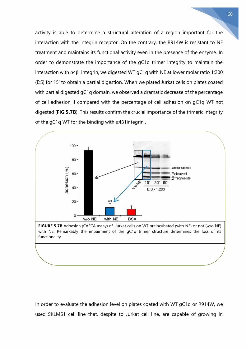
66
activity is able to determine a structural alteration of a region important for the
interaction with the integrin receptor. On the contrary, the R914W is resistant to NE
treatment and maintains its functional activity even in the presence of the enzyme. In
order to demonstrate the importance of the gC1q trimer integrity to maintain the
interaction with α4β1integrin, we digested WT gC1q with NE at lower molar ratio 1:200
(E:S) for 15’ to obtain a partial digestion. When we plated Jurkat cells on plates coated
with partial digested gC1q domain, we observed a dramatic decrease of the percentage
of cell adhesion if compared with the percentage of cell adhesion on gC1q WT not
digested (FIG 5.7B). This results confirm the crucial importance of the trimeric integrity
of the gC1q WT for the binding with α4β1integrin .
In order to evaluate the adhesion level on plates coated with WT gC1q or R914W, we
used SKLMS1 cell line that, despite to Jurkat cell line, are capable of growing in
FIGURE 5.7B Adhesion (CAFCA assay) of Jurkat cells on WT preincubated (with NE) or not (w/o NE)
with NE. Remarkably the impairment of the gC1q trimer structure determines the loss of its
functionality.
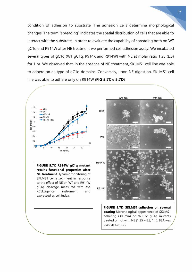
67
condition of adhesion to substrate. The adhesion cells determine morphological
changes. The term “spreading” indicates the spatial distribution of cells that are able to
interact with the substrate. In order to evaluate the capability of spreading both on WT
gC1q and R914W after NE treatment we performed cell adhesion assay. We incubated
several types of gC1q (WT gC1q, R914K and R914W) with NE at molar ratio 1:25 (E:S)
for 1 hr. We observed that, in the absence of NE treatment, SKLMS1 cell line was able
to adhere on all type of gC1q domains. Conversely, upon NE digestion, SKLMS1 cell
line was able to adhere only on R914W (FIG 5.7C e 5.7D)
FIGURE 5.7C R914W gC1q mutant
retains functional properties after
NE treatment Dynamic monitoring of
SKLMS1 cell attachment in response
to the effect of NE on WT and R914W
gC1q cleavage measured with the
XCELLigence instrument and
expressed as cell index.
FIGURE 5.7D SKLMS1 adhesion on several
coating Morphological appearance of SKLMS1
adhering (30 min) on WT or gC1q mutants
treated or not with NE (1:25 – E:S, 1 h). BSA was
used as control.

68
Considering that the WT gC1q, through interaction with α4β1 integrin, is able to
regulate cell proliferation, we performed a proliferation assay (FIG 5.7E). We plated
SKLMS1 cells on plates coated with WT gC1q alone or digested with NE and we
observed that the percentage of cells proliferating was more elevated when WT gC1q
has been treated with NE. Conversely, when we plated SKLMS1 cells on wells coated
with R914W alone or digested with NE, we didn’t observe differences of proliferation
levels.
Moreover, in order to assess if also a low amount of R914W was able to compensate
the functionality loss of gC1q WT after NE treatment, we monitored cell proliferation
(Jurkat cell line) and cell adhesion (SKLMS1) using the full-length EMILIN1 together
with the R914W at different doses as an agonist.
We observed that just a low amount of R914W was able to compete in cell adhesion
after NE treatment, and moreover, we observed in the proliferation assay the presence
of the R914W that is not digested by NE was able to suppress cell proliferation. These
results confirm the hypothesis that R914W is more resistant to NE cleavage respect to
the gC1q WT, and moreover is able to maintain the regulatory features of the gC1q
domain (FIG 5.7F).
FIGURE 5.7E Proliferation of
SKLMS1. Cells plated on gC1q (WT
or R914W mutant) (10 μg/ml)
treated with NE (1:25 – E:S, 1 h). The
cell index after 48 h of dynamic
monitoring calculated as the mean ±
SD from n = 3 experiments with n =
6 replicates is reported. *, P < 0.05

69
FIGURE 5.7F (a-b)In adhesion (Jurkat cells, CAFCA assay, a) and proliferation (SKLMS1 cells, dynamic
monitoring by XCelligence instrument, b) assays R914W gC1q mutant was present as an agonist at
different doses as indicated. The mixtures (R914W gC1q mutant at different doses and EMILIN1, E1
at 5 µg/ml) were preincubated for 1 h with NE or not (w/o NE) and the resulting products used to
coat the wells. Data are expressed as the means ± SD of n= 3 independent experiments with n = 6
replicates. **, P < 0.001; NS, not significant.
a
b

70
Chapter 6
Discussion
During my PhD program, we focused the attention above on the importance of the
structural integrity of EMILIN1. In particular, the gC1q domain was investigated to
understand how the maintenance of its conformational structure was crucial to display
the regulatory properties of the protein. EMILIN1 is an ECM glycoprotein, involved in
several functions such as regulation of blood homeostasis [50], regulation of the
functionality and the maintenance of the structure of the lymphatic system [48], [83],
[154] and, through its gC1q domain, following interaction with α4β1 and α9β1 integrins,
in several important cellular processes such as cell adhesion, migration and skin
proliferation, playing an important role in oncology field [46][53].
Studies previously performed in my laboratory demonstrated that incubating EMILIN1
with several supernatants of inflammatory cells, only supernatant derived from
neutrophils were able to degrade EMILIN1 and then impair its tumor suppressor
functions [162]. In addition to this, among the proteases released by neutrophils, only
NE was able to impair the tumor suppressor role of the protein. NE exerts an important
role in ECM breakdown. During inflammation human neutrophils, among the cells
involved in immunity response, are the first cells to occur in inflammation site. After
stimulation they release proteolytic enzymes such as NE that belongs to serine
proteases family and shows a wide substrate specificity among several ECM
components for example laminin, collagens, elastin and fibronectin [241].
A study performed by Stegemann et al, demonstrated, by a proteomic approach, that
EMILIN1 could be as a possible substrate of several MMPs [164].
One of the principal objective of this thesis was to understand, if, in addition to NE,
there were any others enzymes, involved in ECM break-down, capable to degrade
EMILIN1, and above all, to impair its tumor suppressor functions. For this purpose
“biochemical” and “physiological” approach were performed to verify MMPs activity on
EMILIN1. The presence of salts in activation buffer, composed of CaCl2, NaCl and Tris-
HCl, of recombinant MMPs, had to be taken into consideration. In particular the

71
presence of Tris-HCl is a critical point for viability of cells. Moreover, we observed that
the only presence of Tris-HCl can determine an alteration of EMILIN1 stability and that,
chloride ions, that result from the dissociation of Tris-HCl, can interact with cysteine
residues present in EMILIN1 structure, in particular at EMI domain, and determine
formation of aggregates. With biochemical approach, we did not observe a glaring
proteolytic activity on EMILIN1 by these MMPs. Indeed, only MT1-MMP, among MMPs
tested, showed a modest proteolytic activity but at long times of incubation. Moreover,
the incubation of EMILIN1 with recombinant MMP-9 catalytic domain did not show any
degradation pattern. Interestingly, the partial disappearance of the EMILIN1 main band
after incubation with the full-length form of MMP-9 (FIG 5.1A), may be due to
degradation of the protein by salts rather than its digestion by MMP-9. To obtain as
closely as possible physiological conditions of protein degradation we performed
transfection of HEK 293 cell line with pCMV2-MMP3-Flag for MMP-3 and pCMV-SP-
MT1-MMP-Flag to obtain the expression of full length enzymes. On the other hand, to
test the MMP-9 proteolytic activity we used osteoclast surnatants previously activated
with APMA. This more physiological approach showed a weak activity on EMILIN1 only
for MMP-3. No digestion pattern of EMILIN1 after incubation with osteoclast surnatans
expressing MMP-9 was observed confirming that the partial degradation of EMILIN1
obtained with “biochemical” approach, was due to the saline buffer of recombinant
MMP-9. Interestingly, the proteolytic activity of MT1-MMP and MMP-3 was quite
negligible if compared with the proteolytic activity of NE. More importantly, the weak
proteolytic activity of MT1-MMP on EMILIN1 did not affect the tumor suppressor role
of EMILIN1: the capability of EMILIN1 to regulate cell adhesion and to inhibit cell
proliferation was maintained in SKLMS1 cell after treatment with MT1-MMP. The lack
of interference on regulatory functions of the protein was due to the fact that MT1-
MMP was not able to fragment the gC1q domain. Probably, the MMPs proteolytic
activities, in particular MT1-MMP, may occur in pathological diseases where the
degradation of elastic fibers could affect for example the vascular wall. The study of
Stegemann et al showing a possible proteolytic activity on EMILIN1 was performed
using aortic vascular tissues. This possibility is supported by other authors. Tamarina et
72
al illustrated an increase of MMP-9 expression in aneurysmal aortas, which are
characterized by cleavage of elastin and collagen fibers[242]. They showed that MMP-
9 was the principal MMP present in aneurysm tissue. Kelly et al, demonstrated the
involvement of MT1-MMP in the reorganization in rat uterine artery [243]. Other
studies demonstrated that the levels of MMP-2, MMP-3, MMP-9 MMP13 were elevated
in varicose veins, showing that MMPs can promote the pathogenesis of the diseases
by weakening of vessels walls [244].May be, in these cases it would be interesting to
demonstrate EMILIN1 integrity, or its degradation. One of the most interesting result
obtained in this thesis is represented by the ability of NE to determine an “unrolling”
of the trimeric structure. This effect is detectable at lower concentration of NE and at
short time of incubation. The trimer unrolling is a sufficient conditions to inhibit cell
adhesion suggesting that WT gC1q structure integrity is crucial and needful to allow
the binding to α4β1 integrin. The identification of NE cleavage site on gC1q it has been
an important step of this thesis. The strategy used to obtain this site was developed by
a site-specific mutagenesis approach. gC1q was digested at elevated concentrations in
order to obtain well-defined bands of digestion. MALDI TOF analysis revealed a
possible cleavage site of NE on gC1q between serine amino acid residue located in
position 913 and arginine amino acid residue located in position 914. The NMR
structure located this possible NE cleavage site in a flexible loop structure at the apex
of the WT gC1q domain, resulting in this manner exposed to NE cleavage.
Identification of proteases cleavage sites is generally performed by Edman degradation
and sequencing. Anyway, in presence of high molecular weight proteins Edman
sequencing is difficult to apply. Indeed, each of numerous proteolytic fragments
obtained by proteases may be analyzed and almost never it is possible to obtain an
elevate coverage of cleavage sites. Ultimately, it is very difficult to obtain the precise
cleavage sites of proteases such as MMPs and NE on ECM proteins that present a high
structural complexity such as EMILIN1. Doucet et al [239], by a novel proteomic
approach, identified 44 NE cleavage sites on FN and 11 NE cleavage sites along the
three chain of laminin. Moreover, the authors by analysis of the cleaved peptides
observed that the NE cleavage occurred primarily when at the P1 position were present

73
Val, Ile, Thr, and Ala. Remarkably FN and laminin have a similar aminoacidic sequence
length. The differences in number of NE cleavage sites on FN and laminin suggest that
NE consensus site of cleavage strongly depends from the secondary and terziary
polypeptide structure more than the primary structurepoj.
Several peptidase data-base, such as MEROPS and substitution matrices were
consulted. The first substitution matrix has been designed by Margaret Dayhoff in
1970. These substitution matrices were created by analyzing alignments of similar
proteins sequences and the frequencies of each type of substitution occurred.
Subsequently, these matrices have been adapted to study the prediction of proteases
cleavage sites. I found a substitution matrix in which are reported the frequency of NE
cleavage when each single aminoacidic is at position P1, P2, P3, or P11, P21, P31 is
reported. The most conserved amino acid residues are present in position P1 and P11.
Accordingly, with these databases I generated several gC1q mutants in which serine or
arginine have been substituted with amino acids that rarely are present at position P1
or P11 after NE cleavage. Among the gC1q mutants obtained, some of these presented
a correct folding of the trimeric structure; others, instead, presented an incorrect
folding. We were able to analyze only the mutants that presented a correct folding of
the trimeric structure. The presence or absence of the trimeric folding in the mutants
was probably due to the instauration of new chemical bonds as a result of substitution
of one or more aminoacids. Among the mutants that presented a correct folding of the
trimeric structure, I found that mutant R914W, in which arginine at position P11, was
substituted with tryptophan, was resistant to NE treatment. The correct folding of this
mutant appears very interesting, since arginine and tryptophan present several
differences both in the physical and chemical properties such as the structure and the
aminoacid chain charge. To verify if mutant R914W was able to maintain its properties
after NE treatment I performed functional analyses revealing that the R914W,
differently from WT gC1q, was still able to retain its functional properties after NE
treatment. Interestingly, the NE cleavage site, is very far from the site that is able to
interact with integrins, located in position 933. These data suggest that a specific
conformation of the gC1q trimer is crucial for the interaction with integrins. Probably,
74
the NE proteolytic activity is able to impair the interaction stability of gC1q domain-
integrins. Considering that other amino acid substitutions, in position 914, were not
able to impair NE cleavage, it has showed that the NE cleavage site takes on more
characteristics of conformational activity of substrate rather than the specific sequence.
Given that NE is able to impair the WT gC1q structure and its functions, the NE cleavage
site detection represents a very useful and important result for future studies.
Previously, Pivetta et al, illustrated that the use of NE inhibitors, such as Silvelestat, is
able to restore the lymphatic vessels function [154]. In a scenario, in which neutrophils
are present and secrete NE, determining the loss of EMILIN1 functions, such as tumor
microenvironment, it is thinkable a therapeutics approach in which the administration
of a specific inhibitor is able to restore the EMILIN1 antiproliferative functions.
Alternatively, the administration of R914W fragments that are resistant to proteolytic
activity could be a winning strategy.

75
References [1] D. Hubmacher and S. S. Apte, “The biology of the extracellular matrix: novel insights.,” Curr.
Opin. Rheumatol., vol. 25, no. 1, pp. 65–70, 2013.
[2] R. P. Mecham, “Overview of extracellular matrix,” Curr. Protoc. Cell Biol., no. SUPPL.57, 2012.
[3] J. D. Humphrey, E. R. Dufresne, and M. a. Schwartz, “Mechanotransduction and extracellular
matrix homeostasis,” Nat. Rev. Mol. Cell Biol., vol. 15, no. 12, pp. 802–812, 2014.
[4] J. Rosenbloom, W. R. Abrams, and R. Mecham, “Extracellular matrix 4: the elastic fiber.,” FASEB
J., vol. 7, pp. 1208–1218, 1993.
[5] M. A. Gibson, G. Hatzinikolas, J. S. Kumaratilake, L. B. Sandberg, J. K. Nicholl, G. R. Sutherland,
and E. G. Cleary, “Further characterization of proteins associated with elastic fiber microfibrils
including the molecular cloning of MAGP-2 (MP25),” J. Biol. Chem., vol. 271, no. 2, pp. 1096–
1103, 1996.
[6] G. M. Bressan, D. Daga-Gordini, A. Colombatti, I. Castellani, V. Marigo, and D. Volpin, “Emilin, a
component of elastic fibers preferentially located at the elastin-microfibrils interface,” J. Cell
Biol., vol. 121, no. 1, pp. 201–212, 1993.
[7] P. N. Robinson and M. Godfrey, “The molecular genetics of Marfan syndrome and related
microfibrillopathies.,” J. Med. Genet., vol. 37, no. 1, pp. 9–25, 2000.
[8] G. Veit, B. Kobbe, D. R. Keene, M. Paulsson, M. Koch, and R. Wagener, “Collagen XXVIII, a novel
von Willebrand factor A domain-containing protein with many imperfections in the collagenous
domain,” J. Biol. Chem., vol. 281, no. 6, pp. 3494–3504, 2006.
[9] J. Brinckmann, “Collagens at a glance,” Topics in Current Chemistry, vol. 247. pp. 1–6, 2005.
[10] S. Ricard-Blum, F. Ruggiero, and M. Van Der Rest, “The collagen superfamily,” Top. Curr. Chem.,
vol. 247, pp. 35–84, 2005.
[11] D. E. Birk, “Type V collagen: Heterotypic type I/V collagen interactions in the regulation of fibril
assembly,” Micron, vol. 32, no. 3. pp. 223–237, 2001.
[12] S. Chakravarti, T. Magnuson, J. H. Lass, K. J. Jepsen, C. LaMantia, and H. Carroll, “Lumican
regulates collagen fibril assembly: Skin fragility and corneal opacity in the absence of lumican,”
J. Cell Biol., vol. 141, no. 5, pp. 1277–1286, 1998.
[13] F. H. Silver, J. W. Freeman, and G. P. Seehra, “Collagen self-assembly and the development of
tendon mechanical properties,” Journal of Biomechanics, vol. 36, no. 10. pp. 1529–1553, 2003.
[14] W. HENKEL and R. W. GLANVILLE, “Covalent Crosslinking between Molecules of Type I and Type
III Collagen. The Involvement of the N-Terminal, Nonhelical Regions of the alphal(I) and al(III)
Chains in the Formation of Intermolecular Crosslinks,” Eur. J. Biochem., vol. 122, no. 1, pp. 205–
213, 1982.
[15] A. J. Bailey, S. Bazin, T. J. Sims, M. Le Lous, C. Nicoletis, and A. Delaunay, “Characterization of the
collagen of human hypertrophic and normal scars,” BBA - Protein Struct., vol. 405, no. 2, pp.
412–421, 1975.
[16] T. Moriguchi and D. Fujimoto, “Crosslink of collagen in hypertrophic scar,” J. Invest. Dermatol.,
vol. 72, no. 3, pp. 143–145, 1979.
[17] K. R. Levental, H. Yu, L. Kass, J. N. Lakins, M. Egeblad, J. T. Erler, S. F. T. Fong, K. Csiszar, A.
Giaccia, W. Weninger, M. Yamauchi, D. L. Gasser, and V. M. Weaver, “Matrix Crosslinking Forces
Tumor Progression by Enhancing Integrin Signaling,” Cell, vol. 139, no. 5, pp. 891–906, 2009.
[18] R. Fleischmajer, S. Gay, J. S. Perlish, and J. P. Cesarini, “Immunoelectron microscopy of type III
collagen in normal and scleroderma skin,” J. Invest. Dermatol., vol. 75, no. 2, pp. 189–191, 1980.

76
[19] B. Sykes, B. Puddle, M. Francis, and R. Smith, “The estimation of two collagens from human
dermis by interrupted gel electrophoresis,” Biochem. Biophys. Res. Commun., vol. 72, no. 4, pp.
1472–1480, 1976.
[20] T. H. E. Occurrence, O. F. Collagen, and I. N. Reticulin, “SEROLOGICAL AND
IMMUNOHISTOLOGICAL INVESTIGATION OF THE OCCURRENCE OF COLLAGEN TYPE III ,
FIBRONECTIN AND THE NON-COLLAGENOUS GLYCOPROTEIN OF PRAS AND GLYNN,” pp.
154–166, 1982.
[21] G. S. Montes, R. M. Krisztán, K. M. Shigihara, R. Tokoro, P. A. S. Mourão, and L. C. U. Junqueira,
“Histochemical and morphological characterization of reticular fibers,” Histochemistry, vol. 65,
no. 2, pp. 131–141, 1980.
[22] T. Ushiki, “Collagen fibers, reticular fibers and elastic fibers. A comprehensive understanding
from a morphological viewpoint.,” Archives of Histology and Cytology, vol. 65, no. 2. pp. 109–
126, 2002.
[23] J. E. Wagenseil and R. P. Mecham, “New insights into elastic fiber assembly,” Birth Defects
Research Part C - Embryo Today: Reviews, vol. 81, no. 4. pp. 229–240, 2007.
[24] P. J. McLaughlin, Q. Chen, M. Horiguchi, B. C. Starcher, J. B. Stanton, T. J. Broekelmann, A. D.
Marmorstein, B. McKay, R. Mecham, T. Nakamura, and L. Y. Marmorstein, “Targeted disruption
of fibulin-4 abolishes elastogenesis and causes perinatal lethality in mice.,” Mol. Cell. Biol., vol.
26, no. 5, pp. 1700–9, 2006.
[25] B. A. Kozel, B. J. Rongish, A. Czirok, J. Zach, C. D. Little, E. C. Davis, R. H. Knutsen, J. E. Wagenseil,
M. A. Levy, and R. P. Mecham, “Elastic fiber formation: A dynamic view of extracellular matrix
assembly using timer reporters,” J. Cell. Physiol., vol. 207, no. 1, pp. 87–96, 2006.
[26] L. Shapiro and P. E. Scherer, “The crystal structure of a complement-1q family protein suggests
an evolutionary link to tumor necrosis factor,” Curr. Biol., vol. 8, no. 6, pp. 335–340, 1998.
[27] U. Kishore, C. Gaboriaud, P. Waters, A. K. Shrive, T. J. Greenhough, K. B. M. Reid, R. B. Sim, and G.
J. Arlaud, “C1q and tumor necrosis factor superfamily: Modularity and versatility,” Trends in
Immunology, vol. 25, no. 10. pp. 551–561, 2004.
[28] R. Doliana, M. Mongiat, F. Bucciotti, E. Giacomello, R. Deutzmann, D. Volpin, G. M. Bressan, and
A. Colombatti, “EMILIN, a component of the elastic fiber and a new member of the C1q/tumor
necrosis factor superfamily of proteins,” J. Biol. Chem., vol. 274, no. 24, pp. 16773–16781, 1999.
[29] Y. T. Tang, T. Hu, M. Arterburn, B. Boyle, J. M. Bright, S. Palencia, P. C. Emtage, and W. D. Funk,
“The complete complement of C1q-domain-containing proteins in Homo sapiens,” Genomics,
vol. 86, no. 1, pp. 100–111, 2005.
[30] R. Ghai, P. Waters, L. T. Roumenina, M. Gadjeva, M. S. Kojouharova, K. B. M. Reid, R. B. Sim, and
U. Kishore, “C1q and its growing family,” Immunobiology, vol. 212, no. 4–5, pp. 253–266, 2007.
[31] A. Colombatti, G. M. Bressan, I. Castellani, and D. Volpin, “Glycoprotein 115, a glycoprotein
isolated from chick blood vessels, is widely distributed in connective tissue,” J. Cell Biol., vol.
100, no. 1, pp. 18–26, 1985.
[32] R. Doliana, S. Bot, G. Mungiguerra, A. Canton, S. Paron Cilli, and A. Colombatti, “Isolation and
Characterization of EMILIN-2, a New Component of the Growing EMILINs Family and a Member
of the EMI Domain-containing Superfamily,” J. Biol. Chem., vol. 276, no. 15, pp. 12003–12011,
2001.
[33] C. P. M. Hay Ward, J. W. Smith, P. Horsewood, T. E. Warkentin, and J. G. Kelton, “p-155, a
multimeric platelet protein that is expressed on activated platelets,” J. Biol. Chem., vol. 266, no.
11, pp. 7114–7120, 1991.
[34] D. B. Kershaw, P. E. Thomas, B. L. Wharram, M. Goyal, J. E. Wiggins, C. I. Whiteside, and R. C.
77
Wiggins, “Molecular cloning, expression, and characterization of podocalyxin-like protein 1
from rabbit as a transmembrane protein of glomerular podocytes and vascular endothelium,” J.
Biol. Chem., vol. 270, no. 49, pp. 29439–29446, 1995.
[35] C. Leimeister, C. Steidl, N. Schumacher, S. Erhard, and M. Gessler, “Developmental Expression
and Biochemical Characterization of Emu Family Members,” Dev. Biol., vol. 249, no. 2, pp. 204–
218, 2002.
[36] G. M. Bressan, I. Castellani, A. Colombatti, and D. Volpin, “Isolation and characterization of a
115,000-dalton matrix-associated glycoprotein from chick aorta,” J. Biol. Chem., vol. 258, no. 21,
pp. 13262–13267, 1983.
[37] M. Mongiat, G. Mungiguerra, S. Bot, M. T. Mucignat, E. Giacomello, R. Doliana, and A.
Colombatti, “Self-assembly and supramolecular organization of EMILIN,” J. Biol. Chem., vol. 275,
no. 33, pp. 25471–25480, 2000.
[38] J. M. Hurle, G. Corson, K. Daniels, R. S. Reiter, L. Y. Sakai, and M. Solursh, “Elastin exhibits a
distinctive temporal and spatial pattern of distribution in the developing chick limb in
association with the establishment of the cartilaginous skeleton.,” J. Cell Sci., vol. 107 ( Pt 9, pp.
2623–34, 1994.
[39] J. M. Hurle and a Colombatti, “Extracellular matrix modifications in the interdigital spaces of the
chick embryo leg bud during the formation of ectopic digits.,” Anat. Embryol. (Berl)., vol. 193,
no. 4, pp. 355–64, 1996.
[40] R. Doliana, S. Bot, P. Bonaldo, and A. Colombatti, “EMI, a novel cysteine-rich domain of EMILINs
and other extracellular proteins, interacts with the gC1q domains and participates in
multimerization,” FEBS Lett., vol. 484, no. 2, pp. 164–168, 2000.
[41] A. Colombatti, R. Doliana, S. Bot, A. Canton, M. Mongiat, G. Mungiguerra, S. Paron-Cilli, and P.
Spessotto, “The EMILIN protein family,” Matrix Biology, vol. 19, no. 4. pp. 289–301, 2000.
[42] B. Berger, D. B. Wilson, E. Wolf, T. Tonchev, M. Milla, and P. S. Kim, “Predicting coiled coils by
use of pairwise residue correlations.,” Proc. Natl. Acad. Sci. U. S. A., vol. 92, no. 18, pp. 8259–
8263, 1995.
[43] E. Wolf, P. S. Kim, and B. Berger, “MultiCoil: a program for predicting two- and three-stranded
coiled coils.,” Protein Sci., vol. 6, no. 6, pp. 1179–89, 1997.
[44] S. D. Zhang, J. Kassis, B. Olde, D. M. Mellerick, and W. F. Odenwald, “Pollux, a novel Drosophila
adhesion molecule, belongs to a family of proteins expressed in plants, yeast, nematodes, and
man,” Genes Dev., vol. 10, no. 9, pp. 1108–1119, 1996.
[45] J. A. Pearlman, P. A. Powaser, S. J. Elledge, and C. T. Caskey, “Troponin T is capable of binding
dystrophin via a leucine zipper,” FEBS Lett., vol. 354, no. 2, pp. 183–186, 1994.
[46] P. Spessotto, M. Cervi, M. T. Mucignat, G. Mungiguerra, I. Sartoretto, R. Doliana, and A.
Colombatti, “β1 integrin-dependent cell adhesion to EMILIN-1 is mediated by the gC1q
domain,” J. Biol. Chem., vol. 278, no. 8, pp. 6160–6167, 2003.
[47] L. Zacchigna, C. Vecchione, A. Notte, M. Cordenonsi, S. Dupont, S. Maretto, G. Cifelli, A. Ferrari,
A. Maffei, C. Fabbro, P. Braghetta, G. Marino, G. Selvetella, A. Aretini, C. Colonnese, U. Bettarini,
G. Russo, S. Soligo, M. Adorno, P. Bonaldo, D. Volpin, S. Piccolo, G. Lembo, and G. M. Bressan,
“Emilin1 links TGF- β maturation to blood pressure homeostasis,” Cell, vol. 124, no. 5, pp. 929–
942, 2006.
[48] C. Danussi, P. Spessotto, A. Petrucco, B. Wassermann, P. Sabatelli, M. Montesi, R. Doliana, G. M.
Bressan, and A. Colombatti, “Emilin1 deficiency causes structural and functional defects of
lymphatic vasculature.,” Mol. Cell. Biol., vol. 28, no. 12, pp. 4026–39, 2008.
[49] C. Danussi, A. Petrucco, B. Wassermann, E. Pivetta, T. M. E. Modica, L. D. B. Belluz, A. Colombatti,

78
and P. Spessotto, “EMILIN1-α4/α9 integrin interaction inhibits dermal fibroblast and
keratinocyte proliferation,” J. Cell Biol., vol. 195, no. 1, pp. 131–145, 2011.
[50] M. Zanetti, P. Braghetta, P. Sabatelli, I. Mura, R. Doliana, A. Colombatti, D. Volpin, P. Bonaldo,
and G. M. Bressan, “EMILIN-1 deficiency induces elastogenesis and vascular cell defects.,” Mol.
Cell. Biol., vol. 24, no. 2, pp. 638–50, 2004.
[51] M. A. Swartz and M. Skobe, “Lymphatic function, lymphangiogenesis, and cancer metastasis,”
Microsc. Res. Tech., vol. 55, no. 2, pp. 92–99, 2001.
[52] P. Pelosi, P. R. M. Rocco, D. Negrini, and A. Passi, “The extracellular matrix of the lung and its
role in edema formation,” An. Acad. Bras. Cienc., vol. 79, no. 2, pp. 285–297, 2007.
[53] P. Spessotto, R. Bulla, C. Danussi, O. Radillo, M. Cervi, G. Monami, F. Bossi, F. Tedesco, R.
Doliana, and A. Colombatti, “EMILIN1 represents a major stromal element determining human
trophoblast invasion of the uterine wall.,” J. Cell Sci., vol. 119, no. Pt 21, pp. 4574–84, 2006.
[54] G. Verdone, A. Corazza, S. A. Colebrooke, D. Cicero, T. Eliseo, J. Boyd, R. Doliana, F. Fogolari, P.
Viglino, A. Colombatti, I. D. Campbell, and G. Esposito, “NMR-based homology model for the
solution structure of the C-terminal globular domain of EMILIN1,” J. Biomol. NMR, vol. 43, no. 2,
pp. 79–96, 2009.
[55] G. Verdone, R. Doliana, A. Corazza, S. A. Colebrooke, P. Spessotto, S. Bot, F. Bucciotti, A.
Capuano, A. Silvestri, P. Viglino, I. D. Campbell, A. Colombatti, and G. Esposito, “The solution
structure of EMILIN1 globular C1q domain reveals a disordered insertion necessary for
interaction with the ??4??1 integrin,” J. Biol. Chem., vol. 283, no. 27, pp. 18947–18956, 2008.
[56] M. S. Johnson, N. Lu, K. Denessiouk, J. Heino, and D. Gullberg, “Integrins during evolution:
Evolutionary trees and model organisms,” Biochimica et Biophysica Acta - Biomembranes, vol.
1788, no. 4. pp. 779–789, 2009.
[57] M. Barczyk, S. Carracedo, and D. Gullberg, “Integrins,” Cell and Tissue Research, vol. 339, no. 1.
pp. 269–280, 2010.
[58] R. O. Hynes, “Integrins: Bidirectional, allosteric signaling machines,” Cell, vol. 110, no. 6. pp. 673–
687, 2002.
[59] Y. Takada, X. Ye, and S. Simon, “The integrins.,” Genome Biol., vol. 8, no. 5, p. 215, 2007.
[60] B. Geiger, J. P. Spatz, and A. D. Bershadsky, “Environmental sensing through focal adhesions.,”
Nat. Rev. Mol. Cell Biol., vol. 10, no. 1, pp. 21–33, 2009.
[61] M. Chen and K. L. O’Connor, “Integrin alpha6beta4 promotes expression of autotaxin/ENPP2
autocrine motility factor in breast carcinoma cells.,” Oncogene, vol. 24, no. 32, pp. 5125–5130,
2005.
[62] J. M. Linton, G. R. Martin, and L. F. Reichardt, “The ECM protein nephronectin promotes kidney
development via integrin alpha8beta1-mediated stimulation of Gdnf expression.,”
Development, vol. 134, no. 13, pp. 2501–9, 2007.
[63] A. M. Høye, J. R. Couchman, U. M. Wewer, K. Fukami, and A. Yoneda, “The newcomer in the
integrin family: Integrin α9 in biology and cancer,” Advances in Biological Regulation, vol. 52,
no. 2. pp. 326–339, 2012.
[64] A. C. and P. S. Eliana Pivetta, “A Rare Bird among Major Extracellular Matrix Proteins: EMILIN1
and the Tumor Suppressor Function,” J. Carcinog. Mutagen., 2013.
[65] D. M. Rose, R. Alon, and M. H. Ginsberg, “Integrin modulation and signaling in leukocyte
adhesion and migration,” Immunological Reviews, vol. 218, no. 1. pp. 126–134, 2007.
[66] B. Holzmann, U. Gosslar, and M. Bittner, “alpha 4 integrins and tumor metastasis,” Curr. Top.
Microbiol. Immunol., vol. 231, pp. 125–141, 1998.

79
[67] J. Iida, A. M. Meijne, R. C. Spiro, E. Roos, L. T. Furcht, and J. B. McCarthy, “Spreading and focal
contact formation of human melanoma cells in response to the stimulation of both melanoma-
associated proteoglycan (NG2) and alpha 4 beta 1 integrin,” Cancer Res, vol. 55, no. 10, pp.
2177–2185, 1995.
[68] S. A. Lund, C. L. Wilson, E. W. Raines, J. Tang, C. M. Giachelli, and M. Scatena, “Osteopontin
mediates macrophage chemotaxis via α4 and α9 integrins and survival via the α4 integrin,” J.
Cell. Biochem., vol. 114, no. 5, pp. 1194–1202, 2013.
[69] X. Z. Huang, J. F. Wu, R. Ferrando, J. H. Lee, Y. L. Wang, R. V Farese Jr., and D. Sheppard, “Fatal
bilateral chylothorax in mice lacking the integrin alpha9beta1,” Mol Cell Biol, vol. 20, no. 14, pp.
5208–5215, 2000.
[70] M. D. Allen, R. Vaziri, M. Green, C. Chelala, A. R. Brentnall, S. Dreger, S. Vallath, H. Nitch-Smith, J.
Hayward, R. Carpenter, D. L. Holliday, R. A. Walker, I. R. Hart, and J. L. Jones, “Clinical and
functional significance of alpha9beta1 integrin expression in breast cancer: a novel cell-surface
marker of the basal phenotype that promotes tumour cell invasion,” J Pathol, vol. 223, no. 5, pp.
646–658, 2011.
[71] S. K. Gupta, S. Oommen, M.-C. Aubry, B. P. Williams, and N. E. Vlahakis, “Integrin alpha9beta1
promotes malignant tumor growth and metastasis by potentiating epithelial-mesenchymal
transition.,” Oncogene, vol. 32, no. 2, pp. 141–150, 2013.
[72] D. J. Leahy, I. Aukhil, and H. P. Erickson, “2.0 A° crystal structure of a four-domain segment of
human fibronectin encompassing the RGD loop and synergy region,” Cell, vol. 84, no. 1, pp.
155–164, 1996.
[73] A. Komoriya, L. J. Green, M. Mervic, S. S. Yamada, K. M. Yamada, and M. J. Humphries, “The
minimal essential sequence for a major cell type-specific adhesion site (CS1) within the
alternatively spliced type III connecting segment domain of fibronectin is leucine-aspartic acid-
valine,” J. Biol. Chem., vol. 266, no. 23, pp. 15075–15079, 1991.
[74] R. C. Cherny, M. A. Honan, and P. Thiagarajan, “Site-directed mutagenesis of the arginine-
glycine-aspartic acid in vitronectin abolishes cell adhesion,” J. Biol. Chem., vol. 268, no. 13, pp.
9725–9729, 1993.
[75] M. D. Pierschbacher and E. Ruoslahti, “Cell attachment activity of fibronectin can be duplicated
by small synthetic fragments of the molecule,” Nature, vol. 309, no. 5963, pp. 30–33, 1984.
[76] M. Michishita, V. Videm, and M. Amin Arnaout, “A novel divalent cation-binding site in the a
domain of the β2 integrin CR3 (CD11b/CD18) is essential for ligand binding,” Cell, vol. 72, no. 6,
pp. 857–867, 1993.
[77] E. Ruoslahti and M. D. Pierschbacher, “New perspectives in cell adhesion: RGD and integrins.,”
Science, vol. 238, no. 4826, pp. 491–7, 1987.
[78] R. Soldi, S. Mitola, M. Strasly, P. Defilippi, G. Tarone, and F. Bussolino, “Role of alphavbeta3
integrin in the activation of vascular endothelial growth factor receptor-2.,” EMBO J., vol. 18, no.
4, pp. 882–92, 1999.
[79] E. Ruoslahti, “Fibronectin and its integrin receptors in cancer.,” Adv. Cancer Res., vol. 76, pp. 1–
20, 1999.
[80] I. D. Campbell, “Studies of focal adhesion assembly,” Biochem Soc Trans, vol. 36, no. Pt 2, pp.
263–266, 2008.
[81] R. Milner and I. L. Campbell, “The Extracellular Matrix and Cytokines Regulate Microglial Integrin
Expression and Activation,” J. Immunol., vol. 170, no. 7, pp. 3850–3858, 2003.
[82] C. Danussi, A. Petrucco, B. Wassermann, T. M. E. Modica, E. Pivetta, L. Del Bel Belluz, A.
Colombatti, and P. Spessotto, “An EMILIN1-negative microenvironment promotes tumor cell
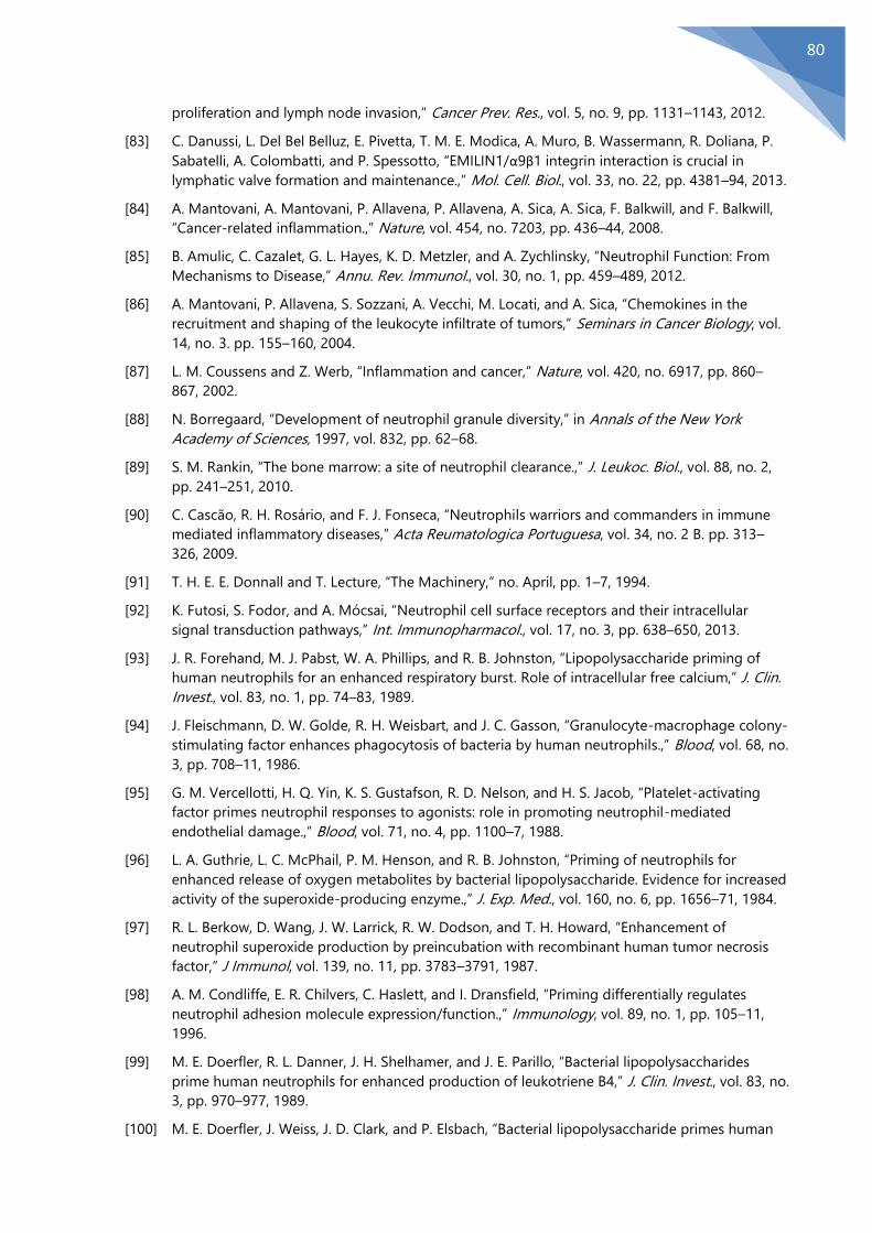
80
proliferation and lymph node invasion,” Cancer Prev. Res., vol. 5, no. 9, pp. 1131–1143, 2012.
[83] C. Danussi, L. Del Bel Belluz, E. Pivetta, T. M. E. Modica, A. Muro, B. Wassermann, R. Doliana, P.
Sabatelli, A. Colombatti, and P. Spessotto, “EMILIN1/α9β1 integrin interaction is crucial in
lymphatic valve formation and maintenance.,” Mol. Cell. Biol., vol. 33, no. 22, pp. 4381–94, 2013.
[84] A. Mantovani, A. Mantovani, P. Allavena, P. Allavena, A. Sica, A. Sica, F. Balkwill, and F. Balkwill,
“Cancer-related inflammation.,” Nature, vol. 454, no. 7203, pp. 436–44, 2008.
[85] B. Amulic, C. Cazalet, G. L. Hayes, K. D. Metzler, and A. Zychlinsky, “Neutrophil Function: From
Mechanisms to Disease,” Annu. Rev. Immunol., vol. 30, no. 1, pp. 459–489, 2012.
[86] A. Mantovani, P. Allavena, S. Sozzani, A. Vecchi, M. Locati, and A. Sica, “Chemokines in the
recruitment and shaping of the leukocyte infiltrate of tumors,” Seminars in Cancer Biology, vol.
14, no. 3. pp. 155–160, 2004.
[87] L. M. Coussens and Z. Werb, “Inflammation and cancer,” Nature, vol. 420, no. 6917, pp. 860–
867, 2002.
[88] N. Borregaard, “Development of neutrophil granule diversity,” in Annals of the New York
Academy of Sciences, 1997, vol. 832, pp. 62–68.
[89] S. M. Rankin, “The bone marrow: a site of neutrophil clearance.,” J. Leukoc. Biol., vol. 88, no. 2,
pp. 241–251, 2010.
[90] C. Cascão, R. H. Rosário, and F. J. Fonseca, “Neutrophils warriors and commanders in immune
mediated inflammatory diseases,” Acta Reumatologica Portuguesa, vol. 34, no. 2 B. pp. 313–
326, 2009.
[91] T. H. E. E. Donnall and T. Lecture, “The Machinery,” no. April, pp. 1–7, 1994.
[92] K. Futosi, S. Fodor, and A. Mócsai, “Neutrophil cell surface receptors and their intracellular
signal transduction pathways,” Int. Immunopharmacol., vol. 17, no. 3, pp. 638–650, 2013.
[93] J. R. Forehand, M. J. Pabst, W. A. Phillips, and R. B. Johnston, “Lipopolysaccharide priming of
human neutrophils for an enhanced respiratory burst. Role of intracellular free calcium,” J. Clin.
Invest., vol. 83, no. 1, pp. 74–83, 1989.
[94] J. Fleischmann, D. W. Golde, R. H. Weisbart, and J. C. Gasson, “Granulocyte-macrophage colony-
stimulating factor enhances phagocytosis of bacteria by human neutrophils.,” Blood, vol. 68, no.
3, pp. 708–11, 1986.
[95] G. M. Vercellotti, H. Q. Yin, K. S. Gustafson, R. D. Nelson, and H. S. Jacob, “Platelet-activating
factor primes neutrophil responses to agonists: role in promoting neutrophil-mediated
endothelial damage.,” Blood, vol. 71, no. 4, pp. 1100–7, 1988.
[96] L. A. Guthrie, L. C. McPhail, P. M. Henson, and R. B. Johnston, “Priming of neutrophils for
enhanced release of oxygen metabolites by bacterial lipopolysaccharide. Evidence for increased
activity of the superoxide-producing enzyme.,” J. Exp. Med., vol. 160, no. 6, pp. 1656–71, 1984.
[97] R. L. Berkow, D. Wang, J. W. Larrick, R. W. Dodson, and T. H. Howard, “Enhancement of
neutrophil superoxide production by preincubation with recombinant human tumor necrosis
factor,” J Immunol, vol. 139, no. 11, pp. 3783–3791, 1987.
[98] A. M. Condliffe, E. R. Chilvers, C. Haslett, and I. Dransfield, “Priming differentially regulates
neutrophil adhesion molecule expression/function.,” Immunology, vol. 89, no. 1, pp. 105–11,
1996.
[99] M. E. Doerfler, R. L. Danner, J. H. Shelhamer, and J. E. Parillo, “Bacterial lipopolysaccharides
prime human neutrophils for enhanced production of leukotriene B4,” J. Clin. Invest., vol. 83, no.
3, pp. 970–977, 1989.
[100] M. E. Doerfler, J. Weiss, J. D. Clark, and P. Elsbach, “Bacterial lipopolysaccharide primes human

81
neutrophils for enhanced release of arachidonic acid and causes phosphorylation of an 85-kD
cytosolic phospholipase A2,” J. Clin. Invest., vol. 93, no. 4, pp. 1583–1591, 1994.
[101] C. Fittschen, R. a Sandhaus, G. S. Worthen, and P. M. Henson, “Bacterial lipopolysaccharide
enhances chemoattractant-induced elastase secretion by human neutrophils.,” J. Leukoc. Biol.,
vol. 43, no. 6, pp. 547–56, 1988.
[102] L. Koenderman, D. Kanters, B. Maesen, J. Raaijmakers, J. W. Lammers, J. de Kruif, and T.
Logtenberg, “Monitoring of neutrophil priming in whole blood by antibodies isolated from a
synthetic phage antibody library.,” J. Leukoc. Biol., vol. 68, no. 1, pp. 58–64, 2000.
[103] N. D. Burg and M. H. Pillinger, “The neutrophil: function and regulation in innate and humoral
immunity.,” Clin. Immunol., vol. 99, no. 1, pp. 7–17, 2001.
[104] K. L. Moore, K. D. Patel, R. E. Bruehl, L. Fugang, D. A. Johnson, H. S. Lichenstein, R. D. Cummings,
D. F. Bainton, and R. P. McEver, “P-selectin glycoprotein ligand-1 mediates rolling of human
neutrophils on P-selectin,” J. Cell Biol., vol. 128, no. 4, pp. 661–671, 1995.
[105] K. D. Patel, M. U. Nollert, and R. P. McEver, “P-selectin must extend a sufficient length from the
plasma membrane to mediate rolling of neutrophils,” J. Cell Biol., vol. 131, no. 6 II, pp. 1893–
1902, 1995.
[106] J. Xu, F. Wang, A. Van Keymeulen, M. Rentel, and H. R. Bourne, “Neutrophil microtubules
suppress polarity and enhance directional migration.,” Proc. Natl. Acad. Sci. U. S. A., vol. 102, no.
19, pp. 6884–9, 2005.
[107] A. Van Keymeulen, K. Wong, Z. A. Knight, C. Govaerts, K. M. Hahn, K. M. Shokat, and H. R.
Bourne, “To stabilize neutrophil polarity, PIP3 and Cdc42 augment RhoA activity at the back as
well as signals at the front,” J. Cell Biol., vol. 174, no. 3, pp. 437–445, 2006.
[108] R. Pick, D. Brechtefeld, and B. Walzog, “Intraluminal crawling versus interstitial neutrophil
migration during inflammation,” Molecular Immunology, vol. 55, no. 1. pp. 70–75, 2013.
[109] T. Kang, J. Yi, A. Guo, X. Wang, C. M. Overall, W. Jiang, R. Elde, N. Borregaard, and D. Pei,
“Subcellular Distribution and Cytokine- and Chemokine-regulated Secretion of Leukolysin/MT6-
MMP/MMP-25 in Neutrophils,” J. Biol. Chem., vol. 276, no. 24, pp. 21960–21968, 2001.
[110] C. A. Owen, M. A. Campbell, P. L. Sannes, S. S. Boukedes, and E. J. Campbell, “Cell surface-bound
elastase and cathepsin G on human neutrophils: A novel, non-oxidative mechanism by which
neutrophils focus and preserve catalytic activity of serine proteinases,” J. Cell Biol., vol. 131, no.
3, pp. 775–789, 1995.
[111] E. J. Campbell, M. A. Campbell, S. S. Boukedes, and C. A. Owen, “Quantum proteolysis by
neutrophils: Implications for pulmonary emphysema in α1-antitrypsin deficiency,” J. Clin. Invest.,
vol. 104, no. 3, pp. 337–344, 1999.
[112] K. Takeyama, C. Agustí, I. Ueki, J. Lausier, L. O. Cardell, and J. a Nadel, “Neutrophil-dependent
goblet cell degranulation: role of membrane-bound elastase and adhesion molecules.,” Am. J.
Physiol., vol. 275, no. 2, pp. L294-302, 1998.
[113] S. Marastoni, G. Ligresti, E. Lorenzon, A. Colombatti, and M. Mongiat, “Extracellular matrix: a
matter of life and death.,” Connect. Tissue Res., vol. 49, no. 3, pp. 203–206, 2008.
[114] R. O. Hynes, “The extracellular matrix: not just pretty fibrils.,” Science, vol. 326, no. 5957, pp.
1216–9, 2009.
[115] E. Cukierman and D. E. Bassi, “The mesenchymal tumor microenvironment: A drug-resistant
niche,” Cell Adhesion and Migration, vol. 6, no. 3. pp. 285–296, 2012.
[116] K. Kessenbrock, V. Plaks, and Z. Werb, “Matrix Metalloproteinases: Regulators of the Tumor
Microenvironment,” Cell, vol. 141, no. 1. pp. 52–67, 2010.
[117] P. Lu, V. M. Weaver, and Z. Werb, “The extracellular matrix: A dynamic niche in cancer
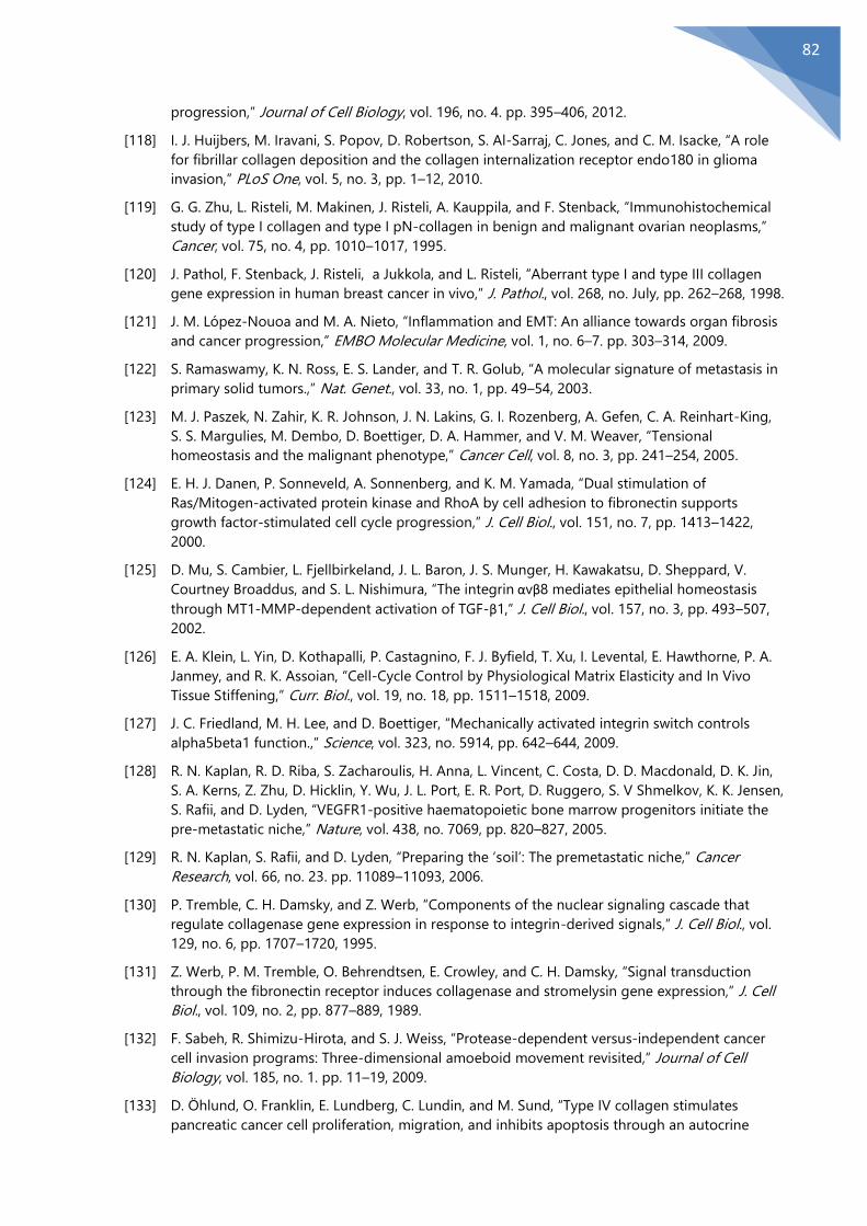
82
progression,” Journal of Cell Biology, vol. 196, no. 4. pp. 395–406, 2012.
[118] I. J. Huijbers, M. Iravani, S. Popov, D. Robertson, S. Al-Sarraj, C. Jones, and C. M. Isacke, “A role
for fibrillar collagen deposition and the collagen internalization receptor endo180 in glioma
invasion,” PLoS One, vol. 5, no. 3, pp. 1–12, 2010.
[119] G. G. Zhu, L. Risteli, M. Makinen, J. Risteli, A. Kauppila, and F. Stenback, “Immunohistochemical
study of type I collagen and type I pN-collagen in benign and malignant ovarian neoplasms,”
Cancer, vol. 75, no. 4, pp. 1010–1017, 1995.
[120] J. Pathol, F. Stenback, J. Risteli, a Jukkola, and L. Risteli, “Aberrant type I and type III collagen
gene expression in human breast cancer in vivo,” J. Pathol., vol. 268, no. July, pp. 262–268, 1998.
[121] J. M. López-Nouoa and M. A. Nieto, “Inflammation and EMT: An alliance towards organ fibrosis
and cancer progression,” EMBO Molecular Medicine, vol. 1, no. 6–7. pp. 303–314, 2009.
[122] S. Ramaswamy, K. N. Ross, E. S. Lander, and T. R. Golub, “A molecular signature of metastasis in
primary solid tumors.,” Nat. Genet., vol. 33, no. 1, pp. 49–54, 2003.
[123] M. J. Paszek, N. Zahir, K. R. Johnson, J. N. Lakins, G. I. Rozenberg, A. Gefen, C. A. Reinhart-King,
S. S. Margulies, M. Dembo, D. Boettiger, D. A. Hammer, and V. M. Weaver, “Tensional
homeostasis and the malignant phenotype,” Cancer Cell, vol. 8, no. 3, pp. 241–254, 2005.
[124] E. H. J. Danen, P. Sonneveld, A. Sonnenberg, and K. M. Yamada, “Dual stimulation of
Ras/Mitogen-activated protein kinase and RhoA by cell adhesion to fibronectin supports
growth factor-stimulated cell cycle progression,” J. Cell Biol., vol. 151, no. 7, pp. 1413–1422,
2000.
[125] D. Mu, S. Cambier, L. Fjellbirkeland, J. L. Baron, J. S. Munger, H. Kawakatsu, D. Sheppard, V.
Courtney Broaddus, and S. L. Nishimura, “The integrin αvβ8 mediates epithelial homeostasis
through MT1-MMP-dependent activation of TGF-β1,” J. Cell Biol., vol. 157, no. 3, pp. 493–507,
2002.
[126] E. A. Klein, L. Yin, D. Kothapalli, P. Castagnino, F. J. Byfield, T. Xu, I. Levental, E. Hawthorne, P. A.
Janmey, and R. K. Assoian, “Cell-Cycle Control by Physiological Matrix Elasticity and In Vivo
Tissue Stiffening,” Curr. Biol., vol. 19, no. 18, pp. 1511–1518, 2009.
[127] J. C. Friedland, M. H. Lee, and D. Boettiger, “Mechanically activated integrin switch controls
alpha5beta1 function.,” Science, vol. 323, no. 5914, pp. 642–644, 2009.
[128] R. N. Kaplan, R. D. Riba, S. Zacharoulis, H. Anna, L. Vincent, C. Costa, D. D. Macdonald, D. K. Jin,
S. A. Kerns, Z. Zhu, D. Hicklin, Y. Wu, J. L. Port, E. R. Port, D. Ruggero, S. V Shmelkov, K. K. Jensen,
S. Rafii, and D. Lyden, “VEGFR1-positive haematopoietic bone marrow progenitors initiate the
pre-metastatic niche,” Nature, vol. 438, no. 7069, pp. 820–827, 2005.
[129] R. N. Kaplan, S. Rafii, and D. Lyden, “Preparing the ‘soil’: The premetastatic niche,” Cancer
Research, vol. 66, no. 23. pp. 11089–11093, 2006.
[130] P. Tremble, C. H. Damsky, and Z. Werb, “Components of the nuclear signaling cascade that
regulate collagenase gene expression in response to integrin-derived signals,” J. Cell Biol., vol.
129, no. 6, pp. 1707–1720, 1995.
[131] Z. Werb, P. M. Tremble, O. Behrendtsen, E. Crowley, and C. H. Damsky, “Signal transduction
through the fibronectin receptor induces collagenase and stromelysin gene expression,” J. Cell
Biol., vol. 109, no. 2, pp. 877–889, 1989.
[132] F. Sabeh, R. Shimizu-Hirota, and S. J. Weiss, “Protease-dependent versus-independent cancer
cell invasion programs: Three-dimensional amoeboid movement revisited,” Journal of Cell
Biology, vol. 185, no. 1. pp. 11–19, 2009.
[133] D. Öhlund, O. Franklin, E. Lundberg, C. Lundin, and M. Sund, “Type IV collagen stimulates
pancreatic cancer cell proliferation, migration, and inhibits apoptosis through an autocrine

83
loop.,” BMC Cancer, vol. 13, p. 154, 2013.
[134] M. Seandel, K. Noack-Kunnmann, D. Zhu, R. T. Aimes, and J. P. Quigley, “Growth factor-induced
angiogenesis in vivo requires specific cleavage of fibrillar type I collagen,” Blood, vol. 97, no. 8,
pp. 2323–2332, 2001.
[135] A. Zijlstrat, R. T. Aimes, D. Zhu, K. Regazzoni, T. Kupriyanova, M. Seandel, E. I. Deryugina, and J.
P. Quigley, “Collagenolysis-dependent angiogenesis mediated by matrix metalloproteinase-13
(collagenase-3),” J. Biol. Chem., vol. 279, no. 26, pp. 27633–27645, 2004.
[136] M. S. O’Reilly, T. Boehm, Y. Shing, N. Fukai, G. Vasios, W. S. Lane, E. Flynn, J. R. Birkhead, B. R.
Olsen, and J. Folkman, “Endostatin: an endogenous inhibitor of angiogenesis and tumor
growth.,” Cell, vol. 88, no. 2, pp. 277–285, 1997.
[137] Y. Hamano and R. Kalluri, “Tumstatin, the NC1 domain of alpha3 chain of type IV collagen, is an
endogenous inhibitor of pathological angiogenesis and suppresses tumor growth,” Biochemical
and Biophysical Research Communications, vol. 333, no. 2. pp. 292–298, 2005.
[138] Y. Maeshima, M. Manfredi, C. Reimerli, K. A. Holthaus, H. Hopfert, B. R. Chandamuri, S.
Kharbanda, and R. Kalluri, “Identification of the Anti-angiogenic Site within Vascular Basement
Membrane-derived Tumstatin,” J. Biol. Chem., vol. 276, no. 18, pp. 15240–15248, 2001.
[139] P. Nyberg, L. Xie, H. Sugimoto, P. Colorado, M. Sund, K. Holthaus, A. Sudhakar, T. Salo, and R.
Kalluri, “Characterization of the anti-angiogenic properties of arresten, an α1β1 integrin-
dependent collagen-derived tumor suppressor,” Exp. Cell Res., vol. 314, no. 18, pp. 3292–3305,
2008.
[140] G. D. Kamphaus, P. C. Colorado, D. J. Panka, H. Hopfer, R. Ramchandran, A. Torre, Y. Maeshima,
J. W. Mier, V. P. Sukhatme, and R. Kalluri, “Canstatin, a novel matrix-derived inhibitor of
angiogenesis and tumor growth,” J. Biol. Chem., vol. 275, no. 2, pp. 1209–1215, 2000.
[141] D. C. Hocking and K. Kowalski, “A cryptic fragment from fibronectin’s III1 module localizes to
lipid rafts and stimulates cell growth and contractility,” J. Cell Biol., vol. 158, no. 1, pp. 175–184,
2002.
[142] N. Zoppi, M. Ritelli, A. Salvi, M. Colombi, and S. Barlati, “The FN13 peptide inhibits human tumor
cells invasion through the modulation of αvβ3 integrins organization and the inactivation of ILK
pathway,” Biochim. Biophys. Acta - Mol. Cell Res., vol. 1773, no. 6, pp. 747–763, 2007.
[143] G. Giannelli, J. Falk-Marzillier, O. Schiraldi, W. G. Stetler-Stevenson, and V. Quaranta, “Induction
of cell migration by matrix metalloprotease-2 cleavage of laminin-5.,” Science (80-. )., vol. 277,
no. 5323, pp. 225–228, 1997.
[144] N. Koshikawa, T. Minegishi, A. Sharabi, V. Quaranta, and M. Seiki, “Membrane-type matrix
metalloproteinase-1 (MT1-MMP) is a processing enzyme for human laminin y2 chain,” J. Biol.
Chem., vol. 280, no. 1, pp. 88–93, 2005.
[145] K. Bein and M. Simons, “Thrombospondin type 1 repeats interact with matrix metalloproteinase
2. Regulation of metalloproteinase activity,” J. Biol. Chem., vol. 275, no. 41, pp. 32167–32173,
2000.
[146] J. Lawler and M. Detmar, “Tumor progression: The effects of thrombospondin-1 and -2,”
International Journal of Biochemistry and Cell Biology, vol. 36, no. 6. pp. 1038–1045, 2004.
[147] A. R. Albig and W. P. Schiemann, “Fibulin-5 function during tumorigenesis.,” Future Oncol., vol.
1, no. 1, pp. 23–35, 2005.
[148] A. R. Albig and W. P. Schiemann, “Fibulin-5 antagonizes vascular endothelial growth factor
(VEGF) signaling and angiogenic sprouting by endothelial cells.,” DNA Cell Biol., vol. 23, no. 6,
pp. 367–79, 2004.
[149] A. R. Albig, J. R. Neil, and W. P. Schiemann, “Fibulins 3 and 5 antagonize tumor angiogenesis in

84
vivo,” Cancer Res., vol. 66, no. 5, pp. 2621–2629, 2006.
[150] W. P. Schiemann, G. C. Blobe, D. E. Kalume, A. Pandey, and H. F. Lodish, “Context-specific effects
of Fibulin-5 (DANCE/EVEC) on cell proliferation, motility, and invasion. Fibulin-5 is induced by
transforming growth factor-β and affects protein kinase cascades,” J. Biol. Chem., vol. 277, no.
30, pp. 27367–27377, 2002.
[151] A. Wlazlinski, R. Engers, M. J. Hoffmann, C. Hader, V. Jung, M. Müller, and W. A. Schulz,
“Downregulation of several fibulin genes in prostate cancer,” Prostate, vol. 67, no. 16, pp. 1770–
1780, 2007.
[152] H. D. Møller, U. Ralfkjær, N. Cremers, M. Frankel, R. T. Pedersen, J. Klingelhöfer, H. Yanagisawa,
M. Grigorian, P. Guldberg, J. Sleeman, E. Lukanidin, and N. Ambartsumian, “Role of fibulin-5 in
metastatic organ colonization.,” Mol. Cancer Res., vol. 9, no. 5, pp. 553–63, 2011.
[153] V. Todorovi, C. C. Chen, N. Hay, and L. F. Lau, “The matrix protein CCN1 (CYR61) induces
apoptosis in fibroblasts,” J. Cell Biol., vol. 171, no. 3, pp. 559–568, 2005.
[154] E. Pivetta, B. Wassermann, L. Del Bel Belluz, C. Danussi, T. M. E. Modica, O. Maiorani, G. Bosisio,
F. Boccardo, V. Canzonieri, A. Colombatti, and P. Spessotto, “Local inhibition of elastase reduces
EMILIN1 cleavage reactivating lymphatic vessel function in a mouse lymphoedema model.,”
Clin. Sci. (Lond)., vol. 130, no. 14, pp. 1221–36, 2016.
[155] M. Ming and Y.-Y. He, “PTEN: new insights into its regulation and function in skin cancer.,” J.
Invest. Dermatol., vol. 129, no. 9, pp. 2109–12, 2009.
[156] N. Komazawa, A. Suzuki, S. Sano, K. Horie, N. Matsuura, T. W. Mak, T. Nakano, J. Takeda, and G.
Kondoh, “Tumorigenesis facilitated by Pten deficiency in the skin: Evidence of p53-Pten
complex formation on the initiation phase,” Cancer Sci., vol. 95, no. 8, pp. 639–643, 2004.
[157] C. Segrelles, S. Ruiz, P. Perez, C. Murga, M. Santos, I. V Budunova, J. Martinez, F. Larcher, T. J.
Slaga, J. S. Gutkind, J. L. Jorcano, and J. M. Paramio, “Functional roles of Akt signaling in mouse
skin tumorigenesis,” Oncogene, vol. 21, no. 1, pp. 53–64, 2002.
[158] E. M. Schindler, A. Hindes, E. L. Gribben, C. J. Burns, Y. Yin, M.-H. Lin, R. J. Owen, G. D. Longmore,
G. E. Kissling, J. S. C. Arthur, and T. Efimova, “p38delta Mitogen-activated protein kinase is
essential for skin tumor development in mice.,” Cancer Res., vol. 69, no. 11, pp. 4648–4655,
2009.
[159] K. Edlund, C. Lindskog, A. Saito, A. Berglund, F. Pontén, H. Göransson-Kultima, A. Isaksson, K.
Jirström, M. Planck, L. Johansson, M. Lambe, L. Holmberg, F. Nyberg, S. Ekman, M. Bergqvist, P.
Landelius, K. Lamberg, J. Botling, A. Östman, and P. Micke, “CD99 is a novel prognostic stromal
marker in non-small cell lung cancer,” Int. J. Cancer, vol. 131, no. 10, pp. 2264–2273, 2012.
[160] R. Salani, I. Neuberger, R. J. Kurman, R. E. Bristow, H.-W. Chang, T.-L. Wang, and I.-M. Shih,
“Expression of extracellular matrix proteins in ovarian serous tumors.,” Int. J. Gynecol. Pathol.,
vol. 26, no. 2, pp. 141–6, 2007.
[161] U. N. M. Rao, B. L. Hood, J. M. Jones-Laughner, M. Sun, and T. P. Conrads, “Distinct profiles of
oxidative stress-related and matrix proteins in adult bone and soft tissue osteosarcoma and
desmoid tumors: A proteomics study,” Hum. Pathol., vol. 44, no. 5, pp. 725–733, 2013.
[162] E. Pivetta, C. Danussi, B. Wassermann, T. M. E. Modica, L. Del Bel Belluz, V. Canzonieri, A.
Colombatti, and P. Spessotto, “Neutrophil elastase-dependent cleavage compromises the
tumor suppressor role of EMILIN1,” Matrix Biol., vol. 34, pp. 22–32, 2014.
[163] A. M. Houghton, D. M. Rzymkiewicz, H. Ji, A. D. Gregory, E. E. Egea, H. E. Metz, D. B. Stolz, S. R.
Land, L. A. Marconcini, C. R. Kliment, K. M. Jenkins, K. A. Beaulieu, M. Mouded, S. J. Frank, K. K.
Wong, and S. D. Shapiro, “Neutrophil elastase-mediated degradation of IRS-1 accelerates lung
tumor growth.,” Nat. Med., vol. 16, no. 2, pp. 219–23, 2010.

85
[164] C. Stegemann, A. Didangelos, J. Barallobre-Barreiro, S. R. Langley, K. Mandal, M. Jahangiri, and
M. Mayr, “Proteomic identification of matrix metalloproteinase substrates in the human
vasculature,” Circulation: Cardiovascular Genetics, vol. 6, no. 1. pp. 106–117, 2013.
[165] E. Hajjar, T. Broemstrup, C. Kantari, V. Witko-Sarsat, and N. Reuter, “Structures of human
proteinase 3 and neutrophil elastase so similar yet so different.,” FEBS J., vol. 277, no. 10, pp.
2238–54, 2010.
[166] K. Felix and M. M. Gaida, “Neutrophil-derived proteases in the microenvironment of pancreatic
cancer-active players in tumor progression,” Int. J. Biol. Sci., vol. 12, no. 3, pp. 302–313, 2016.
[167] Z. Sun and P. Yang, “Role of imbalance between neutrophil elastase and alpha 1-antitrypsin in
cancer development and progression.,” Lancet Oncol., vol. 5, no. 3, pp. 182–190, 2004.
[168] H. Neurath, “Evolution of proteolytic enzymes.,” Science, vol. 224, no. 4647, pp. 350–7, 1984.
[169] S. Shapiro, E. Campbell, R. Senior, and H. Welgus, “Proteinases secreted by human mononuclear
phagocytes,” J.Rheumatol.Suppl, vol. 27, pp. 95–98, 1991.
[170] D. Garwicz, A. Lennartsson, S. E. W. Jacobsen, U. Gullberg, and A. Lindmark, “Biosynthetic
profiles of neutrophil serine proteases in a human bone marrow-derived cellular myeloid
differentiation model,” Haematologica, vol. 90, no. 1, pp. 38–44, 2005.
[171] M. Zimmer, R. L. Medcalf, T. M. Fink, C. Mattmann, P. Lichter, and D. E. Jenne, “Three human
elastase-like genes coordinately expressed in the myelomonocyte lineage are organized as a
single genetic locus on 19pter.,” Proc. Natl. Acad. Sci. U. S. A., vol. 89, no. 17, pp. 8215–9, 1992.
[172] D. Garwicz, a Lindmark, T. Hellmark, M. Gladh, J. Jögi, and U. Gullberg, “Characterization of the
processing and granular targeting of human proteinase 3 after transfection to the rat RBL or
the murine 32D leukemic cell lines.,” J. Leukoc. Biol., vol. 61, no. 1, pp. 113–123, 1997.
[173] U. Gullberg, E. Andersson, D. Garwicz, A. Lindmark, and I. Olsson, “Biosynthesis, processing and
sorting of neutrophil proteins: insight into neutrophil granule development.,” Eur. J. Haematol.,
vol. 58, no. 3, pp. 137–153, 1997.
[174] B. Korkmaz, M. Horwitz, D. Jenne, and F. Gauthier, “Neutrophil elastase, proteinase 3, and
cathepsin G as therapeutic targets in human diseases,” Pharmacol. Rev., vol. 62, no. 4, pp. 726–
759, 2010.
[175] G. Salvesen and J. J. Enghild, “An unusual specificity in the activation of neutrophil serine
proteinase zymogens,” Biochemistry, vol. 29, no. 22, pp. 5304–5308, 1990.
[176] C. T. N. Pham, “Neutrophil serine proteases: specific regulators of inflammation.,” Nat. Rev.
Immunol., vol. 6, no. 7, pp. 541–50, 2006.
[177] N. V. Rao, G. V. Rao, B. C. Marshall, and J. R. Hoidal, “Biosynthesis and processing of proteinase
3 in U937 cells: Processing pathways are distinct from those of cathepsin G,” J. Biol. Chem., vol.
271, no. 6, pp. 2972–2978, 1996.
[178] M. Horwitz, K. F. Benson, Z. Duan, F. Q. Li, and R. E. Person, “Hereditary neutropenia: Dogs
explain human neutrophil elastase mutations,” Trends in Molecular Medicine, vol. 10, no. 4. pp.
163–170, 2004.
[179] C. U. Niemann, M. Åbrink, G. Pejler, R. L. Fischer, E. I. Christensen, S. D. Knight, and N.
Borregaard, “Neutrophil elastase depends on serglycin proteoglycan for localization in
granules,” Blood, vol. 109, no. 10, pp. 4478–4486, 2007.
[180] B. Korkmaz, T. Moreau, and F. Gauthier, “Neutrophil elastase, proteinase 3 and cathepsin G:
Physicochemical properties, activity and physiopathological functions,” Biochimie, vol. 90, no. 2,
pp. 227–242, 2008.
[181] W. Bode, A. Z. Wei, R. Huber, E. Meyer, J. Travis, and S. Neumann, “X-ray crystal structure of the
complex of human leukocyte elastase (PMN elastase) and the third domain of the turkey
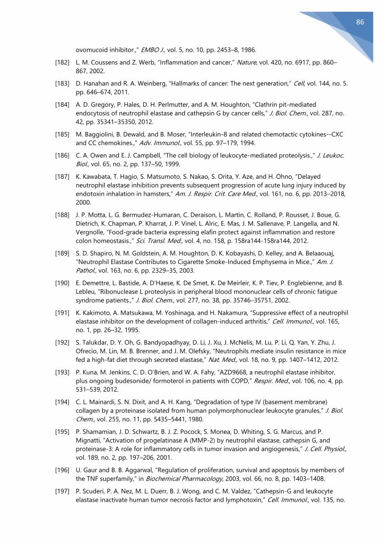
86
ovomucoid inhibitor.,” EMBO J., vol. 5, no. 10, pp. 2453–8, 1986.
[182] L. M. Coussens and Z. Werb, “Inflammation and cancer,” Nature, vol. 420, no. 6917, pp. 860–
867, 2002.
[183] D. Hanahan and R. A. Weinberg, “Hallmarks of cancer: The next generation,” Cell, vol. 144, no. 5.
pp. 646–674, 2011.
[184] A. D. Gregory, P. Hales, D. H. Perlmutter, and A. M. Houghton, “Clathrin pit-mediated
endocytosis of neutrophil elastase and cathepsin G by cancer cells,” J. Biol. Chem., vol. 287, no.
42, pp. 35341–35350, 2012.
[185] M. Baggiolini, B. Dewald, and B. Moser, “Interleukin-8 and related chemotactic cytokines--CXC
and CC chemokines.,” Adv. Immunol., vol. 55, pp. 97–179, 1994.
[186] C. A. Owen and E. J. Campbell, “The cell biology of leukocyte-mediated proteolysis.,” J. Leukoc.
Biol., vol. 65, no. 2, pp. 137–50, 1999.
[187] K. Kawabata, T. Hagio, S. Matsumoto, S. Nakao, S. Orita, Y. Aze, and H. Ohno, “Delayed
neutrophil elastase inhibition prevents subsequent progression of acute lung injury induced by
endotoxin inhalation in hamsters,” Am. J. Respir. Crit. Care Med., vol. 161, no. 6, pp. 2013–2018,
2000.
[188] J. P. Motta, L. G. Bermudez-Humaran, C. Deraison, L. Martin, C. Rolland, P. Rousset, J. Boue, G.
Dietrich, K. Chapman, P. Kharrat, J. P. Vinel, L. Alric, E. Mas, J. M. Sallenave, P. Langella, and N.
Vergnolle, “Food-grade bacteria expressing elafin protect against inflammation and restore
colon homeostasis.,” Sci. Transl. Med., vol. 4, no. 158, p. 158ra144-158ra144, 2012.
[189] S. D. Shapiro, N. M. Goldstein, A. M. Houghton, D. K. Kobayashi, D. Kelley, and A. Belaaouaj,
“Neutrophil Elastase Contributes to Cigarette Smoke-Induced Emphysema in Mice.,” Am. J.
Pathol., vol. 163, no. 6, pp. 2329–35, 2003.
[190] E. Demettre, L. Bastide, A. D’Haese, K. De Smet, K. De Meirleir, K. P. Tiev, P. Englebienne, and B.
Lebleu, “Ribonuclease L proteolysis in peripheral blood mononuclear cells of chronic fatigue
syndrome patients.,” J. Biol. Chem., vol. 277, no. 38, pp. 35746–35751, 2002.
[191] K. Kakimoto, A. Matsukawa, M. Yoshinaga, and H. Nakamura, “Suppressive effect of a neutrophil
elastase inhibitor on the development of collagen-induced arthritis,” Cell. Immunol., vol. 165,
no. 1, pp. 26–32, 1995.
[192] S. Talukdar, D. Y. Oh, G. Bandyopadhyay, D. Li, J. Xu, J. McNelis, M. Lu, P. Li, Q. Yan, Y. Zhu, J.
Ofrecio, M. Lin, M. B. Brenner, and J. M. Olefsky, “Neutrophils mediate insulin resistance in mice
fed a high-fat diet through secreted elastase,” Nat. Med., vol. 18, no. 9, pp. 1407–1412, 2012.
[193] P. Kuna, M. Jenkins, C. D. O’Brien, and W. A. Fahy, “AZD9668, a neutrophil elastase inhibitor,
plus ongoing budesonide/ formoterol in patients with COPD,” Respir. Med., vol. 106, no. 4, pp.
531–539, 2012.
[194] C. L. Mainardi, S. N. Dixit, and A. H. Kang, “Degradation of type IV (basement membrane)
collagen by a proteinase isolated from human polymorphonuclear leukocyte granules,” J. Biol.
Chem., vol. 255, no. 11, pp. 5435–5441, 1980.
[195] P. Shamamian, J. D. Schwartz, B. J. Z. Pocock, S. Monea, D. Whiting, S. G. Marcus, and P.
Mignatti, “Activation of progelatinase A (MMP-2) by neutrophil elastase, cathepsin G, and
proteinase-3: A role for inflammatory cells in tumor invasion and angiogenesis,” J. Cell. Physiol.,
vol. 189, no. 2, pp. 197–206, 2001.
[196] U. Gaur and B. B. Aggarwal, “Regulation of proliferation, survival and apoptosis by members of
the TNF superfamily,” in Biochemical Pharmacology, 2003, vol. 66, no. 8, pp. 1403–1408.
[197] P. Scuderi, P. A. Nez, M. L. Duerr, B. J. Wong, and C. M. Valdez, “Cathepsin-G and leukocyte
elastase inactivate human tumor necrosis factor and lymphotoxin,” Cell. Immunol., vol. 135, no.

87
2, pp. 299–313, 1991.
[198] T. Yamauchi, K. Yasushi, K. Ueki, Y. Tsuji, G. R. Stark, I. M. Kerr, T. Tsushima, Y. Akanuma, I.
Komuro, K. Tobe, Y. Yazaki, and T. Kadowaki, “Growth hormone and prolactin stimulate tyrosine
phosphorylation of insulin receptor substrate-1, -2, and -3, their association with p85
phosphatidylinositol 3-kinase (PI3-kinase), and concomitantly PI3-kinase activation via JAK2
kinase,” J. Biol. Chem., vol. 273, no. 25, pp. 15719–15726, 1998.
[199] E. A. Mittendorf, G. Alatrash, N. Qiao, Y. Wu, P. Sukhumalchandra, L. S. St. John, A. V. Philips, H.
Xiao, M. Zhang, K. Ruisaard, K. Clise-Dwyer, S. Lu, and J. J. Molldrem, “Breast cancer cell uptake
of the inflammatory mediator neutrophil elastase triggers an anticancer adaptive immune
response,” Cancer Res., vol. 72, no. 13, pp. 3153–3162, 2012.
[200] G. T. Thomas, M. P. Lewis, and P. M. Speight, “Matrix metalloproteinases and oral cancer,” Oral
Oncology, vol. 35, no. 3. pp. 227–233, 1999.
[201] L. Yadav, N. Puri, V. Rastogi, P. Satpute, R. Ahmad, and G. Kaur, “Matrix metalloproteinases and
cancer - Roles in threat and therapy,” Asian Pacific Journal of Cancer Prevention, vol. 15, no. 3.
pp. 1085–1091, 2014.
[202] C. Gialeli, A. D. Theocharis, and N. K. Karamanos, “Roles of matrix metalloproteinases in cancer
progression and their pharmacological targeting,” FEBS Journal, vol. 278, no. 1. pp. 16–27, 2011.
[203] A. M. Weaver, “Invadopodia: Specialized cell structures for cancer invasion,” Clinical and
Experimental Metastasis, vol. 23, no. 2. pp. 97–105, 2006.
[204] I. Van Hove, K. Lemmens, S. Van De Velde, M. Verslegers, and L. Moons, “Matrix
metalloproteinase-3 in the central nervous system: A look on the bright side,” Journal of
Neurochemistry, vol. 123, no. 2. pp. 203–216, 2012.
[205] H. Nagase, R. Visse, and G. Murphy, “Structure and function of matrix metalloproteinases and
TIMPs,” Cardiovascular Research, vol. 69, no. 3. pp. 562–573, 2006.
[206] L. J. McCawley, H. C. Crawford, L. E. King, J. Mudgett, and L. M. Matrisian, “A protective role for
matrix metalloproteinase-3 in squamous cell carcinoma,” Cancer Res., vol. 64, no. 19, pp. 6965–
6972, 2004.
[207] J. P. Witty, T. Lempka, R. J. Coffey Jr., and L. M. Matrisian, “Decreased tumor formation in 7,12-
dimethylbenzanthracene-treated stromelysin-1 transgenic mice is associated with alterations in
mammary epithelial cell apoptosis,” Cancer Res, vol. 55, no. 7, pp. 1401–1406, 1995.
[208] M. D. Sternlicht, A. Lochtest, C. J. Sympson, B. Huey, J. P. Rougier, J. W. Gray, D. Pinkel, M. J.
Bissell, and Z. Werb, “The stromal proteinase MMP3/stromelysin-1 promotes mammary
carcinogenesis,” Cell, vol. 98, no. 2, pp. 137–146, 1999.
[209] A. R. Farina and A. R. Mackay, “Gelatinase B/MMP-9 in tumour pathogenesis and progression,”
Cancers, vol. 6, no. 1. pp. 240–296, 2014.
[210] I. Sopata and A. M. Dancewicz, “Presence of a gelatin-specific proteinase and its latent form in
human leucocytes,” BBA - Enzymol., vol. 370, no. 2, pp. 510–523, 1974.
[211] B. Dewald, U. Bretz, and M. Baggiolini, “Release of gelatinase from a novel secretory
compartment of human neutrophils,” J. Clin. Invest., vol. 70, no. 3, pp. 518–525, 1982.
[212] J. M. Shipley, G. A. R. Doyle, C. J. Fliszar, Q. Z. Ye, L. L. Johnson, S. D. Shapiro, H. G. Welgus, and
R. M. Senior, “The structural basis for the elastolytic activity of the 92-kDa and 72-kDa
gelatinases: Role of the fibronectin type II-like repeats,” J. Biol. Chem., vol. 271, no. 8, pp. 4335–
4341, 1996.
[213] H. F. Bigg, A. D. Rowan, M. D. Barker, and T. E. Cawston, “Activity of matrix metalloproteinase-9
against native collagen types I and III,” FEBS J., vol. 274, no. 5, pp. 1246–1255, 2007.
[214] J. a Eble, a Ries, a Lichy, K. Mann, H. Stanton, J. Gavrilovic, G. Murphy, and K. Kühn, “The
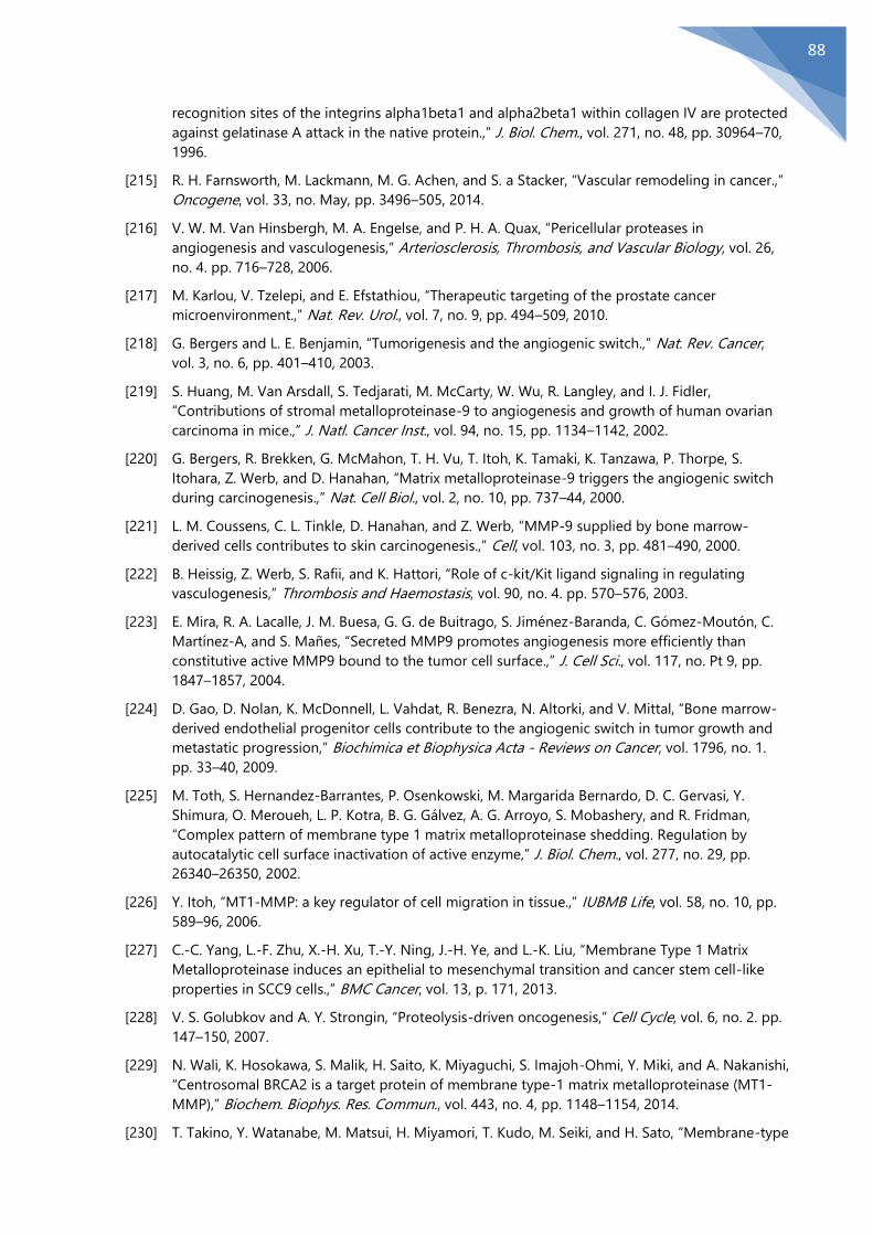
88
recognition sites of the integrins alpha1beta1 and alpha2beta1 within collagen IV are protected
against gelatinase A attack in the native protein.,” J. Biol. Chem., vol. 271, no. 48, pp. 30964–70,
1996.
[215] R. H. Farnsworth, M. Lackmann, M. G. Achen, and S. a Stacker, “Vascular remodeling in cancer.,”
Oncogene, vol. 33, no. May, pp. 3496–505, 2014.
[216] V. W. M. Van Hinsbergh, M. A. Engelse, and P. H. A. Quax, “Pericellular proteases in
angiogenesis and vasculogenesis,” Arteriosclerosis, Thrombosis, and Vascular Biology, vol. 26,
no. 4. pp. 716–728, 2006.
[217] M. Karlou, V. Tzelepi, and E. Efstathiou, “Therapeutic targeting of the prostate cancer
microenvironment.,” Nat. Rev. Urol., vol. 7, no. 9, pp. 494–509, 2010.
[218] G. Bergers and L. E. Benjamin, “Tumorigenesis and the angiogenic switch.,” Nat. Rev. Cancer,
vol. 3, no. 6, pp. 401–410, 2003.
[219] S. Huang, M. Van Arsdall, S. Tedjarati, M. McCarty, W. Wu, R. Langley, and I. J. Fidler,
“Contributions of stromal metalloproteinase-9 to angiogenesis and growth of human ovarian
carcinoma in mice.,” J. Natl. Cancer Inst., vol. 94, no. 15, pp. 1134–1142, 2002.
[220] G. Bergers, R. Brekken, G. McMahon, T. H. Vu, T. Itoh, K. Tamaki, K. Tanzawa, P. Thorpe, S.
Itohara, Z. Werb, and D. Hanahan, “Matrix metalloproteinase-9 triggers the angiogenic switch
during carcinogenesis.,” Nat. Cell Biol., vol. 2, no. 10, pp. 737–44, 2000.
[221] L. M. Coussens, C. L. Tinkle, D. Hanahan, and Z. Werb, “MMP-9 supplied by bone marrow-
derived cells contributes to skin carcinogenesis.,” Cell, vol. 103, no. 3, pp. 481–490, 2000.
[222] B. Heissig, Z. Werb, S. Rafii, and K. Hattori, “Role of c-kit/Kit ligand signaling in regulating
vasculogenesis,” Thrombosis and Haemostasis, vol. 90, no. 4. pp. 570–576, 2003.
[223] E. Mira, R. A. Lacalle, J. M. Buesa, G. G. de Buitrago, S. Jiménez-Baranda, C. Gómez-Moutón, C.
Martínez-A, and S. Mañes, “Secreted MMP9 promotes angiogenesis more efficiently than
constitutive active MMP9 bound to the tumor cell surface.,” J. Cell Sci., vol. 117, no. Pt 9, pp.
1847–1857, 2004.
[224] D. Gao, D. Nolan, K. McDonnell, L. Vahdat, R. Benezra, N. Altorki, and V. Mittal, “Bone marrow-
derived endothelial progenitor cells contribute to the angiogenic switch in tumor growth and
metastatic progression,” Biochimica et Biophysica Acta - Reviews on Cancer, vol. 1796, no. 1.
pp. 33–40, 2009.
[225] M. Toth, S. Hernandez-Barrantes, P. Osenkowski, M. Margarida Bernardo, D. C. Gervasi, Y.
Shimura, O. Meroueh, L. P. Kotra, B. G. Gálvez, A. G. Arroyo, S. Mobashery, and R. Fridman,
“Complex pattern of membrane type 1 matrix metalloproteinase shedding. Regulation by
autocatalytic cell surface inactivation of active enzyme,” J. Biol. Chem., vol. 277, no. 29, pp.
26340–26350, 2002.
[226] Y. Itoh, “MT1-MMP: a key regulator of cell migration in tissue.,” IUBMB Life, vol. 58, no. 10, pp.
589–96, 2006.
[227] C.-C. Yang, L.-F. Zhu, X.-H. Xu, T.-Y. Ning, J.-H. Ye, and L.-K. Liu, “Membrane Type 1 Matrix
Metalloproteinase induces an epithelial to mesenchymal transition and cancer stem cell-like
properties in SCC9 cells.,” BMC Cancer, vol. 13, p. 171, 2013.
[228] V. S. Golubkov and A. Y. Strongin, “Proteolysis-driven oncogenesis,” Cell Cycle, vol. 6, no. 2. pp.
147–150, 2007.
[229] N. Wali, K. Hosokawa, S. Malik, H. Saito, K. Miyaguchi, S. Imajoh-Ohmi, Y. Miki, and A. Nakanishi,
“Centrosomal BRCA2 is a target protein of membrane type-1 matrix metalloproteinase (MT1-
MMP),” Biochem. Biophys. Res. Commun., vol. 443, no. 4, pp. 1148–1154, 2014.
[230] T. Takino, Y. Watanabe, M. Matsui, H. Miyamori, T. Kudo, M. Seiki, and H. Sato, “Membrane-type

89
1 matrix metalloproteinase modulates focal adhesion stability and cell migration,” Exp. Cell Res.,
vol. 312, no. 8, pp. 1381–1389, 2006.
[231] M. A. Lafleur, D. Xu, and M. E. Hemler, “Tetraspanin proteins regulate membrane type-1 matrix
metalloproteinase-dependent pericellular proteolysis.,” Mol. Biol. Cell, vol. 20, no. 7, pp. 2030–
40, 2009.
[232] G. A. Watkins, E. F. Jones, M. Scott Shell, H. F. VanBrocklin, M. H. Pan, S. M. Hanrahan, J. J. Feng,
J. He, N. E. Sounni, K. A. Dill, C. H. Contag, L. M. Coussens, and B. L. Franc, “Development of an
optimized activatable MMP-14 targeted SPECT imaging probe,” Bioorganic Med. Chem., vol. 17,
no. 2, pp. 653–659, 2009.
[233] K. Zarrabi, A. Dufour, J. Li, C. Kuscu, A. Pulkoski-Gross, J. Zhi, Y. Hu, N. S. Sampson, S. Zucker,
and J. Cao, “Inhibition of Matrix Metalloproteinase 14 (MMP-14)-mediated cancer cell
migration,” J. Biol. Chem., vol. 286, no. 38, pp. 33167–33177, 2011.
[234] T. Tomari, N. Koshikawa, T. Uematsu, T. Shinkawa, D. Hoshino, N. Egawa, T. Isobe, and M. Seiki,
“High throughput analysis of proteins associating with a proinvasive MT1-MMP in human
malignant melanoma A375 cells,” Cancer Sci., vol. 100, no. 7, pp. 1284–1290, 2009.
[235] J. Cao, C. Chiarelli, O. Richman, K. Zarrabi, P. Kozarekar, and S. Zucker, “Membrane type 1 matrix
metalloproteinase induces epithelial-to-mesenchymal transition in prostate cancer.,” J. Biol.
Chem., vol. 283, no. 10, pp. 6232–40, 2008.
[236] M. A. Shields, S. Dangi‑Garimella, A. J. Redig, and H. G. Munshi, “Biochemical role of the
collagen-rich tumour microenvironment in pancreatic cancer progression,” Biochem. J., vol. 441,
no. 2, pp. 541–552, 2011.
[237] N. E. Sounni, C. Roghi, V. Chabottaux, M. Janssen, C. Munaut, E. Maquoi, B. G. Galvez, C. Gilles, F.
Frankenne, G. Murphy, J. M. Foidart, and A. Noel, “Up-regulation of Vascular Endothelial Growth
Factor-A by Active Membrane-type 1 Matrix Metalloproteinase Through Activation of Src-
Tyrosine Kinases,” J. Biol. Chem., vol. 279, no. 14, pp. 13564–13574, 2004.
[238] P. Spessotto, E. Giacomello, and R. Perri, “Improving fluorescence-based assays for the in vitro
analysis of cell adhesion and migration.,” Mol. Biotechnol., vol. 20, no. 3, pp. 285–304, 2002.
[239] A. Doucet and C. M. Overall, “Broad coverage identification of multiple proteolytic cleavage site
sequences in complex high molecular weight proteins using quantitative proteomics as a
complement to edman sequencing.,” Mol. Cell. Proteomics, vol. 10, no. 5, p. M110.003533,
2011.
[240] N. Abramowitz, I. Schechter, and A. Berger, “On the size of the active site in proteases II.
Carboxypeptidase-A,” Biochem. Biophys. Res. Commun., vol. 29, no. 6, pp. 862–867, 1967.
[241] L. Hedstrom, “Serine protease mechanism and specificity,” Chem. Rev., vol. 102, no. 12, pp.
4501–4523, 2002.
[242] N. A. Tamarina, W. D. McMillan, V. P. Shively, and W. H. Pearce, “Expression of matrix
metalloproteinases and their inhibitors in aneurysms and normal aorta,” in Surgery, 1997, vol.
122, no. 2, pp. 264–272.
[243] B. A. Kelly, B. C. Bond, and L. Poston, “Gestational profile of matrix metalloproteinases in rat
uterine artery,” Mol Hum Reprod, vol. 9, no. 6, pp. 351–358, 2003.
[244] R. Kowalewski, K. Sobolewski, M. Wolanska, and M. Gacko, “Matrix metalloproteinases in the
vein wall,” Int. Angiol., vol. 23, no. 2, pp. 164–169, 2004.

90
Ringraziamenti
Un ringraziamento va alla Dr.ssa Paola Spessotto che mi ha dato l’opportunità di
lavorare in questi 4 anni presso il suo laboratorio e per avermi supportato in questo
percorso di crescita professionale e personale.
Un sentito ringraziamento va al Dr. Roberto Doliana, il cui rigore scientifico è stato
fondamentale per la realizzazione del mio progetto di dottorato, per la sua umanità e
per avermi strappato un sorriso, incoraggiandomi, nei momenti più difficili.
Inoltre vorrei ringraziare il Prof. Alfonso Colombatti e il Dr. Gustavo Baldassarre, quali
direttori dell’Oncologia Sperimentale 2 del CRO di Aviano, per la loro dedizione nel
seguire i nostri progetti di ricerca.
Un ringraziamento affettuoso e sincero va alla Dr.ssa Alessandra Capuano per la sua
immensa gentilezza e disponibilità nel supportarmi nell’ideazione e realizzazione
della parte scientifica di questa tesi di dottorato.
Come poi non menzionare Francesca e Giulia che con la loro allegria hanno reso le
giornate passate in laboratorio meno lunghe e pesanti.
Particolare gratitudine va ai Sigg. Bruna Wassermann e Francesco Bucciotti per il loro
prezioso contributo tecnico.
Vorrei inoltre, menzionare, tutte quelle persone che hanno reso questi quattro anni
qui al CRO un’esperienza che sarà sempre presente nei verdi giardini dei miei ricordi,
come ad esempio…..
Marta e Naike e le nostre camminate durante la pausa pranzo che non venivano
impedite da nessuna condizione meteo, neanche le più avverse .….
Giulia, Michela, Luigi e Valentina per i momenti di allegria, spensieratezza e
complicità che siete stati capaci di donarmi in questi anni.
Il Signor Roberto Guerrini che ogni mattina è stato sempre pronto a strapparmi un
sorriso
91
E poi……
Ci siete voi le persone che occupano la sfera più importante di una persona: la sfera
affettiva, a voi va il più sentito grazie!
A mio padre e mia madre, che non smetterò mai di ringraziare per tutto l’amore e il
sostegno che mi avete donato, credendo sempre in me. Voi siete un ESEMPIO, di
rettitudine, dignità e onestà, insomma, di tutto quello che un uomo deve essere.
E a te papà, che oramai è quasi un anno che non sei più qui con noi, voglio dirti che
in fondo avevi ragione tu, tu che ci hai sempre creduto, anche più di me.
E poi ci sei tu dolce Eleonora, tu che assieme a me hai condiviso questo percorso, hai
reso meno ripide le salite, meno asfissianti le ansie, più brevi i momenti in cui c’era la
voglia di mollare tutto, e al tempo stesso hai reso indimenticabili i momenti di
allegria, di speranze, di gioie. Sappi che, se sono arrivato alla fine di questo percorso,
è perché c’eri tu, sempre al mio fianco.

1Scientific RepoRts | 7:39974 | DOI: 10.1038/srep39974
www.nature.com/scientificreports
Neutrophil elastase cleavage of the gC1q domain impairs the EMILIN1-α4β1 integrin interaction, cell adhesion and anti-proliferative activityOrlando Maiorani, Eliana Pivetta, Alessandra Capuano, Teresa Maria Elisa Modica, Bruna Wassermann, Francesco Bucciotti, Alfonso Colombatti, Roberto Doliana* & Paola Spessotto*
The extracellular matrix glycoprotein EMILIN1 exerts a wide range of functions mainly associated with its gC1q domain. Besides providing functional significance for adhesion and migration, the direct interaction between α4β1 integrin and EMILIN1-gC1q regulates cell proliferation, transducing net anti-proliferative effects. We have previously demonstrated that EMILIN1 degradation by neutrophil elastase (NE) is a specific mechanism leading to the loss of functions disabling its regulatory properties. In this study we further analysed the proteolytic activity of NE, MMP-3, MMP-9, and MT1-MMP on EMILIN1 and found that MMP-3 and MT1-MMP partially cleaved EMILIN1 but without affecting the functional properties associated with the gC1q domain, whereas NE was able to fully impair the interaction of gC1q with the α4β1 integrin by cleaving this domain outside of the E933 integrin binding site. By a site direct mutagenesis approach we mapped the bond between S913 and R914 residues and selected the NE-resistant R914W mutant still able to interact with the α4β1 integrin after NE treatment. Functional studies showed that NE impaired the EMILIN1-α4β1 integrin interaction by cleaving the gC1q domain in a region crucial for its proper structural conformation, paving the way to better understand NE effects on EMILIN1-cell interaction in pathological context.
Studies in recent decades have established the constituents of the extracellular matrix (ECM) as dynamic struc-tures that can profoundly influence diverse aspects of cellular behaviour and functions1,2. Integrins, a large family of α β heterodimeric cell membrane receptors, have emerged as key sensory molecules that translate chemical and physical cues from the ECM into biochemical signals that regulate cell adhesion and shape, migration and tumour cell metastases, differentiation, proliferation, and survival3,4. EMILIN1 is an ECM glycoprotein contain-ing the amino-terminal EMI domain, a cysteine-rich sequence of approximately 80 amino acids, and a gC1q domain at the carboxyl-terminal end5–7. EMILIN1 belongs to the C1q/TNF superfamily whose hallmark is a homo- and hetero-trimeric gC1q domain. The members of this superfamily play a broad spectrum of functions in innate immunity, insulin sensitivity and collagen hexagonal lattice organization to name a few8,9. EMILIN1 is strongly expressed in the blood and lymphatic vessels and in the connective tissues of a wide variety of organs10–13. It is involved via the EMI domain in the maintenance of blood vascular cell morphology and function14; in addi-tion, EMILIN1 promotes adhesion and migration15,16 and controls cell proliferation13,17 through its gC1q domain. So far, among the gC1q domains of several other ECM components, EMILIN1 gC1q is the only one capable of interacting with the α 4β 1 integrin15–18, which is predominantly expressed on the surface of hemopoietic cells, working as a receptor to allow adhesion to blood vessel wall19 or extracellular matrix constituents20. The inter-action between α 4β 1 and EMILIN1 gC1q is particularly efficient because even very low ligand concentrations provide very strong adhesion and migration15,16. The residue E933 located by NMR in a unstructured loop at the
Experimental Oncology 2, Department of Translational Research, CRO-IRCCS, National Cancer Institute, Aviano 33081, Italy. *These authors jointly supervised this work. Correspondence and requests for materials should be addressed to P.S. (email: [email protected])
Received: 11 August 2016
Accepted: 29 November 2016
Published: 11 January 2017
OPEN
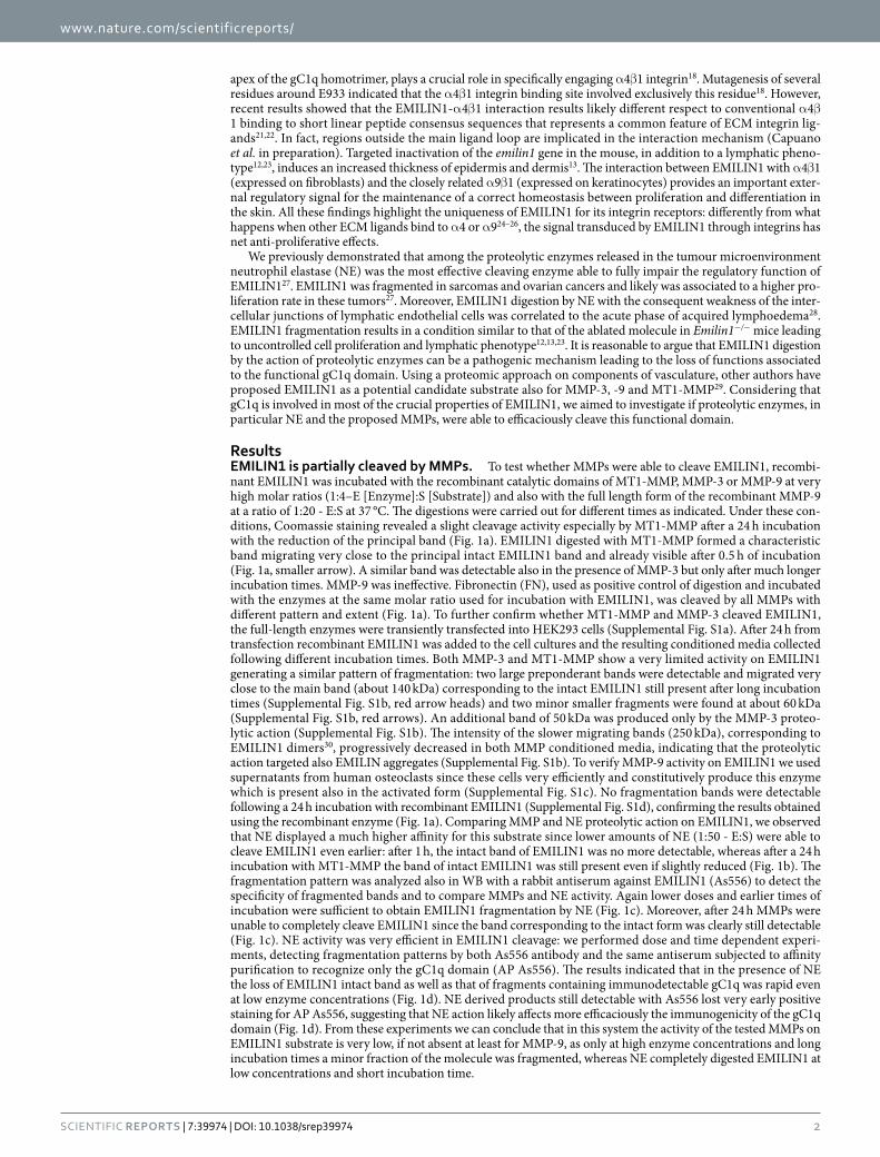
www.nature.com/scientificreports/
2Scientific RepoRts | 7:39974 | DOI: 10.1038/srep39974
apex of the gC1q homotrimer, plays a crucial role in specifically engaging α 4β 1 integrin18. Mutagenesis of several residues around E933 indicated that the α 4β 1 integrin binding site involved exclusively this residue18. However, recent results showed that the EMILIN1-α 4β 1 interaction results likely different respect to conventional α 4β 1 binding to short linear peptide consensus sequences that represents a common feature of ECM integrin lig-ands21,22. In fact, regions outside the main ligand loop are implicated in the interaction mechanism (Capuano et al. in preparation). Targeted inactivation of the emilin1 gene in the mouse, in addition to a lymphatic pheno-type12,23, induces an increased thickness of epidermis and dermis13. The interaction between EMILIN1 with α 4β 1 (expressed on fibroblasts) and the closely related α 9β 1 (expressed on keratinocytes) provides an important exter-nal regulatory signal for the maintenance of a correct homeostasis between proliferation and differentiation in the skin. All these findings highlight the uniqueness of EMILIN1 for its integrin receptors: differently from what happens when other ECM ligands bind to α 4 or α 924–26, the signal transduced by EMILIN1 through integrins has net anti-proliferative effects.
We previously demonstrated that among the proteolytic enzymes released in the tumour microenvironment neutrophil elastase (NE) was the most effective cleaving enzyme able to fully impair the regulatory function of EMILIN127. EMILIN1 was fragmented in sarcomas and ovarian cancers and likely was associated to a higher pro-liferation rate in these tumors27. Moreover, EMILIN1 digestion by NE with the consequent weakness of the inter-cellular junctions of lymphatic endothelial cells was correlated to the acute phase of acquired lymphoedema28. EMILIN1 fragmentation results in a condition similar to that of the ablated molecule in Emilin1−/− mice leading to uncontrolled cell proliferation and lymphatic phenotype12,13,23. It is reasonable to argue that EMILIN1 digestion by the action of proteolytic enzymes can be a pathogenic mechanism leading to the loss of functions associated to the functional gC1q domain. Using a proteomic approach on components of vasculature, other authors have proposed EMILIN1 as a potential candidate substrate also for MMP-3, -9 and MT1-MMP29. Considering that gC1q is involved in most of the crucial properties of EMILIN1, we aimed to investigate if proteolytic enzymes, in particular NE and the proposed MMPs, were able to efficaciously cleave this functional domain.
ResultsEMILIN1 is partially cleaved by MMPs. To test whether MMPs were able to cleave EMILIN1, recombi-nant EMILIN1 was incubated with the recombinant catalytic domains of MT1-MMP, MMP-3 or MMP-9 at very high molar ratios (1:4–E [Enzyme]:S [Substrate]) and also with the full length form of the recombinant MMP-9 at a ratio of 1:20 - E:S at 37 °C. The digestions were carried out for different times as indicated. Under these con-ditions, Coomassie staining revealed a slight cleavage activity especially by MT1-MMP after a 24 h incubation with the reduction of the principal band (Fig. 1a). EMILIN1 digested with MT1-MMP formed a characteristic band migrating very close to the principal intact EMILIN1 band and already visible after 0.5 h of incubation (Fig. 1a, smaller arrow). A similar band was detectable also in the presence of MMP-3 but only after much longer incubation times. MMP-9 was ineffective. Fibronectin (FN), used as positive control of digestion and incubated with the enzymes at the same molar ratio used for incubation with EMILIN1, was cleaved by all MMPs with different pattern and extent (Fig. 1a). To further confirm whether MT1-MMP and MMP-3 cleaved EMILIN1, the full-length enzymes were transiently transfected into HEK293 cells (Supplemental Fig. S1a). After 24 h from transfection recombinant EMILIN1 was added to the cell cultures and the resulting conditioned media collected following different incubation times. Both MMP-3 and MT1-MMP show a very limited activity on EMILIN1 generating a similar pattern of fragmentation: two large preponderant bands were detectable and migrated very close to the main band (about 140 kDa) corresponding to the intact EMILIN1 still present after long incubation times (Supplemental Fig. S1b, red arrow heads) and two minor smaller fragments were found at about 60 kDa (Supplemental Fig. S1b, red arrows). An additional band of 50 kDa was produced only by the MMP-3 proteo-lytic action (Supplemental Fig. S1b). The intensity of the slower migrating bands (250 kDa), corresponding to EMILIN1 dimers30, progressively decreased in both MMP conditioned media, indicating that the proteolytic action targeted also EMILIN aggregates (Supplemental Fig. S1b). To verify MMP-9 activity on EMILIN1 we used supernatants from human osteoclasts since these cells very efficiently and constitutively produce this enzyme which is present also in the activated form (Supplemental Fig. S1c). No fragmentation bands were detectable following a 24 h incubation with recombinant EMILIN1 (Supplemental Fig. S1d), confirming the results obtained using the recombinant enzyme (Fig. 1a). Comparing MMP and NE proteolytic action on EMILIN1, we observed that NE displayed a much higher affinity for this substrate since lower amounts of NE (1:50 - E:S) were able to cleave EMILIN1 even earlier: after 1 h, the intact band of EMILIN1 was no more detectable, whereas after a 24 h incubation with MT1-MMP the band of intact EMILIN1 was still present even if slightly reduced (Fig. 1b). The fragmentation pattern was analyzed also in WB with a rabbit antiserum against EMILIN1 (As556) to detect the specificity of fragmented bands and to compare MMPs and NE activity. Again lower doses and earlier times of incubation were sufficient to obtain EMILIN1 fragmentation by NE (Fig. 1c). Moreover, after 24 h MMPs were unable to completely cleave EMILIN1 since the band corresponding to the intact form was clearly still detectable (Fig. 1c). NE activity was very efficient in EMILIN1 cleavage: we performed dose and time dependent experi-ments, detecting fragmentation patterns by both As556 antibody and the same antiserum subjected to affinity purification to recognize only the gC1q domain (AP As556). The results indicated that in the presence of NE the loss of EMILIN1 intact band as well as that of fragments containing immunodetectable gC1q was rapid even at low enzyme concentrations (Fig. 1d). NE derived products still detectable with As556 lost very early positive staining for AP As556, suggesting that NE action likely affects more efficaciously the immunogenicity of the gC1q domain (Fig. 1d). From these experiments we can conclude that in this system the activity of the tested MMPs on EMILIN1 substrate is very low, if not absent at least for MMP-9, as only at high enzyme concentrations and long incubation times a minor fraction of the molecule was fragmented, whereas NE completely digested EMILIN1 at low concentrations and short incubation time.
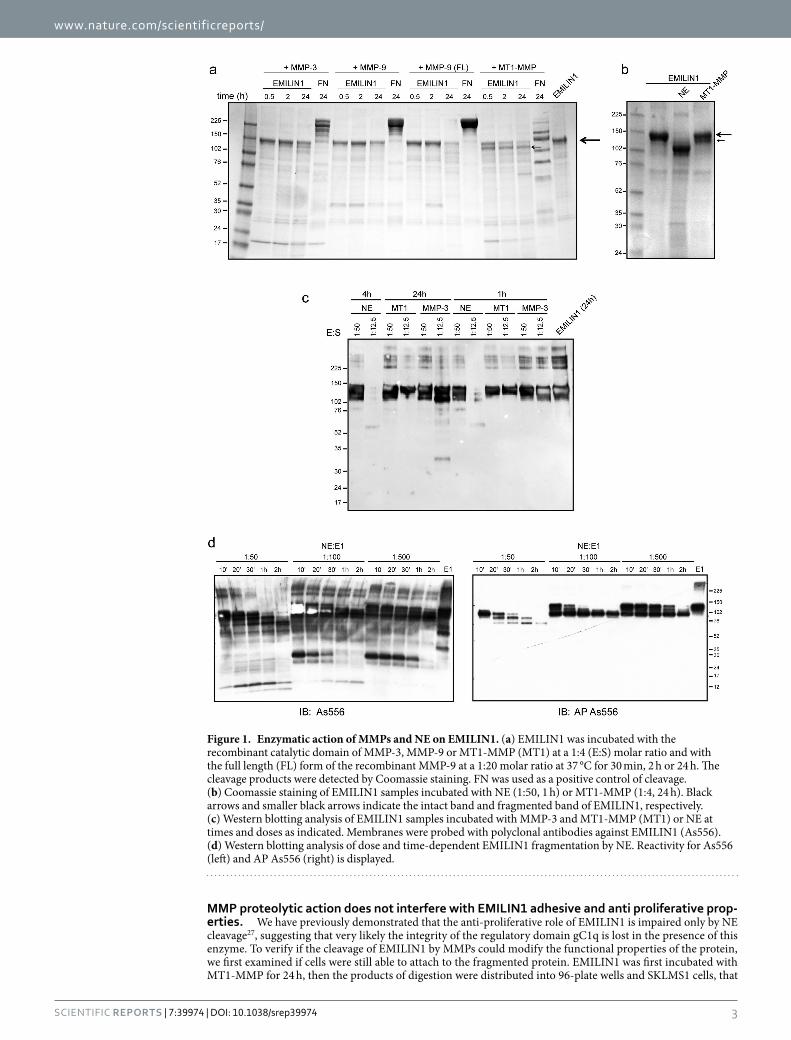
www.nature.com/scientificreports/
3Scientific RepoRts | 7:39974 | DOI: 10.1038/srep39974
MMP proteolytic action does not interfere with EMILIN1 adhesive and anti proliferative prop-erties. We have previously demonstrated that the anti-proliferative role of EMILIN1 is impaired only by NE cleavage27, suggesting that very likely the integrity of the regulatory domain gC1q is lost in the presence of this enzyme. To verify if the cleavage of EMILIN1 by MMPs could modify the functional properties of the protein, we first examined if cells were still able to attach to the fragmented protein. EMILIN1 was first incubated with MT1-MMP for 24 h, then the products of digestion were distributed into 96-plate wells and SKLMS1 cells, that
Figure 1. Enzymatic action of MMPs and NE on EMILIN1. (a) EMILIN1 was incubated with the recombinant catalytic domain of MMP-3, MMP-9 or MT1-MMP (MT1) at a 1:4 (E:S) molar ratio and with the full length (FL) form of the recombinant MMP-9 at a 1:20 molar ratio at 37 °C for 30 min, 2 h or 24 h. The cleavage products were detected by Coomassie staining. FN was used as a positive control of cleavage. (b) Coomassie staining of EMILIN1 samples incubated with NE (1:50, 1 h) or MT1-MMP (1:4, 24 h). Black arrows and smaller black arrows indicate the intact band and fragmented band of EMILIN1, respectively. (c) Western blotting analysis of EMILIN1 samples incubated with MMP-3 and MT1-MMP (MT1) or NE at times and doses as indicated. Membranes were probed with polyclonal antibodies against EMILIN1 (As556). (d) Western blotting analysis of dose and time-dependent EMILIN1 fragmentation by NE. Reactivity for As556 (left) and AP As556 (right) is displayed.
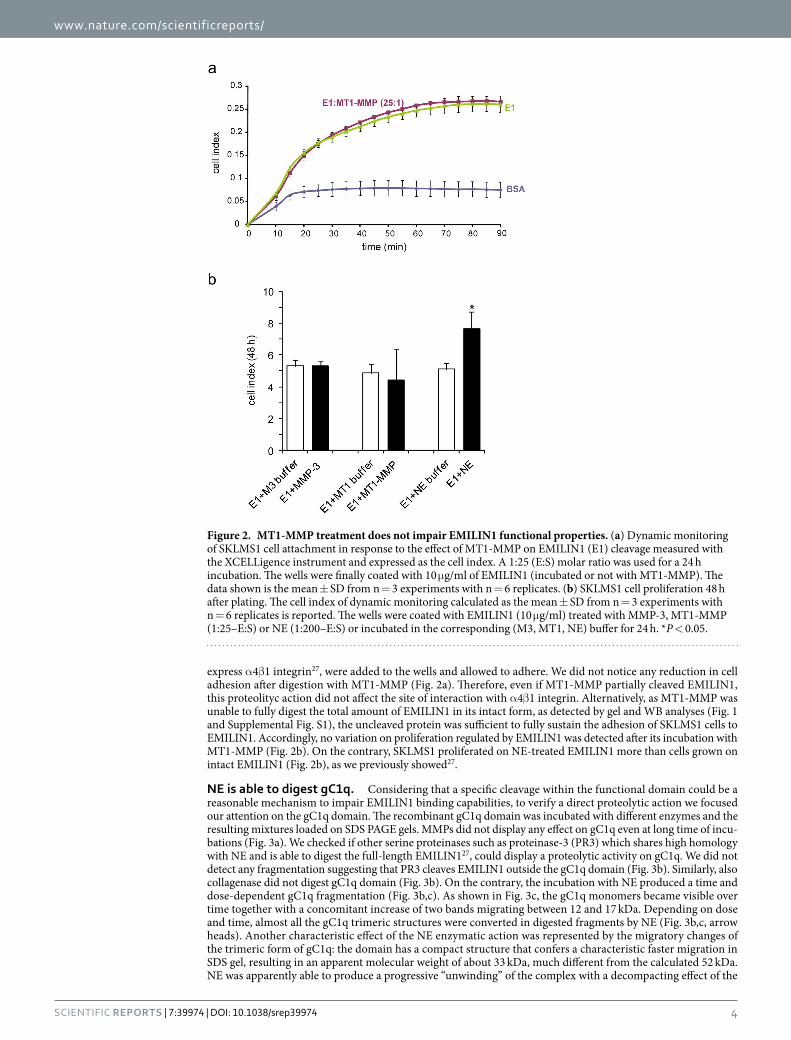
www.nature.com/scientificreports/
4Scientific RepoRts | 7:39974 | DOI: 10.1038/srep39974
express α 4β 1 integrin27, were added to the wells and allowed to adhere. We did not notice any reduction in cell adhesion after digestion with MT1-MMP (Fig. 2a). Therefore, even if MT1-MMP partially cleaved EMILIN1, this proteolityc action did not affect the site of interaction with α 4β 1 integrin. Alternatively, as MT1-MMP was unable to fully digest the total amount of EMILIN1 in its intact form, as detected by gel and WB analyses (Fig. 1 and Supplemental Fig. S1), the uncleaved protein was sufficient to fully sustain the adhesion of SKLMS1 cells to EMILIN1. Accordingly, no variation on proliferation regulated by EMILIN1 was detected after its incubation with MT1-MMP (Fig. 2b). On the contrary, SKLMS1 proliferated on NE-treated EMILIN1 more than cells grown on intact EMILIN1 (Fig. 2b), as we previously showed27.
NE is able to digest gC1q. Considering that a specific cleavage within the functional domain could be a reasonable mechanism to impair EMILIN1 binding capabilities, to verify a direct proteolytic action we focused our attention on the gC1q domain. The recombinant gC1q domain was incubated with different enzymes and the resulting mixtures loaded on SDS PAGE gels. MMPs did not display any effect on gC1q even at long time of incu-bations (Fig. 3a). We checked if other serine proteinases such as proteinase-3 (PR3) which shares high homology with NE and is able to digest the full-length EMILIN127, could display a proteolytic activity on gC1q. We did not detect any fragmentation suggesting that PR3 cleaves EMILIN1 outside the gC1q domain (Fig. 3b). Similarly, also collagenase did not digest gC1q domain (Fig. 3b). On the contrary, the incubation with NE produced a time and dose-dependent gC1q fragmentation (Fig. 3b,c). As shown in Fig. 3c, the gC1q monomers became visible over time together with a concomitant increase of two bands migrating between 12 and 17 kDa. Depending on dose and time, almost all the gC1q trimeric structures were converted in digested fragments by NE (Fig. 3b,c, arrow heads). Another characteristic effect of the NE enzymatic action was represented by the migratory changes of the trimeric form of gC1q: the domain has a compact structure that confers a characteristic faster migration in SDS gel, resulting in an apparent molecular weight of about 33 kDa, much different from the calculated 52 kDa. NE was apparently able to produce a progressive “unwinding” of the complex with a decompacting effect of the
Figure 2. MT1-MMP treatment does not impair EMILIN1 functional properties. (a) Dynamic monitoring of SKLMS1 cell attachment in response to the effect of MT1-MMP on EMILIN1 (E1) cleavage measured with the XCELLigence instrument and expressed as the cell index. A 1:25 (E:S) molar ratio was used for a 24 h incubation. The wells were finally coated with 10 μ g/ml of EMILIN1 (incubated or not with MT1-MMP). The data shown is the mean ± SD from n = 3 experiments with n = 6 replicates. (b) SKLMS1 cell proliferation 48 h after plating. The cell index of dynamic monitoring calculated as the mean ± SD from n = 3 experiments with n = 6 replicates is reported. The wells were coated with EMILIN1 (10 μ g/ml) treated with MMP-3, MT1-MMP (1:25–E:S) or NE (1:200–E:S) or incubated in the corresponding (M3, MT1, NE) buffer for 24 h. *P < 0.05.
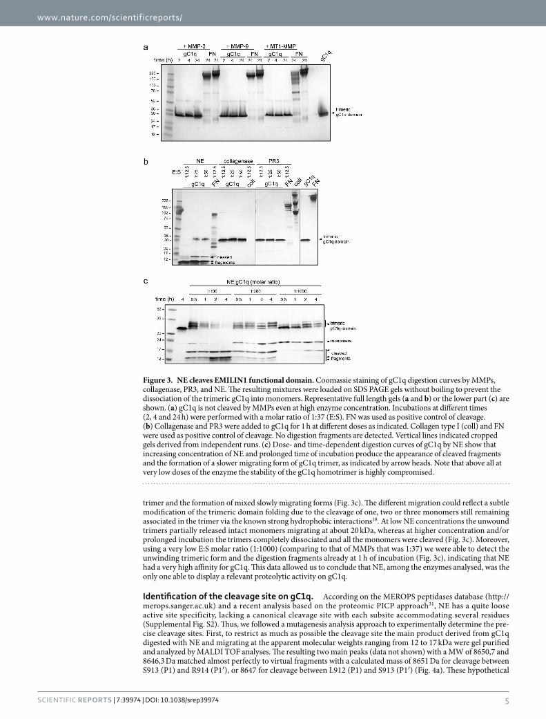
www.nature.com/scientificreports/
5Scientific RepoRts | 7:39974 | DOI: 10.1038/srep39974
trimer and the formation of mixed slowly migrating forms (Fig. 3c). The different migration could reflect a subtle modification of the trimeric domain folding due to the cleavage of one, two or three monomers still remaining associated in the trimer via the known strong hydrophobic interactions18. At low NE concentrations the unwound trimers partially released intact monomers migrating at about 20 kDa, whereas at higher concentration and/or prolonged incubation the trimers completely dissociated and all the monomers were cleaved (Fig. 3c). Moreover, using a very low E:S molar ratio (1:1000) (comparing to that of MMPs that was 1:37) we were able to detect the unwinding trimeric form and the digestion fragments already at 1 h of incubation (Fig. 3c), indicating that NE had a very high affinity for gC1q. This data allowed us to conclude that NE, among the enzymes analysed, was the only one able to display a relevant proteolytic activity on gC1q.
Identification of the cleavage site on gC1q. According on the MEROPS peptidases database (http://merops.sanger.ac.uk) and a recent analysis based on the proteomic PICP approach31, NE has a quite loose active site specificity, lacking a canonical cleavage site with each subsite accommodating several residues (Supplemental Fig. S2). Thus, we followed a mutagenesis analysis approach to experimentally determine the pre-cise cleavage sites. First, to restrict as much as possible the cleavage site the main product derived from gC1q digested with NE and migrating at the apparent molecular weights ranging from 12 to 17 kDa were gel purified and analyzed by MALDI TOF analyses. The resulting two main peaks (data not shown) with a MW of 8650,7 and 8646,3 Da matched almost perfectly to virtual fragments with a calculated mass of 8651 Da for cleavage between S913 (P1) and R914 (P1′ ), or 8647 for cleavage between L912 (P1) and S913 (P1′ ) (Fig. 4a). These hypothetical
Figure 3. NE cleaves EMILIN1 functional domain. Coomassie staining of gC1q digestion curves by MMPs, collagenase, PR3, and NE. The resulting mixtures were loaded on SDS PAGE gels without boiling to prevent the dissociation of the trimeric gC1q into monomers. Representative full length gels (a and b) or the lower part (c) are shown. (a) gC1q is not cleaved by MMPs even at high enzyme concentration. Incubations at different times (2, 4 and 24 h) were performed with a molar ratio of 1:37 (E:S). FN was used as positive control of cleavage. (b) Collagenase and PR3 were added to gC1q for 1 h at different doses as indicated. Collagen type I (coll) and FN were used as positive control of cleavage. No digestion fragments are detected. Vertical lines indicated cropped gels derived from independent runs. (c) Dose- and time-dependent digestion curves of gC1q by NE show that increasing concentration of NE and prolonged time of incubation produce the appearance of cleaved fragments and the formation of a slower migrating form of gC1q trimer, as indicated by arrow heads. Note that above all at very low doses of the enzyme the stability of the gC1q homotrimer is highly compromised.
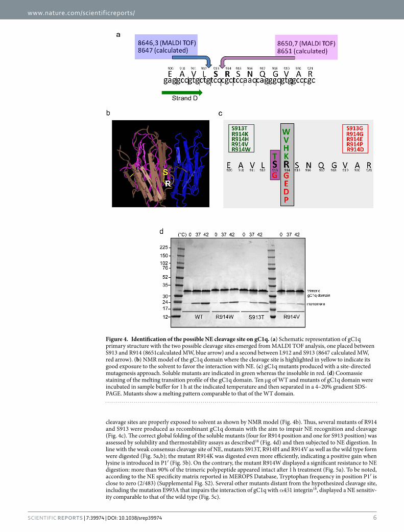
www.nature.com/scientificreports/
6Scientific RepoRts | 7:39974 | DOI: 10.1038/srep39974
cleavage sites are properly exposed to solvent as shown by NMR model (Fig. 4b). Thus, several mutants of R914 and S913 were produced as recombinant gC1q domain with the aim to impair NE recognition and cleavage (Fig. 4c). The correct global folding of the soluble mutants (four for R914 position and one for S913 position) was assessed by solubility and thermostability assays as described18 (Fig. 4d) and then subjected to NE digestion. In line with the weak consensus cleavage site of NE, mutants S913T, R914H and R914V as well as the wild type form were digested (Fig. 5a,b); the mutant R914K was digested even more efficiently, indicating a positive gain when lysine is introduced in P1′ (Fig. 5b). On the contrary, the mutant R914W displayed a significant resistance to NE digestion: more than 90% of the trimeric polypeptide appeared intact after 1 h treatment (Fig. 5a). To be noted, according to the NE specificity matrix reported in MEROPS Database, Tryptophan frequency in position P1′ is close to zero (2/483) (Supplemental Fig. S2). Several other mutants distant from the hypothesized cleavage site, including the mutation E993A that impairs the interaction of gC1q with α 4β 1 integrin18, displayed a NE sensitiv-ity comparable to that of the wild type (Fig. 5c).
Figure 4. Identification of the possible NE cleavage site on gC1q. (a) Schematic representation of gC1q primary structure with the two possible cleavage sites emerged from MALDI TOF analysis, one placed between S913 and R914 (8651calculated MW, blue arrow) and a second between L912 and S913 (8647 calculated MW, red arrow). (b) NMR model of the gC1q domain where the cleavage site is highlighted in yellow to indicate its good exposure to the solvent to favor the interaction with NE. (c) gC1q mutants produced with a site-directed mutagenesis approach. Soluble mutants are indicated in green whereas the insoluble in red. (d) Coomassie staining of the melting transition profile of the gC1q domain. Ten μ g of WT and mutants of gC1q domain were incubated in sample buffer for 1 h at the indicated temperature and then separated in a 4–20% gradient SDS-PAGE. Mutants show a melting pattern comparable to that of the WT domain.
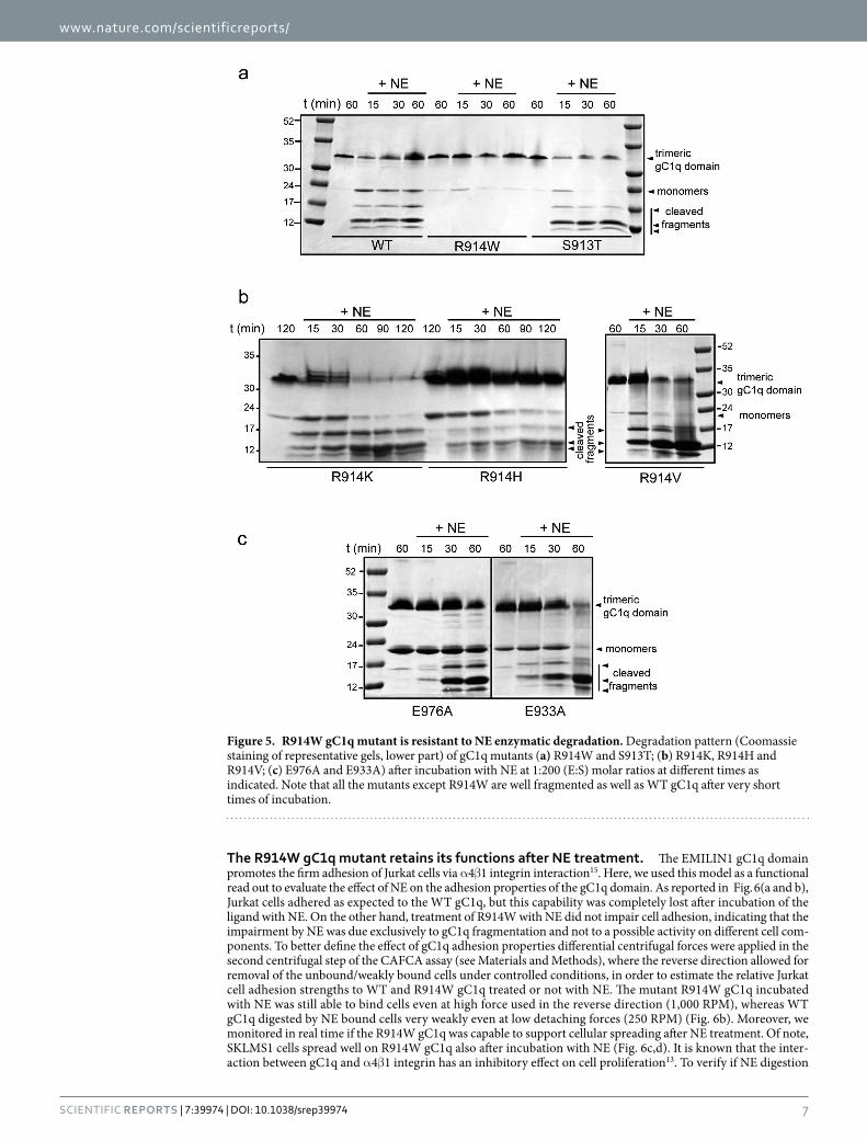
www.nature.com/scientificreports/
7Scientific RepoRts | 7:39974 | DOI: 10.1038/srep39974
The R914W gC1q mutant retains its functions after NE treatment. The EMILIN1 gC1q domain promotes the firm adhesion of Jurkat cells via α 4β 1 integrin interaction15. Here, we used this model as a functional read out to evaluate the effect of NE on the adhesion properties of the gC1q domain. As reported in Fig. 6(a and b), Jurkat cells adhered as expected to the WT gC1q, but this capability was completely lost after incubation of the ligand with NE. On the other hand, treatment of R914W with NE did not impair cell adhesion, indicating that the impairment by NE was due exclusively to gC1q fragmentation and not to a possible activity on different cell com-ponents. To better define the effect of gC1q adhesion properties differential centrifugal forces were applied in the second centrifugal step of the CAFCA assay (see Materials and Methods), where the reverse direction allowed for removal of the unbound/weakly bound cells under controlled conditions, in order to estimate the relative Jurkat cell adhesion strengths to WT and R914W gC1q treated or not with NE. The mutant R914W gC1q incubated with NE was still able to bind cells even at high force used in the reverse direction (1,000 RPM), whereas WT gC1q digested by NE bound cells very weakly even at low detaching forces (250 RPM) (Fig. 6b). Moreover, we monitored in real time if the R914W gC1q was capable to support cellular spreading after NE treatment. Of note, SKLMS1 cells spread well on R914W gC1q also after incubation with NE (Fig. 6c,d). It is known that the inter-action between gC1q and α 4β 1 integrin has an inhibitory effect on cell proliferation13. To verify if NE digestion
Figure 5. R914W gC1q mutant is resistant to NE enzymatic degradation. Degradation pattern (Coomassie staining of representative gels, lower part) of gC1q mutants (a) R914W and S913T; (b) R914K, R914H and R914V; (c) E976A and E933A) after incubation with NE at 1:200 (E:S) molar ratios at different times as indicated. Note that all the mutants except R914W are well fragmented as well as WT gC1q after very short times of incubation.
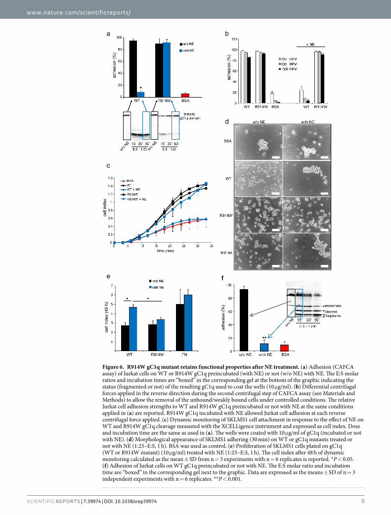
www.nature.com/scientificreports/
8Scientific RepoRts | 7:39974 | DOI: 10.1038/srep39974
Figure 6. R914W gC1q mutant retains functional properties after NE treatment. (a) Adhesion (CAFCA assay) of Jurkat cells on WT or R914W gC1q preincubated (with NE) or not (w/o NE) with NE. The E:S molar ratios and incubation times are “boxed” in the corresponding gel at the bottom of the graphic indicating the status (fragmented or not) of the resulting gC1q used to coat the wells (10 μ g/ml). (b) Differential centrifugal forces applied in the reverse direction during the second centrifugal step of CAFCA assay (see Materials and Methods) to allow the removal of the unbound/weakly bound cells under controlled conditions. The relative Jurkat cell adhesion strengths to WT and R914W gC1q preincubated or not with NE at the same conditions applied in (a) are reported. R914W gC1q incubated with NE allowed Jurkat cell adhesion at each reverse centrifugal force applied. (c) Dynamic monitoring of SKLMS1 cell attachment in response to the effect of NE on WT and R914W gC1q cleavage measured with the XCELLigence instrument and expressed as cell index. Dose and incubation time are the same as used in (a). The wells were coated with 10 μ g/ml of gC1q (incubated or not with NE). (d) Morphological appearance of SKLMS1 adhering (30 min) on WT or gC1q mutants treated or not with NE (1:25–E:S, 1 h). BSA was used as control. (e) Proliferation of SKLMS1 cells plated on gC1q (WT or R914W mutant) (10 μ g/ml) treated with NE (1:25–E:S, 1 h). The cell index after 48 h of dynamic monitoring calculated as the mean ± SD from n = 3 experiments with n = 6 replicates is reported. *P < 0.05. (f) Adhesion of Jurkat cells on WT gC1q preincubated or not with NE. The E:S molar ratio and incubation time are “boxed” in the corresponding gel next to the graphic. Data are expressed as the means ± SD of n = 3 independent experiments with n = 6 replicates. **P < 0.001.
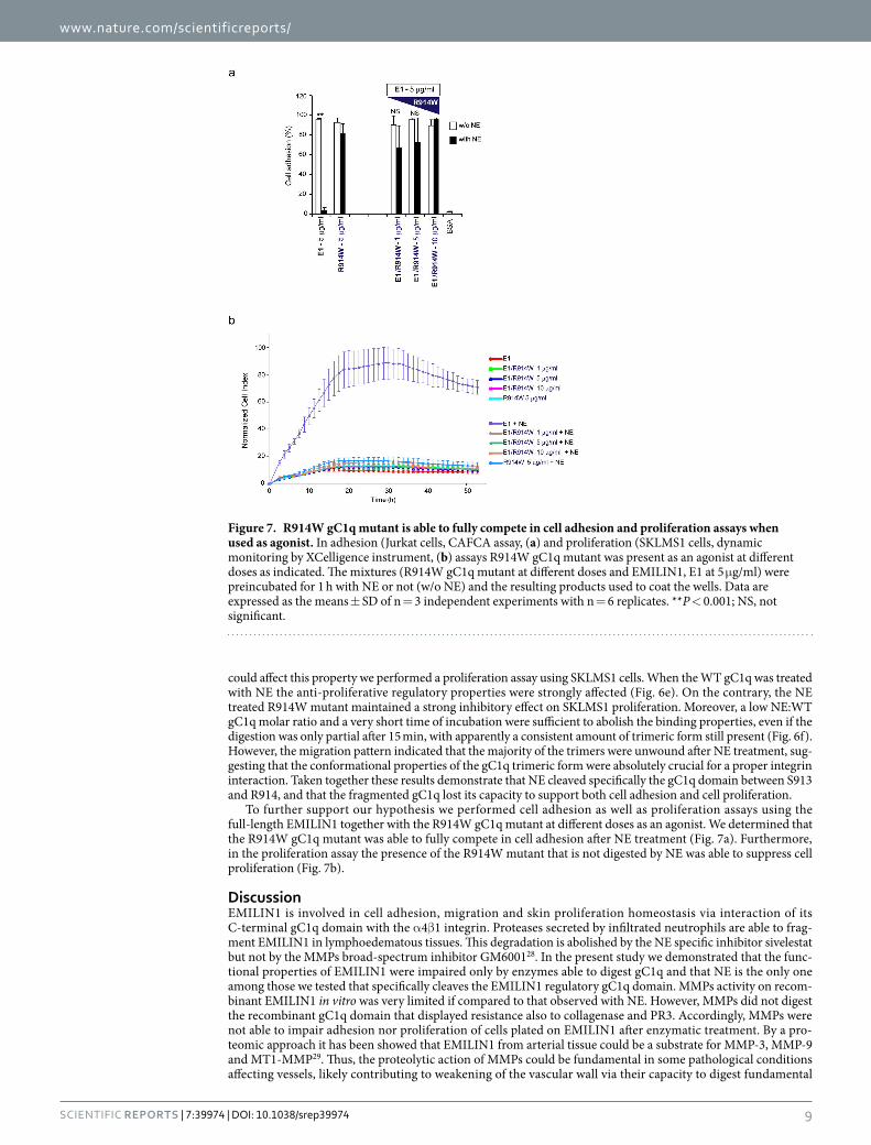
www.nature.com/scientificreports/
9Scientific RepoRts | 7:39974 | DOI: 10.1038/srep39974
could affect this property we performed a proliferation assay using SKLMS1 cells. When the WT gC1q was treated with NE the anti-proliferative regulatory properties were strongly affected (Fig. 6e). On the contrary, the NE treated R914W mutant maintained a strong inhibitory effect on SKLMS1 proliferation. Moreover, a low NE:WT gC1q molar ratio and a very short time of incubation were sufficient to abolish the binding properties, even if the digestion was only partial after 15 min, with apparently a consistent amount of trimeric form still present (Fig. 6f). However, the migration pattern indicated that the majority of the trimers were unwound after NE treatment, sug-gesting that the conformational properties of the gC1q trimeric form were absolutely crucial for a proper integrin interaction. Taken together these results demonstrate that NE cleaved specifically the gC1q domain between S913 and R914, and that the fragmented gC1q lost its capacity to support both cell adhesion and cell proliferation.
To further support our hypothesis we performed cell adhesion as well as proliferation assays using the full-length EMILIN1 together with the R914W gC1q mutant at different doses as an agonist. We determined that the R914W gC1q mutant was able to fully compete in cell adhesion after NE treatment (Fig. 7a). Furthermore, in the proliferation assay the presence of the R914W mutant that is not digested by NE was able to suppress cell proliferation (Fig. 7b).
DiscussionEMILIN1 is involved in cell adhesion, migration and skin proliferation homeostasis via interaction of its C-terminal gC1q domain with the α 4β 1 integrin. Proteases secreted by infiltrated neutrophils are able to frag-ment EMILIN1 in lymphoedematous tissues. This degradation is abolished by the NE specific inhibitor sivelestat but not by the MMPs broad-spectrum inhibitor GM600128. In the present study we demonstrated that the func-tional properties of EMILIN1 were impaired only by enzymes able to digest gC1q and that NE is the only one among those we tested that specifically cleaves the EMILIN1 regulatory gC1q domain. MMPs activity on recom-binant EMILIN1 in vitro was very limited if compared to that observed with NE. However, MMPs did not digest the recombinant gC1q domain that displayed resistance also to collagenase and PR3. Accordingly, MMPs were not able to impair adhesion nor proliferation of cells plated on EMILIN1 after enzymatic treatment. By a pro-teomic approach it has been showed that EMILIN1 from arterial tissue could be a substrate for MMP-3, MMP-9 and MT1-MMP29. Thus, the proteolytic action of MMPs could be fundamental in some pathological conditions affecting vessels, likely contributing to weakening of the vascular wall via their capacity to digest fundamental
Figure 7. R914W gC1q mutant is able to fully compete in cell adhesion and proliferation assays when used as agonist. In adhesion (Jurkat cells, CAFCA assay, (a) and proliferation (SKLMS1 cells, dynamic monitoring by XCelligence instrument, (b) assays R914W gC1q mutant was present as an agonist at different doses as indicated. The mixtures (R914W gC1q mutant at different doses and EMILIN1, E1 at 5 μ g/ml) were preincubated for 1 h with NE or not (w/o NE) and the resulting products used to coat the wells. Data are expressed as the means ± SD of n = 3 independent experiments with n = 6 replicates. **P < 0.001; NS, not significant.

www.nature.com/scientificreports/
1 0Scientific RepoRts | 7:39974 | DOI: 10.1038/srep39974
structural ECM components of elastic fibers. It will be interesting in the future to functionally investigate how crucial the EMILIN1 structural integrity is in the blood vascular context.
EMILIN1 cleavage by NE is critical in the regulation of cell proliferation. In the tumor microenvironment NE is likely the inflammatory enzyme displaying a proteolytic action on gC1q, thus impairing its integrity and then its suppressor role. NE is a neutrophil-derived serine proteinase with broad substrate specificity, including most components of the ECM such as elastin, collagens, laminin, entactin, and fibronectin32. In addition, a key role for NE in cleaving and either activating or inactivating cytokines and growth factors has been shown33,34. NE promotes cellular proliferation in several, different lung and breast cancer cell lines and in fibroblasts, as well35–37. In tumour cells NE accomplishes this by entering endosomes via the classic clathrin pit-mediated endocytosis, and then targeting intracellular substrates within the cytosol, such as IRS-1, a key mediator of PI3K signaling36,38. In our study we describe an indirect modality used by NE to favour cell proliferation.
The cleavage site within the gC1q domain lies between S913 and R914 residues and the mutant R914W was fully resistant to the NE proteolytic action and worked as agonist very well. This mutant was instrumental to demonstrate in a cellular context that the loss of gC1q-integrin interaction in the presence of NE was determined exclusively by the gC1q cleavage and not by other possible NE targets present in the milieu.
The EMILIN1 gC1q domain has been identified as a fundamental component of the whole molecule, both from a structural and a functional viewpoint7: it is the domain able to initiate and drive the homo-trimerization of the protein and it plays regulatory properties through the interaction with the α 4/α 9β 1 integrins13,15,16,23. The α 4β 1 binding site is located in a flexible loop at the apex of the trimeric structure and it is absolutely dependent on the E933 residue18. In addition to the E933 residue it appears that also other regions of the gC1q domain are important for the integrin recognition (Capuano et al., in preparation), suggesting that the global folding of the gC1q is necessary for its full activity. The present data is in agreement with this concept: by partial digestion of gC1q with low concentrations and/or short times of incubation with NE an heterogeneous mix of unwound tri-mers was obtained, possibly due to the cleavage of one, two or three monomers still associated into trimers. These mixed products did not support any cellular adhesion in comparison with the untreated domain, underlying the requirement for a 3D-fold arrangement of the interaction to bind the integrin counterpart: the precise and stable geometry pattern around the flexible loop at the apex of the trimeric structure is essential for the correct interaction of α 4β 1 integrin with the E933 residue. EMILIN1 presents unique structural features compared to all the other members of the C1q superfamily. While it presents the classical jelly-roll β -sandwich fold, EMILIN1 has only 9 instead of 10 β -strands and this different structural organization is associated with the specific and unique capability to be recognized by α 4β 1 integrin at an inserted short flexible sequence18. It is of note that the present findings of a very specific cleavage site for NE in the gC1q domain of EMILIN1 that profoundly affects the pro-adhesive as well as the proliferation suppressive functions of EMILIN1 depend on a cleavage within the gC1q domain. The cleavage of some members belonging to the gC1q-TNF family has been documented39–42, but almost always outside the globular domain. In particular, adiponectin cleavage by NE in the collagen-like region generates globular fragments with distinct biological activity40; the collagen-like region of the C1q subunit of the complement (C1q-CLR) can be cleaved by MMPs42; the cardioprotective cardiokine C1q-TNF-related protein-9, requires proteolytic cleavage to obtain a biologically active globular domain isoform41; and the differential proteo-lytic processing of CTRP12 by PCSK3/furin in the N-terminal domain generates functionally distinct isoforms39. Only one example of a cleavage site for trypsin located in the globular region of C1q that affected the B chain capability to interact with fucoidan in vitro was reported suggesting that the compactness of this domain is very rarely affected by enzymes43. To our knowledge, our study demonstrates for the first time the cleavage within the globular domain under physio-pathological conditions of proteolytic process establishing both a unique role of NE as enzyme and a novel specificity for the EMILIN1-gC1q as substrate.
In conclusion, our findings reinforce the uniqueness of the gC1q-α 4β 1 integrin binding which is highly dependent on the overall structural conformation of the homotrimeric assembly employing a different mode of integrin engagement generally located in mobile loops protruding from the main core of the whole ligand44. In this study we demonstrated that only NE is able to disrupt the correct folding of gC1q domain thus impairing its suppressor role on cellular proliferation.
Materials and MethodsAntibodies and reagents. Rabbit antiserum against human EMILIN1 (As556) was obtained as previously described5,27. The antiserum was also purified by affinity chromatography for gC1q recognition (AP As556). EMILIN1, secreted by HEK293 cells, constitutively expressing the EBNA 1 protein, was obtained as previously described30. Briefly, the cells were expanded to mass culture and were then maintained for 2 days in serum-free medium to allow accumulation of EMILIN1 in the cell supernatant. Partial purification was achieved by dialysis of the conditioned medium at 4 °C against 0.1 M NaCl, 20 mM Tris-HCI, pH 6.8. A further purification step was achieved by chromatography on a DEAE-cellulose column and size exclusion chromatography using Sepharose CL 4B (Amersham Pharmacia Biotech) as described30. The preparation was checked for the presence of laminin and FN by immunoblotting with specific antibodies and proved to be nearly devoid of these contaminants. The C-terminal gC1q domain was produced as previously described15,18. Plasma FN was purchased from Sigma. Rat tail collagen type I was provided by BD Bioscience. Human NE was provided by Calbiochem, (Merk Millipore); recombinant catalytic domain of MMP-3, MMP-9 and MT1-MMP were purchased by Giotto Biotech Srl. Full length recombinant MMP-9 was provided by Merck Millipore. Collagenase Type I-A and PR3 were provided by Sigma-Aldrich and Enzo LifeScience, Inc., respectively.
Cell cultures. The human leiomyosarcoma SKLMS1, the immortalized human T lymphocyte Jurkat cell lines were purchased by ATCC and cultured in DMEM supplemented with 10% FCS and antibiotics.

www.nature.com/scientificreports/
1 1Scientific RepoRts | 7:39974 | DOI: 10.1038/srep39974
Enzymatic digestion. FN, EMILIN1 or gC1q and its mutated variants were incubated with the recombinant MMPs or NE at different molar enzyme/substrate ratios for different times as indicated at 37 °C. The digestion with MMP-3, MMP-9, MT1-MMP and collagenase was carried out in a buffer containing CaCl2 (10 mM for MMP-9 and MT1-MMP; 5 mM for MMP-3), 150 mM NaCl, 50 mM Tris, pH 7.5. The solution for NE digestion contained 10 mM CaCl2, whereas for PR3 a 150 mM NaCl, 50 mM Tris, pH 7.5 buffer was used. Controls were incubated in the appropriate buffer without enzymes. After the indicated times, reactions were stopped by adding 5 × Laemmli buffer, loaded onto 4– 20% polyacrylamide gel, and stained in a 0.05% wt/vol Coomassie G-250, 5% v/v glacial acetic acid solution to monitor substrate degradation. In some experiments we analyzed fragmentation pattern by western blotting technique. The nitrocellulose membranes were saturated with TBS buffer (20 mM Tris and 0.15 M NaCl) containing 0.1% Tween-20 (TBST) and 5% non-fat dry milk for 1 h at room temperature and then incubated at 4 °C overnight with primary antibodies against EMILIN1 or gC1q (As556 and AP As556, respectively). After extensive washing in TBST, the membranes were incubated with rabbit HRP-conjugated sec-ondary antibodies (Amersham, GE Healthcare) and then revealed with the ECL Plus chemiluminescence kit (Amersham, GE Healthcare).
MALDI TOF analysis. To determine the molecular weights of the gC1q NE cleaved fragments, the digestion mixture was separated by SDS-PAGE using a precast, gradient, 4–20% gel (Biorad). After Comassie G-250 stain-ing, selected digestion bands were excised from the gel and submitted to the in-house proteomic facility for the MALDI TOF analyses.
Site-directed Mutagenesis. Matching the molecular weights of the digestion fragments with those calcu-lated on the basis of the gC1q primary sequence, resulted in two possible cutting sites, located between L912 (P1) and S913 (P1′ ) or between S913 (P1) and R914 (P1′ ). Several point mutations of S913 and R914 were generated by site-directed mutagenesis using the overlapping PCR approach45. Briefly, in a first PCR round the primer carrying the desired mutation was used in combination with 5′- and 3′-flanking primers to generate two overlapping frag-ments using as template the wild type EMILIN1 gC1q coding sequence inserted in the expression vector pQE-30 (Qiagen) between the BamHI and KpnI restriction sites. The overlapping fragments were gel-purified and used as templates in a two-step PCR round consisting of 12 elongation cycles in which the overlapping regions work as primers and addition of the 5′- and 3′-flanking primers carrying BamHI and KpnI restriction sites, respectively, followed by 25 amplification cycles. The final products were purified by wizard SV columns (Promega), cut with BamHI and KpnI, and ligated into pQE-30 vector, cut with the same enzymes. Ligation products were trans-formed in MI5 bacterial strain. The expression constructs containing mutant sequences were verified by DNA sequencing. The oligonucleotides were purchased from Sigma Genosys.
Purification of recombinant gC1q mutants and SDS-PAGE analysis. Recombinant proteins (WT and mutant gC1q) were expressed as His6-tagged proteins and extracted under native conditions as pre-viously described30. The purification of the soluble mutants was performed by affinity chromatography on nickel-nitrilotriacetic acid resin (Qiagen). When resistance to denaturation by heating in the presence of 1% SDS was assayed, the samples were incubated in sample buffer (20 mM Tris-HCl, pH 7.6, 100 mM NaCl, 25 mM EDTA, 1% SDS, 2 mM phenylmethanesulfonyl fluoride, 5 mM N-ethylmaleimide) for 1 h at different temperatures just before gel loading.
Functional cellular assays. The quantitative cell adhesion assay used in this study is based on centrifuga-tion (CAFCA assay)46. 6-well strips of flexible polyvinyl chloride covered with double-sided tape (bottom units) were coated with EMILIN1, gC1q, the mutated variants or their cleavage products (10 μ g/ml; unless noted other-wise). Cells were labelled with the vital fluorochrome calcein acetoxymethyl (Invitrogen) and then aliquoted into the bottom CAFCA miniplates, which were centrifuged to synchronize the contact of the cells with the substrate. The miniplates were then incubated for 20 min at 37 °C and were subsequently mounted together with a similar CAFCA miniplate to create communicating chambers for subsequent reverse centrifugation. The relative number of cells bound to the substrate and cells that failed to bind to the substrate was estimated by top/bottom fluores-cence detection in a computer-interfaced Infinite M1000 PRO microplate reader (Tecan Group Ltd.). In addition, to quantitatively and qualitatively monitor cell behaviour in real time, we adopted the technology provided by the Real-Time Cell Analyzer dual plate instrument (XCELLigence system, Roche). The strategy is based on contin-uous quantitative monitoring of cells as they adhere/spread and proliferate by measuring electrical impedance47. The change in impedance caused by cell attachment and dependent on the cell number is expressed as the cell index, which is an arbitrary measurement defined as (Rn − Rb)/15, in which Rb is the background impedance of the well measured with medium alone, and Rn is the impedance of the well measured at any time with cells pres-ent. Thus, the cell index is a reflection of overall cell number, attachment quality, and cell morphology that can change as a function of time. For adhesion/spreading experiments, the E-plates 96 were precoated with EMILIN1, gC1q, the mutated variants or the cleavage products (10 μ g/ml, unless noted otherwise). Cells were then seeded at 20 × 103 cells/well in FCS-free medium and monitored every 3 or 5 min for the time indicated. To monitor cell proliferation, 5% FCS was added to the wells 2 h after plating. Data analysis was performed using Real-Time Cell Analyzer software supplied with the instrument. Experiments were performed in triplicate.
Statistical analysis. Statistical significance of the results was determined by using the two-tailed unpaired Student’s t test to determine whether two datasets were significantly different. A value of P < 0.05 was considered significant.

www.nature.com/scientificreports/
1 2Scientific RepoRts | 7:39974 | DOI: 10.1038/srep39974
References1. Hynes, R. O. The extracellular matrix: not just pretty fibrils. Science 326, 1216–1219 (2009).2. Watt, F. M. & Fujiwara, H. Cell-extracellular matrix interactions in normal and diseased skin. Cold Spring Harb. Perspect. Biol. 3,
(2011).3. Streuli, C. H. Integrins and cell-fate determination. J. Cell Sci. 122, 171–177 (2009).4. Moreno-Layseca, P. & Streuli, C. H. Signalling pathways linking integrins with cell cycle progression. Matrix Biol. 34, 144–153 (2014).5. Doliana, R. et al. EMILIN, a component of the elastic fiber and a new member of the C1q/tumor necrosis factor superfamily of
proteins. J. Biol. Chem. 274, 16773–16781 (1999).6. Colombatti, A. et al. The EMILIN protein family. Matrix Biol. 19, 289–301 (2000).7. Colombatti, A. et al. The EMILIN/Multimerin family. Front Immunol. 2, 93 (2011).8. Ghai, R. et al. C1q and its growing family. Immunobiology 212, 253–266 (2007).9. Kishore, U. et al. C1q and tumor necrosis factor superfamily: modularity and versatility. Trends Immunol. 25, 551–561 (2004).
10. Colombatti, A., Bressan, G. M., Castellani, I. & Volpin, D. Glycoprotein 115, a glycoprotein isolated from chick blood vessels, is widely distributed in connective tissue. J. Cell Biol. 100, 18–26 (1985).
11. Colombatti, A., Poletti, A., Bressan, G. M., Carbone, A. & Volpin, D. Widespread codistribution of glycoprotein gp 115 and elastin in chick eye and other tissues. Coll. Relat Res. 7, 259–275 (1987).
12. Danussi, C. et al. Emilin1 deficiency causes structural and functional defects of lymphatic vasculature. Mol. Cell Biol. 28, 4026–4039 (2008).
13. Danussi, C. et al. EMILIN1-α 4/α 9 integrin interaction inhibits dermal fibroblast and keratinocyte proliferation. J. Cell Biol. 195, 131–145 (2011).
14. Zacchigna, L. et al. Emilin1 links TGF-beta maturation to blood pressure homeostasis. Cell 124, 929–942 (2006).15. Spessotto, P. et al. beta 1 Integrin-dependent cell adhesion to EMILIN-1 is mediated by the gC1q domain. J. Biol. Chem. 278,
6160–6167 (2003).16. Spessotto, P. et al. EMILIN1 represents a major stromal element determining human trophoblast invasion of the uterine wall. J. Cell
Sci. 119, 4574–4584 (2006).17. Danussi, C. et al. An EMILIN1-negative microenvironment promotes tumor cell proliferation and lymph node invasion. Cancer
Prev. Res. (Phila) 5, 1131–1143 (2012).18. Verdone, G. et al. The solution structure of EMILIN1 globular C1q domain reveals a disordered insertion necessary for interaction
with the alpha4beta1 integrin. J. Biol. Chem. 283, 18947–18956 (2008).19. Rose, D. M., Alon, R. & Ginsberg, M. H. Integrin modulation and signaling in leukocyte adhesion and migration. Immunol. Rev.
218, 126–134 (2007).20. Hauzenberger, D., Klominek, J., Bergstrom, S. E. & Sundqvist, K. G. T lymphocyte migration: the influence of interactions via
adhesion molecules, the T cell receptor, and cytokines. Crit Rev. Immunol. 15, 285–316 (1995).21. Humphries, J. D., Byron, A. & Humphries, M. J. Integrin ligands at a glance. J. Cell Sci. 119, 3901–3903 (2006).22. Humphries, M. J. The molecular basis and specificity of integrin-ligand interactions. J. Cell Sci. 97(Pt 4), 585–592 (1990).23. Danussi, C. et al. EMILIN1/alpha9beta1 integrin interaction is crucial in lymphatic valve formation and maintenance. Mol. Cell Biol.
33, 4381–4394 (2013).24. Kamarajan, P., Garcia-Pardo, A., D’Silva, N. J. & Kapila, Y. L. The CS1 segment of fibronectin is involved in human OSCC
pathogenesis by mediating OSCC cell spreading, migration, and invasion. BMC. Cancer 10, 330 (2010).25. Zucchetto, A. et al. The CD49d/CD29 complex is physically and functionally associated with CD38 in B-cell chronic lymphocytic
leukemia cells. Leukemia 26, 1301–1312 (2012).26. Lund, S. A. et al. Osteopontin mediates macrophage chemotaxis via alpha4 and alpha9 integrins and survival via the alpha4 integrin.
J. Cell Biochem. 114, 1194–1202 (2013).27. Pivetta, E. et al. Neutrophil elastase-dependent cleavage compromises the tumor suppressor role of EMILIN1. Matrix Biol. 34, 22–32
(2014).28. Pivetta, E. et al. Local inhibition of elastase reduces EMILIN1 cleavage reactivating lymphatic vessel function in a mouse
lymphoedema model. Clin. Sci. (Lond) 130, 1221–1236 (2016).29. Stegemann, C. et al. Proteomic identification of matrix metalloproteinase substrates in the human vasculature. Circ. Cardiovasc.
Genet. 6, 106–117 (2013).30. Mongiat, M. et al. Self-assembly and supramolecular organization of EMILIN. J. Biol. Chem. 275, 25471–25480 (2000).31. Schilling, O. & Overall, C. M. Proteome-derived, database-searchable peptide libraries for identifying protease cleavage sites. Nat.
Biotechnol. 26, 685–694 (2008).32. Hedstrom, L. Serine protease mechanism and specificity. Chem. Rev. 102, 4501–4524 (2002).33. Bank, U. & Ansorge, S. More than destructive: neutrophil-derived serine proteases in cytokine bioactivity control. J. Leukoc. Biol. 69,
197–206 (2001).34. Pham, C. T. Neutrophil serine proteases: specific regulators of inflammation. Nat. Rev. Immunol. 6, 541–550 (2006).35. Mittendorf, E. A. et al. Breast cancer cell uptake of the inflammatory mediator neutrophil elastase triggers an anticancer adaptive
immune response. Cancer Res. 72, 3153–3162 (2012).36. Houghton, A. M. et al. Neutrophil elastase-mediated degradation of IRS-1 accelerates lung tumor growth. Nat. Med. 16, 219–223
(2010).37. Gregory, A. D. et al. Neutrophil elastase promotes myofibroblast differentiation in lung fibrosis. J. Leukoc. Biol. 98, 143–152 (2015).38. Gregory, A. D., Hale, P., Perlmutter, D. H. & Houghton, A. M. Clathrin pit-mediated endocytosis of neutrophil elastase and cathepsin
G by cancer cells. J. Biol. Chem. 287, 35341–35350 (2012).39. Wei, Z., Lei, X., Seldin, M. M. & Wong, G. W. Endopeptidase cleavage generates a functionally distinct isoform of C1q/tumor
necrosis factor-related protein-12 (CTRP12) with an altered oligomeric state and signaling specificity. J. Biol. Chem. 287, 35804–35814 (2012).
40. Waki, H. et al. Generation of globular fragment of adiponectin by leukocyte elastase secreted by monocytic cell line THP-1. Endocrinology 146, 790–796 (2005).
41. Yuan, Y. et al. C1q-TNF-related protein-9, a novel cardioprotetcive cardiokine, requires proteolytic cleavage to generate a biologically active globular domain isoform. Am. J. Physiol Endocrinol. Metab 308, E891–E898 (2015).
42. Ruiz, S., Henschen-Edman, A. H., Nagase, H. & Tenner, A. J. Digestion of C1q collagen-like domain with MMPs-1,-2,-3, and -9 further defines the sequence involved in the stimulation of neutrophil superoxide production. J. Leukoc. Biol. 66, 416–422 (1999).
43. Tissot, B. et al. Mass spectrometry analysis of the oligomeric C1q protein reveals the B chain as the target of trypsin cleavage and interaction with fucoidan. Biochemistry 44, 2602–2609 (2005).
44. Casasnovas, J. M., Pieroni, C. & Springer, T. A. Lymphocyte function-associated antigen-1 binding residues in intercellular adhesion molecule-2 (ICAM-2) and the integrin binding surface in the ICAM subfamily. Proc. Natl. Acad. Sci. USA 96, 3017–3022 (1999).
45. Cannizzaro, R. et al. Endomicroscopy and cancer: a new approach to the visualization of neoangiogenesis. Gastroenterol. Res. Pract. 2012, 537170 (2012).
46. Spessotto, P. et al. Fluorescence-based assays for in vitro analysis of cell adhesion and migration. Methods Mol. Biol. 522, 221–250 (2009).
47. Xing, J. Z. et al. Dynamic monitoring of cytotoxicity on microelectronic sensors. Chem. Res. Toxicol. 18, 154–161 (2005).

www.nature.com/scientificreports/
13Scientific RepoRts | 7:39974 | DOI: 10.1038/srep39974
AcknowledgementsThis work was supported by AIRC (IG 14192) and Ministry of Health (RF-2010-2309719) grants to P.S. This publication is in partial fulfillment of the requirements for a PhD in Biosciences and Biotechnology (Curriculum in Cell Biology, University of Padua) of Orlando Maiorani.
Author ContributionsO.M. conducted most of the experiments and analyzed the results, E.P. performed most of the functional studies, A.C.a. conducted the biochemical analyses on the mutants; T.M.E.M., B.W. and F.B. prepared the recombinant proteins and analyzed the results, A.C. analyzed the results and wrote the paper, R.D. performed mutagenesis and coordinated the biochemical experiments, P.S. conceived the idea for the project and wrote the paper with A.C.
Additional InformationSupplementary information accompanies this paper at http://www.nature.com/srepCompeting financial interests: The authors declare no competing financial interests.How to cite this article: Maiorani, O. et al. Neutrophil elastase cleavage of the gC1q domain impairs the EMILIN1-α4β1 integrin interaction, cell adhesion and anti-proliferative activity. Sci. Rep. 7, 39974; doi: 10.1038/srep39974 (2017).Publisher's note: Springer Nature remains neutral with regard to jurisdictional claims in published maps and institutional affiliations.
This work is licensed under a Creative Commons Attribution 4.0 International License. The images or other third party material in this article are included in the article’s Creative Commons license,
unless indicated otherwise in the credit line; if the material is not included under the Creative Commons license, users will need to obtain permission from the license holder to reproduce the material. To view a copy of this license, visit http://creativecommons.org/licenses/by/4.0/ © The Author(s) 2017


![S'ESSA NON PASSA PER LE MATEMATICHE DIMOSTRAZIONI …celso/artigos/memocs-v5-n3-p02-s.pdf · lattice. In[Dormann and Deutsch 2002], avascular tumor growth is simulated using a hybrid](https://static.fdocumenti.com/doc/165x107/5e822d118e491c096561a2f0/sessa-non-passa-per-le-matematiche-dimostrazioni-celsoartigosmemocs-v5-n3-p02-spdf.jpg)