Isabella Castellano - Università degli studi di Torino
48
Isabella Castellano Dipartimento di Scienze Mediche Università degli studi di Torino Anatomia Patologica (Direttore: Prof. A. Sapino) Città della Salute e della Scienza-Presidio Molinette, Torino Analisi molecolare intraoperatoria: la soluzione del problema? Problematiche nella diagnosi e terapia della malattia a livello ascellare
Transcript of Isabella Castellano - Università degli studi di Torino

Isabella Castellano
Dipartimento di Scienze Mediche Università degli studi di Torino
Anatomia Patologica (Direttore: Prof. A. Sapino)
Città della Salute e della Scienza-Presidio Molinette, Torino
Analisi molecolare intraoperatoria: la soluzione del problema?
Problematiche nella diagnosi e terapia della malattia a livello ascellare

Tecniche diagnostiche a disposizione per l’analisi del linfonodo sentinella (LS)
ESAME ESTEMPORANEO
INTRAOPERATORIO
ESAME SU LS FISSATO IN FORMALINA ED INCLUSO
IN PARAFFINA
TECNICHE MOLECOLARI
OSNA
CHIRURGHI ANATOMO-PATOLOGI COSTO

PERCHE’ L’ANALISI MOLECOLARE DEL LS QUALI SONO GLI SVANTAGGI LA NOSTRA SOLUZIONE MOMENTANEA

PERCHE’ L’ANALISI MOLECOLARE DEL LS
2. FORNIRE UN DATO RIPRODUCIBILE
1. FORNIRE UN DATO INTRAOPERATORIO

Conduce all’immediata dissezione del cavo ascellare evitendo il secondo reintervento
INTRAOPERATIVE SLN ASSESMENT
SEZIONI CRIOSTATICHE IMPRINT CITOLOGICO
Con o senza immunocitochimica rapida
MA….

SNL imprint cytology analysis
SNL frozen sec?on analysis
Sensibilità varia dal 33% to 84% indipendentemente dal metodo
uFlizzato: rischio di FALSI NEGATIVI

Sensibilità: 89%; Specifictà:100%; Valore PrediLvo Posi?vo: 100%; Valore prediLvo nega?vo: 82.3%
Sapino A, et al Br J Cancer. 2003 Mar 10;88(5):702-‐6.
sospeLo negaFvo
LS Agoaspirato
soLo guida ecografica
PosiFvo : morfologia, immunocitochimica su
striscio
negaFvo
Cavo ascellare
No all’esame estemporaneo Sì ad un accurato esame ecografico pre-‐chirurgico del cavo ascellare con eventuale agoaspirato su nodo sospeXo
PROPOSTA DI PROTOCOLLO DI TRATTAMENTO DEL LINFONODO SENTINELLA IN PATOLOGIA MAMMARIA DELLA SIAPEC REGIONE PIEMONTE F. PIETRIBIASI1, G. DE ROSA2, R. ARISIO3, R. BAGNATO4, N. RAVARINO5, M. PAVESI6, G. CANAVESE7, I. CASTELLANO8, A. SAPINO8 E SIAPEC PIEMONTE
Pathologica 2006

PERCHE’ L’ANALISI MOLECOLARE DEL LS
2. FORNIRE UN DATO RIPRODUCIBILE
1. FORNIRE UN DATO INTRAOPERATORIO

The lack of universally adopted protocols has resulted in a wide heterogeneity in gross-
and micro-sectioning procedures used in different institutions
POOR REPRODUCIBILITY OF THE SNL DIAGNOSIS
VARIABILITY IN GROSS AND MICRO-SECTIONING VARIABILITY IN THE USE OF IMMUNOHISTOCHEMISTRY VARIBILITY IN THE RESULT INTERPRETATION VARIABILITY IN PERFORMING INTRAOPERATIVE DIAGNOSIS

pN1 Based on AJCC/UICC TNM, 7th
ediFon October 2009 pN1 a: METASTASES in 1 to 3 axillary lymph
nodes, at least 1 metastasis greater than 2.0 mm
pN1mi: MICROMETASTASES (greater than 0.2 mm and/or more than 200 cells, but none greater than 2.0 mm).
pN0 No regional lymph node metastasis
histologically, no addi?onal examina?on for isolated tumor cells pN0(i–) No regional lymph node metastases histologically, nega?ve IHC pN0(i+) Malignant cells in regional lymph node(s) not greater than 0.2 mm or single tumor cells, or a cluster of fewer than 200 cells in a single histologic cross-‐sec?on (detected by H&E or IHC including ITC)
LA STADIAZIONE DEL LINFONODO SENTINELLA GUIDA L’EVENTUALE
CLEARANCE DEL CAVO ASCELLARE
FOLLOW UP ASCELLARE
ASPORTAZIONE DEL CAVO ASCELLARE

TNM ClassificaFon for Breast Cancer from the AJCC Cancer Staging Manual, 7th EdiFon pN0: No regional lymph node metastasis histologically, no addi?onal
examina?on for isolated tumor cells pN0(i–) No regional lymph node metastases histologically, nega?ve IHC pN0(i+) Malignant cells in regional lymph node(s) not greater than 0.2 mm or single tumor cells, or a cluster of fewer than 200 cells in a single histologic cross-‐sec?on (detected by H&E or IHC including ITC) #
(i) è usato per indicare le ITC
Nodes containing only ITCs are excluded from the total posiFve node count for purposes of N classificaFon but should be included in the total number of nodes
evaluated.
pN0 (mol-‐): No regional lymph node metastases histologically, nega?ve molecular findings (reverse transcriptase polymerase chain reac?on [RT-‐PCR]) pN0 (mol+): Posi?ve molecular findings (RT-‐PCR), but no regional lymph node metastases detected by histology or IHC
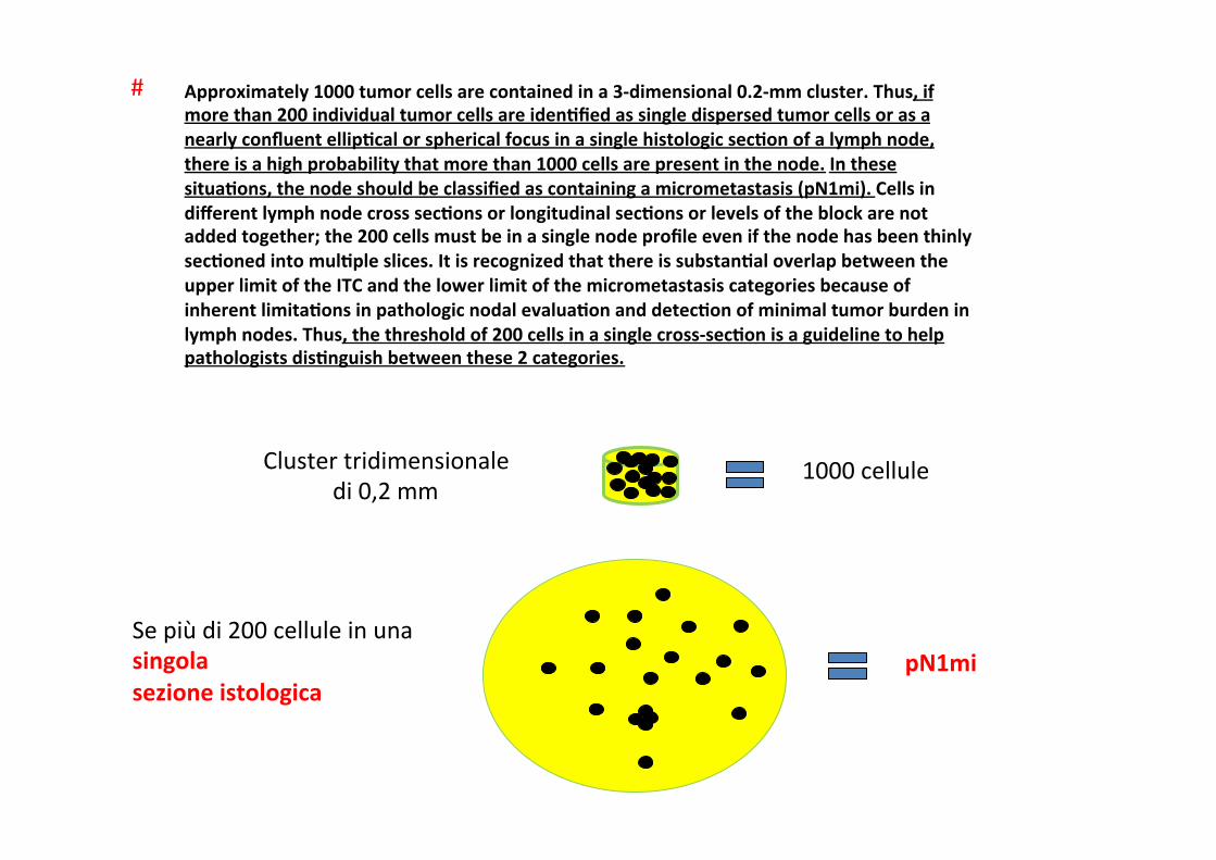
Approximately 1000 tumor cells are contained in a 3-‐dimensional 0.2-‐mm cluster. Thus, if more than 200 individual tumor cells are idenFfied as single dispersed tumor cells or as a nearly confluent ellipFcal or spherical focus in a single histologic secFon of a lymph node, there is a high probability that more than 1000 cells are present in the node. In these situaFons, the node should be classified as containing a micrometastasis (pN1mi). Cells in different lymph node cross secFons or longitudinal secFons or levels of the block are not added together; the 200 cells must be in a single node profile even if the node has been thinly secFoned into mulFple slices. It is recognized that there is substanFal overlap between the upper limit of the ITC and the lower limit of the micrometastasis categories because of inherent limitaFons in pathologic nodal evaluaFon and detecFon of minimal tumor burden in lymph nodes. Thus, the threshold of 200 cells in a single cross-‐secFon is a guideline to help pathologists disFnguish between these 2 categories.
#
Cluster tridimensionale di 0,2 mm
1000 cellule
Se più di 200 cellule in una singola sezione istologica
pN1mi

Noi crediamo che le differen? applicazioni dei protocolli su LS possano creare differenze anche nei risulta? degli studi sulla prediLvità dello
stato ascellare
I FALSI NEGATIVI SI RECUPERANO MA…..

SHAPE Gene
s
MORFOLOGIA BIOLOGIA MOLECOLARE

J Clin Oncol. 1998 Aug;16(8):2632-‐40.
Ann Surg. 2008 Jan;247(1):136-‐42.
Biologia molecolare: PCR
Perché non si è divulgata?
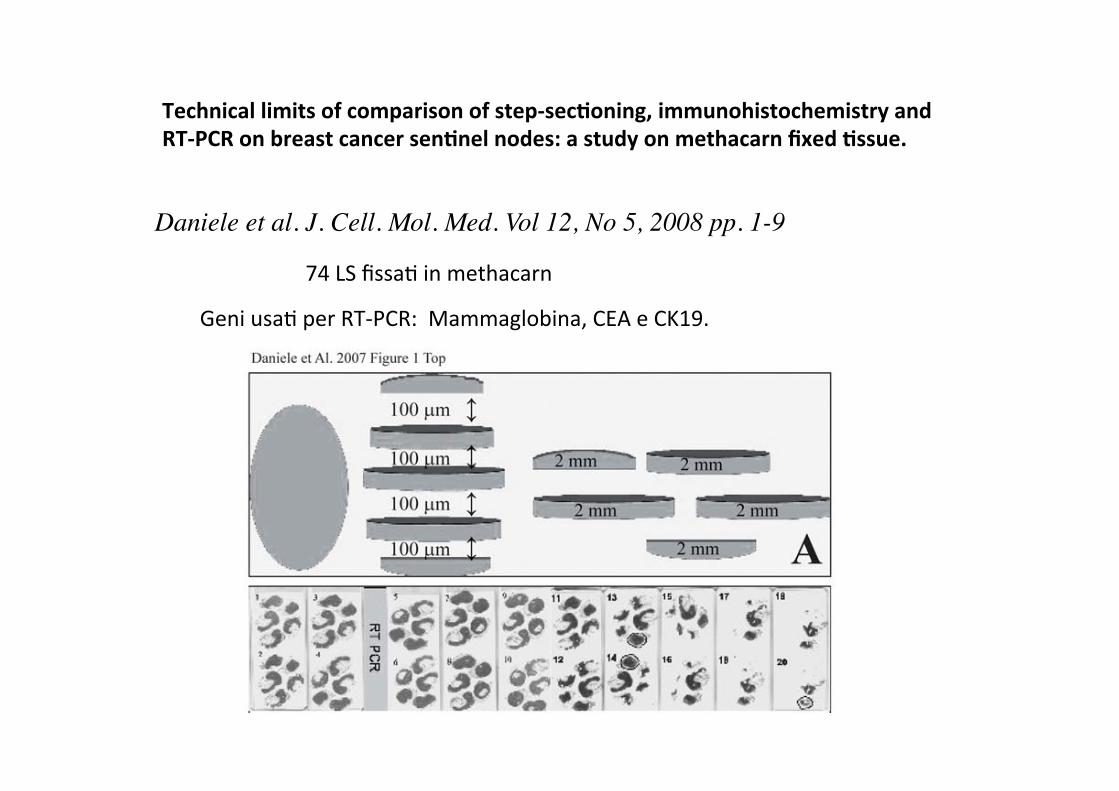
Daniele et al. J. Cell. Mol. Med. Vol 12, No 5, 2008 pp. 1-9
74 LS fissa? in methacarn
Technical limits of comparison of step-‐secFoning, immunohistochemistry and RT-‐PCR on breast cancer senFnel nodes: a study on methacarn fixed Fssue.
Geni usa? per RT-‐PCR: Mammaglobina, CEA e CK19.
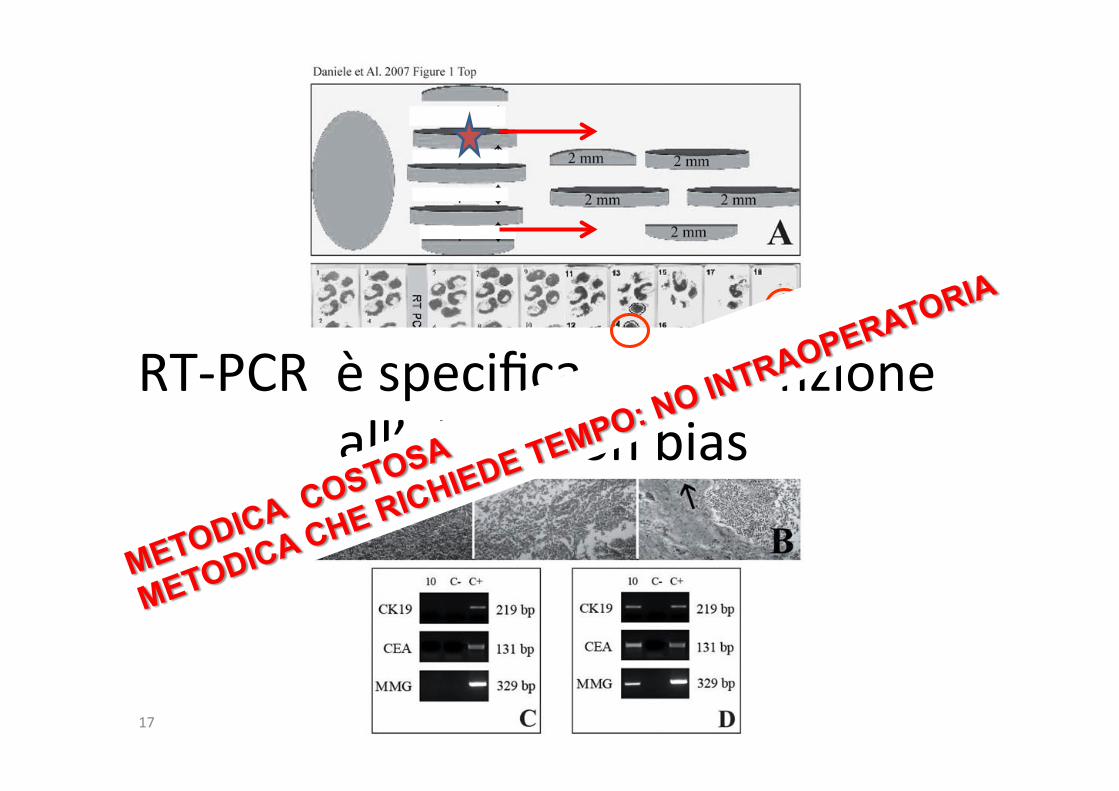
17
RT-‐PCR è specifica ma aXenzione all’alloca?on bias

OSNA A TORINO
Febbraio 2009
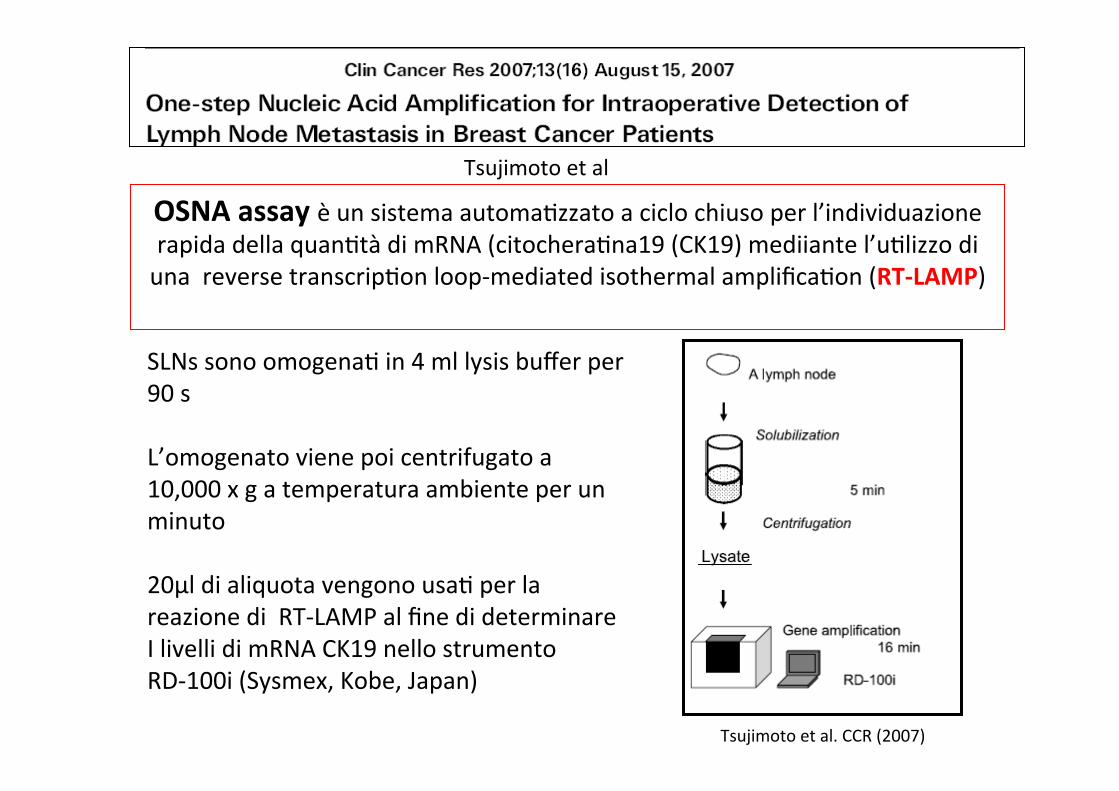
OSNA assay è un sistema automa?zzato a ciclo chiuso per l’individuazione rapida della quan?tà di mRNA (citochera?na19 (CK19) mediiante l’u?lizzo di una reverse transcrip?on loop-‐mediated isothermal amplifica?on (RT-‐LAMP)
Tsujimoto et al
Tsujimoto et al. CCR (2007)
SLNs sono omogena? in 4 ml lysis buffer per 90 s L’omogenato viene poi centrifugato a 10,000 x g a temperatura ambiente per un minuto 20μl di aliquota vengono usa? per la reazione di RT-‐LAMP al fine di determinare I livelli di mRNA CK19 nello strumento RD-‐100i (Sysmex, Kobe, Japan)

DetecFon of pyrophospate
DeterminaFon of RNA amount
DeterminaFon of Rise Time
Magnesium pyrophosphate
RT-‐LAMP REACTION
20
I risulta? sono espressi in numero di copie di mRNA di CK19/ μl Il carico metastas?co viene aXribuito in base a cut off prestabili?
Size of metastasis CK19 mRNA
Macro-Metastasis ++ >5.000 copies mRNA/µL – CK19(1.0 x 10⁸)
Micro-Metastasis + 250 - 5000 copies mRNA/µL – CK19 (5.0 x 10⁶)
ITC / background < 250 copies mRNA/µL – CK19 (5.0 x 10⁶)
Tsujimoto et al. CCR (2007)
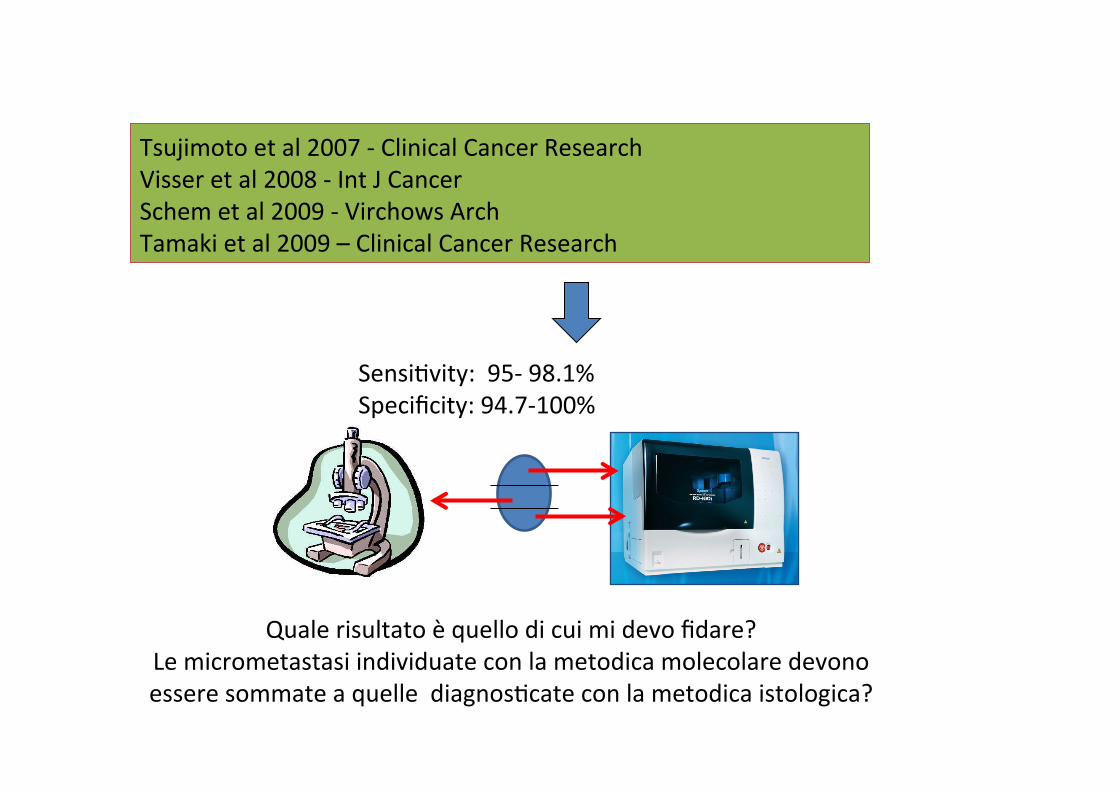
Tsujimoto et al 2007 -‐ Clinical Cancer Research Visser et al 2008 -‐ Int J Cancer Schem et al 2009 -‐ Virchows Arch Tamaki et al 2009 – Clinical Cancer Research
Sensi?vity: 95-‐ 98.1% Specificity: 94.7-‐100%
Quale risultato è quello di cui mi devo fidare? Le micrometastasi individuate con la metodica molecolare devono essere sommate a quelle diagnos?cate con la metodica istologica?

1) LS viene sezionato tradizionalmente
s1
2) Citologico su IMPRINT (due strisci)
Ematossilina-‐Eosina
Immunoistochimica rapida AE1-‐AE3
2 mm
3) OSNA (LS intero)
Tumori CK 19 + all’esame pre-‐operatorio
s2
METODO UTILIZZATO E PROPOSTO AL NOSTRO COMITATO ETICO INTERAZIENDALE
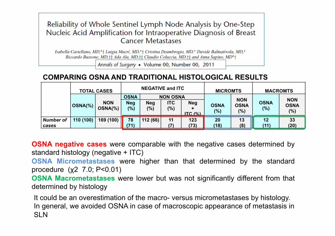
OSNA negative cases were comparable with the negative cases determined by standard histology (negative + ITC) OSNA Micrometastases were higher than that determined by the standard procedure (χ2 7.0; P<0.01) OSNA Macrometastases were lower but was not significantly different from that determined by histology
COMPARING OSNA AND TRADITIONAL HISTOLOGICAL RESULTS
TOTAL CASES NEGATIVE and ITC MICROMTS
MACROMTS
OSNA(%) NON OSNA(%)
OSNA NON OSNA OSNA
(%)
NON OSNA
(%) OSNA
(%)
NON OSNA
(%) Neg (%)
Neg (%)
ITC (%)
Neg +
ITC (%) Number of cases
110 (100) 169 (100) 78 (71)
112 (66) 11 (7)
123 (73)
20 (18)
13 (8)
12 (11)
33 (20)
It could be an overestimation of the macro- versus micrometastases by histology. In general, we avoided OSNA in case of macroscopic appearance of metastasis in SLN
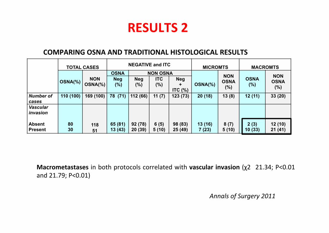
Macrometastases in both protocols correlated with vascular invasion (χ2 21.34; P<0.01 and 21.79; P<0.01)
TOTAL CASES NEGATIVE and ITC
MICROMTS
MACROMTS
OSNA(%) NON OSNA(%)
OSNA NON OSNA
OSNA(%)
NON OSNA
(%) OSNA
(%)
NON OSNA
(%) Neg (%)
Neg (%)
ITC (%)
Neg +
ITC (%) Number of cases
110 (100) 169 (100) 78 (71) 112 (66) 11 (7) 123 (73) 20 (18) 13 (8) 12 (11) 33 (20)
Vascular invasion Absent Present
80 30
118 51
65 (81) 13 (43)
92 (78) 20 (39)
6 (5) 5 (10)
98 (83) 25 (49)
13 (16) 7 (23)
8 (7) 5 (10)
2 (3) 10 (33)
12 (10) 21 (41)
Annals of Surgery 2011
RESULTS 2
COMPARING OSNA AND TRADITIONAL HISTOLOGICAL RESULTS
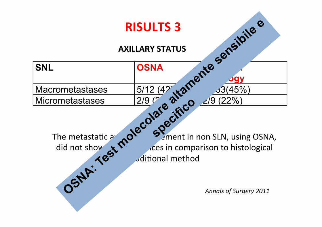
SNL OSNA Standard Histology
Macrometastases 5/12 (42%) 15/33(45%) Micrometastases 2/9 (22%) 2/9 (22%)
The metasta?c axillary involvement in non SLN, using OSNA, did not show any differences in comparison to histological
tradi?onal method
AXILLARY STATUS
Annals of Surgery 2011
RISULTS 3

OSNA sites in Italy
1. Roma -‐ IFO Regina Elena 2. Erba (Co) -‐ Osp. Fatebenefratelli 3. Sanremo -‐ Ospedale Civile 4. Milano Ospedale Luigi Sacco 5. TORINO -‐ A.O. MolineXe 6. Conegliano V.to -‐ A.O de’ Gironcoli 7. Novara -‐A.O. Maggiore della carità – 8. Aviano -‐ CRO 9. Roma -‐S. Filippo Neri 10. Napoli -‐ Pascale 11. Brescia -‐ Clinica Sant’Anna 12. Rozzano (MI) -‐ Humanitas 13. Bergamo -‐Gavazzeni Humanitas 14. Udine – Università 15. Roma -‐Osp. Fatebenefratelli 16. Prato – Ospedale Misericordia 17. Bergamo – Ospedali Riuni? 18. Milano – Clinica Pio X 19. Cosenza – S. Annunziata 20. Bari – Policlinico 21. Como -‐ Valduce 22. San Giovanni rotondo -‐ Casa Sollievo della Sofferenza 23. Catania – Cannizzaro (not in rou?ne yet – demo) 24. Andria -‐_BAT 25. Bari -‐ S. Paolo 26. Foggia – Ospedale Riuni? 27. Perugia-‐ Ospedale S. Maria della Misericordia 28. Alba-‐ Ospedale S. Lazzaro 29. Pisa -‐ AUO Pisana 30. Palermo – ARNAS civico
2008 2009 2009 2009 2009 2010 2010 2010 2010 2011 2011 2011 2011 2011 2012 2012 2012 2012 2012 2012 2012 2012 2012 2012 2012 2012 2012 2012 2012 2013
33 sites in rou?ne use (March 2013) 18 new sites in the last year

0
2
4
6
8
10
12
14
1 2 3 4 5 6 7
Serie1
2007 2008 2009 2010 2011 2012 2013
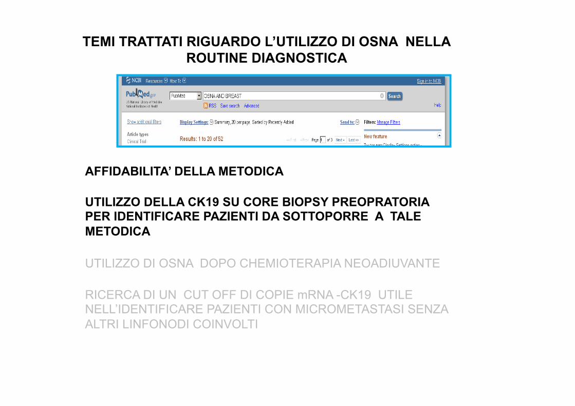
TEMI TRATTATI RIGUARDO L’UTILIZZO DI OSNA NELLA ROUTINE DIAGNOSTICA
AFFIDABILITA’ DELLA METODICA
UTILIZZO DELLA CK19 SU CORE BIOPSY PREOPRATORIA PER IDENTIFICARE PAZIENTI DA SOTTOPORRE A TALE METODICA
UTILIZZO DI OSNA DOPO CHEMIOTERAPIA NEOADIUVANTE
RICERCA DI UN CUT OFF DI COPIE mRNA -CK19 UTILE NELL’IDENTIFICARE PAZIENTI CON MICROMETASTASI SENZA ALTRI LINFONODI COINVOLTI

Tumori nega?vi alla CK19: 1-‐2% Histopathology 2002, 40:403–39
La mancata espressione di CK19 in IHC non necessariamente coincide con la presenza dell’mRNA del gene stesso
Virchows Arch. 2013 Jul;463(1):7-15.
I Tumori nega?vi alla CK19 hanno di solito un immunofeno?po basale. Tale immunofeno?po metasta?zza ai linfonodi meno frequentemente dell’immunofeno?po luminale


TEMI TRATTATI RIGUARDO L’UTILIZZO DI OSNA NELLA ROUTINE DIAGNOSTICA
AFFIDABILITA’ DELLA METODICA
UTILIZZO DELLA CK19 SU CORE BIOPSY PREOPRATORIA PER IDENTIFICARE PAZIENTI DA SOTTOPORRE A TALE METODICA
UTILIZZO DI OSNA DOPO CHEMIOTERAPIA NEOADIUVANTE
RICERCA DI UN CUT OFF DI COPIE mRNA -CK19 UTILE NELL’IDENTIFICARE PAZIENTI CON MICROMETASTASI SENZA ALTRI LINFONODI COINVOLTI

Conclusions: Intraoperative SLNB using OSNA in women with clinically negative axillary lymph nodes at initial presentation who received NAC could predict axillary status with high accuracy. Also it allows us to take decisions about the indication or not to perform an axillary dissection at the moment, thus avoiding delay in the administration of chemotherapy and benefiting the patients from a single surgical procedure
J. Navarro-Cecilia et al. / EJSO 39 (2013) 873e879
Conclusion: The OSNA assay can detect the residual tumour burden as accurately as conventional pathology, although chemotherapy-induced histological changes are present.
British Journal of Cancer (2013) 109, 1693–1698

TEMI TRATTATI RIGUARDO L’UTILIZZO DI OSNA NELLA ROUTINE DIAGNOSTICA
AFFIDABILITA’ DELLA METODICA
UTILIZZO DELLA CK19 SU CORE BIOPSY PREOPRATORIA PER IDENTIFICARE PAZIENTI DA SOTTOPORRE A TALE METODICA
UTILIZZO DI OSNA DOPO CHEMIOTERAPIA NEOADIUVANTE
RICERCA DI UN CUT OFF DI COPIE mRNA -CK19 UTILE NELL’IDENTIFICARE PAZIENTI CON LS+ SENZA ALTRI LINFONODI ASCELLARI COINVOLTI


PERCHE’ L’ANALISI MOLECOLARE DEL LS QUALI SONO GLI SVANTAGGI LA NOSTRA SOLUZIONE MOMENTANEA

pN0 (mol-): No regional lymph node metastases histologically, negative molecular findings (reverse transcriptase polymerase chain reaction [RT-PCR]) pN0 (mol+): Positive molecular findings (RT-PCR), but no regional lymph node metastases detected by histology or IHC
Costo Addestramento specifico del personale Dati di follow-up Codifica del TNM (pN0(mol+))
SVANTAGGI:
QUANDO PENSAVAMO DI AVERE TUTTE LE RISPOSTE….
CI HANNO CAMBIATO TUTTE LE DOMANDE!
NUOVE TENDENZE
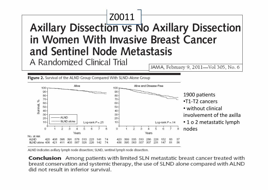
1900 pa?ents • T1-‐T2 cancers • without clinical involvement of the axilla • 1 o 2 metasta?c lymph nodes
Z0011

Lancet Oncol. 2013 Apr;14(4):297-‐305.


WHEN NOT TO DO ALND In patients with one or two positive sentinel nodes following breast-conserving surgery when whole breast radiation therapy is planned. WHEN WE DO NOT KNOWN (The Panel was equally divided) In patients undergoing mastectomy followed by radiotherapy WHEN TO DO ALND If no radiotherapy was planned. In patients with three or more involved sentinel nodes or with nodes that were clinically involved before surgery and confirmed by biopsy.

IL PARADOSSO ITALIANO
33 sites in rouFne use 18 new sites in the last year
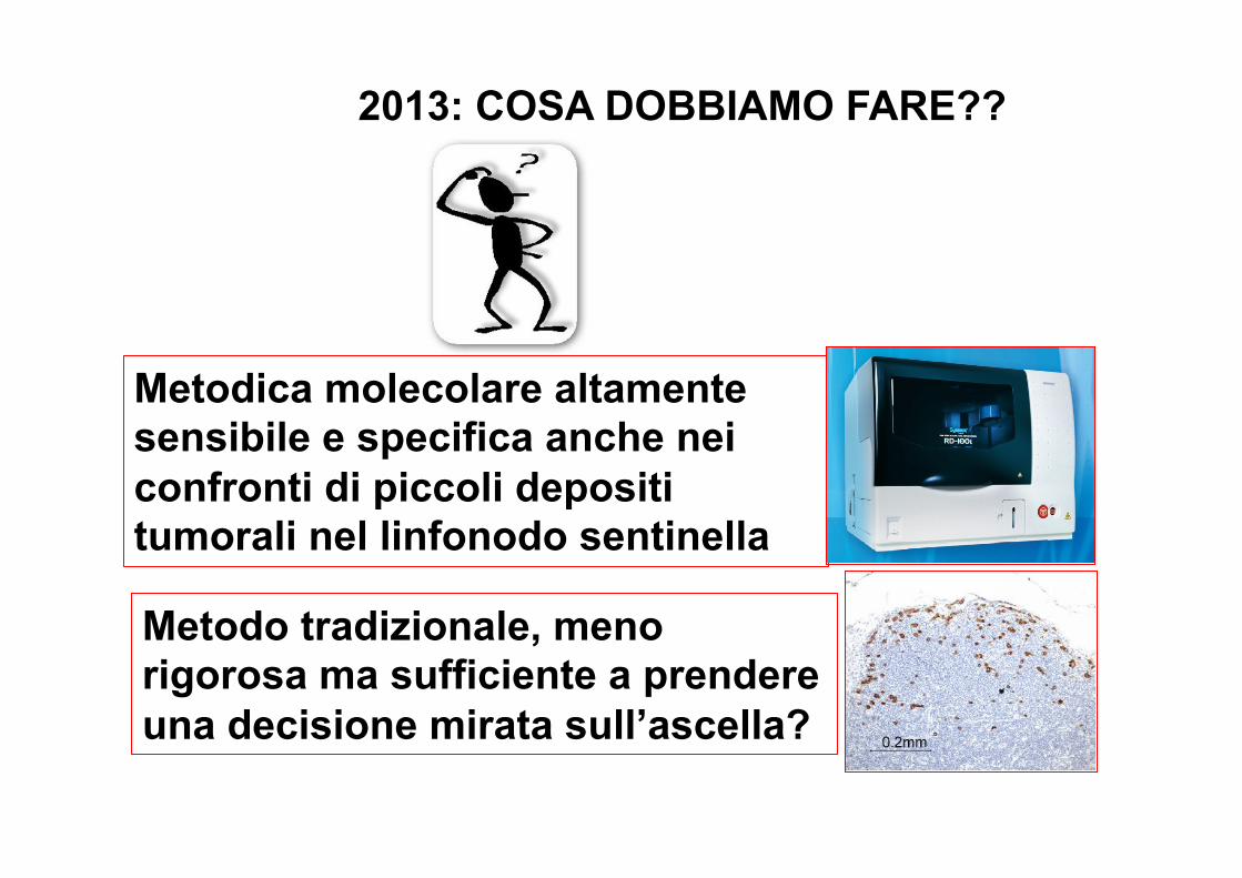
2013: COSA DOBBIAMO FARE??
Metodica molecolare altamente sensibile e specifica anche nei confronti di piccoli depositi tumorali nel linfonodo sentinella
Metodo tradizionale, meno rigorosa ma sufficiente a prendere una decisione mirata sull’ascella?
PERCHE’ L’ANALISI MOLECOLARE DEL LS QUALI SONO GLI SVANTAGGI LA NOSTRA SOLUZIONE MOMENTANEA
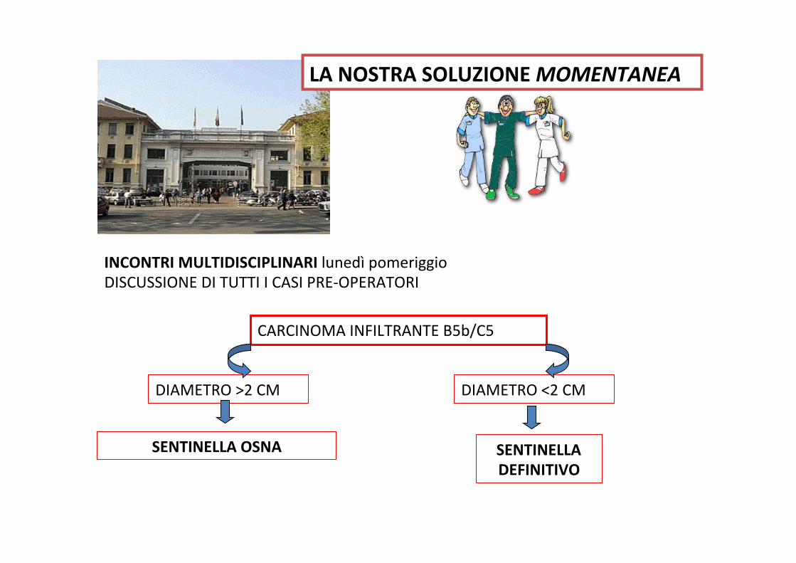
LA NOSTRA SOLUZIONE MOMENTANEA
INCONTRI MULTIDISCIPLINARI lunedì pomeriggio DISCUSSIONE DI TUTTI I CASI PRE-‐OPERATORI
CARCINOMA INFILTRANTE B5b/C5
DIAMETRO >2 CM DIAMETRO <2 CM
SENTINELLA OSNA SENTINELLA DEFINITIVO

G. Cserni et al.Surgical Oncology 21 (2012) 59-‐65
Il diametro del tumore primiFvo è streLamente correlato alla metastaFzzazione ed è uFlizzato in tuo i nomogrammi che predicono il rischio di ulteriori linfonodi metastaFci nel cavo
ascellare
Perche’ il diametro?

LS negaFvo LS micro LS macro Totale
>20 mm 31 13 16 60
<20 mm 53 6 9 68
DA MARZO 2013 A OGGI
I NOSTRI DATI

CONCLUSIONI
La storia e le scelte storiche vanno sempre contestualizzate..
LE MODERNE METODICHE MOLECOLARI SICURAMENTE RAPPRESENTANO UN TENTATIVO
DI UNIFORMARE E STANDARDIZZARE LA DIAGNOSTICA DEL LS….ALMENO FINO A
QUANDO QUESTA METODICA NON TRAMONTERA’ DEL TUTTO…
La prognosi del tumore alla mammella dipende maggiormente dalla biologia del tumore che dal coinvolgimento linfonodale.
METASTASI LINFONODALE = EPIFENOMENO J Clin Oncol 2010 28:3271–3277, J Clin Oncol 2010 28:1684–1691,
Breast Cancer Res Treat 2009 117:199–204, Semin Radiat Oncol 2009 19:204–210

GRAZIE A TUTTI PER L’ATTENZIONE