Dottorato internazionale in Scienze Oncologiche XXVI Ciclo ...
34
UNIVERSITA’ DEGLI STUDI DI CATANIA Dottorato internazionale in Scienze Oncologiche XXVI Ciclo Anno Accademico 2012-2013 Dott.ssa Cinzia Quattrocchi Nitric oxide donating non steroidal anti-inflammatory drugs (NO-NSAIDs) for the treatment of cancer. Coordinatore: Chiar.mo Prof. Ferdinando Nicoletti Tutor: Chiar.mo Prof. Ferdinando Nicoletti
Transcript of Dottorato internazionale in Scienze Oncologiche XXVI Ciclo ...
UNIVERSITA’ DEGLI STUDI DI CATANIA
Dottorato internazionale in Scienze Oncologiche XXVI Ciclo
Anno Accademico 2012-2013
Dott.ssa Cinzia Quattrocchi
Nitric oxide donating non steroidal anti-inflammatory drugs (NO-NSAIDs)
for the treatment of cancer.
Coordinatore: Chiar.mo Prof. Ferdinando Nicoletti
Tutor: Chiar.mo Prof. Ferdinando Nicoletti
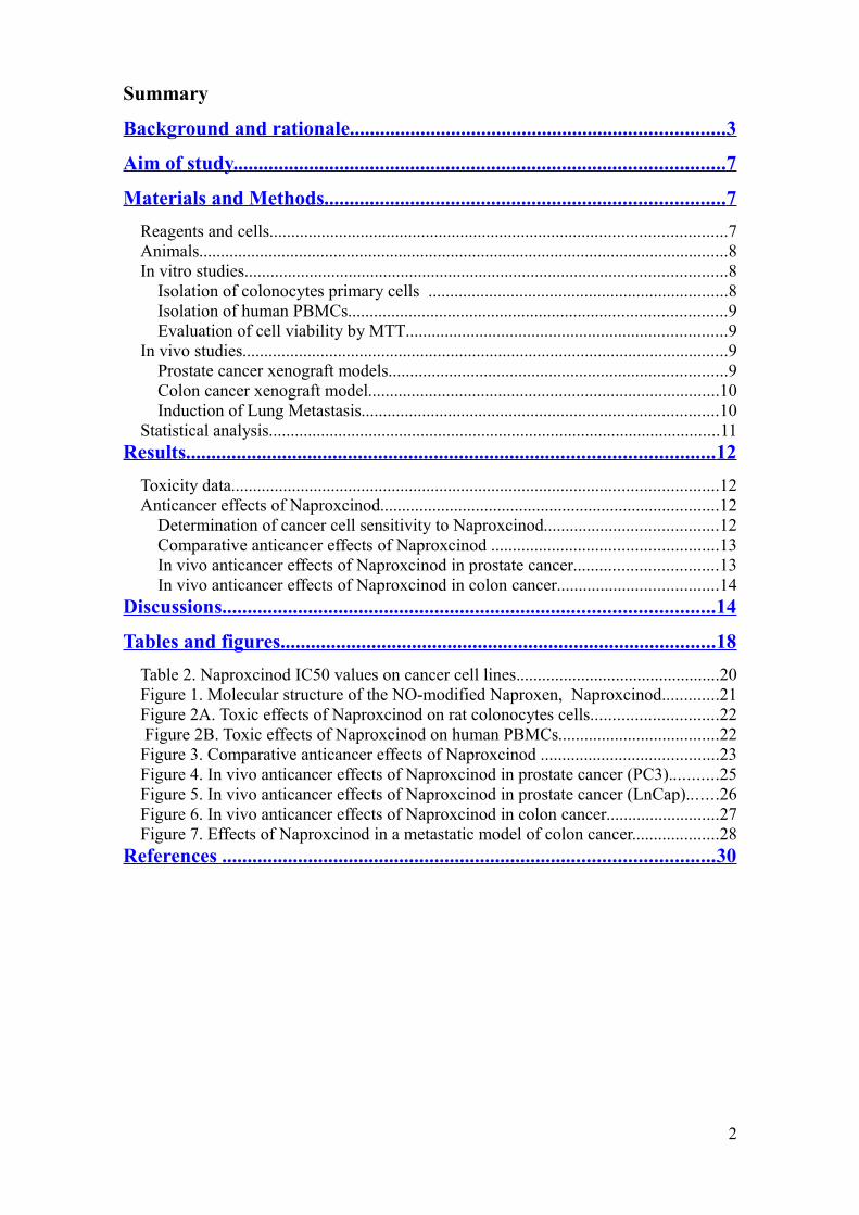
Summary
Background and rationale .......................................................................... 3
Aim of study. ................................................................................................ 7
Materials and Methods ............................................................................... 7 Reagents and cells.........................................................................................................7Animals..........................................................................................................................8In vitro studies...............................................................................................................8
Isolation of colonocytes primary cells .....................................................................8Isolation of human PBMCs.......................................................................................9Evaluation of cell viability by MTT..........................................................................9
In vivo studies................................................................................................................9Prostate cancer xenograft models..............................................................................9Colon cancer xenograft model.................................................................................10Induction of Lung Metastasis..................................................................................10
Statistical analysis........................................................................................................11Results ........................................................................................................ 12
Toxicity data................................................................................................................12Anticancer effects of Naproxcinod..............................................................................12
Determination of cancer cell sensitivity to Naproxcinod........................................12Comparative anticancer effects of Naproxcinod ....................................................13In vivo anticancer effects of Naproxcinod in prostate cancer.................................13In vivo anticancer effects of Naproxcinod in colon cancer.....................................14
Discussions ................................................................................................. 14
Tables and figures. ..................................................................................... 18 Table 2. Naproxcinod IC50 values on cancer cell lines...............................................20Figure 1. Molecular structure of the NO-modified Naproxen, Naproxcinod.............21Figure 2A. Toxic effects of Naproxcinod on rat colonocytes cells.............................22 Figure 2B. Toxic effects of Naproxcinod on human PBMCs.....................................22Figure 3. Comparative anticancer effects of Naproxcinod .........................................23Figure 4. In vivo anticancer effects of Naproxcinod in prostate cancer (PC3)...........25Figure 5. In vivo anticancer effects of Naproxcinod in prostate cancer (LnCap).......26Figure 6. In vivo anticancer effects of Naproxcinod in colon cancer..........................27Figure 7. Effects of Naproxcinod in a metastatic model of colon cancer....................28
References ................................................................................................. 30
2
Background and rationale
Treatment of pain, inflammation, and fever most frequently implies non-steroidal anti-
inflammatory drugs (NSAIDs) administration. Beside their primary role in treatment of
inflammation, evidence clearly shows a chemopreventive effect for aspirin and other
non-steroidal anti-inflammatory drugs on colorectal and gastric cancer and probably
other cancer types [Cuzick et al. 2009; Puntoni et al. 2008; Dube et al. 2007]. How
ever, although selective cyclooxigenase-2 (COX-2) inhibitors are now given to patients
at high risk of colorectal cancer, data on the risk-benefit profile for cancer prevention
are insufficient and no definitive recommendations can be made regarding the lowest ef
fective dose, the age at which to initiate therapy, the optimum treatment duration, and
the subpopulations for which the benefits of chemoprevention outweigh the risks of ad
verse side-effects [Cuzick et al. 2009].
Studies on the mechanisms by which NSAIDs might inhibit carcinogenesis have not
provided conclusive evidence for pathways or molecular targets that are clinically most
relevant [Cuzick, et al. 2009] but several NSAIDs properties have been proposed to
play important roles in cancerogenesis prevention: stimulation of apoptosis, cell growth
suppression, inhibition of angiogenesis, and metastasis prevention [Chan et al. 2002;
Chan et al. 1998]. Furthermore, overexpression of COX-2 has been reported in tumor
cells and tissues [Scartozzi et al. 2004; Lim et al. 2000]. The inhibition of COX by
NSAIDs was previously thought to be the unique explanation for their antitumor effect
[Hanif et al. 1996], but more recently, other COX-independent mechanisms have been
identified [Sun et al. 2009; Yin et al. 1998; Grilli et al. 1996]. It has been recently
demonstrated that aspirin and several other NSAIDs could promote apoptosis through
the inhibition of NF-Kb activity, activation of mitochondrial pathways by cytochrome c
release and activation of caspase-9 and extrinsic pathways by activation of caspase-8,
induction of oxidative stress and inhibition of proteasome functions [Jana 2008].
Unfortunately, serious undesirable effects limit the application of those drugs. The most
common adverse NSAID therapy-related events are the development of ulcers and sub
sequent bleeding in the upper gastrointestinal tract and different renal side effects, such
as acute renal failure, acute interstitial nephritis, worsening of chronic kidney disease,
salt and water retention and hypertension, leading to increased cardiovascular risk [Wal
lace JL et al. 2008; House et al 2007; Rigas and Kashfi 2004]. Moreover, selective
COX-2 inhibitors have a reduced risk of gastrointestinal bleeding, alongside with a sim
3
ilar renal toxicity profile and most likely a worsened cardiovascular toxicity [Whelton
2002].
Chemical modifications of these drugs based on the covalent attachment of a nitric ox
ide (NO) releasing moiety –ONO2, often via a spacer molecule, has been proposed to
overcome the most common NSAID-associated adverse events [Lanas 2008]. This ap
proach was supported by an idea that NO shares similar properties with prostaglandins
(PGs) as regards the capacity of PGs to influence local blood flow [Rigas and Kashfi
2004]. It is indeed hypothesized that the NO molecules bound to the drug through the
spacer molecule might be delivered to the damaged site, thereby decreasing gastric and
renal toxicities induced by diminished PG levels [Lanas 2008; Rigas and Kashfi 2004].
In a phase 2, double-blind, randomized, parallel group study in patients with osteoarth
ritis, the novel NO-NSAID 4-nitrooxybutyl (2S)-2-(6-methoxynaphthalen-2-yl)pro
panoate (Naproxcinod), a NO modified derivative of naproxen, appeared safer than
COX-2 inhibitor positive control rofecoxib [Karlsson et al. 2009]. Up to date, naprox
cinod completed the pivotal phase III studies needed for a New Drug Application, that
has been submitted to the Food and Drug Administration in September 2009 and accep
ted for filing, seeking approval for the treatment of the signs and symptoms of os
teoarthritis [NicOx Press Release 18 October 2009].
Independent studies on NO-donating NSAIDs, alternatively known as cyclooxygenase
inhibiting nitric oxide donators (CINODs), have consistently demonstrated that these
compounds bear up to several thousands-fold augmented antitumoral potentials both in
vitro and in vivo when compared to the parental compounds [Rigas and Williams 2008;
Kashfi and Rigas 2007; Huguenin et al. 2005; Rigas and Kashfi 2004].
However, in oncology, the actions of NO are highly variable as it showed to exert both
anti- and pro-neoplastic activity [ Huerta, Int J Oncol 2008]. Reflecting the duality of
NO function in cancer, both anti-NO and NO-based anticancer strategies appear ef
fective in several preclinical models [ Mocellin et al. 2007].
Most likely, the final activity of NO in tumors is dependent on its working microenvir
onment, including the type of cell exposed to the compound, the redox state of the reac
tion, as well as the final intracellular concentration and the duration of intracellular ex
posure to nitric oxide [Huerta, Int J Oncol 2008].
Current interpretations of the data suggest a dose dependent relationship between NO
concentration and tumor response, and it is generally accepted that at high concentra4
tions of NO may have an anti-neoplastic function by exposing cells to high levels of ni
trosative stress whereas at low levels it can stimulate angiogenesis, cancer cell prolifera
tion and metastatic potential [Chinje and Stratford 1997]. There is, however, no unifying
mechanistic explanation for the biphasic role of nitric oxide in cancer. When released
under appropriate conditions, NO possesses multiple antineoplastic properties both in
vitro and in vivo including inhibition of cellular proliferation by cell cycle arrest induc
tion [Kroncke et al. 1998], stimulation of autophagic cell death [Maksimovic-Ivanic et
al. 2008] or apoptotic cell death through different mechanisms like p53 upregulation or
activation, [Forrester et al 1996], proteosomal degradation of anti-apoptotic mediators
[Glockzin et al. 1999], induction of Smac release [Li et al. 2004] increase in mitochon
drial permeability and consequent cytochrome c release [Boyd and Cadenas, 2002], reg
ulation of angiogenesis by modulation of several kinases like PKC, ERK and AP-1
[Jones et al. 2004], protection against metastatis formation through enhancement of
Raf-1 Kinase Inhibitor Protein expression [Bonavida et al. 2008] or regulation of matrix
metalloproteinase levels [Phillips and Birnby, 2004].
In addition, NO has a well characterized chemo-, radio-and immuno- sensitizing poten
tial [Bonavida et al. 2006], that has been attributed respectively to nitrosation of critic
als thiols in DNA repair enzymes such as alkyltransferase [Laval et al. 1994], to a mim
icry of the effects of oxygen on fixation of radiation-induced DNA damage [De Ridder
et al. 2008] and to inhibition of the multifactorial transcription repressor Yin Yang 1
[Vega et al. 2005].
Moreover, classical nitric oxide donors have been shown beneficial effects also in hu
mans since low dose glyceryl trinitrate treatment significantly delayed the PSA doubling
time in prostate cancer patients after surgery and radiotherapy [Siemens et al.
2009].Thereby, NO-NSAIDs represent an emerging class of compounds with chemo
preventive, chemotherapeutic chemio-, radio- and immuno-sensitizing properties
against a variety of cancers, demonstrated in preclinical models including cell culture
systems and animal tumor models of different origin [Rigas and Williams 2008].
Their mechanism of action appears complex and involves the generation of reactive
oxygen species [Sun Y et al. 2009], suppression of microsatellite instability in mismatch
repair-deficient cells [McIlhatton et al. 2007] alongside the modulation of several sig
naling cascades including nuclear factor kappa B [Williams et al. 2008], Wnt [ Lu et al.
2009] and mitogen activated protein kinases [Hundley et al. 2006] that culminate in in
hibited cell renewal and enhanced apoptosis [Rigas and Williams 2008; Rigas 2007].
5
Remarkably, these effects seemed to be COX-independent [Rigas and Williams 2008].
NO-ASA (NO-aspirin) is the best-studied compound belonging to this group, but sever
al other NO-NSAIDs, including NO-sulindac, NO-ibuprofen, NO-indomethacin, NO-
flurbiprofen, NO-naproxen have been recently synthesized [Sun et al. 2009; Rigas and
Williams 2008].
It has been questioned whether the anticancer effect of the NO-SAIDs is directly de
pendent on the NO release or it may be, at least for the NO-ASA, dependent to the
spacer molecule exerting its own farmacological effects [ Rigas and Williams 2008;
Kashfi and Rigas 2007; Hulsman et al. 2007]. Currently, other NO-donating anti-in
flammatory drugs with the NO-donating group covalently attached to the parental com
pound that possesses strong anticancer activity have been synthesized by our group of
research [Maksimovic-Ivanic et al. 2009; Maksimovic-Ivanic et al. 2008]. Alternatively,
it has been hypotesized that the effects of NO-NSAIDs may depend on multiple mech
anisms somehow arising from a simple NO-release, that can be rather achieved with a
classical NO donor. From this point of view, NO-release is not required but contributes
to the anticancer effect [ Rigas and Williams 2008].
Given the paradoxical effects of NO against cancer, long term therapy with NO-
NSAIDs may actually promote cancer growth by releasing NO. However, critical ana
lysis of the results of the Framingham Heart and Offspring Study for evaluating the ef
fects of nitro-vasodilators on the risk of colorectal cancer show that there was no in
crease in colorectal cancer over a sufficiently long period of observation, suggesting
that unlikely chronic therapy with NO donors may lead to cancer [Muscat et al. 2005].
Up to date, only one phase I clinical trial with NO-ASA for the prevention of colon can
cer has been started but was unfortunately recently terminated prematurely due to con
cerns regarding the potential genotoxicity of one putative metabolite, not directly correl
ated with the –ONO2 group [NicOx Press Release 18 June 2007].
6
Aim of study.
Nitric oxide donating non steroidal anti-inflammatory drugs (NO-NSAIDs) represent
an emerging class of compounds with chemopreventive, chemotherapeutic chemio-, ra
dio- and immunosensitizing properties against a variety of cancers, demonstrated in pre
clinical models including cell culture systems and animal tumor models of different ori
gin. These compounds consist of a conventional NSAID to which an NO-releasing moi
ety –ONO2 has been covalently attached.
The aim of this study will be to evaluate the anticancer potential of the novel NO-
NSAID Naproxcinod, since it is the only NO-NSAID that, differently from other com
pounds belonging to the same class like Aspirin-NO, has so far demonstrated a clear
safe profile in humans and has not been extensively studied yet as a potential novel anti
cancer therapeutic.
The final objective of the study will be to provide solid basis for appropriately design
ing phase II clinical studies based on Naproxcinod administration.
Materials and Methods
Reagents and cells
Cancer cell lines (see Table 1 for a complete list) were available at the Department of
Bio-Medical Sciences, University of Catania or purchased from ATCC, LGC Standards
srl, Milan, Italy. The cells were grown in the appropriate culture media as indicated by
the protocols of American Type Cell Collection (ATCC). All culture media,
supplements, antibiotics and fetal bovine serum were purchased from Life Technologies
Italia. Cells were routinely maintained at 37°C in a humidified atmosphere with 5%
CO2. Cells were collected with 0.25% trypsin-1 mM EDTA solution in PBS, and seeded
at density of 1×10⁴/well in 96-well plates unless otherwise indicated.
Naproxcinod (Figure 1), Naproxen and Cisplatin were obtained from Sigma-Aldrich
(Milan, Italy). MTT (3-(4,5-Dimethyl-2-thiazolyl)-2,5-diphenyl-2H-tetrazolium
Bromide, Thiazole Blue) was purchased from Merck Chemicals Ltd. (Nottingham, UK).
7
Animals
Six- to 8-week-old male BALB/c mice, 4 to 5-week-old BALB/c male and female
athymic nude mice, and male Wistar rats 8-weeks old were purchased from Harlan
Laboratories (Udine, Italy).
The mice were kept under standard laboratory conditions (non specific pathogen free)
with free access to food and water. The animals used in the experiments were protected
in accordance with Directive 86/609/EEC. The animal studies were carried out in
accordance to local guidelines and will be approved by the local Institutional Animal
Care and Use Committee (IACUC).
In vitro studies
Isolation of colonocytes primary cells
Distal colon isolated from male Wistar rats was cut in 4 cm pieces and incubated for 5
minutes at 37°C in a 5% trypsin / 2% EDTA solution. Colon fractions were then
transferred in a Petri dishes with complete medium to block trypsin activity and cells
were detached by the mucosa with a scraper. The collected cellular suspension was
centrifuged, washed, counted and suspended in freezing medium (RPMI, 10%FCS, 10%
DMSO) at the concentration of 2x10⁶ cell/mL.
8
Isolation of human PBMCs
Human Peripheral Blood Mononuclear Cells (PBMCs) were isolated from fresh buffy
coats of healthy volunteers. The buffy coats were diluted with phosphate-buffered saline
(PBS) supplemented with 2.5 mM EDTA and layered onto Ficoll-Hypaque gradients
(Gibco, Invitrogen, Milan, Italy). After 30 min of centrifugation at 400g at room
temperature, mononuclear cells were collected, washed twice with PBS and incubated
in tissue culture multi-well plates.
Evaluation of cell viability by MTT
Cells were seeded in 96-well plates, incubated for 24-72 hrs in the presence of different
concentrations of Naproxcinod, Naproxen and Cisplatin and viability was estimated
using MTT assay as previously described [Mosmann, J.P. (1983) J. Immunol. Methods
65, 55-63]. The viability of treated cells was shown as percentage of value obtained for
untreated cultures that was arbitrarily set to 100%. The MTT assay involves the
conversion of the water soluble MTT to an insoluble formazan. The formazan is then
solubilized in 0.1 N HCl in isopropanol and the concentration determined by optical
density measured at 570 nm.
In vivo studies
Prostate cancer xenograft models
Tumours were induced in female or male Balb/c athymic nude mice by subcutaneous
injection of cultured PC3 (androgen-independent human prostate cancer) or LNCaP
(androgen-dependent human prostate cancer). Cells were dispersed by trypsin, washed
(twice) in serum-free medium RPMI-1640 (10 min centrifugation, 200 x g),
resuspended at the concentration of 2.5 x 10⁷ cells/ml in the same medium and injected
(0.2 ml) s.c. in the right flank of each mouse using a 0.6 mm needle.
Tumour growth was observed daily and measured with calipers (2 perpendicular
diameters), and tumour volume was calculated using the formula 0.52 x a x b2, where a
is the longest and b is the shortest diameter.
9
Three independent experiment were performed and each group consisted of 7-8 mice.
Treatment with Naproxen or Naproxcinod started when the tumors were already
palpable with a range volume of 60-70 mm3. The mice were randomly assigned to each
experimental group. Post randomization analysis revealed no significant differences in
tumor volumes at the beginning of the treatment among the different groups. Naproxen
or Naproxcinod were prepared immediately before treatment and they were
administered orally (per os) at a dose of 40 mg/kg for 20 consecutive days. A group of
mice was treated with the vehicle carboxymethylcellulose (CMC 1 % in water for
injection), and another group with cisplatin intraperitoneally (i.p.) at the dose of
1mg/kg twice a week as positive control. The animals were observed for further 16 days
after the interruption of the treatment.
Colon cancer xenograft model
Balb/c mice were inoculated subcutaneously with 2x10⁵ cells CT26CL25 (murine colon
adenocarcinoma) and have started to be palpable tumors 10-12 days after inoculation.
The animals were treated orally with Naproxcinod and Naproxen at a dose of 40 mg/kg
or vehicle. Mice were treated under a therapeutic regimen starting when the tumor
began to be palpable and continued for 2 consecutive weeks. The animals were treated
with cisplatin (i.p.) twice a week at a dose of 1 mg /kg as a positive control. Tumour
growth was observed daily and measured with callipers (2 perpendicular diameters),
and tumour volume was calculated using the formula 0.52 x a x b2, where a is the
longest and b is the shortest diameter.
Induction of Lung Metastasis
Tumors were induced in BALB/c mice by injection of cultured mouse colon cancer
CT26.CL25 cells. The cells were detached by trypsin, washed (twice) in serum-free
medium RPMI (10 min centrifugation, 200× g), resuspended at the concentration of 2 ×
105 cells/ml in the same medium and injected (0.2 ml) i.v. in the tail of each mouse.
The animals were treated orally with Naproxcinod and Naproxen at a dose of 40 mg/kg
or vehicle starting on day 3, when, from the literature, the micro-metastases are
beginning to be present in the lung and continued for 9-12 days. The animals were
treated with cisplatin (i.p.) twice a week at a dose of 3 mg /kg as a positive control. 10
On day 12-14 post tumor challenge, mouse lungs were removed and weighted on an
analytical scale.
Statistical analysis
The results are presented as mean ± SD of triplicate observations from one
representative of at least three experiments with similar results, unless indicated
otherwise. Student’s t-test was used to determine statistical significance. Values of p <
0.05 were considered to be statistically significant.
11

Results
Toxicity data
In order to assess the toxic effects of Naproxcinod and the parental compound Naproxen on primary
cells, rat colonocytes and human PBMCs were incubated for 24-72hrs in the presence of scalar
concentrations of these drugs, and viability measured as cellular respiration using mitochondria-
dependent reduction of 3-(4,5-dimethylthiazol-2-yl)-2,5-diphenyl-tetrazolium bromide (MTT) to
formazan. None of the compounds reached the LC50 at concentrations up to 200 M (Figure 2).
However, compared to Naproxen, Naproxcinod showed a safer profile with almost no toxic effects
on both cell lines. Viability of rat colonocytes at 72 hrs incubation with 200 M Naproxcinod and
Naproxen was 94% and 59%, respectively (Figure 2A). Viability of human PBMCs at 72hrs
incubation with 200 M Naproxcinod and Naproxen was 90% and 63%, respectively (Figure 2B).
Anticancer effects of Naproxcinod
Determination of cancer cell sensitivity to Naproxcinod
We first evaluated the in vitro antitumoral effect of Naproxcinod on a panel of 20 cancer cell lines.
To this aim, cells were incubated for 24-72 hrs with 7 log concentrations of Naproxcinod and of the
parental compound Naproxen, and viability determined by MTT assay. Cell lines included were
human or murine from prostate, colon, breast, SCLS, NSCLC, astrocytoma, melanoma, kidney and
leukemia cancers (see Table 1 for a complete list). Sensitivity of each cell line was determined and
results are shown in Table 2.
Naproxcinod resulted to be effective in reducing cell growth/proliferation in both murine and
human prostate cancer, colon cancer and astrocytoma cell lines (Table 2).
On the contrary, breast, lung, melanoma, kidney and leukemia cell lines showed to be not sensitive
to Naproxcinod, as the IC50 was higher than 200 M.
12
Comparative anticancer effects of Naproxcinod
After determining the cancer cell lines that were sensitive to Naproxcinod, we evaluated the
comparative effects between Naproxcinod, the parental compound Naproxen and the positive
control drug Cisplatin (Figure 3). To this aim, cells were treated for 24 h with a previously
determined range of concentrations and cell viability was estimated using MTT assay.
Parental compound Naproxen did not show significant anticancer effects in all of the cell lines
tested, since the IC50 was not reached at concentrations up to 160 M (Figure 3 A-F).
Cisplatin was effective in reducing cell growth/viability of all of the cell lines tested, showing an
IC50 lower than 20 M (Figure 3).
Naproxcinod and the positive control drug Cisplatin showed superimposable IC50 values in both
androgen-dependent and androgen-independent prostate cancer cell lines PC3 and LnCap (Figure
3A-B).
Overall, Naproxcinod was less potent than Cisplatin in reducing cell growth in all of the cell lines
tested, however no statistically significant differences was observed between the two drugs (Figure
3).
In vivo anticancer effects of Naproxcinod in prostate cancer
To confirm the data obtained in vitro, we tested the antitumoral effects of Naproxcinod in vivo in
mouse models of cancer. We first evaluated the efficacy of Naproxcinod on tumor growth in nude
mice xenografted with PC3 cells. Mice were treated with Naproxcinod, Naproxen, Cisplatin or
vehicle for 21 consecutive days starting from about 21 days after xenograft. The tumor volumes of
the mice treated with Naproxcinod were significantly reduced (p<0.05) already starting from 9 days
after the beginning of the treatment (day 30) until the end of the observational period. The parental
compound Naproxen started to show significant effects after 19 days of treatment but the effect was
lost around 7 days after the interruption of the treatment (day 50) (Figure 4).
We further tested the anticancer effects of Naproxcinod in the LnCap xenograft mouse model. Mice
were treated with test compounds for 21 consecutive days starting at day 22 after xenograft.
Relative to vehicle-treated mice, the volumes of the tumors of the mice treated with Naproxcinod
were significantly inhibited (p < 0.05) starting from 12 days after the beginning of the treatment
(day 34) until the end of the treatment. The same was observed for the positive control drug
Cisplatin. The parental compound Naproxen showed significant effects after 21 days of treatment
until the end of the study (Figure 5).
13
In vivo anticancer effects of Naproxcinod in colon cancer
Based on the results from the in vitro studies, we determined the antitumoral effects of Naproxcinod
in the colon cancer setting. To this aim we evaluated the efficacy of Naproxcinod on tumor growth
in balb/c mice xenografted with CT26CL25 cells. Naproxcinod treatment significantly inhibited cell
growth starting from day 6 post-inoculation. The inhibitory potential of novel modified drug was
greater than the parental drug, which reached the statistical significance at day 28 post-inoculation
(Figure 6). The effects of Cisplatin treatment was superimposable to that of the Naproxcinod
(Figure 6).
To evaluate the anti-metastatic potential of parental and NO-modified Naproxen, we examined their
effects in the lung metastasis model induced by i.v. injection of CT26CL25 cells. Cisplatin was
effective in reducing the weight of the lung (Figure 7B) and mice exhibited a better clinical
conditions, as observed from the significantly lower loss in body weight (Figure 7A).
Treatment of mice with Naproxcinod resulted in a decrease in lung weight, although the statistical
significance was not reached (Figure 7B). No protective effect was observed for the parental
compound Naproxen (Figure 7A-B).
Discussions
Nitric oxide-releasing non-steroidal anti-inflammatory drugs (NO-NSAIDs) are a class of NSAID
derivatives generated by adding a nitric oxide donating group to the parental NSAID. Several NO-
NSAIDs have recently been developed, including NO-ASA (NO-aspirin), the first in class
compound, NO-sulindac, NO-ibuprofen, NO-indomethacin, NO-flurbiprofen and NO-naproxen
[Sun et al. 2009; Rigas and Williams 2008].
NSAIDs have been extensively investigated in clinical trials for their
chemopreventive/chemotherapeutic effects in different types of tumors. A randomized trial
conducted in 135 patients with advanced stage cancer (colorectal, liver, pancreatic, and gastric
primary cancers) and an expected survival of more than 6 months, showed that the addition of
indomethacin prolonged mean survival with 8.7 months compared to placebo-treated patients
[Lundholm et al, 1994]. In a pilot study 12 patients, who had relapse of their prostate cancer
received celecoxib. Five patients had a decline in their absolute PSA level, three patients had
14
stabilization of the level and of the remaining four patients, three had a marked decrease in their
PSA doubling time [Lundholm et al. 1994]. Parallel to the anti-tumor activity of NSAIDs as single
agents, interest has raised in the effects of a combined therapy of chemotherapy with NSAIDs. A
retrospective study comparing capecitabine in combination with celecoxib compared to
capecitabine alone in colorectal cancer patients showed an increase in median time to tumor
progression (6 months versus 3 months, P = 0.002) and in the proportion of stable disease (62.5%
versus 22.8%, P = 0.001) [Lin et al. 2002].
However, conventional NSAIDs are known for their gastrointestinal side effects and nephrotoxicity.
About 15–30% of regular NSAID users have one or more ulcers when examined endoscopically,
and 3–4.5% of NSAID users have clinically significant upper gastrointestinal events, including
ulcers and ulcer complications [Laine et al. 2001; Wolfe et al.1999]. Different renal side effects,
such as acute renal failure, acute interstitial nephritis, worsening of chronic kidney disease, salt and
water retention and hypertension, leading to increased cardiovascular risk have also been described
[Wallace JL et al. 2008; House et al. 2007; Rigas and Kashfi 2004].
Chemical modifications of NSAIDs based on the covalent attachment of a nitric oxide (NO)
releasing moiety, has been proposed to overcome the most common NSAID-associated side effects
[Lanas 2008]. Beside their safer profiles in comparison to the parental drugs, several studies on NO-
donating NSAIDs have also demonstrated that these compounds exert up to several thousands-fold
increased anticancer effects both in vitro and in vivo [Rigas and Williams 2008; Kashfi and Rigas
2007; Huguenin et al. 2005; Rigas and Kashfi 2004].
NO can modify sulphydryl residues of proteins through S-nitrosylation. S-nitrosylation of proteins
has been recently recognized as a critical cellular regulation mechanism [Lane et al., 2001]. NO can
also modulate NF-kB activity by nitrosilation and oxidation of several different NF-kB proteins
including IkB, kinaseB and p50 and p65 [Marshall et al., 2000; Reynaert et al., 2004]. In addition,
S-nitrosylation can fine-tune cellular homeostasis by maintaining the balance between the induction
and prevention of apoptosis [Son et al. 1995; Li et al. 1997].
Moreover, NO has a well characterized chemo-, radio-and immuno- sensitizing potential [Bonavida
et al. 2006], likely by nitrosilation of thiols in DNA repair enzymes [Laval et al. 1994], a mimicry
of the effects of oxygen on fixation of radiation-induced DNA damage [De Ridder et al. 2008] and
inhibition of the multifactorial transcription repressor Yin Yang 1 [Vega et al. 2005].
Up to date, only one phase I clinical trial with NO-ASA for the prevention of colon cancer has been 15
initiated but was unfortunately recently terminated prematurely due to concerns regarding the
potential genotoxicity of one putative metabolite, correlated to the spacer [NicOx Press Release 18
June 2007].
In this study we have explored both in vitro and in vivo the chemopreventive/chemotherapeutic
properties of the NO-NSAID, NO-naproxen. Naproxcinod is a cyclooxygenase (COX)-inhibiting
nitric oxide (NO) donator, which has been designed to provide at least the same anti-inflammatory
and analgesic efficacy as marketed NSAIDs with an improved safety profile, particularly with
respect to gastrointestinal safety and blood pressure. This profile was intended to be achieved by
designing a molecule with two active moieties both of which are released following rapid, systemic
metabolic cleavage. The first moiety comprises the long established NSAID Naproxen to provide
relief from the signs and symptoms of OA; the second is a novel NO-donating moiety the release of
which is designed to counteract the detrimental effects of naproxen on BP and to some extent
provide protection from the effects of Naproxen on the GI tract and other organs. NO possesses
marked vascular smooth-muscle relaxant properties through activation of soluble guanylyl cyclase
and consequent formation of cyclic guanosine monophosphate (cGMP) [EMA/657046/2011 ©
European Medicines Agency, 2011].
We show here that Naproxcinod is able to significantly inhibit cancer cell growth in vitro at doses
comparable to those of conventional chemotherapeutic drugs, while exerting lower toxicity on
primary cells. The magnitude of the effects exerted by Naproxicinod was, as expected, significantly
higher than those of the parental compound naproxen. We found out that among all the cancer cell
types tested, Naproxcinod is particular effective in both androgen-dependent and androgen-
independent prostate cancer cells and colon cancer cells. Of note, was that the effects of
Naproxcinod in these models was maintained also during the follow up period after the interruption
of the treatment. We have further validated the results obtained in vitro in murine xenograft models.
The data show a strong antitumoral effect of Naproxcinod in all of the models tested. However,
Naproxcinod was not able to significantly decrease the metastatic potential of the CT26CL25 colon
cancer cells to the lungs.
Overall, these data suggest the possible use of Naproxcinod as adjuvant therapy for the treatment
and prevention of cancer. Tertiary prevention makes use of specific xenobiotics to prevent or delay
the development of cancer. The epidemiological observation that NSAID administration is able to
prevent colon cancers has driven the search for novel chemoprevention approaches against cancer.
Indeed, two randomized trials with aspirin given at 300 mg, 500 mg, or 1200 mg daily showed a
decrease in colon cancer incidence compared with placebo [Fiorucci et al. 2003]. A good 16
chemopreventive agent should be effective, devoid of significant side effects, inexpensive and
convenient to administer. To this regards, Naproxcinod seems to meet the before mentioned
characteristics.
In conclusion, these data provide strong support for the design of phase II trials based on the
administration of Naproxcinod to cancer patients in association to conventional therapies or to
prevent disease recurrence.
17

Tables and figures.
Table 1. Cancer cell lines screened for their sensitivity to Naproxcinod.
Cell line Cell typePC3 human prostate cancerLnCap human prostate cancerCT26CL25 murine colon cancerHCT116 human colon cancerSW620 human colon cancerHCC70 human breast cancerMCF7 human breast cancerTA3HA murine breast cancerH69 human small cell lung cancerH1688 human small cell lung cancerH2126 human non-small cell lung cancerH23 human non-small cell lung cancerC6 murine astrocytomaA375 human melanomaCOLO human melanomaMEWO human melanoma786-0 human kidney cancerCAKI-1 human kidney cancerHL60 human leukemiaK562 human leukemia
18
Table 2. Naproxcinod IC50 values on cancer cell lines.
Cell line IC50PC3 prostate cancer 35 MLnCap prostate cancer 40 MCT26CL25 colon cancer 80 MHCT116 colon cancer 90 MSW620 colon cancer >200 MHCC70 breast cancer >200 MMCF7 breast cancer >200 MTA3HA breast cancer >200 MH69 SCLC >200 MH1688 SCLC >200 MH2126 NSCLC >200 MH23 NSCLC >200 MC6 astrocytoma 75 MA375 melanoma >200 MCOLO melanoma >200 MMEWO melanoma >200 M786-0 kidney cancer >200 MCAKI-1 kidney cancer >200 MHL60 leukemia >200 MK562 leukemia >200 M
19
Figure 1. Molecular structure of the NO-modified Naproxen, Naproxcinod.
Figure 1
[EMA/657046/2011 © European Medicines Agency, 2011].
Figure 1Figure 1
[EMA/657046/2011 © European Medicines Agency, 2011].
20
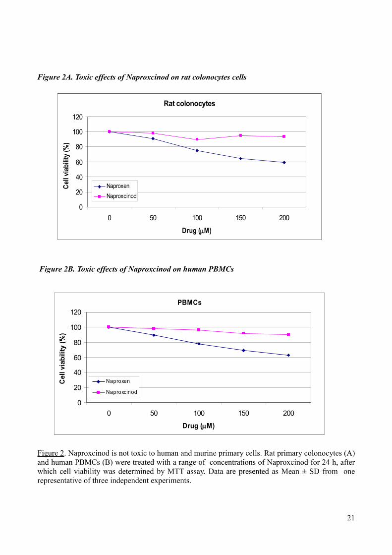
Figure 2A. Toxic effects of Naproxcinod on rat colonocytes cells
Figure 2B. Toxic effects of Naproxcinod on human PBMCs
Figure 2. Naproxcinod is not toxic to human and murine primary cells. Rat primary colonocytes (A) and human PBMCs (B) were treated with a range of concentrations of Naproxcinod for 24 h, after which cell viability was determined by MTT assay. Data are presented as Mean ± SD from one representative of three independent experiments.
PBMCs
0
20
40
60
80
100
120
0 50 100 150 200
Drug (mM)
Cel
l via
bilit
y (%
)
Naproxen
Naproxcinod
Figure 2B
PBMCs
0
20
40
60
80
100
120
0 50 100 150 200
Drug (mM)
Cel
l via
bilit
y (%
)
Naproxen
Naproxcinod
Figure 2B
Rat colonocytes
0
20
40
60
80
100
120
0 50 100 150 200
Drug (mM)
Cell v
iabilit
y (%
)
NaproxenNaproxcinod
Figure 2A
Rat colonocytes
0
20
40
60
80
100
120
0 50 100 150 200
Drug (mM)
Cell v
iabilit
y (%
)
NaproxenNaproxcinod
Figure 2A
21

Figure 3. Comparative anticancer effects of Naproxcinod
CT26CL25 murine colon cancer
0
20
40
60
80
100
120
140
0 10 20 40 80 160
Drug (mM)
Cel
l via
bilit
y (%
)
vehicle
naproxennaproxcinod
cisplatin
A
b
PC3 human prostate cancer
0
20
40
60
80
100
120
140
0 10 20 40 80 160
Drug (M)
Cel
lvia
bilit
y(%
)
vehicle
naproxennaproxcinod
cisplatin
LnCap human prostate cancer
0
20
40
60
80
100
120
140
0 10 20 40 80 160
Drug (M)
Cel
lvia
bilit
y(%
)
vehicle
naproxennaproxcinod
cisplatin
A
B
C CT26CL25 murine colon cancer
0
20
40
60
80
100
120
140
0 10 20 40 80 160
Drug (mM)
Cel
l via
bilit
y (%
)
vehicle
naproxennaproxcinod
cisplatin
A
b
PC3 human prostate cancer
0
20
40
60
80
100
120
140
0 10 20 40 80 160
Drug (M)
Cel
lvia
bilit
y(%
)
vehicle
naproxennaproxcinod
cisplatin
PC3 human prostate cancer
0
20
40
60
80
100
120
140
0 10 20 40 80 160
Drug (M)
Cel
lvia
bilit
y(%
)
vehicle
naproxennaproxcinod
cisplatin
LnCap human prostate cancer
0
20
40
60
80
100
120
140
0 10 20 40 80 160
Drug (M)
Cel
lvia
bilit
y(%
)
vehicle
naproxennaproxcinod
cisplatin
LnCap human prostate cancer
0
20
40
60
80
100
120
140
0 10 20 40 80 160
Drug (M)
Cel
lvia
bilit
y(%
)
vehicle
naproxennaproxcinod
cisplatin
A
B
C
22
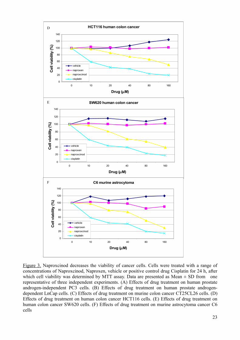
Figure 3. Naproxcinod decreases the viability of cancer cells. Cells were treated with a range of concentrations of Naproxcinod, Naproxen, vehicle or positive control drug Cisplatin for 24 h, after which cell viability was determined by MTT assay. Data are presented as Mean ± SD from one representative of three independent experiments. (A) Effects of drug treatment on human prostate androgen-independent PC3 cells. (B) Effects of drug treatment on human prostate androgen-dependent LnCap cells. (C) Effects of drug treatment on murine colon cancer CT25CL26 cells. (D) Effects of drug treatment on human colon cancer HCT116 cells. (E) Effects of drug treatment on human colon cancer SW620 cells. (F) Effects of drug treatment on murine astrocytoma cancer C6 cells
HCT116 human colon cancer
0
20
40
60
80
100
120
140
0 10 20 40 80 160
Drug (mM)
Cel
l via
bilit
y (%
)
vehicle
naproxennaproxcinod
cisplatin
SW620 human colon cancer
0
20
40
60
80
100
120
140
0 10 20 40 80 160
Drug (mM)
Cel
l via
bilit
y (%
)
vehicle
naproxennaproxcinod
cisplatin
C6 murine astrocytoma
0
20
40
60
80
100
120
140
0 10 20 40 80 160
Drug (mM)
Cel
l via
bilit
y (%
)
vehicle
naproxennaproxcinod
cisplatin
D
E
F
23
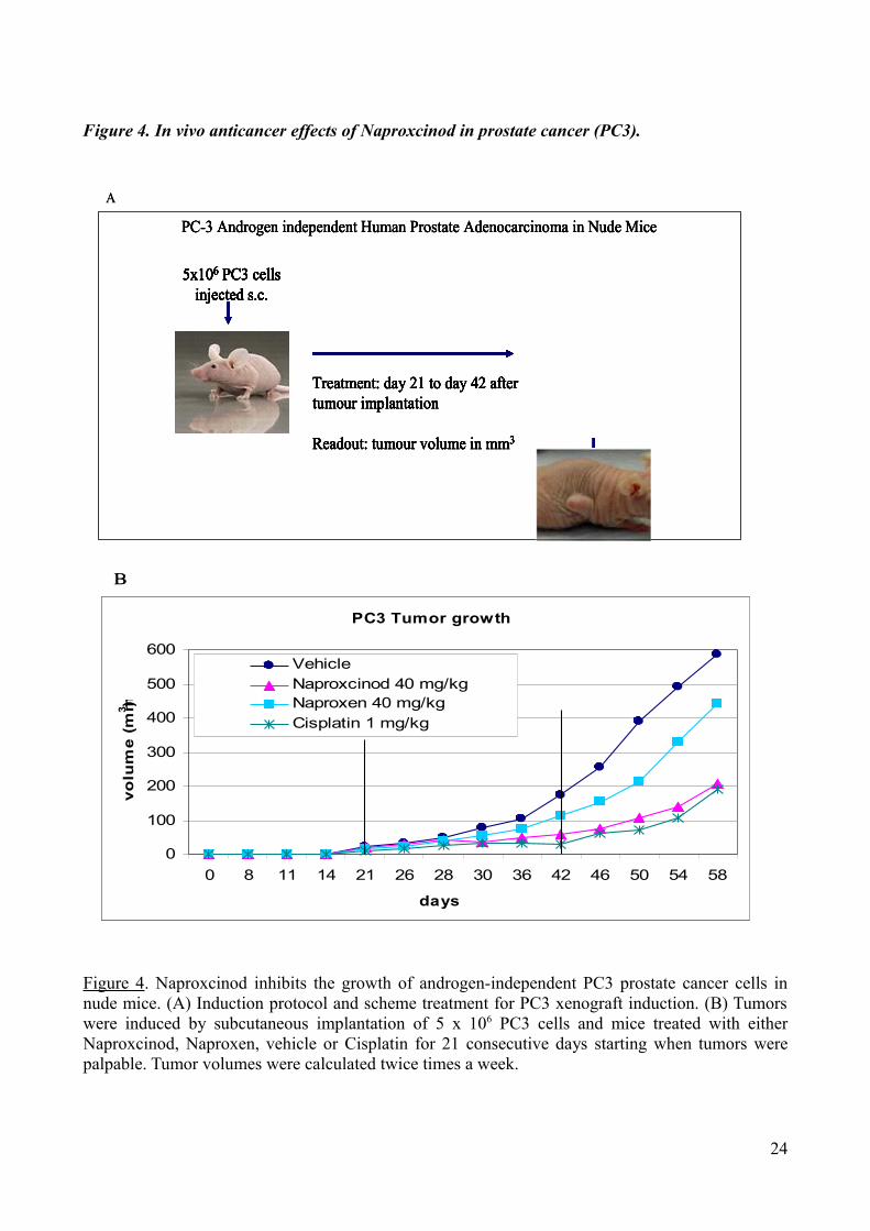
Figure 4. In vivo anticancer effects of Naproxcinod in prostate cancer (PC3).
Figure 4. Naproxcinod inhibits the growth of androgen-independent PC3 prostate cancer cells in nude mice. (A) Induction protocol and scheme treatment for PC3 xenograft induction. (B) Tumors were induced by subcutaneous implantation of 5 x 10⁶ PC3 cells and mice treated with either Naproxcinod, Naproxen, vehicle or Cisplatin for 21 consecutive days starting when tumors were palpable. Tumor volumes were calculated twice times a week.
PC3 Tumor growth
0
100
200
300
400
500
600
0 8 11 14 21 26 28 30 36 42 46 50 54 58
days
volu
me
(mm3 )
VehicleNaproxcinod 40 mg/kgNaproxen 40 mg/kgCisplatin 1 mg/kg
B
PC3 Tumor growth
0
100
200
300
400
500
600
0 8 11 14 21 26 28 30 36 42 46 50 54 58
days
volu
me
(mm3 )
VehicleNaproxcinod 40 mg/kgNaproxen 40 mg/kgCisplatin 1 mg/kg
B
PC-3 Androgen independent Human Prostate Adenocarcinoma in Nude Mice
5x10⁶ PC3 cellsinjected s.c.
Tumour palpable21 days after cells injection
Treatment: day 21 to day 42 after tumour implantation
Readout: tumour volume in mm3
PC-3 Androgen independent Human Prostate Adenocarcinoma in Nude Mice
5x10⁶ PC3 cellsinjected s.c.
Tumour palpable21 days after cells injection
Treatment: day 21 to day 42 after tumour implantation
Readout: tumour volume in mm3
5x10⁶ PC3 cellsinjected s.c.
Tumour palpable21 days after cells injection
5x10⁶ PC3 cellsinjected s.c.
Tumour palpable21 days after cells injection
Treatment: day 21 to day 42 after tumour implantation
Readout: tumour volume in mm3
A
PC-3 Androgen independent Human Prostate Adenocarcinoma in Nude Mice
5x10⁶ PC3 cellsinjected s.c.
Tumour palpable21 days after cells injection
Treatment: day 21 to day 42 after tumour implantation
Readout: tumour volume in mm3
PC-3 Androgen independent Human Prostate Adenocarcinoma in Nude Mice
5x10⁶ PC3 cellsinjected s.c.
Tumour palpable21 days after cells injection
Treatment: day 21 to day 42 after tumour implantation
Readout: tumour volume in mm3
5x10⁶ PC3 cellsinjected s.c.
Tumour palpable21 days after cells injection
5x10⁶ PC3 cellsinjected s.c.
Tumour palpable21 days after cells injection
Treatment: day 21 to day 42 after tumour implantation
Readout: tumour volume in mm3
A
24
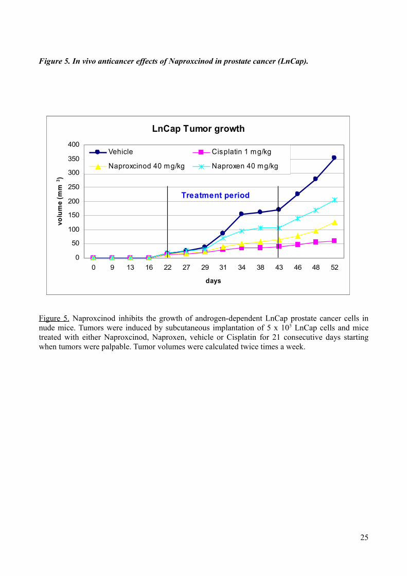
Figure 5. In vivo anticancer effects of Naproxcinod in prostate cancer (LnCap).
Figure 5. Naproxcinod inhibits the growth of androgen-dependent LnCap prostate cancer cells in nude mice. Tumors were induced by subcutaneous implantation of 5 x 10⁵ LnCap cells and mice treated with either Naproxcinod, Naproxen, vehicle or Cisplatin for 21 consecutive days starting when tumors were palpable. Tumor volumes were calculated twice times a week.
LnCap Tumor growth
0
50
100
150
200
250
300
350
400
0 9 13 16 22 27 29 31 34 38 43 46 48 52
days
volu
me
(mm
3 )
Vehicle Cisplatin 1 mg/kg
Naproxcinod 40 mg/kg Naproxen 40 mg/kg
Treatment period
Figure 5
LnCap Tumor growth
0
50
100
150
200
250
300
350
400
0 9 13 16 22 27 29 31 34 38 43 46 48 52
days
volu
me
(mm
3 )
Vehicle Cisplatin 1 mg/kg
Naproxcinod 40 mg/kg Naproxen 40 mg/kg
Treatment period
Figure 5
25
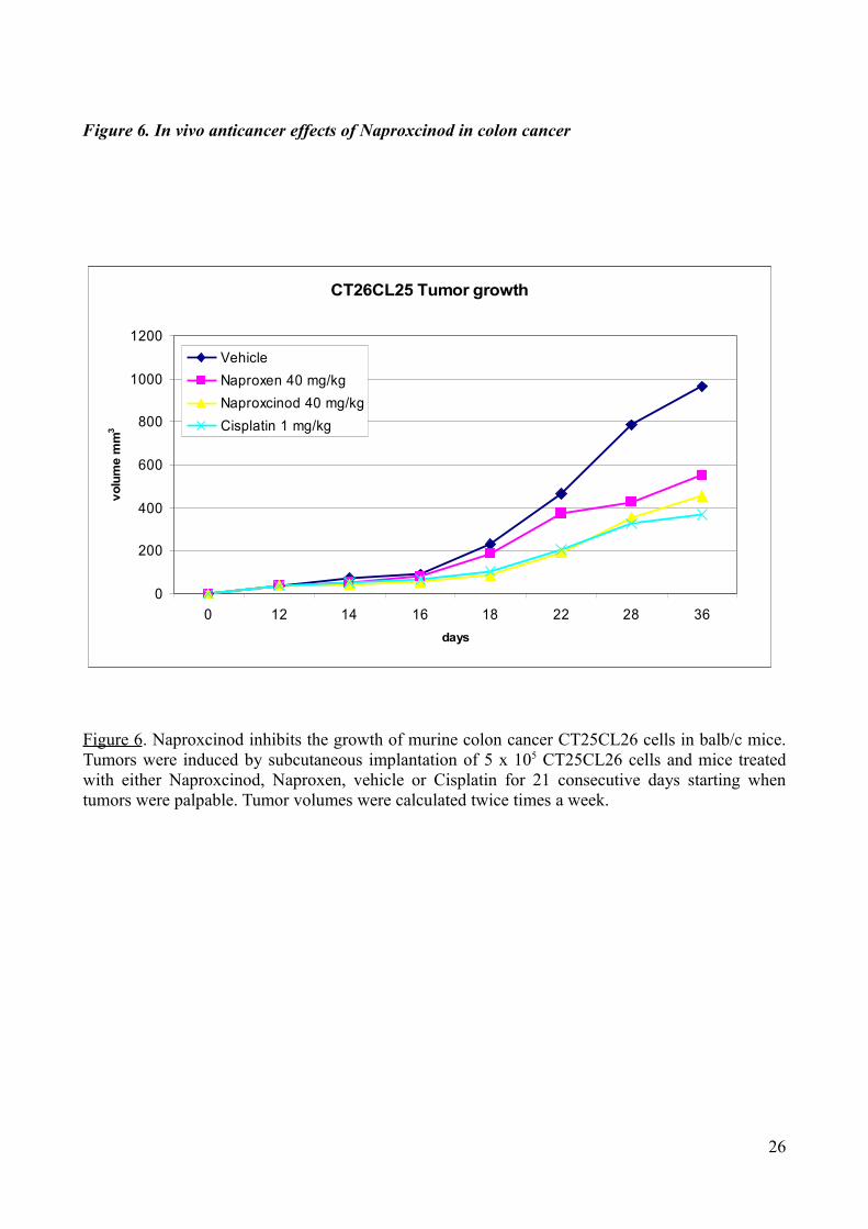
Figure 6. In vivo anticancer effects of Naproxcinod in colon cancer
Figure 6. Naproxcinod inhibits the growth of murine colon cancer CT25CL26 cells in balb/c mice. Tumors were induced by subcutaneous implantation of 5 x 10⁵ CT25CL26 cells and mice treated with either Naproxcinod, Naproxen, vehicle or Cisplatin for 21 consecutive days starting when tumors were palpable. Tumor volumes were calculated twice times a week.
CT26CL25 Tumor growth
0
200
400
600
800
1000
1200
0 12 14 16 18 22 28 36days
volu
me
mm
3
VehicleNaproxen 40 mg/kgNaproxcinod 40 mg/kgCisplatin 1 mg/kg
Figure 6
CT26CL25 Tumor growth
0
200
400
600
800
1000
1200
0 12 14 16 18 22 28 36days
volu
me
mm
3
VehicleNaproxen 40 mg/kgNaproxcinod 40 mg/kgCisplatin 1 mg/kg
Figure 6
26

Figure 7. Effects of Naproxcinod in a metastatic model of colon cancer.
Body weight
17,00
17,50
18,00
18,50
19,00
19,50
20,00
20,50
21,00
21,50
22,00
22,50
0 4 7 11 13 15Days
gr
Naproxen 40 mg/kgNaproxcinod 40 mg/kgCisplatin 3 mg/kgVehicle
* p<0.05 vs vehicle
*
* *
A
Lung weight
0,000,100,200,300,400,500,600,700,800,90
Naproxen40mg/kg
Naproxcinod40 mg/kg
Cisplatin 3 mg/kg
Vehicle
Treatment
gr
*
* p<0.05 vs vehicle
B
Body weight
17,00
17,50
18,00
18,50
19,00
19,50
20,00
20,50
21,00
21,50
22,00
22,50
0 4 7 11 13 15Days
gr
Naproxen 40 mg/kgNaproxcinod 40 mg/kgCisplatin 3 mg/kgVehicle
* p<0.05 vs vehicle
*
* *
A Body weight
17,00
17,50
18,00
18,50
19,00
19,50
20,00
20,50
21,00
21,50
22,00
22,50
0 4 7 11 13 15Days
gr
Naproxen 40 mg/kgNaproxcinod 40 mg/kgCisplatin 3 mg/kgVehicle
* p<0.05 vs vehicle
*
* *
A
Lung weight
0,000,100,200,300,400,500,600,700,800,90
Naproxen40mg/kg
Naproxcinod40 mg/kg
Cisplatin 3 mg/kg
Vehicle
Treatment
gr
*
* p<0.05 vs vehicle
B
27
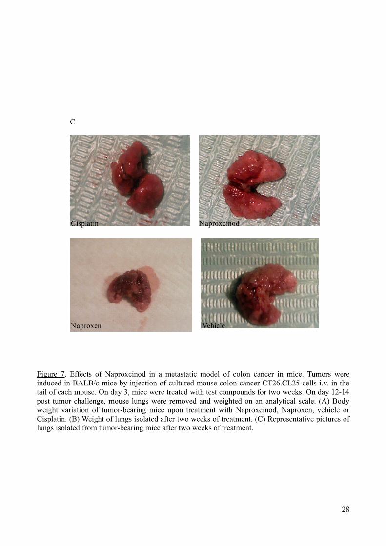
C
Cisplatin Naproxcinod
Naproxen Vehicle
Figure 7. Effects of Naproxcinod in a metastatic model of colon cancer in mice. Tumors were induced in BALB/c mice by injection of cultured mouse colon cancer CT26.CL25 cells i.v. in the tail of each mouse. On day 3, mice were treated with test compounds for two weeks. On day 12-14 post tumor challenge, mouse lungs were removed and weighted on an analytical scale. (A) Body weight variation of tumor-bearing mice upon treatment with Naproxcinod, Naproxen, vehicle or Cisplatin. (B) Weight of lungs isolated after two weeks of treatment. (C) Representative pictures of lungs isolated from tumor-bearing mice after two weeks of treatment.
28
References
Bonavida B et al. . Novel therapeutic applications of nitric oxide donors in cancer: roles in
chemoandimmunosensitization to apoptosis and inhibition of metastases. Nitric Oxide.
2008;19(2):152-7.
Bonavida B et al. Therapeutic potential of nitric oxide in cancer. Drug Resist Updat. 2006;9(3):157-
73.
Boyd CS and Cadenas E: Nitric oxide and cell signaling pathways in mitochondrial-dependent
apoptosis. Biol Chem 2002;383: 411-423,.
Chan, T. A. Nonsteroidal anti-inflammatory drugs, apoptosis, and colon-cancer
chemoprevention.Lancet Oncol 2002;. 3:166–174.
Chan, T. A. et al.. Mechanisms underlying nonsteroidal anti-inflammatory drug-mediated apoptosis.
Proc. Natl. Acad. Sci. USA 1998; 95:681–686.
Chinje EC and Stratford IJ: Role of nitric oxide in growth of solid tumours: a balancing act. Essays
Biochem 1997;32: 61-72,
Cuzick J et al. Aspirin and non-steroidal anti-inflammatory drugs for cancer prevention: an
international consensus statement. Lancet Oncol. 2009;10(5):501-7.
De Ridder M et al. . Hypoxic tumor cell radiosensitization through nitric oxide. Nitric Oxide.
2008;19(2):164-9
Donia M et al.. The novel NO-donating compound GIT-27NO inhibits in vivo growth of human
prostate cancer cells and prevents murine immunoinflammatory hepatitis. Eur J Pharmacol.
2009;615(1-3):228-33
Dube, C. et al.. The use of aspirin for primary prevention of colorectal cancer: a systematic review
prepared for the U.S. Preventive Services Task Force. Ann. Intern. Med. 2007;146:365–375.
29
Fiorucci S, Santucci L, Gresele P, Faccino RM, Del Soldato P, Morelli A. Gastrointestinal safety of
NO-aspirin (NCX-4016) in healthy human volunteers: a proof of concept endoscopic study.
Gastroenterology. 2003 Mar;124(3):600-7.
Forrester K et al. Nitric oxide-induced p53 accumulation and regulation of inducible nitric oxide
synthase expression by wild-type p53. Proc Natl Acad Sci USA 1996;.93: 2442-2447
Glockzin S et al.Activation of the cell death program by nitric oxide involves inhibition of the
proteasome. J Biol Chem 1999;274: 19581-19586,
Grilli M. et al. Neuroprotection by aspirin and sodium salicylate through blockade of NF-kappaB
activation. Science 1996; 274:1383–1385.
Hanif R et al. Effects of nonsteroidal anti-inflammatory drugs on proliferation and on induction of
apoptosis in colon cancer cells by a prostaglandin independent pathway. Biochem. Pharmacol.
1996;52:237–245.
House AA et al..Anti-inflammatory drugs and the kidney. Int J Artif Organs. 2007;30(12):1042-6
Huerta S et al.Nitric oxide donors: novel cancer therapeutics (review). Int J Oncol. 2008;33(5):909-
27
Huguenin, S et al. Evaluation of the antitumoral potential of different nitric oxide-donating
nonsteroidal anti-inflammatory drugs (NO-NSAIDs) on human urological tumor cell lines. Cancer
Lett. 2005;218:163–170.
Hulsman N et al.Chemical insights in the concept of hybrid drugs: the antitumor effect of nitric
oxide-donating aspirin involves a quinone methide but not nitric oxide nor aspirin. J Med Chem
2007;50:2424–2431
Hundley TR and Rigas B. Nitric oxide-donating aspirin inhibits colon cancer cell growth via
mitogen activated protein kinase activation. J Pharmacol Exp Ther 2006;316:25–34
Jana NR. NSAIDs and apoptosis. Cell Mol Life Sci 2008;65(9):1295-301
30
Jones M.K et al.Dual actions of nitric oxide on angiogenesis: possible roles of PKC, ERK, and AP-
1, Biochem. Biophys. Res. Commun. 2004;318 (2) 520–528
Karlsson J et al. Efficacy, safety, and tolerability of the cyclooxygenase-inhibiting nitric oxide
donator naproxcinod in treating osteoarthritis of the hip or knee. J Rheumatol. 2009;36(6):1290-7
Kashfi K and Rigas B. The mechanism of action of nitric oxide-donating aspirin. Biochem Biophys
Res Commun 2007;358:1096–1101
Kröncke KD et al. Nitric oxide: cytotoxicity versus cytoprotection--how, why, when, and where?
Nitric Oxide. 1997;1(2):107-20.
Laine L. Approaches to nonsteroidal anti-inflammatory drug use in the high-risk patient.
Gastroenterology 2001;120:594–606
Lanas A. Role of nitric oxide in the gastrointestinal tract. Arthritis Res Ther. 2008;10 Suppl 2:S4.
P. Lane P et al.. S-nitrosylation is emerging as a specific and fundamental post-translational protein
modification: head-to-head comparison with O-phosphorylation, Sci. STKE 2001 (2001)
Laval F and Wink DA. Inhibition by nitric oxide of the repair protein, O6-methylguanine-
DNAmethyltransferase. Carcinogenesis 1994;15:443–447
Li CQ et al. Apoptotic signaling pathways induced by nitric oxide in human lymphoblastoid cells
expressing wild-type or mutant p53. Cancer Res 2004;64: 3022-3029,
Li J., T.R. Billiar, R.V. Talanian, Y.M. Kim, Nitric oxide reversibly inhibits seven members of the
caspase family via S-nitrosylation,Biochem. Biophys. Res. Commun. 240 (1997) 419–424
Lim H et al. Increased expression of cyclooxygenase-2 protein in human gastric carcinoma. Clin.
Cancer Res. 2000;6:519–525.
Lin E, Morris JS, Ayers GD. Effect of celecoxib on capecitabine induced hand–foot syndrome and
antitumor activity. Oncology (Huntingt) 2002; 16:31–7
31
Lu W et al.Suppression of Wnt/beta-catenin signaling inhibits prostate cancer cell proliferation. Eur
J Pharmacol. 2009;602(1):8-14
Lundholm K, Gelin J, Hyltander A, et al. Anti-inflammatory treatment may prolong survival in
undernourished patients with metastatic solid tumors. Cancer Res 1994;54:5602–6
Maksimovic-Ivanic D et al. Anticancer properties of the novel nitric oxide-donating compound
(S,R)-3-phenyl-4,5-dihydro-5-isoxazole acetic acid-nitric oxide in vitro and in vivo. Mol Cancer
Ther. 2008;7(3):510-20
Maksimovic-Ivanic D et al. The antitumor properties of a nontoxic, nitric oxide-modified version of
saquinavir are independent of Akt. Mol Cancer Ther. 2009
Marshall et al., Nitrosation and oxidation in the regulation of gene expression, FASEB J. 14 (2000)
1889–1900
McIlhatton MA et al. Nitric oxide-donating aspirin derivatives suppress microsatellite instability in
mismatch repair-deficient and hereditary nonpolyposis colorectal cancer cells. Cancer Res.
2007;67(22):10966-75
Mijatovic S et al. Novel nitric oxide-donating compound (S,R)-3-phenyl-4,5-dihydro-5-isoxazole
acetic acid-nitric oxide (GIT-27NO) induces p53 mediated apoptosis in human A375 melanoma
cells. Nitric Oxide. 2008;19(2):177-83
Mijatovic S et al. Induction of caspase-independent apoptotic-like cell death of mouse mammary
tumor TA3Ha cells in vitro and reduction of their lethality in vivo by the novel chemotherapeutic
agent GIT-27NO. Free Radic Biol Med. 2010
Mocellin S et al. Nitric oxide, a double edged sword in cancer biology: searching for therapeutic
opportunities. Med Res Rev. 2007;27(3):317-52
Muscat JE et al. Nitric oxide-releasing medications and colorectal cancer risk: the Framingham
study. Anticancer Res. 2005;25(6C):4471-4
32
NicOx Press Release 18 June 2007. Available at http://www.nicox.com/upload/NCX4016-EF-
180607.pdf. Accessed on March 2, 2010
NicOx Press Release 18/10/2009. Available at
http://www.nicox.com/upload/PR_NDA_filingaccept_181109_EN.pdf . Accessed on
28/02/2010
Phillips P.G. and Birnby L.M. Nitric oxide modulates caveolin-1 and matrix metalloproteinase-9
expression and distribution at the endothelial cell/tumor cell interface, Am. J. Physiol. Lung Cell.
Mol. Physiol. 2004;286 (5) L1055– L1065
Puntoni, M.D. et al.Inflammation and cancer prevention. Annals of Oncology 2008;19 (Suppl7):
225–229.
Reynaert N.L., K. Ckless, S.H. Korn, N. Vos, A.S. Guala, E.F. Wouters, A.van der Vliet, Y.M.
Janssen-Heininger, Nitric oxide represses inhibitory kappaB kinase through S-nitrosylation, Proc.
Natl. Acad.Sci. USA 101 (2004) 8945–8950.
Rigas B. Novel agents for cancer prevention based on nitric oxide. Biochem Soc Trans. 2007;35(Pt
5):1364-8
Rigas, B. and Williams, J. L. NO-donating NSAIDs and cancer: An overview with a note on
whether NO is required for their action. Nitric Oxide 2008;19:199-204.
Rigas, B. and Kashfi K. Nitric-oxide-donating NSAIDs as agents for cancer prevention. Trends
Mol. Med. 2004;10:324–330.
Scartozzi M. et al. Molecular biology of sporadic gastric cancer: prognostic indicators and novel
therapeutic approaches. Cancer Treat. Rev. 2004;30:451–459.
Shoemaker R. H. The NCI60 Human Tumour Cell line Anticancer Drug Screen. Nature Reviews
2006, 6: 813-823
Siemens DR et al. Phase II study of nitric oxide donor for men with increasing prostate-specific
antigen level after surgery or radiotherapy for prostate cancer. Urology. 2009;74(4):878-83 33
Son K., Y.M. Kim, In vivo cisplatin-exposed macrophages increase immunostimulant-induced nitric
oxide synthesis for tumor cell killing, Cancer Res. 55 (1995) 5524–5527
Steele VE et al. Chemopreventive efficacy of naproxen and nitric oxide-naproxen in rodent models
of colon, urinary bladder, and mammary cancers. Cancer Prev Res (Phila Pa). 2009;2(11):951-6
Sun Y et al. Chemopreventive agents induce oxidative stress in cancer cells leading to COX-2
overexpression and COX-2-independent cell death. Carcinogenesis. 2009;30(1):93-100
Tirmenstein MA et al. Glutathione depletion and the production of reactive oxygen species in
isolated hepatocyte suspensions. Chem Biol Interact. 2000;127(3):201-17.
Vega M.I. et al. Rituximab induced inhibition of YY1 and Bcl-xL expression in Ramos non-
Hodgkin’s lymphoma cell line via inhibition of NF-kappa B activity: role of YY1 and Bcl-xL in
Fas resistance and chemoresistance, respectively. J. Immunol. 2005; 175, 2174–2183
Wallace JL and Vong L. NSAID-induced gastrointestinal damage and the design of GI-sparing
NSAIDs. Curr Opin Investig Drugs. 2008; 9(11):1151-6.
Whelton A. COX-2-specific inhibitors and the kidney: effect on hypertension and oedema. J
Hypertens Suppl. 2002; 20(6):S31-5
Williams JL et al. NO-donating aspirin inhibits the activation of NF-kappaB in human cancer cell
lines and Min mice. Carcinogenesis. 2008; 29(2):390-7
Wolfe MM, Lichtenstein DR, Singh G. Gastrointestinal toxicity of nonsteroidal antiinflammatory
drugs. N Engl J Med 1999; 340:1888–99
Yin M. J et al.The anti-inflammatory agents aspirin and salicylate inhibit the activity of I(kappa)B
kinase-beta. Nature 1998; 396: 77–80.
34