
UNIVERSITÀ DEGLI STUDI DI MILANO Scuola di Dottorato … · UNIVERSITÀ DEGLI STUDI DI MILANO...
118
UNIVERSITÀ DEGLI STUDI DI MILANO Scuola di Dottorato in Scienze Biologiche e Molecolari XXVI Ciclo Analysis of the upstream signalling pathway controlling !Np63" protein stability and function Michela Restelli PhD Thesis Scientific Tutor: Luisa Guerrini Academic Year 2012-2013
Transcript of UNIVERSITÀ DEGLI STUDI DI MILANO Scuola di Dottorato … · UNIVERSITÀ DEGLI STUDI DI MILANO...

UNIVERSITÀ DEGLI STUDI DI MILANO
Scuola di Dottorato in Scienze Biologiche e Molecolari
XXVI Ciclo
Analysis of the upstream signalling pathway controlling
!Np63" protein stability and function
Michela Restelli PhD Thesis
Scientific Tutor: Luisa Guerrini
Academic Year 2012-2013

! "!
SSD: BIO/11
Thesis performed at the Department of Biosciences of the University of
Milan, in collaboration with the Telethon Laboratory, Dept. of
Molecular Biotechnologies and Health Sciences, University of Torino,
I-10126 Torino, Italy and Dept. of Dermatology, University of Rome
"Tor Vergata", I-00133 Rome, Italy

! #!
! $!
Index
PART I
1. Abstract Page 7
2. State of the art Page 9
2.1 The p53 family members Page 9
2.2 The p63 transcription factor Page 10
2.2.1 Role of p63 in embryonic development Page 12
2.3 Limb development Page 16
2.3.1 Proximo-Distal patterning Page 17
2.3.2 Anterior-Posterior patterning Page 20
2.3.3 Dorsal-Ventral patterning Page 21
2.3.4 Survey on the molecular pathway controlled by p63
during limb development
Page 25
2.4 Human syndromes and malformations associated to
mutations in the p63 gene
Page 27
2.4.1 Ectrodactyl-Ectodermal dysplasia Clefting Page 29
2.4.2 Ankyloblepharon-Ectodermal dysplasia Page 30
2.4.3 Acro-Dermato-Ungual-Lacrimal-Tooth syndrome Page 31
2.4.4 Limb Mammary Syndrome Page 31
2.4.5 Split-Hand/Split-Foot Malformation type IV Page 32
2.4.6 Genotype-phenotype correlation of p63 mutations Page 35
2.5 Regulatory post-translational modifications of the
p53 family members
Page 37
2.5.1 p300 acetyl-transferase Page 40
2.5.2 c-Abl tyrosine kinase Page 44
2.5.3 PIN1 prolyl-isomerase Page 45
3. Aim of the work and main results Page 49
! %!
4. Conclusion and future perspectives Page 52
5. References Page 53
PART II
Published paper DLX5, FGF8 and the PIN1 isomerase control !Np63" protein
stability during limb development: a regulatory loop at the
basis of the SHFM and EEC congenital malformations
PART III
Manuscript under submission FGF8, c-Abl and p300 cooperate in the regulation of !Np63"
stability
Page 67

! &!
PART I

! '!
1. Abstract The p63 transcription factor, homolog to the p53 tumor suppressor, plays a crucial
role in epidermal and limb development. Dominant mutations in the p63 gene give
rise to several human congenital syndromes characterized by skin, craniofacial and
limb defects. One of the syndromes caused by p63 mutations is the Split-Hand/Foot
Malformation-IV (SHFM-IV) syndrome, characterized by the loss of central rays
of hands and feet. These developmental defects are due to failure of Apical
Ectodermal Ridge (AER) development. The correct limb outgrowth and patterning
is guarantee by the expression of key molecules including Fybroblast Growth
Factor 8 (FGF8), p63 and the DLX5 and DLX6 transcription factors. In this
context, the study of the molecular mechanisms regulating p63 stability and
function is fundamental for understanding the molecular bases of the SHFM-IV
pathogenesis: indeed p63 as been proposed to be one of the crucial regulators of
limb and epidermal development.
Little is known on the post-translational modifications and the upstream signalling
pathway controlling !Np63" functions, one of the most expressed p63 isoform in
epithelial tissues and in the AER cells. The projects performed during my PhD
thesis achieved to the identification of FGF8 as a crucial regulator of !Np63"
stability and activity in human osteosarcoma and keratinocyte cell lines. FGF8
determined also !Np63" protein stabilization in mice embryonic limb buds put in
culture at Embryonic day 10.5 (E10.5). In particular, treatments with FGF8 of
human osteosarcoma cell lines (U2OS) and human keratinocytes (HaCat), activate
the tyrosine kinase c-Abl, leading to !Np63" phosphorylation and consequent
acetylation by the p300 acetyl-transferase, promoting !Np63" stabilization and
transcriptional activation. Moreover, I have found that p300 interacts with !Np63"
determing its acetylation on lysine K193E, in vitro. Interestingly, this regulatory
cascade is not active on the natural !Np63"K193E mutant associated to the
SHFM-IV syndrome. Indeed, the !Np63"K193E mutant displays promoter
! (!
specific altered DNA binding activity that results in altered expression of !Np63"
target genes involved in limb development (like Perp, Ikk! and DLX5 gene)
(Manuscript in preparation).
One of the mechanism by which FGF8 promotes !Np63" stability and activation,
is inhibiting its interaction with Pin1, a prolyl isomerase known to positively
regulate p53 and p73 in response to DNA damage stress. In particular, PIN1 has an
opposite effect on !Np63" respect to p53 and p73: it promotes !Np63"
degradation through the proteasome pathway. Moreover, !Np63" mutant proteins,
associated with SHFM-IV or EEC syndromes, characterized by limb defects, are
not degraded by PIN1 overexpression. These data were confirmed also by in vivo
experiments on PIN1 Knock-Out (KO) mice, where lack of PIN1 expression
caused the accumulation of p63 in the embryonic limbs and ectoderm compared to
wild-type littermates. Moreover, I found that FGF8 is a downstream target of the
transcription factor Dlx5. Indeed, in the limb buds of both p63 and DLX5;DLX6
KO mice, the AER is poorly stratified and FGF8 expression is severely reduced.
All these data suggest that DLX5, !Np63", FGF8 and PIN1 participate in a
regulatory loop essential for AER stratification, normal patterning and
morphogenesis of the limb buds (1).
The work performed during my PhD contributes to a better understanding of the
regulatory mechanisms controlling !Np63" function and stability. We have
identified FGF8 as a crucial upstream signal required for !Np63" activation and
stabilization during limb development: mutations or altered expression of
regulators in this pathway leads to abnormal limb development and onset of
pathogenesis.
! )!
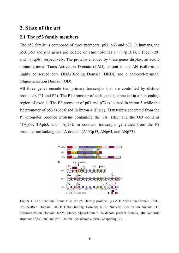
2. State of the art 2.1 The p53 family members The p53 family is composed of three members: p53, p63 and p73. In humans, the
p53, p63 and p73 genes are located on chromosomes 17 (17p13.1), 3 (3q27–29)
and 1 (1q36), respectively. The proteins encoded by these genes display: an acidic
amino-terminal Trans-Activation Domain (TAD), absent in the !N isoforms, a
highly conserved core DNA-Binding Domain (DBD), and a carboxyl-terminal
Oligomerization Domain (OD).
All three genes encode two primary transcripts that are controlled by distinct
promoters (P1 and P2). The P1 promoter of each gene is embeded in a non-coding
region of exon 1. The P2 promoter of p63 and p73 is located in intron 3 while the
P2 promoter of p53 is localized in intron 4 (Fig.1). Transcripts generated from the
P1 promoter produce proteins containing the TA, DBD and the OD domains
(TAp53, TAp63, and TAp73). In contrast, transcripts generated from the P2
promoter are lacking the TA domain (!133p53, !Np63, and !Np73).
Figure 1. The functional domains in the p53 family proteins. (a) AD: Activation Domain; PRD:
Proline-Rich Domain; DBD: DNA-Binding Domain; NLS: Nuclear Localization Signal; TD:
Tetramerization Domain; SAM: Sterile-Alpha-Domain. % denote percent identity. (b) Genomic
structure of p53, p63 and p73. Dotted lines denote alternative splicing (2).

! *+!
Additional complexity to p53 family member isoforms is added by the fact that P1
and P2 transcripts can be spliced at the 3’ end into several spliced variants. For p63
five TA variants (TAp63", TAp63#, TAp63$, TAp63% and TAp63&) and five !N
variants (!Np63", !Np63#, !Np63$, !Np63% and !Np63&) have been identified
(2-6).
While p63 and p73 demonstrate relatively little homology with p53 in their TA and
OD, both share approximately 60% similarity with p53 in the DBD, including
conservation of essential DNA contact residues. However, studies of Knock-Out
(KO) mice have demonstrated that, even though these proteins clearly share some
similarities with p53, their functions are very different. p53 null mice (p53 -/-) are
viable and develop normally, but are prone to spontaneus development of a variety
of neoplasms by 6 months of age (7). p73-null mice are born viable, but show an
high mortality rate within the first 2 months. The animals suffer from
hydrocephalus, indicative of abnormal cerebrospinal fluid dynamics,
immunological problems characterized by chronic infections and inflammation,
and nervous system abnormalities related to hippocampal dysgenesis, olfactory
neuron defects and the loss of sympathetic neurons (8) .
In contrast p63 -/- mice are born alive but show the most severe developmental
phenotype of all p53 family members and die soon after birth because of
dehydration. They fail to develop limbs, stratified epidermis and most epithelial
tissues (hair follicles, teeth, prostate, lacrimal and salivary glands, and mammary
glands) (9,10).
2.2 The p63 transcription factor
The proteins encoded from the p63 gene contain different functional domains
(Fig.2):
• Trans-Activation domain (TA domain): important for transcriptional
activation of p63 target genes (present in all TAp63 isoforms);

! **!
• Second Trans-Activation domain (TA2 domain) located between
aminoacids 410 and 512. The identification of this domain
revealed that also the !Np63 protein isoforms, generally regarded
as dominant-negative isoforms, are able to directly activate gene
expression (11);
• DNA Binding domain (DBD domain) located in the core of the
protein and present in all isoforms, allows binding of p63 to its
Responsive Elements (RE); some are in commons to p73 and p53
(p53 RE), the others are specific for p63 (p63RE) (3, 12, 13);
• Oligomerization domain (OD-ISO domain) is present in all p63
isoforms and allows the formation of omo and etero-tetramers
among p63 isoforms but also with the other p53 family members
(14). The formation of p63 tetramers is essential for p63 functions:
it allows cooperative binding to DNA RE;
• Steryl-Alpha-Motif (SAM) domain is located in the carboxyl-
terminal portion of the protein, present only in TAp63" and
!Np63". It displays a globular and solid structure with an highly
conserved sequence of about 65-75 aminoacids organized into five
helixes (15). This domain is thought to mediate protein-protein
interactions, playing a crucial role during development and
differentiation.
• C-terminal Inhibitor domain (TID) is present only in the "
isoforms and has a negative effect on the transcriptional activity of
p63. In particular, this domain binds the N-terminal region, masking
the TA domain, giving a possible explanation for the reduction of
the trans-activational capacity of TAp63" compared to TAp63# and
TAp63$ isoforms (11,16,17).

! *"!
Figure2. Schematic representation of human p63 gene structure: alternative promoters (P1 and P2),
previously identified alternative splicing events (", #, $) and novel events (%, &) are indicated (6)
In recent years the identification of p63 targets genes has led to a better
understanding of the developmental strategy sustained by p63. About 5800 target
genes for p63 have been identified: p63 targets are enriched for genes involved in
cell adhesion, proliferation, death and signaling pathways (18-20). Furthermore, it
has been demonstrate that p63 associates with the promoter of p53, p73 and of the
p63 gene itself, suggesting that among the p53 family members exists a complex
cross-regulations (21).
2.2.1 Role of p63 in embryonic development
Immuno-histochemical analysis of mouse embryos, show high p63 isoform protein
levels in epithelial cells, especially in progenitor or stem-cell populations of
epithelial tissues (9,10,22). The main isotype expressed in these cells is the !Np63
protein, which likely acts in the maintenance of the proliferative capacity of such
cells (13, 22-25). As these cells start to differentiate, !Np63 protein levels
gradually drop, and the levels of TAp63 proteins increase. It thus appears that
!Np63 protein is crucial for the maintenance of regenerative proliferation capacity
of epithelial stem cells. Cells that no longer express !Np63 proteins loose this
capacity and are committed to differentiate (25).

! *#!
In mouse embryos, p63 expression is first evident in nuclei of cells in the basal
layer of the epidermis, which develop into the progenitor cells of the epidermis and
related derivatives, such as hair and sweat glands. Basal cells of the cervix, tongue,
esophagus, mammary glands, prostate, and urothelium also show high levels of p63
isoform expression. Early p63 protein expression is further evident in ectodermal
cells of the limb buds and tail bud, branchial arches and oral epithelium (9, 10, 13,
26). In the developing limb buds, p63 expression is restricted to the Apical
Ectodermal Ridge (AER), a key structure required for limb-bud emergence and
progression in mice.
The sites of p63 expression are well in line with the phenotypic consequences of
homozygous p63 inactivation in mice. p63 deficient mice are viable at birth, but
have strikingly developmental defects. Their limbs are absent or truncated: in
particular the forelimbs (FL) are truncated and the distal part is missing, while
hindlimbs (HL) are completely absent. The limb buds of p63 deficient embryos are
distinctly smaller and there is no morphologically distinct AER: indeed, mutant
limb buds have a single layer epithelium at the dystal tip, indistinguishable from
the surrounding ectoderm (9, 10) (Fig. 3). The limb defects observed in p63 -/-
mice results from a failure of the ectoderm to undergo growth and differentiation
processes that give rise to a stratified epithelium and to a correct integrity of the
AER structure, required for correct limb development (27). Indeed, Fybroblast
Growth Factor 8 (FGF8), a key signaling molecule expressed in AER cells, is not
detectable in the limbs of p63 homozygous mutant embryos at embryonic day 10.5
(E10.5) (9, 10, 28). Consistently, the stratified organization of the AER and the
expression of morphogenetic molecules are dramatically compromised in p63
mutant limbs.

! *$!
Figure 3. (Left) Analysis of p63 deficient limbs during embryogenesis. Scanning electron
microscopy and histological analysis of wild-type (WT) and p63-deficient (mt) embryos, indicate
that the AER is absent in p63 homozygous mutant embryos (arrows) (mills et al, 1999). (Right)
Front and sagittal views of E17 control (+/+) an p63 -/- embryos. p63 -/- mice on post-natal day 1
(P1) have hypoplastic upper and lower jaws, and have no eyelids, wisker pads, skin and related
appendages wich are present on the wild type control (Yang et al, 1999). (Bottom) FLs of p63-
homozygous mutant mice are truncated: the phalanges, radius and ulna are absent and the humerus
is deformed (9, 10)
Moreover, at birth p63 deficient mice have severe skin defects. These mice die few
hours later for dehydration: they looses thirty times more water than their wild-type
littermates, as a consequence of alteration in the process of epidermal stratification.
Histologycal analysis of neonatal p63-deficient skin revealed the absence of normal
epidermal structure and complete lack of hair follicles. The surface of p63 null skin
is covered by a single layer of flattened cells, lacks stratification and does not
express differentiation markers.
In addition to this well described phenotype of the p63 null mice models, recently it
has been discovered that p63 ablation results also in severe defects of embryonic

! *%!
cardiac development, including dilation of both ventricles, defects in trabeculation
and abnormal septation. Indeed hystological sections of mice hearth at E14.5
revealed dilated cardio-myopathy with deficient trabeculation and thin ventricular
wall (29).
Expression of the p63 gene is required not only during embryonic development but
also in adult tissues such as epidermis. Development of the epidermis requires a
series of coordinated events, which regulate proliferation and differentiation of
keratinocytes. Stem cells in the basal layer undergo asymmetric division to yield
another identical stem cell, and a Transient Amplifying Cell (TAC) that is
committed to differentiate. TACs are also capable of active proliferation
(amplification) but eventually reach terminal differentiation (Fig. 4).
Figure 4. Model for p63 function in maintaining regenerative capacity in epithelial stem cells. On
the left, representation of epidermal differentiation, showing a percentage of stem cells in the basal
layer that undergo asymmetric division to yield another identical stem cell, and a Transient
Amplifying Cell (TAC) that is commited to differentiation (5).
p63 transcript is expressed in the surface ectoderm prior to stratification and
continues to be expressed during embryonic development. As the epidermis
matures, p63 expression becomes restricted to the basal layer of epidermis,
indicating that it is required to maintain the proliferative potential of epidermis (30-
32). In adult tissues, p63 is expressed in stratified epithelia, whereas its expression
is absent from single-layered epithelia. In the presence of lesions in the epidermis

! *&!
and dermic tissues, the repair mechanism of damaged cells includes the formation
of new epidermal layer. During this regenerative process different p63 protein
isoforms are present, supporting the idea that p63 expression is not important only
during embryonic development but also in the regenerative process of epidermal
layers (33).
2.3 Limb development In mice, embryonic limbs are first visible as a small bud that protrudes from the
body and contains morphologically homogenous cells covered by a layer of
ectoderm. Limb buds emerge as a result of a thickening of the somatic layer of the
Lateral Plate Mesoderm (LPM). As the bud elongates, a region of undifferentiated
cells is maintained at the tip, while differentiated tissues are progressively laid
down starting at the base of the limb bud. The developmental patterning of the
limbs results from gradients of signaling molecules in three spatial dimensions:
Proximo-Distal (shoulder-finger direction, Pr-D), Antero-Posterior (thumb-little
finger direction, A-P), and Dorso-Ventral (back-palm direction, D-V). For correct
development, three specialized cell clusters are of primary importance: the Apical
Ectodermal Ridge (AER), the Progress Zone (PZ), and the Zone of Polarizing
Activity (ZPA) (Fig. 5). These groups of cells express signaling molecules that
determine the fate of neighbouring cells by instructing them to remain
undifferentiated, to proliferate, or to differentiate into a particular cell type (34-38).
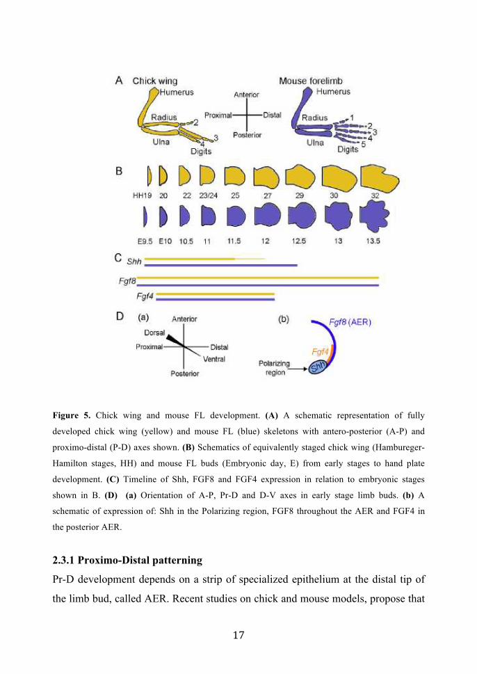
! *'!
Figure 5. Chick wing and mouse FL development. (A) A schematic representation of fully
developed chick wing (yellow) and mouse FL (blue) skeletons with antero-posterior (A-P) and
proximo-distal (P-D) axes shown. (B) Schematics of equivalently staged chick wing (Hambureger-
Hamilton stages, HH) and mouse FL buds (Embryonic day, E) from early stages to hand plate
development. (C) Timeline of Shh, FGF8 and FGF4 expression in relation to embryonic stages
shown in B. (D) (a) Orientation of A-P, Pr-D and D-V axes in early stage limb buds. (b) A
schematic of expression of: Shh in the Polarizing region, FGF8 throughout the AER and FGF4 in
the posterior AER.
2.3.1 Proximo-Distal patterning
Pr-D development depends on a strip of specialized epithelium at the distal tip of
the limb bud, called AER. Recent studies on chick and mouse models, propose that
! *(!
the AER controls the initial size of the limb bud, cell survival and proliferation in
order to generate a limb buds of appropriate size. Removal of the AER in chick
embryos results in limbs lacking distal skeletal elements.
The Fybroblast Growth Factors (FGFs), key factors required for AER function, are
signaling molecules whose activities are mediated by a family of tyrosine kinase
trans-membrane receptors (39-42).
FGFs that are specifically expressed in the AER are: FGF8, FGF10, FGF9 and
FGF17. FGF8 transcript is expressed in the prospective AER cells of nascent limb
and subsequently throughout the AER until it regresses (43-46). By contrast,
FGF4, FGF9 and FGF17 expression starts after the AER is formed, is restricted to
the posterior AER, and ceases at least a day before AER regression. When AER
FGFs are individually eliminated through the generation of KO mice, only loss of
FGF8 perturbs skeletal patterning. The other AER-FGFs have been proposed to be
essential, but functionally redundant, components of a positive feedback loop
between the AER and the patterning center in the posterior limb bud mesenchyme
that produce Sonic-Hedge-Hog (SHH), a crucial factor required for AP patterning
(46-48).
In particular, FGF8 KO mice display a substantial reduction in limb bud size, a
delay in SHH expression, mis-regulation of FGF4 expression, and hypoplasia or
aplasia of specific skeletal elements. In particular, FGF8 null mice display limb
abnormalities: stylopod are severely reduced in HLs, but only mildly affected in
FLs. Zeugopod elements are mildly hypoplastic in both HLs and FLs. The autopod
is missing a digit: digit I in HLs and digit II or III in FL (49, 50) .
The phenotype of FGF8 null mice resembles the phenotype of the p63 KO mice,
indeed both KO models display limb defects that are affecting more seriously HL
rather than FLs (mills and yang 1999) (Fig. 6).
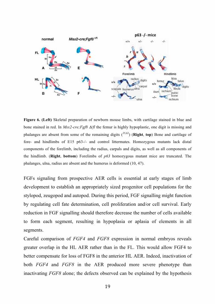
! *)!
Figure 6. (Left) Skeletal preparation of newborn mouse limbs, with cartilage stained in blue and
bone stained in red. In Msx2-cre;Fgf8 "/fl the femur is highly hypoplastic, one digit is missing and
phalanges are absent from some of the remaining digits (35,62) (Right, top) Bone and cartilage of
fore- and hindlimbs of E15 p63-/- and control littermates. Homozygous mutants lack distal
components of the forelimb, including the radius, carpals and digits, as well as all components of
the hindlimb. (Right, bottom) Forelimbs of p63 homozygous mutant mice are truncated. The
phalanges, ulna, radius are absent and the humerus is deformed (10, 47).
FGFs signaling from prospective AER cells is essential at early stages of limb
development to establish an appropriately sized progenitor cell populations for the
stylopod, zeugopod and autopod. During this period, FGF signalling might function
by regulating cell fate determination, cell proliferation and/or cell survival. Early
reduction in FGF signalling should therefore decrease the number of cells available
to form each segment, resulting in hypoplasia or aplasia of elements in all
segments.
Careful comparison of FGF4 and FGF8 expression in normal embryos reveals
greater overlap in the HL AER rather than in the FL. This would allow FGF4 to
better compensate for loss of FGF8 in the anterior HL AER. Indeed, inactivation of
both FGF4 and FGF8 in the AER produced more severe phenotype than
inactivating FGF8 alone; the defects observed can be explained by the hypothesis
! "+!
that a principal function of AER-FGF signaling during limb development is to
ensure that enough progenitor cells are available to form each element of the limb
skeleton (34, 51).
The limb mesenchyme produces FGF10, which signals to the limb ectoderm to
initiate limb outgrowth. FGF10, together with other crucial signaling molecules of
limb development, like Wingless 3a (WNT3a) and Bone Morphogenetic Protein
(BMP), are required to induce FGF8 expression of AER precursors in the
ectoderm. The AER precursors are initially spread over a relatively broad region of
the ectoderm, then became concentrated at the distal tip of the limb bud and
compact to form a columnar epithelium. The compacted AER also serves as
mechanical function to provide directed outgrowth and to maintain a dorso-
ventrally flattened shape of the limb. After the AER has completed its function, it
regresses, returning to a flattened cuboidal epithelium. This is accompanied by
down-regulation of FGFs expression and a reduction of mesenchyme proliferation.
Molecular experiments in the chick show that AER regression is mediated by BMP
signaling (37, 52).
2.3.2 Anterior–Posterior limb patterning
The Anterior-Posterior limb development is assured by a posterior region of limb
mesenchyme called the Zone of Polarizing Activity (ZPA). The ZPA is located in
the posterior limb-bud mesenchyme and specifies A-P identities in the
mesenchyme through the secretion of SHH signaling molecule. SHH is one of the
three vertebrate homologs of Hedgehog segment polarity gene of the Drosophila
melanogaster. Genetic analysis has revealed essential functions of SHH gene in a
large number of morpho-regulatory processes. The active SHH signaling peptide is
generated by auto-proteolytic cleavage of full-length protein, and is covalently
modified by the addition of cholesterol and palmitate moieties. This modified
peptide forms a posterior to anterior gradient in developing limb-bud. The
formation of SHH gradient is fundamental for digit identity and specification.

! "*!
Genetic inactivation of SHH in mice results in the dramatic loss of skeletal
elements along the A-P axis: in particular loss of distal limb structures and
complete absence of digits (35, 53)
2.3.3 Dorsal-Ventral patterning
The mesenchyme already contains the information for D–V limb patterning that
occurs before limb-bud initiation. Just before the limb bud forms, the mesenchyme
transfers this information to the overlying ectoderm; the molecular nature of this
early mesenchymal signal(s) is unknown.
The transcription factor Lmx1b is expressed in the dorsal mesenchyme of the limb
bud and is required for cells to adopt a dorsal character. Although the early
regulator of Lmx1b expression is unknown, as the limb bud forms Lmx1b is
induced by WNT7a, a signalling molecule that has been implicated in the
regulation of cell fate and pattern formation during embryogenesis, expressed in the
dorsal limb ectoderm (54). In the absence of WNT7a, the dorsal pattern of the distal
structures (autopod) is not established and the limbs appear bi-ventral. Expression
of WNT7a is restricted to the dorsal ectoderm because it is repressed in the ventral
ectoderm by the transcription factor Engrailed1 (En1). In En1!/! limbs, Wnt7a is
misexpressed in the ventral ectoderm, and the distal structures of limb bud develop
with bi-dorsal character (52).
As described before, normal limb development occurs along three different axes:
(Pr-D, D-V and A-P) controlled by different signaling centers: the Apical
Ectodermal Ridge (AER), the Progress Zone (PZ), and the Zone of Polarizing
Activity (ZPA). It soon became clear that each limb axes is not specified
independently from the others and that the putative molecular determinants of the
axes are mutually regulated. Indeed, complex feedback loops exist between SHH in
the ZPA, BMPs and their antagonists in the adjacent mesenchyme, WNT7a in the
dorsal ectoderm and FGFs in the AER. In particular, maintenance of FGF

! ""!
expression seems to be regulated by a positive feedback loop between FGF
signaling in the AER and SHH expression in the ZPA. There is also evidence that
signaling from the dorsal ectoderm is required to maintain SHH expression (55) (Fig
7).
Figure 7. Signaling pathways in vertebrate limb development. molecular interactions that
coordinate limb growth and patterning along the three limb axes: Pr-d is under the control of FGFs
from the AER, the A-P is under the control of SHH in the posterior mesenchyme, and the D-V is
under the control of BMP an En1from the ventral ectoderm and Wnt7 from the dorsal ectoderm
(52).
However, for correct limb development and shaping is also required the
establishment of programmed cell death (or apoptosis) process. The formation of
free digits in vertebrates is accompained by massive apoptosis of the interdigital
mesenchime and has the function of sculpturing the shape of the digits. In addition
to mesodermal cell death, apoptosis is also an important feature of the ectoderm of
the AER. In the chick limb buds ectodermal apoptosis appears to exert the function
of controlling the extension of this structure (56,57).
Alteration in processes regulating apoptosis in limb buds causes an enlargement of
the AER structure in limb buds, that results in polydactyly (57, 58).
The molecular machinery responsible for apoptosis exhibits a high degree of
conservation in the course of evolution. During limb programmed cell death,
members of the different groups of apoptotic regulators have been identified. As in
other models of apoptosis, the final steps of limb programmed cell death consist in
the activation of caspases, key mediators of the apoptotic response (57, 58) (Fig. 8).
!

! "#!
Figure 8. Interdigital cell death in the developing limb by TUNEL labeling and Acridine Orange
Staining (58)
In developing limb buds, BMPs have been identified as the triggering apoptotic
signal for both the ectoderm of the AER and the mesodermal cells. According to
their pattern of expression BMP-2, BMP-4 and BMP-7, are the most likely
physiological signals triggering apoptosis in the limb buds. These BMPs are also
involved in the control of limb patterning and in the regulation of chondrogenic
differentiation. There are evidences suggesting that the apoptotic effect of BMPs in
the limb bud and in other developing model systems is mediated by the activation
of the cytoplasmic kinase TAK-1 rather than by the canonical intracellular pathway
of BMPs involving phosphorylation of Smad proteins, intracellular molecules that
mediate the canonical signaling cascade of TGFbeta superfamily growth factors
(35, 57, 58).
Two different models have been proposed to elucidate the mechanism of limb
development (Fig. 9).

! "$!
Figure 9. Representation of two models for proximo-distal limb skeletal patterning: progressive
zone model (a) and early specification model (b) (35)
The ‘progress zone’ model proposed by Wolpert and colleagues postulates that
cells acquire proximo-distal positional information progressively, in a proximal-to-
distal sequence, by measuring time spent in a ‘Progress Zone’ (PZ). The longer the
time spent in the PZ, the more distal the positional values they acquire. Cells that
exit early are specified to form proximal limb structures, whereas those that exit
late are specified to form distal ones. The other model proposed by Dudley et al.,
called ‘early specification model’, postulate that specification to form proximal,
middle or distal limb structures does not occur progressively, but instead occurs
very early, perhaps even before limb bud outgrowth has begun. According to this
model, the specified populations subsequently expand as the limb bud grows, and
become determined in a proximal-to-distal sequence. Cells are specified as
progenitors of stylopod, zeugopod or autopod, and over time they expand and
become determined to form particular structures in response to cell–cell
interactions and signaling (35).
! "%!
2.3.4 Survey on the molecular pathway controlled by p63 during limb
development
It’s well established that p63 expression is fundamental for correct limb growth and
development. The most abundantly protein isoform expressed in the AER of the
limb buds is the !Np63" isoform and its expression increases from E10.5 to E12.5
(28, 59, 60). The expression of !Np63" protein is required to assure proliferation
and stratification of AER cells in order to reach a correct process of limb
outgrowth. In this context the identification of !Np63" target genes expressed
during limb development is fundamental to understand which are the pathway by
which !Np63" guarantee correct formation of the limbs.
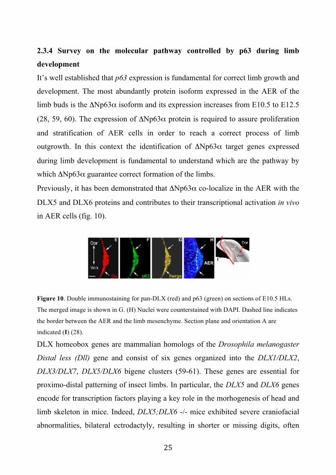
Previously, it has been demonstrated that !Np63" co-localize in the AER with the
DLX5 and DLX6 proteins and contributes to their transcriptional activation in vivo
in AER cells (fig. 10).
Figure 10. Double immunostaining for pan-DLX (red) and p63 (green) on sections of E10.5 HLs.
The merged image is shown in G. (H) Nuclei were counterstained with DAPI. Dashed line indicates
the border between the AER and the limb mesenchyme. Section plane and orientation A are
indicated (I) (28).
DLX homeobox genes are mammalian homologs of the Drosophila melanogaster
Distal less (Dll) gene and consist of six genes organized into the DLX1/DLX2,
DLX3/DLX7, DLX5/DLX6 bigene clusters (59-61). These genes are essential for
proximo-distal patterning of insect limbs. In particular, the DLX5 and DLX6 genes
encode for transcription factors playing a key role in the morhogenesis of head and
limb skeleton in mice. Indeed, DLX5;DLX6 -/- mice exhibited severe craniofacial
abnormalities, bilateral ectrodactyly, resulting in shorter or missing digits, often

! "&!
accompanied by malformation and syndactyly of the remaining digits and profound
medial cleft of the posterior limbs (60). Interestingly, a number of cases of SHFM-I
are associated with chromosomal aberrations involving the 7q21-22 chromosomal
region. Indeed, extensive analysis to locate the minimal deletions associated to
SHFM-I had narrowed down the SHFM-I critical region to 1.5 Mb and six
breakpoints have been found within a 700 kb region. Three candidate genes are
located in the common deletion interval: DLX5, DLX6 and DSS1 (62). Indeed, the
DLX5;DLX6 KO mice, displaying bilateral ectrodactyly with a severe defect of the
central ray of the hindlimbs, phenocopy limb defects observed in SHFM-I patients
(60, 61). Moreover, SHFM-IV, a phenocopy of SHFM-I, is caused by mutations in
the p63 gene. All these discoveries suggest that p63, DLX5 and DLX6 proteins
might be components of a common signaling pathway regulating limb
development. Furthermore, the severity of limb malformations displayed by the
p63 KO mice, respect to the DLX5;DLX6 KO mice, suggest that p63 could be an
upstream crucial regulator of signaling pathway during limb development: indeed,
p63 has been identified as a crucial activator for the DLX5 and DLX6 gene during
limb development (28) (Fig. 11).
Figure 11. (left) Limb defects in DLX5;DLX6 null mice. a: Homozygous mutants die around 18
dpc, showing severe craniofacial abnormalities and hindlimb defects. Hindlimbs of normal (b,e,i,j)
and mutant (c,d,f,g,h) mice at 12.5 dpc (b–d), 14.5 dpc (e–h), and 18 dpc (i,j). At 12.5 dpc, the
central part of the hindlimb palette appears flattened (c) or indented (d), at later stages reduced or
absent digits and median clefts are observed. Note the variable phenotype between left (g) and right

! "'!
(h) mutant hindlimb. In situ hybridization on normal (e) and mutant (f) 14.5 dpc hindlimbs with a
DLX5 probe showing absence of expression in the mutant. Abnormal or missing digits are indicated
(red arrows). No obvious defect was been seen in the forelimbs. Genotypes of the Dlx5–Dlx6 locus
are indicated (61). (right) Characterization of the p63 null mice phenotype (10).
2.4 Human syndromes and malformations associated to mutations
in the p63 gene After the characterization of the p63 null mice phenotype a lot of works focused on
linking the genetic defects of several human syndromes characterized by skin and
limb malformation to a region of chromosome 3q27 encopassing the p63 gene (63-
65). These works lead to the identification of at least 5 human syndromes caused
by mutation in the p63 gene showing defects resembling the p63-null mice
phenotype. They include: Ectodactyl Ectodermal dysplasia Clefting (EEC),
Ankyloblepharon-Ectodermal-displasia (AEC), Acro-Dermato-Ungual-Lacrimal-
Tooth (ADULT), Limb-Mammary-Syndrome (LMS) and Split-Hand/Split-Footh
Malformations (SHFM) type IV (65).
The pattern of mutations in the five human disorders linked to p63 mutations
reveals a remarkable specificity of the molecular defects in this gene and clinical
consequences (Fig. 12).

! "(!
Figure 12. Distribution of mutations in p63 associated to human developmental related syndromes
(65).
All of these disorders are the consequence of a mutation at a single p63 allele:
indeed mutations of the p63 gene associated to these syndromes are heterozygous
dominant mutations. Moreover, the p63 mutant proteins interfere with the normal
activity of the wild-type protein thanks to their capacity to form omo and ethero-
tetramers (65).

! ")!
Figure 13. Table of phenotypic characteristic of p63 human syndromes (65).
2.4.1 Ectrodactyl-Ectodermal-dysplais Clefting (EEC)
The EEC syndrome is characterized by the triad of ectrodactily, ectodermal
dysplasia and facial clefting. A number of associated anomalies are frequently
found, among which there are lacrimal tract anomalies, urogenital anomalies, and
conductive hearing loss. EEC patients occasionally have also mammary
gland/nipple hypoplasia (14%), and hypohidrosis (11%). About two-thirds of these
patients have ectrodactily and syndactyly is also frequent (43%). Cleft lip/palate
(CL-CP) is present in about 40% of the EEC patients.
An extended analysis of EEC syndrome patients showed heterozygous mutations of
p63 gene in 40 of 43 unrelated families, indicating that p63 is the gene mutated in
EEC syndrome (63, 66).
Eighteen different mutations in the DBD have been reported in EEC syndrome
patients. All of these mutations, apart from one, that is a frameshift mutations
affecting only p63" isoforms, give rise to aminoacid substitutions in the DNA-
Binding Domain common to all p63 isoforms. These aminoacids are crucially

! #+!
important for the interaction with DNA target sequences, and their mutation seems
to impair p63 binding to DNA (R204W/Q, R279C/H/S and R304W/Q).
In general, the missense mutations are predicted to affect all p63 isoforms, resulting
in loss of transactivation of p63, while the frameshift mutation resulted in gain of
transactivation potential for the truncated TAp63" isotype. So the missense and
frameshift mutations appeared to exhibit divergent effects on the regulation of
transcriptional activity of p63 as well, suggesting that both an increase and/or a
decrease of transactivation by p63 lead to the developmental defects seen in EEC
syndrome patients (65).
2.4.2 Ankyloblepharon-Ectodermal-displasia
The AEC syndrome, which is also known as “Hay-Wells syndrome,” has little or
no limb involvement but instead includes ankyloblepharon, which is a partial or
complete fusion of the eyelids that is very rare in other p63 associated syndromes.
Approximately 80% of the patients have severe skin erosion at birth, which usually
recover in the first years of life. The eyelid fusion is present in about 45% of AEC
patients. Nail and teeth defects are present in more than 80% of patients, while hair
defects and/or alopecia are constant features (94%). Lacrimal duct obstruction is
seen in 50% of patients, whereas mammary gland hypoplasia and hypohydrosis
occur occasionally (both 13%). Interestingly, almost 40% of patients have hearing
impairment and genital-urinary defects. Cleft lip is present in 44% and cleft palate
in about 80%. Limb malformations are almost absent. Ectrodactyly has never been
reported, but 25% of patients display mild syndactyly. Immunohistochemical
examination of a skin sections from an AEC syndrome patient revealed an aberrant
localization of p63.
Mutations in 12 unrelated patients with AEC have been detected, and 10 of these
are missense mutations within the SAM domain of p63. These mutations are
predicted to disrupt protein-protein interactions, by either destroying the compact
globular structure of the SAM domain or substituting aminoacids that are crucial

! #*!
for such interactions. Missense mutations in AEC syndrome affect only the "
isotypes of p63.
The AEC mutations exert a selective dominant-negative function on wild-type p63
by affecting the expression of a number of p63 target genes, including Fgfr2 and
Fgfr3. Impaired FGF signalling downstream of p63 is an important determinant of
reduced ectodermal cell proliferation and defective self!renewing compartment in
AEC syndrome (67).
2.4.3 Acro-Dermato-Ungual-Lacrimal-Tooth Syndrome
The ADULT syndrome is characterized by ectrodactyly and ectodermal
abnormalities such as nail dysplasia, hypodontia, lacrimal duct obstruction, sparse
hair and thin skin. These patients show neurodermitic signs (exfoliative dermatitis
of the digits) and excessive freckling; on the other hand these patients do not
manifest cleft-lip palate. Teeth, skin and nail defects are constantly present in
ADULT syndrome; moreover hair (53%) and lacrimal duct defects (67%) are
observed in ADULT patients. Four ADULT syndrome families and three sporadic
cases have been reported. All the families and one of the sporadic cases have a
point mutation in exon 8, changing R298 in the DNA binding domain into either a
glutamine or a glycine. While EEC syndrome mutations in the DBD impair the
binding of p63 protein to DNA, arginine 298 is not located close to the DNA-
binding interface, and mutation of this arginine does not affect DNA binding. In
particular, this mutation confers a novel transcriptional activation capacity to the
!Np63$ isoform (68,69).
2.4.4 Limb Mammary Syndrome
The LMS syndrome is characterized by severe hand and/or foot anomalies, and
hypoplasia/aplasia of the mammary gland and nipple. Less frequent findings
include lacrimal-duct atresia, nail dysplasia, hypohydrosis, hypodontia, and cleft
palate with or without bifid uvula. This syndrome differs from the EEC syndrome

! #"!
in at least three aspects: mammary gland and nipple hypoplasia are consistent
features of LMS but are only occasionally seen in EEC syndrome. Second, patients
with LMS do not have hair and skin defects that are seen in EEC syndrome. Third,
whereas patients with LMS have CP, those with EEC syndrome have CL/P but
never have CP only. Phenotypically, LMS is most similar to ADULT syndrome
(70).
Mutations in LMS are located in the N-and C-terminus of the p63 gene. A large
LMS family (29 affected members) has a point mutation in exon 4, causing a
G76W substitution in the DNA-specific putative Trans-Activation Domain (TA2).
One Other point mutation (S90W) is also located between the TA Domain and
DBD. Other LMS mutations are reported in the C-terminus: these deletions in exon
13 (TT deletion) and exon 14 (!AA) cause a frameshift and a premature stop codon
affecting only the p63" protein isoforms. Also a stop mutation in the TID domain
(K632X) has been identified in a sporadic LMS patient. The latter mutation is
predicted to impair the suppressive effect of the TID towards the TA domain, thus
increasing the transactivation activity of p63 (70).
2.4.5 Split Hand/Split Foot Malformation type IV
The Split-Hand/Split-Foot Malformation (SHFM) syndrome, also known as
ectrodactyly, is a congenital limb malformation, characterized by a deep median
cleft of the hands and/or feet; however, the severity of SHFM is highly variable. In
severe cases, the hands and feet have a lobster claw-like appearance, while in
mildly affected patients, SHFM may be limited to syndactyly and several instances
of non-penetrance have been documented. SHFM may occur as an isolated entity
or as part of a syndrome: both forms are frequently found in association with
chromosomal rearrangements such as deletions or translocations. The degree of
phenotypic severity of an affected limb as well as the number of affected limbs
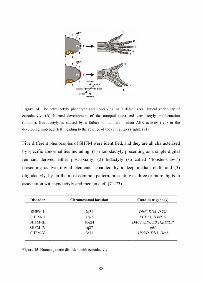
differs from individual to individual (66, 70, 71) (Fig.14).
! ##!
Figure 14. The ectrodactyly phenotype and underlying AER defect. (A) Clinical variability of
ectrodactyly. (B) Normal development of the autopod (top) and ectrodactyly malformation
(bottom). Ectrodactyly is caused by a failure to maintain median AER activity (red) in the
developing limb bud (left), leading to the absence of the central rays (right). (71)
Five different phenocopies of SHFM were identified, and they are all characterized
by specific abnormalities including: (1) monodactyly presenting as a single digital
remnant derived either post-axially; (2) bidactyly (so called ‘‘lobster-claw’’)
presenting as two digital elements separated by a deep median cleft; and (3)
oligodactyly, by far the most common pattern, presenting as three or more digits in
association with syndactyly and median cleft (71-73).
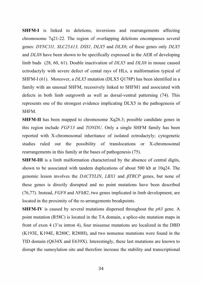
Disorder Chromosomal location Candidate gene (s)
SHFM-I 7q21 Dlx5, Dlx6, DSS1 SHFM-II Xq26 FGF13, TONDU SHFM-III 10q24 DACTYLIN, LBX1,"TRCP SHFM-IV sq27 p63 SHFM-V 2q31 HOXD, Dlx1, Dlx2
Figure 15. Human genetic disorders with ectrodactyly.
! #$!
SHFM-I is linked to deletions, inversions and rearrangements affecting
chromosome 7q21-22. The region of overlapping deletions encompasses several
genes: DYNC1l1, SLC25A13, DSS1, DLX5 and DLX6; of these genes only DLX5
and DLX6 have been shown to be specifically expressed in the AER of developing
limb buds (28, 60, 61). Double inactivation of DLX5 and DLX6 in mouse caused
ectrodactyly with severe defect of cental rays of HLs, a malformation typical of
SHFM-I (61). Moreover, a DLX5 mutation (DLX5 Q178P) has been identified in a
family with an unusual SHFM, recessively linked to SHFM1 and associated with
defects in both limb outgrowth as well as dorsal-ventral patterning (74). This
represents one of the strongest evidence implicating DLX5 in the pathogenesis of
SHFM.
SHFM-II has been mapped to chromosome Xq26.3; possible candidate genes in
this region include FGF13 and TONDU. Only a single SHFM family has been
reported with X-chromosomal inheritance of isolated ectrodactyly; cytogenetic
studies ruled out the possibility of translocations or X-chromosomal
rearrangements in this family at the bases of pathogenesis (75).
SHFM-III is a limb malformation characterized by the absence of central digits,
shown to be associated with tandem duplications of about 500 kb at 10q24. The
genomic lesion involves the DACTYLIN, LBX1 and #TRCP genes, but none of
these genes is directly disrupted and no point mutations have been described
(76,77). Instead, FGF8 and NFkB2, two genes implicated in limb development, are
located in the proximity of the re-arrangements breakpoints.
SHFM-IV is caused by several mutations dispersed throughout the p63 gene. A
point mutation (R58C) is located in the TA domain, a splice-site mutation maps in
front of exon 4 (3’ss intron 4), four missense mutations are localized in the DBD
(K193E, K194E, R280C, R280H), and two nonsense mutations were found in the
TID domain (Q634X and E639X). Interestingly, these last mutations are known to
disrupt the sumoylation site and therefore increase the stability and transcriptional

! #%!
activity of the p63" protein. The R280C and R280H mutations have also been
encountered many times in EEC syndrome (65).
Finally, SHFM-V is associated to deletions encompassing the chromosome region
2q24–q31. Patients with deletions in this region exhibit a number of abnormalities,
including microcephaly, mental retardation, low-set ears, and limb abnormalities.
Recent studies have revealed that the 2q24.3–q31 region can be subdivided into
three distinct loci for limb abnormalities. This region encompasses the HOXD gene
cluster, essential for the development of the extremities of limbs. Candidate genes
located in the critical SHFM-V interval include DLX1 and DLX2, two homeobox
genes expressed in the AER and the PZ of limb buds (30, 78-80).
The generation of the KO mouse models phenocoping the defects showed by the
SHFM patients contributed to the identification of the molecular pathways and the
regulators controlling limb development. In particular, mouse model for SHFM-I
have been generated by the combined deletions of DLX5 and DLX6 genes; the
Dactyplasia mutant mice has been proposed as a good model of SHFM-III
(60,61,77). Dactylin is one of the several factors involved in the regulation of cell
proliferation in the AER (76,77). These KO mice exhibit defects in the
stratification and development of AER structure and AER failure is at the bases of
SHFM pathogenesis (30,71). These discoveries suggest that DLX5, DLX6, p63 and
Dactylin could be link together in a same pathway essential for correct limb
development.
2.4.6 Genotype-phenotype correlation of p63 mutations
The clustering of mutations in the DBD, for EEC syndrome, and in the SAM
domain, for AEC syndrome establishes a clear genotype-phenotype correlation
(Fig. 11). A number of observations can be made from more detailed analysis of
the pattern of TP63 mutations. First, it is notable that the truncating mutations are
all located in the C-terminal part of the protein. Hence, all p63 mutations known to

! #&!
date leave the ISO domain intact. This allows the formation of tetrameric
complexes between wild-type and mutant p63 proteins, which offers an explanation
for the dominant effect of p63 mutations.
Ectrodactyly is only seen in combination with missense mutations in the DBD of
p63 isoforms, or in combination with truncating mutations in the C-terminal part of
the protein. In contrast, missense mutations in the C-terminal part are never
associated with ectrodactyly. A second remarkable phenomenon involves the type
of facial clefting. The facial clefts seen in conjunction with missense mutations in
the conserved part of the DBD always involve the primary palate (CL/P), whereas
mutations toward the C-terminal end of p63 may either involve the primary palate
or the secondary palate (CP) (30, 64, 65) (Fig 16).
Figure 16. Illustration of p63 mutation in human syndromes establishing a clear genotype-
phenotype correlation (64).
Some of p63 mutations could also be ”gain of function” mutations: they caused an
increase in the transcriptional activity of the mutant protein compared to the wild-
type p63. For instance the R298Q, associated to the ADULT syndrome, conferes to
the !Np63$ isoform an exceptional trans-activation capacity, which is absent in the
wild-type !Np63$ protein. There could be two possible explanations for this gain
of trans-activation activity: (1) the mutation creates a novel site for the binding of a

! #'!
transcriptional coactivator; (2) the mutation releases a second TA domain (denoted
as “TA2”) that is normally kept in an inactive state. Simultaneously, some of these
mutations are “loss of function” because they determine a reduction of the p63
transcriptional activity as it as been described for the missense EEC and SHFM-IV
mutations in the regulation of the DLX5 and DLX6 genes (28).
It’s important to consider that most of the mutations are located in the DBD domain
and in the SAM domain. Often these mutations occur on residues that could be
post-transcriptionally modified (like lysines, serine, threonine and tyrosines)
possibly leading to an alteration of the normal signaling pathway regulating p63
isoforms activities.
2.5 Regulatory Post-Translational Modifications of the p53 family
members Post-Translational Modifications (PTMs) are series of covalent processing events
that change the properties of a protein, by either proteolytic cleavage or by the
addition of a modifying group to one or more aminoacids. PTMs have a great
influence on the functions of a protein, as they can regulate its activity, localization,
turnover and interaction with other proteins and molecules like nucleic acids, lipids
and cofactors.
PTMs are often mediated by enzymes and occur at distinct aminoacid residues.
These enzymes include kinases, transferases, phosphatases and ligases, which add
or remove functional groups, proteins, lipids or sugars to or from aminoacid side
chains. Conversely, proteases are enzymes that cleave peptide bonds to remove
specific sequences or regulatory subunits.
PTMs are known to be essential mechanisms used by eukaryotic cells to diversify
their protein functions and dynamically coordinate their signaling networks.
Defects in PTMs have been linked to numerous developmental disorders and
human diseases, highlighting the importance of PTMs in maintaining normal
cellular functions and homeostasis.
! #(!
The most important and studied PTMs for the p53 family members includes:
phosphorylation, acetylation, and ubiquitilation. Indeed, these PTMs strongly
modulate p53 family members function and stability in response to different stimuli
and stress conditions.
Phosphorylation is the addition of a phosphate group to a serine, tyrosine or
threonine residue in a peptide chain. The addition or removal of a phosphate group
can alter protein conformation (and therefore function) by locally altering the
charge and hydrophobicity.
Acetylation is a reversible process by which acetyl groups are placed onto the &
amino-group of lysine residues of a target protein and it is a well studied event
occuring on histone tails during transcription. Indeed, acetylation of internal lysine
residues of core histone has been found associated with transcriptional activation in
eukaryotes. Histone acetylation has been shown to occur both globally throughout
the genome as well as at specific promoters and it is an indicator of actively
transcribed genes.
In the deacetylation reaction, histone deacetylases (HDACs) remove the acetyl
groups re-establishing the positive charge of histones correlated with silencing of
gene expression (81-85).
Acetylation was first discovered on histones, and the significance of histone
acetylation in transcriptional regulation is well accepted. However, histones are not
the only proteins that can be acetylated: a large number of transcription factors can
be acetylated inducing their activation and stabilization (86-88).
Ubiquitilation is an enzymatic PTM process in which an ubiquitin protein is
attached to a substrate protein. This process most commonly binds the last amino
acid of ubiquitin (glycine 76) to a lysine residue on the substrate. Ubiquitination is
carried out in three main steps: activation, conjugation, and ligation, performed by
ubiquitin-activating enzymes (E1s), ubiquitin-conjugating enzymes (E2s), and
ubiquitin ligases (E3s), respectively. A protein can undergo monoubiquitilation or
poly-ubiquitilation on different lysines. In general, monoubiquitilation is associated

! #)!
with modification of cellular localization, affects protein activity, and promotes or
prevents protein interactions. On the other hand, polyubiquitination, the formation
of an ubiquitin chain on a single lysine residue on the substrate protein, is
associated with protein degradation by the proteasome machinery.
In the last decade, studies focused on the identification and characterization of the
PTMs controlling and regulating the members of the p53 family. Indeed p53 is
subject to a huge number of post-translational modifications, which markedly
influences the expression of p53 target genes. The most commonly reported post-
translational modifications of p53 include phosphorylation, acetylation and
ubiquitylation (88-91) (Fig. 17).
Figure 17. Post Translational modification of human p53 (86,109)
p53 is a potent inhibitor of cell proliferation and survival, and it is therefore essential that p53 is
kept inactive during normal growth. At homeostasis, the transcriptional activity of p53 is
downregulated mainly in three ways: 1. By MDM2 ubiquitin–mediated proteasomal degradation; 2.
by a decrease in nuclear p53 levels through nuclear export as a consequence on monoubiquitination

! $+!
of p53 by MDM2; 3. by transcriptional repression of chromatin-associated p53 by formation of
Mdm2-Mdmx-p53 complex. Upon DNA damage, degradation and nuclear export of p53 are
suppressed, and nuclear import is concominantly enhanced, resulting in its nuclear accumulation
leading to the activation of cell-cycle inhibitor and apoptotic related genes (91).
Among PTMs playing a crucial role in p53 activation and stabilization there are
phosphorylation of serines, theronines and tyrosines located in the amino and
carboxyl terminal region of p53 and acetylation of lysines located in the C-terminal
region and in the DBD domain of the p53 protein. Phosphorylation of specific
residues in p53 (Ser15, Thr18, and Ser20) increases p53 association with p300 and
CBP acetyl-transferases and stimulates p53 transactivation functions. Specifically,
acetylation of p53 is required for its activation and enhancement of sequence-
specific DNA binding and target genes activation. Moreover, acetylation of p53 C-
terminal residues is important as docking sites for the subsequent recruitment of
acetyl-transferases and other transcription cofactors to promoter regions. In this
context it’s clear that PTMs of p53, resulting from the activation of different
kinases/phosphatases and acetyl-transferases/deacetyl-transferases, play a critical
role in the outcome of p53 activation (88, 92, 93).
In the following chapters I will concentrate on three crucial regulators of p53 and
p73 that have been demonstrated to be essential for their activation in response to
DNA damage. Moreover, these regulators have been shown to play a crucial role
also during development suggesting that they could regulate also p63 functions
during development.
These regulators are: the p300 acetyl-transferase, the c-Abl tyrosine kinase and the
Pin1 prolyl isomerase.
2.5.1 p300 acetyl-transferase
Acetylation of p53 is an important reversible enzymatic process that occurs in
response to DNA damage and genotoxic stress and is indispensible for p53
transcriptional activity (94).

! $*!
One of the most important and well characterized acetyl-transferase for p53 is
p300. p300 belongs from the p300/CBP family and the acetyl-transferase activity is
located in the C-terminal fragment of p300 spanning aminoacids 1135-2414. In
addition to the histone acetyl-transferase domain, p300 contains different domains,
such as bromodomain and regions rich in Cys/His residues (C/H domains),
conserved from Drosophila to mammals. These domains serve as binding sites for
sequence specific transcription factors and other components regulating gene
expression (83, 95). p300 is a transcriptional co-activator that operate in many
transduction pathways controlling cell differentiation, growth control, and cellular
homeostasis and its expression is essential to sustain a correct process of cell
proliferation and development.
The major acetylation sites for p300 in p53 are located in the DBD domain and in
the C-terminal region of p53; acetylation of these sites is important as docking sites
for the subsequent recruitment of HAT and other transcription factor to promoter of
p53 target genes (90, 92). One of the proposed mechanism by which acetylation
promotes p53 stabilization and activation is that acetylation abrogates Mdm2
mediated repression by blocking its recruitment to p53 responsive promoters (94)
(Fig. 18).

! $"!
Figure 18. Role of p53 acetylation in gene regulation. (A) Unacetylated p53 is capable of
activating genes that are involved in the negative regulation of p53, such as Mdm2, as a mechanism
to keep p53 protein levels low during times of normal homeostasis. (B) Upon DNA damage,
acetylation of p53 allows for the disruption of the Mdm2-p53 interaction and the recruitment of
specific HATs to the promoters of genes involved in DNA repair and cell cycle control. (C) Full
acetylation of p53 at all key acetylation sites promotes the activation of proapoptotic genes. (D)
SIRT1 and HDAC1 are deacetylases that can reverse p53 acetylation and lead to transcriptional
repression (94) Recently, a previously undiscovered site acetylated by p300/CBP was described for
p53 (K164). Deletion of K164 in combination with other known acetylation sites in
p53 completely abrogated p53-dependent trans-activation of p21 and the ability to
induce cell-growth arrest, confirming that p53 acetylation is a crucial PTM
essential for p53 activation (94).
This residue is conserved in all the species known to encode for p53, including
human, mouse, xenopus, and zebrafish, as well as in the p53-related proteins p63
and p73 (Fig. 19). In particular, lysine K164 in p53 corresponds to lysine K193 in
p63; interestingly, this lysine is mutated in patients affected by SHFM-IV (K193E).

! $#!
Figure 19. Alignment of the K164 flanking region of the human p53 protein with those of p53 from
other species and of human p63 and p73. The conserved lysine residue is marked in bold; h: human;
m: mouse; c: chicken; x: Xenopus; and z: zebrafish (94).
Acetylation has been largely demonstrated to be essential also for p73 regulation:
the CH1 domain of the transcriptional co-activator p300 interacts with the N-
terminus of p73 (96). p300 acetylates p73 on lysines 321, 327 and 331 and the
trans-activation or induction of apoptosis by p73 is impaired in human p300
depleted cells, demonstrating an important role for p300 in p73 dependent
apoptosis (97).
In this context, the recent identification of the K164 in p53, acetylated by p300,
corresponding to K193 in !Np63", found mutated in patients affected by SHFM-
IV, suggest that acetylation could be a crucial PTMs occurring not only in response
to DNA damage and cytotoxic stress but also during development.
First indications that p300 play an important role during development came from
the characterization of p300 null mice phenotype. These mice died between days 9
and 11.5 of gestation, exhibiting sever defects in neurolation, cell proliferation and
heart development providing genetic evidence that p300 is essential for cell
proliferation and development (98).
Furthermore, it’s becoming clear that acetylation and deacetylation are crucial
PTMs to guarantee correct development. Indeed, deletion of Histone Deacetyl-
transferase HDAC1 and HDAC2, in KO mice embryos results in dramatic failure of
hair follicle specification and epidermal proliferation and stratification,

! $$!
phenocopying loss of the key ectodermal transcription factor p63. HDAC1/2
proteins are present at the promoter regions of target genes repressed by !Np63
isoforms, suggesting that HDAC1 and HDAC2 are requested to repress those genes
that need to be down-regulated by !Np63 in order to assure proper development
(99).
2.5.2 c-Abl tyrosine kinase
It’s well known that some PTMs are influenced or are promoted by an upstream
modification, supporting the idea that there is a cross-talk between different types
of modifications (100, 101). For instances, acetylation of p73 occurs after a
phosphorylation event (102). The tyrosine kinase c-Abl regulates p73 in apoptotic
response to cisplatin-induced DNA damage. The first kinase identified to
phosphorylate p73, establishing an increase in p73 acetylation levels and
consequent enhancement of its transcriptional activity, is c-Abl. In particular, DNA
damage induces phosphorylation of p73 at tyrosine 99 (Y99) in a c-Abl dependent
manner, and p73 proteins mutated at Y99 are impaired in mediating an apoptotic
response upon ionizing radiations treatment (103, 104).
c-Abl (Abelson tyrosine kinase) is a member of a family of non-receptor tyrosine
kinases and it is implicated in a large range of cellular processes including
regulation of cell growth, oxidative stress and DNA damage. The c-Abl protein is
localized at several subcellular sites, including the nucleus, cytoplasm,
mitochondria, and the cell embrane, where c-Abl interacts with a large variety of
cellular proteins, including signalling adaptors, kinases, phosphatases, cell-cycle
regulators, transcription factors and cytoskeletal proteins (105-108).
For instances, c-Abl, upon DNA damage, physically interacts with p73 through the
SH3 domain of c-Abl and the PXXP motif of p73 located in the linker region
between the DBD and the OD domain, leading to p73 stabilization (102).
However, c-Abl is a critical regulator not only for p73 but also for p53: c-Abl is
required for efficient accumulation of p53 upon DNA damage. Indeed, c-Abl

! $%!
physically interacts with the C-terminal region of p53 and neutralize the ability of
Mdm2 protein to promote p53 ubiquitination and degradation, leading to p53
transcriptional activation and induction of the apoptotic response (109-112).
It’s well established that c-Abl is activated upon DNA damage (107,108); however,
a crucial role in c-Abl activation is also played by cell to cell contact and
stimulation with growth factor, like Platelet-Derived Growth Factor (PDGF),
Epidermal Growth Factor (EGF), basic-Fybroblast Growth Factor (bFGF or Fgf2)
(113). This finding support the idea that c-Abl could fulfill a crucial role during
development. Indeed, c-Abl protein is involved in the development of many
mammalian organ systems, including immune system and bones and exhibiting a
pivotal role also in the regulation of hearth development. Indeed, Abl KO mice
display dramatically enlarged hearts and die perinatally. Moreover, high levels of
c-Abl are found in hyaline cartilage in the adult and Abl null mice are osteoporotic
as a result of dysfunction in osteoblast functions (114,115).
2.5.3 PIN1 prolyl isomerase
As mentioned before, PTMs are known to act alone or in combinations to regulate
protein functions. Moreover, it’s also known that changes in protein conformation
may facilitate specific post-translational modifications.
In literature, it’s known that there are specific enzymes, called isomerases
catalizing conformational change in substrate proteins. Among these enzymes a
critical regulator of the p53 family members is PIN1 (116).
The human PIN1 gene encodes an essential nuclear peptidyl-prolyl isomerase
involved in the regulation of mitosis. PIN1 protein specifically catalyzes the
cis/trans isomerization of phospho-Ser/Thr-Pro bonds and plays an important role
in many cellular events through the effects of conformational change of its
biological substrates (116). In particular, PIN1 is implicated in a large number of
molecular processes related to human diseases, including cancer and Alzheimer’s.

! $&!
PIN1 contains an N-terminal WW domain and a C-terminal peptidyl-prolyl cis-
trans isomerase (PPIase) domain connected by a flexible linker. The N-terminal
WW domain of PIN1 is a small structural motif acting as a protein interaction
module that binds to short proline-rich segments of target proteins. The WW
domain of PIN1 mediates substrate recognition, interaction with anchoring proteins
for subcellular localization and/or facilitation of nuclear import (117).
The C-terminal domain encodes an essential PPIase domain, which is an enzyme
that catalyzes rotation around the peptide bond preceding a proline and may
accelerate the folding and trafficking of some proteins.
It’s well established that PIN1 plays a crucial role in the regulation of p53 and p73
stability.
Indeed, p53 physically interacts with PIN1 following DNA damage; this interaction
requires a phosphorylation event on p53 at Ser33, Thr81, Pro82 mediated by
different kinases including p38 MAPK, Cyclin Dependet Kinase 9 (CDK9) and
Chk2 kinases. Thr81 and Pro82 are crucial sites through which PIN1 promotes
Chk2-dependent phosphorylation of p53 on Ser 20, thereby stimulating
dissociation of p53 from Mdm2 promoting p53 stabilization, activation of
transcriptional activity triggering apoptotic function of p53 at mitochondria (118-
123).
Moreover, PIN1 mediated isomerization of p53 promotes the phosphorylation of
p53 on Ser20 by Chk2 essential for the p53-p300 interaction, required for p53
transcriptional activation. However, acetylation is not sufficient to activate the
apoptotic function of p53 unless it dissociates from the apoptosis inhibitor iASPP.
Several discoveries support a model in which PIN1 isomerase activity stimulates
the release of iASSP inhibithor from p53. In this context, phosphorylation of Ser 46
is essential to allow PIN1 to trigger p53 dissociation from iASSP upon cytotoxic
stress.Phosphorylation,of Ser46 upon DNA damage, could be mediated by
Homeodomain Interacting Protein Kinase-2 (HIPK2) (Fig 20) (118) .

! $'!
Ser46 phosphorylation is specifically induced by severe or persistent stress
conditions and represents a major event in shifting the p53 response from cell-cycle
arrest to apoptosis (123).
Figure 20. Model for regulation of p53 by Pin1 upon stress. (a) Upon DNA damage, Pin1-mediated
prolyl isomerization of phospho-Ser/Thr-Pro sites (in particular Thr81-Pro82) in p53 favors Ser20
phosphorylation (P) by Chk2 and dissociation of p53 from Mdm2, promoting p53 stabilization. (b)
In addition, Pin1 favors the binding of p53 to target promoters and the acetylation of DNA-bound
p53 by p300. Upon phosphorylation of Ser46, Pin1 promotes dissociation of p53 from the apoptosis
inhibitor iASPP (118).
PIN1 is a crucial regulator also for p73: PIN1 and p73 physical interact in a c-Abl-
dependent manner: this interaction requires phosphorylation of p73 on tyrosine 99.
Additional phosphorylation by the p38MAPK seems to further enhance p73–Pin1
interaction. Moreover, PIN1 increases the binding of p73 with p300 and stimulates
subsequent acetylation by p300. PIN1 is an essential factor in determining p73
transactivation of genes encoding pro-apoptotic proteins. Specifically, siRNA-
mediated downregulation of PIN1 abrogates the increases in the protein levels of
PIG3 and BAX, crucial effectors for the apoptocic response, by p73 upon DNA
damage, whereas trans-activation of the p21 gene, involved in the regulation of
cell-cycle, is unaffected (124).

! $(!
All the knowledge on the regulation mediated by PIN1 on the p53 family members
underlined that the presence of PIN1 is particularly important to induce an
apoptotic response in response to DNA damage and stress conditions.
Despite lot of papers showed that PIN1 play a fundamental role in regulating
pathway induced by DNA damage controlling the regulation of cell cycle
progression and apoptosis, recently, it has been discovered that PIN1 could play an
important role also during development. In particular, PIN1 controls the osteogenic
activity of osteoblasts: PIN1 -/- mice exhibited an age dependent decrease in bone
mineral density and trabecular bone formation. Further analysis identified a defect
in BMP signaling, essential for osteoblast commitment and differentiation, in PIN1
-/- osteoblasts. These results suggest that PIN1 is also implicated in bone
homeostasis suggesting that it might contribute to the pathogenesis of skeletal
diseases (125).
This recent finding increases the number of proteins that could be substrates for
PIN1, to proteins involved in limb and skeletal development.
So far, c-Abl, p300 and PIN1 have been linked in the same regulatory pathway,
induced during DNA damage response, controlling p53 and p73 activity in order to
guarantee a correct expression of pro-apoptotic genes and induction of the
apoptotic response.
However, the characterization of the KO mice phenotypes for p300, c-Abl and
PIN1 suggests that these regulators play a crucial role also during development. In
particular, the c-Abl and p300 KO mice phenotypes are characterized by heart
defects, resembling the defects observed in the p63 KO mice phenotype. Moreover,
the c-Abl and the PIN1 KO phenotype indicate a crucial role for these proteins in
the regulation of skeletal development.
All these evidences suggested that also the p63 protein activity could be influenced
by these three regulators and, furthermore, that these factors could be linked
together in the same pathway during development.
! $)!
3. Aim of the work and main results
In the last few years a lot of studies have been centered on the identification of the
p63 target genes in order to understand the complex networks of gene regulation
governing the development of epithelia (18-20, 126, 127). However, little is known
on the post-translational modification and the upstream signaling pathway
regulating p63 function and stability.
As mentioned in the previous paragraphs, the p53 family members are largely
regulated by a huge numbers of PTMs that finely tune their activation, stability and
transcriptional activity in order to guarantee correct expression of target genes.
Despite the identification of some PTMs regulating p63 protein stability upon DNA
damage and keratinocyte differentiation, little is known on the upstream pathway
and the consequent PTMs controlling p63 stability and function during
development (127-130).
In this context, my PhD thesis aimed at the identification of the upstream signaling
pathway involved in the regulation of p63 activation and function that could be
relevant during limb development. Indeed p63, and in particular the !Np63"
isoform, is a crucial transcription factor required during development of epithelial
tissues and limb: the discovery of the molecular mechanisms and post-translational
modifications controlling p63 functions and activities are crucial to understand
which are the cause and the alteration leading to malformation in syndromes where
p63 is the disease gene.
It’s well known that some of the natural p63 mutations, associated to
developmental syndromes and malformations, occur on residues that could be post-
translationally modified, like lysines and tyrosines. The recent identification of a
new lysine acetylated in p53 (K164) (94) corresponding to !Np63"K193, mutated
into glutamic acid (K193E) in patient affected by SHFM-IV, prompted us to verify
! %+!
if acetylation could be a crucial PTM required for !Np63" activation during
development.
My PhD thesis has been centered on uncovering which are the biological
consequences of !Np63" acetylation and the identification of the upstream
signaling pathway leading to this modification. Moreover, particular attention was
placed on the analysis of the alteration caused by the !Np63"K193E mutation in
human cells, with the aim to understand the molecular mechanisms at the basis of
SHFM-IV pathogenesis.
Data collected during my PhD show that p300 acetylates a p63 peptide centered on
lysine K193, in vitro. Furthermore, p300 physically interacts with !Np63" and
contributes to its stabilization, by increasing its protein half-life, and transcriptional
activation in U2OS and HaCat cells. The activity of the tyrosine kinase c-Abl is
required for !Np63"-p300 physical interaction and the consequent stabilization
mediated by p300.
Then, I tried to identify which could be the upstream signal that could activate this
regulatory loop; in literature it’s known that c-Abl is activated by bFGF treatment
in human cells. First of all, I verified the effects of bFGF treatments, in U2OS and
HaCat cells, on !Np63" protein stability. I found that the !Np63" isoform is
stabilized upon bFGF treatment; then I focused my attention on FGF8, a crucial
signaling molecule essential for limb outgrowth and correct development along the
proximo-distal axis. Indeed, I found that treatments with FGF8 induced !Np63"
stabilization in both U2OS and HaCat cells. Moreover, in collaboration with the
laboratory of Professor G. Merlo (Università degli Studi di Torino), I confirmed
that FGF8 treatments determine an increased in !Np63" protein levels also in mice
limbs put in culture ex vivo, suggesting that this regulatory pathway could be acting
during limb development.
Furthermore, FGF8 treatment in HaCat cells, stimulates c-Abl-!Np63" interaction,
which in turns promotes the physical interaction between !Np63" and p300

! %*!
leading to !Np63" acetylation. These data demonstrate that FGF8, c-Abl and p300
are linked together in a regulatory cascade controlling p63 stability.
Importantly, this regulatory cascade is not active on the !Np63"K193E mutant:
indeed this mutant is not stabilized by FGF8 treatment or by p300 over-expression.
Furthermore, this mutant has lost the ability to activate target genes involved in
development and apoptotic response, while it behave as wild-type !Np63" on
genes involved in cell-cycle control. This alteration is caused by a loss of DNA
binding capacity of the !Np63" natural mutant to promoters of genes involved in
the regulation of developmental processes or apoptotic response, while it retains its
ability to bind promoters of genes connected to cell-cycle regulation.
These results suggest that the natural mutation !Np63"K193E alters the regulation
induced by FGF8, which leads in normal conditions to the acetylation and
transcriptional activation of wild-type !Np63". It seems that the K193E mutations
occurs on a crucial acetylation site for the !Np63" isoform required for the its
efficient binding to genes controlling development.
Another project that I’ve exploited is focused on the molecular mechanisms
regulating !Np63" stability and function during limb development. As described
in the previous paragraphs !Np63", together with the transcription factors DLX5
and DLX6, is expressed in the AER, an ectodermal structure required for correct
limb outgrowth. Mutations in these genes, or alterations in the mechanisms
regulating their expression or functions are at the bases of SHFM pathogenesis. In
this context a tight regulation of !Np63" protein levels and activities is required to
assure proper limb development.
We have found that FGF8 and PIN1 are important regulators of !Np63" protein
stability. In vitro, PIN1 induces !Np63" degradation through the proteasome
machinery while, in vivo, in KO PIN1 mouse models, induces the accumulation of
!Np63" in the embryonic limbs and ectoderm. On the other hand FGF8 treatment
promotes !Np63" stabilization and activation by inhibiting the !Np63"-PIN1
! %"!
interaction. Moreover, we found that FG8 is a downstream target of the
transcription factor Dlx5. Indeed, the limb buds of both p63 and DLX5;DLX6 KO
mice, display poor stratification of the AER and FGF8 expression is severely
reduced. Therefore, DLX5, !Np63", FGF8 and PIN1 participate in a time and
location restricted regulatory loop essential for AER stratification, normal
patterning and skeletal morphogenesis of the limb buds (1).
4. Conclusions and future perspectives In conclusion, the work performed during my PhD contributes to a better
understanding of the regulatory mechanisms controlling !Np63" function and
stability in human osteosarcoma and keratynocyte cells. We have identified FGF8
as a crucial upstream signal required for !Np63" activation and stabilization
during limb development: mutations or altered expression of regulators in this
pathway leads to abnormal limb development and onset of pathogenesis. In
particular we found that the signaling pathway induced by FGF8 treatments leading
to !Np63" acetylation and transcriptional activation is not active on the
!Np63"K193E mutant. Indeed, this mutation caused a deep change in the pattern
of !Np63" target genes involved in limb development. These results shed new
light on the molecular mechanism that could be at the bases of SHFM-IV
pathogenesis.
It will be interesting study in more detail, if p300 is specifically required for the
induction of genes required for limb development and in particular, if it is present
together with !Np63", on the promoter of target genes that need to be activated to
guarantee correct limb development by performing Chromatin
Immunoprecipitation (ChIP) experiments.
Intriguing and distinctly results on the role of p300 in limb development could be
also obtained by in vivo experiments generating conditional p300 KO mouse at the
! %#!
time of limb bud induction and outgrowth in order to asses if p300 expression is
essential together with p63 to guarantee correct limb outgrowth.
Indeed this aspect has never been studied carefully in literature. All the data on the
implications of p300 expression on development came from the analysis of the null
mice phenotype, but since this protein plays crucial functions in a large number of
cellular processes and it is widely expressed in embryos, the p300 KO mice is
embryonic lethal.
All these discoveries will contribute to understand and better characterize which
are the molecular pathway necessary for the correct limb bud initiation and
outgrowth. Indeed a clearer knowledge on the regulators and on the upstream
pathway acting on p63 is essential to understand which are the molecular
alterations eliciting pathogenesis.
5. References
1. Restelli, M. et al. DLX5, FGF8 and the Pin1 isomerase control !Np63"
protein stability during limb development: a regulatory loop at the basis of the SHFM and EEC congenital malformations. Hum. Mol. Genet. (2014). doi:10.1093/hmg/ddu096
2. Harms, K. L. & Chen, X. The functional domains in p53 family proteins
exhibit both common and distinct properties. Cell Death and Differentiation 13, 890–897 (2006).
3. Muller, M. et al. One, two, three—p53, p63, p73 and chemosensitivity.
Drug Resistance Updates 9, 288–306 (2006). 4. Murray-Zmijewski, F., Lane, D. P. & Bourdon, J.-C. p53/p63/p73
isoforms: an orchestra of isoforms to harmonise cell differentiation and response to stress. Cell Death and Differentiation 13, 962–972 (2006).
5. Yang, A. & McKeon, F. P63 and P73: P53 mimics, menaces and more. Nat
Rev Mol Cell Biol 1, 199–207 (2000).
! %$!
6. Mangiulli, M. et al. Identification and functional characterization of two new transcriptional variants of the human p63 gene. Nucleic Acids Research 37, 6092–6104 (2009).
7. Donehower, L. A. et al. Mice deficient for p53 are developmentally normal
but susceptible to spontaneous tumours. Nature 356, 215–221 (1992). 8. Yang A, Walker N, Bronson R, Kaghad M, Oosterwegel M, Bonnin J et al.
p73-deficient mice have neurological, pheromonal and inflammatory defects but lack spontaneous tumours. Nature 404, 99–103 (2000).
9. Mills, A. A. et al. p63 is a p53 homologue required for limb and epidermal
morphogenesis. Nature 398, 708–713 (1999). 10. Yang, A. et al. p63 is essential for regenerative proliferation in limb,
craniofacial and epithelial development. Nature 398, 714–718 (1999). 11. Ghioni, P. et al. Complex Transcriptional Effects of p63 Isoforms:
Identification of Novel Activation and Repression Domains. Molecular and Cellular Biology 22, 8659–8668 (2002).
12. Barbieri CE, Perez CA, Johnson KN, Ely KA, Billheimer D, Pietenpol.
JAIGFBP-3 is a direct target of transcriptional regulation by DeltaNp63alpha in squamous epithelium. Cancer Res. Mar 15, 6:2314-20 (2005).
13. Yang, A. et al. p63, a p53 homolog at 3q27-29, encodes multiple products
with transactivating, death-inducing, and dominant-negative activities. Mol. Cell 2, 305–316 (1998).
14. Natan E, Joerger AC. Structure and kinetic stability of the p63
tetramerization domain. J Mol Biol. Jan 20, 415,503-13 (2009) 15. SW, C., A, A. & CH, A. Solution structure of a conserved C-terminal
domain of p73 with structural homology to the SAM domain. EMBO J 18, 4438–45. (2013).
16. Serber, Z. et al. A C-Terminal Inhibitory Domain Controls the Activity of
p63 by an Intramolecular Mechanism. Molecular and Cellular Biology 22, 8601–8611 (2002).
17. Straub, W. E. et al. The C-terminus of p63 contains multiple regulatory
elements with different functions. Cell Death and Disease 1, e5–8 (2010).
! %%!
18. Pozzi, S. et al. Transcriptional Network of p63 in Human Keratinocytes.
PLoS ONE 4, e5008 (2009). 19. Viganò MA, M. R. Hitting the numbers- the emerging network of p63
targets. Cell Cycle 3, 233–239 (2007). 20. Testoni, B. et al. Identification of new p63 targets in human keratinocytes.
Cell Cycle 5, 2805–2811 (2006). 21. Yang, A. et al. Relationships between p63 Binding, DNA Sequence,
Transcription Activity, and Biological Function in Human Cells. Mol. Cell 24, 593–602 (2006).
22. Senoo, M., Pinto, F., Crum, C. P. & McKeon, F. p63 Is Essential for the
Proliferative Potential of Stem Cells in Stratified Epithelia. Cell 129, 523–536 (2007).
23. Nylander, K. et al. Differential expression of p63 isoforms in normal
tissues and neoplastic cells. J. Pathol. 198, 417–427 (2002). 24. Medawar, A. et al. !Np63 Is Essential for Epidermal Commitment of
Embryonic Stem Cells. PLoS ONE 3, e3441 (2008). 25. Koster, M. I. p63 in Skin Development and Ectodermal Dysplasias. J
Invest Dermatol. 130, 2352–2358 (2010). 26. Nylander, K. et al. Differential expression of p63 isoforms in normal
tissues and neoplastic cells. J. Pathol. 198, 417–427 (2002). 27. Johnson, R. L. & Tabin, C. J. Molecular models for vertebrate limb
development. Cell 90, 979–990 (1997). 28. LoIacono, N. et al. Regulation of Dlx5 and Dlx6 gene expression by p63 is
involved in EEC and SHFM congenital limb defects. Development 135, 1377–1388 (2008)).
29. Rouleau, M. et al. TAp63 Is Important for Cardiac Differentiation of
Embryonic Stem Cells and Heart Development. STEM CELLS 29, 1672–1683 (2011)
30. Guerrini L, Costanzo A, Merlo GR. A symphony of regulations centered
! %&!
on p63 to control development of ectoderm-derived structures 2011:864904 J Biomed Biotechnol
31. Koster MI, Kim S, Mills AA, DeMayo FJ, Roop DR. p63 is the molecular
switch for initiation of an epithelial stratification program Genes Dev. 18,126-3 (2004)
32. Carroll DK, Carroll JS, Leong CO, Cheng F, Brown M, Mills AA, Brugge
JS, Ellisen LW. p63 regulates an adhesion programme and cell survival in epithelial cells Nat Cell Biol. 8, 551-61 (2006).
33. Truong AB1, Kretz M, Ridky TW, Kimmel R, Khavari PA. p63 regulates
proliferation and differentiation of developmentally mature keratinocytes Genes Dev. , 20:3185-97 (2006).
34. Towers M, Wolpert L, Tickle C. Gradients of signalling in the developing
limb. Curr Opin Cell Biol., 24:181-7 (2012) 35. Mariani FV1, Martin GR. Deciphering skeletal patterning: clues from the
limb. Nature. 423: 319-25 (2003) 36. Benazet, J. D. & Zeller, R. Vertebrate Limb Development: Moving from
Classical Morphogen Gradients to an Integrated 4-Dimensional Patterning System. Cold Spring Harbor Perspectives in Biology 1, a001339–a001339 (2009).
37. Capdevila J, Izpisúa Belmonte JC. Patterning mechanisms controlling
vertebrate limb development. Annu Rev Cell Dev Biol. ,17:87-132 (2001) 38. Tanaka, M. Molecular and evolutionary basis of limb field specification
and limb initiation. Develop. Growth Differ. 55, 149–163 (2012). 39. Wilkie, A. O. M., Patey, S. J., Kan, S.-H., van den Ouweland, A. M. W. &
Hamel, B. C. J. FGFs, their receptors, and human limb malformations: Clinical and molecular correlations. Am. J. Med. Genet. 112, 266–278 (2002).
40. Mariani, F. V., Ahn, C. P. & Martin, G. R. Genetic evidence that FGFs
have an instructive role in limb proximal–distal patterning. Nature 453, 401–405 (2008).
41. Yu, K. & Ornitz, D. M. FGF signaling regulates mesenchymal

! %'!
differentiation and skeletal patterning along the limb bud proximodistal axis. Development 135, 483–491 (2008).
42. Itoh, N. & Ornitz, D. M. Evolution of the Fgf and Fgfr gene families.
Trends in Genetics 20, 563–569 (2004). 43. Sekine, K. et al. Fgf10 is essential for limb and lung formation. Nat. Genet.
21, 138–141 (1999). 44. Ornitz, D. M. FGFs, heparan sulfate and FGFRs: complex interactions
essential for development. Bioessays 22, 108–112 (2000)71, 1–13 (2002). 45. Boulet, A. M., Moon, A. M., Arenkiel, B. R. & Capecchi, M. R. The roles
of Fgf4 and Fgf8 in limb bud initiation and outgrowth. Developmental Biology 273, 361–372 (2004).
46. Moon AM, Boulet AM, Capecchi MR. Normal limb development in
conditional mutants of Fgf4. Development. 127 :989-96 (2000) 47. Lu, P. Increasing Fgf4 expression in the mouse limb bud causes
polysyndactyly and rescues the skeletal defects that result from loss of Fgf8 function. Development 133, 33–42 (2006).
48. Colvin, J. S., Green, R. P., Schmahl, J., Capel, B. & Ornitz, D. M. Male-to-
female sex reversal in mice lacking fibroblast growth factor 9. Cell 104, 875–889 (2001).
49. Moon, A. M. & Capecchi, M. R. Fgf8 is required for outgrowth and
patterning of the limbs. Nat. Genet. 26, 455–459 (2000). 50. Lewandoski M, Sun X, Martin GR. Fgf8 signalling from the AER is
essential for normal limb development Nat Genet. 2000 Dec;26(4):460-3. 51. Towers, M. & Tickle, C. Growing models of vertebrate limb development.
Development 136, 179–190 (2008) 52. Niswander, L. Pattern formation: old models out on a limb. Nat Rev Genet
4, 133–143 (2003). 53. Chiang, C. et al. Manifestation of the Limb Prepattern: Limb Development
in the Absence of Sonic Hedgehog Function. Developmental Biology 236, 421–435 (2001)

! %(!
54. Dealy CN, Roth A, Ferrari D, Brown AM, Kosher RA. Wnt-5a and Wnt-7a are expressed in the developing chick limb bud in a manner suggesting roles in pattern formation along the proximodistal and dorsoventral axes. Mech Dev. 1993 Oct;43(2-3):175-86.
55. Niswander, L. Interplay between the molecular signals that control
vertebrate limb development. Int. J. Dev. Biol. 46, 877–881 (2002). 56. Mirkes, P. E. Cell death in normal and abnormal development. Congenital
Anomalies 48, 7–17 (2008). 57. Zuzarte-Luís, V. & Hurlé, J. M. Programmed cell death in the developing
limb. Int. J. Dev. Biol. 46, 871–876 (2002). 58. Montero, J. A. & Hurlé, J. M. Sculpturing digit shape by cell death.
Apoptosis 15, 365–375 (2009). 59. Kraus, P. & Lufkin, T. Dlx homeobox gene control of mammalian limb
and craniofacial development. Am. J. Med. Genet. 140A, 1366–1374 (2006)
60. Robledo, R. F. The Dlx5 and Dlx6 homeobox genes are essential for
craniofacial, axial, and appendicular skeletal development. Genes & Development 16, 1089–1101 (2002).
61. Merlo, G. R. et al. Mouse model of split hand/foot malformation type I.
Genesis 33, 97–101 (2002). 62. Crackower MA1, Scherer SW, Rommens JM, Hui CC, Poorkaj P, Soder S,
Cobben JM, Hudgins L, Evans JP, Tsui LC. Characterization of the split hand/split foot malformation locus SHFM1 at 7q21.3-q22.1 and analysis of a candidate gene for its expression during limb development. Hum Mol Genet. 1996 May;5(5):571-9.
63. Celli, J. et al. Heterozygous germline mutations in the p53 homolog p63
are the cause of EEC syndrome. Cell 99, 143–153 (1999). 64. Brunner, H. G., Hamel, B. C. J. & Bokhoven, H. V. p63 gene mutations
and human developmental syndromes. Am. J. Med. Genet. 112, 284–290 (2002).
65. van Bokhoven, H. & Brunner, H. G. Splitting p63. The American Journal
of Human Genetics 71, 1–13 (2002).

! %)!
66. Ianakiev, P. et al. Split-Hand/Split-Foot Malformation Is Caused by
Mutations in the p63 Gene on 3q27. The American Journal of Human Genetics 67, 59–66 (2000).
67. Ferone, G. et al. Mutant p63 causes defective expansion of ectodermal
progenitor cells and impaired FGF signalling in AEC syndrome. EMBO Mol Med 4, 192–205 (2012).
68. Duijf, P.H., Vanmolkot, K.R., Propping, P., Friedl, W., Krieger, E.,
McKeon, F., Dötsch, V., Brunner, H.G., van Bokhoven, H. Gain-of-function mutation in ADULT syndrome reveals the presence of a second transactivation domain in p63. Hum Mol Genet. 11:799-804 (2002).
69. Propping, P., Friedl, W., Wienker, T. F., Uhlhaas, S. & Zerres, K. ADULT
syndrome allelic to limb mammary syndrome (LMS)? Am. J. Med. Genet. 90, 179–182 (2000)
70. van Bokhoven, H. et al. p63 Gene Mutations in EEC Syndrome, Limb-
Mammary Syndrome, and Isolated Split Hand–Split Foot Malformation Suggest a Genotype-Phenotype Correlation. The American Journal of Human Genetics 69, 481–492 (2001).
71. Duijf, P. H. G. Pathogenesis of split-hand/split-foot malformation. Hum.
Mol. Genet. 12, 51–60 (2003). 72. Basel, D., Kilpatrick, M. W. & Tsipouras, P. The expanding panorama of
split hand foot malformation. Am. J. Med. Genet. 140A, 1359–1365 (2006).
73. Elliott, A. M., Reed, M. H., Roscioli, T. & Evans, J. A. Discrepancies in
upper and lower limb patterning in split hand foot malformation. Clinical Genetics 68, 408–423 (2005).
74. Wang, X. et al. Exome sequencing reveals a heterozygous DLX5 mutation
in a Chinese family with autosomal-dominant split-hand/foot malformation. Eur J Hum Genet doi:10.1038/ejhg.2014.7
75. Faiyaz-Ul-Haque, M. et al. Fine mapping of the X-linked split-hand/split-
foot malformation (SHFM2) locus to a 5.1-Mb region on Xq26.3 and analysis of candidate genes. Clinical Genetics 67, 93–97 (2004).
76. Johnson, K. R., Lane, P. W., Ward-Bailey, P. & Davisson, M. T. Mapping

! &+!
the mouse dactylaplasia mutation, Dac, and a gene that controls its expression, mdac. Genomics 29, 457–464 (1995).
77. Crackower, M. A. E. A. Defect in the maintenance of the apical ectodermal
ridge in the Dactylaplasia mouse. Dev Biol. 1998 Sep 1;201(1):78-89. 78. M. Del Campo, M. C. Jones, A. N. Veraksa et al., “Monodactylous limbs
and abnormal genitalia are associated with hemizygosity for the human 2q31 region that includes the HOXD cluster,” American Journal of Human Genetics, vol. 65, no. 1, pp. 104–110, 1999
79. F. R. Goodman, F. Majewski, A. L. Collins, and P. J. Scambler, “A 117-kb
microdeletion removing HOXD9-HOXD13 and EVX2 causes synpolydactyly,” American Journal of Human Genetics, vol. 70, no. 2, pp. 547–555, 2002
80. F. R. Goodman, “Limb malformations and the human HOX genes,”
American Journal of Medical Genetics, vol. 112, no. 3, pp. 256–265, 2002).
81. Jenuwein, T. Translating the Histone Code. Science 293, 1074–1080
(2001). 82. Berger, S. L. The complex language of chromatin regulation during
transcription. Nature 447, 407–412 (2007). 83. Marmorstein, R. Structure of Histone Acetyltransferases. J Mol Biol. Aug
311:433-44. 84. Vettese-Dadey, M., Grant, P.A., Hebbes, T.R., Crane-Robinson, C., Allis,
C.D., Workman, J.L. Acetylation of histone H4 plays a primary role in enhancing transcription transcription factor binding to nucleosomal DNA in vitro. EMBO J 15:2508-18. (1996).
85. Marmorstein, R. & Roth, S. Y. Histone acetyltransferases: function,
structure, and catalysis. Current Opinion in Genetics & Development 11, 155–161 (2001).
86. Gu, W. & Roeder, R. G. Activation of p53 sequence-specific DNA binding
by acetylation of the p53 C-terminal domain. Cell 90, 595–606 (1997). 87. Luo, J. et al. Acetylation of p53 augments its site-specific DNA binding
both in vitro and in vivo. Proc. Natl. Acad. Sci. U.S.A. 101, 2259–2264

! &*!
(2004). 88. Brooks, C. L. & Gu, W. Ubiquitination, phosphorylation and acetylation:
the molecular basis for p53 regulation. Current Opinion in Cell Biology 15, 164–171 (2003).
89. Meek, D. W. & Anderson, C. W. Posttranslational Modification of p53:
Cooperative Integrators of Function. Cold Spring Harbor Perspectives in Biology 1, a000950–a000950 (2009).
90. Brooks, C. L. & Gu, W. The impact of acetylation and deacetylation on the
p53 pathway. Protein Cell 2, 456–462 (2011). 91. Gu, B. & Zhu, W.-G. Surf the Post-translational Modification Network of
p53 Regulation. Int. J. Biol. Sci. 8, 672–684 92. Pietsch EC1, Sykes SM, McMahon SB, Murphy ME. The p53 family and
programmed cell death. Oncogene. 2008 Oct 27;27(50):6507-21. 93. Kruse, J.-P. & Gu, W. Modes of p53 Regulation. Cell 137, 609–622
(2009). 94. Tang, Y., Zhao, W., Chen, Y., Zhao, Y. & Gu, W. Acetylation Is
Indispensable for p53 Activation. Cell 133, 612–626 (2008). 95. Ogryzko, V. V., Schiltz, R. L., Russanova, V., Howard, B. H. & Nakatani,
Y. The transcriptional coactivators p300 and CBP are histone acetyltransferases. Cell 87, 953–959 (1996).
96. Zeng, X. et al. The N-Terminal Domain of p73 Interacts with the CH1
Domain of p300/CREB Binding Protein and Mediates Transcriptional Activation and Apoptosis. Molecular and Cellular Biology 20, 1299–1310 (2000).
97. Costanzo, A. et al. DNA damage-dependent acetylation of p73 dictates the
selective activation of apoptotic target genes. Mol. Cell 9, 175–186 (2002). 98. Yao, T.P., Oh, S.P., Fuchs, M., Zhou, N.D., Ch'ng, L.E., Newsome, D.,
Bronson, R.T., Li, E., Livingston, D.M., Eckner, R. Gene Dosage–Dependent Embryonic Development and Proliferation Defects in Mice Lacking the Transcriptional Integrator p300. Cell 93:361-72. (1998).
99. LeBoeuf, M. et al. Hdac1 and Hdac2 Act Redundantly to Control p63 and

! &"!
p53 Functions in Epidermal Progenitor Cells. Developmental Cell 19, 807–818 (2010).
100. Carter, S. & Vousden, K. H. Modifications of p53: competing for the
lysines. Current Opinion in Genetics & Development 19, 18–24 (2009). 101. Wu, S.-Y. & Chiang, C.-M. Crosstalk between sumoylation and
acetylation regulates p53-dependent chromatin transcription and DNA binding. The EMBO Journal 1–14 (2009). doi:10.1038/emboj.2009.83
102. Humphreys, M. J. Interaction of c-Abl and p73alpha and their
collaboration to induce apoptosis. 1–5 (1999) 103. Costanzo, A. et al. DNA damage-dependent acetylation of p73 dictates the
selective activation of apoptotic target genes. Mol. Cell 9, 175–186 (2002). 104. Sanchez-Prieto, R., Sanchez-Arevalo, V. J., Servitja, J.-M. & Gutkind, J. S.
Regulation of p73 by c-Abl through the p38 MAP kinase pathway. Oncogene 21, 974–979 (2002)
105. Laneuville, P. Abl tyrosine protein kinase. 1–12 (1996). 106. Levav-Cohen, Y. et al. C-Abl as a modulator of p53. Biochemical and
Biophysical Research Communications 331, 737–749 (2005). 107. Hantschel, O. & Superti-Furga, G. Regulation of the c-Abl and Bcr–Abl
Tyrosine Kinases. Nat Rev Mol Cell Biol 5, 33–44 (2004). 108. Shaul, Y. c-Abl: activation and nuclear targets. Cell Death and
Differentiation 7, 10–16 (2000). 109. Lee, J. H., Jeong, M. W., Kim, W., Choi, Y. H. & Kim, K. T. Cooperative
Roles of c-Abl and Cdk5 in Regulation of p53 in Response to Oxidative Stress. Journal of Biological Chemistry 283, 19826–19835 (2008).
110. Vogt Sionov, R. et al. c-Abl Regulates p53 Levels under Normal and
Stress Conditions by Preventing Its Nuclear Export and Ubiquitination. Molecular and Cellular Biology 21, 5869–5878 (2001).
111. Levav-Cohen, Y. et al. C-Abl as a modulator of p53. Biochemical and
Biophysical Research Communications 331, 737–749 (2005). 112. Jing, Y. et al. c-Abl Tyrosine Kinase Activates p21 Transcription Via

! &#!
Interaction with p53. Journal of Biochemistry 141, 621–626 (2007). 113. Yan, W., Bentley, B. & Shao, R. Distinct angiogenic mediators are
required for basic fibroblast growth factor- and vascular endothelial growth factor-induced angiogenesis: the role of cytoplasmic tyrosine kinase c-Abl in tumor angiogenesis. Mol. Biol. Cell 19, 2278–2288 (2008).
114. Li, B. et al. Mice deficient in Abl are osteoporotic and have defects in
osteoblast maturation. Nat. Genet. 24, 304–308 (2000). 115. Qiu Z1, Cang Y, Goff SP. c-Abl tyrosine kinase regulates cardiac growth
and development. Proc Natl Acad Sci U S A. 107:1136-41 (2010) 116. Lippens, G., Landrieu, I. & Smet, C. Molecular mechanisms of the
phospho-dependent prolyl cis/trans isomerase Pin1. FEBS Journal 274, 5211–5222 (2007).
117. Campbell, H.D., Webb, G.C., Fountain, S., Young, I.G. The Human PIN1
Peptidyl-Prolyl cis:trans Isomerase Gene Maps to Human Chromosome 19p13 and the Closely Related PIN1L Gene to 1p31. Genomics. 44:157-62 (1997).
118. Mantovani, F. et al. The prolyl isomerase Pin1 orchestrates p53 acetylation
and dissociation from the apoptosis inhibitor iASPP. Nat Struct Mol Biol 14, 912–920 (2007).
119. D'Orazi, G. et al. Homeodomain-interacting protein kinase-2
phosphorylates p53 at Ser 46 and mediates apoptosis. Nat Cell Biol 4, 11–19 (2002).
120. Bulavin, D. V. et al. Phosphorylation of human p53 by p38 kinase
coordinates N-terminal phosphorylation and apoptosis in response to UV radiation. EMBO J 18, 6845–6854 (1999).
121. Buschmann, T. et al. Jun NH2-Terminal Kinase Phosphorylation of p53 on
Thr-81 Is Important for p53 Stabilization and Transcriptional Activities in Response to Stress. Molecular and Cellular Biology 21, 2743–2754 (2001).
122. Hofmann, T. G. et al. Regulation of p53 activity by its interaction with
homeodomain-interacting protein kinase-2. Nat Cell Biol 4, 1–10 (2001). 123. Oda, K. et al. p53AIP1, a potential mediator of p53-dependent apoptosis,

! &$!
and its regulation by Ser-46-phosphorylated p53. Cell 102, 849–862 (2000).
124. Mantovani, F. et al. Pin1 Links the Activities of c-Abl and p300 in
Regulating p73 Function. Mol. Cell 14, 625–636 (2004). 125. Shen, Z.-J. et al. Pin1 Null Mice Exhibit Low Bone Mass and Attenuation
of BMP Signaling. PLoS ONE 8, e63565 (2013). 126. Lopardo, T. et al. Claudin-1 Is a p63 Target Gene with a Crucial Role in
Epithelial Development. PLoS ONE 3, e2715 (2008). 127. Moretti, F. et al. A regulatory feedback loop involving p63 and IRF6 links
the pathogenesis of 2 genetically different human ectodermal dysplasias. J. Clin. Invest. 120, 1570–1577 (2010).
128. Galli, F. et al. MDM2 and Fbw7 cooperate to induce p63 protein
degradation following DNA damage and cell differentiation. Journal of Cell Science 123, 2423–2433 (2010).
129. Di Costanzo, A., Festa, L., Duverger, O., Vivo, M., Guerrini, L., La
Mantia, G., Morasso, M.I., Calabrò, V. Homeodomain protein Dlx3 induces phosphorylation-dependent p63 degradation. 1–24 (2010).
130. Papoutsaki, M. et al. A p38-dependent pathway regulates !Np63 DNA
binding to p53-dependent promoters in UV-induced apoptosis of keratinocytes. Oncogene 24, 6970–6975 (2005).

! &%!

PART II

DLX5, FGF8 and the Pin1 isomerase control DNp63aprotein stability during limb development: aregulatory loop at the basis of the SHFM and EECcongenital malformationsMichela Restelli1,{, Teresa Lopardo1,{, Nadia Lo Iacono1,{, Giulia Garaffo2, Daniele Conte2,
AlessandraRustighi3, MarcoNapoli4, GianninoDel Sal3, David Perez-Morga5, AntonioCostanzo6,
Giorgio Roberto Merlo2 and Luisa Guerrini1,!
1Department of Biosciences, University of Milano, Milano I-20133, Italy, 2Telethon Laboratory, Department of MolecularBiotechnologies and Health Sciences, University of Torino, Torino I-10126, Italy, 3Molecular Oncology Unit, LNCIB AreaScience Park, Trieste I-34149, Italy, 4Department of Biochemistry and Molecular Biology, Center for Genetics &Genomics, and Center for Stem Cell & Developmental Biology, MD Anderson, Houston, TX, USA, 5Laboratoire deParasitologie Moleculaire, IBMM-DBM, Universite Libre de Bruxelles, Gosselies B-6041, Belgium and 6Department ofDermatology, University of Rome ‘Tor Vergata’, Rome I-00133, Italy
Received December 20, 2013; Revised February 20, 2014; Accepted February 20, 2014
Ectrodactyly, or Split-Hand/Foot Malformation (SHFM), is a congenital condition characterized by the loss ofcentral rays of hands and feet. The p63 and the DLX5;DLX6 transcription factors, expressed in the embryoniclimb buds and ectoderm, are disease genes for these conditions. Mutations of p63 also cause the ectodermaldysplasia–ectrodactyly–cleft lip/palate (EEC) syndrome, comprising SHFM. Ectrodactyly is linked to defectsof the apical ectodermal ridge (AER) of the developing limbbuds. FGF8 is the key signalingmolecule in this pro-cess, able to direct proximo-distal growth and patterning of the skeletal primordial of the limbs. In the limbbudsof both p63 andDlx5;Dlx6murinemodels of SHFM, the AER is poorly stratified and FGF8 expression is severelyreduced. We show here that the FGF8 locus is a downstream target of DLX5 and that FGF8 counteracts Pin1–DNp63a interaction. In vivo, lack of Pin1 leads to accumulation of the p63 protein in the embryonic limbs andectoderm. We show also that DNp63a protein stability is negatively regulated by the interaction with theprolyl-isomerase Pin1, via proteasome-mediated degradation; p63 mutant proteins associated with SHFM orEEC syndromes are resistant to Pin1 action. Thus, DLX5, p63, Pin1 and FGF8 participate to the same time-and location-restricted regulatory loop essential forAERstratification, hence for normal patterning andskeletalmorphogenesis of the limb buds. These results shed new light on themolecular mechanisms at the basis of theSHFM and EEC limb malformations.
INTRODUCTION
The p63 gene codes for a transcription factor related to the p53and p73 tumor suppressor genes, proposed as a master regulatorof epidermal stem cell maintenance and proliferation, able topromote the epithelial stratification program typical of the
mammalian skin. To date, several mutations in the p63 genehave been identified associatedwith distinct human developmen-tal syndromes, characterizedbycommon features suchas limbab-normalities, ectodermal dysplasia, and facial clefts (1–4). Thesesyndromes are: the ectodermal dysplasia–ectrodactyly–cleftpalate (EEC, MIM #129900), the ankyloblepharon–ectodermal
†The first three authors should be regarded as joint first authors.
!To whom correspondence should be addressed at: Department of Biosciences, University of Milano, Via Celoria 26, Milan 20133, Italy.Tel: +39 0250315000; Fax: +39 0250315044; Email: [email protected]
# The Author 2014. Published by Oxford University Press.This is an Open Access article distributed under the terms of the Creative Commons Attribution License (http://creativecommons.org/licenses/by/3.0/),which permits unrestricted reuse, distribution, and reproduction in any medium, provided the original work is properly cited.
Human Molecular Genetics, 2014 1–13doi:10.1093/hmg/ddu096
HMG Advance Access published March 24, 2014
at University degli Studi M
ilano on May 23, 2014
http://hmg.oxfordjournals.org/
Dow
nloaded from

dysplasia–clefting (AEC, MIM #106260), the limb–mammarysyndrome (LMS, MIM #603543), the acro–dermato–ungual–lacrimal–tooth (ADULT, MIM #103285) and non-syndromicsplit-hand/foot malformation type-4 (SHFM-IV, MIM#605289) (1–4).p63 homozygous mutant mice show severe defects affecting
their limbs, skin and craniofacial skeleton (5–7). In p632/2
newborn animals, the hindlimbs (HLs) are absent whereas theforelimbs (FLs) are severely truncated in their distal segment.The limb defects of p632/2 mice have been associated withfailure of stratification and signaling of the cells of the apicalectodermal ridge (AER), a transitory specialization of the ecto-derm at the dorsal–ventral border of the limb bud, essential forproximo-distal growth of the limbs and patterning of thefingers (8–10). p63 is expected to control AER function andmaintenance via transcriptional regulation of AER-restrictedtarget genes (2,11,12). Failure of AER stratification has alsobeen associated with loss of expression of key morphogens forlimb development, such as FGF8 and Dlx genes (2).Within the EEC disease phenotype, ectrodactyly (also known
asSHFM, MIM #183600) is a recurrent finding and consists inthe absence of the distal portion of the central rays of upperand lower limbs, resulting in a deep medial cleft, missing orhypoplastic central fingers and fusion of the lateral ones. Inaddition to be part of the EEC syndrome, SHFM comprisesboth sporadic and hereditary forms, syndromic or isolated,linked to six distinct loci (types I–VI) (2,13–15). The mostcommon form, SHFM type-I, is associated with deletions ofvariable extent on chromosome 7q21, the minimal commondeletion includes DSS1 and the homeogenes DLX5 andDLX6 (16,17). Recently, a point mutation in the DNA-bindingdomain of DLX5 (Q178P) has been reported in a SHFM-Ifamily with a recessive transmission, co-segregating withthe limb malformations (18). In the mouse, the double knock-out (DKO) of Dlx5 and Dlx6 leads to an ectrodactyly pheno-type affecting the HLs (19,20), fully confirming that thehuman orthologs DLX5 and DLX6 are the disease genes forthis malformation.SHFM type-IV (MIM #605289) is caused by mutations in the
p63 gene. In 50 unrelated patients with isolated SHFM, 5 muta-tions in p63 were found, suggesting that these may account for!10% of sporadic cases of SHFM (1,4,21). Finally, SHFMtype-III (MIM #246560) is linked to abnormalities of agenomic region comprising dactylin and several other genes,in mice and man; however, no disease gene has convincinglybeen demonstrated, as to date (22–24). Notably, the FGF8locus resides in the SHFM-III region; thus, this gene may repre-sent a valid candidate for SHFM type-III (2).Several studies have attempted to define p63-dependent tran-
scription regulatory networks (25,26) with the hope to identifycore genes and regulation at the basis of normal ectoderm devel-opment anddifferentiation, aswell as to provide clues on themo-lecular bases of the ectodermal phenotypes in the EEC.Specifically, ectrodactyly has been linked to the ability of p63to regulate transcription of Dlx5 and Dlx6, both in vitro and inthe developing embryonic limbs (7). This regulation takesplace both at the proximal promoter level and via a conservedcis-acting genomic element, located !250 kb centromeric toDLX5, that is deleted in a family with SHFM type-I (25). Thus,the Dlx5 and Dlx6 genes are true p63 transcriptional targets,
whose regulation during limb development is presumablyneeded to maintain the specialization and stratification of theAER cells (2,7).While the pathways downstream of p63 are beginning to be
elucidated, our knowledge on the upstream regulations of p63isminimal. Themain questions that arise are: how is p63 expres-sionmaintained in (proliferating) ectodermal stem cells? How isp63 down-modulated in differentiating cells? How are changesin p63 level linked to loss of AER stratification and the onsetof the SHFM phenotypes? Recently, one such regulation hasbeen identified and consists in a loop-like regulation betweenp63 and IRF6 (27). Several biochemical observations suggestthat the DN- and TA-p63 proteins are tightly regulated at post-translational level, via protein modification (phosphorylation,sumoylation and ubiquitination) and protein–protein interac-tions (28–30).Here we show that the prolyl cis/trans isomerase Pin1 acts an
additional regulator of p63 protein stability, inducing aphosphorylation-dependent, proteasome-mediated degradationof wild-type (WT) DNp63a, but not of a disease-causing p63mutant. Conversely, FGF8 appears to counteract Pin1-inducedp63 degradation, and thus to promote p63 stability by inhibitingthe interaction ofDNp63awith Pin1, in vivo. As theFGF8 locusappears to be regulated by both p63 and DLX5, we propose amodel in which these two SHFM-causing genes and FGF8take part in a regulatory loop that opposes Pin1-mediateddegradation of p63, hence permitting stratification and special-ization of ectoderm cells into the AER, in a time and region-restricted manner during limb development. In SHFM type-I,type-III and type-IV, such regulation is impaired leading toreduced AER stratification, limb malformation and skeletaldefects.
RESULTS
The AER of Dlx5;Dlx6 DKO embryos is poorly stratified
Mutations in DLX5 or complex genomic alterations around theDLX5;DLX6 locus cause, in human, the malformation knownas SHFM type-I (16,18). In mice, the combined deletion ofDlx5 andDlx6 (Dlx5;Dlx6DKO) causes a limb phenotype iden-tical to human SHFM-I and is accompanied by reduced expres-sion of FGF8 in the AER cells (19,20). Thus, the Dlx5;Dlx6DKO mice represent a valid animal model for SHFM-I, andwe set forth to use them to examine whether the loss ofDlx5;Dlx6 may result in altered AER stratification.We stained sections of WT and Dlx5;Dlx6 DKO HL, at the
ages E11.5 and E12.5, with anti-E-cadherin antibody and exam-ined the stratification of the AER cells. We specifically focusedon the central wedge of the AER, because previous publicationshave indicated that only this wedge loses expression of FGF8,Bmp4, Msx2 and Dlx2 (2,19,20,31 and our unpublished data).At earlier ages (E11.5), the organization of the Dlx5;Dlx6mutant AER appeared very similar to the WT (not shown),whereas at later ages (E12.5), the central wedge of theDlx5;Dlx6 mutant AER appeared less stratified compared withthe equivalent region of the normal limbs (sectors 2 and 3,Fig. 1B). Notably, in more lateral regions of the AER (sector1), the stratification was normal.
2 Human Molecular Genetics, 2014
at University degli Studi M
ilano on May 23, 2014
http://hmg.oxfordjournals.org/
Dow
nloaded from

To further document this finding, we carried out scanningelectron microscopy (SEM) on the AER of normal andDlx5;Dlx6 DKO limbs at the age E12.5 and observed that thecells of the central wedge of the Dlx5;Dlx6 mutant AERappeared morphologically abnormal, with an increase in thelength of the maximum diameter (10.8+ 2 versus 19.7+ 4.3;P, 0.03), more irregular borders and fewer microvilli on theapical surface (compare Fig. 1E with D).Thus, between E11.5 and E12.5, in the absence of Dlx5 and
Dlx6, the central wedge of theAER fails to specialize into a plur-istratified epithelium. Notably, we and others have previouslyshown that the AERof p63-null and of p63-R279Hhomozygousmutant limbs is poorly stratified, and this is accompanied byreduced FGF8 expression and the appearance of severe limbdefects (5–7).
The AER of Dlx5;Dlx6 DKO limbs shows reduced levels ofDNp63a
We decided to further investigate the molecular link connectingDlx5;Dlx6 andp63 in theembryonic limbs.Wepreviously estab-lished that the expression of DNp63 and TAp63 mRNAs is notsignificantly changed in the Dlx5;Dlx6 DKO limbs (7), and wealso excluded changes in Pin1mRNA or protein levels (Supple-mentaryMaterial, Fig. S1). Thus, we ruled out a direct transcrip-tional regulation for the observed reduction of p63 in theDlx5;Dlx6 DKO limb buds (Fig. 2) and opted for a post-transcriptional type of regulation.
Considering that p63 is essential for stratificationof ectoderm-derived epithelia, we set forth to determine whether the absenceofDlx5;Dlx6may lead to altered levels of p63protein in theAERcells of the developing limb buds. We carried out immunostain-ing for p63 on serial sections of the HLs, focusing on the centralwedgeof theAER, and semi-quantified the signal intensity alongthe anterior-to-posterior length of the limb bud (schemes inFig. 2D and E). In the AER nuclei of the central wedge(sectors 2 and 3, Fig. 2B), p63 immunostaining is significantlyreduced as compared with the same region of the normal limb(sector 2, 250%; sector 3, 265%), whereas no such differencewas observed in lateral wedges of the AER (sector 1) or in thenon-AER ectoderm (Fig. 2A and B, quantification in C).
Thus, p63 is down-regulated in the central AER cells by post-transcriptionalmechanisms, at the same time as these cells fail toefficiently stratify.As p63 has been directly linked to ectodermalstratification (11), we can hypothesize that the mis-organizationof the AER cells in Dlx5;Dlx6 DKO limbs might be the conse-quence of altered p63 levels.
Pin1 interactswithDNp63a and promotes its destabilization
The stability of the p63 protein is tightly regulated by the actionof several interacting or modifying proteins, including MDM2and p53 (28–30). The enzyme peptidyl-prolyl cis/trans isomer-ase NIMA-interacting-1, Pin1, has been shown to modulate theactivity of p53 and p73 by post-translational modifications(32–36). We therefore examined the possibility that Pin1 may
Figure 1. Impaired AER stratification in the limbs of Dlx5;Dlx6 DKO mutants. (A–C) Immunofluorescent staining on WT and Dlx5;Dlx6 DKO HLs, to detectE-cadherin (green), on serial transverse sections of the limbs, at E11.5. White arrows indicate the extension of the AER ectoderm. The position of the sectionplanes of the micrographs (in A and B) along the anterior-to-posterior (1, 2, 3) are show in C. Scale bar in A ! 20 mm. (D,E) SEM of the surface of the AER cellsof WT (D) andDlx5;Dlx6DKO (E) limbs. The mutant AER cells appear larger, show fuzzy borders and nearly lack microvilli. Scale bar in D ! 10 mm. (F) Quan-tification of the size of AER cells (maximum cell length) comparingWTversusDlx5;Dlx6DKOmutant specimens. TheWTwas used for normalization andmade to1. P , 0.02.
Human Molecular Genetics, 2014 3
at University degli Studi M
ilano on May 23, 2014
http://hmg.oxfordjournals.org/
Dow
nloaded from

also regulate DNp63a protein level and consequently modulateits activity, by a similar mechanism. Notably, previous experi-ments have shown a diminished transcription of two p63targets in Pin1-overexpressing cells in vitro (37), suggestingthat Pin1 could modulate p63 protein levels and/or activities.To verify this point, we adopted an siRNA-based approach to
down-regulate endogenous Pin1 expression in the U2OS humanosteosarcoma cell line, which does not express p63 endogenous-ly.We transfected theU2OScellswithDNp63a and an anti-Pin1siRNA. The depletion of Pin1 resulted in a significant stabiliza-tion of DNp63a, as compared with control-silenced cells(Fig. 3A). Conversely, overexpression of Pin1 resulted in amarked and dose-dependent reduction of DNp63a proteinlevels (Fig. 3B). The same experiment was performed on theA431 human epidermoid squamous carcinoma cell line, whichexpresses DNp63a endogenously and yielded similar results(data not shown).We then tested whether disease-causing mutant p63 proteins
are sensitive to the degrading action of Pin1, by transfecting ex-pression vectors carrying the L518F (linked to AEC syndrome),the DAA (linked to LMS syndrome) or the E639X (linked toSHFM-IV syndrome) point-mutated variants. While the AEC
mutant p63 protein was still sensitive to Pin1-induced degrad-ation, the LMS and SHFM mutants were more resistant to sucheffect (Fig. 3C). Interestingly, while the AEC syndrome is notassociated with limb developmental defects, the LMS andSHFM syndromes typically entail an ectrodactyly phenotype.We then tested whether the effect of Pin1 on p63 might be
mediated by the proteasome. U2OS cells were co-transfectedwith a Pin1 and a DNp63a expression vectors and then treatedwith the proteasome inhibitor MG132; we observed that thePin1-induced depletion of DNp63a was partially reversed inMG132-treated cells, compared with controls (Fig. 3D), sug-gesting that Pin1-induced p63 protein destabilization is in partmediated by the proteasome.We then examined whether Pin1 and p63 proteins physically
interact in vivo. We carried out co-ImmunoPrecipitation (co-IP)experiments in HaCaT cells, which express both proteins en-dogenously, using anti-Pin1 and anti-p63 antibodies. co-IPwith the anti-p63 antibody was able to pull down Pin1, andvice versa, indicating that these two proteins interact, either dir-ectly or indirectly via complex formation (Fig. 3E). Pin1 isknown to interact with its partner proteins in a phosphorylation-dependent manner and to catalyze cis/trans isomerization of
Figure2.p63protein level is reduced inDlx5;Dlx6mutantAERcells. Immunofluorescent detectionof p63 in thenuclei of theAERofWTandDlx5;Dlx6DKOlimbs,at the age E11.5. (A,B) Micrographs of WT (panels in A) andDlx5;Dlx6mutant (panels in B) sections, corresponding to the positions (1, 2, 3 and 4) indicated in E.White arrows indicate the extensionof theAER.Scale bar inA ! 20 mm. (C) Semi-quantitative assessment of p63 signal in theAERofWT(openbars) andDlx5;Dlx6mutant (graybars), expressedas intensityper100nuclei, in arbitraryunits.The results showthatp63staining is significantly reduced (250%) in thepositions2and3oftheAER of themutant limbs, relative to theWT. (D,E) Schemes illustrating the wedge of theAER relevant for the SHFMphenotype (yellow inD) and the position ofthe section planes (1, 2, 3 and 4) along the anterior-to-posterior axis (red rectangles in E). The numbers on the left of each section plane indicate the distance from theanterior margin.
4 Human Molecular Genetics, 2014
at University degli Studi M
ilano on May 23, 2014
http://hmg.oxfordjournals.org/
Dow
nloaded from

Figure 3. The Pin1 isomerase promotes DNp63a degradation. (A) Western blot analyses of whole protein extracts of U2OS cells transiently co-transfected withDNp63a (50 ng) and an anti-Pin1 siRNA (10 pmol/cm2), or an anti-LacZ siRNA (siCtr 10 pmol/cm2) as negative control. Actin is used for loading control. (B)Western blot analyses of whole protein extracts from U2OS cells transiently co-transfected with increasing amounts (20, 40 and 80 ng) of Pin1–HA-taggedvector (indicated on top), andWTDNp63a (30 ng). Thea-isoform is efficiently degraded by Pin1 expression,whereas theb- and theg-isoforms are not (Supplemen-taryMaterial, Fig. S1). (C)Western blot analyses of whole protein extracts fromU2OS cells transiently co-transfected with increasing amounts (20, 40 and 80 ng) ofPin1–HA-tagged vector (indicated on top), and the disease-relevant mutated versions of DNp63a L518F (AEC associated), DAA (LMS associated) and E639X(SHFM associated) (30 ng each) (indicated at the bottom). Actin is used for loading control. The mutant p63 proteins linked to congenital limb malformations(LMS and SHFM) are relatively resistant against Pin1-induced degradation, compared with WT p63, whereas the mutant p63 linked to AEC, with no limbdefects, is sensitive to Pin1. (D) Western blot analyses of whole protein extracts from U2OS cells, transiently co-transfected with DN-p63a and Pin1–HA-taggedexpression vectors, and 20 h later either treated with 5 mM of the proteasome inhibitor MG132 or left untreated (DMSO only). Proteins were extracted after 5 h oftreatment and assayed by western analysis; actin is used for loading control. On the right, quantification of the p63 protein level, expressed as the ratio betweenthe treated and the untreated sample, in cell transfected (gray bar) or not transfected (open bar) with Pin1. The results show that Pin1-mediated destabilization ofp63 ismainlymediatedby theproteasome. (E)Westernblot analyses of proteins immunoprecipitatedwith anti-p63 (IPp63)or anti-Pin1 (IPPin1) antibodies, revealedusing, respectively, anti-Pin1 (bottom, lanes) and anti-p63 (top panel). Input control is shown on the left. (F)Western blot analysis of GST-pull down assay donewithanti-Pin1, revealedwith anti-p63antibody, in thepresence (+lPPase, on the right) or absence (2lPPase, on the left) of proteinphosphataseduring thepull-down.Theinput sample is also loaded as control. While untreated samples contained p63, in the presence of IPP the p63 protein is absent.
Human Molecular Genetics, 2014 5
at University degli Studi M
ilano on May 23, 2014
http://hmg.oxfordjournals.org/
Dow
nloaded from

selected peptide bonds (38). Therefore, we tested whether thePin1–p63 interaction might depend on phosphorylation. Wetransfected the U2OS cells with DNp63a and then detected theinteracting proteins by using GST-WW (or GST as a control),in the presence or absence of l-phosphatase, as previouslydescribed (39). Treatment of the cell lysates with l-phosphataseresulted in a loss of the interaction between Pin1 and p63(Fig. 3F), suggesting that this interaction requires a phosphoryl-ation event.
Absence of Pin1 results in increased levels of DNp63aprotein in vivo
We next sought evidence that Pin1 regulates p63 protein levelsduring embryonic development, and specifically in the embry-onic ectoderm. We collected samples of ectoderm and limbbuds from E11.5 WT and Pin12/2 embryos (40,41) andstained sections with an anti-p63 antibody that recognizes allp63 isoforms. Of note, the DNp63a isoform is the most abun-dantly expressed in the limb buds at this age (7). The resultsshow that the intensity of p63 immunostaining in the nucleiwas increased both in the AER (Fig. 4B) and the non-AER(Fig. 4D) ectodermofPin12/2 embryos, 4- and 3-folds, respect-ively, relative to the WT (Fig. 4A and C).As p63 IFL signal was found to be reduced in the Dlx5;Dlx6
DKO limbs (Fig. 2), we asked whether this was due to increased
Pin1 expression, either mRNA or proteins. Thus, we stained ad-jacent sections ofWT andDlx5;Dlx6DKO limbs with anti-Pin1antibody, but no significant difference in the Pin1 signal wasdetected in the mutant limbs (Supplementary Material, Fig.S2). Antibody specificity was confirmed by complete lack ofstaining in Pin1 KO embryonic limbs. We also determined themRNA abundance of Pin1 mRNA in RNA extracted from WTor Dlx5;Dlx6 DKO limbs, by Real-Time qPCR, but againcould not detect any significant differences (SupplementaryMa-terial, Fig. S2). Thus, we concluded that the reduced p63 levelsobserved in the Dlx5;Dlx6 DKO AER cells are unlikely to bedue to changes in Pin1 level.
FGF8 is downstream of DLX5 and counteractsPin1-dependent degradation of p63
We decided to further investigate the molecular link connectingDlx5;Dlx6 andp63 in theembryonic limbs.Wepreviously estab-lished that the expression of DNp63 and TAp63 mRNAs is notsignificantly changed in the Dlx5;Dlx6 DKO limbs (7), and wealso excluded changes in Pin1mRNA or protein levels (Supple-mentaryMaterial, Fig. S1). Thus, we excluded a direct transcrip-tional regulation for the observed reduction of p63 in theDlx5;Dlx6 DKO limb buds (Fig. 2) and opted for a post-transcriptional type of regulation.
Figure 4. Loss of Pin1 causes stabilization of p63 in the embryonic ectoderm. (A–D) Immunofluorescent detection of p63 on the AER ectoderm (A,B) and thenon-AER ectoderm (C,D) of WT (A,C) and Pin1 KO (B,D) embryonic limbs, at the age E11.5. White arrows in A–C indicate the extension of the AER ectoderm.IFL signal is specifically detected in the nuclei. (E,F) Semi-quantitative assessment of p63 signal in theAER(E) and the non-AERectoderm (F), expressed as intensityper 100nuclei, in arbitrary units.Wild-typewas normalized andmade to 1. In the absence of Pin1, the p63 signal is increased!4-folds in theAER, and!3-folds in thegeneral ectoderm. (G) A scheme to illustrate the approximate position of the section planes in A–D, along the anterior-to-posterior axis. The AER and non-AERectoderm are highlighted in yellow. Scale bar in B ! 20 mm.
6 Human Molecular Genetics, 2014
at University degli Studi M
ilano on May 23, 2014
http://hmg.oxfordjournals.org/
Dow
nloaded from
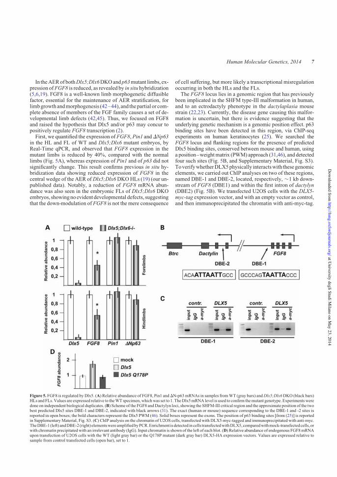
In theAERof bothDlx5;Dlx6DKOandp63mutant limbs, ex-pression ofFGF8 is reduced, as revealed by in situ hybridization(5,6,19). FGF8 is a well-known limb morphogenetic diffusiblefactor, essential for the maintenance of AER stratification, forlimbgrowth andmorphogenesis (42–44), and thepartial or com-plete absence of members of the FGF family causes a set of de-velopmental limb defects (42,45). Thus, we focused on FGF8and raised the hypothesis that Dlx5 and/or p63 may concur topositively regulate FGF8 transcription (2).First, we quantified the expression of FGF8, Pin1 andDNp63
in the HL and FL of WT and Dlx5;Dlx6 mutant embryos, byReal-Time qPCR, and observed that FGF8 expression in themutant limbs is reduced by 40%, compared with the normallimbs (Fig. 5A), whereas expression of Pin1 and of p63 did notsignificantly change. This result confirms previous in situ hy-bridization data showing reduced expression of FGF8 in thecentral wedge of the AER ofDlx5;Dlx6DKOHLs (19) (our un-published data). Notably, a reduction of FGF8 mRNA abun-dance was also seen in the embryonic FLs of Dlx5;Dlx6 DKOembryos, showingnoevident developmental defects, suggestingthat the down-modulation of FGF8 is not the mere consequence
of cell suffering, but more likely a transcriptional misregulationoccurring in both the HLs and the FLs.
The FGF8 locus lies in a genomic region that has previouslybeen implicated in the SHFM type-III malformation in human,and to an ectrodactyly phenotype in the dactylaplasia mousestrain (22,23). Currently, the disease gene causing this malfor-mation is uncertain, but there is evidence suggesting that theunderlying genetic mechanism is a genomic position effect. p63binding sites have been detected in this region, via ChIP-seqexperiments on human keratinocytes (25). We searched theFGF8 locus and flanking regions for the presence of predictedDlx5 binding sites, conserved between mouse and human, usinga position–weightmatrix (PWM) approach (31,46), and detectedfour such sites (Fig. 5B, and Supplementary Material, Fig. S3).ToverifywhetherDLX5physically interactswith these genomicelements, we carried out ChIP analyses on two of these regions,named DBE-1 and DBE-2, located, respectively, !1 kb down-stream of FGF8 (DBE1) and within the first intron of dactylyn(DBE2) (Fig. 5B). We transfected U2OS cells with the DLX5-myc-tag expression vector, and with an empty vector as control,and then immunoprecipitated the chromatin with anti-myc-tag.
Figure 5. FGF8 is regulated by Dlx5. (A) Relative abundance of FGF8, Pin1 andDN-p63mRNAs in samples fromWT (gray bars) andDlx5;Dlx6DKO (black bars)HLs and FLs. Values are expressed relative to theWT specimen, whichwas set to 1. TheDlx5mRNA level is used to confirm themutant genotype. Experimentsweredone on independent biological duplicates. (B) Scheme of the FGF8 andDactylyn loci, showing the SHFM-III critical region and the approximate position of the twobest predicted Dlx5 sites DBE-1 and DBE-2, indicated with black arrows (31). The exact (human or mouse) sequence corresponding to the DBE-1 and -2 sites isreported in open boxes; the bold characters represent the Dlx5 PWM (46). Solid boxes represent the exons. The position of p63 binding sites [from (25)] is reportedin SupplementaryMaterial, Fig. S3. (C) ChIP analysis on the chromatin of U2OS cells, transfected with DLX5-myc-tagged and immunoprecipitated with anti-myc.TheDBE-1 (left) andDBE-2 (right) elementswereamplifiedbyPCR.Enrichment is detected in cells transfectedwithDLX5,comparedwithmock-transfectedcells, orwith chromatin precipitatedwith an irrelevant antibody (IgG). Input chromatin is shown of the left of each blot. (D) Relative abundance of endogenous FGF8mRNAupon transfection of U2OS cells with the WT (light gray bar) or the Q178P mutant (dark gray bar) DLX5-HA expression vectors. Values are expressed relative tosample from control transfected cells (open bar), set to 1.
Human Molecular Genetics, 2014 7
at University degli Studi M
ilano on May 23, 2014
http://hmg.oxfordjournals.org/
Dow
nloaded from
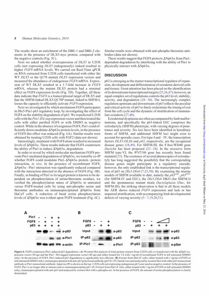
The results show an enrichment of the DBE-1 and DBE-2 ele-ments in the presence of DLX5-myc protein, compared withthe negative controls (Fig. 5C).Next we asked whether overexpression of DLX5 in U2OS
cells (not expressing DLX5 endogenously) indeed resulted inhigher FGF8 mRNA levels. We carried out Real-Time qPCRon RNA extracted from U2OS cells transfected with either theWT DLX5 or the Q178 mutant DLX5 expression vectors andmeasured the abundance of endogenous FGF8mRNA. Expres-sion of WT DLX5 resulted in a 1.7-fold increase in FGF8mRNA, whereas the mutant DLX5 protein had a minimaleffect on FGF8 expression levels (Fig. 5D). Together, all thesedata indicate that FGF8 is a transcriptional target of DLX5 andthat the SHFM-linked DLX5-Q178P mutant, linked to SHFM-I,looses the capacity to efficiently activate FGF8 expression.Next we investigated bywhichmechanism FGF8 participates
in Dlx5-Pin1-p63 regulatory loop, by investigating the effect ofFGF8 on the stability/degradation of p63.We transfected U2OScellswith thePin1-HA-tag expressionvector and then treated thecells with either purified FGF8 or with DMSO as negativecontrol.While in the absence of exogenous FGF8, Pin1 could ef-ficiently down-modulateDNp63a protein levels, in the presenceof FGF8 this effect was reduced (Fig. 6A). Similar results wereobtained by treating U2OS cells with FGF2 (data not shown).Interestingly, treatmentwith FGF8 alone resulted in increased
levels of DNp63a. These results indicate that FGF8 counteractsthe ability of Pin1 to induce DNp63a, degradation.In order to reveal by which molecular mechanism FGF8 pre-
vents Pin1-mediated degradation ofDNp63a, we tested by co-IPwhether FGF8 could modulate Pin1-DNp63a protein–proteininteraction, in vivo. In the presence of recombinant FGF8,DNp63a–Pin1 interaction was significantly reduced comparedwith the interaction detected in the absence of FGF8 (Fig. 6B).Finally, as binding of Pin1 to its target protein is known to be de-pendent on phosphorylation of serine/threonine residues, weverified the phosphorylation status of DNp63a in untreatedversus FGF8-treated cells by using anti-phospho serine andthreonine antibodies on immunoprecipitated DNp63a fromHaCaT cells. A reduction of basal serine phosphorylationlevels of DNp63a was evident upon FGF8 treatment (Fig. 6C).
Similar results were obtained with anti-phospho threonine anti-bodies (data not shown).These results suggest that FGF8 protectsDNp63a from Pin1-
dependent degradation by interfering with the ability of Pin1 tophysically interact with DNp63a.
DISCUSSION
p63 is emerging as themaster transcriptional regulator of expan-sion, development and differentiation of ectoderm-derived cellsand tissues. Great attention has been placed on the identificationof its downstream transcriptional targets (25,26,47); however, anequal complex set of regulations controls the p63 level, stability,activity, and degradation (28–30). The increasingly complexregulation upstream and downstream of p63 reflects the peculiarand critical activity of p63 to finely orchestrate the timing of exitfrom the cell cycle and the dynamic of stratification of mamma-lian ectoderm (27,48).Ectodermal dysplasias are often accompanied by limbmalfor-
mations, and specifically the p63-linked EEC comprises theectrodactyly (SHFM) phenotype, with varying degrees of pene-trance and severity. Six loci have been identified in hereditaryforms of SHFM, and additional SHFM loci might exist toaccount for sporadic cases. For type-I and –IV, the transcriptionfactors DLX5-DLX6 and p63, respectively, are the recognizeddisease genes (18,49). For SHFM-III, the F-box/WD40 geneDactylin has been proposed (22–24). In the recessive formSHFM type-VI, the WNT10b gene has recently been foundmutated (15). The existence of several phenocopies of ectrodac-tyly has long suggested the possibility that the correspondingdisease genes might participate in a regulatory cascade;however, the only established link is the transcriptional regula-tion of p63 on Dlx5;Dlx6 (7,25,50). By examining the murinemodels of SHFM available to date, namely the p63null, p63EEC
(for SHFM-IV and EEC), the Dlx5;Dlx6 DKO (for SHFM-I)and the spontaneous mutant strain Dactylaplasia (Dac, forSHFM-III), the striking observation is that in all these modelsthe AER shows reduced FGF8 expression and lack or hasimpaired stratification, with accompanying limb developmentaldefects of varying severity (5–7,19,20,51).
Figure 6. FGF8 counteracts Pin1-induced p63 degradation. (A) Western blot analysis of total protein extracts from U2OS cells co-transfected with the DNp63 ex-pression vector (30 ng) and the Pin1–HA-tagged expression vector (40 ng) and either treated for 3 h with 1 ng/ml of recombinant FGF8 or left untreated (DMSOonly). In the presence of FGF8, Pin1-induced p63 degradation is significantly less efficient. (B) Extract from HaCaT cells, either treated with 1 ng/ml of FGF8 orleft untreated (DMSOonly), immunoprecipitatedwith anti-p63polyclonal antibody (p63H-129, SantaCruz) and analyzedbywestern blotwith either p63monoclonalantibody or Pin1monoclonal antibody. Input (no IP) is shown on the top. U2OS cells not expressing endogenous p63were used as negative control. In the presence ofFGF8, Pin1 is no longer able to interact and co-immunoprecipitate p63. (C) Extract from HaCaT cells, either treated with 1 ng/ml of FGF8 or left untreated (DMSOonly), immunoprecipitated with anti-p63 and analyzed by western blot witha-phospho-ser. In the presence of FGF8, the amount of serine phosphorylation is clearlydiminished.
8 Human Molecular Genetics, 2014
at University degli Studi M
ilano on May 23, 2014
http://hmg.oxfordjournals.org/
Dow
nloaded from

Here we show that the Pin1 cis/trans isomerase is a regulatorof p63 protein stability, inducing proteasome-mediated degrad-ation ofDNp63a.We also show that FGF8 counteracts this func-tion and thus promotes p63 stability. TheFGF8 locus appears tobe regulated by DLX5; thus, we propose a model (see Fig. 7) inwhich these two SHFM disease genes, together with FGF8 andFGFR1, take part in a regulatory loop that tightly controls p63protein level. According to our model, the activation of thisloop permits stratification and specialization of ectoderm cellsinto the AER, in a time and region-restricted manner duringlimb development. In SHFM type-I, type-III and type-IV, suchregulation is impaired leading to reduced AER stratification,limbmalformation and skeletal defects. Although somemolecu-lar details still remain to be fully clarified, our novel findings to-gether with previous work from our team (7) provide adevelopmental andmolecular explanation for a set of congenitallimb malformations.Our model helps to explain other observations and findings.
The FGF8 locus is located close (!50 kb) to the chromosomalregion implicated in SHFM type-III and in the Dac mice. Al-though Dactylyn has been proposed as the disease gene forthese malformations (22–24), no clear evidence is availableon its role in limb development. Conversely, there are reasonsto implicate FGF8 in themolecular pathogenesis of this disorder.First, the SHFM-III/Dac rearrangement does not interrupt anygene, and therefore, it is likely to act by perturbing the chromo-somal organization and affecting expression of nearby genes;second, the FGF8 expression is reduced in the limb buds ofDlx5;Dlx6, p63null, p63EEC and Dac mutant embryos (5–7,19,22) (and this report); third, the presence of Dlx5 (thisreport) and p63 (25) binding sites in conserved genomicregions near the FGF8 and the Dactylyn loci; fourth, FGF8plays a critical role for limb bud growth, patterning,morphogen-esis aswell asAERmaintenance (9,42,51–53). Thus, it is tempt-ing to propose that misregulation of FGF8 expression is themolecular lesion at the basis of SHFM-III/Dac; however,direct evidence for this is lacking and should be explored infuture works.The role of FGF8 in the signaling from the AER for the prox-
imal limb development iswell known (45,52), however not fullycomprehended in cellular terms. The AER-specific conditionaldisruption of FGF8 does not lead to altered AER inductionand stratification, per se (54); however, this could be explainedby the fact that during limb development, FGF4, FGF9 andFGF17 have been shown to compensate for the loss of FGF8(45). Conversely, the AER-specific conditional disruption ofFgfr2 leads to altered AER stratification and function and limbdefects (55). AER-derived FGFs have been shown to promotenon-directional mesenchymal cell movements during limb budmorphogenesis (44),andconsistently, theconditional inactivationofFgfr1 in the limbmesodermdisrupts the relative proportions ofthe limb elements and leads to profound limbmalformations (56).Most relevant, mutations in FGFR1 have recently been found inpatients with Hartsfield syndrome (OMIM 615465), a congenitalcondition comprising ectrodactyly (57). This finding clearly sup-ports the notion that impairment of the FGF signaling is directlyinvolved in the molecular pathogenesis of ectrodactyly.While FGFs promote mesenchymal cell movements, in the
same article the authors show that Wnt5a promotes orientedcell divisions/movements during limb development (44), and
Wnt5a is a known target ofDlx5 (58).We believe that these find-ings are highly relevant for the comprehensionof theSHFMmal-formation, and we are tempted to speculate that the reducedexpression of FGF8 and Wnt5a (our unpublished data) in thecentral AER ofDlx5;Dlx6DKO limbs may induce mis-orienteddivisions/movements of mesenchymal cells in this sector, hencealtered morphogenesis/loss of central digits. This possibilitywarrants future experimental work. When a more completemodel will be available, the hope is to be able to exploit thisknowledge to restore normal levels of these soluble signalingfactors, toward correcting the SHFM defects.
AlteredPin1-dependent p63 regulationmay impact on severalcellular processes, in addition to ectoderm stratification. Cellsfrom Pin1 knock-out mice have difficulties in exiting the G0and entering the S phase, and Pin1 null animals have meioticdefects and are hypofertile (40,41). In these animals, an alteredphosphorylation levels of RB correlate with tumor growth(59). However, as Pin1 interacts with p53 and p73, the contribu-tion of p63 is uncertain. Likewise, a Pin1/mutant p53 axis hasbeen identified that promotes aggressiveness of breast cancercells; however, the relevance of p63 in this context is not welldefined (37). A role for p63 has been established in cancertypes of ectodermal or endodermal origin, in particular lungand skin carcinomas. Indeed, DNp63a regulates keratinocyteproliferation by controlling PTEN expression and localization(60). Notably, a mis-activation of p63 in squamous cell carcin-omas has been functionally linked with the activation of theFGFR2 receptor (61), further supporting the view that FGFs par-ticipate in Pin1-dependent p63 stability.
It would be important to define whether this regulatory path-way participates in skin carcinogenesis.
MATERIALS ANDMETHODS
Mouse strains
The Dlx5;Dlx6 DKO mouse strain (20) was maintained in amixed C57/BL6:DBA genetic background. The Pin1 nullmouse strain was originally generated by Fujimori and cowor-kers (40), then transferred onto a C57/BL6 pure backgroundby Atchison and coworkers (41) and maintained in this back-ground. The day of the vaginal plugwas considered as embryon-ic age 0.5. Embryos were collected at the indicated ages in coldPBS, fixed in cold 4% PFA for 8–12 h and processed for cryo-preservation and sectioning according to standard protocols.Extra-embryonic tissues were used for genotyping by PCR.
Immunofluorescence on embryonic limbs
Longitudinal sections of 12–15 mm were collected on glassslides, blocked with PBS with 1% BSA for 1 h at RT and incu-bated with the following primary antibodies, diluted from 1 :250 to 1 : 50 in PBS + 1% BSA, ON at 48C: anti-Pin1 (G8sc-46660, Santa Cruz), with anti-p63 (4A4 sc-8431, SantaCruz) and with anti-E-cadherin (36/E 610182, BD) and thenincubated with secondary antibodies anti-mouse-Cy2 andanti-rabbit-Cy3 (Jackson ImmunoResearch) diluted 1 : 200,1 h at RT, washed, stained with DAPI for the nuclei detectionand examinedwith aZeissObserver-Z1 fluorescentmicroscope,equipped with Apotome system. Raw images were digitally
Human Molecular Genetics, 2014 9
at University degli Studi M
ilano on May 23, 2014
http://hmg.oxfordjournals.org/
Dow
nloaded from

Figure 7.Model of the molecular loop between Dlx5 and p63 during AER stratification. Our proposed model of the regulatory loop linking the Dlx5 and the p63SHFM-genes, via the activity of Pin1. (A) In WT AER cells, Dlx5;Dlx6 positively control FGF8 transcription, a function most likely shared with Dlx1 and Dlx2[see ref. (2)]. Likewise, p63 also activates FGF8 transcription, acting on independent genomic elements. AER-expressed FGF8 acts on the AER cells and preventPin1-inducedDNp63proteindegradation: suchfinemechanismdynamicallymaintains a control over the level of p63 in theAERcells, to assure their time- and region-restricted ability of these cells to stratify. (B)Mutation or loss ofDlx5 yield to a reduced FGF8 expression and an augmented ability of Pin1 to induce p63 degradation.Consequently,DNp63 tends tobedepleted in theAERnuclei,whichmay result in a furtherdown-modulationofFGF8.ReducedFGF8andp63cause impairment in theability of theAERcells to stratify. (C)Mutant p63 associatedwith congenital limbmalformations in human (LMSandSHFM) are relatively resistant to Pin1-induceddegradation, although theyappear tobe transcriptionally inactive.Solidarrows indicateactivation; solid stamps indicate repression.Linewidthsareproportional to theintensity and/or efficiency. Transcription factors are frames in squares; the other genes are framed in ovals.
10 Human Molecular Genetics, 2014
at University degli Studi M
ilano on May 23, 2014
http://hmg.oxfordjournals.org/
Dow
nloaded from

processed to normalize the background and optimize the con-trast, with Photoshop (Adobe), and mounted with QuarkXpress(Pantone).Semi-quantitative immunofluorescence analysis was per-
formed with ImageJ-64 (v1.45) software. Images were first con-verted to grayscale, and the DAPI channel was used to countnuclei. p63 intensity was quantified after background correctionand normalized respect to the number of nuclei in the region ofinterest.Dataarepresentedasmeanands.d. of!4/5different sec-tions of three different embryos. A significant T-test score is indi-cated by asterisks: "indicates P, 0.05, ""indicates P, 0.01.
Plasmids
Vectors expressing the WT DNp63a isoform of, or the disease-linked mutant p63, were previously described (62,63). TheDLX5-myc-tagged expression vectors were obtained fromOriGene, and previously used (58). The Q178P DLX5-mycpoint mutation [based on the sequence in NM_005221.5 (18)]was generated by site-directedmutagenesis in theDLX5-myc ex-pression plasmid and sequence-verified (Bio-Fab Research,Rome, Italy). The Pin1 si-RNA was previously described (37).
Cell cultures and transfections
TheU2OS human osteosarcoma and the A431 human epidermoidsquamous carcinoma cell lines were maintained in Dulbecco’smodified Eagle’s medium (D-MEM) and 10% fetal bovine serum.For transfection, 50,000cellswere seeded into24-wellmulti-platesand the next day transfectedwith Lipofectamine 2000 (Invitrogen)according to the manufacturer’s instructions. The total amount oftransfected DNA (500 ng) was kept constant using empty vectorwhen necessary. After 24 h, cells were lysed and assayed forwestern blot analysis (29). MG132 treatment was initiated theday after transfection with 5 mM MG132 (Sigma) for 5 h. FGFstreatments were initiated 20 h after transfection with 1 ng/ml ofFGF8 or FGF2 for 3 h.
Western blot analyses
Twenty-four hours after transfection, cells were lysed in 100 mlof loading buffer (2% sodium dodecyl sulfate, 30% glycerol,144 mM b-mercaptoethanol, 100 mM Tris–HCl pH 6.8 and0.1% Bromo-Phenol Blue): extracts were separated on SDS–10% polyacrylamide gel, transferred on nitrocellulose mem-brane (Protran, Millipore) and incubated with the relative anti-bodies and developed according to the manufacturer’sinstructions (GeneSpin). The following primary antibodieswere used: a-p63 (4A4, sc- 8431, Santa Cruz), a-actin mousemonoclonal (A2066, Sigma), a-Pin1 mouse monoclonal (G9,sc-46660, Santa Cruz),a-Pin1 rabbit polyclonal,a-phospho-serpolyclonal (Invitrogen, 618100) and a phospho-thr polyclonal(Cell Signaling, # 9381). As secondary antibodies, we used thefollowing: a-mouse secondary (sc-2005, Santa Cruz) anda-rabbit secondary (sc-2030, Santa Cruz).
Co-immunoprecipitation
HaCaT cells (4# 106/150 mm plate) were treated with FGF8(1 ng/ml) for 3 h and then harvested for the preparation of whole-
cell lysates using RIPA buffer [10 mM Tris–HCl pH 8, 2 mM
EDTA, 0.1% SDS, 0.1% sodium deoxycholate, 140 mM NaCl,1# Triton, supplementedwith 1 mMphenylmethylsulfonyl fluor-ide and protease inhibitors (all from Sigma)]. Cell lysates wereincubated on ice for 20 min., vortexed and centrifuged at6600#g for 10 min to remove cell debris. Protein concentrationwas determined with Bradford Reagent (Sigma). Three milli-grams of cell lysate was incubated overnight at 48C with 3 mgof anti-p63 (H-129, sc-8344, SantaCruz). The immunocomplexeswere collected by incubating with a mix of Protein A–Agaroseand Protein G–Sepharose (Sigma) overnight at 48C. The beadswere washed three times: the first wash with RIPA buffer andthe others with PBS. The beads were then resuspended in 2#Lysis buffer, loaded directly on a 10% SDS–polyacrylamidegel and subjected to western blot with the indicated antibodies.
mRNA quantification by Real-Time qPCR
Embryonic FLs and HLs were dissected from embryos at theindicated age, in coldRNase-freePBS,undermicroscopic exam-ination.Aminimumof three limbswere pooled, according to thegenotype and collected in Trizol (Invitrogen). Total RNA wasextracted with the TRI reagent (Sigma) and treated withDNAse I (Ambion). Reverse-transcription and cDNA synthesiswere done using kits (Invitrogen), as previously reported (7,64).Three nanograms of each cDNA samplewere used in Real-TimeqPCR analyses using SYBR green IQ reagent (Biorad) on T900HT Fast Real Time PCR Sistem (Applied Biosystems). TheTATA-binding protein (TBP) and the GAPDH mRNAs wereused for normalization. Primer sequences are provided in Sup-plementary Material, Table S1. Experiments were repeatedtwice on independent samples; every point was done on bio-logical duplicates. Analyses were performed with ABI 2.1 soft-ware (Applied Biosystems).
Total RNA from U2OS cells was extracted using the TRIreagent and treated with DNase-I (Ambion). One microgramsof RNA was retrotranscribed with SuperScriptII (Invitrogen).qPCR was performed using SYBR green IQ reagent (Biorad)on the Rotor Gene machine. Primers were designed to amplifyregions of 80–120 bp in size. Tubulin and GAPDH mRNAswere used for normalization. Experiments were repeated twiceon independent samples. Primer sequences are provided in Sup-plementary Material, Table S1.
Chromatin immunoprecipitation
ChIP analyses were performed on sheared genomic DNA from1 # 106 U2OS cells transfected with 12 mg of DLX5-myc orDLX5-Q178P-myc vectors, or with the empty pcDNA3 vectoras control and immunoprecipitated with 5 mg of anti-myc-TAGmouse monoclonal antibody (SantaCruz, sc-40) or 5 mg of anti-Flag antibody (Sigma, F3165) as previously described (7). Fornegative control, an irrelevant antibodywas used.The sequencesof theoligonucleotides used for this analysis are provided inSup-plementary Material, Table S1).
SUPPLEMENTARYMATERIAL
Supplementary Material is available at HMG online.
Human Molecular Genetics, 2014 11
at University degli Studi M
ilano on May 23, 2014
http://hmg.oxfordjournals.org/
Dow
nloaded from

ACKNOWLEDGEMENTS
We thank Drs T. Uchida (Tohoku University, Sendai Japan) andA.R. Means (Duke University Medical Center, Durham, USA)for providing the Pin1 mutant mouse strain and Dr PatriziaDell’Era for providing FGF8 and FGF2 proteins. We alsothank Dr Enzo Calautti (University of Torino) for helpful com-ments on themanuscript andDrMicheleMontrone for technicalhelp.
Conflict of Interest statement. None declared.
FUNDING
This work was supported by grants from the Italian TelethonFoundation (GGP11097 toL.G.,A.C. andM.G.R.), fromFonda-zione Ricerca Biomedica (to M.G.R.), from Ministero ItalianoUniversita e Ricerca (PRIN2009 to A.C. and L.G.;RBAP10XKNC-003 and PRIN 2009–2009YP9AE5 toG.D.S.) and fromAIRCSpecial ProgramMolecularClinicalOn-cology ‘‘5 per mille’’ to G.D.S. Funding to pay the OpenAccesspublication charges for this article was provided by The Fonda-zione Telethon Italy.
REFERENCES
1. Ianakiev, P., Kilpatrick, M.W., Toudjarska, I., Basel, D., Beighton, P. andTsipouras, P. (2000) Split-hand/split-foot malformation is caused bymutations in the p63 gene on 3q27. Am. J. Hum. Genet., 67, 59–66.
2. Guerrini, L., Costanzo, A. and Merlo, G.R. (2011) A symphony ofregulations centered on p63 to control development of ectoderm-derivedstructures. J. Biomed. Biotechnol., 2011, 864904.
3. Berdon-Zapata, V., Granillo-Alvarez, M., Valdes-Flores, M., Garcia-Ortiz,J.E., Kofman-Alfaro, S. and Zenteno, J.C. (2004) p63 gene analysis inMexican patients with syndromic and non-syndromic ectrodactyly.J. Orthop. Res., 22, 1–5.
4. van Bokhoven, H., Hamel, B.C., Bamshad, M., Sangiorgi, E., Gurrieri, F.,Duijf, P.H.,Vanmolkot,K.R., vanBeusekom,E., vanBeersum,S.E.,Celli, J.et al. (2001) p63 Gene mutations in eec syndrome, limb-mammarysyndrome, and isolated split hand-split foot malformation suggest agenotype-phenotype correlation. Am. J. Hum. Genet., 69, 481–492.
5. Mills, A.A., Zheng, B., Wang, X.J., Vogel, H., Roop, D.R. and Bradley, A.(1999) p63 is a p53 homologue required for limb and epidermalmorphogenesis. Nature, 398, 708–713.
6. Yang, A., Schweitzer, R., Sun, D., Kaghad, M., Walker, N., Bronson, R.T.,Tabin, C., Sharpe, A., Caput, D., Crum, C. et al. (1999) p63 is essential forregenerative proliferation in limb, craniofacial and epithelial development.Nature, 398, 714–718.
7. Lo Iacono, N., Mantero, S., Chiarelli, A., Garcia, E., Mills, A.A., Morasso,M.I., Costanzo,A., Levi,G., Guerrini, L. andMerlo,G.R. (2008)Regulationof Dlx5 and Dlx6 gene expression by p63 is involved in EEC and SHFMcongenital limb defects. Development, 135, 1377–1388.
8. Fernandez-Teran, M. and Ros, M.A. (2008) The apical ectodermal ridge:morphological aspects and signaling pathways. Int. J. Dev. Biol., 52,857–871.
9. Zeller, R., Lopez-Rios, J. and Zuniga, A. (2009) Vertebrate limb buddevelopment: moving towards integrative analysis of organogenesis. Nat.Rev. Genet., 10, 845–858.
10. Zeller, R. (2010) The temporal dynamics of vertebrate limb development,teratogenesis and evolution. Curr. Opin. Genet. Dev., 20, 384–390.
11. Koster,M.I., Kim, S., Mills, A.A., DeMayo, F.J. and Roop, D.R. (2004) p63is the molecular switch for initiation of an epithelial stratification program.Genes Dev., 18, 126–131.
12. Koster, M.I., Dai, D., Marinari, B., Sano, Y., Costanzo, A., Karin, M. andRoop, D.R. (2007) p63 induces key target genes required for epidermalmorphogenesis. Proc. Natl. Acad. Sci. USA, 104, 3255–3260.
13. Sifakis, S., Basel, D., Ianakiev, P., Kilpatrick, M. and Tsipouras, P. (2001)Distal limb malformations: underlying mechanisms and clinicalassociations. Clin. Genet., 60, 165–172.
14. Elliott, A.M., Evans, J.A. and Chudley, A.E. (2005) Split hand footmalformation (SHFM). Clin. Genet., 68, 501–505.
15. Aziz, A., Irfanullah Khan, S., Zimri, F.K., Muhammad, N., Rashid, S. andAhmad, W. (2013) Novel homozygous mutations in the WNT10B geneunderlying autosomal recessive split hand/foot malformation in threeconsanguineous families. Gene. doi:10.1016/j.gene.2013.10.047
16. Crackower, M.A., Scherer, S.W., Rommens, J.M., Hui, C.C., Poorkaj, P.,Soder, S., Cobben, J.M., Hudgins, L., Evans, J.P. and Tsui, L.C. (1996)Characterization of the split hand/split foot malformation locus SHFM1 at7q21.3-q22.1 and analysis of a candidate gene for its expression during limbdevelopment. Hum. Mol. Genet., 5, 571–579.
17. Scherer, S.W., Poorkaj, P., Allen, T., Kim, J., Geshuri, D., Nunes,M., Soder,S., Stephens, K., Pagon, R.A., Patton,M.A. et al. (1994) Finemapping of theautosomal dominant split hand/split foot locus on chromosome 7, bandq21.3-q22.1. Am. J. Hum. Genet., 55, 12–20.
18. Shamseldin, H.E., Faden, M.A., Alashram, W. and Alkuraya, F.S. (2012)Identification of a novel DLX5 mutation in a family with autosomalrecessive split hand and foot malformation. J. Med. Genet., 49, 16–20.
19. Robledo, R.F., Rajan, L., Li, X. and Lufkin, T. (2002) The Dlx5 and Dlx6homeobox genes are essential for craniofacial, axial, and appendicularskeletal development. Genes Dev., 16, 1089–1101.
20. Merlo, G.R., Paleari, L.,Mantero, S., Genova, F., Beverdam,A., Palmisano,G.L., Barbieri, O. and Levi, G. (2002) Mouse model of split hand/footmalformation type I. Genesis, 33, 97–101.
21. Witters, I., Van Bokhoven, H., Goossens, A., Van Assche, F.A. and Fryns,J.P. (2001) Split-hand/split-foot malformation with paternal mutation in thep63 gene. Prenat. Diagn., 21, 1119–1122.
22. Sidow, A., Bulotsky, M.S., Kerrebrock, A.W., Birren, B.W., Altshuler, D.,Jaenisch, R., Johnson, K.R. and Lander, E.S. (1999) A novel member of theF-box/WD40 gene family, encoding dactylin, is disrupted in the mousedactylaplasia mutant. Nat. Genet., 23, 104–107.
23. de Mollerat, X.J., Gurrieri, F., Morgan, C.T., Sangiorgi, E., Everman, D.B.,Gaspari, P., Amiel, J., Bamshad, M.J., Lyle, R., Blouin, J.L. et al. (2003) Agenomic rearrangement resulting in a tandem duplication is associated withsplit hand-split foot malformation 3 (SHFM3) at 10q24. Hum. Mol. Genet.,12, 1959–1971.
24. Friedli,M., Nikolaev, S., Lyle, R., Arcangeli, M., Duboule, D., Spitz, F. andAntonarakis, S.E. (2008) Characterization of mouse Dactylaplasiamutations: a model for human ectrodactyly SHFM3.Mamm. Genome, 19,272–278.
25. Kouwenhoven, E.N., van Heeringen, S.J., Tena, J.J., Oti, M., Dutilh, B.E.,Alonso, M.E., de la Calle-Mustienes, E., Smeenk, L., Rinne, T., Parsaulian,L. et al. (2010) Genome-wide profiling of p63 DNA-binding sites identifiesan element that regulates gene expression during limb development in the7q21 SHFM1 locus. PLoS Genet., 6, e1001065.
26. Vigano,M.A., Lamartine, J., Testoni, B.,Merico,D., Alotto, D., Castagnoli,C.,Robert,A.,Candi,E.,Melino,G.,Gidrol,X. et al. (2006)Newp63 targetsinkeratinocytes identifiedbyagenome-wide approach.EMBOJ.,25, 5105–5116.
27. Moretti, F., Marinari, B., Lo Iacono, N., Botti, E., Giunta, A., Spallone, G.,Garaffo, G., Vernersson-Lindahl, E., Merlo, G., Mills, A.A. et al. (2010) Aregulatory feedback loop involving p63 and IRF6 links the pathogenesis of 2genetically different human ectodermal dysplasias. J. Clin. Invest., 120,1570–1577.
28. Galli, F., Rossi, M., D’Alessandra, Y., De Simone, M., Lopardo, T., Haupt,Y., Alsheich-Bartok, O., Anzi, S., Shaulian, E., Calabro, V. et al. (2010)MDM2 and Fbw7 cooperate to induce p63 protein degradation followingDNA damage and cell differentiation. J. Cell. Sci., 123, 2423–2433.
29. Ghioni, P., D’Alessandra, Y., Mansueto, G., Jaffray, E., Hay, R.T., LaMantia, G. and Guerrini, L. (2005) The protein stability and transcriptionalactivity of p63alpha are regulated by SUMO-1 conjugation. Cell Cycle, 4,183–190.
30. Ratovitski, E.A., Patturajan, M., Hibi, K., Trink, B., Yamaguchi, K. andSidransky, D. (2001) p53 associates with and targets Delta Np63 into aprotein degradation pathway. Proc. Natl. Acad. Sci. USA, 98, 1817–1822.
31. Vieux-Rochas, M., Bouhali, K., Mantero, S., Garaffo, G., Provero, P.,Astigiano, S., Barbieri, O., Caratozzolo, M.F., Tullo, A., Guerrini, L. et al.(2013) BMP-mediated functional cooperation between Dlx5;Dlx6 andMsx1;Msx2 during mammalian limb development. PLoS One, 8, e51700.
12 Human Molecular Genetics, 2014
at University degli Studi M
ilano on May 23, 2014
http://hmg.oxfordjournals.org/
Dow
nloaded from

32. Zacchi, P., Gostissa, M., Uchida, T., Salvagno, C., Avolio, F., Volinia, S.,Ronai, Z., Blandino, G., Schneider, C. and Del Sal, G. (2002) The prolylisomerasePin1 reveals amechanism to control p53 functions after genotoxicinsults. Nature, 419, 853–857.
33. Mantovani, F., Piazza,S.,Gostissa,M.,Strano,S., Zacchi, P.,Mantovani,R.,Blandino, G. and Del Sal, G. (2004) Pin1 links the activities of c-Abl andp300 in regulating p73 function.Mol. Cell, 14, 625–636.
34. Mantovani, F., Tocco, F., Girardini, J., Smith, P., Gasco, M., Lu, X., Crook,T. and Del Sal, G. (2007) The prolyl isomerase Pin1 orchestrates p53acetylation and dissociation from the apoptosis inhibitor iASPP.Nat. Struct.Mol. Biol., 14, 912–920.
35. Grison, A., Mantovani, F., Comel, A., Agostoni, E., Gustincich, S.,Persichetti, F. and Del Sal, G. (2011) Ser46 phosphorylation andprolyl-isomerase Pin1-mediated isomerization of p53 are key events inp53-dependent apoptosis induced by mutant huntingtin. Proc. Natl. Acad.Sci. USA, 108, 17979–17984.
36. Sorrentino, G., Mioni, M., Giorgi, C., Ruggeri, N., Pinton, P., Moll, U.,Mantovani, F. andDelSal,G. (2013)Theprolyl-isomerasePin1activates themitochondrial death program of p53. Cell Death Differ., 20, 198–208.
37. Girardini, J.E.,Napoli,M., Piazza, S., Rustighi,A.,Marotta, C., Radaelli, E.,Capaci, V., Jordan, L., Quinlan, P., Thompson, A. et al. (2011) A Pin1/mutant p53 axis promotes aggressiveness in breast cancer. Cancer Cell, 20,79–91.
38. Lu, K.P. (2000) Phosphorylation-dependent prolyl isomerization: a novelcell cycle regulatory mechanism. Prog. Cell Cycle Res., 4, 83–96.
39. Rustighi, A., Tiberi, L., Soldano, A., Napoli, M., Nuciforo, P., Rosato, A.,Kaplan, F., Capobianco, A., Pece, S., Di Fiore, P.P. et al. (2009) Theprolyl-isomerase Pin1 is a Notch1 target that enhances Notch1 activation incancer. Nat. Cell Biol., 11, 133–142.
40. Fujimori, F., Takahashi, K., Uchida, C. and Uchida, T. (1999) Mice lackingPin1 develop normally, but are defective in entering cell cycle from G(0)arrest. Biochem. Biophys. Res. Commun., 265, 658–663.
41. Atchison, F.W., Capel, B. andMeans, A.R. (2003) Pin1 regulates the timingofmammalian primordial germcell proliferation.Development, 130, 3579–3586.
42. Boulet, A.M., Moon, A.M., Arenkiel, B.R. and Capecchi, M.R. (2004) Theroles of Fgf4 and Fgf8 in limb bud initiation and outgrowth.Dev. Biol., 273,361–372.
43. Moon, A.M. and Capecchi, M.R. (2000) Fgf8 is required for outgrowth andpatterning of the limbs. Nat. Genet., 26, 455–459.
44. Gros, J.,Hu, J.K.,Vinegoni,C., Feruglio,P.F.,Weissleder,R. andTabin,C.J.(2010)WNT5A/JNKand FGF/MAPKpathways regulate the cellular eventsshaping the vertebrate limb bud. Curr. Biol., 20, 1993–2002.
45. Mariani, F.V., Ahn, C.P. and Martin, G.R. (2008) Genetic evidence thatFGFs have an instructive role in limb proximal-distal patterning. Nature,453, 401–405.
46. Portales-Casamar, E., Thongjuea, S., Kwon, A.T., Arenillas, D., Zhao, X.,Valen, E., Yusuf, D., Lenhard, B., Wasserman, W.W. and Sandelin, A.(2010) JASPAR 2010: the greatly expanded open-access database oftranscription factor binding profiles. Nucleic Acids Res., 38, D105–D110.
47. Testoni, B., Borrelli, S., Tenedini, E., Alotto, D., Castagnoli, C., Piccolo, S.,Tagliafico, E., Ferrari, S., Vigano, M.A. and Mantovani, R. (2006)Identification of new p63 targets in human keratinocytes. Cell Cycle, 5,2805–2811.
48. Koster, M.I. and Roop, D.R. (2007) Mechanisms regulating epithelialstratification. Annu. Rev. Cell Dev. Biol., 23, 93–113.
49. Celli, J., Duijf, P., Hamel, B.C., Bamshad, M., Kramer, B., Smits, A.P.,Newbury-Ecob, R., Hennekam, R.C., Van Buggenhout, G., van Haeringen,
A. et al. (1999) Heterozygous germline mutations in the p53 homolog p63are the cause of EEC syndrome. Cell, 99, 143–153.
50. Vera-Carbonell, A., Moya-Quiles, M.R., Ballesta-Martinez, M.,Lopez-Gonzalez, V., Bafalliu, J.A., Guillen-Navarro, E. andLopez-Exposito, I. (2012) Rapp-Hodgkin syndrome and SHFM1 patients:delineating the p63-Dlx5/Dlx6 pathway. Gene, 497, 292–297.
51. Crackower, M.A., Motoyama, J. and Tsui, L.C. (1998) Defect in themaintenance of the apical ectodermal ridge in theDactylaplasiamouse.Dev.Biol., 201, 78–89.
52. Sun, X., Mariani, F.V. andMartin, G.R. (2002) Functions of FGF signallingfrom the apical ectodermal ridge in limb development. Nature, 418,501–508.
53. Benazet, J.D. and Zeller, R. (2009) Vertebrate limb development: movingfrom classical morphogen gradients to an integrated 4-dimensionalpatterning system. Cold Spring Harb. Perspect. Biol., 1, a001339.
54. Barrow, J.R., Thomas, K.R., Boussadia-Zahui, O., Moore, R., Kemler, R.,Capecchi, M.R. andMcMahon, A.P. (2003) EctodermalWnt3/beta-cateninsignaling is required for the establishment and maintenance of the apicalectodermal ridge. Genes Dev., 17, 394–409.
55. Gorivodsky, M. and Lonai, P. (2003) Novel roles of Fgfr2 in AERdifferentiation and positioning of the dorsoventral limb interface.Development, 130, 5471–5479.
56. Verheyden, J.M., Lewandoski,M.,Deng, C.,Harfe, B.D. andSun,X. (2005)Conditional inactivation of Fgfr1 in mouse defines its role in limb budestablishment, outgrowth and digit patterning. Development, 132,4235–4245.
57. Simonis, N., Migeotte, I., Lambert, N., Perazzolo, C., de Silva, D.C.,Dimitrov, B., Heinrichs, C., Janssens, S., Kerr, B., Mortier, G. et al. (2013)FGFR1 mutations cause Hartsfield syndrome, the unique association ofholoprosencephaly and ectrodactyly. J. Med. Genet., 50, 585–592.
58. Paina, S., Garzotto, D., Demarchis, S., Marino, M., Moiana, A., Conti, L.,Cattaneo, E., Perera, M., Corte, G., Calautti, E. et al. (2011) Wnt5a is atranscriptional target of dlx homeogenes and promotes differentiation ofinterneuron progenitors in vitro and in vivo. J. Neurosci., 31, 2675–2687.
59. Rizzolio, F., Caligiuri, I., Lucchetti, C., Fratamico, R., Tomei, V., Gallo, G.,Agelan, A., Ferrari, G., Toffoli, G., Klein-Szanto, A.J. et al. (2013)Dissecting Pin1 and phospho-pRb regulation. J. Cell. Physiol., 228, 73–77.
60. Leonard, M.K., Kommagani, R., Payal, V., Mayo, L.D., Shamma, H.N. andKadakia, M.P. (2011) DeltaNp63alpha regulates keratinocyte proliferationby controlling PTEN expression and localization. Cell Death Differ., 18,1924–1933.
61. Ramsey, M.R., Wilson, C., Ory, B., Rothenberg, S.M., Faquin, W., Mills,A.A. and Ellisen, L.W. (2013) FGFR2 signaling underlies p63 oncogenicfunction in squamous cell carcinoma. J. Clin. Invest., 123, 3525–3538.
62. Ghioni, P., Bolognese, F.,Duijf, P.H.,VanBokhoven,H.,Mantovani,R. andGuerrini, L. (2002) Complex transcriptional effects of p63 isoforms:identification of novel activation and repression domains.Mol. Cell. Biol.,22, 8659–8668.
63. Radoja, N., Guerrini, L., Lo Iacono, N., Merlo, G.R., Costanzo, A.,Weinberg, W.C., La Mantia, G., Calabro, V. and Morasso, M.I. (2007)Homeobox gene Dlx3 is regulated by p63 during ectoderm development:relevance in the pathogenesis of ectodermal dysplasias. Development, 134,13–18.
64. Lopardo,T.,Lo Iacono,N.,Marinari,B.,Giustizieri,M.L.,Cyr,D.G.,Merlo,G., Crosti, F., Costanzo, A. andGuerrini, L. (2008) Claudin-1 is a p63 targetgene with a crucial role in epithelial development. PLoS One, 3, e2715.
Human Molecular Genetics, 2014 13
at University degli Studi M
ilano on May 23, 2014
http://hmg.oxfordjournals.org/
Dow
nloaded from

Restelli at al.
DLX5, FGF8 and the Pin1 isomerase control !Np63" protein stability duringlimb development: a regulatory loop at the basis of the SHFM and EEC congenital malformations
Michela Restelli, Teresa Lopardo, Nadia Lo Iacono, Giulia Garaffo, Daniele Conte,
Alessandra Rustighi, Marco Napoli, Giannino Del Sal, David Perez-Morga, Antonio
Costanzo, Giorgio Roberto Merlo and Luisa Guerrini
Supplementary Figures and Table

Legend to Supplementary Figure 2
Western blot analysis of whole protein extracts from U2OS cells transiently co-
transfected with increasing amounts (20, 40 and 80 ng) of Pin1-HA-tagged vector
(indicated on top), and wild-type !Np63# (on the left) or !Np63$ (on the right) (30
ng each). As opposed to the " isoform, the # and $ isoforms are not degraded by Pin1
expression.

Legend to Supplementary Figure 2
A,B. Immunofluorescent staining with anti-Pin1 on sections of the embryonic HLs of
wild-type (A) or Dlx5;Dlx6-/- (B) embryos, at the age E11. Scale bar in A=20 µm. C.
As control, the same staining was done on sections of HLs from Pin1 KO embryos, at
the age E11. The lack of staining in the Pin1-/- specimen indicates that the antibody is
highly specific. D,E. Staining with anti-E-cadherin on adjacent sections,
counterstained with DAPI. The AER is outlined with a dotted line and indicated with
white arrows. F. Real-Time quantitative determination of the relative abundance of
the mRNA of Pin1, Wnt3a and Dlx5 in RNA samples from the HLs of wild-type and
Dlx5;Dlx6-/- embryos, normalized against GAPDH and TBP mRNA. The abundance
of the wild-type is set=1. As expected, the Dlx5 mRNA is nearly absent. The Pin1

mRNA is slightly increased while the Wnt3a mRNA is slightly decreased, however
these differences are not significant.

A. Location of predicted conserved Dlx5 binding elements (DBE) around the around
the murine Dactylyn and the FGF8 loci on chromosome 19q (10q in human), based
on the UCSC mouse genome browser. Dlx5 sites were bio-informatically predicted

using the published PWM, as described (ref. (31)). Sites are indicated with coloured
vertical bars (asterisk) and annotated with the species conservation. A color code is
used to indicate the number of species in which the site is conserved. Red indicated
the most conserved ones. B. Enlargement of the area indicated with a solid box in A,
centred around the genomic region comprising FGF8 and Dactylyn. The
chromosomal position and coordinates are reported on the top, the mammalian
genomic conservation is reported on the bottom. C. (top) Location of the DLX5
DBE-1 and DBE-2 sites, corresponding to the sites tested by ChIP analysis (see Fig. 5
and corresponding text), in the human genomic region around FGF8 and DACTYLYN
(chrom. 10q). (bottom) Location of a p63-binding site (p63 DBS) within a conserved
region of the human genome, between FGF8 and DACTYLYN, as reported (ref. (25)).

Supplementary Table I
A. Sequences of the primers used for Real-Time qPCR on mouse embryonic tissues.
mGAPDH F 5’ TGTCAGCAATGCATCCTGCA
mGAPDH R 5’ TGTATGCAGGGATGATGTTC
mTBP F 5’ GGGTTATCTTCACACACCATGA
mTBP R 5’ CGGTCGCGTCATTTTCTC
mRps9 F 5’ GACCAGGAGCTAAAGTTGATTGGA
mRps9 R 5’ TCTTGGCCAGGGTAAACTTGA
mDlx5 F 5’ TCTTATGGCAAAGCGCTCAA
mDlx5 R 5’ CGTTCACGCCGTGGTACTG
mDlx6 F 5’ TCCAGTGTGGGACGTTTCTG
mDlx6 R 5’ CTGTTGGGAGGCATACTGACG
mFGF8 F 5’ TGAGCTGATCCGTCACCA
mFGF8 R 5’ TCCTGCCTAAAGTCACACAGC
mPin1 F 5’ GTCCCTTCAGCAGAGGTCAG
mPin1 R 5’ ACAGTAGCAGGAAGGGCATC
mWnt3a F 5’ GAGTGCTCAGAGAGGAGTACTGG
mWnt3a R 5’ CTTAGTGCTCTGCAGCCTGA
m!Np63 F 5’ ATGTTGTACCTGGAAAACAATG
m!Np63 R 5’ GATGGAGAGAGGGCATCAAA
B. Sequences of the oligonucleotides used for ChIP analysis on Dlx5 Binding
Elements (DBE) near the FGF8 locus
DBE-1 (FGF8) For CCTCTGAAGACTCGGATGTTCC
DBE-1 (FGF8) Rev AGGAAACGCTTTCATCTGCAC
DBE-2 (FGF8) For GCCCGAGGCAGCTTGTCTA
DBE-2 (FGF8) Rev GAGCCCTCACTAATGGGGTTTTA

! &'!
PART III

FGF8, c-Abl and p300 cooperate in the regulation of !Np63"
protein stability
M. Restelli1, B. Marinari2, N. Gnesutta1, D. Conte3, G.R. Merlo3, A.
Costanzo2 and L. Guerrini1°
1 Dept. of Biosciences, University of Milan, I-20133, Italy 2 Dept. of Dermatology, University of Rome "Tor Vergata", I-00133 Rome, Italy 3 Telethon Laboratory, Dept. of Molecular Biotechnologies and Health Sciences,
University of Torino, I-10126 Torino, Italy
° Corresponding author:
Luisa Guerrini,
Department of Biosciences, University of Milano
Via Celoria 26, 20133 Milan, Italy
Tel +39-02 50315000
Fax +39-02 50315044
e-mail: [email protected]

! &)!
Abstract
The p63 transcription factor, homolog to the p53 tumor suppressor, plays a crucial
role in epidermal and limb development. Dominant mutations in the p63 gene give
rise to several human congenital syndromes characterized by skin, craniofacial and
limb defects. However, little is known on the post-translational modifications
controlling !Np63" functions. Here we report that, FGF8 is a crucial signalling
molecule inducing !Np63" stabilization and activation in both human cells and in
mice embryonic limb buds ex vivo. FGF8 treatments determined an increase in the
binding of !Np63" with the tyrosine kinase c-Abl and in the level of p300 driven
acetylation. In vitro, p300 catalizes acetylation of lysine K193: triggering !Np63"
stabilization and transcriptional activation. A prerequisite for the p300-!Np63" is
interaction is the !Np63" phosphorylation by the tyrosine kinase c-Abl. Notably,
this regulatory pathway induced by FGF8 is not active on the !Np63"K193E
natural mutant, associated to the Split-Hand/split Foot Malformation IV (SHFM-
IV) syndrome: that displays promoter specific altered DNA binding activity that
results in altered expression of !Np63" target genes involved in limb development.
Our results link together FGF8, c-Abl and p300 in a regulatory loop essential for
the activation and stabilization of !Np63": mutations or altered expression of
regulators in this pathway leads to abnormal limb development and possible onset
of SHFM-IV pathogenesis.

! '+!
Introduction The p63 transcription factor is highly homologous to the p53 tumor suppressor
gene in the DNA binding and in the oligomerization domains (1-4). The p63 gene
encodes for at least ten different protein isoforms differing in their amino and
carbossi-terminal regions as a consequence of alternative transcription start site and
alternative splicing respectively (5,6). !Np63" is the most expressed isoform,
largely present in the embryonic ectoderm and in the proliferating adult cells,
including the basal layer of the epidermis, breast and oral epithelium (7,8). p63
plays a pivotal role during development: indeed, p63 null mice show severe defects
in limb, skin and craniofacial development and die after birth due to dehydration
caused by absence of epidermal stratification.
In particular, limb defects of p63 -/- mice have been associated with developmental
failure of the Apical Ectodermal Ridge (AER), an essential structure required for
limb outgrowth, and loss of expression of key morphogens (9, 10). The DLX5 and
DLX6 transcription factors, playing key roles in the development and
morphogenesis of the head and limb skeleton, co-localize with !Np63" in the AER
and are direct !Np63" target genes (11-13). It’s well established that also FGFs
signaling molecules are essential for AER development. Notably, FGF8 is essential
for the correct functions of the AER and the consequent limb bud initiation and
FGF8 knock-out mice display severe defects in skeletal and limb developmet (15-
19). Recently, we linked together p63, DLX5, FGF8 and the prolyl isomerase
PIN1 in a regulatory pathway important for correct stratification and proper AER
development in mouse limb buds. FGF8 induced the expression of the DLX5 gene
and, on the other hand, counteracts the degradation of !Np63" protein induced by
Pin1 expression in both human osteosarcoma cell lines and in vivo (20).
Ectrodactily, also knows as Split-Hand-split-Foot Malformation (SHFM), is
characterized by the absence of the limb central rays, resulting in a deep medial
cleft, missing or hypoplastic central fingers and fusion of the other ones (21). There
are several phenocopies of SHFM: in particular SHFM-I is linked to deletions,

! '*!
inversions and re-rearrangements affecting chromosome 7q21. This region
comprises the DLX5, and DLX6 transcription factors while SHFM-IV is caused by
mutations in the p63 gene, localized in the DBD domain (K193E and K194E) and
in the C-terminal region (Q634X and E639X) of p63 (22-26).
Several p63 target genes have been identified and p63 activity is finely regulated
by different post-translational modifications like sumoylation, phosphorylation and
ubiquitylation (27-32).
However the upstream regulatory pathway controlling p63 functions and activities
are not fully understood and the understanding of these molecular mechanisms is
crucial for understanding p63 function during development.
A huge number of Post-Translational Modifications (PTM) occurs on p53 with a
critical role in the regulation of its stability and function (33-35); among these
modifications, acetylation seems to play a pivotal role (36-39). Acetylation is a
reversible reaction catalyzed by histone acetyl-transferases that occurs on lysines of
a target protein and its functions in transcriptional activation is well accepted (40).
For instances, p73 is acetylated by p300 on lysines located in the DBD and
oligomerization domain in response to DNA damage (41). Acetylation of p73
enhances its ability to bind and activate proapototic target genes. Furthermore, p73-
p300 interaction requires the activity of the prolyl-isomerase Pin1 that induces
conformational changes of p73 when it is phosphorylated by the tyrosine kinase c-
Abl (42, 43). Acetylation of p53 is enhanced in response to stress and well correlate
with p53 stabilization and activation: indeed, acetylation of p53 antagonize the
activity of the principal negative regulator of p53, Mdm2, an ubiquitin-ligase that
keep the p53 protein at low levels in normal conditions. Moreover, acetylation of
p53 by p300 was found to promote its sequence specific DNA binding (36-39).
Recently, a new lysine (K164) acetylated by p300 was identified in p53 (39). This
lysine is well conserved in p63 and p73 and it corresponds to K193 in !Np63".
Interestingly, natural mutations of !Np63" K193 into glutamic acid (K193E) are
associated to the SHFM!-IV syndrome, characterized by limb abnormalities.

! '"!
Here we report that !Np63" is acetylated by p300 acetyl-transferase on K193 in
vitro, and mutations of this lysine alter the transcriptional capacity of p63
specifically on p63 target genes involved in the regulation of limb development.
Furthermore, for the first time we found that FGF8 is the first component of a
regulatory pathway promoting the physical interaction of !Np63" with c-Abl and
p300 and the consequent acetylation and transcriptional activation of !Np63".
RESULTS
p63 is acetylated in vivo
In order to assess whether p63 is acetylated in vivo, HaCat cells, a human
keratynocyte cell line expressing !Np63" protein endogenously, were treated with
the deacetylase inhibitors Trichostatin-A (TSA), inhibiting deacetylases of class I
and II and Valproic-Acid (VPA), inhibiting only class I deacetylases. The
treatments determined an increase in !Np63" abundance (Fig. 1A). Same results
were obtained also in conditions of !Np63" overexpression in U2OS cells, a
human osteosarcoma cell line not expressing endogenous p63 (Fig. 1B). By
immunoprecipitation of !Np63" protein from HaCat total cell extracts, we
observed a strong increase in the levels of !Np63" acetylation upon TSA treatment
(detected by using an antibody against acetylated lysines) (Fig. 1C)).
These results show that !Np63" protein is acetylated in human cells and that one
or more histone acetyl-transferases could be responsible for !Np63" acetylation.
p300 acetyl-transferase positively regulates !Np63" protein stability
Acetylation of p53 and p73 is a crucial post-translational modification required for
their stabilization and activation in response to DNA damage and during the
apoptotic response. (36, 39, 41, 42, 44). One of the most important acetyl-
transferase for the p53 family members is p300 (36, 39, 44, 45).
To determine if p300 was acting also on !Np63", we silenced endogenous p300 in
HaCat cells by transfecting increasing amounts of shRNA plasmid against the p300

! '#!
transcript. p300 depletion induced !Np63" destabilization (Fig. 2A). Accordingly
when U2OS and HaCat cells were co-transfected with increasing amounts of a
p300 and !Np63" encoding plasmids, !Np63" was stabilized in a dose dependent
manner (Fig. 2B). Interestingly, a p300 construct mutated in the HAT domain,
failed to stabilized endogenous !Np63" in HaCat cells (FIG. 2B). Finally, to prove
that p300 overexpression was positively altering !Np63" stability, we co-
transfected !Np63" and p300 plasmids in U2OS cells and measured !Np63"
protein half-life by treating cells with the protein synthesis inhibitor Cycloheximide
(CHX). As shown in Fig. 2C, the half-life of !Np63" was greatly increased by
p300 co-transfection from 8 to 10 hours. In order to verify whether p300 was
directly interacting with !Np63" we performed co-immunoprecipitation
experiment in U2OS cells, by transfecting !Np63" alone or together with p300.
Equal amounts of cell extracts were immunoprecipitated with an antibody against
the p300 protein. As shown in Fig. 2D, !Np63" physically interacts with p300 in
vivo.
Taken together these results demonstrate that p300 interacts and positively
regulates !Np63" stability.
The natural K193E mutation impairs p300 acetylation of !Np63"
Recently, a new p300 acetylation site was identified in p53 (lysine K164) (39).
This lysine is well conserved in p63 and p73 and corresponds to K193 in !Np63"
(Fig. 3A). Interestingly, natural mutations of !Np63" K193 into glutamic acid
(K193E) are associated to the SHFM-IV syndrome, characterized by limb
abnormalities. In order to verify whether p300 acetylates !Np63" K193, we
perform an in vitro acetylation assay by using p300 recombinant protein and
different p63 peptides centred on lysine K193. Moreover, a p53 centred on lysine
K164, acetylated by p300 (39), was used as a positive control for the reaction.
Interestingly, we found that the p63 peptide was acetylated in vitro (Fig. 3B) and

! '$!
acetylation levels were higher than those one obtained with the p53 peptide. The
p63 peptide used in the reaction contains two lysines (K193 and K194) that were
mutated one at a time, or simultaneously to verify whether both were acetylated by
p300. As shown in figure 3B, p300 preferentially acetylates lysine K193.
Accordingly, p300 overexpression in U2OS cells did not induce !Np63"K193E
stabilization (Fig. 3C) suggesting that the integrity of K193 is required to induce
p300 mediated stabilization of !Np63".
The natural !Np63"K193E mutation alters p63 transactivational potential in
a promoter specific manner
In order to verify whether p300 could act as a co-activator for p63 we performed
luciferase assays with the DLX5 promoter, a known !Np63" target gene expressed
in the AER of developing limbs (13). Interestingly, we observed that p300 co-
transfection greatly enhanced the transcriptional activity of !Np63"; on the other
hand, the !Np63" K193E mutant had a reduced transcriptional activity on this
promoter that could not be further enhanced by p300 over-expression (Fig. 4A).
Same results were also obtained with the DLX6 promoter, another !Np63" target
gene expressed in the AER of developing limb (data not shown) (13). We then
investigated whether the K193E mutation could alter the !Np63" transcriptional
activity on other p63 target genes involved in different cellular processes; for this
aim we used the p57kip2 promoter, a p63 target gene involved in cell-cycle
regulation (46). Interestingly, we found that the !Np63"K193E mutant behaved as
the wild type on this promoter (Fig. 4B), suggesting that the K193E mutant alters
the p63 transcriptional activity in a promoter specific manner.
This finding prompted us to verify whether this natural mutation alters the normal
pattern of gene expression: we performed a colony assay and compared the growth
capacity of cell over-expressing !Np63" and !Np63"K193E. As expected, over-
expression of !Np63" protein led to a decrease in the colony forming capacity
respect to the control empty vector (pCDNA3) as previously reported (47).

! '%!
Notably, the expression of the !Np63"K193E mutant caused an increase in the
number of colony compared to !Np63" wild type protein, suggesting a
perturbation in the normal expression of !Np63" target genes (Fig. S1).
In order to further characterize the altered transcriptional activity of the
!Np63"K193E mutant, we performed real-time qPCR analysis in U2OS cells
stably transfected with the !Np63" and !Np63"K193E expressing vectors.
Interestingly, we confirmed that the !Np63"K193E mutant has an impaired
transcriptional activity on genes involved in limb development and apoptosis like
PERP, Ikk", CASP10, EGFR but it behaves as the wild-type !Np63" on genes
involved in different cellular processes like p53 (Fig.5A). These set of data clearly
show that the K193E mutation alters the transcriptional activity of p63 in a gene
specific manner.
In order to understand the molecular basis of differential gene activation, we
decided to verify whether the natural mutant !Np63"K193E display an altered
DNA binding activity by Chromatin Immunoprecipitation assay in U2OS cells
stably transfected with the !Np63" and !Np63"K193E expressing vectors. We
observed that the !Np63"K193E mutant is not recruited on genes involved in the
regulation of developmental and apoptotic processes (Perp, Casp10 and EGFR)
while it is recruited on promoter of genes involved in cell cycle regulation (p21 and
p53) (Fig. 5B). In conclusion, the !Np63"K193E mutant displays altered DNA
binding activity and transcriptional activity restricted to genes involved in the
regulation of developmental processes.
FGF8, c- Abl and p300 act together to stabilize !Np63"
The natural mutation !Np63"K193E is associated to the SHFM-IV syndrome
characterized by ectrodactily and limb defects (21, 24, 25). Many reports have
demonstrated the essential role of FGF8 in the patterning and the development
along the dorsal-ventral axis during limb formation (15, 18, 19). In order to verify

! '&!
whether FGF8 could be an upstream signal for !Np63" activation, we treated
HaCat cells with increasing amounts of FGF8 and obtained a strong stabilization of
the !Np63" isoform (Fig. 6A). We performed the same experiments in U2OS cells
transiently transfected with !Np63" and !Np63"K193E plasmids. Interestingly,
while the wild type !Np63" is stabilized by this treatment the !Np63"K193E
mutant was not influenced by FGF8 treatment (Fig. 6B).
Treatments with bFGF (FGF2) induce the activation of the tyrosine kinase c-Abl
(48) and c-Abl is a crucial regulator of the p53 family members activity (43, 49-
51). In particular, c-Abl phosphorylation of p73 is required to promote the
interaction of p73 with p300, leading to p73 acetylation and transcriptional
activation (43). In order to verify whether c-Abl is required to induce !Np63"
stabilization mediated by FGF8 and bFGF tratments, we stably silenced c-Abl
expression in HaCat cells and then treated these cells with FGF8 and FGF2.
Interestingly, c-Abl silencing abolished !Np63" stabilization by FGF8 (Fig. 6C)
and FGF2 (data not shown), suggesting that FGFs stabilization of !Np63" requires
c-Abl tyrosine kinase activity. To verify whether c-Abl, was promoting the
interaction between p63 and p300, we performed a co-immunoprecipitation
experiments of p300 with wild-type !Np63" and a !Np63" mutant (!Np63"3Y)
that has the three tyrosine known to be phosphorylated by c-Abl (52, 53), mutated
into phenylalanin. Interestingly, we observed that there is a significant reduction in
the binding of !Np63"3Y mutant to p300 (FIG. 6D), suggesting that a
phosphorylation event catalized by c-Abl on !Np63" is required for the interaction
between !Np63" and p300. Furthermore, over-expression of p300 on the
!Np63"3Y mutant has no effect on its stability (Fig. 6E): confirming that
phosphorylation of !Np63" by c-Abl is crucial for the !Np63"-p300 interaction
and !Np63" stabilization upon acetylation by p300.
To further confirm that FGF8, c-Abl and p300 are linked together in the same
regulatory cascade, promoting p63 stabilization and activation, we treated HaCat
cells with FGF8 and performed co-immunoprecipitation of p63 and c-Abl. Notably,

! ''!
upon FGF8 treatment we observed a great increase in p63 c-Abl interaction and in
the levels of !Np63" acetylation (Fig. 6F).
In conclusion, all these data demonstrate that FGF8, c-Abl and p300 act in a
signalling pathway that leads to p63 acetylation, stabilization and activation. The
integrity of this regulatory pathway is required to guarantee a correct development:
indeed, the presence of !Np63"K193E mutation leads to limb malformations.
In order to verify whether FGF8 is a crucial upstream regulator for !Np63"
stability during limb development in vivo, we treated mice limb buds put in culture
ex vivo at E10.5 with FGF8. These treatments determined a great increase of
!Np63" protein abudance (Fig. 7A), demostrating that FGF8 is a crucial activator
of !Np63" also in vivo.
Discussion The p63 transcription factor is arising as a master regulator of development and
differentiation of ectoderm derived cells and tissues. In the last years close
attention has been paid to the analysis and identification of p63 downstream target
genes, and to the characterization of p63 post-translational modification upon DNA
damage and differentiation processes (27-32). However, the upstream pathway
regulating and activating p63 protein, especially during development, are not fully
understood. Here we report that FGF8, c-Abl, p300 and !Np63" are linked
together in a regulatory pathway aimed at regulating !Np63" activity during limb
development. Indeed, we found that treatments with FGF8, a signalling molecule
essential for limb outgrowth and correct development, determine an increase in
!Np63" protein stability in both in human cell lines and in embryonic mice limb
buds put in culture ex vivo at E10.5. In particular, our data support a model in
which FGF8 promotes the interaction of c-Abl and !Np63" that is indispensible
for the interaction between p300 and !Np63" proteins (Fig7B).

! '(!
Acetylation of p53 and p73 transcription factors occurs after DNA damage stress
and is essential for transcriptional activation of their target genes (36, 37, 39, 42,
44). However, the importance of acetylation during developmental processes is not
completely clear; nevertheless, acetylation and deacetylation processes are
fundamental to assure correct limb development. Indeed, double Knock-Out mice
for Histone Deacetylase 1 and 2 (HDAC1/HDAC2) displayed malformations in the
embryo similar to those observed in the p63 null mice. Infact HDAC1 and HDAC2
are required to mediate the repressive function of p63 on its target genes that need
to be down-regulated to assure correct development (54). Similarly, it’s possible to
speculate that Histone acetyl-transferases, like p300, are needed to activate p63
target gene expression once they need to be correctly expressed to assure proper
development. Indeed, Luciferase Assay experiments in human osteosarcoma cell
lines demonstrated that p300 is required to enhance !Np63" transcriptional
activation on the promoter of genes related to limb development, like DLX5 and
DLX6 (13) (Fig 7B). Interestingly, we demonstrated in vitro that a p63 peptide is
acetylated by p300 on lysine K193, homolog to lysine K164 acetylated by p300 in
p53.
K193 in !Np63" is found mutated to glutamic acid in patients affected by the
SHFM-IV syndrome characterized by severe limb defects. Interestingly, the natural
!Np63" mutant is not able to induce the activation of genes required for
development (like Perp, Ikk!, Egfr) and apoptotic process (Casp10). Apoptosis is
considered an important process required for correct limb development because it
allow the correct shaping of limbs and digits (). On the other hand the
!Np63"K193E mutant maintained its ability to correctly induce the expression of
genes connected to cell-cycle regulation (like p53 and p57kip2). We found that the
altered expression of !Np63" target genes linked to development (29, 55-57) was
due to lack in the capacity of !Np63"K193E natural mutant to bind the promoters
of these genes, while the natural mutant is normally bound to the promoters of
target genes involved in cell-cycle. However, it is not clear how this mutation could

! ')!
alter the binding of p63 in a promoter specific manner: our data introduce a model
in which p300 appears to be an important regulator of !Np63" function during
development. It is possible to speculate that p300 is required to selectively induce
and activate, together with !Np63", genes required to assess a correct process of
limb development. Anyway it remains to be characterized the contribution of p300
in limb development and the effect of p300 depletion on limb outgrowth. Indeed,
the depletion of p300 in KO mouse model is embryonic lethal and p300 -/- mice
stops to develop before the limb buds are formed (58).
However, the work presented here run to the identification of an important
regulatory loop activated by FGF8 signalling molecule that is essential for !Np63"
activation and stabilization in both human osteosarcoma and keratynocite cell lines
and in mice limb buds put in culture ex vivo. Importantly, this regulatory cascade
triggered by exposure to FGF8 signalling molecule is not active on the
!Np63"K193E mutant. Indeed, this mutation caused a deep change in the pattern
of !Np63" target genes involved in development. These results shed new light on
the molecular mechanism that could be at the bases of SHFM-IV pathogenesis and
contribute to a better understanding of the molecular mechanism governing
!Np63" function in order to guarantee proper expression of target genes essential
for correct limb development.

! (+!
Materials and Methods Plasmids
All expression vectors encoding p63 cDNAs, p300 cDNAs, c-Abl have been
previously described (42, 43, 47, 59). The shRNA against p300 (shp300) and
control shRNA (shLuc) were purchased from Origene company.
Cell Culture and transfection
U2OS, HaCaT cells were manteined in DMEM supplemented with 10% FBS
(Euroclone) at 37°C in a humified atmosphere of 5% (v/v) CO2 in air.
For transient transfection, 50,000 cells were seeded into 24-multiwell plates and on
the next day transfected with Lipofectamine 2000 (Invitrogen) or Lipofectamine
LTX (Invitrogen) for HaCaT cell, under the conditions suggested by the
manufacturer. Transfection efficiency was always checked by transfection of a #-
gal or GFP expressing vectors. The total amount of transfected DNA (500 ng for
50,000 cells) was kept constant using empty vector as necessary.
For stable transfection 300,000 HaCat or U2OS cells were plated and on the next
day, HaCat cells were transfected with 3 µg of shAbl and 3 µg of shLuc using
Lipofectamine LTX (Invitrogen). After 24 hours, cells were trypsinized and plated
in a medium containing puromycin (0.8 µg/ml; Sigma). After 8 days of selection,
clones were pooled and kept in puromycin. U2OS cells were transfected with 3 µg
!Np63" or 3 µg !Np63"K193E and 3 µg !Np63"K193R using Lipofectamine
LTX (Invitrogen). After 24 hours, cells were trypsinized and plated in a medium
containing Neomicin (G-418, 600 µg/ml). After 3 weeks of selection, clones were
pooled and kept in Neomicin.
U2OS and HaCaT cells were treated with 0.5 or 1 mM Valproic Acid (VPA),
5ng/ml or 10 ng/ml Trichostatin (TSA), 1 or 3 ng/ml FGF8 or FGF2, 10 µM
cycloheximide.

! (*!
Western Blot and antibodies
24 hours after transfection, cells were lysed in 100 µl of Loading Buffer 2X (2%
sodium dodecyl sulfate, 30% glycerol, 144 mM #-mercaptoethanol, 100 mM Tris–
HCl pH 6.8 and 0.1% Bromo-Phenol Blue). Samples were incubated at 98°C for 10
minutes and resolved by SDS-PAGE. Proteins were transferred to a nitrocellulose
membrane (Protran, Millipore). The blots were incubated with the following
antibodies (p63 4A4 sc-8431, Santa Cruz Biotechnology), p300 (Santa Cruz
Biotechnology), c-Abl (Sigma), acetylated lysine (Cell-signalling) and actin (M2,
Sigma). As secondary antibodies, we used the following: a-mouse secondary (sc-
2005, Santa Cruz) and-rabbit secondary (sc-2030, Santa Cruz) Proteins were
visualized by an enhanced chemi-luminescence method (Genespin) according to
manufacture’s instructions.
Luciferase activity assay
For reporter gene assays, cells were transiently co-transfected with the Dlx5, Dlx6
and p57kip2 luciferase reporter plasmids and expression plasmids encoding for
!Np63", !Np63"K193E and p300. Cells were seeded in 24-well plates and
transfected using Lipofectamine 2000 (Invitrogen, Life Sciences) At 24hours post-
transfection, cell extracts were prepared with Luciferase lysis buffer (1% Triton, 25
mM Gly-Gly pH 7.8, 15 mM MgSO4, 4 mM EDTA), and the luciferase activity
was measured using the Beetle Luciferin Kit (Promega Inc.) on a TD 20/20
luminometer (Turner design). Results from experiments, performed three times in
triplicate wells, are expressed as relative luciferase activity after normalization with
the beta-Galactosidase plasmid as internal control. Basal activity of the reporter
was set to 1. Each histogram bar represents the mean of three independent
transfection experiments performed in triplicate. Standard deviations are indicated.

! ("!
Co-Immunoprecipitation
U2OS and HaCaT cells (1.25 x 10⁶/100 mm plate) were transfected with the
indicated vectors. 24 hours after transfection cells were harvested for the
preparation of whole-cell lysates using RIPA buffer (10 mM Tris–HCl pH 8, 2 mM
EDTA, 0.1% SDS, 0.1% sodium deoxycholate, 140 mM NaCl, 1X
Triton,supplemented with 1 mM phenylmethylsulfonylfluoride and protease
inhibitors (all from Sigma)). Cell lysates were incubated on ice for 20 minutes and
vortexed, then were centrifuged at 6600 g for 20 minutes to remove cell debris.
Protein concentration was determined with the Bradford Reagent (Sigma). 2mg/ml
of cell lysates were incubated overnight at 4°C with 2 µg of anti-p63 (H-129 sc-
8344, Santa Cruz), anti–p300 (Santa Cruz) and anti acetyl-lysine (Cell Signaling).
The immuno-complexes were collected by incubating with a mix of Protein A
Agarose and Protein G Sepharose (Sigma) overnight at 4°C. The beads were
washed three times: the first wash with RIPA buffer and the others with PBS. The
beads were then resuspended in 2X Loading buffer loaded directly on a 10% SDS
polyacrylamide gel and subjected to western blot with the indicated antibodies.
ChIP assay
For ChIP assays, U2OS cells were used. ChIP assays were performed as described
previously (42). Briefly, after fixing in 1% formaldehyde, cells were lysed for 5
minutes in 50 mM Tris, pH 8.0, 2 mM EDTA, 0.1% NP-40, and 10% glycerol
supplemented with protease inhibitors (all from Sigma). Nuclei were re-suspended
in 50 mM Tris, pH 8.0, 1% SDS, and 5 mM EDTA. Chromatin was sheared by
sonication, centrifuged, and diluted 10-fold in 50mM Tris, pH 8.0, 0.5% NP-40,
0.2 M NaCl, and 0.5 mM EDTA. After pre-clearing with a 50% suspension of
salmon sperm-saturated protein A, lysates were incubated at 4C overnight with
anti-p63 (H137, Santa-Cruz). Immune complexes were collected with sperm-
saturated protein A, washed three times with high salt buffer (20mM Tris, pH 8.0,
0.1% SDS, 1% NP-40, 2mM EDTA, and 500mM NaCl), and three times with

! (#!
1Tris/EDTA (TE). Immune complexes were extracted in TE containing 1% SDS,
and protein–DNA cross-links were reverted by heating at 65C overnight. DNA was
extracted by phenol–chloroform, and 1/20th of the immunoprecipitated DNA was
used in each PCR reaction. PCR reactions were performed for 25–35 cycles of
denaturation at 95 C for 45seconds, annealing at 55–57 C for 45seconds, and
extension at 72 C for 45seconds. Primer sequences are reported in Table S3.
RNA extraction and RealTime qPCR
For quantitative Real-time qPCR total RNA was extracted from U2OS cells with
the TRI Reagent (Sigma). 1 µg of total RNA was reverse-transcribed using
SuperScriptIII cDNA Preparation Kit (Life-Technology). RealTime quantitative
PCR (qPCR) was performed with SybrGreen supermix (BIORAD). Tubulin mRNA
was used for normalization. For Real-Time qPCR reaction the sequence of the
primer pairs are described in Table S2.
In vitro acetylation assay
In vitro acetylation assay was performed following instructions provided by
Fluorescent HAT Assay Kit (Active Motif, 56100). The purified recombinant p300
catalitic domain was incubated with acetyl-CoA and specific synthetic substrate
peptides. The developer solution used reacts with the free sulfhydryl groups in the
CoA-SH producing fluorescence, which is red by a fluorometer. Peptides
containing the sequence of interest of p63 protein, along with positive and negative
controls were designed and purchased from GeneScript. Sequence are reported in
FIG 3. For fluorescence reading, a BF10000 Fluorocount was used.
! ($!
REFERENCES
1. Harms, K. L. & Chen, X. The functional domains in p53 family proteins
exhibit both common and distinct properties. Cell Death and Differentiation
13, 890–897 (2006)
2. Yang, A. & McKeon, F. P63 and P73: P53 mimics, menaces and more. Nat
Rev Mol Cell Biol 1, 199–207 (2000).
3. Yang A, Kaghad M, Caput D, McKeon F. On the shoulder of giants: p63,
p73 and the rise of p53. Trend in Genet 18, 90-95 (2002).
4. Irwin MS, Kaelin WG. P53 family update: p73 and p63 develop their own
identities. Cell Growth Differ, 1: 337-349 (2001).
5. Murray-Zmijewski, F., Lane, D. P. & Bourdon, J.-C. p53/p63/p73 isoforms:
an orchestra of isoforms to harmonise cell differentiation and response to
stress. Cell Death and Differentiation 13, 962–972 (2006).
6. Mangiulli, M. et al. Identification and functional characterization of two
new transcriptional variants of the human p63 gene. Nucleic Acids Research
37, 6092–6104 (2009).
7. Koster MI, Kim S, Mills AA, DeMayo FJ, Roop DR. p63 is the molecular
switch for initiation of an epithelial stratification program Genes Dev.
18,126-3 (2004).
8. Yang, A. et al. p63, a p53 homolog at 3q27-29, encodes multiple products
with transactivating, death-inducing, and dominant-negative activities. Mol.
Cell 2, 305–316 (1998).
9. Mills, A. A. et al. p63 is a p53 homologue required for limb and epidermal
morphogenesis. Nature 398, 708–713 (1999).
10. Yang, A. et al. p63 is essential for regenerative proliferation in limb,
craniofacial and epithelial development. Nature 398, 714–718 (1999).
! (%!
11. Kraus, P. & Lufkin, T. Dlx homeobox gene control of mammalian limb
and craniofacial development. Am. J. Med. Genet. 140A, 1366–1374
(2006).
12. Robledo, R. F. The Dlx5 and Dlx6 homeobox genes are essential for
craniofacial, axial, and appendicular skeletal development. Genes &
Development 16, 1089–1101 (2002).
13. LoIacono, N. et al. Regulation of Dlx5 and Dlx6 gene expression by p63 is
involved in EEC and SHFM congenital limb defects. Development 135,
1377–1388 (2008)
14. Wilkie, A. O. M., Patey, S. J., Kan, S.-H., van den Ouweland, A. M. W. &
Hamel, B. C. J. FGFs, their receptors, and human limb malformations:
Clinical and molecular correlations. Am. J. Med. Genet. 112, 266–278
(2002).
15. Mariani, F. V., Ahn, C. P. & Martin, G. R. Genetic evidence that FGFs
have an instructive role in limb proximal–distal patterning. Nature 453,
401–405 (2008).
16. FGF signaling regulates mesenchymal differentiation and skeletal
patterning along the limb bud proximodistal axis. Development 135, 483–
491 (2008).
17. Boulet, A. M., Moon, A. M., Arenkiel, B. R. & Capecchi, M. R. The roles
of Fgf4 and Fgf8 in limb bud initiation and outgrowth. Developmental
Biology 273, 361–372 (2004).
18. Moon, A. M. & Capecchi, M. R. Fgf8 is required for outgrowth and
patterning of the limbs. Nat. Genet. 26, 455–459 (2000).
19. Lewandoski M, Sun X, Martin GR. Fgf8 signalling from the AER is
essential for normal limb development Nat Genet. 2000 Dec;26(4):460-3.
20. Restelli, M. et al. DLX5, FGF8 and the Pin1 isomerase control !Np63"
protein stability during limb development: a regulatory loop at the basis of

! (&!
the SHFM and EEC congenital malformations. Hum. Mol. Genet. (2014).
doi:10.1093/hmg/ddu096
21. Duijf, P. H. G. Pathogenesis of split-hand/split-foot malformation. Hum.
Mol. Genet. 12, 51R–60 (2003).
22. Guerrini L, Costanzo A, Merlo GR. A symphony of regulations centered on
p63 to control development of ectoderm-derived structures 2011:864904 J
Biomed Biotechnol
23. Merlo, G. R. et al. Mouse model of split hand/foot malformation type I.
genesis 33, 97–101 (2002).
24. Ianakiev, P. et al. Split-Hand/Split-Foot Malformation Is Caused by
Mutations in the p63 Gene on 3q27. The American Journal of Human
Genetics 67, 59–66 (2000).
25. van Bokhoven, H. & Brunner, H. G. Splitting p63. The American Journal of
Human Genetics 71, 1–13 (2002).
26. Brunner, H. G., Hamel, B. C. J. & Bokhoven, H. V. p63 gene mutations and
human developmental syndromes. Am. J. Med. Genet. 112, 284–290
(2002).
27. Pozzi, S. et al. Transcriptional Network of p63 in Human Keratinocytes.
PLoS ONE 4, e5008 (2009)
28. Viganò MA, M. R. Hitting the numbers- the emerging network of p63
targets. Cell Cycle 3, 233–239 (2007).
29. Testoni, B. et al. Identification of new p63 targets in human keratinocytes.
Cell Cycle 5, 2805–2811 (2006)
30. Galli, F. et al. MDM2 and Fbw7 cooperate to induce p63 protein
degradation following DNA damage and cell differentiation. Journal of Cell
Science 123, 2423–2433 (2010).

! ('!
31. Homeodomain protein Dlx3 induces phosphorylation-dependent p63
degradation. 1–24 (2010).
32. Papoutsaki, M. et al. A p38-dependent pathway regulates !Np63 DNA
binding to p53-dependent promoters in UV-induced apoptosis of
keratinocytes. Oncogene 24, 6970–6975 (2005).
33. Brooks, C. L. & Gu, W. Ubiquitination, phosphorylation and acetylation:
the molecular basis for p53 regulation. Current Opinion in Cell Biology 15,
164–171 (2003).
34. Meek, D. W. & Anderson, C. W. Posttranslational Modification of p53:
Cooperative Integrators of Function. Cold Spring Harbor Perspectives in
Biology 1, a000950–a000950 (2009).
35. Gu, B. & Zhu, W.-G. Surf the Post-translational Modification Network of
p53 Regulation. Int. J. Biol. Sci. 8, 672–684.
36. Brooks, C. L. & Gu, W. The impact of acetylation and deacetylation on the
p53 pathway. Protein Cell 2, 456–462 (2011)
37. Gu, W. & Roeder, R. G. Activation of p53 sequence-specific DNA binding
by acetylation of the p53 C-terminal domain. Cell 90, 595–606 (1997).
38. Luo, J. et al. Acetylation of p53 augments its site-specific DNA binding
both in vitro and in vivo. Proc. Natl. Acad. Sci. U.S.A. 101, 2259–2264
(2004).
39. Tang, Y., Zhao, W., Chen, Y., Zhao, Y. & Gu, W. Acetylation Is
Indispensable for p53 Activation. Cell 133, 612–626 (2008).
40. Marmorstein, R. & Roth, S. Y. Histone acetyltransferases: function,
structure, and catalysis. Current Opinion in Genetics & Development 11,
155–161 (2001).
41. Zeng, X. et al. The N-Terminal Domain of p73 Interacts with the CH1
Domain of p300/CREB Binding Protein and Mediates Transcriptional
Activation and Apoptosis. Molecular and Cellular Biology 20, 1299–1310
! ((!
(2000)
42. Costanzo, A. et al. DNA damage-dependent acetylation of p73 dictates the
selective activation of apoptotic target genes. Mol. Cell 9, 175–186 (2002).
43. Mantovani, F. et al. Pin1 Links the Activities of c-Abl and p300 in
Regulating p73 Function. Mol. Cell 14, 625–636 (2004).
44. Pietsch EC1, Sykes SM, McMahon SB, Murphy ME. The p53 family and
programmed cell death. Oncogene. 2008 Oct 27;27(50):6507-21.
45. Ogryzko, V. V., Schiltz, R. L., Russanova, V., Howard, B. H. & Nakatani,
Y. The transcriptional coactivators p300 and CBP are histone
acetyltransferases. Cell 87, 953–959 (1996).
46. Beretta, C., Chiarelli, A., Testoni, B., Mantovani, R., Guerrini, L.
Regulation of the cyclin-dependent kinase inhibitor p57Kip2 expression by
p63. Cell Cycle. 4:1625-31
47. Ghioni P1, Bolognese F, Duijf PH, Van Bokhoven H, Mantovani R,
Guerrini L. Complex transcriptional effects of p63 isoforms: identification
of novel activation and repression domains. Mol Cell Biol. 22:8659-68
(2002).
48. Yan, W., Bentley, B. & Shao, R. Distinct angiogenic mediators are required
for basic fibroblast growth factor- and vascular endothelial growth factor-
induced angiogenesis: the role of cytoplasmic tyrosine kinase c-Abl in
tumor angiogenesis. Mol. Biol. Cell 19, 2278–2288 (2008).
49. Agami, R., Blandino, G., Oren, M., and Shaul, Y. Interaction of c-Abl and
p73alpha and their collaboration to induce apoptosis. Nature 399: 809–813
(1999).
50. Sanchez-Prieto, R., Sanchez-Arevalo, V.J., Servitja, J.M., and Gutkind, J.S.
Regulation of p73 by c-Abl through the p38 MAP kinase pathway.
Oncogene 21, 974–979 (2002)
51. Levav-Cohen, Y. et al. C-Abl as a modulator of p53. Biochemical and
Biophysical Research Communications 331, 737–749 (2005).

! ()!
52. Gonfloni S, Di Tella L, Caldarola S, Cannata SM, Klinger FG, Di
Bartolomeo C, Mattei M, Candi E, De Felici M, Melino G, Cesareni G.
Inhibition of the c-Abl-TAp63 pathway protects mouse oocytes from
chemotherapy-induced death Nat Med. 15:1179-85 (2009).
53. Yuan M, Luong P, Hudson C, Gudmundsdottir K, Basu S c-Abl
phosphorylation of !Np63" is critical for cell viability. Cell Death
Dis.;1:e16. doi: 10.1038/cddis.2009.15 (2010).
54. LeBoeuf, M. et al. Hdac1 and Hdac2 Act Redundantly to Control p63 and
p53 Functions in Epidermal Progenitor Cells. Developmental Cell 19, 807–
818 (2010).
55. Ihrie RA, Marques MR, Nguyen BT, Horner JS, Papazoglu C, Bronson RT,
Mills AA, Attardi LD. Perp is a p63-regulated gene essential for epithelial
integrity. Cell.120:843-56 (2005).
56. Candi E, Terrinoni A, Rufini A, Chikh A, Lena AM, Suzuki Y, Sayan BS,
Knight RA, Melino G p63 is upstream of IKK alpha in epidermal
development. J Cell Sci. 15:4617-22 (2006).
57. Marinari B, Ballaro C, Koster MI, Giustizieri ML, Moretti F, Crosti F,
Papoutsaki M, Karin M, Alema S, Chimenti S, Roop DR, Costanzo A
IKKalpha is a p63 transcriptional target involved in the pathogenesis of
ectodermal dysplasias. J Invest Dermatol. 129:60-9.(2009).
58. Yao TP1, Oh SP, Fuchs M, Zhou ND, Ch'ng LE, Newsome D, Bronson RT,
Li E, Livingston DM, Eckner R. Gene dosage-dependent embryonic
development and proliferation defects in mice lacking the transcriptional
integrator p300. Cell. 93:361-72 (1999).
59. Rossi M, De Simone M, Pollice A, Santoro R, La Mantia G, Guerrini L,
Calabrò V. Itch/AIP4 associates with and promotes p63 protein
degradation. Cell Cycle, 5:1816-22 (2006).

! )+!
Figure legends
Figure 1. !Np63" protein is acetylated in human keratynocytes and
osteosarcoma cells
A. Western Blot (WB) analyses of whole HaCat cell extracts treated with
increasing amounts of Trichostatin (TSA) (5 ng/ml and 10 ng/ml) for 5 hours or
Valproic acid (VPA) (0,5 mM and 1 mM) for 3 hours. B. WB analysis of U2OS
cell extracts transfected with an equal amount of !Np63" ex pressing vector and
then treated with increasing amounts of Trichostatin (TSA) (5 ng/ml and 10 ng/ml)
for 5 hours or Valproic acid (VPA) (0,5 mM and 1 mM) for 3 hours. C. Whole cell
extracts from HaCat cells treated with 5ng/ml of TSA for 5 hours were analyzed by
immunoprecipitation of endogenous !Np63" with an anti p63 antibody follone by
WB analysis with an anti- acetylated lysines.
Figure 2. p300 positively regulates !Np63" protein stability and half-life
A. WB analysis of whole HaCat cell extracts transiently transfected with increasing
amounts of shRNA ex pressing vector (shp300: 20ng and 40 ng or shLuc as a
control). B. (left) WB analysis of whole HaCat cell extracts transiently transfected
with increasing amounts of p300 expressing vectors (10 and 20 ng) (right). WB
analysis of U2OS whole cell extracts transiently co-transfected with equal amounts
of !Np63 expressing vectors and increasing quantity of p300 encoding plasmids.
C. WB analysis of U2OS whole cell extracts transiently co-transfected with
!Np63" and p300 expressing vectors. Protein hal-life was measured by treating the
cells with 10 µM Cycloheximide (CHX) either in the absence or presence of p300
for the indicated times. D. Whole U2OS cell extracts transiently co-transfected
with !Np63" expressing vectors either in the absence or in the presence of p300
were analyzed by immuno-precipitation with an anti p300 antibody followed by
WB analysis with an anti-p63 antibody.

! )*!
Figure 3. p300 acetylates p63K193 in vitro
A. Alignment of the p53 K164 flanking region of the human p53 protein with those
of p53 from other specie and of human p63 and p73. The conserved lysine is
marked in bold (h: human; m: mouse). B. In vitro acetylation assay performed
according to the HAT assay kit protocol (Active Motif) with an H4 peptide and p53
peptide (positive controls), H4 plus anacardic acid 15 µM (HAT inhibitor) and p63
peptides (peptide sequences are reported in the figure). C. WB analysis of U2OS
whole cell extracts transiently co-transfected with !Np63" and !Np63"K193E
expressing vectors (30 ng) and increasing amounts of p300 encoding plasmid (10
and 20 ng).
Figure 4. !Np63"K193E mutation alters p63 transcriptional activity in a
promoter specific manner.
A. Luciferase assay performed in U2OS cells transiently co-transfected with the -
1200 bp Dlx5 reporter promoter (200 ng) in the presence of !Np63 or
!Np63K193E (50 ng) together with increasing amounts of p300 (5, 10 and 20 ng)
expressing vectors. Cells were lysed 24 hours after transfection and luciferase
activity was determined. The basal activity of the reporter plasmid was set to 1.
Data are presented as fold activation/repression relative to the sample without
effector. Each histogram bar represents the mean of three indipendent transfection
duplicates. Standard deviation are indicated. B. Luciferase assay performed in
U2OS cells transiently co-transfected with the -1200 bp Dlx5 and p57kip2 reporter
promoters (200 ng) in the presence of increasing amounts of !Np63 or
!Np63K193E (50 ng) plasmids. Cells were lysed 24 hours after transfection and
luciferase activity was determined. The basal activity of the reporter plasmid was
set to 1. Data are presented as fold activation/repression relative to the sample
without effector. Each histogram bar represents the mean of three indipendent
transfection duplicates. Standard deviations are indicated.
! )"!
Figure 5. The !Np63"K193E mutant displays an altered DNA binding
activity and transcriptional activity on developmental related genes
A. Expression of Casp10, Ikk!, Perp, Egfr and p53 analyzed by Real-Time qPCR
in U2OS cells stably transfected with pCDNA3 (empty vector), !Np63" and
!Np63"K193E. !Np63"p63 wt and mutant proteins expression was confirmed by
WB analysis. B. U2OS cells were stably transfected with the pCDNA3 (empty
vector), !Np63" and !Np63"K193E. Cells were subjected to ChIP, and the
recovered chromatin was amplified with Perp, EgfR, p53, p21 and Casp10
promoter-specific primers.
Figure 6. FGF8 positively regulates !Np63" protein stability inducing its
interaction with c-Abl and promoting p63 acetylation
A. WB analysis of whole cell extracts of HaCat cells treated with increasing
amounts of FGF8 (0,5 ng/ml and 1 ng/ml) for 3 hours. B. WB analysis of U2OS
whole cell extracts transiently transfected with !Np63" and !Np63"K193E
encoding plasmids. 24 hours after transfection U2OS cells were treated with
increasing amounts of FGF8 for 2 hours (0,5 ng/ml and 1 ng/ml). C. WB analysis
of HaCat cells whole cell extracts stably transfected with an shRNA against c-Abl
or shLuc and treated with increasing amounts of FGF8 (0,5 ng/ml and 1 ng/ml) for
3 hours. D. Whole U2OS cell extracts transiently co-transfected with !Np63" and
!Np63"3Y encoding vectors in the presence of p300 overexpression and analyzed
by co-IP with an antibody against p300 followed by WB analysis with an anti-p63
antibody. E. WB analysis of U2OS whole cell extracts transiently co-transfected
with !Np63" and !Np63"3y expressing vectors and increasing amounts of p300
encoding plasmid (10 and 20 ng). F. Whole HaCat cell extracts treated with FGF8
(0,5 ng/ml) or DMSO (control) for 3 hours were analyzed by immunoprecipitation
of endogenous p63 followed by WB analyses with the indicated antibody (anti-c-
Abl antibody and anti ac-lys antibody).
! )#!
Figure 7. FGF8 positively regulates !Np63" protein stability in mice
embryonic limb buds.
A. Forelimbs (FLs) were isolated at E10.5, put in cultured and treated with
increasing amounts of FGF8 (0,5 µg/ml and 1µg/ml) for 24 hours. B. Our model
link together FGF8, c-Abl and p300 in a regulatory pathway leading to !Np63"
stabilization and transcriptional activation in emvbryonic limb buds. Exposure of
AER cells to FGF8 induces a signalling intracellular cascade leading to c-Abl
activation: once activated c-Abl phosphorylates !Np63" on tyrosine residue. This
phosphorylation event is indispensible for the interaction of !Np63" with the p300
acetyl-transferases and consequent acetylation: essential for !Np63" stabilization
and transcriptional activation. In the absence of FGF8, !Np63" interacts with the
prolyl isomerase PIN1, determing !Np63" degradation through the proteasome.
This pathway induced by FGF8 treatments could be indispensible for the regulation
of the !Np63" protein stability and the consequent expression of !Np63" target
genes regulating limb development. The activation of this pathway could be
essential for AER stratification and limb outgrowth.
Figure S1. Growth suppression by expression of !Np63" and !Np63"K193E.
p53, !Np63", !Np63"K193E and pcDNA expression vectors (0.3 g each) were
transfected into U2OS cells. After 2 weeks of selection with G-418 (Neomicin,
Formedium), colonies were fixed and stained to demonstrate suppression of colony
formation.
! )$!
Figures
!"#$%&&
'&
()'& *+'&
!&,-.&
!&/012&
!&,-.&
!&/012&
*+'&()'&
3/4/5& 678)&9&
!"#$$%&$'$"()$*+#$%&$'$,-./01$$
()'&&
!&/0:;<=&
678
)&&
!&,-.&!2"34$
!5$
>?)8
&
3/4/5&4&
!"#$%&&
'()*++& '(,-.&
!&/.01&
!&)*++&
!&)2*&
3/4/5&6&
)*++&)*++7&
!&)2*&
!&/.01&
3/4/5&
!&)*++&
8&
!&)2*&
!&/.01&
9%:;&
<=)2*>&
)*++&
!&)*++&
!&&/.01&!&&)2*&
+& ?& 2& @& A+&
9%:;&(B-C'&
<=)2*>&D&E7;:&
<=)2*>&D&43F&
<=)2*>&D&)*++&D&43F&
+& ?& 2& @& A+&+& ?& 2& @& A+&
!&&)*++&
4&
E&
!&)2*&
!&)2*&
!&)*++&
!"#$%&
!"#$%&
!'&
!#(&&)*&+&#,--&./(&)*&+�,&&
9%:;&
p63
p300 + + - +

! )%!
!"#$%&&
'()*&
+,-.%/& +,-.%/&012%3&
-%44& -%44&
!&-.%&
!&5678&
!&-%44&
999&
99& 99&
!"##$%&#'%(#)*+,+-.-/012324.*5,,+#'6(#7"#$%&#'%'#)3+,+-.-/011324.*5,,+##'6'#
######$8&#'9'#)-,/+-.:,011-54,*;,,1###<=&##########$6&#'6<#)*-/+-.:,011-54,*;,,1##'><######
:&
;&
<&=>:?@AB00:3CAD3&&-.%&&
CE&&&&*F>F0FF0FGF0FF:0>C>0&-H%&&A>:?:=B0I*IC?D
@J-7KJ&LJMNJ86JL&
!"#$%&& '()*&+&
,&
'()*&
-&!"#$%&.(//&01&
231456&
2314567.859&
15//&
:& :& :& :& :& :& :& :& :&
;& ;& ;&;& ;& ;& ;& ;& ;&;& ;& ;& ;& ;& ;& :& :& :&
:& :& :&
-&!"#$%&.(//&01& :& :& :& :& :&
;& ;& ;&
;& ;& ;&
231456&
2314567.859&
2314567.859&
'%($)*+(,%&.(//&01& :& :& :& :& :&
;& ;& ;&
;& ;& ;&231456&

! )&!
!"#$%&&&
'()*&
+&
,&
p53
p21
Perp
EGFR
Casp10
INPUT IP p63
'()*&
!"
#$%&'"
#%()"
!"*'+"
!",-./"
0,1,2" 3
4 4 4
3
4 4 4
3 3 3
3 3 3
!"*'+"
!",-./"
0,1,2"
#5#)"
6789-"
67!:;"
!"!:;"
<"
#%()"
!"*'+"
!",-./"
=>?@"
AB*'+C"
#%()"
AB*'+C"DEF+G"
1"
H/*92"
0,1,2"
HI"
!"*'+"
!"*'+"
!",-3;J6"
! "!:;"67KL2"MN*&"
! "!:;";K/%"MN*&"
!"#$$%&$'$"()$!"#$$%&$'$")**$+,#$%&$'$"()$$
=>?@"O G"
*+PP" *+PP"
!"*'+"
!",-./"
AB*'+C" AB*'+C"+Q"
=>?@"#"
!"*'+"
!"*'+"
!"*+PP"
!"*+PP"!-"./$
!0$
p63
p300 + + +
+ + -

! )'!
!"#$%&
!&'()&
!&*+,-&
!#./&
01234&!53&&6& 7& !8!/&
9&
9&
9&
6+&
+:6;<&+:6;<&
')==&
9>?@&
'()&
'()& '()&+:6;<&
6AB&!8!/&
C96&DEFFG&
6AB&3HI*,J+*,1-&
6AB&+4<<&
9I1H4131K*<&L4#I*L*,1-&&
6+,M*,1-&1.-43&.1I&<"K;&L4M4<1'K4-H&
7&
!"#$%&'%
()*+,-% (.-% /+(0-1% /+(0-1%2'3-4%
567&%
89:;<%&6%
=>?@4>&%!7>%>4,AB8?@4%C=D>%
E<F<%GHIJ9FK% &<CI<F)<%G.L%MN%-LK%
Tubulin FOR CTTCAGTGAGACGGGCGCTGGCAAGCAC Tubulin REV TGATGAGCTGCTCAGGGTGGAA Ikk! FOR TTCGGGAACGTCTGTCTGTACC Ikk! REV GGCACCATCGTTCTCTGTTT Perp FOR GACCCCAGATGCTTGTCTTC Perp REV AAAGCCGTAGGCCCAGTTAT p53 FOR ACATGACGGAGGTTGTGAGG p53 REV CCAAATACTCCACACGCAAA Casp10 FOR ACCCGACAAAGGGTTTCTCT Casp10 REV GCCAGGAAACTTAGGGAGGT EgfR FOR CCCAGTACCTGCTCAACTGGT Egfr REV TGCCAGGTCGCGGTG
89:;<%&-%
=>?@4>&%!7>%DH?=%
E<F<%GHIJ9FK% &<CI<F)<%G.L%MN%-LK%
Ikk! FOR (see ref. 57) Ikk! REV (see ref. 57) Perp FOR AGGTGGAACACCACACCCTA Perp REV CGGGGATATTGGCAGAACTA p53 FOR AGAGTGTGGGATTCGTGAGC p53 REV CCAGGGACGAGTGTGGATAC Casp10 FOR GAACAGGCAAAGAAGGTGGT Casp10 REV AGGTTGCAGTGAGCCAAAAT EgfR FOR AATGGAAGAATCGGGTGTTG Egfr REV AAAGAGGAGCTGCCCTAACC

! )(!


![MORTE CELLULARE 4 [modalit compatibilit ]) · APOPTOSI E TRASFORMAZIONE NEOPLASTICA proliferazione e/o aapopoptptoossii NEOPLASIA Es. c-myc overexpression. RUOLO DELLA PROTEINA P53.](https://static.fdocumenti.com/doc/165x107/5c65a25209d3f29b6e8d0dea/morte-cellulare-4-modalit-compatibilit-apoptosi-e-trasformazione-neoplastica.jpg)