Crystal-chemical study of R3c natural oxides along the eskolaite–karelianite–hematite...
8
Crystal-chemical study of R3· c natural oxides along the eskolaite ^ karelianite ^ hematite (Cr 2 O 3 ^V 2 O 3 ^Fe 2 O 3 ) join L. SECCO 1, *, F. NESTOLA 1,2 , A. DAL NEGRO 1 AND L. Z. REZNITSKY 3 1 Dipartimento di Geoscienze, Universita ` di Padova, Via Giotto 1, I-35137, Padova, Italy 2 Istituto di Geoscienze e Georisorse, CNR Sezione di Padova, Via Giotto 1, I-35137 Padova, Italy 3 The Siberian Division of Russian Academy of Sciences, Institute of the Earth’s Crust, Irkutsk, 664033 Russia [Received 14 March 2008; Accepted 11 August 2008] ABSTRACT Six natural crystals from the Sludyanka crystalline complex belonging to the eskolaite (Cr 2 O 3 )– karelianite (V 2 O 3 ) hematite (Fe 2 O 3 ) solid solution were studied by means of X-ray diffraction and electron microprobe. The Fe 3+ -poor samples show a general increase in a and c cell parameters with increasing mean cationic radius (MCR), consistent with that shown by the synthetic crystals along the eskolaite–karelianite join. The Fe 3+ -richer sample deviates significantly from the behaviour shown by the Fe 3+ -poor ones, similar to synthetic and natural hematites; with increasing MCR, the a and c cell parameters increase linearly along the eskolaite–karelianite join. However, for the samples rich in Fe 3+ , from karelianite to hematite, a shows a slightly steeper slope whereas the c parameter decreases strongly. The octahedral distortion increases slightly as a function of MCR along the eskolaite– karelianite join, whereas it increases markedly for Fe 3+ -rich samples. The evolution of the octahedral edges and of the octahedral distortions as a function of MCR are responsible for the behaviour of the unit-cell parameters along the eskolaite–karelianite–hematite join. KEYWORDS: X-ray diffraction, single-crystal, solid-solution, eskolaite–karelianite–hematite. Introduction ESKOLAITE (Cr 2 O 3 ), karelianite (V 2 O 3 ) and hema- tite (Fe 2 O 3 ) are trigonal oxides (space group R3 ¯ c) present in several geological environments. Their crystal structure has been described in several works (e.g. Pauling and Hendricks, 1925; Kouvo and Vuorelainen, 1958; Newnham and De Haan, 1962; Blake et al., 1966; Reid et al., 1972; Robinson, 1975; Finger and Hazen, 1980; Sawada, 1994; Oyama et al., 1999; Belonkoneva and Shcherbakova, 2003; Kelm and Mader, 2005) and they were also studied due to their common use in industrial processes (e.g. glass ceramic, Krishna et al., 2008; metal-insulator transitions, Hague et al., 2008). Moreover, no crystal- structure information on the solid solutions of the natural crystals has been reported as far as we are aware. Natural eskolaite and karelianite were discovered as new minerals in the 1950s and 1960s at the famous Outokumpu deposit in Finland (Long et al., 1963). There has been a new find of eskolaite in some types of magmatic and metamorphic rocks, meteorites and, recently, kimberlites (Logvinova et al., 2008). Karelianite is a relatively rare mineral. The isomorphic series of (Cr,V) 2 O 3 from eskolaite to eskolaite–karelia- nite (50:50) was initially recognized in meta- morphic rocks of the Sludyanka and Ol’khon complexes at the shore of Lake Baikal, Russia (Reznitsky et al., 1998; Koneva, 2002). At present, almost complete Cr 2 O 3 -V 2 O 3 binary join and Fe-bearing oxides of the hematite- eskolaite-karelianite series are known for the Sludyanka complex (Koneva et al. , 2001; Reznitsky et al., 2005). However, there have * E-mail: [email protected] DOI: 10.1180/minmag.2008.072.3.785 Mineralogical Magazine, June 2008, Vol. 72(3), pp. 785–792 # 2008 The Mineralogical Society
Transcript of Crystal-chemical study of R3c natural oxides along the eskolaite–karelianite–hematite...
Crystal-chemical study of R3· c natural oxides along theeskolaite^karelianite^hematite (Cr2O3^V2O3^Fe2O3) join
L. SECCO1,*, F. NESTOLA1,2, A. DAL NEGRO
1AND L. Z. REZNITSKY
3
1 Dipartimento di Geoscienze, Universita di Padova, Via Giotto 1, I-35137, Padova, Italy2 Istituto di Geoscienze e Georisorse, CNR Sezione di Padova, Via Giotto 1, I-35137 Padova, Italy3 The Siberian Division of Russian Academy of Sciences, Institute of the Earth’s Crust, Irkutsk, 664033 Russia
[Received 14 March 2008; Accepted 11 August 2008]
ABSTRACT
Six natural crystals from the Sludyanka crystalline complex belonging to the eskolaite (Cr2O3)–karelianite (V2O3)�hematite (Fe2O3) solid solution were studied by means of X-ray diffraction andelectron microprobe. The Fe3+-poor samples show a general increase in a and c cell parameters withincreasing mean cationic radius (MCR), consistent with that shown by the synthetic crystals along theeskolaite–karelianite join. The Fe3+-richer sample deviates significantly from the behaviour shown bythe Fe3+-poor ones, similar to synthetic and natural hematites; with increasing MCR, the a and c cellparameters increase linearly along the eskolaite–karelianite join. However, for the samples rich in Fe3+,from karelianite to hematite, a shows a slightly steeper slope whereas the c parameter decreasesstrongly. The octahedral distortion increases slightly as a function of MCR along the eskolaite–karelianite join, whereas it increases markedly for Fe3+-rich samples. The evolution of the octahedraledges and of the octahedral distortions as a function of MCR are responsible for the behaviour of theunit-cell parameters along the eskolaite–karelianite–hematite join.
KEYWORDS: X-ray diffraction, single-crystal, solid-solution, eskolaite–karelianite–hematite.
Introduction
ESKOLAITE (Cr2O3), karelianite (V2O3) and hema-
tite (Fe2O3) are trigonal oxides (space group R3c)
present in several geological environments. Their
crystal structure has been described in several
works (e.g. Pauling and Hendricks, 1925; Kouvo
and Vuorelainen, 1958; Newnham and De Haan,
1962; Blake et al., 1966; Reid et al., 1972;
Robinson, 1975; Finger and Hazen, 1980;
Sawada, 1994; Oyama et al., 1999; Belonkoneva
and Shcherbakova, 2003; Kelm and Mader, 2005)
and they were also studied due to their common
use in industrial processes (e.g. glass ceramic,
Krishna et al., 2008; metal-insulator transitions,
Hague et al., 2008). Moreover, no crystal-
structure information on the solid solutions of
the natural crystals has been reported as far as we
are aware. Natural eskolaite and karelianite were
discovered as new minerals in the 1950s and
1960s at the famous Outokumpu deposit in
Finland (Long et al., 1963). There has been a
new find of eskolaite in some types of magmatic
and metamorphic rocks, meteorites and, recently,
kimberlites (Logvinova et al., 2008). Karelianite
is a relatively rare mineral. The isomorphic series
of (Cr,V)2O3 from eskolaite to eskolaite–karelia-
nite (50:50) was initially recognized in meta-
morphic rocks of the Sludyanka and Ol’khon
complexes at the shore of Lake Baikal, Russia
(Reznitsky et al., 1998; Koneva, 2002). At
present, almost complete Cr2O3-V2O3 binary
join and Fe-bearing oxides of the hematite-
eskolaite-karelianite series are known for the
Sludyanka complex (Koneva et al., 2001;
Reznitsky et al., 2005). However, there have* E-mail: [email protected]: 10.1180/minmag.2008.072.3.785
Mineralogical Magazine, June 2008, Vol. 72(3), pp. 785–792
# 2008 The Mineralogical Society
been no crystallographic studies focused on their
intermediate natural compositions, which are
needed in order to define how the Cr3+/V3+/Fe3+
substitutions affect the crystal structure of mixed
compositions.
In this study we investigated six natural
samples belonging to the eskolaite–karelianite–
hematite solid solution by single-crystal X-ray
diffraction (XRD) and electron microprobe
(EMP) analysis. The samples come from the
same locality studied by Reznitsky et al. (1998).
Our samples were compared with synthetic
eskolaite and hematite as published by Finger
and Hazen (1980), the synthetic eskolaite 70%–
karelianite 30%, eskolaite 50%�karelianite 50%,
eskolaite 15%�karelianite 85% of Oyama et al.
(1999), the synthetic karelianite of Robinson
(1975) and the natural hematite of Blake et al.
(1966). The aim of our study is to determine the
crystallographic features related to the Cr3+/V3+/
Fe3+ substitutions in natural crystals for which the
compositions are different from those of synthetic
ones; our work will also provide new information
not only for geological purpose but also for those
research fields dedicated to technological indus-
trial processes for which the mechanical and
physical properties of such oxides are crucial.
Experimental
Sample characterizationThe Sludyanka complex belongs to one of the
metamorphic terranes of the Central Asian fold
belt. The terrane is situated to the south of Lake
Baikal near the boundary with the Siberian craton.
The Sludyanka complex includes a folded
supracrustal series of intercalated mafic schist,
gneiss, marble and carbonate-silicate rocks
metamorphosed at granulite facies in the early
Palaeozoic.
The Cr-V mineral assemblage is related to
certain lithological types of metamorphites,
known as quartz-diopside rock suites, derived
from siliceous-dolomite sediments. According to
the ratio between the main rock-forming minerals
(quartz, diopside, calcite, and rarely, dolomite), a
series of petrographical types from diopsidites to
diopside-bearing quartzites and calciphires were
recognized.
The suite of the quartz-diopside rocks includes
thin layers and lenses of Cr-V-bearing varieties,
which contain a large series of Cr-V-bearing
minerals. The main minerals present are clinopyr-
oxenes, amphiboles, garnets, dioctahedral and
trioctahedral micas, spinels, tourmalines,
sulphides and Ti-Cr-V oxides (Reznitsky and
Sklyarov, 1996).
Accessory eskolaite-karelianite is present
within all varieties of the quartz-bearing rocks.
Eskolaite-karelianite inclusions were recognized
in various Cr-V minerals, but euhedral and
subhedral crystals up to 0.1�0.3 mm in size
were found in quartz and rarely in calcite. The
general range of compositions of the Sludyanka
oxides is from eskolaite (97.8 wt.% Cr2O3) to
karelianite (93.4 wt.% V2O3). In general, V-
eskolaite and eskolaite-karelianite with a Cr:V
ratio up to 1:1 are predominant (Reznitsky et al.,
1998).
Ambient quartz-diopside rocks contain practi-
cally no Fe and so the Cr-V minerals also have
small Fe contents. Fe-bearing eskolaite-karelia-
nites were recognized in metasomatically altered
(skarned) 20�40 cm wide rock zones at contacts
with granitoids. Compared to initial quartz-
diopside rocks, the skarned rock types are
enriched in Fe, Al and oxides of alkaline metals
and characterized by the appearance of feldspars,
scapolites and epidotes. As a result the Cr-V
minerals become more aluminous and ferric. In
particular, uvarovite-goldmanites, spinelides and
eskolaite-karelianites attain andradite, magnetite
and hematite components, respectively (Reznitsky
et al., 2005). The Fe2O3 content in various grains
of the eskolaite-karelianite ranges from 2 to
3 wt.% up to a maximum value of 31.9 wt.%.
EMPand XRD analysis
The natural samples studied in this work were
analysed at the ‘Istituto di Geoscienze e
Georisorse’, CNR, Padova (Italy) using a
CAMECA-CAMEBAX EMP operating with a
fine-focused beam (~1 mm) at an acceleration
voltage of 15 kV and a beam current of 15 nA in
wavelength dispersive mode (WDS), with 20 s
counting times for both peak and total back-
ground. X-ray counts were converted to
oxide wt.% using the PAP correction program
supplied by CAMECA. Standards, spectral lines
and analytical crystals used were: Al2O3 (Al-Ka,TAP), MnTiO3 (Mn-Ka, LiF; Ti-Ka, PET), Cr2O3
(Cr-Ka, LiF), Pb5Cl(VO4)3 (V-Ka, LiF), Fe2O3
(Fe-Ka, LiF), olivine for Mg (Ka, TAP). The
estimated errors are ~2.5% and 10% for major and
minor elements, respectively.
The oxide wt.% obtained by averaging ~10
EMP analyses for each single crystal are reported
786
L. SECCO ET AL.
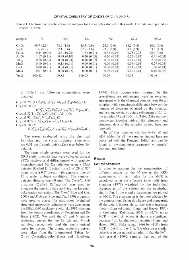
in Table 1, the following compositions were
obtained:
Crystal 79: (Cr3+1.79V3+0.15Fe
3+0.01Al0.03Ti
4+0.01Mn2+0.01)O3
Crystal 100/1:(Cr3+1.47V
3+0.44Fe
3+0.04Al0.03Ti
4+0.01Mg0.01)O3
Crystal 82/1: (Cr3+0.70V3+1.26Fe
3+0.03Al0.01)O3
Crystal 92: (Cr3+0.51V3+1.48Fe
3+0.01)O3
Crystal 82/2: (Cr3+0.36V3+1.57Fe
3+0.06Al0.01)O3
Crystal 100/2:(Cr3+0.33V
3+1.08Fe
3+0.50Al0.01Ti
4+0.04Mg0.01Fe
2+0.03)O3
The errors evaluated using the chemical
formulae and the crystal-structure refinements
are 0.01 per formula unit (p.f.u.) (see below for
details).
The same single crystals were used for the
XRD study. Intensity data were collected using a
STOE single-crystal diffractometer with graphite
monochromated Mo-Ka radiation using a CCD
detector (Oxford Diffraction) in a 5 4 2y 4 85º
range using a 0.2º o-scan with exposure time of
10 s under ambient conditions. The sample–
detector distance was 60 mm. The Crysalis Red
program (Oxford Diffraction) was used to
integrate the intensity data applying the Lorentz-
polarization correction. The X-red (Stoe and Cie,
2001) and X-shape (Stoe and Cie, 1999) programs
were used to correct for absorption. Weighted
structural anisotropic refinements were done using
the SHELX-97 package (Sheldrick, 1997) starting
from the atomic coordinates of Newnham and De
Haan (1962). We used the Cr and V atomic
scattering curves for the Cr-rich and V-rich
samples, respectively, and the partly-ionized
curve for oxygen. The atomic scattering curves
were taken from the International Tables for
X-ray Crystallography (Ibers and Hamilton,
1974). Final occupancies obtained by the
crystal-structure refinements were in excellent
agreement with the chemical compositions for all
samples, with a maximum difference between the
number of electrons obtained by the chemical
analysis and crystal structure refinement of 0.1 for
the samples 79 and 100/1. In Table 2 the unit-cell
parameters, together with all the refinement and
structural data of the samples studied here, are
reported.
The cif files, together with the Fo-Fc, sft and
ADP tables for all the samples studied here are
deposited with the Principal Editor and can be
found at www.minersoc.org/pages/ e_journals/
dep_mat_mm.html.
Results
Unit-cell parametersIn order to account for the superposition of
different cations on the M site in the XRD
experiments, a mean value for the MCR is
calculated using the effective ionic radii from
Shannon (1976) weighted by the individual
occupancies of the cations on the octahedral
site. In Fig. 1, the a and c parameters are plotted
vs. MCR. The c parameter is the most affected by
the composition. Using this figure and comparing
all the data it is possible to note that c increases
linearly from eskolaite (Finger and Hazen, 1980)
to karelianite (Robinson, 1975) by ~2.7% up to
MCR = 0.640 A, where it shows a significant
decrease from karelianite to hematite (Finger and
Hazen, 1980; Blake et al., 1966) by ~1.3% from
MCR = 0.640 to 0.645 A. We observe a similar
behaviour in our natural samples: in fact the Fe3+-
rich crystal (100/2 sample) lies out of the
TABLE 1. Electron microprobe chemical analyses for the samples studied in this work. The data are reported asoxides in wt.%.
Samples 79 100/1 82/1 92 82/2 100/2
Cr2O3 90.7 (2.3) 74.0 (1.8) 35.3 (0.9) 25.6 (0.6) 18.2 (0.4) 16.6 (0.4)V2O3 7.6 (0.2) 22.1 (0.6) 62.7 (1.6) 73.7 (1.8) 78.0 (1.9) 53.3 (1.3)Fe2O3 0.44 (0.04) 2.31 (0.20) 1.64 (0.21) 0.31 (0.03) 3.25 (0.35) 26.4 (0.6)Al2O3 1.17 (0.11) 0.99 (0.10) 0.20 (0.03) 0.10 (0.01) 0.22 (0.04) 0.41 (0.03)TiO2 0.28 (0.03) 0.30 (0.04) 0.18 (0.02) 0.00 (0.01) 0.09 (0.01) 2.08 (0.21)MgO 0.10 (0.01) 0.13 (0.01) 0.09 (0.01) 0.00 (0.01) 0.04 (0.01) 0.27 (0.03)FeO 0.00 (0.01) 0.04 (0.01) 0.00 (0.01) 0.00 (0.01) 0.01 (0.01) 1.30 (0.14)MnO 0.07 (0.01) 0.00 (0.02) 0.00 (0.01) 0.00 (0.01) 0.00 (0.01) 0.10 (0.02)
Total 100.42 99.82 100.05 99.70 99.76 100.46
CRYSTAL CHEMISTRY OF OXIDES OF CR, V AND FE
787
eskolaite–karelianite linear trend as its c para-
meter is significantly smaller. The a parameter
shows an almost linear increase with increasing
MCR; however, a slight change in slope is
observed corresponding to the increased Fe3+
content (sample 100/2 and hematite crystals).
Octahedral site
The volume of the M octahedral site, VM,
increases along the eskolaite–karelianite join,
whereas synthetic and natural hematites show an
increase in MCR from 0.640 A (karelianite) to
0.645 A (hematites) at constant VM. Concerning
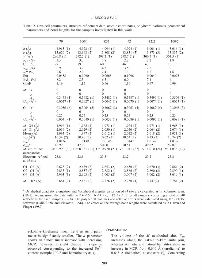
TABLE 2. Unit-cell parameters, structure-refinement data, atomic coordinates, polyhedral volumes, geometricalparameters and bond lengths for the samples investigated in this work.
79 100/1 82/1 92 82/2 100/2
a (A) 4.965 (1) 4.972 (1) 4.994 (1) 4.994 (1) 5.001 (1) 5.016 (1)c (A) 13.620 (2) 13.648 (2) 13.808 (2) 13.831 (5) 13.873 (3) 13.835 (2)V (A3) 290.8 (1) 292.2 (1) 298.2 (1) 298.7 (1) 300.5 (1) 301.5 (1)Rall (%) 3.3 3.3 1.8 2.2 2.2 1.8Un. Refl. 97 79 60 46 67 70Rint (%) 6.9 3.7 4.3 3.5 2.2 2.1Rs (%) 2.6 2.0 2.4 1.3 1.2 1.6Ext 0.0458 0.0090 0.0668 0.1096 0.0040 0.0075WR2 (%) 8.2 8.3 6.3 6.0 7.1 4.1GooF 1.19 1.15 0.96 1.36 0.97 0.99
M x 0 0 0 0 0 0y 0 0 0 0 0 0z 0.3478 (1) 0.3482 (1) 0.3487 (1) 0.3487 (1) 0.3490 (1) 0.3506 (1)
Ueq (A2) 0.0027 (1) 0.0027 (1) 0.0047 (1) 0.0070 (1) 0.0074 (1) 0.0065 (1)
O x 0.3056 (6) 0.3064 (5) 0.3067 (3) 0.3063 (4) 0.3082 (5) 0.3066 (3)y 0 0 0 0 0 0z 0.25 0.25 0.25 0.25 0.25 0.25
Ueq (A2) 0.0041 (1) 0.0040 (1) 0.0053 (1) 0.0089 (1) 0.0095 (1) 0.0081 (1)
M�O4 (A) 1.966 (1) 1.965 (1) 1.973 (1) 1.974 (2) 1.971 (1) 1.968 (1)M�O1 (A) 2.019 (2) 2.029 (2) 2.050 (1) 2.050 (2) 2.064 (2) 2.074 (1)Mean (A) 1.993 (2) 1.997 (2) 2.012 (1) 2.012 (2) 2.018 (2) 2.021 (1)VM (A3) 10.34 (2) 10.40 (2) 10.62 (2) 10.63 (2) 10.73 (2) 10.74 (2)loct
# 1.0136 1.0139 1.0146 1.0147 1.0147 1.0176soct* 46.99 47.90 50.00 50.51 49.82 59.02M site refinedoccupancies
Cr: 0.990 (30) Cr: 0.981 (21) Cr: 0.970 (21) V: 1.011 (27) V: 1.010 (24) V: 1.036 (12)
Electrons refinedat M site
23.8 23.5 23.3 23.2 23.2 23.8
O1�O2 (A) 2.628 (2) 2.639 (3) 2.653 (2) 2.650 (3) 2.670 (3) 2.664 (2)O2�O4 (A) 2.853 (1) 2.857 (2) 2.882 (1) 2.886 (2) 2.890 (2) 2.890 (1)O4�O5 (A) 2.993 (1) 2.993 (2) 3.005 (2) 3.007 (2) 3.002 (2) 3.019 (1)
M1�M2 (A) 2.664 (2) 2.681 (2) 2.726 (2) 2.730 (4) 2.747(2) 2.784 (2)
# Octahedral quadratic elongation and *octahedral angular distortion of M site are calculated as in Robinson et al.(1971). We measured the data with �6 < h < 6, �6 < k < 6, �12 < l < 12 for all samples, collecting a total of 840reflections for each sample (Z = 6). The polyhedral volumes and relative errors were calculated using the IVTONsoftware (Balic-Zunic and Vickovic, 1996). The errors on the average bond lengths were calculated as in Hazen andFinger (1982).
788
L. SECCO ET AL.
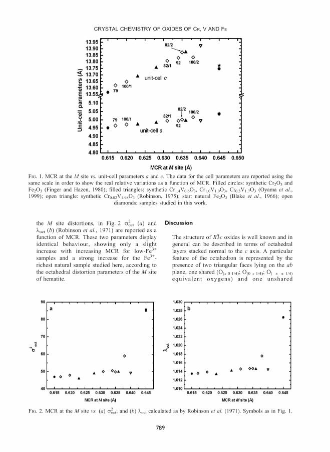
the M site distortions, in Fig. 2 s2oct (a) and
loct (b) (Robinson et al., 1971) are reported as a
function of MCR. These two parameters display
identical behaviour, showing only a slight
increase with increasing MCR for low-Fe3+
samples and a strong increase for the Fe3+-
richest natural sample studied here, according to
the octahedral distortion parameters of the M site
of hematite.
Discussion
The structure of R3c oxides is well known and in
general can be described in terms of octahedral
layers stacked normal to the c axis. A particular
feature of the octahedron is represented by the
presence of two triangular faces lying on the ab
plane, one shared (O(x 0 1/4); O(0 x 1/4); O(�x�x 1/4)
equivalent oxygens) and one unshared
FIG. 1. MCR at the M site vs. unit-cell parameters a and c. The data for the cell parameters are reported using the
same scale in order to show the real relative variations as a function of MCR. Filled circles: synthetic Cr2O3 and
Fe2O3 (Finger and Hazen, 1980); filled triangles: synthetic Cr1.4V0.6O3, Cr1.0V1.0O3, Cr0.3V1.7O3 (Oyama et al.,
1999); open triangle: synthetic Cr0.02V1.98O3 (Robinson, 1975); star: natural Fe2O3 (Blake et al., 1966); open
diamonds: samples studied in this work.
FIG. 2. MCR at the M site vs. (a) s2oct; and (b) loct calculated as by Robinson et al. (1971). Symbols as in Fig. 1.
CRYSTAL CHEMISTRY OF OXIDES OF CR, V AND FE
789
(O(1/3 2/3�x 5/12); O(x�2/3 x�1/3 5/12); O(1/3�x �1/3 5/12)
equivalent oxygens) with another octahedron
(Fig. 3). Hereafter, and in figures presented in
this paper, the following labels will be used:
O(x 0 1/4) = O1; O(0 x 1/4) = O2; O(�x �x 1/4) = O3;
O(1/3 2/3�x 5/12) = O4; O(x�2/3 x�1/3 5/12) = O5;
O(1/3�x �1/3 5/12) = O6.
The variation in the z coordinate of the M site
with increasing MCR causes the increase in
distance between the equivalent M cations
located at 0 0 z (hereafter referred to as M1) and
0 0 1/2�z (hereafter referred to as M2) (Fig. 3),
making the M cations closer to the unshared faces.
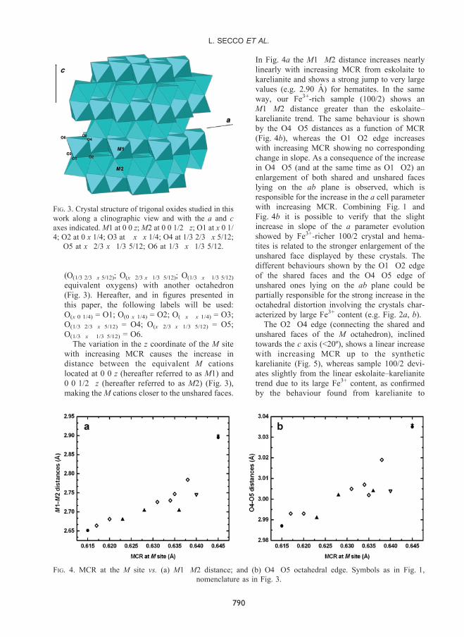
In Fig. 4a the M1�M2 distance increases nearly
linearly with increasing MCR from eskolaite to
karelianite and shows a strong jump to very large
values (e.g. 2.90 A) for hematites. In the same
way, our Fe3+-rich sample (100/2) shows an
M1�M2 distance greater than the eskolaite–
karelianite trend. The same behaviour is shown
by the O4�O5 distances as a function of MCR
(Fig. 4b), whereas the O1�O2 edge increases
with increasing MCR showing no corresponding
change in slope. As a consequence of the increase
in O4�O5 (and at the same time as O1�O2) an
enlargement of both shared and unshared faces
lying on the ab plane is observed, which is
responsible for the increase in the a cell parameter
with increasing MCR. Combining Fig. 1 and
Fig. 4b it is possible to verify that the slight
increase in slope of the a parameter evolution
showed by Fe3+-richer 100/2 crystal and hema-
tites is related to the stronger enlargement of the
unshared face displayed by these crystals. The
different behaviours shown by the O1�O2 edge
of the shared faces and the O4�O5 edge of
unshared ones lying on the ab plane could be
partially responsible for the strong increase in the
octahedral distortion involving the crystals char-
acterized by large Fe3+ content (e.g. Fig. 2a, b).
The O2�O4 edge (connecting the shared and
unshared faces of the M octahedron), inclined
towards the c axis (<20º), shows a linear increase
with increasing MCR up to the synthetic
karelianite (Fig. 5), whereas sample 100/2 devi-
ates slightly from the linear eskolaite–karelianite
trend due to its large Fe3+ content, as confirmed
by the behaviour found from karelianite to
FIG. 3. Crystal structure of trigonal oxides studied in this
work along a clinographic view and with the a and c
axes indicated. M1 at 0 0 z; M2 at 0 0 1/2�z; O1 at x 0 1/
4; O2 at 0 x 1/4; O3 at �x�x 1/4; O4 at 1/3 2/3�x 5/12;
O5 at x�2/3 x�1/3 5/12; O6 at 1/3�x�1/3 5/12.
FIG. 4. MCR at the M site vs. (a) M1�M2 distance; and (b) O4�O5 octahedral edge. Symbols as in Fig. 1,
nomenclature as in Fig. 3.
790
L. SECCO ET AL.
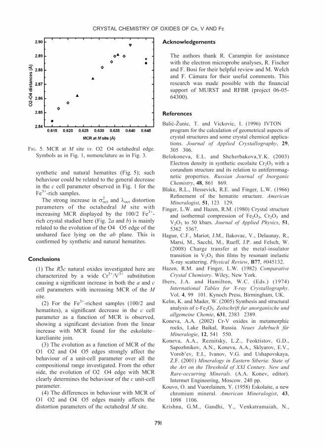
synthetic and natural hematites (Fig. 5); such
behaviour could be related to the general decrease
in the c cell parameter observed in Fig. 1 for the
Fe3+-rich samples.
The strong increase in s2oct and loct distortion
parameters of the octahedral M site with
increasing MCR displayed by the 100/2 Fe3+-
rich crystal studied here (Fig. 2a and b) is mainly
related to the evolution of the O4�O5 edge of the
unshared face lying on the ab plane. This is
confirmed by synthetic and natural hematites.
Conclusions
(1) The R3c natural oxides investigated here are
characterized by a wide Cr3+/V3+ substitution
causing a significant increase in both the a and c
cell parameters with increasing MCR of the M
site.
(2) For the Fe3+-richest samples (100/2 and
hematites), a significant decrease in the c cell
parameter as a function of MCR is observed,
showing a significant deviation from the linear
increase with MCR found for the eskolaite–
karelianite join.
(3) The evolution as a function of MCR of the
O1�O2 and O4�O5 edges strongly affect the
behaviour of a unit-cell parameter over all the
compositional range investigated. From the other
side, the evolution of O2�O4 edge with MCR
clearly determines the behaviour of the c unit-cell
parameter.
(4) The differences in behaviour with MCR of
O1�O2 and O4�O5 edges mainly affects the
distortion parameters of the octahedral M site.
Acknowledgements
The authors thank R. Carampin for assistance
with the electron microprobe analyses, R. Fischer
and F. Bosi for their helpful review and M. Welch
and F. Camara for their useful comments. This
research was made possible with the financial
support of MURST and RFBR (project 06-05-
64300).
References
Balic-Zunic, T. and Vickovic, I. (1996) IVTON �program for the calculation of geometrical aspects of
crystal structures and some crystal chemical applica-
tions. Journal of Applied Crystallography, 29,
305�306.
Belokoneva, E.L. and Shcherbakova,Y.K. (2003)
Electron density in synthetic escolaite Cr2O3 with a
corundum structure and its relation to antiferromag-
netic properties. Russian Journal of Inorganic
Chemistry, 48, 861�869.
Blake, R.L., Hessevick, R.E. and Finger, L.W. (1966)
Refinement of the hematite structure. American
Mineralogist, 51, 123�129.
Finger, L.W. and Hazen, R.M. (1980) Crystal structure
and isothermal compression of Fe2O3, Cr2O3 and
V2O3 to 50 kbars. Journal of Applied Physics, 51,
5362�5367.
Hague, C.F., Mariot, J.M., Ilakovac, V., Delaunay, R.,
Marsi, M., Sacchi, M., Rueff, J.P. and Felsch, W.
(2008) Charge transfer at the metal-insulator
transition in V2O3 thin films by resonant inelastic
X-ray scattering. Physical Review, B77, #045132.
Hazen, R.M. and Finger, L.W. (1982) Comparative
Crystal Chemistry. Wiley, New York.
Ibers, J.A. and Hamilton, W.C. (Eds.) (1974)
International Tables for X-ray Crystallography.
Vol. 4, 99�101. Kynoch Press, Birmingham, UK.
Kelm, K. and Mader, W. (2005) Synthesis and structural
analysis of e-Fe2O3. Zeitschrift fur anorganische und
allgemeine Chemie, 631, 2383�2389.
Koneva, A.A. (2002) Cr-V oxides in metamorphic
rocks, Lake Baikal, Russia. Neues Jahrbuch fur
Mineralogie, 12, 541�550.
Koneva, A.A., Reznitsky, L.Z., Feoktistov, G.D.,
Sapozhnikov, A.N., Koneva, A.A., Sklyarov, E.V.,
Vorob’ev, E.I., Ivanov, V.G. and Ushapovskaya,
Z.F. (2001) Mineralogy in Eastern Siberia: State of
the Art on the Threshold of XXI Century. New and
Rare-occurring Minerals. (A.A. Konev, editor).
Intermet Engineering, Moscow. 240 pp.
Kouvo, O. and Vuorelainen, Y. (1958) Eskolaite, a new
chromium mineral. American Mineralogist, 43,
1098�1106.
Krishna, G.M., Gandhi, Y., Venkatramaiah, N.,
FIG. 5. MCR at M site vs. O2�O4 octahedral edge.
Symbols as in Fig. 1, nomenclature as in Fig. 3.
CRYSTAL CHEMISTRY OF OXIDES OF CR, V AND FE
791
Venkatesan, R. and Veerajah, N. (2008) Features of
the local structural disorder in Li2O-CaF2-P2O5
glass-ceramics with Cr2O3 as nucleating agent.
Physica B – Condensed Matter, 403, 702�710.
Logvinova, A., Wirth, R., Sobolev, N.V., Seryotkin,
Y.V., Yefimova, E.S., Floss, C. and Taylor, L.A.
(2008) Eskolaite associated with diamond from the
Udachnaya kimberlite pipe, Yakutia, Russia.
American Mineralogist, 93, 685�690.
Long, J.V.P., Vuorelainen, Y. and Kouvo, O. (1963)
Karelianite, a new vanadium mineral. American
Mineralogist, 48, 33�41.
Newnham, R.E. and De Haan, Y.M. (1962) Refinement
of the a-Al2O3, Ti2O3, V2O3 and Cr2O3 structures.
Zeitschrift fur Kristallographie, 117, 235�237.
Oyama, T., Iimura, Y., Takeuchi, K. and Ishii, T. (1999)
Synthesis of (CrxV1-x )2O3 fine particles by a laser-
induced vapor-phase reaction and their crystal
structure. Journal of Materials Science, 34,
439�444.
Pauling, L. and Hendricks, B. (1925) The crystal
structures of hematite and corundum. Journal of
the American Chemical Society, 47, 781�790.
Reid, A.F., Sabine, T.M. and Wheeler, D.A. (1972)
Neutron diffraction and other studies of magnetic
ordering in phases based on Cr2O3, V2O3 and Ti2O3.
Journal of Solid State Chemistry, 4, 400�409.
Reznitsky, L.Z. and Sklyarov, E.V. (1996) Unique Cr-V
mineral association in metacarbonate rocks of the
Sludyanka, Russia. Proceedings of the 30th
International Geological Congress, Beijing, China,
Vol. 2, 446 p.
Reznitsky, L.Z., Sklyarov, E.V. and Karmanov, N.S.
(1998) Eskolaite in metacarbonate rocks of the
Sludyanka Group, southern Baikal region. Doklady
Earth Sciences, 363, 1049�1053.
Reznitsky, L.Z., Sklyarov, E.V., Suvorova, L.F.,
Karmanov, N.S. and Ushchapovskaya, Z.F. (2005)
The chromite-coulsonite-magnetite solid solution:
the first find of a rare variety of spinel in terrestrial
rocks. Doklady Earth Sciences, 404, 1121�1125.
Robinson, W.R. (1975) High-temperature crystal chem-
istry of V2O3 and 1% chromium-doped V2 O3. Acta
Crystallographica, B31, 1153�1160.
Robinson, K., Gibbs, G.V. and Ribbe, P.H. (1971)
Quadratic elongation: a quantitative measure of
distortion in coordination polyhedra. Science, 172,
567�570.
Sawada, H. (1994) Residual electron density study of
chromium sesquioxide by crystal structure and
scattering factor refinement. Materials Research
Bulletin, 29, 239�245.
Shannon, R.D. (1976) Revised effective ionic radii and
systematic studies of interatomic distances in halides
and chalcogenides. Acta Crystallographica. A32,
751�767.
Sheldrick, G.M. (1997) SHELX: programs for crystal
structure analysis (Release 97-2). Institut fur
Ano rg an i s che Chemie de r Un iv e r s i t a t ,
Tammanstrasse 4, D-3400 Gottingen, Germany.
Stoe and Cie (1999) Crystal Optimisation for Numerical
Absorption Correction. Stoe and Cie GmbH,
Darmstadt, Germany.
Stoe and Cie (2001) Data Reduction Program. Stoe and
Cie GmbH, Darmstadt, Germany.
792
L. SECCO ET AL.