Chirurgia Robotica del Cancro del Colon Destro: Curva di ...
65
Relatori Prof. Nicola Carlomagno Prof. Francesco Brunetti Dottorando Dott. Nicola de’Angelis UNIVERSITÀ DEGLI STUDI DI NAPOLI “ FEDERICO II ” FACOLTÀ DI MEDICINA E CHIRURGIA DOTTORATO DI RICERCA IN SCIENZE CHIRURGICHE E TECNOLOGIE DIAGNOSTICO-TERAPUETICHE AVANZATE Coordinatore: Prof. Michele Santangelo XXVIII CLICLO Chirurgia Robotica del Cancro del Colon Destro: Curva di Apprendimento ed Analisi dei Costi Anno Accademico: 2015-2016
Transcript of Chirurgia Robotica del Cancro del Colon Destro: Curva di ...

Relatori
Prof. Nicola Carlomagno
Prof. Francesco Brunetti
Dottorando
Dott. Nicola de’Angelis
UNIVERSITÀ DEGLI STUDI DI NAPOLI
“ FEDERICO II ”
FACOLTÀ DI MEDICINA E CHIRURGIA
DOTTORATO DI RICERCA IN
SCIENZE CHIRURGICHE E TECNOLOGIE
DIAGNOSTICO-TERAPUETICHE AVANZATE
Coordinatore: Prof. Michele Santangelo
XXVIII CLICLO
Chirurgia Robotica del Cancro del Colon Destro:
Curva di Apprendimento ed Analisi dei Costi
Anno Accademico: 2015-2016

ii
Relatori
Prof. Nicola Carlomagno
Prof. Francesco Brunetti
Dottorando
Dott. Nicola de’Angelis
UNIVERSITÀ DEGLI STUDI DI NAPOLI
“ FEDERICO II ”
FACOLTÀ DI MEDICINA E CHIRURGIA
DOTTORATO DI RICERCA IN
SCIENZE CHIRURGICHE E TECNOLOGIE
DIAGNOSTICO-TERAPUETICHE AVANZATE
Coordinatore: Prof. Michele Santangelo
XXVIII CLICLO
Chirurgia Robotica del Cancro del Colon Destro:
Curva di Apprendimento ed Analisi dei Costi
Anno Accademico: 2015-2016
iii
Thesis Directors
Pr Nicola Carlomagno
Pr Francesco Brunetti
Candidate
Dr Nicola de’Angelis
UNIVERSITÀ DEGLI STUDI DI NAPOLI
“ FEDERICO II ”
FACOLTÀ DI MEDICINA E CHIRURGIA
PhD PROGRAM in
SURGICAL SCIENCES AND
ADVANCED DIAGNOSTIC-THERAPEUTIC TECHNOLOGIES Director: Pr Michele Santangelo
XXVIII Cycle
Robotic Surgery for Right Colon Cancer:
Learning Curve and Cost Analysis
Academinc Year: 2015-2016
Riassunto
Al giorno d'oggi, i pazienti richiedono senpre più di frequente un approccio
chirurgico mini-invasivo, anche se solo una piccola percentuale di essi sembra avere
accesso a questa chirurgia. I chirurghi, da parte loro, devono adattarsi agli sviluppi della
tecnica chirurgica, soprattutto alla ridotta manovrabilità in campo operatorio e alla perdita
della visione diretta. La curva di apprendimento delle procedure laparoscopiche o robotiche
può essere lunga e difficile. La chirurgia robotica rappresenta la tecnologia più avanzata nel
campo degli approcci minimamente invasivi. Questo lavoro prende in esame le
caratteristiche generali, i vantaggi e gli svantaggi di queste tecniche chirurgiche, oltre a
presentare i risultati di uno studio prospettico condotto per confrontare la curva di
apprendimento del chirurgo e i costi socio-sanitari della colectomia destra eseguita per via
laparoscopica o robotica. I risultati presentati in questa tesi dimostrano che le procedure
laparoscopiche e robotiche hanno risultati operatori e postoperatori simili. La colectomia
robotica destra sembra però avere una curva di apprendimento più veloce della
laparoscopia. Per quanto riguarda i costi, una volta inclusi sia i costi operatori che la
degenza ospedaliera, la laparoscopia e la chirurgia robotica sono associati a costi
equivalenti. Ulteriori studi clinici sono necessari per rispondere alla domanda se l'approccio
robotico potrebbe aumentare, grazie alla sua tecnologia avanzata, l'attuale bassa percentuale
di pazienti sottoposti a chirurgia mini-invasiva.
Parole chiave: Chirurgia mini-invasiva; colectomia robotica destra; colectomia
laparoscopica destra; curva di apprendimento; analisi dei costi.

5
Abstract
Nowadays, patients request minimally invasive surgical approaches if at all possible,
although only a small percentage seems to benefit of it. Surgeons have to adjust to changes
in the surgical techniques, especially in the maneuverability in the operative field and the
loss of direct views. The learning curve of laparoscopic or robotic procedures may be steep.
Robot-assisted surgery represents the most advanced technology in the field of minimally
invasive approaches. The present thesis will discuss the general characteristics, advantages
and pitfalls of this surgical technique, as well as presenting the results of a prospective trial
designed to compare the surgeon’s learning curve and the health-related costs in
laparoscopic versus robotic right colectomy for colon cancer. The results presented in this
thesis support that laparoscopic and robotic preocedures yield similar operative and
postoperative outcomes. Robotic right colectomy appears to have a faster learning curve
than laparoscopy. Concerning the costs, once included both the surgical costs and the
hospital stay, laparoscopy and robotic surgery are associated with equivalent costs. Further
clinical trials are awaited to address the question whether the robotic approach could
increase, thanks to its advanced technology, the currently small number of patients
undergoing minimally invasive surgery.
Keywords : Minimally invasive surgery; robotic right colectomy; laparoscopic right
colectomy; learning curve; health-related cost analysis

6
Table of Content
Introduction..................................................................................................................................8
MinimallyInvasiveColorectalSurgery................................................................................9BriefHistoryofRoboticSurgery................................................................................................................12TechnicalAspectsofRoboticSurgery......................................................................................................14RoboticRightColectomy................................................................................................................................16
TheRationalofRoboticLearningCurve..........................................................................23TheSurgicalLearningCurve........................................................................................................................27TraininginRoboticSurgery.........................................................................................................................32
AimsofthePresentStudy.....................................................................................................33
Materials&Methods...............................................................................................................33StudyDesign........................................................................................................................................................33StudyPopulation...............................................................................................................................................34SurgicalTechniques.........................................................................................................................................35OutcomesMeasures.........................................................................................................................................37StatisticalAnalyses...........................................................................................................................................38
Results..........................................................................................................................................39
Discussion...................................................................................................................................50
Conclusion..................................................................................................................................53
References..................................................................................................................................55

7
A mia moglie Clotilde

8
Introduction
Human anatomy has not changed and the surgical manipulation of many organs has
basically remained the same for centuries, however, surgical techniques have evolved in the
last decades in favor of smaller incisions, videoscopic visualization, and advanced
technologies allowing for a shorter hospital stay, a reduced postoperative pain, an improved
cosmesis, and a quicker return to normal activities.
Nowadays, patients request minimally invasive surgical approaches if at all
possible. To acquire these benefits for patients, surgeons have had to adjust to changes in
maneuverability in the operative field and the loss of direct views. For many, the transition
has been difficult and the learning curve steep1. Still, several common procedures, such as
cholecystectomy, are now performed by a minimally invasive approach as the gold
standard, and basic laparoscopic skills have become formally incorporated into general
surgery training programs2.
Robot-assisted surgery represents the most advanced technology in the field of
minimally invasive approaches3. The present thesis will discuss the general characteristics,
advantages and pitfalls of this surgical technique, as well as presenting the results of a
prospective trial on the surgeon’s learning curve in robotic right colectomy for colon
cancer. In addition, a health-related cost analysis concerning this emerging technology will
be described.
9
Minimally Invasive Colorectal Surgery
Since the introduction of laparoscopic surgery, minimally invasive techniques have
been broadly applied across multiple specialties for both benign and malignant conditions.
The first laparoscopic colectomy was reported by Jacobs et al.4 in 1991, and the enthusiasm
for laparoscopic colectomy grew when recovery benefits for patients became apparent.
Since then, many surgeons became soon comfortable with laparoscopic colectomy for
benign disease, but the application of minimally invasive surgery (MIS) to malignant
colorectal disease was much slower mainly due to oncologic concerns5. However, during
the past 20 years, a rapid evolution of techniques, technology, and experience has
occurred6, 7, and several randomized clinical trials have demonstrated that laparoscopic
colectomy for cancer is comparable to conventional open surgery in terms of long-term
oncologic outcomes8-10, allowing for safe and appropriate oncologic resections. Also for
rectal cancer, recent studies demonstrated that MIS have equivalent results than open
surgery11. Moreover, the MIS approach was proved to offer several advantages compared to
conventional open surgery, including9, 10, 12, 13:
• Smaller incisions and better cosmetic results
• Less blood loss and lower rate of transfusions
• Reduced postoperative pain and thus need of narcotic pain medication
• Faster return of normal bowel function
• Faster recovery translating into shorter hospital stays
• Higher quality of life and patient’s acceptance.

10
The Minimally Invasive Colorectal Resection Outcomes (MICRO) review
identified 22 randomized controlled trials and 66 cohort series for benign and malignant
colorectal disease14. The large randomized controlled trials included, such as the COST,
COLOR, and CLASICC trials, clearly demonstrated the short-term benefits of laparoscopic
colectomy for colon cancer8-10, 14. Moreover, several studies have also identified a
decreased rate of postoperative morbidity following laparoscopic colectomy including
fewer wound infections14-16. The same trials also examined the tumor specimens and
reported long-term data on recurrence and survival. The surgical specimens evaluated
showed that oncologic parameters, such as the number of lymph nodes harvested, the
circumferential radial margins, and the longitudinal margins were not different between
laparoscopic and open colectomies8-10, 14, 16. Similar recurrence patterns, wound or port site
metastases, long-term disease-free and overall survival rates were also observed between
laparoscopic and open colectomy. The concern that conversion from laparoscopic to open
surgery in patients with colon cancer may lead to worse oncologic outcomes was not seen
in the randomized controlled trials, which showed no statistical difference between these
two surgical approaches8, 12.
Nevertheless, despite the evidence demonstrating improved short-term outcomes by
laparoscopic colectomy and equivalent oncologic results, the widespread implementation of
this technique was slow. Initially, the lack of formalized training in laparoscopy and the
relatively long learning curve17-19 likely represented the main barriers to adoption. Based on
surgical parameters like the decline in operating time, intraoperative complications, and
conversion rate, the learning curve for performing colorectal resections laparoscopically
11
was initially estimated at approximately 30-50 procedures18, 20-23. More recently, a
study assessing the learning curve for laparoscopic colectomy of a surgical fellow in an
university colorectal unit using a structured training protocol over 100 consecutive patients
indicated that laparoscopic colectomy can be safely performed by the fellow surgeon
independently and without jeopardizing the clinical outcomes after 50 procedures24.
As the surgeon experience increased as well as the number of studies demonstrated
that laparoscopic colectomy is an acceptable alternative to open surgery for both benign
and malignant diseases, the overall ratio of laparoscopic to open colectomies has
progressively increased. Between 2000 and 2004, the incidence of laparoscopic colectomy
raised from 3% to 6.5% in the USA25. In 2011, a French survey estimated that laparoscopy
accounted for up to 29% of colorectal cancer surgeries26. However, these data should be
attentively analyzed in the light of the fact that after more than 20 years from its
introduction, laparoscopy is still reserved to few (less than one third) surgical cases.
Various socioeconomic factors, technical limitations and the steep learning curve have been
advocated as the possible causes hampering the widespread of laparoscopy27.
In this perspective, and considering the large acceptance of MIS procedures by both
patients and surgeons, many surgical innovators and industry were pushed to develop new
technologies with the goal of even less invasive and easier approaches. The introduction in
colorectal surgery of robotics, which was already popular in other specialties such as
urology, aimed to overcome the drawbacks and limitations of laparoscopy in the confined
working space of the pelvis27, 28. The robotic approach appeared to offers all the advantages
of MIS but with a learning curve relatively short1. The results of robotic surgery in terms of
oncologic outcome and anastomotic leakage are presently comparable to laparoscopy, but

12
with longer operating times and greater costs29. Nonetheless, in high volume and
experienced centers, robotic surgery may be indicated for difficult cases where open
surgery would most likely be preferred or in cases where laparoscopy would have a high
risk of conversion30.
Right hemicolectomy has been proposed as the training procedure in order to gain
surgical experience with the robotic technology29.
Brief History of Robotic Surgery
The application of minimally invasive surgery to complex operations has been
facilitated by the development of telemanipulation systems, also referred to as robots6. The
first robotic system approved for intra-abdominal surgery in the United States by the Food
and Drug Administration (FDA) was the AESOP (Automated Endoscopic System for
Optimal Position) system in 1993. AESOP (Computer Motion, Goleta, California) is a
computerized robotic camera assistant for laparoscopic surgery. It has gone through several
modifications since then and is still available today as a voice-activated, surgeon-
controlled, camera assistant. AESOP offers a stable camera platform but has no arm for
direct manipulation or dissection of the tissues.
More dexterous robots have been designed by the US Department of Defense for the
purpose of allowing surgeons to operate on patients in remote or unsafe locations. Since
these systems restore pitch and yaw at the end of the instruments, the two degrees of
freedom lost with the use of traditional laparoscopic instruments, and also add benefits such
13
as tremor reduction, motion scaling, surgeon camera control, comfortable ergonomics,
and three-dimensional optics, it is no surprise the technology has found its way into
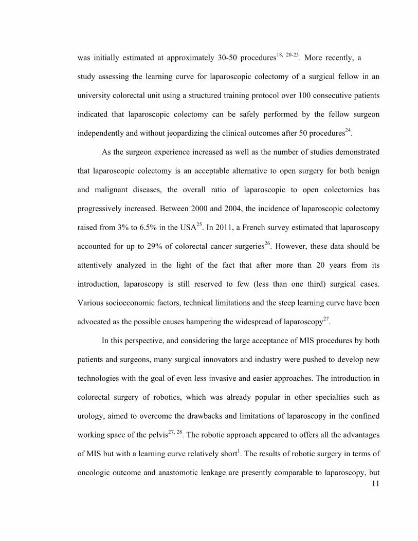
conventional operating environments. Intuitive Surgical's DaVinci robotic system has been
used in Europe since 1997, and in 2000 the FDA approved use in USA. DaVinci has three
or four arms that allow tissue manipulation and retraction as well as camera control (Figure
1). The first robot-assisted colectomies were reported in 200231. Numerous papers since
then have shown robotic colectomy to be safe and feasible7, 32-37.
Figure 1. Intuitive Surgical's DaVinci® Robotic System.
(Source: Google Images)

14
Technical Aspects of Robotic Surgery
Robotic surgery provides several technological improvements compared to
laparoscopy, including:
• Three-dimensional (3D) view of the operating field
• Seven-degrees-of-freedom motion with wristed instruments
• Absence of fulcrum effect
• Absence of surgeon tremor
• Greater ergonomics and comfort for the surgeon.
Moreover, the two-headed robotic platforms represent an exceptional teaching tool
whereby residents in training can achieve optimal anatomical knowledge and surgical
skills, by following the intervention and practicing on the mentoring console.
The current literature has most often focused on comparing the surgical and clinical
advantages of robotics versus laparoscopy, but this may not be the clue of the problem.
Indeed, we should be able to look further and analyze the advantages of robotic surgery in
terms of advanced technology that could increase the currently small number of patients
undergoing MIS also for complex procedures that might be too challenging for laparoscopy
and are still approached by open surgery. Indeed, the recent results of the randomized
controlled trial COLOR II showed that laparoscopy is as safe and effective as open surgery
for rectal cancer resections but this approach remains technically demanding and associated
with high conversion rates (estimated at 17%)11. On the other hand, the available literature
shows that robotic surgery provides all advantages of the MIS approach but it may allow
performing complex procedures, such as rectal resections, with greater ease, lower

15
conversion rate, less pelvic autonomic nerve damage and a reduced learning curve30,
38. However, it remains unclear, to date, whether these advantages translate into significant
clinical benefits that may justify the increased costs associated to the application of robotic
surgery3.
Petrucciani et al.39 recently conducted a meta-analysis comparing the robotic versus
laparoscopic approach and focusing only on right colectomies. They included a total of six
studies with a limited sample size (total of 168 patients in the robotic group and 348 in the
laparoscopic group). The meta-analysis showed no differences between the robotic and
laparoscopic approach in the analyzed peri-operative and postoperative outcomes, except
for a longer operative time for robotic right colectomy. The authors did not perform
sensitivity or subgroup analysis, but concluded that robotic right colectomy is s feasible,
safe, and effective in selected patients39. Another systematic review and meta-analysis
mainly based on observational studies that was published by Trastulli et al.40 in 2015
showed that robotic colectomy is more time-consuming and expensive than laparoscopic
colectomy but it results in faster recovery of bowel function, a shorter hospital stay, less
blood loss and lower rates of both overall postoperative complications and wound
infections40. These results appear highly promising and support the implementation of
robotic platforms in surgical units as well as the introduction of robotic training for fellow
surgeons.

16
Robotic Right Colectomy
The detailed description of a robotic right colectomy include the patient positioning,
port placement, robot docking, and the step-by-step description of the procedure.
Patient Positioning
For a right colectomy, the patient is lying supine on a bean bag. The bag is
positioned flush with the patient's right side, allowing excess bag on the left side with
which to wrap the left side of the patient. The chest is secured circumferentially to the table
with heavy tape at the level of the clavicles. The legs are secured at the thigh and calf with
straps. Patient’s iliac crest is positioned over the mid-point joint of the table. Patient’s arms
are alongside the body to lessen possibility of shoulder injury. A urinary catheter is placed,
along with optional nasogastric or orogastric tube, if used. After positioning, padding,
securing and preparing the patient in the supine position, the table is then placed in 10-15°
reverse Trendelenburg and rolled to the left 10-15°. Then the table is flexed at kidney rest
(10-15°) to lower the patient’s legs and prevent external collisions with the patient cart
arms. Final table adjustments should be made during the initial exposure step (Figure 2).
Figure 2. Patient positioning (Source: DaVinci Technical Manual).
17
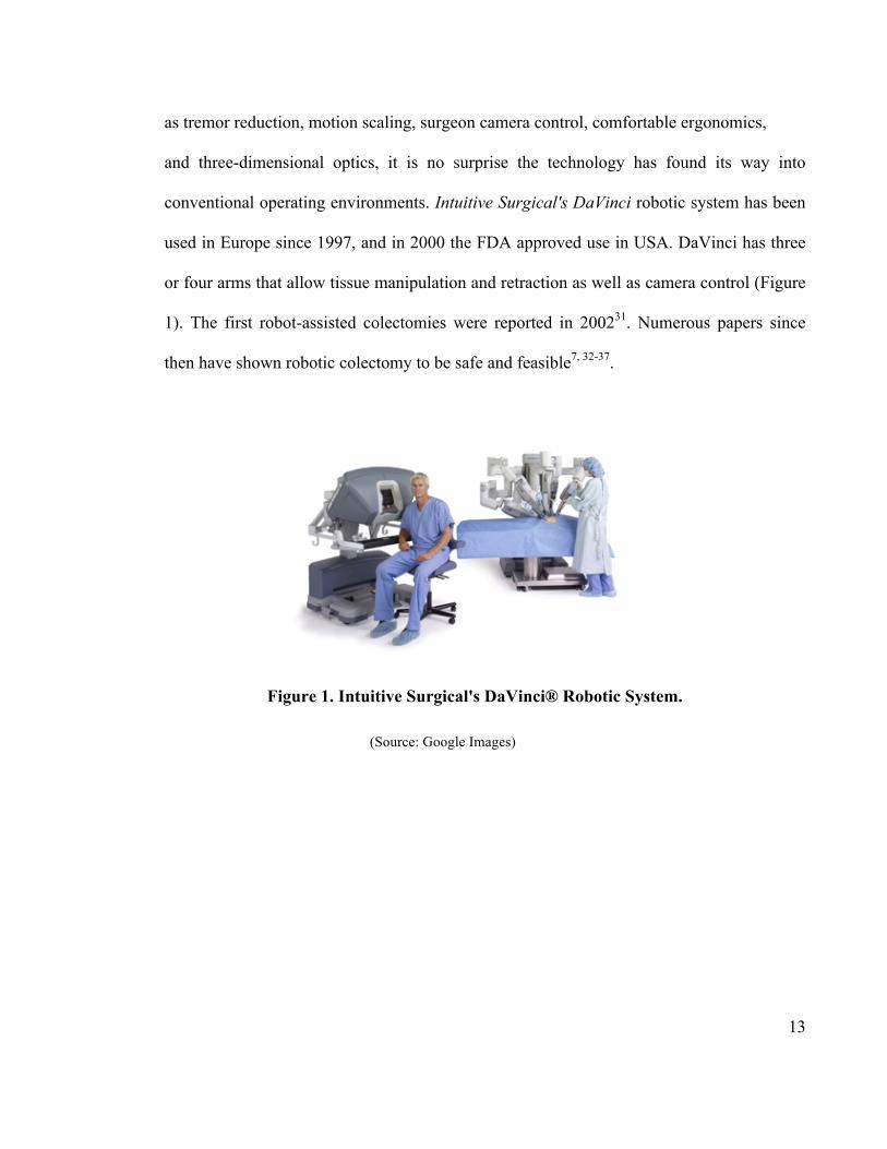
Establishment of pneumoperitoneum, trocar placement, and initial exploration
are performed with the patient in the supine position. The tattooed lesion or pathology is
located and the planned point of transverse mesocolic division is marked based on the
location of the right branch of the middle colic artery. The table is then tilted to the left to
allow the small intestine to fall away from the midline. The robot is then brought in over
the right upper quadrant to dock with the camera port periumbilically, as well as the right
lower and left upper quadrant ports. The robot is brought in from the right side and the
bedside assistant and the scrub nurse are situated to the patient’s left side. Once the robot is
docked, there can be no change to the patient’s position or the robot’s position, without first
undocking the robotic arms (Figure 3).
Figure 3. Patient and Robotic Positioning. (Source: Witkiewicz W et al. 41)

18
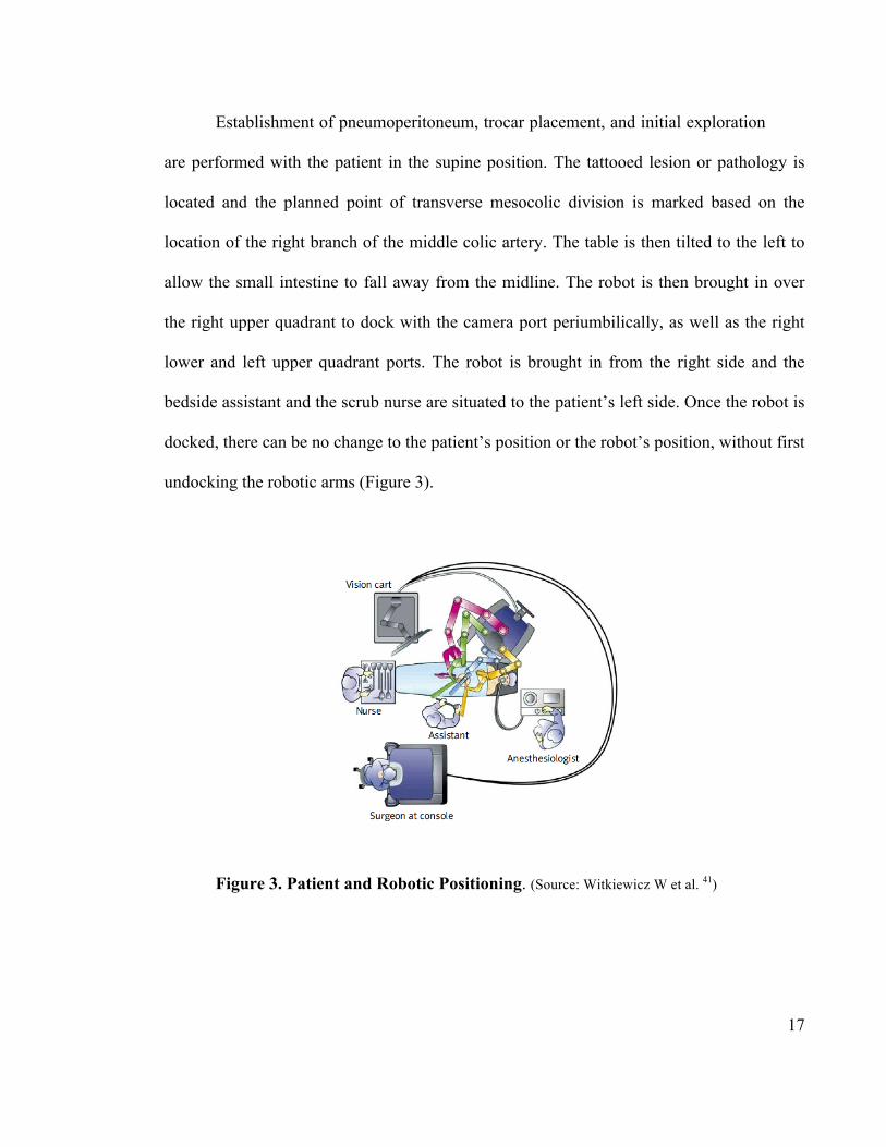
Port Placement
Port placement for the robotic procedure closely resembles the port configuration
for laparoscopic right hemicolectomy. Three robotic working arms are usually used along
with a camera. One assistant laparoscopic port is added for additional retraction, as well as
an energy device or an endostapler. The following principles should be respected while
positioning the robotic ports for distal lesions (Figure 4):
• Maintain remote center at level of the peritoneum
• Maintain 8-10 cm between all da Vinci ports
• Placement of camera port should be consistent
• Instrument arm ports need to shift based on patient size and anatomy
• The camera port (12 mm) should be placed ~1 cm inferior to the spinoumbilical
line (SUL) and ~3-4 cm medial to the mid-clavicular line (MCL) in the left
lower quadrant
• Instrument Arm Port 1 (8 mm) should be placed 1 cm lateral to the MCL,
approximately halfway between the costal margin and SUL in the left upper
quadrant
• Distance to the camera should be at least 8-10 cm
• Instrument Arm Port 2 (8 mm) should be placed on the midline, ~6-8 cm
inferior to the umbilicus.
• Instrument Arm Port 3 (8 mm) should be placed 1 cm lateral to the MCL and 2
cm inferior to the SUL in the lower right quadrant. Distance from other
19
instrument ports and the camera should be at least 8-10 cm while allowing
for at least 2 cm between port and anterior superior iliac spine (ASIS).
• The assistant port (12 mm) should be triangulated between ports 1 and 2 and ~3-
5 cm lateral to the MCL and ~1 cm inferior to umbilicus in the left lower
quadrant. Distance from surrounding ports should be a minimum of 5 cm.
• All measurements should be made AFTER insufflation.
Figure 4. Robotic port positioning for distal lesions (Source: DaVinci Technical
Manual).
The following principles should be respected while positioning the robotic ports for
proximal lesions (Figure 5):
• Camera port (12 mm) should be placed ~2-3 cm lateral and slightly inferior
to the umbilicus

20
• Instrument Arm Port 1 (8 mm) should be placed in the left upper
quadrant, ~2-3 cm lateral to the MCL and ~3-4 cm inferior to the costal
margin. Distance from other instrument ports and the camera should be at
least 8-10 cm.
• Instrument Arm Port 2 (8 mm) should be placed on the midline, inferior to
the umbilicus. Maintain ~3-4 cm distance to symphysis pubis.
• Instrument Arm Port 3 (8 mm) should be placed 2-3 cm sub-xiphoid and 4
cm to the left of midline.
• Assistant Port (12 mm) should be placed in the left-lower quadrant, ~ 4 cm
inferior to the SUL and slightly lateral to the MCL. Distance from
surrounding ports should be a minimum of 5 cm.
• All measurements should be made AFTER insufflation.
Figure 5. Robotic port positioning for proximal lesions (Source: DaVinci Technical Manual).

21
Procedure
The robotic right colectomy procedure begins with diagnostic laparoscopy. The
abdomen is inspected to determine the feasibility of minimally invasive resection and to
identify the extent of the disease. The patient is placed in the Trendelenburg position with
the right side up. This allows for the small bowel and omentum to be displaced to the left
upper quadrant, exposing the cecum and terminal ileum. The robot is then brought from the
right side of the patient and docked onto the ports. Robotic monopolar scissor are on the
right robotic arm (Instrument Arm Port 1) while bipolar grasp are on the left robotic arm
(Instrument Arm Port 2). Depending on the surgeon’s preference and anatomical variations,
either a medial to lateral or lateral to medial approach can be used. However, in case of
malignancy, a medial to lateral approach is preferred33, 42.
The medial to lateral dissection begins by retracting the cecum anteriorly, laterally,
and superiorly with the cadier grasper (Instrument Arm Port 3). Parietal peritoneum is
incised under ileocecocolic mesentery identifying Gerota fascia posteriorly and Toldt fascia
anteriorly. Second and third duodenal sections and pancreatic head are visualized.
Ileocecocolic vein and artery are isolated and sectioned with Hemolock (or linear vascular
stapler) apposition at their origin. The intestinal segment to be resected is stretched with the
third robotic arm, and transection of the right mesocolon is performed in a caudocranial
direction, on the lateral margin of superior mesenteric axis. The right colic vessels (if
present) and right branch of the middle colic pedicles are sectioned at their origins. If the
tumor involves the hepatic flexure or proximal transverse colon, needing an extended
colectomy, also the pedicle of middle colic vessels are sectioned at its origin. Dissection is
22
completed under Gerota fascia until preduodenal pancreatic fascia of Fredet. In this
way, mediolateral access is concluded, and parietal peritoneal incision is continued in
parietocolic sulcus along the Toldt fascia next to the cecum and ascending colon up to the
previous dissection posteriorly.
Once the specimen is totally dissected, the robot is undocked and an approximately
5 cm incision is done peri-umbilically to create a small midline mini-laparotomy. The
mobilized right colon is then exteriorized through this incision and resected. The standard
mechanical anisoperistaltic side-to-side ileocolic anastomosis is created in open fashion.
23
The Rational of Robotic Learning Curve
Minimally invasive colorectal surgery has been used for inflammatory, benign and
malignant disease entities and has been shown to reduce postoperative pain and length of
hospital stay, provide faster recovery, and shown to be cost-effective in comparison to open
surgery4, 10, 16, 17, 30, 43. Among MIS techniques, laparoscopy is the most commonly applied
to date.
As the demand for laparoscopic colorectal surgery increases, patient selection, case-
mix, and laparoscopic outcomes such as conversion rates are expected to vary between
surgeons and institutions. However, laparoscopic surgery requires a high degree of special
resolution, dexterity, and technical skills. An initial training period is usually required for
the majority of surgeons to become proficient in these complex procedures by continuous
repetition of these tasks. As a result, to become technically proficient at laparoscopic
colorectal resections, a long training period is expected. Several studies tried to assess the
learning curve of specific laparoscopic colorectal procedures, and an average of 50-80
procedures appeared necessary to reach a plateau on the learning curve (Box 1).

24
Box 1. Summary of studies assessing the learning curve of different surgical
procedures.
Reference Surgical Procedure Outcome Time or number of case
to reach a plateau Meinke et al.44 Laparoscopic fundoplication Complication rate, conversion rate,
reoperation rate 20 cases
Tekkis et al.19 Laparoscopic colorectal surgery Conversion rate, complication rate, operative time
55-80 cases
Richardson et al.45 Laparoscopic cholecystectomy Bile duct injury rate 3 years Parikh et al.46 Gastrectomy Morbidity, mortality, lymph node
harvestes 18-24 months
15-25 cases Sutton et al.47 Oesophagectomy Operative time, blood loss, ITU stay,
inpatient stay, lymph node harvested 150 cases 7 years
Despite the increasing experience with laparoscopic colorectal surgery and the
continuous improvements in the available equipment and technology, laparoscopy remains
associated to several disadvantages. The image projected onto the operating monitor is a 2-
dimensional representation of a 3-dimensional operating field. This can result in difficulties
with depth perception. The ergonomics of laparoscopy can be poor and can lead to both
operator and assistant fatigue3, 48. Another well-known limitation in the difficulty of obtain
a high-precision suturing, due to the fixed tips and limited dexterity of surgical laparoscopic
instruments. Finally, the physiological tremor from the camera operator cannot be
eliminated and may be exaggerated toward the end of a lengthy operative case.
As the most advanced technology in MIS, robotic surgery has the potential to
address most of the drawbacks and limitations of laparoscopy. The robotic surgical system
offers a camera system that is controlled by the operating surgeon combined with a 10-fold
magnification vision, thus allowing a perfectly still and 3-dimensional visibility of the

25
operative field. The tips of the instruments of a robotic arm have an EndoWrist
technology that has functions of 7 degrees of freedom, 180° articulation, and 540° rotation.
A recent systematic review of robotic surgery in the resection of rectal cancer
showed that the current evidence suggests that robotic rectal surgery could potentially offer
better short-term outcomes especially when applied in selected patients compared to
laparoscopy49. Obesity, male sex, preoperative radiotherapy, and tumors in the lower two-
thirds of the rectum may represent selection criteria for robotic surgery to justify its
increased cost related to this advanced technology. However, the role of robotics in colonic
surgery is largely undefined. The results of a recent systematic review on robotic colonic
surgery compared with multiport laparoscopic colonic surgery showed that the median age
of patients, length of postoperative stay, and time to first bowel movement were similar
between the two approaches3. The robotic colonic group showed a greater median lymph
node harvest, whereas the median operative time for robotic surgery was much longer than
for laparoscopic surgery. The morbidity data suggested that robotic colectomy had a
favorable complication profile in comparison with multiport laparoscopic surgery.
However, as the authors of the systematic review acknowledged, there were significant
selection and complication reporting biases that precluded meaningful comparisons3.
Nonetheless, these data supports robotic surgery as a safe and feasible procedure when
undertaken in selected patients and high-volume experienced surgical centers3, 30, 50.
Overall, the reported conversion rate for robotic colectomies was approximately
8%. The most frequent reasons for conversion to open surgery included dense adhesions
from sigmoid colon to bladder, descending colon ischemia after division of the inferior

26
mesenteric artery, venous bleeding around the pancreas, inability to identify the
tattooed tumor, locally invasive tumors, and stapler malfunction. The most frequent reasons
for conversion to laparoscopic surgery included fatty mesentery precluding safe robotic
dissection, robotic malfunction3.
These results are promising, but for robotic colorectal surgery to become an
accepted alternative to open or laparoscopic surgery, it needs to address several challenges
that face any new emerging surgical technique. It should be safe and result in a comparable,
if not better, oncological outcome when used for resection of malignant disease. It should
also have a reasonable learning curve, and, ideally, the skills that the surgeon would gain
during training for robotics would be transferable to other possible developments in
minimally invasive colorectal surgery. Equally important, especially with the increasing
financial strains on the current health care systems, robotic surgery should be cost-effective.
Unfortunately, the conclusion of many studies is that robotics is too expensive and
probably not as cost-effective as laparoscopy. The real benefits of robotics, however, are
difficult to quantify by means of preliminary cost-effectiveness analyses performed on the
early experience of a few specialized centres3, 27, 51. Thus, before discouraging the
implantation of robotic platforms, further clinical trials should be designed to assess the
efficacy of robotics, its learning curve, and more importantly, studies should aim to answer
a specific patient-oriented questions, such as “What are the supplement benefits of robotics
for those patients or procedures in which MIS still struggles to be applied?”
27
The Surgical Learning Curve
A learning curve is a graphical representation of the increase of
learning/performance (vertical axis) with experience (horizontal axis) (Figure 6). When
learning a new procedure, performance tends to improve with experience, thus clinicians
inexperienced in a procedure are said to be on the early phase of their learning curve with
improvements expected with increasing experience52, 53.
A hypothetical plot of an ideal learning curve has four main phases (Figure 6). The
starting coordinate A represents commencement of training. Secondly, the curve ascends.
The gradient of this ascent indicates how quickly the individuals’ performance improves;
this part of the curve may be a stepwise ascent as individuals learn and master stages of a
complex procedure. Improvements in performance tend to be most rapid at first and then
tail off, as the degree of improvement attained with each case reduces as technique is
refined. Thirdly, assuming adequate aptitude, a point is reached when the procedure can be
performed independently and competently (coordinate B). Additional experience improves
outcomes by small amounts (coordinate C), until a plateau, or asymptote, is reached
(coordinate D). Fourthly, with advancing age, manual dexterity, eyesight, memory and
cognition may deteriorate, outweighing any advantage derived from long experience,
leading to a fall in the level of performance (coordinate E).

28
Figure 6. An idealized surgical learning curve. The learning cure has a slope (representing the rate of learning), which may eventually plateau, suggesting that performance has reached a steady level. A: commencement of training; B: tipping point when the procedure can be performed independently and competently; C: experience improves outcomes by only small amounts; D: reaching of a plateau; E: fall in the level of performance due to aging. The small dotted horizontal line represents the acceptable standard. The dotted curve represents an alternative learning curve between the points B and C described as a temporary performance deterioration after technical competence has been achieved. The reasons postulated for this scenario are case mix effect (undertaking more difficult cases), or over confidence resulting in lapses in technique or judgment. (Source: Hopper A et al. 200752)
The concept of the learning curve applies across the full spectrum of medical
specialities and procedures; however, with the advent of technically demanding minimally
invasive techniques, it is surgery in particular where there are specific and important
implications. The learning curve is indeed particularly interesting in surgery where a
constant stream of new skills must be acquired safely and efficiently. It would thus be
useful to know how many procedures a surgeon may have to carry out before reaching a
safe and competent level of performance autonomously. Knowledge about the specific
learning curve of a surgical procedure could also enhance the surgical training program. A
surgeon may have to perform a procedure a certain number of times under supervision or in

29
a simulated environment before performing it independently. Training programs
could use the learning curve data to map progress of trainee surgeons. Furthermore, an
understanding of the learning curve is crucial in randomized control trials comparing new
procedures with older interventions.
The effect of the learning curve of acquiring the new procedure must be considered
in order to reach valid conclusions. However, on which variable the learning curve should
be measured on is still a matter of debate53.
By measuring specific outcomes it is possible to estimate the location of an
individual on a learning curve. Measures of learning related to a surgical technique fall into
two categories: measures of surgical process, and measures of patient outcome. Surgical
process measures include operative factors such as operative time, blood loss, and technical
adequacy of resection for cancer surgery—margin involvement and lymph node yield.
Patient outcomes include postoperative factors such as analgesia requirement, transfusion
requirement, length of stay in intensive care, length of stay in hospital, morbidity rates,
mortality rates, and cumulative survival.
Surgical process outcomes are generally easier to analyze and therefore more
commonly used, though they are only indirectly related to patient outcomes However, when
considering outcomes of cancer surgery, improvements in case adjusted long-term survival
probably represent the best measure of performance. Indeed, it is possible to plot curves
based on long-term survival related to progression within a case series.

30
It is also worth noting that in many studies on surgical learning curves the
definition of the variables used to assessed it are often different (e.g. operative time, rate of
complications), which makes harder to compare the learning curve between studies on the
same procedure. Moreover, the choice of the measurement must be carefully evaluated,
since not all variables are reliable measures of learning. For example, it is easier to measure
and statistically analyze data on operating time than patient complications. Yet, a learning
curve based on operating time alone may not be the best indictor of good practice54. Also,
variables should be tailored to the type of procedure. For example, operative mortality may
not be a good indictor in low-risk procedures where patient satisfaction and quality-of-life
measures may be more important. In cancer surgery, it may be more meaningful to measure
long-term survival.
Finally, confounding factors must be minimized to draw valid conclusions.
Organizational factors (facilities, equipment), surgical team (experience, co-operation),
case mix, complexity of cases and surgeon’s characteristics (previous experience, natural
abilities, motivation etc.) can all affect the learning curve of a procedure53. Surgeon’s
previous experience is often difficult to assess and poorly defined in many studies.
Statistical Analyses to Quantify the Learning Curve
There many different methods to assess the learning curve. The most common is the
graphical representation on a simple plot. However, there is much variation and no
particular rationale in the type of regression analysis or curve fitting model used (e.g. least-
31
squares regression, logarithmic or negative exponential curves etc.). Another method
includes chronologically dividing the case number into consecutive groups to compare
learning with increasing time and experience, for example dividing into quartiles and
comparing performance in each quartile over time. This method is ideal for comparison
between large case numbers, but the size of groups may hinder the learning curve
interpretation.
Finally, another statistical method that is gaining popularity within the learning
curve literature is the cumulative sum (CUSUM) analysis55. This is a very useful sequential
analysis method that detects change in the individual surgeon’s performance. The trend in
outcome is graphically represented as a straight line with acceptable performance and an
upward slope with less than optimum performance. A trainee surgeon is expected to show a
rising curve (the rate of learning), which may eventually plateau when performance
stabilizes. Although, this is an excellent statistical method for detecting change and the
possibility of adverse events, it is less good for comparing inter-operator differences53.
Factors Affecting the Learning Curve
A complex hierarchy of factors can affect the surgical learning curve. At the bottom,
factors like guidelines, protocols and standards for clinical governance agreed upon by the
medical fraternity are vital. Next the Institutional policies and cost effectiveness are
contributory. Needless to say the surgical team, the case mix and public awareness are
relevant. The final level in the hierarchy that can influence individual learning is the
32
characteristics of the surgeon such as attitude, capacity for acquiring new skills and
previous experience53.
Training in Robotic Surgery
The learning curve of robotic colorectal surgery is claimed to be reduced in
comparison to laparoscopic colorectal surgery. The estimated number of cases required to
be proficient and achieve competence is of only 15 to 25 procedures1.
The robotic learning curve has some peculiar characteristics that can be categorized
into 3 phases: the phase 1, which entails adaptation to the loss of tactile and tensile
feedback, the phase 2, which is the understanding of the spatial relationship of the robotic
instruments with the patient’s body, and the phase 3, which involves the ability to operate
from the console without direct visualization of the patient. It is highly debated whether the
previous surgeon’s experience in MIS techniques can reduce the length of the learning
curve in robotic surgery. However, if the learning curve of robotic surgery is indeed proven
to be reduced compared to laparoscopy, then this may facilitate the dissemination of robotic
platforms even to surgeons who perform open surgery and who have not adopted
laparoscopy into their clinical practice.
These aspects are also very important to support the definitive introduction of
training in robotic surgery in the curriculum of the resident surgeons.
33
Aims of the Present Study
The present study aimed:
1) To compare the operative and postoperative outcomes of robotic-assisted right
colectomy vs. laparoscopic right colectomy.
2) To evaluate and compare, by using CUSUM analysis, the learning curve of a single
fellow surgeon in robotic-assisted right colectomy vs. laparoscopic right colectomy
for colon cancer.
3) To assess the health-related cost of robotic surgery compared to laparoscopy.
Materials & Methods
Study Design
Between November 2012 and December 2015, 30 consecutive patients underwent
robotic-assisted right colectomy (RRC) and 50 consecutive patients underwent laparoscopic
right colectomy (LRC) for colon cancer at the Henri Mondor Hospital of Créteil (France).
All procedures were performed by a fellow surgeon (NdeA), who, at the time of the study,
was autonomous in general MIS training (> 75 hads-on laparoscopic
cholecystectomy/appendectomy) and had assisted to > 50 laparoscopic colectomies. He had
also attended training courses and simulations in robotic surgery, including periodical
proctoring by an European Academy of Robotic Colorectal Surgery (EARCS) accredited
surgeon. However, the fellow could be considered at the beginning of his learning curve in
both RRC and LRC.

34
In the surgical unit of the Henri Mondor Hospital of Créteil, there is one senior
surgeon (FB) highly experienced in laparoscopic and robotic colorectal surgery (> 10 years
of experience). He works together with the fellow and provide team approach in MIS
colorectal surgery. For this study, the senior surgeon supervised the surgical fellow in all
procedures.
The study was conducted in accordance with the ethical principles ascertained in the
Declaration of Helsinki.
Study Population
All consecutive patients with right colon cancer requiring surgical resection were
eligible for inclusion in the study. Decision on which approach, laparoscopic or robotic one,
performed was based on the patient’s choice and the availability of the robotic platform,
without applying any clinical contraindications. Both laparoscopic and robotic surgeries are
not associated with extra costs for the patient, for whom both procedures are covered by the
national assurance system at 100%.
Demographic data, intraoperative findings, operative procedures, postoperative
parameters, morbidities, and outcomes were prospectively collected into the colorectal
database of the surgical unit of the Henri Mondor Hospital of Créteil. To ensure patient
safety and oncological clearance, bulky tumors (e.g. AJCC cT4a or cT4b) were not
operated by the fellow surgeon and they were thus excluded from the study analysis.

35
Surgical Techniques
Preoperative preparations, operative steps, instrumentations, and postoperative care
were standardized.
All patients underwent a preoperative evaluation including a physical examination,
colonoscopy with tumor biopsy, and a total body computed tomography (CT) scan with
contrast enhancement. In cases of suspected lymphatic packets, positron emission
tomography (PET) with lymphatic biomarkers was performed for preoperative staging.
Preoperative laboratory data included complete blood cell count, biochemical profile, and
tumor markers (carcinoembryonic antigen, CEA). Intravenous antibiotic prophylaxis was
administered according to the guidelines of the infection control committee of the hospital.
The laparoscopic right colectomy was performed as previously reported56-59. In
brief, four laparoscopic ports were placed as follows:
• 12-mm umbilical, camera port for 30 degree telescope
• 12-mm left lumbar, pararectally port (right hand working)
• 5-mm right lumbar, pararectally port (assitant hand working)
• 5-mm suprapubic port (left hand working).
LCR were approached medial to lateral. First, the resectability of the mass was
assessed. Then, the transverse colon was lifted up and the C of duodenum was identified.
On the inferior side of duodenum, an incision over peritoneum was made with Harmonic
Ace (Ethicon Endosurgery, Cincinnati). A gauze piece was passed and CO2 itself was
insufflated in to dissect duodenum and kept away from operative injury.

36
For carcinoma of caecum, ileocolic and right branch of middle colic vessels
were clipped proximally and on specimen side and divided them with harmonic ace. All
fibro fatty tissue and lymph nodes were dissected towards the specimen. Usually only right
branch of middle colic vessels was clipped. The entire middle colic vessel was divided for
right colon cancer near hepatic flexure or in transverse colon. The mesentery was divided
completely from colon to caecum with Harmonic Ace. Then, along the white line of Toldt,
the entire right colon was mobilized up to midtransverse level. The resection and
anastomoses were done extra corporeally by delivering colon by enlarging the umbelical
trocar incision (up to 5 cm). Extra-corporeal side-to-side anastomoses were carried on by
using Linear Staplers (Ethicon). On either side of tumor, the resectional margin of colon
should be at least of 5 cm. All ports and incision were closed with Vicryl and Ethilon
sutures.
The robotic right colectomy procedures were perdormed with the robot docked as
described above (see Robotic Right Colectomy Chapter, Page 16). The main surgical steps
were the same of the laparoscopic right colectomy, using always a medial to lateral
approach. In some cases, Energy devices (EndoWrist® Vessel Sealer) were used.
Postoperatively, diet was resumed as soon as bowel function returned clinically.
Patients were discharged when they tolerated diet and regained ambulation. Patients were
regarded to be suffering from prolonged ileus if they were unable to resume diet after
postoperative day 4 and required parenteral nutrition supplementation. Time to ull
ambulation was defined as time when the patient could walk independently in the ward
without assistance. Once discharged, all patients were followed every 3 months for the first

37
3 years and every 6 months thereafter (French Guidelines from the Thesaurus
National de Cancerologie Digestive, 2011). At the follow-up visits, physical examination,
CT, and serum chemistry analysis were performed. Colonoscopy was carried out if
abnormalities were detected during any follow-up visit.
Outcomes Measures
Operative time was measured from skin incision to completion of wound closure.
For robotic-assisted procedures, docking time (i.e. the time required to position the robot
and secure the robotic arms to the corresponding post sites) was calculated separately from
the operative time. The surgeon console time was the actual time the surgeon spent at the
robotic console during the procedure, and it was also calculated for the RRC. Mean
operative time was used as the main outcome variabale to plot the learning curve.
Conversion was defined as the shift from laparoscopic to open approach, or from
robotic to laparoscopic or open approach to complete the procedure.
Postoperative morbidity and mortality were defined as events occurring during the
hospital stay or within 90 days after resection. Postoperative complications were
categorized by the Dindo-Clavien classification60. All complications were assessed by a
clinician and prospectively registered in the databases at discharge or during the first
outpatient visit. Morbidity included postoperative medical and surgical complications such
as cardiovascular, respiratory, stoma-related complications, intra-abdominal abscess,
anastomotic leakage, wound infection, prolonged ileus, anastomotic hemorrhage and

38
reoperation. The oncological outcomes included quality of surgical resection (i.e., R0,
R1), and number of lymph nodes harvested. R0 resection was defined as the
macroscopically complete removal of the tumor with a microscopically free resection
margin and no peritoneal spread.
Statistical Analyses
To evaluate the surgeon’s learning curve in laparoscopic and robotic right
colectomy, all cases were ordered chronologically, from the earliest to the latest date of
surgery. The LRC and RRC groups were divided into three subgroups of patients following
the chronological order withot the presence of any other obvious grouping criteria.
Grouping was the following: Cases 1 to 10, Cases 11 to 20, and Cases 21 and more.
The CUSUM technique was used for quantitative assessment of the learning curve.
The CUSUM is the running total of differences between the individual data points and the
mean of all data points. The CUSUM technique used for the 30 RRC cases and the 50 LRC
was based on the operative time.
Demographic data, operative outcomes, and complications were compared between
RRC and LRC groups by Pearson chi-squared test, Fisher’s exact test, and Mann-Whitney
U-test. Within group differences (by comparing the three subgroups of patients) were
assessed by ANOVA calculations. Significant difference was defined when p value was <
0.05. Statistics were performed with SPSS (Statistical Package for Social Science, IBM
SPSS Statistics, Version 23 for Macintosh; IBM Corp., Armonk, NY, USA).
39
Results
Data summarizing demographic and preoperative variables are shown in Table 1.
The LRC and RRC did not differ significantly for any of the considered variables. No
patients with ASA score IV was included. The absence of between group difference was
observed when comparing the LRC vs. the RRC whole groups, as well as when comparing
LRC and RRC by cases 1-10, cases 11-20, and cases 21 or more.
Data summarizing operative and postoperative outcomes of LRC and RRC groups
are displayed in Table 2. As shown for the whole sample, RRC procedure were associated
with significantly reduced blood loss (p=0.012). The mean operative time was 204.1 (26.7)
min for the LRC procedures and 200.5 (29.5) min for the RRC procedures (p=0.408). By
subgroup analysis, the LRC and RRC groups showed a trend toward statistically significant
difference only in the case series 11-20, in favor to RRC (p=0.07). No difference was
observed between the other two series of procedures.
Two patients (4%) in the LRC were converted to laparotomy due technical
difficulties related to bleeding during the right colic artery dissection and to inadequate
view. No conversion was required in the RRC group. The LRC vs. RRC groups did not
differ for the other considered variables, both in the whole group analysis and by
subgroups. Overall, 10 (20%) patients in the LRC group and 4 (13.2%) patients in the RRC
group developed postoperative complications, which included: ileus (1), anastomotic
leakage (2), intra-abdominal abscess (1), cardiopulmunary diseases (10), and urinary
infections (3). Two patients (4%) in the LRC group were classified as Dindo-Clavien grade

40
IV and underwent reoperation (one by laparoscopy, one by laparotomy) for
anastomotic leakage. Both patients had stoma diversion and peritoneal lavage.
One patient in the RRC group died at post-operative day 3 due to a suicide. The 90-
day mortality was nil. The mean length of hospital stay was 8.2 (4.4) days for LRC and 7.1
(3.1) days for RRC (p=0.133).
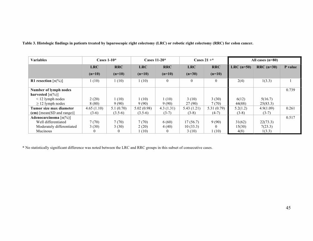
Data summarizing the histological findings are shown in Table 3. No group
difference was observed for any variables considered, in both the whole sample
comparision that the subgroups analyses.

Table 1. Demographic data and clinical characteristics of patients treated by laparoscopic right colectomy (LRC) or robotic right colectomy (RRC) for colon
cancer.
Variables Cases 1-10* Cases 11-20* Cases 21 +* All cases (n=80)
LRC (n=10) RRC (n=10) LRC (n=10) RRC (n=10) LRC (n=30) RRC (n=10) LRC
(n=50)
RRC
(n=30)
P value
Gender (F/M) [n] 8/2 5/5 5/5 7/3 18/12 3/7 31/19 15/15 0.353
Age (yr) [mean(SD)] 73.4 (16.73) 66.8 (10.21) 71 (13.83) 72.6 (4.47) 70.3 (11.56) 73.6 (8.85) 71.1(12.92) 71(8.50) 0.747
BMI (kg/m2) [mean(SD)] 24.27 (2.54) 25.33 (3.66) 26.1 (3.5) 27.06 (3.32) 25.3 (4.83) 26.89 (2.52) 25.26(4.19) 26.43(3.21) 0.180
Albumin Serum Level (g/L) [mean(SD)]
35.5 (5.9) 33.6 (4.5) 32.2 (5.1) 32 (4.3) 33.4 (6.25) 32.5 (4.06) 33.50(5.95) 32.7(4.18) 0.250
Diabetes [n(%)] 2 (20) 1 (10) 1 (10) 1 (10) 8 (26.7) 4 (40) 11(22) 6(21.4) 1 Cardiovascular Diseases [n(%)]
5 (50) 5 (50) 7 (70) 4 (40) 21 (70) 7 (70) 33(66) 16(53.3) 0.344
Pulmonary Disease [n(%)] 3 (30) 2 (20) 2 (20) 2 (20) 4 (13.3) 3 (30) 9(18) 7(23.3) 0.576 Previous abdominal surgery [n(%)]
7 (70) 3 (30) 5 (50) 5 (50) 14 (46.7) 4 (40) 26(52) 12(31.6) 0.358
ASA Score I/II/III [n] 1/3/6 0/7/3 3/2/5 0/5/5 0/13/17 0/3/7 4/18/28 0/15/15 0.181 Smoking [n(%)] 0 3 (30) 4 (40) 6 (60) 8 (26.7) 0 12(24) 9(33.3) 0.428 Tumor location[n(%)] Ileo-caecal valve Caecum Right ascending colon Right splenic flexure Right transverse colon
1 (10) 3 (30) 2 (20) 2( 20) 2 (20)
2 (20) 3 (30) 2 (20) 2 (20) 1 (10)
2 (20) 3 (30) 3 (30) 2 (20)
0
0
4 (40) 2 (20) 3 (30) 1 (10)
5 (16.7) 9 (30)
7 (23.3) 5 (16.7) 4 (13.3)
0
3 (30) 3 (30) 4 (40)
0
8(16)
15(30) 12(24) 9(18) 6(12)
2(6.7)
10(33.3) 7(23.3) 9(30) 2(6.7)
0.540
TNM AJCC Stage** [n(%)] I II III
5 (50) 5 (50)
0
2 (20) 6 (60) 2 (20)
3 (30) 4 (40) 3 (30)
3 (30) 4 (40) 3 (30)
10 (33.3) 12 (40) 8 (26.7)
3 (30) 3 (30) 4 (40)
18(36) 21(42) 11(22)
8(26.7)
13(43.3) 9(30)
0.609
42
BMI stands for body mass index; ASA for American Society of Anesthesiology; TNM for tumor, nodes and metastasis score; and AJCC for American Joint
Committee on Cancer.
* No statistically significant difference was noted between the LRC and RRC groups in this subset of consecutive cases.
**In the TNM AJCC categories II and III, the subcategories including T4b tumors (i.e., tumors directly invading or adherent to other organs or structures) were not
included.

43
Table 2. Operative and postoperative variables of patients treated by laparoscopic right colectomy (LRC) or robotic right colectomy (RRC) for colon
cancer.
Variables Cases 1-10* Cases 11-20* Cases 21 +* All cases (n=80)
LRC
(n=10)
RRC
(n=10)
LRC
(n=10)
RRC
(n=10)
LRC
(n=30)
RRC
(n=10)
LRC
(n=50)
RRC
(n=30)
P value
Operative time (min) [mean(SD) and (range)]
240(26.14) (220-300)
237.8(27.07) (200-290)
214.5(12.57) (195-230) #
193.8(24.24) (168-230) #
188.6(14.73) (160-230)
180(12) (168-200)
204.1(26.7) (160-300)
200.5(29.5) (168-290)
0.408
Docking time (min) [mean(SD) and (range)]
NA 25.7 (4.8) (20-34)
NA 21.6 (1.95) (20-25)
NA 18 (4.16) (12-25)
NA 21.76(4.9) (12-34)
NA
Surgical console time (min) [mean(SD) and (range)]
NA 196.5 (13.7) (180-220)
NA 174 (26.64) (140-220)
NA 152 (8.56) (140-170)
NA 174.16(25.36) (140-220)
NA
Conversion to laparoscopy [n(%)]
0 0 0 0 0 0 NA 0 NA
Conversion to laparotomy [n(%)]
1 (10) 0 0 0 1 (3.3) 0 2(4) 0 0.525
Operative blood loss (mL) [mean(SD)]
165 (24.1) 137 (32) 170 (25.8) 147 (25.4) 160 (25.5) 162 (34.5) 164 (24.80) 148.6 (31.59) 0.012
Number of transfused patient [n(%)]
0 0 1 (10) 0 0 0 1(2) 0 1
Time to flatus [mean(SD) and (range)]
2.2(0.63) (1-3)
1.9(0.56) (1-3)
2.3(0.48) (2-3)
2.1(0.56) (1-3)
1.63(0.49) (1-2)
1.9(0.31) (1-2)
1.88(0.59) (1-3)
1.96(0.49) (1-3)
0.471
Return to regular diet [mean(SD) and (range)]
3.4(0.96) (2-5)
2.9(0.99) (2-5)
3.3(0.82) (2-5)
2.8(0.78) (2-4)
2.7(0.59) (2-4)
2.4(0.51) (2-3)
2.96(0.78) (2-5)
2.7(0.79) (2-5)
0.108
Post-operative complications [n(%)] Ileus Anastomotic leakage Intra-abdominal abscess Cardiopulmonary complication Urinary infection
0 0 0
2(20) 0
0 0 0
1(10) 1(10)
0 1(10)
0 2(20)
0
1(10) 0 0
1(10) 0
0 1(3.3) 1(3.3) 3(10) 1(3.3)
0 0 0
1(10) 1(10)
0 2(4) 1(2)
7(14) 1(2)
1(3.3) 0 0
3(10) 2(6.7)
0.375 0.525
1 0.736 0.553
Dindo-Clavien classification 0.499

44
[n(%)] I-II III-IV
2(20)
0
2(20)
0
1(10) 1(10)
1(10)
0
5(16.7) 1(3.3)
1(10)
0
8(16) 2(4)
4(13.3)
0 Mortality within 30 days [n (%)]
0 1(10) 0 0 0 0 0 1(10) 1
Mortality within 90 days [n (%)]
0 0 0 0 0 0 0 0 NA
Length of hospital stay (days) [mean(SD) and (range)]
7.8(2.61) (5-13)
8.3(4.21) (6.20)
8.4(4.4) (5-18)
7(2.66) (5-14)
8.36(5.01) (4-30)
5.9(1.79) (4-10)
8.26(4.4) (4-30)
7.06(3.1) (4-20)
0.133
*No statistically significant difference was noted between the LRC and RRC groups in this subset of consecutive cases, if not otherwise specified.
# Trend toward statistically significant difference between LRC and RRC (p=0.07).
45
Table 3. Histologic findings in patients treated by laparoscopic right colectomy (LRC) or robotic right colectomy (RRC) for colon cancer.
* No statistically significant difference was noted between the LRC and RRC groups in this subset of consecutive cases.
Variables Cases 1-10* Cases 11-20* Cases 21 +* All cases (n=80)
LRC
(n=10)
RRC
(n=10)
LRC
(n=10)
RRC
(n=10)
LRC
(n=30)
RRC
(n=10)
LRC (n=50) RRC (n=30) P value
R1 resection [n(%)] 1 (10) 1 (10) 1 (10) 0 0 0 2(4) 1(3.3) 1
Number of lymph nodes harvested [n(%)]
< 12 lymph nodes ≥ 12 lymph nodes
2 (20) 8 (80)
1 (10) 9 (90)
1 (10) 9 (90)
1 (10) 9 (90)
3 (10) 27 (90)
3 (30) 7 (70)
6(12) 44(88)
5(16.7) 25(83.3)
0.739
Tumor size max diameter (cm) [mean(SD and range)]
4.65 (1.10) (3-6)
5.1 (0.70) (3.5-6)
5.02 (0.98) (3.5-6)
4.3 (1.31) (3-7)
5.43 (1.21) (3-8)
5.31 (0.79) (4-7)
5.2(1.2) (3-8)
4.9(1.09) (3-7)
0.261
Adenocarcinoma [n(%)] Well differentiated Moderately differentiated Mucinous
7 (70) 3 (30)
0
7 (70) 3 (30)
0
7 (70) 2 (20) 1 (10)
6 (60) 4 (40)
0
17 (56.7) 10 (33.3)
3 (10)
9 (90)
0 1 (10)
31(62) 15(30)
4(8)
22(73.3) 7(23.3) 1(3.3)
0.517

Consecu(ve*cases*of*right*colectomy*
Opera(ve*(m
e*(m
in)*
Cases%1'10% Cases%11'20% Cases%21%+%
140$150$160$170$180$190$200$210$220$230$240$250$260$270$280$290$300$310$
1$ 3$ 5$ 7$ 9$ 11$ 13$ 15$ 17$ 19$ 21$ 23$ 25$ 27$ 29$ 31$ 33$ 35$ 37$ 39$ 41$ 43$ 45$ 47$ 49$
Series1$Series3$Linear$(Series1)$Linear$(Series3)$
LRC%RRC%Trend%lines%
The operative time showed a trend towards reduction in both RRC and LRC groups
(Figure 7).
Figure 7. Plot of the operative time of LRC (blue line) and RRC (green line)
procedures by consecutive patients with the respective trend lines.
In both groups, a significant reduction in the operative time was observed over time
(ANOVA, for LRC p<0.0001; for RRC p<0.0001). Specifically, in the LRC group, a
significant difference was observed between the case series 1-10 and the case series 11-20
(p=0.002), and between the case series 1-10 and the case series 21+ (p<0.0001), and
between case series 11-20 and 21+ (p<0.0001). In the RRC group, a significant difference
was observed between the case series 1-10 and the case series 11-20 (p=0.002), between
case series 1-10 and case series 21+ (p<0.0001), whereas no difference was observed
between case series 11-20 and case series 21+ (p=0.174). The curves showed that a drop in
the operative time (≤ 170 min, red line) was observable after 31 cases of LRC (blue arrow)
and after 16 cases of RRC (green arrow).

47
Consecu(ve*cases*of*robo(c*right*colectomy*
Opera(ve*(m
e*(m
in)*
Cases%1'10% Cases%11'20% Cases%21%+%
0"
30"
60"
90"
120"
150"
180"
210"
240"
270"
300"
1" 2" 3" 4" 5" 6" 7" 8" 9" 10" 11" 12" 13" 14" 15" 16" 17" 18" 19" 20" 21" 22" 23" 24" 25" 26" 27" 28" 29" 30"
docking"3me"
console"3me"
opera3ve"3me""
Concerning the robotic procedures, the progressive decrease in the operative time was
also observed in the docking and surgeon console time, as shown in Figure 8.
Figure 8. Plot of the operative time (green line), surgeon console time (red line), and
docking time (blue line) of RRC procedures by consecutive patients.
A significant reduction over time was observed for the operative time (p<0.0001), the
surgeon console time (p<0.0001) and the docking time (p<0.0001). A drop in the operative
time and console time is observable after 16 RRC cases (black arrow).
The CUSUM analyses are displayed in Figure 9 and 10 for respectively LRC and RRC
procedures. As shown, the number of cases necessary to show a change in the operative
time is set at 25 for LRC and 16 for RRC.

48
!200.0%
!150.0%
!100.0%
!50.0%
0.0%
50.0%
100.0%
150.0%
200.0%
250.0%
1% 2% 3% 4% 5% 6% 7% 8% 9%10%11%12%13%14%15%16%17%18%19%20%21%22%23%24%25%26%27%28%29%30%31%32%33%34%35%36%37%38%39%40%41%42%43%44%45%46%47%48%49%50%
Cusum%Cha
rt%
Cases%
Cusum%Chart%
Upper%Cusum%
C+%
C!%
Lower%Cusum%
!150.0&
!100.0&
!50.0&
0.0&
50.0&
100.0&
150.0&
200.0&
250.0&
1& 2& 3& 4& 5& 6& 7& 8& 9& 10& 11& 12& 13& 14& 15& 16& 17& 18& 19& 20& 21& 22& 23& 24& 25& 26& 27& 28& 29& 30&
Cusum%
Cases%
Cusum%Chart%
Upper&Cusum&
C+&
C!&
Lower&Cusum&
Figure 9. CUSUM analysis of LRC procedures.
Figure 10. CUSUM analysis of RRC procedures.

49
0"
2000"
4000"
6000"
8000"
10000"
12000"
14000"
LRC" RRC"
Hospitalisa3on""
Surgery;related"Costs"
p=0.632(
Euros"
In the Health Care Cost analysis, surgery-related costs of the LRC procedures
resulted significantly less expensive compared with the RRL, with a mean cost of 1800
(90.9) Euros for the LRC vs. 2990 (298) Euros for the RRC (p<0.0001). However, when
the surgery-related costs are combined with the hospitalisation-related costs (estimated at
approximately 1300 euros per day), the LRC or RRC procedures did not differ significantly
(p=0.632). Overall, the mean cost (surgery + hospitalisation costs) per right colectomy
procedure was 12400 (5180) Euros (Figure 11).
Figure 11. Health Care cost analysis for LRC and RRC procedures by taking into
account the surgery-related costs and the hospitalisation.
50
Discussion The present study shows that robotic right colectomy yields similar operative and
postoperative outcomes of laparoscopic right colectomy performed by a fellow surgeon.
The learning curve appears to be longer for LRC, whereas with only 16 robotic procedures
a significant reduction in the operative time is already observable. RRC are also associated
with significantly less blood loss compared to LRC. The postoperative complication rate
and the histologic variables were not different between the two approaches. Interestingly,
the present results also show that the health related costs are similar between LRC and RRC
when considering the mean cost per procedure by adding the surgical costs (significantly
more important in RRC) to the hospital stay.
Paucity of reports exists on learning curves involving robotic colonic surgery1,
while more literature is available for laparoscopic procedures17-19, 21, 23, 24, 44, 61. Comparing
the two techniques is also hard in the literature due to the fact that most of the time the
surgeons beginning their learning curve in robotics are already experienced in laparoscopy,
and this can have a direct and indirect influence on the robotic skills. In the present study,
the operating surgeon was simultaneously adopting laparoscopic and robotic surgery for
right colectomy procedures and he was at the beginning of his minimally invasive career.
Thus, the results provide further elucidations on MIS learning curves and allows for direct
comparisons. In our knowledge, only one previous study used this design to compare
laparoscopy vs. robotic rectal surgery and concluded that the simultaneous development of
laparoscopy and robotics provides acceptable perioperative outcomes, whereas robotics has
a faster learning curve62. The present results confirmed those previous findings. Indeed,

51
robotic right colectomy appears to have a learning curve of 16 procedures whereas the
laparoscopic ones of 26. In discordance with the previous literature40, 63, the present study
showed that the mean operative time of RRC is similar to LRC, which falls into the
previously reported ranges for laparoscopic right colectomy (85-214 min29, 64, 65).
Moreover, the operative time decreases progressively with the number of procedures and
the increasing surgeon experience for both robotic and laparoscopic approaches, but this
process looked faster for robotic surgery. Indeed, after 16 RRC procedures, an
improvement in the operative time is observable and appears quite stable for the following
14 procedures analyzed in this study. For LRC, the same improvement in the operative time
is set at 31 procedures and it is more difficult to identify a plateau even after 30 procedures.
Previous studies reported that an operative time proficiency in robotic rectal surgery
is reached in 15–25 cases1. Another study has recently estimated that 80 robotic total
mesorectal excision cases are the necessary learning curve66, whereas the operative times
did not improve during the course of the study. Thus, data in the literature are still
controversial but it must be noted that the learning curve may be substantially different
between right colectomy, rectal resections, or total mesorectal excision, which require
various levels of difficulties and technical skills. In this regard, right colectomy appeared to
be a suitable intervention to start the learning curve29, and was chosen in this study as the
sole procedure (performed with the same approach, i.e. medial to lateral, in both RRC and
LRC) in order to limit the possible confounders in the evaluation of the learning curve.
The results presented in this study further show that the initially longer total
operative time for robotic surgery improves rapidly and after only 15 cases it become faster
52
(trend toward significance) than that of laparoscopic surgery. Interestingly, this
decrease seems to be attributed to the both the decrease in the docking time, which is
finally assessed at 15-18 min, and the surgeon console time, which directly corresponds to
the laparoscopic time.
Considering these data, the robotic approach can be safely chosen as an alternative
approach to laparoscopy for right colectomy. The major concern will remain the costs for
the Public Health System. The present results must be interpreted with caution since they
refer to a specific situation of procedures performed by a fellow surgeon during the learning
curve. Despite this, the present study shows that the two surgical techniques are not
associated to different health related costs when combining the surgery-related costs and
the hospitalization. Indeed, RRC procedures carry higher intraoperative costs in terms of
surgical materials of laparoscopy, but the costs of laparoscopy per se might be higher than
for experienced surgeon. Thus the gap between the two approaches may be inferior than
what previously reported in the literature and is balanced out by the costs in the
postoperative period. Another important point is the low incidence of complications, which
can drastically influence the costs for the Health Care System. These aspects cannot be
neglected and the present results cannot be generalized. However, they may suggest that the
argument of cost-effectiveness or saving resources are not longer valid to discourage
robotic surgery training and implementation 27, 32, 48, 51.
Study Limitations and Strenghts
53
One of the main limitations of the present study is the relative small number of
cases, especially for the robotic procedures. Moreover, the learning curve was evaluated on
a single surgeon experience and potential additive or even synergistic learning effects
transferring from laparoscopy to robotic surgery and vice versa (which were developped
simultaneoulsy) cannot be excluded. On the other hand, the fact that this is a single surgeon
experience performing both procedures at the onset of his career allows for a unique head-
to-head comparison where many variables and effects stay similar for both techniques.
However, the external validity of this findings is not assessed and all operative,
postoperative and health costs results should be interpreted taking into account the specific
situation of a fellow surgeon at the beginning of his MIS carreer. Points of strenght include
the homogeneous sample of patients undergone LRC and RRC during the same time
period. The two groups were not balanced in numbers (due to the lower availability of the
robotic platform compared to the laparoscopic instruments), but they did not differ for the
most important demographic, clinical, and tumor-related variables.
Conclusion
Robotic right colectomy yields similar operative and postoperative outcomes of
laparoscopic right colectomy once performed by a fellow surgeon during his learning
curve. The advantages of MIS are observable in both robotic and laparoscopic procedures,
but these latter ones appear to be associated with a longer learning curve. With increasing
experience, operative time will further decrease, and it is not longer when operating with
54
the robotic platform. This suggest that it is reasonable and important to include
robotic colorectal training for fellow surgeons, and this advanced MIS approach sould be
considered as safe, feasable and advisable in colorectal surgery. Moreover, the real
challenge of cost-effectiveness of robotic procedures appear to be on the way of
improvement. Especially in the present time characterised by important financial
constraints of health care systems, robotic surgery had appeared not appropriate because
not cost-effective. However, evidence are growing to support that the costs of robotic
colonic surgery are reducing over time and they are projected to be further reduced in the
near future because of the progressive decrease in equipment costs and consuption, and the
progressive shortening in hospital stay. It would be more and more difficult to argue that
robotic surgery will not offer any potential advantage over laparoscopy, mostly in terms of
further reduction of hospital stay. When analyzing the outcome of robotic surgery, too often
we read the message that it is safe and feasible but too costly to be chosen over
laparoscopy. Indeed, we keep answering the important clinical patient-oriented question
“What are the surgical and clinical advantages of robot-assisted rectal surgery?” with a
reply based on an often biased cost-effectiveness analysis. Surgeon are starting to use
robotic surgery in a cost-effective way by decreasing the number of instruments used, being
faster in docking and undocking and getting dedicated personnel in the robotic room. It is
thus important that further clinical trials will be conducted to address the question whether
the robotic approach could increase, thanks to its advanced technology, the currently small
number of patients undergoing MIS.

55
References 1. Bokhari MB, Patel CB, Ramos-Valadez DI, Ragupathi M, Haas EM. Learning curve
for robotic-assisted laparoscopic colorectal surgery. Surg Endosc. 2011;25:855-860.
2. Soper NJ, Stockmann PT, Dunnegan DL, Ashley SW. Laparoscopic
cholecystectomy. The new 'gold standard'? Arch Surg. 1992;127:917-921; discussion 921-
913.
3. Fung AK, Aly EH. Robotic colonic surgery: is it advisable to commence a new
learning curve? Dis Colon Rectum. 2013;56:786-796.
4. Jacobs M, Verdeja JC, Goldstein HS. Minimally invasive colon resection
(laparoscopic colectomy). Surg Laparosc Endosc. 1991;1:144-150.
5. Berends FJ, Kazemier G, Bonjer HJ, Lange JF. Subcutaneous metastases after
laparoscopic colectomy. Lancet. 1994;344:58.
6. Alasari S, Min BS. Robotic colorectal surgery: a systematic review. ISRN Surg.
2012;2012:293894.
7. Araujo SE, Seid VE, Klajner S. Robotic surgery for rectal cancer: Current
immediate clinical and oncological outcomes. World J Gastroenterol. 2014;20:14359-
14370.

56
8. Group CS. COLOR: a randomized clinical trial comparing laparoscopic and
open resection for colon cancer. Dig Surg. 2000;17:617-622.
9. Guillou PJ, Quirke P, Thorpe H, et al. Short-term endpoints of conventional versus
laparoscopic-assisted surgery in patients with colorectal cancer (MRC CLASICC trial):
multicentre, randomised controlled trial. Lancet. 2005;365:1718-1726.
10. Jayne DG, Guillou PJ, Thorpe H, et al. Randomized trial of laparoscopic-assisted
resection of colorectal carcinoma: 3-year results of the UK MRC CLASICC Trial Group. J
Clin Oncol. 2007;25:3061-3068.
11. Bonjer HJ, Deijen CL, Abis GA, et al. A randomized trial of laparoscopic versus
open surgery for rectal cancer. N Engl J Med. 2015;372:1324-1332.
12. Fleshman J, Sargent DJ, Green E, et al. Laparoscopic colectomy for cancer is not
inferior to open surgery based on 5-year data from the COST Study Group trial. Ann Surg.
2007;246:655-662; discussion 662-654.
13. Ohtani H, Tamamori Y, Arimoto Y, Nishiguchi Y, Maeda K, Hirakawa K. A meta-
analysis of the short- and long-term results of randomized controlled trials that compared
laparoscopy-assisted and open colectomy for colon cancer. J Cancer. 2012;3:49-57.
14. Noel JK, Fahrbach K, Estok R, et al. Minimally invasive colorectal resection
outcomes: short-term comparison with open procedures. J Am Coll Surg. 2007;204:291-
307.

57
15. Kiran RP, El-Gazzaz GH, Vogel JD, Remzi FH. Laparoscopic approach
significantly reduces surgical site infections after colorectal surgery: data from national
surgical quality improvement program. J Am Coll Surg. 2010;211:232-238.
16. Lacy AM, Garcia-Valdecasas JC, Delgado S, et al. Laparoscopy-assisted colectomy
versus open colectomy for treatment of non-metastatic colon cancer: a randomised trial.
Lancet. 2002;359:2224-2229.
17. Reissman P, Cohen S, Weiss EG, Wexner SD. Laparoscopic colorectal surgery:
ascending the learning curve. World J Surg. 1996;20:277-281; discussion 282.
18. Schlachta CM, Mamazza J, Seshadri PA, Cadeddu M, Gregoire R, Poulin EC.
Defining a learning curve for laparoscopic colorectal resections. Dis Colon Rectum.
2001;44:217-222.
19. Tekkis PP, Senagore AJ, Delaney CP, Fazio VW. Evaluation of the learning curve
in laparoscopic colorectal surgery: comparison of right-sided and left-sided resections. Ann
Surg. 2005;242:83-91.
20. Dincler S, Koller MT, Steurer J, Bachmann LM, Christen D, Buchmann P.
Multidimensional analysis of learning curves in laparoscopic sigmoid resection: eight-year
results. Dis Colon Rectum. 2003;46:1371-1378; discussion 1378-1379.
21. Senagore AJ, Luchtefeld MA, Mackeigan JM. What is the learning curve for
laparoscopic colectomy? Am Surg. 1995;61:681-685.
58
22. Simons AJ, Anthone GJ, Ortega AE, et al. Laparoscopic-assisted colectomy
learning curve. Dis Colon Rectum. 1995;38:600-603.
23. Wishner JD, Baker JW, Jr., Hoffman GC, et al. Laparoscopic-assisted colectomy.
The learning curve. Surg Endosc. 1995;9:1179-1183.
24. Li JC, Hon SS, Ng SS, Lee JF, Yiu RY, Leung KL. The learning curve for
laparoscopic colectomy: experience of a surgical fellow in an university colorectal unit.
Surg Endosc. 2009;23:1603-1608.
25. Kemp JA, Finlayson SR. Nationwide trends in laparoscopic colectomy from 2000 to
2004. Surg Endosc. 2008;22:1181-1187.
26. Panis Y, Maggiori L, Caranhac G, Bretagnol F, Vicaut E. Mortality after colorectal
cancer surgery: a French survey of more than 84,000 patients. Ann Surg. 2011;254:738-
743; discussion 743-734.
27. de'Angelis N, Portigliotti L, Azoulay D, Brunetti F. Robotic surgery: A step forward
in the wide spread of minimally invasive colorectal surgery. J Minim Access Surg.
2015;11:285-286.
28. Pigazzi A, Luca F, Patriti A, et al. Multicentric study on robotic tumor-specific
mesorectal excision for the treatment of rectal cancer. Ann Surg Oncol. 2010;17:1614-1620.

59
29. deSouza AL, Prasad LM, Park JJ, Marecik SJ, Blumetti J, Abcarian H.
Robotic assistance in right hemicolectomy: is there a role? Dis Colon Rectum.
2010;53:1000-1006.
30. Aly EH. Robotic colorectal surgery: summary of the current evidence. Int J
Colorectal Dis. 2014;29:1-8.
31. Weber PA, Merola S, Wasielewski A, Ballantyne GH. Telerobotic-assisted
laparoscopic right and sigmoid colectomies for benign disease. Dis Colon Rectum.
2002;45:1689-1694; discussion 1695-1686.
32. de'Angelis N, Felli E, Azoulay D, Brunetti F. Robotic-assisted reversal of
Hartmann's procedure for diverticulitis. J Robot Surg. 2014;8:381-383.
33. Felli E, Brunetti F, Disabato M, Salloum C, Azoulay D, De'angelis N. Robotic right
colectomy for hemorrhagic right colon cancer: a case report and review of the literature of
minimally invasive urgent colectomy. World J Emerg Surg. 2014;9:32.
34. Huettner F, Dynda D, Ryan M, Doubet J, Crawford DL. Robotic-assisted minimally
invasive surgery; a useful tool in resident training--the Peoria experience, 2002-2009. Int J
Med Robot. 2010;6:386-393.
35. Huettner F, Pacheco PE, Doubet JL, Ryan MJ, Dynda DI, Crawford DL. One
hundred and two consecutive robotic-assisted minimally invasive colectomies--an outcome
and technical update. J Gastrointest Surg. 2011;15:1195-1204.
60
36. Rawlings AL, Woodland JH, Crawford DL. Telerobotic surgery for right and
sigmoid colectomies: 30 consecutive cases. Surg Endosc. 2006;20:1713-1718.
37. Buchs NC, Pugin F, Bucher P, Morel P. Totally robotic right colectomy: a
preliminary case series and an overview of the literature. Int J Med Robot. 2011.
38. Broholm M, Pommergaard HC, Gogenur I. Possible benefits of robot-assisted rectal
cancer surgery regarding urological and sexual dysfunction: a systematic review and meta-
analysis. Colorectal Dis. 2015;17:375-381.
39. Petrucciani N, Sirimarco D, Nigri GR, et al. Robotic right colectomy: A worthwhile
procedure? Results of a meta-analysis of trials comparing robotic versus laparoscopic right
colectomy. J Minim Access Surg. 2015;11:22-28.
40. Trastulli S, Cirocchi R, Desiderio J, et al. Robotic versus Laparoscopic Approach in
Colonic Resections for Cancer and Benign Diseases: Systematic Review and Meta-
Analysis. PLoS One. 2015;10:e0134062.
41. Witkiewicz W, Zawadzki M, Rzaca M, et al. Robot-assisted right colectomy:
surgical technique and review of the literature. Wideochir Inne Tech Maloinwazyjne.
2013;8:253-257.
42. Trastulli S, Desiderio J, Farinacci F, et al. Robotic right colectomy for cancer with
intracorporeal anastomosis: short-term outcomes from a single institution. Int J Colorectal
Dis. 2013;28:807-814.

61
43. Lin S, Jiang HG, Chen ZH, Zhou SY, Liu XS, Yu JR. Meta-analysis of robotic
and laparoscopic surgery for treatment of rectal cancer. World J Gastroenterol.
2011;17:5214-5220.
44. Meinke AK, Kossuth T. What is the learning curve for laparoscopic appendectomy?
Surg Endosc. 1994;8:371-375; discussion 376.
45. Richardson MC, Bell G, Fullarton GM. Incidence and nature of bile duct injuries
following laparoscopic cholecystectomy: an audit of 5913 cases. West of Scotland
Laparoscopic Cholecystectomy Audit Group. Br J Surg. 1996;83:1356-1360.
46. Parikh D, Johnson M, Chagla L, Lowe D, McCulloch P. D2 gastrectomy: lessons
from a prospective audit of the learning curve. Br J Surg. 1996;83:1595-1599.
47. Sutton DN, Wayman J, Griffin SM. Learning curve for oesophageal cancer surgery.
Br J Surg. 1998;85:1399-1402.
48. de'Angelis N, Alghamdi S, Renda A, Azoulay D, Brunetti F. Initial experience of
robotic versus laparoscopic colectomy for transverse colon cancer: a matched case-control
study. World J Surg Oncol. 2015;13:295.
49. Scarpinata R, Aly EH. Does robotic rectal cancer surgery offer improved early
postoperative outcomes? Dis Colon Rectum. 2013;56:253-262.
62
50. Xu H, Li J, Sun Y, et al. Robotic versus laparoscopic right colectomy: a meta-
analysis. World J Surg Oncol. 2014;12:274.
51. de'Angelis N, Portigliotti L, Brunetti F. Robot-assisted rectal cancer surgery
deserves a fair trial. Colorectal Dis. 2015;17:824-825.
52. Hopper AN, Jamison MH, Lewis WG. Learning curves in surgical practice.
Postgrad Med J. 2007;83:777-779.
53. Khan N, Abboudi H, Khan MS, Dasgupta P, Ahmed K. Measuring the surgical
'learning curve': methods, variables and competency. BJU Int. 2014;113:504-508.
54. Chen W, Sailhamer E, Berger DL, Rattner DW. Operative time is a poor surrogate
for the learning curve in laparoscopic colorectal surgery. Surg Endosc. 2007;21:238-243.
55. Waller HM, Connor SJ. Cumulative sum (Cusum) analysis provides an objective
measure of competency during training in endoscopic retrograde cholangio-
pancreatography (ERCP). HPB (Oxford). 2009;11:565-569.
56. Clinical Outcomes of Surgical Therapy Study G. A comparison of laparoscopically
assisted and open colectomy for colon cancer. N Engl J Med. 2004;350:2050-2059.
57. Croce E, Olmi S, Azzola M, Russo R, Di Bonifacio M. Laparoscopic colectomy:
indications, standardized technique and results after 6 years experience.
Hepatogastroenterology. 2000;47:683-691.

63
58. Deo SV, Puntambekar SP. Laparoscopic right radical hemicolectomy. J Minim
Access Surg. 2012;8:21-24.
59. Garcia-Ruiz A, Milsom JW, Ludwig KA, Marchesa P. Right colonic arterial
anatomy. Implications for laparoscopic surgery. Dis Colon Rectum. 1996;39:906-911.
60. Dindo D, Demartines N, Clavien PA. Classification of surgical complications: a
new proposal with evaluation in a cohort of 6336 patients and results of a survey. Ann Surg.
2004;240:205-213.
61. Kiran RP, Kirat HT, Ozturk E, Geisler DP, Remzi FH. Does the learning curve
during laparoscopic colectomy adversely affect costs? Surg Endosc. 2010;24:2718-2722.
62. Melich G, Hong YK, Kim J, et al. Simultaneous development of laparoscopy and
robotics provides acceptable perioperative outcomes and shows robotics to have a faster
learning curve and to be overall faster in rectal cancer surgery: analysis of novice MIS
surgeon learning curves. Surg Endosc. 2015;29:558-568.
63. Ezekian B, Sun Z, Adam MA, et al. Robotic-Assisted Versus Laparoscopic
Colectomy Results in Increased Operative Time Without Improved Perioperative
Outcomes. J Gastrointest Surg. 2016.
64. Tan WS, Chew MH, Ooi BS, et al. Laparoscopic versus open right hemicolectomy:
a comparison of short-term outcomes. Int J Colorectal Dis. 2009;24:1333-1339.

64
65. Salloum RM, Bulter DC, Schwartz SI. Economic evaluation of minimally
invasive colectomy. J Am Coll Surg. 2006;202:269-274.
66. Akmal Y, Baek JH, McKenzie S, Garcia-Aguilar J, Pigazzi A. Robot-assisted total
mesorectal excision: is there a learning curve? Surg Endosc. 2012;26:2471-2476.

i
i