AMNIOTIC FLUID STEM CELLS AND KIDNEY...
155
1 UNIVERSITA' DEGLI STUDI DI PADOVA DIPARTIMENTO DI PEDIATRIA SCUOLA DI DOTTORATO DI RICERCA IN MEDICINA DELLO SVILUPPO E SCIENZE DELLA PROGRAMMAZIONE INDIRIZZO: MALATTIE RARE XXII CICLO TESI DI DOTTORATO AMNIOTIC FLUID STEM CELLS AND KIDNEY REGENERATION Direttore della Scuola: Ch.mo Prof. GIUSEPPE BASSO Coordinatore d’indirizzo: Ch.mo Prof. GIORGIO PERILONGO Supervisore: Ch.mo Prof. PIERGIORGIO GAMBA Dottorando: STEFANO GIULIANI
Transcript of AMNIOTIC FLUID STEM CELLS AND KIDNEY...
1
UNIVERSITA' DEGLI STUDI DI PADOVA
DIPARTIMENTO DI PEDIATRIA
SCUOLA DI DOTTORATO DI RICERCA IN
MEDICINA DELLO SVILUPPO E SCIENZE DELLA
PROGRAMMAZIONE
INDIRIZZO: MALATTIE RARE
XXII CICLO
TESI DI DOTTORATO
AMNIOTIC FLUID STEM CELLS AND KIDNEY
REGENERATION
Direttore della Scuola: Ch.mo Prof. GIUSEPPE BASSO
Coordinatore d’indirizzo: Ch.mo Prof. GIORGIO PERILONGO
Supervisore: Ch.mo Prof. PIERGIORGIO GAMBA
Dottorando: STEFANO GIULIANI

2
3
Alla mia famiglia per l’amore costante sempre dimostratomi…
A Laura Perin per l’amicizia, la grande passione per la ricerca e
l’insegnamento

4
5
INDEX
ABSTRACT 9
ABSTRACT IN ITALIAN 11
INTRODUCTION: 19
1. ESRD, causes and outcomes 19
• Acute Kidney Disease Therapy
• Chronic Kidney Disease Therapy
2. Regenerative Medicine and Kidney regeneration 33
• Tissue Engineering for Kidney regeneration
• Stem Cells:
§ Embryonic Stem Cells
§ Primordial Germ Cells
§ Adult Stem Cells
§ Somatic Cell Nuclear Transfer
• Stem Cells and Kidney Regeneration
3. Amniotic Fluid: Stem Cells and Progenitors 39
OBJECTIVES 41
MATERIALS AND METHODS 43
1. Expansion of human Amniotic Fluid Total Cell Population 43
2. Characterization of Amniotic Fluid Cells 43
• Analysis and characterization by RT-PCR
• Analysis and characterization by Real Time PCR
• Analysis and characterization by Western Blotting
3. Ex Vivo whole embryonic kidney culture 53
4. Selection of Amniotic Fluid Stem Cells (AFSC) 55
5. In vitro model for the renal differentiation of hAFSC 56
• Microinjection of hAFSC and co-culture
• Live Imaging
• Histology, Chromogenic in situ hybridization
• Reverse transcriptase-polymerase chain reaction
6. In vivo experiment with a model of Acute Tubular Necrosis 59
• ATN induction and injection of hAFSC
6
• Tissue processing
• Labeling of the AFSC with luciferase and bioluminescence
• Immunostaining
• Blood collection, creatinine and BUN measurement
• Morphological studies
• Cytokine analysis
7. Selection and characterization of Metanephric Mesenchyme Derived
Cells (MMDC) and Kidney Progenitor Cells (AKPC) from the whole
Amniotic Fluid: 66
• Immunoseparation from whole Amniotic Fluid
• Analysis and characterization by RT-PCR
• Analysis and characterization by Real Time PCR
RESULTS 69
1. Characterization of Amniotic Fluid cells by expression of markers for the
three germ layers and progenitor cells 69
• Amniotic Fluid Total Cell Population Culture
• Analysis and characterization by RT-PCR
• Analysis and characterization by Western Blotting
• Analysis and characterization by Real Time PCR
2. Long term ex-vivo whole embryonic kidney culture 72
3. Selection of Amniotic Fluid Stem Cells 73
4. In vitro renal differentiation of hAFSC 74
• Ureteric Bud induces hAFSC to form tubular structures
• Evidence of hAFSC integration into embryonic kidneys
• Molecular evidence of primordial kidney differentiation
5. Protective effect of hAFSC in Acute Tubular Necrosis 76
• Acute Tubulonecrosis mouse model
• Histology, Period Acid Schiff staining (PAS), TUNEL
• In vivo detection of hAFSC by bioluminescence
• Detection of hAFSC by immunohistochemistry and gene expression
• Creatinine and Blood Urea Nitrogen (BUN) measurements
• Morphological studies
• Immuno-Cytokine characterization

7
6. Selection and characterization of Metanephric Mesenchyme derived
cells (MMDC) and Kidney Progenitor cells (AKPC) from the whole
Amniotic Fluid 80
• Immunoseparation from the whole Amniotic Fluid
• Analysis and characterization by RT-PCR
• Analysis and characterization by Real Time PCR
DISCUSSION 83
1. hAFSC differentiation in renal parenchyma in vitro and in vivo 88
2. Characterization and isolation of Metanpehric Mesenchyme Derived
cells (MMDC) and Amniotic Fluid Kidney Progenitor Cells (AKPC) 96
CONCLUSIONS 109
REFERENCES 113
FIGURES AND TABLES 125

8
9
ABSTRACT
Acute Kidney Disease (CKD) is a major public health problem that affects
some 3-7% of patients admitted to the hospital and approximately 25-30%
of patients in the intensive care unit. None of the existing therapies are
exempt from side effects and kidney physiological functionality is never
restored. Transplantation has been reported as the preferred cure for CKD
management but organ shortage and risks due to the immunosuppressive
therapy makes it far from being the perfect treatment for ESRD. In this
study we have focused our attention on finding novel strategies, in vitro
and in vivo, to obtain kidney regeneration in case of acute and chronic
kidney damage.
First we have demonstrated the ability of hAFSCs to survive, proliferate
and integrate into the embryonic kidney, while it undergoes organ
development, in an in vitro culture system. We observed the presence of
hAFSCs within kidney primordial, including tubules and developing
nephrons. Thus, hAFSCs seem to have the capacity to undergo the
expected mesenchymal to epithelial transition that occurs in normal renal
development and are induced to express important early kidney markers
such as GDNF, ZO-1 and Claudin. Moreover, hAFSCs do not appear to
require prior genetic modification or exogenous production of kidney
proteins for their differentiation to occur. This is a very important
advantage that hAFSCs have for potential future regenerative or
bioengineering application.
With the in vivo experiments, we have demonstrated that early direct
injection of hAFSCs into the kidney strongly ameliorates ATN injury as
reflected by more rapid resolution of tubular structural damage and by
normalization of creatinine and BUN levels. In addition, our data show
evidence of immunomodulatory and antinflammatory effect of hAFSCs, at
an early time point, comparable in magnitude to endogenous cytokine
production. Understanding how donor and host cells combine to attenuate
10
tubular damage may lead eventually to the application of hAFSCs for
therapeutic purposes in acute kidney diseases.
Nonetheless, beside the presence of a small number (1%) of cells with
pluripotent characteristics, the composition of the other 99% of Amniotic
Fluid cells is diverse, with a great amount of cells exhibiting commitment to
a defined germ line or cellular endpoint.
There seems to be clear evidence for the existence of progenitor cells in
Amniotic Fluid, which can give rise to different cell types of mature organs.
By 17 weeks of gestation is notable an increase tissue specific cellular
presence and this data may indicate that the choice of the time point for
cell selection is fundamental. In addition, we demonstrated in the amniotic
fluid, the presence of a renal population with specific traits of commitment.
In particular, the presence of podocytes at both undifferentiated and
almost mature stages could favour their use for kidney regeneration in
vitro and in vivo animal models. The presence and identification of specific
renal progenitor cells in the Amniotic Fluid, committed to different
compartments of the kidney environment, could represent a valuable new
tool for regenerative purposes with regards to the treatment of a broad
range of renal diseases.
The discovery of renal specific progenitor cells within Amniotic Fluid could
bring a breakthrough in the study for novel and more selective approaches
in the renal therapy. However, the real pluripotential capability of these
progenitors cells, in particular the kidney progenitors presenting more
differentiation characteristics, has to be established. Moreover, their
potential for survival, proliferation, integration, and differentiation needs to
be assessed in in vivo models involving different types of renal damage.
11
ABSTRACT IN ITALIAN
L’insufficienza renale terminale ha raggiunto ormai proporzioni epidemiche
in tutto il mondo e, tutt’oggi, non sono ancora state trovate terapie
sostitutive o rigenerative efficaci a lungo termine. Attualmente la terapia
dialitica e il trapianto allogenico rimangono le uniche alternative valide da
utilizzare in questi pazienti nonostante se ne conoscano i numerosi limiti e
complicanze. Recenti dati epidemiologici, in America e in Europa,
mostrano che l’insufficienza renale colpisce circa l’8% della popolazione.
[1] L’aumentata domanda di organi, in aggiunta all’insufficiente
disponibilita’ di donatori, sta spingendo sempre piu’ i ricercatori di tutto il
mondo a sviluppare nuove alternative terapeutiche per la sostituzione dei
reni non funzionanti. [2] La creazione di organi bio-artificiali, attraverso
l’utilizzo delle tecniche di ingegneria tissutale, ha finora dimostrato grandi
difficolta’ specialmente nel riprodurre quegli organi e tessuti la cui struttura
e funzione risultino particolarmente complesse, come nel caso dei reni.
Storicamente gli ingegneri tissutali che si sono cimentati in questo campo
hanno potuto utilizzare esclusivamente linee cellulari adulte dando origine
a costrutti bidimensionali caratterizzati da limitata funzione e difficile
applicabilita’ in vivo. [3]
Nell’ultima dacade le cellule staminali stanno ricevendo sempre maggiore
attenzione scientifica grazie al loro crescente impiego nella medicina
rigenerativa per la ricostruzione e rigenerazione di tessuti bio-artificiali ed
organi. Le cellule Staminali Embrionali (SE), derivate dalla blastocisti,
hanno come caratteristiche peculiari il fatto che si replichino ampliamente
e che siano capaci di formare aggregati (corpi embrioidi) che possono dar
luogo ad una varietà di cellule specializzate come, ad esempio, cellule
neurali, cardiache e pancreatiche. [3, 4] Il reclutamento di questo tipo di
cellule staminali, tuttavia, comporta la distruzione di embrioni umani
creando spinosi problemi etici e morali che portano, in molti Paesi, a
vietarne l’utilizzo e il progresso scientifico. Per evitare questo tipo di
controversie ricercatori di varie discipline hanno identificato potenziali fonti
12
di cellule staminali alternative. [4, 5] E’ ormai ben noto che in molti tessuti
adulti esistono cellule progenitrici con il compito di rigenerare o riparare
l'organo a seguito dei fisiologici processi di senescenza o in caso di
danno. [6, 7] Ci sono sempre piu’ evidenze che questi progenitori d’organo
abbiano caratteristiche di plasticità piu’ elevate di quanto si pensasse
originariamente. Parallelamente molti ricercatori credono che la
rigenerazione di organi adulti derivi principalmente dalla mobilizzazione di
cellule staminali provenienti dal midollo osseo. E’ stato dimostrato che
cellule staminali del midollo osseo possono attraversare la barriera
endolteliale e dar luogo a differenti linee cellulari differenziate,
trasformando cellule circolanti in fegato, cervello, pancreas, pelle, intestino
e anche rene. [27, 29]
Il liquido amniotico e’stato usato per anni come uno strumento sicuro e
valido per la ricerca di malattie genetiche e congenite del feto. Tuttavia, il
liquido amniotico contiene un grande numero di cellule progenitrici che
posono avere un importante ruolo nelle applicazioni della bioingegneria
tissutale. Streubel et al. [8] hanno riportato l’utilizzo di cellule non
emopoietiche per la conversione di amniociti in miociti. Recentemente una
popolazione di cellule staminali c-Kit+, isolate nel liquido amniotico umano
e murino, e’ stata caratterizzata e differenziata in tessuti originati dai tre
foglietti embrionali: muscolare, neuronale, adipocitario, epatico, osseo ed
endoteliale [9]
Nel laboratorio diretto dal dr. R.E. De Filippo, Assistant Professor presso il
Childrens Hospital di Los Angeles, abbiamo ampiamente studiato e
utilizzato questa nuova popolazione di cellule staminali derivate dal liquido
amniotico focalizzando le nostre ricerche sul loro utilizzo nella
rigenerazione renale. Abbiamo dimostrato che questa popolazione
totipotente di cellule mesenchimali e’ capace di riprodurre alcune tappe
essenziali della nefrogenesi dopo essere state iniettate in reni embrionici.
Tuttavia, le cellule staminali da liquido amniotico rapresentano meno
dell’1% dell’intera popolazione cellulare e forse esistono altri progenitori
cellulari, nel liquido stesso, gia’ orientati e piu’ proni alla differenziazione di
13
particolari linee cellulari renali che possano essere utilizzate per gli stessi
scopi rigenerativi ma con risultati migliori.
Il volume e la composizione del liquido amniotico cambia durante la
gravidanza e dall’ottava settimana di gestazione i reni fetali iniziano a
produrre liquido che rapidamente aumenta di volume durante il secondo
trimestre. [10] Il contatto tra il liquido amniotico e i diversi tessuti fetali
sembra giustificare la presenza dei differenti tipi cellulari disciolti nel
liquido stesso. La presenza di cellule mature derivanti dai tre foglietti
germinali e’ stata gia’ dimostrata in passato come pure la presenza di
progenitori cellulari di origine mesenchimale ed emopoietica prima della
12ma settimana gestazionale nell’uomo. [11,12,13] Cellule esprimenti
proteine e markers genetici tipici di tessuti diversi come cervello, cuore, e
pancreas sono state ritrovate nel liquido amniotico ma ulteriori indagini
sono necessarie per completare la caratterizzazione dei diversi tipi
cellulari presenti alle diverse eta’ gestazionali. [14, 15, 16]
Lo sviluppo renale e’ un complesso processo di attivazione genica,
interazioni cellulari ed extracellulari che devono aver luogo secondo un
ordine spazio-temporale preciso e nella quantita’ adeguata. Durante
l’embriogenesi, il rene metanefrico origina dall’invasione da parte della
gemma ureterale, derivata dal dotto epiteliale di Wolffian, nel mesenchima
metanefrico. [17] La gemma ureterale inizia la sua arborizzazione
all’interno del mesenchima e portera’ alla formazione dell’intero sistema
escretore, dall’uretere ai dotti collettori, mentre il mesenchima metanefrico
dara’ luogo alle strutture epiteliali costituenti i nefroni (dal tubulo distale
alla capsula di Bowman). CD-24 e Caderina 11 sono due markers di
superficie che vengono usati per identificare cellule staminali ancora
indifferenziate ma presenti nel mesenchima metanefrico prima di ricevere
l’induzione da parte della gemma ureterale. [18] In aggiunta, altri markers
di superficie che identificano una sottopopolazione di cellule appartenenti
al mesenchima metanefrico nei vari stadi dell’induzione verso la
nefrogenesi sono stati recentemente descritti in letteratura: Caderine E,
PDGFRα, PDGFRβ, e NGFR ad alta affinita’. [19]
14
Cellule Staminali derivate da liquido amniotico e differenziazione renale in
vitro e in vivo
Negli ultimi sette anni il gruppo di ricerca di cui ho fatto parte per due anni
negli Stati Uniti (University of Southern California - Childrens Hospital Los
Angeles) sta studiando una popolazione di cellule staminali ricavate da
liquido amniotico (Amniotic Fluid Stem Cells, AFSC), umano e murino.
Caratterictiche peculiari di questa popolazione cellulare sono:
l’espressione di geni e marcatori di superficie comuni a cellule staminali di
origine embrionale e mesenchimale; propagazione in vitro senza
necessita’ di feeder-layer; mantenimento della lunghezza dei telomeri;
capacità di differenziarsi in vitro e in vivo in molti tipi differenti di cellule e
tessuti provenienti da tutti e tre i foglietti embrionali. [7] In particolare,
negli ultimi 4 anni, il nostro gruppo si e’ concentrato sull’utilizzo di questa
particolare popolazione di cellule staminali derivate da liquido amniotico
nella rigenerazione renale in vitro e in vivo. [20, 21]
Brevemente, siamo stati in grado di dimostrare, basandoci su un sistema
in vitro, come le hAFSC abbiano la capacità di differenziarsi in parenchima
renale dopo iniezione diretta in reni embrionici (12.5-16 giorni di
gestazione) coltivati in vitro fino a 10 giorni. Le cellule staminali da liquido
amniotico erano state precedentemente transfettate con il gene codificante
una proteina fluorescente verde (GFP) e un secondo gene codificante per
il Lac-Z. Mediante queste transfezioni siamo stati in grado di distinguere le
cellule iniettate e dopo aver coltivato gli organi, anche a lungo termine (10
giorni), e’ stato possibile dimostrare la loro integrazione ed assimilazione
nelle differenti tappe dello sviluppo renale. Analisi istologica dei reni
iniettati con le staminali ha rivelato quanto questa popolazione di cellule
sia capace di contribuire alle strutture primordiali del rene a partire dalla
vescicola renale fino alle ultime fasi della nefrogenesi (tubuli e glomeruli).
Mediante RT-PCR abbiamo quindi dimostrato la neoespressione, da parte
delle AFSC iniettate, di geni attivi nelle diverse fasi dello sviluppo
embrionale del nefrone. [20]
Dopo aver dimostrato questa abilità di integrazione nel tessuto renale in
via di sviluppo e la compartecipazione a tutte le tappe utili alla formazione
15
del nefrone maturo in vitro, la nostra idea e’ stata quella di procedere
all’applicazione in vivo delle cellule staminali da liquido amniotico.
L’obiettivo e’ stato quello di dimostrare la loro reale capacità di
sopravvivere, replicarsi ed integrarsi attivamente nei reni danneggiati di un
modello di topo immunodepresso. Cellule staminali da liquido amniotico di
topo (mouse Amniotic Fluid Stem Cells, mAFSC), esprimenti Lac-z e
Luciferasi come marcatori, sono quindi state iniettate per via endovenosa
(vena della coda) in un modello di topi immunodepressi con tubulonecrosi
acuta. Il nostro ultimo obiettivo e’ stato quello di dimostrare se le cellule
staminali venissero utilizzate dai reni danneggiati per riparare il danno e
quindi fossero in grado di velocizzare la ripresa funzionale dell’organo. I
risultati di tali esperimenti hanno dimostrato che le AFSC hanno una
buona capacita’, anche in vivo, di integrarsi e partecipare attivamente alla
riparazione del danno. Esse hanno iniziato ad esprimere GDNF, un fattore
di trascrizione precoce presente nello sviluppo renale e in particolare nella
formazione tubulare e glomerulare, e diversi altri markers tubulari quali
Acquaporina-2, Agglutinina P, Agglutinina DB.
Dagli esperimenti in vivo e’ quindi emerso che la popolazione di cellule
staminali totipotenti, derivata da liquido amniotico (hAFSC), e’ capace di
differenziarsi in diversi tipi cellulari appartenenti sia a strutture glomerulari
(capsula di Bowman) che tubulari (tubulo distale e prossimale) senza
dimostrare una chiara specificita’ per una delle due strutture. [9] In
accordo con recenti pubblicazioni, abbiamo dimostrato un effetto immuno-
modulartorio delle cellule staminali. Lo studio approfondito delle citochine,
endogene ed esogene (prodotte dalle hAFSC iniettate), e il loro effetto nel
migliorare la porzione infiammatoria del danno renale sono il passo
successivo delle nostre ricerche.
Un limite potenziale all’utilizzo terapeutico di questa popolazione cellulare
totipotente risiede nel fatto che la maggior parte delle malattie renali che
portano ad insufficienza renale terminale, colpiscono primariamente le
strutture tubulari o quelle glomerulari, ma difficilmente entrambe
contemporaneamente. Utilizzando dunque cellule staminali troppo
indifferenziate, e quindi totipotenti, si rischierebbe di perdere efficacia

16
terapeutica a causa del fatto che esse riceverebbero troppi segnali
contemporaneamente in senso differenziativo e sarebbero indotte a
seguire petterns riparativi non mirati e meno efficaci nella riparazione del
danno principale. Se infatti avessimo bisogno di trattare selettivamente un
danno tubulare piuttosto che uno glomerulare, l’utilizzo di cellule staminali
totipotenti non sarebbe cosi’ ottimale come invece l’utilizzo di progenitori
tubulo specifici opportunamente espansi ed eventualmente modificati.
Questo concetto insieme al fatto che il liquido amniotico e’ composto da
differenti popolazioni cellulari ha spinto a considerare la possibilita’ che ci
possano essere linee cellulari maggiormente orientate in senso renale
(progenitori organo specifici) che possano essere utilizzate in modo piu’
vantaggioso per la rigenerazione di strutture renali specifiche (id. cellule
tubulari prossimali o distali, podociti, cellule mesangiali, cellule endoteliali
e altro)
Caratterizzazione cellulare del liquido amniotico e ricerca di progenitori
renali specifici o gia’ parzialmente differenziati
L’ultima parte della tesi si e’ concentrata nello studiare ed identificare le
varie popolazioni cellulari presenti nel liquido amniotico a diverse
settimane di gestazione. I campioni, di eta’ compresa tra le 15 e le 20
settimane di gestazione, sono stati ottenuti tramite amniocentesi, tecnica
usata per studiare il cariotipo del feto durante lo sviluppo. Sono stati
valutati differenti terreni di coltura, indagando proliferazione e
conservazione della morfologia nei campioni ottenuti. L’analisi e la
caratterizzazione della popolazione totale presente nel liquido amniotico e’
stata effettuata utilizzando RT-PCR, Real Time PCR e Western Blotting,
analizzando l’espressione specifica di geni che sono coinvolti nel
mantenimento della pluripotenzialita’, geni che identificano
specificatamente i tre foglietti embrionali ed infine geni che identificano
progenitori organo-specifici. Sono state inoltre identificate popolazioni
specifiche renali, tramite immunoseparazione con biglie magnetiche
(MASC). L’espressione di marcatori per i foglietti embrionali endoderma e
mesoderma e’ piu’ alta in campioni piu’ giovani rispetto a campioni con
17
tempo di gestazione maggiore mentre, per l’ectoderma, rimane pressoche’
invariata nel tempo. La presenza di cellule pluripotenti e’ costante cosi’
come le cellule staminali mesenchimali mentre le cellule progenitrici
ematopoietiche, investigate tramite CD34, fanno la loro comparsa
successivamente alle 17 settimane di gestazione.
La presenza di progenitori tessuto specifici già “committed” e’ evidente nei
campioni di gestazione più avanzata sia per quantitita’ che per specificità
dell’organo preso in esame.
E’ stata approfondita l’analisi di cellule progenitrici renali, utilizzando un
ampio pannello di marcatori che identificano sia la componente tubulare
che quella glomerulare del nefrone, struttura fondamentale per la
filtrazione renale. I risultati ottenuti confermano la presenza di cellule
progenitrici renali dopo le 18 settimane di gestazione.
E’ stata identifica e studiata una popolazione esprimente CD24 e Caderin
11 isolata da campioni di liquido amniotico di 18 o piu’ settimane. CD24 e
OB-cadehrin sono stati identificati nel topo come co-espressi in vivo nel
mesenchima metanefrico. Dal mesenchima metanefrico ha origine il
nefrone ed e’ una delle due strutture embrionali fondamentali per lo
sviluppo del rene. Da questa popolazione principale sono state ottenute 4
nuove sottopopolazioni che identificano sottocompartimenti del glomerulo,
come per esempio le cellule corticali stromogeniche (tramite selezione per
la Tyrosin Kinase, TrKA), i podociti (selezionati per la Nefrina), le cellule
del mesangio (con selezione positiva per PDGFR Alpha) e le cellule in
transizione mesenchima-epitelio (con selezione per la Cadherina-E).
Tramite PCR e Real Time PCR e’ stata dimostrata la forte specificita’ di
ogni singola linea cellulare.
E’ necessario uno studio approfondito che preveda per le AKPC
differenziazioni in vitro ed in vivo, utilizzando fattori di crescita nefro-
specifici e diversi modelli di danno renale acuto e cronico, in modo tale da
confermare la loro possibile completa differenziazione in cellule renali
mature.

18
Un approfondimento sul meccanismo d’azione e sulle migliori tempisitiche
di somministrazione, infine, sono i punti fondamentali da chiarire per
comprendere il meccanismo d’azione delle hAFSC in vivo.
Queste ricerche sono una base fondamentale per future applicazioni
cliniche in pazienti che soffrono di nefropatie acute e croniche.
19
INTRODUCTION
1. ESRD, causes and outcomes
End Stage Renal Disease (ESRD) is a condition of chronic and
progressive injury of the kidney, leading to a complete failure of the renal
system. ESRD usually occurs when renal functionality is less than 10% of
normal activity [1].
According to the 2007 US renal data system, the number of United States
patients in treatment for ESRD was 400.000, with more than 20.000
waiting for organ transplantation. Predictions for the year 2020 are
showing an increase in patients undergoing dialysis and in need of kidney
replacement.
Progression to ESRD can be simplified within two major processes known
as Acute Kidney Failure and Chronic Kidney Disease.
Acute Kidney Failure (AKF) is characterized by sudden and fast kidney
function deterioration. Pathological kidney functionality is characterized by
a decrease in filtration rate, starting from previously called pre-renal acute
kidney injury and up to unresponsiveness. In the acute setting, the two
most significant threats to renal perfusion pressure are systemic arterial
hypotension and increased intra-abdominal pressure and about 4% of all
critically ill patients with acute kidney injury will require dialysis. Kidney
stones, infections, cancer or drugs intoxication can be causes for AKF [2-
3].
Chronic Kidney Disease (CKD) is recognized as a major health problem
affecting approximately 13% of the United States population. Numbers of
prevalent CKD patients will continue to rise, reflecting the growing elderly
population [4].
CKD is defined as the presence of kidney damage, manifested by
abnormal albumin excretion or decreased kidney function that persists for
more than 3 months.
Typically, kidney function is quantified by glomerular filtration rate (GFR).
The glomerular filtration rate (GFR) is the rate at which an ultrafiltrate of
20
plasma is produced by glomeruli per unit of time. It is the best estimate of
the number of functioning nephrons or functional renal mass. To obtain the
GFR it’s required to measure the renal clearance of molecules with a
steady blood concentration, in order to minimize errors in the evaluation. In
the current clinical practice GFR value is established considering many
factors such as age, weight, race and most frequently estimated using
equations that incorporate serum creatinine along with demographic data
[5].
To facilitate assessment of CKD severity, the National Kidney Foundation
developed criteria as part of its Kidney Disease Outcomes Quality Initiative
(NKF K/DOQI) to stratify CKD patients:
• Stage 1: normal eGFR ≥ 90 mL/min per 1.73 m2 and persistent
albuminuria
• Stage 2: eGFR between 60 to 89 mL/min per 1.73 m2
• Stage 3: eGFR between 30 to 59 mL/min per 1.73 m2
• Stage 4: eGFR between 15 to 29 mL/min per 1.73 m2
• Stage 5: eGFR < 15 mL/min per 1.73 m2 or end-stage renal disease
The prevalence of these stages of CKD in the United States population is
as follows: 1.8% for stage 1, 3.2% for stage 2, 7.7% for stage 3, and 0.35
% for stages 4 and 5. Patients with stage 3 or 4 disease progress to End-
Stage Renal Disease or stage 5 at a rate of 1.5% per year. Stage 1 or 2
CKD patients progress to more advanced stages at approximately 0.5%
per year [6].
The early stages of CKD (stages 1 and 2) are manifested by kidney
damage and are generally asymptomatic: the kidney functions normally
but the risk for progressive disease is significant.
As kidney disease worsens, renal function begins to deteriorate (stages 3
and 4 CKD). Eventually, kidney failure (stage 5) occurs and kidney
replacement therapy is required [7].
Common origins for CKD are pathologies affecting the kidney
compartment like analgesic nephropathy, glomerulonephritis, kidney
21
stones, obstructive uropathy and reflux nephropathy, lupus, and polycystic
kidney disease, genetic malformations or diseases affecting other organs
like diabetes and hypertension [8].
Nearly 45% of incident kidney failure is attributed to diabetes and another
20% is attributed to chronic hypertension. More than 10 million Americans
are diabetic and 40 to 50 million American adults have hypertension,
constituting an enormous at-risk population for kidney disease [9].
Complications derived from CKD are various. In chronic renal failure the
loss of function is usually coupled with an increase of fibrosis, amyloid
deposition and glomeruli destruction. Major sequelae of CKD include
continued progression of CKD and development of kidney failure requiring
kidney replacement therapy, development and/or progression of
cardiovascular disease, anemia, and bone disease.
Acute Kidney Disease Therapy
Acute renal failure can be treated by inhibiting injury or enhancing repair,
or the injury process itself managed by treating the metabolic
consequences of acute renal failure [10-13]. These consequences include
volume overload, solute overload (hyperkalemia acidosis, uremia,
cytokines), endocrine deficiencies (erythropoietin), and the non-renal
complications, including sepsis, gastrointestinal (GI) bleeding, delirium,
and respiratory failure. The current treatment for ARF is empirical, that is,
agents are used indiscriminately without regard to underlying etiology, with
the hope that these agents will influence the course of acute renal failure
[14]. At the present time, more often than not this hope remains unfulfilled.
Many agents are effective in animal models; however, most of these
agents are effective only if started before injury. Since clinicians are
generally not present at the time of injury, it is important that any
pharmaceutical agents are effective when started after the injury has
occurred [15-18].
22
Diuretics and mannitol (hemodynamic paradigm)
Furosemide is a loop diuretic and a vasodilator; it may decrease the
metabolic work of the thick ascending limb and may flush obstructing casts
from the nephron. In addition, furosemide may decrease the concentration
of toxins such as myoglobin or hemoglobin in the tubules. Based on the
hemodynamic paradigm, furosemide should prevent ARF. In normal
patients, furosemide does cause a large diuresis. In some patients with
ARF, furosemide may convert oliguric ARF to non-oliguric ARF. However,
there is no solid evidence that furosemide alters the natural history of
human acute renal failure. The single randomized controlled trial did not
show any change in azotemia or mortality. Indeed, furosemide may
worsen radiocontrast-induced acute renal failure [19]. Conversion of
oliguric ARF to non-oliguric ARF simplifies the patient management
because the patient can receive a more liberal fluid intake and it is easier
to administer parenteral nutrition. However, the conversion does not alter
the natural history of the disease, but instead supplies prognostic
information that the patient had less severe ARF. Large doses of
furosemide are ototoxic, and the large infusion volume can cause
pulmonary edema. Thus, it is reasonable to give a single trial of
furosemide in escalating doses. If the patient does not respond to
furosemide, the agent should not be readministered.
Mannitol is a diuretic that also may scavenge extracellular free hydroxyl
radicals, although the importance of this effect on ARF is unknown. Use of
mannitol in ARF has been comprehensively reviewed recently. Mannitol is
beneficial when added to organ preservation solutions during renal
transplantation. Mannitol may also protect against ARF caused by crush
injury involving myoglobinuria, but only if given extremely early. Other than
these limited uses, mannitol has not been shown to be useful in prevention
or treatment of ARF. In contrast, mannitol aggravates radiocontrast
induced ARF.
23
Renal low dose dopamine (hemodynamic paradigm)
Dopamine is a selective renal vasodilator that causes profound natriuresis
and increases urine output. It is widely used despite little clinical data
supporting its use. The renal selective dose of dopamine is about 1
g/kg/min and not 3 to 5 g/kg/min as routinely used. The use of dopamine
was examined in the placebo group of a recent randomized control trial of
atrial natriuretic peptide. Dopamine did not improve survival or delay
dialysis. A recent review by Denton, Chertow and Brady concludes that
"the routine use of dopamine should be discouraged until it is shown to be
effective".
Atrial natriuretic peptide (hemodynamic paradigm)
Atrial natriuretic peptide (ANP) vasodilates the afferent arteriole and
constricts the efferent arteriole, resulting in an increase in GFR. ANP also
inhibits tubular sodium absorption. The net effect is dramatic increase in
urine output. ANP is very effective in animal models even if first started
two days after the ischemic or nephrotoxic insult. Because of these
dramatic effects in animal studies, an open label trial of ANP was
performed at the University of Colorado [20-23]. Fifty-three patients were
selected based on a rise in creatinine of 0.7 mg% per day for three days.
ANP had dramatic effects: it doubled the GFR and reduced the need for
dialysis by almost 50%. Based on these positive results, a multicenter,
randomized, double-blind, placebo-controlled trial in 504 critically ill
patients with intrinsic acute renal failure was initiated. Patients were
included if they had an increase of creatinine greater than 1 mg over 48
hours. Many of the patients were critically ill; 85% of the patients were in
the ICU; 50% of the patients were intubated. Patients were excluded if
they were hypotensive despite pressors. The trial had an excellent
balanced randomization, which was probably aided by the large size of the
trial. However, ANP had no effect on 21-day dialysis-free survival,
mortality, or change in plasma creatinine. A pre-specified subgroup
analysis suggested that ANP improved dialysis-free survival in oliguric
patients (baseline creatinine clearance 4 ml/min), but not in non-oliguric
24
patients (baseline creatinine clearance 13 ml/min). It was hypothesized
that ANP was ineffective in non-oliguric patients because the ANP induced
hypotension and caused fresh ischemic injury. While the oliguric group
was also hypotensive, their kidneys were already injured and evidently not
subject to additional hypotensive ischemic injury. Of note, if ANP
converted oliguric acute renal failure to non-oliguric acute renal failure, the
outcome was improved. A follow-up randomized controlled clinical trial of
ANP in oliguric patients with acute renal failure was initiated, but halted
after an interim analysis showed that the trial was unlikely to find any
therapeutic benefit [24].
Insulin-like growth factor-1 (cell fate paradigm)
Insulin-like growth factor-1 (IGF-1) is made in high concentrations by the
developing kidney, where it induces cell proliferation and differentiation. It
was hypothesized that IGF-1 might potentiate renal repair mechanisms
after renal injury, since the cell fate paradigm states that repair
recapitulates renal development. In animal models of renal injury, IGF-1
enhanced repair following renal ischemia even when started 24 hours after
injury, and it may prevent renal injury following renal transplantation in
dogs. IGF-1 also has direct hemodynamic effects. This agent was tested in
two clinical trials. The first trial, performed at Washington University in St.
Louis, was a randomized, double-blind, placebo-controlled trial of 58
patients undergoing vascular repair of the renal arteries or aorta. The
surgeries are associated with a relatively high rate of acute renal failure,
often approaching 25%. IGF-1 was started post-operatively just as the
patient entered the Intensive Care Unit. IGF-1 was well tolerated with no
notable side effects. IGF-1 produced a modest ( 8 ml/min) increase in
creatinine clearance, whereas the placebo group had a slight fall in
creatinine clearance. Thus, IGF-1 prevented the decline of GFR. There
was no effect on morbidity, mortality, or length of stay. However, no patient
needed dialysis in either group. Evidently the surgeons did not inflict very
much renal injury during the operation. IGF-1 was also tested in a
multicenter, randomized, double-blind, placebo-controlled trial. The study
25
enrolled 72 ICU patients with acute renal failure caused by surgery,
trauma, hypertension, sepsis, or drugs of less than six days duration. Initial
iothalamate GFR on randomization was 6.4 ml/min in the IGF-1 group and
8.6 ml/min in the placebo group. These patients had severe renal injury.
Unfortunately, there was no difference in post-treatment GFR, need for
dialysis, or morbidity. On the basis of this trial, testing of IGF-1 to treat or
prevent acute renal failure was discontinued. IGF-1 is still being tested for
use as an adjunct to nutritional supplementation in a variety of wasting
disorders, including acute and chronic renal failure, and is being tested in
kidney transplantation [20-23, 25].
Nutritional support
Nitrogen balance is extremely negative in patients with ARF, and protein
catabolic rate (PCR) is very high. Nutritional supplementation increases
azotemia, which increases the need for renal replacement therapy, so that
nutritional support is frequently delayed in these patients to obviate the
need for dialysis. Initial studies showed the benefit of essential amino acid
supplementation, but subsequent studies have been conflicting reviewed
in. However, these studies were performed before the recent advances in
parenteral nutrition and dialysis techniques. Most nephrologists
recommend that nutritional supplementation should not be withheld to
minimize azotemia [24,25].
Nephrologic consultation
Only one non-dialytic intervention has been successful in improving the
morbidity and mortality of acute renal failure. There is new evidence that
early consultation with a nephrologist improves the outcome of patients
with ARF. Mehta et al showed that nephrologic consultation was delayed
in 28% of ICU patients with ARF in the ICU. Delay in consultation was
associated with higher mortality, longer ICU length of stay, and increased
number of organ systems failing at the time of consultation. Delay in
nephrologic consultation was likely if the degree of ARF was
underestimated because of low creatinine (4.5 mg%) or high urine output
(400 ml/day). The lower creatinine was often a consequence of volume
26
overload that diluted the plasma creatinine, or severe malnutrition that
decreased creatinine generation. While delay in consultation may have
occurred in sicker patients and thus may be a proxy for severity of illness,
this study demonstrates that interventions early in the course of ARF may
influence outcome.
The role of hemodialysis in ARF has been reviewed recently. Dialysis is
required in about 85% of patients with oliguric ARF, and 30% of patients
with non-oliguric ARF. Retrospective studies have shown that dialysis is
better than no dialysis, but establishing a dose-response relationship has
been very difficult. Dialysis is a risky procedure, with risks of bleeding and
hemorrhage from the site of vascular access. Hypotension and
arrhythmias are frequently produced as a consequence of rapid changes
in compartment volumes. Finally, recent studies reviewed below have
suggested that dialysis itself may delay the recovery of renal function with
ARF. This may be caused by hypotension or activation of the inflammatory
cascades by the blood-dialyzer interface. Hypotension occurs frequently
during the dialysis of sick ARF patients and can cause recurrent ischemic
renal injury. Animal studies have shown that kidneys with ARF have
impaired renal autoregulation, and frequently have increased
vasoconstriction because of injury to the vascular endothelium, that results
in increased sensitivity to vasoconstrictors and a deceased release of
vasodilators. Thus, hypotension in the setting of ARF causes additional
ischemic injury because of impaired autoregulatory response to
hypotension.
Hemodialysis with biocompatible membranes
Dialysis with a bio-incompatible membrane elicits an inflammatory
response consisting of complement activation and subsequent neutrophil
activation. The amount of the response can be easily measured by a
transient neutropenia, as the activated neutrophils are removed from the
circulation by the lungs. Animal studies have shown that activated
neutrophils are also deposited in the kidneys, where they either infiltrate
into the organs or block small blood vessels and cause renal injury.
27
Recent prospective randomized studies by Schiffl et al and Hakim,
Wingard and Parker have shown that dialysis with biocompatible
membranes shortens the course of non-oliguric ARF, reduces
hospitalization, and increases survival. Dialysis with biocompatible
membranes resulted in less complement generation, better survival from
sepsis, and fewer dialysis sessions. The results in the Hakim trials were
more striking in the non-oliguric patients than the oliguric patients. Non-
oliguric patients have higher renal blood flow and GFR, which may render
the kidney more susceptible to ischemic injury. A similar selective
deleterious effect of hypotension was also seen in the ANP trial. The
biocompatible membrane trials have been criticized because the criteria
for dialysis was not defined, and the decision was left to the discretion of
the nephrologist. However, subsequent analysis showed that the two
groups had similar blood chemistries at the time of initiation and
discontinuation of dialysis. The hypothesis is also supported by data
showing that biocompatible membranes preserve residual function in
patients on chronic hemodialysis, and that bioincompatible membranes
are associated with a higher rate of infections [20, 21].
These positive results have not been reproduced in ARF after renal
transplantation nor in several studies published recently in abstract form. A
recent abstract by Mehta et al of a non-randomized study showed that the
effect of dialyzer membrane on mortality and renal recovery was not
significant when patients were stratified for APACHE III scores; however, a
more accurate scale such as the Liano or Cleveland Clinical Severity of
Illness Score was not used. Finally, recent animal studies did not find any
differences between dialysis membranes and recovery of renal function.
Unlike the study by Schulman et al, the rats received hemodialysis rather
than injection of complement activated plasma. On the other hand, the
exposure to dialysis membranes was short, and only after the renal injury
was established. Thus, the issue remains very controversial. Nevertheless,
the published randomized trials do show impressive effects.
28
Does more dialysis enhance survival?
Retrospective trials have shown that dialysis used to keep BUN below 150
mg% improves survival, when compared to no dialysis. However,
establishing whether more dialysis is beneficial has been extremely
difficult. Conger performed a paired (not randomized) trial during the
Vietnam war, and found that sufficient dialysis to keep the pre-dialysis
BUN below 150 mg% caused an 80% mortality, while more dialysis to
keep the pre-dialysis BUN below 70 was associated with a 36% mortality.
Unfortunately, because of the small size of the trial (8 to 10 patients per
group), the difference was not statistically significant. In a prospective trial
by Gillum et al that included a better randomized design, the more
intensive dialysis (defined to keep BUN below 60 mg%) had less GI
bleeding, but the mortality in the intensive dialysis group was higher (59%)
than in the non-intensive group (47%) dialyzed to keep the predialysis
BUN below 100 mg%.
Paganini et al recently showed a link between dialysis therapy and
outcome in ICU patients with ARF; however, this link was only present
when the underlying comorbidity was taken into account using the
Cleveland Clinic Severity of Illness Score. This severity of illness score
incorporates male gender, intubation/mechanical ventilation, platelet and
leukocyte count, bilirubin level, number of organ failures, change in BUN
since admission, and serum creatinine. This index shares some similar
variables (intubation, bilirubin) with the Liano index, although there are
differences of which the gender is most notable. Without factoring for
comorbidity, dialysis had no effect on survival. When comorbidity was
taken into account, dialysis had no effect at the two ends of the spectrum:
mortality of 0% in patients with very low (<4) severity of illness scores and
nearly 100% at high (>15) scores. However, the dose of dialysis did affect
outcome in patients with an intermediate score. Higher delivery of dialysis
(URR 58%, Kt/V 1, TAC urea 45 mg%) was associated with significant
reduction in morbidity when compared to low dose delivery in the same
severity of illness quartile. Whereas the underlying patient morbidity has a
29
significant effect on survival in ARF, the dose of dialysis also plays a major
role in patients with intermediate severity of illness [19-21].
Schiffl et al have recently reported the preliminary results of a trial in 72
critically ill patients with ARF who were randomized to either daily or
alternative day dialysis using biocompatible high-flux dialyzers. The two
groups were well matched in age, severity of ARF, APACHE II scores, and
prescribed dialysis techniques. Overall mortality was significantly improved
in the daily dialysis group (21% vs. 47% for the alternative day group).
When analyzed in terms of delivered dialysis dose (Kt/V), mortality was
16% in the group receiving a weekly Kt/V greater than 6, which was
significantly less than the 57% mortality in patients receiving underdialysis
(weekly Kt/V < 3). This is the first study to show that the amount of dialysis
is an independent determinant of mortality in critically ill patients with acute
renal failure. Why did this trial have a positive result that was not seen in
previous trials? Unlike previous dosing trials, this trial used biocompatible
synthetic membranes, which may have allowed an effect of dialysis dose
to be seen for the reasons discussed above. The study also suggests that
the alternative day dialysis typically prescribed for acute renal failure is
'grossly inadequate.' More studies are needed to define how to measure
dialysis dose in patients with acute renal failure. Recent studies in chronic
renal failure have found that equilibrated or 'double pool' Kt/V is more
accurate than the traditional single pool Kt/V. Whether reliance on
equilibrated kinetics is also more accurate in acute renal failure is
unknown [16, 22-24].
Mode of renal replacement therapy
In the past, intermittent hemodialysis (IHD) has been the therapy of choice
for ARF, since peritoneal dialysis does not remove sufficient solute or
volume. However, IHD is associated with wide swings in body wt, blood
pressure, ventricular filling pressures, and solute concentrations (BUN,
potassium, and bicarbonate). Because of the concern that recurrent
hypotension perpetuates renal injury and lengthens recovery from ARF,
newer modes of dialysis therapy have been developed that minimize
30
hypotension. Continuous renal replacement therapy (CRRT) removes fluid
and solutes at a slow and controlled rate, thus minimizing hypotension
reviewed in. Because it is more complicated to perform, CRRT is usually
reserved for hemodynamically unstable patients (including those with
sepsis, burns, and multiple organ dysfunction syndrome) in the ICU who
often cannot tolerate the hemodynamic effects of intermittent
hemodialysis. The solute clearance of CRRT may be larger than IHD with
four treatments a week. The CRRT dialysis membrane has large pores
that may allow removal of inflammatory cytokines. CRRT also allows for
easier drug dosing. Because of its theoretical advantages, it was hoped
this would lead to improved patients survival or recovery from renal failure.
IHD and CRRT have been compared in many non-randomized or
retrospective studies reviewed in. Prospective randomized trials are
difficult to perform because the hemodynamically unstable patients cannot
tolerate hemodialysis, while it may be ethically problematic to confine a
hemodynamically stable patient to bed while receiving CRRT. A recent
prospective trial from Barcelona failed to find any difference in survival.
Mehta et al recently completed a multi-center prospective randomized trial
of CRRT versus IHD in IHD patients with ARF. One hundred and sixty-six
patients were randomized to receive either IHD or CRRT (which was
performed as CAVH or CAVHD). The total mortality was only 50%, which
was less than that expected from historical studies. An intention to treat
analysis found that the mortality was higher in the patients randomized to
CRRT (65.5%) than IHD (47.6%). Unfortunately, the randomization did not
balance the groups very well; for example, the APACHE III scores were
significantly different (85 for IHD vs. 102 for CRRT). Attempts to control for
the unbalanced randomization using the APACHE scores still led to the
same conclusion. Mehta et al have not reported their results using either
the Liaño or Paganini severity of illness scales, which are more
appropriate for renal patients. Subgroup analyses suggest a beneficial
effect of CRRT, since patients who crossed over from IHD to CRRT had a
higher mortality than those who crossed over from CRRT to IHD. Also,
31
despite the higher mortality in the CRRT group, patients initially treated
with CRRT had higher rates of recovery of renal function. At the present
time, it appears that intermittent hemodialysis and chronic renal
replacement therapy are roughly equivalent methods for treatment of ARF.
Chronic Kidney Disease Therapy
Many are the therapeutic and pharmacological tools used by clinicians to
slow the progression and symptoms of ESRD but the only effective
treatments are dialysis and transplantation.
In the treatment of patients with impaired kidney functionality, it is
important to recognize that the major risk for patients with Chronic Kidney
Disease is death from cardiovascular disease or diabetic complications.
Currently CKD drug therapy in the early stages is limited to administration
of antihypertensive medication to decrease blood pressure, and
consequentially decrease the risk of injury provoked by high blood
pressure, and limit proteinuria. The use of a particular class of
antihypertensives, called Angiotensin-Converting Enzyme (ACE) inhibitors,
was reported to give additional renoprotection.
Generally, two major mechanisms have been implicated to explain the
additional effects of ACE inhibitors: (i) suppression of angiotensin II
formation (ii) increase of kinin concentration via inhibition of bradykinin
degradation. Both pathways can cause a decrease in glomerular capillary
pressure, proteinuria, or growth of renal cells, accompanied by increased
degradation of extracellular matrix.
Treatments with pharmaceuticals are a good start point to treat kidney
disease but at long term are not effective because they interfere with very
specific pathways. Mechanisms that lead to ESRD are multiple and very
complex and the administration of one or more medicine is not enough to
treat and cure the CKD.
Even if on pharmacological therapy many patients eventually require renal
replacement therapy, or dialysis. Dialysis is a clinical procedure that
32
substitutes the loss function of the kidney for what concerns the blood
purification. The use of external hemodialyzer or “inside the body”
dialysate administration is the current therapy for dialysis.
Although either intermittent or continuous current artificial renal
replacement therapies can administer substantive small- and middle-
molecule solute and fluid clearance, dialysis is not a complete replacement
therapy. In addition to its major role in maintaining the constant
extracellular environment, the kidney has many other roles. It is regarded
as an endocrine organ, responsible for the secretion of hormones that are
critical in maintaining hemodynamics (renin, angiotension II,
prostaglandins, nitric oxide, endothelin, and bradykinin), red blood cell
production (erythropoietin), bone metabolism (1,25-dihydroxyvitamin D 3
or calcitriol). The traditional renal replacement therapies, based on
diffusion, convection, or absorption, provide only filtration; they do not
replace these lost homeostatic, regulatory, metabolic, and endocrine
functions of the kidney.
In addition, although life-sustaining, dialysis does not provide a high quality
of life to most patients and several side effects may occur during dialysis
such as hypotension, arrhythmia, and complications of vascular access
placement. Plus, some studies highlighted concerns regarding an
increased risk of slowing the recovery of renal function and developing
ESRD by dialysis treatment [26-30]
Several studies reported that a kidney transplant from a live donor should
be promoted as the first choice for eligible patients who require renal
replacement therapy. Outcomes with deceased donor kidneys are also
significantly better than with dialysis [31].
Since the first kidney transplants were performed in the 1950s, there have
been major advances in both transplantation and dialysis and the risks and
benefits for both options have changed. Kidney transplantation has
lifestyle advantages and is cheaper than dialysis and this makes
transplantation the first choice treatment for ESRD patients [32].
Nonetheless, availability of donor kidneys is very limited. Many adults on
the deceased donor waiting list will die on dialysis before they receive an
33
organ. Unrelated kidney transplantation is increasing due to organ
shortages [33]
Increase of risk factors such as age, obesity and hypertension is raising
the demand for organs and by now is possible to transplant not well
matched organs, thanks to the use of immunosuppressive drugs. At first,
kidney transplant regimens relied on steroids and azathioprine to prevent
rejection with some success, but the real success was the introduction of
cyclosporin over 30 years ago [34]. More recently, new and potent
immunosuppressive drugs have been introduced including tacrolimus,
mycophenolate and sirolimus. As might be expected with better
immunosuppressive regimens, acute rejection rates fell over the decades.
However, immunosuppression takes its toll in both the short and long term.
In the short term, infection is a particular concern, especially with viruses
such as cytomegalovirus. However, in the long term, the incidence of most
cancers is increased in patients who are immunosuppressed [35]. The risk
of cancer incidence in patients now under the novel, more aggressive
immunosuppressive therapies, may be higher than expected.
2. Regenerative Medicine and Kidney regeneration
In order to overcome the limits of current therapies, scientist and clinician
have looked for alternative approaches for CKD management.
In the last years Regenerative Medicine has grown as an alternative for
many diseases.
Regenerative Medicine defines a wide field of both research and clinical
therapy involving the improvement of healing and reconstruction of tissues
and/or organs. The main goal of regenerative medicine is the complete
replacement of a damaged organ through the regeneration in situ or the
transplantation of a bio-engineered and functional organ reconstructed in
vitro [36]. In the last two decades regenerative medicine has been
increasing the efforts of the scientific community toward the discovery of
new clinical tools for treatment of acute, chronic and genetic kidney
disorders.
34
Tissue Engineering for Kidney regeneration
Tissue Engineering, that combines natural or biodegradable polymers with
cells and growth factors, has contributed to the field of kidney regeneration
in recent years. The perfect implantable device needs to mimic main
physiological function of the native kidney and it needs to operate
incessantly to remove solutes. It will be optimum to design perfect
membrane that has the same filtration capability as the nephron.
Humes et al showed the creation of membrane that has both pore
selectivity and at the same time hydraulic permeability as the native kidney
[37]. The creation of better bioartificial hemofilters is important to
overcome the problem of loss of filtration due to thrombotic occlusion and
protein deposition and that exclude the use of anticoagulant in the
extracorporeal units that very often results in bleeding for the patient [38].
Few experiments have been conducted where renal cells were cultured in
vitro, seeded into a polyglycolic acid polymer scaffold and subsequently
implanted into athimic mice [39]. Over time, formation of nephron-like
structures was observed within the polymer. These preliminary results
when improved could easily be used to produce three-dimensional
functional renal structures that can be used in ex-vivo or in vivo filtering
units. This approach is called cell-based tissue-engineering as it refers to
the use of scaffolds (natural or synthetic) and cells mixed together to
recreate a tissue that mimics the physiological one by size and
functionality.
Nonetheless since adult cells are completely differentiated and their
response to growth factors can be absent or different from cells in the
developing kidney, the seeding, the integration and the interaction in vitro
or in vivo systems can fail or be partial. Therefore a great improvement
came from the discovery of stem cells and their pluripotential capability.
35
Stem Cells
In recent past, the potential use of stem cells and the advancement in
stem cell research for regenerative medicine and in particular for repairing
kidney injury is considered as an alternative therapeutic strategy.
The interest about stem cells has been increasing over the past years,
since their discovery in the early ’90s. Stem Cells might be a promising
tool for regenerative purposes because of their capability to become
almost any cell of an adult organism. The definition of stem cell is not yet
clear, but it is universally accepted that a stem cell possesses two
fundamental characteristics: long term self-renewal and pluripotentiality.
Self renewal describes the unique capability of these cells to reproduce
itself indefinitely while producing also cell progeny that mature into more
specialized, - organ specific cells. In this process, called asymmetric
division, a stem cells divide into another stem cell and a cell that is going
to differentiate and divide symmetrically. Pluripotentiality of stem cells is
defined as the ability of a stem cell to give rise to different tissues. The
fertilized oocyte is totipotent, able to differentiate into all the embryonic and
extraembryonic tissues. Pluripotent stem cells are defined by the ability to
differentiate, under certain stimuli from the surrounding environment, into
many different mature cells of all the three germ layers and germ cells. A
cell is defined as multipotent if can give rise to more than one cell type and
unipotent if it can differentiate into one cell type. Based on their capability
to differentiate into different cell types, stem cells are divided into different
categories.
Embryonic Stem Cells
Embryonic Stem Cells (ESC) are collected from the Inner Cell Mass (ICM)
of the blastocyst at five days from the fertilization of the egg. The
blastocyst includes three structures: the trophoblast, which is the layer of
cells that surrounds the blastocyst; the blastocoel, which is the hollow
cavity inside the blastocyst; and the inner cell mass, which is a group of
approximately 30 cells at one end of the blastocoel. ESCs are defined as
36
pluripotent, meaning they have the capability to give rise to cells derived
from all the three germ layers (endoderm, ectoderm and mesoderm). For
this reason ESCs have been widely investigated for their wide capability to
differentiate into any cell line of the body, being a plastic and reliable tool
for therapeutic applications.
Primordial Germ Cells
Primordial Germ Cells (PGC) are retrieved from the embryo at 5-9 weeks
of gestation. The germ cell lineage is discriminated from somatic cell
lineage during development and repression of the somatic cell fate is
therefore a key event during germ cell specification. The specification is
initiated by signals provided by the Extraembryonic Ectoderm (ExE), and
the Visceral Endoderm (VE) that surrounds the epiblast cells and instructs
a small number of epiblast cells to become PGCs. Since PGCs are
retrieved at a later time where the original ESCs have started to commit to
a specific pathway of differentiation, they present limited plastic properties
if compared with ESCs. More hopes are coming from the study of Adult
Stem Cells (ASC)
Adult Stem Cells
Adult Stem Cells (ASC) are located within the tissues of the adult body.
Their function is, under specific stimulation, to undergo differentiation and
replace the loss of cells in an injured/ compartment. A specific organ
localization called niche is thought to harbor the stem cells in an
environment that protect cells from differentiation. One example of Adult
Stem Cells is hematopoietic stem cells (HSCs). HSCs, localized within the
bone marrow, are the most important adult stem cell line, discovered over
30 years ago. HSCs are commonly used for transplant for the treatment of
leukemia diseases and are, by now, the only stem cell line currently used
in therapy. In bone marrow another population called bone marrow stromal
stem cells (MSC) show to differentiate into mesenchymal lineages. In vivo
they support hematopoietic events establishing a microenvironment with
function of stem cell niche. MSC where shown capable to differentiate in
37
vitro into adipocytes, chondrocytes, muscular cells, tendons, osteoblasts
and endothelial cells. Many ASC were localized within the kidney
compartment.
Somatic Cell Nuclear Transfer and Induced Pluripotent Stem Cells
In the most recent years many studies were published regarding the
retrieval or the discovery of stem cells populations. Scientific knowledge
allows us to modify cell genetic background and gene expression and
creating tin the laboratory different types of exogenous stem cells like.
Somatic Cell Nuclear Transfer (SCNT) cells were derived from the
injection of an endogenous somatic cell nucleus within an oocyte. The
result was the creation of a pluripotent cell, capable of being implanted in
utero or used to retrieve new stem cell lineages. Induced Pluripotent Stem
Cells (iPS) were obtained with insertion of pluripotent genes within the
DNA of a somatic cell. Retroviral introduction of transcription factors OCT-
4, SOX-2, KLF4 and MYC induced pluripotency within somatic cells.
Recent studies have shown that OCT-4 and SOX-2 could be combined
with other genes to produce iPS cells. IPS cells were able to participate to
the embryonic development when injected in a blastocyst.
Stem Cells and Kidney Regeneration
In the last few years some important scientific publications have shown
evidence that stem cells, mainly mesenchymal stem cells derived from
bone marrow, or of kidney-specific progenitors [40, 41], have the capability
to ameliorate renal injury. Transplanted bone marrow stem cells were
found integrated into damaged kidney [42, 43]. Morigi et al [44, 45] and
Herrara et al. [46] demonstrated that MSC are capable of integrating into
damaged tubules and speculate that exogenous MSC from bone marrow
have the ability to differentiate into renal epithelial cells. Yokoo et al. [47]
injected MSC from bone marrow into kidneys during development and
confirmed their integration into various compartments of the kidney,
suggesting engraftment of these cells within nephron structures. However,
38
whether there is any physiologic benefit of incorporation of these cells
within damaged tubules of the kidney is still unclear. In contrast, there
have been other groups, who have shown that MSC have a role in
restoring function to damaged kidneys through some other mechanism
other than incorporation and replication [48-50]. Bonventre et al.[51]
underscored the importance of MSC in renal repair and raised the
possibility that MSC may mediate their reparative effect by affecting the
inflammatory response following acute renal injury.
With the possibility to choose from so many different pluripotential cell
lineages, each one with different characteristics such as differentiation
potential, committed status that could increase tissue specificity, autologos
source and easiness of collection, stem cells have been the center of
interest for scientists, clinicians and patients.
Despite the discoveries and the promising results, many are the
controversies raised by stem cells. ESCs are strongly opposed by many
for ethical reasons regarding their source and because of their ability to
form teratomas in vivo. PGCs present a more limited pluripotentiality and
share the same ethical issues as ESCs, making them even less attractive
for clinical purposes. Free from moral argumentations are ASCs but their
yet partially committed state makes them less reliable for a wide broad use
for many applications. In fact, HSCs are broadly used in leukemia and
some types of anemia therapy with discrete success.
A different problem is presented by SCNT where the incomplete technical
knowledge and the know-how are still the major opposition for their
therapeutic use as well as for iPS, where safety concerns for the use of
retroviral infection are debated in the scientific community.
Feasibility of stem cells for human therapeutic use is regulated by many
requirements such as safety, accessibility to a source that can provide an
adequate amount of cells for in vitro expansion, absence of ethical issues
and repeatability of the results.
39
3. Amniotic Fluid as an alternative source of Stem Cells and
Progenitors
To overcome all the above described concerns we and others are
investigating new sources of pluripotent cells with low risk for their use,
easy access to the source and capacity of giving rise to many mature cells
through a safe and specific pathway.
Amniotic Fluid (AF) derives mostly from fetal urine and lung secretions,
with minor contribution from the amnion [52]. Due to its contact with the
developing fetus along the gestation, many cells are present within AF. In
the last years many are the studies performed about the cells populating
AF. Pluripotent cells were found within AF based on the expression of
pluripotency marker OCT-4 [53] and is demonstrated the presence of
mesenchymal and hematopoietic cells with pluripotent or multipotent
characteristics. But mostly the AF cell population is still poorly known.
In 2007 Atala [54] widely described a pluripotent population characterized
by the expression of C-kit, a surface marker expressed by stem cells of
mesenchymal origin. The c-kit receptor is a protein-tyrosine kinase that is
specific for stem cell factor. This complex has been suggested to be
involved in embryogenesis as well as carcinogenesis.
The stem cells population was about 0.8%-1% of the entire AF population
and was shown a fibroblast-like morphology. AFSCs express some
surface markers and transcription factors distinctive of Embryonic Stem
Cells such us OCT-4 and SSEA-4 indicating they can actually, posses
some important characteristics that also Embryonic Stem Cells have,
showing their pluripotential capability. In addition, they stained positively
for a number of surface markers characteristic of mesenchymal and/or
neural stem cells, including CD29, CD44 (hyaluronan receptor), CD73,
CD90 and CD105 [54]. The cells were positive for Class I major
histocompatibility (MHC) antigens (HLA-ABC), and some were weakly
positive for MHC Class II (HLA-DR). The AFS cells were negative for
markers of the hematopoietic lineage (CD45) and of hematopoietic stem
cells (CD34, CD133). AFSC have a very high proliferative capacity
40
exceeding Hayflick’s limit and the doubling time is around 36 hours with
some variation between samples. Over the population doublings the cells
maintain a normal karyotype, and also they present normal regulation of
the control checkpoints of the cell cycle, in particular the G1 and G2, in
preparation for chromosomes replication and entrance into mitosis. When
injected into a blastocyst AFSC were able to integrate and participate to
the development of several embryonic organs. In vitro C-Kit+ cells were
able to differentiate into adipocytes, myocytes, neurons, and bone [54].
in this thesis we show the capability of AFSC to participate in vitro to the
development of embryonic kidneys [55]. AFSC labeled with CM-Dil were
integrating within the developing structures of the kidney. Integration of
AFSC into the metanephric structures was additionally confirmed by the
migration of the injected cells from the site of injection, the center of the
embryonic kidney, to the periphery, strongly correlates to the centrifugal
pattern of induction, morphogenesis and differentiation of the
metanephros, proceeding from the center to the periphery of the
embryonic organ [55].
In 2008 Carraro et al demonstrated in vivo integration and differentiation of
AFSC into murine injured lungs [56]. Since safety of stem cells is essential
for a human therapeutic application tumorogenicity was tested and AFSC
were not showing any carcinogenic potentiality when injected in vivo.
This preliminary work sustains the possible capability of using AFSC in
therapeutic applications, especially for Kidney regeneration.
41
OBJECTIVES
Acute Kidney Disease (CKD) is a major public health problem that affects
some 3-7% of patients admitted to the hospital and approximately 25-30%
of patients in the intensive care unit. None of the existing therapies are
exempt from side effects and kidney physiological functionality is never
restored. Transplantation has been reported as the preferred cure for CKD
management but organ shortage and risks due to the immunosuppressive
therapy makes it far from being the perfect treatment for ESRD.
In this study we have focused our attention on finding novel strategies, in
vitro and in vivo, to obtain kidney regeneration in case of acute and
chronic kidney damage.
1. In order to establish a model for in vitro kidney regeneration and
investigate the pluripotential capacity of hAFSC, we combine technologies
of tissue engineering with those of developmental and stem cell biology,
based on the principle that stem cells will develop more appropriately in an
embryonic tissue environment. An in vitro system of renal organogenesis
is established to demonstrate this concept and to assist in differentiating
hAFSC down to a kidney lineage: hAFSC are injected and cultured into
mouse embryonic kidneys, at E12.5-E18 day gestation, to determine their
ability to survive, replicate and contribute to the formation of primordial
renal structures during organ development in vitro.
2. We evaluate the function of hAFSC to rescue damaged kidneys in an in
vivo mouse model. In kidneys with acute tubulonecrosis, caused by
glycerol-induced rhabdomyolysis, hAFSC are studied for their ability to
integrate, replicate and differentiate into kidney structures. Moreover, we
characterize the cytokine immuno-modulatory response and the capability,
of this stem cells population, in restoring the kidney function.
42
3. It is known that by 8 weeks of gestation Amniotic Fluid derives for the
most part by fetal urination, and it could represent a repository of cells with
kidney commitment and these progenitors (for example podocytes-like
cells) may be a potential source of cells for kidney regeneration. Beside
the well known population of AFSC, corresponding to 1% of the total cell
population, the most part of the cells within Amniotic Fluid are poorly
characterized. To better identify and select a renal population from
Amniotic Fluid we performed a wide characterization of the cells present in
the liquid, ranging from cells derived from all the three germ layers and
organ specific progenitors, from multipotent to unipotent cells. In addition,
we focuse our research in the molecular characterization of several
specific kidney progenitor cells present in the amniotic fluid. We want to
obtain a selection of progenitor cells to be specific in the repair of the
different damaged cells in the nefropathy (glomerular, stromal, or tubular
oriented progenitor stem cells).
43
MATERIALS AND METHODS
1. Expansion of human Amniotic Fluid Total Cell Population
Under Institutional Review Board approval of Children’s Hospital Los
Angeles, 28 human amniotic samples were obtained from discarded
amniocentesis fluid between 15 and 20 weeks of gestation. Samples with
normal male karyotype and normal fetal ultrasound were collected from
discarded cultures from Genzyme (Pasadena, CA). Cells were expanded
in Tissue Culture Dishes (BD Falcon, Franklin Lakes, NJ) with three
different culture media (Figure 1).
1. Chang’s media (α-MEM, 20% Chang B and 2% Chang C) (Irvine
Scientific, Santa Ana, CA), L-Glutamine 20% of ES-FBS (Gibco/Invitrogen,
Carlsbad, CA) and 1% of antibiotic (Pen/Strep, Gibco/Invitrogen, Carlsbad,
CA).
2. Amniomax II was added as supplied (Gibco).
3. DMEM was supplemented with 10% FBS (Gibco/Invitrogen, Carlsbad,
CA) and 1% antibiotic.(Gibco/Invitrogen, Carlsbad, CA)
Cells were trypsinized using trypsin 0.25% EDTA (Gibco/Invitrogen,
Carlsbad, CA).
Cells were cultured at 37° Celsius and 5% CO2 for 50 passages.
2. Characterization of Amniotic Fluid Cells
16 samples of AF cells were analyzed with RT-PCR for a wide panel of
markers for all the three germ layers, early progenitor cells from different
organs and pluripotent cells (see Table below).
12 samples were analyzed by real-time PCR to determine the quantitative
variation in the expression of the different markers at different gestational
ages.
Analysis and characterization by RT-PCR
44
Between passages 4 and 5 and after trypsinization, cells were collected for
RT-PCR.
Total RNA was isolated using the RNeasy Mini Kit (Invitrogen,Carlsbad,
CA) as described on the data sheet. Briefly, with the use of silica-gel
columns RNA is separated from DNA through centrifugation after lysis and
homogenization of the samples. Ethanol addition allows RNA to bind the
silica-gel before the centrifugation step. The RNA solution obtained was
then processed with DNAse treatment (DNAse I, Invitrogen, Carlsbad, CA)
to avoid any possible genomic contamination. One microgram of total RNA
was reverse transcribed using SuperScript II reverse transcriptase
(Invitrogen, Carlsbad, CA). The cDNA was amplified with Taq Polymerase
(Invitrogen, Carlsbad, CA) in the presence of gene specific primers
(Operon, Huntsville, AL). Amplification conditions were as follows: 94°C, 3
minutes; 94° C, 45 seconds; annealing temperature specific for each
primer between 55° and 62° C, 30 seconds; 72°C, 1:30 minutes in a total
of 36 cycles followed by 10 minutes at 72°C. RT-PCR products were
separated in a 1.0% agarose/ethidium bromide gel and visualized using
Blue/Orange Loading Dye (Promega, San Luis Obispo, CA).
Gene Primer Sequenze (5’àààà3’) Size
(bp)
Annealing
Temperature Function
AFP AGCTTGGTGGTGGATGAAAC
CCCTCTTCAGCAAAGCAGAC 248 56.2
Early expressed by
the visceral yolk
sac and, later, by
the visceral
endoderm
GOOSECOID AGGAGAAAGTGGAGGTCTGGTT
TACAGCTCCTCGTTCCTCTTTC 299 58.6
Transcription
factor expressed
in the definitive
endoderm.
45
Eessential for
normal embryo
development.
SOX-17 AGCGCCCTTCACGTGTACTA
CTTGCACACGAAGTGCAGAT 216 56.2
Extraembryonic
visceral and
definitive
endoderm. Leading
cells toward a
pathway of
endodermal
differentiation
CXCR-4 TGAGAAGCATGACGGACAAG
GACGCCAACATAGACCACCT 275 56.2
Chemokine
Receptor 4,
expressed by pre
lymphocytes B
cells. Involved in
haematopoiesis
E-Cadherin TGCCCAGAAAATGAAAAAGG
GTGTATGTGGCAATGCGTTC 200 58.8
Cell adhesion
molecule
expressed in the
primitive ectoderm
and down regulated
in cells of primordial
germ layer origin. Is
also expressed by
cells going through
Mesenchymal to
Epithelial Transition
(MET)
FGF-5 AAAGAGGAAAAGCCAAACGAG
CCAAAGCGAAACTTGAGTCTG 223 54.7
Structurally
related mitogen
that promotes
neural
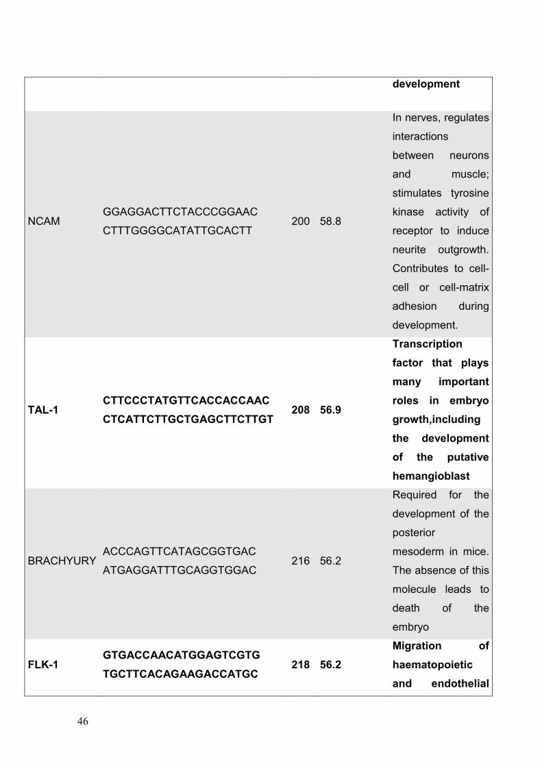
46
development
NCAM GGAGGACTTCTACCCGGAAC
CTTTGGGGCATATTGCACTT 200 58.8
In nerves, regulates
interactions
between neurons
and muscle;
stimulates tyrosine
kinase activity of
receptor to induce
neurite outgrowth.
Contributes to cell-
cell or cell-matrix
adhesion during
development.
TAL-1 CTTCCCTATGTTCACCACCAAC
CTCATTCTTGCTGAGCTTCTTGT 208 56.9
Transcription
factor that plays
many important
roles in embryo
growth,including
the development
of the putative
hemangioblast
BRACHYURY ACCCAGTTCATAGCGGTGAC
ATGAGGATTTGCAGGTGGAC 216 56.2
Required for the
development of the
posterior
mesoderm in mice.
The absence of this
molecule leads to
death of the
embryo
FLK-1 GTGACCAACATGGAGTCGTG
TGCTTCACAGAAGACCATGC 218 56.2
Migration of
haematopoietic
and endothelial
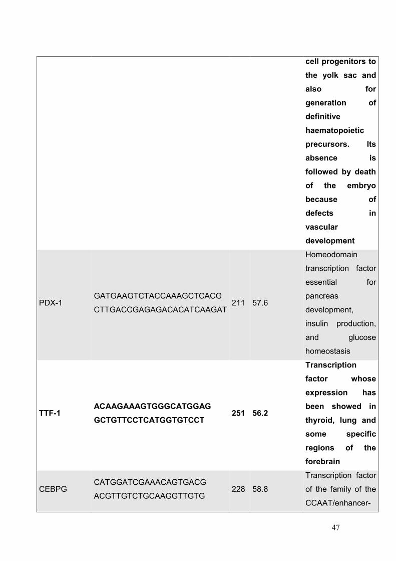
47
cell progenitors to
the yolk sac and
also for
generation of
definitive
haematopoietic
precursors. Its
absence is
followed by death
of the embryo
because of
defects in
vascular
development
PDX-1 GATGAAGTCTACCAAAGCTCACG
CTTGACCGAGAGACACATCAAGAT 211 57.6
Homeodomain
transcription factor
essential for
pancreas
development,
insulin production,
and glucose
homeostasis
TTF-1 ACAAGAAAGTGGGCATGGAG
GCTGTTCCTCATGGTGTCCT 251 56.2
Transcription
factor whose
expression has
been showed in
thyroid, lung and
some specific
regions of the
forebrain
CEBPG CATGGATCGAAACAGTGACG
ACGTTGTCTGCAAGGTTGTG 228 58.8
Transcription factor
of the family of the
CCAAT/enhancer-
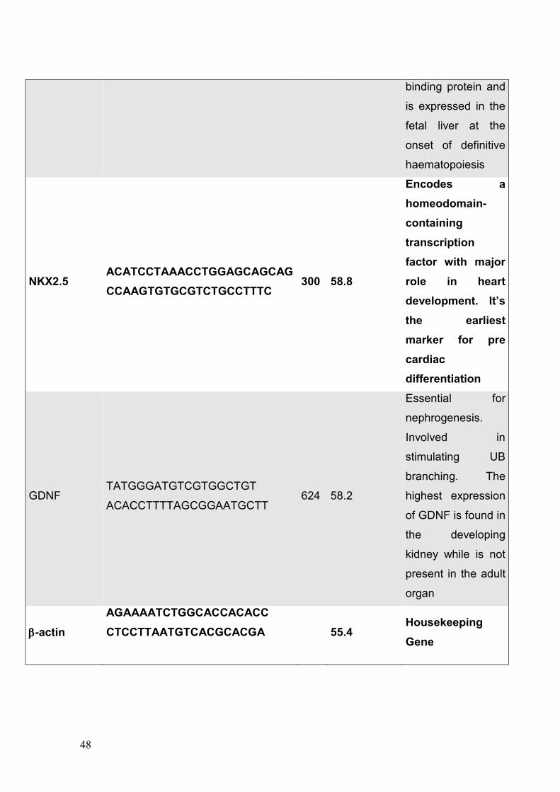
48
binding protein and
is expressed in the
fetal liver at the
onset of definitive
haematopoiesis
NKX2.5 ACATCCTAAACCTGGAGCAGCAG
CCAAGTGTGCGTCTGCCTTTC 300 58.8
Encodes a
homeodomain-
containing
transcription
factor with major
role in heart
development. It’s
the earliest
marker for pre
cardiac
differentiation
GDNF TATGGGATGTCGTGGCTGT
ACACCTTTTAGCGGAATGCTT 624 58.2
Essential for
nephrogenesis.
Involved in
stimulating UB
branching. The
highest expression
of GDNF is found in
the developing
kidney while is not
present in the adult
organ
ββββ-actin
AGAAAATCTGGCACCACACC
CTCCTTAATGTCACGCACGA
55.4 Housekeeping
Gene
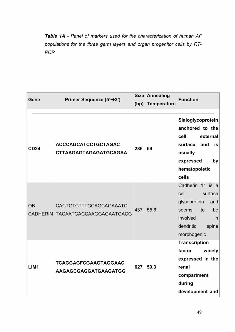
49
Table 1A - Panel of markers used for the characterization of human AF
populations for the three germ layers and organ progenitor cells by RT-
PCR
Gene Primer Sequenze (5’àààà3’) Size
(bp)
Annealing
Temperature Function
CD24 ACCCAGCATCCTGCTAGAC
CTTAAGAGTAGAGATGCAGAA 286 59
Sialoglycoprotein
anchored to the
cell external
surface and is
usually
expressed by
hematopoietic
cells
OB
CADHERIN
CACTGTCTTTGCAGCAGAAATC
TACAATGACCAAGGAGAATGACG 437 55.6
Cadherin 11 is a
cell surface
glycoprotein and
seems to be
involved in
dendritic spine
morphogenic
LIM1 TCAGGAGFCGAAGTAGGAAC
AAGAGCGAGGATGAAGATGG 627 59.3
Transcription
factor widely
expressed in the
renal
compartment
during
development and
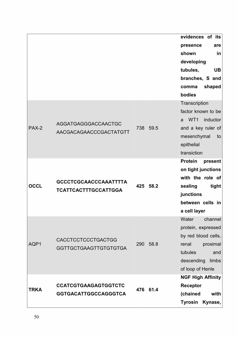
50
evidences of its
presence are
shown in
developing
tubules, UB
branches, S and
comma shaped
bodies
PAX-2 AGGATGAGGGACCAACTGC
AACGACAGAACCCGACTATGTT 738 59.5
Transcription
factor known to be
a WT1 inductor
and a key ruler of
mesenchymal to
epithelial
transiction
OCCL GCCCTCGCAACCCAAATTTTA
TCATTCACTTTGCCATTGGA 425 58.2
Protein present
on tight junctions
with the role of
sealing tight
junctions
between cells in
a cell layer
AQP1 CACCTCCTCCCTGACTGG
GGTTGCTGAAGTTGTGTGTGA 290 58.8
Water channel
protein, expressed
by red blood cells,
renal proximal
tubules and
descending limbs
of loop of Henle
TRKA CCATCGTGAAGAGTGGTCTC
GGTGACATTGGCCAGGGTCA 476 61.4
NGF High Affinity
Receptor
(chained with
Tyrosin Kynase,
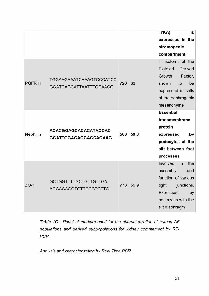
51
Table 1C - Panel of markers used for the characterization of human AF
populations and derived subpopulations for kidney commitment by RT-
PCR.
Analysis and characterization by Real Time PCR
TrKA) is
expressed in the
stromogenic
compartment
PGFR \ TGGAAGAAATCAAAGTCCCATCC
GGATCAGCATTAATTTGCAACG 720 63
\ isoform of the
Plateled Derived
Growth Factor,
shown to be
expressed in cells
of the nephrogenic
mesenchyme
Nephrin ACACGGAGCACACATACCAC
GGATTGGAGAGGAGCAGAAG 568 59.8
Essential
transmembrane
protein
expressed by
podocytes at the
slit between foot
processes
ZO-1 GCTGGTTTTGCTGTTGTTGA
AGGAGAGGTGTTCCGTGTTG 773 59.9
Involved in the
assembly and
function of various
tight junctions.
Expressed by
podocytes with the
slit diaphragm
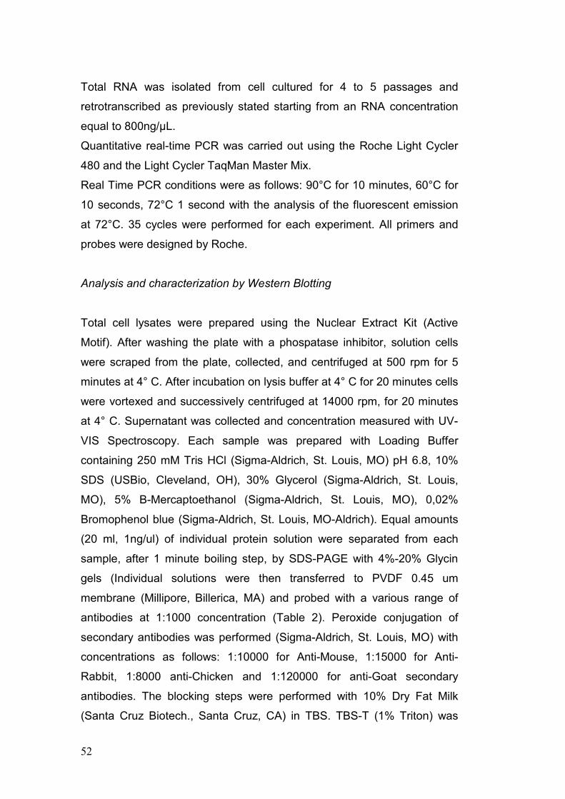
52
Total RNA was isolated from cell cultured for 4 to 5 passages and
retrotranscribed as previously stated starting from an RNA concentration
equal to 800ng/µL.
Quantitative real-time PCR was carried out using the Roche Light Cycler
480 and the Light Cycler TaqMan Master Mix.
Real Time PCR conditions were as follows: 90°C for 10 minutes, 60°C for
10 seconds, 72°C 1 second with the analysis of the fluorescent emission
at 72°C. 35 cycles were performed for each experiment. All primers and
probes were designed by Roche.
Analysis and characterization by Western Blotting
Total cell lysates were prepared using the Nuclear Extract Kit (Active
Motif). After washing the plate with a phospatase inhibitor, solution cells
were scraped from the plate, collected, and centrifuged at 500 rpm for 5
minutes at 4° C. After incubation on lysis buffer at 4° C for 20 minutes cells
were vortexed and successively centrifuged at 14000 rpm, for 20 minutes
at 4° C. Supernatant was collected and concentration measured with UV-
VIS Spectroscopy. Each sample was prepared with Loading Buffer
containing 250 mM Tris HCl (Sigma-Aldrich, St. Louis, MO) pH 6.8, 10%
SDS (USBio, Cleveland, OH), 30% Glycerol (Sigma-Aldrich, St. Louis,
MO), 5% B-Mercaptoethanol (Sigma-Aldrich, St. Louis, MO), 0,02%
Bromophenol blue (Sigma-Aldrich, St. Louis, MO-Aldrich). Equal amounts
(20 ml, 1ng/ul) of individual protein solution were separated from each
sample, after 1 minute boiling step, by SDS-PAGE with 4%-20% Glycin
gels (Individual solutions were then transferred to PVDF 0.45 um
membrane (Millipore, Billerica, MA) and probed with a various range of
antibodies at 1:1000 concentration (Table 2). Peroxide conjugation of
secondary antibodies was performed (Sigma-Aldrich, St. Louis, MO) with
concentrations as follows: 1:10000 for Anti-Mouse, 1:15000 for Anti-
Rabbit, 1:8000 anti-Chicken and 1:120000 for anti-Goat secondary
antibodies. The blocking steps were performed with 10% Dry Fat Milk
(Santa Cruz Biotech., Santa Cruz, CA) in TBS. TBS-T (1% Triton) was
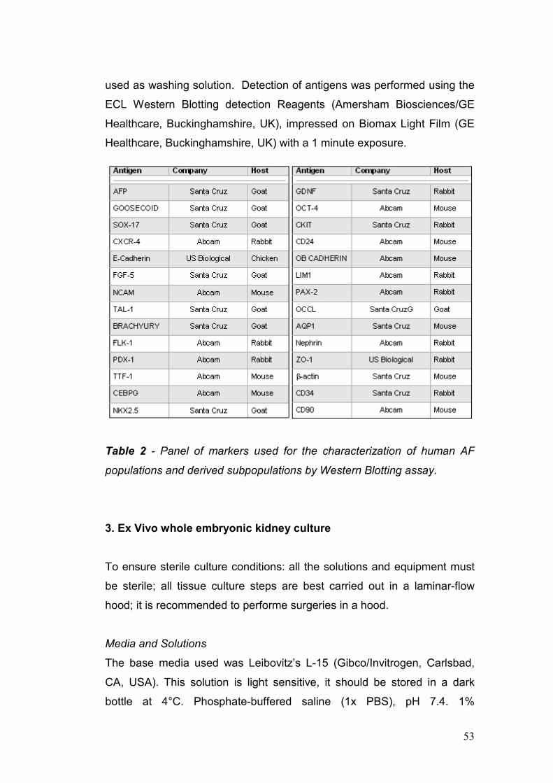
53
used as washing solution. Detection of antigens was performed using the
ECL Western Blotting detection Reagents (Amersham Biosciences/GE
Healthcare, Buckinghamshire, UK), impressed on Biomax Light Film (GE
Healthcare, Buckinghamshire, UK) with a 1 minute exposure.
Table 2 - Panel of markers used for the characterization of human AF
populations and derived subpopulations by Western Blotting assay.
3. Ex Vivo whole embryonic kidney culture
To ensure sterile culture conditions: all the solutions and equipment must
be sterile; all tissue culture steps are best carried out in a laminar-flow
hood; it is recommended to performe surgeries in a hood.
Media and Solutions
The base media used was Leibovitz’s L-15 (Gibco/Invitrogen, Carlsbad,
CA, USA). This solution is light sensitive, it should be stored in a dark
bottle at 4°C. Phosphate-buffered saline (1x PBS), pH 7.4. 1%
54
penicillin/streptomycin (Gibco/Invitrogen). 2% fetal bovine serum
(Gibco/Invitrogen)
Tools for the Organ Dissection
Have on hand the following supplies:
§ Microsurgery instruments: straight-tip scissors, No.5 Dumont
forceps, plastic pipettes (Fine Science Tools Inc., Foster City,
California)
§ 0.4 µm pore size Transwell membrane (Corning Incorporated,
Acton, MA, USA)
§ 8x dissecting microscope
§ Ice box
Organ Dissection Procedure
Wild-type C57BL/C6 mice metanephric kidneys were dissected under
sterile conditions from timed-pregnant embryonic day 13 to 16 (E13 to
E16) under a dissecting microscope (8x). Embryonic staging was verified
using the criteria of Theiler. The embryos were placed in a 10-cm plastic
Petri dish containing cold 1xPBS, their abdominal walls were carefully
dissected and then we peeled out the whole gut to expose the
retroperitoneum and kidneys. To transfer the organs in a sterile manner
into a culture dish, with sterile media, we used plastic pipets. Perfusion
channels were created under direct microscopy with a 10 µm diameter
glass needle fashioned manually in the laboratory without conveying
subsequent damage to the kidney while permitting homogeneous
perfusion and bathing throughout the whole organ. The preparation and
dissection of tissues should be done as quickly as possible having care to
keep the embryonic organs in a cold medium before placing them on the
Transwell membrane. Obtained developing kidneys were placed on a 0.4
µm pore size Transwell membrane (Corning Incorporated, Acton, MA,
USA) and cultivated in the medium-gas interphase in a 37°C incubator,
fully humidified 5% CO2 for 4-10 days. Excess medium results in floating of
the organ. Media was not changed during the culture but added if needed.
55
In addition a lower concentration of fetal bovine serum (2%) assisted in
slowing and significantly decreased the necrosis process over the time
span of the culture.
Fixation and Staining
A pipette with medium was used to detach the organs from the membrane.
The kidneys were then washed for 1 minute X 3 with ice-cold 1 X PBS.
The kidneys were fixed for 1 hour in 4% paraformaldehyde in PBS. Only
fresh paraformaldehyde was used (stored at 4C no longer than 1 week
old). Fixation was followed by paraffin embeddin, serial sectioning and
staining with hematoxylin and eosin (Sigma-Aldrich).
For immunostaining paraffin slides were placed in Vector Antigen Retrieval
solution (Vector Laboratories, Burlingame, California) and chilled.
Subsequently slides were processed first with Avidin/Biotin Block solution
(Vector laboratories), then with 20% hydrogen peroxide (Sigma-Aldrich) for
1 hour to block exogenous peroxidase and finally with 20% goat serum in
PBS block for 1 hour. Sections were incubated overnight with anti-
caspase-3 primary antibody (Cell Signaling Technology) followed by
incubation with an anti-rabbit secondary antibody (Vector Laboratories)
For immunofluorescent staining sections were blocked with Avidin/Biotin
Block after deparaffinization, followed by protein block for 30 minutes
using the appropriate 5% goat serum in PBS. The slides were incubated in
primary antibody (Santa Cruz Biotechnology, Santa Cruz, California)
overnight at 4C. Subsequently the slides were washed in PBS for 5 min X
3, incubated with secondary antibody for a half-hour at room temperature
and washed in PBS before development with Fluorescein/Avidin DCS
(Vector Laboratories). Finally, slides were mounted using 4’,6-diamidino-2-
phenylindole mounting medium.
4. Selection of Amniotic Fluid Stem Cells (AFSC)
hAFSCs were collected by amniocentesis from the amniotic fluid of male
fetuses (14-20 weeks gestation). The stem cell population was separated
56
from the general cellular milieu using standard Magnetic Sorting (MACS)
techniques (Miltenyi Biotech). Stem cells were cultured in medium
containing α-MEM supplemented with 20% Chang B and 2% Chang C
solutions, 15% Fetal Bovine Serum, 1% L-Glutamine, and 1% antibiotics
(pen-strep) [Gibco/BRL]. hAFSCs were analyzed for embryonic stem cell
marker expression, clonalality, maintenance of cell cycle control, and
preservation of normal kariotype and telomeric length. hAFSCs were
differentiated in vitro into multiple cell lines derived from all the three germ
layers using various conditioned medium. Pluripotentiality was also
confirmed by demonstrating the capacity of hAFSCs to participate in the
formation of tissue and organs during the process of embryogenesis after
injection into mouse blastocysts. Lac-Z and GFP labeled hAFSCs was
achieved through infection with a retrovirus construct containing the
cytomegalovirus promoter driving Lac-z and GFP sequences. Expression
of GFP and Lac-z was seen in the infected cells after 20 hours post-
infection. A clonal population was also labeled with a cell surface marker
CM- DIL (Molecular Probe) using standard protocols in order to track the
cells after injection.
5. In vitro experiment for the renal differentiation of hAFSC
Co-culture system
Kidney rudiments were dissected from timed pregnant C57 mice and
sprague dawley rat embryos at E 13.5 days of gestation. The UB was
separated from the metanephric mesenchyme by incubating kidney
rudiments in trypsin and DNase [Sigma-Aldrich] and by mechanical
separation. After suspension in Matrigel (BD Bioscience), the UB was
cultured in media conditioned with the following growth factors: Glial-
Derived Neurotrophic Factor and Fibroblast Growth Factor-1 (BD
Biosciences), FBS (Gibco/BRL), and antibiotics (pen-strep) (Gibco/BRL) in
an incubator at 5% CO2 and 370C. A co-culture system was designed
using isolated UB isolated as above placed on top of a polycarbonate filter
with a total of 1000 hAFSCs suspended in 50 µl of media seeded onto the
57
tip of the branching UB. The entire co-culture apparatus was incubated at
5% CO2 and 37 0C and cultured up to 10 days.
Microinjection
hAFSC were trypsinized, counted, and loaded into a 15µm diameter
transfer tip (Eppendorf AG), guided by a micromanipulator (Eppendorf
TransferMan NK2) and a CellTram Oil injector (Eppendorf), at a
concentration of 1000 cells/µL of medium with no serum. Immediately
before injection, kidneys were placed on a polyethylene terephthalate
track-etched membrane (Sterlitech Corporation). Each kidney received a
single injection (1-2 µL) and immediately placed on the top of a Transwell
filter in the incubator for 2 to 10 days.
Live imaging
A microscope incubator-chamber (Leica) was used to monitor position,
migration, and possible replication of hAFSC within the embryonic kidney
environment. Pictures were taken every 4 hours for 4 days using Leica
Deblur software.
Histology
Kidneys were fixed in 4% buffered paraformaldehyde [Sigma-Aldrich] for 1
hr, routinely processed, embedded in paraffin, and sectioned at 5 µm. The
sections were stained with hematoxylin and eosin (H&E) [Sigma-Aldrich]
and tuluidine blue [Sigma-Aldrich] using standard histological protocols.
For in total organ X-Gal staining, kidneys were fixed in 4%
paraformaldehyde [Sigma-Aldrich] in PBS at 4°C for 30 min while rocking,
washed twice for 10 min in PBS at 4°C, transferred into freshly prepared
X-gal [ rpi Research Products International Corp.] solution, and stained at
37°C until a clear precipitate formed [16]. For vibratome [Leica] sections,
samples were embedded in an albumin (300mg/ml)-gelatin (5mg/ml) mix
[Sigma-Aldrich], cross-linked with glutaraldehyde (0.6%) [Sigma-Aldrich],
and sectioned at 30-40 µm before viewing under light microscopy.
58
Chromogenic In Situ Hybridization (CISH)
CISH was used to detect the Y-Chromosome of hAFSCs after injection
into the embryonic kidney. Formalin-fixed paraffin embedded (FFPE)
tissue sections were used for this procedure according to the
manufacturer’s instructions. Zymed’s SPOT-Light CISH (Chromogenic In
Situ Hybridization) Centromere Kit detected Zymed SPOT-Light Human
Chromosome Y Probes [Zymed Laboratories Inc.]. Briefly, FFPE sections
were incubated at 55°C overnight, and the slides deparaffinized in xylene
and graded ethanol [Sigma-Aldrich]. Heat pretreatment was carried out in
pretreatment buffer at 94-95°C for 5 minutes and after application of
Zymed SpotLight Chromosome Y Probe the slides were cover-slipped and
the edges sealed with rubber cement. The slides were heated at 95°C for
5 minutes followed by overnight incubation at 37°C using a moisturized
chamber. Post-hybridization wash was performed the next day and
followed by immunodetection using the CISH polymer detection kit [Zymed
Laboratories Inc.]. The CISH signals were detected under light microscopy
using a 40x objective.
Reverse transcription (RT)-polymerase chain reaction (PCR)
Total mRNA was extracted from the embryonic kidney, using QIAGEN
mini KIT [QIAGEN], and reverse transcribed using SSII RT [Invitrogen] and
Random Hexamers as primers for the first strand cDNA synthesis. The
amplification of the resulting cDNA was carried out using specific human
primers. RealTime PCR thermal cycler [Eppendorf] was employed after an
initial denaturation step at 95°C for 10 min. We used a denaturation step
at 95°C for 30 seconds, an annealing step at 60°C for 45 seconds, and an
extension step at 72°C for 45 seconds for a total of 40 cycles. To rule out
the possibility of amplifying genomic DNA, RNA samples were treated with
a DNA-free kit [Ambion Inc.]. Detection of the PCR amplification products
was performed by size fractionation on 2% agarose gel electrophoresis.
As a positive control, amplification of fragments of the human β-actin RNA
59
was performed. Primer sequences and predicted sizes of amplicons were
as follows:
1) ephitelial membrane protein-1 (EMP-1):
sense 5’-ATG TCA CTC CTC TTG CTG GTG-3’ -3’,
antisense 5’-CGC TTC CGT AGG TGG ATG TAG-3’
2) zona occludens-1 (ZO-1):
sense 5’-AGG AGA GGT GTT CCG TGT TG-3’
antisense 5’-GCT GGT TTT GCT GTT GTT GA-3’
3) glial-derived neurotrophic factor (GDNF):
sense 5’-TAT GGG ATG TCG TGG CTG T-3’
antisense 5’-ACA CCT TTT AGC GGA ATG CTT-3’
4) aquaporin-1 (AQP-1): sense 5’-CAC CTC CTC CCT GAC TGG-3’
antisense 5’-GGT TGC TGA AGT TGT GTG TGA-3’
5) aquaporin-2 (AQP-2): sense 5’-GAT CAC GCC AGC AGA CAT C-3’
antisense 5’-GGG CAG GAT TCA TAG AGC AG-3’
6) tamm-horsfall protein (THP): sense 5’-TAG ACG AGG ACT GCA AAT
CG-3’
antisense 5’-GTC CCG GTT GTC TCT GTC AT-3’
7) β-actin: sense 5’-AGA AAA TCT GGC ACC ACA CC-3’
antisense 5’-CTC CTT AAT GTC ACG CAC GA-3’
6. In vivo experiment with a mouse model of Acute Tubular Necrosis
Isolation and labelling of hAFSC
Samples of human Amniotic Fluid were obtained, under IRB approval,
from discarded amniocentesis. No written or verbal consent was required
since information obtained about the samples were limited to karyotype
60
and fetus health status. The stem cell population was separated from the
general human amniotic cellular milieu using standard Magnetic Sorting
(MACS) techniques [Miltenyi Biotech] against the cell surface marker, c-kit,
as described by Atala et al. [54]. Pluripotential characteristics of the clonal
and subclonal groups were tested according to protocols also outlined by
Atala et al. [54] Clones were then cultured in petri dishes in medium
containing α-MEM supplemented with 20% Chang B and 2% Chang C
solutions, 20% Fetal Bovine Serum, 1% L-Glutamine, and 1% antibiotics
(pen-strep) [Gibco/BRL]. hAFSC, used for in vivo injection, were
karyotyped using standard protocols.
Before injection, a clonal hAFSC population was trypsinized in 0.05M
trypsin/EDTA solution and centrifuged at 1500rpm for 5 min, and then
labeled with a cell surface marker CM-DiI [Molecular Probe] following the
manufacturer’s instructions, in order to track the cells after injection.
Briefly, the cells were incubated with 1mg/ml of CM Dil for 5 minutes at
37°C followed by an incubation of 15 minutes at 4°C and 3 washes with
PBS.
ATN induction and injection of hAFSC
Rhabdomyolysis-related ATN was induced in female nu/nu mice [Jackson
Laboratories] by intramuscular injection with 50% hypertonic glycerol
solution (10 ml/kg body/wt) [Sigma-Aldrich] following deprivation of water
for 22 hours. Controlled intramuscular injection of glycerol was performed
under anesthesia by surgically exposing the caudal thigh muscle and
slowly injecting the glycerol solution prior to delivery of cells. Animal
experiments were performed in adherence to the National Institutes of
Health Guide for the Care and Use of Laboratory Animals, with institutional
Animal Care and Use Committee approval.
The mice were carefully anesthetized using isofluorane inhalation. Once
satisfactory anesthesia was achieved, the mice were prepared for surgery
using chlorhexidine. A 1 cm dorsal incision was made, both kidneys
carefully delivered via the incision, and the hAFSC (1x10⁶ diluted in PBS)
carefully injected into the renal cortex of both kidneys with a 30-33 gauge
61
needle using a microinjector Eppendorf TransferMan NK2 Injector
[Eppendorf]. The kidneys were then replaced into the retroperitoneum, the
incision closed with polypropylene suture and the mice were allowed to
recover from anesthesia. The animals were maintained on a heating pad
throughout the period of anesthesia. 0.1 mg/kg of buprenorphine was
administered subcutaneously and 1 mg/kg bupivicaine (a local anesthetic)
along the incision margins just prior to wound closure to provide post-
operative pain relief. The animals were sterilely draped to prevent contact
of the kidneys with the skin of the animal to reduce risk of peritonitis.
Control mice were also injected with PBS.
Tissue processing
At different times points (from 24 hours to 3 weeks), the injected and the
control mice were sacrificed. The kidneys were minced and processed in
one of the following ways depending on the analysis performed.
1. RNA/DNA extraction. The kidneys were minced in small pieces and
the RNA extracted using Qiagen RNeasy kit according to the
manufacturer’s instructions. Briefly, total mRNA was extracted and reverse
transcribed. Amplification of the resulting cDNA was carried out using
specific human primers not coding for mouse sequences. A PCR thermal
cycler [Eppendorf] was employed after an initial denaturation step at 95°C
for 10 minutes. We used a denaturation step at 95°C for 30 seconds, an
annealing step at the temperature specific for each primer (ranging from
54°C to 60°C) for 45 seconds, and an extension step at 72°C for 45
seconds for a total of 35 cycles. To rule out the possibility of amplifying
genomic DNA, RNA samples were treated with a DNA-free kit [Ambion
Inc.]. Detection of the PCR amplification products was performed by size
fractionation on 1% agarose gel electrophoresis. As a housekeeping gene,
amplification of fragments of the human β-actin RNA was performed.
Primer sequences, predicted sizes of amplicons and specific annealing
temperatures are shown in Table 1. In order to perform PCR on the
genomic DNA to evaluate the presence of the luciferase gene, DNA
62
extraction was performed following standard protocols of the Qiagen
DNeasy kit.
2. Histology. Kidneys were fixed in 4% buffered paraformaldehyde
[Sigma-Aldrich] for 8 hours at 40C, dehydrated through a gradual series of
alcohol, embedded in paraffin, and sectioned at 4-5 µm. The sections were
stained with (H&E) [Sigma-Aldrich], and PAS [Sigma-Aldrich] following
standard histological protocols.
In addition, some kidneys were frozen in liquid nitrogen and stored at -
20°C. When the antibody required, kidneys were cryosectioned at 5 µm
and then used for immuno-histochemistry.
Labeling of the AFSC with luciferase and bioluminescent detection
tecnique
hAFSC were transduced with a lentiviral vector (SMPU-R-MNCU3-LUC
based on HIV-1 that transduces the firefly luciferase gene) made by the
Vector Facility at Childrens Hospital Los Angeles following standard
protocols. Two cycles of transduction were performed by removing old
medium and adding new virus supernatant and medium. 24 hours after the
initial transduction, cells were thoroughly washed 3 times with PBS before
transplantation or in vitro analysis. Before in vivo injections, a simple in
vitro test was employed to determine the minimum amount of hAFSC
detectable by bioluminescence. Different concentrations of the cells
ranging from 5x10⁴ to 2x10⁶ were evaluated. In addition, it was confirmed
that after 20 passages in culture the cells were still expressing the
luciferase gene by PCR. 10-week old nu/nu mice, obtained from Jackson
Laboratories were injected directly into the kidney with luciferase-
transduced hAFSC (1 x 10⁶ cells/mouse diluted in PBS) after glycerol
damage. In vivo optical imaging was performed with a prototype IVIS 3-
dimensional bioluminescence/fluorescence optical imaging system
[Xenogen] at different time points. Prior to imaging, each mouse was given
an intravenous injection of luciferin [Promega] at a dose of 125 mg/kg, as
previously described [29]. General anesthesia was then induced with 5%
isoflurane and the mouse was placed in the light-tight heated chamber;
63
anesthesia was continued during the procedure with 2% isoflurane
introduced via a nose cone. The imaging system consists of a cooled,
back-thinned charge-coupled device (CCD) camera to capture both a
visible light photograph of the animal taken with light-emitting diodes and a
luminescent image. A rotating mirror and translatable animal stage allowed
for images to be acquired over 360°.
Immunostaining
Frozen and paraffin slides were stained for immunofluorescence. Paraffin
slides were deparaffinized, placed in 1% Triton for 5 minutes (if the antigen
was nuclear) and briefly washed in PBS. The paraffin slides were then
placed in Vector Antigen Retrieval Solution [Vector Laboratories] for three
cycles. The frozen slides were fixed for 5 minutes in 80% methanol. After
Avidin/Biotin blocking [Vector Laboratories] a second block was carried out
for 30 minutes using the appropriate 5% normal serum in PBS. The slides
were then incubated in primary antibody [Dolicholus Biflorus and Peanut
Agglutinin form Vector Laboratories, Luciferase from Promega and Glial
Derived Neurotrophic Factor and Aquaporin 2, Santa Cruz] solution for
one hour at room temperature or overnight at 4°C. Afterwards, the slides
were washed in PBS for 5 minutes for 3 times. Secondary antibodies
[Vector Laboratories] were diluted 1:200 in 5% normal serum – slides were
incubated in this solution for 1.5 hours at room temperature, followed by 5
minutes for 3 times in PBS. The appropriate fluorescent marker [Texas
Red or Fluorescein Avidin DCS from Vector Laboratories] was then
applied in a concentration of 1:500 in PBS buffer for 5-10 minutes,
followed by a final 5 minutes for 3 times in PBS wash. TUNEL staining
[Roche, Applied-Science] was performed to determine the presence of
apoptotic cells. Briefly, the cells were incubated at 37oC for one hour with
the TUNEL reagent and then washed in PBS. Slides were mounted with
Vector DAPI mounting medium [Vector Laboratories]. In the experimental
groups, the number of positive apoptotic nuclei was counted per 300
nuclei and hAFSC treated animals were compared to untreated controls.
64
Values are mean ± SD. A Leica DM RA fluorescent microscope was used
in conjunction with Open Lab 3.1.5 software to image the staining.
Blood Collection, Creatinine and BUN measurements
The facial vein was lanced with a 5mm animal lancet and blood collected
using standard protocols approved by the Animal Core Facility at
Childrens Hospital of Los Angeles and Saban Research Institute. Animals
were divided into different groups as follow:
1. group of 10 animals for measuring baseline creatinine and BUN levels;
2. group of 10 animals that underwent ATN with no injection of hAFSC;
3. group of 10 animals that underwent ATN and intrarenal injection of
hAFSC after 2 hours of glycerol injection;
4. group of 10 animals that underwent induction of ATN with glycerol and
intrarenal injection of PBS after 2 hours of glycerol injection.
The blood samples (30µL) were collected into plasma separation tubes
with lithium heparin. They were centrifuged at 13,000-RPM for 3 minutes
and the plasma (upper layer) was removed and stored at –80OC until
analysis. A maximum of 15% of circulating blood was sampled in a given
14-day period (total blood volume ~0.6% of total body weight). Post-
damage measurements were obtained every 24 hours. The blood samples
were used to monitor renal function, by analyzing creatinine and BUN
levels. ELISA was performed according to the manufacture for both
creatinine [BioAssay Systems Cat # DUCT-500] and BUN [BioAssay
System Cat # DIUR-500] using 30µL serum samples loaded into 96-well
microplates. Comparison between groups was made using an unpaired t
test. A value of p< 0.05 was considered statistically significant. Analyses
were done using GraphPad Prism software. Data are shown as mean ±
SD.
Morphological studies
65
Kidney sections were prepared at 4 µm thickness by a routine procedure
and stained with PAS reagents as described above. The kidney sections
were divided into six main groups:
1. Mice that underwent ATN with no injection of hAFSC at sacrificed at 24
hours;
2. Mice that underwent ATN and injection of hAFSC after 2 hours of
glycerol injection sacrificed at 24 hours;
3. Mice that underwent ATN with no injection of hAFSC sacrificed at 48
hours
4. Mice that underwent ATN and injection of hAFSC after 2 hours of
glycerol injection sacrificed at 48 hours
5. Mice that underwent ATN with no injection of hAFSC sacrificed at 72
hours;
6. Mice that underwent ATN and injection of hAFSC after 2 hours of
glycerol injection sacrificed at 72 hours.
Tubular injury was evaluated based on three major parameters using PAS
staining: 1. Disruption of the tubular membrane; 2. Disruption of brush
borders 3. Cast formation.
In the experimental groups, the damaged tubules were counted as a
fraction of the total number of tubules present in the section using
consecutive, non-overlapping fields of PAS-stained specimens. The
percentage of damaged tubules was estimated without knowledge of the
experimental group Values are mean ± SD.
Cytokine analysis
To examine pro-inflammatory and anti-inflammatory cytokines that were
generated after glycerol-induced ATN (with or without injection of cells),
human and mouse cytokines levels were measured in digested mouse
kidneys using a multiple cytokine array technique Proteome Profiler Array
Kit. Briefly, kidney tissue was homogenized in a cell lysis buffer, and the
homogenates were centrifuged at 12,000 rpm for 15 minutes at 4°C. Total
protein concentration in each supernatant was determined using a
Cytokine Array Kit (for human c# ARY005 and for mouse c# ARY006), as
66
suggested from the protocol [R&D Systems, Inc.]. The data were analyzed
using the Array Vision Program [R&D Systems, Inc.].
7. Selection and characterization of Metanephric Mesenchyme
derived cells (MMDC) and kidney progenitor cells (AKPC) from the
whole Amniotic Fluid
Immunoseparation of MMDC and AKPC from whole Amniotic Fluid
A positive population from AF for both CD24 and OB-Cadherin (MMDC)
was selected incubating the total cell population with these two specific
antibodies for 30 minutes at 4° C on a rocking platform, followed by
second incubation with immunomagnetic microbeads for 5 minutes at 25°
C and then 15 minutes at 4° C followed by immunoseparation by MS
columns (Miltenyi Biotech, Germany). Positive and negative (used as a
negative control) selected populations were replated on Tissue Culture
dishes with Chang’s Media for subsequent expansion.
A further immunoselection from the MMDC to identify 4 subpopulations of
renal progenitors (AKPC) was performed as above described, using anti-
Human antibodies for Nephrin, TrKA, PDGFR α and E-Cadherin following
the previously described immunoseparation technique. Final
subpopulations were obtained after a total of 18 passages from the original
samples. Cells were reseeded under the same conditions used for the
total AF cell population and the main selection for MMDC cells.
Characterization of MMDC and AKPC by RT-PCR
The CD24+OB-Cadherin+ population (MMDC) was investigated by RT-
PCR for early and mature kidney markers.
After the immunoselection, the four subpopulations of AKPC were
analyzed according to the same panel of markers in order to investigate
differences and common traits between the Nephrin+, TrKA+, PDGFR α+
67
and E-Cadherin+ AKPC derived populations following the protocol
previously described.
Analysis and characterization of MMDC and AKPC by Real Time PCR
MMDC and AKPC were analyzed by Real Time PCR as previously
described. Markers analyzed were: GDNF, LIM-1, PAX-2, Nephrin, OCT-4,
TrKA, PDGFRα, E-Cadherin, ZO-1 and Occludin. Analysis was performed
following the previously described protocol.

68
69
RESULTS
1. Characterization of Amniotic Fluid cells by expression of markers
for the three germ layers and progenitor cells
Amniotic Fluid Total Cell Population Culture
The morphology of the total cell population is very heterogeneous with a
preponderance of fibroblastoid shapes (Figure 1). The expansion of the
total population of Amniotic Fluid was possible for up to 10 passages using
DMEM. Therefore, after these few passages, the cells stopped growing
and started dying. Nevertheless when the cells were cultured in Amniomax
II and Chang media they could be expanded for more then 50 passages in
culture. The cells cultured with Amniomax II acquired a defined
fibroblastoid shape. We therefore chose to culture cells in Chang media for
all experiments, since cell morphology did not change significantly over
subsequent population doublings.
Analysis and characterization of human Amniotic Fluid cells by RT-PCR
AF Total Cell Population was stratified by week of gestation (from 15 to 20
weeks) and analyzed using RT-PCR. As shown in Figure 2 A-B-C,
expression of markers for the three germ layers, for pluripotent cells as
well as for mesenchymal, hematopoietic and early progenitor cells of
several organs were analyzed.
Expression of genes characteristics of the Endodermal and Mesodermal
germ layers was found to decrease over time, while ectodermal markers
remained constantly expressed (Figure 2A).
Pluripotent markers were expressed in all samples younger than 19
weeks.
While mesenchymal marker CD90 is expressed at all the time points
analyzed, CD34 (Marker for mature haematopoietic lineages) was absent
in early gestation samples, but appeared in samples of 18 weeks of
gestation or older (Figure 2A).

70
Early progenitor markers were expressed in 18 weeks and older samples
(Figure 2B) as well as specific kidney markers. (Figure 2C)
Analysis and characterization of human Amniotic Fluid cells by Western
Blotting
The protein expression of the cells showed a decrease on endodermal and
mesodermal layers over the course of gestation while, as seen with the
mRNA expression, ectodermal proteins are constantly present along the
gestational age investigated in this study. Pluripotent, hematopoietic and
mesenchymal markers follow the same trend seen in the RT-PCR
analysis. (Figure 3A)
The early proteins of progenitor cells from different organs, as shown in
Figure 3B, show increased expression into Amniotic Fluid cells over the
course of gestation. Kidney specific proteins were shown to increase
around 17-18 weeks of gestation as shown in Figure 3C.
Analysis and characterization of total Amniotic Fluid cell population by
Real Time PCR
Four samples for each chosen time points (15-16, 17-18 and 19-20
Gestational Age Weeks) for a total of 12 samples were analyzed. The
investigated samples for all the markers analyzed confirmed the same
overall trends showed by RT-PCR analysis. However, some of the
markers such as Brachyury, Tal-1, Nephrin, GDNF, TrKA were not
expressed in one or more samples. Goosecoid and PDX-1 were not found
in any of the twelve samples analyzed.
The ectodermal marker E-Cadherin increased 15-fold between 15-16 and
17-18 weeks of gestation. In contrary NCAM and FGF5, while confirmed to
be present, did not change significantly between 15 and 20 weeks. (Figure
4A). The Endodermal marker CXCR-4 increased 3.5-fold between 15-16
and 19-20 weeks while the other Endodermal markers Sox-17 and AFP
tended to decrease. (Figure 4A)
71
The mesodermal marker Brachyury was expressed at 15-16 weeks in one
sample but not later on. TAL-1 appeared to decrease over time, whileFLK1
increased 4-fold. (Figure 4A)
Pluripotency marker OCT4 didn’t change over the investigated weeks of
gestation while C-kit marker increased by 3-fold between 15-16 and 17-18
weeks and disappeared in the older samples. (Figure 4B)
Hematopoietic marker CD34 decreased between the 17-18 and the 19-20
weeks in contrast with mesenchymal marker CD90 that increased 2 fold by
17-18 weeks old samples. (Figure 4B)
Progenitor markers, excluded PDX-1 that showed no expression, generally
increased with the progression of the gestation; NKX2.5, early cardiac
marker showed a 6-fold increase between 17-18 and 19-20 weeks while
lung/Thyroid marker TTF-1 increased 1 fold between 15-16 weeks and 17-
18 weeks and an additional 2.5 fold to 19-20 weeks. (Figure 4C)
CEBPG showed a 5 fold increased expression at 17-18 weeks if compared
with the 15-16 and 19-20 weeks of gestation. (Figure 4C)
Renal marker GDNF was expressed only in two of the samples, increasing
5 fold between the 15-16 sample and the 19-20 weeks sample. (Figure
4D)
Undifferentiated Metanephric Mesenchyme markers CD24 and OB
Cadherin increased 1 fold between 15-16 and 17-18 weeks. While CD24
expression remained unchanged between 17-18 weeks and 19-20 weeks,
Ob-Cadherin decreased to the previous expression. (Figure 4D)
PAX-2 increased slightly between 15-16 and 19-20 weeks while LIM1
didn’t change its expression.(Figure 4D)
One sample for each time period was positive for Nephrin, showing a
constant increase over the time with a 1-fold increase between 15-16
weeks and 19-20 weeks of gestation. (Figure 4D)
Zo-1 and Aquaporin-1 marker didn’t change significantly overtime while
Occludin increased 8-fold between 17-18 weeks and 19-20 weeks. (Figure
4E)
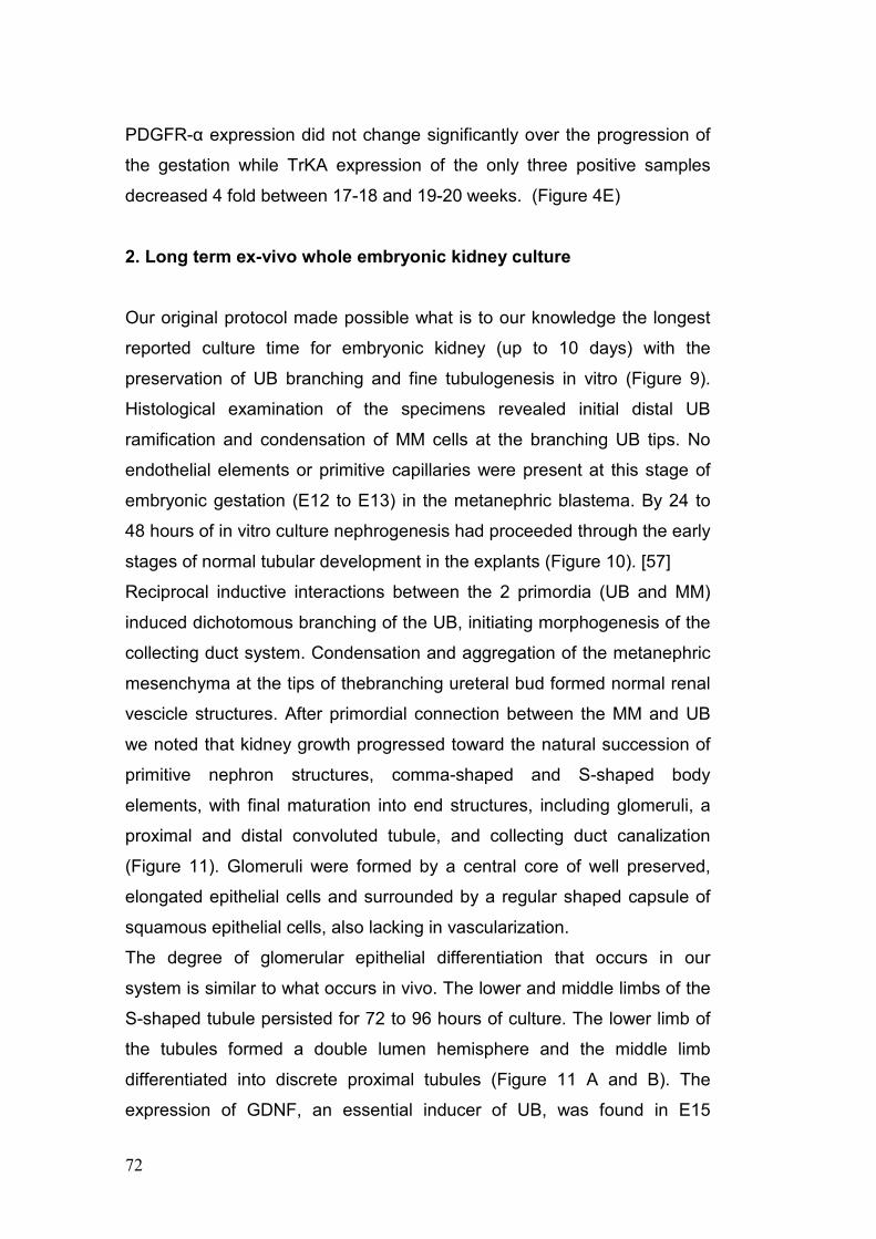
72
PDGFR-α expression did not change significantly over the progression of
the gestation while TrKA expression of the only three positive samples
decreased 4 fold between 17-18 and 19-20 weeks. (Figure 4E)
2. Long term ex-vivo whole embryonic kidney culture
Our original protocol made possible what is to our knowledge the longest
reported culture time for embryonic kidney (up to 10 days) with the
preservation of UB branching and fine tubulogenesis in vitro (Figure 9).
Histological examination of the specimens revealed initial distal UB
ramification and condensation of MM cells at the branching UB tips. No
endothelial elements or primitive capillaries were present at this stage of
embryonic gestation (E12 to E13) in the metanephric blastema. By 24 to
48 hours of in vitro culture nephrogenesis had proceeded through the early
stages of normal tubular development in the explants (Figure 10). [57]
Reciprocal inductive interactions between the 2 primordia (UB and MM)
induced dichotomous branching of the UB, initiating morphogenesis of the
collecting duct system. Condensation and aggregation of the metanephric
mesenchyma at the tips of thebranching ureteral bud formed normal renal
vescicle structures. After primordial connection between the MM and UB
we noted that kidney growth progressed toward the natural succession of
primitive nephron structures, comma-shaped and S-shaped body
elements, with final maturation into end structures, including glomeruli, a
proximal and distal convoluted tubule, and collecting duct canalization
(Figure 11). Glomeruli were formed by a central core of well preserved,
elongated epithelial cells and surrounded by a regular shaped capsule of
squamous epithelial cells, also lacking in vascularization.
The degree of glomerular epithelial differentiation that occurs in our
system is similar to what occurs in vivo. The lower and middle limbs of the
S-shaped tubule persisted for 72 to 96 hours of culture. The lower limb of
the tubules formed a double lumen hemisphere and the middle limb
differentiated into discrete proximal tubules (Figure 11 A and B). The
expression of GDNF, an essential inducer of UB, was found in E15
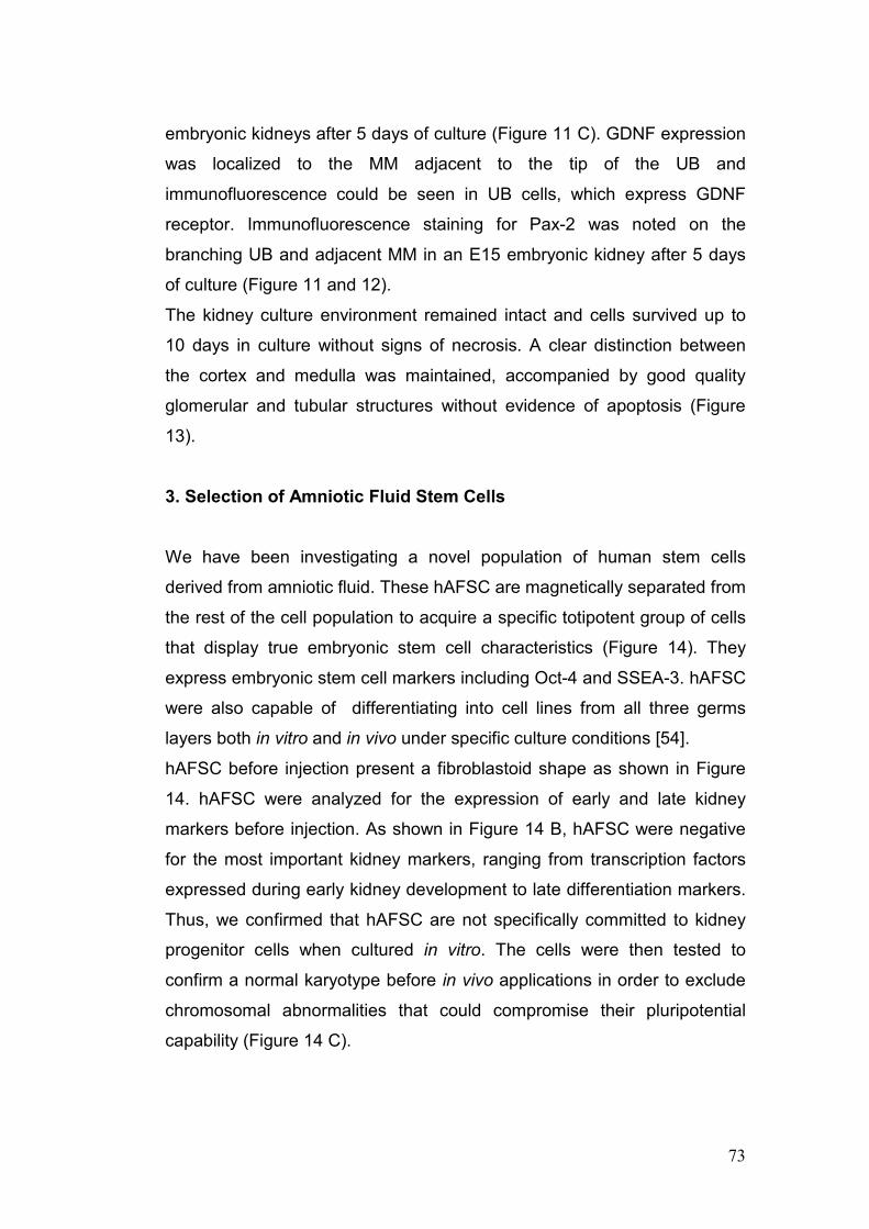
73
embryonic kidneys after 5 days of culture (Figure 11 C). GDNF expression
was localized to the MM adjacent to the tip of the UB and
immunofluorescence could be seen in UB cells, which express GDNF
receptor. Immunofluorescence staining for Pax-2 was noted on the
branching UB and adjacent MM in an E15 embryonic kidney after 5 days
of culture (Figure 11 and 12).
The kidney culture environment remained intact and cells survived up to
10 days in culture without signs of necrosis. A clear distinction between
the cortex and medulla was maintained, accompanied by good quality
glomerular and tubular structures without evidence of apoptosis (Figure
13).
3. Selection of Amniotic Fluid Stem Cells
We have been investigating a novel population of human stem cells
derived from amniotic fluid. These hAFSC are magnetically separated from
the rest of the cell population to acquire a specific totipotent group of cells
that display true embryonic stem cell characteristics (Figure 14). They
express embryonic stem cell markers including Oct-4 and SSEA-3. hAFSC
were also capable of differentiating into cell lines from all three germs
layers both in vitro and in vivo under specific culture conditions [54].
hAFSC before injection present a fibroblastoid shape as shown in Figure
14. hAFSC were analyzed for the expression of early and late kidney
markers before injection. As shown in Figure 14 B, hAFSC were negative
for the most important kidney markers, ranging from transcription factors
expressed during early kidney development to late differentiation markers.
Thus, we confirmed that hAFSC are not specifically committed to kidney
progenitor cells when cultured in vitro. The cells were then tested to
confirm a normal karyotype before in vivo applications in order to exclude
chromosomal abnormalities that could compromise their pluripotential
capability (Figure 14 C).
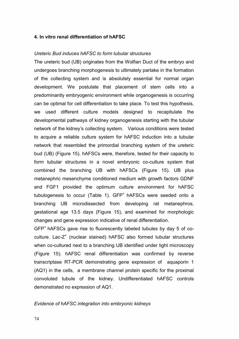
74
4. In vitro renal differentiation of hAFSC
Ureteric Bud induces hAFSC to form tubular structures
The ureteric bud (UB) originates from the Wolfian Duct of the embryo and
undergoes branching morphogenesis to ultimately partake in the formation
of the collecting system and is absolutely essential for normal organ
development. We postulate that placement of stem cells into a
predominantly embryogenic environment while organogenesis is occurring
can be optimal for cell differentiation to take place. To test this hypothesis,
we used different culture models designed to recapitulate the
developmental pathways of kidney organogenesis starting with the tubular
network of the kidney’s collecting system. Various conditions were tested
to acquire a reliable culture system for hAFSC induction into a tubular
network that resembled the primordial branching system of the ureteric
bud (UB) (Figure 15). hAFSCs were, therefore, tested for their capacity to
form tubular structures in a novel embryonic co-culture system that
combined the branching UB with hAFSCs (Figure 15). UB plus
metanephric mesenchyme conditioned medium with growth factors GDNF
and FGF1 provided the optimum culture environment for hAFSC
tubulogenesis to occur (Table 1). GFP+ hAFSCs were seeded onto a
branching UB microdissected from developing rat metanephros,
gestational age 13.5 days (Figure 15), and examined for morphologic
changes and gene expression indicative of renal differentiation.
GFP+ hAFSCs gave rise to fluorescently labeled tubules by day 5 of co-
culture. Lac-Z+ (nuclear stained) hAFSC also formed tubular structures
when co-cultured next to a branching UB identified under light microscopy
(Figure 15). hAFSC renal differentiation was confirmed by reverse
transcriptase RT-PCR demonstrating gene expression of aquaporin 1
(AQ1) in the cells, a membrane channel protein specific for the proximal
convoluted tubule of the kidney. Undifferentiated hAFSC controls
demonstrated no expression of AQ1.
Evidence of hAFSC integration into embryonic kidneys
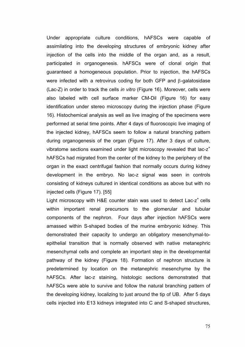
75
Under appropriate culture conditions, hAFSCs were capable of
assimilating into the developing structures of embryonic kidney after
injection of the cells into the middle of the organ and, as a result,
participated in organogenesis. hAFSCs were of clonal origin that
guaranteed a homogeneous population. Prior to injection, the hAFSCs
were infected with a retrovirus coding for both GFP and β-galatosidase
(Lac-Z) in order to track the cells in vitro (Figure 16). Moreover, cells were
also labeled with cell surface marker CM-Dil (Figure 16) for easy
identification under stereo microscopy during the injection phase (Figure
16). Histochemical analysis as well as live imaging of the specimens were
performed at serial time points. After 4 days of fluoroscopic live imaging of
the injected kidney, hAFSCs seem to follow a natural branching pattern
during organogenesis of the organ (Figure 17). After 3 days of culture,
vibratome sections examined under light microscopy revealed that lac-z+
hAFSCs had migrated from the center of the kidney to the periphery of the
organ in the exact centrifugal fashion that normally occurs during kidney
development in the embryo. No lac-z signal was seen in controls
consisting of kidneys cultured in identical conditions as above but with no
injected cells (Figure 17). [55]
Light microscopy with H&E counter stain was used to detect Lac-z+ cells
within important renal precursors to the glomerular and tubular
components of the nephron. Four days after injection hAFSCs were
amassed within S-shaped bodies of the murine embryonic kidney. This
demonstrated their capacity to undergo an obligatory mesenchymal-to-
epithelial transition that is normally observed with native metanephric
mesenchymal cells and complete an important step in the developmental
pathway of the kidney (Figure 18). Formation of nephron structure is
predetermined by location on the metanephric mesenchyme by the
hAFSCs. After lac-z staining, histologic sections demonstrated that
hAFSCs were able to survive and follow the natural branching pattern of
the developing kidney, localizing to just around the tip of UB. After 5 days
cells injected into E13 kidneys integrated into C and S-shaped structures,
76
stroma and renal vesicles (Figure 18). No Lac-z+ cells were detected in the
controls.
hAFSC labeled with a surface marker CM-Dil were identified after injection
as having assimilated into developing kidney primordial structures. After 6
days of culture, these cells has integrated into embryonic tubular and
glomerular structures which ultimately give rise to mature nephrons
(Figure 18).
The presence and the integration of the hAFSC in the injected kidneys
were also confirmed using Chromogenic In Situ Hybridization (CISH)
(Zymed, San Francisco, CA). The population of hAFSC used in all
experiments were derived from male fetuses and, therefore,
chromosomally XY. Specimens were incubated with biotinylated probes
against the Y chromosome and counterstained with DAB(3,3-
diaminobenzidine) in a peroxidase reaction. Positively stained cells were
visualized under light microscopy lining the walls of the embryonic kidney
tubules (Figure 19). This comprised both medullary and cortical portions of
the kidney. No signal was identified in the controls.
Molecular evidence of primordial kidney differentiation
After 9 days of culture, RT-PCR was performed on specimens and
expression of several specific human kidney genes in the injected
embryonic kidneys were identified. Zo-1, Claudin and GDNF, early
markers for kidney differentiation, were detected when compared to
controls, which consisted of hAFSCs before injection (Figure 20).
5. In vivo model of acute tubulonecrosis integration
Glycerol induced muscle damage and ATN. Hematoxylin and Eosin (H&E),
Period Acid Schiff staining (PAS), TUNEL
Figure 22 A demonstrates the normal morphology of a mouse kidney
(nu/nu) before any damage. The distinction between the medulla and the
cortex is clearly evident, and the tubules are intact as well as the
glomeruli. Figure 22D shows the morphology of the kidney 3 days after the
77
intramuscular injection of glycerol. ATN caused a marked disorganization
in the structure so that medulla and cortex are not distinguishable. The
normal structure of proximal and distal tubules is lost (with cast formation),
while most of the glomeruli remain intact. This type of damage is typical of
the injury induced by rhabdomyolysis, where the main structures of the
kidneys that undergo failure are the tubules and not the glomeruli. Figure
22 F shows the increase in apoptotic cells (TUNEL staining) when
compared with the control, that did not undergo glycerol induced muscle
damage (Figure 22 E). The difference in the number of apoptotic cells
present in the treated glycerol mice when compared with the untreated
control mice was highly significant (Figure 22 F)
In vivo detection of hAFSC by bioluminescence
hAFSC transduced with a lentivirus coding for luciferase showed stable
expression of the transgene over many population doublings. Figure 23 A
shows control cells that were not infected versus infected cells exposed to
luciferin (the luciferase substrate) to confirm the presence of the signal
under bioluminescence detection after 20 population doublings. In Figure
23 B it is shown an in vitro experiment determining the limit starting point
of 1x10⁵ cells that exhibits a signal optically detected under
bioluminescence.
1.2 x10⁶ hAFSC injected directly into the right kidney after damage
induction (Figure 23 C) were easily detected. The signal is clearly evident
and spreads into multiple zones of the body such as the lung over the first
few days. The signal for hAFSC in the area of the kidney can be seen at
24 hours after injection (panel 3), was strongest at 48 hours and 72 hours,
and persisted for up to 6 days (panels 4-6), after which the signal began to
diminish over the next several days (panel 7). However, 21 days after
injection, the signal was still evident in the area of the kidney (panel 8).
DNA extraction and PCR were performed at 21 days on injected and non-
injected kidneys in order to determine the presence of luciferase. Results
show that luciferase and human ATCB DNA, was present only in the
78
injected kidney tissue, as confirmed by the absence of the housekeeping
ATCB (Figure 23D). DNA was extracted from the entire injected kidney.
The results were also confirmed with a positive immunostaining against
luciferase as shown in Figure 23
Detection of hAFSC in damaged kidneys by immunohistochemistry and
gene expression
The presence of injected hAFSC was evaluated histologically. Frozen
sections performed at 1 week after injection confirmed the presence of
hAFSC, detected by red fluorescence of the surface marker CM-DiI
(Figure 24 A). We were able to observe several instances where the CM-
Dil signal from the hAFSC overlapped with the fluorescent-staining of a
kidney marker as follows: luciferase positive hAFSC were double stained
for Aquaporin2, Peanut Agglutinin as well as Dolichus Biflorus Agglutinin
at 3 weeks after injection; indicating that hAFSC are able to differentiate
into cells expressing both adult proximal and distal tubular agglutinins
(Figure 24 B, D). In some rare cases hAFSC were also found in glomerular
structures expressing Glial Derived Neurotrophic Factor (Figure 24 E),
indicating that the stem cells were also able to express early glomerular
markers of differentiation.
After 21 days, RT-PCR was performed using human specific primers on
the harvested kidneys and expression of several human specific kidney
genes (early as well as late markers of differentiation) by the hAFSC in the
injected kidneys were identified: NPHS1, AQP2, PAX2, and OCLN when
compared to hAFSC before injection (Figure 24 F)
Creatinine and Blood Urea Nitrogen (BUN) measurements
A control group of 10 nu/nu mice was used to determine the basal level of
serum creatinine before any treatment, which averaged 0.6 mg/dl. After
intramuscular injection of glycerol on day 0, creatinine levels increased to
as high as 1.10 mg/dl, showing a peak between 48 and 72 hours after
injections. Similarly, the level of BUN (basal level of 27mg/dl) increased
79
after glycerol injection and the peak was detected around 48 and 72 hours.
The concentration of both creatinine and BUN returned to normal level
after 3 weeks. Further analysis showed no statistical in significant
difference in creatinine and BUN levels between the animals that were
injected with saline vehicle solution versus no injection following
intramuscular glycerol, and therefore these groups were pooled for
statistical analysis. In contrast, animals subjected to damage induced with
intramuscular glycerol and receiving an intrarenal hAFSC injection
demonstrated no increase in levels of creatinine (Figure 25 A) or BUN
(Figure 25 B) during the expected acute phase of injury.
Morphological studies
In Figure 26 an increase in the number of damaged tubules is seen from
24 hours to 72 hours in the glycerol-treated animals. By 72 hours the
damage is more severe due to the cast formation (arrow, Figure 26 C)
within damaged tubules. In Figure 26 (D,E) is evident that in glycerol-
injected animals treated with hAFSC the number of damaged tubules
increased at 48 hours compared to the animals that were not injected with
stem cells, but by 72 hours the number of damaged tubules decreased
significantly. Two-factor ANOVA showed a highly significant effect of time
(p= 0.03) and interaction of time with treatment (p=0.01) as shown in
Figure 26
Immuno-cytokine profile
Since the salutary effect of hAFSC injection occurred during the acute
phase of ATN, we postulated that this protective effect might involve acute
changes in the kidney’s cytokine milieu. The cytokine profile of human as
well as mouse cytokines expressed in the kidney at 24 hours and 48 hours
after intramuscular injection of glycerol is compared to undamaged
controls and mice with glycerol-injected and intrarenal injection of hAFSC.
Cytokine levels are shown as comprehensive suite of sequential bar
graphs (Mean and SD) in Figure 27 For relative ease of interpretation, the
different cytokines were displayed as four broad functional clusters based
80
on their principal immunological functions. 1. Anti-inflammatory 2. Pro-
inflammatory 3. Chemoattractants and 4. Multiple biological effects. For
each individual cytokine, the bars in the figure show from left to right,
control cytokine levels, ATN kidney at 24 hours, ATN kidney plus hAFSC
injection at 24 hours (shown as the sum of mouse derived plus hAFSC
derived human cytokine levels). This order of display is then repeated at
48 hours.
The mouse specific cytokine assay does not cross react with the human
cytokine assay. This was confirmed by incubating mouse digested kidneys
with membranes specific for human cytokines and conversely incubating
hAFSC with membranes specific for mouse cytokines (data not shown).
The mouse tissue is exposed to the activity of both human and mouse
cytokines at both time points shown (24 hours and 48 hours). When
viewed in this fashion it becomes clear that the major trend of the analyzed
combined cytokine levels is a significant increase in the early cytokine
response. Thus, 24 and 48 hours following intramuscular injection of
glycerol, at the time of peaking kidney damage, mice without hAFSC
injection had significant elevation (as much as 5 or 6-fold) of cytokine
expression level across all 4 classes of cytokine. However, when glycerol-
treated mice were injected with hAFSC, an even greater elevation of
cytokine levels was evident, particularly at 24 hours when both mouse and
human cytokine levels were combined. However, by 48 hours, this trend in
combined cytokine levels is reversed so that the majority of combined
cytokine levels are either reduced significantly or no longer elevated
relative to the kidneys that did not receive hAFSC. Further, by 48 hours
the relative contribution of human versus mouse cytokines is also
reversed, with the human component of the cytokine milieu being relatively
small.
6. Selection and characterization of Metanephric Mesenchyme
derived cells (MMDC) and Kidney Progenitor cells (AKPC) from the
whole Amniotic Fluid
81
Immunoseparation of MMDC and AKPC from whole Amniotic Fluid
A specific cell population positive for CD24 and OB-Cadherin (MMDC) was
successfully isolated from whole Amniotic Fluid and cultured under the
conditions described above.
MMDC selected cells present a different morphology with typical fibroblast
shapes (Figure 29). From the CD24+OB-Cadherin+ population, AKPC
subpopulations expressing TrKA, Nephrin, PDGFR-\ and E-Cadherin
were successfully obtained by immunoseparation. (Figure 7)
Characterization of MMDC and AKPC by RT-PCR
The MMDC population (CD24+ Ob Cadherin+) (Figure 30) and the four
derived AKPC subpopulations were characterized by RT-PCR for early
and mature kidney markers, as well as pluripotency markers (Figure 31).
Expression of renal markers differed in the investigated populations as
shown in the figures. The AKPC E-Cadherin+ population expressed E-
Cadherin, GDNF and was slightly positive for Nephrin.
AKPC Nephrin+ cells were positive for Nephrin, Aquaporin 1 and Zona-
Occludens 1. The population immunoselected for AKPC PDGFR-α + was
positive for ZO-1 and PDGFR α while the AKPC TrkA+ population
expressed TrkA, ZO-1 and, slightly, PDGFR α. (Figure 8)
Analysis and characterization of MMDC and AKPC by Real Time PCR
The main population MMDC and the four AKPC subpopulations were
analyzed by Real Time PCR (Figure 7) for the expression of specific
kidney markers. GDNF, Metanephric Mesenchyme specific marker, was
expressed by the main population and was almost 12 fold greater in the
Nephrin+ subpopulation, but absent in the other subpopulations.
LIM-1 was 11 folds greater in the Nephrin population and increased 9 folds
in E-Cadherin population. It was increased in the TRKA+ population. PAX-
2 increased 4 fold in Nephrin. Nephrin expression was 2 fold greater in
Nephrin population. (Figure 7A)
82
Pluripotent marker OCT-4 was increased 10 fold in Nephrin+ AKPCs , 15
fold in E-cadherin+ population and 2 fold in TRKA. (Figure 7A) E-
Cadherin+ cells showed a 3 fold increase in E-Cadherin expression,
decreased in PDGFR α + and slightly in TRKA. (Figure 7B) TRKA showed
no increase in TrKA expression if compared with the main population but
the other population were negative for TrKA. PDGFR α increased 2 fold in
PDGFR α + APKCs, decreased in Nephrin+ and was practically absent in
E-cadherin+ APKCs. ZO-1 was increased 7 fold in the Nephrin population
but absent or slightly expressed in the other subpopulations. Occludin was
3.5 fold greater in Nephrin+ APKCs, 7 fold greater in E-Cadherin and 1.5
fold greater in PDGFR α + cells. (Figure 7B - 8)
83
DISCUSSION
The adult kidney is a highly vascularized organ, receiving about 20% of
the blood supply of the body. The kidneys filter about 180 liters of fluid per
day with an electrolyte composition similar to that of plasma and the
nephrons have the responsibility of handling this large volume of filtrate
and separating that which must be conserved and that which needs to be
excreted. The basic functional unit of the kidney is the nephron, composed
of a tubular system and a glomerulus. The tubular system has four compo-
nents: (a) proximal tubule, (b) loop of Henle, (c) distal tubule, and (d)
collecting tubule (see below).
The kidneys filter about 180 liters of fluid per day with an electrolyte
composition similar to that of plasma. The role of tubules is the re-
absorption of water and electrolytes in order to maintain the body
homeostasis and each segment is specialized in a different process or
water and electrolytes intake.
In the proximal tubule, water, electrolytes and glucose are reabsorbed
from the filtrate. The connection between the proximal tubule and the loop
of Henle is called descending loop. In the descending loop, the
concentrations inside and outside the tubule are increasing with the
current, with the maximum concentration being reached at the bottom of
the loop. The increased concentration is the result of the passive diffusion
of Na+ into the tubule and water out of the tubule. When the filtrate
reaches the distal tubule, a net loss of Na+ and water has occurred
through the loops of Henle.
In the ascending loop, Na+ (or any solute) is actively pumped out of the
tubule. As flow continues up the loop, the tubular concentration decreases
as does the interstitial concentration. Because water is impermeable in the
ascending loop, the volume at the bottom of the loop is the same as that
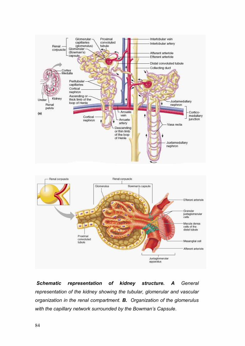
84
Schematic representation of kidney structure. A General
representation of the kidney showing the tubular, glomerular and vascular
organization in the renal compartment. B. Organization of the glomerulus
with the capillary network surrounded by the Bowman’s Capsule.
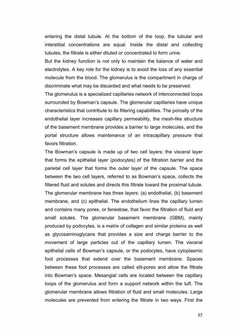
85
entering the distal tubule. At the bottom of the loop, the tubular and
interstitial concentrations are equal. Inside the distal and collecting
tubules, the filtrate is either diluted or concentrated to form urine.
But the kidney function is not only to maintain the balance of water and
electrolytes. A key role for the kidney is to avoid the loss of any essential
molecule from the blood. The glomerulus is the compartment in charge of
discriminate what may be discarded and what needs to be preserved.
The glomerulus is a specialized capillaries network of interconnected loops
surrounded by Bowman’s capsule. The glomerular capillaries have unique
characteristics that contribute to its filtering capabilities. The porosity of the
endothelial layer increases capillary permeability, the mesh-like structure
of the basement membrane provides a barrier to large molecules, and the
portal structure allows maintenance of an intracapillary pressure that
favors filtration.
The Bowman’s capsule is made up of two cell layers: the visceral layer
that forms the epithelial layer (podocytes) of the filtration barrier and the
parietal cell layer that forms the outer layer of the capsule. The space
between the two cell layers, referred to as Bowman’s space, collects the
filtered fluid and solutes and directs this filtrate toward the proximal tubule.
The glomerular membrane has three layers: (a) endothelial, (b) basement
membrane, and (c) epithelial. The endothelium lines the capillary lumen
and contains many pores, or fenestrae, that favor the filtration of fluid and
small solutes. The glomerular basement membrane (GBM), mainly
produced by podocytes, is a matrix of collagen and similar proteins as well
as glycosaminoglycans that provides a size and charge barrier to the
movement of large particles out of the capillary lumen. The visceral
epithelial cells of Bowman’s capsule, or the podocytes, have cytoplasmic
foot processes that extend over the basement membrane. Spaces
between these foot processes are called slit-pores and allow the filtrate
into Bowman’s space. Mesangial cells are located between the capillary
loops of the glomerulus and form a support network within the tuft. The
glomerular membrane allows filtration of fluid and small molecules. Large
molecules are prevented from entering the filtrate in two ways. First the
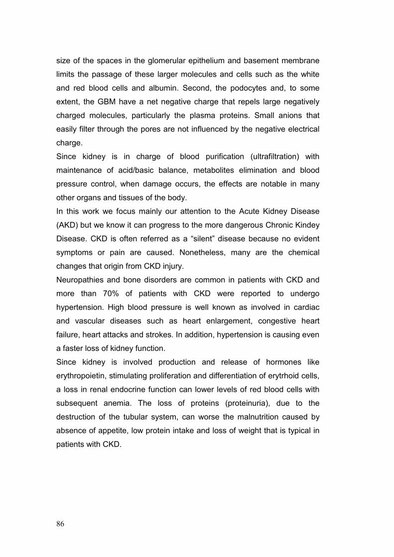
86
size of the spaces in the glomerular epithelium and basement membrane
limits the passage of these larger molecules and cells such as the white
and red blood cells and albumin. Second, the podocytes and, to some
extent, the GBM have a net negative charge that repels large negatively
charged molecules, particularly the plasma proteins. Small anions that
easily filter through the pores are not influenced by the negative electrical
charge.
Since kidney is in charge of blood purification (ultrafiltration) with
maintenance of acid/basic balance, metabolites elimination and blood
pressure control, when damage occurs, the effects are notable in many
other organs and tissues of the body.
In this work we focus mainly our attention to the Acute Kidney Disease
(AKD) but we know it can progress to the more dangerous Chronic Kindey
Disease. CKD is often referred as a “silent” disease because no evident
symptoms or pain are caused. Nonetheless, many are the chemical
changes that origin from CKD injury.
Neuropathies and bone disorders are common in patients with CKD and
more than 70% of patients with CKD were reported to undergo
hypertension. High blood pressure is well known as involved in cardiac
and vascular diseases such as heart enlargement, congestive heart
failure, heart attacks and strokes. In addition, hypertension is causing even
a faster loss of kidney function.
Since kidney is involved production and release of hormones like
erythropoietin, stimulating proliferation and differentiation of erytrhoid cells,
a loss in renal endocrine function can lower levels of red blood cells with
subsequent anemia. The loss of proteins (proteinuria), due to the
destruction of the tubular system, can worse the malnutrition caused by
absence of appetite, low protein intake and loss of weight that is typical in
patients with CKD.
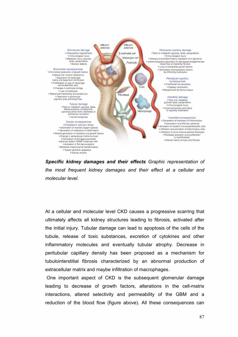
87
Specific kidney damages and their effects Graphic representation of
the most frequent kidney damages and their effect at a cellular and
molecular level.
At a cellular and molecular level CKD causes a progressive scarring that
ultimately affects all kidney structures leading to fibrosis, activated after
the initial injury. Tubular damage can lead to apoptosis of the cells of the
tubule, release of toxic substances, excretion of cytokines and other
inflammatory molecules and eventually tubular atrophy. Decrease in
peritubular capillary density has been proposed as a mechanism for
tubulointerstitial fibrosis characterized by an abnormal production of
extracellular matrix and maybe infiltration of macrophages.
One important aspect of CKD is the subsequent glomerular damage
leading to decrease of growth factors, alterations in the cell-matrix
interactions, altered selectivity and permeability of the GBM and a
reduction of the blood flow (figure above). All these consequences can
88
damage the podocytes leading to loss of function and apoptosis. Since
podocytes are thought to be the key cell of the glomerular compartment,
their malfunctionality induces a cascade of other effects that can lead to
ESRD like alterated proliferation of mesangial proliferation and changes in
microvascular permeability.
As described in the Introduction one of the main challenging in therapeutic
treatment of CKD using stem cells and regenerative medicine principles, is
to be able to identify a podocyte precursor cell that can rescue the
functionality of the glomerolus. In addition, it is fundamental to understand
the processes involved in the progression of the fibrosis in order to slow
down the progression of the disease.
In this work we have decided to use two different strategies as
experimental approaches to cure acute and chronic kidney failure: the
differentiation of hAFSC into kidney parenchyma, in vitro and in vivo, and
the isolation, from the amniotic fluid, of specific and already committed
kidney progenitor cells (AKPC).
1. Amniotic Fluid Stem Cells differentiation in renal parenchyma in
vitro and in vivo
Bioengineering of the kidney with all of its multifaceted function and
structure still remains a challenge. However, some progress has been
made towards this goal. Both autologous and pluripotential cells have
been used to either propagate or, regenerate de novo, kidney cell lines
under appropriate culture conditions [58-61]. These cells can then be used
to create a wide range of filtration devices some with potential life
sustaining properties and others with only limited functionality [61, 62]
Extracorporeal perfusion circuits constructed from cell seeded
hemofiltration devices are described in the literature but these are only
capable of limited perfusion [63-65]. One of the first applications of an
artificial renal device implanted in vivo was recently reported but only
demonstrated limited excretory function [60, 66]. Perhaps the development
of immunocompatible tissues or therapies from stem cells can offer
89
advantages over current methods of allogenic transplantation or dialysis
and provide investigators with a better method for regenerating organs like
the kidney de novo. The novelty in our approach to this complex field of in
vitro kidney regeneration strives to match the field of tissue engineering
with the fields of developmental and stem cell biology. A combination of
utilizing stem cells for tissue regeneration with an understanding of
developmental biology and organogenesis is imperative if one chooses to
reproduce in vitro a normal embryonic niche where cells can interact with
their environment for regenerative medicine applications.
Few studies to date have addressed the application of stem cells for
kidney differentiation. Jiang et al. report the isolation of a multipotent adult
progenitor cell (MAPC) from murine bone marrow and have shown the
contribution by these cells to virtually all somatic tissues of a developing
embryo after injection [67]. This included several commonly seen cell
types in the kidney, such as endothelial and tubular cells. More recently,
exogenously derived genetically modified human mesenchymal stem cells
(hMSC), in combination with whole-embryo culture, have been shown to
differentiate and contribute to functional complex structures of the kidney
during embryogenesis in vitro [68]. In some cases, entire nephrons were
hMSC-derived. Thus, hMSC are reprogrammed for other fates and organ
structures, depending on the embryonic environment into which they are
placed. Embryonic stem (ES) cells have been also used in cases of direct
injection into a mouse embryonic kidney [69] This established a new
model system in which patterns of epithelialization within the developing
kidney can be studied. Therefore, direct injection of cells provides a useful
tool for the evaluation of steps required for epithelial differentiation and
tubulogenesis.
Over the past 2 years, we have studied and characterized a new stem cell
population isolated from human amniotic fluid [70]. hAFSCs are a novel
population of cells that can be used for regenerative medicine applications
They possess many of the same proprieties as traditional embryonic stem
cell lines. Namely, hAFSC are pluripotential and can give rise to many
different cells types both in vitro and in vivo. hAFSC express embryonic
90
stem cell markers, Oct-4 and SSEA 3 and 4. However, hAFSC posses
many important advantages over ES cells: They can be easily isolated
with no harm to the fetus and are cultured without feeder layers using very
simple culture conditions. hAFSC can also be expanded in multiple
passages, frozen and thawed to provide an unlimited source of stem cells
when required [54].
In this study we investigated the ability of hAFSC to differentiate into renal-
like cells [55, 57, 71]. As mentioned above, the kidney is a very complex
organ and standard culture protocols available until now were not sufficient
to accomplish differentiation of the stem cells into all the various renal cell
types. Our novel approach physically placed hAFSCs into a developing
embryonic kidney which allowed differentiation to occur by providing the
stem cells not only with the necessary growth factors but the appropriate
extracellular environment for differentiation. The UB organ culture model,
more specifically, highlighted the ability of the hAFSCs to respond to
inductive signals and develop into individual kidney structures.
Nigam et al. demonstrated branching of the UB is possible in a gel
suspension [72, 73] Thus during culture, the UB secretes into the
extracellular environment all the embryonic co-factors necessary for
development. When hAFSCs are physically placed close to these
branching UB tips they are in turn influenced by the presence of the
embryonic structure and the circulating co-factors to differentiate their
morphology and become fully formed tubular structures resembling the
branching network observed with the UB. This was demonstrated by lac-z
positive hAFSC tubular cells observed close to the branching UB tips as
shown in the previous figures. hAFSC not only have the capacity to give
rise to specialized cells as observed by structural change but also cells
capable of expressing AQ1, a specific kidney cell marker, when induced to
differentiate by the embryonic environment of the UB.
The origin of the kidney is from two separate embryonic constituents that
ultimately fuse together to form the completed organ: the UB, which forms
the collecting system of the kidney, and the metanephric mesenchyme
(MM), which gives rise to the glomerular and tubular nephron, or filtration,
91
portion of the kidney. Bioengineering both constitutents separately from a
pluripotential stem cell source is a major focus of research in our
laboratory in our ultimate pursuit of tissue engineering a viable renal unit
for eventual in vivo applications for the future. On the one hand, the UB is
quite easy to culture which made applying hAFSCs to this culture system
straightforward. However, the MM still remains a formidable challenge to
culture by itself, and therefore, we needed to employ a entirely different
culture system to study the capacity that hAFSCs would have to
differentiate into this crucial portion of the developing kidney and obtain an
in vitro primordial nephron. Therefore, we examine the ability of hAFSCs
to form kidney primordial structures in culture after injection directly into
the embryonic kidney, essentially the developing MM. We postulated that
this embryonic niche surrounded by normal developing kidney structures
in close proximity to the injected hAFSCs would provide all the necessary
stimuli to differentiate the hAFSCs into kidney specific lineages and
sructures. The novelty of our work also serendipitously established a
protocol for long term organ culture of the embryonic kidney devoid of
tissue necrosis never before reported [57]. No previously published reports
addressing similar culture systems demonstrated culture of embryonic
kidneys for more than 5 days without necrosis [74-77]
We were successful at culturing embryonic kidneys at different stages of
gestation (E11.5- E16) without necrosis for 10 days, which was imperative
to initiate the differentiation process. In this context we could inject hAFSC
with reasonable confidence they would survive and replicate in a normal
and well preserved environment. Furthermore, it was possible to follow the
proliferation and differentiation of these cells during the entire culture
period (10 days). It is well known that the process of induction,
morphogenesis, and differentiation of the metanephros occurs in a
centrifugal pattern, proceeding from the center to the periphery. This was
also observed with our lac-z stained hAFSC where movement of the
injected cells from the site of injection, the center of the embryonic kidney,
to the periphery was documented by both live imaging and histologic
techniques [78-80] Thus, hAFSC put into the differentiating metanephric

92
mesenchyme follow all the necessary steps of differentiation into a
nephron. Further evidence of this fact was conveyed by our histologic data
whcih also demonstrated that hAFSC integrated into the renal vescicle,
C- and S-shaped bodies, which are the primordial structures of the kidney
that eventually develop into mature glomerular and tubular structures of
the kidney. In addition, there was also molecular evidence exhibited by
expression of GDNF, ZO-1, and Claudin by the hAFSC toward a renal
fate. Expression of late kidney gene markers AQ1 or AQ2, indicating that
perhaps they did not go through the entire differentiation process. Possibly
more time in the embryonic environment is necessary to complete the
differentiation process. Nevertheless, our data suggest that hAFSC
complete an initial step essential for commitment to a renal fate and that
during organ culture, they further undergo a mesenchymal-to-epithelial
transition [55].
Recently published reports have demonstrated that it was possible to
obtain kidney cell differentiation from bone marrow derived mesenchymal
stem cells only when the cells were induced (genetically modified) to
produce GDNF. In this case bone marrow stem cells were driven toward a
kidney fate before injection. In our study hAFSC were not induced to
express any kidney genes and were not genetically modified prior to
injection. This underscores the importance that hAFSC do not appear to
need any prior manipulation before installment into this particular culture
system. We therefore conclude that hAFSCs appear to posses a greater
pluripotentiality than mesenchymal stem cells from bone marrow, since it
appears that the signals from the embryonic renal environment are
sufficient to induce hAFSC toward the differentiation process into kidney
cells and structures.
In the in vivo experiments we have demonstrated a protective role of
hAFSC when injected directly into kidneys with glycerol-induced ATN [81].
In the last few years, published studies have supported the potential
therapeutic role of stem cells, mainly mesenchymal stem cells derived
from bone marrow or of kidney-specific progenitors [82, 83] to ameliorate
renal injury. Transplanted bone marrow stem cells were found to be

93
integrated into damaged kidneys [84, 85]. In this study, we have found that
in nu/nu mice the amount of glycerol required for induction of ATN was
50% higher than the dose needed in wild type mice (data not shown). This
suggests that the T-deficient mice are relatively protected as compared to
wild type mice against glycerol-rhabdomyolysis-induced ATN [86]. This
model of ATN involves a complex sequence of events wherein myoglobin,
released from the damaged muscle, damages the epithelial cells of the
proximal tubules, causing cast formation, vasoconstriction and decreased
glomerular filtration. The high level of apoptotic cells, the increased levels
of creatinine and BUN, and the histological analysis confirmed the
presence of ATN in our model.
The number of hAFSC that survived after injection is reduced over time as
evidenced by the luciferase detection. Nevertheless, injected hAFSC can
differentiate into tubule epithelial cells. hAFSC were found within the
damaged kidneys three weeks post injection located within damaged
tubules, and expressing epithelial markers, as measured by both
immunohistochemistry and RT-PCR using specific human antibodies and
human primers. These markers were not present in hAFSC in vitro prior to
injection.
We further found that injected hAFSC are also capable of expressing
kidney genes such as PAX2 and NPHS1, indicating that they can be
induced to commit toward renal differentiation. Furthermore, in some rare
instances, injected hAFSC cells could express Glial Derived Neurotrophic
Factor (GDNF), which is expressed during very early kidney development;
GDNF is not usually expressed in the adult kidney.
We also evaluated whether hAFSC can modulate kidney function after
damage, as reflected in the serum creatinine and BUN. When hAFSC
were injected during the established acute phase of the damage (between
48-72 hours after the injection of the glycerol), the levels of creatinine and
BUN did not decrease (data not shown). This implies that injection of
hAFSC when damage is already established is too late to attenuate the
damage. In contrast, when hAFSC were injected into the kidney on the
same day of glycerol injection no peaks in creatinine and BUN levels were

94
observed, underscoring the potential protective effects of hAFSC. We
therefore speculate that hAFSC can, when injected early enough, in this
case contemporaneously with the time of injury, attenuate acute renal
injury.
Furthermore, the histology analysis demonstrated that by 72 hours after
glycerol injection, the kidneys that were injected with the hAFSC show
fewer damaged tubules compared with the glycerol-injected kidneys not
treated with hAFSC. There was less disruption of tubular membranes and
no cast formation in the hAFSC-treated animals. We can speculate that
the injection of hAFSC accelerates the proliferation of epithelial tubular
cells that were partially damaged and prevents additional apoptosis. This
mechanism of protection lead to an overall better maintenance of the
tubular structure, thus avoiding the increase in BUN and creatinine
typically seen in glycerol-induced ATN.
During acute renal injury the immune-response plays a key role especially
in the first 48 hours; damaged kidney endothelial cells attract leukocytes,
vasomediators are released with injury, and epithelial cells of the tubule
produce proinflammatory and chemotactic cytokines [87].
Bonventre et al. [88] and Lin et al. [82] have speculated recently that the
mechanism by which bone marrow stem cells contribute to renal repair
was by attenuating the immune response, rather than through integration
or differentiation of the stem cells into the cells of the damaged organ.
Togel et al. have shown that injection of MSC is protective against
ischemic renal injury as early as 24 hours, based on measurement of
serum creatinine levels. They also speculated that the protection in these
animals was not through integration and differentiation of the injected
MSC, because of the very short period of time with which a protective
response was observed.
We also observed that the beneficial effect of hAFSC injection occurred
early in the course of ATN. Therefore, to further investigate the potential
mechanisms by which hAFSC enhance renal protection, we examined
intrarenal cytokines in order to determine whether there is a general
change in inflammatory cytokine pattern in mice that were treated with
95
hAFSC compared to untreated mice during the first 48 hours, thus when
the immuno-system acts in a very significant manner in determining the
course of the acute damage.
In the animals that were subjected only to glycerol injection it was
observed a significant increase in kidney cytokine expression when
compared with control mice that were not so treated. This demonstrates
that in acute ATN the kidney responds with a brisk outpouring of cytokines.
When mice were injected with hAFSC, an even greater elevation of
cytokine levels was evident, particularly at the earlier (24 hours) time point,
when compared with the cytokines levels measured in kidney treated only
with glycerol. This suggests to us that an important function of hAFSC may
be to augment the kidney cytokine milieu early in the course of ATN.
Moreover by 48 hours, this trend in combined cytokine levels is reversed
so that the majority of combined cytokine levels are either reduced
significantly or no longer elevated relative to the kidneys that did not
receive hAFSC. Further, by 48 hours the relative contribution of human
versus mouse cytokines is also reversed, with the human component of
the cytokine milieu being relatively small.
With a few exceptions, most human cytokines are also active on mouse
cells [89-94], so both the human and mouse cytokines likely affect the
kidney milieu. We think this is an important concept, since it is likely that
the complex interaction of cytokines derived from both the injected human
cells as well as the endogenous mouse cytokines are responsible for any
protective effects.
Therefore we propose that hAFSC, when injected directly into the kidney
in a mouse model of ATN, can be recruited as previously shown by us in
two mouse models of lung injury. Thus, hAFSC can home to injury sites,
where they protect damaged tissue from further injury and accelerate
repair probably through cytokine-mediated paracrine mechanisms. We
further propose that in this model the cytokines secreted by hAFSC work
in synergy with the endogenous mouse cytokines to promote and maintain
overall homeostasis of the tissues and thus interact with the inflammatory
96
environment favoring damage resolution, thus allowing the prevention of
progression of the acute phase in glycerol-induced ATN.
In addition, it is important to observe that at 48n hours after injection of
hAFSC into the ATN kidney, the major contribution to the pattern of
cytokine expression is from mouse cytokines, when compared with the
combined cytokine levels at 24 hours, at which point the relative
contribution of human cytokines is very high. This suggests that the human
cytokines first play a key role only in the earliest phase of renal response
to injury.
In conclusion, we have demonstrated a trophic effect of hAFSC on
resident kidney cells that survive a toxic injury, rather than via direct
repopulation of the damaged structures, even if they show potential of
differentiation into epithelial tubular cells over time. We demonstrated that
early direct injection of hAFSC into the kidney strongly ameliorates ATN
injury as reflected by more rapid resolution of tubular structural damage
and by normalized creatinine and BUN levels. In addition, our data show
evidence of immunomodulatory effects of hAFSC at a very early time
point, comparable in magnitude to endogenous cytokine production.
Understanding how donor and host cells combine to attenuate tubular
damage may lead eventually to the application of hAFSC for therapeutic
purposes in kidney diseases.
2. Amniotic Fluid Cells characterization and isolation of Metanpehric
Mesenchyme Derived cells (MMDC) and Amniotic Fluid Kidney
Progenitor Cells (AKPC)
AF is known source of stem cells, but almost 99% of cell population is still
poorly investigated. It seems reasonable to speculate that within AF there
is a repository of different progenitor cells included, due to its origin,
progenitors derived from the renal compartment.
AF fills the amniotic cavity, providing an environment in which floats the
developing embryo and later on the fetus. The volume and the
composition of the Amniotic Fluid change during pregnancy following the
97
physiological variations of the developing fetus. During embryogenesis,
maternal plasma is the main protagonist of Amniotic Fluid volume increase
and water flows osmotically, though fetal membranes, and, later on,
through the placental membrane. The composition of AF during the first
weeks of gestation is similar to the fetal plasma with a volume of 25 ml at
10 weeks to about 400 ml at 20 weeks [95]. By 8 weeks of gestation the
fetal kidney begins fluid production that rapidly increases in volume during
the second trimester. The exchange of fluids through the skin is present
until keratinisation that occurs between 20 and 24 weeks of gestation. The
molecular composition of AF and the presence of nutritive substances
have been shown to play a key role, in animals, in the proliferation and
differentiation of various intestinal cell types such as epithelial and mucosa
cells. AF has been used as a safe and reliable screening tool for genetic
and congenital diseases in the fetus for many years and, by now, is being
deeply investigated trying to match its molecular composition (or its
variation) with preterm delivery, infective processes and embryo diseases.
Contact between Amniotic Fluid and compartments of the developing
fetus, such as lung and gastrointestinal tract could explain the presence of
different cell types in the milieu of the Amniotic Fluid as reported in
literature.
The first approach was to characterise more specifically the presence of
cells derived from all the three germ layers. The development of the
embryo goes through a defined pathway of differentiation that follows
specific steps of maturation. From the small cell agglomerate call blastula,
derived from the fertilized oocyte, a subsequent separation of the cells in
agglomerates gives rise to three layers called germ layers. From this three
germ layers (Endoderm, Mesoderm and Ectoderm) are deriving all the
cells that originates all the tissues and organs. The cells within each germ
layer are multipotent, able to give rise only to some cell lineages. Each
germ layer develops along the first phases of the gestation to form
different compartments of the fetal body.
98
In addition to the multipotent cells belonging to the three germ layers were
investigated the presence of more committed cells and in particular organ
specific progenitor cells focusing on kidney precursors.
Since the composition of Amniotic Fluid changes over time as the fetus
develops during pregnancy, we divided samples according to gestational
age. Since the identification of a single marker to identify a specific
embryological tissue is difficult, do to the frequent co-expression of a gene
in many cells at the same or different time, we found the necessity to
analyze several markers for each germ layer in order to obtain more
precise results. The choice of the markers was made according to gene
expression in the early passages of germ layer specification, in order to
avoid the chance of false positives. Endodermal marker Sox-17, for
example, is expressed in the early stages of endoderm specification but, in
the mouse, expression was confirmed in mature lung cells. But since Sox-
17 was not revealed before E18 and our human amnioic fluid samples are
far from the end of gestation, we can speculate that positive results
obtained were not from mature cells derived from lung [95] On the other
hand, AFP was shown to be expressed in endodermal tissues and only at
very low levels in the adult as well as Goosecoid [96-97]. Cells from
mesodermal lineages were detected through expression of FLK-1,
Brachyury and SCL/TAL-1. FLK-1 is expressed broadly and transiently in
the mesodermal cells in the embryo but is known to be expressed in adults
too [98]. TAL-1 has been shown to be essential for hematopoiesis in the
early stages of life, and was shown to be expressed in the adult. But its
expression is limited to a short term repopulating cell population, with
characteristics of pluripotentiality, confirming that the marker was specific
enough for our purposes [99]. Brachyury was shown to be present in adult
almost only in pathological situation, becoming a reliable marker for
Endodermal specification [100]. The expression of markers from
endoderm and mesoderm was higher in samples of Amniotic Fluid of
earlier gestational age versus those samples taken from later gestational
age. In contrast, ectodermal markers NCAM, FGF-5 and E-Cadherin were
99
equally expressed in early and later samples of human Amniotic Fluid
maybe due to the turnover of the fetal skin cells during the development.
(Figure 3A-4A)
The presence of pluripotent cells within Amniotic Fluid is well known since
the studies of Prusa but possible information about pluripotent cells
variation over the gestational time could give us better data for the best
time for their collection. Therefore were analyzed OCT-4, pluripotent
marker essential for the development of the embryo, and C-Kit,
characterizing a population of mesenchymal stem cells.
Expression of OCT-4 and C-Kit was constant over time, indicating the
presence of pluripotent cells in AF in a large number of the samples
investigated, with no significant difference in presence between the earlier
and the later samples. These data prove that the pluripotent population
within AF is maintaining a strong of presence over the time, with no overall
difference in the choice of the time for the collection, at least for the time
range considered in this study.
We investigated also the presence and variation of markers for
mesenchymal and hematopoietic cells through expression of CD90 and
CD34.
Results revealed a constant and strong expression of mesenchymal-
derived cells, while hematopoietic cells were absent from the most part of
AF samples at 15-16 weeks (Figure 3A-4A) and only increased slightly in
older samples. Real Time PCR showed an increase number of samples
positive for CD34 in older weeks but with a decreased expression of the
marker (Figure 5B). Previous studies about AF MSCs [101] confirmed the
expression of CD90 in this population. CD34 is a well known marker for
hematopoietic lineage [102], even if recent publications are debating the
necessity of CD34 expression for cells to undergo hematopoietic
differentiation [103].
Interestingly previous studies showed that hematopoietic cells were
present before the 12 weeks of gestation but no further characterizations
were performed on older samples.
100
Secretions from the lung, gastrointestinal tract, skin and the urinary system
are the origin of a repository of a heterogeneous population of cells within
AF that has been demonstrated to be present in the Amniotic Fluid in our
study. The development of the human embryo follows a precise pathway
and the cells committed to various organs are differentiating at different
time points.
Any adult cell follows a specific pathway of gene expression that leads to
maturation with specificity.
We investigated, for this analysis, early markers of differentiation to the
specific organ cell lineage in order to identify cells committed, but not yet
mature, through a precise pathway. The choice of the markers was done
looking for an early expression but with a certain specificity that could
grant the definitive commitment to a cell lineage or group of cell lineages.
Since Nkx2.1 expression is limited to lung, thyroid and certain regions of
the forebrain [104] was used for the identification of lung progenitors as
well as Nkx2.5, essential for cardiac differentiation, is present at low levels
in other organs of the murine embryo [105]
In our study we found the presence of early transcription factors that
regulate differentiation processes of pluripotent cells into mature cells
including Nkx2.1 (TTF-1, Thyroid Transcription Factor-1) for lung, Nkx2.5
(CSX, Cardiac Specific Homeobox) for cardiomyocytes, CEBPG
(CCAAT/enhancer binding protein, gamma) for liver, PDX-1 (pancreatic
and duodenal homeobox 1) for pancreas, and GDNF (Glial Cell Line-
Derived Neurotrophic Factor) for kidney. As expected, the expression of
markers for committed cell populations is increased in the older samples
but is weak or absent in samples of earlier weeks of gestation. (Figure 3B-
4B) As the fetus develops, pluripotential cells subsequently may then give
way to multipotent cells, which in turn become committed to tissue-specific
lineages. These data suggest the presence of cells undergoing specific
differentiation. A deeper analysis should be performed within these
populations to evaluate cell expression paired with other markers that
could confirm the cell commitment toward a definitive differentiation.
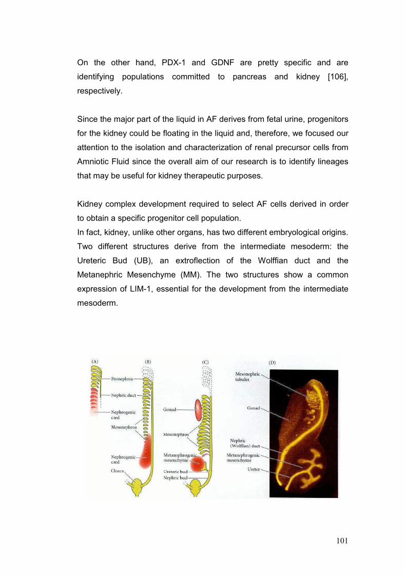
101
On the other hand, PDX-1 and GDNF are pretty specific and are
identifying populations committed to pancreas and kidney [106],
respectively.
Since the major part of the liquid in AF derives from fetal urine, progenitors
for the kidney could be floating in the liquid and, therefore, we focused our
attention to the isolation and characterization of renal precursor cells from
Amniotic Fluid since the overall aim of our research is to identify lineages
that may be useful for kidney therapeutic purposes.
Kidney complex development required to select AF cells derived in order
to obtain a specific progenitor cell population.
In fact, kidney, unlike other organs, has two different embryological origins.
Two different structures derive from the intermediate mesoderm: the
Ureteric Bud (UB), an extroflection of the Wolffian duct and the
Metanephric Mesenchyme (MM). The two structures show a common
expression of LIM-1, essential for the development from the intermediate
mesoderm.
102
Early kidney development The picture shows kidney development from
the pronephros to the formation of nephric duct and the nephrogenic cord
leading to the formation of the metanephros.
The evolution of the kidney reflects the need of land-adaptive vertebrates
to conserve water, excrete waste and maintain electrolyte homeostasis
within a variety of challenging environments [107].
Human kidney development starts around E18 and is ending between
gestational weeks 32-36 and also after birth; in the mouse is starting
around E7.5 and is ending 2 weeks after birth. Kidney develops from the
intermediate mesoderm located between the axial, or somitic, mesoderm
and the lateral plate mesoderm [106]
The UB gives rise to two transient structures called the pronephros and
the mesonephros and extend caudally along the axis with a structure
called Nephric Duct. (Figure 14). At the caudal end of the UB is present an
agglomerate of cells, called Metanephric blastema, that forms the MM.
While the pronephros and the mesanephros degenerates, the
metanephros, a structure present only in mammals, goes through an
intensification of signals between two structures: the MM and the UB, an
outgrowth of the nephric duct.
The invasion of the UB into the MM represents a crucial point for the
kidney development. This process is probably guided by signals secreted
from the MM, although this has not yet been demonstrated. Inductive
signals exchanged between the MM and the UB are pushing the bud to
grow and branch while the MM cells replicate themselves prior to
condensate around the UB tips.
GDNF (Glial derived neural factor) is secreted by the MM and addressed
to the RET receptor expressed on the UB cells surface starting the UB
branching. This process is essential to induce the UB branching that
stimulates MM condensation through expression of factors such as
molecules of the FGF’s family. The induced MM at this point starts to
express PAX-2, first indicator of the nephrogenic cell lineage (Podocytes,
mesangial cells, stromal cells and MET cells) at this stage.
103
The condensed mesenchymal cells induce the branching of UB giving rise
to two new ureteric tips that are going to form pretubular aggregates.
These pretubular aggregates undergo a mesenchyme-to epithelial
transition and form an epithelial tubule. Kidney tubules develop into
nephrons, through several stages of development. First, a tubule first
develops into a comma shaped body and then into an S-shaped body. In
the S-Shaped stage is possible to retrieve mesangial cell precursors
expressing PDGF receptor and podocyte cells precursors beginning to
express Nephrin at the cleft of the S-shaped body [108-109]. The most
proximal end of the S-Shaped body undergoes angiogenesis [110]. These
processes are started by repression of PAX-2 expression, and
consequentially the S-Shaped body forms the glomerulus and proximal
and distal tubules, which fuse to the UB. The middle part of the tubule
develops into the loop of Henle. The proximal tubule and the loop of Henle
are the kidney regions that are responsible for most of the reabsorption of
essential molecules and salts from urine, before they pass to the collecting
duct system. During the branching morphogenesis of the epithelial UB,
each tip acts as an inductive center to initiate nephrogenesis, after it has
elongated and branched to generate new epithelial tips. The branches of
the UB eventually form the collecting-duct system, which collects urine into
the renal pelvis and urinary bladder. These stages are repeated to
generate ~12,000 nephrons in the mouse kidney and almost 1,000,000
nephrons in the human kidney. In humans, nephrogenesis is complete
around 35-37 weeks while in rats and mice it continues postnatally for
about 2 weeks.
The understanding of the kidney development helped us to determine
which are the more specific genes involved in particular pathway that
identify the different cells present in the nephron.
We focus our attention in defining and characterizing a more specific cell
population isolated from AF for kidney regenerative purposes, capable of
more precise homing, integration and differentiation in the injured
glomerular structures.
104
In order to increase the specificity of a stem cell lineage for the glomerulus
compartment, and in particular for the podocytes cell type we investigated
reliable markers that could be used to retrieve a progenitor cell population
from the Amniotic Fluid.
Since many are the cells constituting the glomeruli, the first necessary step
was the identification of markers that allow us to separate the different
progenitor cells.
Based on literature review we identified Nephrin as a reliable marker for
immature and mature podocytes, PDGFR \ for mesangial cell progenitors,
TrKA for stromogenic cortical mesenchymal cells and E-Cadherin for
epithelial cells of the Bowman’s Capsule [107]. It is important to mention
that the identification of these particular markers was not granting us the
selection of a reliable renal population since their expression is not
exclusive of the kidney compartment, but they are expressed also in other
organs pathways development. Nephrin, for example, expressed in the
differentiating and mature podocytes, has been found in other extrarenal
tissues such as brain and pancreas and E-Cadherin, epithelial marker,
has been known to be present, beside in the first steps of gastrulation, in
many epithelial tissues [111-112].
One important in vivo study [113] for our purpose reported that CD24 and
OB Cadherin are co-expressed in cells of the undifferentiated MM in a
mouse developing kidney.
Since Metanephric Mesenchyme is the fetal structure that gives rise only
to the nephron, it was a reliable starting point for our purposes, giving us
the capability to select a more specific MM population from which retrieves
the more specific progenitor populations. In this way we could exclude the
possibility of isolating other progenitors that would eventually give rise to
cells with no specific renal fate.
We showed that in the total population of AF is evident the expression of
renal markers (Early and mature) such as PAX-2, LIM-1, Nephrin ,
105
PDGFR\, NGF High Affinity Receptor (chained with Tyrosin Kynase,
TrKA) , E- Cadherin, CD24, and OB-Cadherin (Cadherin 11) with a
strong increase expression by the end of the 17th week of gestation, when
kidney C-Shaped bodies are formed and are turning into S-Shaped bodies
(Figure 3C-4C). Real time PCR revealed an increase expression for
Nephrin and GDNF, markers of the MM derived lineages.
After having confirmed that within AF we can identify the expression of
renal markers, we focused our attention in isolating the MM lineage using
immunoseparation techniques for the CD24 and OB Cadherin. Since the
origin of cells within AF is from different organ, as described previously,
the first necessary step was to confirm that the MMDC population was
sharing with the in vivo renal population specific traits of expression
patterns. We confirmed that CD24+ OB-Cadherin+ population (MMDC)
expresses several kidney markers including AQP1, LIM-1, PAX-2,
Nephrin, GDNF, Occludin and ZO-1 by RT-PCR.
Subsequently we performed specific selection from MMDC population in
order to obtain four different subpopulations, called AKPC, expressing for
1. E-Cadherin (MET cells), 2. Nephrin (podocytes), 3. TrkA (for
stromogenic mesenchymal cells) and 4. PDGFR \ (mesangial cells)
respectively. In doing this we could be confident that the expression of
these markers were not involved in other organs pathways, but very
specific for the kidney since the were immunosepareted form and already
renal induced population, the MM.
These four subpopulations were then analyzed by the more sensitive Real
Time PCR. The expression pattern for the four population confirmed that
the four progenitor cell lineages are very distinct populations, with specific
and particular roles in the glomeruli structure of the nephron.
106
Among others, expression of LIM-1 and PAX-2 were evaluated in order to
confirm their not complete maturation since the two transcription factors
are not present in the renal adult cells.
TrKA+ APKC cells showed expression of TrKA, LIM-1, PAX-2 and OCT-4
at important levels and expressed E-Cadherin, Occludin and PDGFR\ at
low level. No expression of Nephrin or GDNF was shown. TrKA has been
shown not to be expressed in other than Stromogenic Cortical
Mesenchymal cells and since the expression of this marker has been
found only within this selection, we can with high confidence speculate that
this population represents the progenitors of the renal stromal cells
(PDGFR\AKPC).
Mesangial precursor cells, selected for PDGFR\ were positive at different
levels for PDGFR\ and E-Cadherin (Figure 8A-8B). Expression of E-
Cadherin is normally not reported in cells of the mesangium but since is
evident the slightly expression of uninduced mesenchyme markers (LIM-1,
PAX-2 and OCT-4) and E-Cadherin is expressed in the uninduced
mesenchyme it is not unreasonable to think that the differentiation process
is still in progress.
E-Cadherin+ APKC expressed high levels of OCT-4 and Occludin and
high levels of E-Cadherin and LIM-1, both expressed in the uninduced
mesenchyme. E-Cadherin+ cells expressed low levels of PAX-2 and
showed no expression of TrKA, PDGFR\ and ZO-1 (Figure 8A-8B). These
data may indicate that, since E-Cadherin is expressed in the uninduced
mesenchyme, the selection performed was not precise enough to retrieve
cells undergoing Mesenchymal to Epithelial Transition but more analysis
have to be performed.
As reported by Pavenstadt et al. [115] mature podocytes are expressing
several markers such as Nephrin and ZO-1 but data are lacking regarding
eventual precursors of this visceral epithelial cells. Our Nephrin+ APKC
populations was highlighted the expression of the pluripotent marker OCT-
4 and of LIM-1 and PAX-2, that may let us confirm the traits as a renal
precursor that haven’t yet committed to glomerulus or proximal/distal
tubules, due to the presence of E-Cadherin (Figure 8A-8B). At the same

107
time, the expression of Nephrin that in the kidney is localized only in the
differentiating and mature podocytes may indicate these cells are
undergoing through visceral epithelial cell maturation. An additional data in
support of this thesis is the expression of Zo-1 that usually co-localized
within the podocytes, coupled with Nephrin.

108
109
CONCLUSIONS
First we have demonstrated the ability of hAFSCs to survive, proliferate
and integrate into the embryonic kidney, while it undergoes organ
development, in an in vitro culture system. We observed the presence of
hAFSCs within kidney primordial, including tubules and developing
nephrons. Thus, hAFSCs seem to have the capacity to undergo the
expected mesenchymal to epithelial transition that occurs in normal renal
development and are induced to express important early kidney markers
such as GDNF, ZO-1 and Claudin. Moreover, hAFSCs do not appear to
require prior genetic modification or exogenous production of kidney
proteins for their differentiation to occur. This is a very important
advantage that hAFSCs have for potential future regenerative or
bioengineering application.
With the in vivo experiments, we have demonstrated a trophic effect of
hAFSC on resident kidney cells that survive a toxic injury, rather than via
direct repopulation of the damaged structures, even if they show potential
of differentiation into epithelial tubular cells over time. We demonstrated
that early direct injection of hAFSCs into the kidney strongly ameliorates
ATN injury as reflected by more rapid resolution of tubular structural
damage and by normalization of creatinine and BUN levels. In addition,
our data show evidence of immunomodulatory and antinflammatory effect
of hAFSCs, at an early time point, comparable in magnitude to
endogenous cytokine production. Understanding how donor and host cells
combine to attenuate tubular damage may lead eventually to the
application of hAFSCs for therapeutic purposes in acute kidney diseases.
Nonetheless, beside the presence of a small number (1%) of cells with
pluripotent characteristics, the composition of the other 99% of Amniotic
Fluid cells is diverse, with a great amount of cells exhibiting commitment to
a defined germ line or cellular endpoint.
110
There seems to be clear evidence for the existence of progenitor cells in
Amniotic Fluid, which can give rise to different cell types of mature organs.
By 17 weeks of gestation is notable an increase tissue specific cellular
presence and this data may indicate that the choice of the time point for
cell selection is fundamental. In addition, we demonstrated in the amniotic
fluid, the presence of a renal population with specific traits of commitment.
In particular, the presence of podocytes at both undifferentiated and
almost mature stages could favour their use for kidney regeneration in
vitro and in vivo animal models. The presence and identification of specific
renal progenitor cells in the Amniotic Fluid, committed to different
compartments of the kidney environment, could represent a valuable new
tool for regenerative purposes with regards to the treatment of a broad
range of renal diseases.
The discovery of renal specific progenitor cells within Amniotic Fluid could
bring a breakthrough in the study for novel and more selective approaches
in the renal therapy. However, the real pluripotential capability of these
progenitors cells, in particular the kidney progenitors presenting more
differentiation characteristics, has to be established. Moreover, their
potential for survival, proliferation, integration, and differentiation needs to
be assessed in in vivo models involving different types of renal damage.
Future aims of the project are:
1. assessment of the Amniotic Kidney Progenitor Cells differentiative
potential, performing in vitro and in vivo studies, in order to evaluate their
specificity for glomerular, stromal or tubular structures. In vitro
differentiation and functional investigation will be used as a preliminary
evaluation of their regenerative capability. In vivo studies on AKF, ATN or
CKD will be performed to understand their integration, differentiation and
immunogenicity. The understanding of AKPC maturation pathways can
reveal new tools for the comprehension of kidney development and
pathology formation;

111
2. investigation of AFSCs immunomodulatory mechanisms in Acute and
Chronic Kidney Disease models. In addition, the establishment of a more
effective administration of the cells, like injection at different timepoint
along the progression of the disease or multiple injections, are a priority in
order to maximize their beneficial effects.

112
113
REFERENCES
1. US Renal Data System. USRDS 2002. Annual Data Report : Atlas
of End.-Stage Renal Desease in the United States. Bethesda., MD :
National Institute of Health, National Institute of Diabetes and
Digestive and Kidney Diseases
2. Lanza, R. P., Chung, H. Y., Yoo, J. J. et al.: Generation of
histocompatible tissues using nuclear transplantation. Nat
Biotechnol, 20: 689, 2002
3. Stocum D.L., 2001. Stem cell in regenerative biology and medicine.
Wound Rep. Reg. 9: 429-442.
4. Thomson J.A. et al., 1998. Embryonic stem cells derived from
human blastocysts. Science 282: 1145-1147.
5. Reubinoff B.E. et al. 2000. Embryonic stem cell lines from human
blastocytes: somatic differentiation in vitro. Nature Biotech. 18: 399-
404.
6. Pittenger M.F Pittenger MF, Mackay AM, Beck SC, Jaiswal RK,
Douglas R, Mosca JD, Moorman MA, Simonetti DW, Craig S,
Marshak DR. 1999. Multilineage potential of adult human
mesenchymal stem cells. Science 284:143-147.
7. Goodwin HS. et al., 2001. Multilineage differentiation activity by
cells isolated from umbelical cord blood: expression of bone, fat and
neural markers. Biology of blood and marrow transplantation, 7:
581-588.
8. Streubel B, Martucci-Ivessa G, Fleck T, Bittner RE., In vitro
transformation of amniotic cells to muscle cells--background and
outlook, Wien Med Wochenschr.1996;146(9-10):216-7
9. P. De Coppi, G. Bartsch, M. Siddiqui, T. Xu, C. Santos, L. Perin, G.
Mostoslavsky, A. Serre, E. Snyder, J. Yoo, M. Furth, S. Soker and
A. Atala. Isolation of amniotic stem cell lines with potential for
therapy. 2007. Nature Biotechnology, 25: 100-106.
114
10. Anderson, RJ Prevention and management of acute renal failure.
Hospital Practice 1993 61–75
11. Thadhani, R, Pasqual, M, Bonventre, JV: Medical progress—Acute
renal failure. N Engl J Med 1996 334: 1448–1460
12. Dubose, TD, JR, Warnock, DG, Mehta, RL Acute renal failure in the
21st century: Recommendations for management and outcomes
assessment. Am J Kidney Dis 1997 29: 793–799
13. Lieberthal, W: Biology of acute renal failure: Therapeutic
implications. Kidney Int 1997 52: 1102–1115
14. Mindell, JA, Chertow, GM: A practical approach to acute renal
failure. Med Clin North Am 1997 81: 731–748,
15. Ronco, C, Bellomo, R, Feriani Critical care nephrology: The time
has come. Kidney Int 1998 53(Suppl 66): S1–S2
16. Shemesh, O, Golbetz, H, Kriss, JP, Myers, BD: Limitations of
creatinine as a filtration marker in glomerulopathic patients. Kidney
Int 1985 28: 830–838,
17. Moran, SM, Myers, BD: Course of acute renal failure studied by a
model of creatinine kinetics. Kidney Int 1985 27: 928–937
18. Liano, F, Pasqual, J, MADRID ACUTE RENAL FAILURE STUDY
GROUP: Epidemiology of acute renal failure: A prospective,
multicenter, community-based study. Kidney Int 1996 50:811–818,
19. Hou, SH, Bushinsky, DA, Wish, JB Hospital-acquired renal
insufficiency: A prospective study. Am J Med 1983 74: 243–248
20. Shusterman, N, Stromb, BL, Murray Risk factors and outcome of
hospital acquired acute renal failure: Clinical epidemiologic study.
Am J Med 1987 83: 65–71
21. Cecka, JM, Cho, YW, TerasakiI, PI: Analyses of the UNOS
scientific renal transplant registry at three years—Early events
affecting transplant success. Transplantation 1992 53: 59–64,
22. Troppman, C, Gilligham, KJ, Benedetti, E Delayed graft function,
acute rejection, and outcome after cadaver renal transplantation.
The multivariate analysis. Transplantation 1995 59: 962–968
115
23. Alexander, JW, Zola, JC: Expanding the donor pool: Use of
marginal donors for solid organ transplantation. Clin Transplant
1996 10: 1–19
24. Biesenbach, G, Zazgornik, J, Kaiser, W Improvement in prognosis
of patients with acute renal failure over a period of 15 years: An
analysis of 710 cases in a dialysis center. Am J Nephrol 1992 12:
319–325
25. MCCarthy, JT: Prognosis of patients with acute renal failure in the
intensive-care unit: A tale of two eras. Mayo Clin Proc 1996 71:
117–126,
26. Adams PL, Adams PF, Bell PD, Navar LG: Impaired renal blood
flow autoregulation in ischemic acute renal failure. Kidney Int 1980;
18:68–76.
27. Conger J: The role of blood flow autoregulation in pathophysiology
of acute renal failure. Circ Shock 1983;11:235–244.
28. Conger JD: Does hemodialysis delay recovery from acute renal
failure? Semin Dial 1990;3: 146–147.
29. Meier-Kriesche HU, Kaplan B. Waiting time on dialysis as the
strongest modifiable risk factor for renal transplant outcomes: a
paired donor kidney analysis. Transplantation 2002;74:1377-81.
30. Mange KC, Joffe MM, Feldman HI. Effect of the use or nonuse of
long-term dialysis on the subsequent survival of renal transplants
from living donors. N Engl J Med 2001;344:726-31.
31. Wolfe RA, Ashby VB, Milford EL, et al. Comparison of mortality in
all patients on dialysis, patients on dialysis awaiting transplantation,
and recipients of a first cadaveric transplant. N Engl J Med
1999;341:1725-30
32. Murray JE, Merrill JP, Harrison JH. Renal homotransplantation in
identical twins. Surg Forum (1955) 6:432–36
33. Takahashi K, Takahara S, Uchida K, Yoshimura N, Toma H,
Oshima S, et al. Successful results after 5 years of tacrolimus
therapy in ABO-incompatible kidney transplantation in Japan.
Transplant Proc (2005) 37:1800–3
116
34. Calne RY, White DJ, Thiru S, Evans DB, McMaster P, Dunn DC, et
al. Cyclosporin A in patients receiving renal allografts from cadaver
donors. Lancet (1978) 2:1323–7.
35. Vajdic CM, McDonald SP, McCredie MR, van Leeuwen MT, Stewart
JH, Law M, et al. Cancer incidence before and after kidney
transplantation. JAMA (2006) 296:2823–31.
36. Haseltine A. The Emergence of Regenerative Medicine: A New
Field and a New Society The Journal of Regenerative Medicine
Volume 2—2001 June 7, 2001
37. Humes HD, Buffington DA, MacKay SM, Funke AJ, Weitzel WF.
Replacement of renal function in uremic animals with a tissue-
engineered kidney. Nat Biotechnol. 1999 May;17(5):451-5.
38. Amiel GE, Yoo JJ, Atala A. Renal therapy using tissue-engineered
constructs and gene delivery. World J Urol. 2000 Feb;18(1):71-9.
39. Amiel GE, Yoo JJ, Atala A. Renal therapy using tissue-engineered
constructs and gene delivery. World J Urol. 2000 Feb;18(1):71-9.
40. Abkowitz JL, Robinson AE, Kale S, Long MW, Chen J. Mobilization
of hematopoietic stem cells during homeostasis and after cytokine
exposure. Blood. 2003 Aug 15;102(4):1249-53.
41. Sagrinati C, Netti GS, Mazzinghi B, Lazzeri E, Liotta F, Frosali F,
Ronconi E, Meini C, Gacci M, Squecco R, Carini M, Gesualdo L,
Francini F, Maggi E, Annunziato F, Lasagni L, Serio M, Romagnani
S, Romagnani P. Isolation and characterization of multipotent
progenitor cells from the Bowman's capsule of adult human
kidneys. J Am Soc Nephrol. 2006 Sep;17(9):2443-56.
42. Lin F, Cordes K, Li L, Hood L, Couser WG, Shankland SJ, Igarashi
P: Hematopoietic stem cells contribute to the regeneration of renal
tubules after renal ischemia-reperfusion injury in mice. J Am Soc
Nephrol 2003;14:1188-1199.
43. Abkowitz JL, Robinson AE, Kale S, Long MW, Chen J. Mobilization
of hematopoietic stem cells during homeostasis and after cytokine
exposure. Blood. 2003 Aug 15;102(4):1249-53.
117
44. Morigi M, Benigni A, Remuzzi G, Imberti B The regenerative
potential of stem cells in acute renal failure. 2006 Cell Transplant 15
Suppl 1:S111-117.
45. Morigi M, Imberti B, Zoja C, Corna D, Tomasoni S, Abbate M,
Rottoli D, Angioletti S, Benigni A, Perico N, Alison M, Remuzzi G.
Mesenchymal stem cells are renotropic, helping to repair the kidney
and improve function in acute renal failure. 2004 J Am Soc Nephrol.
Jul;15(7):1794-804.
46. Herrera MB, Bussolati B, Bruno S, Fonsato V, Romanazzi GM,
Camussi G Mesenchymal stem cells contribute to the renal repair
of acute tubular epithelial injury. 2004 Int J Mol Med 14:1035-1041.
47. Yokoo T, Fukui A, Ohashi T, Miyazaki Y, Utsunomiya Y, Kawamura
T, Hosoya T, Okabe M, Kobayashi E Xenobiotic kidney
organogenesis from human mesenchymal stem cells using a
growing rodent embryo. 2006 J Am Soc Nephrol 17:1026-1034.
48. Duffield JS, Park KM, Hsiao LL, Kelley VR, Scadden DT, Ichimura
T, Bonventre JV 2005 Restoration of tubular epithelial cells during
repair of the postischemic kidney occurs independently of bone
marrow-derived stem cells. J Clin Invest 115:1743-1755.
49. Duffield JS, Bonventre JV Kidney tubular epithelium is restored
without replacement with bone marrow-derived cells during repair
after ischemic injury. 2005 Kidney Int 68:1956-1961.
50. Lin F, Moran A, Igarashi P Intrarenal cells, not bone marrow-derived
cells, are the major source for regeneration in postischemic kidney.
2005 J Clin Invest 115:1756-1764.
51. Humphreys BD, Bonventre JV Mesenchymal Stem Cells in Acute
Kidney Injury. 2007 Annu Rev Med.
52. Underwood MA, Gilbert WM, Sherman MP. Amniotic fluid: not just
fetal urine anymore. J Perinatol. 2005 May;25(5):341-8. Review
53. Prusa AR, Marton E, Rosner M, Bernaschek G, Hengstschläger M.
Oct-4-expressing cells in human amniotic fluid: a new source for
stem cell research? Hum Reprod. 2003 Jul;18(7):1489-93
118
54. De Coppi P, Bartsch G, Jr., Siddiqui MM, et al. Isolation of amniotic
stem cell lines with potential for therapy. Nat Biotechnol. Jan
2007;25(2):100-106.
55. Giuliani S, Perin L, Jin D, Sedrakyan S, Carraro G, Habibian R, et
al. Renal differentiation of amniotic fluid stem cells. Cell Prolif. 2007
Dec;40(6):936-48.
56. Carraro G, 2008. Human Amniotic Fluid Stem Cells Can Integrate
and Differentiate Into Epithelial Lung Lineages. Stem Cells. 2008
Aug 28
57. Giuliani S, Perin L, Sedrakyan S, Jin D, Parnigotto PP, Warburton
D, Zanon GF, De Filippo RE. Ex-vivo whole embryonic kidney
culture: a novel method for long term analysis in regeneration and
transplantation. 2005 J Urology 179:365-370.
58. US Renal Data System. USRDS 2002. Annual Data Report : Atlas
of End.-Stage Renal Desease in the United States. Bethesda., MD :
National Institute of Health, National Institute of Diabetes and
Digestive and Kidney Diseases
59. Stocum, D.L. Stem cells in regenerative biology and medicine.
Wound Repair Regen 9, 429-442 (2001).
60. Lanza, R.P. et al. Generation of histocompatible tissues using
nuclear transplantation. Nat Biotechnol 20, 689-696 (2002).
61. MacKay, S.M., Funke, A.J., Buffington, D.A. & Humes, H.D. Tissue
engineering of a bioartificial renal tubule. Asaio J 44, 179-183
(1998).
62. Aebischer, P., Ip, T.K., Panol, G. & Galletti, P.M. The bioartificial
kidney: progress towards an ultrafiltration device with renal
epithelial cells processing. Life Support Syst 5, 159-168 (1987).
63. Amiel, G.E., Yoo, J.J. & Atala, A. Renal therapy using tissue-
engineered constructs and gene delivery. World J Urol 18, 71-79
(2000).
64. Ip, T.K., Aebischer, P. & Galletti, P.M. Cellular control of membrane
permeability. Implications for a bioartificial renal tubule. ASAIO
Trans 34, 351-355 (1988).
119
65. Amiel, G.E. & Atala, A. Current and future modalities for functional
renal replacement. Urol Clin North Am 26, 235-246, xi (1999).
66. Yoo, J.J. & Atala, A. Tissue engineering applications in the
genitourinary tract system. Yonsei Med J 41, 789-802 (2000).
67. Jiang, Y. et al. Pluripotency of mesenchymal stem cells derived
from adult marrow. Nature 418, 41-49 (2002).
68. Yokoo, T. et al. Human mesenchymal stem cells in rodent whole-
embryo culture are reprogrammed to contribute to kidney tissues.
Proc Natl Acad Sci U S A 102, 3296-3300 (2005).
69. Steenhard, B.M. et al. Integration of embryonic stem cells in
metanephric kidney organ culture. J Am Soc Nephrol 16, 1623-
1631 (2005).
70. Siddiqui, M.M. & Atala, A. Amniotic Fluid Derived Pluripotential
Cells (Ch 16), Vol. Editors: Robert Lanza, Helen Blau, Douglas
Melton, Malcolm Moore, E. Donnall Thomas, Catherine Verfaille,
Irving Weismann, and Michael West. (2004).
71. Perin L, Giuliani S, Sedrakyan S, Da Sacco S, De Filippo RE. Stem
Cell and regenerative science applications in the development of
bioengineering of renal tissue. Pediatr Res 2008 63:467-71
72. Qiao, J. et al. Multiple fibroblast growth factors support growth of
the ureteric bud but have different effects on branching
morphogenesis. Mech Dev 109, 123-135 (2001)
73. Qiao, J., Sakurai, H. & Nigam, S.K. Branching morphogenesis
independent of mesenchymal-epithelial contact in the developing
kidney. Proc Natl Acad Sci U S A 96, 7330-7335 (1999).
74. Gupta, I.R., Lapointe, M. & Yu, O.H. Morphogenesis during mouse
embryonic kidney explant culture. Kidney Int 63, 365-376 (2003).
75. Hardman, P., Klement, B.J. & Spooner, B.S. Growth and
morphogenesis of embryonic mouse organs on non-coated and
extracellular matrix-coated Biopore membrane. Dev Growth Differ
35, 683-690 (1993).
76. Rogers, S.A., Ryan, G. & Hammerman, M.R. Insulin-like growth
factors I and II are produced in the metanephros and are required
120
for growth and development in vitro. J Cell Biol 113, 1447-1453
(1991).
77. Steer, D.L., Bush, K.T., Meyer, T.N., Schwesinger, C. & Nigam,
S.K. A strategy for in vitro propagation of rat nephrons. Kidney Int
62, 1958-1965 (2002).
78. Evan, A.P., Gattone, V.H., 2nd & Schwartz, G.J. Development of
solute transport in rabbit proximal tubule. II. Morphologic
segmentation. Am J Physiol 245, F391-407 (1983)
79. Evan, A.P., Satlin, L.M., Gattone, V.H., 2nd, Connors, B. &
Schwartz, G.J. Postnatal maturation of rabbit renal collecting duct.
II. Morphological observations. Am J Physiol 261, F91-107 (1991).
80. Potter, E.L. Glomerular development in the kidney as an index of
fetal maturity. J Pediatrics 22, 695 (1943).
81. Perin L, Giuliani S, Sedrakyan S, Da Sacco S, Carraro G, Shiri L,
Lemley K, Rosol M, Wu S, Atala A, Warburton D, De Filippo RE.
Submitted to Open Access J 2009
82. Lin F. Renal repair: role of bone marrow stem cells. Pediatr
Nephrol. Nov 9 2007.
83. Al-Awqati Q, Oliver JA. The kidney papilla is a stem cells niche.
Stem Cell Rev. 2006;2(3):181-184.
84. Gupta S, Verfaillie C, Chmielewski D, Kim Y, Rosenberg ME. A role
for extrarenal cells in the regeneration following acute renal failure.
Kidney Int. Oct 2002;62(4):1285-1290.
85. Poulsom R, Forbes SJ, Hodivala-Dilke K, et al. Bone marrow
contributes to renal parenchymal turnover and regeneration. J
Pathol. Sep 2001;195(2):229-235.
86. Semedo P, Palasio CG, Oliveira CD, Feitoza CQ, Gonçalves GM,
Cenedeze MA, Wang PM, Teixeira VP, Reis MA, Pacheco-Silva A,
Câmara NO. Early modulation of inflammation by mesenchymal
stem cell after acute kidney injury. Int Immunopharmacol. 2009
Jun;9(6):677-82.
121
87. Bonventre JV. Molecular response to cytotoxic injury: role of
inflammation, MAP kinases, and endoplasmic reticulum stress
response. Semin Nephrol. Sep 2003;23(5):439-448.
88. Humphreys BD, Bonventre JV. Mesenchymal Stem Cells in Acute
Kidney Injury. Annu Rev Med. Feb 18 2008;59:311-325.
89. Maliszewski CR, Schoenborn MA, Cerretti DP, Wignall JM, Picha
KS, Cosman D, Tushinski RJ, Gillis S, Baker PE. Bovine GM-CSF:
molecular cloning and biological activity of the recombinant protein.
Mol Immunol. 1988 Sep;25(9):843-50.
90. Hu-Li J, Shevach EM, Mizuguchi J, Ohara J, Mosmann T, Paul WE.
B cell stimulatory factor 1 (interleukin 4) is a potent costimulant for
normal resting T lymphocytes. J Exp Med. 1987 Jan 1;165(1):157-
72.
91. Liu C, Bai Y, Ganea D, Hart RP. Species-specific activity of rat
recombinant interleukin-1 beta. J Interferon Cytokine Res. 1995
Nov;15(11):985-92.
92. Schwabe M, Cox GW, Bosco MC, Prohaska R, Kung HF. Multiple
cytokines inhibit interleukin-6-dependent murine
hybridoma/plasmacytoma proliferation. Cell Immunol. 1996 Feb
25;168(1):117-21.
93. Kennedy J, Rossi DL, Zurawski SM, Vega F Jr, Kastelein RA,
Wagner JL, Hannum CH, Zlotnik A. Mouse IL-17: a cytokine
preferentially expressed by alpha beta TCR + CD4-CD8-T cells. J
Interferon Cytokine Res. 1996 Aug;16(8):611-7.
94. De Haan G, Ausema A, Wilkens M, Molineux G, Dontje B. Efficient
mobilization of haematopoietic progenitors after a single injection of
pegylated recombinant human granulocyte colony-stimulating factor
in mouse strains with distinct marrow-cell pool sizes. Br J Haematol.
2000 Sep;110(3):638-46.
95. Kwon-Sik Parka, James M. Wellsb, Aaron M. Zornb, Susan E.
Werta and Jeffrey A. Whitsetta, Sox17 influences the differentiation
of respiratory epithelial cells Developmental Biology Volume 294,
Issue 1, 1 June 2006, Pages 192-202
122
96. Jones EA, Clement-Jones M, James O and Wilson D, Differences
between human and mouse alpha-fetoprotein expression during
early development, J. Anat. (2001) 198, pp. 555±559
97. Perea-Gomez, A. et al. Nodal antagonists in the anterior visceral
endoderm prevent the formation of multiple primitive streaks. Dev.
Cell 3, 745–756 (2002)
98. Victoria L Bautch Flk1 expression: promiscuity revealed Blood, 1
January 2006, Vol. 107, No. 1, pp. 3-4.
99. Curtis DJ, Hall MA, Van Stekelenburg LJ et al. SCL is required for
normal function of short-term repopulating hematopoietic stem cells.
Blood 2004;103:3342–3348
100. Palena C, Polev D, Tsang K, Fernando R, Litzinger M,
Krukovskaya L, Baranova A, Kozlov A, Schlom J . 2007. The
human T-box mesodermal transcription factor Brachyury is a
candidate target for T-cell-mediated cancer immunotherapy..
Clinical Cancer Research. 13:2471-2478
101. Ming-Song Tsai, Jia-Ling Lee, Yu-Jen Chang and Shiaw-Min
Hwang. Isolation of human multipotent mesenchymal stem cells
from second-trimester amniotic fluid using a novel two-stage culture
protocol Human Reproduction, Vol. 19, No. 6, 1450-1456, June
2004
102. Krause DS, Ito T, Fackler MJ, Smith OM, Collector MI,
Sharkis SJ, May WS. Characterization of murine CD34, a marker
for hematopoietic progenitor and stem cells. Blood. 1994 Aug
1;84(3):691-701
103. Yalin Guo, Michael Lübbert, Monika Engelhardt CD34-
Hematopoietic Stem Cells: Current Concepts and Controversies
Stem Cells 2003;21:15-20
104. Pan Q, Li C, Xiao J, Kimura S, Rubenstein J, Puelles L,
Minoo P. In vivo characterization of the Nkx2.1 promoter/enhancer
elements in transgenic mice. Gene. 2004 Apr 28;331:73-82
105. Hideko Kasahara, Sonia Bartunkova, Martina Schinke,
Makoto Tanaka, , Seigo Izumo Cardiac and Extracardiac
123
Expression of Csx/Nkx2.5 Homeodomain Protein Circulation
Research. 1998;82:936-946.
106. Hideaki Kaneto, Takeshi Miyatsuka, Toshihiko Shiraiwa,
Kaoru Yamamoto, Ken Kato, Yoshio Fujitani, Taka-aki Matsuoka
Crucial Role of PDX-1 in Pancreas Development, β-Cell
Differentiation, and Induction of Surrogate β-Cells Current
Medicinal Chemistry, Volume 14, Number 16, July 2007 , pp. 1745-
1752
107. Dressler GR. The cellular basis of kidney development. Annu
Rev Cell Dev Biol. 2006;22:509-29. Review
108. Horster MF, Braun GS, Huber SM. Embryonic renal epithelia:
induction, nephrogenesis, and cell differentiation.Physiol Rev. 1999
Oct;79(4):1157-91.
109. Juan Oliver and Qais Al-Awqati Development of vascular
elements during renal organogenesis Kidney International (2000)
57, 2167–2168;
110. Pöschl E, Schlötzer-Schrehardt U, Brachvogel B, Saito K,
Ninomiya Y, Mayer U. Collagen IV is essential for basement
membrane stability but dispensable for initiation of its assembly
during early development.
111. Beltcheva O, Kontusaari S, Fetissov S, Putaala H,
Kilpeläinen P, Hökfelt T, Tryggvason K. Alternatively used
promoters and distinct elements direct tissue-specific expression of
nephrin.J Am Soc Nephrol. 2003 Feb;14(2):352-8.
112. K Boller, D Vestweber and R Kemler Cell-adhesion molecule
uvomorulin is localized in the intermediate junctions of adult
intestinal epithelial cells The Journal of Cell Biology, Vol 100, 327-
332
113. M. Kasper , O. Huber, H. Großmann, B. Rudolph, C.
Tränkner and M. Müller Immunocytochemical distribution of E-
cadherin in normal and injured lung tissue of the rat Histochemistry
and Cell Biology Volume 104, Number 5 / November, 1995

124
114. Challen GA, Bertoncello I, Deane JA, Ricardo SD, Little MH.
Kidney side population reveals multilineage potential and renal
functional capacity but also cellular heterogeneity. J Am Soc
Nephrol. 2006 Jul;17(7):1896-912.
115. Pavenstädt H, Kriz W, Kretzler M. Cell biology of the
glomerular podocyte. Physiol Rev. 2003 Jan;83(1):253-307.
Review.

125
TABLES AND FIGURES
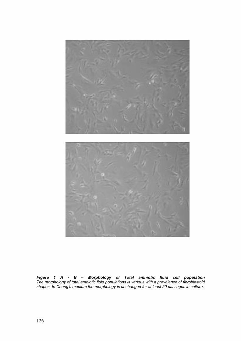
126
Figure 1 A - B – Morphology of Total amniotic fluid cell population The morphology of total amniotic fluid populations is various with a prevalence of fibroblastoid shapes. In Chang’s medium the morphology is unchanged for at least 50 passages in culture.
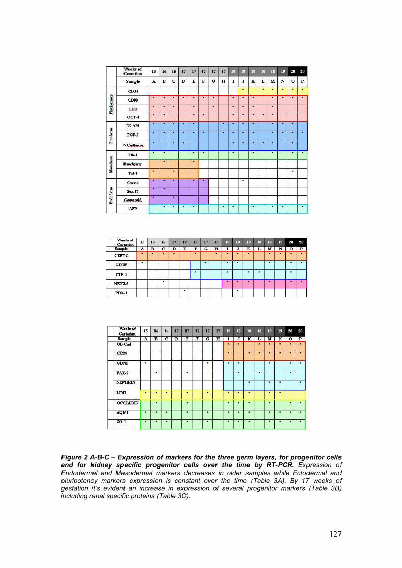
127
Figure 2 A-B-C – Expression of markers for the three germ layers, for progenitor cells and for kidney specific progenitor cells over the time by RT-PCR. Expression of Endodermal and Mesodermal markers decreases in older samples while Ectodermal and pluripotency markers expression is constant over the time (Table 3A). By 17 weeks of gestation it’s evident an increase in expression of several progenitor markers (Table 3B) including renal specific proteins (Table 3C).
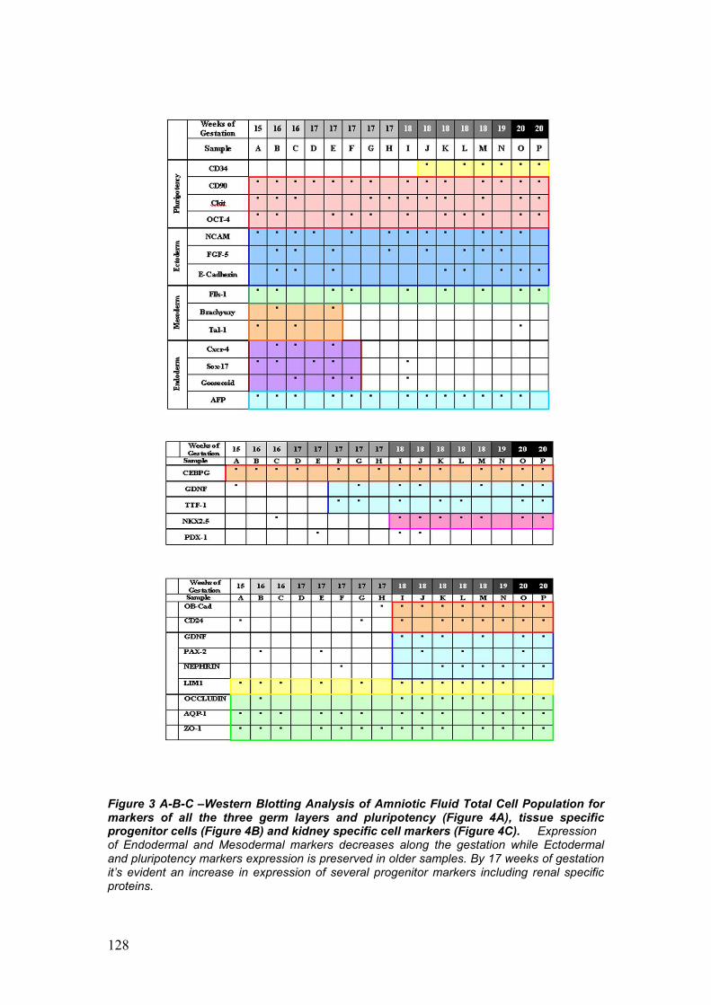
128
Figure 3 A-B-C –Western Blotting Analysis of Amniotic Fluid Total Cell Population for markers of all the three germ layers and pluripotency (Figure 4A), tissue specific progenitor cells (Figure 4B) and kidney specific cell markers (Figure 4C). Expression of Endodermal and Mesodermal markers decreases along the gestation while Ectodermal and pluripotency markers expression is preserved in older samples. By 17 weeks of gestation it’s evident an increase in expression of several progenitor markers including renal specific proteins.
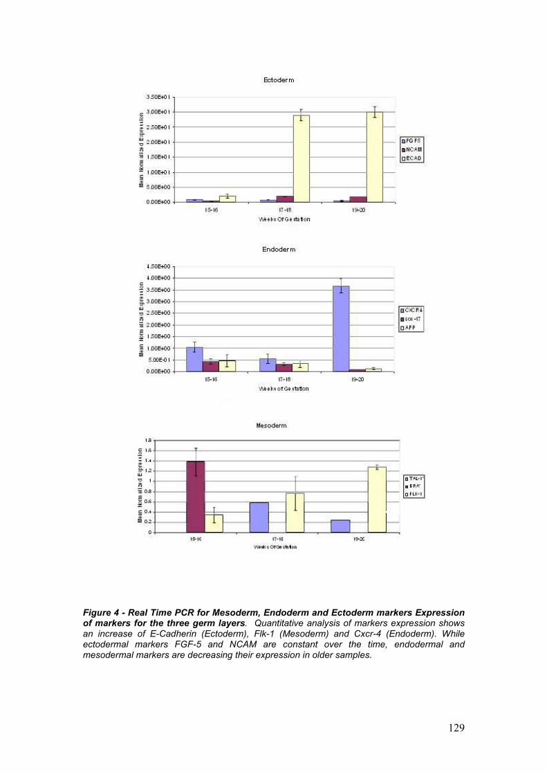
129
Figure 4 - Real Time PCR for Mesoderm, Endoderm and Ectoderm markers Expression of markers for the three germ layers. Quantitative analysis of markers expression shows an increase of E-Cadherin (Ectoderm), Flk-1 (Mesoderm) and Cxcr-4 (Endoderm). While ectodermal markers FGF-5 and NCAM are constant over the time, endodermal and mesodermal markers are decreasing their expression in older samples.
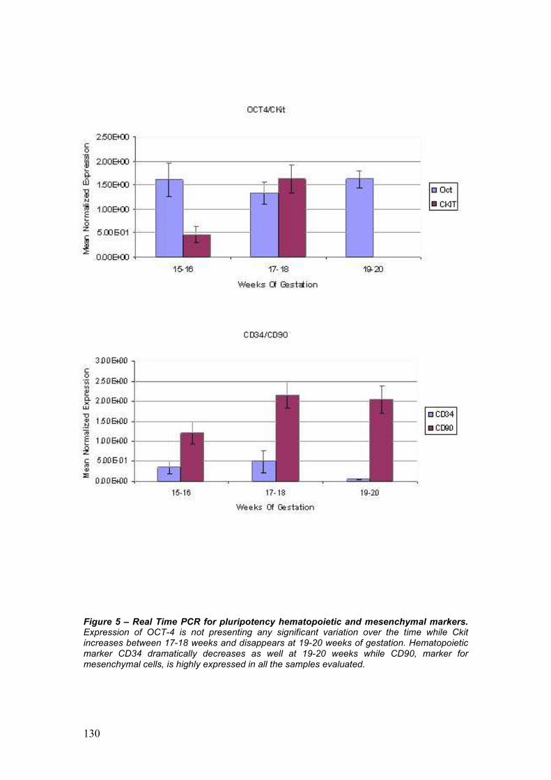
130
Figure 5 – Real Time PCR for pluripotency hematopoietic and mesenchymal markers. Expression of OCT-4 is not presenting any significant variation over the time while Ckit increases between 17-18 weeks and disappears at 19-20 weeks of gestation. Hematopoietic marker CD34 dramatically decreases as well at 19-20 weeks while CD90, marker for mesenchymal cells, is highly expressed in all the samples evaluated.
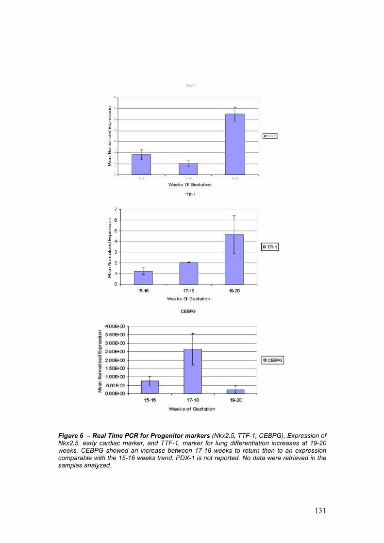
131
Figure 6 – Real Time PCR for Progenitor markers (Nkx2.5, TTF-1, CEBPG). Expression of Nkx2.5, early cardiac marker, and TTF-1, marker for lung differentiation increases at 19-20 weeks. CEBPG showed an increase between 17-18 weeks to return then to an expression comparable with the 15-16 weeks trend. PDX-1 is not reported. No data were retrieved in the samples analyzed.
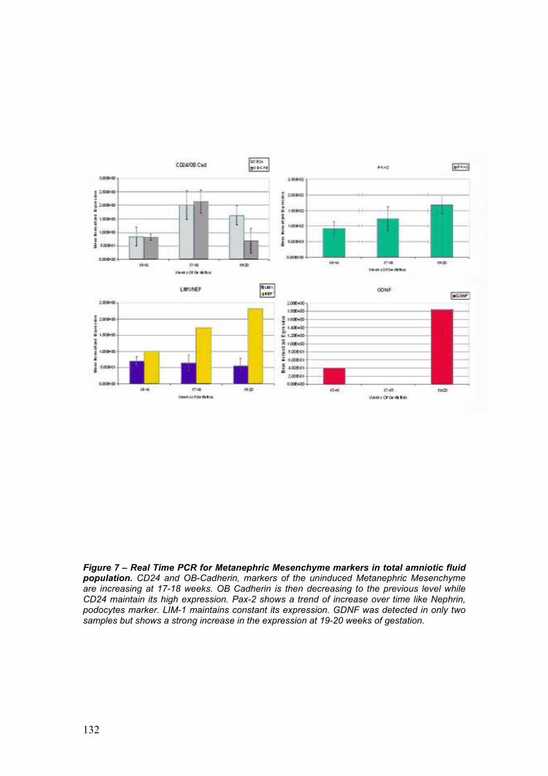
132
Figure 7 – Real Time PCR for Metanephric Mesenchyme markers in total amniotic fluid population. CD24 and OB-Cadherin, markers of the uninduced Metanephric Mesenchyme are increasing at 17-18 weeks. OB Cadherin is then decreasing to the previous level while CD24 maintain its high expression. Pax-2 shows a trend of increase over time like Nephrin, podocytes marker. LIM-1 maintains constant its expression. GDNF was detected in only two samples but shows a strong increase in the expression at 19-20 weeks of gestation.
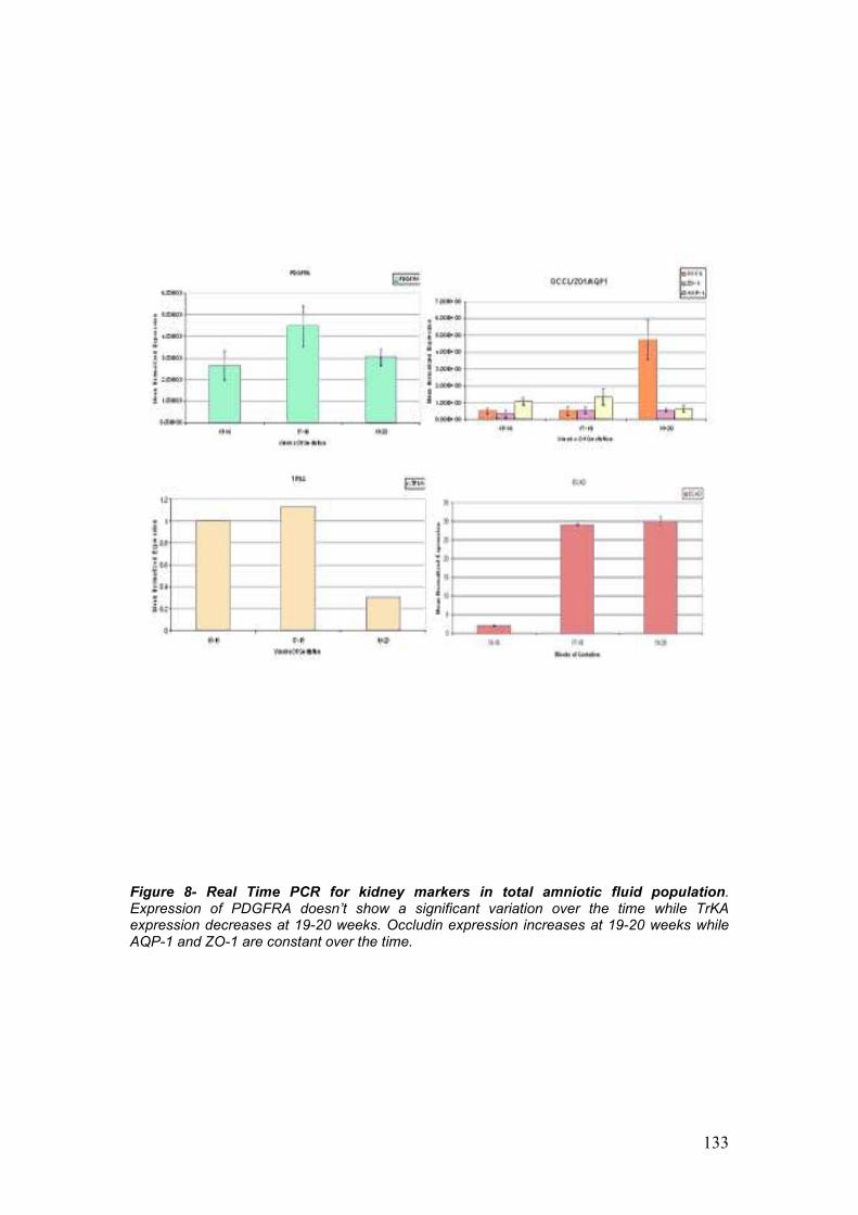
133
Figure 8- Real Time PCR for kidney markers in total amniotic fluid population. Expression of PDGFRA doesn’t show a significant variation over the time while TrKA expression decreases at 19-20 weeks. Occludin expression increases at 19-20 weeks while AQP-1 and ZO-1 are constant over the time.
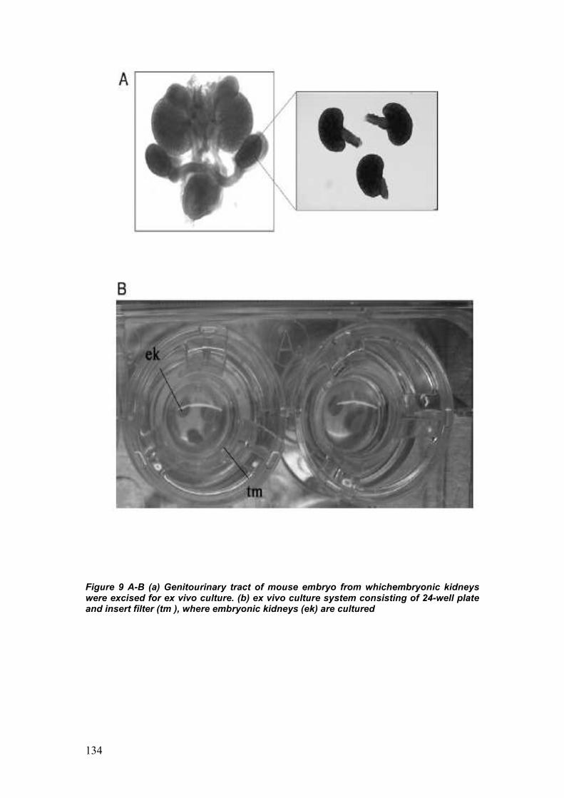
134
Figure 9 A-B (a) Genitourinary tract of mouse embryo from whichembryonic kidneys were excised for ex vivo culture. (b) ex vivo culture system consisting of 24-well plate and insert filter (tm ), where embryonic kidneys (ek) are cultured
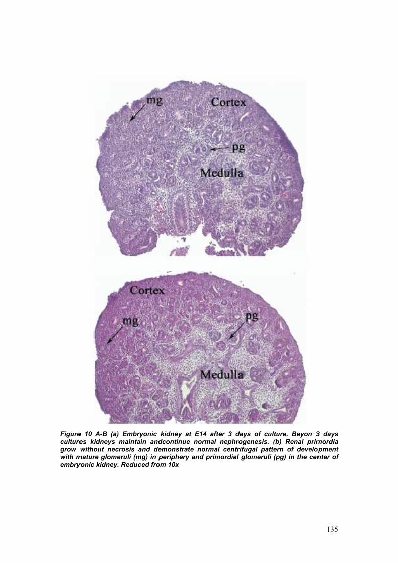
135
Figure 10 A-B (a) Embryonic kidney at E14 after 3 days of culture. Beyon 3 days cultures kidneys maintain andcontinue normal nephrogenesis. (b) Renal primordia grow without necrosis and demonstrate normal centrifugal pattern of development with mature glomeruli (mg) in periphery and primordial glomeruli (pg) in the center of embryonic kidney. Reduced from 10x
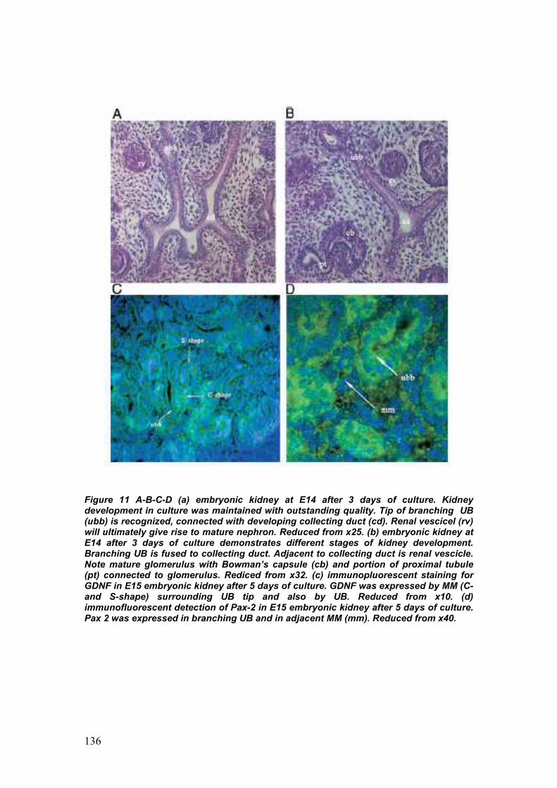
136
Figure 11 A-B-C-D (a) embryonic kidney at E14 after 3 days of culture. Kidney development in culture was maintained with outstanding quality. Tip of branching UB (ubb) is recognized, connected with developing collecting duct (cd). Renal vescicel (rv) will ultimately give rise to mature nephron. Reduced from x25. (b) embryonic kidney at E14 after 3 days of culture demonstrates different stages of kidney development. Branching UB is fused to collecting duct. Adjacent to collecting duct is renal vescicle. Note mature glomerulus with Bowman’s capsule (cb) and portion of proximal tubule (pt) connected to glomerulus. Rediced from x32. (c) immunopluorescent staining for GDNF in E15 embryonic kidney after 5 days of culture. GDNF was expressed by MM (C-and S-shape) surrounding UB tip and also by UB. Reduced from x10. (d) immunofluorescent detection of Pax-2 in E15 embryonic kidney after 5 days of culture. Pax 2 was expressed in branching UB and in adjacent MM (mm). Reduced from x40.
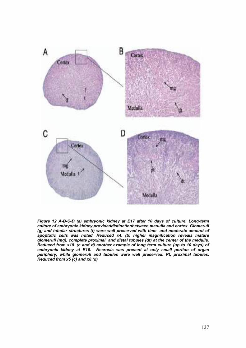
137
Figure 12 A-B-C-D (a) embryonic kidney at E17 after 10 days of culture. Long-term culture of embryonic kidney provideddistinctionbetween medulla and cortex. Glomeruli (g) and tubular structures (t) were well preserved with time and moderate amount of apoptotic cells was noted. Reduced x4. (b) higher magnification reveals mature glomeruli (mg), complete proximal and distal tubules (dt) at the center of the medulla. Reduced from x10. (c and d) another example of long term culture (up to 10 days) of embryonic kidney at E16. Necrosis was present at only small portion of organ periphery, while glomeruli and tubules were well preserved. Pt, proximal tubules. Reduced from x5 (c) and x8 (d)
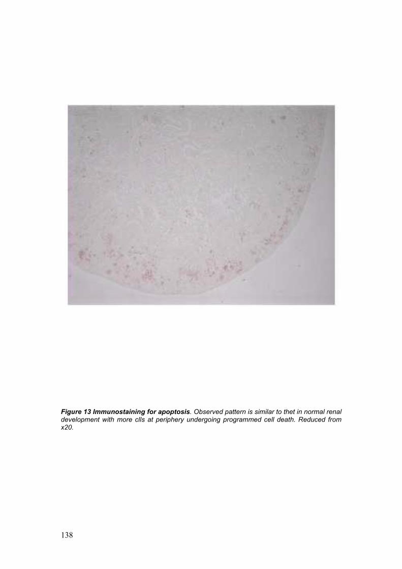
138
Figure 13 Immunostaining for apoptosis. Observed pattern is similar to thet in normal renal development with more clls at periphery undergoing programmed cell death. Reduced from x20.
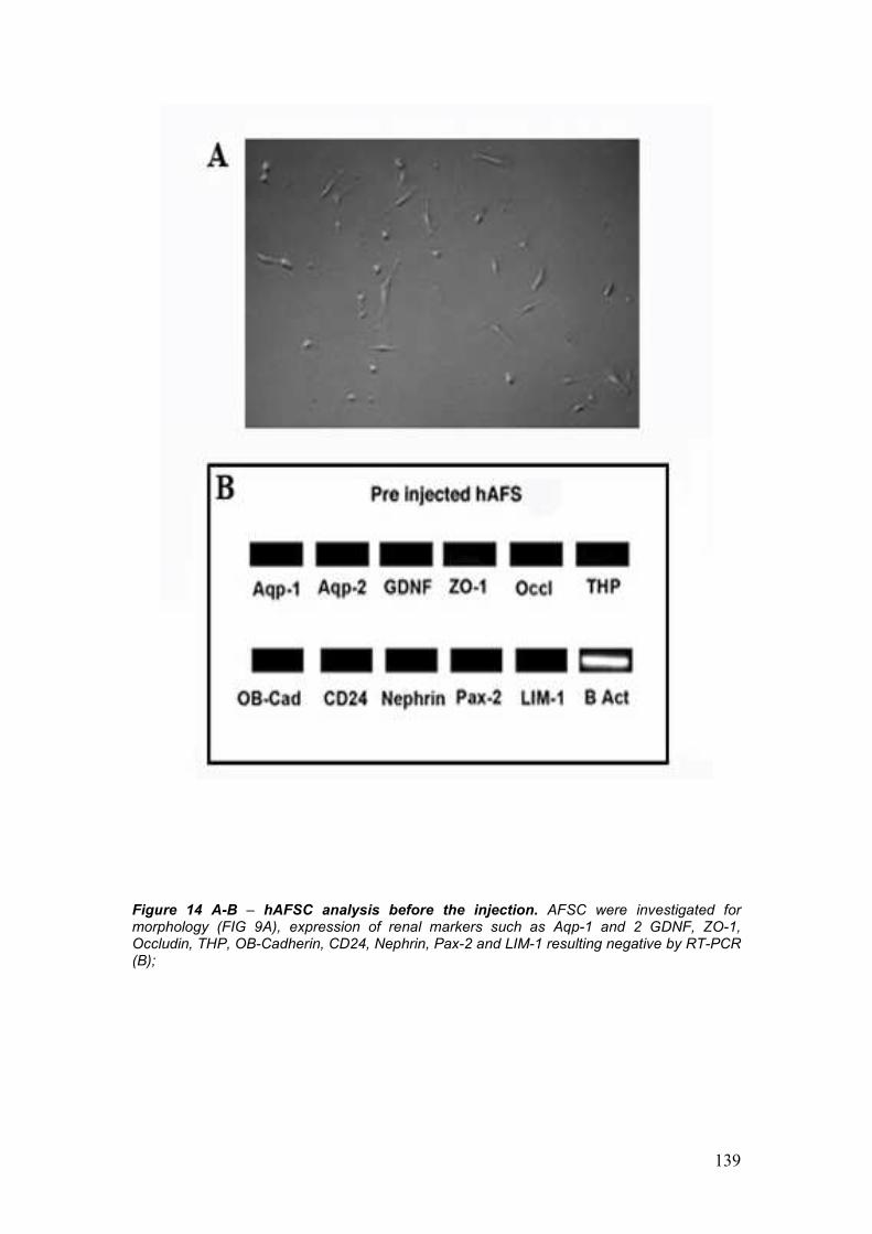
139
Figure 14 A-B – hAFSC analysis before the injection. AFSC were investigated for morphology (FIG 9A), expression of renal markers such as Aqp-1 and 2 GDNF, ZO-1, Occludin, THP, OB-Cadherin, CD24, Nephrin, Pax-2 and LIM-1 resulting negative by RT-PCR (B);
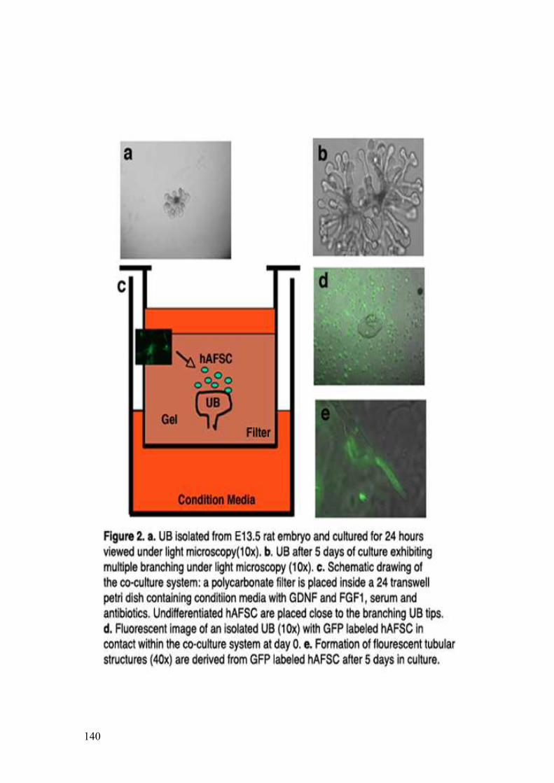
140
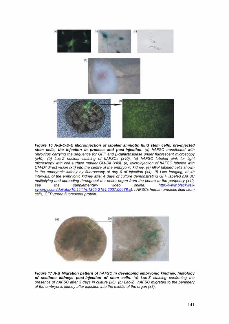
141
Figure 16 A-B-C-D-E Microinjection of labeled amniotic fluid stem cells, pre-injected stem cells, the injection in process and post-injection. (a) hAFSC transfected with retrovirus carrying the sequence for GFP and β-galactosidase under fluorescent microscopy (x40). (b) Lac-Z nuclear staining of hAFSCs (x40). (c) hAFSC labeled pink for light microscopy with cell surface marker CM-Dil (x40). (d) Microinjection of hAFSC labeled with CM-Dil direct vision (x4) into the centre of the embryonic kidney. (e) GFP labeled cells shown in the embryonic kidney by fluoroscopy at day 0 of injection (x4). (f) Lice imaging, at 4h intervals, of the embryonic kidney after 4 days of culture demonstrating GFP labeled hAFSC multiplying and spreading throughout the entire organ from the centre to the periphery (x40, see the supplementary video online: http://www.blackwell-synergy.com/doi/abs/10.1111/j.1365-2184.2007.00478.x). hAFSCs human amniotic fluid stem cells, GFP green fluorescent protein.
Figure 17 A-B Migration pattern of hAFSC in developing embryonic kindney, histology of sectione kidneys post-injection of stem cells. (a) Lac-Z staining confirming the presence of hAFSC after 3 days in culture (x6). (b) Lac-Z+ hAFSC migrated to the periphery of the embryonic kidney after injection into the middle of the organ (x8).
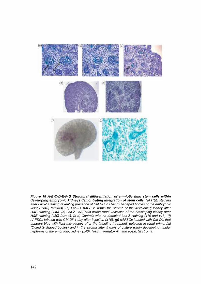
142
Figure 18 A-B-C-D-E-F-G Structural differentiation of amniotic fluid stem cells within developing embryonic kidneys demontrating integration of stem cells. (a) H&E staining after Lac-Z staining revealing presence of hAFSC in C-and S-shaped bodies of the embryonic kidney (x40) (arrows). (b) Lac-Z+ hAFSCs within the stroma of the developing kidney after H&E staining (x40). (c) Lac-Z+ hAFSCs within renal vescicles of the developing kidney after H&E staining (x30) (arrow). (d-e) Controls with no detected Lac-Z staining (x10 and x16). (f) hAFSCs labeled with CM-Dil 1 day after injection (x10). (g) hAFSCs labeled with CM-Dil, that appears blue with light microscopy after the toluidine treatment, detected in renal primordial (C-and S-shaped bodies) and in the stroma after 5 days of culture within developing tubular nephrons of the embryonic kidney (x40). H&E, haematoxylin and eosin, St stroma.
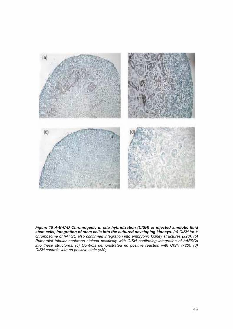
143
Figure 19 A-B-C-D Chromogenic in situ hybridization (CISH) of injected amniotic fluid stem cells, integration of stem cells into the cultured developing kidneys. (a) CISH for Y chromosome of hAFSC also confirmed integration into embryonic kidney structures (x20). (b) Primordial tubular nephrons stained positively with CISH confirming integration of hAFSCs into these structures. (c) Controls demonstrated no positive reaction with CISH (x20). (d) CISH controls with no positive stain (x30).
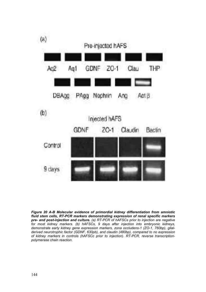
144
Figure 20 A-B Molecular evidence of primordial kidney differentiation from amniotic fluid stem cells, RT-PCR markers demonstrating expression of renal specific markers pre- and post-injection and culture. (a) RT-PCR of hAFSCs prior to injection are negative for most kidney markers. (b) hAFSCs, 9 days after injection into embryonic kidneys, demonstrate early kidney gene expression markers, zona occludens-1 (ZO-1, 760bp), glial-derived neurotrophic factor (GDNF, 630pb), and claudin (480bp), compared to no expression of kidney markers in controls (hAFSCc prior to injection). RT-PCR, reverse transcription-polymerase chain reaction.
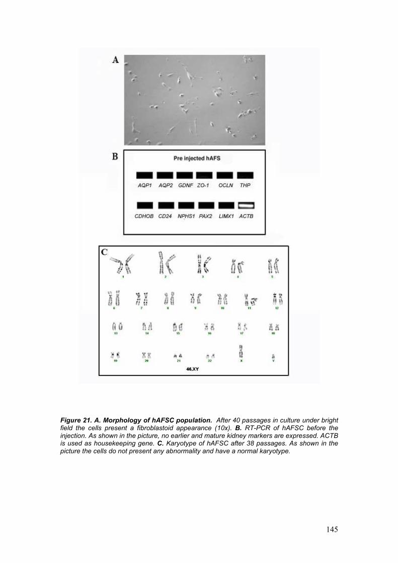
145
Figure 21. A. Morphology of hAFSC population. After 40 passages in culture under bright field the cells present a fibroblastoid appearance (10x). B. RT-PCR of hAFSC before the injection. As shown in the picture, no earlier and mature kidney markers are expressed. ACTB is used as housekeeping gene. C. Karyotype of hAFSC after 38 passages. As shown in the picture the cells do not present any abnormality and have a normal karyotype.
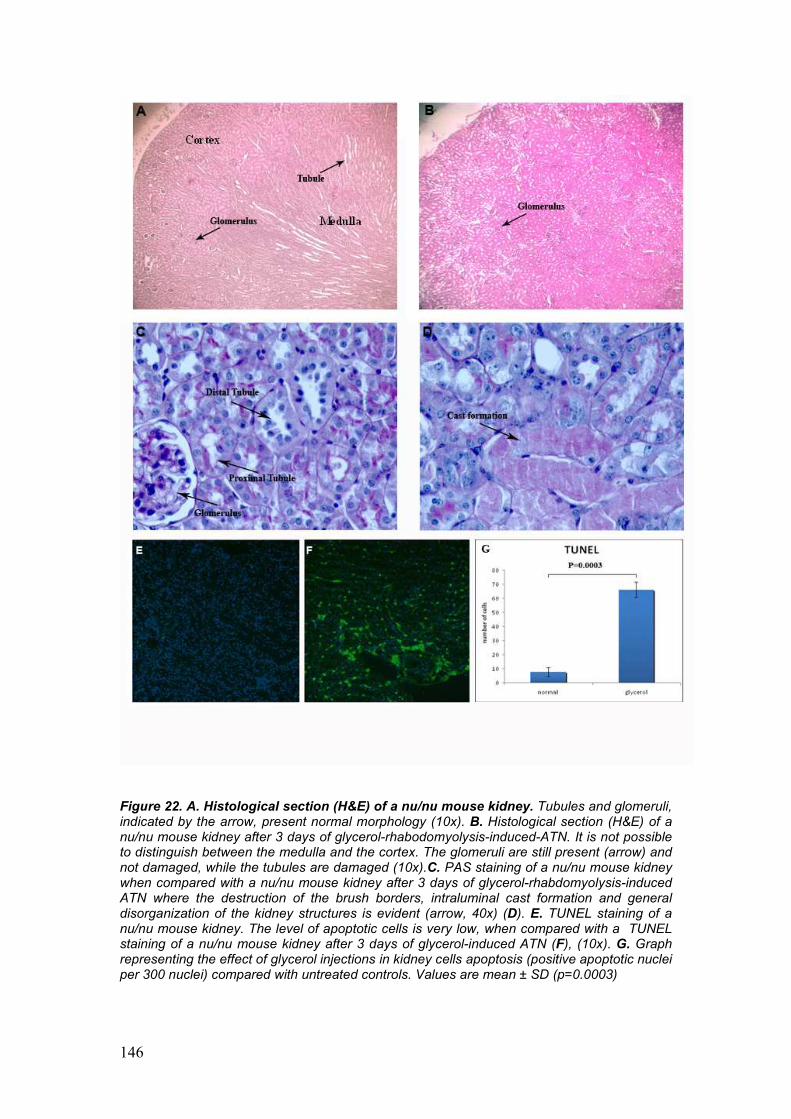
146
Figure 22. A. Histological section (H&E) of a nu/nu mouse kidney. Tubules and glomeruli, indicated by the arrow, present normal morphology (10x). B. Histological section (H&E) of a nu/nu mouse kidney after 3 days of glycerol-rhabodomyolysis-induced-ATN. It is not possible to distinguish between the medulla and the cortex. The glomeruli are still present (arrow) and not damaged, while the tubules are damaged (10x).C. PAS staining of a nu/nu mouse kidney when compared with a nu/nu mouse kidney after 3 days of glycerol-rhabdomyolysis-induced ATN where the destruction of the brush borders, intraluminal cast formation and general disorganization of the kidney structures is evident (arrow, 40x) (D). E. TUNEL staining of a nu/nu mouse kidney. The level of apoptotic cells is very low, when compared with a TUNEL staining of a nu/nu mouse kidney after 3 days of glycerol-induced ATN (F), (10x). G. Graph representing the effect of glycerol injections in kidney cells apoptosis (positive apoptotic nuclei per 300 nuclei) compared with untreated controls. Values are mean ± SD (p=0.0003)
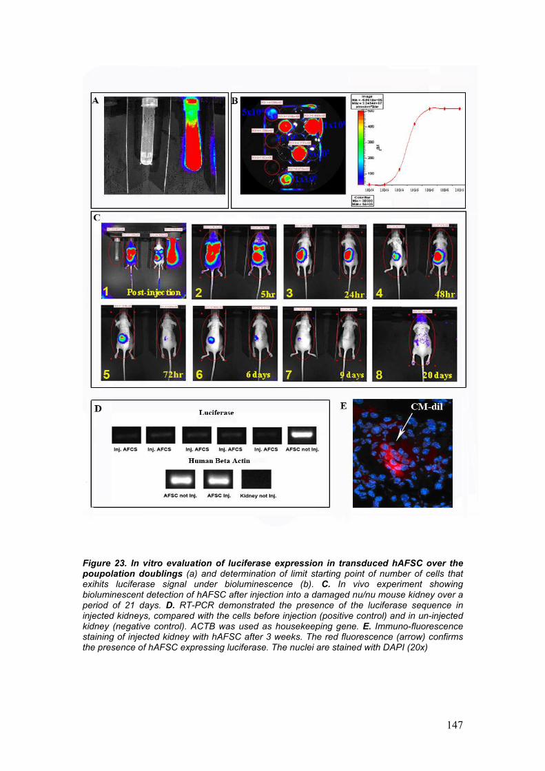
147
Figure 23. In vitro evaluation of luciferase expression in transduced hAFSC over the poupolation doublings (a) and determination of limit starting point of number of cells that exihits luciferase signal under bioluminescence (b). C. In vivo experiment showing bioluminescent detection of hAFSC after injection into a damaged nu/nu mouse kidney over a period of 21 days. D. RT-PCR demonstrated the presence of the luciferase sequence in injected kidneys, compared with the cells before injection (positive control) and in un-injected kidney (negative control). ACTB was used as housekeeping gene. E. Immuno-fluorescence staining of injected kidney with hAFSC after 3 weeks. The red fluorescence (arrow) confirms the presence of hAFSC expressing luciferase. The nuclei are stained with DAPI (20x)
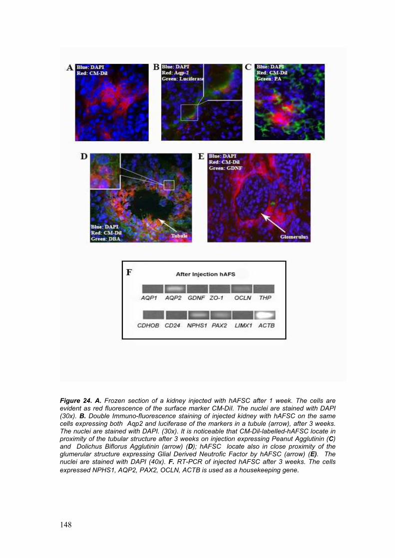
148
Figure 24. A. Frozen section of a kidney injected with hAFSC after 1 week. The cells are evident as red fluorescence of the surface marker CM-DiI. The nuclei are stained with DAPI (30x). B. Double Immuno-fluorescence staining of injected kidney with hAFSC on the same cells expressing both Aqp2 and luciferase of the markers in a tubule (arrow), after 3 weeks. The nuclei are stained with DAPI. (30x). It is noticeable that CM-Dil-labelled-hAFSC locate in proximity of the tubular structure after 3 weeks on injection expressing Peanut Agglutinin (C) and Dolichus Biflorus Agglutinin (arrow) (D); hAFSC locate also in close proximity of the glumerular structure expressing Glial Derived Neutrofic Factor by hAFSC (arrow) (E). The nuclei are stained with DAPI (40x). F. RT-PCR of injected hAFSC after 3 weeks. The cells expressed NPHS1, AQP2, PAX2, OCLN, ACTB is used as a housekeeping gene.
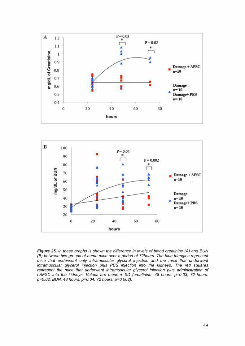
149
Figure 25. In these graphs is shown the difference in levels of blood creatinine (A) and BUN (B) between two groups of nu/nu mice over a period of 72hours. The blue triangles represent mice that underwent only intramuscular glycerol injection and the mice that underwent intramuscular glycerol injection plus PBS injection into the kidneys. The red squares represent the mice that underwent intramuscular glycerol injection plus administration of hAFSC into the kidneys. Values are mean ± SD (creatinine: 48 hours: p=0.03; 72 hours: p=0.02; BUN: 48 hours: p=0.04; 72 hours: p=0.002).
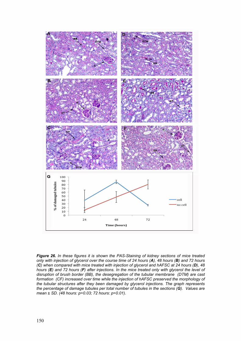
150
Figure 26. In these figures it is shown the PAS-Staining of kidney sections of mice treated only with injection of glycerol over the course time of 24 hours (A), 48 hours (B) and 72 hours (C) when compared with mice treated with injection of glycerol and hAFSC at 24 hours (D), 48 hours (E) and 72 hours (F) after injections. In the mice treated only with glycerol the level of disruption of brush border (BB), the desegregation of the tubular membrane (DTM) are cast formation (CF) increased over time while the injection of hAFSC preserved the morphology of the tubular structures after they been damaged by glycerol injections. The graph represents the percentage of damage tubules per total number of tubules in the sections (G). Values are mean ± SD. (48 hours: p=0.03; 72 hours: p=0.01).
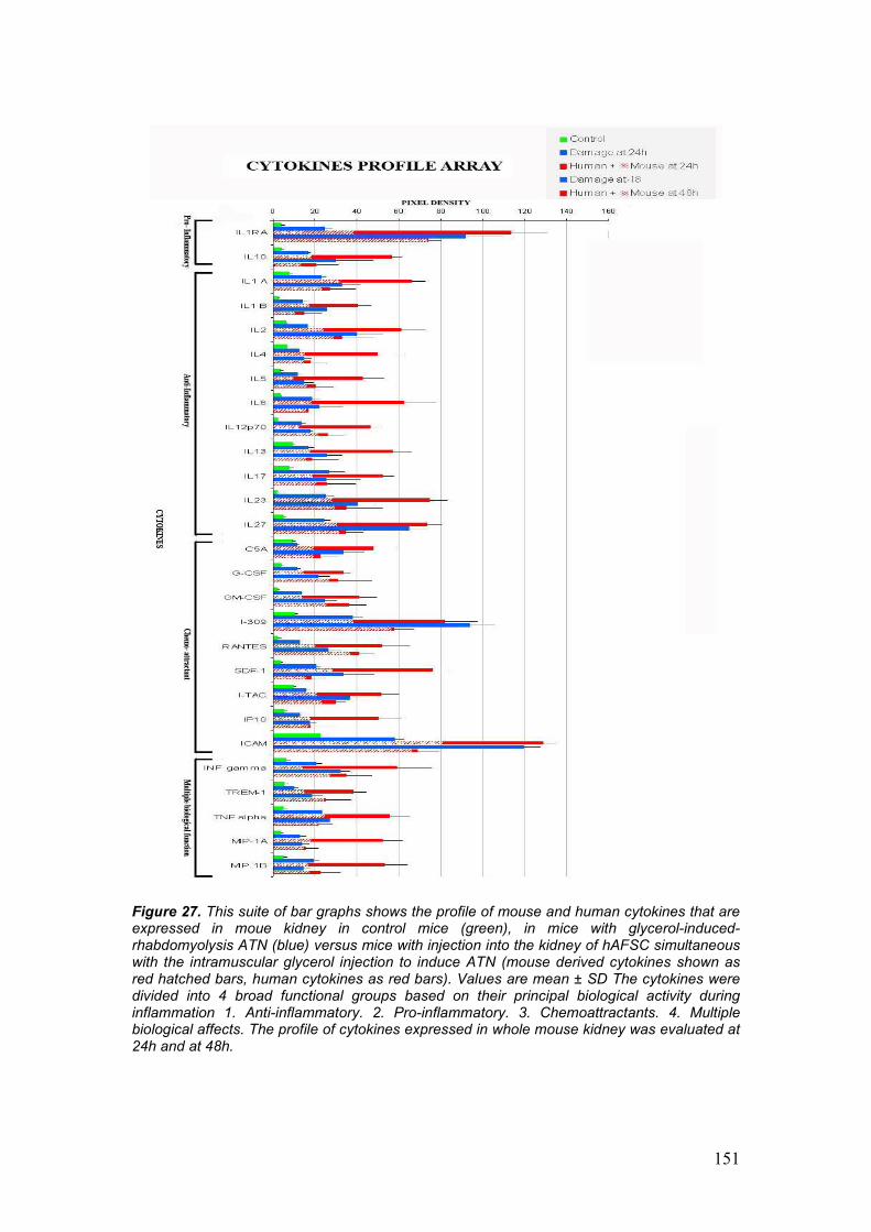
151
Figure 27. This suite of bar graphs shows the profile of mouse and human cytokines that are expressed in moue kidney in control mice (green), in mice with glycerol-induced-rhabdomyolysis ATN (blue) versus mice with injection into the kidney of hAFSC simultaneous with the intramuscular glycerol injection to induce ATN (mouse derived cytokines shown as red hatched bars, human cytokines as red bars). Values are mean ± SD The cytokines were divided into 4 broad functional groups based on their principal biological activity during inflammation 1. Anti-inflammatory. 2. Pro-inflammatory. 3. Chemoattractants. 4. Multiple biological affects. The profile of cytokines expressed in whole mouse kidney was evaluated at 24h and at 48h.
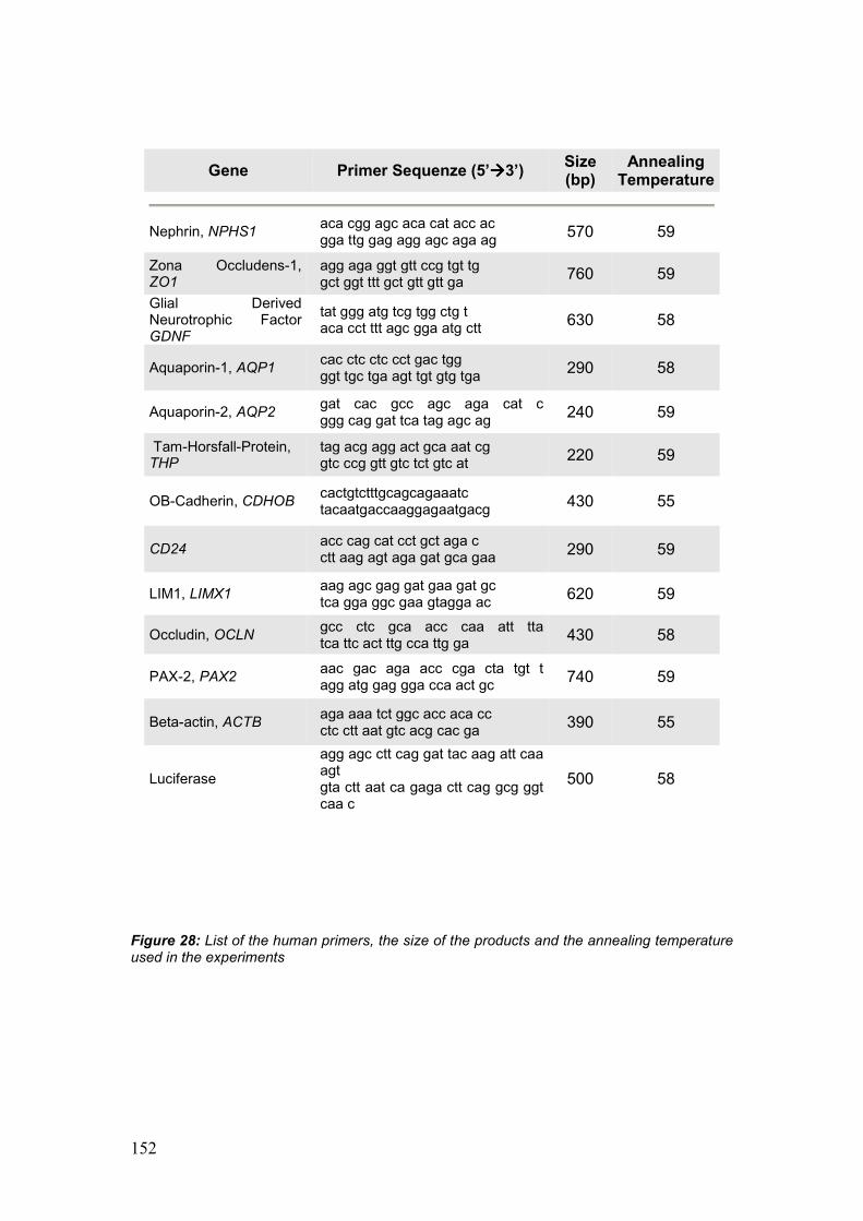
152
Figure 28: List of the human primers, the size of the products and the annealing temperature used in the experiments
Gene Primer Sequenze (5’àààà3’) Size (bp)
Annealing Temperature
Nephrin, NPHS1 aca cgg agc aca cat acc ac gga ttg gag agg agc aga ag 570 59
Zona Occludens-1, ZO1
agg aga ggt gtt ccg tgt tg gct ggt ttt gct gtt gtt ga 760 59
Glial Derived Neurotrophic Factor GDNF
tat ggg atg tcg tgg ctg t aca cct ttt agc gga atg ctt 630 58
Aquaporin-1, AQP1 cac ctc ctc cct gac tgg ggt tgc tga agt tgt gtg tga 290 58
Aquaporin-2, AQP2 gat cac gcc agc aga cat c ggg cag gat tca tag agc ag 240 59
Tam-Horsfall-Protein, THP
tag acg agg act gca aat cg gtc ccg gtt gtc tct gtc at 220 59
OB-Cadherin, CDHOB cactgtctttgcagcagaaatc tacaatgaccaaggagaatgacg 430 55
CD24 acc cag cat cct gct aga c ctt aag agt aga gat gca gaa 290 59
LIM1, LIMX1 aag agc gag gat gaa gat gc tca gga ggc gaa gtagga ac 620 59
Occludin, OCLN gcc ctc gca acc caa att tta tca ttc act ttg cca ttg ga 430 58
PAX-2, PAX2 aac gac aga acc cga cta tgt t agg atg gag gga cca act gc 740 59
Beta-actin, ACTB aga aaa tct ggc acc aca cc ctc ctt aat gtc acg cac ga 390 55
Luciferase
agg agc ctt cag gat tac aag att caa agt gta ctt aat ca gaga ctt cag gcg ggt caa c
500 58
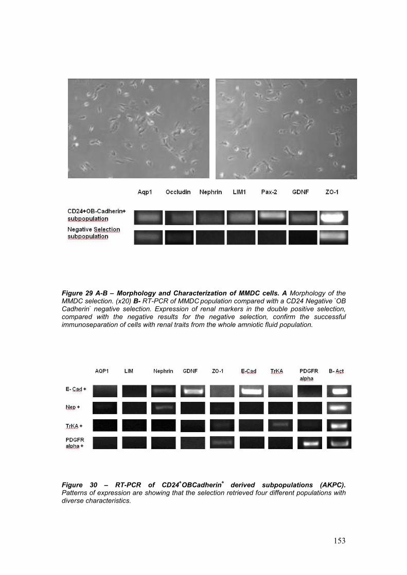
153
Figure 29 A-B – Morphology and Characterization of MMDC cells. A Morphology of the MMDC selection. (x20) B- RT-PCR of MMDC population compared with a CD24 Negative -OB Cadherin- negative selection. Expression of renal markers in the double positive selection, compared with the negative results for the negative selection, confirm the successful immunoseparation of cells with renal traits from the whole amniotic fluid population.
Figure 30 – RT-PCR of CD24+OBCadherin+ derived subpopulations (AKPC). Patterns of expression are showing that the selection retrieved four different populations with diverse characteristics.
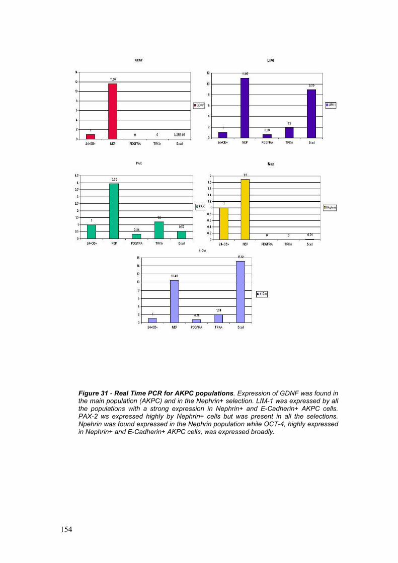
154
Figure 31 - Real Time PCR for AKPC populations. Expression of GDNF was found in the main population (AKPC) and in the Nephrin+ selection. LIM-1 was expressed by all the populations with a strong expression in Nephrin+ and E-Cadherin+ AKPC cells. PAX-2 ws expressed highly by Nephrin+ cells but was present in all the selections. Npehrin was found expressed in the Nephrin population while OCT-4, highly expressed in Nephrin+ and E-Cadherin+ AKPC cells, was expressed broadly.
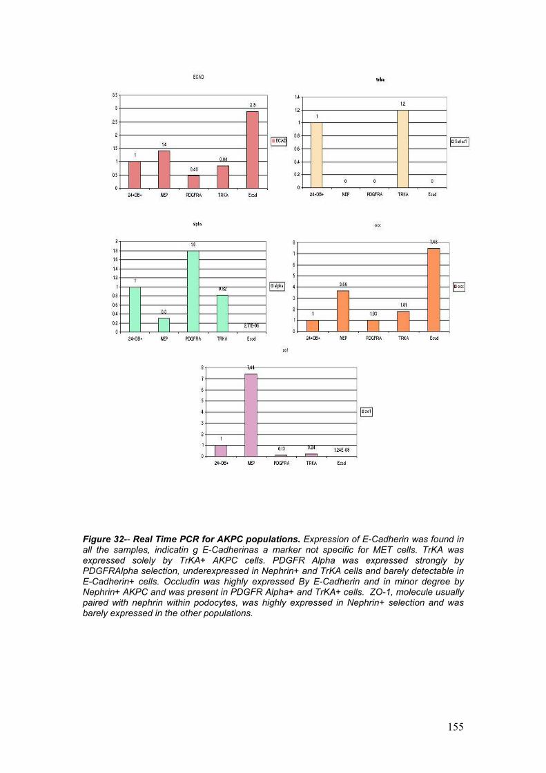
155
Figure 32-- Real Time PCR for AKPC populations. Expression of E-Cadherin was found in all the samples, indicatin g E-Cadherinas a marker not specific for MET cells. TrKA was expressed solely by TrKA+ AKPC cells. PDGFR Alpha was expressed strongly by PDGFRAlpha selection, underexpressed in Nephrin+ and TrKA cells and barely detectable in E-Cadherin+ cells. Occludin was highly expressed By E-Cadherin and in minor degree by Nephrin+ AKPC and was present in PDGFR Alpha+ and TrKA+ cells. ZO-1, molecule usually paired with nephrin within podocytes, was highly expressed in Nephrin+ selection and was barely expressed in the other populations.