UNIVERSITÀ DEGLI STUDI DI PADOVA - …paduaresearch.cab.unipd.it/5017/1/TESI_Valentina.pdf ·...
91
1 UNIVERSITÀ DEGLI STUDI DI PADOVA Dipartimento di Medicina Clinica e Sperimentale "G. Patrassi" Ematologia e Immunologia Clinica SCUOLA DI DOTTORATO DI RICERCA IN ONCOLOGIA E ONCOLOGIA CHIRURGICA XXIV CICLO ROLE OF NOCODAZOLE ON THE SURVIVAL OF CHRONIC LYMPHOCYTIC LEUKEMIA B CELLS Direttore della Scuola: Ch.ma Prof.ssa Paola Zanovello Supervisore: Dott.ssa Monica Facco Correlatore: Dott. Livio Trentin Dottoranda: Dott.ssa Valentina Trimarco
Transcript of UNIVERSITÀ DEGLI STUDI DI PADOVA - …paduaresearch.cab.unipd.it/5017/1/TESI_Valentina.pdf ·...

1
UNIVERSITÀ DEGLI STUDI DI PADOVA
Dipartimento di Medicina Clinica e Sperimentale "G. Patrassi"
Ematologia e Immunologia Clinica
SCUOLA DI DOTTORATO DI RICERCA IN
ONCOLOGIA E ONCOLOGIA CHIRURGICA
XXIV CICLO
ROLE OF NOCODAZOLE ON THE SURVIVAL OF
CHRONIC LYMPHOCYTIC LEUKEMIA B CELLS
Direttore della Scuola: Ch.ma Prof.ssa Paola Zanovello
Supervisore: Dott.ssa Monica Facco
Correlatore: Dott. Livio Trentin
Dottoranda: Dott.ssa Valentina Trimarco

2
3
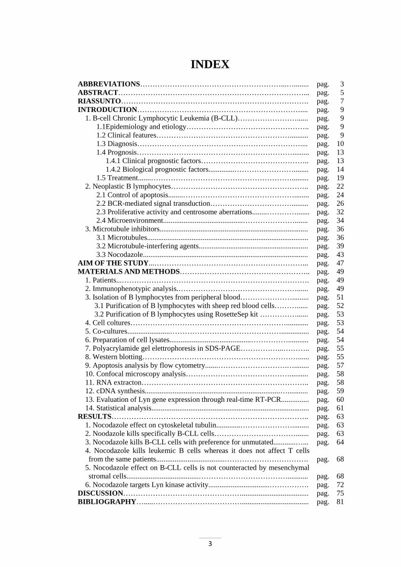
INDEX
ABBREVIATIONS …………………………………………………...…........ pag. 3 ABSTRACT…………………………………………………………………... pag. 5 RIASSUNTO…………………………………………………………………. pag. 7 INTRODUCTION ………………………………………………………….... pag. 9 1. B-cell Chronic Lymphocytic Leukemia (B-CLL)……………………...... pag. 9 1.1Epidemiology and etiology………………………………………….. pag. 9 1.2 Clinical features……………………………………………….......... pag. 9 1.3 Diagnosis………………………………………………………….... pag. 10 1.4 Prognosis………………………………………………………......... pag. 13 1.4.1 Clinical prognostic factors…………………………………….. pag. 13 1.4.2 Biological prognostic factors..............……………………........ pag. 14 1.5 Treatment.......…………………………………………………......... pag. 19 2. Neoplastic B lymphocytes……………………………………………….. pag. 22 2.1 Control of apoptosis........………………………………………........ pag. 24 2.2 BCR-mediated signal transduction……………………………......... pag. 26 2.3 Proliferative activity and centrosome aberrations........…………....... pag. 32 2.4 Microenvironment...........................................…………………....... 3. Microtubule inhibitors................................................................................ 3.1 Microtubules....................................................................................... 3.2 Microtubule-interfering agents........................................................... 3.3 Nocodazole.........................................................................................
pag. pag. pag. pag. pag.
34 36 36 39 43
AIM OF THE STUDY...……………………………………………………... pag. 47 MATERIALS AND METHODS…………………………………………….. pag. 49 1. Patients...…………………………………………………………………. pag. 49 2. Immunophenotypic analysis.…………………………………………...... pag. 49 3. Isolation of B lymphocytes from peripheral blood…………………......... pag. 51 3.1 Purification of B lymphocytes with sheep red blood cells………...... pag. 52 3.2 Purification of B lymphocytes using RosetteSep kit ……………...... pag. 53 4. Cell coltures………………………………………………………............ pag. 53 5. Co-cultures.......................………………………………………............... pag. 54 6. Preparation of cell lysates............................................……………........... pag. 54 7. Polyacrylamide gel elettrophoresis in SDS-PAGE……………...……….. pag. 55 8. Western blotting………………………………………………………...... pag. 55 9. Apoptosis analysis by flow cytometry.......……………………….…........ pag. 57 10. Confocal microscopy analysis…………………………………….......... pag. 58 11. RNA extracton………………………………………………………….. pag. 58 12. cDNA synthesis.........................................................................…........... 13. Evaluation of Lyn gene expression through real-time RT-PCR............... 14. Statistical analysis.....................................................................................
pag. pag. pag.
59 60 61
RESULTS…………………………………………………………………….. pag. 63 1. Nocodazole effect on cytoskeletal tubulin.............…………………......... pag. 63 2. Noodazole kills specifically B-CLL cells……………………………....... pag. 63 3. Nocodazole kills B-CLL cells with preference for unmutated............…... pag. 64 4. Nocodazole kills leukemic B cells whereas it does not affect T cells
from the same patients.....................................……………………………. pag.
68
5. Nocodazole effect on B-CLL cells is not counteracted by mesenchymal stromal cells.......................................………………………………...........
pag.
68
6. Nocodazole targets Lyn kinase activity.................................……………. pag. 72 DISCUSSION………………………………………….................................... pag. 75 BIBLIOGRAPHY ….....……………………………….................................... pag. 81

4
5
ABBREVIATIONS
Ab Antibody Ag Antigen APC Allophycocyanin ATM Ataxia Teleangectasia Mutated BAD Bcl-2 Associated Death promoter B-CLL B-cell Chronic Lymphocytic Leukemia Bcl-2 B-Cell lymphoma-2 BCR B-Cell Receptor B-PLL B-cell Prolymphocytic Leukemia cDNA complementary DNA CDR Complementarity Determining Region CD40L CD40 Ligand CMV Cytomegalovirus GC Germinal Centre CR Complete Remission DAG Diacylglycerol ECL Enhanced ChemiLuminescence EDTA Ethylenediaminetetraacetic Acid ERK Extracellular signal Regulated Kinase Fab Antigen binding fragment FBS Fetal Bovin Serum Fc Crystallizable fragment F/H Ficoll/Hypaque FITC Fluorescein isothiocyanate GDP Guanosine diphosphate GSK3 Glycogen Synthase Kinase 3 GTP Guanosine triphosphate HBV Hepatitis B virus HCV Hepatitis C virus HLA Human Leukocyte Antigen HS1 Hematopoietic lineage cell-Specific protein 1 Hsp90 Heat shock protein of 90kDa hTERT human Telomerase Reverse Transcriptase IFN-γ Interferon-γ Ig Immunoglobulin IgVH Immunoglobulin heavy chain variable regions IL Interleukin ITAM Immunoreceptor Tyrosine-based Activation Motif

6
ITIM Immunoreceptor Tyrosine-based Inhibitory Motif mAb monoclonal Antiboby MAP Microtubule-Associated Protein MAPK Mitogen Activated Protein Kinase Mcl-1 Mantle cell lymphoma-1 MHC Major Histocompatibility Complex MIIC MHC-class-II-peptide-loading-compartment MSC Mesenchymal Stromal Cell MTOC Microtubule Organizing Centre mTOR mammalian Target Of Rapamycin NF-kB Nuclear Factor-kappa B NHL Non-Hodgkin Lymphoma NLC Nurse-like Cell NK Natural Killer OS Overall Survival PARP Poli-ADP-Ribose Polymerase PBMC Peripheral Blood Mononuclear Cell PBS Phosphate Buffered Saline PE Phycoerythrin PFS Progression Free Survival PI3K Phosphatidylinositol 3-Kinase Plcγ2 Phospholipase Cγ2 PS Phosphatidylserine SD Standard Deviation SDF-1α Stromal Derived Factor-1α SDS-PAGE Sodium Dodecyl Sulphate/PolyAcrylamide Gel Electrophoresis SFKs Src Family Kinases SHM Somatic Hypermutation sIgM surface Immunoglobulin M Syk Spleen tyrosine kinase TC Tri-Color Th T helper lymphocyte WB Western blotting WBC White Blood Cell ZAP-70 Zeta-Associated Protein of 70kDa
7
ABSTRACT
B-cell Chronic Lymphocytic Leukemia (B-CLL) is the most common
leukemia in adults and is characterized by the accumulation of clonal
CD19+/CD5+/CD23+ B lymphocytes, due to uncontrolled growth and resistance
to apoptosis. Leukemic cells from B-CLL show reduced crosslink with specific
molecules and high susceptibility to microtubule disrupting drugs, which suggest
cytoskeletal alterations.
Microtubules play a crucial role in the vital functions of neoplastic cells,
including mitosis, motility and cell-cell contact, and for this reason they became
an important target in cancer therapies. In particular, tubulin, a cytoskeletal
member, is the target of specific drugs, named microtubule inhibitors. Among
these inhibitors, nocodazole induces tubulin depolimerization, mitotic process
blocking and shows an apoptotic effect in B leukemic cells.
The aim of this study was to define the effects of nocodazole on B-CLL
cells.
First of all, we verified nocodazole capability to favour the
depolymerization of tubulin cytoskeleton in different cell types. In addition, we
tested nocodazole-induced apoptosis in normal and leukemic B cells, in cell lines
(Jurkat, Raji, and K562), in mesenchymal stromal cells (MSCs), and in T
lymphocytes of B-CLL patients. Our data pointed out the high specificity of
nocodazole for B-CLL cell apoptosis (leukemic cells: 57±25% vs normal B cells:
98±6%, p<0.0001; data are expressed as mean±standard deviation (SD) of
percentage of viable cells after treatment with nocodazole) and the absence of
toxicity to others cell types.
Growing evidence suggests that the marrow microenvironment, where
MSCs are present, protects B-CLL cells from conventional anti-neoplastic drugs.
The cultures of neoplastic B cells with MSCs and nocodazole demonstrated that
nocodazole is able to overcome MSC protective effect, even after survival signal
supplemental, such as CD40L or plasma from the same patients.
The action mechanism of nocodazole in B-CLL cells is still under
investigation. However, we observed that nocodazole is able to turn off the

8
increased basal tyrosine phosphorylation of leukemic cells mediated by Src-kinase
Lyn through the down-modulation of Lyn active site. Since the specific inhibition
of Lyn induces B-CLL cells apoptosis, this linking will be further investigated.
The results obtained in this study suggest a future role of nocodazole as a
possible agent for treatment of B-CLL, for its extreme selectivity, the absence of
toxicity and its ability to counteract the protective effect provided by marrow
microenvironment.
9
RIASSUNTO
La Leucemia Linfatica Cronica di tipo B (LLC-B) è la forma più comune
di leucemia nell’adulto ed è caratterizzata dall’accumulo clonale di piccoli
linfociti B CD19+/CD5+/CD23+, dovuto sia ad una crescita incontrollata che ad
una resistenza all’apoptosi. Le cellule leucemiche di LLC-B presentano inoltre
alcune anomalie, come ridotta capacità di legare specifiche molecole e
suscettibilità a farmaci che distruggono i microtubuli, che indicano la presenza di
alterazioni a livello citoscheletrico.
Il ruolo cruciale che i microtubuli rivestono nelle funzioni vitali delle
cellule neoplastiche, quali mitosi, motilità e contatti cellula-cellula, li ha resi un
importante target nelle terapie anti-tumorali. In particolar modo la tubulina,
componente dei microtubuli, è il bersaglio di una categoria specifica di farmaci
anti-tumorali, gli inibitori dei microtubuli; di questa famiglia fa parte anche il
nocodazolo, un agente sintetico che induce la depolimerizzazione della tubulina,
arresta il processo mitotico ed ha una peculiare specificità nell’indurre l’apoptosi
nelle cellule B di LLC-B.
Sulla base di queste considerazioni, abbiamo voluto approfondire gli effetti
ed il meccanismo d’azione del nocodazolo sulle cellule di LLC-B.
Dopo aver verificato che il nocodazolo sia effettivamente responsabile
della depolimerizzazione dei filamenti di tubulina citoscheletrica in numerosi tipi
cellulari, abbiamo valutato l’effetto apoptotico indotto dal nocodazolo in cellule B
normali e di LLC-B, in linee cellulari (Jurkat, Raji e K562), in cellule stromali
mesenchimali (MSC) e nei linfociti T residui di pazienti affetti da LLC-B. I
risultati ottenuti evidenziano l’estrema selettività del nocodazolo nell’indurre
l’apoptosi nelle sole cellule B di LLC-B (linfociti B di LLC-B: 57±25% vs B
normali: 98±6%, p<0,0001; dati espressi come media±deviazione standard (DS)
della percentuale di cellule vive dopo trattamento con nocodazolo) e l’assenza di
tossicità nei confronti delle altre popolazioni cellulari prese in esame.
Studi recenti suggeriscono che il microambiente midollare, in cui si
trovano anche le MSC, sia in grado di proteggere le cellule leucemiche dall’azione

10
dei farmaci chemioterapici convenzionali. La co-coltura di MSC e cellule B di
LLC-B in presenza di nocodazolo ha dimostrato che tale inibitore è in grado di
annullare l'effetto protettivo esercitato dalle MSC, nonostante la presenza di
segnali di sopravvivenza quali CD40L o plasma ricavato dagli stessi pazienti.
I meccanismi d’azione del nocodazolo rimangono ancora da chiarire,
tuttavia abbiamo osservato come nelle cellule leucemiche di LLC-B il nocodazolo
sia in grado di ridurre l’aumentata fosforilazione tirosinica basale mediata dalla
Src-chinasi Lyn, mediante down-regolazione del sito attivatorio di Lyn. Dal
momento che abbiamo dimostrato che l’inibizione specifica di Lyn induce
apoptosi nelle cellule di LLC-B, questi primi risultati diventano rilevanti e
dovranno essere ulteriormente indagati.
In conclusione, i risultati ottenuti in questo studio hanno evidenziato
l’estrema selettività del nocodazolo nell’indurre apoptosi nei linfociti B leucemici,
l’assenza di tossicità in vitro e la capacità di contrastare l’effetto protettivo fornito
dal microambiente midollare, suggerendo un futuro ruolo di questa sostanza quale
possibile agente terapeutico per la cura della LLC-B.

11
INTRODUCTION
1. B-cell Chronic Lymphocytic Leukemia (B-CLL)
1.1 Epidemiology and etiology
B-cell Chronic Lymphocytic Leukemia (B-CLL) is a lymphoproliferative
disorder characterized by the accumulation of long-lived monoclonal B cells in
the bone marrow, lymph node and blood. B-CLL lymphocytes show a
CD19+/CD5+/CD23+ membrane phenotype and are blocked in G0/G1 phase of
the cell cycle1. B-CLL is the most common adult leukemia in the Western world
and is more prevalent in men than in women with a male to female ratio of 1.5-
2:1. The incidence rates between men and women are 5.6 (in men) and 4.3 (in
women) cases per 100,000 inhabitants per year, respectively. In Europe, these
incidence rates are 5.87 and 4.01 cases per 100,000 population per year,
respectively. B-CLL is considered to be mainly a disease of the elderly, with a
mean age at diagnosis of 70 years; however, it is not unusual to diagnose it in
younger individuals from 30 years of age. The incidence increases rapidly with
increasing age2.
The etiology is still unknown; the exposure to common carcinogens does
not seem to be associated with the disease progression. More studies are in
progress to assess a potential relation among B-CLL onset, inflammation, and
autoimmune conditions3,4. A familiarity of this pathology is well documented in
the 5-10% of cases. Moreover, it has been highlighted the phenomenon of the
anticipation in which inherited disease is diagnosed at an earlier age and in a more
aggressive form in the later generations of a family5.
1.2 Clinical features
Clinical course and survival of B-CLL patients are quite variable: some
patients remain asymptomatic without any treatment, while others present an

12
aggressive outcome that is difficult to control with chemotherapy. B-CLL is often
determined with routine laboratory tests; in other cases, the pathology occurs with
asthenia, weight loss, fever, lymphadenopathy, splenomegaly, and hepatomegaly.
Some patients could show autoimmune phenomenons, such as hemolytic anemia
(11% of cases) or autoimmune thrombocytopenia (2% cases) that are typically
present in advanced and multi-treated disease. The typical B-CLL
hypogammaglobulinemia could induce immunodeficiency and high mortality for
infections6,7.
Although the causes of death are often attributed to the underlying disease,
in some cases progressing syndromes with a poor prognosis could occur: one of
these is the Richter's syndrome in which B-CLL changes into a fast-growing
diffuse large B cell lymphoma. Another evolution could be the B-cell
prolymphocytic leukemia (B-PLL) that is more aggressive and characterized by
malignant B cells larger than average. The occurrence of acute lymphoblastic
leukemia is very rare, while acute myeloid leukemia, such as non-hematological
disease, could be correlated with B-CLL immunological deficit and
chemotherapy8.
1.3 Diagnosis
It is crucial to verify that the patient is really suffering from B-CLL and
not by another lymphoproliferative disease that can masquerade as a B-CLL, such
as hairy cell leukemia, or leukemic manifestations of mantle cell lymphoma,
marginal zone lymphoma, splenic marginal zone lymphoma with circulating
villous lymphocytes, or follicular lymphoma. To achieve this, it is essential to
evaluate the blood count, blood smear, and the immune phenotype of the
circulating lymphoid cells. The National Cancer Institute diagnostic criteria
include9:
1) the presence of at least 5x10⁹ B lymphocytes/L (5,000/µL) in the peripheral
blood. Leukemic cells found in the blood smear are characteristically small,
mature lymphocytes with a narrow border of cytoplasm and a dense nucleus,
lacking discernible nucleoli, with partially aggregated chromatin. These cells may
be found mixed with larger or atypical cells, broken cells, or prolymphocytes,
13
which may comprise up to 55% of the blood lymphocytes. Finding a higher
percentage of prolymphocytes would favour a diagnosis of B-PLL. Gumprecht
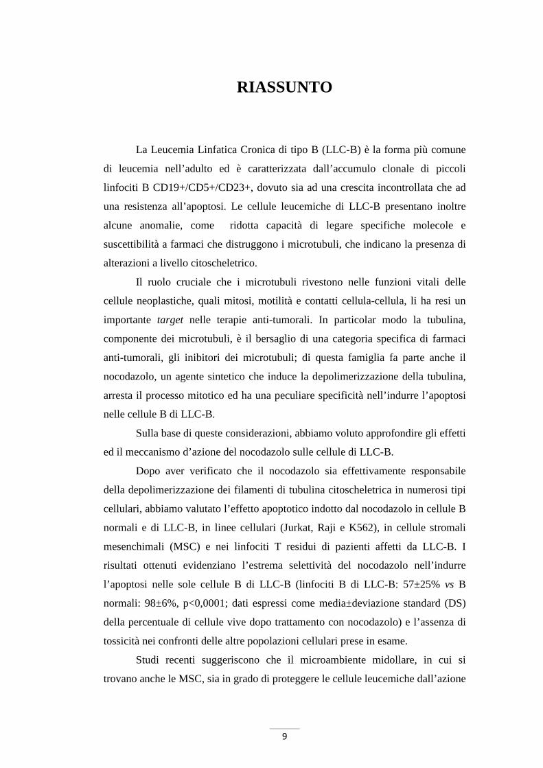
shadows, found as cell debris, are other morphological features found in B-CLL
(figure 1);
Figure 1. Peripheral blood smear of a B-CLL patient. Arrows indicate Gumprecht shadows typical of B- CLL.
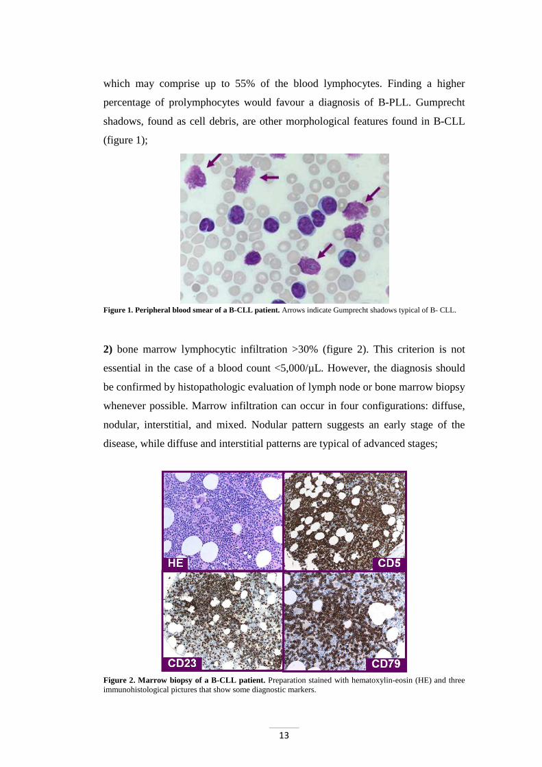
2) bone marrow lymphocytic infiltration >30% (figure 2). This criterion is not
essential in the case of a blood count <5,000/µL. However, the diagnosis should
be confirmed by histopathologic evaluation of lymph node or bone marrow biopsy
whenever possible. Marrow infiltration can occur in four configurations: diffuse,
nodular, interstitial, and mixed. Nodular pattern suggests an early stage of the
disease, while diffuse and interstitial patterns are typical of advanced stages;
Figure 2. Marrow biopsy of a B-CLL patient. Preparation stained with hematoxylin-eosin (HE) and three immunohistological pictures that show some diagnostic markers.

14
3) immunophenotype analysis. B-CLL phenotype is characterized by three elements: a) the expression of a unique type of immunoglobulin light chains (κ or λ);
b) the co-expression of the T-cell antigen CD5 and the B-cell surface
antigens CD19, CD20, and CD23; CD23 is of particular importance in the
differential diagnosis with mantle cell lymphoma (CD5+ but CD23-);
c) low levels of CD79b and surface immunoglobulin (sIg) that in B-CLL
appear to be mainly IgM followed by IgD, IgG, and IgA; it is not unusual to find
an IgM and IgD co-expression (figure 3).
In B-CLL, T lymphocytes, in particular CD8+ T cells, are often increased,
with a reduced CD4/CD8 ratio. They show activation markers such as CD25 and
HLA-DR. Natural-Killer (NK) cells (CD16+/CD56+) are present in high levels.
Several analyses are performed to confirm the diagnosis and to prevent
complications: serum protein electrophoresis, Ig dosage, Coombs' test, and
analysis of renal and liver function. Before starting an immunotherapy, it is
important to assess the absence of viral infection (HBV, HCV, CMV)10.
Figure 3. Cytograms of a representative case of B-CLL. B lymphocytes analyzed (CD19+) are positive to CD5 (panel A) and to CD23 (panel C), express one type of immunoglobuline light chain (λ, panel B), and surface IgM (sIgM), low density (panel D).
CD19 CD19
CD19 Kappa
CD
23 IgM
CD
5
Lam
bda
CD19 CD19
CD19 Kappa
CD
23 IgM
CD
5
Lam
bda
A B
C DCD19 CD19
CD19 Kappa
CD
23 IgM
CD
5
Lam
bda
CD19 CD19
CD19 Kappa
CD
23 IgM
CD
5
Lam
bda
A B
C D
A B
C D
15
1.4 Prognosis
Since it is difficult to predict the course of the disease at the time of
diagnosis and the value of an early treatment is uncertain, therapy is currently
recommended only for patients with a disease progressive, symptomatic, or
both11. During the years, different parameters or prognostic factors were proposed
to define the clinical course of B-CLL patients.
1.4.1 Clinical prognostic factors
1) Clinical staging: There are two widely accepted staging methods in both
patient care and clinical trials: the Rai and the Binet system. The original Rai
classification was modified to reduce the number of prognostic groups from 5 to
3. Both systems now describe 3 major subgroups with different clinical outcomes.
These 2 staging systems are simple, inexpensive, and can be applied by physicians
worldwide. Both rely exclusively on physical examination and standard laboratory
tests and do not require ultrasound, computed tomography (CT), or magnetic
resonance imaging9.
The Rai system is so developed:
− low-risk disease (stage 0): absolute lymphocytosis >15,000/µl and
marrow lymphocytosis >40%;
− intermediate-risk disease (stage I or II): lymphocytosis, enlarged nodes
in any site, and splenomegaly and/or hepatomegaly (lymph nodes being
palpable or not);
− high-risk disease (stage III or IV): disease-related anemia (Hb<110g/L)
or thrombocytopenia (as defined by a platelet count <100x109/L);
The Binet system is based on the number of involved areas, as defined by
the presence of lymph nodes with a diameter greater than 1 cm or organomegaly,
and the presence of anemia or thrombocytopenia. It is subdivided into:
− Stage A. Hb≥100g/L (10g/dL), platelets ≥100x109/L, and up to 2 lymph
node areas involved.
16
− Stage B. Hb≥100g/L, platelets≥100x109/L, and lymphoadenopathy greater
than that defined for stage A (i.e., 3 or more areas of nodal or organ
enlargement).
− Stage C. All patients who have Hb<100g/L and/or a platelet
count<100x109/L, irrespective of lymphoadenopathy.
2) Lymphocyte doubling time: it is less than 12 months and it is associated with
a worse clinical course.
3) Bone marrow infiltration: a diffuse infiltration pattern correlates with a bad
prognosis12.
1.4.2 Biological prognostic factors
The less recent biological prognostic factors are correlated with the
expansion of the leukemic clone; they thus become indicative only when the
disease is worsening. Their utility is limited because it is not possible to program
the therapeutic strategy basing on the progression risk of patients. The biological
prognostic factors comprehend:
1) Prolymphocyte (PL) percentage: if it is less than or equal to 10% (typical B-
CLL) the probability of PL leukemia evolution is very low; if the percentage is
between 11% and 55% there is an intermediate risk of B-CLL/PL leukemia, and if
it is greater than 55% the transformation in PL leukemia may occur13.
2) β2 microglobulin: this parameter is inversely correlated with the survival. It
is related with the lymphocyte doubling time so that an increase in β2
microglobulin indicates an high neoplastic cell proliferation13.
3) Thymidine kinase (TK) level: it has been shown that elevated serum
thymidine kinase (s-TK) levels predict disease progression in B-CLL. Patients
with s-TK values greater than 7.1U/L have a median progression free survival
(PFS) of 8 months, whereas patients with s-TK values ≤7.1U/L expect a much
longer PFS14.
4) Soluble CD23 value: serum CD23 level provides significant additional
prognostic information in terms of overall survival (OS) in B-CLL. Among early

17
stage patients, sCD23 determination, at diagnosis and during the course of the
disease, may help to the early identification of patients who will rapidly progress
to upper stages. Functions ascribed to sCD23 include prevention of germinal
center (GC) B cells from their apoptosis, proliferation of myeloid precursor cells,
and, more recently, costimulation of interferon-γ (IFN-γ) production by T cells
and triggering of monokine release by monocytes15.
The progressive discoveries on B-CLL pathogenesis have identified new
prognostic markers that can better determine the clinical course. They describe
biological characteristic of leukemic clone that are crucial to evaluate its
proliferation and invasion capability. The study of these markers is performed by
flow cytometry, cytogenetic and molecular biology techniques. The main markers
are:
1) Somatic Hypermutations (SHM) of the Ig heavy chain variable region (VH)
genes. Based on the numbers of somatic mutations detected in these genes, the
cases were divided into 2 categories: "unmutated" (SHM-) or "mutated" (SHM+)
(figure 4). Conventionally, patients with <2% differences from the most similar
germline gene in both the expressed VH and VL genes were define unmutated;
mutated cases were defined as those in which the B-CLL cells displayed ≥2%
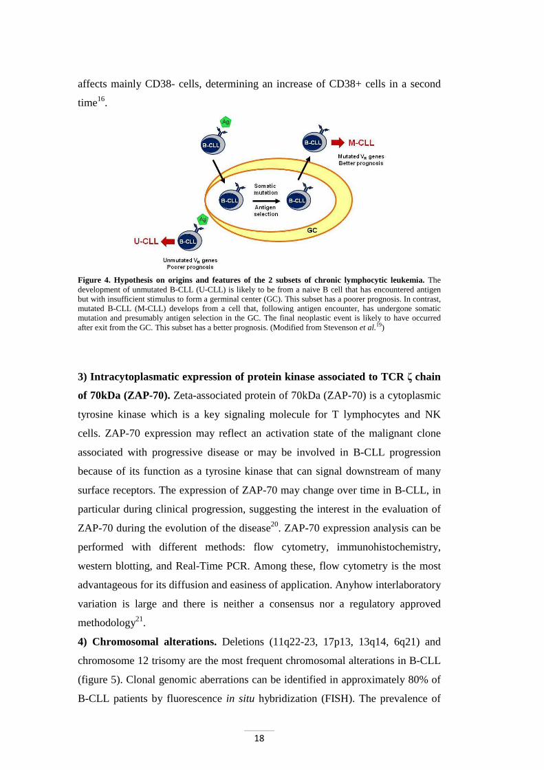
differences in either the expressed VH or VL gene16. In addition, the stereotyped
VH3.21 gene is an unfavorable prognostic marker independent of the IgVH
mutational status. However, this result has been highlighted especially in patients
living in Northern Europe, while it was not confirmed in Mediterranean
countries9.
2) CD38 expression. CD38 is a transmembrane glycoprotein expressed on the
surface of cells in a significant percentage of patients with B-CLL. A previous
study suggested that CD38 expression has prognostic value in B-CLL. Cases with
CD38+ B cells >30% show a bad prognosis. Indeed, the cut-off to discriminate
CD38+ to CD38- is not unanimously defined: some authors place it at 20%17 or to
7%18 of CD38+ B cells. CD38 has an independent prognostic value. Moreover,
variability is the limit of this marker, in particular after treatment: chemotherapy
18
affects mainly CD38- cells, determining an increase of CD38+ cells in a second
time16.
Figure 4. Hypothesis on origins and features of the 2 subsets of chronic lymphocytic leukemia. The development of unmutated B-CLL (U-CLL) is likely to be from a naive B cell that has encountered antigen but with insufficient stimulus to form a germinal center (GC). This subset has a poorer prognosis. In contrast, mutated B-CLL (M-CLL) develops from a cell that, following antigen encounter, has undergone somatic mutation and presumably antigen selection in the GC. The final neoplastic event is likely to have occurred after exit from the GC. This subset has a better prognosis. (Modified from Stevenson et al.19)
3) Intracytoplasmatic expression of protein kinase associated to TCR ζ chain
of 70kDa (ZAP-70). Zeta-associated protein of 70kDa (ZAP-70) is a cytoplasmic
tyrosine kinase which is a key signaling molecule for T lymphocytes and NK
cells. ZAP-70 expression may reflect an activation state of the malignant clone
associated with progressive disease or may be involved in B-CLL progression
because of its function as a tyrosine kinase that can signal downstream of many
surface receptors. The expression of ZAP-70 may change over time in B-CLL, in
particular during clinical progression, suggesting the interest in the evaluation of
ZAP-70 during the evolution of the disease20. ZAP-70 expression analysis can be
performed with different methods: flow cytometry, immunohistochemistry,
western blotting, and Real-Time PCR. Among these, flow cytometry is the most
advantageous for its diffusion and easiness of application. Anyhow interlaboratory
variation is large and there is neither a consensus nor a regulatory approved
methodology21.
4) Chromosomal alterations. Deletions (11q22-23, 17p13, 13q14, 6q21) and
chromosome 12 trisomy are the most frequent chromosomal alterations in B-CLL
(figure 5). Clonal genomic aberrations can be identified in approximately 80% of
B-CLL patients by fluorescence in situ hybridization (FISH). The prevalence of

19
the most common alterations was estimated in a German multicentre study22: 13q-
55%, 11q- 18%, +12 16%, 17p- 7%, 6q- 7%. 17p- and 11q- are independent
prognostic factors identifying subgroups of patients with rapid disease progression
and short survival times in multivariate analysis, whereas 13q- as a single
aberration is associated with favorable outcome. In addition, 17p- abnormalities
and TP53 mutations have been associated with treatment failure. The presence of
chromosome alterations with high risk justifies the use of more aggressive
treatment23. Chromosome alterations are independent from IgVH mutational status
though is evident a more frequency of 11q- and 17p- in unmutated and 13q- in
mutated cases. These data show that analysis conducted by cytogenetics could be
used as further risk stratification instrument together with the other prognostic
factors24.
Figure 5. Probability of survival among patients in the most common chromosomal alterations. The median survival times for the groups with 17p deletion, 11q deletion, 12q trisomy, normal karyotype, and 13q deletion as single abnormality were 32, 79, 114, 111, and 133 months, respectively25. 5) Telomerase expression and telomere length in B-CLL. Activation of
telomerase reverse transcriptase (hTERT) is essential for unlimited cell growth
and plays a critical role in tumorigenesis26. Recently, the levels of telomerase
activity (TA) and/or hTERT expression were related to clinical aggressiveness
and prognosis in a variety of malignancies, including B-CLL.

20
During the last year, a study analyzed for the first time both hTERT levels
and telomere length, and related them with IgVH mutational status and
chromosomal aberrations in a large cohort of B-CLL patients. Although the main
function of hTERT is to stabilize telomere length, an inverse relationship between
hTERT levels and telomere lengths was found in B-CLL cases; B-CLL cases with
high telomerase levels and short telomeres were frequently characterized by an
unmutated IgVH status and high-risk chromosomal aberrations. Conversely, B-
CLL cases with low telomerase levels and long telomeres were associated with a
mutated IgVH status and low-risk abnormalities. Moreover, the unmutated IgVH B-
CLL cases with short telomeres had higher levels of hTERT than the mutated IgVH
cases with long telomeres. Unmutated IgVH status, 11q- or 17p- and +12 aberrations,
high levels of hTERT, and low telomere length were all associated with a poor
clinical outcome. Finding that the 13q-, characterized by low levels of hTERT, was
associated with a prognosis even better than the normal group, supports the notion
that hTERT may contribute to lymphomagenesis beyond just preservation of telomere
length (figure 6). The evaluation of hTERT and telomere length might help the
clinician in the management of B-CLL patients with mutated IgVH and/or no high-
risk chromosomal aberrations since cases with high hTERT/short telomere B-CLL
will progress more rapidly and might require therapy earlier than those with low
hTERT/long telomeres27.
Recently, several microRNAs (miRNAs) have been proposed as
prognostic markers for B-CLL and other human cancers. MicroRNAs are short
(20-22 nucleotides in humans), endogenous non-coding single-strand RNA
molecules that regulate gene expression via translational repression or transcript
degradation and gene silencing. In particular, it was demonstrated that in B-CLL
patients with an overexpression of the anti-apoptotic protein Bcl-2, miR-15a and
miR-16-1 (localized in 13q14) are deleted or downregulated. Moreover, it was
demonstrated that when B-CLL cells were transfected with miR-15a and miR-16-
1, Bcl-2 is blocked and the normal apoptotic process is restored. Current studies
aim to detect which miRNAs are involved in B-CLL in order to discover new
prognostic factors as well as to develop new targeted gene therapy28.

21
Figure 6. Curves of treatment-free survival. Time from diagnosis to first treatment (TTFT) according to IgVH mutational status, chromosomal categories, and hTERT level/telomere length profile27.
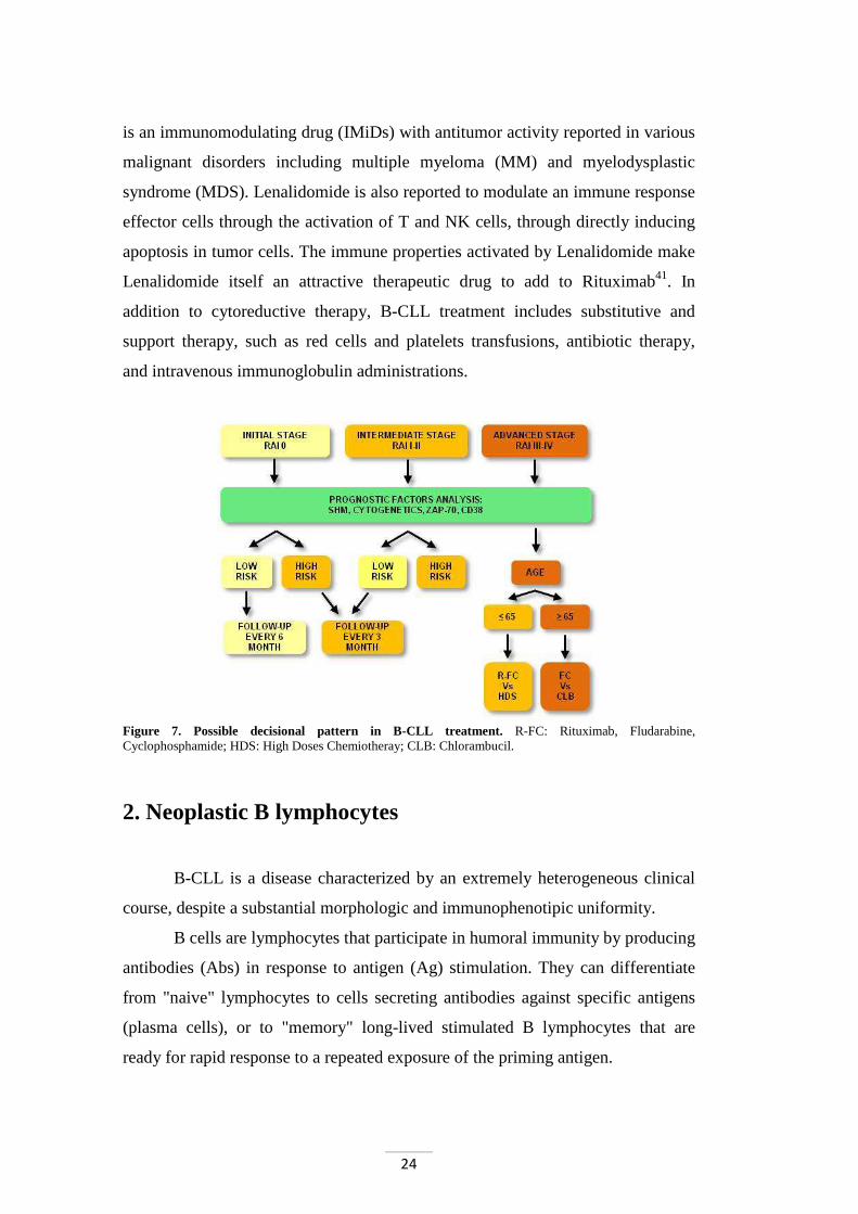
1.5 Treatment
Criteria for initiating treatment depend on clinic symptoms, stage and
disease activity. In general practice, newly diagnosed patients with asymptomatic
early-stage disease (Rai 0, Binet A) should be monitored without therapy unless
they have evidence of progression. On the contrary, patients at intermediate (I and
II) and high risk stages (III and IV), according to the modified Rai classification
or at Binet stage B or C, usually benefit from the start of a treatment; some of
these patients (in particular Rai intermediate risk or Binet stage B) can be
monitored without therapy until they have evidence for progressive or
symptomatic disease9.
Therapeutic possibilities comprehend drugs with different mechanisms of
action, up to stem cells auto/allotransplantation. Since B-CLL is an incurable
disease, current therapy is intended control the expansion of the neoplastic clone.
The choice of the therapy is linked to patient age and general conditions. In older
patients (>65) primary treatment consist of Chlorambucil (10mg/die for 1-4
weeks) associated with Prednisone (25mg/die for 1-4 weeks), while in other cases
the therapy is based on Fludarabine, alone or in association with
Cyclophosphamide. Chlorambucil treatment induces a response in 70% of cases,
but only 10% shows a complete response (CR) and has no effect on survival;

22
these considerations make it suitable for palliative treatment. Steroids have not
demonstrated a significant effect on survival, while their side effects, such as
opportunistic infections, are well known. However, they are useful to contain
autoimmune complications.
During the past years, basing on the experience on other lymphomas,
therapeutic combinations such as CHOP (Cyclophosphamide, Vincristine,
Prednisone, and Adriamycin) or COP (Cyclophosphamide, Vincristine, and
Prednisone) were tested; although they display a high frequency of CR in respect
of an increased toxicity, they did not improve survival29.
From the mid of '90 years, the first line treatment for B-CLL was the use
of purine analogous. This class of drugs comprehends Pentostatin, an adenosine
deaminase inhibitor, Clabridine, Fludarabine, and DNA-polimerase inhibitors.
The more effective in B-CLL treatment is Fludarabine (25-30mg/die for 5 days, 3-
6 month); 80% of cases show global response, and 30% a CR. Moreover, the time
of remission is greater than the one obtained with Chlorambucil plus Prednisone30
(figure 7).
Despite the positive overall response, Fludarabine and purine analogous
are not so effective in improving the survival rate than Chlorambucil or alkylating
agents31-33. The US Intergroup Trial has recently demonstrated that the
combination of Cyclophosphamide and Fludarabine, compared to Fludarabine
alone, gives higher overall responses (74.3% vs 59.5%), CR (23.4% vs 4.6%), and
PFS (31.6 vs 19.2 months). Conversely, the combination of the two drugs resulted
in a greater bone marrow toxicity, neutropenia, anemia, and thrombocytopenia
with infectious complications34.
Monoclonal antibodies provided a significant advantage in the treatment of
hematological malignancies. CD20, a surface membrane phosphoprotein, has
become the preferred target of immunotherapy. The chimeric mouse anti-human
monoclonal antibody Rituximab (IDEC-C2B8) is specific for the CD20 antigen
and has been used in clinical trials to treat patients with Non-Hodgkin Lymphoma
(NHL). Preclinical studies identified the ability of Rituximab to increase the
effectiveness of cytotoxic drugs in resistant cell lines, blocking the anti-apoptotic
signaling. The combination of Rituximab with other drugs results in a synergistic
cytotoxicity and apoptosis35; Fludarabine, in fact, downmodulate the complement-

23
resistance proteins CD46, CD55, and CD59 on leukemia cells, thereby potentially
making cells more vulnerable to Rituximab-induced complement-mediated lysis36.
The combination of Rituximab with Fludarabine and Cyclophosphamide was
evaluated both as initial therapy in those cases of recurrent or refractory to prior
therapies. In previously untreated patients, PFS is greater than four years in about
2/3 of cases37. Rituximab side effects are essentially related to the intravenous
infusion of cytokines (fever, chills, nausea, and hypotension). Alemtuzumab
(Campath-1H) is a humanized anti-CD52 antibody; CD52 is expressed at high
levels on most of normal and malignant mature lymphocytes but not on
hematopoietic stem cells. It can be administered subcutaneously and it is very
effective in inducing remission in relapsing B-CLL patients38. However,
Campath-1H may cause a marked immunosuppression that require prophylactic
therapy for Pneumocistis carinii, VZV, and CMV infections. The association with
Fludarabine and Cyclophosphamide is burdened by significant toxicity to bone
marrow, so precautions are necessary during Alemtuzumab administration.
The expression of the anti-apoptotic protein Bcl-2 is associated with the
pathogenesis of B-CLL. As negative regulator of the intrinsic apoptotic pathway,
overexpression of Bcl-2 confers chemoresistance in a number of hematologic
cancers and solid tumors39. Although protein levels vary among cells and patients,
Bcl-2 is expressed in virtually all patients with B-CLL and Bcl-2 upregulation
plays a critical role in this disease. Deletion of miRNA regulators of Bcl-2
expression is frequently found in B-CLL cells also in association with Bcl-2
upregulation. Oblimersen, an antisense oligonucleotide that binds Bcl-2 mRNA,
induces enzymatic cleavage of the mRNA preventing protein translation39.
Allogeneic and autologous stem cell transplantation (SCT) are increasingly
considered for treatment of B-CLL patients. With appropriate supportive care, it is
safe and can induce a long-lasting clinical and molecular remissions. Feasibility of
autologous SCT appears to be best early during the course of the disease, but there
is only limited hope that autotransplantation can cure the disease40.
Cytokines support from the malignant microenvironment prolong B-CLL
cell survival, immune evasion, and resistance to therapy. Interrupting these
prosurvival effects from the malignant microenvironment is a potential new
approach in treating patients with B-CLL. Lenalidomide, a thalidomide analogue,
24
is an immunomodulating drug (IMiDs) with antitumor activity reported in various
malignant disorders including multiple myeloma (MM) and myelodysplastic
syndrome (MDS). Lenalidomide is also reported to modulate an immune response
effector cells through the activation of T and NK cells, through directly inducing
apoptosis in tumor cells. The immune properties activated by Lenalidomide make
Lenalidomide itself an attractive therapeutic drug to add to Rituximab41. In
addition to cytoreductive therapy, B-CLL treatment includes substitutive and
support therapy, such as red cells and platelets transfusions, antibiotic therapy,
and intravenous immunoglobulin administrations.
Figure 7. Possible decisional pattern in B-CLL treatment. R-FC: Rituximab, Fludarabine, Cyclophosphamide; HDS: High Doses Chemiotheray; CLB: Chlorambucil.
2. Neoplastic B lymphocytes
B-CLL is a disease characterized by an extremely heterogeneous clinical
course, despite a substantial morphologic and immunophenotipic uniformity.
B cells are lymphocytes that participate in humoral immunity by producing
antibodies (Abs) in response to antigen (Ag) stimulation. They can differentiate
from "naive" lymphocytes to cells secreting antibodies against specific antigens
(plasma cells), or to "memory" long-lived stimulated B lymphocytes that are
ready for rapid response to a repeated exposure of the priming antigen.
25
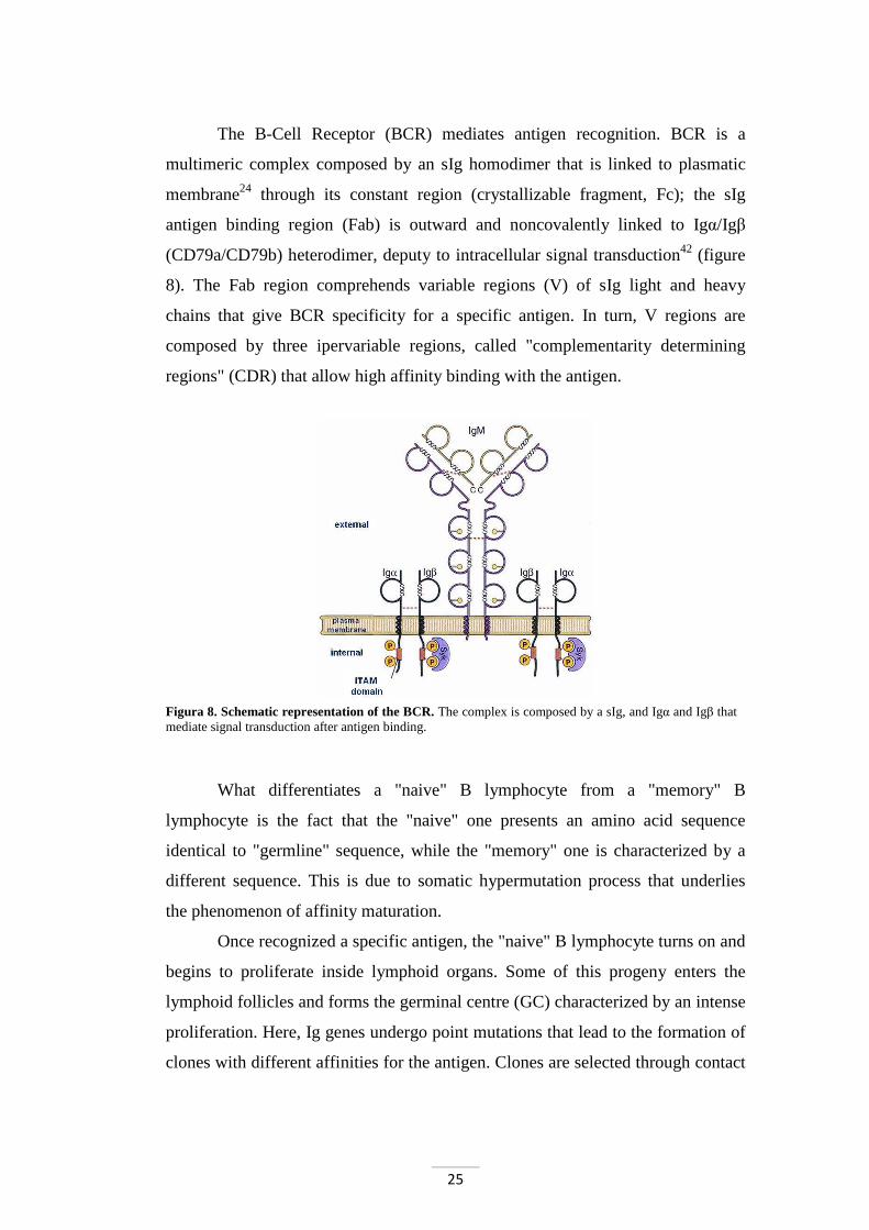
The B-Cell Receptor (BCR) mediates antigen recognition. BCR is a
multimeric complex composed by an sIg homodimer that is linked to plasmatic
membrane24 through its constant region (crystallizable fragment, Fc); the sIg
antigen binding region (Fab) is outward and noncovalently linked to Igα/Igβ
(CD79a/CD79b) heterodimer, deputy to intracellular signal transduction42 (figure
8). The Fab region comprehends variable regions (V) of sIg light and heavy
chains that give BCR specificity for a specific antigen. In turn, V regions are
composed by three ipervariable regions, called "complementarity determining
regions" (CDR) that allow high affinity binding with the antigen.
Figura 8. Schematic representation of the BCR. The complex is composed by a sIg, and Igα and Igβ that mediate signal transduction after antigen binding.
What differentiates a "naive" B lymphocyte from a "memory" B
lymphocyte is the fact that the "naive" one presents an amino acid sequence
identical to "germline" sequence, while the "memory" one is characterized by a
different sequence. This is due to somatic hypermutation process that underlies
the phenomenon of affinity maturation.
Once recognized a specific antigen, the "naive" B lymphocyte turns on and
begins to proliferate inside lymphoid organs. Some of this progeny enters the
lymphoid follicles and forms the germinal centre (GC) characterized by an intense
proliferation. Here, Ig genes undergo point mutations that lead to the formation of
clones with different affinities for the antigen. Clones are selected through contact

26
with follicular dendritic cells expressing antigen: lymphocytes that bind antigen
with greater affinity survive, while others undergo apoptosis.
B-CLL lymphocytes are small "memory" B cells blocked in G0/G1 and
characterized by surface markers recognized by specific monoclonal antibodies;
some of these markers, such as CD19 and CD21, are B-related, while others, like
CD5, CD23, CD25, and HLA-DR (human Leukocyte Antigen D-related), are not
specific for B lymphocytes (figure 9). In particular, B-CLL cells express markers
typical of mature B cells localized in the mantle zone of secondary lymphoid
follicles.
Recent studies have shown that 50-70% of B-CLL have undergone IgVH
hypermutation, a phenomenon that characterizes normal B cells subjected to a T
cell-dependent GC reaction. This finding has led to the hypothesis that B-CLL
cases displaying mutated IgVH may derive from a cell that had transited through
the GC, whereas those with germline IgVH may derive from a GC-independent
cells. This hypothesis has both biological and clinical relevance since the two
subgroups have different prognosis, with IgVH-mutated B-CLL (M-CLL)
displaying a better clinical course43. The factors involved in B-CLL pathogenesis
comprehend control of apoptosis, signal transduction BCR-mediated, proliferative
activity and the microenvironment.
Figure 9. Typical phenotype of a B-CLL lymphocyte. CD19 is a B-related antigen, while CD23, CD25, CD5, HLA-DR and sIgM are not specific to the B lineage.
2.1 Control of apoptosis
The dysregulation of the process of programmed cell death (apoptosis) is
now widely recognized as one of the main mechanism in the pathogenesis of

27
many tumors. The accumulation of B-CLL cells is related to the fact that they do
not undergo apoptosis, thus failing the homeostatic mechanism that normally
limits the number of circulating cells.
Paradoxically, when B-CLL cells derived from peripheral blood were
cultured in vitro, a substantial proportion of them spontaneously died by
apoptosis44. In this way, it is becoming increasingly clear that the B-CLL
defective apoptosis has to be ascribed not only to intrinsic defects of the
neoplastic cells, but also to extrinsic factors that influence their behavior.
Malignant B cells retain the ability to respond to microenvironmental signals, but
have devised a monothematic responsiveness. They have a specific sensitivity to
anti-apoptotic signals that favour their survival and become insensitive to pro-
apoptotic signals18. With respect to intrinsic factors, the balance between pro- and
anti-apoptotic factors is very important. Among these, the principal apoptosis
regulators are proteins of the Bcl-2 family (B-cell lymphoma-2 factors) that play a
crucial role in this mechanism by inhibiting (Bcl-2, Bcl-xL, Bcl-w, Bfl-1, and Mcl-
1) or promoting (Bax, Bak, Bcl-xS, Bid, Bik, and Hrk) apoptosis.
Heterodimerization between pro- and anti-apoptotic members, and their relative
levels, may determine the predisposition to respond to a given apoptotic stimulus
(figure 10). Many investigators have reported altered expression of Bcl-2, Bax,
and Mcl-1 in B-CLL45.
Other intrinsic factors, critical for apoptosis control, are 17p13 and 11q23
deletions containing 2 prominent tumor-suppressor genes mutated at varying
proportions: TP53 (tumor protein 53) and ATM (Ataxia telangiectasia mutated),
respectively. Mutations of TP53 and ATM, even in the absence of a chromosomal
deletion, have been identified to have adverse effects on patient survival. p53 and
ATM proteins are central regulators of the DNA-damage-response pathway and
their activation leads to cell-cycle arrest and DNA repair, apoptosis, or
senescence, depending on the cellular context. Impaired p53 function through
mutations and/or deletions is the best-characterized factor associated with
chemoresistance in B-CLL46.
Moreover, TOSO, also known as Fas-inhibitory molecule 3, was identified
as a candidate gene overexpressed in B-CLL.

28
Figure 10. The molecular mechanisms of apoptosis. Apoptosis pathways can be initiated via different stimuli, that is, at the plasma membrane by death receptor ligation (extrinsic pathway) or at the mitochondria (intrinsic pathway). Stimulation of death receptors results in receptor aggregation and recruitment of the adaptor molecule Fas-associated protein with death domain (FADD) and caspase-8. Caspase-8 initiates apoptosis by direct cleavage of downstream effector caspases. Mitochondria are engaged via the intrinsic pathway, which can be initiated by a variety of stress stimuli, including ultraviolet (UV) radiation, γ-irradiation, heat, DNA damage, the actions of some oncoproteins and tumour suppressor genes (i.e. p53), viral virulence factors, and most chemotherapeutic agents. CAD, caspase activated DNase; FAS, fibroblast associated antigen. ICAD, inhibitor of CAD; ROS, reactive oxygen species; TNF, tumour necrosis factor; TRAIL, TNF related apoptosis inducing ligand47.
TOSO is a transmembrane protein that inhibits Fas-mediated apoptosis by
binding Fas-associated death domain (FADD) via its C-terminal intracellular
domain. In B-CLL, high levels of TOSO expression have been correlated with a
more aggressive disease48.
2.2 BCR-mediated signal transduction
For effective humoral immunity, mature B cells must respond to foreign
antigens and generate antigen-specific effector cells; so, it is not surprising that
the BCR complex is required for the later stages of B-cell maturation. BCR has
two main roles: the first is to transmit signals that regulate B-cell fate decision and
the second is to mediate antigen processing leading to the presentation of antigen
to T cells, which allows full activation of B cells in the effector phase. The BCR
29
complex consist of immunoglobulin heavy (IgH) and light (IgL) chains associated
with Igα and Igβ containing ITAM (Immunoreceptor Tyrosine-based Activation
Motif) domains. After BCR ligation by antigen, both the protein tyrosine kinases
(PTKs) Syk and Lyn are activated. Then, Lyn phosphorylates ITAMs which, in
turn, recruit and facilitate the activation of Syk and Tec-family PTKs. This
phosphorylation results in the recruitment of other molecules involved in BCR-
mediated signal transduction49 (figure 11).
BCR signaling can be also regulated by the membrane organization of
signaling components. Literature data proposed that in the absence of antigen
binding, the BCR is already pre-assembled into oligomeric receptor complexes,
which generate a basal level of signaling essential for B-cell maintenance50. The
low levels of sIgs may explain the reduced ability of B-CLL cells to capture,
present, and respond to antigen. Defects in the BCR of B-CLL have been
attributed to functional deficiency in the CD79 heterodimer, especially CD79b,
which is expressed at low levels on these tumor51. During B cells activation
process, an important function is carried out by plasma membrane microdomains
called lipid rafts. These domains are rich in glycosphingolipids and cholesterol
which create a liquid-ordered phase within the plasma membrane. Lipid rafts are
fluid at physiological temperatures, allowing lateral diffusion of proteins and
lipids within the plane of the membrane. In addition, they are constitutively
enriched in certain types of proteins such as glycosphingolipids-linked proteins
and lipid chain-modified proteins, including heterotrimeric G proteins, the Src
kinases Lyn and Fyn, and other molecules involved in signal transduction, such as
Blk, Ras, c-Abl, and actin52.
Additional proteins, like CD45 and Syk, seem to be excluded from raft and
recruited only after BCR translocation into them after the engagement by the Ag.
In fact, according to the most recent model proposed to explain BCR functions in
the activation of B cells, in resting B cells the BCR is initially excluded from lipid
rafts; once having bound the antigen, BCR translocates into the rafts thus starting
the signal transduction cascade.

30
Figure 11. BCR-induced signal transduction pathways. After antigen ligation, tyrosine-kinase Lyn phosphorylates ITAMs of Igα and Igβ, creating binding sites to protein SH2 domain, such as Syk kinase. Follow different biochemical reactions that culminate in the B cell activation, differentiation and/or proliferation49
Lyn is a Src-family tyrosine kinase, which, with Blk and Fyn Src-like
kinases, is necessary for BCR signaling considering its role in phosphorylation of
the ITAM on Igα/Igβ. Tyrosine phosphorylated Igα/Igβ recruits Syk via the
latter’s tandem SH2 domains leading to downstream signaling events. Lyn also
provide feedback inhibition of BCR signaling by phosphorylation of cell surface
proteins containing immunoreceptor tyrosine-based inhibitory motifs (ITIMs).
These ITIM-containing inhibitory receptors include the inhibitory Fc receptor for
IgG, FcγRIIb, and the sialic acid-binding protein expressed on B cells, CD22. The
phosphorylation of these ITIMs generates binding sites for the membrane
recruitment of phosphatases that inhibit BCR signaling, including the SH2-
domain-containing inositol phosphatase (SHIP-1) and the SH2-domain-containing
tyrosine phosphatase (SHP-1). The net result of Lyn-deficiency is an exaggerated

31
signaling by the BCR, a phenotype that is moderate in immature B cells and
highly pronounced in mature follicular B cells. Lyn also acts inhibiting receptor
signaling in myeloid cells and recent studies demonstrated that hyperactivity of
myeloid cells contributes, in a relevant manner, to autoimmunity in Lyn-/- mice53.
In B lymphocytes, Lyn may also be associated to the non-receptor tyrosin-kinase
Fak (Focal Adhesion Kinase), involved in different signal transduction cascades.
This complex may contribute to cytoskeletal reorganization after antigen
binding54.
Our group demonstrated that in B-CLL, as compared to normal B cells,
protein Lyn is upregulated and shows a different subcellular localization55.
Moreover, Lyn displays a remarkable constitutive activity, which leads to an
increased basal tyrosine protein phosphorylation and a low responsiveness to BCR
ligation. Whereas Lyn was concentrated in membrane lipid rafts in normal B cells,
the enzyme was present all over the cell surface membrane in B-CLL cells. Lyn
was also detected in the cytosol of the malignant B cells. The release of Lyn into
the cytosol following caspase-dependent cleavage of the tyrosine kinase at its N-
terminus has been described as a general mechanism in hematopoietic cells during
BCR-induced apoptosis56. The findings that B-CLL cells contain a cytosolic Lyn
fraction and are defective in programmed cell death suggest that the tyrosine
phosphorylation of specific cytosolic targets might account, at least in part, for
cell resistance to apoptosis.
The activity of Lyn is critically regulated through its C-terminal Tyr507,
which is phosphorylated by the tyrosine kinase Csk and dephosphorylated by the
receptor tyrosine phosphatase CD45. In resting B lymphocytes, Lyn is present in
its inactive conformation, as result of Csk phosphorylation of Tyr507, which gives
rise to an intramolecular association of the phosphorylated residue with Lyn’s
own SH2 domain57. Since Csk, unlike Lyn, was similarly expressed in normal and
B-CLL cells, the constitutive activity of Lyn could be due to the fact that the
amount of Csk is insufficient to phosphorylate and downregulate its
overexpressed substrate. However, it is likely that other factors are responsible for
the presence of the active form of Lyn in B-CLL cells, first among which might
be the dephosphorylation of Lyn at Tyr507 by the tyrosine phosphatase CD45, an
abundant membrane protein that, in normal B cells, has access to Lyn only after

32
its migration to lipid rafts induced by BCR engagement52. Furthermore, the high
concentration of Lyn in B-CLL cells could promote the kinase intermolecular
autophosphorylation at Tyr396, which in turn induces Lyn activation58.
It is known that, after activation, SFK level is regulated by the balance of
two opposing mechanisms: degradation by ubiquitinylation or rescue by
association with Hsp90 (Heat shock protein of 90kDa), a chaperone interacting
with the N-terminal lobe of the SFK catalytic domain59. Recently, we
demonstrated that, in B-CLL cells, Lyn is an integral component of an aberrant
cytosolic 600kDa complex, where Lyn is associated both with Hsp90 through its
catalytic domain, HS1 (Hematopoietic lineage cell Specific protein 1), and SHP-
1L through its SH3 domain. Moreover, Hsp90 stabilizes the complex by
contributing to converting a network of transient interactions into permanent ones,
thus maintaining Lyn in an active conformation and preventing its degradation60.
HS1, one of the most important Lyn substrate, is an F-actin binding protein
involved in the apoptosis of several hematopoietic cell lines. HS1 phosphorylation
occurs in a sequential model mediated by Syk and Lyn. It seems that tyrosin-
phosphorylation of cortactin, an HS1 homologous protein involved in cell
motility, occur by the same mechanism of recruitment of the SFKs.
PI3K and PLCγ2 are both crucial effector enzymes that generate key
second messengers in BCR signaling. PI3K phosphorylates phosphatidylinositol-
4,5-bisphosphate (PtdInsP2) to produce phosphatidylinositol-3,4,5-trisphosphate
(PtdInsP3), which, in turn, recruits some BCR signaling molecules to the
membrane through PH domains. PLCγ2 uses PtdInsP2 to generate inositol-1,4,5-
trisphosphate (InsP3) and diacylglycerol (DAG), which are required for the release
of intracellular calcium (Ca2+) and activation of protein kinase C (PKC),
respectively. Subsequently, Ca2+ flux and PKC activation induce the activation of
mitogen-activated protein kinase (MAPK)-family kinases, extracellular signal-
regulated kinase (ERK), c-JUN NH2-terminal kinase (JNK), p38 MAPK, and
transcription factors, including nuclear factor-κB (NF-κB) and nuclear factor of
activated T cells (NFAT). It is probable that the profile of these activated
transcription factors then determines B-cell fate.
Non-enzymatic adaptor proteins are also important in regulating BCR
signaling. Among them, B-cell linker (BLNK) efficiently connects Syk and Btk

33
with PLCγ2. Disruption of the BLNK gene leads to impaired activation of PLCγ2
in B cells; BLNK also associates with Vav and Nck, both of which regulate
cytoskeletal organization in B cells.
Another B-cell adaptor, B-lymphocyte adaptor molecule of 32kDa
(BAM32), also binds PLCγ2 and regulates its activation. Since it is recruited to
the membrane in a PI3K-dependent manner, BAM32 integrates the PI3K and
PLCγ2 pathways. The B-cell-specific co-receptor CD19 can work as an adaptor
for PI3K also in B cells 49.
In mature B cells, BCR associates with lipid raft after Ag engagement and
the signal transduction induces transcription of genes responsible for B cell
activation. Then, the BCR is internalized and can be degraded or sent to an
intracellular compartment called MIIC (MHC-class-II-peptide-loading-
compartment) where Ag processing and the synthesis of peptide-MHC complexes
to Ag presentation occur. These complexes are brought to cell surface, presented
to T cells and then recognized by TCR of Th cells which activate B cells through
cytokines release.
In immature B cells, BCR is excluded from lipid rafts even after Ag
binding and, in this case, cell apoptosis is induced. A behavior similar to that of
immature B cells is described also to cells rendered tolerant or anergic by chronic
exposure to Ag: BCR is still excluded from rafts even after Ag binding and the
result is the lack of a cellular response. Conversely, in B pre-lymphocytes a
significant proportion of BCR and signaling molecules, such as PLCγ2 and PI3K,
are constitutively associated with rafts and this seems to generate signals of
survival and cell differentiation. In these cases, molecule such as PI3K, RAS,
RAF, ERK and NF-κB are fundamental in signal transduction in association with
BCR. PI3K activates and phosphorylates Akt/PKB which, in turn, phosphorylate
cellular targets involved in cell survival including apoptotic factors and glycogen
metabolism. One function of Akt is to inhibit the activation of the pro-apoptotic
Bcl-2-family member BAD (Bcl-2 antagonist of cell death); moreover, Akt
phosphorylates and inhibits glycogen synthase kinase 3 (GSK3) in B cells. In
unstimulated cells GSK3 is constitutively active; it phosphorylates and
destabilizes Myc and cyclin D, both of which are required for cell-cycle

34
progression. In this way, it seems that Akt functions to promote BCR-induced cell
proliferation, as well as survival49.
2.3 Proliferative activity and centrosome aberrations
The conceptual framework of the biology of B-CLL cells has changed in
the course of the past decade. The traditional view consider B-CLL as a disease
deriving from an apoptosis defect in which slowly proliferating B lymphocytes
accumulate because of a diminished cell death. In this view these cells are
relatively inert and divide minimally, rarely dying; they continue to accumulate
passively until they reached an harmful level not more supportable by the patient.
However, recent studies suggest that B-CLL is a dynamic condition, comprising
leukemic cells that multiply and die at measurable rates. Furthermore, since B-
CLL cells do not appear to be inherently immortal, the impairment of the patient
does not occur from passive accumulation, but from active generation of
subclones that, over time, develop dangerous genetic abnormalities which further
change birth/death ratio61.
The centrosome is a cellular structure essential for a proper proliferative
activity; it is a small non-membranous organelle (1-2µm in diameter) often
denoted as a major microtubule organizing center (MTOC). During interphase, the
centrosome organizes an astral array of microtubules (MTs) that participate in
intracellular trafficking, cell motility, cell adhesion, and cell polarity. In
proliferating cells, the centrosome starts duplicating just before, or at, the onset of
S phase and the two newly formed centrosomes participate in the assembly and
organization of the mitotic spindle, its orientation with respect to cortical cues,
and the late events of cytokinesis. The animal centrosome consists of a pair of
centrioles linked together through their proximal regions by a matrix consisting in
part of large coiled-coil proteins of the pericentrin family (pericentrin, γ-tubulin,
ninein, centriolin, katanin), which anchor other matrix components (figure 12).
The architecture of the microtubule array in differentiated cell types results not
only from the dynamic behavior of MTs but also from a balance between MTs
nucleation and MTs-anchoring activities at the centrosome. Microtubules are
nucleated by the γ-tubulin ring that is present throughout the cell cycle in the
35
matrix, close to the proximal walls of centrioles. Its levels increase significantly
prior to mitosis, concurrently with the recruitment of MT-associated proteins
required for mitotic spindle formation62.
Normally, the single centrosome of a G1 cell duplicates precisely once
prior to mitosis in a process that is intimately linked to the cell division cycle via
cyclin-dependent kinase (cdk) 2 activity that couples centrosome duplication to
the onset of DNA replication at the G1/S transition. Accurate control of
centrosome duplication is critical for symmetric mitotic spindle formation and
thereby contributes to the maintenance of genome integrity.
Numerical and structural centrosome abnormalities are hallmarks of
almost all solid tumors and have been implicated in the generation of multipolar
mitoses and chromosomal instability. In addition to solid neoplasias, centrosome
aberrations have been described in several different hematological malignancies
like acute myeloid leukemias, MDS, Hodgkin lymphoma as well as NHL, MM,
and B-CLL. A correlation between centrosome abnormalities on the one hand and
karyotype aberrations as well as clinical aggressiveness on the other hand seems
to exist in myeloid malignancies, B-CLL and at least in some types of NHL63.
It was demonstrated that centrosome aberrations are already present in B
lymphocytes from patients with monoclonal B-cell lymphocytosis (MBL), a
lesion considered to represent a premalignant stage of B-CLL, suggesting that
centrosome abnormalities do occur early during B-CLL evolution. In analogy to
other NHLs, centrosomal abnormalities of circulating B-CLL cells might reflect
cellular generation emanated from proliferation centers in lymph nodes and bone
marrow of B-CLL patients.
The detailed mechanisms by which centrosome aberrations develop are
still largely unknown. Several oncogenes and tumor suppressor genes, among the
p53 and ATM, have been implicated in the formation of centrosomal defects in
human malignancies. Both p53 and ATM abnormalities are associated with a poor
prognosis in this disorder. Therefore, it is tempting to speculate that aberrations of
the ATM/p53 pathway might be involved in the generation of centrosomal
abnormalities in B-CLL64.

36
Figure 12. Microtubules cytoskeleton structure. Confocal microscopy analysis of microtubules in neoplastic B cells. Cells were stained with Ab anti-α-tubulin followed by secondary Ab Alexa-488. The point from which microtubules depart is the centrosome or MTOCs (circled). The image on the right shows in detail the structure of the centrosome.
2.4 Microenvironment
Bone marrow (BM) precursors derived from pluripotent stem cells are in
intimate contact with stromal cells and generate B cells in an Ag-independent
process. BM precursors differentiate to mature virgin B lymphocytes endowed
with membrane Ag receptors that migrate to peripheral lymphoid tissues
searching an Ag. The encounter with a foreign Ag triggers B cell activation,
proliferation, and a second wave of differentiation. The microenvironment for the
active social life of a mature B cell is provided by the germinal centers of
secondary lymphoid organs. It is within GC that trafficking mature B cells are
brought into close contact with specialized T cells and Ag-presenting cells. This
dialogue, finely regulated by cytokines, adhesion structures, and surface
molecules, leads to the generation of B memory cells and plasma cell precursors
and to the apoptotic elimination of inefficient or potentially dangerous cells. As
all normal B cells evolve and operate thanks to microenvironmental cross-talks, it
becomes consequent to ask whether the microenvironment may also influence the
natural history of B cell malignancies65.
Isolated B-CLL cells undergo relatively rapid apoptosis in vitro. This
observation has led to the speculation that the microenvironment is necessary
and/or plays a pivotal role in maintaining the enhanced survival of B-CLL cells in

37
vivo. Human bone marrow stromal cells (BMSCs) have been demonstrated to
support the survival of B-CLL cells when both cell types were co-cultured in
vitro. Further investigations have suggested that B-CLL cells need to have
intimate contact with BMSC in bone marrow, with T cells in lymph nodes, and
with nurse-like cells (NLCs) in lymphatic tissues to maintain survival66.
Mesenchymal Stromal Cells (MSCs) from both normal healthy donors and B-CLL
patients were able to protect leukemic cells from undergoing spontaneous and
drug-induced apoptosis. Close contact between B-CLL cells and MSCs is capable
to mediate the most effective drug-resistance and it is this latter interaction that
could be the most important in providing a niche for residual B-CLL cells post
treatment. Recent studies demonstrated the significance of CD49d (α4 integrin) in
the prognosis of B-CLL disease67,68 as well as its biological role of regulating
matrix metalloproteinase-9 (MMP-9)69. These observations imply that α4β1
integrin could be a critical mediator of tight interactions between MSCs and B-
CLL cells and MSC-mediated B-CLL protection.
TNF-family cytokines can provide survival signals or alternatively induce
apoptosis. T cells modulate survival of B-CLL cells through the CD40/CD40L
system. The signals delivered for normal B-lymphocytes by activated T-cells
through CD40L induce B-cell growth, differentiation, and rescue from apoptosis.
In contrast to normal B cells, a subset of B-CLL cells expresses both CD40L and
its receptor, enabling an autocrine loop by which B-CLL cells can promote
survival signals on their own. CD40 stimulation of B-CLL cells had been shown
to prevent apoptosis and induce proliferation in vitro. It has been observed a
correlation between levels of the anti-apoptotic protein Bcl-2 and survival induced
by stromal cell contact, suggesting a mechanism by which stromal cells induce
protection of B-CLL cells against spontaneous apoptosis.
In contrast, it was found that normal CD5+ B lymphocytes were unable to
survive in co-culture with stromal cells. Stromal cells can, in addition, induce
survival of B-CLL cells by stromal cell-derived factor-1 (SDF-1/CXCL12), which
is a homeostatic chemokine that signals through the CXCR4 chemokine receptor
and plays an important role in lymphopoiesis. High-levels of SDF-1 are
constitutively produced by stromal cells within the marrow, the primary site of
early B cell differentiation. B-CLL cells express high levels of the CXCR4

38
surface receptor, and undergo chemotaxis in response to SDF-1. In addition, it has
been demonstrated that marrow stromal cells attract B-CLL cells via the
chemokine receptor CXCR4, providing a possible explanation accounting for the
infiltration of marrow by B-CLL cells70 (figure 13).
3. Microtubule inhibitors
3.1 Microtubules
The ability of eukaryotic cells to adopt different forms and perform
coordinated movements depends on a complex network of protein filaments called
cytoskeleton, which extends throughout the cytoplasm. The cell cytoskeleton
consists of actin microfilaments, intermediate filaments and microtubules.
Microtubules are long, filamentous, tube-shaped protein polymers that are
crucial in the development and maintenance of cell shape, in the transport of
vesicles, mitochondria and other components throughout cells, in cell signaling, as
well as in cell division and mitosis. Microtubules are composed of α- and β-
tubulin heterodimers (4nm × 5nm × 8nm) arranged in the form of slender
filamentous tubes that can be several micrometres long (figure 14). They are
highly dynamic polymers and their polymerization dynamics are tightly regulated
both spatially and temporally.
The functional diversity of microtubules is achieved in several ways: i)
through the binding of various regulatory proteins, including microtubule-
associated proteins (MAPs), to soluble tubulin and to the microtubule surfaces and
ends; ii) by expression of different tubulin isotypes, which have different
functions; iii) through several post-translational modifications of tubulin. The
polymerization dynamics of microtubules are created by the gain and loss of a
short region of tubulin-GTP or tubulin-GDP-inorganic phosphate (Pi) at the two
microtubule ends, called the GTP cap (figure 15a). Tubulin-GTP is hydrolysed to
tubulin-GDP and Pi at the time that tubulin-GTP adds to the microtubule ends, or
shortly after. Finally, the cap dissociates from the microtubule, leaving a
microtubule core consisting of tubulin with stoichiometrically bound GDP in β-

39
tubulin. The tubulin-GDP remains non-dissociable and non-exchangeable until the
tubulin subunit dissociates from the microtubule71.
Polymerization of microtubules occurs through two important steps:
nucleation and elongation. Initially, an oligomer consisting of 6-12 αβ-tubulin
dimers is formed in the nucleation step. Further, the GTP bound to αβ-tubulin
dimers add up to the nucleus of γ-tubulin and lead to its elongation and formation
of the protofilament. After a rapid elongation phase, the assembly of microtubules
reaches the steady state, where the addition and the dissociation of tubulin
subunits at the ends of the microtubules are balanced and there is not a net
increase in the polymer level. Hydrolysis of GTP introduces unusual equilibrium
behaviors in microtubules. Microtubules are labile polymers that display two
types of dynamic behaviors: "treadmilling" and "dynamic instability".
Figure 13. Molecular crosstalk between B-CLL cells and microenvironment. Contact between B-CLL cells and NLCs or MSCs is established and maintained by chemokine receptors and adhesion molecules. NLCs express the chemokines CXCL12 and CXCL13, whereas MSCs predominantly express CXCL12. NLCs and MSCs attract B-CLL cells via the chemokine receptors CXCR4 and CXCR5, which are expressed at high levels on B-CLL cells. Integrins, particularly CD49d, expressed on the surface of B-CLL cells, cooperate with chemokine receptors in establishing cell-cell adhesion through respective ligands on the stromal cells (VCAM-1). NLCs also express the TNF family members BAFF and a proliferation-inducing ligand, providing survival signals to B-CLL cells via corresponding receptors (BCMA, TACI, BAFF-R). CD38 expression allows B-CLL cells to interact with CD31 on stromal and NLCs. Ligation of CD38 activates ZAP-70 and downstream survival pathways. Stimulation of the BCR complex (BCR and CD79a,b) induces downstream signaling by recruitment and activation of Lyn, Syk and ZAP-70. BCR stimulation and co-culture with NLCs also induce B-CLL cells to secrete high levels of CCL3 and CCL4 chemokines, which are potent T cell–attracting chemokines. Through this mechanism, B-CLL cells can actively recruit T cells for cognate T-cell interactions with B-CLL cells. CD40L+ T cells are preferentially found in B-CLL proliferation centers and can interact with B-CLL cells via CD40. Collectively, this crosstalk between B-CLL cells and accessory cells results in activation of survival and drug resistance pathways, such as those provided by Bcl-2 and Mcl-1. (Modified from Bruger J A et al.72).

40
Treadmilling refers to a net addition of tubulin dimers at the plus end
coupled with a net dissociation at the minus end producing a flow of subunits
from one end of the microtubule to the other end without significantly changing
the average length of microtubules. Microtubule ends also alternate between
growing and shortening phases, which is called as dynamic instability. A
transition from a growing phase to a shortening phase is termed as a "catastrophe"
while a transition from a shortening phase to a growing phase is termed as a
"rescue"73 (figure 15b).
Figure 14. Polymerization of microtubules. Heterdimers of α- and β-tubulin assemble to form a short microtubule nucleus. Nucleation is followed by elongation of the microtubule at both ends to form a cylinder that is composed of tubulin heterodimers arranged head-to-tail in 13 protofilaments. Each microtubule has a so-called plus (+) end, with β-tubulin facing the cytoplasm, and a minus end (–), with α-tubulin facing the cytoplasm71.
B-CLL malignant cells are characterized by abnormalities that suggest
defects of cytoskeletal functions. B-CLL cells have low mobility, show decreased
capping by different ligands, and are unusually susceptible to microtubule
disrupting drugs74. In particular, in B-CLL cells, microtubules are tightly
connected with abnormalities of the BCR: in fact, molecules associated with
BCR-mediated signal transduction, such as Syk, Vav, and Cbl bind tubulin and
BCR members, like CD79a and CD79b, co-immunoprecipitate with tubulin75.
Moreover, in rat basophilic leukemia Lyn kinase is complexed with γ-tubulin of
cell centrosome76.
Microtubules are involved in a large number of cellular functions
including chemotaxis, membrane and cellular scaffolding, intracellular transport,
secretory processes, and transmission of receptor signaling77. For this reason

41
microtubules are already considered as potential drug targets for several diseases
including cancer, neuronal diseases, fungal, and parasitic diseases73. In this
context, microtubule inhibitors may have an important role in B-CLL treatment as
they already have in other diseases.
Figure 15. Polymerization dynamics and the GTP cap. a) The head-to-tail association of the αβ heterodimers makes microtubules polar structures and they have different polymerization rates at the two ends. In each protofilament, the αβ heterodimers are oriented with their β-tubulin monomer pointing towards the faster-growing end (plus end) and their α-tubulin monomer exposed at the slower-growing end (minus end). A third tubulin isoform, γ-tubulin, functions as a template for the correct assembly of microtubules. On addition of a new dimer at the plus end, the catalytic domain of α-tubulin contacts the nucleotide exchangeable site (E site) of the previous β-subunit and becomes ready for hydrolysis; the plus end generally has a minimum GTP cap of one tubulin layer that stabilizes the microtubule structure. b) When this GTP cap is stochastically lost, the protofilaments splay apart and the microtubule rapidly depolymerizes. During, or soon after the polymerization, the tubulin subunits hydrolyse their bound GTP and become non-exchangeable. Thus, the microtubule lattice is predominantly composed of GDP–tubulin, with depolymerization being characterized by the rapid loss of GDP–tubulin subunits and oligomers from the microtubule plus end. At the minus end, contact is made between the E site of the new dimer and the catalytic region of the last subunit at the end; therefore, no GTP cap should be present. Tubulin-bound GTP is hydrolysed to tubulin-GDP and inorganic phosphate (Pi) at the time that tubulin adds to the microtubule ends, or shortly thereafter. Pi dissociates from the microtubule, leaving a microtubule core consisting of tubulin with stoichiometrically bound GDP. A microtubule end containing tubulin-bound GTP or GDP-Pi is stable, or "capped", against depolymerization. Hydrolysis of tubulin-bound GTP, and the subsequent release of Pi, induces conformational changes in the tubulin molecules that destabilize the microtubule polymer, resulting in catastrophe and shortening of the microtubule78.
3.2 Microtubule-interfering agents
A large number of chemically different compounds, many of which are
derived from natural products, are able to bind tubulin or microtubules and inhibit

42
proliferation by acting on the mitotic spindle. Some of these compounds inhibit
microtubule polymerization, whereas others stabilize them. Although these
compounds exert opposite effects on microtubules, both types of microtubule-
interfering agents (MIAs) share the common property of suppressing microtubule
dynamics and thereby microtubule function, leading to the disruption of the
mitotic spindle function and blocking cell cycle progression.
Microtubule-interfering agents are divided in:
1) Depolymerizing agents:
a) The Vinca alkaloids. The Vinca alkaloids have been responsible for
many stories of successful chemotherapeutic since their introduction into
the clinic 40 years ago. The naturally occurring members of the family,
vinblastine and vincristine, were isolated from the leaves of the periwinkle
plant Catharanthus roseus. They came into widespread use for the single-
agent treatment of childhood haematological and solid malignancies and,
shortly after, also for adult haematological malignancies. Their principle
side effects are peripheral neuropathy and reversible myelosuppression.
The Vinca alkaloids bind to β-tubulin near the GTP-binding site71 (figure
16a).
b) Colchicine. Colchicine was originally extracted from plants of the
genus Colchicum. The interaction of colchicine with tubulin and
microtubules presents one more variation in the mechanisms by which
microtubule-active drugs inhibit microtubule functions. Colchicine inhibits
microtubule polymerization substoichiometrically, indicating that it
inhibits microtubule polymerization by binding to microtubule ends rather
than to the soluble-tubulin pool (figure 16b).
Tubulin-colchicine complexes might have a conformation that
disrupts the microtubule lattice in a way that slows, but does not prevent,
new tubulin addition. The incorporated tubulin-colchicine complex must
bind more tightly to its tubulin neighbours than tubulin itself does, so that
the normal rate of tubulin dissociation is reduced71.

43
Figure 16. Antimitotic drugs bind to microtubules at diverse sites. a) A few molecules of vinblastine bound to high-affinity sites at the microtubule plus end are sufficient to suppress microtubule dynamics. b) Colchicine forms complexes with tubulin dimers and copolymerizes into the microtubule lattice, suppressing microtubule dynamics. c) A microtubule section to display the interior surface is shown. Paclitaxel binds along the interior surface of the microtubule, suppressing its dynamics71.
At doses required for antitumor effect, colchicine present important
toxicity and the clinician has excluded its use. Relatively recently (2007)
colchicine has been use in the treatment of pericarditis and in preventing
the recurrence of the inflammatory process; in fact, by its destabilizing
effect on microtubules, colchicine prevents leukocyte motility, thus
blocking the inflammatory phenomenon79.
c) Estramustine. Estramustine is an estradiol molecule linked to a nitrogen
mustard through a carbamate ester group and was rationally synthesized as
an alkylating agent for the treatment of prostate carcinoma. However,
estramustine has been shown to bind to tubulin and MAPs, which results
in their dissociation from microtubules and subsequently in microtubule
disassembly and metaphase arrest, leading to apoptosis. Thus,
estramustine is considered as a microtubule-active drug currently in use
for the treatment of advanced prostatic carcinoma and for hormone
resistant prostate cancer patients. In addition, experimental data
concerning the antitumor effect of estramustine in other malignancies are
accumulating and clinical studies are ongoing. Estramustine has potent
44
antiproliferative effects against malignant glioma both in vitro and in vivo,
whereas non-neoplastic astrocytes are spared77. The estramustine-binding
site on tubulin has been suggested to be distinct from the colchicine and
vinblastine sites and may partially overlap with the Taxol-binding site in
tubulin80.
2) Microtubules stabilizer:
a) Taxanes. The taxanes are diterpenes produced by the plants of the genus
Taxus. As their name suggests, they were first derived from natural
sources, but some have been then synthesized artificially. Taxanes include
paclitaxel (Taxol) and docetaxel (Taxotere). Taxanes bind directly, with
high affinity, to tubulin along the length of the microtubule (figure 16c).
Paclitaxel binding site is located in the β-subunit, which is on the inner
surface of the microtubule. It is thought that, to gain access to its binding
sites, paclitaxel diffuses through small openings in the microtubule or
fluctuations of the microtubule lattice. Binding of paclitaxel to microtubule
inner site stabilizes microtubules and increases their polymerization,
presumably by inducing a conformational change in the tubulin that, by an
unknown mechanism, increases its affinity for neighbouring tubulin
molecules. Paclitaxel can induce cell death by the activation of apparently
different signal transduction pathways, depending on drug concentration.
Incubation of cells with high concentrations (≥200nM) of paclitaxel
stabilizes extensively the microtubule polymerization, causing the
formation of stable bundles of microtubules that disrupt the normal
polymerization/ depolymerization cycle of microtubules and thereby
suppressing microtubule dynamics, resulting in the arrest of cells in
mitosis77. The clinical success of the taxanes has led to a search for other
drugs that enhance microtubule polymerization, yielding several promising
compounds, including discodermolide, eleutherobin, laulimalide, the
sarcodictyins, and the epothilones. Some of these compounds compete
with paclitaxel for binding to microtubules and are said to bind at or near
the taxane site (epothilones, discodermolide, eleutherobins and
sarcodictyins); others, such as laulimalide, seem to bind to unique sites on
microtubules71.

45
b) Epothilones. Epothilones are secondary metabolites that are produced
by myxobacteria and that were first discovered in a screening program for
secondary metabolites by virtue of their selective antifungal activity
against Mucor hiemalis. In contrast to paclitaxel, epothilones were also
shown to inhibit the growth of cells overexpressing the P-glycoprotein
efflux pump, thus indicating that these compounds (or related analogs)
might eventually be useful for the treatment of multidrug-resistant tumors.
Epothilones have also been quoted as being significantly more water-
soluble than paclitaxel. This should allow the use of formulation vehicles
less problematic than cremaphor, which in the case of Taxol is believed to
contribute to the drug's clinical side-effects81.
Different studies initiated in the past are till now valuating the possible
anti-neoplastic efficacy of microtubules inhibitors in B-CLL. In particular, it was
analyzed vincristine efficacy in chemotherapeutic combinations, such as CHOP
and COP. Results were so positive to introduce its use in B-CLL treatment in the
last decade82.
More recently it was evaluated the efficacy of Bryostatin 1, a macrocytic
lactone isolated from the marine invertebrate Bugula neritina. Short-term
exposure activates protein kinase C, a family kinase involved in cell proliferation
and apoptosis. Despite its limited single agent activity, Bryostatin 1 has
demonstrated the ability to potentiate the cytotoxicity of several chemotherapeutic
agents. Preclinical data suggest the superiority of Bryostatin 1 in combination
with CHOP when compared with CHOP alone. In combination with fludarabine,
for indolent NHL and B-CLL, Bryostatin 1 did not appear to add significant
toxicity to full-dose fludarabine83.
3.3 Nocodazole
The synthetic drug nocodazole (methyl N-[6-(thiophene-2-carbonyl)-1H-
benzimidazol-2-yl] carbamate) is a depolymerizing agent and is reported to have
antimitotic and antitumoral activity. Its molecular structure is C14H11N3O3S
(figure 17). The potential usefulness of this drug is due to its action being readily
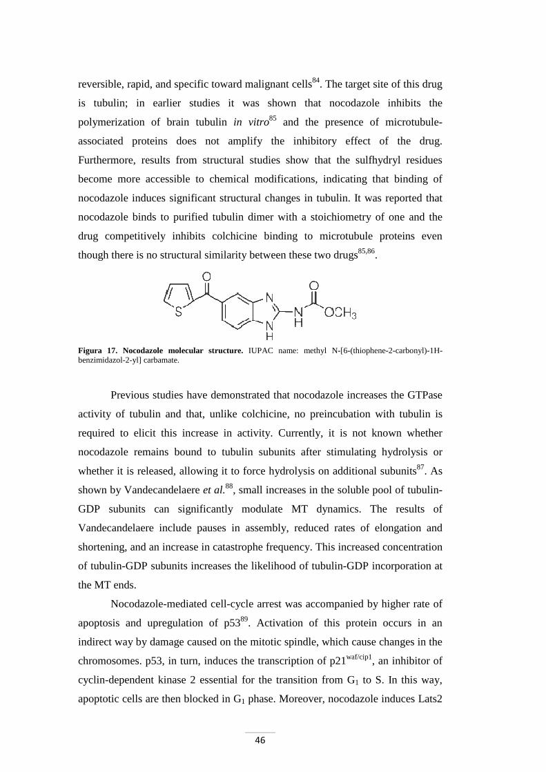
46
reversible, rapid, and specific toward malignant cells84. The target site of this drug
is tubulin; in earlier studies it was shown that nocodazole inhibits the
polymerization of brain tubulin in vitro85 and the presence of microtubule-
associated proteins does not amplify the inhibitory effect of the drug.
Furthermore, results from structural studies show that the sulfhydryl residues
become more accessible to chemical modifications, indicating that binding of
nocodazole induces significant structural changes in tubulin. It was reported that
nocodazole binds to purified tubulin dimer with a stoichiometry of one and the
drug competitively inhibits colchicine binding to microtubule proteins even
though there is no structural similarity between these two drugs85,86.
Figura 17. Nocodazole molecular structure. IUPAC name: methyl N-[6-(thiophene-2-carbonyl)-1H-benzimidazol-2-yl] carbamate.
Previous studies have demonstrated that nocodazole increases the GTPase
activity of tubulin and that, unlike colchicine, no preincubation with tubulin is
required to elicit this increase in activity. Currently, it is not known whether
nocodazole remains bound to tubulin subunits after stimulating hydrolysis or
whether it is released, allowing it to force hydrolysis on additional subunits87. As
shown by Vandecandelaere et al.88, small increases in the soluble pool of tubulin-
GDP subunits can significantly modulate MT dynamics. The results of
Vandecandelaere include pauses in assembly, reduced rates of elongation and
shortening, and an increase in catastrophe frequency. This increased concentration
of tubulin-GDP subunits increases the likelihood of tubulin-GDP incorporation at
the MT ends.
Nocodazole-mediated cell-cycle arrest was accompanied by higher rate of
apoptosis and upregulation of p5389. Activation of this protein occurs in an
indirect way by damage caused on the mitotic spindle, which cause changes in the
chromosomes. p53, in turn, induces the transcription of p21waf/cip1, an inhibitor of
cyclin-dependent kinase 2 essential for the transition from G1 to S. In this way,
apoptotic cells are then blocked in G1 phase. Moreover, nocodazole induces Lats2

47
translocation from centrosomes to the nucleus and p53 accumulation. Lats2
(Large Tumor Suppressor 2) interacts physically with Mdm2 inhibiting p53
ubiquitination and promoting its activation. This interaction is enhanced in cells
treated with nocodazole, which causes microtubule and mitotic spindle damage90.
Beswick et al.91 have analyzed nocodazole effects in B-CLL cells finding
that it causes selective death of neoplastic cells. They show that the mechanism of
nocodazole-induced apoptosis involves mitochondrial membrane depolarisation,
caspase activation and PARP cleavage; in particular, caspase-9 activation is the
important route of nocodazole-induced death in B-CLL. In order to analyze the
mechanism of apoptosis induction by nocodazole it was considered the known
link between microtubule network and apoptosis: microtubule damage induces
Bcl-2 phosphorylation on specific residues including serine-70. A recent
suggestion is that phosphorylated Bcl-2 may exist for lengthy periods after
microtubule damage representing a pre-apoptotic phase. In this scheme
subsequent dephosphorylation initiates apoptosis. In cycling cells nocodazole
causes mitotic arrest, which in turn leads to apoptosis. An effect on non-cycling
B-CLL cells is, therefore, unexpected. Beswick et al. proposed two major
explanations for nocodazole-induced apoptosis in B-CLL. Firstly, primary
disturbances in the apoptotic machinery, which may include alterations in the
relative amounts of pro- and anti-apoptotic proteins, could be sufficient to lead to
apoptosis in B-CLL cells at lower nocodazole concentrations than in normal cells.
Alternatively primary abnormalities of microtubule quantity or quality could
increase sensitivity to the effects of depolymerising agents. It is interesting, in this
regard, that there is considerable variation in tubulin content in B-CLL91.

48

49
AIM OF THE STUDY
B-CLL cells are characterized by abnormalities that depend also on
cytoskeletal defects; neoplastic B cells, in fact, have low mobility5, show decreased
capping by different ligands92,93 and are susceptible to microtubule disrupting drugs7.
In particular, in B-CLL cells microtubules are tightly connected with functional
anomalies of the B Cell Receptor (BCR): molecules associated with BCR-mediated
signal transduction bind tubulin, and BCR members, like CD79a and CD79b, co-
immunoprecipitate with β-tubulin8.
Microtubules are important targets in the anti-tumor therapy for the crucial
role they play in cancer cell functions, including mitosis, motility and cell-cell
contacts73,77. Microtubule inhibitors are already employed in pathologies such as
Hodgkin lymphoma or acute lymphocytic leukemia. In spite of their potent anti-
cancer action, many anti-microtubule drugs show limited clinical use because of their
strong toxic effects. Hence the importance of identifying new microtubule inhibitors
highly selective for leukemic cells and not damaging for the other cell types.
Nocodazole is an anti-neoplastic agent which exerts its cellular effects by
favouring microtubule depolymerization94. It is highly effective in altering
microtubule dynamics and arresting cell-cycle progression at mitosis phase95. It
induces significant structural changes in tubulin and the usefulness of this drug is due
to its action, that is readily reversible, rapid and specific towards malignant cells84,86.
During this PhD program we planned to further investigate nocodazole acting
and to analyze how nocodazole exerts its effect specifically on B-CLL. In particular,
we studied:
- nocodazole action on several cell types (normal B cells, B and T cells from B-CLL
patients, MSCs from both B-CLL patients and donors, Jurkat, Raji, and K562 cell
lines;
- nocodazole apoptotic effect on B-CLL cells and residual T lymphocytes obtained
from the same patients;
- nocodazole effects on B-CLL cells co-cultured with MSC and/or in presence of
CD40L or plasma;
- the molecular mechanisms of nocodazole activity.

50
51
MATHERIALS AND METHODS
1. Patients
We analyzed 52 B-CLL patients (37 males and 15 females), aged between
44 and 80 years, enrolled by the Hematology and Clinical Immunology branch
(chief Prof. G. Semenzato), Padova University School of Medicine.
Clinical characteristics of patients are listed in Table I. In particular, for
each patient we reported: the number of white blood cells (WBCs), the percentage
of lymphocytes, mutational status of IgVH genes and the expression of ZAP-70.
All neoplastic B cells of the 52 patients examined were positive for CD5, CD19
and CD23 markers, typically co-expressed in B-CLL and those of 12 subjects
were also CD38 positive.
B lymphocytes obtained from peripheral blood of 10 healthy subjects were
used as normal controls.
2. Immunophenotypic analysis
The immunophenotypic analysis on lymphocytes obtained from peripheral
blood of B-CLL patients was performed through flow cytometry. This technique
allows a multiparametric evaluation of antigenic characteristic of the single cells
by the analysis of visible and fluorescent light they emit when flow through a
liquid medium.
The immunophenotyping is based on the identification of surface and
intracellular Ag using mAb conjugated with fluorochromes. The presence of a
certain Ag is revealed and used as an indicator of belonging to a cell line as well
as its level of maturation. The fluorochromes used in this thesis were fluorescein
isothiocyanate (FITC), which emits a fluorescence signal at 530nm (green),
phycoerythrin (PE) emitting at 585nm, tri-color (TC) that emits at 667nm when
hit by a monochromatic laser beam with λ equal to 488nm, and finally, the
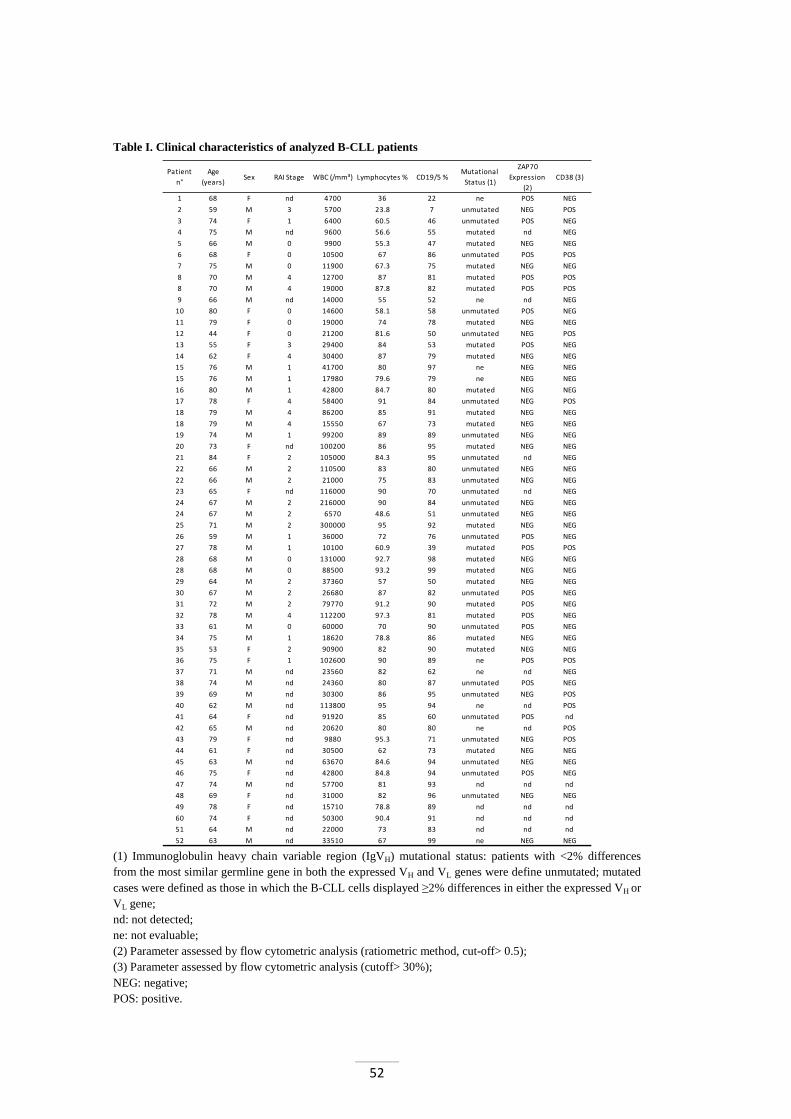
52
Table I. Clinical characteristics of analyzed B-CLL patients
Patient
n°
Age
(years)Sex RAI Stage WBC (/mm³) Lymphocytes % CD19/5 %
Mutational
Status (1)
ZAP70
Expression
(2)
CD38 (3)
1 68 F nd 4700 36 22 ne POS NEG
2 59 M 3 5700 23.8 7 unmutated NEG POS
3 74 F 1 6400 60.5 46 unmutated POS NEG
4 75 M nd 9600 56.6 55 mutated nd NEG
5 66 M 0 9900 55.3 47 mutated NEG NEG
6 68 F 0 10500 67 86 unmutated POS POS
7 75 M 0 11900 67.3 75 mutated NEG NEG
8 70 M 4 12700 87 81 mutated POS POS
8 70 M 4 19000 87.8 82 mutated POS POS
9 66 M nd 14000 55 52 ne nd NEG
10 80 F 0 14600 58.1 58 unmutated POS NEG
11 79 F 0 19000 74 78 mutated NEG NEG
12 44 F 0 21200 81.6 50 unmutated NEG POS
13 55 F 3 29400 84 53 mutated POS NEG
14 62 F 4 30400 87 79 mutated NEG NEG
15 76 M 1 41700 80 97 ne NEG NEG
15 76 M 1 17980 79.6 79 ne NEG NEG
16 80 M 1 42800 84.7 80 mutated NEG NEG
17 78 F 4 58400 91 84 unmutated NEG POS
18 79 M 4 86200 85 91 mutated NEG NEG
18 79 M 4 15550 67 73 mutated NEG NEG
19 74 M 1 99200 89 89 unmutated NEG NEG
20 73 F nd 100200 86 95 mutated NEG NEG
21 84 F 2 105000 84.3 95 unmutated nd NEG
22 66 M 2 110500 83 80 unmutated NEG NEG
22 66 M 2 21000 75 83 unmutated NEG NEG
23 65 F nd 116000 90 70 unmutated nd NEG
24 67 M 2 216000 90 84 unmutated NEG NEG
24 67 M 2 6570 48.6 51 unmutated NEG NEG
25 71 M 2 300000 95 92 mutated NEG NEG
26 59 M 1 36000 72 76 unmutated POS NEG
27 78 M 1 10100 60.9 39 mutated POS POS
28 68 M 0 131000 92.7 98 mutated NEG NEG
28 68 M 0 88500 93.2 99 mutated NEG NEG
29 64 M 2 37360 57 50 mutated NEG NEG
30 67 M 2 26680 87 82 unmutated POS NEG
31 72 M 2 79770 91.2 90 mutated POS NEG
32 78 M 4 112200 97.3 81 mutated POS NEG
33 61 M 0 60000 70 90 unmutated POS NEG
34 75 M 1 18620 78.8 86 mutated NEG NEG
35 53 F 2 90900 82 90 mutated NEG NEG
36 75 F 1 102600 90 89 ne POS POS
37 71 M nd 23560 82 62 ne nd NEG
38 74 M nd 24360 80 87 unmutated POS NEG
39 69 M nd 30300 86 95 unmutated NEG POS
40 62 M nd 113800 95 94 ne nd POS
41 64 F nd 91920 85 60 unmutated POS nd
42 65 M nd 20620 80 80 ne nd POS
43 79 F nd 9880 95.3 71 unmutated NEG POS
44 61 F nd 30500 62 73 mutated NEG NEG
45 63 M nd 63670 84.6 94 unmutated NEG NEG
46 75 F nd 42800 84.8 94 unmutated POS NEG
47 74 M nd 57700 81 93 nd nd nd
48 69 F nd 31000 82 96 unmutated NEG NEG
49 78 F nd 15710 78.8 89 nd nd nd
60 74 F nd 50300 90.4 91 nd nd nd
51 64 M nd 22000 73 83 nd nd nd
52 63 M nd 33510 67 99 ne NEG NEG (1) Immunoglobulin heavy chain variable region (IgVH) mutational status: patients with <2% differences from the most similar germline gene in both the expressed VH and VL genes were define unmutated; mutated cases were defined as those in which the B-CLL cells displayed ≥2% differences in either the expressed VH or VL gene; nd: not detected; ne: not evaluable; (2) Parameter assessed by flow cytometric analysis (ratiometric method, cut-off> 0.5); (3) Parameter assessed by flow cytometric analysis (cutoff> 30%); NEG: negative; POS: positive.

53
allophycocyanin (APC) that emits a fluorescence signal at 690nm when excited
by a laser beam with λ of 635nm. The cell samples were analyzed by the flow
cytometer FACScan (Becton Dickinson; Franklin Lakes, NJ) and data obtained
were processed using the program Cell Quest. For each analysis 15.000 events
were acquired.
3. Isolation of B lymphocytes from peripheral blood
B lymphocytes were isolated from peripheral blood of B-CLL patients.
From a sample of heparinized venous blood, mononuclear cells were obtained
proceeding with a layering on Ficoll/Hypaque (F/H) (Amersham Biosciences; San
Francisco, CA). This method exploits the difference of density of mononuclear
cells (lymphocytes and monocytes) with respect to the other elements of the
blood. Mononuclear cells, which have lower density, focus on the layer of F/H
while the red blood cells and granulocytes are collected on the bottom of the tube.
Considered the high WBC count in B-CLL patients, peripheral blood was first
diluted in 1:6 ratio with 0.9% sodium chloride (saline) at room temperature,
gently agitated, and later layered slowly over F/H solution. We proceeded with a
centrifugation at 900g for 20 min at 20°C, without brake. The ring of
mononuclear cells formed at F/H interface was aspirated and subjected to two
successive washes with saline by centrifugation at 400g for 10 minutes at 20°C
(figure 18). The pellet was resuspended in a adequate amount of saline and the
cells were counted in a Burker chamber.
Figure 18. Isolation of mononuclear cells from peripheral blood by stratification on Ficoll/Hipaque. By centrifugation on F/H, mononuclear cells were isolated from peripheral blood. Mononuclear cells and platelets were concentrated above the layer of F/H because they have lower density; on the contrary, the red blood cells (RBC) and granulocytes (PMN) have a higher density than the F/H and collect on the bottom of the tube. RT: room temperature.

54
3.1 Purification of B lymphocytes with sheep red blood cells
(SRBCs)
In most of B-CLL cases, the percentage of leukemic B cells was greater
than 90% of PBMCs (peripheral blood mononuclear cells) isolated.
When the cell population had more than 10% of T lymphocytes, we further
performed the purification of B lymphocytes by SRBC method. This purification
allows the removal of T cells from the other mononuclear cells, taking advantage
from their ability to bind and form complexes, called "rosettes", with SRBCs. The
latter, in fact, express on their surface a specific receptor for the T lymphocyte
marker CD2; SRBCs treatment with neuraminidase make more accessible the
receptor to CD2 binding.
25x106 aliquots of PBMCss were transferred into a 10ml centrifuge tube
and 1ml of SRBC treated with neuraminidase was added. PBMCs and SRBCs
were then incubated at 37°C for 25 min, centrifuged at 4°C for 10 min at 200g
without brake, and finally subjected to a new incubation at 4°C for 30 min. The
supernatant was then aspirated and culture medium RPMI 1640 (Invitrogen;
Paisley, UK) was added. The mixture was gently resuspended, layered over F/H
solution, and centrifuged at 4°C for 20 min at 900g without brake. T cells,
surrounded by SRBCs, accumulated to the bottom of the tube, having a density
greater than F/H, while the non-T mononuclear cells (monocytes and B cells)
were concentrated above the F/H layer. The interface layer, which contains B
cells, was transferred into a 10ml tube. Two washes with saline were performed at
400g at 20°C for 10 min and, in the end, cells were resuspended in an adequate
amount of saline and counted in a Burker chamber (figure 19).
Figure 19. Purification with SRBCs. When the mononuclear cells obtained by separation over F/H contained a percentage of lymphocytes T≥10%, we used SRBCs to remove them. This method exploits the ability of T cells to bind, through their marker CD2, to SRBCs treated with neuraminidase and forming rosettes that are removed by further stratification on F/H.

55
3.2 Purification of B lymphocytes using RosetteSep kit
We used the RosetteSep kit (StemCell Technologies; Vancouver, CND) to
obtain B cells from whole blood of normal healthy donors. The kit consist of a
cocktail of antibodies directed against surface antigens expressed by
hematopoietic cells (CD2, CD3, CD16, CD36, CD56, CD66b) and glycophorin A
expressed by red blood cells. This mixture of antibodies binds "not-B" cells and
red blood cells creating immunorosette. In this way, CD19+ B lymphocytes were
isolated from whole blood by negative selection.
Each ml of blood was incubated with 50µl of RosetteSep at room
temperature for 20 minutes. The samples were then diluted 1:2 with PBS
(Phosphate Buffered Saline)1X + 2% FBS (Fetal Bovine Serum), gently agitated,
and then layered over F/H. We proceeded with a centrifugation at 900g for 30
minutes at room temperature, followed by the aspiration of the ring formed at the
F/H interface containing B cells. It was resuspended in PBS1X + 2% FBS and
centrifuged at 400g for 10 minutes. Finally, the cells were resuspended in PBS1X
and counted in a Burker chamber (figure 20).
Figure 20. Purification with RosetteSep kit. CD19+ B lymphocytes were isolated from whole blood of healthy donors by negative selection. 10ml of venous whole blood were incubated for 20 min at RT with 500µl of RosetteSep. Afterwards, through stratification on F/H, we get the CD19+ B cells, which are concentrated just above the layer of F/H, while the rest of the cells related to the rosettes were collected on the bottom of the tube.
4. Cell cultures
Aliquots of B (CD19+/CD5+) and T lymphocytes (CD5+) obtained from
patients with B-CLL, normal B lymphocytes (CD19 +) and cell lines (Raji, Jurkat,
and K562) were cultured in RPMI 1640 medium with 10% FBS and antibiotics at
a concentration of 1x10⁶/ml in 24 or 48-well plates. Cells were incubated at 37˚C
in a humidified atmosphere containing 5% CO2 with or without 16µM nocodazole

56
for 24, 48 and 72 hours. The same experiments were also performed on MSC
from healthy subjects or from B-CLL patients. MSCs were previously obtained
and characterized in our laboratory (CD19+/CD105+/CD73+; CD34-/CD14-
/CD45-/ CD31-; differentiation into adipocytes and osteocytes).
Nocodazole concentration of 16µM was chosen according to the article by
Beswick et al91.
5. Co-cultures
For co-culture experiments, 2x10⁵/well MSCs from B-CLL were seeded
into 12 well plates and incubated a few days before the experiment at 37˚C in 5%
CO2 up to confluence. Then B-CLL cells were added to MSCs layer at a ratio of
2,5:1. Cells were then treated with 16µM nocodazole for 24, 48, 72 hours and 5
days to evaluate a possible resistance to this agent due to the co-culture with
MSCs.
In other experiments we added CD40L 1µg/ml and its enhancer 1µg/ml
(Enzo Life Sciences; Inc. Farmingdale, NY), according to the instructions
provided by the kit, or 10% plasma coming from the same patients.
In order to compare the results, the B-CLL cells were also plated under the
same conditions but not in co-culture.
6. Preparation of cell lysates
For each sample aliquots of 250.000 and 500.000 B lymphocytes were
lysed in 50µl of the following buffer:
- 50mM Tris (hydroxymethyl) aminomethane hydrochloride (Tris-HCl) pH 6.8;
- 5mM Ethylenediaminetetraacetic acid (EDTA);
- 10% glycerol;
- 2% SDS (sodium dodecyl sulphate);
- 1% β-mercaptoethanol;
- Dye (Bromophenol blue or pyronin) (Sigma-Aldrich; Milan, IT).

57
Subsequently, the lysates were vortex, boiled at of 100°C for 5 minutes
and then subjected to SDS-PAGE (polyacrylamide gel electrophoresis).
7. Polyacrylamide gel electrophoresis in SDS (SDS-PAGE)
The polyacrylamide gel electrophoresis in SDS is one of the methods used
to separate a mixture of proteins on the basis of their molecular weight. SDS is a
ionic detergent that binds tightly to proteins causing their denaturation. In the
presence of an excess of SDS, approximately 1.4g of detergent will bind to each
gram of protein, providing a constant amount of negative charge per unit mass.
Therefore, during electrophoresis, all protein-SDS complexes move toward the
anode, and thanks to the molecular sieve properties of the gel, their mobility is
inversely proportional to their molecular weight. By the migration of standard
proteins of known molecular weight simultaneously to samples, it is possible to
determine the protein sample weights.
SDS polyacrylamide gel is prepared following Laemmli method96. The
electrophoretic plate consists of two types of gel:
- Stacking gel at pH 6.8, which concentrates the protein samples so that they are
all aligned at the start of electrophoresis;
- Running gel at pH 8.8, in which the real separation of proteins occurs.
The plate size of 10×8cm is fixed in the Hoefer Mighty Small-If 250 Scientific
Instruments machine (Amersham Biosciences). The electrophoresis was run for
about 2 hours at 25mA.
8. Western blotting
The western blotting (WB) or immunoblotting is an immunoassay able to
detect traces of a specific protein in a heterogeneous mixture, combining the high
resolving power of gel electrophoresis with the specificity of the antibodies. The
WB is a technique with high sensitivity, able to detect quantities of protein in the
order of nanogram.
58
After SDS-PAGE, proteins are transferred onto a nitrocellulose membrane
by the action of an electric field, obtained by applying the appropriate current of
350mA for 2 hours and 30 minutes. The buffer used for the transfer consists of:
25mM Tris, 192mM glycine, 20% methanol and 0.1% SDS with a final pH of 8.0
(buffer A). After the transfer, the membrane is left overnight in the saturation
buffer consisting of 50mM Tris-HCl, pH 7.5, 150mM NaCl and 5% bovine serum
albumin (BSA) (buffer B), for nonspecific sites saturation. Follows the incubation
for 2 hours and 30 min at RT of the primary Abs, diluted in: 50mM Tris-HCl, pH
7.5, 150mM NaCl, 1% BSA (buffer C).
For our study we used the following antibodies: anti-P-Tyr, which
recognizes proteins phosphorylated on tyrosine residues; polyclonal anti-Lyn
(Cell Signaling Technology Inc.; Danvers, MA); anti-Lyn-Tyr396, which
recognizes the protein only when phosphorylated in Lyn active site (Epitomics
Onc.; Burlingame, CA); and anti-β-actin (Sigma-Aldrich). In addition, for
apoptosis study, we used an anti-PARP Ab (Cell Signaling Technology Inc.).
Three washes of 10 min, each at RT were subsequently performed, using
buffer C supplemented with 0.1% Tween. Membranes were then incubated for 30
minutes with a secondary anti-IgG Ab, obtained against the animal species
immunized for the primary Ab. The secondary Ab is conjugated with horseradish
peroxidase (Amersham International Biotechnology; Buckingamshire, UK) and
diluted in buffer C. After three additional washes, the membrane was subjected to
the detection antibody with the enhanced ChemiLuminescence system (ECL)
(Pierce; Rockford, Illinois): the membrane is incubate for 1 min with 1ml of
luminol and 1 ml of H2O2, which in contact with the peroxidase and, as a result,
with the Ag-Ab complex, giving rise to an oxidation reaction with light emission.
The membrane is placed over a autoradiographic plate, which is impressed by the
light emitted.
The densitometric analysis of bands obtained in the plate was performed
using the Image J program.

59
9. Apoptosis analysis by flow cytometry
Apoptosis of different cell samples (pathological and normal B
lymphocytes, T lymphocytes from B-CLL, MSCs, and cell lines) was assessed
using the Annexin V Apoptosis Detection Kit (Becton Dickinson).
During the early stages of apoptosis the plasma membrane undergoes
profound changes that indicate the status of apoptotic cells to macrophages, which
ensure its elimination. Phosphatidylserine (PS), a negatively charged
aminofosfolipide normally expressed only on the inner side of the plasma
membrane, is exposed on the outer surface. The Annexin V is a protein that, in the
presence of high concentrations of Ca2+, recognizes and binds selectively the PS,
making it useful for the identification of apoptotic cells that expose the
phospholipid on their surface. After 24 hours of incubation with 16µM
nocodazole (or 24, 48, 72 hours and 5 days according to the experiment) aliquots
of 5x10⁵ cells were harvested, washed, and incubated for 10 min in the dark and at
RT with: 100µl of binding buffer, a Ca2+-rich solution that optimizes the binding
of Annexin V to the PS, 5µl of Annexin V-FITC, and 10µl of Propidium iodide
(PI), provided by the kit (1µl/ml final concentration). After the incubation, 100µl
of binding buffer were added and cells were analyzed by flow cytometer
FACScan. For each sample 20.000 events were acquired and the number of
apoptotic cells was expressed as percentage of Annexin V positive cells in the
total cells analyzed.
For the assessment of apoptosis of MSC, cells were detached from the
bonding surface by trypsinization (incubation for a few minutes in 25%
trypsin/EDTA).
To differentiate the percentage of apoptosis in B and T cells from the same
patient with B-CLL, the following antibodies were used: Annexin V FITC
(Bender Medsystem; Vienna, A), anti-CD5 PE-Cy5 (Becton Dickinson) and anti-
CD19 APC (Invitrogen). The number of apoptotic cells was expressed as
percentage of CD19+/CD5+/Annexin V+ cells for B lymphocytes, and
CD5+/Annexin V+ for T lymphocytes, respectively (figure 21).
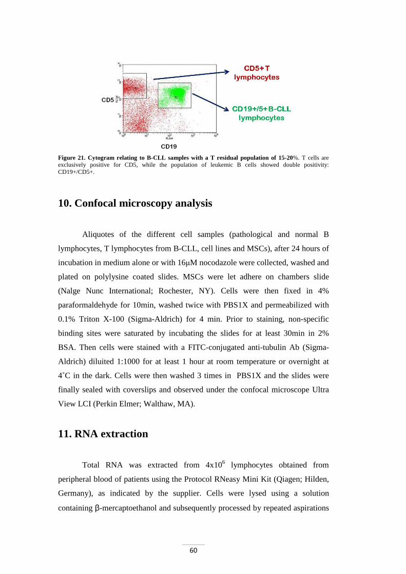
60
Figure 21. Cytogram relating to B-CLL samples with a T residual population of 15-20%. T cells are exclusively positive for CD5, while the population of leukemic B cells showed double positivity: CD19+/CD5+.
10. Confocal microscopy analysis
Aliquotes of the different cell samples (pathological and normal B
lymphocytes, T lymphocytes from B-CLL, cell lines and MSCs), after 24 hours of
incubation in medium alone or with 16µM nocodazole were collected, washed and
plated on polylysine coated slides. MSCs were let adhere on chambers slide
(Nalge Nunc International; Rochester, NY). Cells were then fixed in 4%
paraformaldehyde for 10min, washed twice with PBS1X and permeabilized with
0.1% Triton X-100 (Sigma-Aldrich) for 4 min. Prior to staining, non-specific
binding sites were saturated by incubating the slides for at least 30min in 2%
BSA. Then cells were stained with a FITC-conjugated anti-tubulin Ab (Sigma-
Aldrich) diluited 1:1000 for at least 1 hour at room temperature or overnight at
4˚C in the dark. Cells were then washed 3 times in PBS1X and the slides were
finally sealed with coverslips and observed under the confocal microscope Ultra
View LCI (Perkin Elmer; Walthaw, MA).
11. RNA extraction
Total RNA was extracted from 4x10⁶ lymphocytes obtained from
peripheral blood of patients using the Protocol RNeasy Mini Kit (Qiagen; Hilden,
Germany), as indicated by the supplier. Cells were lysed using a solution
containing β-mercaptoethanol and subsequently processed by repeated aspirations

61
with a 20G needle. After addition of 350µl of 70% ethanol, samples were
transferred to a purification column (supplied by the kit) and then centrifuged at
10.000rpm for 15 seconds, followed by eluate removal. After the addition of
350µl of RW1 buffer (supplied by the kit), samples were again centrifuged at
10.000 rpm for 15 seconds and the eluates were discarded. Then we proceeded
with 20 minutes of incubation with 80µl of a DNase I solution. After having
washed with 350µl of RW1buffer, followed by centrifugation at 10.000rpm for 15
seconds, the collector tube was replaced. 500µl of RPE buffer (supplied by the
kit) were added, the samples were centrifuged at 10.000rpm for 15 seconds, and
the eluates were discarded. After adding 500µl of RPE buffer, a centrifugation at
15.000g for 2 minutes was carried out. Columns were then transferred to 1.5ml
tubes, 30µl of H2O were added on the membrane-free RNase, and purification was
performed by centrifugation at 10.000 rpm for 1 minute, so as to dissociate the
RNA from the membranes and bring it into solution.
To check the quality of RNA obtained, 2µl of sample were sown in a 1%
agarose gel in tris-acetate (TAE) 1X with ethidium bromide (10mg/ml) 5% V/V;
electrophoresis was performed at 60V for 20 minutes. The RNA extracted was
then quantified by reading in a spectrophotometer Ultrospec 1100 pro (Amersham
Biosciences). It was prepared 1:200 dilution of RNA samples, and the absorbance
(A), at wavelengths of 260nm (λ nucleotides) and 280nm (λ proteins), were
determined. It has been estimated, therefore, the A260/A280 ratio: if a value less
than 2, the RNA preparation is contaminated with protein and is not usable. It was
finally calculated the concentration of RNA sample, using the following formula:
A260 x dilution factor x 40 = RNA concentration in µg/ml.
The value of 40 corresponds to the concentration of RNA expressed in µg/ml,
which A260 obtained is 1.
12. cDNA synthesis
The reverse transcription from mRNA to cDNA (complementary DNA)
uses a DNA-dependent RNA polymerase, which generates a cDNA strand from a
template of mRNA. In this thesis it has been used as template total RNA and the
62
reaction was performed in the presence of an oligo-dT primer, able of pairing to
the tail of poly-3' mRNA reverse transcriptase. The RT used is the Myeloblastoma
Avian Virus (AMV) polymerase; this enzyme has 5'→3' activity (with RNA or
DNA as a template) and an 3'→5' RNase H, which degrades the RNA filament of
the double helix DNA-RNA produced during cDNA synthesis. For cDNA
synthesis the Reverse Trascription System kit (Promega Corporation; Madison,
Wis.) was used.
The reaction mixture is composed of:
- 4µl of 25mM MgCl2;
- 2µl of 10X RT buffer;
- 2µl of the mixture of the 4 deoxynucleosides triphosphates (dNTP mix) 10mM;
- 0.5µl of an enzyme inhibitor of RNase 40µg/µl;
- 0.75µl of AMV RT 25µg/µl;
- 1µl of primer Oligo (dT) 0.5mg/ml;
- 2µg of RNA;
- RNase-free H2O to final volume of 20µl.
This mixture was incubated at 42°C for 15 minutes, then placed at 95°C
for 5 minutes (to stop the reaction) and, finally, put on ice for a few minutes.
Thus, the cDNA obtained was stored at -80°C or used immediately to set up the
amplification reaction by means of polymerase chain reaction (PCR) of the strand
of DNA complementary to mRNA target (Reverse Transcriptase-PCR, RT-PCR).
13. Evaluation of Lyn gene expression through real-time
RT-PCR
After having extracted and reverse-transcripted RNA from B cells of B-
CLL patients, we proceeded with the quantification of mRNA encoding for Lyn
by real-time RT-PCR. The amplification of the genes of interest were obtained
using ABI PRISM 7000 sequence detection system (Applied Biosystems; Foster
City, CA) in a volume of 15µl. The master mix used, SYBR Green PCR, was
provided by the company Applied Biosystems, containing AmpliTaq Gold DNA

63
polymerase; the primers (5µM) and 1.5µl of cDNA for a final reaction volume of
15µl were added.
The primers used for amplification of Lyn and GAPDH were:
- Lyn:
forward 5'-GCT CAG ATT GCA GAG GGA ATG G-3'
reverse 5'-GAG CCG TCC ACT TAA TAG GGA AC-3'
- GAPDH:
forward 5'-AAT GGA AAT CCC ATC ACC ATC T - 3'
reverse 5'-CGC CCC ACT TGA TTT TGG - 3'
These primers were obtained using Primer Express software (Applied
Biosystems).
The reaction conditions were:
- Denaturation at 95°C for 10 minutes;
- Annealing at 60°C for 15 seconds;
- Extension at 72°C for 1 second.
Each sample was amplified in duplicate. For each Master Mix was
performed a negative control; both for Lyn and GAPDH were generated two
standard curves using the cDNA of Raji cell line to the following dilutions: 1, 1:5,
1:25, and 1:125. The relative amount of mRNA was determined by comparison
with standard curves. The result of each sample was normalized to GAPDH
expression. A dissociation curve to distinguish specific from nonspecific
amplification was also generated.
14. Statistical analysis
Statistical analysis of apoptosis in patients analyzed was performed using
Student's t test, paired Student's t test, and ANOVA. Data were expressed as mean
± standard deviation (SD) and were considered statistically significant when p
values were <0.05.
for 45 cycles

64
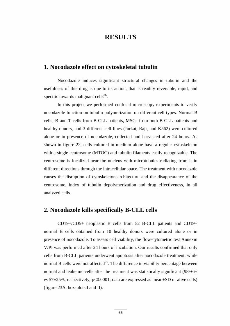
65
RESULTS
1. Nocodazole effect on cytoskeletal tubulin
Nocodazole induces significant structural changes in tubulin and the
usefulness of this drug is due to its action, that is readily reversible, rapid, and
specific towards malignant cells86.
In this project we performed confocal microscopy experiments to verify
nocodazole function on tubulin polymerization on different cell types. Normal B
cells, B and T cells from B-CLL patients, MSCs from both B-CLL patients and
healthy donors, and 3 different cell lines (Jurkat, Raji, and K562) were cultured
alone or in presence of nocodazole, collected and harvested after 24 hours. As
shown in figure 22, cells cultured in medium alone have a regular cytoskeleton
with a single centrosome (MTOC) and tubulin filaments easily recognizable. The
centrosome is localized near the nucleus with microtubules radiating from it in
different directions through the intracellular space. The treatment with nocodazole
causes the disruption of cytoskeleton architecture and the disappearance of the
centrosome, index of tubulin depolymerization and drug effectiveness, in all
analyzed cells.
2. Nocodazole kills specifically B-CLL cells
CD19+/CD5+ neoplastic B cells from 52 B-CLL patients and CD19+
normal B cells obtained from 10 healthy donors were cultured alone or in
presence of nocodazole. To assess cell viability, the flow-cytometric test Annexin
V/PI was performed after 24 hours of incubation. Our results confirmed that only
cells from B-CLL patients underwent apoptosis after nocodazole treatment, while
normal B cells were not affected91. The difference in viability percentage between
normal and leukemic cells after the treatment was statistically significant (98±6%
vs 57±25%, respectively; p<0.0001; data are expressed as mean±SD of alive cells)
(figure 23A, box-plots I and II).
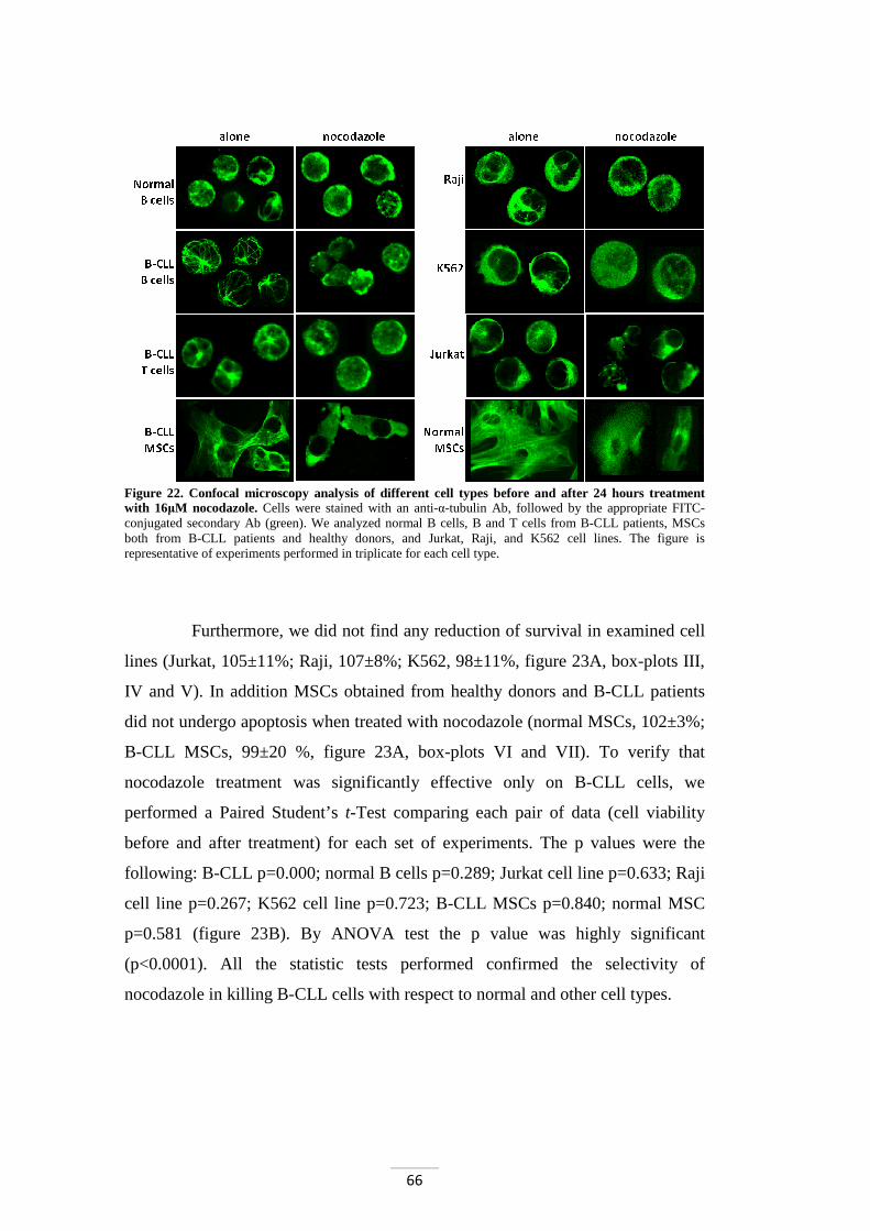
66
Figure 22. Confocal microscopy analysis of different cell types before and after 24 hours treatment with 16µM nocodazole. Cells were stained with an anti-α-tubulin Ab, followed by the appropriate FITC-conjugated secondary Ab (green). We analyzed normal B cells, B and T cells from B-CLL patients, MSCs both from B-CLL patients and healthy donors, and Jurkat, Raji, and K562 cell lines. The figure is representative of experiments performed in triplicate for each cell type.
Furthermore, we did not find any reduction of survival in examined cell
lines (Jurkat, 105±11%; Raji, 107±8%; K562, 98±11%, figure 23A, box-plots III,
IV and V). In addition MSCs obtained from healthy donors and B-CLL patients
did not undergo apoptosis when treated with nocodazole (normal MSCs, 102±3%;
B-CLL MSCs, 99±20 %, figure 23A, box-plots VI and VII). To verify that
nocodazole treatment was significantly effective only on B-CLL cells, we
performed a Paired Student’s t-Test comparing each pair of data (cell viability
before and after treatment) for each set of experiments. The p values were the
following: B-CLL p=0.000; normal B cells p=0.289; Jurkat cell line p=0.633; Raji
cell line p=0.267; K562 cell line p=0.723; B-CLL MSCs p=0.840; normal MSC
p=0.581 (figure 23B). By ANOVA test the p value was highly significant
(p<0.0001). All the statistic tests performed confirmed the selectivity of
nocodazole in killing B-CLL cells with respect to normal and other cell types.

67
Figure 23. A. Percentage of cell viability after 24 hours of culture with 16µM nocodazole. Cell apoptosis was assessed by Annexin V/PI test. The graph shows the percentage of cell viability after 24h of nocodazole treatment. Data are expressed as median, upper and lower quartiles, minimum and maximum. *p<0.0001 ANOVA test between all the cell types analyzed; **p<0.0001 Paired Student’s t-Test between leukemic and normal B cells. B. Percentage of cell viability after 24 hours of culture with or without nocodazole. Histograms show the mean±SD of cell viability percentage after 24h of cell analyzed in point A with and without the addition of 16µM nocodazole (B-CLL, n=52; normal B, n=10; Jurkat, n=3, Raji, n=3, K562, n=3; normal MSC, n=3; B-CLL MSC, n=8), *p<0.0001, compared with cells cultured in medium alone, Paired Student’s t-Test.
Considering the high variability of nocodazole effect on B-CLL cells (the
range varies from 11% to 100% of viable cells after 24 hours of treatment), we
compared the viability of normal and leukemic B cells after 24, 48 and 72 hours

68
of treatment. B cells obtained from healthy donors (figure 24A, right panel) were
not damaged even after 72 hours of nocodazole treatment; conversely,
approximately only 1/3 of leukemic B cells was still alive after 72 hours of
treatment (15±10%; figure 24A, left panel).
Figure 24. A. Percentage of cell viability after incubation with or without nocodazole in normal and leukemic B cells. Histograms show the mean±SD of percentage viability of normal B cells and B-CLL cells (left) with or without 16µM nocodazole after 24, 48 and 72 hours of all cases analyzed. *p<0.0001, Paired Student’s t-Test between treated and untreated cells. B. PARP protein expression. B-CLL and normal B cells were cultured alone or in the presence of 16µM nocodazole for 24, 48 and 72 hours. The total cell lysates were subjected to SDS-PAGE, transferred to nitrocellulose membrane and detected sequentially with anti-PARP Ab, to highlight the apoptosis, and anti-β-actin Ab. As control we used a lysate freshly isolated from B-CLL or normal B cells (t=0). The figure shows a representative case of B-CLL cells and one of normal B cells.
To confirm the effect of nocodazole on neoplastic B cell viability, we
assessed the PARP protein expression in normal and leukemic B cells after 24, 48
and 72 hours of treatment by immunoblotting. The results obtained show the
maintenance of the whole form of PARP (116kDa electrophoretic band) even
after 72 hours of treatment in normal B cells (figure 24B, right panel). In B-CLL
cells, starting from 24 hours, nocodazole treatment causes the 89kDa band of the
cleaved form of PARP, index of apoptosis induction (figure 24B, left panel), and
the disappearance of the full length of PARP.

69
3. Nocodazole kills specifically B-CLL cells with preference
for unmutated B cell clones
B-CLL cases have mutated as well as unmutated IgVH genes which largely
correlate with a favorable and unfavorable clinical prognosis, respectively16. Therefore, we correlated the ability of nocodazole to induce apoptosis in
B-CLL cells with the presence or absence of somatic hypermutations in IgVH
genes90. Taken into account the cell viability of B-CLLs after nocodazole
treatment, obtained in previous experiments. After 24 hours of nocodazole
treatment, mutated B-CLL cells showed a viability average of 67%±24, whereas
those unmutated showed a viability average of 50%±24 (figure 25), demonstrating
that nocodazole is more effective on unmutated vs mutated patients (p<0.05,
Student’s t test).
Figure 25. Correlation of leukemic B cell mutational status and cell viability after 24 hours of nocodazole treatment. Cell apoptosis was assessed by Annexin V/PI test. The graph shows the percentage of cell viability after 24h of nocodazole treatment. Data are expressed as median, upper and lower quartiles, minimum and maximum. *p<0.05, Student’s t-Test.

70
4. Nocodazole kills leukemic B cells whereas it does not
affect T cells from the same patients
Nocodazole is unable to induce Jurkat cell (a T cell line) apoptosis (figure
23A, box-plots III). This evidence prompted us to analyze residual normal T cells
from B-CLL patients. In B-CLL samples with a discrete T cell subset (at least 25-
30% of lymphocytes), we evaluated nocodazole action on T lymphocytes
performing a specific Annexin V/PI staining for different cell populations
(CD19+/5+ neoplastic B cells and CD5+ T cells).
Figure 26A shows a representative case. The overall data obtained from all
patients analyzed are summarized in the histograms in figure 26B. Interestingly,
nocodazole killed B cells but it did not affect T cells obtained from the same B-
CLL PBMCs pool (70±23% vs 30±12%; data are expressed as percentage of
viable cells after 24 hours treatment with or without nocodazole), showing a very
low toxicity for T cells (figure 26B). Data were confirmed by western blotting
analysis of cleaved form of PARP on separated B and T cell subsets. Only B, but
not T, cells showed an high amount of the PARP cleaved form, index of apoptosis
induction (figure 26C). The observation that nocodazole is not effective on T cells
is intriguingly; in fact, B-CLL patients which still have a certain percentage of T
cells show a stable disease with a relatively less aggressive clinical course. In this
view, nocodazole could be used, after proper studies, to treat these B-CLL
patients, preserving the T cell population.
5. Nocodazole effect on B-CLL cells is not counteracted
by mesenchymal stromal cells
Mesenchymal stromal cells (MSCs) regulate normal hematopoiesis by
providing attachment sites and secreted or surface-bound growth factors that
constitute the bone marrow microenvironment.

71
Figure 26. A. Percentage of annexin-positive B and T cells from the same B-CLL patients after 24 hours of nocodazole treatment. Cytograms are relative to a representative case in which the apoptosis of B and T cells from a B-CLL patient was evaluated by Annexin V/PI test after 24 hours of incabation with or without 16µM nocodazole. B. Histograms of cell viability percentage of B and T cells from B-CLL patients after 24 hours of nocodazole treatment. Histograms show the mean±SD of the percentage of viable CD19+/CD5+ B cells (left) and CD5+ T cells after 24 hours with and without 16µM nocodazole of all patients analyzed (n=19). *p<0.001, Paired Student’s t-Test. C. PARP protein expression. B and T cells from the same B-CLL patients were cultured in medium alone or in presence of 16µM nocodazole for 24 hours. The total cell lysates were subjected to SDS-PAGE, transferred to nitrocellulose membrane and detected sequentially with the anti-PARP Ab, to highlight the apoptosis, and the anti-β-actin Ab. As control we used freshly isolated B and T lymphocytes lysates from B-CLL patients (t=0). The figure shows a representative case.

72
Growing evidence suggests that MSCs protect B-CLL cells from
conventional drugs (fludarabine, dexamethasone and cyclophosphammide)97.
In this context, we investigated the reciprocal effects between nocodazole
and MSCs, obtained from B-CLL patients and healthy donors. We previously
demonstrated that MSCs from donors (102±3%, p=0.840) and from B-CLL
patients (99±20%, p=0.581) did not undergo apoptosis when cultured with
nocodazole (figure 23A, box-plots VI and VII).
Subsequently, we performed co-cultures of leukemic B and MSC cells
obtained from B-CLL patients to verify the role of bone marrow environment
influence on nocodazole activity. Surprisingly, MCSs were not able to protect B
neoplastic cells from nocodazole-mediated apoptosis. Figure 27A shows the
percentage of B-CLL viability of leukemic B cells cultured with or without a
MSC layer and incubated in presence or in absence of nocodazole for 24, 48, 72,
and 120 hours (5 days). Moreover, to increase the effectiveness of the "in vitro
environment", we performed the same experiments adding CD40L to MSCs layer,
mimicking the effect of T lymphocytes on CD40 receptor present on leukemic B
cell surface, that improves B-CLL cell survival.
Viability of leukemic B cells co-cultured with MSC without nocodazole
(B-CLL+MSC, blue line, figure 27A) remains high and essentially unchanged for
the duration of the experiment. Data on the culture of cells with nocodazole and
without the MSC layer (positive control; B-CLL+nocodazole, green line, figure
27A) confirm the apoptotic effect of the drug; there is, in fact, an important
decrease of viable cells after 24 hours of incubation, and a further gradual
reduction in subsequent detections. Data obtained from the two experimental
conditions, cultures with nocodazole (B-CLL+MSC+nocodazole, red line, figure
27A) and co-cultures in the presence of nocodazole and CD40L (B-
CLL+MSC+nocodazole+CD40L, light blue line, figure 27A), are substantially
overlapping; in fact, the percentage of viable cells is reduced at all time points
analyzed. These observations indicate that the apoptotic effect of nocodazole
overcomes the protective action given by bone marrow microenvironment.
To confirm these evidences, we evaluated the PARP protein expression by
western blotting analysis (figure 27B). The results obtained show the preservation

73
of the electrophoretic band of 116kDa relative to PARP full length only when B
cells were co-cultured with MSC at 24, 48, 72 hours and 5 days. On the contrary,
the incubation of B-CLL cells co-cultured with MSC and nocodazole, or with
MSC, nocodazole and CD40L, revealed an absolute prevalence of the
electrophoretic band of 89kDa relative to the cleaved form of PARP, index of
apoptosis.
Figure 27. A. Percentage viability of B-CLL cells co-cultured with MSCs at different conditions. B-CLL cells were co-cultured for 24, 48, 72 hours and 5 days with MSCs obtained from B-CLL patients at the following conditions: medium alone (B-CLL+MSC, blue line), with the addition of 16µM nocodazole (B-CLL+MSC+nocodazole, red line) and with the addition of nocodazole and CD40L (B-CLL+MSC+nocodazole+CD40L, light blue line); the positive control (B-CLL+nocodazole, green line) is represented by cells treated with nocodazole but not in co-culture. The graph shows the mean±SD of results obtained in all cases analyzed (n=31). B. PARP protein expression. B-CLL cells were co-cultured with MSCs in medium alone, with16µM nocodazole, and with nocodazole and CD40L for 24, 48, 72 hours and 5 days. The total cell lysates were subjected to SDS-PAGE, transferred to nitrocellulose membrane and detected sequentially with the anti-PARP Ab, to highlight the apoptosis, and the anti-β-actin Ab. The figure shows a representative case.
Finally, we performed experiments adding patient's plasma to the culture
of B-CLL with MSCs and evaluating viability percentages after 24, 48 and 72
hours of nocodazole treatment (figure 28). Once more, nocodazole activity was
not blocked.

74
Figure 28. Percentage of B-CLL cells viability after incubation with nocodazole and plasma. B-CLL cells were incubated with MSCs for 24, 48, 72 hours with 16µM nocodazole and with the addition of plasma coming from the same patients. The graph shows the mean±SD of results obtained in all cases analyzed (n=5). *p<0.005 between B-CLL+MSC and B-CLL+MSC+nocodazole and between B-CLL+MSC+plasma and B-CLL+MSC+nocodazole+plasma treatment, Paired Student’s t-Test.
6. Nocodazole targets Lyn kinase activity
Tyrosine kinase Lyn is the protein that, through its phosphorylation
activity, initiates the signal transduction cascade induced by BCR stimulation. In
B-CLL this protein is overexpressed, constitutively activated, and part of an
abnormal cytosolic complex of 600kDa.
Moreover, Lyn is responsible for the increased tyrosine phosphorylation of
B-CLL cells and sustains the survival of the neoplastic clone55,60. The constitutive
Lyn activation correlates with extended cell survival and defective apoptosis. In
fact, the inhibition of Lyn activity, obtained by treating leukemic cells with Lyn
specific inhibitors, is sufficient to restore cell apoptosis, providing a correlation
between high basal Lyn activity and defects in the induction of the programmed
cell death in B-CLL cells55.
The treatment of neoplastic cells with nocodazole revealed a decrease of
total tyrosine phosphorylation in cell lysate, which was directly correlated with
the apoptosis of neoplastic clone (see PARP protein detection, figure 29).

75
According to these results, we analyzed nocodazole effect on Lyn kinase
activity. The activity is controlled by tyrosine phosphorylation in two opposite
ways: the tyrosine phosphorylation of Lyn C-terminal Tyr507 inhibits Lyn
activity, while the phosphorylation, or autophosphorylation, at Tyr396 induces
Lyn activation. After nocodazole treatment, the expression level of Lyn protein
did not change while the phosphorylation level of Lyn active site Tyr396 was
significantly reduced (figure 29), suggesting that nocodazole may inhibit Lyn
kinase activity thus preventing B-CLL survival.
Figure 29. B-CLL proteic profile after nocodazole treatment. B-CLL cells were cultured in medium alone or in presence of 16µM nocodazole for 24, 48 and 72 hours. The total cell lysates were subjected to SDS-PAGE, transferred to nitrocellulose membrane and detected sequentially with: anti-PARP Ab to highlight the apoptosis, anti-P-Tyr which recognizes phosphorylated tyrosines, anti-Lyn-Tyr396 Ab which recognizes Lyn active conformation, anti-Lyn Ab which recognizes the entire form of Lyn, and anti-β-actin Ab. As control we used a lysate from freshly isolated B-CLL cells (t=0). The figure shows a representative case.
In addition, by real time RT-PCR, we analyzed the expression of mRNA
encoding for Lyn in B-CLL cells co-cultured with MSCs and in presence of
nocodazole and CD40L (see paragraph 4, figure 27). The results obtained show
that the treatment with nocodazole reduce mRNA level of Lyn, and this effect is
not counteracted by the presence of CD40L (figure 30A). However, these are
preliminary data, and ulterior analysis have to be performed to understand Lyn
mRNA behavior after nocodazole treatment.
Furthermore, we assessed Lyn-Tyr396 expression of the same samples by
immunoblotting (figure 30B). Also in this case, the phosphorylation level of Lyn

76
active site Tyr396 is significantly reduced, while the total Lyn expression level is
not affected, further supporting our hypothesis that nocodazole could be used Lyn
inhibitor.
Figure 30. A. Quantification of mRNA of Lyn by real time RT-PCR of B-CLL cells co-cultured with MSC. B-CLL cells were co-cultured for 24, 48, 72 hours and 5 days with MSCs obtained from B-CLL patients with the addition of 16µM nocodazole and with CD40L+nocodazole. From all the conditions the RNA was extracted and retro-transcribed to analyze the Lyn gene expression by real time RT-PCR. The graph shows the mean±SD of results obtained in all cases analyzed (n=6). *p<0.001 between medium alone and nocodazole treatment and between medium alone and CD40L+nocodazole, Paired Student’s t-Test. B. Lyn-Tyr396 and Lyn protein expression. B-CLL cells were co-cultured with MSCs in medium alone, with16µM nocodazole, and with nocodazole and CD40L for 24, 48, 72 hours and 5 days. The total cell lysates were subjected to SDS-PAGE, transferred to nitrocellulose membrane and detected sequentially with anti-Lyn-Tyr396 Ab which recognizes Lyn active conformation, anti-Lyn Ab which recognizes the entire form, and anti-β-actin Ab. The figure shows a representative case.
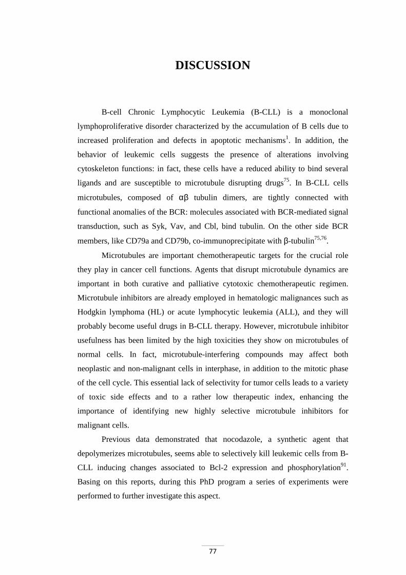
77
DISCUSSION
B-cell Chronic Lymphocytic Leukemia (B-CLL) is a monoclonal
lymphoproliferative disorder characterized by the accumulation of B cells due to
increased proliferation and defects in apoptotic mechanisms1. In addition, the
behavior of leukemic cells suggests the presence of alterations involving
cytoskeleton functions: in fact, these cells have a reduced ability to bind several
ligands and are susceptible to microtubule disrupting drugs75. In B-CLL cells
microtubules, composed of αβ tubulin dimers, are tightly connected with
functional anomalies of the BCR: molecules associated with BCR-mediated signal
transduction, such as Syk, Vav, and Cbl, bind tubulin. On the other side BCR
members, like CD79a and CD79b, co-immunoprecipitate with β-tubulin75,76.
Microtubules are important chemotherapeutic targets for the crucial role
they play in cancer cell functions. Agents that disrupt microtubule dynamics are
important in both curative and palliative cytotoxic chemotherapeutic regimen.
Microtubule inhibitors are already employed in hematologic malignances such as
Hodgkin lymphoma (HL) or acute lymphocytic leukemia (ALL), and they will
probably become useful drugs in B-CLL therapy. However, microtubule inhibitor
usefulness has been limited by the high toxicities they show on microtubules of
normal cells. In fact, microtubule-interfering compounds may affect both
neoplastic and non-malignant cells in interphase, in addition to the mitotic phase
of the cell cycle. This essential lack of selectivity for tumor cells leads to a variety
of toxic side effects and to a rather low therapeutic index, enhancing the
importance of identifying new highly selective microtubule inhibitors for
malignant cells.
Previous data demonstrated that nocodazole, a synthetic agent that
depolymerizes microtubules, seems able to selectively kill leukemic cells from B-
CLL inducing changes associated to Bcl-2 expression and phosphorylation91.
Basing on this reports, during this PhD program a series of experiments were
performed to further investigate this aspect.

78
We verified the depolymerizing effect of nocodazole on tubulin, and
investigated the apoptosis induced by this drug in B-CLL cells, and in a large
spectrum of different cells, such as normal B cells from healthy donors,
Mesenchymal Stromal Cells (MSCs) both from B-CLL patients and donors, T
lymphocytes from B-CLL patients, Jurkat, Raji, and K562 cell lines. The obtained
results confirmed that only B-CLL cells undergo apoptosis after nocodazole
treatment while the other cell types are not affected by this inhibitor. It is actually
unclear the molecular mechanism underlying the specific anti-cancer activity of
nocodazole vs B-CLL cells. To further deepen the analysis regarding nocodazole
activity on B-CLL cells, we related nocodazole effects to the mutational status of
B-CLL clones, in order to inestigate whether nocodazole action changes between
B-CLL subgroups (mutated or unmutated). Interestingly, our results demonstrated
that nocodazole effect was significantly higher in the subset of B-CLL cells with
unmutated IgVH genes, which is related to unfavorable prognosis, suggesting a
possible role of nocodazole in the treatment of unmutated B-CLLs, in association
with conventional chemotherapy.
On the other side, nocodazole was unable to induce apoptosis in Jurkat cell
line (a T cell line). This evidence prompted us to analyse residual T cells from B-
CLL patients. Our results demonstrated that nocodazole kills B cells but does not
affect T cells obtained from the same B-CLL PBMC pool, showing a very low
toxicity for T lymphocyte viability. The observation that nocodazole is not
effective on T cells is intriguing: in fact, patients who still have a discrete
percentage of T cells, show a stable disease with a relatively less aggressive
clinical course, suggesting a possible role of nocodazole also in the treatment of
these B-CLL patients, thus preserving the T cell population. Recent data indicate
that also T lymphocytes from B-CLL patients present functional deficiencies and
a compromised ability during the immune response98. This situation can be
resolved with the administration of lenalidomide, an immunomodulatory drug,
which is able to restore the T-dependent anti-tumor immune response98,99. These
data, together with our results, open the possibility of a combined use of
nocodazole and lenalidomide in B-CLL treatment and maintenance, in order to
achieve a selective pro-apoptotic action against leukemic B cells and a recovery of
T cell functions.
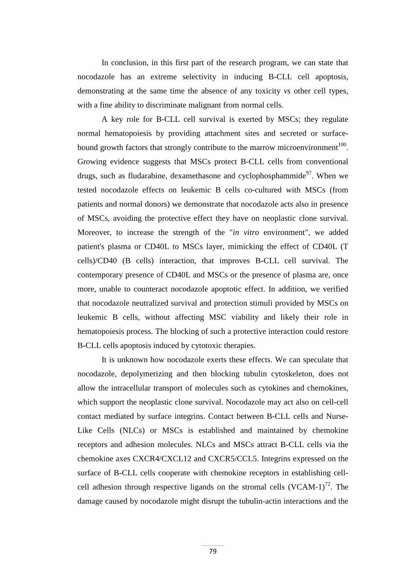
79
In conclusion, in this first part of the research program, we can state that
nocodazole has an extreme selectivity in inducing B-CLL cell apoptosis,
demonstrating at the same time the absence of any toxicity vs other cell types,
with a fine ability to discriminate malignant from normal cells.
A key role for B-CLL cell survival is exerted by MSCs; they regulate
normal hematopoiesis by providing attachment sites and secreted or surface-
bound growth factors that strongly contribute to the marrow microenvironment100.
Growing evidence suggests that MSCs protect B-CLL cells from conventional
drugs, such as fludarabine, dexamethasone and cyclophosphammide97. When we
tested nocodazole effects on leukemic B cells co-cultured with MSCs (from
patients and normal donors) we demonstrate that nocodazole acts also in presence
of MSCs, avoiding the protective effect they have on neoplastic clone survival.
Moreover, to increase the strength of the "in vitro environment", we added
patient's plasma or CD40L to MSCs layer, mimicking the effect of CD40L (T
cells)/CD40 (B cells) interaction, that improves B-CLL cell survival. The
contemporary presence of CD40L and MSCs or the presence of plasma are, once
more, unable to counteract nocodazole apoptotic effect. In addition, we verified
that nocodazole neutralized survival and protection stimuli provided by MSCs on
leukemic B cells, without affecting MSC viability and likely their role in
hematopoiesis process. The blocking of such a protective interaction could restore
B-CLL cells apoptosis induced by cytotoxic therapies.
It is unknown how nocodazole exerts these effects. We can speculate that
nocodazole, depolymerizing and then blocking tubulin cytoskeleton, does not
allow the intracellular transport of molecules such as cytokines and chemokines,
which support the neoplastic clone survival. Nocodazole may act also on cell-cell
contact mediated by surface integrins. Contact between B-CLL cells and Nurse-
Like Cells (NLCs) or MSCs is established and maintained by chemokine
receptors and adhesion molecules. NLCs and MSCs attract B-CLL cells via the
chemokine axes CXCR4/CXCL12 and CXCR5/CCL5. Integrins expressed on the
surface of B-CLL cells cooperate with chemokine receptors in establishing cell-
cell adhesion through respective ligands on the stromal cells (VCAM-1)72. The
damage caused by nocodazole might disrupt the tubulin-actin interactions and the
80
cytoskeleton, altering the correct docking site for the integrins on the inner surface
of the plasma membrane.
The attempt to understand nocodazole remarkable selectivity for leukemic
B cells prompted us to evaluate its actions in relation to cellular kinase activity. In
fact, it was recently demonstrated that nocodazole cell treatment induces
significant phosphoproteomic changes in a variety of tumor cell lines, such as
HeLa (cervical cancer), HCT-116 (colorectal carcinoma), and NCI-H460 (lung
cancer), suggesting that nocodazole may modulate kinase activity. In addition,
docking simulations indicate that the aminobenzimidazole moiety of nocodazole
can fit into the ATP binding sites of kinases, displaying high affinity toward a set
of cancer-related kinases, including ABL, c-KIT, BRAF, and MEK101. We
demonstrated that in B-CLL Lyn, a Src-kinase, is overexpressed and crucially
involved in the neoplastic B cell survival. Our experiments showed that
nocodazole modifies the anomalous Lyn-mediated phosphorylation restoring
apoptosis sensibility. In fact, nocodazole is able to down-modulate the
phosphorylation of Lyn at Tyr396, its active site. Moreover, nocodazole treatment
decreases mRNA encoding for Lyn, strongly suggesting that kinase Lyn may be
one of nocodazole targets.
Recently, we showed that, in B-CLL cells, Lyn is an integral component of
an aberrant cytosolic 600kDa complex, where Lyn is associated both with Hsp90,
HS1 (Hematopoietic lineage cell Specific protein 1), and SHP-1L. Hsp90
stabilizes the complex by contributing to converting a network of transient
interactions into permanent ones, thus maintaining Lyn in an active conformation
and preventing its degradation60. In this context, after proper studies, nocodazole
may be used to disrupt the aberrant complex, restoring the normal apoptosis
process. In fact, although several Src-family inhibitors are available and already
used in clinical management for other lymphomas, i.e. Imatinib and Dasatinib, it
is crucial to find specific inhibitors of Lyn, considering its fundamental role in B-
CLL. There are several encouraging pre-clinical reports on the use of Dasatinib in
B-CLL treatment, but they have shown few significant clinical responses.
Considering that B-CLL cells also harness a number of additional stimuli for
survival, it is possible that, in vivo, B-CLL cells may be rescued from apoptosis
by additional microenvironment factors. The pro-apoptotic effect of Dasatinib on
81
B-CLL in vitro is significantly reduced in the presence of stromal cells102, while
our results demonstrate that nocodazole is able to overcome their protective effect.
The generation of phosphotyrosine residues by Lyn and Syk results in the
activation of multiple pathways. Constitutively activated protein tyrosine kinases
(PTKs) may contribute to activate multiple signal transducers and pathways
including the PI3K/Akt/mTOR pathway, the RAS/RAF/MAPK and ERK kinase
cascade, phospholipase Cγ2 and PKC, with subsequent activation of downstream
transcription factors promoting cell survival103,104. Considering the fact that
nocodazole influences Lyn kinase activity, we can speculate that, despite it is a
known microtubule inhibitor which use is actually limited to laboratory practice, a
new role as therapeutic compound for B-CLL treatment could be taken into
account.

82
83
BIBLIOGRAPHY
1. Caligaris-Cappio F, Hamblin TJ. B-cell chronic lymphocytic leukemia: a bird of a different feather. J Clin Oncol. 1999;17:399-408.
2. Siegel R, Ward E, Brawley O, Jemal A. Cancer statistics, 2011: the impact of eliminating socioeconomic and racial disparities on premature cancer deaths. CA Cancer J Clin;61:212-236.
3. Landgren O, Gridley G, Check D, Caporaso NE, Morris Brown L. Acquired immune-related and inflammatory conditions and subsequent chronic lymphocytic leukaemia. Br J Haematol. 2007;139:791-798.
4. Landgren O, Rapkin JS, Mellemkjaer L, Gridley G, Goldin LR, Engels EA. Respiratory tract infections in the pathway to multiple myeloma: a population-based study in Scandinavia. Haematologica. 2006;91:1697-1700.
5. Wiernik PH, Ashwin M, Hu XP, Paietta E, Brown K. Anticipation in familial chronic lymphocytic leukaemia. Br J Haematol. 2001;113:407-414.
6. Barcellini W, Capalbo S, Agostinelli RM, et al. Relationship between autoimmune phenomena and disease stage and therapy in B-cell chronic lymphocytic leukemia. Haematologica. 2006;91:1689-1692.
7. Binet JL, Caligaris-Cappio F, Catovsky D, et al. Perspectives on the use of new diagnostic tools in the treatment of chronic lymphocytic leukemia. Blood. 2006;107:859-861.
8. Landgren O, Pfeiffer RM, Stewart L, et al. Risk of second malignant neoplasms among lymphoma patients with a family history of cancer. Int J Cancer. 2007;120:1099-1102.
9. Hallek M, Cheson BD, Catovsky D, et al. Guidelines for the diagnosis and treatment of chronic lymphocytic leukemia: a report from the International Workshop on Chronic Lymphocytic Leukemia updating the National Cancer Institute-Working Group 1996 guidelines. Blood. 2008;111:5446-5456.
10. Cheson BD, Bennett JM, Grever M, et al. National Cancer Institute-sponsored Working Group guidelines for chronic lymphocytic leukemia: revised guidelines for diagnosis and treatment. Blood. 1996;87:4990-4997.
11. Rassenti LZ, Huynh L, Toy TL, et al. ZAP-70 compared with immunoglobulin heavy-chain gene mutation status as a predictor of disease progression in chronic lymphocytic leukemia. N Engl J Med. 2004;351:893-901.
84
12. Mauro FR, De Rossi G, Burgio VL, et al. Prognostic value of bone marrow histology in chronic lymphocytic leukemia. A study of 335 untreated cases from a single institution. Haematologica. 1994;79:334-341.
13. Melo JV, Catovsky D, Galton DA. Chronic lymphocytic leukemia and prolymphocytic leukemia: a clinicopathological reappraisal. Blood Cells. 1987;12:339-353.
14. Hallek M, Langenmayer I, Nerl C, et al. Elevated serum thymidine kinase levels identify a subgroup at high risk of disease progression in early, nonsmoldering chronic lymphocytic leukemia. Blood. 1999;93:1732-1737.
15. Sarfati M, Chevret S, Chastang C, et al. Prognostic importance of serum soluble CD23 level in chronic lymphocytic leukemia. Blood. 1996;88:4259-4264.
16. Damle RN, Wasil T, Fais F, et al. Ig V gene mutation status and CD38 expression as novel prognostic indicators in chronic lymphocytic leukemia. Blood. 1999;94:1840-1847.
17. Ibrahim S, Keating M, Do KA, et al. CD38 expression as an important prognostic factor in B-cell chronic lymphocytic leukemia. Blood. 2001;98:181-186.
18. Ghia P, Caligaris-Cappio F. The indispensable role of microenvironment in the natural history of low-grade B-cell neoplasms. Adv Cancer Res. 2000;79:157-173.
19. Stevenson FK, Caligaris-Cappio F. Chronic lymphocytic leukemia: revelations from the B-cell receptor. Blood. 2004;103:4389-4395.
20. Poulain S, Benard C, Daudignon A, Le Baron F, Morel P, Duthilleul P. Is ZAP-70 expression stable over time in B chronic lymphocytic leukaemia? Leuk Lymphoma. 2007;48:1219-1221.
21. Degheidy HA, Venzon DJ, Farooqui MZ, et al. Methodological comparison of two anti-ZAP-70 antibodies. Cytometry B Clin Cytom. 2011. Epub ahead of print.
22. Grever MR, Lucas DM, Dewald GW, et al. Comprehensive assessment of genetic and molecular features predicting outcome in patients with chronic lymphocytic leukemia: results from the US Intergroup Phase III Trial E2997. J Clin Oncol. 2007;25:799-804.
23. Krober A, Seiler T, Benner A, et al. V(H) mutation status, CD38 expression level, genomic aberrations, and survival in chronic lymphocytic leukemia. Blood. 2002;100:1410-1416.
24. Oscier DG, Gardiner AC, Mould SJ, et al. Multivariate analysis of prognostic factors in CLL: clinical stage, IGVH gene mutational status, and loss or mutation of the p53 gene are independent prognostic factors. Blood. 2002;100:1177-1184.
85
25. Dohner H, Stilgenbauer S, Benner A, et al. Genomic aberrations and survival in chronic lymphocytic leukemia. N Engl J Med. 2000;343:1910-1916.
26. Terrin L, Trentin L, Degan M, et al. Telomerase expression in B-cell chronic lymphocytic leukemia predicts survival and delineates subgroups of patients with the same igVH mutation status and different outcome. Leukemia. 2007;21:965-972.
27. Rampazzo E, Bonaldi L, Trentin L, et al. Telomere length and telomerase levels delineate subgroups of B-cell chronic lymphocytic leukemia with different biological characteristics and clinical outcomes. Haematologica. 2012. Epub ahead of print.
28. Ward BP, Tsongalis GJ, Kaur P. MicroRNAs in chronic lymphocytic leukemia. Exp Mol Pathol;90:173-178.
29. Chemotherapeutic options in chronic lymphocytic leukemia: a meta-analysis of the randomized trials. CLL Trialists' Collaborative Group. J Natl Cancer Inst. 1999;91:861-868.
30. Rai KR, Peterson BL, Appelbaum FR, et al. Fludarabine compared with chlorambucil as primary therapy for chronic lymphocytic leukemia. N Engl J Med. 2000;343:1750-1757.
31. Dillman RO, Mick R, McIntyre OR. Pentostatin in chronic lymphocytic leukemia: a phase II trial of Cancer and Leukemia group B. J Clin Oncol. 1989;7:433-438.
32. Robak T, Blonski JZ, Kasznicki M, et al. Cladribine with prednisone versus chlorambucil with prednisone as first-line therapy in chronic lymphocytic leukemia: report of a prospective, randomized, multicenter trial. Blood. 2000;96:2723-2729.
33. Yamauchi T, Nowak BJ, Keating MJ, Plunkett W. DNA repair initiated in chronic lymphocytic leukemia lymphocytes by 4-hydroperoxycyclophosphamide is inhibited by fludarabine and clofarabine. Clin Cancer Res. 2001;7:3580-3589.
34. Flinn IW, Neuberg DS, Grever MR, et al. Phase III trial of fludarabine plus cyclophosphamide compared with fludarabine for patients with previously untreated chronic lymphocytic leukemia: US Intergroup Trial E2997. J Clin Oncol. 2007;25:793-798.
35. Alas S, Emmanouilides C, Bonavida B. Inhibition of interleukin 10 by rituximab results in down-regulation of bcl-2 and sensitization of B-cell non-Hodgkin's lymphoma to apoptosis. Clin Cancer Res. 2001;7:709-723.
36. Wierda W, O'Brien S, Wen S, et al. Chemoimmunotherapy with fludarabine, cyclophosphamide, and rituximab for relapsed and refractory chronic lymphocytic leukemia. J Clin Oncol. 2005;23:4070-4078.

86
37. Montillo M, Cafro AM, Tedeschi A, et al. Safety and efficacy of subcutaneous Campath-1H for treating residual disease in patients with chronic lymphocytic leukemia responding to fludarabine. Haematologica. 2002;87:695-700.
38. Stilgenbauer S, Dohner H. Campath-1H-induced complete remission of chronic lymphocytic leukemia despite p53 gene mutation and resistance to chemotherapy. N Engl J Med. 2002;347:452-453.
39. O'Brien S, Moore JO, Boyd TE, et al. Randomized phase III trial of fludarabine plus cyclophosphamide with or without oblimersen sodium (Bcl-2 antisense) in patients with relapsed or refractory chronic lymphocytic leukemia. J Clin Oncol. 2007;25:1114-1120.
40. Dreger P, Montserrat E. Autologous and allogeneic stem cell transplantation for chronic lymphocytic leukemia. Leukemia. 2002;16:985-992.
41. Chanan-Khan A, Miller KC, Musial L, et al. Clinical efficacy of lenalidomide in patients with relapsed or refractory chronic lymphocytic leukemia: results of a phase II study. J Clin Oncol. 2006;24:5343-5349.
42. Dighiero G. CLL biology and prognosis. Hematology Am Soc Hematol Educ Program. 2005:278-284.
43. Klein U, Tu Y, Stolovitzky GA, et al. Gene expression profiling of B cell chronic lymphocytic leukemia reveals a homogeneous phenotype related to memory B cells. J Exp Med. 2001;194:1625-1638.
44. Collins RJ, Verschuer LA, Harmon BV, Prentice RL, Pope JH, Kerr JF. Spontaneous programmed death (apoptosis) of B-chronic lymphocytic leukaemia cells following their culture in vitro. Br J Haematol. 1989;71:343-350.
45. Saxena A, Viswanathan S, Moshynska O, Tandon P, Sankaran K, Sheridan DP. Mcl-1 and Bcl-2/Bax ratio are associated with treatment response but not with Rai stage in B-cell chronic lymphocytic leukemia. Am J Hematol. 2004;75:22-33.
46. Mohr J, Helfrich H, Fuge M, et al. DNA damage-induced transcriptional program in CLL: biological and diagnostic implications for functional p53 testing. Blood. 2011;117:1622-1632.
47. Ghavami S, Hashemi M, Ande SR, et al. Apoptosis and cancer: mutations within caspase genes. J Med Genet. 2009;46:497-510.
48. Pallasch CP, Wendtner CM. Overexpression of the Fas-inhibitory molecule TOSO: a novel antiapoptotic factor in chronic lymphocytic leukemia. Leuk Lymphoma. 2009;50:498-501.
49. Niiro H, Clark EA. Regulation of B-cell fate by antigen-receptor signals. Nat Rev Immunol. 2002;2:945-956.

87
50. Schamel WW, Reth M. Monomeric and oligomeric complexes of the B cell antigen receptor. Immunity. 2000;13:5-14.
51. Cragg MS, Chan HT, Fox MD, et al. The alternative transcript of CD79b is overexpressed in B-CLL and inhibits signaling for apoptosis. Blood. 2002;100:3068-3076.
52. Gupta N, DeFranco AL. Visualizing lipid raft dynamics and early signaling events during antigen receptor-mediated B-lymphocyte activation. Mol Biol Cell. 2003;14:432-444.
53. Gross AJ, Proekt I, Defranco AL. Elevated BCR signaling and decreased survival of Lyn-deficient transitional and follicular B cells. Eur J Immunol. 2011. Epub ahead of print.
54. Mlinaric-Rascan I, Yamamoto T. B cell receptor signaling involves physical and functional association of FAK with Lyn and IgM. FEBS Lett. 2001;498:26-31.
55. Contri A, Brunati AM, Trentin L, et al. Chronic lymphocytic leukemia B cells contain anomalous Lyn tyrosine kinase, a putative contribution to defective apoptosis. J Clin Invest. 2005;115:369-378.
56. Luciano F, Ricci JE, Auberger P. Cleavage of Fyn and Lyn in their N-terminal unique regions during induction of apoptosis: a new mechanism for Src kinase regulation. Oncogene. 2001;20:4935-4941.
57. Thomas ML, Brown EJ. Positive and negative regulation of Src-family membrane kinases by CD45. Immunol Today. 1999;20:406-411.
58. Donella-Deana A, Cesaro L, Ruzzene M, Brunati AM, Marin O, Pinna LA. Spontaneous autophosphorylation of Lyn tyrosine kinase at both its activation segment and C-terminal tail confers altered substrate specificity. Biochemistry. 1998;37:1438-1446.
59. Caplan AJ, Mandal AK, Theodoraki MA. Molecular chaperones and protein kinase quality control. Trends Cell Biol. 2007;17:87-92.
60. Trentin L, Frasson M, Donella-Deana A, et al. Geldanamycin-induced Lyn dissociation from aberrant Hsp90-stabilized cytosolic complex is an early event in apoptotic mechanisms in B-chronic lymphocytic leukemia. Blood. 2008;112:4665-4674.
61. Chiorazzi N. Cell proliferation and death: forgotten features of chronic lymphocytic leukemia B cells. Best Pract Res Clin Haematol. 2007;20:399-413.
62. Azimzadeh J, Bornens M. Structure and duplication of the centrosome. J Cell Sci. 2007;120:2139-2142.
63. Kramer A, Neben K, Ho AD. Centrosome aberrations in hematological malignancies. Cell Biol Int. 2005;29:375-383.

88
64. Hensel M, Zoz M, Giesecke C, et al. High rate of centrosome aberrations and correlation with proliferative activity in patients with untreated B-cell chronic lymphocytic leukemia. Int J Cancer. 2007;121:978-983.
65. Ghia P, Granziero L, Chilosi M, Caligaris-Cappio F. Chronic B cell malignancies and bone marrow microenvironment. Semin Cancer Biol. 2002;12:149-155.
66. Ding W, Nowakowski GS, Knox TR, et al. Bi-directional activation between mesenchymal stem cells and CLL B-cells: implication for CLL disease progression. Br J Haematol. 2009;147:471-483.
67. Gattei V, Bulian P, Del Principe MI, et al. Relevance of CD49d protein expression as overall survival and progressive disease prognosticator in chronic lymphocytic leukemia. Blood. 2008;111:865-873.
68. Shanafelt TD, Geyer SM, Bone ND, et al. CD49d expression is an independent predictor of overall survival in patients with chronic lymphocytic leukaemia: a prognostic parameter with therapeutic potential. Br J Haematol. 2008;140:537-546.
69. Redondo-Munoz J, Jose Terol M, Garcia-Marco JA, Garcia-Pardo A. Matrix metalloproteinase-9 is up-regulated by CCL21/CCR7 interaction via extracellular signal-regulated kinase-1/2 signaling and is involved in CCL21-driven B-cell chronic lymphocytic leukemia cell invasion and migration. Blood. 2008;111:383-386.
70. Munk Pedersen I, Reed J. Microenvironmental interactions and survival of CLL B-cells. Leuk Lymphoma. 2004;45:2365-2372.
71. Jordan MA, Wilson L. Microtubules as a target for anticancer drugs. Nat Rev Cancer. 2004;4:253-265.
72. Burger JA, Ghia P, Rosenwald A, Caligaris-Cappio F. The microenvironment in mature B-cell malignancies: a target for new treatment strategies. Blood. 2009;114:3367-3375.
73. Singh P, Rathinasamy K, Mohan R, Panda D. Microtubule assembly dynamics: an attractive target for anticancer drugs. IUBMB Life. 2008;60:368-375.
74. Caligaris-Cappio F, Bergui L, Tesio L, Corbascio G, Tousco F, Marchisio PC. Cytoskeleton organization is aberrantly rearranged in the cells of B chronic lymphocytic leukemia and hairy cell leukemia. Blood. 1986;67:233-239.
75. Fernandez JA, Keshvara LM, Peters JD, Furlong MT, Harrison ML, Geahlen RL. Phosphorylation- and activation-independent association of the tyrosine kinase Syk and the tyrosine kinase substrates Cbl and Vav with tubulin in B-cells. J Biol Chem. 1999;274:1401-1406.
89
76. Draberova L, Draberova E, Surviladze Z, Draber P, Draber P. Protein tyrosine kinase p53/p56(lyn) forms complexes with gamma-tubulin in rat basophilic leukemia cells. Int Immunol. 1999;11:1829-1839.
77. Mollinedo F, Gajate C. Microtubules, microtubule-interfering agents and apoptosis. Apoptosis. 2003;8:413-450.
78. Conde C, Caceres A. Microtubule assembly, organization and dynamics in axons and dendrites. Nat Rev Neurosci. 2009;10:319-332.
79. Imazio M, Bobbio M, Cecchi E, et al. Colchicine in addition to conventional therapy for acute pericarditis: results of the COlchicine for acute PEricarditis (COPE) trial. Circulation. 2005;112:2012-2016.
80. Mohan R, Panda D. Kinetic stabilization of microtubule dynamics by estramustine is associated with tubulin acetylation, spindle abnormalities, and mitotic arrest. Cancer Res. 2008;68:6181-6189.
81. Altmann KH, Wartmann M, O'Reilly T. Epothilones and related structures--a new class of microtubule inhibitors with potent in vivo antitumor activity. Biochim Biophys Acta. 2000;1470:M79-91.
82. Jaksic B, Brugiatelli M, Krc I, et al. High dose chlorambucil versus Binet's modified cyclophosphamide, doxorubicin, vincristine, and prednisone regimen in the treatment of patients with advanced B-cell chronic lymphocytic leukemia. Results of an international multicenter randomized trial. International Society for Chemo-Immunotherapy, Vienna. Cancer. 1997;79:2107-2114.
83. Barr PM, Lazarus HM, Cooper BW, et al. Phase II study of bryostatin 1 and vincristine for aggressive non-Hodgkin lymphoma relapsing after an autologous stem cell transplant. Am J Hematol. 2009;84:484-487.
84. De Brabander MJ, Van de Veire RM, Aerts FE, Borgers M, Janssen PA. The effects of methyl (5-(2-thienylcarbonyl)-1H-benzimidazol-2-yl) carbamate, (R 17934; NSC 238159), a new synthetic antitumoral drug interfering with microtubules, on mammalian cells cultured in vitro. Cancer Res. 1976;36:905-916.
85. Hoebeke J, Van Nijen G, De Brabander M. Interaction of oncodazole (R 17934), a new antitumoral drug, with rat brain tubulin. Biochem Biophys Res Commun. 1976;69:319-324.
86. Head J, Lee LL, Field DJ, Lee JC. Equilibrium and rapid kinetic studies on nocodazole-tubulin interaction. J Biol Chem. 1985;260:11060-11066.
87. Vasquez RJ, Howell B, Yvon AM, Wadsworth P, Cassimeris L. Nanomolar concentrations of nocodazole alter microtubule dynamic instability in vivo and in vitro. Mol Biol Cell. 1997;8:973-985.
88. Vandecandelaere A, Martin SR, Bayley PM. Regulation of microtubule dynamic instability by tubulin-GDP. Biochemistry. 1995;34:1332-1343.

90
89. Kallas A, Pook M, Maimets M, Zimmermann K, Maimets T. Nocodazole treatment decreases expression of pluripotency markers Nanog and Oct4 in human embryonic stem cells. PLoS One. 2011;6:e19114.
90. Aylon Y, Michael D, Shmueli A, Yabuta N, Nojima H, Oren M. A positive feedback loop between the p53 and Lats2 tumor suppressors prevents tetraploidization. Genes Dev. 2006;20:2687-2700.
91. Beswick RW, Ambrose HE, Wagner SD. Nocodazole, a microtubule de-polymerising agent, induces apoptosis of chronic lymphocytic leukaemia cells associated with changes in Bcl-2 phosphorylation and expression. Leuk Res. 2006;30:427-436.
92. Braun J, Unanue ER. The lymphocyte cytoskeleton and its control of surface receptor functions. Semin Hematol. 1983;20:322-333.
93. Cohen HJ. Human lymphocyte surface immunoglobulin capping. Normal characteristics and anomalous behavior of chronic lymphocytic leukemic lymphocytes. J Clin Invest. 1975;55:84-93.
94. Modriansky M, Dvorak Z. Microtubule disruptors and their interaction with biotransformation enzymes. Biomed Pap Med Fac Univ Palacky Olomouc Czech Repub. 2005;149:213-215.
95. Polioudaki H, Kastrinaki MC, Papadaki HA, Theodoropoulos PA. Microtubule-interacting drugs induce moderate and reversible damage to human bone marrow mesenchymal stem cells. Cell Prolif. 2009;42:434-447.
96. Wu X, Koiwa H. One-step casting of Laemmli discontinued sodium dodecyl sulfate-polyacrylamide gel electrophoresis gel. Anal Biochem;421:347-349.
97. Kurtova AV, Balakrishnan K, Chen R, et al. Diverse marrow stromal cells protect CLL cells from spontaneous and drug-induced apoptosis: development of a reliable and reproducible system to assess stromal cell adhesion-mediated drug resistance. Blood. 2009;114:4441-4450.
98. Ramsay AG, Johnson AJ, Lee AM, et al. Chronic lymphocytic leukemia T cells show impaired immunological synapse formation that can be reversed with an immunomodulating drug. J Clin Invest. 2008;118:2427-2437.
99. Gorgun G, Ramsay AG, Holderried TA, et al. E(mu)-TCL1 mice represent a model for immunotherapeutic reversal of chronic lymphocytic leukemia-induced T-cell dysfunction. Proc Natl Acad Sci U S A. 2009;106:6250-6255.
100. Roberts R, Gallagher J, Spooncer E, Allen TD, Bloomfield F, Dexter TM. Heparan sulphate bound growth factors: a mechanism for stromal cell mediated haemopoiesis. Nature. 1988;332:376-378.

91
101. Park H, Hong S, Hong S. Nocodazole is a High-Affinity Ligand for the Cancer-Related Kinases ABL, c-KIT, BRAF, and MEK. ChemMedChem. 2012;7:53-56.
102. McCaig AM, Cosimo E, Leach MT, Michie AM. Dasatinib inhibits B cell receptor signalling in chronic lymphocytic leukaemia but novel combination approaches are required to overcome additional pro-survival microenvironmental signals. Br J Haematol. 2011. Epub ahead of print.
103. Wickremasinghe RG, Prentice AG, Steele AJ. Aberrantly activated anti-apoptotic signalling mechanisms in chronic lymphocytic leukaemia cells: clues to the identification of novel therapeutic targets. Br J Haematol;153:545-556.
104. Ma S, Rosen ST. Signal transduction inhibitors in chronic lymphocytic leukemia. Curr Opin Oncol. 2011;23:601-608.