UNIVERSITÀ DEGLI STUDI DI PADOVApaduaresearch.cab.unipd.it/724/1/PhD%20stampata%20per... ·...
229
UNIVERSITÀ DEGLI STUDI DI PADOVA Sede Amministrativa: Università degli Studi di Padova Dipartimento di Biotecnologie Agrarie DOTTORATO DI RICERCA IN: Viticoltura, Enologia e Marketing delle Imprese vitivinicole XX° CICLO TITOLO TESI Caratterizzazione biochimica e funzionale delle proteine dei vini bianchi e studio di metodi per la prevenzione della loro instabilità Biochemical and functional characterisation of the proteins of white wines and studies on methods to prevent their instability Coordinatore: Ch.mo Prof. Giorgio Ponchia Supervisore: Ch.mo Prof. Andrea Curioni Dottorando: Matteo Marangon DATA CONSEGNA TESI 31 gennaio 2008
Transcript of UNIVERSITÀ DEGLI STUDI DI PADOVApaduaresearch.cab.unipd.it/724/1/PhD%20stampata%20per... ·...
UNIVERSITÀ DEGLI STUDI DI
PADOVA
SedeAmministrativa: Università degli Studidi Padova
Dipartimento di BiotecnologieAgrarie
DOTTORATO DI RICERCA IN: Viticoltura,EnologiaeMarketing delle Imprese
vitivinicole
XX° CICLO
TITOLO TESI
CCaarraatttteerriizzzzaazziioonnee bbiioocchhiimmiiccaa ee ffuunnzziioonnaallee ddeellllee pprrootteeiinnee ddeeii vviinnii bbiiaanncchhii ee ssttuuddiioo ddiimmeettooddii ppeerr llaa pprreevveennzziioonnee ddeellllaa lloorroo iinnssttaabbiilliittàà
BBiioocchheemmiiccaall aanndd ffuunnccttiioonnaall cchhaarraacctteerriissaattiioonn ooff tthhee pprrootteeiinnss ooff wwhhiittee wwiinneess aanndd ssttuuddiieessoonn mmeetthhooddss ttoo pprreevveenntt tthheeiirr iinnssttaabbiilliittyy
Coordinatore: Ch.moProf.Giorgio Ponchia
Supervisore: Ch.moProf.AndreaCurioni
Dottorando: Matteo Marangon
DATA CONSEGNATESI31gennaio2008
II
Acknowledgements
Firstly I would like to thank the “Provincia di Treviso” for the financial
supportto my research activity in the“Dottoratoin Vi ticoltura,Enologiae Marketing
delle Impresevitivinicole”.
I wouldalsolike to thankall thosepeoplewhohavehelped finish my PhD.
To my supervisor, Prof. AndreaCurioni, for giving methepreciousoccasion
to work with him during thesethree years, for the helpful discussion about the
experimentsand for all the time he spent in the revision of this manuscript; to
Simone Vincenzi, for his constantcollaboration with my research activity, for his
precious teachings and, not last, for his friendship; to Prof. Francesco Favaron,
SilvanaOdorizzi, DeborahFranceschi,Mara Vegro, Marco Lucchetta, Luca Sella,,
Mario Licari, and Loris Billo for their teachings, assistance in the experiments
conduction and valuable discussions about this work; to the entire staff of “The
AustralianWine ResearchInstitute” of Adelaide (SouthAustralia), whereI spent6
fantasticmonths. Thanksin particularto Elizabeth WatersandSteveVan Sluyterfor
their scientific support,for thevaluablediscussionsandthecritical review of part of
this manuscript.
Thank you also to the most important people around me, my family and
friends,in particular to those I met during my way to thePhD: to Simone,Stefania,
Tiziana,Raul,Elena,Maura,Deborah,Luca,Mi lena, Robyn,Maurizio, Nadia,Steve,
Leslie,Coca,Toni, Claudia,Geoff, Maria, Inma and manyother for your friendship
and to havedividedwith methepleasantandbadmoments.
A specialthanksto Robertafor the pricelesssupportyou gave to me during
this time.Wordscan’t sayhowmuchof this PhDis dueto your loveandsupport.

III
IV
Thesis summary
The presenceof a residual amount of unstable proteins in wines is a
concern for winemakersbecausethesepolymers can precipitate from solution
during storage causingappearanceof sediments andhazes.Such precipitatesare
commonly the resultof denaturationandsubsequentaggregation of heat-unstable
wine proteins.It hasbeendemonstratedthat thevast majority of thewine proteins
derive from grapes and that proteins responsible for haze formation are
Pathogenesis-Related(PR)proteins, in particular thaumatin-like (TL) proteinsand
chitinases. Moreover,theseproteinsarehighly resistant to acidic pHs,proteolysis
and fermentation conditions. However, they can became insoluble during wine
storage and thusoriginateperceptible turbidity in the bottles. Despitethe efforts
madein the recentpast,the white wine protein instabilit y is still a main problem
during white winemakingandbentonitetreatmentsareevennow indispensable to
stabilisewhitewines.
This thesisis focusedon thestudyof grapeand wineproteins in relation to
white wine instability. This three-yearsstudy has beendoneby facing different
problems.
Firstly, theeffectsof the alcoholicfermentation on themacromoleculesof
a white wine wasevaluated,in orderto make clearhow this processcan affect the
heat-stability of different wine protein fractions, as obtained through Anion-
Exchange Chromatography(AEC). In particular, through the study of the
macromolecular composition of a must/wine throughout the alcoholic
fermentationand by the study of the intrinsic heat-instability of fractionated
proteins,thevariation in both quantityandrelative proportionof macromolecules
andstabilityof particularproteinswasassessed.
Besides, a methodsuitablefor fractionationand purification of grape and
wine proteins was set up by using Hydrophobic Interaction Chromatography
(HIC). This method was usedto purify a thaumatin-like protein. Moreover, 26
grape juice proteinswereidentifiedby matching peptide LC-MS/MS spectra with
theoreticalpeptidesfrom aplantproteindatabase.
Furthermore,HIC was also usedas a methodto preparegrapejuice and
wine protein fractionsdiffering in hydrophobicity. After partial characterisation of
V
these fractions by means of different chromatographic techniques, protein
hydrophobicity was studied in relation to the heat-stability of the separated
fractions and also to their capability in forming insoluble aggregates through
reactionwith seedtannins.
At the sametime, the study of methodsalternative to bentonite fining for
protein removalof from wines hasbeencarried out. In particular, this problem
wasfacedtrying to find proteolyticenzymes, active at wine pH, able to degrade
the grape PR-proteins. To this aim, the acidic protease activit y of four
phytopathogenicfungalstrainswastested.
During the study of one of these fungi, we noticed that a polysaccharide
(scleroglucan)emitted by the fungusSclerotium rolfsii during its growth,hadthe
ability to adsorb grape and wine proteins in solution. For this reason, the
functionality of scleroglucanhas been studied to verify the possibili ty of its
utilizationasanewmaterialsuitablefor protein removal from wine.

VI
Riassunto
La presenzadi quantitàresiduedi proteineinstabili nei vini è un motivo di
apprensione per i produttori, in quantotali polimeri possonodivenire insolubili
durantelo stoccaggiodei vini e precipitare,causando la comparsadi sedimenti e
torbidità. Tali precipitati sono generalmente il risultato di una denaturazione e
successiva aggregazionedelleproteineinstabili del vino.È stato dimostratochela
maggioranza delle proteine del vino derivano dall’uva e che le proteine
responsabili per la formazionedi torbidità sono proteine legate alla patogenesi
(PR proteins),in particolareproteine taumatina-simili (TL) e chitinasi. È stato
osservato che tali proteine sono resistenti a pH acidi, alla proteolisi ed alle
condizioni di fermentazione,anche sequesta resistenza può veniremenodurante
lo stoccaggio del vino. Nonostantegli sforzi fatti nel recentepassato,l’i nstabilità
proteica è ancora il principale problema di origine non microbiologica nella
produzione di vini bianchi e i trattamenti con bentonite sono ancora oggi
indispensabiliduranteil processodi produzionedi tali vini.
In questa tesi si sono volute studiare le proteine dell’uva e del vino in
relazione al problemadell’instabilità proteica dei vini bianchi e questostudio è
stato condotto in questo contesto cercandodi dare delle risposte a diversi
problemi.
Sono stativalutati gli effetti dellafermentazionealcolica sullacomponente
macromolecolare di un vino (Manzoni bianco), al fine di chiarire come la
fermentazione influisse sulla stabilità al calore di frazioni proteiche ottenute
mediantecromatografiaa scambioanionico(AEC).
In seguito,è stato messo a punto un metodo per il frazionamento e la
purificazione delle proteine di uva e vino mediante cromatografia a scambio
idrofobico (HIC), e tale metodo è stato utili zzato per la purificazione
all’omogeneità di una proteina taumatina-simile. Inoltre, 26 bande proteiche
ottenute dal frazionamentodi proteine di mosto Semillon sono state analizzate
mediante LC-MS/MS ed identificateper mezzo di comparazione delle sequenze
depositatein database.
Inoltre, la cromatografiaad interazioneidrofobica è stata utili zzata anche
comemetodopreparativofinalizzatoad ottenerefrazioni proteichecaratterizzate

VII
dadifferenti livelli di idrofobicità.Dopounaprima caratterizzazionedelle frazioni
ottenutamediantevarie tecniche cromatografiche, l’i drofobicità delle frazioni
proteicheottenute è statamessa in relazionealla loro stabilità al caloreedalla loro
capacitàdi reagirecontanninidi vinaccioli formandocompostiinsolubili.
Parallelamente, il lavoro di tesi si è incentrato sullo studio di metodi
alternativiallabentoniteperla rimozionedelleproteine fonted’instabilitàproteica
nei vini bianchi.In particolare,si è cercato di risolveretale problemamediante la
ricercadi proteasiattiveapH acidi in gradodi degradarelePR-proteins.A tal fine
sono stati studiati quattro ceppi fungini per valutarne l’attitudine a produrre
enzimi proteolitici in gradodi degradarele proteinedell’uva.
Dallo studiodi uno di questi funghi (Sclerotium rolfsii) si è notato cheun
polisaccaride da essoprodottodurantela suacrescita, lo scleroglucano, aveva la
capacitàdi adsorbire le proteinedi uva e vino. Perquestomotivo è statastudiata
la funzionalità di tale polimero al fine di verificare la possibilità di un suo
possibileutilizzo perla rimozionedelleproteinedal vino.

VIII
List of abbreviations
aa: aminoacid;
AEC: Anion ExchangeChromatography;
Asx: Aspartic acid or asparagine(undefined);
B24: Sclerotinia sclerotiorum;
BC: Botrytis cinerea;
BSA: bovin serumalbumin;
Cyt C: horseheartcytochromeC;
Glx: Glutamicacid or glutamine(undefined).
HIC: HydrophobicInteractionChromatography;
LTP: Lipid TransferProtein;
MW: molecularweight;
MWCO: molecularweightcut off;
PAS: Periodicacid-Schiff staining;
pI: Isoelectricpoint;
PR-proteins: Pathogenesis-relatedproteins;
RI: refractiveindex;
RT: Retention time;
SEC: Sizeexclusionchromatography;
SM: Sclerotinia minor;
SR: Sclerotium rolfsii;
TL-protein: thaumatin-likeprotein;
UF: Ultrafil teredwine;
VvTL: Vitis vinifera ThaumatinLike protein.

IX

X
Table of contents
Acknowledgements I
Thesis summary IV
Riassunto VI
List of abbreviations VIII
CHAPTER 1
Introduction 1
1.1 Theorigin of thewineproteins 2
1.2 Characteristicsof thewineproteins 4
1.3 Proteinhazein white wines 6
1.4 GrapePathogenesisRelatedproteins(PR-proteins) 7
1.5 Enologicalcontrolof white winesproteininstability 111.5.1 History of wine protein fining 111.5.2 Bentonite fining 111.5.3 Bentonite alternativetechniquesfor protein removal 12
1.5.3.1 Tangential ultrafiltration 121.5.3.2 Immobil izedphenoliccompounds 131.5.3.3 Alternative adsorbents 131.5.3.4 Flashpastorization. 141.5.3.5 Proteolytic enzymes 141.5.3.6 Hazeprotectivefactors 151.5.3.7 Genetic methods 15
1.6 Scientificobjectivesof thethesis 16
1.7 References 16
CHAPTER 2
White wine protein evolution during fermentation and post-fermentation operations:relationship with protein stability 29
2.1 Abstract 29
2.2 Introduction 29
2.3 MaterialsandMethods 302.3.1 Wine preparation 302.3.2 Analytical methods 312.3.3 Protein contentdetermination 312.3.4 Total polysaccharide contentdetermination 312.3.5 Total polyphenols contentdetermination 312.3.6 Total protein preparation 322.3.7 AnionExchangeChromatography (AEC) 322.3.8 Reverse Phase(RP)-HPLC 322.3.9 Sodiumdodecyl sulfatepolyacrylamidegel electrophoresis (SDS-PAGE) 332.3.10 Heattest 33
2.4 Results 34
XI
2.4.1 Discussion 46
2.5 References 48
CHAPTER 3
Fractionation of grape juice and wine proteins by hydrophobic interactionchromatography 55
3.1 Abstract 55
3.2 Introduction 55
3.3 MaterialsandMethods 573.3.1 Materials 573.3.2 Ammoniumsulphateproteinprecipitation 573.3.3 Residual Ammoniadetermination 573.3.4 Sodiumdodecyl sulfatepolyacrylamidegel electrophoresis (SDS-PAGE) 583.3.5 Grapeandwine protein chromatography 583.3.6 Hydrophobic InteractionChromatography 583.3.7 RP-HPLC proteinAnalyses 59
3.3.7.1 Analysisandquantificationof protein by PR-HPLC 593.3.7.2 HPLCproteinanalysesby Size ExclusionChromatography 59
3.3.8 Samplesdesalting 603.3.9 Protein identification through LC-MS/MS analyses 603.3.10 Amino Acidsanalysis 61
3.4 ResultsandDiscussion 623.4.1 Grapeproteins fractionation: Resinselection 623.4.2 Grapeproteins fractionation: largescale experimentsand VvTL proteinpurification
633.4.2.1 VvTL protein quantification 70
3.4.3 Semil lon grapeproteinsidentifi cation 733.4.3.1 ESI-MS/MS Proteinidentification 75
3.4.4 Semil lon wine proteinfractionation 843.4.5 Conclusions 89
3.5 References 91
CHAPTER 4
Fractionation of wine proteins based on hydrophobicity and characterization of theirheat instability and reactivity with tannins 97
4.1 Abstract 97
4.2 Introduction 98
4.3 MaterialsandMethods 994.3.1 Materials 994.3.2 Protein extraction from wine 100
4.3.2.1 Concentrationby ultrafiltration 1004.3.2.2 Proteinprecipitation with potassiumdodecylsulphate(kds) 100
4.3.3 Grapeandwine protein contentdetermination 1004.3.4 Total polysaccharide content determination 1014.3.5 Heattest 1014.3.6 Sodiumdodecyl sulfatepolyacrylamidegel electrophoresis (sds-page) 1014.3.7 Zymographyfor chitinaseactivity detection 1024.3.8 Wine proteinseparation by chromatography 1024.3.9 SizeExclusionChromatography 103

XII
4.3.10 Hydrophobic InteractionChromatography 1034.3.11 High Performance Liquid Chromatography (HPLC) 103
4.3.11.1 ReversePhase(RP)-HPLC 1034.3.11.2 SizeExclusion(SE) - HPLC 104
4.4 Resultsanddiscussion 1044.4.1 Fractionationof wine proteins by SizeExclusionChromatography 1044.4.2 Studiesonproteinfractionationbased on their hydrophobicity 107
4.4.2.1 Hazepotential of wine proteinsasrelatedto their hydrophobicity 1164.4.3 Studieson wine proteinreactivity with seed tannins 119
4.4.3.1 Preliminary experiments 1194.4.3.2 HIC fractionation of wineproteins 1214.4.3.3 Studieson thenature of HIC wine protein fractions 1224.4.3.4 Heatstability of wine proteinfractionsseparated by Hydrophobic InteractionChromatography 1314.4.3.5 Hazeformationafteraddition of seedtanninsto wine proteinfractionsdifferingin hydrophobicity 133
4.5 REFERENCES 139
CHAPTER 5
Selection of fungal proteases for the degradation of grape proteins 147
5.1 Abstract 147
5.2 Introduction 147
5.3 MaterialsandMethods 1495.3.1 Materials 1495.3.2 Protein extraction from grapes,wine andfungal cultures 149
5.1.1.1. Concentrationby ultrafiltration 1495.1.1.2. Proteinprecipitation with potassiumdodecylsulphate (KDS) 149
5.3.3 Grapeand wine protein contentdetermination 1505.3.4 Total polysaccharide contentdetermination 1505.3.5 Enzymatic assayfor acidic proteasesdetermination with heamoglobinassubstrate
1505.3.6 Assay for acidic proteasesactivity determination with wine proteinsasthesubstrate
1515.3.7 Heattest 1515.3.8 Sodiumdodecyl sulfatepolyacrylamidegel electrophoresis (SDS-PAGE) 1525.3.9 Grapeand wine protein chromatography 1525.3.10 Reverse Phase(RP)-HPLC 1535.3.11 Fungal cultures: experimentaldesign 153
5.1.1.3. Fungal culturesin liquid media 1545.3.12 Statisticalanalysis 154
5.4 ResultsandDiscussion 1545.4.1 Preliminary resultsonproteasesemissionby fungal strains 1545.4.2 Proteolytic activity of Sclerotium minor 1675.4.3 Proteolytic activity of Sclerotium rolfsii 174
5.5 References 185
CHAPTER 6
Scleroglucan-Protein interaction: a tool for protein removal from wine? 191
6.1 Abstract 191
XI II
6.2 Introduction 192
6.3 MaterialsandMethods 1936.3.1 Materials 1936.3.2 Grapeandwine protein contentdetermination 1936.3.3 Total polysaccharide content determination 1936.3.4 Total polyphenolscontentdetermination 1946.3.5 Heattest 1946.3.6 Sodiumdodecyl sulfatepolyacrylamidegel electrophoresis (SDS-PAGE) 1946.3.7 Statistical analysis 195
6.4 ResultsandDiscussion 1956.4.1 Kinetic of scleroglucan-proteininteractions 1956.4.2 Solventeffect on scleroglucan-protein interactions 2016.4.3 Ionic strengtheffect onscleroglucan-protein interactions 2026.4.4 Effect of thehighMW endogenouswine compoundsonscleroglucan-proteininteractions. 2036.4.5 Effect of protein typeon theinteractionswith scleroglucan 2036.4.6 Effectsof scleroglucanaddition onheatstability of model solutions 2056.4.7 Conclusions 206
6.5 REFERENCES 207
CHAPTER 7
Conclusions 211

1
1CHAPTER 1
Introduction
Soluble heat-unstable proteins, mainly deriving from grapes, are
recoverablein white wines also after bottling. Haze formation in white wine is
still a matter of concernfor winemakers, and the presenceof residualprotein in
fined wines is mainly related to the possibleappearance of haze during wine
storagein the bottle. Haze appearanceis considered the worst fault of non-
microbiological origin affecting white wines, leading consumersto refuse the
product also if it is no significantly modified from a sensorialpoint of view
(Bayly andBerg, 1967;Hsu andHeatherbell, 1987a; Waterset al., 1992).
Proteins are one of the threemain macromolecular compoundsof must
andwine togetherwith polysaccharidesandpolyphenols.Theproteinsresponsible
for hazehavebeenidentified asPathogenesis-Related(PR) proteins, in particular
thaumatin-like (TL) proteins andchitinases, deriving from grapeberries (Waters
et al., 1996, 1998). These PR-proteins are likely to protect the berry during
ripeningagainstfungalpathogens(Høj et al., 2000).
As reported by several authors (Bayly and Berg, 1967; Somers and
Ziemelis, 1973;Hsu andHeatherbell,1987a;Murpheyet al., 1989a;Murpheyet
al., 1989b; Dorrestein et al., 1995;Santoro,1995;Pocock et al., 1998),theprotein
level measuredin winescanbeveryvariabledueto thenumerousfactors(variety,
climate, ripening time, harvest methods, type of winemaking, stabilising
treatments,assayadoptedto measureprotein content,etc.) affecting it. Generally
protein amounts varying from few to several hundreds mill igrams per litre are
detectablein white wines. However,no relationship betweenprotein content and
wines instability hasbeenfound to date(Sarmento et al., 2000; Ferreira et al.,
2002).
Despitebentonite is effective in protein removal from wines (Blade and
Boulton, 1988;Achaerandioet al., 2001; Ferreira et al., 2002),its utili sation is not
without consequences on wine quality. In particular, bentonite is considered as
responsiblefor simplification of the aromatic profile of the wine and for the loss
of colour and of compoundsuseful for the wine structure (Høj et al., 2000).
2
Moreover,thewine volumelost after bentonite fining can vary from a 3 to 10 %
(Tattersall et al., 2001) with highcosts for wineries(Høj et al., 2000).
For these reasons,theresearchis activeon trying to improvethebentonite
efficacy (Muhlack et al., 2006; Nordestgaard et al., 2007) but also in finding
alternative methods economicallyconvenient andwith a lessdramatic impacton
wine quality. To these aims severaltechniqueshavebeenstudied suchas wine
ultrafiltration (Hsu andHeatherbell, 1987b;Peri et al., 1988;Floreset al., 1990),
addition of enological tannins(Weetallet al., 1984;Powerset al., 1988),useof
haze protective factors (Waters et al., 1994; Moine-Ledoux and Dubourdieu,
1999; Dupin et al., 2000),proteinadsorption on differentmatrices(Pachovaet al.,
2004a, Vincenzi et al., 2005), polysaccharide finings (Marchal et al., 2002;
Cabello-Pasiniet. al, 2005) anduseof proteolytic enzymes(Feuillat et al., 1980;
BakalinskyandBoulton, 1985;LagaceandBisson,1990).
Proteinhazeformation in winescanbe inducedby factorsas pH changes,
inappropriatestoragetemperatureand/or reaction with polyphenols(Siebertet al.,
1996; Sarmento et al., 2000; Mesquitaet al., 2001). The mechanism of haze
formation is probablyrelatedto the slow denaturation of heat-unstable proteins
during wine storage (Tattersall et al., 2001) although recently it has been
suggested that the sulphatecontentof the wine can play a key role in the haze
developmentprocess(Pococket al., 2007).
This thesisis focusedon thecharacterization of thestill unclear grapeand
wine protein characteristicswhich canbe involvedin hazeformation, suchasthe
hazepotential andtanninreactivity of thedifferent proteinsfractions.
Moreover,the searchfor methodsalternative to bentonite fining for wine
protein stabilisation wascarriedout by using phytopathogenicfungi asthesource
of proteolytic enzymesandsuitablepolysaccharides to beusedto removeproteins
from wine.
THE ORIGIN OF THE WINE PROTEINS
Theorigin of thewine proteinshas beenextensively investigated from the
fifties,although contradictoryconclusionshave beenreported. Wine proteinshave

3
long been consideredasa mixture of grape proteins and proteinsfrom autolyzed
yeasts.This suppositionwasdisprovedby Bayly andBerg (1967)which showed
that, after the fermentationof a model must, the yeasts contribution to the final
protein level was not significant. Lee (1985) suggested that the main protein
source on wines is the grape berry and that the final wine protein level is
especiallyaffectedby the variety, the ripening gradeof grapesand the climate.
Several authors, by using more modern techniques, reached at the same
conclusion(Hsu andHeatherbell,1987a;Ruiz-Larrea et al., 1998;Ferreira et al.,
2000; Dambrouck et al., 2003). However, other authorssuggestedthat some
differencesbetweengrape and wine protein composition were noticeable by
detecting, in the wine, proteinsof yeastorigin (Yokotsukaet al., 1991;Monteiro
et al., 2001;Kwon, 2004).Accordingto this idea, Watersandcolleagues(1994)
isolated two mannoproteins from white and red wines fermented with
Saccharomyces cerevisiae strains. They affirmed that these compoundswere
releasedfrom yeastsduringboth theexponential phaseof growthand wine fining
on lees. Similar resultshavebeenachievedby Yokotsukaandco-workers(1997)
which demonstrated that someglycoproteins recoverable from red wines were
from yeasts and that they appearedduring both alcoholic and malolactic
fermentations. With a chromatographicapproach, Lugeraet al. (1998) observed
that alcoholic fermentationand the successivestabilisation processesled to a
decrease on the protein contentof a Chardonnay wine. In this study, authors
highlightedthatno proteinsreleaseoccurredfrom yeaststhroughthefermentation
but only after18monthsof fining on lees.
However,yeastscaninfluencethewine protein composition in two ways:
throughproteintransferinto thewine during theautolysisprocessand/or through
the emissionof extracellular proteolytic enzymes that contribute to the must
proteinhydrolysis (Feuillatet al., 1980).
As above discussed,it is then possible to generally affirm that wine
proteinscome mainly from grapes, althougha certain percentage of them can
derivefrom micro-organisms, particularlyyeasts(Marchal et al., 1996;Lugeraet
al., 1998;Goncalveset al., 2002).
Proteins synthesis proceeds rapidly after veraison (Luis, 1983).
Nevertheless,the proteins present in white wines do not correspond to a

4
representative fraction of the grapepulp proteins, since most of them are lost
during vinification (Ferreiraet al., 2000).Fermentation is primarily responsible
for the differencebetweengrapejuice andwine protein content (Murphey et al.,
1989a). The low protein levels typically found in wines are mainly due to
proteolysis anddenaturationof thegrape proteins during fermentation, causedby
protease activities and changesin pH, respectively (Bayly and Berg, 1967;
Feuillat, 1980;Murpheyet al., 1989a). Moreover, it hasbeen estimated that half
of the grapeproteins are bound to polyphenols and consequently they incur in
precipitationsduringwinemaking(Somersand Ziemelis,1973).
CHARACTERISTICS OF THE WINE PROTEINS
The introduction of new analytical techniquesgave a large impulse to
wine proteinscharacterization.In the sixties, four protein bandswere discovered
by electrophoresisby the Berg group (Moretti andBerg, 1965;Berg and Bayly,
1967), showing a variableconcentrationdependingon thetypeof wine and on the
Vitis Vinifera cultivar. Theseresearchershave been the fi rsts to hypothesize that
only some wine proteinfractions,andnot their whole pattern,canbe responsible
for the protein instabilities in white wine. By using size exclusion
chromatography,Somers and Ziemelis (1973) fractionated wine proteins from
othercomponents andconcludedthat thewine protein size was between 10 to 50
kDa. In 1987 Hsu et al., by removing phenolic compoundsfrom white wines
before theprotein assay,discoveredmanyfractionswith molecularweights (MW)
in therange 11.2- 65 kDa.Following studies(HsuandHeatherbell, 1987b)led to
the hypothesis that low MW proteins(20-30 kDa) were the most important for
haze formation compared to higher MW fractions. This guesshas been lately
confirmed by Waters and colleagues (1991, 1992) which describedthree major
wine protein fractions (from V. vinifera cv. Muscat Gordo Blanco; respectively
with MW of 24, 32 and 63 kDa) and highlighted that the fraction of 24 kDa
producedup to 50 % morehazethantheothertwo fractions.Besides,theprotein
of 63 kDa was found to be the more termostable and this finding was deeper
studiedwith researchesfor naturalhaze-protective factors in wines(Waterset al.,
1993). Furtherstudies (Waterset al., 1996)showed that the wine proteins of 24
5
and 32 kDa presentedhigh homologywith PR-proteins from other plants, and
particularly with thaumatinandchitinases.
In addition to the studiesconductedon the size of wine proteins, several
investigationshavebeencarriedout to determine their isoelectric point (pI). At
the wine pH, proteinsarepositively charged, and this fact permits their removal
by bentonite (negativelycharged) treatments and could play also a role in the
interactionbetweenproteinsand non-protein factors leadingto haze formation.
Proteins with low pI representthe main part of the wine proteins (Moretti and
Berg, 1965)and havebeenclaimedastheprincipal responsible for hazeformation
(Bayly andBerg, 1967). Severalauthorsconfirmedthis idea(Lee, 1985;Hsuand
Heatherbell 1987a;Paetzoldet al., 1990), reporting that wine proteins havepI
valuesbetween4 and7.
After wine protein fractionationbased on their pI, Daweset al. (1994)
foundthat the five obtainedfractionswereall able to developturbidity whenheat
tested. The insoluble particlesformedshowed differentsizes andthis observation
led to theconclusionthat, to deeplyunderstand themechanismof haze formation,
it wasnecessaryto considerother winecomponentssuchasphenolic compounds.
To date, wine has been reported to contain polypeptides ranging in
molecularmassfrom 9 to 63 kDa andhavingisoelectric points from 3 to 9 (Hsu
and Heatherbell, 1987b; Lamikanraand Inyang, 1988; Brissonet and Maujean,
1993). However, the vast majority of the wine proteins exhibit low molecular
masses(20-30 kDa) and low isoelectricpoints (4.1-5.8), possessing a positive
chargeat thepH valuesencounteredin wines(Brissonet and Maujean,1993; Hsu
andHeatherbell, 1987b;Ferreiraet al., 2000).
Using two-dimensional(2D) electrophoresis,it waspossibleto highlight a
highvariability of theproteinprofilesof grapes which is undetectablewith normal
(one-dimensional)SDS-PAGE techniques.In particular, it waspossible to obtain
two-dimensionalmapsof the grapeberry in which the presence of about 270
protein spots was detected (Sarry et al., 2004). However,wine protein profiles
very often results surprisinglysimple with the predominance of low MW bands
(HsuandHeatherbell,1987b;Murpheyet al., 1989a; Pueyo et al., 1993).
It seems that the suddenpH variation and the interaction with tannin
during grape crushing causesthe precipitation of a high number of proteins

6
resultingin a simplified electrophoretic profile. Therecoverable proteins arethose
able to remain solubleat acidic pH, resistant to both endogenous proteaseaction
and precipitation by tannins (Sarry et al., 2004). Moreover, to theseproteins
surviving thepre-fermentation processes,it is necessary to subtractthosethat are
degraded or precipitate during fermentation mainly because of the yeast and
ethanol actions. However, this decreasein grape protein content should be
partially compensatedby theemission of protein by the fermenting yeastsduring
andafterfermentation.
The describedselectionprocessleadsto the presence in wine of proteins
with a high resistanceto variationsin the external factors and proteolysis. As a
matter of fact, several authors reported that proteins responsible for haze
formation in white wines(PR-proteins) arevery stable againstboth theconditions
of fermentation and proteolysis although,paradoxically, they became unstable
during thewinestorage(FeuillatandFerrari,1982;Waters et al., 1992;Waterset
al., 1995).
PROTEIN HAZE IN WHITE WINES
In white wine winemaking,the appearanceof haze during storagein the
bottle is a frequentproblem.Different typesof hazes can occur in wines after
bottling and theycanbebothof microbiological or chemical origin.
The most important non-microbiological hazeis due to the presencein
wine of heat-unstableproteins(Høj et al. 2000;Tattersallet al. 2001; Ferreira et
al. 2002).Theseproteinsarethe grape(Vitis vinifera) Pathogenesis-Related (PR)
proteins, namely, thaumatin-like proteinsand chitinasesthat tend to aggregate
during wine storage, resulting in formationof light-dispersingparticles(Høj et al.
2000; Tattersallet al. 2001; Ferreiraet al. 2002), which abovecertain dimensions
can be visually detectedashaze.Although white wine containing protein haze is
not dangerousfor consumption,it becomes unattractive, and thus, tends to be
rejectedby consumers, resulting in agreateconomical damage.
During winemaking, grapeproteinsundergoto the “stressful” conditions
of the fermentation process. Consequently, the lessresistant grapeproteins are
degraded or precipitatedduringthis step,with a sortof selection of thegrapePR-

7
proteinsthat are highly resistant to the fermentation conditions (Waters et al.,
1992). Theseproteins, that are very stable in the short-medium period, became
insolubleduringthelong termstorageandthusoriginateperceptible turbidity.
The full mechanismof protein haze formation is not fully understood
despite much research has been done worldwide on this problem. Slow
denaturation of wine proteins is thought to lead to protein aggregation,
flocculation into hazy suspensionand, finally, formation of precipitates (Bayly
andBerg,1967;HsuandHeatherbell, 1987a;Waters et al., 1991,1992)
Figure 1.1. Hypothetical haze formation mechanism in a bottle of white wine during
storage.
GRAPE PATHOGENESIS RELATED PROTEINS (PR-PROTEINS)
The conceptof pathogenesis-related(PR) protein was introducedin 1980
to designateany protein codedby the host plant in responseto pathological or
relatedsituations(Antoniw et al., 1980).In general, PR proteinsareknown to be
acidic, of low molecularmass,highly resistant to proteolytic degradation and to
low pH values(Ferreiraet al., 2007).The induction of some PR proteins under
pathologicalconditions suggests,but does not prove,a role for theseproteins in
plant defence(vanLoon,1990).
To date, seventeenclassesof PR-proteins are known, numbered in the
order in which they were discoveredfrom PR-1 to PR-17. It is noteworthy that
8
among PR-protein familiesmanyproteinshomologuesto commonfood allergens
can be found (Van Loon andVan Strien, 1999;Hoffmann-Sommergruber,2002,
Pastorello et al., 2002).
In grapevine berriesthere areevidencesof a strongconstitutive expression
of somePR-proteins,that aresimply regulated by thedevelopmental stageof the
plant (Derckelet al., 1996;Robinson et al., 1997).The synthesis of PR-proteins
occurspredominantly in theskin of the grapes. Therefore, their expression in the
grape berryis regulatedin a developmental and tissuespecific manner(Igartuburu
et al., 1991; Pococket al., 1998,Monteiroet al., 2001).
In all cultivarsof V. vinifera studiedso far, Thaumatin-Like (TL) proteins
and chitinaseshavebeenfoundto bethemajor solubleproteinsof grapes(Penget
al., 1997; Tattersall et al., 1997; Pocock et al., 1998, 2000). In V. vinifera cv.
MuscatGordoBlanco, the levelsof the major TL protein increaseddramatically
after the beginning of veraisonand continuedduring ripening(Tattersall et al.,
1997; Salzmanet al., 1998). Therefore,it was presumedthat the haze-forming
potential increasesduring berry ripening(Murpheyet al., 1989a; Tattersallet al.,
1997; Pococket al., 2000).
Grape PR-proteins demonstrateantifungal activity in vitro against
commonfungal pathogensof grapevine(Giananakis et al., 1998;Salzman et al.,
1998; Tattersall et al., 2001; Jayasankar et al., 2003; Monteiro et al., 2003).
Girbau andcolleagues(2004)showed that grape bunchesinfection with powdery
mildew hada significant impact in the hazepotential of wine asassessedby the
heat test. On the contrary, Marchal et al. (1998) showed that berry infection by
Botrytis cinerea resultedin a juice with a reducedprotein level, suggesting a
proteolytic action of this pathogenagainstgrapeproteins. Cilindre et al. (2007)
haverecentlyconfirmedthese resultsby meansof 2D electrophoretic analysesof
B. cinerea infectedgrapes.
About the19%of thetotal proteinsfrom grapeberrymesocarpbelongs to
the PR-protein category.Among theseproteins, the most represented are TL
proteins, chitinases, β-glucanasesand an isoflavon reductase-like protein,
presumablyinvolvedin thesynthesis of phytoalessins (Sarryet al., 2004).
The total quantity of PR-proteins detectable in the ripe grape berry
dependson the variety, on the geographical collocation of the vineyard, on the
9
climateand on the agronomical practices(Ferreira et al., 2002).Also the post-
harvestpractices,asmechanicalharvest, areknownto leadto a general increasein
PR-proteins content of the grape juice because of the physical damages that
mechanicaloperations causesto theplantsandthebunches(Pocock et al., 1998).
In any way, the majority of soluble proteins in grape juice have been
identified aschitinases andTL proteins(Tattersall et al., 1997).Thanks to their
intrinsic resistance,theseproteinsendureto the fermentation and remain in the
wine, wheretheycancausehazeappearanceduringstorage.
Chitinases(EC 3.2.1.14)constitute the second largestgroupof antifungal
proteinsafter the PR 1 family (Jayarajet al., 2004;Ferreira et al., 2007). These
proteinshavebeenfound in a very wide rangeof organisms,containing or not
containing chitin, such as viruses, bacteria, fungi, plants (gymnosperms and
angiosperms)and evenanimals(insects,snails, fish, amphibiansand mammals)
(Goormachtiget al., 1998).Chitinasescatalysethe hydrolytic cleavageof β-1,4-
glycoside bonds presentin biopolymersof N-acetyl-d-glucosamine, mainly in
chitin (Kasprzewska,2003).In general,theseenzymes catalysechitin degradation,
acting mostly asendochitinasesandproducing chito-oligosaccharidesmade of 2
to 6 N-acetyl-d-glucosamineresidues(Stintzi et al., 1993).Theantifungalactivity
displayedby manychitinaseswasinitially assumed to derive from their ability to
digestchitin, leadingto a weakening of the fungal cell wall and subsequent cell
lysis. However, recent evidence indicates that the mechanisms by which
chitinasesinhibit fungal growth seemto be moredependenton the presenceof a
chitin-bindingdomain thanon thechitinolytic activi ty (Ferreiraet al., 2007).
In grape,chitinasesrepresentabout50%of thetotal mustproteinsand are
considered the main responsible,along with the thaumatin-like proteins, for
proteinhazeformationin whitewines (Waters et al., 1998).
TheThaumatin-Like proteinsandtheOsmotin-Like proteins arebasic,24-
kDa proteinsbelonging to the PR-5 family. Theseproteins sharehigh homology
with Thaumatin, a sweet-tasting (to humans)protein from the South African
Ketemfe berry bush(Thaumatococcus danielli) (vander Wel and Loeve, 1972).It
is likely thattheseproteinsactby inducing fungalcell leakinessthrougha specific

10
interaction with the plasma membrane that results in the formation of
transmembranepores (Roberts and Selitrennikoff, 1986; Ki tajima and Sato,
1999). As observedfor chitinases,theseproteins exhibit antifungal activity in
vitro (Woloshuk et al., 1991; Melchers et al., 1993; Liu et al., 1994).
Furthermore,the simultaneouspresenceof both Osmotin and TL-protein from
grapevinedisplaysasynergistic antifungal effect (Monteiro et al., 2003).
The TL proteinsare, after the chitinases,the most represented grapeand
wineproteins (Waterset al., 1998,Pocock et al., 2000;Hayasakaet al., 2001).
After these two main classes of grape PR-proteins, other proteins
belonging to thesegroupsaredetectablein grapes:plant Lipid Transferproteins
(LTP) andβ-glucanases.
LTPs (PR-14) are small, basic proteins, stabilized by four disulphide
bonds, which transfer phospholipids between membranes. LTPs contain an
internal, tunnel-like hydrophobic cavity that runs through the molecule
(Selitrennikoff, 2001; Chenget al., 2004). The mechanism responsiblefor their
antifungalactivity remainsunknown,althoughit was suggestedthattheseproteins
insert themselvesinto the fungal cell membranewith their central hydrophobic
cavity forming a pore, allowing efflux of intracellular ionsand leading to fungal
cell death (Selitrennikoff, 2001). In grapevine, a LTP of 9 kDa having high
homologywith that of peachand cherry hasbeen detected and indicated as the
main grapeandwine allergen(Pastorello et al., 2002). In the same study,also a
type 4 endochitinase and a TL protein of 24 kDa were indicated as minor
allergens in grapeandwine.
Plant β-1,3-glucanasesare referred to as PR-2 proteins (Ferreira et al.,
2007). They participate in several physiological and developmental plant
processes.In addition, classI β-1,3-glucanasesexhibit antifungalactivity both in
vitro and in planta, asshownby usingtransgenic plantsover-expressing a PR-2
protein (Mauchet al., 1988; Joshi et al., 1998).ClassII β-1,3-glucanasesexhibit
in vitro antifungal activity only if appliedin combination with chitinasesor classI
β-1,3-glucanases(TheisandStahl,2004).
11
ENOLOGICAL CONTROL OF WHITE WINES PROTEIN INSTABILITY
1.1.1 HISTORY OF WINE PROTEIN FINING
The presenceof proteins in wineshas been a matter of concern sincethe
beginningof thenineteenthcentury. In 1904, Labordesuggestedheating thewine
at 70-80 °C for 15 minutesto eliminateproteins.The useof a cation exchanger
was firstly proposedin 1932 by using caolin, although too high dosageswere
required to eliminateprotein. In 1934 Saywell proposed bentonite as a tool for
protein removal because of its net negative charge at wine pH that allowed the
electrostaticinteractionwith thepositively chargedwine proteins producing their
flocculation(Hsu and Heatherbell, 1987a; Lamikanra and Inyang,1988;Ferreira
et al., 2002).Sincethen,bentonitefining wasdevelopedand this techniqueis still
the most used treatmentfor protein removalfrom wines. However,the doses of
bentonite required to stabilizewhite wines hasincreasedover the last 25 years,
passingfrom 0.2-0.4 g/L to 0.8-1 g/L (Hsu and Heatherbell, 1987a;Paetzold et
al., 1990).
Alternativefining treatmentsto bentonitehavebeen extensively studied
over thelast30 yearsbut noneof themresulted successful.
1.1.2 BENTONITE FINING
Bentonite (a montmorillonitic clay) is commonly utilised in winemaking
for prevention of wine proteininstability. Wine protein adsorption by bentoniteis
due to its cation exchangecapability. Indeed, at acidic pH, grape and wine
proteins are positively charged,hence they can bound to bentonite that is
negatively chargedat wine pH (Blade and Boulton, 1988; Høj et al., 2000;
Ferreiraet al., 2002).
One of the main problemsof bentonite fining is that this clay is not
specific for wine proteinsadsorption, but may adsorbother molecules, including
aroma compounds. From a sensorialpoint of view, the effects on bentonite
addition to winearestill not clear.Someauthorsaffirmed that this treatment does
not leadto sensible variationsof thearomatic profile of wines (Leskeet al., 1995;
Pococket al., 2003),while other authorsstate that bentonite addition on musts
12
and winesleads to a decrease of aromaticcompoundsconcentration (Mill er et al.,
1985; Rankine1989,Pollnitz et al., 2003). However, it is generally assumed that
bentonite fining hasa detrimentaleffecton wine aromaandflavour (Waterset al.,
2005).
Several authorshaveinvestigatedthe adsorption mechanism of bentonite
against different standardproteins in model solutions (Lee, 1985; Blade and
Boulton, 1988; Achaerandioet al., 2001; Gougeonet al., 2002, 2003). These
studiesled to the statementthat bentonite acts very rapidly in protein adsorption
(30 s – 1 min), but no relevant evidences about bentonite specifici ty against
standardproteinsweredetected.
Another problemrelatedto bentonitein winemaking is the high quantity
of wastederiving from its the use.For instance,the bentonite usedfrom Spanish
wineriesis about4000 tonnesa year,andtheir annualbentonite sludgeproduction
is this figure plus the weight of adsorbed proteins and other impurities. This
estimate gives some idea of the size of the bentonite waste disposal (Arias-
Estévez et al., 2007).In orderto solve theproblem relatedto thebentonite waste
treatment, thepossibilityof bentoniteregeneration hasbeenconsideredby several
authors (Armstrong and Chesters,1964; Fogler, 1992). The most efficient
technique wasbasedon the bentonitetreatment with sodium hydroxide, but this
applicationdid not foundlargeapplication.
Finally, also the problem of the wine losses resulting from bentonite
treatments shouldbehighlighted.As reportedby Høj et al. (2000), some 3 to 10%
of thewinecanbelost asbentonite lees,resulting in great economical damage.
1.1.3 BENTONITE ALTERNATIVE TECHNIQUES FOR PROTEIN REMOVAL
During the last 30 years, severaltechniquesalternative to the bentonite
fining havebeenstudiedbut, for the present, noneof thoseresulted suitable for
fully substitute bentonite in treating wines for prevention of protein hazing.
Generally, thesestudies were focusedon techniques exploiting ultrafiltration,
proteolytic enzymes,flashpasteurizationanddifferentadsorbent materials.
1.1.3.1 Tangential ultrafiltration
This techniquehasbeenobjectof severalstudiesfocusedon its effect in
13
protein stabilization of wines (Hsu et al., 1987c; Flores et al., 1990). The
increment of soluble proteins retention according to the diminution of the
membraneporesize,reachinga 99%of protein removal with MWCO of 10 kDa,
wasshown. However, Hsu et al. (1987c)demonstrated that 3-20 mg/L of protein
are often detectable in ultrafiltered wine, which can lead to haze formation.
Al thoughprotein stability is not alwaysachievablewith 10 or 30 kDa of MWCO
ultrafiltration, thesetreatmentsallow reducing the requiredbentonite up to 95%.
However,ultrafiltration is still unattractive for usebecauseit leadsto great losses
in importantorganolepticcompounds,doesnot eliminateall theproteins from the
wineandrequireshigh setup andrunningcosts (Mil ler et al., 1985;Feuillat et al.,
1987,Voil ley et al., 1990).
1.1.3.2 Immobilized phenolic compounds
Tannins are well known to interact with proteins, resulting in mutual
precipitation. A methodproposed in 1984by Weetall andcolleaguessuggests the
possibility to stabilise wine protein by using immobilised phenolic compounds
(condensedseedtannins) to bind proteins.The treatment with proanthocyanidins
resulted in a stable wine. Powers and co-workers (1988) showed that by
immobilisingproanthocyanidinsin an agarosematrix it was possible to preparea
column for continuous wine stabilisation. However, trials to regenerate the
column matrix showed a reduction in protein-binding capacity after a small
regeneration cycles.
1.1.3.3 Alternative adsorbents
A range of alternative adsorbentsincluding other clays, ion exchange
resins, silica gel, hydroxyapatite, amberlite and alumina have been evaluated
(Gumpand Huang, 1999; Sarmentoet al., 2000)for their ability to stabilisewhite
wines. Someof the ion exchangeresins showed favourable behaviour in packed
bedapplications.Besides,metaloxidematerials, in particular zirconium oxide,for
continuous flow applications have been proposedas alternatives to bentonite
fining (Pachova et al., 2002; Pachovaet al., 2004 a, b). However,wine protein
adsorptionresulted relevantonly at flow ratestoo low to proposethe useof this
systemin winemakingconditions.

14
Another application regardinga protein removal in a continuous flow
applicationwas suggestedby Vincenziet al. (2005).The authorsutilised chitin,
the natural substrate of chitinases,to bind this protein consideredoneof themain
haze-forming component(Waterset al., 1998).By using a chitin column, it was
possibleto reach a goodproteinremoval but not the complete wine stabilisation
becauseof thepresenceof theotherheatunstable proteinsin wine.
Finally, the addition of polysaccharides of seaweed origin was suggested
by Cabello-Pasini et al. (2005). The authors tested the binding capabilit y of
negativelychargedpolysaccharidessuchasagar, carragenansandalginic acid and
found that the maximumadsorption was at protein content lower than 50 mg/L
although a certain effect was detecteduntil 400 mg/L of protein. However,a non
specific adsorption effect was highlighted, with a behavioursimilar to that of
bentonite.
1.1.3.4 Flash pastorization.
Ferenczy(1966)suggestedthat flashpasteurisation has negative effects on
wine quality, but subsequentresearchesaffirmed thata shorttimeheating at 90°C
of the wine do not havethosenegativeeffects from the sensorialpoint of view
(Franciset al., 1994; Pococket al., 2003).Moreover, it hasbeen demonstrated
that shorttermheatingallow a reductionof thebentonite requiredbetween50 and
70%. Pococket al. (2003) proposed to couple the flash pasteurisation with an
enzymatictreatmentand found that a further reduction of the bentonite required
wasachievable with this method.Thesestudiesarepromising but expensivein a
large-scaleapplicationin termsof energyandapparatuses.
1.1.3.5 Proteolytic enzymes
The endogenousand exogenousproteolytic enzymes have been largely
studied in musts and wines because the possibility to exploit their activi ty to
reduceor eliminateunstableproteinsfrom wine is consideredthebest alternative
to bentonite fining (Lagaceand Bisson, 1990; Waters et al., 1992; Dizy and
Bisson, 2000). Several authors have investigated the effects of addition of
microbial proteasessuchasthosefrom Aspergillus niger (Bakalinski andBoulton,
1985), Saccharomyces cerevisiae (Feuillat et al., 1980;Lurton et al., 1988),and
15
Botrytis cinerea (Marchalet al., 1998,Girbauet al., 2004;Marchal et al., 2006;
Cilindre et al., 2007).However,in eachstudy, theenzymesshowed not to beable
to effectively degradegrape PR-proteins because of their high resistance to
proteolysis and for the unfavourableconditions for the enzymeactivi ty existing
during winemaking conditions (Heatherbell et al., 1984; Waters et al., 1992;
Waterset al., 1995;ModraandWilliams, 1988).
1.1.3.6 Haze protective factors
In the nineties,techniquealternativeto bentonite fining has beenproposed
by meansof polysaccharide-rich proteinshaving a protective effect against haze
formation (Waters et al., 1993; 1994; Dupin et al., 2000). A main compound
showing protective effect resulted to be a 420 kDa mannoprotein of which about
30% was protein (Waterset al., 1994). Besides, other glycoproteins have been
shown to exhibit haze protective activity such as yeast invertase (McKinnon,
1996; Moine-Ledoux and Dubourdieu,1999) and its fragments (Ledoux et al.,
1992; Moine-Ledoux and Dubourdieu,1999; Lomolino and Curioni, 2007), a
wine arabinogalactan-protein, and also arabinogalactan-protein from apple
(Waterset al., 1994).
The exact mechanism by which mannoproteins prevent haze formation is
still unclear.It hasbeen demonstratedthat mannoproteins do not aggregate on
their own,although their presencein winestogetherwith wineproteinsresults in a
decrease in the particle size of the hazeformed after heating from 30 to 5 µm,
resulting in visuallyundetectableparticles(Waterset al., 1993).
1.1.3.7 Genetic methods
A possibility to overtakethe problemof white wineshazing would be to
modify grapegenesin order to not allow PR-proteins production by the plant.
While this hypothesisseemsinteresting, researchers generally think that this way
will have little chanceto solve theproblemof protein instability in wines without
incurring in other inconveniences such as high susceptibilit y of vines to fungal
attacksor to stresses in general(Ferreiraet al., 2002; Waters et al., 2005).
16
SCIENTIFIC OBJECTIVES OF THE THESIS
This thesisaims to improve theknowledgeon grape and wine proteins by
studyingboth their chemicalnatureandfunctionality.
Taking into accountthat,in general,hydrophobicity canhavea great effect
on protein behaviour and that this aspecthasnot been studied in detail for the
grape and wine proteins, this work hasbeen focusedon testing the suitabili ty of
the HydrophobicInteractionChromatographynon only for protein purification,
but also for their characterisationin functional terms. In particular theaim wasto
clear the relationship occurring between the hydrophobicity of fractionated
proteins and their hazepotentialand to study the reactivi ty of protein fractions
differing in hydrophobicity with tannins.
Besides, the thesiswork was focused on the effects of the fermentation
process, in orderto identify thecritical stepswhich canhave aneffect on both the
quantity and heatstability of individual grapeproteins. Also this aspect, at the
moment, is noncompletelyclarified.
Finally, this thesisaimedto find alternative methods for the removal of
haze-forming proteins from white wines. Starting from the idea that some
phytopathogenic fungi are able to grown in the presence of the (haze-forming)
grape PR proteins, one can supposethat these fungi must possesssome
mechanisms to preventthewell-knowntoxicity of thesegrapeproteins.Therefore,
the strategy adoptedwas that to focus on the substances that the fungi emit to
eliminate or removethe PR proteinsfrom the medium in conditions similar to
thoseof winemakingin order to identify thosethat canbe proposedas possible
meansto degradeor removethehaze-formingproteinsfrom whitewines.
REFERENCES
AchaerandioI., PachovaV., Güell C. andLópezF. (2001).Protein Adsorptionby
Bentonite in a White Wine Model Solution: Effect of Protein Molecular
WeightandEthanolConcentration.Am. J. Enol. Vitic. 52(2), 122-126.
Antoniw J.F.,Ritter C.E.,Pierpoint W.S.andvan Loon L.C. (1980).Comparison
of three pathogenesis-relatedproteinsfrom plantsof two cultivarsof tobacco
17
infectedwith TMV. J. Gen. Virol. 47,79-87.
Arias-Estevez M., Lopez-PeriagoE., Novoa-Munoz J.C., Torrado-Agrasar A.
and Simal-GandaraJ. (2007).Treatmentof an Acid Soil with Bentonite Used
for Wine Fining: Effects on Soil Properties and the Growth of Lolium
multif lorum.J. Agric. Food Chem. 55(18), 7541-7546.
Armstrong D.E. and Chesters G. (1964). Properties of protein-bentonite
complexesasinfluencedby equilibrationconditions.Soil Science 98,39-52.
Bakalinsky A.T. and Boulton R. (1985). The Study of an Immobilized Acid
Proteasefor theTreatmentof WineProteins.Am. J. Enol. Vitic. 36(1),23-29.
Bayly F.C.andBerg H. (1967).Grapeandwineproteinsof white winevarietals.
Am. J. Enol. Vitic. 18(1),18-32.
Blade W.H. and Boulton R. (1988). Adsorption of protein by bentonite in a
model winesolution.Am. J. Enol. Vitic. 39(3), 193-199.
BrissonnetF. andMaujeanA. (1993).Characterization of Foaming Proteins in a
ChampagneBaseWine.Am. J. Enol. Vitic. 44 (3), 297-301.
Cabello-Pasini A., Victoria-CotaN., Macias-Carranza V., Hernandez-GaribayE.
and Muñiz-SalazarR. (2005). Clarification of wines using polysaccharides
extractedfrom seaweeds.Am. J. Enol. Vitic. 56(1), 52-59.
ChengC.S.,Samuel D., Liu Y.J., Shyu J.C., Lai S.M., Lin K.F. and Lyu P.C.
(2004). Binding mechanismof nonspecific lipid transfer proteins and their
role in plantdefense.Biochemistry 43,13628-13636.
CilindreC., CastroA.J.,ClémentC., JeandetP.andMarchalR. (2007).Influence
of Botrytis cinerea infection on Champagne wine proteins (characterized by
two-dimensional electrophoresis/immunodetection) and wine foaming
properties.Food Chemistry 103(1), 139-149.
DambrouckT., Marchal R., Marchal-DelahautL., Parmentier M., Maujean A.,
and JeandetP. (2003).Immunodetectionof Proteinsfrom GrapesandYeastin
aWhiteWine.J. Agric. Food Chem., 51 (9), 2727-2732.
DawesH., BoyesS., Keene J. andHeatherbell D. (1994).Protein instabilit y of
wines: Influenceof protein isoelectricpoint. Am. J. Enol. Vitic. 45(3), 319-
326.
Derckel J.P., Legendre L., Audran J.C., Haye B. and Lambert B. (1996).
Chitinasesof thegrapevine(Vitis vinifera L.): Five isoformsinducedin leaves
18
by salicylic acid are constitutively expressedin other tissues.Plant Sci. 119,
31-37.
Dizy M. and BissonL.F. (2000). Proteolytic Activit y of Yeast Strains During
GrapeJuiceFermentation.Am. J. Enol. Vitic. 51(2),155-167.
Dorrestein E., Ferreira R.B., Laureano O. and Teixeira A.R. (1995).
Electrophoretic and FPLC Analysesof Soluble Proteins in Four Portuguese
Wines.Am. J. Enol. Vitic. 46(2), 235-241.
Dupin I.V.S., McKinnon B.M., RyanC., Boulay M., MarkidesA.J., JonesG.P.,
Williams P.J. and Waters E.J. (2000). Saccharomyces cerevisiae
Mannoproteins That ProtectWine from Protein Haze: Their Release during
Fermentation and Lees Contact and a Proposal for Their Mechanism of
Action. J. Agric. Food Chem. 48(8),3098-3105.
FerenczyS. (1966). Étudedesprotéineset dessubstancesazotées.Bulletin de
l’O.I.V. 93,1311-1336.
FerreiraR.B.,MonteiroS.,FreitasR., SantosC.N.,Chen Z., BatistaL.M., Duarte
J., Borges A. andTeixeiraA.R. (2007).The role of plant defence proteins in
fungalpathogenesis.Molecular Plant Pathology 8(5), 677-700.
Ferreira R.B., Monteiro S., Picüarra-Pereira M.A., TanganhoM.C., Loureiro
V.B. and Teixeira A.R. (2000).Characterisation of the proteins from grapes
andwinesby immunologicalmethods.Am. J. Enol Vitic. 51(1),22-28.
FerreiraR.B., Piçarra-PereiraM.A., Monteiro S., Loureiro V.B. and Teixeira
A.R. (2002). Thewineproteins.Trends Food Sci. Technol. 12,230-239.
Feuillat M. andFerrariG. (1982).Hydrolyseenzymatiquedesproteinesdu raisin
en vinification. Comptes Rendus des Seances de l’Academie d’Agriculture de
France 68,1070-1075.
Feuillat M., Brillant G. and Rochard J. (1980). Mise en evidence d’une
production de proteasesexocellulaires par les levures au cours de la
fermentationalcooliquedumoutderaisin.Connaiss. Vigne Vin 14,37-52.
Feuillat M., PeyronD. andJousset-Drouhin V. (1987).Influence de la filt ration
tangentielle desvins sur leur compositionphysico-chimiqueet leur caractères
sensoriels.Application auxvins deBourgogne.Bull. O.I.V. 60,227-240.
FloresJ.H.,Heatherbell D.A. andMcDanielM.R. (1990).Ultrafilt ration of wine:
effect of ultrafiltration on white Riesling and Gewürztraminer wine
19
composition andstability.Am. J. Enol. Vitic. 41(3), 207-214.
Fogler H.S. (1992). Elementsof chemical reaction engineering. Prentice-Hall
Inc.: New Jersey.
Francis I.L., Sefton M.A. andWilliams P.J.(1994).Thesensoryeffects of pre- or
post-fermentationthermalprocessingonChardonnayandSemill onwines.Am.
J. Enol. Vitic. 45(2), 243-251.
GiananakisC., Bucheli C.S., Skene K.G.M., Robinson S.P. and Scott N.S.
(1998). Chitinase and beta-1,3-glucanase in grapevine leaves:A possible
defenceagainstpowdery mildew infection.Austral. J. Grape Wine Res. 4, 14-
22.
GirbauT., StummerB.E.,PocockK.F., Baldock G.A., Scott E.S.andWatersE.J.
(2004). The effect of Uncinula necator (powdery mildew) and Botrytis
cinerea infection of grapeson the levelsof haze-forming pathogenesis-related
proteinsin grapejuice andwine.Austral. J. Grape Wine Res. 10,125-133.
Goncalves F., Heyraud A., De Pinho M. N. and Rinaudo M. (2002)
Characterization of White Wine Mannoproteins. J. Agric. Food Chem. 50,
6097-6101.
Goormachtig S.,LievensS.,Van deVeldeW., Van MontaguM. andHolsters M.
(1998). Srchi13, a novel early nodulin from Sesbania rostrata, is related to
acidic classIII chitinases. Plant Cell 10,905-915.
Gougeon R.D., ReinholdtM., DelmotteL., Miehe-BrendleJ., Chézau J.M., Le
Dred R., MarhcalR. andJeandetP. (2002).Direct observation of polylysine
side.chain interaction with smectites interlayer surfaces through 1H-27Al
heteronuclear correlation NMR spectroscopy. Langmuir, the American
ChemicalSociety Journal of Surfaces and Colloids 18,3396-3398.
Gougeon R.D., Soulard M., Reinholdt M., Delmotte L., Miehe-Brendle J.,
ChézauJ.M., Le Dred R., Marhcal R. and Jeandet P. (2003). Polypeptide
adsorptiononto a syntheticmontmorillonite: A combined solid.state NMR, X-
ray diffraction, thermalanalysis andN2 adsorption study. European Journal of
Inorganic Chemistry 2003,1366-1372.
Gump B.M. and Huang C.F. (1999). Removalof unstable proteins in wine. I.
Characterization and removal by adsorbentsresins. Publication 990402.
California Agricultural Technology Institute, CSU,Fresno.
20
HayasakaY., AdamsK.S., PocockK.F., Baldock G.A., WatersE.J.and Høj P.B.
(2001). Use of electrospraymassspectrometry for mass determination of
grape(Vitis vinifera) juice pathogenesis-related proteins: A potential tool for
varietal differentiation.J. Agric. Food Chem. 49(4), 1830-1839.
Heatherbell D., NgabaP., FombinJ., WatsonB., Garcia Z., FloresJ. andHsu J.
(1984). Recent developments in the application of the ultrafiltration and
protease enzymes to grape juice and wine processing. Proceedings of the
International Symposium on Cool Climate Viticulture and Enology, Corvallis,
Oregon (Oregon State University: Corvallis, OR) pp.418-445.
Hoffmann-Sommergruber K. (2002). Pathogenesis-related (PR)–Proteins
identifiedasallergens. Biochem. Soc. Trans. 30,930-935.
Høj P.B.,TattersallD.B., AdamsK., Pocock K.F., HayasakaY., van Heeswijck
R. and Waters E.J. (2000). The ‘haze proteins’ of wine – a summary of
properties,factors affecting their accumulation in grapes,and the amount of
bentonite requiredfor their removalfrom wine. Proceedings of the ASEV 50th
Anniversary Annual Meeting, Seattle, Washington, 19-23 June 2000
(American Society for Enology and Viticulture: Davis, California) pp. 149-
154.
Hsu J.C.andHeatherbellD.A. (1987a).Isolation and characterization of soluble
proteinsin grapes,grapejuiceandwine.Am. J. Enol. Vitic. 38(1), 6-10.
Hsu J.C. and Heatherbell D.A. (1987b). Heat-unstable proteins in wine I.
Characterization and removalby bentonitefining and heat treatment.Am. J.
Enol. Vitic. 38(1), 11-15.
HsuJ.C.,HeatherbellD.A., FloresJ.H. andWatsonB.T. (1987c).Heat-unstable
proteins in wine II. Characterizationand removal by ultrafilt ration. Am. J.
Enol. Vitic. 38(1), 17-22.
Igartuburu J.M., del Rio R.M., Montiel J.A., Pando E. and Luis F.R. (1991).
Study of agricultural by-products.Extractabili ty andamino acid composition
of grape(Vitis vinifera) skin proteins from cv. Palomino.Journal of Science of
Food and Agriculture 57(3), 437-440.
JayarajJ.,AnandA. andMuthukrishnanS. (2004). Pathogenesis-related proteins
and their roles in resistanceto fungal pathogens. In: Fungal Disease
Resistance in Plants. Biochemistry, Molecular Biology, and Genetic

21
Engineering ( Punja Z.K., ed.),pp.139-177.New York: HaworthPress.
JayasankarS., Li Z. and Gray D.J. (2003). Constitutive expression of Vitis
vinifera thaumatin-like protein after in vitro selection and its role in
anthracnoseresistance.Functional Plant Biology 30,1105-1115.
Joshi B.N., SainaniM.N., BastawadeK.B., Gupta V.S.andRanjekarP.K. (1998).
Cysteine proteaseinhibitor from pearl mill et: a new class of antifungal
protein.Biochem. Biophys. Res. Commun. 246,382-387.
KasprzewskaA. (2003). Plantchitinases-regulation and function. Cell Mol. Biol.
Lett. 8, 809-824.
Kitajima S. and Sato F. (1999). Plant pathogenesis-related proteins: molecular
mechanismsof geneexpression and protein function. J. Biochem. (Tokyo)
125, 1-8.
Kwon S.W. (2004). Profiling of soluble proteins in wine by nano-high-
performance liquid chromatography/tandem mass spectrometry. J. Agric.
Food Chem. 52(24),7258-7263.
LabordeJ. (1904).Rev. Vitic. 21, p. 8.
Lagace L.S. and Bisson L.F. (1990). Survey of yeast acid proteases for
effectivenessof winehazereduction.Am. J. Enol. Vitic. 41(2), 147-155.
LamikanraO. and InyangI.D. (1988).Temperature influenceon Miscadinewine
proteincharacteristics. Am. J. Enol. Vitic. 41(2),147-155.
Ledoux V., Dulau L. andDubourdieuD. (1992).Interprétation de l’amélioration
de la stabilité protéiquedesvins au coursde l’élevagesur lies.J. Intern. Sci.
Vigne Vin 26,239-251.
Lee T.H. (1985). Protein instability: Nature, characterization and removal by
bentonite. In: Physical Stability of Wine. T. H. Lee (Ed.). pp 23-40. Austral.
Soc. Vitic. Enol. Glen Osmond, Australia.
Leske P.A., Bruer N.G.C., CapdeboscqV. (1995). An evaluation of some
characteristic of commercialbentonites.Aust. New Zealand Wine Ind. J. 10,
73-77.
Liu D., RaghothamaK.G., HasegawaP.M. and BressanR.A. (1994). Osmotin
overexpressionin potatodelaysdevelopment of diseasesymptoms.Proc. Natl
Acad. Sci. USA 91,1888-1892.
Lomolino G. and Curioni A. (2007).Protein Haze Formation in White Wines:
22
Effect of Saccharomyces cerevisiae Cell Wall ComponentsPreparedwith
DifferentProcedures. J. Agric. Food Chem. 55(21),8737-8744.
LugeraC., Moreno-Arribas V., PueyoE., BortolomèB. andPolo M. C. (1998).
Fractionation and partial characterization of protein fraction present at
different stagesof the productionof sparkling wines.Food Chemistry 63,465-
461.
Luis E.S. (1983).A studyof proteinsduring grapematuration, juice preparation
andwineprocessing. Dissertation, University New S. Wales, Australia.
Lurton L. andGuerreauJ. (1988).Studyof the proteolysis of wine yeast during
storageof wineon its lees.Rev. Fr. Oenol. 113,35-41.
Marchal R., Berthier L., LegendreL., Marchal-Delahaut L., Jeandet P. and
MaujeanA. (1998). Effectsof Botrytis cinerea infection on the must protein
electrophoretic characteristics.J. Agric. Food Chem. 46(12), 4945-4949.
Marchal R., Bouquelet S., Maujean A. (1996). Purification and partial
biochemical characterizationof glycoproteins in a champenoisChardonnay
wine. J. Agric. Food Chem. 44(7), 1716-1722.
MarchalR., Marchal-DelahautL., Michels F., Parmentier M., LallementA. and
Jeandet P. (2002). Use of Wheat Gluten as Clarifying Agent of Musts and
White Wines. Am. J. Enol. Vitic. 53(4), 308-314.
MarchalR., WarcholM., Cilindre C. andJeandetP. (2006).Evidencefor protein
degradationby Botrytis cinerea and relationship with alteration of synthetic
wine foamingproperties. J. Agric. Food Chem. 54(14),5157-5165.
Mauch F., Mauch-Mani B. and Boller T. (1988). Antifungal hydrolases in pea
tissue.II. Inhibition of fungal growth by combinations of chitinases andbeta-
1,3-glucanase.Plant Physiol. 88,936-942.
McKinnon B. (1996). A study of protein haze prevention in model wine
solutions. Horticulture, viticulture and enology. Adelaide, University of
Adelaide,p 79.
MelchersL.S.,Sela-BuurlageM.B., VloemansS.A.,WoloshukC.P.,Van Roekel
J.S., Pen J., van den Elzen P.J. and Cornelissen B.J. (1993). Extracellular
targeting of the vacuolar tobacco proteins AP24, chitinase and beta-1,3-
glucanasein transgenicplants.Plant Mol. Biol. 21,583-593.
MesquitaP.R.,Piçarra-Pereira, M.A., Monteiro S., Loureiro V.B., Teixeira A.R.

23
and FerreiraR.B. (2001).Effect of wine composition on protein stability. Am.
J. Enol. Vitic. 52(4), 324-330.
Miller G.C., Amon J.M., GibsonR.L. and SimpsonR.F. (1985). Loss of wine
aroma attribute to protein stabilization with bentonite or ultrafiltration. Aust.
Grapegrower Winemaker 256, 49-50.
ModraE.J.,Will iamsP.J. (1988).Are proteases active in wines andjuices?Aust.
Grapegrower Winemaker 292,42-46.
Moine-LedouxV. andDubourdieuD. (1999).An invertasefragment responsible
for improving theproteinstability of dry white wines.J. Sci. Food Agric. 79,
537-543.
Monteiro S., BarakatM., Piçarra-PereiraM.A., Teixeira A.R and Ferreira R.B.
(2003). Osmotin and thaumatin from grape: A putative general defense
mechanismagainstpathogenicfungi. Phytopathol. 93,1505-1512.
MonteiroS.,Picarra-PereiraM.A., MesquitaP.R.,Loureiro V.B., Teixeira A. and
Ferreira R.B. (2001). The wide diversity of the structurally similar wine
proteins.J. Agric. Food Chem. 49(8), 3999-4010.
Moretti R.H. and Berg H.W. (1965). Variabili ty among wines to protein
clouding. Am. J. Enol. Vitic. 16(2), 69-78.
Muhlack R., NordestgaardS., WatersE.J., O’Neill B., Lim A. and Colby C.
(2006). In-line dosing for bentonitefining of wine or juice: Contact time,
clarification, productrecoveryandsensory effects. Aust. J. Grape Wine Res.
12, 221-234.
Murphey J.M., Powers J.R. and Spayd S.E. (1989a). Estimation of soluble
protein concentration of white wines usingCoomassieBrill iant Blue G-250.
Am. J. Enol. Vitic. 40(3),189-193.
MurpheyJ.M., SpaydS.E.andPowersJ.R.(1989b). Effect of GrapeMaturation
on Soluble Protein Characteristicsof Gewürztraminer and White Riesling
JuiceandWine. Am. J. Enol. Vitic. 40(3), 199-207.
NordestgaardS., ChuanY.P., O’Neill B., WatersE.J., Deans L., Policki P. and
Colby C. (2007). In-line Dosing of White Wine for Bentonite Fining with
CentrifugalClarification.Am. J. Enol. Vitic. 58(2),283-285.
PachovaV., FerrandoM., Guell C. andLòpezF. (2002).Protein adsorptiononto
metal oxide materials in white wine model system. J. Food Sci. 67, 2118-
24
2121.
Pachova V., Güell C and López F. (2004a). White wine continuousprotein
stabilizationby packedcolumn.J. Agric. Food Chem. 52(6), 1558-1563.
Pachova V., Güell C., PueyoE., López-Barajas M., Polo M.C. and López F.
(2004b). White wine protein stabilizationby a continuous processusing a
packedcolumn. Am. J. Enol. Vitic. 55(2),195-198.
Paetzold M., Dulau L. and Dubourdieu D. (1990). Fractionnement et
caractérisation desglycoprotéinesdanslesmoûts deraisinsblancs.J. Int. Sci.
Vigne Vin 24,13-28.
Pastorello E.A., Farioli L., PravettoniV., Ortolani C., Fortunato D., Giuffrida
M.G., PeronoGaroffo L., CalamariA.M, Brenna O. and Conti A. (2002).
Identification of grape and wine allergensa san endochitinase 4, a lipid-
transfer protein,anda thaumatin.J. Allergy Clin. Immunol. 111(2),350-359.
Pellerin P., Waters E.J., Brillouet J.M. and Moutounet M. (1994). Effet des
polysaccharidessur la formationdetroubleprotéiquedansun vin blanc, J. Int.
Sci. Vigne Vin 3, 213-225.
Peng Z., PocockK.F., WatersE.J., Francis I.L. andWiliamsP.J. (1997).Taste
Propertiesof Grape (Vitis vinifera) Pathogenesis-Related Proteins Isolated
from Wine.J. Agric. Food Chem. 45(12), 4639-4643.
Peri C, Riva M. andDecio P. (1988). Crossflowmembranefil tration of wines:
comparison of performanceof ultrafiltration, microfilt ration, andintermediate
cut-off membranes.Am. J. Enol. Vitic. 39(2),162-168.
Pocock K.F., Alexander G.M., HayasakaY., JonesP.R.and Waters E.J. (2007).
Sulfate-a Candidate for the Missing Essential Factor That Is Requiredfor the
Formation of Protein Hazein White Wine. J. Agric. Food Chem. 55(5),1799-
1807.
Pocock K.F., HayasakaY., McCarthyM.G. andWatersE.J.(2000).Thaumatin-
like proteinsand chitinases,the haze-forming proteins of wine, accumulate
during ripening of grape(Vitis vinifera) berries and droughtstress doesnot
affect the final levelsper berry at maturity. J. Agric. Food Chem. 48, 1637-
1643.
Pocock K.F., HayasakaY., PengZ., Wil liams P.J.andWatersE.J. (1998). The
effect of mechanical harvesting and long-distance transport on the

25
concentration of haze-forming proteinsin grapejuice. Aust. J. Grape Wine
Res. 4, 23-29.
Pocock K.F., Høj P.B., AdamsK.S., KwiatkowskiM.J. and Waters E.J. (2003).
Combined heatandproteolyticenzyme treatment of white winesreducehaze
forming proteincontentwithout detrimentaleffect. Aust. J. Grape Wine Res.
9, 56-63.
Pollnitz A.P., Capone D.L., Caldersmith M.C. and Sefton M.A. (2003). The
effect of variouswine bottle closuresandfining agents on flavour and aroma
compounds in wine. In: Grapegrowing at the Edge; Managing the Wine
Business; Impact on Wine Flavour. Proceedings of a seminar held on 10-11
July 2003 at Tanunda, South Australia. (Australian Society of Viticulture and
Oenology: Adelaide, South Australia) pp.59-63.
PowersJ.R.,Nagel C.W. andWeller K. (1988).Protein Removal from a Wineby
ImmobilizedGrapeProanthocyanidins.Am. J. Enol. Vitic. 39(2),117-120.
Pueyo E., Dizy M. and Polo M.C. (1993). Varietal differentiation of must and
winesby meansof proteinfraction.Am. J. Enol. Vitic. 44(3), 255-260.
RankineB.C. (1989). Making Good Wine. Pan MacMillan Australia Pty Ltd.,
Sydney.
Roberts W.K. and Selitrennikoff C.P. (1986). Isolation and partial
characterization of two antifungal proteins from barley. Biochim. Biophys.
Acta 880,161-170.
RobinsonS.P,JacobsA.K. andDry I.B. (1997).A classIV chitinases is highly
expressedin grapeberriesduringripening.Plant Physiology 114,771-778.
Ruiz-LarreaF., LopezR., SantamariaP.,Sacristan M., Ruiz M.C., ZarazagaM.,
GutierrezA.R. andTorresC. (1998).Solubleproteinsand freeamino nitrogen
contentin mustandwineof cv. Viura in La Rioja.Vitis 3, 139-142.
SalzmanR.A., TikhonovaI., BordelonB.P., HasegawaP.M. and BressanR.A.
(1998). Coordinate accumulation of antifungal proteins and hexoses
constitutes a developmentally controlled defense responseduring fruit
ripeningin grape.Plant Physiology 117,465-472.
Santoro M. (1994).Fractionationandcharacterization of mustand wine proteins.
Am. J. Enol. Vitic. 46(2), 250-254.
SarmentoM.R., Oliveira J.C.,SlatnerM. andBoulton R.B. (2000).Influenceof

26
intrinsic factorson conventionalwine protein stability tests.Food Control 11,
423-432.
SarryJ.E.,SommererN., SauvageF.X., Bergoin A., RossignolM., AlbagnacG.
and Romieu C. (2004). Grapeberry biochemistry revisited upon proteomic
analysisof themesocarp.Proteomic 4, 201-215.
Saywell L.G. (1934).Clarificationof wine.Ind. Eng. Chem. 26,p. 981-986.
Selitrennikoff C.P. (2001). Antifungal proteins. Appl. Environ. Microbiol. 67,
2883-2894.
SiebertK.J., TroukhanovaN.V. and Lynn P.Y. (1996). Natureof Polyphenol-
ProteinInteraction.J. Agric. Food Chem. 44(1), 80-85.
SomersC. and ZiemelisG. (1973).Direct determination of wineproteins. Am. J.
Enol. Vitic. 24(2),47-50.
Stintzi A., Heitz T., PrasadV., Wiedemann-Merdinoglu S., Kauffmann S.,
Geoffroy P., Legrand M. and Fritig B. (1993). Plant ‘pathogenesis-related’
proteinsand their role in defenseagainstpathogens.Biochimie 75, 687-706.
Tattersall D.B., PocockK.F., HayasakaY., AdamsK., van Heeswijck R., Waters
E.J.andHøj P.B.(2001). Pathogenesis relatedproteins– their accumulation in
grapes during berry growth and their involvement in white wine heat
instability. Current knowledge and future perspectives in relation to
winemaking practices. In: Molecular Biology and Biotechnology of the
Grapevine. Ed. K.A. Roubelakis-Angelakis (Kluwer Academic Publishers:
Dordrecht, Netherlands), pp.183-201.
Tattersall D.B., Van Heeswijck R. and Hoj P.B. (1997). Identifi cation and
characterization of a fruit-specific, thaumatin-like protein that accumulates at
veryhigh levelsin conjunction with theonsetof sugaraccumulation andberry
softening in grapes.Plant Physiol. 114,759-769.
Theis T. and Stahl U. (2004). Antifungal proteins: targets, mechanisms and
prospectiveapplications. Cell Mol. Life Sci. 61,437-455.
van derWel H. andLoeveK. (1972). Isolation andcharacterization of thaumatin
I andII, thesweet-tastingproteinsfrom Thaumatococcus daniellii Benth. Eur.
J. Biochem. 31,221-225.
van Loon L. (1990). Thenomenclatureof pathogenesis-relatedproteins.Physiol.
Mol. Plant Pathol. 37,229-230.

27
van Loon L.C. and Strien E.A. (1999). The families of pathogenesis-related
proteins, their activities, and comparativeanalysis of PR1 type proteins.
Physiol. Mol. Plant Pathol. 55,85-97.
Vincenzi S., PolesaniM. and Curioni A. (2005). Removal of Specific Protein
Components by Chitin EnhancesProtein Stabili ty in a White Wine. Am. J.
Enol. Vitic. 56(3), 246-254.
Voilley A., Lamer C., Dubois P. and Feuillat M. (1990). Influence of
macromolecules and treatmentson the behaviour of aroma compoundsin a
model wine.J. Agric. Food Chem. 38(1), 248-251.
WatersE.J., Alexander G., Muhlack R., PocockK.F., Colby C., O’Neil l B.K.,
Høj P.B. andJonesP. (2005). Preventingprotein haze in bottled white wine.
Aust. J. Grape Wine Res. 11,215-225.
WatersE.J.,HayasakaY., TattersallD.B., AdamsK.S. andWill iams P.J. (1998).
Sequence analysisof grape (Vitis vinifera) berry chitinasesthat causehaze
formationin wines.J. Agric. Food Chem. 46(12), 4950-4957.
Waters E.J., Pellerin P. and Brillouet J.M. (1994). A Saccharomyces
mannoprotein that protectswine from protein haze, Carbohydrate polymers
23, 185-191.
Waters E.J.,PengZ., PocockK.F. and Willi amsP.J.(1995). Proteins in white
wine. II. Their resistanceto proteolysisis not dueto their phenolic association
or glycosilation. Aust. J. Grape Wine Res. 1, 94-99.
WatersE.J., Shirley N.J., Williams P.J. (1996). Nuisance proteins of wine are
grapepathogenesis-relatedproteins.J. Agric. Food Chem. 44(1), 3-5.
WatersE.J., Wallace W. andWilliams P.J.(1991).Heathaze characteristics of
fractionatedwineproteins.Am. J. Enol. Vitic. 42(2), 123-127.
Waters E.J., Wallace W. Tate M.E. and Wil liams P.J. (1993). Isolation and
partial characterizationof a naturalhazeprotective factor from wine. J. Agric.
Food Chem. 41(5), 724-730.
WatersE.J., Wallace W., Williams P.J. (1992). Identification of heat-unstable
wine proteinsandtheir resistanceto peptidases. J. Agric. Food Chem. 40(9),
1514-1519.
Weetall H.H., Zelko J.T. and Bailey L.F. (1984). A new method for the
stabilizationof whitewine.Am. J. Enol. Vitic. 35(4),212-215.
28
Woloshuk C.P., Meulenhoff J.S., Sela-Buurlage M., van den Elzen P.J. and
Cornelissen B.J. (1991). Pathogen-induced proteins with inhibitory activit y
toward Phytophthora infestans.Plant Cell 3, 619-628.
YokotsukaK., Ebihara T. andSatoT. (1991).Comparisonof solubleproteins in
juice and wine from Koshu juice. Journal of Fermentation Technology 71,
248-253.
YokotsukaK., Yajima M., ToshihideM. (1997).Production of bottle-fermented
sparkling wine usingyeastimmobilized in double-layer gel beadsor strands.
Am. J. Enol. Vitic. 48(4), 471-481.

29
CHAPTER 2
White wine protein evolution during fermentation and post-
fermentation operations: relationship with protein stability
ABSTRACT
Wine proteinsplay a key role in determining the qualit y of white wines,
mainly becausethey are unstable, thus causing haze formation during wine
storage.It hasbeendemonstratedthatthevast majority of thewineproteins derive
from grapes,but thestability of the grapeproteinshasnever beenstudied.Grape
proteinsundergo somemodificationduring winemaking. However, it is not clear
whether this modification affects their instability in wine. In this chapter, the
effect of the fermentationprocesson both the quantity and heat stability of
individualgrapeproteinsis described.
Key words: Wine,PR-proteins,haze,proteases.
INTRODUCTION
Proteinsaretypically presentin winesat low concentration; however, they
have a considerabletechnological and economical importance because they
greatlyaffect clarity andstability of white wines.Thepresenceof haze in bottled
white wines results in a seriousquality defect becauseturbidity makesthe wine
undesirable for consumersandwine proteins,which havethetendencyto become
insoluble during wine storage(Bayly andBerg 1967,Hsu andHeatherbell 1987,
Waters et al., 1992), are the main causefor this defect. However, the hazing
potential of a wine doesnot seemto correlatewith its total protein concentration
(Bayly andBerg 1967),suggestinga different contribution of individual protein
componentsto the phenomenonof hazeformation (Hsu and Heatherbell 1987,
Waters et al. 1992). Proteins presentin wine have long been consideredas a
mixture of grapeproteinsandproteinsfrom yeasts.Moreover,yeasts may affect
the wine protein compositiondirectly by releasing cell wall componentsand/or
30
indirectly by secretionof exocellular proteasesthat might contribute to the
hydrolysisof theproteinsin themust.
Ruiz-Larreaet al. (1998), by comparingmustand wine proteinsby SDS-
PAGE analysis concluded that wine proteins come exclusively from grape.
Accordingly, Lugeraet al. (1998) observedthat alcoholic fermentation lowered
the total protein contentandthatno releaseof yeastproteinswas detectablebefore
18 monthsof wine ageingon lees. By usingan immunologicapproach, Ferreiraet
al. (2000)confirmedthat no anti-yeastantibody-reactive proteins werepresentin
a white wine. Conversely, Waters et al. (1994) purified two Saccharomyces
cerevisiae mannoproteins both in white and red wines, and found that yeasts
releasedtheseproteinsalready during the exponential phaseof growth. Marchal
and colleagues(1996)gaveanother evidence of this statementby purifying seven
wine glycoproteins throughaffinity chromatography on Concanavalin A, finding
that severalof theseglycoproteinswereof yeastorigin. In addition, Monteiro and
colleagues(2001) showeda high degreeof homology between the N-terminal
sequenceof severalproteinspurified from a Moscatel wine andsomemicrobial
and yeast proteins. These disagreeingresults might dependon the particular
composition of yeast mannoproteins, which are characterized by high sugar
content (Waters et al., 1994), which makes difficult their detection with the
commonprotein stainingmethods.
MATERIALS AND METHODS
1.1.4 WINE PREPARATION
Grapesof V. vinifera white cv. "Manzoni bianco" harvested in 2006(about
100 kg) werepressed at ≤ 2 atm.The must wastreatedwith SO2 (50 mg/L) and
rackedbeforefermentationby settlingfor 24 h with pectolytic enzymes (Everzym
MPL, Ever)at 4°C. Thefermentationtook placein stainlesssteel tanks(100L) at
15-18°Cafter addition of a selectedS. cerevisiae strain (AnchorVIN13, Ever). At
the end of fermentation (7 days), two wine rackingswerecarriedout after 10 and
29 days, respectively. Sampleswere takenbefore fermentation (before and after
settling, 11 and12 September,respectively),duringfermentation (every day),and

31
aftereachof thetwo rackings(28September and17October).
1.1.5 ANALYTICAL METHODS
Total andvolatile acidity, reducing sugars,alcohol content andpH, were
determined following the O.I.V. (Office International de la Vigne et du Vin)
official methodsof analysis (1990).
1.1.6 PROTEIN CONTENT DETERMINATION
Theproteincontentdeterminationwasperformedaccording to Vincenziet
al. (2005b). Firstly, proteins wereprecipitated from 1 mL of wine with the KDS
method (Zoccatelli et al. 2003).Thepellets weredissolvedinto 1 mL of distilled
water and quantified by using the BCA-200 protein assaykit (Pierce). The
calibrationcurvewaspreparedby usingserial dilution of bovine serumalbumin
(BSA, Sigma) in water. The measurements were performed
spectrophotometrically at 562nm (ShimadzuUV 6010).
1.1.7 TOTAL POLYSACCHARIDE CONTENT DETERMINATION
The polysaccharidecontent wasdeterminedcolorimetrically accordingto
Segarraandco-workers (1995).After addition of 5 volumesof absolute ethanol
(Baker), samples were left at 4°C overnight before centrifugation (30 min,
14000g).The collectedpelletswerewashed twice with ethanol (Baker) andthen
dissolvedin bi-distilled water.1 mL of sample was thenaddedof 25 µL of 80%
phenol (w/w, Fluka) and2.5 mL of sulphuric acid (Merck). Sampleswere mixed
and the reaction carried on for 30 minutes at room temperature. Absorbance
valueswere spectrophotometricallymeasuredat 490 nm (Shimadzu UV 6010).
Thecalibration curvewaspreparedby usingserial dilution of galactose(Fluka) in
water.
1.1.8 TOTAL POLYPHENOLS CONTENT DETERMINATION
The phenolic content in samplewasdetermined colometrically according
with the methodproposedby Singletonand Rossi (1965) optimised for small
32
samplevolumesby Waterhouse(2002).200µL of waterdilutedsample(1:10v/v)
were added with 1 mL of water diluted (1:10 v/v) 2N Folin-Ciocalteau reagent
(Sigma). 800 µL of 7.5% (w/v) Na2CO3 (Merck) solution were addedto the
sampleand the incubationcarriedout for 30 min at 40°C. The calibration curve
was preparedby usingserial dilution of galli c acid (GAE, Fluka) in water. The
measurementswere performedspectrophotometrically at 725 nm (ShimadzuUV
6010).
1.1.9 TOTAL PROTEIN PREPARATION
Mustsandwinesweredialysedagainstdistill edwater in 3500Da porosity
dialysisbags (Spectrapore) andpassedon solid phaseextraction C-18 cartridge(1
mL resin, Supelco) to “clean” the protein extract from residual polyphenols.
Afterwards,theobtainedpreparationswere frozen, freeze-dried anddissolved in a
small volumeof waterfor long termstorageat -20°C.
1.1.10 ANION EXCHANGE CHROMATOGRAPHY (AEC)
The chromatographicseparations were performed by meansof a HPLC
(Waters1525)equippedwith a Dual λ AbsorbanceDetector (Waters2487)anda
Refractiveindexdetector(Waters2414).Collected datawereprocessedby Waters
BreezeTM Chromatography Software(Version 3.30). Total protein preparations
from must andwine weresolubilized in 20 mM Tris-HCl pH 8.5 (eluentA) and
loaded onto a ResourceTM Q column (Amersham)equilibratedwith the same
buffer at a flow rate of 1 mL/min. Bound proteinswere eluted with a gradient of
eluentB (eluentA addedwith 1M NaCl) asfollows: from 0% to 14%of eluentB
in 70 minutes,thento 50% B in 30 minutesandthento 100%B in 1 minute.This
latter concentration was kept for 15 minutes. Protein peaks were collected,
dialyzedandfreeze-dried.
1.1.11 REVERSE PHASE (RP)-HPLC
The protein composition of wine fractions was determined by HPLC,
according to themethodproposedby Penget al. (1997).
33
100 µL of samplewas loadedat 1 mL/min onto a semi-preparative C18
column(4.6x 250mm,Vydac218 MS 54,Hesperia, CA) fi tted with a C18guard
column(Vydac218 MS 54, 4.6 x 5 mm, Hesperia, CA) equilibrated in a mixture
of 83% (v/v) solvent B [0.1% trifluoroacetic acid (TFA) in 92%Acetonitrile] and
17% solventA [80% Acetonitrile, 0.1% (v/v) TFA] and held at 35°C. Proteins
wereelutedby a gradientof solventA from 17% to 49% in the first 7 minutes,
49% to 57% from 7 to 15 minutes,57% to 65% from 15 to 16 minutes,65% to
81% from 16 to 30 minutes and than held at 81% for 5 minutes before re-
equilibrating thecolumnin thestartingconditionsfor 6 moreminutes.Peakswere
detectedat 220nm.
1.1.12 SODIUM DODECYL SULFATE POLYACRYLAMIDE GEL ELECTROPHORESIS
(SDS-PAGE)
Electrophoretic analyses were performedaccording to Laemmli (1970).
Samples to be analysedwere dissolved in a 0.5 M Tris-HCl pH 6.8 buffer
containing 15% (v/v) glycerol (Sigma)and1.5 % (w/v) SDS(Bio-Rad) (loading
buffer) and heated at 100°C for 5 minutes before loading. For SDS-PAGE in
reducingconditions,3% (v/v) of 2-mercaptoethanol(Sigma) wasalsoadded to the
loading buffer. Electrophoresiswas performedin a Mini -Protean III apparatus
(Bio-Rad) with T = 14% (acrylamide-N, N’ metylen-bisacrylamide 29:1; Fluka)
gels. The molecular weight standardproteins were Myosin (200,000 Da), β-
galactosidase(116,250Da), Phosphorylaseb (97,400),Bovine SerumAlbumin
(66,200 Da), Ovalbumin (45,000Da), Carbonicanhydrase(31,000Da), Trypsin
inhibitor (21,500Da), Lysozyme(14,400Da) and Aprotinin (6,500Da) (Broad
Range Molecular Weight Markers, Bio-Rad). After electrophoresis,gels were
stained for 18 h with Coomassie brilliant blue R-250 (Sigma) andthendestained
with 7 % acetic acid for 24 h (Koenig et al., 1970). The PAS (Periodic Acid-
Schiff) method was used to stain glycoproteins as suggested by Segrestand
Jackson(1972).
1.1.13 HEAT TEST
Solutions in ultrafiltered(3.5 kDa MWCO) wine of both the total protein

34
fraction (300mg/L) andeachHPLC fraction (150 mg/L) were heated at 80°C for
6 h and placed at 4°C for 16 h. Hazing was then assessed by calculating the
difference(beforeand after heating)in the absorbance at 540 nm (Pocock and
Rankine,1973;Waterset al., 1991).
RESULTS
Themust obtainedfrom white grapesof V. vinifera cv. "Manzonibianco"
wasinitially treatedwith pectolyticenzymes to allow static settling. Theturbidity
measuredbeforeyeastinoculumwas181 NTU, which is consideredto be in the
rangefor an optimal alcoholic fermentation (Singleton et al., 1975). Must and
wine sampleswerecollectedbefore(M, must;M+P, mustafter pectolytic enzyme
treatment overnight), during (F1-F7, from day 1 to 7 of fermentation) and after
(R1 andR2, fi rst andsecondracking, respectively) thealcoholic fermentation.
The analysis of the reducing sugars showed a regular trend of
fermentation.In addition, total acidity and pH did not show any considerable
variation during the sameperiod. At the time of the secondracking, the volatile
acidity was 0.23 g/L, showing no acetic bacteria development. This data is
significant becauseacetic bacteriapossessthe abilit y to produce extracellular
proteasesableto degradewineproteins(Bossiet al., 2006).
Sampling pH Total acidity(g/L tartaric acid)
Reducing sugars(% w/v)
Ethanol(%)
M (day0) 3.32 7.0 22.9 -M + P (day1) 3.26 7.0 24.1 -
F1 (day2) 3.22 7.3 18.2 -F2 (day3) 3.21 7.6 14.7 -F3 (day4) 3.20 7.8 9.6 -F4 (day5) 3.23 7.5 3.2 -F6 (day7) 3.25 7.2 < 1 13
R1(day17) 3.21 7.1 - 13.4R2 (day36) 3.22 7.2 - 13.3
Table 0.1. Main wine analytical parameters during fermentation of the Manzoni Bianco
wine.
The total polysaccharidescontent showeda significant decrease(46%)
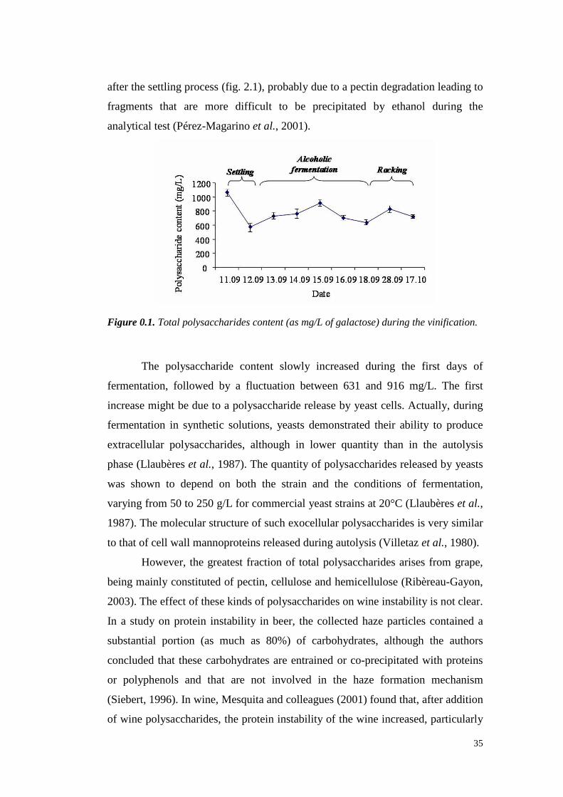
35
after thesettling process(fig. 2.1), probablydueto a pectin degradation leading to
fragments that are more difficult to be precipitated by ethanol during the
analytical test(Pérez-Magarinoet al., 2001).
Figure 0.1. Total polysaccharides content (as mg/L of galactose) during the vinification.
The polysaccharide content slowly increased during the first days of
fermentation,followed by a fluctuation between 631 and 916 mg/L. The first
increasemight bedue to a polysaccharidereleaseby yeastcells. Actually, during
fermentationin synthetic solutions, yeasts demonstrated their ability to produce
extracellular polysaccharides, although in lower quantity than in the autolysis
phase(Llaubèreset al., 1987). Thequantity of polysaccharidesreleasedby yeasts
was shown to dependon both the strain and the conditions of fermentation,
varyingfrom 50 to 250g/L for commercial yeaststrainsat 20°C(Llaubèreset al.,
1987).Themolecular structureof suchexocellular polysaccharidesis very similar
to thatof cell wall mannoproteinsreleasedduringautolysis(Vi lletazet al., 1980).
However,the greatest fraction of total polysaccharides arisesfrom grape,
being mainly constitutedof pectin,celluloseand hemicellulose(Ribèreau-Gayon,
2003). Theeffectof thesekindsof polysaccharidesonwine instabilit y is not clear.
In a studyon protein instability in beer, the collected haze particles containeda
substantial portion (as much as 80%) of carbohydrates, although the authors
concludedthat thesecarbohydratesareentrained or co-precipitatedwith proteins
or polyphenols and that are not involved in the haze formation mechanism
(Siebert, 1996).In wine,Mesquitaandcolleagues (2001)foundthat, after addition
of wine polysaccharides, theproteininstabilit y of thewine increased, particularly
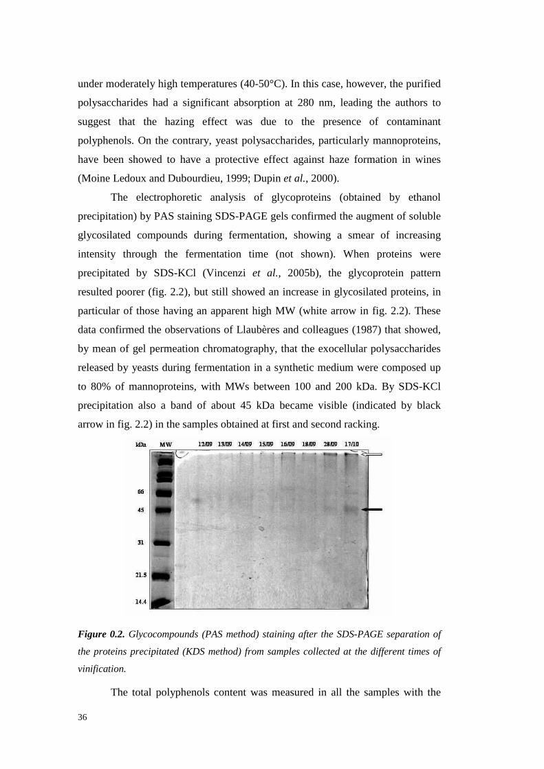
36
under moderately high temperatures(40-50°C).In this case,however, thepurified
polysaccharideshad a significant absorption at 280 nm, leading the authors to
suggest that the hazing effect was due to the presence of contaminant
polyphenols. On the contrary, yeast polysaccharides, particularly mannoproteins,
have beenshowedto have a protectiveeffect against haze formation in wines
(MoineLedouxandDubourdieu,1999;Dupin et al., 2000).
The electrophoretic analysis of glycoproteins (obtained by ethanol
precipitation) by PASstainingSDS-PAGE gels confirmedtheaugmentof soluble
glycosilated compounds during fermentation, showing a smear of increasing
intensity through the fermentation time (not shown). When proteins were
precipitated by SDS-KCl (Vincenzi et al., 2005b), the glycoprotein pattern
resultedpoorer(fig. 2.2), but still showed an increasein glycosilated proteins, in
particular of thosehavingan apparenthigh MW (white arrow in fig. 2.2). These
dataconfirmedtheobservationsof Llaubèresandcolleagues(1987)that showed,
by meanof gel permeationchromatography,that the exocellular polysaccharides
releasedby yeasts during fermentation in a synthetic mediumwerecomposedup
to 80% of mannoproteins, with MWs between100 and 200 kDa. By SDS-KCl
precipitation also a band of about 45 kDa became visible (indicated by black
arrow in fig. 2.2) in thesamplesobtainedat first andsecondracking.
Figure 0.2. Glycocompounds (PAS method) staining after the SDS-PAGE separation of
the proteins precipitated (KDS method) from samples collected at the different times of
vinification.
The total polyphenolscontentwas measuredin all the samples with the
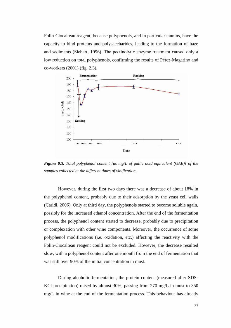
37
Folin-Ciocalteaureagent,becausepolyphenols,and in particular tannins,havethe
capacityto bind proteinsand polysaccharides, leading to the formation of haze
andsediments(Siebert,1996).The pectinolytic enzyme treatmentcaused only a
low reduction on total polyphenols, confirming theresults of Pérez-Magarino and
co-workers(2001)(fig. 2.3).
Figure 0.3. Total polyphenol content [as mg/L of gallic acid equivalent (GAE)] of the
samples collected at the different times of vinification.
However,during the first two days therewasa decreaseof about18% in
the polyphenolcontent,probably due to their adsorptionby the yeast cell walls
(Caridi,2006).Only at third day,thepolyphenolsstartedto becomesolubleagain,
possiblyfor theincreasedethanolconcentration. After theendof thefermentation
process, the polyphenolcontent startedto decrease, probably dueto precipitation
or complexation with other wine components.Moreover, the occurrenceof some
polyphenol modifications (i.e. oxidation, etc.) affecting the reactivit y with the
Folin-Ciocalteaureagentcould not be excluded.However,the decreaseresulted
slow,with a polyphenolcontentafterone month from theendof fermentation that
wasstill over90%of theinitial concentration in must.
During alcoholic fermentation,the protein content (measuredafter SDS-
KCl precipitation) raisedby almost30%,passingfrom 270 mg/L in must to 350
mg/L in wine at the endof the fermentation process. This behaviourhas already
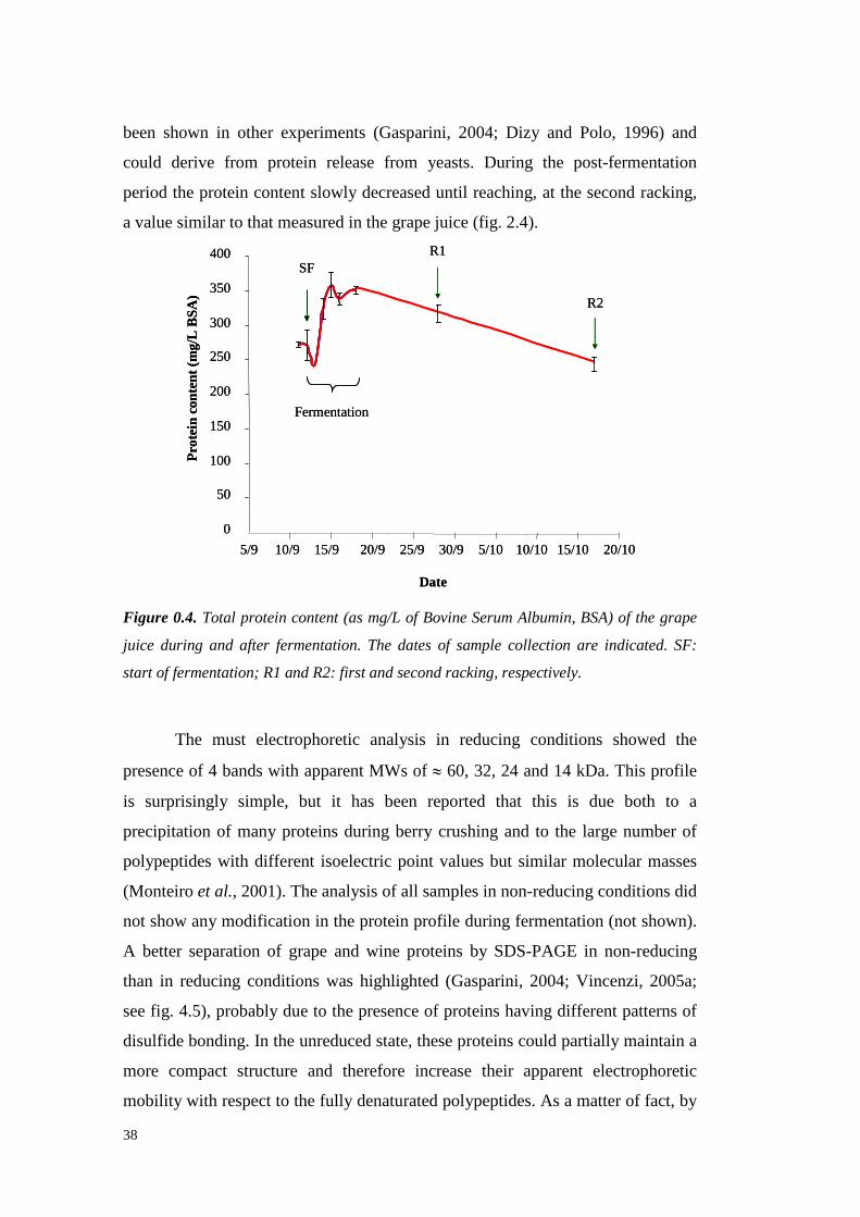
38
been shown in other experiments(Gasparini, 2004; Dizy and Polo, 1996) and
could derive from protein releasefrom yeasts. During the post-fermentation
period theproteincontentslowly decreased until reaching, at thesecondracking,
avaluesimilar to thatmeasuredin thegrapejuice(fig. 2.4).
0
50
100
150
200
250
300
350
400
5/9 10/9 15/9 20/9 25/9 30/9 5/10 10/10 15/10 20/10
Date
Pro
tein
cont
ent(
mg/
LB
SA)
Fermentation
R1
R2
SF
0
50
100
150
200
250
300
350
400
5/9 10/9 15/9 20/9 25/9 30/9 5/10 10/10 15/10 20/10
Date
Pro
tein
cont
ent(
mg/
LB
SA)
Fermentation
R1
R2
SF
Figure 0.4. Total protein content (as mg/L of Bovine Serum Albumin, BSA) of the grape
juice during and after fermentation. The dates of sample collection are indicated. SF:
start of fermentation; R1 and R2: first and second racking, respectively.
The must electrophoreticanalysis in reducing conditions showed the
presenceof 4 bandswith apparentMWs of ≈ 60, 32, 24 and14 kDa. This profile
is surprisingly simple, but it has been reported that this is due both to a
precipitation of many proteinsduring berry crushing and to the largenumber of
polypeptideswith different isoelectricpoint valuesbut similar molecular masses
(Monteiro et al., 2001).Theanalysisof all samples in non-reducingconditionsdid
not show anymodification in theproteinprofile duringfermentation (not shown).
A betterseparation of grapeand wine proteins by SDS-PAGE in non-reducing
than in reducing conditionswas highlighted (Gasparini, 2004; Vincenzi, 2005a;
seefig. 4.5), probably dueto thepresence of proteinshaving differentpatternsof
disulfide bonding. In theunreducedstate,theseproteinscould partially maintain a
more compact structure and therefore increasetheir apparent electrophoretic
mobility with respectto thefully denaturatedpolypeptides.As a matter of fact, by
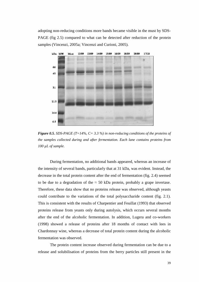
39
adoptingnon-reducingconditionsmorebandsbecamevisible in themustby SDS-
PAGE (fig 2.5) comparedto what canbe detected after reduction of the protein
samples(Vincenzi,2005a;VincenziandCurioni, 2005).
Figure 0.5. SDS-PAGE (T=14%, C= 3.3 %) in non-reducing conditions of the proteins of
the samples collected during and after fermentation. Each lane contains proteins from
100 µL of sample.
During fermentation, no additionalbandsappeared,whereasanincreaseof
theintensityof severalbands,particularlythatat 31kDa, wasevident.Instead,the
decrease in thetotal proteincontentaftertheend of fermentation (fig. 2.4)seemed
to be due to a degradation of the ≈ 50 kDa protein, probably a grapeinvertase.
Therefore,thesedatashow thatno proteinsreleasewasobserved, althoughyeasts
could contribute to the variations of the total polysaccharide content (fig. 2.1).
This is consistentwith theresultsof Charpentier andFeuillat (1993)thatobserved
proteinsreleasefrom yeastsonly during autolysis, which occurs severalmonths
after the end of the alcoholic fermentation. In addition, Lugera and co-workers
(1998) showed a releaseof proteins after 18 months of contact with lees in
Chardonnaywine, whereasa decreaseof total protein contentduring thealcoholic
fermentationwasobserved.
Theproteincontentincreaseobservedduringfermentation can bedueto a
release and solubilisationof proteinsfrom the berry particlesstill present in the

40
must.This release,probablyenhancedby the yeastaction andby the increaseof
ethanolconcentration,canexplain theobservation thatduringfermentationonly a
changein thebandsintensity is detectable in themust,without theappearanceof
new bands.However,the presencein mustof heavily glycosilated yeastproteins
not stainedby Coomassie cannot beexcluded.
Good separation of grape and wine proteins was achieved with anion
exchangechromatography(AEC), asalreadyreportedby other authors(Waterset
al., 1992;Dorrestein et al., 1995;Pastorello et al., 2002).In order to eliminate the
polyphenols from the dialyzed samples, a passagetrough a cartridge was
performed. This operationmight result in a certain quantitative protein loss, but
doesnot affect proteincompositionof thesample,aspreviously reported(Waters
et al., 1992).
In must, six peaksweredetectedat 280 nm by AEC (fig. 2.6). The same
peakswere detected in all must/wine samples collected during vinifi cation (fig.
2.7) andtwo newsmallpeaksweredetected only in thewine sample collectedone
month after theendof thealcoholic fermentation (fig. 2.8). However, therelative
proportion betweenthe different peakschanged during the time, asmeasured by
thequantificationof thepeakareas(fig. 2.9).
1
2
3
4
5
61
2
3
4
5
6
Figure 0.6. Anion-Exchange Chromatography of the proteins obtained starting from 100
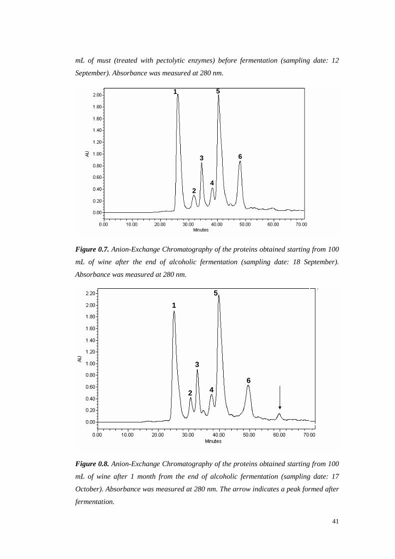
41
mL of must (treated with pectolytic enzymes) before fermentation (sampling date: 12
September). Absorbance was measured at 280 nm.
1
24
5
3 6
1
24
5
3 6
Figure 0.7. Anion-Exchange Chromatography of the proteins obtained starting from 100
mL of wine after the end of alcoholic fermentation (sampling date: 18 September).
Absorbance was measured at 280 nm.
1
2
3
4
5
6
1
2
3
4
5
6
Figure 0.8. Anion-Exchange Chromatography of the proteins obtained starting from 100
mL of wine after 1 month from the end of alcoholic fermentation (sampling date: 17
October). Absorbance was measured at 280 nm. The arrow indicates a peak formed after
fermentation.

42
0
50
100
150
200
250
300
Are
a(µ
V*s
ec)
x100
0000
11/09 18/09 28/09 17/10
Date
Peak5
Peak1
Peak6
Peak3Peak4Peak2
0
50
100
150
200
250
300
Are
a(µ
V*s
ec)
x100
0000
11/09 18/09 28/09 17/10
Date
Peak5
Peak1
Peak6
Peak3Peak4Peak2
Figure 0.9. Variation of the Anion-Exchange Chromatography peak areas during the
time of the experiment.
During the time of the experiment,the peak areasshoweda trendsimilar
to that of the total proteincontent,with a drop in the first threedaysfollowed by
an increaseuntil the end of fermentationand a slight decreaseafterward. The
increaseof 30%observedin thetotal proteincontent (fig. 2.4) seemsto beduein
particular to the increaseof AEC peak1 (+ 72%) (fig. 2.9). The analysis of this
peak by Reverse-Phase(RP) HPLC showed the presenceof 98% of thaumatin-
like proteinswith a little contaminationof chitinases, asassessedon the basisof
the retentiontime (not shown). The AEC peak5 contributed from 35 to 42% to
the total peak area, confirming our previous data on Manzoni bianco wine
(Gasparini,2004; Vincenzi, 2005). The chromatographicanalysisby RP-HPLC
showed this peak to be mainly constituted of chitinases (not shown), in
accordancewith the findings obtainedpreviouslyby chitinaseactivity detection
on SDS-PAGEgels (Gasparini,2004;Vincenzi andCurioni, 2005). Theseresults
confirmedthat thePR-proteinsarethemainproteinsin grapeandwine (Waterset
al., 1992).
The six peaksobtainedfrom AEC separation of the must proteins were
collectedandanalysedby SDS-PAGE(fig. 2.10).

43
Figure 0.10. SDS-PAGE (T=14%, C=3.3%) in non-reducing conditions of the peak
fractions collected from Anionic Exchange Chromatography of the wine proteins at the
end of fermentation (sampling date: 18 September). Each lane contains 15 µg of protein.
TQ: total proteins of the wine before fractionation.
With theexception of peaks 1 and3, which bothshowedan only band,all
theAEC fractionscontained two or moreprotein bands.In particular, a bandat ≈
20 kDa waspresentin everypeak,confirming thatgrapecontainsa largenumber
of polypeptides with different pI values (affecting the elution from the AEC
column) but similar apparentmolecularmassesin SDS-PAGE (Monteiro et al.,
2001).
Considering that individual wine proteins can differently contribute to the
phenomenonof hazeformation (Hsu andHeatherbell 1987,Waterset al. 1992),
thesechangesin therelativeconcentrations of theAEC peaksduringfermentation
might be relatedto variationsin the hazing potential of the mixture of the grape
proteinspassing from must to wine.
To evaluatethe haze stability throughout the fermentation process, the
total proteins from must/wine sampleswere heat tested (Pocock and Rankine,
1973), with a slightly modified method (Waters et al., 1991). It has been
demonstratedthat the environmental conditions, and in particular the pH andthe
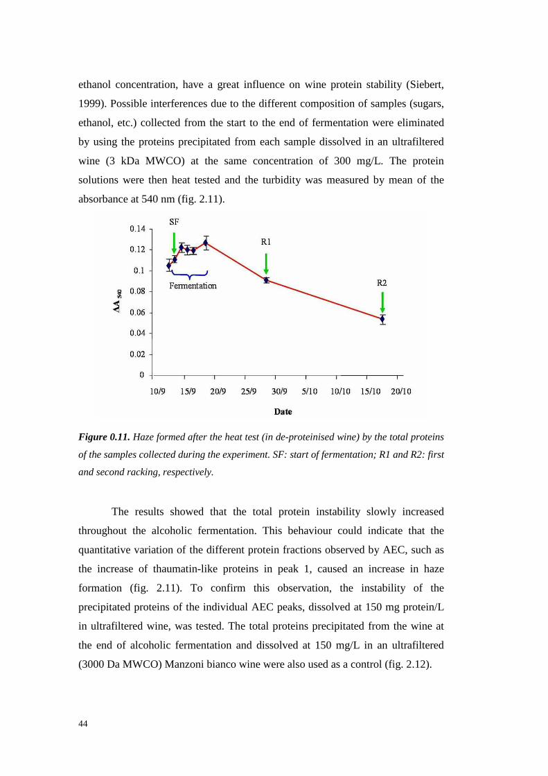
44
ethanol concentration, havea great influenceon wine protein stability (Siebert,
1999). Possibleinterferencesdueto thedifferent composition of samples(sugars,
ethanol,etc.) collectedfrom the start to the end of fermentation wereeliminated
by using the proteinsprecipitatedfrom eachsample dissolved in an ultrafiltered
wine (3 kDa MWCO) at the same concentration of 300 mg/L. The protein
solutions were then heat tested and the turbidity was measured by mean of the
absorbanceat 540nm (fig. 2.11).
Figure 0.11. Haze formed after the heat test (in de-proteinised wine) by the total proteins
of the samples collected during the experiment. SF: start of fermentation; R1 and R2: first
and second racking, respectively.
The results showed that the total protein instability slowly increased
throughout the alcoholic fermentation.This behaviourcould indicate that the
quantitative variation of thedifferent protein fractionsobserved by AEC, suchas
the increaseof thaumatin-like proteins in peak 1, causedan increase in haze
formation (fig. 2.11). To confirm this observation, the instabili ty of the
precipitatedproteinsof the individual AEC peaks, dissolvedat 150 mg protein/L
in ultrafiltered wine, wastested. The total proteinsprecipitated from the wine at
the end of alcoholic fermentationand dissolved at 150 mg/L in an ultrafiltered
(3000DaMWCO)Manzonibiancowinewerealsousedas acontrol(fig. 2.12).
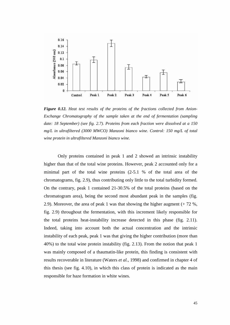
45
Figure 0.12. Heat test results of the proteins of the fractions collected from Anion-
Exchange Chromatography of the sample taken at the end of fermentation (sampling
date: 18 September) (see fig. 2.7). Proteins from each fraction were dissolved at a 150
mg/L in ultrafiltered (3000 MWCO) Manzoni bianco wine. Control: 150 mg/L of total
wine protein in ultrafiltered Manzoni bianco wine.
Only proteinscontainedin peak 1 and 2 showedan intrinsic instability
higherthanthat of the total wine proteins. However, peak2 accounted only for a
minimal part of the total wine proteins (2-5.1 % of the total area of the
chromatograms, fig. 2.9), thuscontributingonly li ttle to thetotal turbidity formed.
On the contrary, peak 1 contained21-30.5% of the total proteins (based on the
chromatogramarea), being the secondmost abundantpeak in the samples (fig.
2.9). Moreover,theareaof peak1 wasthat showingthehigheraugment(+ 72 %,
fig. 2.9) throughout the fermentation,with this increment likely responsible for
the total proteins heat-instability increasedetected in this phase (fig. 2.11).
Indeed, taking into account both the actual concentration and the intrinsic
instability of eachpeak,peak1 wasthatgiving thehighercontribution (morethan
40%) to the total wine proteininstability (fig. 2.13). From the notion that peak1
wasmainly composedof a thaumatin-like protein, this finding is consistent with
results recoverable in literature(Waterset al., 1998) andconfirmed in chapter4 of
this thesis (seefig. 4.10), in which this classof protein is indicated as the main
responsiblefor hazeformationin whitewines.

46
Figure 0.13. Percent contribute of each peak to the total turbidity developed (by the heat
test) in wine at the end of fermentation (sampling date: 18 September).
After theendof fermentation,theinstabili ty of thetotal proteins tendedto
decreaseandtheheat-inducedhazeat secondracking was evenlower thanthatof
the starting must. However,this variation could not beascribedto a modification
in the relative protein concentrations, because the ratios between the different
protein peaksremainedquiteconstantduringtheone-monthstorage. This stability
increasecould be instead attributed to the mannoproteins released by yeasts
during the storage (on total lees), which actually seemto appearin the wine (fig.
2.2).
1.1.14 DISCUSSION
During winemaking, some modifications of the must proteins profile are
detectable.Severalauthorshighlighted the releaseof protoplasmatic proteases
from yeasts during autolysis(Feuillat et al., 1980;Lurton, 1988). However, these
enzymesdo not significantly influencetheprotein pattern,althoughit is not clear
whethertheseenzymesareinhibitedby wine polyphenolsor by thelow pH of the
must. Lurton (1988) showedthat theS. cerevisiae endoproteaseA was still active
at pH 3.0, leadingto the releaseof peptidesinto the wine. In optimal conditions
(pH 4.5-5.0, 35-40°C) the cell disorganization of yeasts starts in several hours,
47
while thesameprocessin wine takesup to 2-3 months(Flanzy, 1998).
However,S. cerevisiae resultedableto hydrolysehaemoglobinandcasein
in vivo by secreting extracellularproteasesin thefermenting medium (Rosiet al.,
1987; Feuillat et al., 1980). Moreover,S. cerevisiae possess someperiplasmic
peptidases, involved in the active transport system, that are able to partially
degrade the proteins of the medium (Cartwright et al., 1989). Actually, wine
yeastsareable to grow in a mediumcontaining only mustproteinsas thenitrogen
source(Conternoet al., 1994).
Only few studies have analysed the fate of must proteins during
vinification, andall postulateda decreaseof the total protein contentduring this
process(Lugera et al., 1998; Moreno-Arribas et al., 1996). Thesedata are in
disagreementwith our resultsand with the observation that grape proteins are
protease-resistant andstableat acidic pH (Modra andWil liams, 1988; Waterset
al., 1992; Waterset al., 1995). Beside,it is importantto considerthat the cited
works aimedto clearproteinchangesoccurringduring thesecondfermentation in
sparkling winesproductionand after a bentonite treatment (Lugera et al., 1998;
Moreno-Arribaset al., 1996). Moreover, thevariationof thenitrogen compounds
was monitored for long times (up to 18 months), that are compatible with the
degradation and precipitation even of the more resistant wine proteins. In
particular, Manteauet al. (2003) statedthat grape proteins tend to disappear
during the Champagnewine production. These authors suggested that this
phenomenonwasmainly dueto proteinprecipitation, adsorptionon cell walls of
the yeasts anddenaturation. In this case, the reducingenvironment (150 mV) of
the bottle during the secondfermentationmight determine a partial reduction of
disulfide bonds, increasing protein susceptibil ity to the action of proteases
secretedby yeasts(Manteauet al., 2003).
In a normal white wine vinification, however, the yeast cells remain in
contact with the wine for a time too short to allow autolysis.The few works that
have studiedthemodificationsof grapeproteins in theearlystages of vinification
showed that during the first alcoholic fermentation lit tle quantitative variations
occur resulting, in most cases, in an increaseof the protein content (Bayly and
Berg, 1967; Dizy and Polo, 1996).This increasewas considered to be due to a
protein release from the yeast cells. By means of chromatographic and

48
electrophoretic techniques, Bayly andBerg (1967)showedboth quantitative and
qualitative modifications in wine proteins during fermentation, with the
appearanceof newelectrophoreticbands.
Theresultshereobtainedindicatethatthesolubleproteinsof theberry of a
white grape variety vary during and after the alcoholic fermentation in both
quantity and relative proportion. According to what stated in another work
(Vincenzi et al., 2006),theproteinfraction of AEC peak 1, containing a single20
kDa band (probablya thaumatin-like protein), showedthe lowest heat-stability
whenindividually heat-testedin de-proteinisedwine. Thesameprotein fraction is
that with thelargestquantitativeincreaseduring fermentation, constituting a large
proportion of the total wine proteins. Taking into account that bands with the
sameSDS-PAGE mobility, correspondingto that of theTL protein family, could
be detectedalsoin otherAEC fractions,theprecisenature of theprotein of peak 1
warrantsfurtherinvestigation.
Moreover, from theresultsherereported,it is confirmedthatthereleaseof
compounds of polysaccharidic nature by the fermenting yeast (probably
mannoproteins) results in an increasedheatstability of the total wine proteins,
despitetheincreasein the relativeproportion of their mostunstablecomponent.
Acknowledgements
The author would like to thank SimoneVincenzi of Padua University for his
support in the conduction of the experiments and for the valuable discussions
aboutthis work.
REFERENCES
Bayly F.C.andBerg H. (1967).Grapeandwineproteinsof white winevarietals.
Am. J. Enol. Vitic. 18(1),18-32.
Bossi A., Bonizzato L. andZapparoliG. (2006).Acidic Extracellular Proteases
from Microrganisms of Fairly Acidic Niche. Protein and Peptide Letters
13(7),737-741.
Caridi A. (2006).Enologicalfunctionsof parietal yeastmannoproteins. Antonie
49
van Leeuwenhoek 86,417-422.
Cartwright C.P., Rose A.H., CalderbankJ. and Keenan H.J. (1989). Solute
transport. The Yeast. Metabolism and Physiology of Yeast, Vol. 3 (RoseAH &
HarrisonJS,eds),pp.5-56.AcademicPressLimited,London.
CharpentierC. andFeuillatM. (1993).Yeastautolysis.In: Fleet G.H. (eds), Wine
Microbiology and Biotechnology. Harwood Academic Publishers, Chur,
Switzerland,pp.225-242.
ConternoL. and Delfini C. (1994). Peptidaseactivity and the ability of wine
yeastto util ise grapemust proteinsassolenitrogensource.Journal of Wine
Research 5(2),113-126.
Dizy M. and Polo M.C. (1996). Changesof the concentration of nitrogenous
compoundsduringfermentationof mustat pilot-plantscale. Food Science and
Technology International 2(2),87-93.
Dorrestein E., Ferreira R.B., Laureano O. and Teixeira A.R. (1995).
Electrophoretic and FPLC Analysesof Soluble Proteins in Four Portuguese
Wines.Am. J. Enol. Vitic. 46(2),235-241.
Dupin I.V.S., McKinnon B.M., Ryan C., Boulay M., MarkidesA.J., JonesG.P.,
Will iams P.J. and Waters E.J. (2000). Saccharomyces cerevisiae
Mannoproteins That Protect Wine from Protein Haze: Their Releaseduring
Fermentation and Lees Contact and a Proposal for Their Mechanism of
Action. J. Agric. Food Chem. 48(8),3098-3105.
Ferreira R.B., Monteiro S., Picüarra-Pereira M.A., TanganhoM.C., Loureiro
V.B. and Teixeira A.R. (2000).Characterisation of the proteins from grapes
andwinesby immunologicalmethods. Am. J. Enol Vitic. 51(1),22-28.
Feuillat M., Brillant G. and Rochard J. (1980). Mise en evidence d’une
production de proteases exocellulaires par les levures au cours de la
fermentationalcooliquedumoutderaisin.Connaiss. Vigne Vin 14,37-52.
Flanzy C. (1998). Œnologie: Fondements scientifiques et technologiques.
EditionsTEC & DOC,Paris.
GaspariniG. (2004).Parzialecaratterizzazione di proteineda uva “I.M. 6.0.13”
in relazione all’i nstabilità proteica del vino. Degree thesis, University of
Verona(Italy).
Hsu J.C.,Heatherbell D.A., FloresJ.H. and WatsonB.T. (1987).Heat-unstable

50
proteins in wine II. Characterizationand removal by ultrafilt ration. Am. J.
Enol. Vitic. 38(1),17-22.
KoenigR., StegemannH., FrancksenH. andPaulH.L. (1970).Protein subunitin
the potato virus X group. Determination of the molecular weight by
polyacrilamideelectrophoresis.Biochim. Biophys. Acta 297,184-189.
Laemmli U.K. (1970).Cleavageof structuralproteinsduring theassembly of the
headof bacteriophageT4. Nature 227,680-685.
Llaubères R.M., Dubourdieu D., Vill ettaz J.C. (1987). Exocellular
polysaccharidesfrom Saccharomyces in Wine. J. Agric. Food Chem. 41(3),
277-286.
LugeraC., Moreno-Arribas V., Pueyo E., BortolomèB. andPolo M.C. (1998).
Fractionation and partial characterization of protein fraction present at
different stagesof the productionof sparkling wines.Food Chemistry 63,465-
461.
Lurton L. andGuerreauJ. (1988).Studyof the proteolysis of wine yeast during
storageof wineon its lees.Rev. Fr. Oenol. 113,35-41.
Manteau S., Lambert B., JeandetP. and Legendre L. (2003). Changes in
ChitinaseandThaumatin-like Pathogenesis-RelatedProteinsof GrapeBerries
during the ChampagneWinemakingProcess.Am. J. Enol. Vitic. 54(4), 267-
272.
Marchal R., Bouquelet S., Maujean A. (1996). Purification and partial
biochemical characterizationof glycoproteins in a champenoisChardonnay
wine. J. Agric. Food Chem. 44(7),1716-1722.
MesquitaP.R.,Piçarra-Pereira, M.A., Monteiro S., Loureiro V.B., Teixeira A.R.
and Ferreira R.B. (2001). Effect of wine composition on proteinstabilit y. Am.
J. Enol. Vitic. 52(4),324-330.
Modra E.J. andWilliams P.J.(1988).Are proteasesactive in winesand juices?
Aust. Grapegrower Winemaker 292,42-46.
Moine-LedouxV. andDubourdieuD. (1999).An invertasefragmentresponsible
for improving theproteinstability of dry white wines.J. Sci. Food Agric. 79,
537-543.
Monteiro S.,Picarra-PereiraM.A., MesquitaP.R.,LoureiroV.B., Teixeira A. and
Ferreira R.B. (2001). The wide diversity of the structurall y similar wine
51
proteins.J. Agric. Food Chem. 49(8),3999-4010.
Moreno-Arribas V., Pueyo E. and Polo M.C. (1996). Peptides in musts and
wines.Changesduring the manufactureof cavas(sparkling wines).J. Agric.
Food Chem. 44(12),3783-3788.
OIV (1990). Recueil des méthodes internationales d’analyse des vins et des
moûts.
Pastorello E.A., Farioli L., Pravettoni V., Ortolani C., Fortunato D., Giuffrida
M.G., PeronoGaroffo L., CalamariA.M, BrennaO. and Conti A. (2002).
Identification of grape and wine allergens a san endochitinase 4, a lipid-
transferprotein, anda thaumatin.J. Allergy Clin. Immunol. 111(2),350-359.
PengZ., PocockK.F., WatersE.J.,Francis I.L. andWili amsP.J.(1997).Taste
Propertiesof Grape (Vitis vinifera) Pathogenesis-Related Proteins Isolated
from Wine.J. Agric. Food Chem. 45(12),4639-4643.
Pérez-Magariño S. and González-Sanjosé M.L. (2001). Differentiation
parametersof “Riberadel Duero” D.O. winesfrom other Spanish D.O. Food
Science and Technology International 7, 237-244.
Pocock K.F. andRankineB.C. (1973).Heat test for detecting protein instability
in wine.Australian Wine, Brewing and Spirit Review 91(5),42-43.
Ribéreau-GayonP.,GloriesY., MaujeanA. and Dubourdieu D. (2003).Trattato
di enologiaII, Chimica del vino-Stabilizzazione-Trattamenti, Edagricole.
Rosi I., CostamagnaL., Bertuccioli M. (1987).Screening for extracellular acid
protease(s)production by wine yeasts. Journal of the Institute of Brewing 93,
322-324.
Ruiz-LarreaF., LopezF., SantamariaR., Sacristan P., Ruiz M.C., ZarazagaM.,
GutierrezA.R. andTorresC. (1998).Solubleproteinsand freeamino nitrogen
contentin mustandwineof cv. Viura in La Rioja.Vitis 3, 139-142.
Segarra I., Lao C., López-TamamesE. and De La Torre-BoronatM.C. (1995).
Spectrophotometric Methods for the Analysis of Polysaccharide Levels in
Winemaking Products.Am. J. Enol. Vitic. 46(4),564-570.
Segrest J.P. and Jackson R.L. (1972). Molecular weight determination of
glycoproteinsby polyacrylamidegel electroforesis in sodium dodecyl sulfate.
In: Ginsburg V, ed. Methods in enzymology XXVIII, Complex carbohydrates,
Part B. NewYork: AcademicPress,54-63.
52
SiebertK.J. (1999).Effectsof protein-polyphenolinteractionson beveragehaze,
stabilization,andanalysis.J. Agric. Food Chem. 47(2),353-362.
SiebertK.J.,CarrascoA. andLynn P.Y. (1996).Formationof Protein-Polyphenol
Hazein Beverages.J. Agric. Food Chem. 44 (8), 1997-2005.
Singleton V.L. and Rossi J.A. (1965). Colorimetry of total phenolics with
phosphomolybdic-phosphotungsticacid reagents. Am. J. Enol. Vitic. 16(3),
144-158.
Singleton V.L., Sieberhagen H.A., De Wet P. and Van Wyk C.J. (1975).
Composition andSensory Qualitiesof Wines Preparedfrom White Grapes by
Fermentationwith andwithout GrapeSolids.Am. J. Enol. Vitic. 26(2),62-69.
Villetaz J.C., Amado R., NeukomH., HorinsbergerM. and Horman I. (1980).
Comparative structural studies of the D-mannans from a rosé wine and
Saccharomyces uvarum. Carbohydr. Res. 81,341-344.
Vincenzi S. (2005a).Studio delle proteinedell’uva e del vino in relazione alla
qualitàdei vini bianchi.PhD thesis, University of Padua(Italy), pp83-84.
Vincenzi S. and Curioni A. (2005). Anomalous electrophoretic behaviour of a
chitinase isoform from grape berries and wine in glycol chitin-containing
sodium dodecyl sulfate-polyacrylamide gel electrophoresis gels.
Electrophoresis 26,60-63.
Vincenzi S.,MarangonM. andCurioni A. (2006).Chitinaseand Thaumatin-Like
Pathogenesis-Related Proteins from Grapes and their Effects on Wine
Stability. In Macromolecules and Secondary Metabolites of Grapevine and
Wine. Jeandet P.et al (Eds.),177-182.EditionsTec & Doc,Paris.
Vincenzi S.,Mosconi S.,ZoccatelliG., Dalla PellegrinaC., Veneri G., Chignola
R., D.B. PeruffoA., Curioni A. andRizzi C. (2005b).Development of a new
procedure for proteinrecoveryandquantification in wine.Am. J. Enol. Vitic.
56(2),182-187.
WaterhouseA.L. (2002)Winephenolics.Ann. N. Y. Acad. Sci. 957,21-36.
WatersE.J.,HayasakaY., Tattersall D.B., AdamsK.S. andWilli amsP.J.(1998).
Sequenceanalysisof grape(Vitis vinifera) berry chitinasesthat cause haze
formationin wines.J. Agric. Food Chem. 46(12),4950-4957.
Waters E.J., Pellerin P. and Brillouet J.M. (1994). A Saccharomyces
mannoprotein that protectswine from protein haze. Carbohydrate polymers

53
23, 185-191.
WatersE.J.,PengZ., PocockK.F. and Willi amsP.J.(1995).Proteins in white
wine. II. Their resistanceto proteolysisis not dueto their phenolic association
or glycosilation. Aust. J. Grape Wine Res. 1, 94-99.
WatersE.J.,Wallace W. andWilliams P.J.(1991).Heathaze characteristics of
fractionatedwineproteins.Am. J. Enol. Vitic. 42(2),123-127.
WatersE.J., Wallace W., Williams P.J. (1992). Identification of heat-unstable
wine proteinsandtheir resistanceto peptidases. J. Agric. Food Chem. 40(9),
1514-1519.
Zoccatelli G., MosconiS., Rizzi C., Vincenti S. andCurioni. A. (2003).A new
method for the recoveryof wine proteinsallowing their quantification and
electrophoretic analysis. In: Oenologie 2003. A. Lonvaud-Funel et al. (Eds.),
pp. 542-545.EditionsTec& Doc,Paris.

54
55
CHAPTER 3
Fractionation of grape juice and wine proteins by hydrophobic
interaction chromatography
ABSTRACT
Thaumatin-like (TL) proteinsandchitinases are the predominantproteins
in ripegrapes.TL proteinsandchitinasesinhibit fungalgrowth in vitro andlikely
playa role in grapepathogendefence.
In this work, a method to separategrape juice and wine proteins by
hydrophobic interactionchromatography(HIC) using Phenyl Sepharose® High
Performance resin is proposed.The purification was performed in two steps:
protein precipitation with ammoniumsulphate followed by HIC fractionation.
HIC fractionsof ammoniumsulphateprecipitated juice and wine proteins were
further fractionatedby reversephaseHPLC andSDS-PAGEto assesstheir nature
and purity. Grapejuice proteinswere also identified by matching peptide LC-
MS/MS spectra with theoretical peptides from a plant protein database.
Identificationsof twenty-six LC-MS/MS samples included severalTL-proteins
andchitinases,vacuolarinvertase,anda lipid transfer protein.
Keywords: grape,protein,wine,HIC, LC-MS/MS, thaumatin-like protein,
chitinases.
INTRODUCTION
Because of the very low amount of proteins recoverable in grape and
wines, researchersneed to find easy and high-yield protein purification
procedures.A wide varietyof proteinpurification techniquesareavailable today,
however,different typesof chromatographyhave become dominant due to their
high resolving power. In gel filtration chromatography, ion-exchange
chromatography, affinity chromatography and hydrophobic interaction
chromatography(HIC), protein separationis dependenton their biological and
56
physico-chemical properties: molecular size, net charge, biospecific
characteristicsand hydrophobicity,respectively (Kennedy, 1990; Garcia, 1993).
The result of a protein purificationprocedureis evidently dependenton thechoice
of separation equipment and techniques. The order in which the different
techniques are combined is also of great importance. The development of
techniques and methodsfor the separation and purification of proteins hasbeen
essentialfor many of the recentadvancesin biotechnology research. The global
aim of a protein purification process is not only the removal of unwanted
contaminants,but also theconcentrationof thedesired protein and their transferto
an environment whereit is stableandin a form readyfor theintendedapplication
(Queirozet al., 2001).
Techniqueslike ion exchangechromatographyandgel fil tration have been
widely usedin fractionationsof grapeandwine macromolecules (Bayly andBerg,
1967; SomersandZiemelis,1973;Waterset al., 1992,1993;Daweset al., 1994;
Dorresteinet al., 1995; Canalset al., 1998;Monteiro et al., 1999,2001;Pastorello
et al., 2002). Other chromatographicmethods, such as HIC, have been only
recentlyapplied in wine studiesby Brissonet and Maujean (1993), which used
HIC for thecharacterizationof foamingproteinsof champagnewine.
HIC takes advantage of the hydrophobicity of proteins promoting its
separationbasedon hydrophobicinteractions betweenimmobilized hydrophobic
ligandsandnon-polarregionson thesurfaceof proteins.Theadsorption increases
with high salt concentrationin the mobile phase and the elution is achievedby
decreasing the salt concentrationof the eluent (Melander and Horváth, 1977;
Fausnaughand Regnier,1986; Roe, 1989). Therefore, the term ‘salt-promoted
adsorption’ could beusedfor this typeof chromatography (Porath, 1986).
Different types of elution conditions can be used for purification of
complex mixtures of proteins that would be difficult to separate using other
chromatographic techniques. In fact, HIC has been successfully used for
separationpurposesasit displaysbinding characteristics complementary to other
protein chromatographic techniques(Jansonand Rydén,1993). Van Oss et al.
(1986) proposedthat thevanderWaalsforcesare themajor contributingfactor to
the hydrophobicinteractions(‘interfacial forces’)despite thecomplex mechanism
involved. HIC that today is now an established and powerful bioseparation
57
techniquein laboratory-scale,aswell asin industrial-scale purification of proteins
(Wu andKarger, 1996; Sofer,1997;Grund,1998).
In this chapter,thestudyof bothSemillongrapejuiceandwineproteinsby
meansof HIC is illustrated.Themethodof proteinfractionation involved protein
salting out followed by direct fractionation through a HIC matrix. Fractions
collected were characterisedby meansof HPLC, SDS-PAGE, and LC-MS/MS
techniques.
MATERIALS AND METHODS
1.1.15 MATERIALS
Grapeandwine proteinswerepurified from Semill on variety harvestedon
2006 in AdelaideHills region(SouthAustralia).
1.1.16 AMMONIUM SULPHATE PROTEIN PRECIPITATION
Grape proteins from Semillon juice were concentrated by ammonium
sulphateprecipitation. Thesalt wasaddedat 80% of saturation to thegrapejuice,
previously buffered to pH 5.0 (with KOH). After 16 hours at 4ºC, the pellet
collected by centrifugation (30 min, 14000g,4ºC) was treated in two ways: i)
dissolvedin 30 mM citrate buffer (pH 3.50) and desalted throughpassage on a
columnequippedwith 20 mL of Bio-Gel® PD-10 resin (Bio-Rad)andfreeze-dried
beforebeing dissolved in 50 mM citratebuffer (pH 3.5); ii ) directly dissolvedin
theeluantA of HIC fractionation(seebelow).
1.1.17 RESIDUAL AMMONIA DETERMINATION
The ammoniacontent on purified fractionswas determined by meansof
the UV test for the determinationof Urea and Ammonia in foodstuffs (r-
biopharm;Roche).

58
1.1.18 SODIUM DODECYL SULFATE POLYACRYLAMIDE GEL ELECTROPHORESIS
(SDS-PAGE)
Electrophoretic analyseswere performedaccording to Laemmli (1970).
The samplesweredissolved in a Tris-HCl pH 6.8 buffer containing 15% (v/v) of
glycerol (Sigma) and 1.5 % (w/v) SDS (Bio-Rad) and heated at 100°C for 5
minutesbeforeto be loadedfor the analysisperformed by usinga Mini-Protean
III apparatus(Bio-Rad).Eachanalysiswas conducted in non-reducing conditions.
The molecular weight standardswere the Broad Range (Bio-Rad). Ready Tris-
HCl Gels[Bio-Rad,4-20%,15-well, 15 µl, 8.6x 6.8cm (W x L)] wereused. Gels
were alternatively stainedwith Bio-SafeTM Coomassie stain (Bio-Rad) or Silver
stain procedurefor high sensibility protein detection, according to Blum et al.
(1987).
1.1.19 GRAPE AND WINE PROTEIN CHROMATOGRAPHY
Thechromatographicseparationswereperformedby meansof:
o An ÄKTA Prime FPLC (Amersham Biosciences, Sweden)
equipped with an UV detector (λ AbsorbanceDetector). Data
collectedwereelaboratedby thePrimeViewsoftware.
o A RP-HPLC Agilent 1200Series(Agilent Technologies,Germany)
equipped with autosampler, fraction collector and Diode Array
Detector.
Every solution util ised and sample loadedhad previously been filt ered
with celluloseacetatefilters (Millipore) with poresizeof 0.20µmanddegassed.
1.1.20 HYDROPHOBIC INTERACTION CHROMATOGRAPHY
Grapeand wine proteins were fractionated with an ÄKTA Prime FPLC
system (Amersham Bioscience, Sweden). Five columns (1 mL resin each)
contained in the HiTrap Hydrophobic Interaction Chromatography (HIC) kit
(Pharmacia)were used. The matrixes of the kit were: Phenyl Sepharose® High
Performance,PhenylSepharose6 FastFlow (low substitution), PhenylSepharose
6 FastFlow (high substitution), Butyl Sepharose4 FastFlow and Octyl Sepharose
59
4 FastFlow.
ThePhenylSepharose® High Performanceresin wasalsoutilized to packa
chromatographyglass column(55 mL) having a diameter of 1.6cm. Eluent A was
50 mM SodiumPhosphate+ 1.25M Ammonium sulfate,pH 5.0andeluentB was
eluent A without ammoniumsulfate.Sampleswere loaded at 3 mL/min onto the
resin previously equilibratedin 100%of buffer A. The loading lastedfor 200mL
beforethebeginningof a lineargradient to 0% A (from 200 to 800mL of elution
volume).After theendof thegradient,100%buffer B wasapplied for 400 mL to
re-equilibratethecolumnin thestartingconditions.
1.1.21 RP-HPLC PROTEIN ANALYSES
1.1.21.1 Analysis and quantification of protein by PR-HPLC
The proteincompositionof the grapeandwine fractions was determined
by reversedphaseHPLC,accordingto themethodproposedby Penget al. (1997).
Samples(100 µL) were loadedat 1 mL/min onto a semi preparative C8
column (4.6 x 250 mm, Vydac 208 TP 54, Hesperia, CA) fit ted with a C8 guard
column kit (Vydac 208 GK 54, 4.6 x 5 mm, Hesperia,CA) equilibrated in a
mixture of 83% (v/v) solvent B [0.1% trifluoroacetic acid (TFA) in 92%
Acetonitrile] and17%solvent A [80% Acetonitril e, 0.1%(v/v) TFA] and held at
35°C. Proteinswereelutedby a gradientof solventA from 17%solventA to 49%
solvent A in the fi rst 7 minutes,49% to 57% from 7 to 15 minutes,57% to 65%
from 15 to 16 minutes,65%to 81%from 16 to 30 minutes andthanheld at 81%
for 5 minutesbeforeto re-equilibrate the column in the starting conditions for 6
moreminutes.Elution wasfollowed by absorbanceat 210,220,260,280and320
nm. From the 210 nm spectrum,their identity was assignedby comparison of
their retention timesto thoseof purified grapePR proteins (Waterset al., 1996)
and quantified by comparison to the peak area of two standard proteins: horse
heartcytochromeC (cyt C) (Sigma)or bovinserumalbumin (BSA) (Sigma).
1.1.21.2 HPLC protein analyses by Size Exclusion Chromatography
Proteinfractionswere analysedby Size ExclusionChromatographyusing
a RP-HPLC equippedwith a PhenomenexBioSep SEC S2000(7.8 x 300 mm)
column with guard column at 1 mL/min in 50 mM KH2PO4 buffer at ambient

60
temperature.20 µL of samplewereinjected.The absorbance was detected at 210
nm.
1.1.22 SAMPLES DESALTING
Samples were desalted by using an ÄKTA Prime FPLC apparatus
equippedwith a 20 mL columncontaining Bio-Gel® P-10DG gel resin (Bio-rad).
The separation wasperformedin isocratic conditions (50 mM Citric acid-NaOH
buffer, pH 3.50).The flow ratewas2 mL/min andthe maximum loadedvolume
was2 mL.
1.1.23 PROTEIN IDENTIFICATION THROUGH LC-MS/MS ANALYSES
Grapeproteins were excised after electrophoretic separation and sent to
Australian Proteome Analyses Facility Ltd. Bands were reduced (25mM
Dithiorethiol/50mM NH4CO3 at 56°C) and alkylated (55mM
Iodoacetamide/50mM NH4CO3 at room temperature in thedark) followedby a 16
hour tryptic digestion at 37°C.Sampleswereextractedwith 0.1%TFA/2%CH3CN
and sonicated in waterbathfor 15 minutes.The instrumentused for nanoLCwas
Agilent 1100 Series(Agilent Technologies, Germany). Samples were injected
(40µL each)ontoapeptidetrap(MichromepeptideCaptrap)for pre-concentration
and desalted with 0.1%TFA, 2% Acetonitrile at 10µL/min. Thepeptide trapwas
thenswitchedinto line with theanalyticalcolumn containing C18RPsilica (SGE
ProteCol C18, 300A, 3µm, 150µm x 10 cm). Peptides were eluted from the
column using a linearsolventgradient from H2O:CH3CN (95:5; + 0.1% formic
acid) to H2O:CH3CN (70:30,+ 0.1% formic acid) at 600nL/min over a 60 min
period. The LC eluent wassubjectto positive ion nanoflowelectrosprayanalysis
on an Applied BiosystemsQSTAR XL massspectrometer (ABI, CA, USA). The
QSTAR was operatedin an information dependant acquisition mode (IDA). In
IDA mode a TOFMS surveyscanwas acquired (m/z 370-1600,1.0s),with the
three largestmultiply chargedions (counts >50) in the surveyscansequentially
subjectedto MS/MS analysis. MS/MS spectrawere accumulated for 2 s (m/z 100-
1600). Results were a peak list in Mascot Generic Format using MASCOT.dll
script (Applied Biosystems) and mascot search result PDF fi le with NCBInr
61
databasewith Vi ridiplantaetaxonomy.
A secondbatchof analyseswasperformedby excising protein bandsfrom
a SDS-PAGEgel. Peptideswerepreparedin 10 µL of 1% formic acid, loadedby
auto sampleronto a 5 cm x 100µmID C18column.Thegradient was 0-50%Bin
35 min after 12 minutesfrom the loading. Eluant A was 5% Acetonitrile, 0.1%
formic acid. Eluant B was 90% Acetonitrile, 0.1% formic acid. LCQ DecaIon
trap massspectrometer,set to do MS 400-1500amu,MS/MS of 3 most intense
ions,dynamicexclusion enabled.The raw fi les wereconverted to mzxml format
with the program readw.exe.Files in mzxml were submitted to X!tandem
database via locally installedGPM-XE version.Parention masswindow 4 da,
fragmention tolerance0.4 Da, Cys + 57 for iodoacetamide, met differentially
modified with oxidation. Reverse database searching enabled concurrently.
PlantProtein databasedownloadedfrom TAIR as fastafile, convertedto .pro file
for GPM use.
1.1.24 AMINO ACIDS ANALYSIS
Amino acidsanalysiswasperformedby theAustralian ProteomeAnalyses
Facility Ltd. Thesampleunderwent24 h to a gas phasehydrolysiswith 6N HCl at
110°C. Amino acids were analysed using the Waters AccQTag chemistry.
Cysteinewas separately analysedby using performic acid oxidation followed by
24 h acid hydrolysis with 6N HCl at 110°C.Sampleswereanalysedin duplicate
andresultswereexpressedasanaverage.
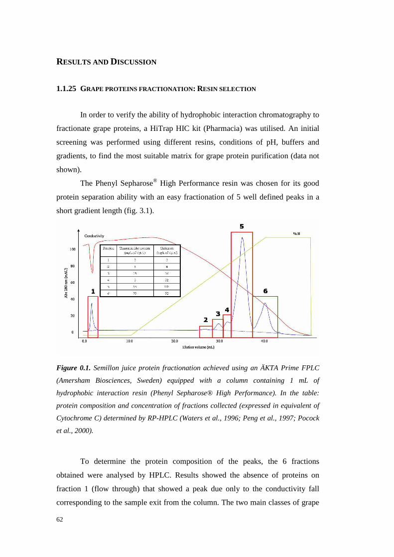
62
RESULTS AND DISCUSSION
1.1.25 GRAPE PROTEINS FRACTIONATION: RESIN SELECTION
In orderto verify theability of hydrophobic interaction chromatographyto
fractionategrapeproteins, a HiTrap HIC kit (Pharmacia) wasutilised.An initial
screeningwas performedusing different resins,conditions of pH, buffers and
gradients, to find the mostsuitablematrix for grapeproteinpurification (datanot
shown).
The PhenylSepharose® High Performanceresin waschosenfor its good
protein separation ability with an easyfractionation of 5 well defined peaksin a
short gradientlength (fig. 3.1).
Figure 0.1. Semillon juice protein fractionation achieved using an ÄKTA Prime FPLC
(Amersham Biosciences, Sweden) equipped with a column containing 1 mL of
hydrophobic interaction resin (Phenyl Sepharose® High Performance). In the table:
protein composition and concentration of fractions collected (expressed in equivalent of
Cytochrome C) determined by RP-HPLC (Waters et al., 1996; Peng et al., 1997; Pocock
et al., 2000).
To determine the protein composition of the peaks, the 6 fractions
obtained were analysed by HPLC. Resultsshowedthe absence of proteins on
fraction 1 (flow through) that showed a peak due only to the conductivi ty fall
corresponding to thesampleexit from thecolumn.Thetwo main classes of grape

63
proteins(PR-protein) weredetectedin fractions2 to 6. Particularly, TL-proteins
andchitinaseswereboth presentat a similar concentration in the lower peaks(2
and3). The other fractions appearedmoreinterestingbecauseof the presenceof
chitinaseswithout TL-proteinson fraction 4, the predominanceof chitinaseson
fraction 5 andof TL-proteinson fractions6. Thesepreliminary resultsshowed the
possibility to use hydrophobic interactionchromatographyto fractionategrape
proteins in general and PR-proteins in particular by exploiting their different
hydrophobiccharacteristics.
1.1.26 GRAPE PROTEINS FRACTIONATION: LARGE SCALE EXPERIMENTS AND
VVTL PROTEIN PURIFICATION
A chromatographic column (Ø = 1.6 cm) was packed with 55 mL of
Phenyl Sepharose® High Performance resin (GE-Healthcare). Passing from a
small to abiggerscale,theseparationefficiencywasnoticeablyimproved.
Two methodsfor the sample preparationwere applied to an unfined
Semillon grapejuice:
i. Addition of ammoniumsulphateto 80% of saturation (567 g/L at
25°C), collection of the pellet by centrifugation (15000g,4°C, 30
min), dissolution of it in 50 mM Na-phosphate pH 5.0 buffer
(eluent A) until the requiredsalt concentration (1.25 M, starting
conditions for HIC fractionations)wasreached.
ii. Addition of ammoniumsulphateat 80% of saturation (567 g/L at
25°C), collection of the pellet by centrifugation (15000g,4°C, 30
min). Thepellets werewashedwith citratebuffer andconcentrated
by ultrafiltration (3000MWCO) until reaching a protein contentof
≈ 20 mg/mL.Grapeproteinstocksopreparedwerediluted1:1 with
100 mM Na-phosphatepH 5.0+ 2.5M ammoniumsulphatebefore
theHIC fractionation.
No differencesin proteinfractionationwere seen with both methods(data
not shown).Therefore,the first methodwaschosenbecauseof its simplicity and
shortness. For this reason,theobtainedpellet from anunfinedSemill on juicewas
treatedaspreviously describedandloaded directly into theHIC column(fig 3.2).
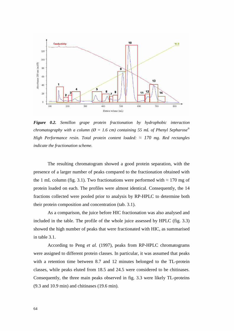
64
Figure 0.2. Semillon grape protein fractionation by hydrophobic interaction
chromatography with a column (Ø = 1.6 cm) containing 55 mL of Phenyl Sepharose®
High Performance resin. Total protein content loaded: ≈ 170 mg. Red rectangles
indicate the fractionation scheme.
The resulting chromatogram showeda good protein separation, with the
presenceof a largernumberof peakscompared to thefractionation obtainedwith
the 1 mL column(fig. 3.1).Two fractionationswere performedwith ≈ 170 mg of
protein loadedon each.The profiles werealmost identical. Consequently, the 14
fractions collectedwerepooledprior to analysis by RP-HPLC to determine both
their protein compositionandconcentration(tab.3.1).
As a comparison,thejuice beforeHIC fractionation wasalsoanalysedand
includedin the table.The profile of the whole juice assessedby HPLC (fig. 3.3)
showedthehigh numberof peaksthatwere fractionatedwith HIC, assummarised
in table3.1.
According to Peng et al. (1997), peaks from RP-HPLC chromatograms
wereassignedto differentproteinclasses.In particular, it was assumed that peaks
with a retention time between8.7 and 12 minutes belongedto the TL-protein
classes,while peakselutedfrom 18.5 and24.5 were considered to be chitinases.
Consequently, the threemain peaksobservedin fig. 3.3 were likely TL-proteins
(9.3and10.9min) andchitinases(19.6min).

65
Figure 0.3. RP-HPLC chromatogram of the unfractionated Semillon juice.
FractionVvTLpeaks
number
Area%
Chitinasespeaks
number
Area%
Numberof otherpeaks
Area%
Proteinconcentration(mg/L BSA)
TotalProteincontent
(mg)Juice 5 41.7 4 37.3 6 21 1874.1 337.5
1 4 60 1 40 \ \ 11.8 1.12 2 61 1 39 \ \ 18.6 1.13 2 70 1 30 \ \ 35.1 2.74 2 67 1 27 1 6 62.9 6.25 1 0.5 1 28.5 1 71 129.4 13.66 1 0.8 2 97.5 1 1.7 110.0 7.57 \ \ 4 88 3 12 115.2 5.28 2 2.2 4 58 3 39.8 207.8 4.29 2 12.2 4 34.4 2 52.5 783.9 58.810 4 38 3 59.5 5 12.5 484.2 98.311 4 87 1 10.3 2 2.7 74.2 3.012 4 88 1 4.6 2 7.4 225.6 12.913 4 92 1 2 1 6 293.8 35.314 2 82 3 11 1 7 28.0 2.8
Table 0.1. Protein class, concentration and area percentage of fractions collected after
Semillon juice HIC fractionation. In total ≈ 340 mg of protein from Semillon juice were
loaded while 252.7 mg were recovered. The chromatograms from which these data were
acquired are shown in figure 3.8. Bold fonts indicate the main protein class of each
fraction.
Thedataof table 3.1 indicatedthatgrapeproteinscouldbedividedin three
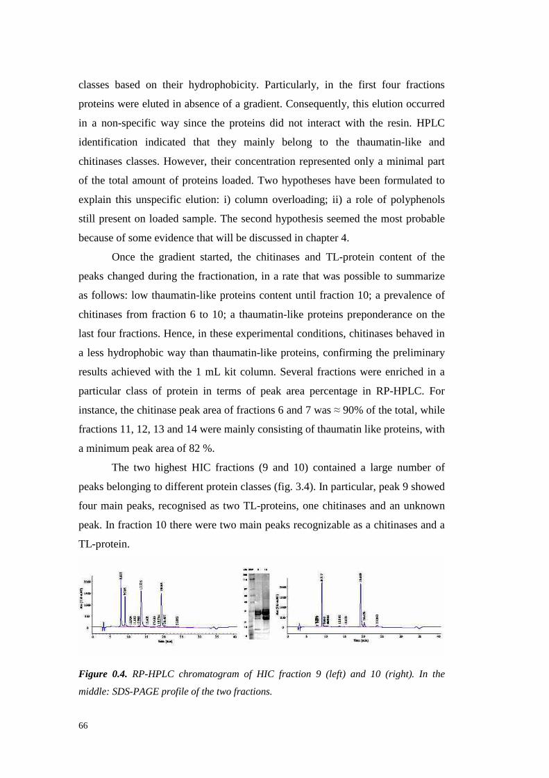
66
classesbasedon their hydrophobicity. Particularly, in the fi rst four fractions
proteinswereeluted in absenceof a gradient.Consequently, this elution occurred
in a non-specific way since the proteinsdid not interact with the resin. HPLC
identification indicated that they mainly belong to the thaumatin-like and
chitinases classes.However,their concentration represented only a minimal part
of the total amountof proteinsloaded.Two hypotheseshavebeen formulated to
explain this unspecificelution: i) column overloading; ii) a role of polyphenols
still presenton loaded sample.The secondhypothesis seemed the mostprobable
becauseof someevidencethatwill bediscussedin chapter 4.
Once the gradientstarted, the chitinasesand TL-protein content of the
peakschangedduring the fractionation,in a rate that waspossible to summarize
as follows: low thaumatin-like proteinscontent until fraction 10; a prevalenceof
chitinases from fraction 6 to 10; a thaumatin-like proteinspreponderanceon the
lastfour fractions.Hence,in theseexperimental conditions,chitinasesbehaved in
a lesshydrophobic way thanthaumatin-like proteins,confirming the preliminary
resultsachievedwith the 1 mL kit column. Several fractions wereenriched in a
particular classof protein in terms of peak area percentage in RP-HPLC. For
instance,the chitinasepeakareaof fractions6 and7 was≈ 90%of thetotal,while
fractions11,12,13 and14weremainlyconsisting of thaumatin likeproteins,with
aminimumpeakareaof 82%.
The two highest HIC fractions(9 and 10) contained a large number of
peaksbelonging to differentproteinclasses(fig. 3.4). In particular,peak 9 showed
four main peaks, recognisedastwo TL-proteins,onechitinasesandan unknown
peak.In fraction 10 therewere two mainpeaksrecognizable asa chitinasesanda
TL-protein.
Figure 0.4. RP-HPLC chromatogram of HIC fraction 9 (left) and 10 (right). In the
middle: SDS-PAGE profile of the two fractions.

67
It resultedevidentthediscrepancyin MW andin RT of bandssupposedto
be TL-proteins, with the lower molecular weight of the TL-proteinsof fractions
10 in comparison to that of following fractions (not shown). Thesedifferences
were attributableto the presenceof different TL-proteins isoforms(Penget al.,
1997).
Preliminary SDS-PAGE showedthat fractions12, 13 and14 containeda
proteinof about65 kDa in additionto theTL protein (not shown). This additional
higherMW protein wasnot visible on theRP-HPLC chromatograms (eg fraction
13, figure3.5).
Figure 0.5. HPLC profile of faction 13 obtained by hydrophobic interaction
chromatography. In the table: retention tine, area (absolute value and percentage) and
height of each peak detected.
Thechromatogramshowedthepresence of 1 main peakthatwasassumed
to be a TL-protein with an estimatepurity of 87.1%. Two lateral peaks were
detected at 10.0 and 12.8 minutes of retention time that were respectively
recognisedas a minor thaumatinlike protein (the first) andan unknownprotein
thesecond, probablyaninvertasesasfollowingshown(figures3.6and3.7).
In any case,thesedata indicatedthe possibilit y to nearly purify a TL-
protein from grape juice in a one-steppreparative processbasedon the different
hydrophobicity characteristicsof grapeproteins.
To confirm HPLC results, a further SDS–PAGE of fraction 13 was
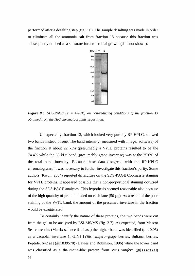
68
performedaftera desaltingstep(fig. 3.6).Thesampledesaltingwas madein order
to eliminate all the ammoniasalt from fraction 13 becausethis fraction was
subsequentlyutili sedasa substratefor amicrobial growth (datanot shown).
Figure 0.6. SDS-PAGE (T = 4-20%) on non-reducing conditions of the fraction 13
obtained from the HIC chromatographic separation.
Unexpectedly, fraction13, which lookedvery pureby RP-HPLC, showed
two bands insteadof one.Thebandintensity(measured with ImageJsoftware)of
the fraction at about 22 kDa (presumably a VvTL protein) resulted to be the
74.4% while the 65 kDa band (presumablygrapeinvertase)wasat the 25.6%of
the total band intensity. Becausethese data disagreedwith the RP-HPLC
chromatograms,it wasnecessaryto further investigate this fraction’s purity. Some
authors (Kwon, 2004)reporteddifficulties on theSDS-PAGECoomassiestaining
for VvTL proteins. It appearedpossiblethat a non-proportional staining occurred
during theSDS-PAGE analyses.This hypothesisseemed reasonable also because
of thehigh quantity of proteinloadedon each lane(50 µg).As a result of thepoor
staining of the VvTL band,the amountof the presumed invertasein the fraction
would beexaggerated.
To certainly identify the natureof theseproteins, the two bandswere cut
from the gel to be analysedby ESI-MS/MS (fig. 3.7). As expected, from Mascot
Searchresults (Matrix sciencedatabase) thehigherband wasidentified(p < 0.05)
as a vacuolar invertase 1, GIN1 [Vitis vinifera=grapeberries, Sultana, berries,
Peptide, 642 aa] (gi|1839578) (DaviesandRobinson,1996)while the lower band
was classified as a thaumatin-like protein from Vitis vinifera (gi|33329390)
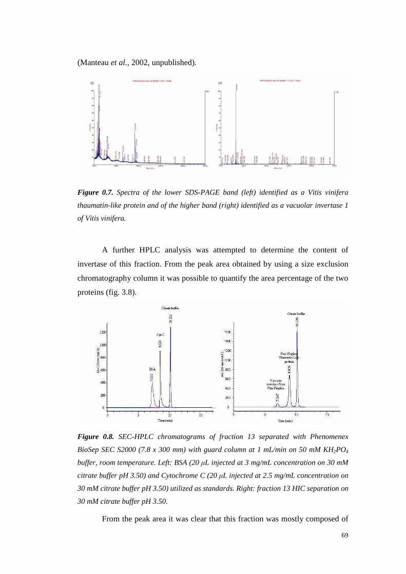
69
(Manteauet al., 2002, unpublished).
Figure 0.7. Spectra of the lower SDS-PAGE band (left) identified as a Vitis vinifera
thaumatin-like protein and of the higher band (right) identified as a vacuolar invertase 1
of Vitis vinifera.
A further HPLC analysis was attempted to determine the content of
invertaseof this fraction. From the peakarea obtained by using a size exclusion
chromatographycolumnit waspossibleto quantify thearea percentageof thetwo
proteins(fig. 3.8).
Figure 0.8. SEC-HPLC chromatograms of fraction 13 separated with Phenomenex
BioSep SEC S2000 (7.8 x 300 mm) with guard column at 1 mL/min on 50 mM KH2PO4
buffer, room temperature. Left: BSA (20 µL injected at 3 mg/mL concentration on 30 mM
citrate buffer pH 3.50) and Cytochrome C (20 µL injected at 2.5 mg/mL concentration on
30 mM citrate buffer pH 3.50) utilized as standards. Right: fraction 13 HIC separation on
30 mM citrate buffer pH 3.50.
From thepeakareait wasclearthat this fraction wasmostly composedof
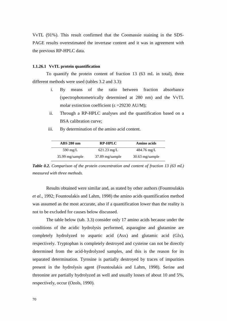
70
VvTL (91%). This result confirmed that the Coomassie staining in the SDS-
PAGE results overestimatedthe invertasecontent and it was in agreement with
thepreviousRP-HPLC data.
1.1.26.1 VvTL protein quantification
To quantify the protein content of fraction 13 (63 mL in total), three
differentmethodswereused(tables3.2and3.3):
i. By means of the ratio between fraction absorbance
(spectrophotometrically determinedat 280 nm) and the VvTL
molar extinctioncoefficient(ε =29230AU/M);
ii. Through a RP-HPLC analysesand the quantification basedon a
BSA calibrationcurve;
iii. By determinationof theaminoacidcontent.
ABS 280 nm RP-HPLC Amino acids
590mg/L 621.23mg/L 484.76mg/L
35.99mg/sample 37.89mg/sample 30.63mg/sample
Table 0.2. Comparison of the protein concentration and content of fraction 13 (63 mL)
measured with three methods.
Resultsobtainedweresimilar and, asstatedby otherauthors(Fountoulakis
et al., 1992; Fountoulakis andLahm, 1998) theamino acidsquantification method
wasassumedasthemost accurate, also if a quantification lower than thereality is
not to beexcludedfor causesbelow discussed.
Thetablebelow (tab.3.3) consideronly 17 amino acidsbecauseunder the
conditions of the acidic hydrolysis performed, asparagine and glutamine are
completely hydrolyzed to aspartic acid (Asx) and glutamic acid (Glx),
respectively. Tryptophanis completelydestroyedandcysteinecannot bedirectly
determined from the acid-hydrolyzed samples, and this is the reason for its
separateddetermination. Tyrosine is partially destroyedby traces of impurities
present in the hydrolysis agent (Fountoulakis and Lahm, 1998). Serine and
threoninearepartially hydrolyzedaswell andusually lossesof about10 and5%,
respectively, occur(Ozols,1990).
71
Amino AcidAmino Acid
(−H2O)
(µg/mL)a
AminoAcid
(µg/mL)b
Mole(%)
Expectedaacompositionof TL-protein(% Mole)c
Expectedaacompositionof
Invertase(% Mole)c
Histidine 5.06 5.70 0.87 0.40 3.00Serine 26.61 32.35 7.18 7.60 6.70
Arginine 20.55 22.90 3.09 2.20 4.00Glycine 29.81 39.20 12.27 9.80 7.90
Asxe 68.50 79.20 13.98 12.90 12.50Glxf 32.56 37.10 5.92 5.40 7.00
Threonine 50.24 59.15 11.67 10.20 6.70Alanine 26.62 33.35 8.79 8.90 6.70Proline 26.00 30.80 6.29 5.80 6.20Lysine 16.72 19.05 3.06 2.70 2.50
Tyrosine 29.25 32.45 4.21 3.60 4.50Methionine 7.22 8.20 1.29 1.30 2.60
Valine 24.02 28.40 5.69 4.40 7.60Isoleucine 13.12 15.20 2.72 2.70 4.80Leucine 28.33 32.85 5.88 6.20 9.70
Phenylalanine 44.53 49.95 7.10 7.10 3.90Cysteined 33.65 41.9 n.d. 7.60 0.80
Total 484.76 567.75 100.00 98.80 97.10
a Calculationbasedon amino acidresiduemassin protein (molecular weightminusH2O).
b Calculation basedon freeamino acidmolecularweight.c Amino acidcomposition from thecomputationof thecompleteprotein sequenceobtainedfrom the
ProtParamtool of theExPASy ProteomicsServerd Cysteinewas separatelydeterminede Results deriving from theaddition of asparagineandaspartic acid.f Resultsderiving from theaddition of glutamineandglutamicacid.
Table 0.3. Amino acids composition of HIC fraction 13. 17 amino acids have been
quantified in HIC fraction 13, while the expected amino acids composition of invertase
and TL-protein is expressed as a molar percentage on 20 amino acids.
Watersandcolleagues(1992)purified a heat unstable protein with MW of
24 kDa that was following identified as a thaumatin-like protein (Waters et al.,
1996). The authorsshowedthat this protein contained large amountsof aspartic
acid, glycine,alanine,serine, threonine,phenylalanineand tyrosine. Theseresults
strongly agreewith those of the tableabove.However,some differencesfrom the
comparison between the amino acids composition of HIC fraction 13 and the
expectedamino acid compositionof the thaumatin-like protein were highlighted.
In particular, fraction 13 showed a higher amount of histidine, arginine, proline
and tyrosine than the expected.These differences seem attributable to the
invertasescontamination that, as visible from its expected amino acids content,
could shift the molar percentageof some amino acid. Besides, the serine
percentageresultedslightly lower thantheexpected. As previously discussed,the
72
amino acid quantification by acid hydrolysis can result in an underestimation of
the level of this aminoacid because of its fragility (Simpsonet al., 1976;Ozols,
1990). In addition, several amino acids (alanine, proline, lysine, tyrosine,
methionine, valine, isoleucine, leucine, phenylalanineandcysteine) weredetected
in amountsvery similar to the expected.According to Waters et al. (1992), the
amount of threeaminoacids(Asx, Glx andThreonine) resulted a li ttle higher in
HIC fraction13 thanthepredicted, andthis occurrence cannotbeexplainedby the
invertasecontamination.It seemspossible that thesedisagreeing resultsaredueto
some post-translational modification that is not taken into account in the amino
acids computation of table 3.3. Looking at the glycine contentof the sample, it
resultedsensibly higherthantheexpectedandnot influencedby theinvertase.It is
possible that a chitinasecontaminationled to this content.In fact, the chitinase
impurity (2.1%)presentin this fraction(seefig. 3.5)couldexplain this occurrence
becauseof thehigh glycinecontent of this protein (13%,not shown).
In general, the results formerly discussedare in accord with the
observationthat themost representedamino acids in grapejuice are aspartic acid,
threonineandphenylalanine(Yokotsukaand Singleton,1997).Besides,a relevant
amount of proline was detected.Several authors reported that polypeptides
containing proline are able to form haze when combined with phenolic
compounds(Asanoet al., 1982; Siebertet al., 1996).This observation agrees with
the assumption that PR-proteinsin general and VvTL in particular are the main
haze-forming proteinsin white whines (Høj et al. 2000; Tattersall et al. 2001;
Ferreiraet al. 2002).
The whole fraction 13 (63 mL) was concentrated by using a stirred
ultrafiltration cell system(Amicon) equippedwith membraneswith porosity of
3000 MWCO. The concentratedsamplewas washedwith 30 mM citrate buffer
(pH 3.50) in order to eliminate all the residualammonia salt. The absenceof
ammonia wasdetectedusing an ammoniakit (not shown).Theprotein contentof
the concentratedsample,assessed spectrophotometrically, was8.7 mg/mL with a
total proteincontentof 30.26mg(tab.3.4).
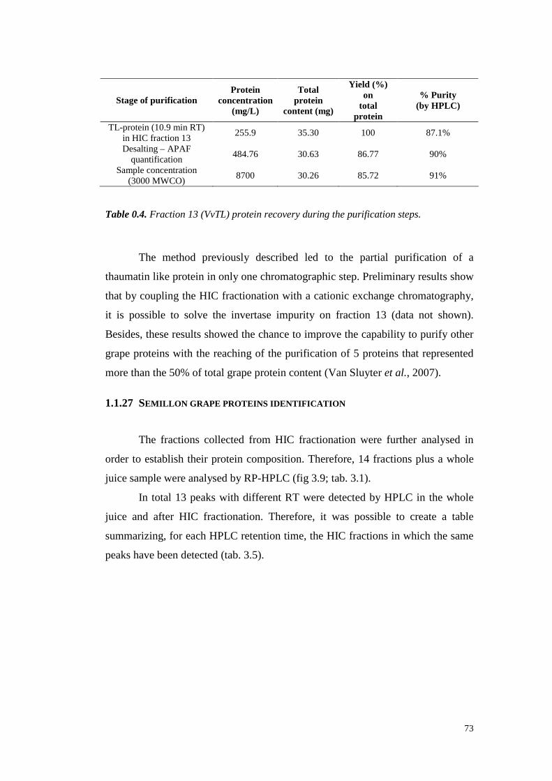
73
Stage of purificationProtein
concentration(mg/L)
Totalprotein
content (mg)
Yield (%)on
totalprotein
% Purity(by HPLC)
TL-protein(10.9min RT)in HIC fraction13
255.9 35.30 100 87.1%
Desalting– APAFquantification
484.76 30.63 86.77 90%
Sampleconcentration(3000 MWCO)
8700 30.26 85.72 91%
Table 0.4. Fraction 13 (VvTL) protein recovery during the purification steps.
The method previously describedled to the partial purification of a
thaumatinlike protein in only onechromatographicstep.Preliminary resultsshow
that by coupling theHIC fractionationwith a cationic exchangechromatography,
it is possible to solve the invertase impurity on fraction 13 (data not shown).
Besides,theseresultsshowedthechanceto improvethecapabili ty to purify other
grapeproteinswith the reachingof the purification of 5 proteinsthat represented
morethanthe50%of total grapeproteincontent(VanSluyter et al., 2007).
1.1.27 SEMILLON GRAPE PROTEINS IDENTIFICATION
The fractions collected from HIC fractionation were further analysedin
orderto establish their proteincomposition. Therefore, 14 fractionsplus a whole
juicesample wereanalysedby RP-HPLC (fig 3.9; tab.3.1).
In total 13 peakswith different RT weredetected by HPLC in the whole
juice and after HIC fractionation.Therefore,it was possibleto create a table
summarizing, for eachHPLC retentiontime, theHIC fractions in which thesame
peaks havebeendetected(tab.3.5).

74
Figure 0.9. RP-HPLC chromatograms of Semillon juice fractionated by HIC
chromatography.
RT (minutes) protein in whole juice HIC fractions in which it was present5.5 6-7-8-9
8.1 (8.0) 8-9-10-119.0 (8.9) 1-2-3-4
9.3 8-9-10-1110.1 12-1310.9 11-12-13-14
13.3(12.8) 7-8-9-10-11-12-13-1413.9 6-7-8-9-1014.1 4-518.6 7-8-9-10
19.5(19.6) In every fraction20.4 7-8-9-10
24.5(23.9) 8-9-10
Table 0.5. Peaks retention time of the whole Semillon juice and fraction in which they are
detectable after HIC chromatography (between brackets there are the equivalent RTs
obtained with a slightly different HPLC set up). Bold fonts indicate the main protein class
of each fraction.
The table summarisedhow grapeproteins werefractionated by HIC. The
peak distribution was, for all the RT considered, in adjacent HIC fractions,
indirectly confirming the appropriateness of Phenyl Sepharose® HP resin for
grapeprotein fractionation.
In detail, it seemedthat the peak with 9.0 min of RT was eluted in a
75
similar way in the first 4 fractions in which no elution buffer was present.
Consequently, this proteinwasnot bound by theresin.
Once the elution gradientstarted,the fi rst protein eluted had RT of 14.1
andcame out in presenceof low concentration of the elution buffer (fractions4
and5) suggesting avery low hydrophobicity degreefor this protein.
A HPLC protein peakwith RT 5.5 min wasdetectable in fractions6 to 9,
with a prevalenceon fraction 7. The elution of this protein started with high salt
concentration,showingits low-mediumhydrophobicity.
A similar hydrophobicitywasobservedfor protein with 18.6 min of RT,
with its major concentrationin HIC fraction 8. In the same fraction (8) the peak
with RT 13.3min wasmainly recoverable,also if its presencewasdetected from
fraction 6 to the end of the separation.As afterward demonstrated, this protein
seemedto belongto theinvertasesclasses,andthecauses of this spreadingwill be
laterdiscussed.
Fraction9 and10 were thosewith the largernumberof protein classes.In
particular, proteinwith RT of 8.1 and13.9were mostly elutedon fraction 9 while
proteinwith RT of 9.3,19.5,20.4and24.5weremainly eluted on fraction 10.
As previously discussed,a TL-protein was mainly contained in the last
partof theseparation (from fraction11). In thosepeaks,besidesthemain TL peak
(RT 10.9 min) only two other peakshave been detected: a peak at 10.1 min
mainly containedon fraction 12 anda peakat 19.5 min (presumably chitinases)
that wasobservedin everyHIC fraction.
Two protein peaksdid not show a fractionation behaviour in agreement
with the hydrophobicity. Peaksat 13.3 and 19.5 min of RT were detected
respectively in fractions7 to 14 andin all the fractions considered, respectively,
showing a not strictly hydrophobicdependantelution. This occurrenceneedto be
furtherinvestigated.
1.1.27.1 ESI-MS/MS Protein identification
Okuda and colleagues (2006) affirmed that more than 310 protein or
polypeptidefractions wererecoverablein JapaneseChardonnay winesbelonging
to the thaumatin and osmotin-like proteins,invertase, lipid transferproteins and
their hydrolysis products.

76
In order to identify the origin of Semillon grapeproteins,5 HIC fractions
(4, 5, 9, 11 and13) werechosenbecauseof their protein content representative of
all the HPLC retention time detectable in the whole juice. Three consecutive
HPLC fractionation for eachof thesefractions were madeand peaks, collected
with a fraction collector,werepooled. In total, 12 different peakswereobtained
and,afterconcentrationandequilibrationwith theloadingbuffer, a SDS-PAGE in
non-reducing conditions (fig. 3.10) was performed. Proteins havebeen revealed
by MS-compatiblestains(Bio-SafeTM Coomassiestainor Silverstainprocedure).
Figure 0.10. SDS-PAGE of peaks collected by RP-HPLC. Bands were cut and analysed
by ESI-MS/MS. SDS-PAGE analyses was performed according to Laemmli (1970) by
using Ready Gel Tris-HCl Gel [Bio-Rad, 4–20%, 15-well, 15 µl, 8.6 x 6.8 cm (W x L)],
in absence of reducing agents. RP-HPLC peaks characterized by a high protein
concentration were stained with Bio-Safe® Bio-Rad Coomassie. RP-HPLC peaks with low
protein content were stained with silver procedure. 26 samples in total. The sample 25
(RT 24.500) was not loaded on the gel but analysed in liquid form. Sample 26
corresponded to the VvTL protein (gi|33329390) (already identified by APAF during the
VvTL protein purification of fraction 13). Sample 9.000 was first stained with Coomassie,
than two bands were excised before the lane was re-stained with silver that allowed the
appearance of other two bands (18 and 19).

77
Unexpectedly,the SDS-PAGE showedthe appearance, in somelanes, of
morethanonebandthatderivedfrom theHPLC peak collection. Hence, a HPLC
peakdid not correspondto a singleprotein, and consequently band wereanalysed
via LC-MS/MS analysisto understandtheir nature.
Resultsof 26samplesaresummarisedin table3.6.
Spotname
HICfraction
RP-HPLCRT
ApparentSDS-PAGE
MW
Accessionnumber
1st X! Tandemsignificant ID pI MW
2nd X! Tandemsignificant ID pI MW
1 5 14.555 50 unknown no matches / / no matches / /
2 9 8.100 53 gi|33414046| classIV chitinase[Galegaorientalis] 7.42 29.4PR-4 typeprotein
[Vit is vinifera]5.5 15.2
3 9 8.100 24 gi|33414046| classIV chitinase[Galegaorientalis] 7.42 29.4PR-4 typeprotein
[Vit is vinifera]5.5 15.2
4 9 8.100 15 gi|3511147| PR-4 typeprotein[Vi tis vinifera] 5.5 15.2 / / /
5 9 9.300 40 gi|7406716|putativethaumatin-like protein[Vitis
vinifera]4.94 24 / / /
6 9 9.300 22 gi|7406716|putativethaumatin-like protein[Vitis
vinifera]4.94 24
VVTL1 [Vitisvinifera]
5.09 24
7 9 9.300 16 gi|7406716|putativethaumatin-like protein[Vitis
vinifera]4.94 24
PR-4 typeprotein[Vit is vinifera]
5.5 15.2
8 9 13.990 100 gi|33414046| classIV chitinase[Galegaorientalis] 7.42 29.4 / / /
9 9 13.990 70 gi|33414046| classIV chitinase[Galegaorientalis] 7.42 29.4 / / /
10 9 13.990 65 gi|33414046| classIV chitinase[Galegaorientalis] 7.42 29.4 / / /
11 9 13.990 27 gi|116329|Endochitinase A precursor (Seed
chit inaseA) [Zeamais]8.3 29.1 / / /
12 11 14.300 70 gi|116329|Endochitinase A precursor (Seed
chit inaseA) [Zeamais]8.3 29.1 / / /
13 11 19.560 80 gi|33329392| classIV chitinase[Vi tis vinifera] 5.38 27.5 / / /
14 11 19.560 31 gi|33329392| classIV chitinase[Vi tis vinifera] 5.38 27.5 / / /
15 11 19.560 29 gi|33329392| classIV chitinase[Vi tis vinifera] 5.38 27.5 / / /
16 4 9.000 55 gi|30679715|early-responsive to dehydration
protein-related/ ERD protein-related[Arabidopsis thaliana]
8.68 87.6classIV chitinase[Vit is vinifera]
5.38 27.5
17 4 9.000 24 gi|7406671|putative ripening-relatedprotein
[Vitis vinifera]4.83 22.9 / / /
18 4 9.000 110 unknown no matches no matches / /
19 4 9.000 90 unknown no matches no matches / /
20 9 5.570 10 gi|28194084|lipid transferprotein isoform 1 [Vitis
vinifera]9 11.6 / /
21 11 10.000 25 gi|33329390|thaumatin-like protein[Vitis
vinifera]4.67 23.9 / / /
22 13 10.250 23 gi|33329390|thaumatin-like protein[Vitis
vinifera]4.67 23.9 / / /
23 13 13.330 26 gi|1839578|vacuolarinvertase1, GIN1 [Viti svinifera=grapeberries,Sultana,
berries,Peptide,642aa]4.6 71.5 / / /

78
24 13 13.330 22 gi|33329390|thaumatin-like protein[Vitis
vinifera]4.67 23.9 / / /
25 9 24.500 Not loaded unknown no matches no matches / /
26 13 10.970 23 gi|33329390|thaumatin-like protein[Vitis
vinifera]4.67 23.9
osmotin-like protein[Vitis vinifera]
4.56 23.9
Table 0.6. Summarizing table of the ESI-MS/MS protein identification in comparison to
the HIC fraction from which they were purified and with the retention time with which
they came out from the HPLC separation.
In table3.6,anID is associateto eachband.Fromthealignmentof theraw
datawith X!Tandemdatabase, it seemedthatonly 14sampleson26 arefrom Vitis
vinifera. The numberof sampleidentified as Vitis vinifera protein rises to 17
consideringthe2nd match recoverablein X!Tandemdatabase.
About the 19% of the total proteinsfrom grapeberry mesocarpbelong to
the PR-protein category(Sarry et al., 2004). From the identification of SDS-
PAGE protein spots by ESI-MS/MS it was possible to identify mainly PR-
proteins such as PR-4 type proteins,putative thaumatin-like proteins, class IV
chitinase, putative ripening-related protein, lipid transfer protein isoform 1,
thaumatin-like proteinanda vacuolar invertase1, GIN1. Besides,otherprotein of
no grapeorigin havebeendetected,mainlybelongingto theArabidopsis thaliana,
Galega orientalis and Zea mays speciesandpresenting high homology with grape
PR-proteins,mainly chitinases. It is worth mentioning the absenceof microbial
proteins,indicating the healthinessof the grape used. The non-total Vitis vinifera
origin of the protein analysed is relatedto the databasequality utilised for the
alignment tests.In fact, preliminary resultsconfirmed this hypothesis with the
identification of otherproteinsof grapeorigin, mainly hypothetical protein from
Vitis vinifera (data not shown)that were not includedin thedatabaseusedfor the
first protein recognition (The French-Italian Public Consortium for Grapevine
GenomeCharacterization,2007).
Putative thaumatin-like protein [Vitis vinifera] and Thaumatin-like
protein [Vitis vinifera]: the thaumatin-like proteinsare, after the chitinases,the
mostrepresentedgrapeandwine protein(Waters et al., 1998,Pocock et al., 2000;
Hayasaka et al., 2001). This statementis confirmed in theresultsaboveshown,in
which 10 spotswererecognisedaschitinaseswhile 7 asTL-proteins.Particularly,
79
three spots(5, 6 and7) were identified asputative thaumatin-like proteinswhile
four (21, 22, 24 and26) asthaumatin-like proteins.Thesetwo thaumatin classes
differ in both HPLC retentiontime (respectively 9.3 min and 10.0 to 13.3 min)
and hydrophobicity characteristics(higherfor thethaumatin-like proteins).It is to
be notedthat the threeputativethaumatin-like protein bands derived all from the
same SDS-PAGE lane. Particularly, the HPLC peak (9.3 min) showed the
appearanceof threebandsin SDS-PAGE respectively at 40, 22 and 16 kDa of
apparentMWs. The theoreticalMW of this protein is 24 kDa, so the two other
bandsare likely modification of it, whichnatureneedsto befurther investigated.
Spots 21, 22, 24 and 26 were all identified as the samethaumatin-like
protein. This occurrence could be explained in two ways: i) the presenceof
differentTL-proteins isoformsin our samples recognisedas the sameprotein in
the database;ii) a partial modification of the same thaumatin-like protein that
resulted in a changing of propertiesas its MW or hydrophobicity. The second
hypothesisseemssupportedby dataobtained by Pocockand colleagues(2000),
which showedthe presenceof a main VvTL protein and a minor TL protein that
behaved, in HPLC, similarly respectively to spot 26 and 24. Accordingly, our
experimental data suggestedthe second hypothesis as the most probable. In
particular, we retain that the thaumatin-like protein could be incurred in some
modifications, likely proteolysis.A certain content of endogenousproteolytic
activity is detectablein musts (Cantagrel et al., 1982; Ribéreau-Gayon et al.,
2003), but this activity is not sufficient to degradethehighly resistant PR-proteins
(Waterset al., 1992).However,these enzymes could be responsiblefor a partial
modification of somepeptides, resulting in a slightly different behaviour of the
proteinduring the fractionationprocesses.In fact, the four thaumatin-like protein
bands identified presentedsimilar, but not identical, hydrophobicity. Besides,in
HPLC this proteinshowedup at four RTs,while by SDS-PAGEshowedapparent
MWs from 22 to 25kDa.
The samediscussion might explainalso the appearance of three bands in
the sameSDS-PAGE lanefrom only oneHPLC peak(spot5, 6 and 7) formerly
discussed.
PR-4 type protein [Vitis vinifera]: this classof protein wasmostly eluted
in HIC fraction 9 andshoweda HPLC RT of 8.1min. Generally, thePR-4 classof

80
protein is mainly composed by chitin binding proteins (Theis and Stahl, 2004).
Theseproteinspresentantifungal activity that is mainly dueto their abili ty to bind
fungal cell wall chitin (Bormann et al., 1999). The presence of this class of
protein in grapevineseemsconsequentlydueto someantifungalmechanismof the
plant, also if Tattersallandcolleagues(1998,unpublished)attributed to this class
a ripening-related role. PR-4 proteinsbehaved,in SDS-PAGE, similarly to the
putative thaumatin-like proteins, with the appearance of 3 bandsat different MWs
on thesamelane, recognisedasthesameprotein (spots2, 3 and 4). Actually, only
one of thosespotsresulteda PR-4 protein,while the other two (thosewith MWs
not compatible with the theoreticalMW of 15.2 kDa) resulted moresimilar to a
classIV chitinases.This occurrencemight beexplainedby theorganization of the
PR-4 family of proteins, which is similar to that of the plant chitinasefamily
(Friedrich et al., 1991). Moreover,Van Dammeand colleagues (1999)showedthe
existenceof hevein-like chitin-binding protein isolated from mature elderberry
fruits (Sambucus nigra). The authors demonstrated that this protein was
synthesized as a chimeric precursor consisting of an N-terminal chitin-binding
domainandanunrelatedC-terminaldomain.Sequence comparisonsindicated that
the N-terminal domainhadhigh sequencesimilarity with the N-terminal domain
of classI PR-4 proteins, whereasthe C terminuswas mostclosely related to that
of class V chitinases.This finding contributesto better explain the recognition of
spots 2 and3 aschitinasesinsteadof PR-4 proteins,also if the reasonsfor their
apparentMW in SDS-PAGEwarrantsfurtherinvestigations.
Class IV chitinase [Vitis vinifera]: amongthe 26 samples analysed,10
were identified as chitinasesas first X!Tandem match plus one identified as
second databasematch.Theseresultsconfirmedthepredominance of chitinases in
grape proteinshighlighted by other authors(Waters et al., 1998,Pococket al.,
2000; Hayasaka et al., 2001). By observing the HIC distribution of chitinases
spots, it canbeobservedthat most of theseproteins wereelutedin the middle of
the HIC gradient (HIC fraction9 and11).Particularly, spotsfrom 8 to 15 wereall
recognisedas proteinswith chitinasefunction. However, theseeightbandsdid not
behavesimilarly in SDS-PAGEin which a great MW variabilit y in a rangefrom ≈
97 to ≈ 28 kDa was detectable.This SDS-PAGE mobilit y might be partially
explainedby the absenceof reducing agents in the SDS-PAGE loading buffer,
81
which could be relatedto major differencesin the hydrodynamic volumesof the
protein deriving from structuresstabilized by S-S bonds,although a different
binding of the unreducedprotein to the detergent SDS seems also possible
(Vincenzi and Curioni, 2005). However, thesenotions seemnot sufficient to
explain these great differences in MWs, thus additional investigations are
required.
Putative ripening-related protein [Vitis vinifera]: one spot (17) was
identified asa putative ripening-relatedprotein. This protein presented a very low
hydrophobicity becauseit was eluted in HIC fraction 4, so before the gradient
started. Besides,its HPLC RT (9.0 min) was similar to thoseof the putative
thaumatinlike proteins.This occurrenceseemedexplained by theslight difference
in pI betweenthis protein (4.83) and the thaumatin one (4.94), resulting in a
slightly different RT in HPLC. Besides,the apparentMW in SDS-PAGE of this
proteinresultedhigherthanthehypothetical (≈ 25 insteadof 22.9kDa).
Lipid transfer protein isoform 1 [Vitis vinifera]: in li terature thereare
two closely-related types of nsLTPs, types 1 and 2, which differ in protein
sequence,molecular weight,andbiological properties (Chenget al., 2004). Many
nsLTP1 proteins,alsoof grapevineorigin, have been characterizedasallergensin
humans(Pastorello et al., 2002).TheLTP hereidentified belongto the isoform1
andshowedvery low MW in SDS-PAGEandearly RT in HPLC, in which it was
well separatedfrom theotherproteins. HIC fractionation alsoshoweda goodLTP
separation ability asit wasmainlyelutedin fraction7.
Vacuolar invertase 1, GIN1 [Vitis vinifera=grape berries, Sultana,
berries, Peptide, 642 aa]: someauthors(DaviesandRobinson,1996;Sarry et al.,
2004) highlightedthepreferentialexpressionof theGIN1 isogenewith respectto
the GIN2 in the pericarp, confirming the results here described. Band 23,
recognisedas a vacuolar invertase,did not show a MW of about 70 kDa as
expected, but its apparentMW wasof 28 kDa. Okudaandco-workers (2006),by
using 2D-PAGEhavenoticed thepresence of invertasefragmentson Chardonnay
winewith MWs of 39, 38 and29 kDa, highlightingfor the first timethepresence
of hydrolysedinvertasesin wine. However,in this study, proteins were derived
from grapes.Consequently, theobservedinvertasehydrolysiscouldnot havebeen
due to thefermentationprocessassuggestedby Okuda, but might bedueto some
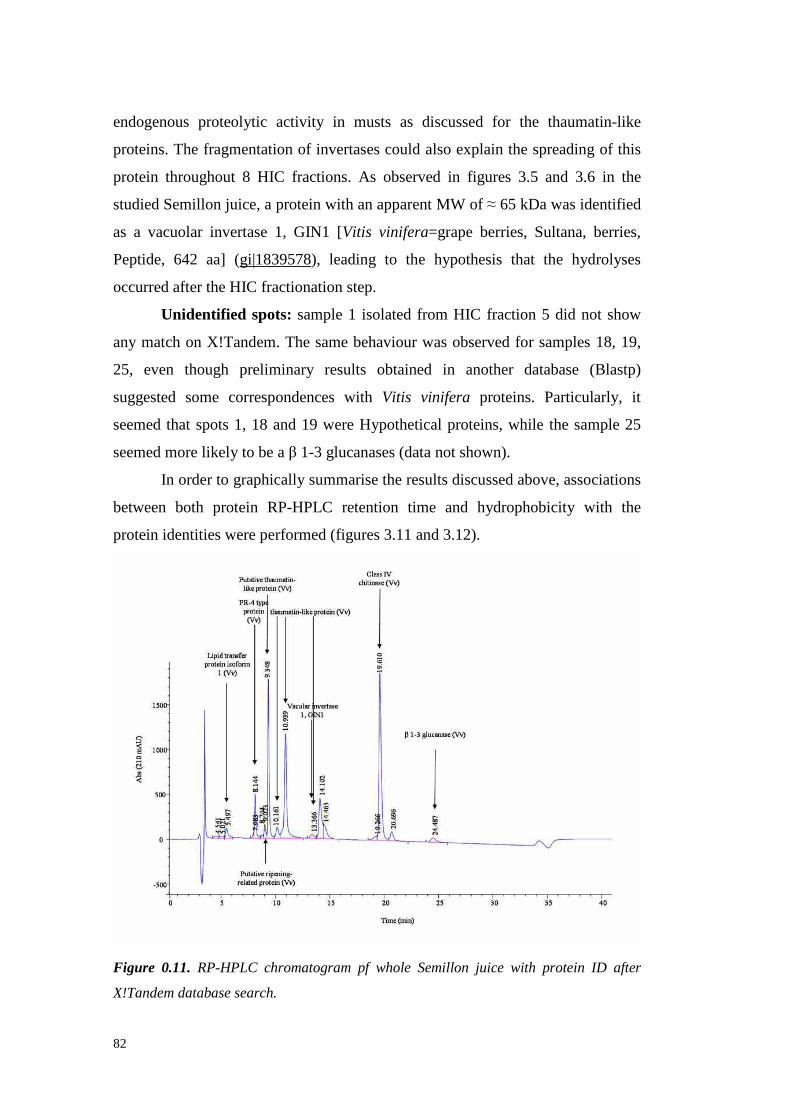
82
endogenousproteolytic activity in musts as discussed for the thaumatin-like
proteins. The fragmentationof invertasescould also explain thespreading of this
protein throughout 8 HIC fractions.As observed in figures 3.5 and 3.6 in the
studiedSemil lon juice, a proteinwith anapparent MW of ≈ 65 kDa wasidentified
as a vacuolar invertase 1, GIN1 [Vitis vinifera=grapeberries, Sultana, berries,
Peptide, 642 aa] (gi|1839578), leading to the hypothesisthat the hydrolyses
occurredaftertheHIC fractionationstep.
Unidentified spots: sample1 isolatedfrom HIC fraction 5 did not show
any match on X!Tandem.The samebehaviour was observedfor samples18, 19,
25, even though preliminary results obtained in another database (Blastp)
suggestedsome correspondenceswith Vitis vinifera proteins. Particularly, it
seemedthat spots 1, 18 and19 wereHypothetical proteins,while the sample 25
seemedmorelikely to beaβ 1-3 glucanases(datanot shown).
In orderto graphicallysummarisetheresultsdiscussedabove, associations
between both protein RP-HPLC retention time and hydrophobicity with the
protein identitieswereperformed(figures3.11and3.12).
Figure 0.11. RP-HPLC chromatogram pf whole Semillon juice with protein ID after
X!Tandem database search.

83
Figure 0.12. Nature of proteins fractionated during HIC chromatography.
From the generalschemesabove represented, it was possible to make
someremarks:
o Putative ripening-related proteins and chitinasesseem the grape
proteins less influenced from the hydrophobicity. Chitinases
seemednot to beaffectedby theresinbecausetheelution occurred
all alongthegradient.Theelutionappeared to benot specific, also
if themain chitinasesconcentrationwasdetectablefrom fraction 5
to 10, while in the last 4 fractionschitinaseswere detectableonly
in traces. This phenomenoncould be ascribed to the presence of
several classesof chitinases(Waterset al., 1998)thatcanassumea
different behaviourduringthegradientand/or to a fragmentation of
this proteinthatled to its modified hydrophobicity.
o After thechitinases, thesecondprotein morespreadalongtheHIC
fractionation was the vacuolar invertase that started to be
detectablefrom fraction 7 until the endof the gradient. It appears
that this protein was initially precipitated by the resin,while with
the reachingof an ammoniumsulphateconcentration of about0.8
M in the buffer, a re-solubilization of the invertasestarted and
continued until the end of the gradient.The causesof this event

84
need to be investigated. We hypothesize that an invertase
glycosilation resultingin an interactionwith the resin could be a
possible cause. Besides, an invertase fragmentation due to
proteolysisassuggestedby Okudaet al. (2006)could alsoexplain
this phenomenon.It is noteworthythat,at RP-HPLC retention time
of 13.3 min were associatedboth invertases and thaumatin-like
proteins.This fact could further explainthe difficulty in obtaining
a TL-proteinwithout invertase contamination discussedin section
3.4.
o Someproteins wereelutedin a small chromatogramzone,such as
the Lipid TransferProteinisoform 1, detected in themiddle of the
fractionationandmainly in fraction 7. This resultwas unexpected
becausetheLTP is meantto bea veryhydrophobicprotein. In fact,
LTP is known to have a hydrophobicpocket that endowsthem
with the capacity to bind hydrophobic molecules (Blein et al.,
2002).
o A thaumatin-like protein (gi|33329390) resulted the most
hydrophobic protein in Semillon grape juice. This fact
differentiatesthis protein classfrom all the other, bringing to its
high purification gradealready aftertheHIC fractionation.
ResultspreviouslydiscussedhighlightedthatHIC chromatography canbe
usedfor grapeproteinfractionationandthat it is a tool for partial or total protein
purification.By coupling this techniquewith RP-HPLC analyses,SDS-PAGEand
ESI-MS/MS it was possibleto identify a large number of grapeproteins and to
obtain preliminaryresultson theirhydrophobiccharacteristics.
1.1.28 SEMILLON WINE PROTEIN FRACTIONATION
To validatethe data collectedon Semillon grapeproteins in the previous
part of this work, theHIC methodproposedwastestedfor its proteinfractionation
abili ty of Semill onwine.
To this aim, proteins from severallitres of an unfined Semillon wine were

85
precipitated with ammonium sulphate (80% saturation) and collected by
centrifugation.Theproteinrecovery wasunexpectedly low, with a total amountof
proteinprecipitatedof 145.4mg/L (58.15mg in total). After equilibrationwith the
loading conditions,precipitatedwine proteins wereinjected into theHIC column.
Theresulting chromatograms(fig. 3.13)showeda high similarity with thatof the
Semillon juice.
Figure 0.13. Semillon juice and wine chromatograms of protein fractionation by
hydrophobic interaction chromatography.
Three consecutive and almost identical fractionations were performed.
Consequently, fractions collectedwerepooled prior to be analysedby RP-HPLC
(fig. 3.14;tab.3.7) to determineboththeir protein composition andconcentration.
Differently to the juice fractionation,only 8 fractions wereobtained. As reported

86
by severalauthors(Murphey et al., 1989; Dizy and Bisson,2000; Fukui et al.,
2003), a protein simplification can occur during the fermentation. The main
differences were noticed in the first half of the gradient, in which the less
hydrophobic proteins were eluted. As shown in the juice chromatogram, only
small amounts of proteinwere detectedin the fi rst part of the fractionation. This
behaviour could indicate that, during the fermentation, the less hydrophobic
proteins were more affectedthan the most hydrophobic(mainly Chitinasesand
TL-proteins), which seemednot to be subjected to significant modifications
during this process(Waterset al., 1992).
Figure 0.14. RP-HPLC chromatograms of fractions collected through HIC Semillon wine
protein fractionation.
FractionVvTLpeaks
number
Area%
Chitinasespeaks
number
Area%
Numberof otherpeaks
Area%
Proteinconcentration(mg/L BSA)
TotalProteincontent
(mg)Wine 6 52.1 3 30.9 5 17.0 145.4 58.15
1 3 24.5 1 74 2 1.5 78.5 9.82 2 20.2 1 79.8 1 11 24.9 2.13 1 8.5 1 85.2 1 6.2 7.8 0.94 2 11.7 1 5.7 1 82.6 53.9 6.75 1 93 1 0.9 3 4.3 171.1 13.76 1 56.1 1 16.1 2 27.7 7.0 0.47 2 86 1 14 / / 4.8 0.78 3 93.4 2 2.8 1 3.8 132.6 14.6
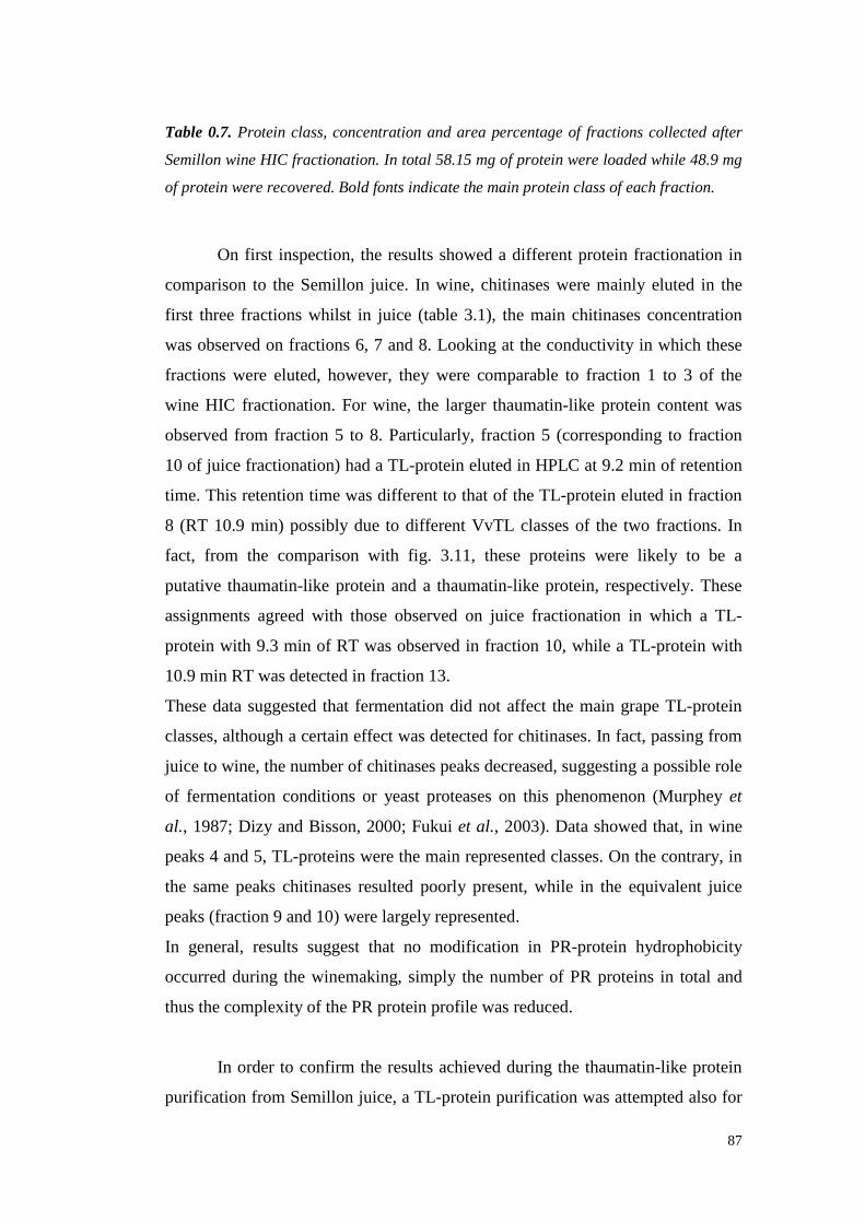
87
Table 0.7. Protein class, concentration and area percentage of fractions collected after
Semillon wine HIC fractionation. In total 58.15 mg of protein were loaded while 48.9 mg
of protein were recovered. Bold fonts indicate the main protein class of each fraction.
On first inspection, the resultsshoweda different protein fractionation in
comparison to the Semillon juice. In wine, chitinaseswere mainly eluted in the
first threefractions whilst in juice (table 3.1), the main chitinasesconcentration
wasobserved on fractions 6, 7 and8. Looking at theconductivity in which these
fractions were eluted, however,they were comparable to fraction 1 to 3 of the
wine HIC fractionation.For wine, the larger thaumatin-like protein content was
observedfrom fraction 5 to 8. Particularly,fraction 5 (corresponding to fraction
10 of juice fractionation)hada TL-proteineluted in HPLC at 9.2 min of retention
time. This retention time wasdifferent to thatof theTL-protein eluted in fraction
8 (RT 10.9 min) possibly dueto different VvTL classesof the two fractions. In
fact, from the comparison with fig. 3.11, theseproteins were likely to be a
putative thaumatin-like proteinand a thaumatin-like protein, respectively. These
assignments agreedwith those observed on juice fractionation in which a TL-
proteinwith 9.3 min of RT wasobservedin fraction 10, while a TL-protein with
10.9 min RT wasdetectedin fraction13.
Thesedatasuggestedthat fermentationdid not affect the main grapeTL-protein
classes, although a certaineffectwasdetectedfor chitinases.In fact, passing from
juice to wine,thenumberof chitinasespeaksdecreased,suggesting a possible role
of fermentation conditionsor yeastproteaseson this phenomenon(Murphey et
al., 1987;Dizy andBisson,2000;Fukui et al., 2003).Data showedthat, in wine
peaks 4 and5, TL-proteinswerethemainrepresented classes. On thecontrary, in
the samepeakschitinasesresultedpoorly present,while in the equivalent juice
peaks (fraction 9 and10) werelargelyrepresented.
In general,results suggest that no modification in PR-protein hydrophobicity
occurred during the winemaking,simply the number of PR proteins in total and
thusthecomplexity of thePRproteinprofilewasreduced.
In order to confirm the results achievedduring the thaumatin-like protein
purification from Semillonjuice, a TL-protein purification wasattemptedalsofor

88
the wine. As abovementioned,HIC fraction 8 contained a TL-protein almost
identical of thatpurified in juiceHIC fraction13 (seeparagraph3.4.2).Proteinsof
fraction 8 wereprecipitatedthroughthe addition of ammoniumsulphate(99% of
saturation) andthepelletdissolvedin citrate buffer. The fraction soprepared was
desaltedby meansof a 20 mL column containing Bio-Gel® P-10DG gel. The
absenceof ammoniawasassessedby meansof anammoniakit (data not shown).
The desalting was performedbecausethe purified TL-protein was subsequently
util ised asasubstratefor amicrobialgrowth(data not shown).
The protein content of the eluateafter the desalting step was assessed
spectrophotometrically (tab.3.8).
Stage of purificationProtein
concentration(mg/L)
Totalprotein
content (mg)
Yield (%) ontotal protein
% Purity(by HPLC)
TL-protein(10.9min RT)in wholewine
31.1 12.44 100 23%
TL-protein(10.9min RT)in HIC fraction 8
123.5 12.14 97.9 91%
Desalting 187.0 6.17 42.26 93.9%
Table 0.8. Fraction 8 (VvTL) protein recovery during the purification steps.
The desaltedsample reacheda proteinconcentration of 187 mg/L, so the
total protein collected was 6.17 mg/33 mL of final sample volume, with an
estimate purity of 93.9%. In wine TL-protein purification, the yield was lower
than the expected.As visible from the table, this loss was mainly due to the
desaltingstep.In fact, in orderto get rid of all theammoniasalt from thesample,
the tails of the protein peaksfrom the desalting column were discarded, with a
consequential greatlossof protein(not shown).
Theresidualimpurity of this fractionwas dueto 2 peaks,respectively with
9.300 and 13.200min of retentiontime.
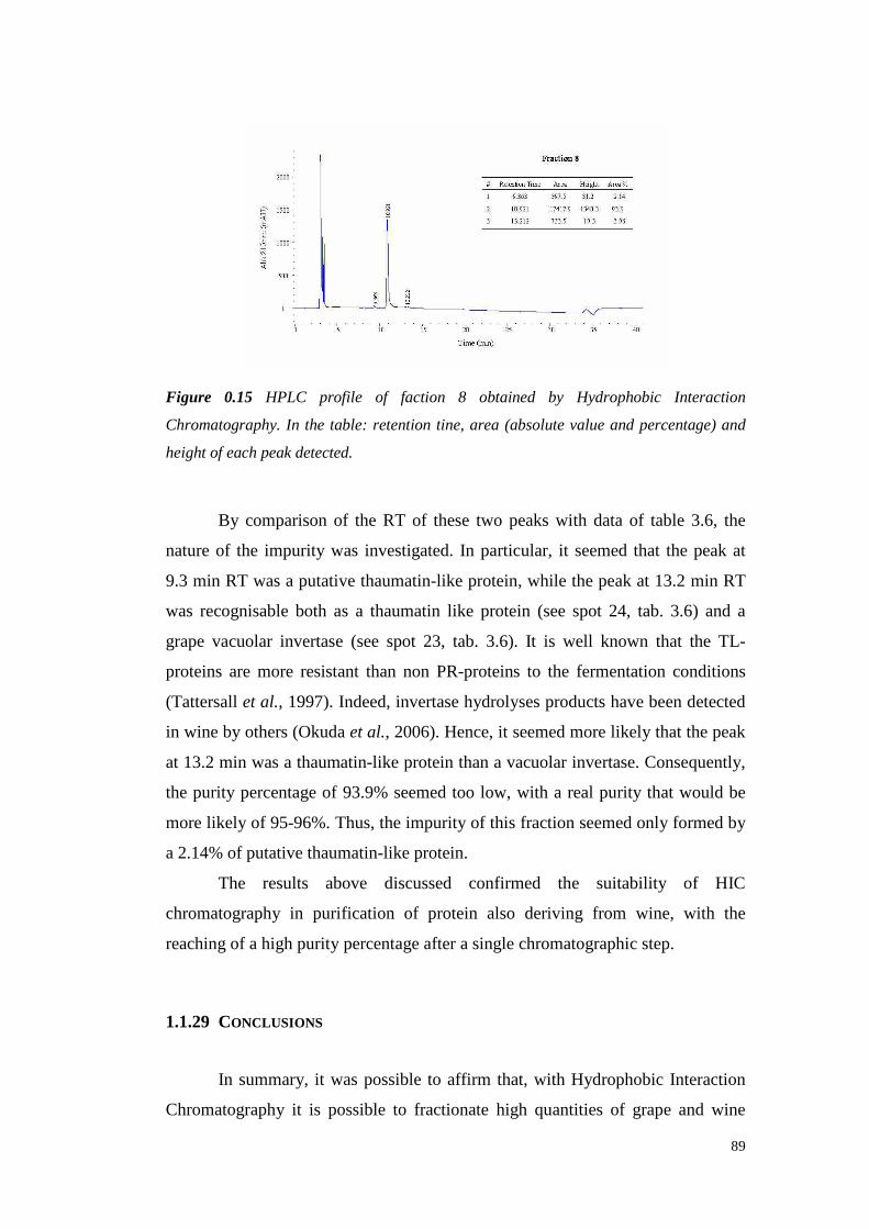
89
Figure 0.15 HPLC profile of faction 8 obtained by Hydrophobic Interaction
Chromatography. In the table: retention tine, area (absolute value and percentage) and
height of each peak detected.
By comparisonof the RT of these two peaks with dataof table 3.6, the
nature of the impurity was investigated.In particular, it seemed that the peak at
9.3 min RT wasa putativethaumatin-like protein, while thepeak at 13.2min RT
was recognisable both as a thaumatinlike protein (seespot 24, tab. 3.6) and a
grapevacuolar invertase(seespot 23, tab. 3.6). It is well known that the TL-
proteinsare more resistantthan non PR-proteins to the fermentation conditions
(Tattersallet al., 1997).Indeed,invertasehydrolyses productshave beendetected
in wine by others(Okuda et al., 2006).Hence,it seemedmorelikely that thepeak
at 13.2min wasa thaumatin-like proteinthana vacuolarinvertase.Consequently,
the purity percentageof 93.9%seemedtoo low, with a real purity that would be
morelikely of 95-96%.Thus, theimpurity of this fraction seemedonly formedby
a2.14%of putative thaumatin-like protein.
The results above discussed confirmed the suitabilit y of HIC
chromatographyin purification of protein also deriving from wine, with the
reachingof ahighpurity percentageafterasinglechromatographicstep.
1.1.29 CONCLUSIONS
In summary,it waspossible to affirm that, with HydrophobicInteraction
Chromatography it is possibleto fractionate high quantities of grapeand wine
90
protein combining a good preparativefractionation with the reaching of high
protein purity for severalfractions. In particular, a Vitis vinifera thaumatin-like
protein purification in a single step with high recovery percentages from both
Semillon juiceandwinewasachieved.
Moreover,thechromatographicsystemused hadshownthepotentiality of
purify morethanoneprotein(especiallyin wine in which theprofile is simplified)
and, for this reason,further studiesshould utili ze this knowledgeto purify other
grape and wineprotein classes.Preliminary results showedthepossibility to reach
these objectives with a purification in a two-step chromatographyof 5 grape
proteins(VanSluyter et al., 2007).
It seems possible to affirm that theapplication of HIC chromatography in
wine studiescan contributeto improvetheknowledgeon grapeand wine protein.
Moreover,by coupling this techniquewith other chromatographic methods, a
more efficient protein purification in termsof both quantity and quality can be
exploited.
Acknowledgements
The authorwould like to thankElizabethJ. WatersandSteveVan Sluyter of The
Australian Wine ResearchInstitute and Paul A. Haynes of the Macquarie
University for their supportin theconduction of theexperiments, for thevaluable
discussionsaboutthis work andfor therevisionof this manuscript.
This work was partially supportedfinanciall y by Australia's grapegrowersand
winemakersthrough their investment body the Grapeand Wine Research and
DevelopmentCorporation,with matchingfundsfrom theAustralianGovernment.
This researchhasbeenfacilitatedby accessto the Australian Proteome Analysis
Facility establishedunderthe AustralianGovernment’s Major National Research
Facilities Program.
91
REFERENCES
AsanoK., ShinagawaK. andHashimmoto N. (1982). Characterization of haze-
forming proteins of beerand their roles in chill haze formation. J. Am. Soc.
Brew. Chem. 40,147-154.
Bayly F.C.andBerg H. (1967).Grapeandwineproteinsof white winevarietals.
Am. J. Enol. Vitic. 18(1),18-32.
Blein J.P.,Coutos-ThévenotP.,MarionD., PonchetM. (2002).Fromelicitins to
lipid-transfer proteins: a new insight in cell signalling involved in plant
defencemechanisms.Trends Plant Sci. 7, 293-296.
Blum H., Beier H. and Gross H.J. (1987). Improved silver staining of plant
proteins,RNA andDNA in polyacrylamidegels. Electrophoresis 8, 93-99.
BormannC., Baier D., Horr I., RapsC., Berger J., Jung G. and Schwarz,H.
(1999). Characterization of a novel, antifungal, chitin-binding protein from
Streptomyces tendae Tu901that interfereswith growth polarity. J. Bacteriol.
181, 7421-7429.
BrissonnetF. andMaujeanA. (1993).Characterization of Foaming Proteins in a
ChampagneBaseWine.Am. J. Enol. Vitic. 44(3),297-301.
CanalsJ.M., Arola L., andZamoraF. (1998).Protein Fraction Analysisof White
Wineby FPLC.Am. J. Enol. Vitic. 49(4),383-388.
CantagrelR., SymondsP.andCarlesJ. (1982).Composition enacidesaminésdu
moùt enfonction du cépageet dela technologieet soninfluencesur la qualité
du vin. Sci. Aliment. 2, 109-142.
ChengC.S.,Samuel D., Liu Y.J., Shyu J.C., Lai S.M., Lin K.F. and Lyu P.C.
(2004). Binding mechanismof nonspecific lipid transfer proteins and their
role in plantdefense.Biochemistry 43,13628-13636.
Davies C. and Robinson S.P. (1996). Sugar accumulation in grape berries.
Cloning of two putative vacuolar invertasecDNAs and their expressionin
grapevinetissues.Plant Physiol. 11(1),275-283.
DawesH., BoyesS., Keene J. andHeatherbell D. (1994).Protein instabilit y of
wines: Influenceof protein isoelectricpoint. Am. J. Enol. Vitic. 45(3), 319-
326.
Dizy M. and BissonL.F. (2000). Proteolytic Activi ty of Yeast StrainsDuring
92
GrapeJuiceFermentation.Am. J. Enol. Vitic. 51(2)155-167.
Dorrestein E., Ferreira R.B., Laureano O. and Teixeira A.R. (1995).
Electrophoretic and FPLC Analysesof Soluble Proteins in Four Portuguese
Wines.Am. J. Enol. Vitic. 46(3),235-241.
Fausnaugh J.L. andRegnierF.E. (1986).Soluteandmobile phasecontributions
to retention in hydrophobic interaction chromatography of proteins. J.
Chromatogr. 359,131-146.
FerreiraR.B., Piçarra-PereiraM.A, Monteiro S.,Loureiro V.B. andTeixeira A.R.
(2002).Thewineproteins.Trends Food Sci. Technol. 12,230-239.
FountoulakisM. andLahmH-W. (1998).Hydrolysisandaminoacid composition
analysisof proteins.Journal of Chromatography A 826(2),109-134.
FountoulakisM., JuranvilleJ.F. and Manneberg M, (1992).Comparison of the
Coomassiebrilliant blue, bicinchoninicacid and Lowry quantitation assays,
using non-glycosylated and glycosylated proteins. J Biochem. Biophys.
Methods 24,265-274.
Friedrich L., Moyer M., Ward E. and Ryals J. (1991). Pathogenesis-related
protein 4 is structurally homologousto the carboxy-terminal domains of
hevein,Win-1 andWin-2. Molecular and General Genetics 230,113-119.
Fukui M., OkudaT., TakayanagiT. andYokotsuka K. (2003).Characteristics of
proteins in juice and wine producedfrom Semillon grapes(in japanese). J.
Brew. Soc. Japan 98,582-588.
Garcia F.A.P. andPiresE.M.V. (1993).Chromatography.In: Kennedy J.F.and
Cabral J.M.S., Editors, 1993. Recovery Processes for Biological Materials,
Wiley, London,pp.415-451.
Grund E. (1998).Hydrophobic interaction chromatographyof proteins. Biosep.
Bioprocess 1, 65-88.
HayasakaY., AdamsK.S.,PocockK.F., Baldock G.A., WatersE.J.and Høj P.B.
(2001). Use of electrospraymassspectrometry for mass determination of
grape(Vitis vinifera) juice pathogenesis-related proteins: A potential tool for
varietal differentiation.J. Agric. Food Chem. 49,1830-1839.
Høj P.B.,Tattersall D.B., AdamsK., PocockK.F., HayasakaY., van Heeswijck
R. and Waters E.J. (2000). The ‘haze proteins’ of wine – a summary of
properties, factors affecting their accumulation in grapes,and the amount of

93
bentonite requiredfor their removalfrom wine. Proceedings of the ASEV 50th
Anniversary Annual Meeting, Seattle, Washington, 19-23 June 2000
(American Society for Enology and Viticulture: Davis, California) 149-154.
Janson J.C.andRydénL. (1993).Proteinseparation andpurification. In: Rehm
H.J. and Reed G., Editors. Biotechnology 3, VCH, Weinheim, pp.617-642.
Kennedy R.M. (1990). Hydrophobic chromatography. In: Deutscher M.P.,
Editor. Guide to Protein Purification, Methods Enzymology 182, Academic
Press Inc, London, pp.339-343.
Kwon S.W. (2004). Profiling of soluble proteins in wine by nano-high-
performance liquid chromatography/tandem mass spectrometry. J. Agric.
Food Chem. 52 (24), 7258-7263.
LaemmliU.K. (1970).Cleavageof structural proteinsduring theassemblyof the
headof bacteriophageT4. Nature 227,680-685.
Manteau S., Bezier A., Mazeryat-Gourbeyre F., Lambert B., Legendre L. and
Baillieul F. (2002).Cloningof a thaumatin-like gene in mature grapeberries,
Vitis vinifera L. cv. Pinot Noir. Direct submission to the NCBI protein
database.
MelanderW. andHorváthC. (1977).Salt effects on hydrophobic interaction in
precipitation and chromatography of proteins: an interpretation of lyotropic
series.Arch. Biochem. Biophys. 183,200-215.
MonteiroS., Picarra-PereiraM. A., MesquitaP. R., Loureiro V. B., Teixeira A.
and FerreiraR. B. (2001).The wide diversity of the structurally similar wine
proteins.J. Agric. Food Chem. 49,3999-4010.
Monteiro S., Picarra-Pereira M.A., Conceicao TanganhoM., Pedro Rente J.,
Loureiro V.B., Teixeira A.R. and Ferriera R.B. (1999). Preparation of
polyclonal antibodiesspecific for wine proteins. J. Sci. Food Agric. 79(5),
772-778.
Murphey J.M., SpaydS.E. andPowersJ.R. (1989).Effect of GrapeMaturation
on Soluble Protein Characteristicsof Gewürztraminer and White Riesling
JuiceandWine.Am. J. Enol. Vitic. 40(3),199-207.
Okuda T., Fukui M., TakayanagiT. andYokotsukaK. (2006).Characterization
of Major Stableproteins in Chardonnaywine. Food Sci. Technol. Res. 12(2),
131-136.
94
OzolsJ. (1990).Amino acidanalysis.Methods in Enzymology 182,587-601.
Pastorello E.A., Farioli L., PravettoniV., Ortolani C., Fortunato D., Giuffrida
M.G., Perono Garoffo L., CalamariA.M, Brenna O. and Conti A. (2002).
Identification of grape and wine allergensa san endochitinase 4, a lipid-
transfer protein,anda thaumatin.J. Allergy Clin. Immunol. 111(2),350-359.
Peng Z., PocockK.F., WatersE.J.,FrancisI.L. and Wil liams P.J.(1997).Taste
Propertiesof Grape (Vitis vinifera) Pathogenesis-Related Proteins Isolated
from Wine.J. Agric. Food Chem. 45,4639-4643.
Pocock K.F., HayasakaY., McCarthyM.G. andWatersE.J.(2000).Thaumatin-
like proteinsand chitinases,the haze-forming proteins of wine, accumulate
during ripening of grape(Vitis vinifera) berries and droughtstress doesnot
affect the final levelsper berry at maturity. J. Agric. Food Chem. 48, 1637-
1643.
Porath J., Sundberg L., Fornstedt N. and Olson I. (1973). Salting-out in
amphiphilic gels as a new approach to hydrophobicadsorption. Nature 245,
465-466.
Queiroz J.A., Tomaz C.T. and Cabral J.M.S. (2001). Hydrophobicinteraction
chromatographyof proteins. Journal of Biotechnology 87,143-159.
Ribéreau-GayonP.,GloriesY., MaujeanA. and Dubourdieu D. (2003).Trattato
di enologia II, Chimica del vino-Stabilizzazione-Trattamenti, Edagricole.
RoeS. (1989).Purificationbasedon hydrophobicity. In: Harris E.L.V. and Angal
S., Editors. Protein Purification Methods: A Practical Approach, IRL Press,
Oxford, pp.221-232.
SarryJ.E.,SommererN., SauvageF.X., Bergoin A., RossignolM., AlbagnacG.
and Romieu C. (2004). Grapeberry biochemistry revisited upon proteomic
analysisof themesocarp.Proteomic 4, 201-215.
SiebertK.J., TroukhanovaN.V. and Lynn P.Y. (1996). Natureof Polyphenol-
ProteinInteraction.J. Agric. Food Chem. 44,80-85.
Simpson R.J., Neuberger M.R. and Liu T.Y. (1976). Complete amino acid
analysis of proteins from a single hydrolysate. J. Biol. Chem.251(7), 1936-
1940.
Sofer G.K. and Hagel L. (1997). Handbook of Process Chromatography: A
Guide to Optimization, Scale-up and Validation, Academic Press,London.
95
SomersC. andZiemelisG. (1973).Direct determination of wineproteins.Am. J.
Enol. Vitic. 24,47-50.
TattersallD.B., PocockK.F., HayasakaY., Adams K., vanHeeswijck R., Waters
E.J.andHøj P.B.(2001).Pathogenesisrelatedproteins – their accumulation in
grapes during berry growth and their involvement in white wine heat
instability. Current knowledge and future perspectives in relation to
winemaking practices. In: Molecular Biology and Biotechnology of the
Grapevine. Ed. K.A. Roubelakis-Angelakis (Kluwer Academic Publishers:
Dordrecht, Netherlands), pp.183-201.
Tattersall D.B., Van Heeswijck R. and Hoj P.B. (1997). Identifi cation and
characterization of a fruit-specific,thaumatin-like protein that accumulatesat
veryhigh levelsin conjunctionwith theonset of sugaraccumulation andberry
softening in grapes.Plant Physiol. 114,759-769.
Tattersall D.B., van HeeswijckR. and Høj P.B. (1998).A geneencoding for a
ripening-relatedPR-4 type protein in grapevine berries.Direct submission to
the NCBI protein database.
The French-Italian Public Consortiumfor Grapevine GenomeCharacterization.
(2007). The grapevine genomesequencesuggestsancestral hexaploidization
in majorangiospermphyla.Nature 449(7161),463-467.
Theis T. and Stahl U. (2004). Antifungal proteins: targets, mechanisms and
prospectiveapplications.Cell Mol. Life Sci. 61,437-455.
Van Damme E.J.M., CharelsD., Roy S., Tierens K., Barre A., Martins J.C.,
Rougé P., Van Leuven F., Does M. and Peumans W.J. (1999). A Gene
Encodinga Hevein-Like Proteinfrom Elderberry Fruits Is Homologousto PR-
4 andClassV ChitinaseGene.Plant Physiol. 119(4),1547-1556.
Van Oss C.J., Good R.J. and Chaudhury M.K. (1986). Nature of the antigen-
antibody interaction. Primary and secondary bonds:optimal conditions for
associationanddissociation.J. Chromatogr. 376,111-119.
Van Sluyter S., MarangonM., HaynesP.A., Pettolino F., Bacic A., WatersE.J.
(2007). Fractionation of grape juice proteins by hydrophobic interaction
chromatography.Book of Abstract of In Vino Analytica Scientia 2007,
Melbourne 22-25 July 2007, p.30.
Vincenzi S. and Curioni A. (2005). Anomalous electrophoretic behavior of a
96
chitinase isoform from grape berries and wine in glycol chitin-containing
sodium dodecyl sulfate-polyacrylamide gel electrophoresis gels.
Electrophoresis 26,60-63.
WatersE.J.,HayasakaY., Tattersall D.B., AdamsK.S. andWilli amsP.J.(1998).
Sequenceanalysisof grape(Vitis vinifera) berry chitinasesthat cause haze
formationin wines.J. Agric. Food Chem. 46(12),4950-4957.
WatersE.J.,Shirley N.J.andWilliams P.J.(1996).Nuisanceproteinsof wineare
grapepathogenesis-relatedproteins.J. Agric. Food Chem. 44(1),3-5.
Waters E.J., Wallace W., Tab M.E. and Wil liams P.J. (1993). Isolation and
Partial Characterization of a Natural Haze Protective Factor from Wine. J.
Agric. Food Chem. 41, 724-730.
Waters E.J., Wallace W., Williams P.J. (1992). Identification of heat-unstable
wine proteinsand their resistanceto peptidases. J. Agric. Food Chem. 40,
1514-1519.
Wu S.L. and Karger B.L. (1996). Hydrophobic interaction chromatography of
proteins. In: Karger B.L. and Hancock W.S., Editors, 1996. High Resolution
Separation and Analysis of Biological Macromolecules, Fundamentals.
Methods Enzymology 270, Academic Press Inc, London, pp.27-47.
YokotsukaK. and SingletonV.L. (1997). Glycoproteins: Characterization in a
Hybrid Grape Variety (Muscat Bailey A) Juice, Fermenting Must, and
ResultantRedWine.Am. J. Enol. Vitic. 48(1),100-114.

97
CHAPTER 4
Fractionation of wine proteins based on hydrophobicity and
characterization of their heat instability and reactivity with tannins
ABSTRACT
Fractionationof Manzoni biancowine proteins was performed by using
both SizeExclusion andHydrophobicInteraction Chromatography.The obtained
fractions were analysedby SDS-PAGE and HPLC. HIC fractionation resulted
appropriatefor both preparativeandanalytical aims.Besides,therelation between
wine proteinhydrophobicity andtheir aptitudeto develop heat-inducedhaze was
assessed.A certain relationship between the level of hydrophobicity and the
turbidity formed was found. Identification of the proteins presentin the most
hazing fractions revealed the presenceof thaumatin-like proteins, indicating a
major roleof theseproteinsin hazeformation.
Thesecond partof this chapteris focusedon thereactivity with tanninsof
wine protein fractions differing in hydrophobicity. Moreover, the effects of
protein denaturation by heating and sulphate addition were investigated. The
turbidity developedby adding seedtanninsin modelwine containing increasing
amountsof total wine proteinswas affected only by the tannin dosage, with a
maximum at 250 mg/L, which was followed by a plateau effect. Wine proteins
fractionatedaccordingto their hydrophobicity were tested for tannin reactivity.
Most of the protein fractions developed turbidity immediately after tannin
addition andthis turbidity increasedaccording to the elution order from the HIC
column,confirming that the level of protein hydrophobicity affects the reactivity
with tannins.Besides, the effect of sulphateaddition (0.5 g/L) wastestedon this
system.The samples in which sulphate was addedwith tannins after protein
heating showed the lowest haze in 5 of the 6 fractions tested,suggesting a
possiblerole of sulphateon this occurrence. This phenomenondid not occur only
for the most hydrophobic fraction, which showed the highest turbidity level.
Further analyses showed this fraction as the richest in thaumatin-like proteins,
which werethemosthydrophobicwineproteins.
98
The turbidity developmentfor each fraction was followed during 144
hours. The highest long-term turbidity was always detected with un-heated
proteins and in the absenceof sulphate. It is noteworthy that a HIC fraction
containing ≈ 90 % of chitinaseshowedan oppositebehaviour, with a turbidity
formation when un-heatedlower than that observed when heated. However,this
fraction showed a linear turbidity increase,leading to hypothesize that also
chitinases might play a role in the mechanism of wine proteins-tannin haze
formation.
Key words: wine, PR-proteins, haze, tannin, chromatography, HIC,
chitinase, thaumatin-like protein.
INTRODUCTION
Wine proteinsare generallyconsideredto be detrimental to white wine
quality due to their role in formation of hazes.During winemaking, part of the
soluble grapeproteinsis precipitatedvia interaction with tannins(Powerset al.,
1988) which are, by definition, protein-binding and precipitating agents
(Schofieldet al., 2001).
Grape andwine proteinshavebeenextensively investigated by meansof
several chromatographic techniques.By using Ion-exchangechromatography on
DEAE-cellulose,Bayly and Berg (1967) discoveredfour different wine protein
bandsby electrophoresis.SomersandZiemelis (1973)studied the wine proteins
by using Size Exclusion Chromatography(SEC) and concluded that the wine
proteinmolecularweightrangesfrom 10 to 50kDa.
Sodium DodecylSulphate- (SDS) (Waterset al., 1991;Dorrestein et al.,
1994), Lithium Dodecyl Sulphate(LDS) - Polyacrylamide Gel Electrophoresis
(PAGE) (Hsu and Heatherbell,1987a, 1987b, 1987c) and Isoelectric Focusing
(IEF) (Pueyoet al., 1995;Santoro et al., 1994)havebeenusedfor fractionation
and characterization of the different wine proteins with good results although
thesetechniquesresultedsuitable only for analytical aims. Fast Protein Liquid
Chromatography(FPLC) (Daweset al., 1995;Dorrestein et al., 1994;Canals et
al., 1998) was more useful for grapeand wine protein studies becauseof its
99
preparative characteristics. Thereareseveral evidencesthat hydrophobicbonding
may be the major modeof interactionbetweencondensed tannins and proteins.
Oh and colleagues(1980) studied the interaction in tannin-protein complexes,
concluding that the dominantmode was the hydrophobicbonding rather than
hydrogen bonding as previously supposed.Siebert and colleagues (1996)
confirmed this statementand definedthathydrogenbondingis not asimportantas
hydrophobic bonding in the interaction between proteins and polyphenols.
However, Hagerman and co-workers (1998) suggested that different types of
tannin have modesof interactionwith proteins that seemed dependenton the
tannin polarity. In particular, they suggested that the interaction of a nonpolar
tannin with a standardprotein(BSA) resulted in precipitatesdueto the formation
of a hydrophobiccoat aroundthe protein, whereas a more polar tannin formed
precipitatedby meansof hydrogen-bondedcross-links between protein molecules.
Brissonet and Maujean (1993) proposed the use of Hydrophobic
Interaction Chromatography for the characterization of foaming proteins of
Champagnewine,but otherapplicationsof this techniqueto thewine proteinshas
neverbeenreported.
In this chapter,wine proteinshave beenfractionated according to their
hydrophobicity and fractions collected have been analysed to clear the
relationship between protein hydrophobicity and hazepotential. Moreover, the
relationbetweenproteinhydrophobicityand reactivi ty with grapeseedtanninshas
beenstudied.
MATERIALS AND METHODS
1.1.30 MATERIALS
Thewine utilisedin this work (Manzonibianco, vintage2006,)waskindly
supplied by the winery of the “Scuola Enologica G.B. Cerletti” of Conegliano
(Italy). This wine hadan averageprotein content of ≈ 300 mg/L asassessedby
KDS-BCA method(Vincenziet al., 2005).
The tannins used in the experiments were grape seed tannins
Premium®VinaccioloSG(VasonGroup)
100
Themodelwine usedwaspreparedwith 5 g/L tartaric acid, 12 % ethanol,
pH 3.20
1.1.31 PROTEIN EXTRACTION FROM WINE
1.1.31.1 Concentration by ultrafiltration
After wine sterile filtration with celluloseacetatefi lters (poresizeof 0.20
µm, Millipore), Manzoni biancowine proteinswere, concentratedby meansof a
stirred cell ultrafiltration system(Amicon) equipped with 3000 Da (MWCO)
membranes.Theretentates(on average20 mL from 1 li tre of wine)weredialysed
against5 li tresof distilled wateron tubeswith porosity of 3500Da (Spectrapore)
beforebeing washed with citratebuffer andstoredat -20°C.
1.1.31.2 Protein precipitation with potassium dodecyl sulphate (kds)
In order to beanalysedby SDS-PAGE or to bequantifiedby bicinchoninic
acid (BCA) method(Smith et al. 1985), proteins wereprecipitated by using the
KDS method accordingto the procedureproposedby Zoccatelli et al. (2003).10
µL of SDS (10% in water,Bio-Rad) wereaddedto 1 mL of protein sample and
heatedfor 5 min at 100°C. 250µL of 1M KCl (Carlo Erba)werethenaddedto the
samplesand, after at least two hours of incubation, the formed pellets were
collectedby centrifugation(15 min, 4°C). Further washes with 1 mL of 1M KCl
were required to completely eliminate polyphenols from the sample. Every
quantificationwastheaverageof at leastthreereplicates.
1.1.32 GRAPE AND WINE PROTEIN CONTENT DETERMINATION
Theprotein contentwasdeterminedaccording to Vincenziandco-workers
(2005). Firstly, proteins were precipitated from 1 mL of wine with the KDS
method according to Zoccatelliet al. 2003(see 4.3.2.2). After centrifugation (15
min, 4°C), pelletsweredissolvedinto 1 mL of distilled water and quantified by
usingthe BCA-200proteinassay kit (Pierce,Rockford,IL). Thecalibration curve
waspreparedby usingserialdilutions of bovineserumalbumin (BSA, Sigma) in
water.Absorbancewasmeasuredat 562nm (ShimadzuUV 6010).
101
1.1.33 TOTAL POLYSACCHARIDE CONTENT DETERMINATION
The polysaccharidecontentwasdeterminedcolorimetrically accordingto
Segarraandco-workers (1995).After addition of 5 volumesof absolute ethanol
(Baker), samples were left at 4°C overnight before being centrifuged (30 min,
14000g).Collectedpelletswerewashed twice with ethanol(Baker)before being
dissolvedin bi-distilled water.1 mL of the resulting solution wasadded of 25 µL
of 80% phenol(w/w, Fluka)and2.5 mL of sulphuricacid (Merck). Sampleswere
mixed and the reaction carried on for 30 minutes at room temperature.
Absorbancevalues were determinedat 490 nm (Shimadzu UV 6010). The
calibration curve was preparedby using serial dilution of galactose (Fluka) in
water.
1.1.34 HEAT TEST
According to Pocockand Rankine(1973), a heat test was performed to
determine grape and wine protein stabilit y. After heating (80°C for 6 hours),
samples were chilled (16 hours at 4°C) and, after equilibration at room
temperature,turbidity values were measurednephelometrically (Hach 2100P
turbidimeter)or spectrophotometrically(Shimadzu UV 6010)at 540 nm (Waters
et al., 1991). Net turbidity valueslower than 2 NTU (Net Turbidity Unit) or 0.02
AU (AbsorbanceUnit) indicatedsamplestabilit y.
1.1.35 SODIUM DODECYL SULFATE POLYACRYLAMIDE GEL ELECTROPHORESIS
(SDS-PAGE)
Electrophoretic analyseswere performedaccording to Laemmli (1970).
Samples were dissolved in a Tris-HCl buffer pH 6.8 containing 15% (v/v)
glycerol (Sigma) and 1.5 % (w/v) SDS (Bio-Rad) and heated at 100°C for 5
minutes before loading. Electrophoreticanalyses were performed with a Mini-
Protean III apparatus (Bio-Rad).For analysesin reducing conditions,3% (v/v) of
β-mercaptoethanol(Sigma) was added to the loading buffer. The molecular
weight standardproteinswere: Myosin (200,000Da), β-galactosidase(116,250
Da), Phosphorylaseb (97,400),SerumAlbumin (66,200Da), Ovalbumin (45,000
102
Da), Carbonic anhydrase (31,000Da), Trypsin inhibitor (21,500Da), Lysozyme
(14,400Da) andAprotinin (6,500Da) (BroadRangeMolecular Weight Markers,
Bio-Rad).
Gels were generallypreparedwith T = 14% (acrylamide-N, N’ metilen-
bisacrylamide29:1;Fluka)unlessotherwisestatedandalternatively stainedwith:
o Coomassiebrilliant blue R-250 (Sigma) (18h of staining followed
by 24 h of destainingwith 7 % aceticacid) (Koenig et al., 1970);
o Thesilver stain procedure accordingto Blum et al. (1987)for high
sensitivity proteindetection;
o The PAS (Periodic Acid-Schiff) stain procedure to stain
glycoproteinsassuggestedby Segrest andJackson(1972).
Gel pictureswereacquiredby meansof ScanJet3400C(HP) scanner and
processedwith theAdobe® Photoshop® 6.0software.
1.1.36 ZYMOGRAPHY FOR CHITINASE ACTIVITY DETECTION
Chitinolytic activity wasassayedaccordingto Trudel and Asselin(1989).
Sampleswerepreparedwith the samereagentsusedfor SDS-PAGE and loaded
into a gel (T = 14%) containingglycol-chitin (0.01% w/v). Glycol-chitin was
preparedasreportedby Molanoet al. (1979).After protein separation, thegel was
incubatedovernight at room temperaturein a 50 mM sodiumacetate buffer pH
5.5 with 1% (w/v) Triton X-100 (Sigma).Afterwards, gel were incubated for 10
minuteswith 0.5 M Tris-HCl buffer pH 8.9 containing 0.01 % (w/v) Calcofluor
white MR2 , followed by a wash in bi-distil led water.Gel images wereacquired
with an EDAS290 image capturing system (Kodak, Rochester, NY) and
photograph processedusingtheAdobe® Photoshop® 6.0software.
1.1.37 WINE PROTEIN SEPARATION BY CHROMATOGRAPHY
The chromatographic separations were performed by means of four
instruments:
o An ÄKTA purifier FPLC (GE-Healthcare) equippedwith an UV
detector(λ Absorbance Detector). Collecteddatawere processed
by theUnicorn5.11software.
103
o A HPLC (Waters 1525) equippedwith a Dual λ Absorbance
Detector (Waters2487) and a Refractiveindex detector (Waters
2414). Collecteddatawereanalysedby theBreezesoftware.
Every solution util ised and sampleloadedwere previously fil tered with
cellulose acetate filters (Millipore) with pore size of 0.20 µm (MFS) and
degassed.
1.1.38 SIZE EXCLUSION CHROMATOGRAPHY
An FPLC system (ÄKTA purifier, GE-Healthcare) equipped with a
HiLoad 26/60 Superdex75 prep grade (Amersham Biosciences) column was
used. Fractionations were performedisocratically with 30 mM citric acid buffer
pH 3.50 at a flow rate of 1.5 mL/min. Fractions were collected by a Frac-920
(GE-Healthcare)collectorandconcentrated throughcentrifugation with Vivaspin
20devices(20mL tubes,VivaScience).
1.1.39 HYDROPHOBIC INTERACTION CHROMATOGRAPHY
Wine proteins were fractionatedwith an ÄKTA purifier FPLC system
(GE-Healthcare)equipped with a HIC BioSuiteTM Phenyl 10 µm HIC 7.5 x 75
mm column(Waters). Eluent A was50 mM SodiumPhosphate containing 1.25M
Ammonium Sulfate, pH 5.0 and eluent B was eluent A without ammonium
sulfate. The flow ratewas1 mL/min and the gradient wasas follows: 0-15 mL,
100% A; 15-45 mL, 0% A (linear) and then 45-60 mL 0% A. Samples were
loadedafterequilibrationto thestartingconditions(100%A).
1.1.40 HIGH PERFORMANCE LIQUID CHROMATOGRAPHY (HPLC)
1.1.40.1 Reverse Phase (RP)-HPLC
The protein compositionof wine fractions was determined by HPLC,
accordingto themethodproposedby Peng et al. (1997).
100 µL of samplewas loadedat 1 mL/min onto a semi-preparative C18
column(4.6x 250mm,Vydac218 MS 54,Hesperia, CA) fi tted with a C18guard
column(Vydac218 MS 54, 4.6 x 5 mm, Hesperia, CA) equilibrated in a mixture
of 83% (v/v) solvent B [0.1% trifluoroacetic acid (TFA) in 92%Acetonitrile] and
104
17% solventA [80% Acetonitrile, 0.1% (v/v) TFA] and held at 35°C. Proteins
wereeluted by a gradient of solventA from 17% to 49% in the first 7 minutes,
49% to 57% from 7 to 15 minutes,57% to 65% from 15 to 16 minutes, 65% to
81% from 16 to 30 minutes and than held at 81% for 5 minutes before re-
equilibratingthecolumnin thestarting conditionsfor 6 moreminutes.Peaks were
detectedat 220nm.
1.1.40.2 Size Exclusion (SE) - HPLC
Total and fractionated wine proteins were analysed through a Size
Exclusion Chromatographycolumn (Protein Pak 125, Waters) installed on a
HPLC (Waters1525)system equippedwith a Dual λ AbsorbanceDetector anda
RefractiveIndex detector.Sampleswere injected in a 20 µL loop. The flow rate
was 0.6 mL/min in isocraticmodewith tartrate buffer (5 g/L tartaric acid, pH
3.50). Absorbancewasdetectedat 280nm.
RESULTS AND DISCUSSION
1.1.41 FRACTIONATION OF WINE PROTEINS BY SIZE EXCLUSION
CHROMATOGRAPHY
In orderto fractionateproteinsfrom anunfined ManzoniBiancowine with
a proteincontent of ≈ 300mg/L, a SizeExclusion Chromatography(SEC) column
wasused.Initially, 5 litres of wine wereconcentrated (500 times)by meansof a
stirred ultrafiltration cell (Amicon, 3000MWCO). The cell retentatewaswashed
several times with 30 mM citrate buffer (pH 3.50). The sample so treated was
loadedinto a HiLoad26/60Superdex75 prepgradechromatographycolumn(fig.
4.1).
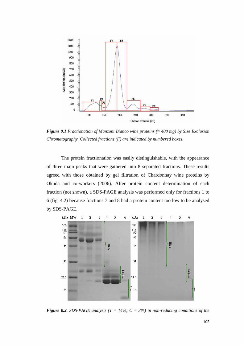
105
Figure 0.1 Fractionation of Manzoni Bianco wine proteins (≈ 400 mg) by Size Exclusion
Chromatography. Collected fractions (F) are indicated by numbered boxes.
The protein fractionation waseasil y distinguishable, with the appearance
of threemain peaksthat were gatheredinto 8 separated fractions. These results
agreedwith those obtainedby gel filt ration of Chardonnaywine proteins by
Okuda and co-workers (2006). After protein content determination of each
fraction (not shown), a SDS-PAGE analysiswasperformed only for fractions1 to
6 (fig. 4.2) becausefractions7 and8 hada protein contenttoo low to beanalysed
by SDS-PAGE.
Figure 0.2. SDS-PAGE analysis (T = 14%; C = 3%) in non-reducing conditions of the

106
fractions from Size Exclusion Chromatography (see fig.4.1). Left panel: fractions stained
for proteins with Coomassie brilliant blue R-250. Right panel: fractions stained for
sugars with Periodic Acid-Schiff (PAS) procedure).
As expected,all thehigh molecularweight proteins werecontained in the
firsts threefractions,in which a major band at about 65 kDa appeared.The PAS
stainingshowedthat thesefactionscontainedalmostall theglycosilatedhigh MW
compounds(>200kDa) of thewine. Besides, bands at about65 kDa werevisible
in both gelsandfor that reasonwereassumedto bean invertase(Porntaveewatet
al., 1994;Kwon et al., 2004;Okudaet al., 2006).
Fractions4 and5 displayedonly bandsbetween 32 and17 kDa. Fromthe
literaturedata (Tattersallet al., 1997;Waters et al., 1998;DaviesandRobinson,
2000; Pococket al., 2000)it wasreasonable to supposetheseproteinsto belongto
the chitinaseand thaumatin-like protein classes.Moreover,fractions 4, 5 and 6
did not give anysignal whenstainedwith PAS,indicating theabsence in thewine
of glycosilatedproteinswith low molecular weight. In fraction 6 only two classes
of proteinswere detected.The upper band showeda MW similar to that of the
thaumatin-like proteins, while the lower hadan apparentMW of ≈ 10 kDa. The
presenceof LTP hydrolysis productswith MW of 9.6 kDa insteadof 11.6 kDa
(the MW estimated from the cDNA sequence) hasbeensuggested (Okudaet al.,
2006), andthis occurrencematcheswith theSDS-profileof HIC fraction6.
SEC separation allowed to “clean” the proteins with intermediate MW
from those with MWs higherthan≈ 32 kDa andlower than≈ 17 kDa. This fact
hasbeenconsideredof interestfor a multi-step protein chromatography finalised
to purify PR-proteins for their characterisation in relation to hazedevelopment in
whitewines.
Fraction 4 and 5 were considered suitable for a two-step protein
purification achieved by coupling a preparative SEC with a Hydrophobic
InteractionChromatography(HIC). To this aim, ammoniumsulphate(761mg/mL
at 25°C) wasadded to fraction 4 until reachingthe99%of saturation to promotea
complete protein salting out. After centrifugation, the collected pellet was
dissolvedwith 50 mM Na-Phosphatebuffer pH 5.0 to achievetheclarity andthe
salt concentration required (1.25 M) for HIC fractionation. The sample so

107
preparedwasloadedinto aHIC analytical column(fig. 4.3).
Figure 0.3. Hydrophobic Interaction Chromatography of fraction 4 from SEC (see
fig.4.1) Collected fractions (F) are indicated by numbered boxes
The HIC column showed a good protein separation abilit y with the
achievementof 5-6 peaks from a fraction that in SDS-PAGEpresentedonly three
proteinbands(fig. 4.2).
This two-stepchromatography gavepromising results in terms of protein
purification. However, the methodhere proposed showed an analytical nature
morethana preparativeonebecauseof the low volumeof sample loadablein the
SECcolumn,thehigh dilution of fractionscollectedand thenecessityof a further
proteinprecipitation stepbeforetheHIC fractionation.
1.1.42 STUDIES ON PROTEIN FRACTIONATION BASED ON THEIR
HYDROPHOBICITY
The wine proteins separatedby HIC were studied in relation to their
potential in hazeproduction.HIC chromatography was utilised on a preparative
scaleto fractionate proteinsaccordingto their hydrophobicity level. For this aim,
a500-timesconcentratedManzoniBiancowinewas used(fig. 4.4).

108
Figure 0.4. Hydrophobic Interaction Chromatography of the proteins of Manzoni bianco
wine proteins. Collected fractions (F) are indicated by numbered boxes. Green line:
buffer A percentage; Brown line: conductivity.
Due to the large quantityof protein loadedon the column, the separation
was not as good as that obtainedwith other HIC fractionation experiments(see
fig. 4.13).However, 8 separatedfractionswere collected andanalysed.The very
high peakof fraction2 (fig. 4.4) (unretained fraction) containedall theunbounded
proteins that did not interactwith the resin. Besides,fractions 4, 5 and 6 were
eluted at a conductivity level similar to thatpreviouslyobserved for theelution of
the Semillon chitinases and VvTL proteins (see tab. 3.1 and fig. 3.12). After
dialysis and protein content determination (not shown), each fraction was
analysedby SDS-PAGE(fig. 4.5).
Figure 0.5. SDS-PAGE (T = 14%, C = 3%) of the 8 fractions separated by HIC in non-
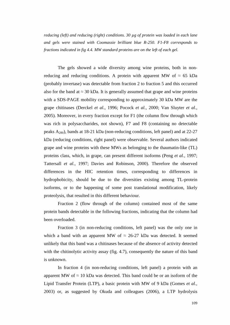
109
reducing (left) and reducing (right) conditions. 30 µg of protein was loaded in each lane
and gels were stained with Coomassie brilliant blue R-250. F1-F8 corresponds to
fractions indicated in fig 4.4. MW standard proteins are on the left of each gel.
The gels showeda wide diversity among wine proteins, both in non-
reducing and reducing conditions. A protein with apparent MW of ≈ 65 kDa
(probablyinvertase)wasdetectablefrom fraction 2 to fraction 5 andthis occurred
alsofor the bandat≈ 30 kDa. It is generallyassumedthatgrapeandwine proteins
with a SDS-PAGE mobility corresponding to approximately 30 kDa MW are the
grapechitinases(Derckel et al., 1996; Pococket al., 2000; Van Sluyter et al.,
2005). Moreover,in everyfractionexceptfor F1 (thecolumnflow throughwhich
was rich in polysaccharides,not shown), F7 and F8 (containing no detectable
peaks A280), bandsat 18-21kDa(non-reducingconditions,left panel) andat 22-27
kDa (reducing conditions, right panel)wereobservable. Several authorsindicated
grapeandwine proteinswith theseMWs asbelonging to the thaumatin-like (TL)
proteinsclass,which, in grape,canpresent different isoforms(Peng et al., 1997;
Tattersall et al., 1997; Davies and Robinson,2000). Therefore the observed
differences in the HIC retention times, corresponding to differences in
hydrophobicity, should be due to the diversities existing among TL-protein
isoforms, or to the happening of some post translational modification, likely
proteolysis, thatresultedin this differentbehaviour.
Fraction 2 (flow through of the column) contained most of the same
proteinbandsdetectablein thefollowing fractions,indicating that thecolumnhad
beenoverloaded.
Fraction 3 (in non-reducingconditions,left panel) was the only one in
which a band with an apparentMW of ≈ 26-27 kDa was detected.It seemed
unlikely thatthis bandwasa chitinasesbecauseof theabsenceof activity detected
with thechitinolytic activity assay(fig. 4.7), consequently thenatureof this band
is unknown.
In fraction 4 (in non-reducingconditions, left panel) a protein with an
apparentMW of ≈ 10 kDa wasdetected.This bandcouldbe or an isoformof the
Lipid Transfer Protein (LTP), a basic protein with MW of 9 kDa (Gomes et al.,
2003) or, as suggested by Okuda and colleagues(2006), a LTP hydrolysis
110
products with MW of 9.6 kDa. However, this protein, which should be very
hydrophobic (Blein et al., 2002) waselutedin themiddle of thegradient, showing
a behaviour similar to that observed during the Semillon juice protein
fractionationin chapter3 (seeparagraph3.4.3.1).
Fraction6, thelastproteinpeakelutedfrom thecolumn, seemed thepurest
among all theHIC fractions,containingtwo major bandswith similar MWs that,
from the li terature (Tattersall et al., 1997; Ferreira et al., 2007) presumably
correspondedto 2 or more TL protein isoforms.Comparing thesedata to those
achievedworking with Semillonjuice (seechapter 3), theTL-proteins of F6 were
confirmedasthemost hydrophobicwineproteins.
It is interestingto underlinethe different migration rate observable for
some bandsin reducingandnon-reducing conditions,aspreviouslyobserved by
Vincenzi and Curioni (2005).Proteinswith MWs lower than 40 kDa increased
their relative apparentMWs whenreduced (compare left andright panels of fig.
4.5). For instance,the 4 bandswith apparent MW of ≈ 30 kDa migrated almost
equally in the left gel but, after reduction, their apparentMW changed with
fraction 3 thatmovedat ≈ 32 kDa, fractions4 and5 at ≈ 31 kDa and fraction2 at
≈ 30 kDa. Theselatter result agreeswith thoseobserved by Pococket al. (2000),
who divided grape chitinasesin 4 classes(Chit A, Chit B, Chit C, Chit D)
showing different SDS-PAGE MWs in reducing conditions (32, 33, 32, 34 kDa
respectively).
Besides,the bands assumed to be thaumatin-like proteins changed their
migration rate from an apparentMW of 18-22 kDa (when not reduced) to the
apparentMW of 23-27 kDa in reducingconditions. Moreover after reduction of
the samples,two bandsappearedat≈ 12-13 kDa in F2 andF4 (right panel),which
were absent in non-reducingconditions (left panel). This should indicate that
these proteins appearedas a result of the splitting of some disulphide bonds
linking together protein aggregatesof higher MW. The existence and the
significanceof suchproteinaggregatesin wineswarrantfurtherinvestigation.
The presence of glycoproteinsin the samesamples of figure 4.5 was
studiedby staining thegelswith thePAS method(fig. 4.6)
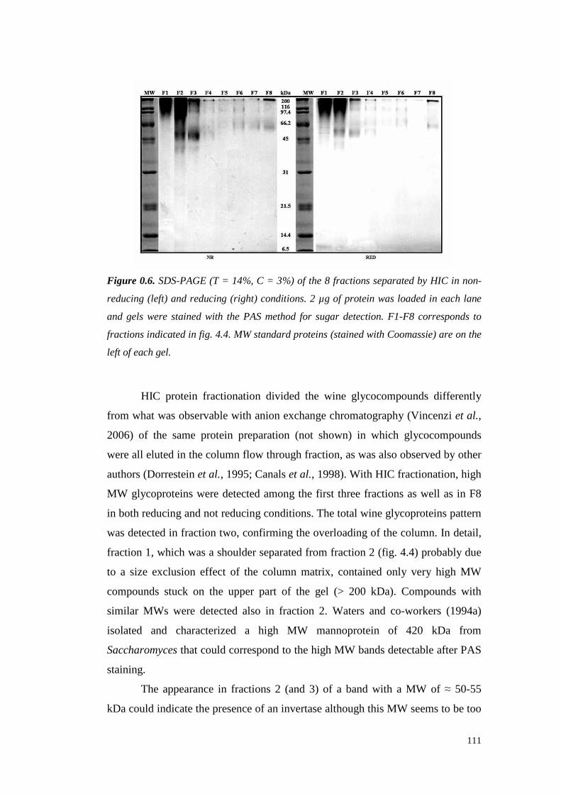
111
Figure 0.6. SDS-PAGE (T = 14%, C = 3%) of the 8 fractions separated by HIC in non-
reducing (left) and reducing (right) conditions. 2 µg of protein was loaded in each lane
and gels were stained with the PAS method for sugar detection. F1-F8 corresponds to
fractions indicated in fig. 4.4. MW standard proteins (stained with Coomassie) are on the
left of each gel.
HIC protein fractionation divided the wine glycocompoundsdifferently
from what wasobservable with anion exchangechromatography (Vincenzi et al.,
2006) of the sameprotein preparation (not shown) in which glycocompounds
wereall elutedin thecolumnflow through fraction,aswasalsoobserved by other
authors(Dorrestein et al., 1995;Canalset al., 1998).With HIC fractionation, high
MW glycoproteins weredetectedamongthe fi rst threefractionsas well as in F8
in bothreducing andnot reducingconditions.Thetotal wineglycoproteinspattern
wasdetected in fraction two, confirmingtheoverloadingof thecolumn.In detail,
fraction 1, which wasa shoulderseparated from fraction 2 (fig. 4.4) probably due
to a size exclusion effect of the column matrix, containedonly very high MW
compoundsstuck on the upper part of the gel (> 200 kDa). Compoundswith
similar MWs were detectedalso in fraction 2. Waters and co-workers (1994a)
isolated and characterizeda high MW mannoprotein of 420 kDa from
Saccharomyces thatcouldcorrespondto thehigh MW bandsdetectable after PAS
staining.
The appearancein fractions2 (and 3) of a band with a MW of ≈ 50-55
kDa couldindicate the presenceof aninvertasealthoughthis MW seemsto betoo
112
low for the grape invertase,which has been shown to display a SDS-PAGE
mobility corresponding to 60-64 kDa (Nakanishi and Yokotsuka, 1991;
Takayanagiet al., 1995;Kwon et al., 2004).However, Porntaveewatet al., (1994)
purified an invertasewith MW of 72 kDa from grapes(variety MuscatBailey A)
and highlighted that, analysing this glycoprotein by SDS-PAGE three bands
appearedat 56, 25 and 24 kDa. This data seemed to agreewith the MW of the
band detected in lanesF2 (and F3) of fig. 4.6 leading to consider it as a grape
invertase.
In fraction3 the PAS-stainedprofile resulted similar to that of fraction 2
but with aninvertedratio betweentheintensityof thetwo bands.
Fractions4 and5 did not showa significantbandappearanceafter staining
for sugars, whereasfractions6, 7 and8 showedthepresenceof faint bandsat ≈ 65
kDa whoseMW couldmatchwith that of a vacuolar grape invertase(Daviesand
Robinson,1996).
Fraction 8 showedthe appearance of a well marked bandblocked at the
boundary betweenthestackingand theresolving gels. This bandhada high MW
as confirmed by further analyseswith size exclusion chromatography (fig. 4.9).
Due to the nature of the separation,the PAS-stained compound(s)in fraction 8
(eluted with the lowest ionic strength) should present the highest level of
hydrophobicity, and so it is unlikely to belong to the wine compoundsthat are
known to havehydrophilicnature,suchaspolysaccharides(Vernhetet al., 1996;
De Freitas et al., 2003). It seemedmorelikely to supposethatfraction 8 contained
a hydrophobic glycoprotein (may be a mannoprotein) deriving from yeast cell
walls, whosehydrophobic naturehavebeenlargely demonstrated (Iimura et al.,
1980; Farriset al., 1993; Masuokaet al., 1997;Martinezet al., 1997;Alexandre
et al., 1998).
An additionalanalysis was performedto better understandthe nature of
the proteins fractionated on the basis of their hydrophobicity. To this aim,
chitinolytic activi ty on gel wasassayedin the8 HIC fractions(fig. 4.7) according
to VincenziandCurioni (2005).

113
Figure 0.7. Chitinolytic activity detection on glycol-chitin after SDS-PAGE separation (T
= 14%, C = 3%, reducing conditions) of the wine protein fractions obtained from HIC
separation. F1-F8 correspond to fractions indicated in fig. 4.4.
Apart for fraction 1, the chitinaseactivity wasspreadamong all the HIC
fractionscollected, althoughwith decreasingstaining from fraction 2 to fraction8.
In thesefractions, the main chitinolytic activi ty belonged to the bands at ≈ 35
kDa. Thesechitinasebandscorrespondto thosedetectable at ≈ 30 kDa on the
Coomassie-stained gel not containing the glycol chitin (fig. 4.5, right panel),
because the presenceof the substrateslows down protein bandsmigration as
demonstratedby Vincenzi and Curioni (2005). A fainter chitinolytic band
appearedalsoat ≈ 50 kDa (lanesF2-F6) andalso at thetop of thegel. Thereason
of this appearanceshould be due to the presenceof other enzymes acting on
glycol chitin, althoughtheirprecisenatureremainsto beassessed.
It is noteworthy to focustheattentionto fraction 2, in which anadditional
chitinolytic activity at ≈ 25 kDa was detectable. In the Coomassie-stained SDS-
PAGEgel (fig. 4.5,right panel),fraction2 showed a band at a MW thatshouldbe
responsiblefor this activity. Van Sluyter and co-workers(2005) highlightedthe
presenceof active chitinaseswith MW of 26 kDa in CabernetSauvignon and
Chardonnaygrapes.A confirmation for this hypothesis was achievedafter size
exclusion chromatography(SEC)analysisof theseparatedHIC fractions (fig. 4.8
and4.9).As a matter of fact, themainSEC peak of HIC fraction 2 (53 % of total
peakarea)hada retentiontime correspondingto that observed for the chitinases
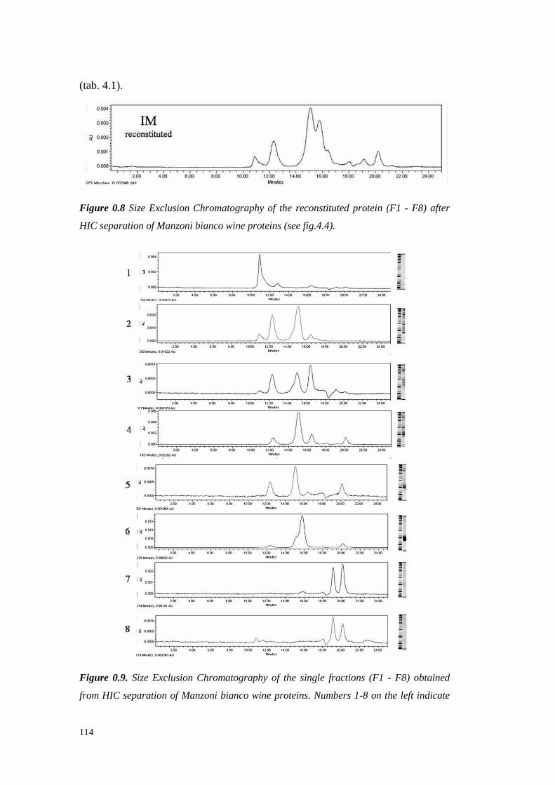
114
(tab. 4.1).
Figure 0.8 Size Exclusion Chromatography of the reconstituted protein (F1 - F8) after
HIC separation of Manzoni bianco wine proteins (see fig.4.4).
Figure 0.9. Size Exclusion Chromatography of the single fractions (F1 - F8) obtained
from HIC separation of Manzoni bianco wine proteins. Numbers 1-8 on the left indicate

115
the HIC fraction F1-F8 (see fig. 4.4), respectively. The SDS-PAGE profile of each
fraction is shown for comparison on the right of the corresponding chromatogram.
PresumableMW(kDa)
PeakRetention
Time(min)
HIC1
(Area%)
HIC2
(Area%)
HIC3
(Area%)
HIC4
(%Area)
HIC5
(Area%)
HIC 6(Area%)
HIC7
(Area%)
HIC8
(Area%)
> 200 10.900 83.32 7.45 2.39 n.d. n.d. n.d. n.d. 5.53? 70 12.330 n.d. 31.24 26.44 10.5 24.56 3.28 n.d. n.d.? 64 12.830 9.66 n.d. n.d. n.d. n.d. n.d. n.d. n.d.35-31 15.010 n.d. n.d. 35.51 64.5 n.d. n.d. n.d. n.d.30-22 15.120 n.d. 53.08 n.d. n.d. 57.05 n.d. n.d. n.d.? 22 15.810 n.d. n.d. n.d. n.d. n.d. 91.23* 4.10 n.d.21-18 16.460 3.33 6.3 35.67 15.06 3.63 n.d. n.d. n.d.< 18 17.040 n.d. 1.92 n.d. n.d. n.d. n.d. n.d. n.d.< 14 18.080 n.d. n.d. n.d. 1,08 n.d. n.d. n.d. n.d.< 10 19.150 1.82 n.d. n.d. n.d. n.d. n.d. 42.64 50.61
Not protein 20.160 1.88 n.d. n.d. 8.86 14.76 5.49 53.26 43.87Total(%)
100 100 100 100 100 100 100 100
n.d: not detected.* a shoulder at 15.1minutesof RT wasdetected.
Table 0.1. Area percent of the SEC peaks of HIC fractions F1 - F8 calculated from the
chromatograms of fig 4.9.
The SEC results are to be comparedto those of SDS-PAGE in non-
reducingconditionsbecausetheHIC fractionswerenot reduced beforeloadingon
the SE-HPLC column. By SEC, HIC fraction 1 showeda main peak with a
retention time of 10.9 min. This is the peakwith the lowestretention time thus
containing the highest molecularweight compounds.Theseresults agreedwith
those of PAS analyses,indicating the presence of high molecular weight
glycocompoundswith low hydrophobicity.Moreover, the SEC chromatogramof
fraction 1 indicated its low proteincontent, showingonly two small peaksat 12.8
and 16.4 min. of RT, thus confirming the results observedby SDS-PAGE (fig.
4.5).
From the SEC, 4 well defined peaks were visible in fraction 2, the
unretained HIC fraction, while fraction 3 gave4 peakswith RTs very similar to
those of fraction 2. Starting from fraction 4, the peak at 10.9 min. of RT
disappeared, while a new peak at 18.0 min. of RT was detected, which
presumablycorrespondedto the low molecular weight band observed by SDS-
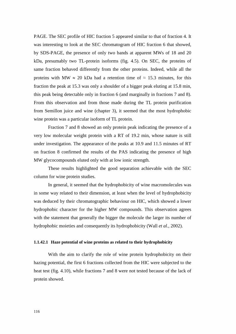
116
PAGE. TheSEC profile of HIC fraction5 appearedsimilar to thatof fraction 4. It
wasinteresting to look at the SECchromatogramof HIC fraction 6 that showed,
by SDS-PAGE, the presenceof only two bandsat apparentMWs of 18 and 20
kDa, presumablytwo TL-protein isoforms (fig. 4.5). On SEC, the proteins of
samefraction behaveddifferently from the other proteins. Indeed, while all the
proteins with MW ≈ 20 kDa had a retention time of ≈ 15.3 minutes, for this
fraction thepeakat 15.3wasonly a shoulder of a biggerpeak eluting at 15.8min,
this peakbeingdetectableonly in fraction6 (and marginally in fractions7 and8).
From this observation and from thosemadeduring the TL protein purification
from Semillon juice and wine (chapter3), it seemed that the most hydrophobic
wineproteinwas aparticularisoform of TL protein.
Fraction7 and8 showedanonly proteinpeak indicating thepresenceof a
very low molecularweight protein with a RT of 19.2 min, whosenature is still
under investigation. The appearanceof the peaksat 10.9and 11.5minutesof RT
on fraction 8 confirmed the results of the PAS indicating the presenceof high
MW glycocompoundselutedonly with at low ionic strength.
Theseresults highlighted the good separation achievable with the SEC
columnfor wineprotein studies.
In general, it seemedthat thehydrophobicity of wine macromoleculeswas
in someway related to their dimension,at least whenthe levelof hydrophobicity
wasdeducedby their chromatographicbehaviour on HIC, which showeda lower
hydrophobic characterfor the higher MW compounds.This observation agrees
with thestatementthatgenerallythebiggerthemolecule the larger its numberof
hydrophobic moietiesand consequentlyits hydrophobicity (Wall et al., 2002).
1.1.42.1 Haze potential of wine proteins as related to their hydrophobicity
With the aim to clarify the role of wine protein hydrophobicity on their
hazingpotential, thefirst 6 fractionscollected from theHIC weresubjectedto the
heattest(fig. 4.10), while fractions7 and8 werenot testedbecauseof the lackof
proteinshowed.

117
Figure 0.10. Heat test results for fraction 1 to 6 (F1-F6) collected from HIC of a
Manzoni Bianco wine. Proteins were precipitated from each fraction by ethanol addition
a re-dissolved at 200 mg/L in Manzoni bianco ultrafiltered wine (UF). Green and red
bars are the stable and instable samples (turbidity lower and higher than 0.02),
respectively. Results for both the Ultrafiltered Manzoni bianco wine without protein
addition (IM UF) and the original (unfined) Manzoni bianco wine (protein content: 200
mg/L) (IM) are also shown.
Siebertandcolleagues(1996)affirmed that protein hazing increasedwith
increasingtheprotein heatingtemperature, suggesting thathydrogenbonding was
not as important in the interaction between proteins and polyphenols as
hydrophobicinteractionwas.Moreover,other authorshave highlightedtherole of
hydrophobicinteractionsin the formationof protein-tannincomplexes(Oh et al.,
1980; Charlton et al., 2002). The results of the heat tests confirmed the data
recoverablefrom theliterature.Waterset al. (1996)havebeen thefirst to identify
the proteinsthat causehazein winesasPR proteinsderiving from grapeberries
and, in particular, it seemedthat the fractions characterized by the highest
instability were thosecontainingthe grapeTL proteins. In our hands,amongall
theHIC fraction heattestedat thesameprotein concentration, the fraction mainly
composedby TL protein (fraction 6) resulted indeedthe most instable, followed
by fraction 4 in which both TL andchitinaseswerecontained (with a prevalence
of chitinases).Fraction5 produceda significant turbidity but lower than that of

118
fraction 4 and 6. From the SEC data, the main peak of all the HIC protein
fractionscombinedwasthatat 15 min of retention time,which was assumedto be
a chitinase.Theareaof thepeakat 16.4(correspondingto TL protein) waslower
in fraction5 thanin fraction4. This observationsuggested that thelower turbidity
developedin fraction 5 than in fraction 4 depended from the lower TL protein
contentof the former. AssumingTL proteins as the main responsible for wine
haze formation, this turbidity value scale seemed to be reasonable, with the
highestinstabili ty of fraction6 (highestTL protein content), followedby fraction
4 (secondTL proteincontent)and5 (third TL proteincontent).
Threefirst eluting HIC fractions(fractions1, 2 and3) resultedstable after
the heattest (fig. 4.10), despitethey contained proteins apparently belonging to
the chitinaseand thaumatin-like proteinclasses. A possibleinterpretation of this
eventwasbasedon thedataregardingthepolysaccharidic contentof thedifferent
fractions, as determinedby combining the information from both UV (protein)
andRefractive index(sugar)detectionof theSECanalysis for eachHIC fraction
To simplify this interpretation,the ratio between protein (asmeasured at
280 nm) and refractive index peak areasobtained by SEC was calculated (tab.
4.2).
HIC fractions Retention Time (min)*Refractive Index
(Area µV*sec)
UV Absorbance
(Area 280 nm)Ratio RI/UV
11.342 688073 128777 5.34Fraction 114.896 19912 n.d. -11.344 436981 132813 3.2912.683 34000 614082 0.06Fraction 2
15.430 111180 1137353 0.1011.319 5307 2492 2.1312.633 1168 20662 0.06Fraction 3
15.367 2275 29088 0.0811.200 426 n.d. -12.750 2114 37055 0.05715.390 20320 210699 0.096
Fraction 4
16.850 2245 48850 0.046
11.183 466 n.d. -Fraction 5
12.633 824 n.d. -
Fraction 6 16.14 81994 821280 0.10
Fraction 7 n.d. n.d. n.d. -
Fraction 8 11.167 4367 2837 1.54
n.d.: not detected. * RTs of the peaks were delayed of 0.3-0.4 minutes of compared to those of
119
table 4.1 due to the passage through the refractometer.
Table 0.2. Ratio between the Peak areas (µV*sec) detected by the Refractive Index (RI)
and UV (A280 nm) detector after SEC analyses of the HIC protein fractions
TheRI/UV ratio for eachpeaksuggestedtherelation betweenthevalue of
this ratio andthehazeproducedafter theheat test by thedifferent HIC fractions,
the higher being the ratio the lower the turbidity. This should indicate that the
presenceof glycocompounds(detectedby refractometry) in a fraction impairedits
hazing, alsoif potentially unstableproteinsare presentin thesamefraction. From
the literature,the hypothesisof a stabilisingeffect of glycocompound,including
polysaccharidesderivedfrom both the grapeberry and yeast (mannoproteins) is
generally supported(Waterset al., 1994a; Waters et al., 1994b;Moine-Ledoux
andDubourdieu,1999;Dupin et al., 2000; Lomolino and Curioni, 2007). It was
interesting to note that the protein with RT of 11.1 min of HIC fraction 1
(correspondingto the proteinwith 10.800min RT on table 4.1) had the highest
RI/UV ratio (table4.1), confirming the data of sugardetection on gels that were
confirmedalsofor fraction8 (fig. 4.6).
1.1.43 STUDIES ON WINE PROTEIN REACTIVITY WITH SEED TANNINS
1.1.43.1 Preliminary experiments
In order to study the interactionsoccurring between wine proteins and
grapeseedtannins,severaltestswereperformed.As well known for a long time
tanninsarepolyphenoliccompoundsthat form insoluble complexes with proteins
(Swain,1965)and the protein reactivity with thesecompoundshasbeen studied
asa tool for proteinremoval(Powerset al., 1988).Tannin-protein interactions is
important for the sensationof astringency in the mouth, but also for several
phenomenaoccurring during winemaking, including protein haze formation in
white wines (Luck et al., 1994; Sarni-Manchado et al., 1999; Sarmento et al.,
2000; Mesquita et al., 2001). Therefore,wine protein reactivity with endogenous
grapetannins hasbeenextensivelystudied,althougha precisecharacterisation of
the effect of the single wine proteincomponents is scant(SomersandZiemelis,
1973; Siebert,1999).

120
Initially, to confirm thestatementthatwineproteinscanbeinsolubilised in
the presence of tannins (Powerset al., 1988), an experiment was set up by
dissolvingthetotal lyophilisedwineproteinsin modelwineandby monitoring the
turbidity formation (measured spectrophotometrically at 540 nm and taken as a
measureof protein-tannin reactivity) at increasinggrapeseedtanninsdosage(fig.
4.11).
Figure 0.11. Turbidity produced after reaction at room temperature of wine proteins
from an unfined Manzoni bianco wine (200 mg/L in model wine) with increasing seed
tannin concentrations. Turbidity was monitored spectrophotometrically at 540 nm
against blanks prepared without protein.
Resultsshowedthat the highest hazevaluewasachieved at 250 mg/L of
tannins dosage, followed by a plateaueffect probably dueto thesaturation of the
proteinbindingsites.
Furthermore,to determinetheeffectprotein concentration on turbidity, an
experiment with both different dosages of tannins and wine proteins was
performed(fig. 4.12).

121
Figure 0.12. Turbidity produced after reaction, at room temperature, of wine proteins
from unfined Manzoni bianco wine at increasing concentrations (from 37.5 mg/L to 300
mg/L) in model wine with increasing seed tannins dosages (from 0 to 1000 mg/L).
Turbidity was monitored spectrophotometrically at 540 nm against blanks prepared
without protein.
The results showedthat astanninconcentration increasedat a fixed level
of protein content, the observedturbidity at first rose, thenreached a plateau (at
250 mg/L) and thendeclined.A similar behaviour wasobservedby Siebert and
colleagues(1996) by monitoringthehazeformation at differentdosagesof gelatin
andtanninacid.
The relation existing betweenthe hydrophobicity of single wine protein
fractions, separatedby HIC, and the tannin reactivi ty was then studied. As a
matter of fact, protein hydrophobicity is one of the characteristics that mainly
affecttannin-protein interactions(Oh et al., 1980;Siebertet al., 1996).
1.1.43.2 HIC fractionation of wine proteins
In orderto obtain proteinamountssufficient to characteriseprotein-tannin
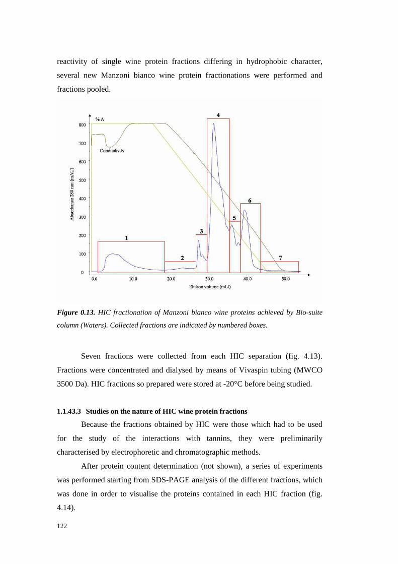
122
reactivity of single wine protein fractions differing in hydrophobic character,
several new Manzoni bianco wine protein fractionations were performed and
fractionspooled.
Figure 0.13. HIC fractionation of Manzoni bianco wine proteins achieved by Bio-suite
column (Waters). Collected fractions are indicated by numbered boxes.
Seven fractions were collected from each HIC separation (fig. 4.13).
Fractionswereconcentratedanddialysedby meansof Vivaspin tubing (MWCO
3500Da).HIC fractionsso preparedwerestoredat -20°Cbeforebeingstudied.
1.1.43.3 Studies on the nature of HIC wine protein fractions
Becausethe fractionsobtainedby HIC were thosewhich had to be used
for the study of the interactions with tannins, they were preliminarily
characterisedby electrophoreticandchromatographic methods.
After protein contentdetermination(not shown),a series of experiments
wasperformedstarting from SDS-PAGE analysis of thedifferent fractions,which
was donein order to visualisethe proteins contained in each HIC fraction (fig.
4.14).
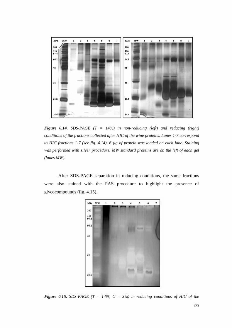
123
Figure 0.14. SDS-PAGE (T = 14%) in non-reducing (left) and reducing (right)
conditions of the fractions collected after HIC of the wine proteins. Lanes 1-7 correspond
to HIC fractions 1-7 (see fig. 4.14). 6 µg of protein was loaded on each lane. Staining
was performed with silver procedure. MW standard proteins are on the left of each gel
(lanes MW).
After SDS-PAGE separation in reducingconditions, the same fractions
were also stained with the PAS procedure to highlight the presenceof
glycocompounds(fig. 4.15).
Figure 0.15. SDS-PAGE (T = 14%, C = 3%) in reducing conditions of HIC of the

124
fractions collected after HIC of the wine proteins. Lanes 1-7 correspond to HIC fractions
1-7 (see fig. 4.14). Staining performed with the PAS procedure.
The PAS resultsconfirmedwhat hadbeenobservedon the previousHIC
fractionationof thewine proteins(see4.4.2)showing that theHIC fraction richest
in polysaccharides was fraction 1 as confirmed by the total polysaccharide
quantification (datanot shown).
After staining for both proteins and sugars (fig. 4.14 and 4.15,
respectively), fraction1 showedtheappearanceof astreakingmaterial resulting in
a shadowall along the lane,suggestingthat polyphenols might beboundto some
wine proteinandthat,consequentlytheycoulddisturb theprotein interaction with
the resin duringanalyses.Accordingly,Hagerman andcolleagues (1998)observed
that nonpolartannins canbind proteinsforming a hydrophobic coataroundthem
resulting in their modified solubility. This fact could explain the hiding of the
protein interactioncapacitywith theHIC matrix.
Fractions 2, 3 and 4 showed to contain high molecular weight
glycocompoundsvisibleat thetop of thegel, whereasfractions4, 5 and 6 showed
a glycosilated band around65 kDa (fig. 4.15), presumably corresponding to a
grape invertase.The PAS staining of bandsat low molecular weight (≈ 22 kDa)
could bedue to the overloadingof the gel, which led to detection of glycosilated
proteins or protein fragmentsnormally not visible with lower protein loadings.
This result was observed other times and always when high protein quantities
wereloadedon theSDS-PAGE gel (not shown).
Further analyseswere done to studythenatureof proteins fractionated by
HIC by using themethodproposedby Peng andco-workers(1997)for RP-HPLC
protein identification(fig. 4.16andtab4.3).

125
Figure 0.16. RP-HPLC chromatograms (C18 Vydac column) of HIC wine protein
fractions (1 to 7, see fig. 4.14)) and of the total Manzoni bianco wine proteins.
PeakRT
(min)
HIC 1(Area
%)
HIC 2(Area%)
HIC 3(Area%)
HIC 4(%
Area)
HIC 5(Area%)
HIC 6(Area%)
HIC 7(Area%)
5.800 0.96 n.d. n.d. n.d n.d n.d n.d6.100 0.67 n.d. n.d. n.d n.d n.d n.d7.200 63.34 8.7 n.d. n.d n.d n.d n.d7.700 n.d. n.d. n.d. 7.31 n.d n.d n.d8.500 2.75 28.71 n.d. n.d n.d n.d n.d8.900 17.57 n.d. 6.45 77.36 37.50 2.11 15.0010.000 n.d. n.d. n.d n.d 2.18 1.94 n.d10.400 n.d. n.d. 1.93 0.55 17.01 15.05 11.5110.700 6.45 n.d. 1.53 0.37 17.02 62.66 47.0119.400 1.48 5.38 n.d 0.48 n.d n.d n.d19.900 5.70 57.21 n.d 13.94 n.d 17.17 n.d20.100 n.d. n.d. 90.09 n.d 26.29 n.d 26.4824.800 1.07 n.d. n.d n.d n.d 1.07 n.d
Total (%) 100 100 100 100 100 100 100
Table 0.3. Area percent of the RP-HPLC peaks (see fig. 4.16) within the wine protein
HIC fractions (1-7, fig. 4.14). Bold numbers indicate the percentage of the area of the
main peak of each fraction.
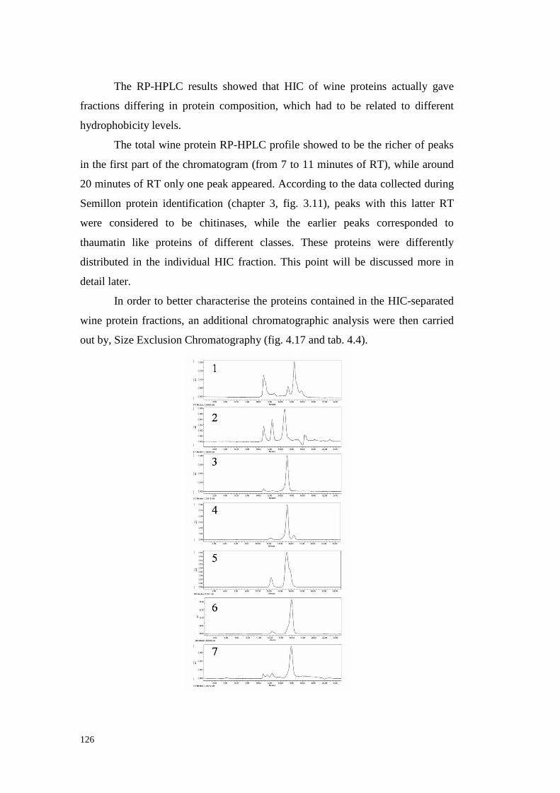
126
The RP-HPLC resultsshowed that HIC of wine proteins actually gave
fractions differing in protein composition, which had to be related to different
hydrophobicity levels.
The total wine protein RP-HPLC profile showedto be the richerof peaks
in the fi rst part of thechromatogram(from 7 to 11 minutesof RT), while around
20 minutesof RT only onepeakappeared.Accordingto thedatacollectedduring
Semillon protein identification (chapter 3, fig. 3.11), peaks with this latter RT
were considered to be chitinases,while the earlier peaks corresponded to
thaumatin like proteins of different classes.These proteins were differently
distributed in the individual HIC fraction. This point will be discussed more in
detail later.
In order to better characterisethe proteinscontained in the HIC-separated
wine protein fractions, an additionalchromatographic analysis were thencarried
out by, SizeExclusionChromatography(fig. 4.17andtab.4.4).

127
Figure 0.17. Size Exclusion Chromatography- HPLC (Protein Pak 125, Waters) of the
HIC wine protein fractions (1 to 7, see fig. 4.14).
PeakRetention
Time(min)
HIC 1(Area%)
HIC 2(Area%)
HIC 3(Area%)
HIC 4(%
Area)
HIC 5(Area%)
HIC 6(Area%)
HIC 7(Area%)
10.90 28.0 16.4 4.3 0.5 n.d. n.d. 5.9 11.54 n.d. n.d. n.d. n.d. n.d. n.d. 6.1 12.27 2.3 n.d. n.d. 4.8 14.2 n.d. n.d.12.43 n.d. 24.7 1.2 n.d. n.d. 5.1 9.9 12.81 4.5 n.d. n.d. n.d. n.d. n.d. n.d.14.35 3.4 n.d. n.d. n.d. n.d. n.d. n.d.14.72 n.d. 51.2 n.d. n.d. n.d. n.d. n.d.15.16 n.d. n.d. 92.1 n.d. 85.4 n.d. n.d.15.32 10.4 n.d. n.d. 83.8 n.d. n.d. n.d.15.90 n.d. n.d. n.d. n.d. n.d. 94.5 74.116.47 43.1 n.d. 0.6 10.7 n.d. n.d. n.d.17.77 8.3 n.d. n.d. n.d. n.d. n.d. n.d.18.31 n.d. 5.0 n.d. n.d. n.d. n.d. n.d.20.14 n.d. 0.6 n.d. n.d. n.d. n.d. n.d.22.91 n.d. 2.1 n.d. n.d. n.d. n.d. n.d.
Total (%) 100 100 97.6 99.8 99.6 99.6 96
Table 0.4. Area percent of the SE-HPLC peaks (see fig. 4.17) within each wine protein
HIC fractions (1-7, fig. 4.14). Bold numbers indicate the percentage of the area of the
main peak of each fraction.
By combining the datacollectedfrom SDS-PAGE stainedfor protein and
sugar detection, and HPLC analysesin both ReversePhase and Size Exclusion
modesthe following considerationsfor each HIC wine protein fraction could be
made.
HIC Fraction 1. Whenanalysedby SDS-PAGE, fraction 1 showeda poor
protein pattern, while resulted the fraction with the highest glycocompound
content,asdeterminedby PASstaining.RP-HPLC analysisshoweda major peak
at 7.2 minutesof retentiontime. This peak resulted peculiar of this fraction (a
minimal amountbeingdetectedonly in the following fraction (2), andprobably
corresponded to the first part of the peakat 10.9 min of RT visualisedby SEC,
suggestingthatglycosilatedcompoundswerecontained in it. Thesecondpeak (in
termsof percentagearea)detectedby RP-HPLC showed RT of 8.9 min, andwas
128
likely to correspondto the 16.47 min RT on SEC that, from the protein
identification of chapter3, could reasonably to be a thaumatin-like protein. This
hypothesiswas confirmedfrom theSDS-PAGEanalysis in which a band at about
21 kDa (in reducing conditions) wasdetectable(fig. 4.14, right panel). However,
the relativeheight of peaksin SE- andRP-HPLC was in disagreement with the
former discussion. An explanationcould be found in the higher polyphenols
content of fraction 1 (not shown) which might interfere with the UV signals
leadingto a misunderstandingof therealquantitiesof each protein. Otherproteins
were detectable in HIC fraction 1, which on the basisof their RT in RP-HPLC
(RT of 19.4 and 19.9 min) in were classifiable as two chitinases(Waterset al.,
1996; Peng et al., 1997).
HIC Fraction 2. Fraction 2 contained a residual amount of the
glycosilatedcompoundsrecoverablein fraction 1 (RP-HPLC RT 7.2 min; SEC
RT 10.9min) confirmingtheresultsshowed with PASstainingof theSDS-PAGE
gel (fig. 4.15). From the RP-HPLC analysis this fraction resulted to contain only
proteinsrecoverablealso in fraction 1, but in different percentages. Indeed, HIC
fraction 2 seemedto be mainly composed of chitinases (SECRT 14.7 min, RP-
HPLC RT 19.9 and 19.4 min), in accordance with the SDS-PAGE results (fig.
4.14).
HIC Fraction 3. The RP-HPLC profile of HIC fraction 3 was the easiest
to interpret, with the presenceof a main peak (90 % of total area) recognised as
corresponding to a chitinase(Waterset al., 1996; Penget al., 1997) and three
small peaksappearingin the thaumatinlike protein chromatographic position
(Waterset al., 1996;Penget al., 1997)..Theseresultswereconfirmedby thoseof
the SEC analysis,wherea similar profile wasidentified with a main peak at 15.1
minutesof RT. Thedifferencesin thechitinasesRTsnotedbetweenfraction 2 and
3, (19.9and20.1min respectively)by RP-HPLC and thosedetectedby SEC14.7
and 15.1min), togetherwith thedifferent migration rateshowedby thesebandsin
SDS-PAGE (Fig. 4.14) led to assess the occurrenceof chitinasesof different
nature in the two HIC fractions,confirming the existence of different chitinase
isoforms,assuggestedby Pocockandcolleagues(2000).
HIC Fraction 4. Fraction 4 showed four main bands in SDS-PAGE
(reducingconditions, fig. 4.14, right panel) with apparentmolecular weights of

129
66, 35, 22 and12 kDa. The12 kDa bandwas assumedto bea chitinasefragment
derivingfrom theproteinreductionprocess,becausethis bandwasundetectablein
non-reducing conditions(fig. 4.14, left panel). However, the escapeof this band
from the gel in non-reducingconditions,which can result in an increasedSDS-
PAGE migration rate, could not be excluded. Besides,three peaks have been
detectedin both RP- HPLC andSEC analyses.Themain RP- HPLC peak showed
a RT of 8.9 min, which suggested the identification of this protein as a thaumatin
like protein (Waterset al., 1996;Penget al., 1997).This peak accounted for the
77% of thetotal areathoughthesamepeakshowedanareapercentageof 83 % by
SECanalysis (RT of 15.3min). TheRP-HPLC results indicated thepresenceof a
13% of chitinasesin HIC peak4 (19.900min RT) (Waterset al., 1996;Penget
al., 1997). The SDS-PAGE band intensity seemed to confirm the RP-HPLC
results by showing thehighest stainingfor theTL protein band(22 kDa). Another
proteinof interestdetectablein fraction4 wasthatshowinga MW of ≈ 66 kDa in
SDS-PAGE. This protein appearedalso in the PAS-stained gels leading to
suppose it as being an invertase (Hsu and Heatherbell, 1987a; Brissonetand
Maujean,1993;Marchalet al., 1996;Dambroucket al., 2005).ThePASstaining
of the gel showedthis fraction to containalso high MW glycosilatedcompounds
(fig. 4.15).Thesehigh MW compoundswere detectableby SECanalysis while no
trace of them were noted by RP-HPLC, probably due to the chromatographic
systemadopted,which was specifically developedto distinguish the thaumatin-
like proteinfrom thechitinases(Waterset al., 1996;Penget al., 1997).
HIC Fraction 5. Fraction5 showedthe presence of 4 principal bands in
SDS-PAGE with apparentMW of 66, 35, 30 and 22 kDa, showing a profile
similar to that of fraction 4 (fig. 4.14). In this fraction, the PAS-stained gel
revealeda band at 66 kDa, showing the probable presence of invertasein this
fraction too. By SECanalysisit waspossible to detect a peak at 12.3min of RT
that indicated the presenceof a high MW compounds.This peakwas likely to
correspondto high MW materialstainedat the top of the SDS-PAGE gel (fig.
4.14). The other SEC peak had a RT of 15.18 min, though a shoulder was
detectableat about15.9min of RT. This fact wasprobablydueto thesimilar MW
of the chitinases andthaumatinlike proteinscontainedin fraction 5, resulting in a
badSECseparationalso dueto thehighprotein loading.A better understandingof

130
the nature of the proteinsof this fraction was achievedwith RP-HPLC analysis,
which allowedto distinguish 5 peakswith RT of 8.9 (thesameof that in fraction
4), 10, 10.4,10.7and20.1min. By RP-HPLC, thepeak at 8.9 minutesof RT was
recoverable in every single fraction except for fraction 2. From previous
experiencesandfrom the SDS-PAGE analysis it was possible to deduce that this
peak(8.9 RT) correspondedto a proteinof about22 kDa, probably a thaumatin-
like protein. The three peaks detectedduring minute 10 of the RP-HPLC
separationwere assumedto be different forms of TL proteins. Instead,the RP-
HPLC peakeluting at 20.1 min shouldbe a chitinases although it had to be an
isoform different from that observedon fraction 3 because of the different
hydrophobicity showedby theHIC fractions5 and3.
HIC Fraction 6. Fraction6, which hadto contain very hydrophobic wine
proteins,showeda SDS-PAGE profile (in reducing conditions)with a band at 66
kDa anda heavybandat 22-23 kDa, that possibly hided other minor bandswith
similar MW. In non-reducingconditions,the SDS-PAGE pattern appeared less
simple with the manifestationof a bandwith an apparent MW of ≈ 27 kDa (fig.
4.14). This bandwassupposedto be a chitinasebecausein the RP-HPLC data 4
detectedpeakswere assumedto be thaumatin-like proteins (RT 8.9, 10, 10.4and
10.7min) while a peak(RT 19.9min) wasassumedasa chitinase(Fig. 4.16). The
main percent area (62.6%) was calculated for the peak at 10.7 min of RT,
indicating fraction 6 asthatcontainingthemain portion of this protein which was
similar to that identified asVvTL (gi|33329390) during Semillon grapeproteins
identification (seeparagraph3.4.2). The SECanalysis confirmed this hypothesis,
showing a main peak with a RT (15.9 min) compatible with the mass of a
thaumatin-likeprotein.
HIC Fraction 7. Fraction7, which was the most hydrophobicone,was
only a tail of fraction 6, as appearsfrom the HIC fractionation chromatogram
(Fig. 4.13). Actually, the main peak detected in both the chromatographic
analyses(fig. 4.16and4.17)aswell astheSDS-PAGEpattern (fig. 4.14)wasthe
same of fraction 6. Consequently,the protein composition of fraction 7 was
similar to thatof fraction6 alsoif somedifferenceswere noticed.In particular, in
this HIC fraction threeadditionalsmall peakswerevisible by SEC analysis (RT
10.8, 11.5 and14.4minutes)indicatingthepresence,althoughin low amounts, of

131
highMW compounds with highhydrophobicity.
1.1.43.4 Heat stability of wine protein fractions separated by Hydrophobic
Interaction Chromatography
TheHIC fractionswereheattestedto determine their hazing potential (fig.
4.18).
Figure 0.18. Heat test results of fraction 1 to 6 (HIC 1- HIC 6) collected by HIC
fractionation of wine proteins. Each HIC fraction was prepared at 200 mg/L of protein
concentration in an ultrafiltered Manzoni bianco wine. The total wine proteins (wine)
were tested by dissolving the unfractionated proteins at 200 mg/L in ultrafiltered
Manzoni bianco wine. The ultrafiltered wine without protein addition (UF was also
tested. Turbidity values ( A540 ) higher than 0.02 means instability.
The first three fractions eluted from HIC showed the lowest turbidity
formationafter heating.In fraction 1 and2, the absence of haze formation could
be explained by the large content of high molecular weight glycosilated
compounds(seeabove), which areknownto showa protective effect againsthaze
formation. Indeed,the disrupting action of structurally different polysaccharides
towardsgrapeseedprocyanidin complexation and aggregation by bovine serum
albumin has beenreported(de Freitaset al., 2003; Mateus et al., 2004). The
explanationfor the absenceof heat-induced haze for fraction three seemed to be
different. This fraction, showing a relatively low a hydrophobicity level,
132
containedmainly chitinases(90% of the proteins, as determined by RP-HPLC)
and someglycosilatedcompounds, ashighlighted from PASstaining of theSDS-
PAGE gels (seeabove).Therefore,the reasonfor the lack of haze formation
seemeddue to the protective action against chitinases flocculation made by
glycocompoundsor, less probably,to the low hazepotential of chitinasespresent
in this fraction.
In contrastto the first 3 fractions,hazeformation (turbidity values higher
than0.02 AU) wasobservedfor HIC fractions4, 5 and6, indicating thepresence
of heat-unstablecompounds. Differently from the first heat test experiment
performedon HIC fractions(fig. 4.10), the highesthaze formation wasobserved
for fraction 4. The reasonsfor the high turbidity formedby fraction 4 arerelated
to thenature of its proteins,which mainly comprised a thaumatin-like protein (but
different from thatof fraction6) andalsochitinases,both thesePR-proteinsbeing
knownasthemain responsiblefor hazeformation in wines(Waterset al., 1996).
Moreover,the SDS-PAGE pattern showedthat the bandsintensity in fraction 4
was higher than that found for the other HIC fractions (fig. 4.14), leading to
hypothesizethat proteincontentdetermination by KDS-BCA method might lead
to a underestimation of theproteincontentof fraction 4, which actually contained
a quantity of protein higher than believed, thus explaining the high turbidity
formedafterheating.. Fractions5 and6 showedsimilar levels of hazeformation.
However,theanalysesof their proteincomposition suggesteddifferentcauses for
the turbidity level showedby thetwo fractionsafter heating. In particular, fraction
5 contained4 different TL protein peaks(by RP-HPLC) with the prevalence of
the TL protein with 8.9 min of RT, the sameobservedfor the TL protein of
fraction 4. Moreover,a chitinaseswasrecoverable in fraction 5 but different from
that detectedin fraction 6. Therefore these two fractions strongly differed for
proteincomposition althoughtheybehavedsimilarly when heattested.
For the previous resultsit seemsthat the wine proteins eluted after the
middle of the HIC gradientwere the mostheat-unstablewhile thosecollected on
the first half of thegradient,althoughcontaining PR-protein components,showed
high heat stabili ty. This should indicate that a certain relation exists between
protein hydrophobicity andhazingpotential. Takinginto accountthat theheat test
experimentsweredonewith proteinfractionsdissolved in (ultrafiltered)wine, that

133
contained the original wine polyphenols, and considering haze formation as
mainly due to protein-polyphenol interactions (Somers and Ziemelis, 1973;
Yokotsukaet al., 1983; Waterset al., 1995) the protein hydrophobicity can be
seenasaffectinghazeformationby determining thedegreeof protein interactions
with winepolyphenols.
1.1.43.5 Haze formation after addition of seed tannins to wine protein fractions
differing in hydrophobicity
Due to theimportanceof tannin-proteininteractionsin hazeformation, the
capability of Manzoni bianco wine protein fractions (deriving from the HIC
fractionationof fig. 4.13)differing in hydrophobicity in reacting with tanninswas
studied. Moreover,in order to study the factors involved in protein hazing, the
effectsof proteinheatingandsulphateaddition wereinvestigated.
Initially, the effects of seed tannin addition on turbidity formation in
model wine containing 25 mg/L of protein from the HIC fractions was assayed
spectrophotometrically at 540 nm (fig. 4.19).The final tanninconcentration was
250 mg/L, according to thepreliminaryresultsindicating this dosageasthat able
to give themaximum turbidity development.
Someturbidity wasformedimmediatelyafter tannin addition (greenbars),
but the turbidity strongly increasedafter boiling the samples for 5 minutes (red
bars).Moreover,theproteins of HIC fractionswereheated(10 minutes,100°C)in
modelwine beforethetanninaddition,obtaining a turbidity (greybars) similar or
evenlower thanthoseobservedin theothercases.
Figure 0.19. Reactivity of HIC fractions 1-7 (see fig. 4.13) with seed tannins. Reactivity

134
was calculated by measuring the turbidity developed immediately after tannin (250 mg/L)
addition to model wine containing 25 mg/L of protein from each HIC fraction. Blanks
were prepared without protein. Green bars: reaction at room temperature; Red bars:
reaction after heating the mixture (100 °C, 10 minutes); Grey series: reaction at room
temperature with proteins heated (100 °C, 10 minutes) before tannin addition.
Theresultsshowedin eachof thecasesthelackof protein-tanninreaction,
as measuredby the developmentof turbidity, only for both fraction 1 and 7 in
which absorbancevaluesclose to zeroweredetected. In contrast, fractions 2 to 6
immediatelydevelopedturbidity whenseed tannins were added(fig. 4.19,green
bars), although at different extent. By boiling the same samples, the turbidity
dramatically increasedfollowing the sameranking of the unheated samples. In
particular, theturbidity valuestendedto increasewith thefraction number(from 2
to 6), suggesting a relationshipbetweenwine protein hydrophobicity and tannin
reactivity. As suggested by Oh and co-workers (1980), tannins have a
hydrophobic natureandprotein-tannininteraction occurswith the involvement of
hydrophobic bonding. The results here showed agreedwith this hypothesis,
confirming that the level of protein hydrophobicity affects their reactivity with
tannins, supporting the idea that the level of protein hydrophobicity is a major
factor affecting tannin-proteininteractions(Oh et al., 1980;Siebertet al., 1996).
Moreover, the higher turbidity resulting from heating the samples
indicatedthe role of the temperature in inducinghaze formation. Apart from the
effect on the reaction rate,heatingthe protein-tannin mixture shouldincreasethe
extent of interactionby inducing protein denaturation and exposition of a high
numberof tannin binding sites,leadingto an increaseof hazeformation (Koch
and Sajak,1959;SomersandZiemelis, 1973;Yokotsukaet al., 1991;Waterset
al., 1995).Siebertandco-workers(1996)also reported thatduringprotein heating
the polyphenolbinding sitesbecameexposed becausehydrogenbondsarebroken.
This modification of the protein structurecould probably leadto higher tannin-
protein reactivity.
To verify whetherproteindenaturationby heating could beresponsiblefor
the increasedtannin reactivity, the HIC fractions were heated in model wine
(without tannins) before the tanninsaddition. After cooling the fractions at room

135
temperaturetannins were addedand the formed turbidity measured.The results
showed a turbidity much lower than that obtained after heating the mixture and
even slightly lower than that developed by the unheated fractions in the same
reaction conditions, althoughthe sametrend was maintained. Therefore,protein
heating in model wine without tanninsdid not allow a turbidity formation after
tannin addition. A possible explanationof this fact is that protein denaturation
occurring duringheatingin theabsenceof tanninsis a reversible process.Heating
should result in breaking of proteinhydrogenbondsat high temperature(Siebert
et al., 1996)but thesebondsarere-formed uponcoolingat roomtemperature,thus
restoringa proteinstructurewith anaffinity for tanninsevenlower thanthatof the
original (un-heated) proteins, which actually showedslightly higher turbidity
values after tannin addition (fig. 4.19). Consequently, it seems that protein
reactivity with tanninsis enhancedonly when both the compoundsare heated
together,with the tanninsbinding to the protein during its presencein the heat-
denaturedstate.
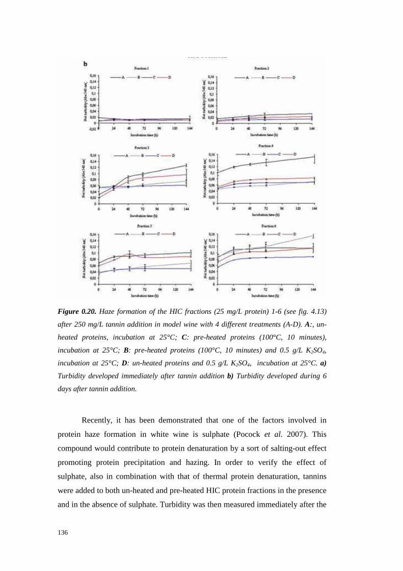
136
Figure 0.20. Haze formation of the HIC fractions (25 mg/L protein) 1-6 (see fig. 4.13)
after 250 mg/L tannin addition in model wine with 4 different treatments (A-D). A:, un-
heated proteins, incubation at 25°C; C: pre-heated proteins (100°C, 10 minutes),
incubation at 25°C; B: pre-heated proteins (100°C, 10 minutes) and 0.5 g/L K2SO4,
incubation at 25°C; D: un-heated proteins and 0.5 g/L K2SO4, incubation at 25°C. a)
Turbidity developed immediately after tannin addition b) Turbidity developed during 6
days after tannin addition.
Recently, it has beendemonstrated that one of the factors involved in
protein haze formation in white wine is sulphate(Pocock et al. 2007). This
compound would contributeto proteindenaturation by a sort of salting-out effect
promoting protein precipitation and hazing. In order to verify the effect of
sulphate,also in combinationwith that of thermal protein denaturation, tannins
wereaddedto bothun-heatedandpre-heatedHIC protein fractionsin thepresence
and in the absenceof sulphate.Turbidity was thenmeasuredimmediately after the

137
addition (fig. 4.20 a) and during 6 daysof incubation of the different mixtures
(fig. 4.20b). Immediatelyafter tanninaddition, thesamplespreparedwithoutpre-
heating the proteins and in the absenceof sulphate(treatmentA) showedresults
similar, but not identical to those of the previous experiment of fig. 4.19.
However, the trend indicating lower haze formation of the less hydrophobic
fractionswasmaintained. Moreover,a lower turbidity formation afterpre-heating
the proteins(treatmentsA and C in fig. 4.20, comparewith fig. 4.19) was also
confirmed for all the fractions andthe highesthaze formation in all the fractions
except for fraction 3 was that obtainedwith treatment A. Contrary to what was
expected,the addition of sulphateto the un-heated proteins-tannin mixturesat a
doseclaimedto enhancewine proteinhazing (Pocock et al. 2007)(treatmentD)
alwaysresultedin lower turbidity valuescompared to that foundin theabsenceof
sulphate, especially for HIC fraction 3 (fig. 4.20). This would indicate that
sulphateimpairs tannin-proteininteractionsby changingthe ionic strength of the
solution, which may increasethe burying of tannin binding siteson the proteins
probablyasa consequenceof the reinforcementof the hydrophobicforcesin the
core of the molecule. Therefore, it can be hypothesized that, in the presenceof
sulphate,a lower areaof hydrophobicsites may be available for the interactions
with tannins.
Pre-heating the protein fractions (at 100°C for 10 min in model wine)
before tannin addition at 25°C (treatments B and C), generally confirmed to
decrease the turbidity compared to the corresponding un-heated samples
(treatmentsA andD), with theexceptionof fraction 3. In this casethepresenceof
sulphatedid not affect theresults,except for themosthydrophobicfraction 6 (fig.
4.20).
However, in some HIC fractions, the haze initially formed showed to
develop differently according to the different treatments. This was assedby
following the variationof the turbidity valuesfor each sample during 144 hours
(fig. 4.20B). Thehighestlong-termturbidity wasalwaysdetectedwith un-heated
proteinsand in the absenceof sulphate(treatmentA) in all the fractions, except
the most hydrophobicfraction 6, which developed the strongest haze when the
pre-heated proteinswere incubatedin the presence of sulphate (treatment B, fig.
4.20). In theother fractions,however,pre-heated proteins generally did not show

138
a turbidity increment as relevantas that noted for the corresponding un-heated
samples.Another exceptionwasHIC fraction 3, mainly madeof chitinase(≈ 90
%, asassessedby HPLC) which showedan oppositebehaviour, with a turbidity
formation whenun-heatedlower thanthat observedwhenpre-heated.Fraction 3,
but only whenproteinswerepre-heated,showed a linearturbidity increasein the
first 48 hours, leadingto hypothesizethat chitinasesmight play a key role in the
mechanismof proteins-tannin hazeformation during wine storage. In this case,
differently from fraction 6, the presenceof sulphate seemed to reduce the long-
termturbidity, but only in thepre-heatedsamples(fig. 4.20b).
Thedifferentbehaviour in hazeformationof chitinases (mainly presentin
fraction 3) and thaumatin-like protein (mainly present in fraction 6) has been
documentedby Pocockandcolleagues(2007),which affirmed that 150 mg/L of
chitinases required10 times less sulphate(15 mg/L) than that required by the
same amount of thaumatin-like protein to form haze when heat tested. In our
experimental conditions the sulphate was added at a dosage (500 mg/L)
consideredof saturationfor theproteincontent(25 mg/L). Our data indicatedthat
sulphate did not cause instabilization as observedwhen sample were heated,
suggesting a different role playedby thesulphate on un-heated proteins.Besides,
a sulphate effect was detected in fraction 6 in which the B seriesshowedthe
higher turbidity increaseduringthetime.Thesulphatepresenceduring theheating
of protein of fraction 6 (mainly thaumatin-like protein) seemedto affect the long-
term protein-tannin reactivity. A theory is that tanninsaffect the particle size of
denaturedaggregatedproteins,possiblythroughcross-linking.Severalresearchers
have suggesteda hydrophobicmechanism for the interaction betweenphenolic
compounds and proteins,in which the protein has a fixed number of phenolic
binding sites (Oh et al., 1980; Siebert et al., 1996). More of thesesites are
exposedwhen the protein is denatured, but this exposition should be in some
casesa transientphenomenonoccurringonly at high temperature and influenced
by the composition of the solution. However,the behaviourof the different wine
protein fractions separatedby HIC is clearly different whenthey aremixed with
tannins, as also demonstratedby the different effects causedby heating and
sulphateaddition. Thereforethe aspectsrelated to both the protein structure and
hydrophobicity in relation to their interactions with tannins leading to haze
139
formationwarrantsfurther investigation.
REFERENCES
AlexandreH., BlanchetS. andCharpentier C. (2000).Identification of a 49-kDa
hydrophobiccell wall mannoproteinpresentin velum yeastwhich may be
implicatedin velumformation.FEMS Microbiology Letters 185,147-150.
Bayly F.C.andBerg H. (1967).Grapeandwineproteinsof white winevarietals.
Am. J.Enol. Vi tic. 18(1),18-32.
Blein J.P.,Coutos-ThévenotP.,MarionD., PonchetM. (2002).Fromelicitins to
lipid-transfer proteins: a new insight in cell signalling involved in plant
defencemechanisms.Trends Plant Sci. 7, 293-296.
Blum H., Beier H. and Gross H. J. (1987). Improved silver staining of plant
proteins,RNA andDNA in polyacrylamidegels. Electrophoresis 8, 93-99.
BrissonnetF. andMaujeanA. (1993).Characterization of Foaming Proteins in a
ChampagneBaseWine.Am. J. Enol. Vitic. 44 (3), 297-301.
CanalsJ.M., Arola L., andZamoraF. (1998).Protein Fraction Analysisof White
Wineby FPLC.Am. J. Enol. Vitic. 49(4),383-388.
Charlton A.J., Baxter N.J., Khan M.L., Moir A.J.G.,HaslamE., Davis A.P. and
Will iamson M.P. (2002). Polyphenol/peptide binding and precipitation. J.
Agric. Food Chem 50(6),1593-1601.
DambrouckT., Marchal R., Marchal-DelahautL., Parmentier M., Maujean A.,
and JeandetP. (2003).Immunodetectionof Proteinsfrom GrapesandYeastin
aWhiteWine.J. Agric. Food Chem. 51(9),2727-2732.
Davies C. and Robinson S.P. (1996). Sugar accumulation in grape berries.
Cloning of two putative vacuolar invertasecDNAs and their expressionin
grapevinetissues.Plant Physiol. 11(1),275-283.
DaviesC. and RobinsonS.P. (2000). Differential screening indicates dramatic
changes in mNA profile during grape berry ripening. Cloning and
characterization of cDNAs encodingputative cell wall and stress response
proteins.Plant Physiol. 122,803-812.
DawesH., BoyesS., Keene J. andHeatherbell D. (1994).Protein instabilit y of
wines: Influenceof protein isoelectricpoint. Am. J. Enol. Vitic. 45(3), 319-

140
326.
de Freitas V., Carvalho E. and Mateus N. (2003). Study of carbohydrate
influenceon protein-tanninaggregationby nephelometry. Food Chem. 81(4),
503-509.
Derckel J.P., Legendre L., Audran J.C., Haye B. and Lambert B. (1996).
Chitinasesof thegrapevine(Vitis vinifera L.): Five isoformsinducedin leaves
by salicylic acid are constitutively expressedin other tissues.Plant Sci. 119,
31-37.
Dorrestein E., Ferreira R.B., Laureano O. and Teixeira A.R. (1995).
Electrophoretic and FPLC Analysesof Soluble Proteins in Four Portuguese
Wines.Am. J. Enol. Vitic. 46(2),235-241.
Dupin I.V.S., McKinnon B.M., RyanC., Boulay M., MarkidesA.J., JonesG.P.,
Williams P.J. and Waters E.J. (2000). Saccharomyces cerevisiae
Mannoproteins That ProtectWine from Protein Haze: Their Release during
Fermentation and Lees Contact and a Proposal for Their Mechanism of
Action. J. Agric. Food Chem. 48(8),3098-3105.
Farris G.A., SinigagliaM., BudroniM. andGuerzoni M.E. (1993).Cellular fatty
acid composition in fil m-forming strains of two physiological races of
Saccharomyces cerevisiae. Lett. Appl. Microbiol. 17,215-219.
Ferreira R.B., Monteiro S., Freitas R., SantosC.N., Chen Z., Batista L.M.,
Duarte J., BorgesA. and Teixeira A.R. (2007). The role of plant defence
proteinsin fungalpathogenesis.Molecular Plant Pathology 8 (5), 677-700.
GomèsE., SagotE., Gaillard C., Laquitaine L., PoinssotB., SanejouandY.H.,
Delrot S. and Coutos-ThévenotP. (2003). Nonspecific lipid-transferprotein
genes expressionin grape (Vitis sp.) cells in responseto fungal elicitor
treatments.Mol Plant Microbe Interact. 16(5),456-464.
Hagerman A.E., Rice M.E. and Ritchard N.T. (1998). Mechanismsof Protein
Precipitationfor Two Tannins, PentagalloylGlucoseand Epicatechin16(48)
Catechin (Procyanidin). J. Agric. Food Chem. 46(7),2590-2595.
Hsu J.C.andHeatherbellD.A. (1987a).Isolation and characterization of soluble
proteinsin grapes,grapejuiceandwine.Am. J. Enol. Vitic. 38(1),6-10.
Hsu J.C. and Heatherbell D.A. (1987b). Heat-unstable proteins in wine I.
Characterization and removalby bentonitefining and heat treatment.Am. J.

141
Enol. Vitic. 38(1),11-15.
HsuJ.C.,Heatherbell D.A., FloresJ.H.and WatsonB.T. (1987c).Heat-unstable
proteins in wine II. Characterizationand removal by ultrafiltration. Am. J.
Enol. Vitic. 38(1),17-22.
Iimura Y., Hara S. and Otsuka K. (1980). Cell surfacehydrophobicity as a
pellicle formation factor in a film strainof Saccharomyces. Agric. Biol. Chem.
44, 1215-1222.
KochJ. andSajak E. (1959). A reviewandsomestudieson grapeprotein. Am. J.
Enol. Vitic. 10(3),114-123.
KoenigR., StegemannH., FrancksenH. andPaulH.L. (1970).Protein subunit in
the potato virus X group. Determination of the molecular weight by
polyacrilamideelectrophoresis. Biochim. Biophys. Acta 297,184-189.
Kwon S.W. (2004). Profiling of soluble proteins in wine by nano-high-
performance liquid chromatography/tandem mass spectrometry. J. Agric.
Food Chem. 52(24),7258-7263.
LaemmliU.K. (1970).Cleavageof structural proteinsduring theassemblyof the
headof bacteriophageT4. Nature 227,680-685.
Lomolino G. and Curioni A. (2007).Protein Haze Formation in White Wines:
Effect of Saccharomyces cerevisiae Cell Wall Components Prepared with
DifferentProcedures.J. Agric. Food Chem. 55(21),8737-8744.
Luck G., Liao H., MurrayN.J.,GrimmerH.R.,Warminski E.E.,Will iansonM.P.,
Lilley T.H. and HaslamE. (1994).Polyphenols, astringencyandproline-rich
proteins.Phytochemistry 37,357-371.
Marchal R., Bouquelet S., Maujean A. (1996). Purification and partial
biochemical characterizationof glycoproteins in a champenois Chardonnay
wine.J. Agric. Food Chem. 44(7),1716-1722.
Martinez P.,Perez-RodriguezL. andBenitezT. (1997).Velum formation by flor
yeastsisolatedfrom sherrywine.Am. J. Enol. Vitic. 48(1),55-62.
Masuoka J. andHazenK.C. (1997).Cell wall protein mannosylation determines
Candida albicans cell surfacehydrophobicity. Microbiology 143,3015-3021.
MateusN., CarvalhoE., Luis C. And deFreitasV. (2004).Influenceof thetannin
structure on the disruption effect of carbohydrates on protein-tannin
aggregates.Analytica Chimica Acta 513,135-140.
142
Mesquita P.R.,Piçarra-Pereira, M.A., Monteiro S., Loureiro V.B., Teixeira A.R.
and Ferreira R.B. (2001).Effect of wine composition on proteinstabilit y. Am.
J. Enol. Vitic. 52(4),324-330.
Moine-LedouxV. andDubourdieuD. (1999).An invertasefragmentresponsible
for improving theproteinstability of dry white wines.J. Sci. Food Agric. 79,
537-543.
MolanoJ., PolacheckI., DuranA. andCabib E. (1979).An endochitinasefrom
wheat germ. Activity on nascentand preformed chitin. J. Biol. Chem. 254,
4901-4907.
Nakanishi K. and Yokotsuka K. (1991). Purification and some properties of
thermostableinvertasefrom wine.J. Ferment. Bioeng. 71,66-68.
Oh H.I., Hoff J.E., Armstrong G.S. and Haff L.A. (1980). Hydrophobic
interaction in tannin-protein complexes. J. Agric. Food. Chem. 28(2), 394-
398.
Okuda T., Fukui M., TakayanagiT. andYokotsukaK. (2006).Characterization
of major stable proteinsin Chardonnaywine. Food Sci. Technol. Res. 12(2),
131-136.
Peng Z., PocockK.F., WatersE.J.,Francis I.L. andWiliamsP.J.(1997).Taste
Propertiesof Grape (Vitis vinifera) Pathogenesis-Related Proteins Isolated
from Wine.J. Agric. Food Chem. 45(12),4639-4643.
Pocock K.F. andRankine B.C. (1973).Heat test for detecting protein instability
in wine.Australian Wine, Brewing and Spirit Review 91(5),42-43.
Pocock K.F., Alexander G.M., HayasakaY., JonesP.R.and Waters E.J.(2007).
Sulfate-a Candidate for the Missing Essential Factor That Is Requiredfor the
Formationof Protein Hazein White Wine.J. Agric. Food Chem. 55(5),1799-
1807.
Pocock K.F., HayasakaY., McCarthy M.G. andWatersE.J.(2000).Thaumatin-
like proteinsand chitinases,the haze-forming proteins of wine, accumulate
during ripening of grape(Vitis vinifera) berries and droughtstress doesnot
affect thefinal levelsperberryat maturity.J. Agric. Food Chem. 48(5),1637-
1643.
Porntaveewat W., Takayanagi T. and Yokotsuka K. (1994). Purification and
propertiesof invertasefrom MuscatBaileyA grapes.Journal of Fermentation
143
and Bioengineering 78(4), 288-292.
PowersJ.R.,Nagel C.W. andWeller K. (1988). Protein Removal from a Wineby
ImmobilizedGrapeProanthocyanidins.Am. J. Enol. Vitic. 39(2),117-120.
PueyoE., Martín-AlvarezP.J.andPolo M.C. (1995).Relationshipbetweenfoam
characteristics and chemical composition in wines and cavas (sparkling
wines).Am. J. Enol. Vitic. 46(4),518-524.
Santoro M. (1994).Fractionationandcharacterization of mustand wine proteins.
Am. J. Enol. Vitic. 46(2),250-254.
SarmentoM.R., Oliveira J.C., Slatner M., Boulton R.B. (2000) Influence of
intrinsic factorson conventionalwine protein stabili ty tests.Food Control 11,
423-432.
Sarni-ManchadoP.,Cheynier V. andMoutounet M. (1999).Interactionsof grape
seedtanninswith salivaryproteins.J. Agric. Food Chem. 47(1),42-47.
Schofield P.,Mbugua D.M. andPell A.N. (2001).Analysisof condensedtannins
a review. Anim. Feed Sci. Technol. 91,21-40.
Segarra I., Lao C., López-TamamesE. and De La Torre-BoronatM.C. (1995).
Spectrophotometric Methods for the Analysis of Polysaccharide Levels in
Winemaking Products.Am. J. Enol. Vitic. 46(4),564-570.
Segrest J.P. and Jackson R.L. (1972). Molecular weight determination of
glycoproteinsby polyacrylamidegel electroforesis in sodiumdodecyl sulfate.
In: Ginsburg V, ed. Methods in enzymology XXVIII, Complex carbohydrates,
Part B. NewYork: AcademicPress,54-63.
Siebert K.J. (1999).Effectsof protein-polyphenolinteractionson beveragehaze,
stabilization,andanalysis.J. Agric. Food Chem 47(2),353-362.
Siebert K.J., TroukhanovaN.V. and Lynn P.Y. (1996). Natureof Polyphenol-
ProteinInteraction.J. Agric. Food Chem. 44(1),80-85.
Smith P.K.,Krohn R.I., HermansonG.T.,Mallia A.K., Gartner F.H., Provenzano
M.D., Fujimoto E.K., Goeke N.M., Olson B.J. and Klenk D.C. (1985).
Measurementof proteinusing bicinchoninic acid. Anal Biochem. 150,76-85.
SomersC. andZiemelisG. (1973).Direct determination of wineproteins.Am. J.
Enol. Vitic. 24(2),47-50.
Swain T. (1965). The tannins. In J. Bonner and J.E. Varner [ed.], Plant
biochemistry 552-580. Academic Press, New York.

144
Takayanagi T., Porntaveewat W. andYokotsukaK. (1995).Estimation of active
site residues essential for activity of grape invertase by chemical
modifications.J. Appl. Glycosci. 42,121-128.
Tattersall D.B., Van Heeswijck R. and Hoj P.B. (1997). Identifi cation and
characterization of a fruit-specific, thaumatin-like protein that accumulates at
veryhigh levelsin conjunction with theonsetof sugaraccumulation andberry
softening in grapes.Plant Physiol. 114,759-769.
Trudel J. and Asselin A. (1989). Detection of chitinase activity after
polyacrilammidegel electrophoresis.Anal. Biochem. 178,362-366.
Van Sluyter S., Durako M.J. and Halkides C.J. (2005). Comparison of Grape
Chitinase Activities in Chardonnayand Cabernet Sauvignon with Vitis
rotundifolia cv. Fry. Am. J.Enol.Vitic. 56(1),81-85.
Vernhet A., Pellerin P., Prieur C., Osmianski J. and Moutounet M. (1996).
ChargePropertiesof SomeGrapeandWine PolysaccharideandPolyphenolic
Fractions.Am. J. Enol. Vitic. 47(1),25-30.
Vincenzi S. and Curioni A. (2005). Anomalous electrophoretic behaviour of a
chitinase isoform from grape berries and wine in glycol chitin-containing
sodium dodecyl sulfate-polyacrylamide gel electrophoresis gels.
Electrophoresis 26,60-63.
Vincenzi S.,MarangonM. andCurioni A. (2006).Chitinaseand Thaumatin-Like
Pathogenesis-Related Proteins from Grapes and their Effects on Wine
Stability. In Macromolecules and Secondary Metabolites of Grapevine and
Wine. Jeandet P. et al (Eds.), 177-182.EditionsTec& Doc, Paris.
Vincenzi S.,Mosconi S.,ZoccatelliG., Dalla PellegrinaC., Veneri G., Chignola
R., D.B. PeruffoA., Curioni A. andRizzi C. (2005).Development of a new
procedure for proteinrecoveryandquantification in wine.Am. J. Enol. Vitic.
56(2),182-187.
Wall D.B., ParusS.J.andLubmanD.M. (2002).Three-dimensional protein map
according to pI, hydrophobicity and molecular mass. Journal of
Chromatography B: Analytical Technologies in the Biomedical and Life
Sciences 774(1),53-58.
Waters E.J., Pellerin P. and Brillouet J.M. (1994a). A Saccharomyces
mannoprotein that protectswine from protein haze.Carbohydr. Polym. 23,
145
185-191.
WatersE.J.,Pellerin P., Brillouet J.M. (1994b).A wine arabinogalactan-protein
that reducesheat-induced wine protein haze. Biosci Biotechnol Biochem.
58(1), 43-48.
WatersE.J.,PengZ., PocockK.F. and.Will iams P.J.(1995).Proteins in white
wine, I: procyanidin occurrencein soluble proteinsandinsoluble protein hazes
and its relationship to protein instability. Australian Journal of Grape Wine
Research 1, 86-93.
WatersE.J., Shirley N.J., Williams P.J. (1996). Nuisance proteins of wine are
grapepathogenesis-relatedproteins.J. Agric. Food Chem. 44(1),3-5.
WatersE.J.,Wallace W. andWilliams P.J.(1991).Heathaze characteristics of
fractionatedwineproteins.Am. J. Enol. Vitic. 42(2),123-127.
WatesE.J.,HayasakaY., TattersallD.B., Adams K. and Willi amsP.J.(1998).
Sequence analysis of grape (Vitis vinifera) berry chitinasesthat causehaze
formationin wines.J. Agric. Food Chem. 46(12),4950-4957.
YokotsukaK., EbiharaT. andSatoT. (1991).Comparisonof soluble proteins in
juice and wine from Koshu juice. Journal of Fermentation Technology 71,
248-253.
YokotsukaK., NozakiK. andKushida T. (1983).Turbidity formation causedby
interactionof must proteinswith wine tannins.J. Ferment. Technol. 61, 413-
416.
Zoccatelli G., MosconiS., Rizzi C., Vincenzi S. and Curioni A. (2003). A new
method for the recoveryof wine proteinsallowing their quantification and
electrophoretic analysis.In Oenologie 2003. A. Lonvaud-Funel et al. (Eds.),
542-545.EditionsTec& Doc,Paris.

146
147
CHAPTER 5
Selection of fungal proteases for the degradation of grape proteins
ABSTRACT
Currently bentonite is still requiredfor the stabilisation of white wines,
exploiting theability of this adsorbentto removetheheat-unstable grapeproteins
from wine. The utilisation of proteolytic enzymesis widely considered an ideal
replacementfor bentonitebecauseit does not producethe problems that are
incurred with bentonite usage,suchas lowering wine quality, wine loss in lees,
fil tration inconveniences,and waste disposal problems. In order to find an
alternative to bentonite for haze prevention in white wines, acid proteases
producedby four phytopathogenicfungal strains weretested.Theabilit y of these
proteasesto degrade grape and wine proteins was preliminary evaluated by
proteaseactivity assays, residual protein content quantification and SDS-PAGE
analyses.Subsequently,chromatographic separation wasattemptedto purify and
concentrate the proteolytic activity and to check the effect on hazereduction of
theobtainedpreparations.Sclerotinia minor and Sclerotium rolfsii highlighted the
possibility to produceproteasesactive againstgrapeand wine proteinsalthough
thepurification stepsresultedin significantactivit y losses.
Key words: Acid proteases, grape, wine, PR-proteins, fungi, haze,
Sclerotinia minor, Sclerotium rolfsii.
INTRODUCTION
The quality of white wines can be impaired by the manifestation of
sedimentsandhazesafterbottling. Suchprecipitatesare theresult of denaturation
of wineproteins(Bayly andBerg,1967;HsuandHeatherbell, 1987;Waterset al.,
1992) thathavebeenidentifiedaspathogenesis-related(PR)proteins(in particular
thaumatin-like proteinsandchitinases) deriving from the grapeberry (Waters et
al., 1996,1998).These PR-proteinsarelikely to protect theberry duringripening

148
againstfungal pathogens(Høj et al., 2001).
Botrytis cinerea is an importantfungal necrotrophic pathogenthat infects
at least235 plant species(Jarvis,1977). Indeed, the secretion of proteases by B.
cinerea hasbeenshown in culture media (Ten Have et al., 2004) and in fruits
such asgrapes,apples,tomatoes,zucchini, bell peppersandcarrots (Touzaniand
Muna, 1994; Urbanekand Kaczmarek,1985; Brown and Adikaram, 1983; Ten
Haveet al., 2004;MovahediandHeale,1990).
Marchal and co-workers (1998) immunodecteted the presenceof B.
cinerea proteinsin a must obtainedfrom highly botrytised (80%) grapes, with
some of theseproteins possibly having proteolytic activity responsible for the
degradation of grape proteins. Similarly, Modra and Wil liams (1988), using
commercial enzymepreparations,indicated that both plant and fungal proteases
could significantly alter the chromatographic profile of a must protein fraction.
Karmona et al. (1990),usinghaemoglobin as a substrate, showedthat B. cinerea
secretedanaspartic protease.
Damagescausedby B. cinerea to grapeberrieshaveattractedtheattention
of manyresearchlaboratoriesthroughoutthe world (Donèche,1993;Marchal et
al., 1998;Cilindreet al., 2007).Recently,it wasobserved that the levelsof most
of the soluble proteinsrecoverablein the free run juice from Botrytis-infected
grapeswerelowerthanthose in thefreerun juicefrom healthy grapes(Marchal et
al., 1998; Girbau et al., 2004,Marchal et al., 2006; Cilindre et al., 2007),as a
result of the activity of proteolytic enzymesfrom the funguswhich degradethe
grapeproteins.
Proteasessecretion hasbeenhighlightedfor manyotherfungalstrains.For
instance,Bil lon-Grand and co-workers (2002) detected the emission of three
groups of mechanisticallydistinct proteases (aspartyl protease,non-aspartylacid
protease andserineprotease) from Sclerotinia sclerotiorum andSclerotinia minor.
Thesefungi are necrotrophicpathogensthat penetrateplant hostsurfaces, kill ing
the underlying plant cells and invading the surroundingtissues.A common
characteristicdetectedamongthese fungal strainsis their abili ty to grow at acidic
pHs (Billon-Grand,2002).Consequently,they havebeenconsideredsuitable for
the production of proteolyticenzymesto beusedfor thedegradation of grapeand
wine proteins,thusrepresentingandalternative to the useof bentonite for white

149
wineproteinstabilization.
MATERIALS AND METHODS
1.1.44 MATERIALS
Grapesand wines(vintages2005and2006,variety Manzoni bianco)were
kindly supplied from the “ScuolaEnologica G.B. Cerletti” of Conegliano (Italy).
Grapeswereharvestedin theexperimental vineyardwhile wines wereproducedin
the school winery. All the wines, having an averageprotein content of ≈ 300
mg/L, werenot treatedwith bentonite.
1.1.45 PROTEIN EXTRACTION FROM GRAPES, WINE AND FUNGAL CULTURES
5.1.1.1. Concentration by ultrafiltration
The protein purification was conductedas a multi-step procedure. After
sterile filtration of the liquid (grapejuice, wine or fungal media) with cellulose
acetatefilt erswith pore sizeof 0.20 µm (Mi lli pore), samples were concentrated
by using a stirred ultrafiltration cell system(Amicon) equippedwith 3000 Da
MWCO membranes.Theobtainedretentatesweredialysedagainstdistilled water
in 3500 Da porosity dialysis bags(Spectrapore). When required,a passageon
solid phaseextraction C-18 cartridge(1 mL resin, Supelco) was performed to
“clean” the protein extract from residual polyphenols. Eventually, the obtained
preparationswerefrozen,freeze-driedanddissolvedin asmallvolumeof water or
citratebuffer (30mM, pH 3.50) for long-termstorageat -20°C.
5.1.1.2. Protein precipitation with potassium dodecyl sulphate (KDS)
In order to beanalysedby SDS-PAGEor to bequantified by bicinchoninic
acid(BCA) method(Smithet al. 1985), proteinswereprecipitatedfrom themedia
by usingtheKDS methodaccordingto theprocedureproposed by Zoccatelli and
co-workers(2003).10 µL of SDS(10%in water,Bio-Rad)wereaddedto 1 mL of
samplewhich wasthenheated(5 min, 100°C).250 µL of 1M KCl (Carlo Erba)
wereaddedto thesamplesand,afterat leasttwo hoursof incubation, the formed
pelletswerecollectedby centrifugation(15 min, 4°C). Furtherwasheswith 1 mL
150
of 1M KCl were required to completely eliminate the polyphenols from the
collectedproteins.Every measurewastheaverageof at leastthree replicates.
1.1.46 GRAPE AND WINE PROTEIN CONTENT DETERMINATION
Theprotein contentdeterminationwasperformed accordingto Vincenzi et
al. (2005). Firstly, proteinswere precipitated from 1 mL of wine with the KDS
method (Zoccatelli et al. 2003).Thepellets weredissolved into 1 mL of distilled
water and quantified by using the BCA-200 protein assaykit (Pierce). The
calibration curvewaspreparedby usingserial dilution of bovineserumalbumin
(BSA, Sigma) in water. The measurements were performed
spectrophotometrically at 562nm (ShimadzuUV 6010).
1.1.47 TOTAL POLYSACCHARIDE CONTENT DETERMINATION
The polysaccharide contentwasdetermined colorimetrically according to
Segarra andco-workers (1995).After addition of 5 volumesof absolute ethanol
(Baker), samples were left at 4°C overnight before centrifugation (30 min,
14000g). The collectedpelletswerewashedtwice with ethanol(Baker) andthen
dissolvedin bi-distilled water.1 mL of sample wasthenadded of 25 µL of 80%
phenol(w/w, Fluka) and2.5 mL of sulphuric acid (Merck). Sampleswere mixed
and the reaction carried on for 30 minutes at room temperature. Absorbance
valueswere spectrophotometricallymeasured at 490 nm (Shimadzu UV 6010).
The calibration curve waspreparedby usingserial dilution of galactose(Fluka) in
water.
1.1.48 ENZYMATIC ASSAY FOR ACIDIC PROTEASES DETERMINATION WITH
HEAMOGLOBIN AS SUBSTRATE
As a result of the necessityto measurethe enzymatic activity secreted by
the fungal strains in liquid cultures, a new enzymatic assaywas developed as a
modification of the Anson (1938) method for the determination of aspartil
proteasesactivity with haemoglobinasthe substrate. 900 µL of substrate(0.5 %
w/v bovine haemoglobin in Glycine-HCl buffer,pH 3.20)was addedof 200µL of
151
sampleto betestedfor enzymaticactivity andthereaction carried out at 37̊ C for
30 minutes. The reaction was stopped by adding 900 µL of 20 % (w/v)
Trichloroacetic Acid (TCA, Baker) in bi-distilled water. Blanks were madeby
adding TCA beforestarting the reaction.Afterwards,samples were centrifuged
(14000g, 15 min) and the absorbance of the supernatants was
spectrophotometrically measuredat 280 nm. The difference betweenthe sample
and blank values gave the net enzymatic activity. Pepsin(EC 3.4.23.1,Sigma)
wasusedasapositive control.
1.1.49 ASSAY FOR ACIDIC PROTEASES ACTIVITY DETERMINATION WITH WINE
PROTEINS AS THE SUBSTRATE
An assay for aspartil protease activity determination, based on a
modification of the methodproposedby Doi et al. (1981), was set. 100 µL of
sampleto be tested for enzymaticactivity were addedto 200 µL of a 0.5%(w/v)
wineprotein(purified from Manzonibiancowineasformerly described)solution
in 5 g/L tartaric acid buffer (pH 3.20) and the reaction kept at a 37̊ C for 30
minutes. After this time, proteinswere precipitated by adding1 mL of absolute
ethanol.200µL of a2% ninhydrinsolution (Sigma)wereaddedto thesupernatant
collectedafter centrifugation(14000g,10 min) and the sample heated at 100°C
for 10 minutes to allow the reactionto take place. After cooling the samplesat
room temperature,the absorbancewas spectrophotometrically measured at 570
nm.Blankswerepreparedby addingethanolbeforetheincubation.
1.1.50 HEAT TEST
According to Pocockand Rankine(1973), a heat test was performed to
determine grape and wine protein stabilit y. After heating (80°C for 6 hours),
samples were chilled (16 hours at 4°C) and, after equilibration at room
temperature,turbidity values were measurednephelometrically (Hach 2100P
turbidimeter)or spectrophotometrically(Shimadzu UV 6010)at 540 nm (Waters
et al., 1991). Net turbidity valueslower than 2 NTU (Net Turbidity Unit) or 0.02
AU (AbsorbanceUnit) indicatedsamplestabilit y.

152
1.1.51 SODIUM DODECYL SULFATE POLYACRYLAMIDE GEL ELECTROPHORESIS
(SDS-PAGE)
Electrophoretic analyseswere performed according to Laemmli (1970).
Samplesto be analysed were dissolvedin a Tris-HCl pH 6.8 buffer containing
15% (v/v) glycerol (Sigma)and1.5 % (w/v) SDS(Bio-Rad) andheated at 100°C
for 5 minutesbeforeloading.For SDS-PAGEin reducing conditions,3% (v/v) of
β-mercaptoethanol(Sigma)wasalso addedto the loadingbuffer. Electrophoresis
was performed in a Mini -Protean II I apparatus (Bio-Rad) with T = 14%
(acrylamide-N, N’ metilen-bisacrylamide 29:1;Fluka)gels.Themolecular weight
standard proteins were: Myosin (200,000 Da), β-galactosidase (116,250 Da),
Phosphorylaseb (97,400),SerumAlbumin (66,200Da), Ovalbumin (45,000Da),
Carbonic anhydrase (31,000 Da), Trypsin inhibitor (21,500 Da), Lysozyme
(14,400Da) andAprotinin (6,500Da) (BroadRangeMolecular Weight Markers,
Bio-Rad).
After electrophoresis, gelswerestainedfor 18 h with Coomassie brilli ant
blue R-250 (Sigma) andthendestainedwith 7 % acetic acid for 24 h (Koenig et
al., 1970).
1.1.52 GRAPE AND WINE PROTEIN CHROMATOGRAPHY
Thechromatographicseparationswereperformedby meansof two instruments:
o An ÄKTA purifier FPLC (GE-Healthcare) equipped with an UV detector
(λ AbsorbanceDetector). Collecteddatawere processedby the Unicorn
5.11 software.
o A HPLC (Waters 1525) equippedwith a Dual λ AbsorbanceDetector
(Waters2487) and a Refractiveindex detector (Waters 2414).Collected
data were processed by Waters BreezeTM ChromatographySoftware
(Version3.30).
Each solution utilised and sampleloadedwere previously filt ered with
celluloseacetatefil ters(Millipore) with aporesizeof 0.20µmanddegassed.
Proteinseparation wasachievedby usingananionexchangeResourceTMQ
column (Amersham).EluentA was20 mM Tris-HCl buffer pH 8.5 andeluent B
153
was1 M NaCl in eluentA. Sampleswerepreviously equilibrated in eluent A and,
beforethe loading, the pH wascheckedwith an universalindicator paper (Carlo
Erba). The flow rate was1 mL/min andtheeluting gradientwas asfollows: from
0% to 14% of eluent B in 70 minutes, then to 50% B in 30 minutesandthento
100%B in 1 minute.This latterconcentration waskeptfor 15minutes.To recover
the main wine proteins, fractionswere collected by a Frac-920 (GE-Healthcare)
collector.Fractionsweredialysedagainstbi-distilled wateron tubes with porosity
of 3500 Da (Spectrapore).These preparations were finally frozen, freeze-dried
anddissolvedin a small volumeof water or citrate buffer (30 mM, pH 3.50) for
thelong-termstorageat -20°C.
1.1.53 REVERSE PHASE (RP)-HPLC
The protein compositionof wine fractions was determined by HPLC,
accordingto themethodproposedby Peng et al. (1997).
100 µL of sample was loadedat 1 mL/min onto a semi-preparative C18
column(4.6x 250mm,Vydac218MS 54,Hesperia, CA) fi tted with a C18guard
column(Vydac218 MS 54, 4.6 x 5 mm, Hesperia, CA) equilibrated in a mixture
of 83% (v/v) solvent B [0.1% trifluoroacetic acid (TFA) in 92%Acetonitrile] and
17% solventA [80% Acetonitrile, 0.1% (v/v) TFA] and held at 35°C. Proteins
wereelutedby a gradientof solventA from 17% to 49% in the first 7 minutes,
49% to 57% from 7 to 15 minutes,57% to 65% from 15 to 16 minutes,65% to
81% from 16 to 30 minutes and than held at 81% for 5 minutes before re-
equilibrating thecolumnin thestartingconditionsfor 6 moreminutes.Peakswere
detectedat 220nm.
1.1.54 FUNGAL CULTURES: EXPERIMENTAL DESIGN
The fungal culturesutilised in this work were helpfully supplied from the
Plant Pathology laboratory of Prof. Francesco Favaron, University of Padova
Agripolis, Legnaro(Italy).
Four fungal strains, belonging to Botrytis cinerea (BC), Sclerotinia
sclerotiorum (B 24), Sclerotium rolfsii (SR) and Sclerotinia minor (SM), were
cultured in a medium containing only purified wine proteins as the nitrogen

154
source.After fil tration, the mediawere assayedfor proteolytic activity, residual
protein content and SDS-PAGE, in order to screenthe fungi for their ability of
protein degradation. Some chromatographic steps were attempted to purify the
enzymesby using anion exchange chromatography. The effects on mustsand
wineproteinstabilisationof theenzymaticpreparationswerefinally heat tested.
5.1.1.3. Fungal cultures in liquid media
After a period of growth at 25°C in potatodextroseagar(PDA, Difco), B.
cinerea wastransferred into a liquid medium(potato dextrosebroth,PDB, Difco)
for 5, 10or 15daysof incubationat 25°Conarotary shaker (200rpm).
Besides, Botrytis cinerea (BC), Sclerotinia sclerotiorum (B 24),
Sclerotium rolfsii (SR) and Sclerotinia minor (SM) were inoculated in a liquid
medium (Billon-Grand et al., 2002). The four fungal cultures were grown in
Erlenmeyerflask on a rotary shaker (200 rpm) at 25°C. To induce proteases
emission,themedium hada pH of 3.20.All theessential elements weresupplied
to the fungi and nitrogen was addedas protein purified from wine (Manzoni
bianco)at a concentrationof 300 mg/L. After the growth period, the mediawere
filtered(0.20µm)andanalysed.
1.1.55 STATISTICAL ANALYSIS
Datawereanalysedby one-way completelyrandomizedANOVA with the
CoHort Software (CoStat version6.311,Monterey,CA) and datasignificativity
assessedby Student-Newman-Keulstest.
RESULTS AND DISCUSSION
1.1.56 PRELIMINARY RESULTS ON PROTEASES EMISSION BY FUNGAL STRAINS
The ability of Botrytis cinerea to modify and degrade grapeand wine
proteins has been widely demonstrated(Marchal et al., 1998; Cilindre et al.,
2007). This part of the project, focusedon the acidic proteasesfrom Botrytis
cinerea, startedby checkingits proteolytic abilit y againstgrapeproteins.
After a growing period in PDB media, a mixture of different Botrytis
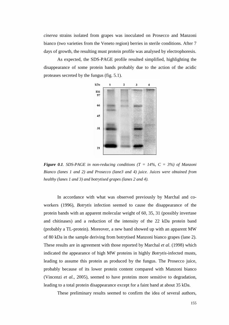
155
cinerea strains isolatedfrom grapeswas inoculated on Prosecco and Manzoni
bianco(two varieties from theVenetoregion) berriesin sterile conditions.After 7
daysof growth, theresulting must proteinprofile wasanalysedby electrophoresis.
As expected, the SDS-PAGE profile resulted simplifi ed, highlighting the
disappearanceof someprotein bandsprobably due to the action of the acidic
proteasessecretedby thefungus(fig. 5.1).
Figure 0.1. SDS-PAGE in non-reducing conditions (T = 14%, C = 3%) of Manzoni
Bianco (lanes 1 and 2) and Prosecco (lane3 and 4) juice. Juices were obtained from
healthy (lanes 1 and 3) and botrytised grapes (lanes 2 and 4).
In accordancewith what was observedpreviously by Marchal and co-
workers (1996), Botrytis infection seemed to cause the disappearance of the
proteinbandswith anapparentmolecularweight of 60, 35, 31 (possiblyinvertase
and chitinases)and a reduction of the intensity of the 22 kDa protein band
(probablya TL-protein).Moreover,a newbandshowedup with anapparent MW
of 80 kDa in thesamplederiving from botrytisedManzonibianco grapes(lane2).
Theseresults are in agreementwith thosereportedby Marchalet al. (1998)which
indicatedthe appearanceof high MW proteins in highly Botrytis-infectedmusts,
leading to assumethis protein as produced by the fungus.The Prosecco juice,
probably becauseof its lower protein content comparedwith Manzoni bianco
(Vincenzi et al., 2005), seemedto have proteins more sensitive to degradation,
leading to a total proteindisappearanceexceptfor a faint bandat about35kDa.
Thesepreliminary results seemedto confirm the idea of several authors,

156
although the possibleeffectsof otherfungal enzymes,suchaslaccase, should be
considered. Actually, in these experimentalconditions,there weresomeevidences
that laccaseactivity can causesome modifications of the grapeproteins,which
could leadto their insolubilisationresulting in a simplification of theSDS-PAGE
profile (datanot shown)(Zamoraniet al., 1993).
Thedegradation anddisappearanceof the grapeproteinsfrom the juice is
generallyconsideredasa modification improving wine stabilit y. For this reason,
the juices produced from infected berries were heated to measure their hazing
potential in comparisonto thatof thehealthy juices(fig. 5.2).
Figure 0.2. Heat test on Manzoni bianco juices deriving from healthy- and Botrytis
cinerea- infected grapes. Different letters means significant differences among values for
P ≤ 0.01 (Student-Newman-Keuls test).
The heattest results showedthat the infection dramatically reduced juice
hazing,although the turbidity level reachedby the infected grapes juice wasstill
not sufficient for its completestabilisation, which should correspondto turbidity
valueslower than2 NTU aftertheheattest(PocockandRankine,1973).
After thesefirsts experimentsto confirm the presenceof a proteolytic
activity secreted by B. cinerea, an attempt to obtain preparations enriched in
protease activity wasmade.To this aim, the funguswasinoculated in PDB liquid
medium and incubatedfor 5, 10and15days.
The collectedpreparationswere initially assayed by SDS-PAGE. The 15
days-incubated medium was left for 24 h at 20°C in contact with the juice
obtainedfrom ManzoniBiancohealthy grapes (fig. 5.3).
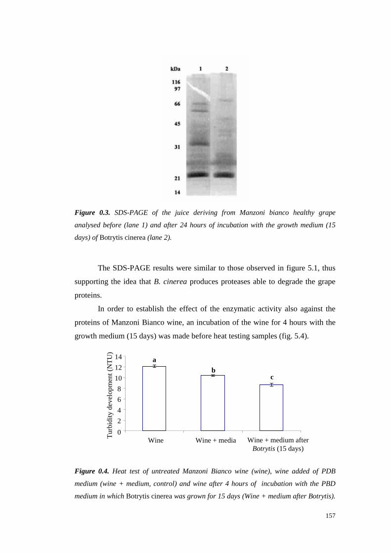
157
Figure 0.3. SDS-PAGE of the juice deriving from Manzoni bianco healthy grape
analysed before (lane 1) and after 24 hours of incubation with the growth medium (15
days) of Botrytis cinerea(lane 2).
The SDS-PAGE resultsweresimilar to thoseobservedin figure5.1, thus
supporting the ideathat B. cinerea producesproteases able to degrade the grape
proteins.
In order to establish the effect of the enzymatic activity also against the
proteinsof ManzoniBiancowine, an incubation of thewine for 4 hourswith the
growthmedium (15days)wasmadebeforeheattestingsamples(fig. 5.4).
2
4
6
8
10
12
14
Wine Wine+ media Wine+ mediumafterBotrytis (15days)
Tur
bidi
tyde
velo
pmen
t(N
TU
)
a
bc
0
Figure 0.4. Heat test of untreated Manzoni Bianco wine (wine), wine added of PDB
medium (wine + medium, control) and wine after 4 hours of incubation with the PBD
medium in which Botrytis cinereawas grown for 15 days (Wine + medium after Botrytis).
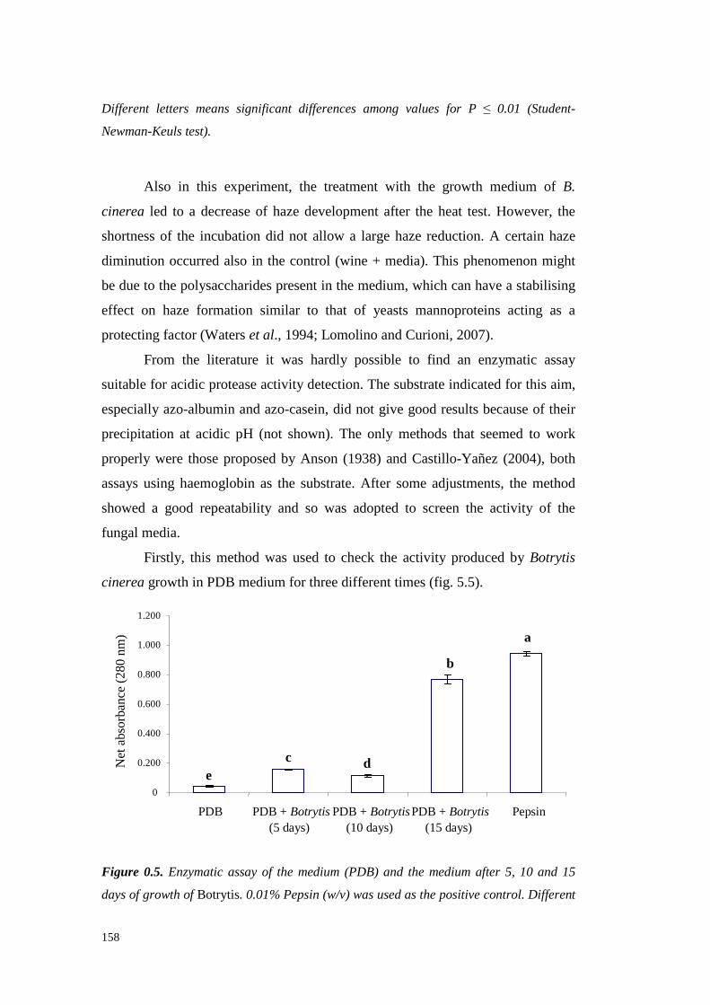
158
Different letters means significant differences among values for P ≤ 0.01 (Student-
Newman-Keuls test).
Also in this experiment,the treatmentwith the growth medium of B.
cinerea led to a decreaseof hazedevelopment after the heat test.However, the
shortnessof the incubationdid not allow a large haze reduction. A certain haze
diminution occurredalso in the control (wine + media). This phenomenonmight
be due to thepolysaccharidespresentin themedium, which can havea stabilising
effect on haze formation similar to that of yeastsmannoproteinsacting as a
protectingfactor(Waterset al., 1994;LomolinoandCurioni, 2007).
From the literature it was hardly possible to find an enzymatic assay
suitablefor acidic proteaseactivity detection. Thesubstrateindicated for this aim,
especially azo-albumin andazo-casein,did not give goodresults becauseof their
precipitation at acidic pH (not shown). The only methods that seemed to work
properly were thoseproposed by Anson (1938)andCastill o-Yañez (2004),both
assaysusing haemoglobinas the substrate. After someadjustments,the method
showed a good repeatabilityand so was adopted to screen the activity of the
fungal media.
Firstly, this method was used to checkthe activity producedby Botrytis
cinerea growthin PDB mediumfor threedifferenttimes(fig. 5.5).
0
0.200
0.400
0.600
0.800
1.000
1.200
PDB PDB + Botrytis(5 days)
PDB + Botrytis(10days)
PDB + Botrytis(15days)
Pepsin
a
b
c de
Figure 0.5. Enzymatic assay of the medium (PDB) and the medium after 5, 10 and 15
days of growth of Botrytis. 0.01% Pepsin (w/v) was used as the positive control. Different
Net
abso
rban
ce(2
80nm
)

159
letters means significant differences among values for P ≤ 0.01 (Student-Newman-Keuls
test).
This experimenthasbeenreplicatedseveraltimes with different conditions
of incubation, storage,substrateconcentration. The obtained results (not shown)
showed a proteolytic activity in the medium that wasalways significantly higher
after 15 days of incubation, suggestingthis length as the best for a proteases
emissionandfor thestudyof theirability to degradethegrapeandwineproteins.
An attemptto separateandconcentratetheproteaseswas madeby adding
ammoniumsulphate(80% saturation)to the media in which Botrytis wasgrown
for 15 days.Theresultsshowedthecomplete lossof proteolytic activity after this
precipitationstep(datanot shown).
This first batchof resultsconfirmedthe simplifi cation of the SDS-PAGE
profile of juicesmadefrom grapesinfectedby Botrytis. Theheat teston juice and
wine showedthe possibility of a reduction of hazeformation for samplestreated
with the enzymatic preparation.However, the effect of the PDB medium
composition has to be taken into accountbecauseof its polysaccharide content
that caninterferewith the process of turbidity development. Thevery preliminary
attemptsof proteasesconcentrationby proteinsalting out showeda total activity
loss, thereforefurther trials neededto be doneto better concentrate the protease
fractionandto getrid of thePDB media.
To these aim, additional experimentswere planned. B. cinerea was
inoculatedin a mediumcontainingManzoni biancowine proteinsasonly nitrogen
sourceto stimulateproteasesemission from thefungus.
Thenewculturewasassayedfor proteasesactivity on haemoglobin asthe
substrate(fig. 5.6).
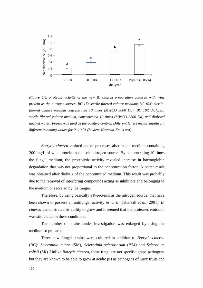
160
0
0.2
0.4
0.6
0.8
1
1.2
BC 1X BC 10X BC 10Xdialyzed
Pepsin (0.05%)
Net
abso
rban
ce(2
80nm
) a
b
c
d
Figure 0.6. Protease activity of the new B. cinereapreparation cultured with wine
protein as the nitrogen source. BC 1X: sterile-filtered culture medium; BC 10X: sterile-
filtered culture medium concentrated 10 times (MWCO 3000 Da); BC 10X dialysed:
sterile-filtered culture medium, concentrated 10 times (MWCO 3500 Da) and dialysed
against water; Pepsin was used as the positive control. Different letters means significant
differences among values for P ≤ 0.01 (Student-Newman-Keuls test).
Botrytis cinerea emitted active proteases also in the medium containing
300 mg/L of wine proteinasthe solenitrogensource.By concentrating 10 times
the fungal medium, the proteolytic activity revealed increase in haemoglobin
degradation that wasnot proportionalto the concentration factor. A better result
wasobtained after dialysisof theconcentrated medium. This resultwasprobably
dueto theremoval of interferingcompoundsacting asinhibitorsandbelongingto
themedium or secretedby thefungus.
Therefore,by using basicallyPR-proteinsasthenitrogensource, thathave
beenshownto possess an antifungal activity in vitro (Tattersallet al., 2001),B.
cinerea demonstratedits ability to grow andit seemed that theproteasesemission
wasstimulatedin theseconditions.
The number of strains under investigation was enlarged by using the
medium so prepared.
Three new fungal strains were cultured in addition to Botrytis cinerea
(BC): Sclerotinia minor (SM), Sclerotinia sclerotiorum (B24) and Sclerotium
rolfsii (SR).Unlike Botrytis cinerea, thesefungi arenot specific grapepathogens
but theyareknownto beableto growat acidic pH aspathogensof juicy fruits and

161
plants (Billon-Grand, 2002). For these reasons they have theoretically been
consideredsuitableto releaseacidicproteases activeat thepH of mustandwine.
Initially, after a growth periodon a mediumpreparedwith purified wine
protein as the sole nitrogen source, the fungi were tested for their enzymatic
activity againsthaemoglobinin comparisonto Botrytis cinerea andpepsin,both
usedaspositivecontrols(fig. 5.7).
dcdc
b
a
0
0.1
0.2
0.3
0.4
0.5
Pepsin SM B24 BC SR
Net
abso
rban
ce(2
80nm
)
Figure 0.7. Proteolytic activity assayed on haemoglobin of four fungi after a growth
period in a medium containing 300 mg/L of Manzoni bianco wine proteins. BC: Botrytis
cinerea; SM: Sclerotinia minor; B24: Sclerotinia sclerotiorum; SR: Sclerotium rolfsii.
Different letters means significant differences among values for P ≤ 0.05 (Student-
Newman-Keuls test).
The four fungi showed a different degradation aptitude against
haemoglobin. In particular, the fungus that showed the largest substrate
degradationwasSclerotinia minor, the otherfungi showing a proteolyticactivity
significantly lower thanthat of SM but not very different to that of BC, that was
assumedasthereference.
In order to better define the growth conditions able to stimulate the best
proteaseemissioninto themedium,thefour fungi were daily checkedfor protease
activity by thehaemoglobinassaystartingfrom theinoculation moment(fig. 5.8).
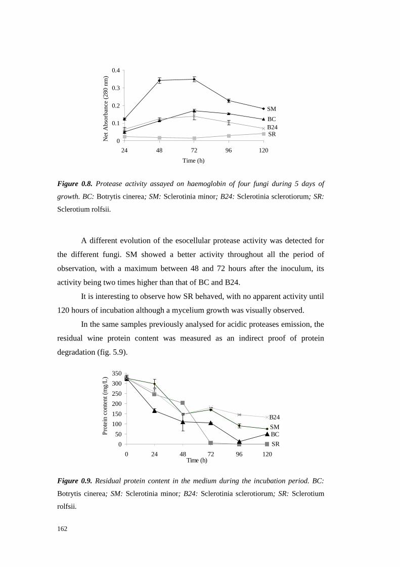
162
SM
SR
BCB24
0
0.1
0.2
0.3
0.4
24 48 72 96 120
Time (h)
Net
Abs
orba
nce
(280
nm)
Figure 0.8. Protease activity assayed on haemoglobin of four fungi during 5 days of
growth. BC: Botrytis cinerea; SM: Sclerotinia minor; B24: Sclerotinia sclerotiorum; SR:
Sclerotiumrolfsii.
A different evolution of the esocellularproteaseactivit y was detected for
the different fungi. SM showed a better activity throughoutall the period of
observation,with a maximumbetween 48 and 72 hoursafter the inoculum, its
activity being two timeshigherthanthatof BC andB24.
It is interesting to observe how SRbehaved,with no apparent activi ty until
120hours of incubationalthoughamycelium growthwasvisually observed.
In thesamesamplespreviouslyanalysedfor acidic proteases emission,the
residual wine protein content was measuredas an indirect proof of protein
degradation (fig. 5.9).
SM
SR
BC
B24
0
50
100
150
200
250
300
350
0 24 48 72 96 120Time(h)
Pro
tein
cont
ent(
mg/
L)
Figure 0.9. Residual protein content in the medium during the incubation period. BC:
Botrytis cinerea; SM: Sclerotinia minor; B24: Sclerotinia sclerotiorum; SR: Sclerotium
rolfsii.
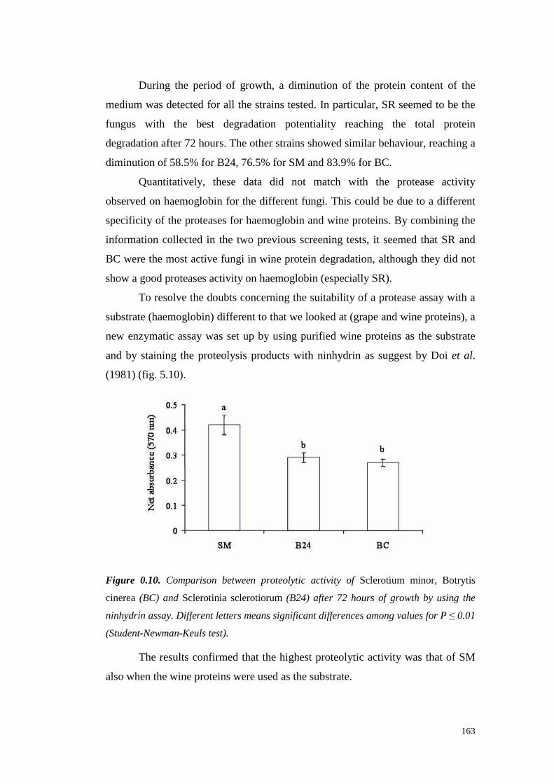
163
During the period of growth, a diminution of the protein contentof the
medium wasdetectedfor all thestrainstested.In particular, SRseemedto be the
fungus with the best degradation potentiality reaching the total protein
degradationafter72 hours.Theotherstrainsshowed similar behaviour, reachinga
diminutionof 58.5%for B24,76.5%for SM and83.9%for BC.
Quantitatively, these data did not match with the protease activi ty
observedon haemoglobinfor thedifferent fungi. This could bedueto a different
specificity of theproteasesfor haemoglobin andwine proteins.By combining the
informationcollected in the two previousscreening tests,it seemed that SR and
BC were themostactivefungi in wine protein degradation, althoughtheydid not
show a goodproteasesactivity onhaemoglobin (especially SR).
To resolve thedoubtsconcerningthesuitabili ty of a proteaseassaywith a
substrate(haemoglobin) differentto thatwe lookedat (grapeandwine proteins), a
new enzymatic assay wassetup by using purified wine proteins as the substrate
andby staining the proteolysis productswith ninhydrin as suggestby Doi et al.
(1981) (fig. 5.10).
Figure 0.10. Comparison between proteolytic activity of Sclerotium minor, Botrytis
cinerea(BC) and Sclerotinia sclerotiorum (B24) after 72 hours of growth by using the
ninhydrin assay. Different letters means significant differences among values for P ≤ 0.01
(Student-Newman-Keuls test).
The results confirmedthat the highestproteolytic activit y wasthat of SM
alsowhenthewineproteinswereusedasthesubstrate.
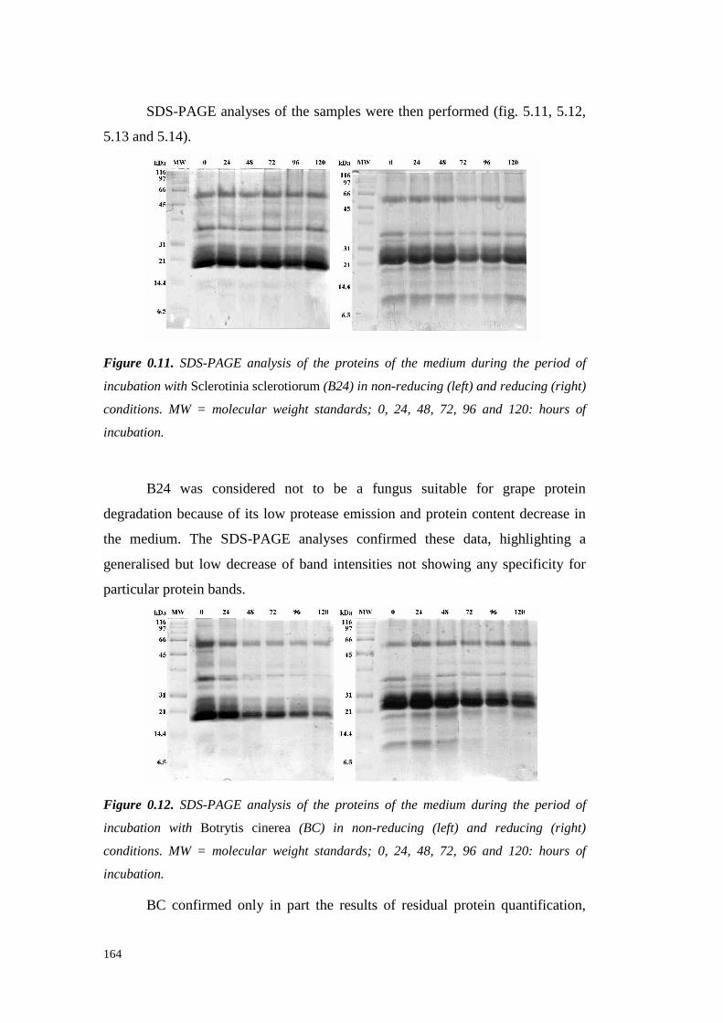
164
SDS-PAGE analysesof the sampleswerethenperformed (fig. 5.11,5.12,
5.13and5.14).
Figure 0.11. SDS-PAGE analysis of the proteins of the medium during the period of
incubation with Sclerotinia sclerotiorum (B24) in non-reducing (left) and reducing (right)
conditions. MW = molecular weight standards; 0, 24, 48, 72, 96 and 120: hours of
incubation.
B24 was considerednot to be a fungus suitable for grape protein
degradation becauseof its low proteaseemission and protein contentdecreasein
the medium. The SDS-PAGE analysesconfirmed these data, highlighting a
generalisedbut low decreaseof bandintensities not showing any specificity for
particularprotein bands.
Figure 0.12. SDS-PAGE analysis of the proteins of the medium during the period of
incubation with Botrytis cinerea (BC) in non-reducing (left) and reducing (right)
conditions. MW = molecular weight standards; 0, 24, 48, 72, 96 and 120: hours of
incubation.
BC confirmed only in part the resultsof residual protein quantification,

165
showing only a generaliseddiminution of band intensities that did not led to the
disappearanceof any protein. This fact seemed to be due firstly to the shorter
incubationperiod of BC in this culturein comparisonto thoseobtained from PDB
media. Secondarily,the incubationof this funguswith the wine proteins seemed
not to stimulatea proteasesemission asexpected, at leastnot for the incubation
timestested.
Figure 0.13. SDS-PAGE analysis of the proteins of the media during the period of
incubation with Sclerotinia minor (SM) in non-reducing (left) and reducing (right)
conditions. MW = molecular weight standards; 0, 24, 48, 72, 96 and 120: hours of
incubation.
Thelargestprotein degradationwasobserved for SM starting from the2nd
day of growth, with a generalised protein decrease and with the complete
disappearanceof protein bandswith an apparent molecular weight of 35 kDa
(probably chitinases)and lessthan20 kDa. Besides,a general decrease of band
intensitieswas detected.
Figure 0.14. SDS-PAGE analysis of the proteins of the media during the period of

166
incubation with Sclerotium rolfsii (SR) in non-reducing (left) and reducing (right)
conditions. MW = molecular weight standards; 0, 24, 48, 72, 96 and 120: hours of
incubation.
SR showedthe most interestingbehaviour,leading to a complete protein
disappearancebetween the second and the third day of culture, confirming the
dataof protein quantification but contrasting with thoseof thehaemoglobin assay.
This suddenchangeof proteinprofile wasvery different from thoseobservedfor
the other fungi. Taking into accountthe very low proteaseactivity of SR on
haemoglobin (Fig. 5.8) and the rapidity of protein disappearance in the growing
medium, it seemedunlikely thata proteasecould leadto a similar result, although
the activation of a proteaseafter an unidentified step of growth could not be
excluded.
Sclerotium rolfsii is a fungus commonly used for the production of
scleroglucan(Fariñaet al. 1998), a polysaccharide that the fungusreleases in the
medium and whose presencewas assumed as a possible reason of protein
disappearance.Indeed,the role of scleroglucanin protein sequestration hasbeen
demonstrated with further experiments(see chapter 6; Vincenzi et al., 2007).
Consequently, it wasassumedthat a proteaseactivi ty emission into the medium
had to be present, because the fungus grew, but this activi ty was very low.
Therefore wine proteins were only minimally degraded, but they disappeared
from the mediumas a result of the action of the scleroglucanproduced by the
fungus.Taking into accountthe antifungalactivity of the PR-proteins,a possible
reasonof this unexpectedphenomenonis that theproduction of scleroglucanacts
as a sort of defencemechanismthat the fungus adopts to trap proteins and
inactivatetheir toxicity.
An attempt to grow the fungi (BC, SR and SM) on a media containing
proteinsfrom dialysedManzonibiancojuice insteadthat from winewasmade.In
this case,a partial precipitationof grapeproteins addedto the medium occurred,
resultingin a 5 timeslower proteincontentafter 24 hours (about50 mg/L instead
of 250 mg/L added).This phenomenonseemedto depend on theemission fungal
laccase in the media, causingpolyphenolsoxidation and their reaction with the
unfermentedmustproteinsthat arebelieved to be morereactive in this situation

167
than after fermentation (Zamorani et al., 1993). Becauseof the too low final
protein level, no significantproteolytic activity was detectable(datanot shown),
although a generalised protein content decrease was measured in the fungal
medium during theincubationperiod(fig. 5.15).
0
50
100
150
200
250
0 24 48 72 96
Incubationtime (h)
Res
idua
lpro
tein
cont
ent
(mg/
LB
SA)
BC SR SM
Figure 0.15. Residual protein content in the medium containing juice proteins as the
nitrogen source during the growth of Sclerotiumrolfsii (SR); Sclerotiniaminor (SM) and
Botrytis cinerea(BC).
Theresultspartially confirmedthatSM hadthebest proteolytic activity on
grapePR-proteins,but led to the conclusionthat the mediumcontaining proteins
from wine as the nitrogen sourcewas more suitable than that prepared starting
from juice.
1.1.57 PROTEOLYTIC ACTIVITY OF SCLEROTIUM MINOR
Sclerotium minor (SM) wasselectedbecausethis fungusshowedthe best
proteasesemissionandgoodwineproteindegradation.
A largequantity of mediumwasinoculated with SM andthe growth was
carriedout in theconditionsconsideredasoptimal after thepreliminary trials: 72
h of growth in a liquid media (Billon-Grand, 2002) containing 300 mg/L of
proteinspurified from wine.
The presence of a good proteolytic activity in this new culture was
detected(datanot shown).
SDS-PAGE analysis(fig. 5.16) confirmedthe degradation of mostof the
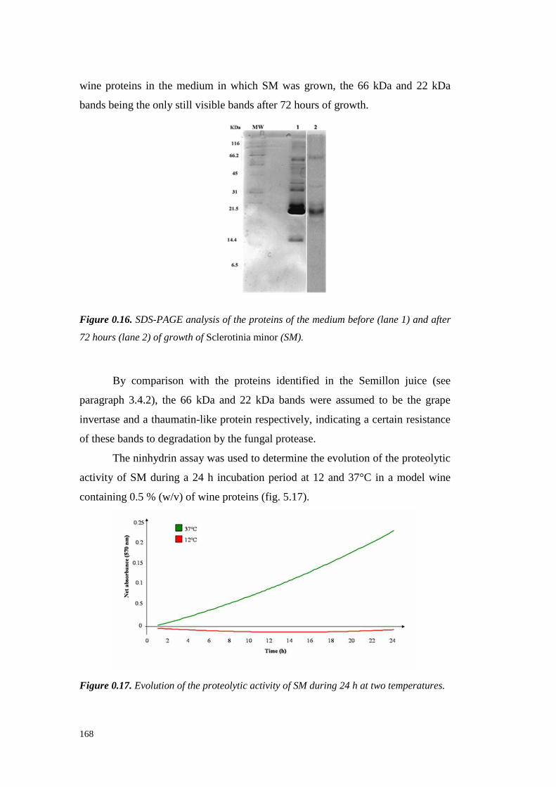
168
wine proteins in the mediumin which SM was grown, the 66 kDa and 22 kDa
bandsbeingtheonly still visible bandsafter72hoursof growth.
Figure 0.16. SDS-PAGE analysis of the proteins of the medium before (lane 1) and after
72 hours (lane 2) of growth of Sclerotinia minor (SM).
By comparisonwith the proteins identified in the Semill on juice (see
paragraph 3.4.2), the 66 kDa and 22 kDa bandswere assumed to be the grape
invertaseanda thaumatin-like proteinrespectively, indicating a certain resistance
of thesebandsto degradationby thefungalprotease.
Theninhydrin assaywasusedto determinetheevolution of theproteolytic
activity of SM during a 24 h incubationperiodat 12 and37°C in a modelwine
containing0.5% (w/v) of wineproteins(fig. 5.17).
Figure 0.17. Evolution of the proteolytic activity of SM during 24 h at two temperatures.
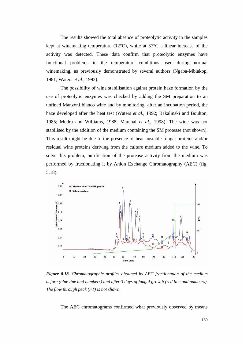
169
Theresultsshowedthe total absenceof proteolytic activity in thesamples
kept at winemakingtemperature(12°C), while at 37°C a linear increaseof the
activity was detected. These data confirm that proteolytic enzymes have
functional problems in the temperature conditions used during normal
winemaking, as previously demonstratedby severalauthors(Ngaba-Mbiakop,
1981; Waterset al., 1992).
Thepossibili ty of wine stabilisationagainst protein haze formation by the
use of proteolytic enzymeswas checked by adding the SM preparation to an
unfined Manzonibiancowine andby monitoring, after an incubation period, the
hazedevelopedafter the heattest (Waterset al., 1992; Bakalinski and Boulton,
1985; Modra and Willi ams, 1988; Marchal et al., 1998). The wine was not
stabilisedby theadditionof themediumcontaining theSM protease(not shown).
This resultmight be due to the presenceof heat-unstablefungal proteins and/or
residual wine proteins deriving from the culture medium addedto the wine. To
solve this problem, purification of the proteaseactivit y from the medium was
performedby fractionating it by Anion ExchangeChromatography (AEC) (fig.
5.18).
Figure 0.18. Chromatographic profiles obtained by AEC fractionation of the medium
before (blue line and numbers) and after 3 days of fungal growth (red line and numbers).
The flow through peak (FT) is not shown.
The AEC chromatogramsconfirmedwhat previouslyobserved by means
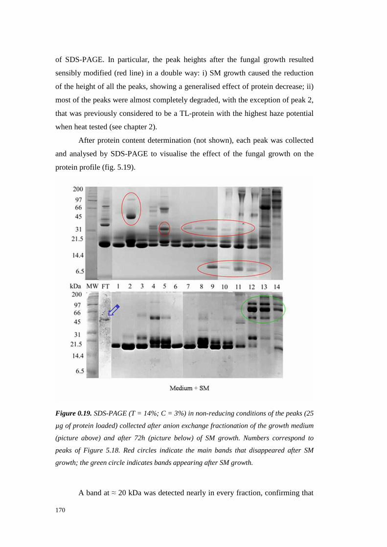
170
of SDS-PAGE. In particular, the peak heights after the fungal growth resulted
sensibly modified (red line) in a doubleway: i) SM growth causedthe reduction
of theheight of all thepeaks,showing a generalisedeffect of protein decrease;ii)
mostof thepeakswerealmost completely degraded, with theexception of peak 2,
that waspreviouslyconsideredto bea TL-protein with thehighest haze potential
whenheattested(seechapter2).
After proteincontentdetermination(not shown),each peakwascollected
and analysedby SDS-PAGE to visualisethe effect of the fungal growth on the
proteinprofile (fig. 5.19).
Figure 0.19. SDS-PAGE (T = 14%; C = 3%) in non-reducing conditions of the peaks (25
µg of protein loaded) collected after anion exchange fractionation of the growth medium
(picture above) and after 72h (picture below) of SM growth. Numbers correspond to
peaks of Figure 5.18. Red circles indicate the main bands that disappeared after SM
growth; the green circle indicates bands appearing after SM growth.
A bandat ≈ 20 kDa wasdetected nearlyin everyfraction, confirming that
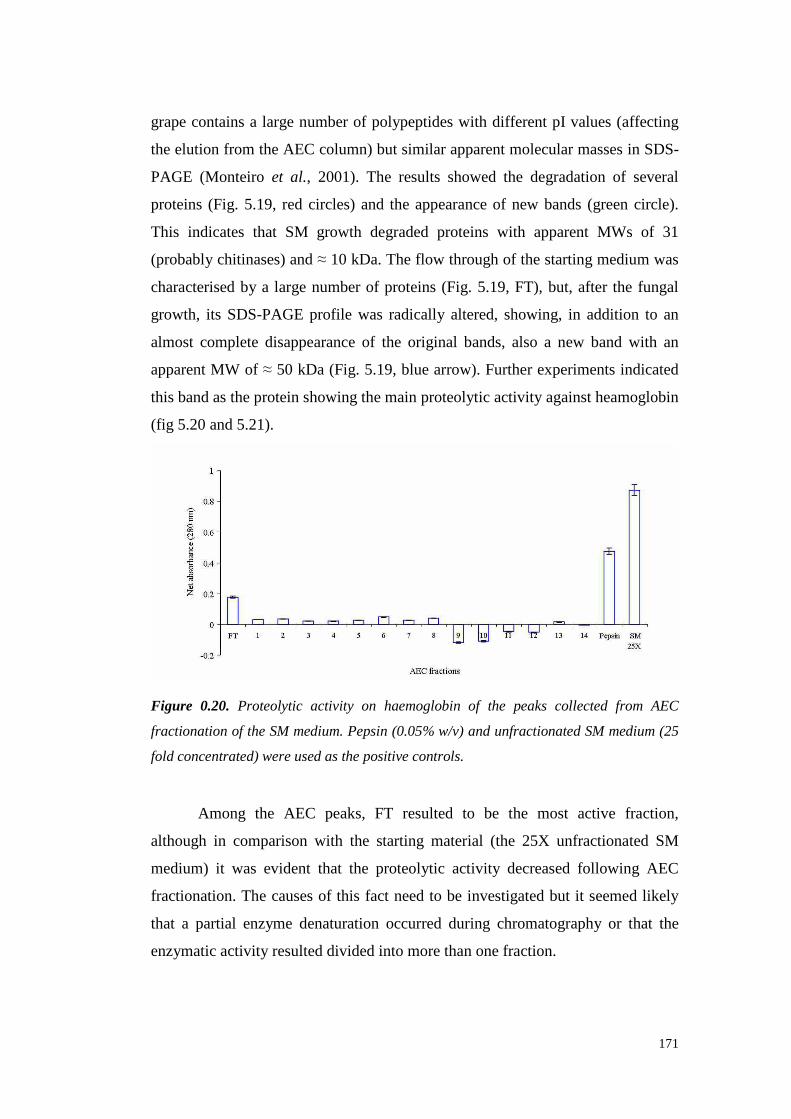
171
grapecontainsa largenumberof polypeptides with different pI values(affecting
theelutionfrom theAEC column) but similar apparentmolecularmassesin SDS-
PAGE (Monteiro et al., 2001). The results showed the degradation of several
proteins(Fig. 5.19, red circles) and the appearance of new bands(green circle).
This indicates that SM growth degradedproteins with apparent MWs of 31
(probablychitinases)and≈ 10 kDa. Theflow throughof the startingmedium was
characterised by a largenumberof proteins (Fig. 5.19,FT), but, after the fungal
growth, its SDS-PAGE profile was radically altered,showing,in addition to an
almostcomplete disappearanceof the original bands,also a new band with an
apparentMW of ≈ 50 kDa (Fig. 5.19,bluearrow). Furtherexperiments indicated
this bandastheproteinshowing themainproteolytic activi ty against heamoglobin
(fig 5.20and5.21).
Figure 0.20. Proteolytic activity on haemoglobin of the peaks collected from AEC
fractionation of the SM medium. Pepsin (0.05% w/v) and unfractionated SM medium (25
fold concentrated) were used as the positive controls.
Among the AEC peaks, FT resulted to be the most active fraction,
although in comparisonwith the starting material (the 25X unfractionated SM
medium) it was evident that the proteolytic activit y decreasedfollowing AEC
fractionation.The causesof this fact need to be investigated but it seemedlikely
that a partial enzyme denaturation occurredduring chromatography or that the
enzymaticactivi ty resulteddividedinto morethanonefraction.
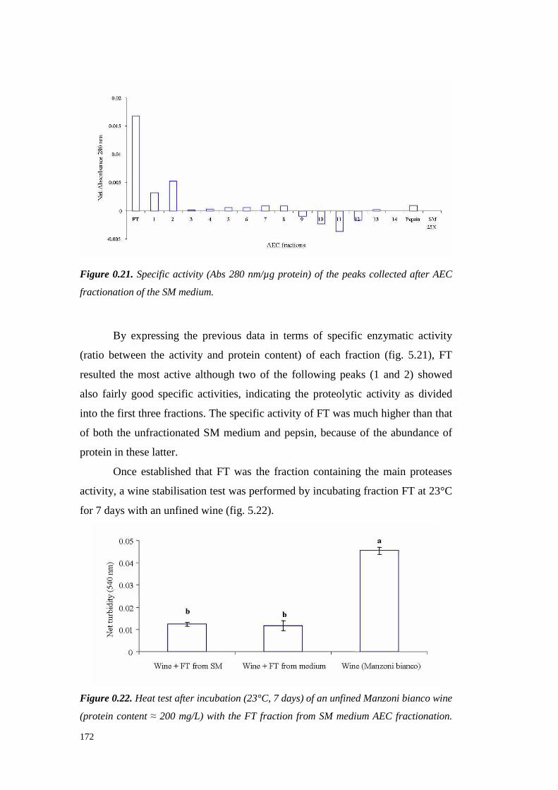
172
Figure 0.21. Specific activity (Abs 280 nm/µg protein) of the peaks collected after AEC
fractionation of the SM medium.
By expressing the previousdata in terms of specific enzymatic activity
(ratio betweenthe activity and protein content) of each fraction (fig. 5.21), FT
resultedthe most active althoughtwo of the following peaks (1 and 2) showed
also fairly good specific activities, indicating the proteolytic activity as divided
into thefirst threefractions.Thespecificactivity of FT was muchhigherthan that
of both the unfractionatedSM mediumandpepsin, becauseof the abundanceof
protein in theselatter.
Once establishedthat FT was the fraction containing the main proteases
activity, a winestabilisationtest wasperformedby incubating fraction FT at 23°C
for 7 dayswith anunfinedwine (fig. 5.22).
Figure 0.22. Heat test after incubation (23°C, 7 days) of an unfined Manzoni bianco wine
(protein content ≈ 200 mg/L) with the FT fraction from SM medium AEC fractionation.
173
An incubation with the FT fraction of the un-inoculated medium was executed as the
control. Different letters means significant differences among values for P ≤ 0.01
(Student-Newman-Keuls test).
The obtainedresults highlightedthe possibilit y to reachwine stabilisation
(turbidity valueslower than0.02AU). Unexpectedly, addition to the wine of FT
from SM and from the starting (un-inoculated) culture medium gave similar
results. This phenomenonwas attributedto two different mechanisms of action:
for the FT fraction deriving from the SM medium, the stabilisation could be
ascribed to the wine protein degradation, whereas in the caseof the FT fraction
from the un-inoculatedmediumthe stabilisation could be dueto its high content
of polysaccharides(225.6g/L for theFT from Manzoni biancowinecompared to
37.9 g/L for FT from SM) thatactedasprotective factorsagainsthaze formation,
aspointedout by severalauthors(Waterset al., 1991,1994;Pellerin et al., 1994).
These results show the possibility to utili ze S. minor as a source of
proteolytic enzymesfor grapeand wine protein degradation. From SDS-PAGE
analyses, a wine protein degradationwas detected althoughnot complete. Two
proteinbandsseemednot to beaffectedby theaction of S. minor enzymes, these
proteinsprobablybeing thaumatin-like proteins and invertases.This information
revealsa maindrawbackbecause thaumatin-like proteinsare considered themajor
responsible for heat-inducedhazedevelopment in wines (Waters et al., 1993,
1996)as confirmedin chapter4 of this thesis.
First attempts of wine stabilizationby addition of the whole enzymatic
preparation highlighted someproblemsfor the passagefrom laboratory trials to
therealconditionsof winemaking,theproteolytic activity of thewhole enzymatic
preparation being totally inhibitedat normal winemaking temperatures (12°C).
A certain purification level was achieved by using Anionic Exchange
Chromatography, although a loss on activit y was observed during the
fractionationprocess, probablydue to the distribution of the enzyme in several
chromatographicfractions.Besides, the quantities of purified enzyme were too
low to plan a large-scaleexperimentalso starting from a quite high volume of
sample.Consequently,thepurificationprocessshould beimprovedby using other

174
chromatographictechniquesor, theexpressionof theproteolytic activity of SM in
etherologoussystems,suchasin yeast,couldbeattempted.
In conclusion, the reportedpreliminary results indicate the possibility to
usephytopathogenicfungi to produceproteases,active at thewine pH, which are
able to degradethe grape proteins responsiblefor haze formation in white wines,
although further studies are necessary to make this approach suitable for a
practical usein winemaking.
1.1.58 PROTEOLYTIC ACTIVITY OF SCLEROTIUM ROLFSII
Scletorium rolfsii (SR)wasstudiedin orderto inducea proteolytic activity
that wasnot detected in the preliminaryresults abovedescribed.To this aim, SR
wasinoculatedin a medium(Czapeck)containing 0.26g/100mL of YeastExtract
and incubated(at 24°C) for 7 days. After this incubation, the medium was
centrifugedand testedfor its proteolytic activit y with the haemoglobinassay. In
theseconditionsa certainproteaseemissionwas detected (0.255AU), in contrast
to theresults of thescreeningexperiments. An incubation of this preparation with
grapeproteinsled to thedisappearanceof someof themasshowedby SDS-PAGE
analysis(fig. 5.23).
Figure 0.23. SDS-PAGE analysis of the Manzoni bianco juice proteins (270 µL of
dialysed juice) before (0) and after 48 hours of incubation with the SR growth medium
(150 µL) (48). MW standard proteins are on the left (MW).
A general decreasein band intensity was detected with the complete
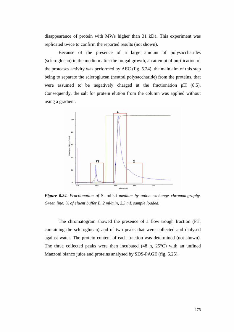
175
disappearanceof protein with MWs higher than 31 kDa. This experiment was
replicatedtwice to confirm thereportedresults (not shown).
Because of the presence of a large amount of polysaccharides
(scleroglucan)in themediumafterthefungal growth,anattempt of purification of
theproteasesactivity wasperformedby AEC (fig. 5.24),themain aim of this step
being to separatethescleroglucan(neutralpolysaccharide) from theproteins,that
were assumed to be negatively charged at the fractionation pH (8.5).
Consequently, the salt for protein elution from the column was applied without
using a gradient.
Figure 0.24. Fractionation of S. rolfsii medium by anion exchange chromatography.
Green line: % of eluent buffer B. 2 ml/min, 2.5 mL sample loaded.
The chromatogram showedthe presence of a flow trough fraction (FT,
containing the scleroglucan) and of two peaksthat were collected and dialysed
against water. The proteincontentof each fraction wasdetermined (not shown).
The three collected peakswere then incubated (48 h, 25°C) with an unfined
Manzonibiancojuiceandproteinsanalysedby SDS-PAGE(fig. 5.25).
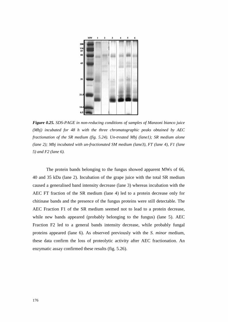
176
Figure 0.25. SDS-PAGE in non-reducing conditions of samples of Manzoni bianco juice
(Mbj) incubated for 48 h with the three chromatographic peaks obtained by AEC
fractionation of the SR medium (fig. 5.24). Un-treated Mbj (lane1); SR medium alone
(lane 2); Mbj incubated with un-fractionated SM medium (lane3), FT (lane 4), F1 (lane
5) and F2 (lane 6).
The proteinbandsbelonging to the fungusshowed apparent MWs of 66,
40 and35 kDa (lane2). Incubationof the grape juice with the total SR medium
causeda generalisedbandintensitydecrease(lane3) whereasincubation with the
AEC FT fraction of the SR medium(lane 4) led to a protein decreaseonly for
chitinase bandsandthe presenceof the fungusproteins werestill detectable. The
AEC Fraction F1 of the SR medium seemed not to lead to a protein decrease,
while new bandsappeared(probably belonging to the fungus) (lane 5). AEC
Fraction F2 led to a generalbandsintensity decrease,while probably fungal
proteins appeared(lane 6). As observedpreviously with the S. minor medium,
thesedata confirm the loss of proteolytic activi ty after AEC fractionation. An
enzymaticassayconfirmedthese results (fig. 5.26).

177
Figure 0.26. Proteolytic activity on heamoglobin of the total (un-fractionated) S. rolfsii
medium (SR) and of the fractions (FT, F1 and F2) collected after anion exchange
chromatography. Different letters means significant differences among values for P ≤
0.01 (Student-Newman-Keuls test).
The un-fractionated SR mediumshowed the highestproteaseactivity on
haemoglobin. It seemedthatanactivity lossoccurred after AEC separation, which
gave fractionsshowingthe highestactivity in the flow trough(FT; in which the
scleroglucanshouldbe). The total activity lossresulting from fractionation was
confirmed from the amount of the activity measuredafter combining the three
fractions that resulted sensibly lower (-78.2%) than that of the unfractionated
preparation.
Taking into accountthat haemoglobincould not be suitablesubstrate for
theSRprotease,a furtherattemptto establishif S. rolfsii really possesseda useful
proteolytic activity was performed by developing a zymography method for the
detectionof proteolytic activity directly on grapeandwine proteins. To this aim,
lyophilised Pinot grigio proteinswere dissolved into a polyacrilamydesolution
that wasthenpolymerized.In this way, a gel matrix containing 0.5 % of purified
wineproteinswasobtainedand,aftermaking a hole in thegel, 25 µL of total SR
preparation wereapplied.After an incubation at 25°C for 72 hours,the proteins
weretreated with Coomassie stainandthe proteolytic activi ty detectedas a clear
background.

178
Figure 0.27. Detection of the proteolytic activity of the unfractionated S. rolfsii medium
in a gel containing 0.5 % lyophilized wine proteins.
The presenceof a clear background aroundthe point in which the SR
medium wasapplied confirmedthe proteolytic activi ty on the wine proteins (fig.
5.27). In orderto establishwhichproteins wereaffectedby theSRprotease,anew
incubation testwascarriedout using thewineproteins (fig. 5.28).
Figure 0.28. SDS-PAGE in non-reducing condition of the products of different
incubations (6 days, 25°C) of the un-fractionated SR medium with grape and wine
proteins. Manzoni bianco wine un-treated (lane 1) and after incubation with SR medium
(lane 2); Manzoni bianco juice un-treated (lane 3) and after incubation with SR medium
(lane 4); Manzoni bianco wine fraction 6 from hydrophobic interaction chromatography
(see paragraph 4.4.2) un-treated (lane 5) and after incubation with SR medium (lane 6).
Each incubation was carried out in presence of NaNO3 (0.01% final concentration). MW
standard proteins are on the left (lane MW).

179
Comparing the pattern of the un-treated wine with that of the wine
incubatedwith the fungus (lanes1 and 2, respectively), it was confirmed that a
certainproteinreductionoccurred,especially for thebandsat ≈ 30 kDaandfor the
TL-proteinbands(≈ 21 kDa) thatboth resulted lessintenseafter the treatment. A
similar resultwasobservedwhenthesamplesof grapejuice wereanalysed(lane3
and4). In this case,a major proteindecreaseoccurred,with thedisappearanceof
two bandsat ≈ 40 and≈ 24 kDa. The wine protein fraction showing the highest
hazing potential, deriving from HIC separation (fraction 6, seefig. 4.13,chapter
4) was also usedto test the proteolytic activity of SR. From the comparisonof
lane 5 (un-treatedHIC fraction 6) and6 (the same fraction 6 but incubated with
SR), it waspossible to observeanalmosttotal disappearanceof this band.
Generally,from this experimentit seemedthatthetotal SRpreparationhad
effectson wine chitinases(lanes1 and 2) and on juice 40 and 24 kDa proteins
(lanes3 and 4). Moreover, the study of S. rolfsii effect on the HIC fraction 6
showed its degradativeability on a very heat-instable TL-protein. These results
indicate the active role of SR on the degradation or subtraction of the wine
proteins. However, the presenceof scleroglucanexcreted by the fungusneed to
considerits particular effectonwineproteins.
In orderto better understandif thescleroglucaninterferedon theobtained
results, threeculturesof S. rolfsii wereachievedusing differentmedia preparedas
follows:
o SR1= 7 dayscultureson Czapeck+ 2.6g/L of Yeastextract(same
conditions of thefirst experiments);
o SR 2 = 9 days cultureson Czapeckwithout NaNO3 + Manzoni
bianco juice;
o SR3 = 9 daysculturesonCzapeckwithoutNaNO3 + Yeast extract.
The three enzymatic preparationswere assayedfor proteolytic activi ty
with the haemoglobin assay. To havea further proof of the involvement of an
enzymaticactivity in SR preparations,samplesboiled beforeto be assayed were
alsotested(fig. 5.29).

180
Figure 0.29. Proteolytic activity (haemoglobin assay) of the three SR preparations (see
text). A series of samples was boiled (100°C, 10 min) as negative controls for the
enzymatic activity. Pepsin 0.05% (w/v) tested as the positive control. Different letters
means significant differences among values for P ≤ 0.01 (Student-Newman-Keuls test).
The presenceof proteasesin S. rolfsii medium was confirmedby the loss
of activity occurring after boiling the samples, although the samples showed
different behaviours.SR 1 (a replicateof thesample described in thepreviouspart
of the results), showedthebestproteaseactivit y, which wascompletely inhibited
by heating.As expected,the sampleSR 3, also prepared with Yeast extract,
behavedsimilarly to SR 1. In contrast, the activi ty of SR 2, which wasactually
very low, was not significantly affected by boiling. An explanation of this
unexpectedresult maybethat theconditionsof culture(prepared with grapejuice
insteadof yeastextract as the nitrogensource)stimulated scleroglucan emission
(indicated by a very high density of the culture medium, not shown). It was
supposedthat, the sterile filtration applied to all the preparations testedcaused
retention on the fi lter of theSR 2 proteasesbecauseof thecomplexation of these
latterby thescleroglucan.Theseresultsweresupported by testing theproteolytic
activity of non-fil teredculturesof SR2, that in this casewassimilar to thatof SR
1 andSR3 (not shown).
All these data indicatedthe stimulating effect of the grape proteins on
scleroglucanemissionby S. rolfsii. A hypothesiscould be madeto explain this
phenomenon:due to the toxicity of the PR-proteins against fungi, S. rolfsii

181
defendeditself by subtracting proteinsfrom themediumby trapping them into the
scleroglucan,ratherthanby usinga proteolytic action. This theorywould explain
the low proteaseactivity detectedfor SR and lead to hypothesize a cooperative
action of scleroglucanandproteasesin defendingthe fungusfrom the antifungal
activity of thegrapePR-proteins.
Thefungalsamplesformerlydescribedwereanalysedalso by zymography
for proteolytic degradation of thewineproteins(fig. 5.30).
Figure 0.30. Detection of the proteolytic activity of unfractionated S. rolfsii media in a
gel containing 0.5 % lyophilized wine proteins. 25 µL of SR 1, SR2, SR3 (see text)
filtered, unfiltered and boiled were incubated at 25°C for 120 hours. After incubation the
gel was stained with Coomassie and de-stained with 7% acetic acid.
SR1 confirmed its activity againstthewine proteinsboth beforeandafter
being filtered.SR 2 andSR3 showedlargerprotein degradationwhennot fil tered.
Boiled samples did not show any protein degradation, confirming the
heamoglobin assayresults.Once more, the hypothesisof a S. rolfsii protease
activity against grape and wine protein seemed to be confirmed becauseno
scleroglucan action could interfere with protein disappearance in these
experimental conditions.
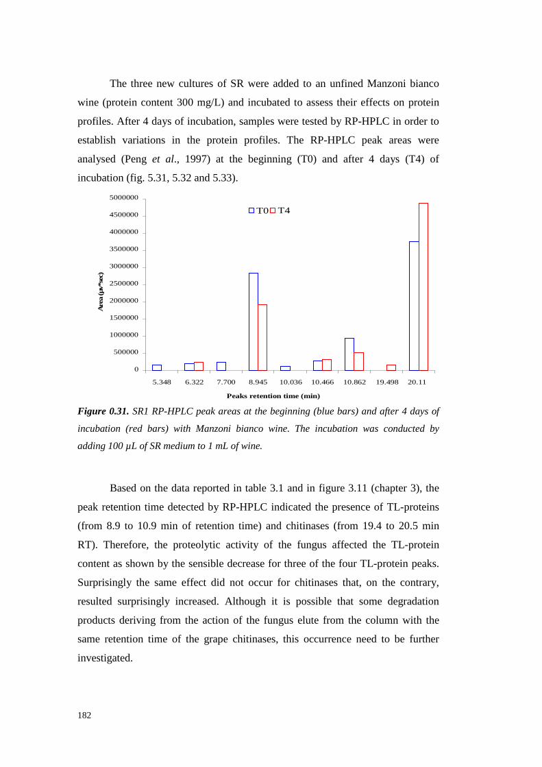
182
The threenew culturesof SR were added to an unfinedManzoni bianco
wine (protein content300 mg/L) and incubated to assesstheir effects on protein
profiles. After 4 daysof incubation,samples weretestedby RP-HPLC in orderto
establish variations in the protein profiles. The RP-HPLC peak areas were
analysed(Peng et al., 1997) at the beginning (T0) and after 4 days (T4) of
incubation (fig. 5.31,5.32and5.33).
0
500000
1000000
1500000
2000000
2500000
3000000
3500000
4000000
4500000
5000000
5.348 6.322 7.700 8.945 10.036 10.466 10.862 19.498 20.11
Peaks retention time (min)
Are
a(µ
v*se
c)
T0 T4
Figure 0.31. SR1 RP-HPLC peak areas at the beginning (blue bars) and after 4 days of
incubation (red bars) with Manzoni bianco wine. The incubation was conducted by
adding 100 µL of SR medium to 1 mL of wine.
Basedon the datareportedin table3.1 andin figure3.11 (chapter 3), the
peakretention time detectedby RP-HPLC indicated the presenceof TL-proteins
(from 8.9 to 10.9 min of retentiontime) and chitinases (from 19.4 to 20.5 min
RT). Therefore, the proteolytic activity of the fungus affected the TL-protein
contentasshown by the sensibledecreasefor three of the four TL-protein peaks.
Surprisingly the sameeffect did not occur for chitinasesthat, on the contrary,
resultedsurprisingly increased.Although it is possible that some degradation
productsderiving from the action of the funguselute from the column with the
sameretention time of the grape chitinases,this occurrence needto be further
investigated.
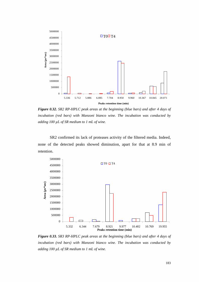
183
0
500000
1000000
1500000
2000000
2500000
3000000
3500000
4000000
4500000
5000000
5.336 5.712 5.886 6.085 7.704 8.950 9.960 10.367 10.845 20.071
Peaks retention time (min)
Are
a(µ
v*se
c)
T0 T4
Figure 0.32. SR2 RP-HPLC peak areas at the beginning (blue bars) and after 4 days of
incubation (red bars) with Manzoni bianco wine. The incubation was conducted by
adding 100 µL of SR medium to 1 mL of wine.
SR2confirmedits lack of proteasesactivity of the fil teredmedia. Indeed,
none of the detected peaksshoweddiminution, apart for that at 8.9 min of
retention.
0
500000
1000000
1500000
2000000
2500000
3000000
3500000
4000000
4500000
5000000
5.332 6.344 7.679 8.921 9.977 10.402 10.769 19.955Peaks retention time (min)
Are
a(µ
v*se
c)
T0 T4
Figure 0.33. SR3 RP-HPLC peak areas at the beginning (blue bars) and after 4 days of
incubation (red bars) with Manzoni bianco wine. The incubation was conducted by
adding 100 µL of SR medium to 1 mL of wine.

184
Confirming the data previously shown, SR 3, showed behaviour very
similar to that of SR1.This result is obvious,since the only difference of these
two preparationswastheincubationtime (7 daysfor SR1, 9 for SR3).
The final trial to assessthe actual effect of S. rolfsii media in wine
stabilisation wasdone throughheattestingthewinetreated with theculturemedia
(fig. 5.34).
Figure 0.34. Heat test of an unfined Manzoni bianco wine added of the three SR
preparation (SR1, SR2, SR3, see text) in a ratio 1:20 (50 µL/ 1 mL of wine). 10X:
enzymatic preparation concentrated 10 times by Vivaspin tubes (3000 MWCO).
Incubation was carried out at 25°C for 90 hours before heat-testing the samples (80°C, 6
hours). The turbidity developed was measured as the difference between the absorbance
at 540nm before and after the heat test (Pocock and Rankine, 1973; Waters et al., 1991).
Different letters means significant differences among values for P ≤ 0.01 (Student-
Newman-Keuls test).
Boiling thefungalpreparationsbeforetheir addition to thewineresultedin
the absenceof stabilisation and the turbidity was even higher than that of the
untreatedwine.In contrast,thenon-boiled preparations(exceptSR2)were able to
diminish thewineturbidity formedafterheat test.In particular, samplesSR110X
and SR3 10X showeda certainstabilizing effect although it was not enoughto
fully stabilize thewine.
In summary, Sclerotium rolfsii hasbeenshownto bea fungusvery active
on proteinremovalfrom grapejuice andwine.This removal wasdueto theaction
of the scleroglucan emitted by the fungus in the presenceof grape proteins.
Besides, a certainproteolytic activity hasbeen detected in the SR culture media,
185
although the presenceof scleroglucaninterferedwith the experimental results.
The hypothesis formulatedis that a cooperative action between scleroglucanand
proteasesexists as a protectionmechanism exerted by the fungusagainstgrape
PR-proteinstoxicity. However, attemptsto purify theprotease/s by AEC and HIC
(not shown) brought to a significant enzymatic activity loss and impaired the
possibility to experimentallyconfirm this idea.
In conclusion, the datacollectedon the fungi studiedin this chapter (B.
cinerea, S. rolfsii and S. minor) indicatedthepotentialit y of their useas a source
of proteasesactiveagainst grapeandwine proteins,S. minor and S. rolfsii being
potentially moresuitablethanB. cinerea for this aim. However,further analyses
are required to definitely assessthe possibilit y of the practical application of
proteolytic enzymesfrom the studiedfungal sourcesin the actual conditions of
winemakingto stabilisewhitewinesagainstprotein hazeformation.
Acknowledgements
The author would like to thank FrancescoFavaron, Simone Vincenzi, Silvana
Odorizzi, Mario Licari, Marco LucchettaandLuca Sella of the PaduaUniversity
for their support in the conduction of the experiments and for the helpful
discussionsaboutthis work.
REFERENCES
AnsonM.L. (1938).Theestimationof pepsin,trypsin,papain and cathepsinwith
haemoglobin.J. Gen. Physiol. 22,79-89.
Bakalinski A.T. and Boulton R. (1985). The study of an immobilized Acid
Proteasefor the treatmentof wineproteins.Am. J. Enol. Vitic. 36(1),23-29.
Bayly F.C.andBerg H. (1967).Grapeandwineproteinsof white winevarietals.
Am. J. Enol. Vitic. 18(1),18-32.
Billon-GrandG., PoussereauN., FèvreM. (2002). The Extracellular Proteases
Secretedin vitro and in planta by the Phytopathogenic FungusSclerotinia
sclerotiorum. Journal of Phytophatology 5, 507-511.
Brown A.E., AdikaramN.K. (1983).A role for pectinaseandproteaseinhibitors

186
in fungalrot developmentin tomatofruits. Phytopathol. Z. 106,239-251.
Castillo-Yañez F.J., Pacheco-Aguilar R., Garcia-Carreño F.L. and De los
AngelesNavarrete-Del Toro M. (2004). Characterization of acidic proteolytic
enzymesfrom Monterey sardine (Sardinops sagax caerulea) viscera. Food
Chem. 85,343-350.
Cilindre C., CastroA.J.,ClémentC., JeandetP.and MarchalR. (2007).Influence
of Botrytis cinereainfection on Champagne wine proteins (characterized by
two-dimensional electrophoresis/immunodetection) and wine foaming
properties.Food Chem. 103(1), 139-149.
Doi E., Shibata D., Teruyoshi M. (1981). Modified Colorimetric Ninhydrin
Methodsfor PeptidaseAssay.Analytical Biochemistry 118,173-174.
DonècheB. (1993).Botrytizedwines.In: Wine Microbiology and Biotechnology,
Ed by Fleet G H , Harwood Academic Publishers, Chur, Switzerland, 327-
351.
FariñaJ.I., Siñeriz F., Molina O.E. and Perotti N.I. (1998). High scleroglucan
production by Sclerotium rolfsii: influence of medium composition.
Biotechnology Letters 20,825-831.
GirbauT., StummerB.E.,PocockK.F., BaldockG.A., Scott E.S.and WatersE.J.
(2004). The effect of Uncinula necator (powdery mildew) and Botrytis
cinerea infection of grapeson the levelsof haze-forming pathogenesis-related
proteinsin grapejuice and wine.Austral J. Grape Wine Res. 10,125-133.
Høj P.B.,TattersallD.B., AdamsK., Pocock K.F., HayasakaY., van Heeswijck
R. and Waters E.J. (2001). The ‘haze proteins’ of wine – a summary of
properties,factors affecting their accumulation in grapes,and the amount of
bentoniterequiredfor their removal from wine.Proceedings of the ASEV 50th
Anniversary Annual Meeting, Seattle, Washington, 19-23 June 2000
(American Society for Enology and Viticulture: Davis, California) 149-154.
Hsu J.C. and Heatherbell D.A. (1987). Heat-unstable proteins in wine. I.
Characterization and removalby bentonitefining and heat treatment.Am. J.
Enol. Vitic. 38(1),11-16.
Jarvis W.R. (1977).Botryotinia andBotrytis species:Taxonomy,physiology,and
pathogenicity. Mongr.Res. Branch Can. Department Agric. 15.
KarmonaZ., Bar Nun N., andMayerA.M. (1990).Utilisation du Botrytis cinerea

187
pers.commesourced'enzymespour la clarification des vins. J. Int. Sci. Vigne
Vin. 24,103-124.
KoenigR., StegemannH., FrancksenH. andPaulH.L. (1970).Protein subunitin
the potato virus X group. Determination of the molecular weight by
polyacrilamideelectrophoresis. Biochim. Biophys. Acta 297,184-189.
LaemmliU.K. (1970).Cleavageof structural proteinsduring theassemblyof the
headof bacteriophageT4. Nature 227,680-685.
Lomolino G. and Curioni A. (2007).Protein Haze Formation in White Wines:
Effect of Saccharomyces cerevisiae Cell Wall Components Prepared with
DifferentProcedures.J. Agric. Food Chem. 55(21),8737-8744.
Marchal R., Berthier L., LegendreL., Marchal-Delahaut L., Jeandet P. and
MaujeanA. (1998).Effectsof Botrytis cinerea infection on the must protein
electrophoretic characteristics.J. Agric. Food Chem. 46(12),4945-4949.
MarchalR., WarcholM., Cilindre C. andJeandetP. (2006).Evidencefor protein
degradation by Botrytis cinerea and relationship with alteration of synthetic
wine foamingproperties.J. Agric. Food Chem. 54(14),5157-5165.
Marchal, R., Bouquelet, S., Maujean, A. (1996). Purification and partial
biochemical characterizationof glycoproteins in a champenois Chardonnay
wine.J. Agric. Food Chem. 44(7),1716-1722.
Modra,E. J.; Will iams,P. J. (1988). Are proteases active in winesand juices?
Aust. Grapegrower Winemaker 292,42-46.
MonteiroS.,Picarra-PereiraM.A., MesquitaP.R.,Loureiro V.B., Teixeira A. and
Ferreira R.B. (2001). The wide diversity of the structurally similar wine
proteins.J. Agric. Food Chem. 49(8),3999-4010.
Movahedi S. and Heale J.B. (1990). Purification and characterization of an
aspartic proteinasesecretedby Botrytis cinerea Persex. Pers in cultureand in
infectedcarrots.Physiol. Mol. Plant Pathol 36,289-302.
Ngaba-Mbiakop P.R. (1981). Investigation of methods for determination and
preventionof protein instability in wines.Thesis, OregonStateUniversity.
Pellerin P., Waters E.J., Brillouet J.M. and Moutounet M. (1994). Effet des
polysaccharidessur la formationdetroubleprotéiquedansun vin blanc.J. Int.
Sci. Vigne Vin 28(3),213-225.
PengZ., PocockK.F., WatersE.J.,Francis I.L. andWili amsP.J.(1997).Taste
188
Propertiesof Grape (Vitis vinifera) Pathogenesis-Related Proteins Isolated
from Wine.J. Agric. Food Chem. 45(12),4639-4643.
Pocock K.F. andRankine B.C. (1973).Heat test for detecting protein instability
in wine.Australian Wine, Brewing and Spirit Review 91(5),42-43.
SegarraI., Lao C., López-TamamesE. and De La Torre-BoronatM.C. (1995).
Spectrophotometric Methodsfor the Analysis of Polysaccharide Levels in
WinemakingProducts.Am. J. Enol. Vitic. 46(4),564-570.
SmithP.K.,Krohn R.I., HermansonG.T.,Mallia A.K., GartnerF.H., Provenzano
M.D., Fujimoto E.K., Goeke N.M., Olson B.J. and Klenk D.C. (1985).
Measurementof protein usingbicinchoninic acid. Anal. Biochem. 150,76-85.
Tattersall D.B., PocockK.F., HayasakaY., AdamsK., van Heeswijck R., Waters
E.J.andHøj P.B.(2001).Pathogenesis relatedproteins– their accumulation in
grapes during berry growth and their involvement in white wine heat
instability. Current knowledge and future perspectives in relation to
winemaking practices. In: Molecular Biology and Biotechnology of the
Grapevine. Ed. K.A. Roubelakis-Angelakis (Kluwer Academic Publishers:
Dordrecht, Netherlands), pp.183-201.
TenHaveA., DekkersE., Kay J.,Phylip L.H., Van Kan J.A. (2004).An aspartic
proteinasegenefamily in the filamentousfungus Botrytis cinerea contains
members with novelfeatures. Microbiology 150,2475-2489.
Touzani A., Muna J.P. (1994). Effet du pool enzymatique exocellulaire de
Botrytis cinerea sur lescellulesdeVitis vinifera. Application à la pellicule du
raisin.J. Int. Sci. Vigne Vin 1, 19-31.
UrbanekH. andKaczmarekA. (1985).Extracellular proteinasesof the isolate of
Botrytis cinerea virulent to appletissues.Acta Biochim. Pol. 32,101-109.
Vincenzi S., MarangonM., Coviello T., Favaron F. and Curioni A.. (2007).
Scleroglucan-Protein interaction: a tool for protein removal from wine?
Abstract Book of In vino Analytica Scientia, Melbourne, 22-25 July 2007.
Vincenzi S.,Mosconi S.,ZoccatelliG., Dalla PellegrinaC., Veneri G., Chignola
R., D.B. PeruffoA., Curioni A. andRizzi C. (2005).Development of a new
procedure for proteinrecoveryandquantification in wine.Am. J. Enol. Vitic.
56(2),182-187.
WatersE.J.,HayasakaY., Tattersall D.B., AdamsK.S. andWilli amsP.J.(1998).

189
Sequence analysisof grape (Vitis vinifera) berry chitinasesthat causehaze
formationin wines.J. Agric. Food Chem. 46(12),4950-4957.
Waters E.J., Pellerin P. and Brillouet J.M. (1994). A Saccharomyces
mannoprotein that protectswine from protein haze. Carbohydr. Polym. 23,
185-191.
WatersE.J.,ShirleyN.J.andWilliams P.J.(1996).Nuisanceproteinsof wine are
grapepathogenesis-relatedproteins.J. Agric. Food Chem. 44(1),3-5.
WatersE.J.,Wallace W. andWilliams P.J.(1991).Heathaze characteristics of
fractionatedwineproteins.Am. J. Enol. Vitic. 42(2),123-127.
WatersE.J.,WallaceW. andWilliams P.J.(1992).Identification of heat-unstable
wine proteinsand their resistanceto peptidasesJ. Agric. Food Chem. 40(9),
1514-1519.
Waters E.J., Wallace W., Tab M.E. and Willi ams P.J. (1993). Isolation and
Partial Characterization of a Natural Haze Protective Factor from Wine. J.
Agric. Food Chem. 41,724-730.
ZamoraniP.,Spettoli P.,LanteA., CrapisiA. andPasini G. (1993). Immobilized
laccase and tyrosinase: an approachfor wine stabilization. Italian Journal of
Food Science 4, 409-414.
Zoccatelli G., MosconiS., Rizzi C., Vincenti S. andCurioni. A. (2003).A new
method for the recoveryof wine proteinsallowing their quantification and
electrophoretic analysis.In: Oenologie 2003. A. Lonvaud-Funel et al. (Eds.),
pp. 542-545.EditionsTec& Doc,Paris.

190
191
CHAPTER 6
Scleroglucan-Protein interaction: a tool for protein removal from
wine?
ABSTRACT
Many fungi areable to form esocellularpolysaccharides. Scleroglucanis a
water-soluble polymer produced by fermentation of the fil amentous fungus
Sclerotium rolfsii. This polysaccharide yields only glucose on complete
hydrolysis, its molecularstructureconsisting of beta-1,3-D-glucoseresidueswith
one beta-1,6-D-glucose side chain every three main residues. Dissolved
scleroglucanchainsassumea rod-like triple helical structure,in which theglucose
residuesareon theoutside,thuspreventingthehelicesfrom coming closeto each
otherandaggregating.
Interaction of scleroglucanwith proteinshas never beenstudied. When
dissolvedin water at a 0.5%concentration, this polysaccharide (usedin the form
of commercial product Actigum CS) showed no interaction with a standard
protein(BovineSerumAlbumin),evenafter 2h of incubation at roomtemperature
under agitation. However, when the scleroglucan was dissolved at the same
concentration in a wine model solution (12%ethanol,5g/L tartaric acid,pH 3.3),
BSA (addedat a final concentrationof 300mg/L) disappeared completely after
only 10minutesfrom theaddition.
Scleroglucaninteractedwith proteinsalso whenusedto treat a real wine,
containing≈ 350 mg/L of proteins. Theinteractionappearedslowerthanin model
solution and protein removalwas nevercompleted, leaving 15% of the original
protein evenafter a 30h incubation.The interaction with polyphenols was very
low, with a maximum removal of 15%. The interaction of scleroglucan with
standard proteinsandwith purified wine proteinsand the effects of the solution
composition were extensively studied. However, being water soluble,
scleroglucandissolvedin bothwater and wine model solution, thusexcluding the
possibility to usethenativepolysaccharidefor winestabilisation.
192
Key words: Sclerotium rolfsii, wine, scleroglucan, haze, protein,
polysaccharide.
INTRODUCTION
Wine proteins play a key role in determining the white wines quality,
mainly becausethey are involved in haze formation during wine storage. The
mostcommonmethodto stabilisewhite winesagainsthazeformation is basedon
bentonite fining. The negatively charged bentonite particles interact
electrostatically with wine proteinsallowing their removalfrom wine. However,
this treatment has some drawbacks because bentonite adsorption is rather
aspecific and,in additionto proteins,removes different moleculesor aggregates,
including aroma andflavour compounds (Voilley et al., 1990).For thesereasons,
alternative proceduresfor proteinremoval from white wine have beendeveloped,
including fining with silica sol/gelatine(Mi ll ies,1975),useof immobilizedtannic
acid (Weetall et al., 1984) or proanthocynidins (Powerset al., 1988), use of
exchangeresins(Sarmentoet al., 2000),adsorption on thesurfaceof metal oxides
(Pachova et al., 2002),ultrafiltration (Hsu et al., 1987)andadsorption on chitin
(Vincenzi et al., 2005b). Recentlythe useof negatively chargedpolysaccharides
hasbeenalsoproposedto stabilize thewine(Cabello-Pasiniet al., 2005).
Many fungi are able to form extracellular polysaccharides. The
phytopathogenic fungus Sclerotium rolfsii (the anamorph form of the
basidiomyceteAthelia rolfsii) attacksa wide variety of plants,primarily annuals
and herbaceousperennials, but somewoody plants are also attacked when they
are young (Aycock, 1961). The filamentousfungus Sclerotium rolfsii has also
beenextensivelystudiedover thelast years by virtueof its abili ty to excretelarge
amounts of β-1,3-β-1,6-d-glucan or 'scleroglucan'(Fariña et al., 1998). This
exopolysaccharide exhibits interesting rheological properties for different
industrial areas(Fariñaet al., 2001) andit has beenrecently included asoneof the
mostpotentbiological responsemodifiers(Pretuset al., 1991).
Scleroglucanis a water-soluble polymer which yields only glucoseon
complete hydrolysis; its repeating unit consistsof beta-1,3-D-glucoseresidues
with one beta-1,6-D-glucoseside chain every three main residues. Dissolved

193
scleroglucanchainsassumea rod-like triple helical structure,in which theglucose
residuesareon theoutside,thuspreventingthehelicesfrom coming closeto each
otherandaggregating. Scleroglucanis a neutral polysaccharideandits interaction
with proteinshasneverbeenstudied.
MATERIALS AND METHODS
1.1.59 MATERIALS
The wine used (vintage 2005, variety Manzoni bianco) was kindly
supplied from the“ScuolaEnologicaG.B.Cerletti” of Conegliano(Italy).
The experiments were performedby adding (under mild agitation) the
commercial dried scleroglucanACTIGUM CS 11 (Degussa) obtained from
Sclerotium rolfsii, to the protein solution for an adequate incubation time. The
insolublepellets werecollectedby centrifugation (14000gfor 5 min).
1.1.60 GRAPE AND WINE PROTEIN CONTENT DETERMINATION
Protein contentdeterminationwasperformed according to Vincenzi et al.
(2005a). Firstly, proteinswere precipitated from 1 mL of wine with the KDS
method (Zoccatelli et al. 2003).Thepellets weredissolvedinto 1 mL of distilled
water and quantified by using the BCA-200 protein assaykit (Pierce). The
calibrationcurvewaspreparedby usingserial dilution of bovine serumalbumin
(BSA, Sigma) in water. The measurements were performed
spectrophotometrically at 562nm (ShimadzuUV 6010).
1.1.61 TOTAL POLYSACCHARIDE CONTENT DETERMINATION
The polysaccharide contentwasdetermined colorimetrically according to
Segarraandco-workers (1995).After addition of 5 volumesof absolute ethanol
(Baker), samples were left at 4°C overnight before centrifugation (30 min,
14000g).The collectedpelletswerewashed twice with ethanol (Baker) andthen
dissolvedin bi-distilled water.1 mL of sample was thenaddedof 25 µL of 80%
phenol (w/w, Fluka) and2.5 mL of sulphuric acid (Merck). Sampleswere mixed

194
and the reaction carried on for 30 minutes at room temperature. Absorbance
valueswere spectrophotometricallymeasured at 490 nm (Shimadzu UV 6010).
The calibration curve wasprepared by usingserial dilution of galactose(Fluka) in
water.
1.1.62 TOTAL POLYPHENOLS CONTENT DETERMINATION
The phenolic content in samplewasdetermined colometrically according
with the methodproposedby Singletonand Rossi (1965) optimised for small
samplevolumesby Waterhouse(2002).200µL of waterdilutedsample(1:10v/v)
were added with 1 mL of water diluted (1:10 v/v) 2N Folin-Ciocalteau reagent
(Sigma). 800 µL of 7.5% (w/v) Na2CO3 (Merck) solution were addedto the
sampleand the incubationcarriedout for 30 min at 40°C. The calibration curve
was preparedby usingserial dilution of galli c acid (GAE, Fluka) in water. The
measurementswere performedspectrophotometrically at 725 nm (ShimadzuUV
6010).
1.1.63 HEAT TEST
Accordingto Watersandcolleagues(1991), a heat testwasperformedto
determineprotein stability. After heating(80°Cfor 6 hours),samples were chilled
(16 hours at 4°C) and, after equilibrationat room temperature, turbidity values
were measured nephelometrically (Hach 2100P turbidimeter) or
spectrophotometrically at 540 nm (Shimadzu UV 6010). Net turbidity values
lower than2 NTU (Net Turbidity Unit) or 0.02 AU (AbsorbanceUnit) indicated
samplestability.
1.1.64 SODIUM DODECYL SULFATE POLYACRYLAMIDE GEL ELECTROPHORESIS
(SDS-PAGE)
Electrophoretic analyseswere performedaccording to Laemmli (1970).
Samplesto be analysed were dissolvedin a Tris-HCl pH 6.8 buffer containing
15% (v/v) glycerol (Sigma)and1.5 % (w/v) SDS(Bio-Rad) andheated at 100°C
for 5 minutesbeforeloading.For SDS-PAGEin reducing conditions,3% (v/v) of
195
β-mercaptoethanol(Sigma)wasalso addedto the loadingbuffer. Electrophoresis
was performed in a Mini-Protean III apparatus (Bio-Rad) with T = 14%
(acrylamide-N, N’ metilen-bisacrylamide29:1;Fluka)gels. Themolecularweight
standard proteins were Myosin (200,000 Da), β-galactosidase(116,250 Da),
Phosphorylaseb (97,400),SerumAlbumin (66,200Da), Ovalbumin (45,000Da),
Carbonic anhydrase (31,000 Da), Trypsin inhibitor (21,500 Da), Lysozyme
(14,400 Da) andAprotinin (6,500Da) (Broad RangeMolecular Weight Markers,
Bio-Rad). After electrophoresis,gels were stained for 18 h with Coomassie
brilliant blue R-250 (Sigma)and then destained with 7 % acetic acid for 24 h
(Koeniget al., 1970).
1.1.65 STATISTICAL ANALYSIS
Datawere analysed by one-way completelyrandomized ANOVA with the
CoHort Software (CoStatversion6.311,Monterey, CA) and data significativity
assessedby Student-Newman-Keulstest.
RESULTS AND DISCUSSION
1.1.66 KINETIC OF SCLEROGLUCAN-PROTEIN INTERACTIONS
As discussedin chapter 5, thegrowthof the fungusSclerotium rolfsii, in a
medium containingonly wineproteinsasthenitrogensource,causedthecomplete
disappearanceof theselatterafter72h of incubation (fig.6.1).

196
Figure 0.1. SDS-PAGE analysis of the proteins of the media during the period of
incubation with Sclerotium rolfsii (SR) in non-reducing conditions. MW = molecular
weight standards; 0, 24, 48, 72, 96 and 120: hours of incubation.
Further analysesindicatedthat protein disappearance was due to protein
sequestration from the scleroglucanproduced in large quantities by the fungus,
rather thanto a proteaseactivity that wasseen to be not relevant (seeChapter 5,
fig. 5.8).
Grape and wine PR-proteins demonstrate in vitro antifungal activity
againstcommonfungalpathogensof grapevine(Giananakiset al., 1998;Salzman
et al., 1998;Tattersallet al., 2001;Jayasankar et al., 2003;Monteiro et al., 2003).
Consequently, the emissionof scleroglucan by the fungus might be part of a
defence mechanismagainstPR-proteins.This mechanismof action is probably
necessaryto keepthe fungi safefrom PR-proteins toxicity and to usethem asa
nitrogen source,probably by meansof proteolytic enzymes adsorbedinto the
scleroglucan(seechapter5).
Sclerotium rolfsii is a fungus that releaseshigh quantities of esocellular
scleroglucanin the culture medium.Preliminary experiments wereperformed in
order to assessthe adsorption capability of the scleroglucan towardsManzoni
biancowine proteins. To this aim, a commercial dried scleroglucanwasaddedto
an unfined Manzonibiancowineat dosagesbetween0 and10mg/mL.

197
Figure 0.2. Effect of the addition of scleroglucan at increasing concentration (0.1, 1, 2,
5, 10 mg/mL) on the wine total protein content after 48 hours of incubation with an
unfined Manzoni bianco wine (original protein content 351 mg/L). Each data is the mean
of at least three replicates. Different letters means significant differences among values
for P ≤ 0.01 (Student-Newman-Keuls test).
A significant protein diminutionwasdetectable with dosages higherthan1
mg/mL, until reaching of a decreaseof the 80% at 10 mg/mL. These results
confirmed the ability of the scleroglucanto adsorbwine proteins also in real
conditions.
The effectsof the scleroglucantreatmenton some wine parameters were
studied.Firstly, a quantificationof thetotal polysaccharidecontentwasperformed
in thesamesamplesof figure6.2(fig. 6.3).
Figure 0.3. Effect of the addition of scleroglucan at increasing concentrations (0.1, 1, 2,
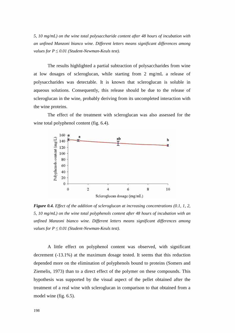
198
5, 10 mg/mL) on the wine total polysaccharide content after 48 hours of incubation with
an unfined Manzoni bianco wine. Different letters means significant differences among
values for P ≤ 0.01 (Student-Newman-Keuls test).
The resultshighlighteda partial subtraction of polysaccharidesfrom wine
at low dosagesof scleroglucan, while starting from 2 mg/mL a release of
polysaccharideswas detectable. It is known that scleroglucan is soluble in
aqueoussolutions. Consequently,this release should be due to the release of
scleroglucanin thewine, probablyderivingfrom its uncompleted interaction with
thewineproteins.
The effect of the treatmentwith scleroglucan was also assessedfor the
wine total polyphenol content(fig. 6.4).
Figure 0.4. Effect of the addition of scleroglucan at increasing concentrations (0.1, 1, 2,
5, 10 mg/mL) on the wine total polyphenols content after 48 hours of incubation with an
unfined Manzoni bianco wine. Different letters means significant differences among
values for P ≤ 0.01 (Student-Newman-Keuls test).
A little effect on polyphenol content was observed, with significant
decrement(-13.1%) at the maximum dosagetested. It seems that this reduction
depended moreon theeliminationof polyphenols boundto proteins (Somersand
Ziemelis,1973) thanto a direct effect of the polymer on thesecompounds.This
hypothesiswas supportedby the visual aspect of the pellet obtained after the
treatment of a real wine with scleroglucanin comparisonto that obtainedfrom a
model wine (fig. 6.5).

199
Figure 0.5. Left: Pellet of a model wine containing BSA treated with scleroglucan. Right:
pellet of a Manzoni bianco wine treated with scleroglucan.
Theyellow colourof thepelletachievedfrom thereal wine (fig. 6.5,right)
should indicatethepresenceof polyphenols, which areabsentin model wine(fig.
6.5, left).
To determine the kinetics of scleroglucan-protein interactions, the
polysaccharidewas addedat both four concentrations and incubation timesin a
model solution (tartaric acid 5g/L, ethanol12%, pH 3.2) in the presence of 300
mg/L of Bovine Serum Albumin (BSA). The data showed the reaching of
completeBSA elimination after only 10 minutes with 5 mg/mL of scleroglucan
(fig. 6.6), whereas the presence of little protein was observed at higher
concentrations.
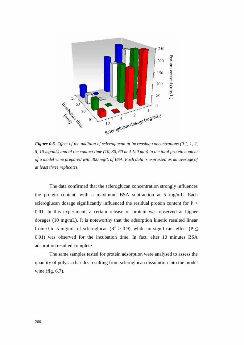
200
Figure 0.6. Effect of the addition of scleroglucan at increasing concentrations (0.1, 1, 2,
5, 10 mg/mL) and of the contact time (10, 30, 60 and 120 min) in the total protein content
of a model wine prepared with 300 mg/L of BSA. Each data is expressed as an average of
at least three replicates.
Thedataconfirmedthat thescleroglucanconcentration strongly influences
the protein content, with a maximum BSA subtraction at 5 mg/mL. Each
scleroglucandosagesignificantly influenced the residual protein contentfor P ≤
0.01. In this experiment,a certain release of protein was observed at higher
dosages(10 mg/mL). It is noteworthy that the adsorption kinetic resulted linear
from 0 to 5 mg/mL of scleroglucan(R2 > 0.9), while no significant effect (P ≤
0.01) was observedfor the incubation time. In fact, after 10 minutes BSA
adsorption resultedcomplete.
Thesamesamplestestedfor proteinadsorptionwereanalysed to assessthe
quantityof polysaccharidesresultingfrom scleroglucandissolution into themodel
wine (fig. 6.7).

201
Figure 0.7. Effect of the addition of scleroglucan at increasing concentrations (0.1, 1, 2,
5, 10 mg/mL) and of the contact time (10, 30, 60 and 120 min) on the total
polysaccharide content of a model wine prepared with 300 mg/L of BSA. Each data is
expressed as an average of at least three replicates.
The results confirmed what observed formerly, by highlighting a
polysaccharidereleasethat wassignificantly influencedby both the scleroglucan
dosage (P ≤ 0.01) and the incubation time (P ≤ 0.01). Thesedata confirmed
scleroglucansolubility in aqueoussolutions.
1.1.67 SOLVENT EFFECT ON SCLEROGLUCAN-PROTEIN INTERACTIONS
Thescleroglucanwasincubatedat severalconcentrationsfor 30’ in water,
tartaric acid (5 g/L, pH 3.2), ethanol (12%) and wine model solution, all
containing300mg/L of BSA (fig. 6.8).
In water and ethanol solutions the interaction with proteins was
significantly lower (P ≤ 0.01) than in acidic solutions. The highest protein
removal wasobservedin modelwine, suggesting a synergistic effect of pH and
ethanol, probablydue to a betterhydrationof the polysaccharidein the presence
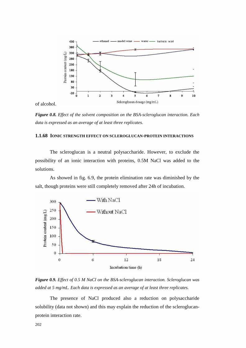
202
of alcohol.
Figure 0.8. Effect of the solvent composition on the BSA-scleroglucan interaction. Each
data is expressed as an average of at least three replicates.
1.1.68 IONIC STRENGTH EFFECT ON SCLEROGLUCAN-PROTEIN INTERACTIONS
The scleroglucan is a neutral polysaccharide. However, to exclude the
possibility of an ionic interaction with proteins, 0.5M NaCl was added to the
solutions.
As showedin fig. 6.9, the proteinelimination rate was diminished by the
salt, thoughproteinswerestill completelyremoved after 24hof incubation.
Figure 0.9. Effect of 0.5 M NaCl on the BSA-scleroglucan interaction. Scleroglucan was
added at 5 mg/mL. Each data is expressed as an average of at least three replicates.
The presence of NaCl produced also a reduction on polysaccharide
solubility (data not shown) andthis mayexplain thereduction of thescleroglucan-
protein interaction rate.

203
1.1.69 EFFECT OF THE HIGH MW ENDOGENOUS WINE COMPOUNDS ON
SCLEROGLUCAN-PROTEIN INTERACTIONS.
An ultrafiltered wine (3 kDa MWCO) was used to study the effects of
scleroglucanaddition (5 mg/mL,30 min incubation) on BSA (addedat 300mg/L)
removal in the presence of all the non-macromolecular componentsof wine,
includingpolyphenols (fig. 6.10).
Figure 0.10. Effect of the removal of macromolecular compounds (> 3 kDa) from wine
on the protein-scleroglucan interaction. Each data is expressed as an average of at least
three replicates.
The results showedthat the protein removalwascomplete only in model
wine. In the presence of endogenouswine compoundswith a MW lower than3
kDa the protein removal resultedincomplete, with a diminution of 65 % in the
proteincontent, which correspondsto the quantity observed for the whole wine.
Therefore,some endogenouscompoundswith low MW presentin theultrafiltered
wineimpair to someextentscleroglucan-protein interactions. Althoughthenature
of thesecompoundshasnot beenelucidated, an involvementof wine polyphenols
(fig. 6.4)on this resultcanbehypothesized.
1.1.70 EFFECT OF PROTEIN TYPE ON THE INTERACTIONS WITH SCLEROGLUCAN
Because in the presence of wine low MW compounds (may be

204
polyphenols) the protein reduction obtained by scleroglucan addition resulted
similar using botha modelprotein(BSA) and thetotal wine proteins(fig. 6.10),it
could be hypothesized that the compositional characteristic of the solution
influencesthe interactionmorethan theprotein nature. Therefore,to confirm this
idea, the effect of scleroglucanaddition was studied on solutions prepared with
variousproteins,differing in bothmolecular weightandglycosilation degree (fig.
6.11).
Figure 0.11. Interaction of different proteins with scleroglucan in a model wine solution.
Each data is expressed as an average of at least three replicates.
The protein removal effectof thescleroglucanappeared to be independent
from the protein molecularweight (seethe overlapping curvesfor BSA, 66 kDa,
and lysozyme, 14.4 kDa), whereasprotein glycosilation seemed to have some
effect on the interaction with the polysaccharide. It is to be noted that the
ovalbumin, a protein with a MW betweenBSA and lysozymebut with a high
glycosylation degree,neededthe lowest dose of scleroglucan to be removed,
indicating a strong affinity for the polysaccharide. This effect wasobservedalso
with yeastinvertase(about 50%of glycosylation), althoughthe interaction of the
scleroglucan with this protein was progressively reduced at scleroglucan
concentrationshigherthan≈ 1.5 mg/mL. Therefore,theeffect of theprotein type
on scleroglucan-proteininteractionswarrants furtherinvestigation.
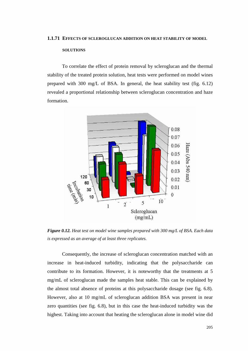
205
1.1.71 EFFECTS OF SCLEROGLUCAN ADDITION ON HEAT STABILITY OF MODEL
SOLUTIONS
To correlatetheeffectof proteinremoval by scleroglucanand the thermal
stability of thetreatedproteinsolution,heat testswereperformedon model wines
preparedwith 300 mg/L of BSA. In general, the heat stabilit y test (fig. 6.12)
revealeda proportional relationshipbetween scleroglucan concentration andhaze
formation.
Figure 0.12. Heat test on model wine samples prepared with 300 mg/L of BSA. Each data
is expressed as an average of at least three replicates.
Consequently, the increaseof scleroglucanconcentration matched with an
increase in heat-induced turbidity, indicating that the polysaccharide can
contribute to its formation. However, it is noteworthythat the treatments at 5
mg/mL of scleroglucanmadethe samples heatstable. This can be explained by
the almosttotal absenceof proteinsat this polysaccharide dosage(seefig. 6.8).
However, also at 10 mg/mL of scleroglucan addition BSA was present in near
zero quantities (see fig. 6.8), but in this casethe heat-induced turbidity was the
highest.Taking intoaccount thatheatingthescleroglucan alonein modelwinedid

206
not result in hazeformation(datanot shown),this occurrence maybeexplainedas
follows. The presenceof an excess of scleroglucan with respectto the protein in
solution, could result in the formationof heat-unstable complexes different from
thoseformedat thepolysaccharide/BSA ratio existing at 5 mg/mL of scleroglucan
addition, which wereinsteadheatstable.
Moreover, this experimentindicatedalsoan effect of the incubation time,
at least for the samplestreatedwith 5 mg/mL of scleroglucan (fig. 6.12). In
particular, after 10 minutes this sample was still instable (∆ Abs > 0.02) and
complete stabilisation was achievedonly after 30 min of treatment. This would
indicatethat, despitean almost immediate interaction between scleroglucan and
proteins(fig. 6.6), theformationof heatstable complexesneeded a longertime to
occur.
1.1.72 CONCLUSIONS
Scleroglucanis able to interact with proteins when dissolved in acidic
buffer, andthepresenceof little quantityof ethanolenhancesthis binding.
This interaction seemsto be of non-ionic nature because of the neutral
characteristicof the scleroglucan.The presence of polyphenolsreducesof only
30% the ability of the polysaccharideto removeproteins.These characteristics
would make the scleroglucana good tool for protein removal from wine.
However, this polysaccharideis solublein aqueoussolutionsandtendsto remain
in the wine after the treatment.Consequently, in order to usescleroglucan for
wine stabilisation, it seems necessary to overcome this inconvenientby using
chemicalmodifications to make the polysaccharide completely insoluble in the
wines to bestabilised.
Acknowledgements
The author would like to thank Simone Vincenzi, FrancescoFavaron, Silvana
Odorizzi, Loris Bil lo, Marco Lucchettaand Luca Sella of PaduaUniversity and
Tommasina Coviello of La Sapienza University for their support in the
experimentsconductionandfor thevaluablediscussionsaboutthis work.

207
REFERENCES
Aycock R. (1961). Symposium on Sclerotium rolfsii. Phytopathology 51, 107-
128.
Cabello-Pasini A., Victoria-CotaN., Macias-Carranza V., Hernandez-GaribayE.
and Muñiz-SalazarR. (2005). Clarification of wines using polysaccharides
extractedfrom seaweeds.Am. J. Enol. Vitic. 56(1),52-59.
Fariña J.I., Siñeriz F., Molina O.E. and Perotti N.I. (1998). High scleroglucan
production by Sclerotium rolfsii: influence of medium composition.
Biotechnology Letters 20,825-831.
Fariña J.I., Siñeriz F., Molina O.E. and Perotti N.I. (2001). Isolation and
physicochemical characterizationof soluble scleroglucan from Sclerotium
rolfsii. Rheological properties, molecular weight and conformational
characteristics.Carbohydrate Polymers 44,41–50.
GiananakisC., Bucheli C.S., Skene K.G.M., Robinson S.P. and Scott N.S.
(1998). Chitinase and beta-1,3-glucanase in grapevine leaves:A possible
defence againstpowderymildew infection. Austral J Grape Wine Res 4, 14-
22.
Hsu J.C.,HeatherbellD.A., FloresJ.H. andWatsonB.T. (1987).Heat-unstable
proteins in wine II. Characterizationand removal by ultrafiltration. Am. J.
Enol. Vitic. 38(1),17-22.
JayasankarS., Li Z. and Gray D.J. (2003). Constitutive expression of Vitis
vinifera thaumatin-like protein after in vitro selection and its role in
anthracnoseresistance.Functional Plant Biology 30,1105-1115.
KoenigR., StegemannH., FrancksenH. andPaulH.L. (1970).Protein subunitin
the potato virus X group. Determination of the molecular weight by
polyacrilamideelectrophoresis. Biochim. Biophys. Acta 297,184-189.
LaemmliU.K. (1970).Cleavageof structural proteinsduring theassembly of the
headof bacteriophageT4. Nature 227,680-685.
Milli es K. (1975). Protein stabilization of wine using silica sol/gelatin fining.
Mitteilungsblatt der GDCh-Fachgruppe. Lebensm. Gerich. Chemie. 29,50-53.
Monteiro S., BarakatM., Piçarra-PereiraM.A., Teixeira A.R and Ferreira R.B.
(2003). Osmotin and thaumatin from grape: A putative general defense
208
mechanismagainstpathogenicfungi. Phytopathol. 93,1505-1512.
Pachova V., FerrandoM., Guell C. andLòpezF. (2002).Protein adsorption onto
metal oxide materials in white wine model system. J. Food Sci. 67, 2118-
2121.
PowersJ.R.,Nagel C.W. andWeller K. (1988).ProteinRemovalfrom a Wineby
ImmobilizedGrapeProanthocyanidins.Am. J. Enol. Vitic. 39(2),117-120.
PretusH.A., Ensley H.E., McNameeR.B., JonesE.L., BrowderW. and Willi ams
D.L. (1991). Isolation,physicochemical characterization and preclinical
efficacy evaluation of soluble scleroglucan.Journal of Pharmacology and
Experimental Therapeutics 257,500-510.
Salzman R.A., TikhonovaI., Bordelon B.P., Hasegawa P.M. and Bressan R.A.
(1998). Coordinate accumulation of antifungal proteins and hexoses
constitutes a developmentally controlled defense responseduring fruit
ripeningin grape.Plant Physiology 117,465-472.
SarmentoM.R., Oliveira J.C., SlatnerM., Boulton R.B. (2000). Influence of
intrinsic factorson conventionalwine protein stability tests.Food Control 11,
423-432.
SegarraI., Lao C., López-TamamesE., andDe La Torre-BoronatM. C. (1995).
Spectrophotometric Methodsfor the Analysis of Polysaccharide Levels in
WinemakingProducts.Am. J. Enol. Vitic. 46(4),564-570.
Singleton V.L. and Rossi J.A. (1965). Colorimetry of total phenolics with
phosphomolybdic-phosphotungsticacid reagents. Am. J. Enol. Vitic. 16(3),
144-158.
SomersC. and ZiemelisG. (1973).Direct determination of wineproteins. Am. J.
Enol. Vitic. 24(2),47-50.
Tattersall D.B., PocockK.F., HayasakaY., AdamsK., van Heeswijck R., Waters
E.J.andHøj P.B.(2001).Pathogenesis relatedproteins– their accumulation in
grapes during berry growth and their involvement in white wine heat
instability. Current knowledge and future perspectives in relation to
winemaking practices. In: Molecular Biology and Biotechnology of the
Grapevine. Ed. K.A. Roubelakis-Angelakis (Kluwer Academic Publishers:
Dordrecht, Netherlands), pp.183-201.
Vincenzi S.,Mosconi S.,ZoccatelliG., Dalla PellegrinaC., Veneri G., Chignola
209
R., D.B. PeruffoA., Curioni A. andRizzi C. (2005a). Developmentof a new
procedure for proteinrecoveryandquantification in wine. Am. J. Enol. Vitic.
56(2), 182-187.
Vincenzi S., PolesaniM. and Curioni A. (2005b).Removal of Specific Protein
Components by Chitin EnhancesProtein Stabili ty in a White Wine. Am. J.
Enol. Vitic. 56(3),246-254.
Voilley A., Lamer C., Dubois P. and Feuillat M. (1990). Influence of
macromolecules and treatmentson the behaviour of aroma compoundsin a
model wine.J. Agric. Food Chem. 38(1), 248-251.
WaterhouseA.L. (2002)Winephenolics.Ann. N. Y. Acad. Sci. 957,21-36.
WatersE.J.,Wallace W. andWilliams P.J.(1991).Heathaze characteristics of
fractionatedwineproteins.Am. J. Enol. Vitic. 42(2),123-127.
Weetall H.H., Zelko J.T. and Bailey L.F. (1984). A new method for the
stabilizationof whitewine.Am. J. Enol. Vitic. 35(4), 212-215.

210

211
CHAPTER 7
Conclusions
This work aimedto improvethecomprehensionof severalgrapeandwine
proteinfeatures,suchastheeffectof fermentation on protein stabilit y, the role of
protein hydrophobicity on haze formation and on their reactivi ty with seed
tannins.Moreover,theinvestigationfor novelchromatographicmethodsfor grape
and wine proteins fractionationwas carried out. Besides,in order to solve the
problem of hazeformationin white wines,two alternative approachesto remove
proteinsresponsiblefor this drawbackwerestudied.
Firstly, this thesisaspiredto clear the fate of grapeproteins during the
alcoholic fermentation.In particular,it washighlighted thatthesolubleproteins of
theberry of a white grapevariety(cv. Manzoni bianco)vary during and after the
alcoholic fermentation in bothquantityandrelative proportions.Indeed,a protein
fraction containinga single20 kDa band (thaumatin-like protein), obtainedwith
Anion Exchange Chromatography, resulted thatwith the lowest heat-stabilit y and
with the largest quantitative increase throughout the fermentation process,
constituting a large proportion of the total wine proteins. These results are
consistentwith thestatementthat thaumatin-like proteinsarethemain responsible
for hazeformation in white wines,asestablishedin chapter 4. Moreover,it was
confirmed that fermenting yeasts releaseglycocompoundsin the wine (probably
mannoproteins),resulting in an improvedheat stabilit y of the total wine proteins,
despitetheincreasein therelativeproportionof their mostunstablecomponent.
In chapter3, a novel methodfor grapeand wine proteins purification is
proposed.The chromatographicapproachusedtakes advantage of the different
hydrophobicity of thegrapeandwineproteins,which werefractionatedaccording
to this characteristic. The main outcomewas that with Hydrophobic Interaction
Chromatography (HIC) it is possible to effectively fractionate grapejuice and
wine proteins, combining a good preparative separation with the attainment of
high proteinrecoveryandpurity for severalprotein fractions.In particular,a Vitis

212
vinifera thaumatin-like proteinwaspurified with high yields,from both Semillon
grape juice andwine, by exploiting its highesthydrophobicity level amonggrape
proteins. Additionally, this chromatographicsystem demonstrated thepotentiality
to purify more than one protein, especially in wine in which the number of
proteins is low. For this reason,this knowledge will be very helpful for the
purificationof othergrapeandwine proteins.Thereforetheapplication of HIC in
enological studies should contribute to improve the understanding on protein
characteristicsandfunctionality.
Besides, by using a multi-steppurification procedure, involving HIC and
RP-HPLC it was possible to separateby SDS-PAGE 26 different protein spots
that havebeenanalysed by LC-MS/MS. A large numberof grapeproteins were
thus identified by MS and database searching, with a majority of chitinases,
followed by thaumatin like proteins. However, not all the identified proteins
presentedthehighesthomologywith Vitis vinifera proteins,indicating that further
investigation are required to establish the complexity of the grape protein
composition.
From these sequenceresults it was possible to create a scheme
summarizingtheputativehydrophobicity of thegrapeproteins, that can beuseful
to interpret their physical-chemical behaviour. Moreover, the precise
identification of the single grapeproteins allowed to assign them to each RP-
HPLC peakand this outcomecould be useful to researchers for an unequivocal
and rapid identificationof grapeproteinson the basis of the RP-HPLC retention
time.
An exampleof how HIC could be utili zed in protein studies is given in
chapter 4, in which an extension of this techniquewas performed in order to
fractionate and study proteins from an unfined Manzoni bianco wine. By
combining a preparativeHIC fractionation with other analyti cal techniques (i.e.
SDS-PAGE, RP-HPLC, SE-HPLC), it was possibleto partially characterize the
fractionated Manzoni bianco wine proteins. A relationship between protein
hydrophobicity andhazepotentialafter the heat test wasfound.Furthermore,the
thaumatin-like protein content of each fraction resulted strictly related to its
hazing potential, confirming the assumptionthat thaumatin-like proteins are the
213
main heat-unstable wine proteins. Moreover, by means of Size Exclusion
Chromatography it was also confirmed the protective role of glycocompounds
against proteinsinsolubilisation.
From the study of wine protein fractions with different hydrophobicity
some conclusions are reached.In particular, it can be stated that the turbidity
formedafter tanninadditionto proteinfractionsin modelwine tendedto increase
according to their hydrophobicity, suggesting a relationship between this
parameterandwine proteinsreactivitywith tannins.Moreover, temperatureplays
a key role in the haze induction, since a dramatic increase of tannin-protein
reactivity occurs after heating. Heating of the protein-tannin mixture might
increasetheextentof interactionsby inducing protein denaturation andexposition
of a high numberof tannin binding site. In the protein native state, thesesites
should be buried in its core, which can be supposedto contain the most
hydrophobic portion of the molecule. Therefore a role of hydrophobic
interactions,which, on theotherhandarefavoured at high temperature, shouldbe
confirmed. However, pre-heating the proteins in model wine did not increase
turbidity developmentafter tanninaddition.A possibleexplanation of this fact is
that the changein protein conformation occurring during the heating in the
absenceof tannins is a reversible process.Therefore, protein reactivit y with
tannins is enhancedonly when protein is presentin its heat-denatured state.
Moreover,anenhancementof hydrophobic interactions,whosestrength increases
with the temperature, would be supposed to contribute to tannin-protein
interactionsat high temperature.Therefore,if the increasein turbidity formation
with the increasing of protein hydrophobicity, as demonstrated in chapter 4, is
considered,the idea that hydrophobicity plays a key role in determining wine
proteinhazingseemsto bejustified.
All these notionswill be useful to betterdefine the relationship between
the presencedifferent typesof proteinandtannins,in order to clear their role in
white wineshazing.
In the secondpart of this thesis, two approaches to overtake the still
unsolvedproblemof hazeformationin whitewinesareil lustrated.
Taking into account that treatmentswith proteolytic enzymes are widely
214
consideredthe ideal system to removethe heat-unstable proteins from mustsand
wines, a selection of fungal strains with the potential to produceproteasesacting
on wine proteins was conducted.Our resultsconfirmed that Botrytis cinerea is
able to secrete exocellular proteasesactive against grapeproteins. Besides,the
suitability of other filamentousphytopathogenic fungi in growing utili zing wine
proteins as the sole nitrogen source was demonstrated. In particular Sclerotina
minor and Sclerotium rolfsii seemedmoresuitable than B. cinerea in eliminating
grape and wine proteins responsible for haze formation in white wines,
highlighting thepotentiality to beusedasa source of proteolytic enzymes, active
at the wine pH, useful to this aim. Howeverthe attempts to purify the proteases
resultedin significant activity lossesand further investigations are required to
definitely assessthe possibility of a practical application of the studied fungi in
the actual conditions of winemaking.In particular, the study hasto continue by
trying to obtaina high productionof active proteasesby thefungi andby studying
purification techniquesmoreefficient and able to preservetheenzymatic activity.
In addition, single purified protein fractions of heat-unstable grape and wine
proteins(thaumatin-like proteinsandchitinases)shouldbetestedfor degradability
with thepurified fungal proteases.In this way, a better understanding of thewell
known PR-protein resistanceto proteolysiswil l beachieved,with theobtainment
of useful information for the selection of fungal proteasesto be used in
winemaking.
By studyingSclerotium rolfsii for its possible proteases production, it was
found that this fungusemitsin theculturemedium,in addition to a protease,also
a polysaccharide (scleroglucan)having the capabili ty to completely remove the
wine proteins, which were also seen to act as an enhancing factor for its
production.The probableexplanationfor this occurrencewas that a cooperative
action between scleroglucanand proteases exists as a protection mechanism
exerted by the fungus againstgrape and wine PR-proteins toxicity. From the
characterisationof the ability of the scleroglucan to interact with proteins it was
demonstrated that this polysaccharideis able to interact with both a standard
(BSA) andwine proteinswhendissolvedin acidic buffer, andthat thepresence of
little quantity of ethanolenhancesthis binding. This interaction seems not to

215
depend on the ionic strength becauseof the neutral characteristic of the
scleroglucan.Besides,thepresenceof thenon-macromolecular fraction (< 3 kDa)
of the wine reducesof only by 35% the abilit y of the polysaccharideto remove
proteins. These characteristicswould make the scleroglucan a good tool for
protein removal from wine. However, this polysaccharide showed a main
drawback,becauseit dissolvedin part in the solution. Consequently, in order to
use scleroglucan for wine stabilization, it wil l be essential to overcome this
inconvenientby usingchemicalmodificationsof its structure(i.e.by cross-linking
and/or immobilization) in order to makethe polysaccharide completely insoluble
in thewinesto bestabilized.
In conclusion, main resultsof this thesis work are the improvement in
grapeandwine proteinfractionationandthe study of alternative methodsfor the
removal of heat-unstable protein from wines. A demonstration of how the
combination of different chromatographic techniques can lead to a deeper
comprehension of the functional and biochemical characteristics of grape and
wine proteins is given. Thesedataallowedto clear somegrapeandwine protein
characteristic, such as their hydrophobicity, their peculiar heat-stabilit y and
reactivity with tanninswhich,at date,arenot fully understood.
It seemsthat this line of research can offer useful information to the
researchersin finding new strategies to understand andpreventprotein hazing in
white wine.