Steroid hydroxylations with Botryodiplodia malorum and Colletotrichum lini
6
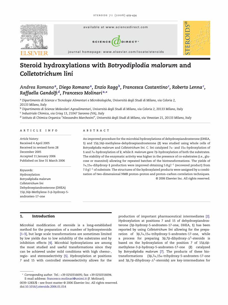
steroids 71 ( 2 0 0 6 ) 429–434 available at www.sciencedirect.com journal homepage: www.elsevier.com/locate/steroids Steroid hydroxylations with Botryodiplodia malorum and Colletotrichum lini Andrea Romano a , Diego Romano a , Enzio Ragg b , Francesca Costantino c , Roberto Lenna c , Raffaella Gandolfi d , Francesco Molinari a,∗ a Dipartimento di Scienze e Tecnologie Alimentari e Microbiologiche, Universit` a degli Studi di Milano, via Celoria 2, 20133 Milano, Italy b Dipartimento di Scienze Molecolari Agroalimentari, Universit ` a degli Studi di Milano, via Celoria 2, 20133 Milano, Italy c Industriale Chimica, via Grieg 13, 21047 Saronno (VA), Italy d Istituto di Chimica Organica “Alessandro Marchesini”, Universit` a degli Studi di Milano, via Venezian 21, 20133 Milano, Italy article info Article history: Received 4 April 2005 Received in revised form 28 December 2005 Accepted 11 January 2006 Published on line 31 March 2006 Keywords: Hydroxylation Botryodiplodia malorum Colletotrichum lini Dehydroepiandrosterone (DHEA) 15,16-Methylene-3--hydroxy-5- androsten-17-one abstract An improved procedure for the microbial hydroxylations of dehydroepiandrosterone (DHEA, 1) and 15,16-methylene-dehydroepiandrosterone (2) was studied using whole cells of Botryodiplodia malorum and Colletotrichum lini. C. lini catalyzed 7- and 15-hydroxylation of 1 and 7-hydroxylation of 2, while B. malorum gave 7-hydroxylation of both the substrates. The stability of the enzymatic activity was higher in the presence of co-substrates (i.e., glu- cose or mannitol) allowing for repeated batches of the biotransformations. The yields of 7,15-dihydroxy-1 production were improved obtaining 5.8 g l −1 (recovered product) from 7.0 g l −1 of substrate. The structures of the hydroxylated products were assigned by a combi- nation of two-dimensional NMR proton–proton and proton–carbon correlation techniques. © 2006 Elsevier Inc. All rights reserved. 1. Introduction Microbial modification of steroids is a long-established method for the preparation of a number of hydroxysteroids [1–3], but large scale transformations are sometimes limited by low yields due to low solubility of the substrates and by inhibition effects [4]. Microbial hydroxylations are among the most studied and useful transformations since they can be achieved under mild conditions with high chemo-, regio- and stereoselectivity [5]. Hydroxylation at positions 7 and 15 with controlled stereoselectivity allows for the ∗ Corresponding author. Tel.: +39 0250316695; fax: +39 0250316694. E-mail address: [email protected] (F. Molinari). production of important pharmaceutical intermediates [2]. Hydroxylation at positions 7 and 15 of dehydroepiandros- terone (3-hydroxy-5-androsten-17-one, DHEA, 1) has been reported by using Colletotrichum lini allowing for the prepa- ration of 3,7,15-trihydroxy-5-androsten-17-one, while a process for preparing 3,7-dihydroxy- 5 -steroids is based on the hydroxylation of the position 7 of 15,6- methylene-3--hydroxy-5-androsten-17-one (2) catalyzed by Botryodiplodia malorum [7]. The products of these bio- transformations (3,7,15-trihydroxy-5-androsten-17-one and 3,7-dihydroxy- 5 -steroids) are key-intermediates for 0039-128X/$ – see front matter © 2006 Elsevier Inc. All rights reserved. doi:10.1016/j.steroids.2006.01.014
-
Upload
andrea-romano -
Category
Documents
-
view
215 -
download
0
Transcript of Steroid hydroxylations with Botryodiplodia malorum and Colletotrichum lini
s t e r o i d s 7 1 ( 2 0 0 6 ) 429–434
avai lab le at www.sc iencedi rec t .com
journa l homepage: www.e lsev ier .com/ locate /s tero ids
Steroid hydroxylations with Botryodiplodia malorum andColletotrichum lini
Andrea Romanoa, Diego Romanoa, Enzio Raggb, Francesca Costantinoc, Roberto Lennac,Raffaella Gandolfid, Francesco Molinaria,∗
a Dipartimento di Scienze e Tecnologie Alimentari e Microbiologiche, Universita degli Studi di Milano, via Celoria 2,20133 Milano, Italyb Dipartimento di Scienze Molecolari Agroalimentari, Universita degli Studi di Milano, via Celoria 2, 20133 Milano, Italyc Industriale Chimica, via Grieg 13, 21047 Saronno (VA), Italyd Istituto di Chimica Organica “Alessandro Marchesini”, Universita degli Studi di Milano, via Venezian 21, 20133 Milano, Italy
a r t i c l e i n f o a b s t r a c t
Article history:
Received 4 April 2005
Received in revised form 28
December 2005
Accepted 11 January 2006
Published on line 31 March 2006
Keywords:
Hydroxylation
Botryodiplodia malorum
Colletotrichum lini
Dehydroepiandrosterone (DHEA)
15�,16�-Methylene-3-�-hydroxy-5-
androsten-17-one
An improved procedure for the microbial hydroxylations of dehydroepiandrosterone (DHEA,
1) and 15�,16�-methylene-dehydroepiandrosterone (2) was studied using whole cells of
Botryodiplodia malorum and Colletotrichum lini. C. lini catalyzed 7�- and 15�-hydroxylation of
1 and 7�-hydroxylation of 2, while B. malorum gave 7�-hydroxylation of both the substrates.
The stability of the enzymatic activity was higher in the presence of co-substrates (i.e., glu-
cose or mannitol) allowing for repeated batches of the biotransformations. The yields of
7�,15�-dihydroxy-1 production were improved obtaining 5.8 g l−1 (recovered product) from
7.0 g l−1 of substrate. The structures of the hydroxylated products were assigned by a combi-
nation of two-dimensional NMR proton–proton and proton–carbon correlation techniques.
© 2006 Elsevier Inc. All rights reserved.
1. Introduction
Microbial modification of steroids is a long-establishedmethod for the preparation of a number of hydroxysteroids[1–3], but large scale transformations are sometimes limitedby low yields due to low solubility of the substrates and byinhibition effects [4]. Microbial hydroxylations are amongthe most studied and useful transformations since theycan be achieved under mild conditions with high chemo-,regio- and stereoselectivity [5]. Hydroxylation at positions7 and 15 with controlled stereoselectivity allows for the
∗ Corresponding author. Tel.: +39 0250316695; fax: +39 0250316694.E-mail address: [email protected] (F. Molinari).
production of important pharmaceutical intermediates [2].Hydroxylation at positions 7 and 15 of dehydroepiandros-terone (3�-hydroxy-5-androsten-17-one, DHEA, 1) has beenreported by using Colletotrichum lini allowing for the prepa-ration of 3�,7�,15�-trihydroxy-5-androsten-17-one, whilea process for preparing 3�,7�-dihydroxy-�5-steroids isbased on the hydroxylation of the position 7 of 15�,6�-methylene-3-�-hydroxy-5-androsten-17-one (2) catalyzedby Botryodiplodia malorum [7]. The products of these bio-transformations (3�,7�,15�-trihydroxy-5-androsten-17-oneand 3�,7�-dihydroxy-�5-steroids) are key-intermediates for
0039-128X/$ – see front matter © 2006 Elsevier Inc. All rights reserved.doi:10.1016/j.steroids.2006.01.014
430 s t e r o i d s 7 1 ( 2 0 0 6 ) 429–434
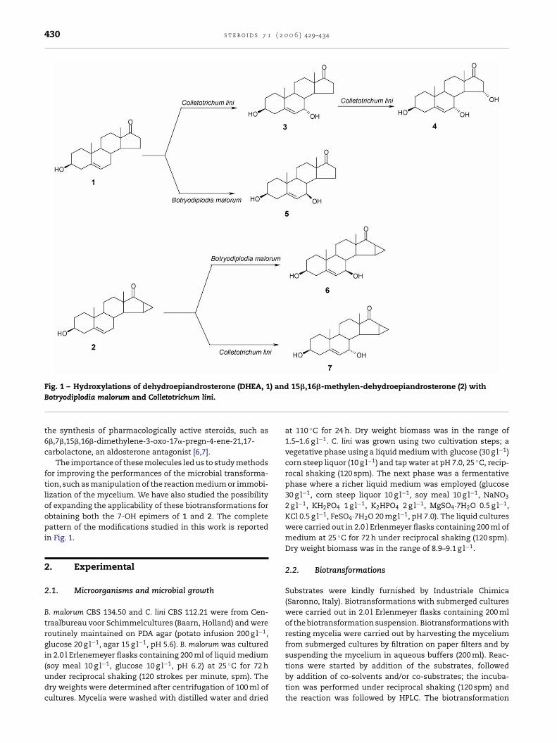
Fig. 1 – Hydroxylations of dehydroepiandrosterone (DHEA, 1) and 15�,16�-methylen-dehydroepiandrosterone (2) withBotryodiplodia malorum and Colletotrichum lini.
the synthesis of pharmacologically active steroids, such as6�,7�,15�,16�-dimethylene-3-oxo-17�-pregn-4-ene-21,17-carbolactone, an aldosterone antagonist [6,7].
The importance of these molecules led us to study methodsfor improving the performances of the microbial transforma-tion, such as manipulation of the reaction medium or immobi-lization of the mycelium. We have also studied the possibilityof expanding the applicability of these biotransformations forobtaining both the 7-OH epimers of 1 and 2. The completepattern of the modifications studied in this work is reportedin Fig. 1.
2. Experimental
2.1. Microorganisms and microbial growth
B. malorum CBS 134.50 and C. lini CBS 112.21 were from Cen-traalbureau voor Schimmelcultures (Baarn, Holland) and wereroutinely maintained on PDA agar (potato infusion 200 g l−1,glucose 20 g l−1, agar 15 g l−1, pH 5.6). B. malorum was culturedin 2.0 l Erlenemeyer flasks containing 200 ml of liquid medium(soy meal 10 g l−1, glucose 10 g l−1, pH 6.2) at 25 ◦C for 72 hunder reciprocal shaking (120 strokes per minute, spm). Thedry weights were determined after centrifugation of 100 ml ofcultures. Mycelia were washed with distilled water and dried
at 110 ◦C for 24 h. Dry weight biomass was in the range of1.5–1.6 g l−1. C. lini was grown using two cultivation steps; avegetative phase using a liquid medium with glucose (30 g l−1)corn steep liquor (10 g l−1) and tap water at pH 7.0, 25 ◦C, recip-rocal shaking (120 spm). The next phase was a fermentativephase where a richer liquid medium was employed (glucose30 g l−1, corn steep liquor 10 g l−1, soy meal 10 g l−1, NaNO3
2 g l−1, KH2PO4 1 g l−1, K2HPO4 2 g l−1, MgSO4·7H2O 0.5 g l−1,KCl 0.5 g l−1, FeSO4·7H2O 20 mg l−1, pH 7.0). The liquid cultureswere carried out in 2.0 l Erlenmeyer flasks containing 200 ml ofmedium at 25 ◦C for 72 h under reciprocal shaking (120 spm).Dry weight biomass was in the range of 8.9–9.1 g l−1.
2.2. Biotransformations
Substrates were kindly furnished by Industriale Chimica(Saronno, Italy). Biotransformations with submerged cultureswere carried out in 2.0 l Erlenmeyer flasks containing 200 mlof the biotransformation suspension. Biotransformations withresting mycelia were carried out by harvesting the myceliumfrom submerged cultures by filtration on paper filters and bysuspending the mycelium in aqueous buffers (200 ml). Reac-tions were started by addition of the substrates, followedby addition of co-solvents and/or co-substrates; the incuba-tion was performed under reciprocal shaking (120 spm) andthe reaction was followed by HPLC. The biotransformation
s t e r o i d s 7 1 ( 2 0 0 6 ) 429–434 431
medium was then filtered and extracted with methyl isobutylketone. The organic extracts were dried over Na2SO4 and thesolvent was removed; the crude product was purified by flashchromatography (hexane/acetone, 1/1, vanillin stained).
2.3. Immobilization
The mycelia obtained by filtration of the culture broths (200 ml)were suspended in 60 ml of sodium alginate (Sigma Chem-icals) (3.0%, w/w) dissolved in distilled water. The resultingsuspension was degassed and extruded through a thin injec-tion needle into 50 ml of an iced calcium chloride solution(0.1 M). The bead size had an average diameter in the rangeof 2.5–2.8 mm. After hardening for 3 h at 4 ◦C, the beads werefiltered under vacuum and used for the biotransformation orstored at 4 ◦C in fresh calcium chloride (0.01 M) solution. Theamount of mycelium in the beads was estimated by dissolv-ing 10 g of beads in 2% sodium poliphosphate solution; thesuspension was then filtered through a pre-dried weighted fil-ter paper (Whatman No. 1) and dried at 100 ◦C to a constantweight.
2.4. Repeated use of mycelium
Mycelium was reused in successive biotransformation cycles.At the end of 24 h, the mycelium was paper filtered and resus-pended in fresh reaction mixture, and substrate was added.T8
2
Sec6tto5
tRfpp(PCcs(lcsuuwpt
polynomial. Chemical shifts were measured in ı (ppm), usingthe acetone residual signal as a reference and setting themethyl proton and carbon resonances at 2.1 and 29.8 ppm,respectively.
The [˛]D were measured in methanol (c 1.0) using a Perkin-Elmer Polarimeter (Model 343 Plus). Melting points were mea-sured by differential scanning calorimetry analysis (Perkin-Elmer DSC 6).
3. Results
3.1. Biotransformations with C. lini
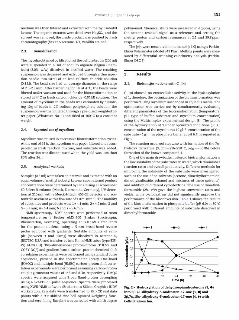
C. lini showed no extracellular activity in the hydroxylationof 1; therefore, the optimization of the biotransformation wasperformed using mycelium suspended in aqueous media. Theoptimization was carried out by simultaneously evaluatingdifferent parameters of the biotransformation (temperature,pH, type of buffer, substrate and mycelium concentration)using the Multisimplex experimental design [8]. The profileof the hydroxylation of 1 under optimized conditions (30 ◦C,concentration of the mycelium = 10 g l−1, concentration of thesubstrate = 1 g l−1 in phosphate buffer at pH 6.4) is reported inFig. 2.
The reaction occurred stepwise with formation of the 7�-hydroxy derivative (3, mp = 216–218 ◦C, [˛]D = −56.86) before
he reaction was discontinued when the yield was less than0% after 24 h.
.5. Analytical methods
amples (0.5 ml) were taken at intervals and extracted with anqual volume of methyl isobutyl ketone; substrate and productoncentrations were determined by HPLC using a Lichrospher0 Select B column (Merck, Darmstadt, Germany), UV detec-ion at 210 nm with a Merck-Hitachi 655-22 detector and ace-onitrile as eluent with a flow rate of 1.0 ml min−1. The mobilityf substrates and products was: 1 = 4.1 min; 2 = 4.2 min; 3 and= 5.7 min; 4 = 6.4 min; 6 and 7 = 5.6 min.
NMR spectroscopy. NMR spectra were performed at roomemperature on a Bruker AMX-600 (Bruker Spectrospin,heinstetten, Germany), operating at 600.1 MHz frequency
or the proton nucleus, using a 5 mm broad-band reverserobe equipped with gradients. Suitable amounts of sam-le (between 3 and 10 mg) were dissolved in acetone-d6
ISOTEC, USA) and transferred into 5 mm NMR tubes (type 535-P, ALDRICH). Two-dimensional proton–proton (TOCSY andOSY-DQF) and gradient based carbon–proton chemical shiftorrelation experiments were performed using standard pulseequences, present in the spectrometer library. One-bondHMQC) and multiple-bond (HMBC) carbon–proton shift corre-ation experiments were performed assuming carbon–protonoupling constant values of 145 and 8 Hz, respectively. HMQCpectra were acquired with Broad Band-proton decouplingsing a WALTZ-16 pulse sequence. Spectra were processedsing XWINNMR software (Bruker) on a Silicon Graphics INDYorkstation. Raw data were transformed to 2K × 2K real dataoints with a 90◦ shifted-sine bell squared weighting func-ion and zero-filling. Baseline was corrected with a fifth degree
formation of the known compound 4.One of the main drawbacks in steroid biotransformation is
the low solubility of the substrates in water, which diminishesreaction rates and overall productivity. Different methods forimproving the solubility of the substrate were investigated,such as the use of co-solvents (acetone, dimethylformamide,dimethylsulfoxide, ethanol and mixtures of these solvents),and addition of different cyclodextrins. The use of dimethyl-formamide (2%, v/v) gave the highest conversion rates andyields, while cyclodextrins did not significantly improve theperformance of the bioconversion. Table 1 shows the resultsof the biotransformation in phosphate buffer (pH 6.0) at 30 ◦Cperformed with different amounts of substrate dissolved indimethylformamide.
Fig. 2 – Hydroxylation of dehydroepiandrosterone (1, �)into 3�,7�-dihydroxy-5-androsten-17-one (3, �) and3�,7�,15�-trihydroxy-5-androsten-17-one (4, �) withColletotrichum lini.
432 s t e r o i d s 7 1 ( 2 0 0 6 ) 429–434
Table 1 – Production of3�,7�-dihydroxy-5-androsten-17-one (3) and3�,7�,15�-trihydroxy-5-androsten-17-one (4) bybiotransformation using different concentrations ofdehydroepiandrosterone (DHEA, 1) with Colletotrichumlini
Time (h) 1 g l−1 2.5 g l−1 5 g l−1
3 4 3 4 3 4
4 0.55 0 0.52 0 0 024 0 0.98 1.05 0.97 0.50 048 0 1.02 0 1.94 1.50 072 0 0.85 0 1.80 2.75 0
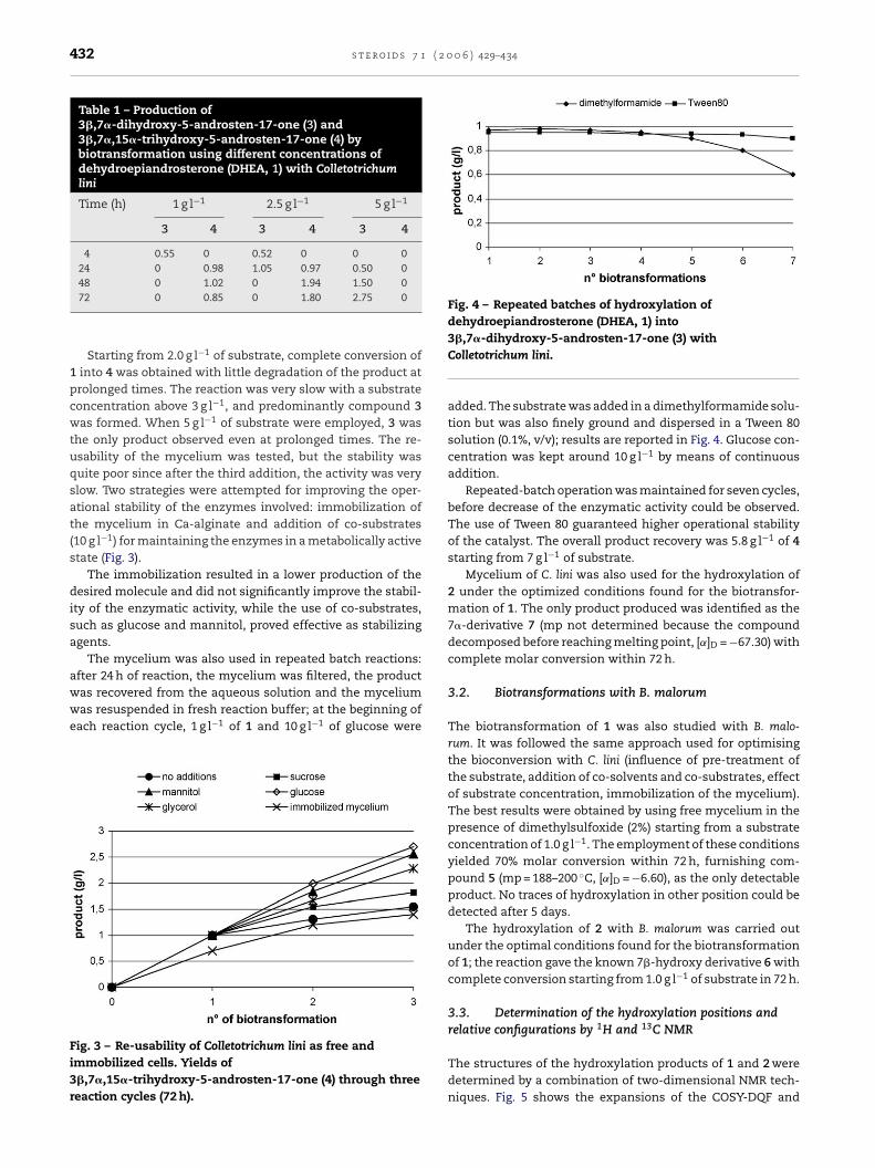
Starting from 2.0 g l−1 of substrate, complete conversion of1 into 4 was obtained with little degradation of the product atprolonged times. The reaction was very slow with a substrateconcentration above 3 g l−1, and predominantly compound 3was formed. When 5 g l−1 of substrate were employed, 3 wasthe only product observed even at prolonged times. The re-usability of the mycelium was tested, but the stability wasquite poor since after the third addition, the activity was veryslow. Two strategies were attempted for improving the oper-ational stability of the enzymes involved: immobilization ofthe mycelium in Ca-alginate and addition of co-substrates(10 g l−1) for maintaining the enzymes in a metabolically activestate (Fig. 3).
The immobilization resulted in a lower production of thedesired molecule and did not significantly improve the stabil-ity of the enzymatic activity, while the use of co-substrates,such as glucose and mannitol, proved effective as stabilizingagents.
The mycelium was also used in repeated batch reactions:after 24 h of reaction, the mycelium was filtered, the productwas recovered from the aqueous solution and the myceliumwas resuspended in fresh reaction buffer; at the beginning ofeach reaction cycle, 1 g l−1 of 1 and 10 g l−1 of glucose were
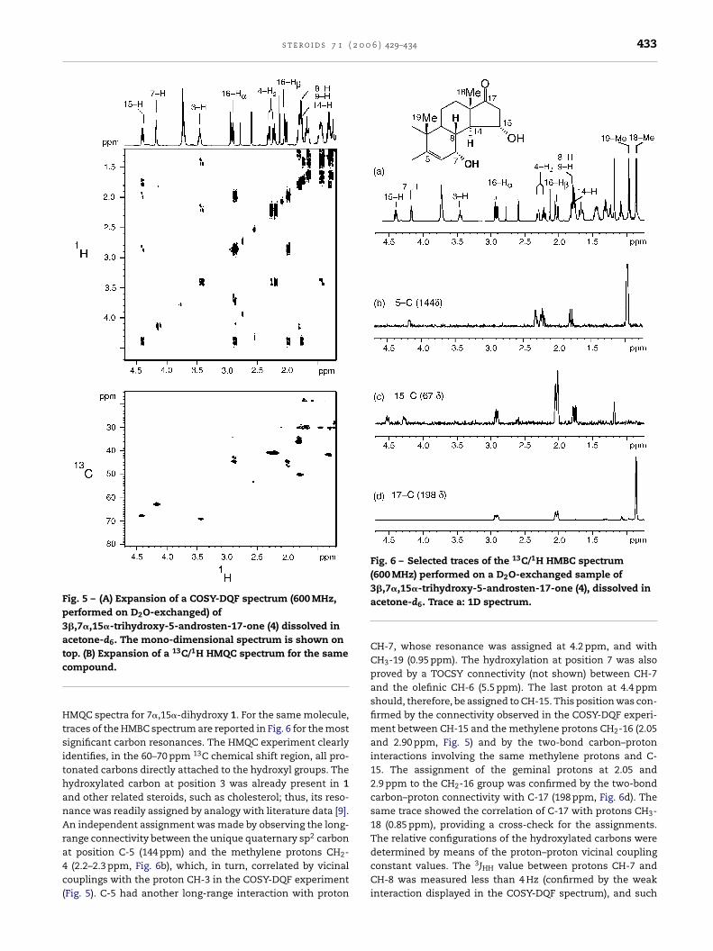
Fig. 4 – Repeated batches of hydroxylation ofdehydroepiandrosterone (DHEA, 1) into3�,7�-dihydroxy-5-androsten-17-one (3) withColletotrichum lini.
added. The substrate was added in a dimethylformamide solu-tion but was also finely ground and dispersed in a Tween 80solution (0.1%, v/v); results are reported in Fig. 4. Glucose con-centration was kept around 10 g l−1 by means of continuousaddition.
Repeated-batch operation was maintained for seven cycles,before decrease of the enzymatic activity could be observed.The use of Tween 80 guaranteed higher operational stabilityof the catalyst. The overall product recovery was 5.8 g l−1 of 4starting from 7 g l−1 of substrate.
Mycelium of C. lini was also used for the hydroxylation of2 under the optimized conditions found for the biotransfor-mation of 1. The only product produced was identified as the7�-derivative 7 (mp not determined because the compounddecomposed before reaching melting point, [˛]D = −67.30) withcomplete molar conversion within 72 h.
3.2. Biotransformations with B. malorum
The biotransformation of 1 was also studied with B. malo-rum. It was followed the same approach used for optimisingthe bioconversion with C. lini (influence of pre-treatment ofthe substrate, addition of co-solvents and co-substrates, effectof substrate concentration, immobilization of the mycelium).The best results were obtained by using free mycelium in thepresence of dimethylsulfoxide (2%) starting from a substrateconcentration of 1.0 g l−1. The employment of these conditions
Fig. 3 – Re-usability of Colletotrichum lini as free andimmobilized cells. Yields of3�,7�,15�-trihydroxy-5-androsten-17-one (4) through threereaction cycles (72 h).
yielded 70% molar conversion within 72 h, furnishing com-pound 5 (mp = 188–200 ◦C, [˛]D = −6.60), as the only detectableproduct. No traces of hydroxylation in other position could bedetected after 5 days.
The hydroxylation of 2 with B. malorum was carried outunder the optimal conditions found for the biotransformationof 1; the reaction gave the known 7�-hydroxy derivative 6 withcomplete conversion starting from 1.0 g l−1 of substrate in 72 h.
3.3. Determination of the hydroxylation positions andrelative configurations by 1H and 13C NMR
The structures of the hydroxylation products of 1 and 2 weredetermined by a combination of two-dimensional NMR tech-niques. Fig. 5 shows the expansions of the COSY-DQF and
s t e r o i d s 7 1 ( 2 0 0 6 ) 429–434 433
Fig. 5 – (A) Expansion of a COSY-DQF spectrum (600 MHz,performed on D2O-exchanged) of3�,7�,15�-trihydroxy-5-androsten-17-one (4) dissolved inacetone-d6. The mono-dimensional spectrum is shown ontop. (B) Expansion of a 13C/1H HMQC spectrum for the samecompound.
HMQC spectra for 7�,15�-dihydroxy 1. For the same molecule,traces of the HMBC spectrum are reported in Fig. 6 for the mostsignificant carbon resonances. The HMQC experiment clearlyidentifies, in the 60–70 ppm 13C chemical shift region, all pro-tonated carbons directly attached to the hydroxyl groups. Thehydroxylated carbon at position 3 was already present in 1and other related steroids, such as cholesterol; thus, its reso-nance was readily assigned by analogy with literature data [9].An independent assignment was made by observing the long-range connectivity between the unique quaternary sp2 carbonat position C-5 (144 ppm) and the methylene protons CH2-4 (2.2–2.3 ppm, Fig. 6b), which, in turn, correlated by vicinalcouplings with the proton CH-3 in the COSY-DQF experiment(Fig. 5). C-5 had another long-range interaction with proton
Fig. 6 – Selected traces of the 13C/1H HMBC spectrum(600 MHz) performed on a D2O-exchanged sample of3�,7�,15�-trihydroxy-5-androsten-17-one (4), dissolved inacetone-d6. Trace a: 1D spectrum.
CH-7, whose resonance was assigned at 4.2 ppm, and withCH3-19 (0.95 ppm). The hydroxylation at position 7 was alsoproved by a TOCSY connectivity (not shown) between CH-7and the olefinic CH-6 (5.5 ppm). The last proton at 4.4 ppmshould, therefore, be assigned to CH-15. This position was con-firmed by the connectivity observed in the COSY-DQF experi-ment between CH-15 and the methylene protons CH2-16 (2.05and 2.90 ppm, Fig. 5) and by the two-bond carbon–protoninteractions involving the same methylene protons and C-15. The assignment of the geminal protons at 2.05 and2.9 ppm to the CH2-16 group was confirmed by the two-bondcarbon–proton connectivity with C-17 (198 ppm, Fig. 6d). Thesame trace showed the correlation of C-17 with protons CH3-18 (0.85 ppm), providing a cross-check for the assignments.The relative configurations of the hydroxylated carbons weredetermined by means of the proton–proton vicinal couplingconstant values. The 3JHH value between protons CH-7 andCH-8 was measured less than 4 Hz (confirmed by the weakinteraction displayed in the COSY-DQF spectrum), and such
434 s t e r o i d s 7 1 ( 2 0 0 6 ) 429–434
a low value indicated that the relative orientation must havebeen sin. Since CH-8 was known to be �, CH-7 must also havebeen � and, consequently, OH-7 was �. In the same compound,OH-15 was found to be � because of the large vicinal cou-pling constant value with H-14 (3J14,15 = 9.7 Hz, 3J15,16� = 7.9 Hz,3J14,15� = 6.2 Hz). Such values were measured from the analysisof the H-15 multiplet in a resolution-enhanced 1D spectrumand the corresponding COSY-DQF cross-peak.
With the above described reasoning, the relative configu-rations of the hydroxylation sites in the isolated derivatives of2 were similarly determined.
4. Conclusions
The hydroxylation of dehydroepiandrosterone 1 and 15�,16�-methylene-dehydroepiandrosterone 2 by microbial means hasbeen optimized in terms of yields and selectivity. A suitedcombination of substrates and whole-cell biocatalysts allowedfor the production of different hydroxylated compounds (3–7),as optically pure epimers. The structures and relative con-figurations of the hydroxylated products were assigned bya combination of two-dimensional NMR proton–proton andproton–carbon correlation techniques.
The production of 3�,7�,15�-trihydroxy-5-androsten-17-one was performed under repeated batch mode allowing for
the recovery of 5.7–5.8 g l−1 of product with 75–80% molarconversion.
r e f e r e n c e s
[1] Murray HC, Peterson DH. Upjohn Co., USA. Oxygenation ofsteroids by Mucorales fungi. U.S. Patent 2602769; 1952.
[2] Mahato B, Mazumder I. Current trends in microbial steroidtransformations. Phytochemistry 1989;34:883–98.
[3] Fernandes P, Cruz A, Angelova B, Pinheiro HM, Cabral JMS.Microbial conversion of steroid compounds: recentdevelopments. Enzyme Microb Technol 2003;32:688–705.
[4] Mahato BM, Garai S. Advances in microbial steroidbiotransformation. Steroids 1997;62:332–45.
[5] Holland HL. Recent advances in applied and mechanisticaspects of the enzymatic hydroxylation of steroids bywhole-cell biocatalyst. Steroids 1999;64:178–86.
[6] Petzoldt K, Laurent H, Wiechert R. Schering, Germany.3�,7�,15�-Trihydroxy-5-androsten-17-one, its 3,15-dipivalate,and their preparation. U.S. Patent 4435327; 1984.
[7] Petzoldt K, Wiechert R, Laurent H, Nicklasch K, Bittler D.Schering, Germany. Process for preparing3�,7�-dihydroxy-�5-steroids. U.S. Patent 4416985, in press.
[8] Walters FH, Parker LR, Morgan SL, Deming SN. Sequentialsimplex optimization. Boca Raton: CRC Press; 1991.
[9] Breitmaier E, Voelter W. 13C NMR spectroscopy. Weinheim,New York: Verlag Chemie; 1978.