PROHIBITED COPY READ-ONLY...2018/10/06 · patients. These benefits, added to the reduction of...
9
206 Ann. Ital. Chir., 88, 3, 2017 Complications of laparoscopic gastric banding: detection and treatment Ann. Ital. Chir., 2017 88, 3: 206-214 pii: S0003469X17026835 Pervenuto in Redazione Diceembre 2016. Accettato per la pubblicazione Marzo 2017 Correspondence to: Alberto Sartori, MD, Department of Surgery, Montebelluna Treviso Hospital Italy (e-mail: [email protected]) Alberto Sartori*, Maurizio De Luca*, Nicola Clemente*, Cesare Lunardi,* Gianni Segato**, Natale Pellicanò* *Department of General Surgery, “San Valentino” Hospital, Montebelluna, Treviso, Italy **Department of Surgery, Regional Hospital of Vicenza, Italy Complications of laparoscopic gastric banding: detection and treatment INTRODUCTION: Laparoscopic adjustable gastric banding (LAGB) is acknownledged as a popular and effective surgical option in the management of obesity and related metabolic diseases. This procedure is a remarkably safe operation from both a general surgical and bariatric perspective. It facilitates brief hospitalization and can be performed by single inci- sion. METHODS: We analyzed the most common LAGB complications as intraoperative and postoperative gastric perforation, stomach slippage/dilatation, port/tubing complications and intragastric band migration which occurred in our long decades clinical experience. Detection, treatment and rate of presentation of each complication was evaluated. RESULTS: LAGB showed good long term results in terms of weight loss and resolution of obesity related diseases. Moreover, mortality due to obesity and related diseases appeared significantly lower in LAGB patients than in medically treated patients. CONCLUSION: Gastric Banding has a very low rate of early and late complications; these are also less severe when com- pared to more invasive procedures and are likely to be managed with mini-invasive techniques. In any case referral to a bariatric surgeon is deemed appropriate. KEY WORDS: Complication, Laparoscopic gastric banding, Morbid obesity results have been proved to be widely unsatisfactory. On the other hand, the surgical treatment of obesity is effec- tive in terms of weight reduction both in the short and long-term; it carries a dramatic improvement of co-mor- bidities leading to an increase in life-expectancy of obese patients. These benefits, added to the reduction of mor- tality and morbidity achieved through modern laparo- scopic surgery, explain why obesity surgery is so whis- pread. Bariatric surgery procedures are divided into restrictive (gastric banding, sleeve gastrectomy), malabsorptive (bilio-pancreatic diversion) and mixed (gastric bypass, mini gastric bypass). Actually this classification is con- sidered to be less strict because of different neuro-hor- monal mechanisms of action coming up. In each different geographical area, one tecnique is pre- ferred over another. For example, while gastric bypass is the preferred operation in the US, sleeve gastrectomy is Introduction The incidence of obesity is increasing worldwide, due to changes in lifestyle and diet. The non-operative treat- ment of obesity includes diet, physical exercise and some- times pharmacological treatment; nevertheless long-term READ-ONLY COPY PRINTING PROHIBITED
Transcript of PROHIBITED COPY READ-ONLY...2018/10/06 · patients. These benefits, added to the reduction of...
206 Ann. Ital. Chir., 88, 3, 2017
Complications of laparoscopic gastric banding: detection and treatment Ann. Ital. Chir., 2017 88, 3: 206-214
pii: S0003469X17026835
Pervenuto in Redazione Diceembre 2016. Accettato per la pubblicazioneMarzo 2017Correspondence to: Alberto Sartori, MD, Department of Surgery,Montebelluna Treviso Hospital Italy (e-mail: [email protected])
Alberto Sartori*, Maurizio De Luca*, Nicola Clemente*, Cesare Lunardi,* Gianni Segato**,Natale Pellicanò*
*Department of General Surgery, “San Valentino” Hospital, Montebelluna, Treviso, Italy**Department of Surgery, Regional Hospital of Vicenza, Italy
Complications of laparoscopic gastric banding: detection and treatment
INTRODUCTION: Laparoscopic adjustable gastric banding (LAGB) is acknownledged as a popular and effective surgicaloption in the management of obesity and related metabolic diseases. This procedure is a remarkably safe operation fromboth a general surgical and bariatric perspective. It facilitates brief hospitalization and can be performed by single inci-sion.METHODS: We analyzed the most common LAGB complications as intraoperative and postoperative gastric perforation,stomach slippage/dilatation, port/tubing complications and intragastric band migration which occurred in our long decadesclinical experience. Detection, treatment and rate of presentation of each complication was evaluated.RESULTS: LAGB showed good long term results in terms of weight loss and resolution of obesity related diseases. Moreover,mortality due to obesity and related diseases appeared significantly lower in LAGB patients than in medically treatedpatients. CONCLUSION: Gastric Banding has a very low rate of early and late complications; these are also less severe when com-pared to more invasive procedures and are likely to be managed with mini-invasive techniques. In any case referral toa bariatric surgeon is deemed appropriate.
KEY WORDS: Complication, Laparoscopic gastric banding, Morbid obesity
results have been proved to be widely unsatisfactory. Onthe other hand, the surgical treatment of obesity is effec-tive in terms of weight reduction both in the short andlong-term; it carries a dramatic improvement of co-mor-bidities leading to an increase in life-expectancy of obesepatients. These benefits, added to the reduction of mor-tality and morbidity achieved through modern laparo-scopic surgery, explain why obesity surgery is so whis-pread.Bariatric surgery procedures are divided into restrictive(gastric banding, sleeve gastrectomy), malabsorptive(bilio-pancreatic diversion) and mixed (gastric bypass,mini gastric bypass). Actually this classification is con-sidered to be less strict because of different neuro-hor-monal mechanisms of action coming up.In each different geographical area, one tecnique is pre-ferred over another. For example, while gastric bypass isthe preferred operation in the US, sleeve gastrectomy is
Introduction
The incidence of obesity is increasing worldwide, due tochanges in lifestyle and diet. The non-operative treat-ment of obesity includes diet, physical exercise and some-times pharmacological treatment; nevertheless long-term
READ-ONLY
COPY
PRINTIN
G PROHIB
ITED
more favoured in Europe. Many studies have analyzedand compared results and complications of various oper-ations, but it is not yet possible to determine which isthe best. LAGB is an effective and safe operation with decreasedperi-operative morbidity and mortality. However, it isessential to be aware of the specific complications of thistechnique in order to prevent, identify and treat them.In our Institution, we have 20 years of experience in allthe bariatric operations in use today, ranging from gas-tric banding to sleeve gastrectomy (SG), single anasto-mosis bypass (also known as “mini bypass”), and Roux-en-Y Gastric Bypass. At the same time, we have gath-ered a considerable experience in revisional surgery.Therefore, the purpose of this paper is to describe andanalyze the specific LAGB complications and their treat-ment from the standpoint of a high-volume BariatricSurgery Center.
Intraoperative and postoperative esophageal andgastric perforation (0.2-0.8%)
DINDO CLAVIEN CLASSIFICATION III B
Intraoperative perforation
Gastric and esophageal perforation during band place-ment are more frequent in male patients with high degreeof visceral obesity and and/or Belsey fat pad and inpatients with hiatal hernia. Perforation occurs during thecreation of the retrogastric tunnel; indeed gastric perfo-ration is almost always located along the posterior wallof the stomach, close to the lesser curvature and theangle of Hiss. Usually this area makes intraoperativedetection very difficult. Direct visualization of methyl-ene blue leakage is the best method of intraoperativedetection. The treatment consists in the suture of thegastric perforation. It recommended to avoid the implan-tation of the band because of high risk of infection andmigration. If the perforation is not visible and/or sutur-ing is difficult it is worthless to convert to open surgerybecause laparotomy does not improve visualization; itseems more convenient to place a drainage vialaparoscopy route, a nasogastric tube, and avoid per osnutrition for al least 6-7 days. A Gastrographin swallowafter 6-7 postoperative days is mandatory. An upper gas-trointestinal study with gastrographin detects 33% of allfistulae with a specificity of 100%.
Postoperative perforation
Postoperative perforation is generally caused by gastrogastric stitches, and therefore can be considered an intra-operative perforation detected postoperatively. It israrely responsible of clear peritonitis (abdominal pain,
Ann. Ital. Chir., 88, 3, 2017 207
Complications of laparoscopic gastric banding: detection and treatment
abdominal defense, fever). It’s rather associated withtachycardia (bpm> 120/min) and tachypnea.Gastrographin swallow and CT scan are mandatory inthe presence of the above mentioned symptoms. Aprompt diagnosis is crucial as any delay considerablyincreases mortality. Treatment, possibly by laparoscopicroute, consists in the identification of the perforationand placement of a Kehr tube or Petzer tube into theperforation with the aim of outsourcing the fistula with-in 4-6 weeks. A further drainage should be placed closeto the site of perforation; a naso-gastric tube is manda-tory. Finally the band can be removed. If peritonitis, itis widespread is essential to thoroughly clean the peri-toneal cavity with large quantities of saline so as toreduce the bacterial load. The patient is kept in stateuntil the septic state subsides and a contrast X-ray orCT scan demostrates the absence of leakage from thestomach except through the Petzer or Kher drain; theseare removed after 4-6 weeks when the maturation of agastro-cutaneous fistula is completed.
STOMACH SLIPPAGE (1.0-5.0%)
Dindo Clavien I in case of Band deflation (90%)Dindo Clavien III b in case of band removal or band repo-sitioning (10%)
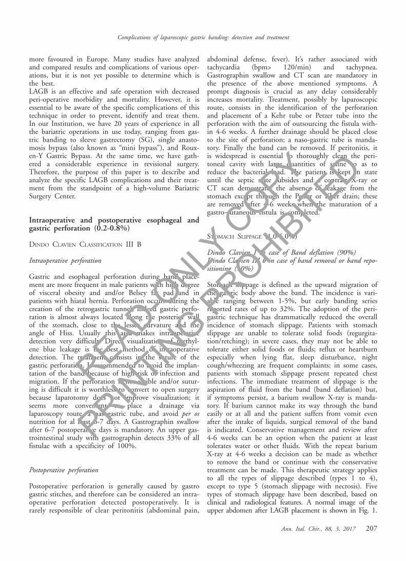
Stomach slippage is defined as the upward migration ofthe gastric body above the band. The incidence is vari-able ranging between 1-5%, but early banding seriesreported rates of up to 32%. The adoption of the peri-gastric technique has drammatically reduced the overallincidence of stomach slippage. Patients with stomachslippage are unable to tolerate solid foods (regurgita-tion/retching); in severe cases, they may not be able totolerate either solid foods or fluids; reflux or heartburnespecially when lying flat, sleep disturbance, nightcough/wheezing are frequent complaints; in some cases,patients with stomach slippage present repeated chestinfections. The immediate treatment of slippage is theaspiration of fluid from the band (band deflation) but,if symptoms persist, a barium swallow X-ray is manda-tory. If barium cannot make its way through the bandeasily or at all and the patient suffers from vomit evenafter the intake of liquids, surgical removal of the bandis indicated. Conservative management and review after4-6 weeks can be an option when the patient at leasttolerates water or other fluids. With the repeat bariumX-ray at 4-6 weeks a decision can be made as whetherto remove the band or continue with the conservativetreatment can be made. This therapeutic strategy appliesto all the types of slippage described (types 1 to 4),except to type 5 (stomach slippage with necrosis). Fivetypes of stomach slippage have been described, based onclinical and radiological features. A normal image of theupper abdomen after LAGB placement is shown in Fig. 1.
READ-ONLY
COPY
PRINTIN
G PROHIB
ITED
A. Sartori, et al.
208 Ann. Ital. Chir., 88, 3, 2017
The band is placed just below the gastroesofageal junction.The size of the pouch is appropriately sized to 50-80 ml.The most appropriate placement of the band is at anapproximate 45° angle toward the left shoulder with themedial aspect of the band juxtaposed to the left pedicleof the vertebra. Type 1 slippage results from upwardmigration of the gastric anterior wall through the band(Fig. 2). The band is rotated horizontally or downwards;barium accumulates over the left side of the band witheither very little or no flow into the distal stomach. Type1 slipppage is generally due to insufficient anterior fix-ation or disruption of the fixation suturers. Another pos-sible cause is an increased pressure in the pouch due toearly solid food intake, overeating or early (<4 weeks)band fill. In case of non operative management failure,laparoscopic repositioning is required. Once access to theperitoneal cavity has been gained, the band buckle isdetected by placing the tubing under traction. A carefuldissection of the gastric wrap around the band is car-ried out using endoshears and hook cautery. Previous
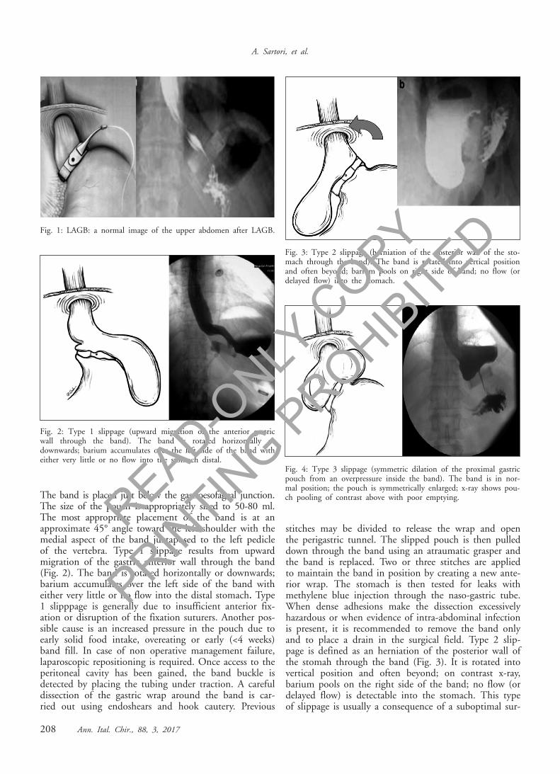
stitches may be divided to release the wrap and openthe perigastric tunnel. The slipped pouch is then pulleddown through the band using an atraumatic grasper andthe band is replaced. Two or three stitches are appliedto maintain the band in position by creating a new ante-rior wrap. The stomach is then tested for leaks withmethylene blue injection through the naso-gastric tube.When dense adhesions make the dissection excessivelyhazardous or when evidence of intra-abdominal infectionis present, it is recommended to remove the band onlyand to place a drain in the surgical field. Type 2 slip-page is defined as an herniation of the posterior wall ofthe stomah through the band (Fig. 3). It is rotated intovertical position and often beyond; on contrast x-ray,barium pools on the right side of the band; no flow (ordelayed flow) is detectable into the stomach. This typeof slippage is usually a consequence of a suboptimal sur-
Fig. 1: LAGB: a normal image of the upper abdomen after LAGB.
Fig. 2: Type 1 slippage (upward migration of the anterior gastricwall through the band). The band is rotated horizontally ordownwards; barium accumulates over the left side of the band witheither very little or no flow into the stomach distal.
Fig. 3: Type 2 slippage (herniation of the posterior wall of the sto-mach through the band). The band is rotated into vertical positionand often beyond; barium pools on right side of band; no flow (ordelayed flow) into the stomach.
Fig. 4: Type 3 slippage (symmetric dilation of the proximal gastricpouch from an overpressure inside the band). The band is in nor-mal position; the pouch is symmetrically enlarged; x-ray shows pou-ch pooling of contrast above with poor emptying.READ-O
NLY C
OPY
PRINTIN
G PROHIB
ITED
gical technique. Fortunately, type 2 slippage is less fre-quent nowadays with the adoption of the pars flaccidaapproach instead of the perigastric approach. Its man-agement is similar to that of type 1, generally via thelaparoscopic route.Type 3 slippage is also known as Symmetrical PouchDilatation (SPD). It consists in a dilation of the proxi-mal gastric pouch with or without any change in theangle of the band and in the absence of signs of obstruc-tion (Fig. 4). It results from an overpressure into thegastric pouch induced by high inflation or overeating.The patient complains of a lack of satiety, heartburn,regurgitation and occasional chest pain. This complica-tion is likely to be solved non-operatively by band defla-tion and re-education of the patient who should beadviced not to exceed in meal size. If the pouch size isdemostrated on a 4-6 weeks contrast study to be backto normal size, the band can be reinflated. Otherwise,surgical treatment with either band removal or replace-ment should be considered. Type 4 slippage is definedas an immediate post-operative prolapse and is usuallydue to placing the band too low on the stomach. Unlikethe previous types and f type 5, this is a chronic com-plication. It can be managed non-operatively in the firstinstance. Only in case of failure, surgical repositioningis needed. Type 5 is a type 1 or 2 slip with gastricnecrosis; it is the consequence of an acute pouch dila-tion and requires a prompt surgical operation (possiblylaparoscopy).
PORT AND TUBING COMPLICATION (3%)DINDO CLAVIEN IIIA
Port and tubing leak
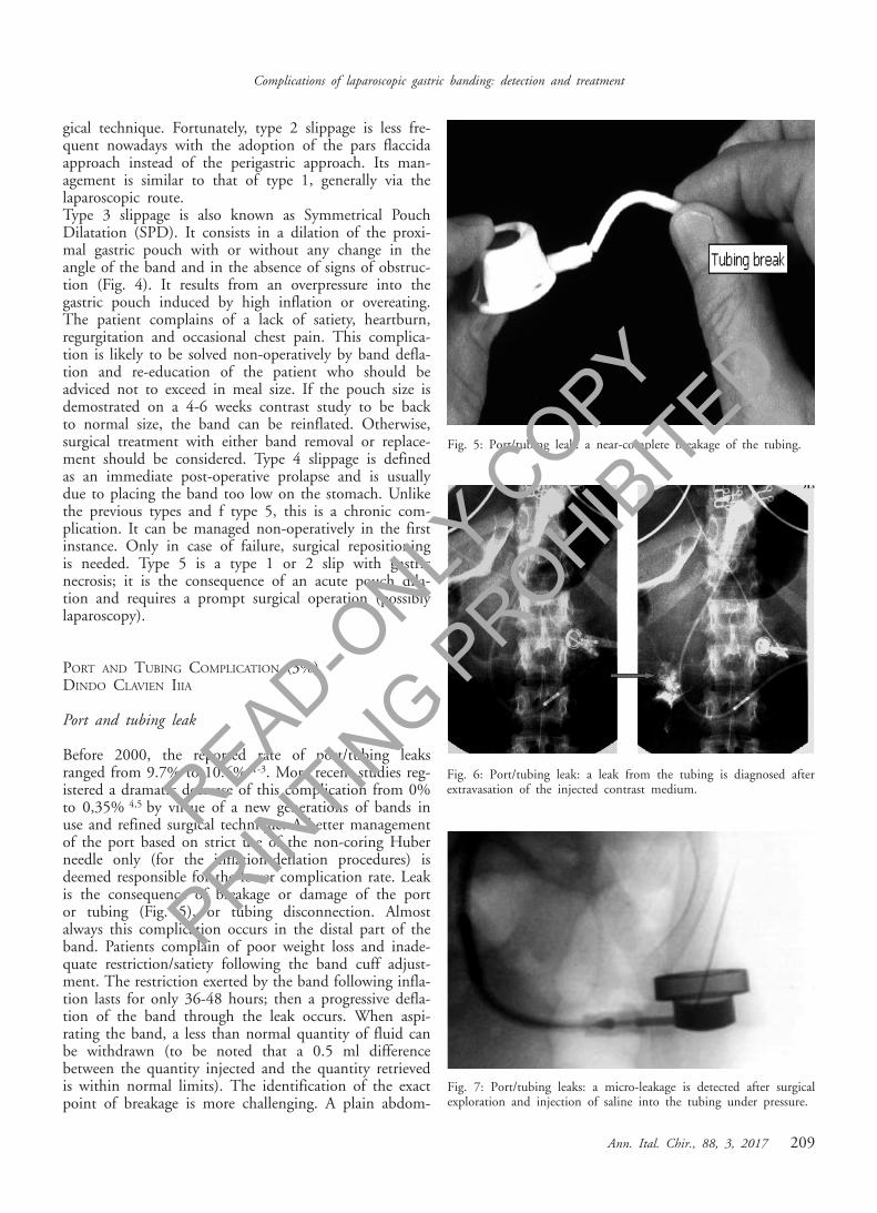
Before 2000, the reported rate of port/tubing leaksranged from 9.7% to 10.6% 1-3. More recent studies reg-istered a dramatic decrease of this complication from 0%to 0,35% 4,5 by virtue of a new generations of bands inuse and refined surgical technique. A better managementof the port based on strict use of the non-coring Huberneedle only (for the inflation-deflation procedures) isdeemed responsible for the lower complication rate. Leakis the consequence of breakage or damage of the portor tubing (Fig. 5), or tubing disconnection. Almostalways this complication occurs in the distal part of theband. Patients complain of poor weight loss and inade-quate restriction/satiety following the band cuff adjust-ment. The restriction exerted by the band following infla-tion lasts for only 36-48 hours; then a progressive defla-tion of the band through the leak occurs. When aspi-rating the band, a less than normal quantity of fluid canbe withdrawn (to be noted that a 0.5 ml differencebetween the quantity injected and the quantity retrievedis within normal limits). The identification of the exactpoint of breakage is more challenging. A plain abdom-
Ann. Ital. Chir., 88, 3, 2017 209
Complications of laparoscopic gastric banding: detection and treatment
Fig. 5: Port/tubing leak: a near-complete breakage of the tubing.
Fig. 7: Port/tubing leaks: a micro-leakage is detected after surgicalexploration and injection of saline into the tubing under pressure.
Fig. 6: Port/tubing leak: a leak from the tubing is diagnosed afterextravasation of the injected contrast medium.READ-O
NLY C
OPY
PRINTIN
G PROHIB
ITED
A. Sartori, et al.
210 Ann. Ital. Chir., 88, 3, 2017
inal X-ray after injection of contrast medium into theport and tubing is rarely helpful because only high flowleakages are visible (Fig. 6). A local exploration of theport site is often necessary (Fig. 7); another approach isto inject diluted methylene blue into the port underdirect laparoscopic visualization of the tubing and band.When a leakage is diagnosed, port, tubing or bandreplacement is usually necessary depending on the siteof the leakage and type of band used.
Twist of the port
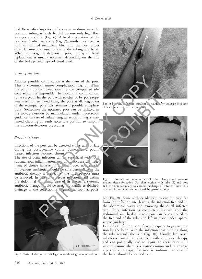
Another possible complication is the twist of the port.This is a common, minor complication (Fig. 8). Whenthe port is upside down, access to the compressed sili-cone septum is impossible. To avoid this complication,some surgeons fix the port with stitches or by polypropi-lene mesh; others avoid fixing the port at all. Regardlessof the tecnique, port twist remains a possible complica-tions. Sometimes the upturned port can be replaced inthe top-up position by manipulation under fluoroscopyguidance. In case of failure, surgical repositioning is war-ranted choosing an easily accessible position to simplifythe inflation-deflation procedures.
Port-site infection
Infections of the port can be detected either early or lateduring the postoperative course. Sometimes a poorlytreated infection becomes chronic. The site of acute infection can be superficial with localsubcutaneous inflammation; oral antibiotics are the treat-ment of choice however if infection does not subside,intravenous antibiotics should be considered. If even theantibiotic therapy is ineffective, the infected port mustbe removed. In case of a deeper infection, still withinthe abdominal wall, or in case of an abscess, a systemicantibiotic therapy should be straightforwardly established;drainage of the collection is required as soon as possi-
ble (Fig. 9). Some authors advocate to cut the tube farfrom the infection site, leaving the infection-free end inthe abdominal cavity and removing the distal infectedone. Once infection is completely resolved and theabdominal wall healed, a new port can be connected tothe free end of the tube and left in place under laparo-scopic guidance. Late onset infections are often subsequent to gastric ero-sion by the band, with the infection that running alongthe tube towards the skin (Fig. 10). Usually, late onsetinfections cannot be controlled with antibiotic therapyand can potentially lead to sepsis. In these cases it iswise to assume there is a gastric erosion and to arrangea prompt endoscopy; if erosion is confirmed, removal ofthe band should be carried out.
Fig. 9: Port-site infection: purulent discharge after drainage in a caseof acute infection of the port site.
Fig. 10: Port-site infection: eczema-like skin changes and granulo-matous tissue formation (A), skin erosion with tube (B) and port(C) rejection secondary to chronic discharge of infected fluids in acase of chronic infection sustained by gastric erosion.
Fig. 8: Twist of the port: a radiologic image showing the upturned port.
READ-ONLY
COPY
PRINTIN
G PROHIB
ITED
Ann. Ital. Chir., 88, 3, 2017 211
Complications of laparoscopic gastric banding: detection and treatment
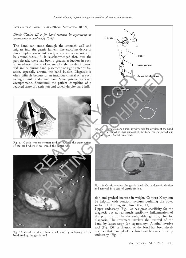
INTRAGASTRIC BAND EROSION/BAND MIGRATION (0.8%)
Dindo Clavien III b for band removal by laparotomy vslaparoscopy vs endoscopy (5%)
The band can erode through the stomach wall andmigrate into the gastric lumen. The exact incidence ofthis complication is unknown; recent studies report it tobe around 0.8% 4-8. It is acknowledged that, over thepast decade, there has been a gradual reduction in suchan incidence. The etiology may be the result of gastricwall injury during band placement or tight anterior fix-ation, especially around the band buckle. Diagnosis isoften difficult because of an insidious clinical onset suchas vague, mild abdominal pain. Some patients are evenasymptomatic. Sometimes the patient complains of areduced sense of restriction and satiety despite band infla-
tion and gradual increase in weight. Contrast X-ray canbe helpful, with contrast medium outlining the outersurface of the migrated band (Fig. 11). Upper endoscopy (Fig. 12) has great specificity for thediagnosis but not as much sensibility. Inflammation ofthe port site can be the only, although late, clue fordiagnosis. The treatment involves the removal of theband by laparoscopy (or laparotomy). A mini invasivetool (Fig. 13) for division of the band has been devel-oped so that removal of the band can be carried out byendoscopy (Fig. 14).
Fig. 12: Gastric erosion: direct visualization by endoscopy of theband eroding the gastric wall.
Fig. 11: Gastric erosion: contrast medium outlines the outer surfaceof the band where it has eroded the gastric wall.
Fig. 13: Gastric erosion: a mini invasive tool for division of the bandhas been developed so that removal of the band can be carried outby endoscopy (Band-Cutter TM).
Fig. 14: Gastric erosion: the gastric band after endoscopic divisionand removal in a case of gastric erosion.
READ-ONLY
COPY
PRINTIN
G PROHIB
ITED
A. Sartori, et al.
212 Ann. Ital. Chir., 88, 3, 2017
Discussion
LAGB represents one of most frequently performedbariatric operations in morbidly obese patients. LAGB isconsidered to be a safe and effective method to achieveweight loss and resolution of obesity-associated comor-bidities. For this reason LAGB is considered by manysurgeons an optimal treatment for morbid obesity andit is attractive for the patients because it is less invasiveas described by Michieletto 8. This is in line with theliterature review conducted in our Institution but - asdescribed by Launat-Savary- morbidity after bariatricsurgery is an underestimated problem 9. We identified,via electronic search the most relevant papers to date ingastric banding. The search yielded 137 articles. Dataregarding reoperation rate and perioperative mortalitywere extracted from each one. On the whole populationof 29,980 patients a perioperative mortality of 0.1% wascalculated. This result is similar to that reported in themeta-analysis by Cunneen 10. This outcome highlightsthat gastric banding can be considered an extremely safeprocedure however the reoperation rate ranges from 2.5%to 23.9%. To add more strength to this finding, we car-ried out, on the same study population a subgroup analy-sis. We compared the perioperative mortality of two peri-ods: before and after 2000. The hypotesis of this studyis that improvements in surgical technique, anaesthesia,patient selection and care have been responsible for adeceptively low mortality. In fact, we found that before2000, perioperative mortality ranged from 0% to 0.1%11-12 and after 2000 perioperative mortality was between0% and 0.2% [13-14]. This difference does not reachany statistical significance, indicating that mortality isevenly distributed and is neglectful (Table I). Howeverin a recent study carried out by Gagner based on ananonymous questionnaire sent to the members of theAmerican Society for Bariatric Surgery, it was conclud-ed that late deaths are under-reported 15. They haveadvanced several scales for pre-operative quantification ofmortality risk. Currently the score suggested by De Mariaand validated by Canadian and U.S. multi-center stud-ies seems a good solution for stratification of preopera-
tive risk of death 16-18. Nevertheless, some procedure-spe-cific complications can occur . Pouch enlargement, bandslippage, band erosion, port site infections and port leakrepresent the most commonly band associated compli-cations. They require an equally specific assessment andmanagement process. In some cases, particular medicaldevices such as the Band-CutterTM are mandatory espe-cially in the present mini-invasive era. As a consequence,a dedicated team is warranted. This is confirmed by theliterature review we conducted. In fact, we observed areduction in reoperation rates due to complications after2000. This reflects the refinement in surgical techniquethroughout our long-course experience. From the 24.4%described by Tolonen or the lower, but still high 10.5%reported by Belachew, reoperation rate dropped after2000, to 2,6%-2.1% 3,12,19,20 (Table II). Port complica-tions are often under-reported because of a short follow-up. The absence of an access-port and the use ofEasybandtm as described by Handgraaf is advocated as asolution to this problem 21. Tog recently reported thatcomplications related to the port or tubing are the mostfrequent in patients with LAGB 22. These authors asMicheletto reported an incidence of 8.7% in 1928patients with LAGB; 27% of patients required at least2 or more procedures. Probably the incidence of thiscomplication is underestimated because of a partial fol-low up 22. Migration of gastric banding is a long termcomplication; it is believed to be the consequence of thechronic trauma to the gastric wall enhanced by the phys-iological movements of the diaphragm and peristalsis ofthe stomach. Port infection can be caused by migrationof gastric banding. Silecchia et al. reported an incidenceof migration of 7.5% and pointed out that patients areoften asymptomatic; in most cases, band migration isdiagnosed incidentally 23,24. Micheletto et al. in our clin-ical records reported an incidence of migration only of 1.1%but it is explained by not long term of follow-up 8.Alternatively, when we suspect a band migration, an uppergastrointestinal X-ray is mandatory and is usually diag-nostic. One of the reasons of migration is gastric slip-page; treatment consists in simple a deflation of the band(90% of the cases) or surgical removal and/or reposi-
TABLE I - Operative mortality after gastric banding: no significative dif-ference over the two study periods (before and after 2000). TABLE II - Reoperation rate after LAGB: drammatic reduction of reo-
peration rate over the two study periods (before and after 2000)
READ-ONLY
COPY
PRINTIN
G PROHIB
ITED
tioning of the band (10% of the cases). The preventionof this complication has been clearly described with theapposition of 2 or 3 nonabsorbable sutures between thegastric fundus and left hemidiaphragm. Recently Findlayet al. proposed two simple interventions to reduce thistype of complication with good results: band filling pro-tocol and post-operative dietary programme 25. Pouchdilatation is a different complication to band slippage asdescribed by Moser et al. 26. The etiology of this com-plication is multifactorial, and the etiologic factors arechronic overeating or overinflation of the band. Banddeflation is the most simple treatment. In this periodthe patient should be instructed to follow a low-calorydiet. Surgical treatment should be considered if medicaltreatment fails. LAGB is a effective option to treat mor-bid obesity; but the rate of short and long term com-plications and their specific profile, makes a strict fol-low-up mostly important to early diagnosis and correcttreatment.
Conclusion
From these data, it is possible to draw the conclusionthat gastric banding has at least a medium-term effica-cy in the treatment of morbid obesity. By virtue of theexperience accrued over time, this procedure is now evensafer than in the past because of the reduced incidenceof complications requiring reoperation. Not least, mostof these complications require only minor interventionsand often can be treated with mini invasive techniques(such laparoscopy or endoscopy). Nevertheless, it isimportant for general surgeons to be aware of the com-mon presentations of gastric banding complications asthey are procedure-specific and require an equally spe-cific treatment.
Riassunto
INTRODUZIONE: il bendaggio gastrico laparoscopico(LAGB) è riconosciuto regolabile è come un’opzione chi-rurgica comune ed efficace o nel trattamento dell’obesitàe delle relative malattie metaboliche. Questa proceduraè un’operazione notevolmente sicura da un punto di vistachirurgico generale sia da un punto di vista bariatrico.Essa facilitaun breve degenza e può essere eseguita tra-mite una sola incisione.METODI: Abbiamo analizzato le complicanze più comu-ni del LAGB sia intraoperatoria sia postoperatorie perfo-razione gastrica, lo slittamento dello stomaco / dilata-zione, complicanze del port, la migrazione del bendag-gio intragastrico che si sono verificati nei nostri lunghidecenni di esperienza clinica confrontandole con la let-teratura. RISULTATI: LAGB ha mostrato buoni risultati a lungo ter-mine in termini di perdita di peso e di risoluzione di
Ann. Ital. Chir., 88, 3, 2017 213
Complications of laparoscopic gastric banding: detection and treatment
patologie legate all’obesità. Inoltre, la mortalità dovuta apatologie correlate con l’obesità è apparsa significativa-mente più bassa nei pazienti LAGB rispetto ai pazientiin terapia farmacologica.CONCLUSIONE: Il bendaggio gastrico ha un tasso moltobasso di complicanze precoci e tardive; queste sono anchemeno gravi rispetto alle procedure più invasive e sonosuscettibili di una gestione con tecniche mini-invasive.In ogni caso, l’invio di questi casi a un chirurgo conesperienza di chirurgia bariatrica è ritenuta opportuna.
References
1. Mittermair RP, Weiss H, Nehoda H, et a.: Laparoscopic Swedishadjustable gastric banding: 6-year follow-up and comparison to otherlaparoscopic bariatric procedures. Obes Surg, 2003; 13:412-17.
2. Favretti F, Segato G, Ashton D, et al.: Laparoscopic adjustablegastric banding in 1,791 consecutive obese patients: 12-year results.Obese Surg, 2007; 17:168-75.
3. Tolonen P, Victorzon M, Makela J: 11-year experience withlaparoscopic adjustable gastric banding. What happened to the first 123patients? Obes Surg, 2008; 18:251-55.
4. Chevallier JM, Zinzindohoue F, Douard R, et al: Complicationsafter laparoscopic adjustable gastric. Laparoscopic adjustable gastricbanding in an ambulatory surgery center banding for morbid obesity:Experience with 1.000 patients over 7 years. Obes Surg, 2004; 14:407-14.
5. Watkins BM, Ahroni JH, Michaelson R, et al.: Laparoscopicadjustable gastric banding in an ambulatory surgery center. Surg ObesRel Dis, 2008; 4 (Suppl):S56-62.
6. Singhal R, Kitchen M, Ndirika S, et al: The “Birmingham”stitch”. Avoiding slippage in laparoscopic gastric banding. Obes Surg,2008; 18:359-63.
7. Abu-Abeid S, Szold A: Laparoscopic management of Lap-Banderosion. Obes Surg, 2001; 11:87-9.
8. Micheletto G, Roviaro G, Lattuada E, Zappa MA, Mozzi E,Perrini M, Lanni M, Francese M, Librenti MC, Doldi SB:Adjustable gastric banding for morbid obesity. Our experience. AnnItal Chir, 2006; 77(5):397-400.
9. Launay Savary MV, Slim K, Brugere C, Buc E, Nini E, ForestierD, et al.: Band and port related morbidity after biariatric surgery: Anunderstimated problem. Obesity Surg, 2008; 18:1406-410.
10. Cunneen SA, Phillips E, Fielding G, et al.: Studies of Swedishadjustable gastric band and Lap-band: Systematic review and meta-analysis. Surg Obes Relat Dis, 2008; 4:174-85.
11. Steffen R, Biertho L, Ricklin T, et al.: Laparoscopic Swedishadjustable gastric banding: A five-year prospective study. Obes Surg,2003; 13:404-11.
12. Belachew M, Belva PH, Desaive C: Long-term results of laparo-scopic adjustable gastric banding for the treatment of morbid obesity.Obes Surg, 2002; 12:564-68.
13. Parikh MS, Fielding G, Ren CJ: US experience with 749 laparo-scopic adjustable gastric bands: Intermediate outcomes. Surg Endosc,2005; 19:1631-635.
READ-ONLY
COPY
PRINTIN
G PROHIB
ITED
A. Sartori, et al.
214 Ann. Ital. Chir., 88, 3, 2017
14. Ren CJ, Weiner M, Allen RW: Favourable early results of gas-tric banding for morbid obesity: The American experience. SurgEndosc, 2004; 18:543-46.
15. Gagner M, Milone L, Yung E, Broseus A, Gumbs AA: Causesof early mortality after LAGB J. Am Coll Surg, 2008; 206:664-69.
16. DeMaria EJ, Portennier D, Wolfe L: Obesity surgery morbidityrisk score: Proposal for a clinically useful score to predict mortality riskin patient undergoing gastric banding. Surg Obes Relat Dis, 2007;3(2):134-40.
17. DeMaria EJM, Murr MM, Bryne TKM, et al.: Validation ofthe Obesity Surgery Mortality Risk Score in a multi center study provesit stratifies mortality risk in patients undergoing gastric bypass for mor-bid obesity. Ann Surg, 2007; 246(4):578-84.
18. Efthimiou E, Court O, Simpalis J et al.: Validation of Obesitymortality Risk score in patients undergoing gastric bypass in a Canadiancenter. Surg Obes Relat Dis, 2009; 5(6):643-47.
19. Sarker S, Myers J, Serot J, et al.: Three-year follow-up weightloss results for patients undergoing laparoscopic adjustable gastric band-ing at a major university medical center: Does the weight loss persist?Am J Surg, 2006; 191:372-76.
20. Singhal R, Kitchen M, Ndirika S, et al.: The “Birmingham”stitch”. Avoiding slippage in laparoscopic gastric banding. Obes Surg,2008; 18:359-63.
21. Handgraaf HJM, Ashton D, Favretti F, Segato G, vanRamshorst B, Meesters B, Greve JWM: The gastric band that is noto be. Obes Surg, 2015; 1704-709.
22. Tog CH, Halliday J, Khor Y, Yong T, Wilkinson S: Evolvingpattern of laparoscopic gastric band access port complications. ObesSurg, 2012; 22(6):863-65.
23. Silecchia G, Restuccia A, Elmore U, Polito, Perrotta N, GencoA, Bacci V, Basso N: Laparoscopic adjustabrle gastric banding:Prospective evaluation of intragastric migration o flap-band. SurgLaparosc Endosc Percutan Tech, 2001; 11:229-34.
24. Aarts EO, van Wageningen B, Berends F, Janssen I, Wahab P,Groenen M: Intragastric band erosion: Experiences with gastrointesti-nal endoscopic removal. W J of Gastroenterology, 2015; 21:1567-572.
25. Findlay L, Ball W, Ramus J: Gastric band slippage: The impactof a change in education and band filling. Obes Surg, 2015;25(7):1302-306.
26. Moser F, Gorondner MV, Galvani CA, Baptista M, ChretienC, Horgan S: Pouch enlargment and band slippage: two different enti-ties. Surg Endosc, 2006; 20:1021-29.
READ-ONLY
COPY
PRINTIN
G PROHIB
ITED