M. baetica - CSIC
7
Transcript of M. baetica - CSIC

Nematology, 2004, Vol. 6(5), 749-754
SEM studies on the Mediterranean olive root-knot nematode,Meloidogyne baetica, and histopathology
on two additional natural hostsNicola VOVLAS 1, Gracia LIÉBANAS 2 and Pablo CASTILLO 3,∗
1 Istituto per la Protezione delle Piante, Sezione di Bari: Nematologia Agraria,Consiglio Nazionale delle Ricerche (CNR), Via G. Amendola 165/A, 70126-Bari, Italy
2 Departamento de Biología Animal, Biología Vegetal y Ecología, Universidad de Jaén,Campus ‘Las Lagunillas’ s/n, Edificio B3, 23071-Jaén, Spain
3 Instituto de Agricultura Sostenible, Consejo Superior de Investigaciones Científicas (CSIC),Apdo. 4084, 14080-Córdoba, Spain
Received: 28 July 2004; revised: 12 August 2004Accepted for publication: 17 August 2004
Summary – SEM studies on a Meloidogyne baetica population provided additional details of the external morphology for female,male and second-stage juveniles. The labial disc in female and male specimens is fused with the medial lips forming a single structure.In second-stage juveniles the lateral lips are triangular with rounded margins. The amphidial opening for all life stages appears ovalto rectangular in shape and is located between the labial disc and lateral lips. Lateral fields of male and second-stage juveniles havefour incisures irregularly areolated along the entire body. The results of a host-range study for additional natural hosts of M. baeticaconducted in wild olive communities growing at Vejer de la Frontera (Cádiz province) in southern Spain are also reported. Apart fromthe type host, M. baetica was found to infect two natural woody host plants, lentisc (Pistacia lentiscus) and Aristolochia baetica. Host-parasite relationships in these new hosts confirmed the typical susceptible reaction observed in wild and cultivated olives. Similarly,the reproductive fitness, evaluated as the number of eggs per egg mass, was not significantly different in all plant hosts. No infectionsor galled roots were observed in herbaceous plant species studied and M. baetica must therefore be considered as a parasite of woodyplants.
Keywords – Aristolochia baetica, candilito, histopathology, host-parasite relationships, lentisc, mastic tree, Pistacia lentiscus,reproductive fitness, Spain.
Meloidogyne baetica Castillo, Vovlas, Subbotin & Troc-coli, 2003 was recently described infecting wild olive atsouthern Spain (Castillo et al., 2003). To date, its geo-graphical distribution is limited to a small area of nat-ural sandy soils at Vejer de la Frontera (Cádiz province)in southern Spain. Although the nematode type localityincludes soil and plant communities referred to as Oleo-Lenticetum and Rosmarino-Ericion, otherwise known as‘matorral’ in Spain (Rivas Martinez, 1982), informationon the host-range of this root-knot nematode was lim-ited to wild and cultivated (cvs Arbequina and Picual)olives (Castillo et al., 2003). It is important to establish
∗ Corresponding author, e-mail: [email protected]
the natural hosts of this nematode and therefore a nema-tode survey was conducted in wild olive and associatedplant communities growing at the type locality during thespring of 2003. In addition, although a complete morpho-logical description, based on light microscopy, of the lifestages of the nematode was presented in the original de-scription, no scanning electron microscopy (SEM) stud-ies were done. Consequently, the objectives of this studywere: i) to describe the morphology of life stages of thenematode as seen by SEM; and ii) to study the naturalhosts of M. baetica and describe their host-parasite rela-tionships.
© Koninklijke Brill NV, Leiden, 2004 749Also available online - www.brill.nl

N. Vovlas et al.
Materials and methods
SEM STUDIES
For SEM studies, mature females were dissected fromnaturally-infected roots of wild olive, and migratorystages (including males and second-stage juveniles) wereextracted from infested soils by the centrifugal flotationmethod (Coolen, 1979). Specimens were killed by gentleheat, fixed in a solution of 4% formaldehyde + 1% pro-pionic acid, and processed to pure glycerine using Sein-horst’s method (Seinhorst, 1966). Fixed specimens weredehydrated in a graded ethanol series, critical point dried,sputter-coated with gold, and observed with a JEOL JSM-5800 microscope (Abolafia et al., 2002).
HOST-RANGE STUDY
A nematode survey was conducted in wild olive com-munities growing at Vejer de la Frontera (Cádiz province)in southern Spain to obtain more information about thenatural hosts of M. baetica. Thirty samples consisting ofca 3-4 kg soil and associated roots and plant parts werecollected, placed in plastic bags, and stored at 7-10◦Cfor up to 3-4 days before observation and nematode ex-traction. Soil was processed by the centrifugal flotationmethod (Coolen, 1979) in order to collect M. baeticasecond-stage juveniles and males. Roots and plant partsfrom soil samples infested with second-stage juvenilesand males of M. baetica were separated according to plantspecies, washed, and examined with the aid of a stereomi-croscope for nematode infection. Frequency of soil infes-tation and population density was determined. Frequencywas calculated as the percentage of samples in which thenematode was found. For morphological and diagnosticstudies, specimens were processed as above (Seinhorst,1966). Measurements were done using a drawing tube at-tached to a light microscope.
In each natural host, including wild olive, the reproduc-tive fitness of M. baetica was assessed by the number ofeggs per egg mass. In each host, 20 mature egg massesof M. baetica-infected roots were individually separated,each egg mass being deposited in an individual 10 mlglass tube. Egg number per egg mass was determined afterexposure to 1% NaOCl (Hussey & Barker, 1973) and vor-texing for 5 min. Data of number of eggs per egg mass (X)
were normalised before analysis by transforming them tolog10 (X + 1) (Gomez & Gomez, 1984). Analysis of vari-ance (ANOVA) was carried out using Statistix 8.0 (NHAnalytical Software, Roseville, MN, USA). Means val-
ues of number of eggs per egg mass were compared usingFisher’s protected least significant difference test (LSD)at P = 0.05.
HISTOPATHOLOGY
Infected roots from naturally-infected host plants weregently washed free of adhering soil and debris and in-dividual galls were selected together with healthy roots.Root tissues were fixed in formaldehyde chromo-aceticsolution for 48 h, dehydrated in a tertiary butyl alco-hol series (40-70-85-90-100%) and embedded in 58◦C(melting point) paraffin wax for histopathological obser-vations. Embedded tissues were sectioned with a rotarymicrotome. Sections 10-12 µm thick were placed on glassslides, stained with safranin and fast-green, mounted per-manently in a 40% xylene solution of a polymethacrylicester (Synocril 9122X), examined microscopically andphotographed (Johansen, 1940).
Results
SEM STUDIES
Female
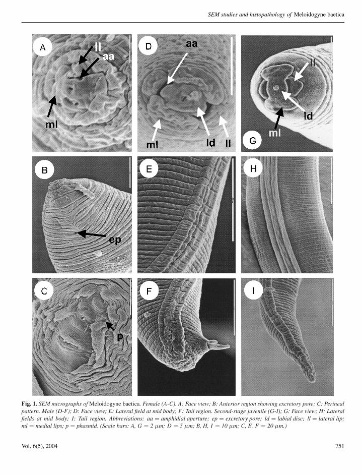
In face view, the labial disc is small, rounded, andslightly raised above the medial lips. The labial disc andmedial lips are fused to form a narrow lip structure in faceview. Amphidial openings are oval shaped and are locatedbetween the labial disc and lateral lips (Fig. 1A). Theexcretory pore is located near the lip region, six to eightannuli posterior to labial plate (Fig. 1B). Examination ofthe perineal pattern by SEM revealed the same featuresas with light microscopy (LM) studies, the pattern beingtypically formed from striae and cuticular ridges, thelatter being more pronounced in the vicinity of the vulvaand anus. The dorsal arch encloses the relatively distinctphasmids and the anus (Fig. 1C).
Male
Lip region usually marked by a short, incompleteannulation in lateral view. In SEM, the labial disc isoval and is fused with the elongate-oval medial lipsforming a rectangular structure (Fig. 1D). The laterallips are rounded-oval (Fig. 1D). The amphidial aperturesare elongate slits located between the labial disc and thelateral lips (Fig. 1D). The lateral fields are marked by fourincisures and are faintly and irregularly areolated at themid-body and tail region (Fig. 1E, F).
750 Nematology
SEM studies and histopathology of Meloidogyne baetica
Fig. 1. SEM micrographs of Meloidogyne baetica. Female (A-C). A: Face view; B: Anterior region showing excretory pore; C: Perinealpattern. Male (D-F); D: Face view; E: Lateral field at mid body; F: Tail region. Second-stage juvenile (G-I); G: Face view; H: Lateralfields at mid body; I: Tail region. Abbreviations: aa = amphidial aperture; ep = excretory pore; ld = labial disc; ll = lateral lip;ml = medial lips; p = phasmid. (Scale bars: A, G = 2 µm; D = 5 µm; B, H, I = 10 µm; C, E, F = 20 µm.)
Vol. 6(5), 2004 751

N. Vovlas et al.
Table 1. Plant species studied as potential hosts of Meloidogyne baetica Castillo et al., 2003.
Common name Scientific name Host (+)non-host (–)
wild garlic Allium ursinum L. –‘candilito’ Aristolochia baetica L. +white asparagus Asparagus albus L. –asphodel Asphodelus albus Mill. –daisy Bellis sylvestris Cyr. –small pink bindweed Convolvulus althaeoides L. –field bindweed Convolvulus arvensis L. –wild artichoke Cynara cardunculus L. –black broom Cytisus baeticus (Webb) Steudel –lesser chickpea or vetch Lathyrus cicera L. –wild olive Olea europaea spp. sylvestris (Miller) Hegi +red or common poppy Papaver rhoeas L. –Jerusalem sage Phlomis purpurea L. –lentisc or mastic tree Pistacia lentiscus L. +buttercup Ranunculus rupestris Guss. –false sowthistle Reichardia tingitiana (L.) Roth –
Second-stage juvenile
In face view, labial disc and medial lips fused intoone structure (Fig. 1G). Labial disc small and rounded,slightly elevated above medial lips (Fig. 1G). Medial lipselongate-oval, not indented medially with rounded mar-gin. Amphidial apertures appearing as elongate slits be-tween the labial disc and lateral lips (Fig. 1G). Laterallips triangular with rounded margins. (Fig. 1H). Lip re-gion smooth. (Fig. 1G). Lateral fields with four incisures,irregularly areolated along entire body (Fig. 1G-I).
NATURAL HOSTS
Meloidogyne baetica was detected in 14 out of the30 soil samples (46.7%) collected in the type habitatlocality, with population densities ranging between 128and 1190 eggs and second-stage juveniles per 100 cm3
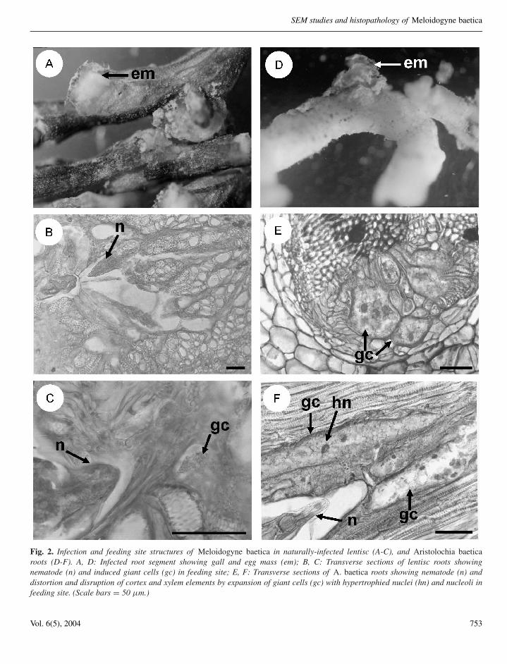
of soil. Meloidogyne baetica infested soil samples weretaken from around the herbaceous and woody plantspecies listed in Table 1, all of which were studiedfor potential infection with the nematode. Examinationof root systems of these plant species indicated thatthe only natural hosts of M. baetica in the studiedarea, apart from wild olive, were the lentisc or mastictree (Pistacia lentiscus L.) and ‘candilito’ (Aristolochiabaetica L.), mature females with mature egg massesbeing observed on the root surface (Fig. 2A, D). Noinfections and no galled roots were detected in the other
plant species studied (Table 1). Reproductive fitness, asdetermined by the number of eggs per egg mass, was notsignificantly (P = 0.172) different among natural-hostsof M. baetica, with 229 ± 80.8 (132-450) eggs in wildolive, 189 ± 73.5 (122-480) in lentisc, and 177 ± 72.9(112-380) in A. baetica.
HISTOPATHOLOGY
Root galls induced by M. baetica on lentisc andcandilito varied in size and location, the majority of eggmasses being observed on the root surface (Fig. 2A, D).Histological observations of M. baetica-infected lentiscand candilito roots revealed cellular alterations inducedby the nematode in the cortex, endodermis, pericycleand vascular parenchyma (Fig. 2B-F). In the permanentfeeding sites, nematode-induced, large, multinucleate,giant cells located adjacent to the vascular tissues wereobserved in both natural hosts (Fig. 2C, F). This formationled to the distortion and disruption of xylem elementsand primary phloem cells (Fig. 2B, E). Nematode feedingsites comprised three to six giant cells surrounding thelip region of a single female (Fig. 2). Giant cells hada thickened cell wall and granular cytoplasm with fourto ten hypertrophied nuclei and nucleoli (Fig. 2F). Thehistological modifications induced by M. baetica in rootsof lentisc and candilito revealed a typical susceptiblereaction to infection by the nematode.
752 Nematology
SEM studies and histopathology of Meloidogyne baetica
Fig. 2. Infection and feeding site structures of Meloidogyne baetica in naturally-infected lentisc (A-C), and Aristolochia baeticaroots (D-F). A, D: Infected root segment showing gall and egg mass (em); B, C: Transverse sections of lentisc roots showingnematode (n) and induced giant cells (gc) in feeding site; E, F: Transverse sections of A. baetica roots showing nematode (n) anddistortion and disruption of cortex and xylem elements by expansion of giant cells (gc) with hypertrophied nuclei (hn) and nucleoli infeeding site. (Scale bars = 50 µm.)
Vol. 6(5), 2004 753
N. Vovlas et al.
Discussion
SEM studies of life stages of M. baetica confirmed,but in greater detail, those features observed with LM inthe original description (Castillo et al., 2003). In addition,SEM studies revealed new data on the face view of female,male and second-stage juveniles, as well as some mod-ifications to the original description, i.e. lateral fields ofmales and second-stage juveniles were revealed as faintlyand irregularly areolated whereas they were described asnon-areolated in the original description (Castillo et al.,2003). As in most Meloidogyne species, the labial disc offemales, males and second-stage juveniles of M. baeticais fused with the medial lips (Eisenback et al., 1980; Jep-son, 1987). Male and second-stage juvenile en face SEMviews of M. baetica clearly differ from those of M. artiel-lia Franklin which were previously considered unique inthe genus (Jepson, 1987). The following differences canbe observed between both species in SEM studies: a) malelabial disc of M. baetica is oval vs rounded in M. artiellia;b) male medial lips (elongate-oval not separated by con-striction vs oval separated by a deep constriction); c) malelateral lips (rounded-oval vs rounded or almost semicir-cular); d) second-stage juvenile medial lips (not indentedvs indented medially); and e) second-stage juvenile laterallips (triangular vs rounded or almost semicircular).
Studies on the natural host status of herbaceous andwoody plant species revealed that M. baetica has a narrowhost-range including wild and cultivated (cvs Arbequinaand Picual) olives (Castillo et al., 2003) and the twonew host plants lentisc and candilito. Data on nematodepopulations in soil indicate that M. baetica was foundin small clumps of wild olive and the new woodyhosts growing in the type locality. The morphologicalfeatures of M. baetica specimens detected on these newnatural hosts did no differ from those reported in theoriginal description. These results, as well as those of theoriginal description, demonstrate that M. baetica does notparasitise natural herbaceous plants at the type localityor cultivated herbaceous plants, such as tomato, pea andchickpea (Castillo et al., 2003) and, therefore, must beconsidered as a parasite of woody plants. In addition,results on the reproductive fitness of the nematode innatural hosts, as well as those from histopathologicalstudies indicate that all of these woody hosts showed asimilar response to nematode infection and reproduction.The feeding behaviour of M. baetica and disease responseon lentisc and candilito roots, was similar to that observedin wild and cultivated (cvs Arbequina and Picual) olives,
as well as those of Meloidogyne spp. in cultivated oliveplanting stocks (Nico et al., 2002).
Acknowledgements
Authors thank Dr J.A. Navas-Cortés, IAS-CSIC forhis assistance in the identification of plant species, andMr V. Hernández Fernández, and Mr J. Martín Barbarroja,IAS-CSIC, for their technical assistance.
References
ABOLAFIA, J., LIEBANAS, G. & PEÑA-SANTIAGO, R. (2002).Nematodes of the order Rhabditida from Andalucia Oriental,Spain. The subgenus Pseudacrobeles Steiner, 1938, with de-scription of a new species. Journal of Nematode Morphologyand Systematics 4, 137-154.
CASTILLO, P., VOVLAS, N., SUBBOTIN, S. & TROCCOLI, A.(2003). A new root-knot nematode: Meloidogyne baetican. sp. (Nematoda: Heteroderidae), parasitizing wild olive insouthern Spain. Phytopathology 93, 1093-1102.
COOLEN, W.A. (1979). Methods for extraction of Meloido-gyne spp. and other nematodes from roots and soil. In:Lamberti, F. & Taylor, C.E. (Eds). Root-knot nematodes(Meloidogyne species). Systematics, biology and control.New York, NY, USA, Academic Press, pp. 317-329.
EISENBACK, J.D., HIRSCHMANN, H. & TRIANTAPHYLLOU,A.C. (1980). Morphological comparison of Meloidogynefemale head structures, perineal patterns, and stylets. Journalof Nematology 12, 300-313.
GOMEZ, K.A. & GOMEZ, A.A. (1984). Statistical proceduresfor agricultural research. 2nd Edition. New York, NY, USA,John Wiley & Sons, 680 pp.
HUSSEY, R.S. & BARKER, K.R. (1973). A comparison ofmethods of collecting inocula of Meloidogyne spp. includinga new technique. Plant Disease Reporter 57, 1025-1028.
JEPSON, S.B. (1987). Identification of root-knot nematodes(Meloidogyne species). Wallingford, UK, CAB International,265 pp.
JOHANSEN, D.A. (1940). Plant microtechnique. New York, NY,USA, McGraw-Hill, 523 pp.
NICO, A.I., RAPOPORT, H., JIMÉNEZ-DÍAZ, R.M. & CAS-TILLO, P. (2002). Incidence and population density of plantparasitic nematodes associated with olive planting stocks atnurseries in southern Spain. Plant Disease 86, 1075-1079.
RIVAS MARTINEZ, S. (1982). Étages bioclimatiques, secteurschorologiques et séries de végétation de l’Espagne méditer-ranéenne. Ecologia Mediterranea 8, 175-288.
SEINHORST, J.W. (1966). Killing nematodes for taxonomicstudy with hot f.a. 4:1. Nematologica 12, 178.
754 Nematology