Calcio Enel Emba Razo
32
Calcium and Bone Disorders During Pregnancy and Lactation Christopher S. Kovacs, MD a, * , Ghada El-Hajj Fuleihan, MD, MPH b, * a Health Sciences Centre, St. John’s, NL, Canada b Calciu m Metabo lism and Osteop orosis Program, American University of Beirut–Medical Center, Riad El Solh, Beirut, Lebanon Mineral metabolism in the mother must adapt to the demand created by the fet us and pla cen ta, whi ch together dra w calciu m and other min erals from the maternal circulation to mineralize the developing fetal skeleton. Similarly, mineral metabolism must adapt in the lactating woman to supply sufficient calcium to milk and the suckling neonate. Potential adaptations include increased intake of mineral, increased efficiency of intestinal absorp- tion of mineral, mobilization of mineral from the skeleton, and increased re- nal conservation of mineral. Despite a similar magnitude of calcium demand by pregnant and lactating women, the adjustmen ts made in each of thes e re- productive periods differ significantly ( Fig. 1). These hormone-mediated ad- justments normally satisfy the needs of the fetus and infant with short-term depletions of maternal skeletal calcium content, but without long-term con- sequences to the maternal skeleton. In states of maternal malnutrition and vit ami n D defi cie ncy , how eve r, the dep leti on of ske let al min eral content may be proportionately more severe and may be accompanied by increased skeletal fragility. This article reviews present understanding of the adaptations in mineral metabolism that occur during pregnancy and lactation and how these adap- tations affect the presentation, diagnosis, and management of disorders of calcium and bone metabolism. Animal data are cited to fill in the gaps where * Cor res pon ding aut hor s. Basic Med ical Scienc es, He alth Sciences Centre, 300 Pri nce Phi lip Dri ve, St. Joh n’s , NL, A1B 3V6 Can ada (C. S. Kov acs ); Cal ci um Me taboli sm and Oste oporos is Progr am, Amer ican Unive rsity of Beirut–Me dica l Cent er, P.O. Box 11-0 236, Riad E1 Solh 4407 2020, Beirut, Lebanon (G.E.-H. Fuleihan). E-mail addresses: [email protected] (C.S. Kovacs); [email protected] (G.E.-H. Fuleihan). 0889-8529/06/$ - see front matter Ó 2005 Elsevier Inc. All rights reserved. Endocrinol Metab Clin N Am 35 (2006) 21–51
-
Upload
carlos-elio-polo-vargas -
Category
Documents
-
view
228 -
download
0
Transcript of Calcio Enel Emba Razo
7/30/2019 Calcio Enel Emba Razo
http://slidepdf.com/reader/full/calcio-enel-emba-razo 1/31
Calcium and Bone Disorders During
Pregnancy and Lactation
Christopher S. Kovacs, MDa,*,Ghada El-Hajj Fuleihan, MD, MPHb,*
aHealth Sciences Centre, St. John’s, NL, CanadabCalcium Metabolism and Osteoporosis Program,
American University of Beirut–Medical Center, Riad El Solh, Beirut, Lebanon
Mineral metabolism in the mother must adapt to the demand created by
the fetus and placenta, which together draw calcium and other minerals
from the maternal circulation to mineralize the developing fetal skeleton.
Similarly, mineral metabolism must adapt in the lactating woman to supply
sufficient calcium to milk and the suckling neonate. Potential adaptationsinclude increased intake of mineral, increased efficiency of intestinal absorp-
tion of mineral, mobilization of mineral from the skeleton, and increased re-
nal conservation of mineral. Despite a similar magnitude of calcium demand
by pregnant and lactating women, the adjustments made in each of these re-
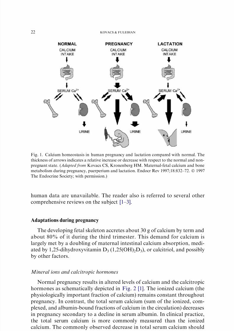
productive periods differ significantly (Fig. 1). These hormone-mediated ad-
justments normally satisfy the needs of the fetus and infant with short-term
depletions of maternal skeletal calcium content, but without long-term con-
sequences to the maternal skeleton. In states of maternal malnutrition and
vitamin D deficiency, however, the depletion of skeletal mineral contentmay be proportionately more severe and may be accompanied by increased
skeletal fragility.
This article reviews present understanding of the adaptations in mineral
metabolism that occur during pregnancy and lactation and how these adap-
tations affect the presentation, diagnosis, and management of disorders of
calcium and bone metabolism. Animal data are cited to fill in the gaps where
* Corresponding authors. Basic Medical Sciences, Health Sciences Centre, 300 Prince
Philip Drive, St. John’s, NL, A1B 3V6 Canada (C.S. Kovacs); Calcium Metabolism and
Osteoporosis Program, American University of Beirut–Medical Center, P.O. Box 11-0236,
Riad E1 Solh 4407 2020, Beirut, Lebanon (G.E.-H. Fuleihan).
E-mail addresses: [email protected] (C.S. Kovacs); [email protected] (G.E.-H. Fuleihan).
0889-8529/06/$ - see front matter Ó 2005 Elsevier Inc. All rights reserved.
doi:10.1016/j.ecl.2005.09.004 endo.theclinics.com
Endocrinol Metab Clin N Am
35 (2006) 21–51
7/30/2019 Calcio Enel Emba Razo
http://slidepdf.com/reader/full/calcio-enel-emba-razo 2/31
human data are unavailable. The reader also is referred to several other
comprehensive reviews on the subject [1–3].
Adaptations during pregnancy
The developing fetal skeleton accretes about 30 g of calcium by term andabout 80% of it during the third trimester. This demand for calcium is
largely met by a doubling of maternal intestinal calcium absorption, medi-
ated by 1,25-dihydroxyvitamin D3 (1,25(OH)2D3), or calcitriol, and possibly
by other factors.
Mineral ions and calcitropic hormones
Normal pregnancy results in altered levels of calcium and the calcitropic
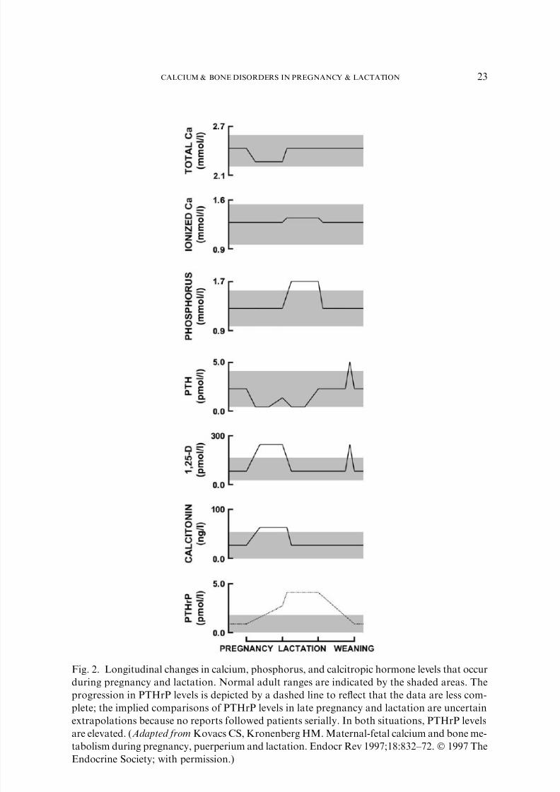
hormones as schematically depicted in Fig. 2 [1]. The ionized calcium (thephysiologically important fraction of calcium) remains constant throughout
pregnancy. In contrast, the total serum calcium (sum of the ionized, com-
plexed, and albumin-bound fractions of calcium in the circulation) decreases
in pregnancy secondary to a decline in serum albumin. In clinical practice,
the total serum calcium is more commonly measured than the ionized
calcium. The commonly observed decrease in total serum calcium should
Fig. 1. Calcium homeostasis in human pregnancy and lactation compared with normal. The
thickness of arrows indicates a relative increase or decrease with respect to the normal and non-
pregnant state. (Adapted from Kovacs CS, Kronenberg HM. Maternal-fetal calcium and bone
metabolism during pregnancy, puerperium and lactation. Endocr Rev 1997;18:832–72. Ó 1997
The Endocrine Society; with permission.)
22 KOVACS & FULEIHAN
7/30/2019 Calcio Enel Emba Razo
http://slidepdf.com/reader/full/calcio-enel-emba-razo 3/31
Fig. 2. Longitudinal changes in calcium, phosphorus, and calcitropic hormone levels that occur
during pregnancy and lactation. Normal adult ranges are indicated by the shaded areas. The
progression in PTHrP levels is depicted by a dashed line to reflect that the data are less com-plete; the implied comparisons of PTHrP levels in late pregnancy and lactation are uncertain
extrapolations because no reports followed patients serially. In both situations, PTHrP levels
are elevated. (Adapted from Kovacs CS, Kronenberg HM. Maternal-fetal calcium and bone me-
tabolism during pregnancy, puerperium and lactation. Endocr Rev 1997;18:832–72. Ó 1997 The
Endocrine Society; with permission.)
23CALCIUM & BONE DISORDERS IN PREGNANCY & LACTATION
7/30/2019 Calcio Enel Emba Razo
http://slidepdf.com/reader/full/calcio-enel-emba-razo 4/31
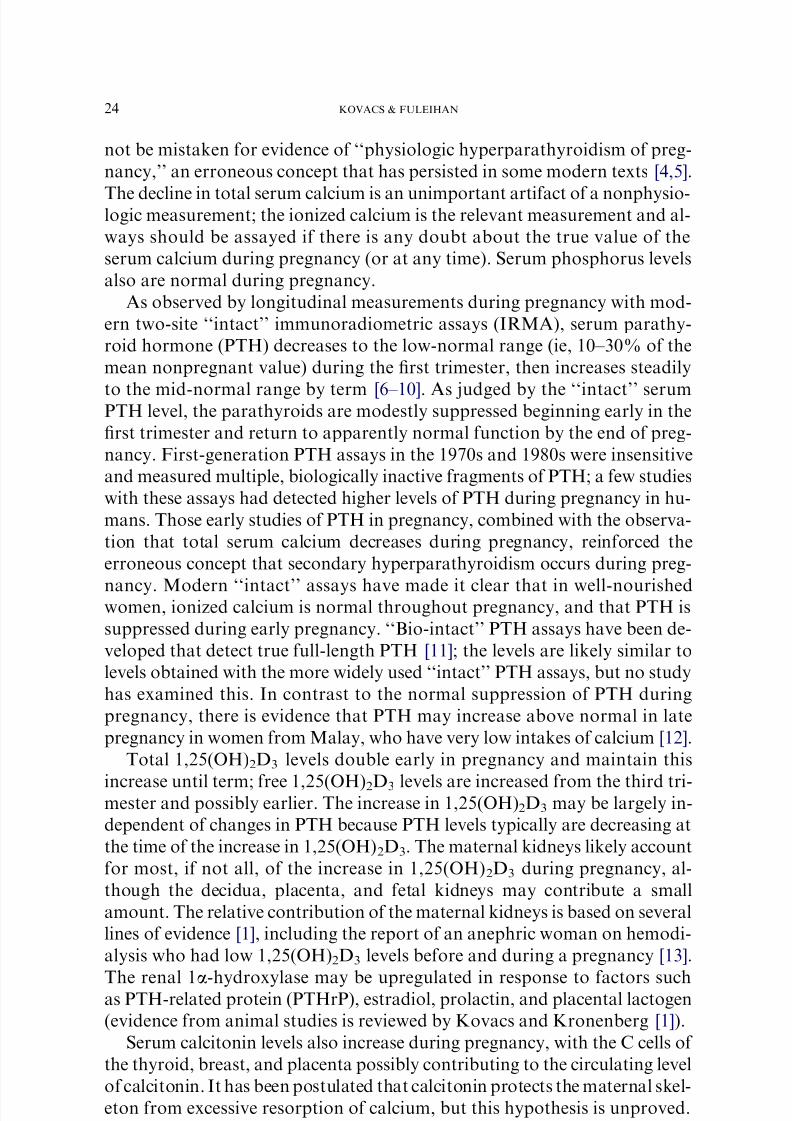
not be mistaken for evidence of ‘‘physiologic hyperparathyroidism of preg-
nancy,’’ an erroneous concept that has persisted in some modern texts [4,5].
The decline in total serum calcium is an unimportant artifact of a nonphysio-logic measurement; the ionized calcium is the relevant measurement and al-
ways should be assayed if there is any doubt about the true value of the
serum calcium during pregnancy (or at any time). Serum phosphorus levels
also are normal during pregnancy.
As observed by longitudinal measurements during pregnancy with mod-
ern two-site ‘‘intact’’ immunoradiometric assays (IRMA), serum parathy-
roid hormone (PTH) decreases to the low-normal range (ie, 10–30% of the
mean nonpregnant value) during the first trimester, then increases steadily
to the mid-normal range by term [6–10]. As judged by the ‘‘intact’’ serumPTH level, the parathyroids are modestly suppressed beginning early in the
first trimester and return to apparently normal function by the end of preg-
nancy. First-generation PTH assays in the 1970s and 1980s were insensitive
and measured multiple, biologically inactive fragments of PTH; a few studies
with these assays had detected higher levels of PTH during pregnancy in hu-
mans. Those early studies of PTH in pregnancy, combined with the observa-
tion that total serum calcium decreases during pregnancy, reinforced the
erroneous concept that secondary hyperparathyroidism occurs during preg-
nancy. Modern ‘‘intact’’ assays have made it clear that in well-nourishedwomen, ionized calcium is normal throughout pregnancy, and that PTH is
suppressed during early pregnancy. ‘‘Bio-intact’’ PTH assays have been de-
veloped that detect true full-length PTH [11]; the levels are likely similar to
levels obtained with the more widely used ‘‘intact’’ PTH assays, but no study
has examined this. In contrast to the normal suppression of PTH during
pregnancy, there is evidence that PTH may increase above normal in late
pregnancy in women from Malay, who have very low intakes of calcium [12].
Total 1,25(OH)2D3 levels double early in pregnancy and maintain this
increase until term; free 1,25(OH)2D3 levels are increased from the third tri-mester and possibly earlier. The increase in 1,25(OH)2D3 may be largely in-
dependent of changes in PTH because PTH levels typically are decreasing at
the time of the increase in 1,25(OH)2D3. The maternal kidneys likely account
for most, if not all, of the increase in 1,25(OH)2D3 during pregnancy, al-
though the decidua, placenta, and fetal kidneys may contribute a small
amount. The relative contribution of the maternal kidneys is based on several
lines of evidence [1], including the report of an anephric woman on hemodi-
alysis who had low 1,25(OH)2D3 levels before and during a pregnancy [13].
The renal 1a-hydroxylase may be upregulated in response to factors suchas PTH-related protein (PTHrP), estradiol, prolactin, and placental lactogen
(evidence from animal studies is reviewed by Kovacs and Kronenberg [1]).
Serum calcitonin levels also increase during pregnancy, with the C cells of
the thyroid, breast, and placenta possibly contributing to the circulating level
of calcitonin. It has been postulated that calcitonin protects the maternal skel-
eton from excessive resorption of calcium, but this hypothesis is unproved.
24 KOVACS & FULEIHAN
7/30/2019 Calcio Enel Emba Razo
http://slidepdf.com/reader/full/calcio-enel-emba-razo 5/31
No human studies have addressed the question, although studies in genetically
engineered mice have shown that the absence of calcitonin does not impair the
ability of mice to increase skeletal mineral content during pregnancy [14].PTHrP levels are increased during late pregnancy, as determined by as-
says that detect PTHrP fragments encompassing amino acids 1 through
86. Because PTHrP is produced by many tissues in the mother and fetus (in-
cluding the placenta, amnion, decidua, umbilical cord, fetal parathyroids,
and breast), it is unclear which sources contribute to the increase detected
in the maternal circulation. PTHrP may contribute to the elevations in
1,25(OH)2D3 and the suppression of PTH that are noted during pregnancy,
although there is evidence that PTHrP may not be as potent as PTH in stim-
ulating the renal 1a-hydroxylase in vivo [15]. PTHrP has other roles duringpregnancy, including the regulation of placental calcium transport in the fe-
tus [1,16]. PTHrP also may have a role in protecting the maternal skeleton
during pregnancy because the carboxyl-terminal portion of PTHrP (‘‘os-
teostatin’’) has been shown to inhibit osteoclastic bone resorption [17].
Pregnancy induces significant changes in the levels of other hormones, in-
cluding the sex steroids, prolactin, placental lactogen, and insulin-like growth
factor type 1. Each of these may have direct or indirect effects on calcium and
bone metabolism during pregnancy, but these issues have not been explored.
Intestinal absorption of calcium
Several clinical studies have shown that intestinal absorption of calcium
is doubled during pregnancy from 12 weeks of gestation (the earliest time
point studied); this seems to be a major maternal adaptation to meet the fe-
tal need for calcium [1]. This increase may be the result of a 1,25(OH)2D3-
mediated increase in intestinal calbindin9K-D and other proteins. Based on
evidence from limited animal studies [1], prolactin and placental lactogen
(and possibly other factors) also may mediate part of the increase in intes-
tinal calcium absorption. The increased absorption of calcium early in preg-nancy may allow the maternal skeleton to store calcium in advance of the
peak fetal demands that occur later in pregnancy.
Renal handling of calcium
The 24-hour urine calcium excretion is increased by 12 weeks of gestation
(the earliest time point studied), and the amount excreted may exceed the
normal range [1]. Because fasting urine calcium values are normal or low,
the increase in 24-hour urine calcium likely reflects the increased intestinal
absorption of calcium (absorptive hypercalciuria). The elevated calcitonin
levels of pregnancy also may promote renal calcium excretion.
Skeletal calcium metabolism
Animal models indicate that histomorphometric parameters of bone turn-
over are increased during pregnancy, which could be interpreted to mean that
25CALCIUM & BONE DISORDERS IN PREGNANCY & LACTATION

7/30/2019 Calcio Enel Emba Razo
http://slidepdf.com/reader/full/calcio-enel-emba-razo 6/31
mineral is mobilized from the maternal skeleton to contribute to the fetal
skeleton [18]. Serial measurements of bone mineral density by dual x-ray ab-
sorptiometry (DXA) in several strains of normal mice have shown, however,that bone mineral content increases by 5% to 10% during pregnancy [14,19],
and the increased bone turnover of pregnancy might reflect (at least in ro-
dents) an anabolic or bone formative state, as opposed to a net bone resorp-
tive state. As noted later in the lactation section, a net loss of bone mineral
content occurs during lactation in humans and rodents. An increase in
bone mineral content during pregnancy might serve to protect the maternal
skeleton against excessive demineralization and fragility during lactation.
Comparable histomorphometric data are not available for human preg-
nancy. In one study [20], 15 women who electively terminated a pregnancyin the first trimester (8–10 weeks) had bone biopsy evidence of increased
bone resorption, including increased resorption surface, increased numbers
of resorption cavities, and decreased osteoid. These findings were not present
in biopsy specimens obtained from nonpregnant controls or in biopsy speci-
mens obtained at term from 13 women who had elective cesarean sections.
Most human studies of skeletal calcium metabolism in pregnancy have ex-
amined changes in ‘‘bone markers,’’ that is, serum indices that reflect bone
formation and serum or urine indices that reflect bone resorption. These
studies have been fraught with numerous confounding variables that cloudthe interpretation of the results, including the lack of prepregnancy baseline
values; effects of hemodilution in pregnancy on serum markers; increased
glomerular filtration rate (GFR) and renal clearance; altered creatinine ex-
cretion; placental, uterine, and fetal contributions to the markers; degrada-
tion and clearance by the placenta; and lack of diurnally timed or fasted
specimens. Given these limitations, many studies have reported that urinary
markers of bone resorption (24-hour collection) are increased from early
pregnancy to mid-pregnancy (including deoxypyridinoline, pyridinoline,
and hydroxyproline). Conversely, serum markers of bone formation (gener-ally not corrected for hemodilution or increased GFR) often decrease from
prepregnancy or nonpregnant values in early pregnancy or mid-pregnancy,
increasing to normal or greater before term (including osteocalcin, procolla-
gen I carboxypeptides and bone-specific alkaline phosphatase). It is conceiv-
able that the bone formation markers are artifactually lowered by normal
hemodilution and increased renal clearance of pregnancy, obscuring any
real increase in the level of the markers. One study that adjusted for the con-
founding effects of hemodilution and altered GFR showed that osteocalcin
production was not reduced in pregnancy [21]. Total alkaline phosphatase in-creases early in pregnancy largely because of contributions from the placen-
tal fraction and is not a useful marker of bone formation in pregnancy.
Based on the scant bone biopsy data and the measurements of bone
markers (with the aforementioned confounding factors), one cautiously
may conclude that bone turnover is increased in human pregnancy from
10 weeks of gestation. There is comparatively little maternal-fetal calcium
26 KOVACS & FULEIHAN

7/30/2019 Calcio Enel Emba Razo
http://slidepdf.com/reader/full/calcio-enel-emba-razo 7/31
transfer occurring at this stage of pregnancy compared with the peak rate
of calcium transfer in the third trimester. One might have anticipated that
markers of bone turnover would increase particularly in the third trimester;however, no further increase was seen at that time.
Changes in skeletal calcium content have been assessed in humans through
the use of sequential bone density studies during pregnancy. Because of con-
cerns about fetal radiation exposure, few such studies have been done. Such
studies are confounded by changes in body composition and weight during
normal pregnancy, which can lead to artifactual changes in bone density. Us-
ing single-photon or dual-photon absorptiometry (SPA/DPA), several pro-
spective studies did not find a significant change in cortical or trabecular
bone density during pregnancy [1]. Several more recent studies have usedDXA before conception (range 1–8 months prior, but not always stated)
and after delivery (range 1–6 weeks postpartum) [21–27]. Most studies in-
volved 16 or fewer subjects. One study found no change in lumbar spine
bone density measurements obtained preconception and within 1 to 2 weeks
postdelivery [23], whereas the other studies reported decreases of 4% to 5% in
lumbar spine bone density with the postpartum measurement taken 1 to 6
weeks postdelivery. The puerperium is associated with bone density losses
of 1% to 3% per month in women who lactate (see lactation section), and
it is important that the postpartum measurement be done as soon as possibleafter delivery. Other longitudinal studies have found a progressive decrease
during pregnancy in indices thought to correlate with bone mineral density,
as determined by ultrasound measurements at a peripheral site, the os calcis
[28]. Although the longitudinal studies with SPA/DPA suggested no change
in trabecular or cortical bone density during pregnancy, the subsequent evi-
dence from preconception and postdelivery DXA measurements and periph-
eral ultrasound measurements suggests that there may be a small net loss of
maternal bone mineral content during normal human pregnancy. None of all
the aforementioned studies could address the question as to whether skeletalcalcium content increases early in pregnancy in advance of the third trimester,
as has been observed in normal mice. Further studies, with larger numbers of
patients, are needed to clarify the extent of bone loss during pregnancy.
It seems certain that any acute changes in bone metabolism during preg-
nancy do not normally cause long-term changes in skeletal calcium content
or strength. Numerous studies of osteoporotic or osteopenic women have
failed to find a significant association of parity with bone density or fracture
risk [1,29]; however, a few studies of women with extremely low calcium or
vitamin D intake found that pregnancy may compromise skeletal strengthand density (see later). Although most clinical studies could not separate
out the effects of parity from the effects of lactation, it may be reasonable
to conclude that if parity has any effect on bone density or fracture risk,
it normally must be only a modest effect. A more recent study of twins in-
dicated that there may be a small protective effect of parity and lactation on
maintaining bone mineral content [30].
27CALCIUM & BONE DISORDERS IN PREGNANCY & LACTATION

7/30/2019 Calcio Enel Emba Razo
http://slidepdf.com/reader/full/calcio-enel-emba-razo 8/31
Adaptations during lactation
About 280 to 400 mg of calcium is lost through breast milk daily, with
losses of 1000 mg or more in women who are nursing twins. A temporary
demineralization of the skeleton seems to be the main mechanism by which
lactating women meet these calcium requirements. This demineralization
does not seem to be mediated by PTH or calcitriol, but may be mediated
by PTHrP in the setting of a decrease in estrogen levels.
Mineral ions and calcitropic hormones
The normal lactational changes in maternal calcium, phosphorus, andcalcitropic hormone levels are schematically depicted in Fig. 2 [1]. The
mean ionized calcium level of exclusively lactating women is increased, al-
though it remains within the normal range. Serum phosphorus levels also
are higher during lactation, and the level may exceed the normal range. Be-
cause reabsorption of phosphorus by the kidneys seems to be increased, the
increased serum phosphorus levels may reflect the combined effects of the
increased flux of phosphorus into the blood from diet and from skeletal re-
sorption in the setting of decreased renal phosphorus excretion.
‘‘Intact’’ PTH, as determined by a two-site IRMA assay, has been foundto be reduced 50% or more in lactating women during the first several
months. It increases to normal at weaning, but may rise above normal after
weaning (levels of bio-intact PTH have not been reported yet during lacta-
tion). In contrast to the high 1,25(OH)2D3 levels of pregnancy, maternal free
and bound 1,25(OH)2D3 levels decrease to normal within days of parturi-
tion and remain there throughout lactation.
PTHrP levels are significantly higher in lactating women and mice than
in nonlactating controls, as measured by two-site IRMA assays. The
source of PTHrP seems be the breast or mammary tissue because PTHrPhas been detected in milk at concentrations exceeding 10,000 times the
level found in the blood of patients with hypercalcemia of malignancy
or normal human controls. A small increase in the maternal level of
PTHrP can be shown after suckling [31]. Blood levels of PTHrP were re-
duced in lactating mice that had the PTHrP gene ablated only from mam-
mary tissue compared with normal lactating mice [32]. PTHrP seems to
play several roles within the breast, as indicated by studies in animals
that suggest PTHrP may regulate mammary development and blood
flow. The calcium-sensing receptor is expressed in the breast during lacta-tion, where it regulates PTHrP production and the calcium and water con-
tent of the milk (Fig. 3) [33].
PTHrP plays a key role during lactation in regulating the demineraliza-
tion of the skeleton. In response to suckling [31] and in response to signaling
from the calcium-sensing receptor expressed by lactating mammary tissue
(see Fig. 3) [33], PTHrP reaches the maternal circulation from mammary
28 KOVACS & FULEIHAN
7/30/2019 Calcio Enel Emba Razo
http://slidepdf.com/reader/full/calcio-enel-emba-razo 9/31
tissue and stimulates resorption of calcium from the maternal skeleton, renal
tubular reabsorption of calcium, and (indirectly) suppression of PTH
(Fig. 4). In a sense, the breast becomes an accessory parathyroid gland dur-
ing lactation, but the ‘‘hyperparathyroidism’’ of lactation is increased secre-tion of PTHrP, not PTH. The strongest evidence in support of this model
comes from the study of mice in which the PTHrP gene was ablated at
the onset of lactation, but only within mammary tissue [32]. The lactational
decrease in bone mineral content was significantly blunted in the absence of
mammary gland production of PTHrP. Other evidence for the central role
of PTHrP in lactation comes from humans, in that PTHrP levels correlate
negatively with PTH levels and positively with the ionized calcium levels
of lactating women [31,34], and that higher PTHrP levels correlate with
greater losses of bone mineral density during lactation in humans [35]. Ob-servations in aparathyroid women provide evidence of the impact of PTHrP
in calcium homeostasis during lactation (see later).
Calcitonin levels are elevated in the first 6 weeks of lactation. Studies in
mice lacking the gene that encodes calcitonin indicate that calcitonin may
modulate the rate of skeletal resorption during lactation. Calcitonin-null
mice lost more than 50% of skeletal mineral content during 3 weeks of
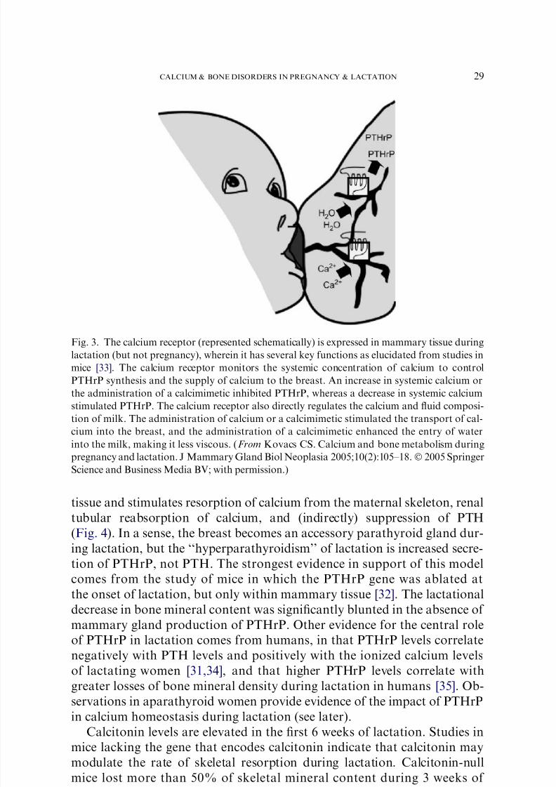
Fig. 3. The calcium receptor (represented schematically) is expressed in mammary tissue during
lactation (but not pregnancy), wherein it has several key functions as elucidated from studies in
mice [33]. The calcium receptor monitors the systemic concentration of calcium to control
PTHrP synthesis and the supply of calcium to the breast. An increase in systemic calcium or
the administration of a calcimimetic inhibited PTHrP, whereas a decrease in systemic calcium
stimulated PTHrP. The calcium receptor also directly regulates the calcium and fluid composi-
tion of milk. The administration of calcium or a calcimimetic stimulated the transport of cal-
cium into the breast, and the administration of a calcimimetic enhanced the entry of water
into the milk, making it less viscous. (From Kovacs CS. Calcium and bone metabolism during
pregnancy and lactation. J Mammary Gland Biol Neoplasia 2005;10(2):105–18.Ó 2005 Springer
Science and Business Media BV; with permission.)
29CALCIUM & BONE DISORDERS IN PREGNANCY & LACTATION
7/30/2019 Calcio Enel Emba Razo
http://slidepdf.com/reader/full/calcio-enel-emba-razo 10/31
lactation, approximately twice that of normal littermate sisters [14,36]. The
calcitonin-depleted mice still regained all of the lost mineral content after
weaning, which indicates that although calcitonin is needed in the short-
term to prevent severe losses of mineral content and potential skeletal fragil-
ity, calcitonin is not required in the long-term because the skeletal losses of
mineral are restored anyway. The human equivalent of absence of calcitonin
might explain some cases of osteoporosis of lactation (see later).
Intestinal absorption of calcium
Intestinal calcium absorption decreases to the nonpregnant rate from the
increased rate of pregnancy. This decrease in absorption corresponds to the
decrease in 1,25(OH)2D3 levels to normal.
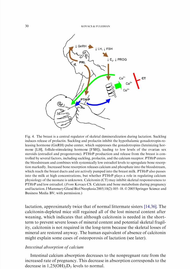
Fig. 4. The breast is a central regulator of skeletal demineralization during lactation. Suckling
induces release of prolactin. Suckling and prolactin inhibit the hypothalamic gonadotropin-re-leasing hormone (GnRH) pulse center, which suppresses the gonadotropins (luteinizing hor-
mone [LH], follicle-stimulating hormone [FSH]), leading to low levels of the ovarian sex
steroids (estradiol and progesterone). PTHrP production and release from the breast is con-
trolled by several factors, including suckling, prolactin, and the calcium receptor. PTHrP enters
the bloodstream and combines with systemically low estradiol levels to upregulate bone resorp-
tion markedly. Increased bone resorption releases calcium and phosphate into the bloodstream,
which reach the breast ducts and are actively pumped into the breast milk. PTHrP also passes
into the milk at high concentrations, but whether PTHrP plays a role in regulating calcium
physiology of the neonate is unknown. Calcitonin (CT) may inhibit skeletal responsiveness to
PTHrP and low estradiol. (From Kovacs CS. Calcium and bone metabolism during pregnancy
and lactation. J Mammary Gland Biol Neoplasia 2005;10(2):105–18.Ó2005 Springer Science andBusiness Media BV; with permission.)
30 KOVACS & FULEIHAN
7/30/2019 Calcio Enel Emba Razo
http://slidepdf.com/reader/full/calcio-enel-emba-razo 11/31
Renal handling of calcium
In humans, the GFR decreases during lactation, and the renal excretion
of calcium typically is reduced to very low levels. This situation suggests that
tubular reabsorption of calcium must be increased, to account for reduced
calcium excretion in the setting of increased serum calcium.
Skeletal calcium metabolism
Histomorphometric data from animals consistently show increased bone
turnover during lactation, with losses of 30% of bone mineral achieved dur-
ing 2 to 3 weeks of normal lactation in the rat [1], whereas a similar amount
is lost in the lactating mouse within 21 days [19]. The loss is greatest in thetrabecular bone of rats and mice. Comparative histomorphometric data are
lacking for humans, and in place of that, serum markers of bone formation
and urinary markers of bone resorption have been assessed in numerous
cross-sectional and prospective studies of lactation. Some confounding fac-
tors discussed with respect to pregnancy apply to the use of these markers in
lactating women. During lactation, GFR is reduced, and the intravascular
volume is more contracted. Urinary markers of bone resorption (24-hour
collection) increase two to three times above normal during lactation and
are higher than the levels attained in the third trimester. Serum markersof bone formation (not adjusted for hemoconcentration or reduced GFR)
are generally high during lactation and increase over the levels attained dur-
ing the third trimester. Total alkaline phosphatase declines immediately
postpartum owing to loss of the placental fraction, but still may remain
above normal because of the elevation in the bone-specific fraction. Despite
the confounding variables, these findings suggest that bone turnover is sig-
nificantly increased during lactation.
In women, serial measurements of bone density during lactation (by SPA,
DPA, or DXA) have shown a decline of 3% to 10% in bone mineral contentafter 2 to 6 months of lactation at trabecular sites (lumbar spine, hip, femur
and distal radius), with smaller losses at cortical sites [1,29,37]. The peak
rate of loss is 1% to 3% per month, far exceeding the rate of 1% to 3%
per year that can occur in women with postmenopausal osteoporosis, who
are considered to be losing bone rapidly. Loss of bone mineral from the ma-
ternal skeleton seems to be a normal consequence of lactation and may not
be preventable by increasing the calcium intake above the recommended di-
etary allowance. Several studies have shown that calcium supplementation
does not reduce significantly the amount of bone lost during lactation[38–41]. The lactational decrease in bone mineral density correlates with
the amount of calcium lost in the breast milk [42].
The mechanisms controlling the rapid loss of skeletal calcium content are
not fully understood. The reduced estrogen levels of lactation are important,
but are unlikely to be the sole explanation. To estimate the effects of estro-
gen deficiency during lactation, it is worth noting the alterations in calcium
31CALCIUM & BONE DISORDERS IN PREGNANCY & LACTATION
7/30/2019 Calcio Enel Emba Razo
http://slidepdf.com/reader/full/calcio-enel-emba-razo 12/31
and bone metabolism that occur in reproductive-age women who have es-
trogen deficiency induced by gonadotropin-releasing hormone agonist ther-
apy for endometriosis and other conditions. Six months of acute estrogendeficiency induced by gonadotropin-releasing hormone agonist therapy
leads to 1% to 4% losses in trabecular (but not cortical) bone density, in-
creased urinary calcium excretion, and suppression of 1,25(OH)2D3 and
PTH levels [1]. During lactation, women are not as estrogen deficient, but
they lose more bone mineral density (at trabecular and cortical sites),
have normal (as opposed to low) 1,25(OH)2D3 levels, and have reduced
(as opposed to increased) urinary calcium excretion. The difference between
isolated estrogen deficiency and lactation may be due to the effects of other
factors (eg, PTHrP) that add to the effects of estrogen withdrawal in lacta-tion (Fig. 5). The relative influences of estrogen deficiency and PTHrP have
been partially discerned in normal mice, in which it has been shown that
treatment with pharmacologic doses of estrogen blunted, but did not abol-
ish, the normal demineralization that occurs during lactation [43].
The bone density losses of lactation are substantially reversed during wean-
ing at a rate of 0.5% to 2% per month [1,29,40]. The mechanism for this res-
toration of bone density is uncertain and largely unexplored, but preliminary
evidence from animal models suggests that PTH, calcitriol, calcitonin, and es-
trogen may not be required to achieve that restoration. In the long-term, theconsequences of lactation-induced depletion of bone mineral seem clinically
unimportant in most women. Most epidemiologic studies of premenopausal
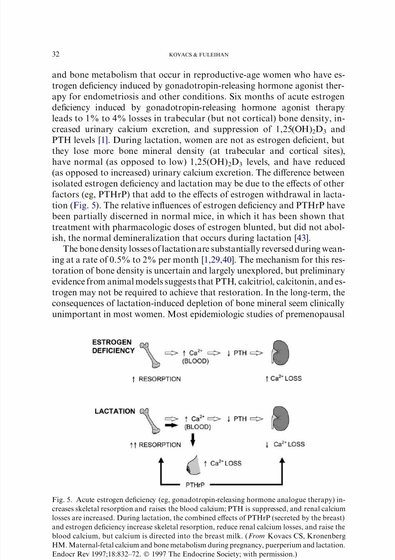
Fig. 5. Acute estrogen deficiency (eg, gonadotropin-releasing hormone analogue therapy) in-
creases skeletal resorption and raises the blood calcium; PTH is suppressed, and renal calcium
losses are increased. During lactation, the combined effects of PTHrP (secreted by the breast)
and estrogen deficiency increase skeletal resorption, reduce renal calcium losses, and raise the
blood calcium, but calcium is directed into the breast milk. (From Kovacs CS, Kronenberg
HM. Maternal-fetal calcium and bone metabolism during pregnancy, puerperium and lactation.
Endocr Rev 1997;18:832–72. Ó 1997 The Endocrine Society; with permission.)
32 KOVACS & FULEIHAN
7/30/2019 Calcio Enel Emba Razo
http://slidepdf.com/reader/full/calcio-enel-emba-razo 13/31
and postmenopausal women have found no adverse effect of a history of
lactation on peak bone mass, bone density, or hip fracture risk [1,29].
Disorders of bone and mineral metabolism during pregnancy and lactation
As previously discussed, pregnancy is a state of hyperabsorptive hyper-
calciuria, characterized by high levels of calcitriol, increasing levels of
PTHrP, suppressed PTH levels, stable serum ionized calcium levels, and
enhanced urinary calcium excretion (see Fig. 2). Lactation is characterized
by further increments in PTHrP levels, whereas calcitriol levels return to
normal. The estimated daily increase in calcium requirements (0.3 g/d) to
meet the fetal demands for bone mineralization and the maternal require-
ments for milk synthesis are largely through enhanced intestinal calcium ab-
sorption during pregnancy and through maternal bone resorption during
lactation [42]. Disorders of bone and mineral homeostasis that occur in
the nonpregnant state may manifest differently during pregnancy and lacta-
tion as a result of the differing hormonal changes that occur in these two dis-
tinct reproductive intervals.
Primary hyperparathyroidism
Primary hyperparathyroidism occurs rarely during pregnancy; the trueincidence is unknown because hyperparathyroidism may remain asymptom-
atic and go undiagnosed in uncomplicated pregnancies. In the general pop-
ulation, the estimated incidence of hyperparathyroidism increased from 16/
100,000 before 1974 (before routine automated screening) to a peak of 112/
100,000 years later, then subsequently declined to 4/100,000 [44]. Most sub-
jects were older than age 45 years [44]. The incidence of hyperparathyroid-
ism in women of childbearing age in an older series was estimated to be
approximately 8/100,000 per year [45]. Approximately 150 cases have
been reported in the English literature to date [46,47]. Two studies retrospec-tively evaluated 850 parathyroidectomies during the period 1960–1991 and
750 parathyroidectomies during 1975–1996 and revealed that parathyroid-
ectomies during pregnancies accounted for 1.4% and 0.8% of total surgeries
[46,48]. The diagnosis may be obscured by the normal pregnancy-induced
changes that decrease the total serum calcium and suppress PTH; finding
the ionized calcium to be increased and the PTH to be detectable would in-
dicate primary hyperparathyroidism in most cases.
Hyperparathyroidism in a pregnant patient can mean considerable mor-
bidity for the mother and the fetus. Complications have been reported in67% of mothers and 80% of fetuses and neonates [49], complications that
are in large part due to maternal hypercalcemia. The histopathologic distri-
bution in hyperparathyroidism of pregnancy is comparable to that reported
in large series spanning all age groups [50]. A series of 100 cases of hyper-
parathyroidism diagnosed during pregnancy or postdelivery revealed adeno-
mas in 89%, hyperplasia in 9%, and carcinoma in 2% [46].
33CALCIUM & BONE DISORDERS IN PREGNANCY & LACTATION
7/30/2019 Calcio Enel Emba Razo
http://slidepdf.com/reader/full/calcio-enel-emba-razo 14/31
Manifestations and complications in pregnant women
In a report of 45 pregnant women with hyperparathyroidism, 38% com-
plained of nausea, vomiting, or abdominal pain; 24% reported renal colic;22% had muscular weakness; 22% manifested mental symptoms; and
11% complained of skeletal pain or fatigue; only 20% were asymptomatic
[51]. Other clinical manifestations included hyperemesis gravidarum, weight
loss, seizure, or other symptoms mimicking preeclampsia [46,48]. Many of
the aforementioned symptoms are nonspecific and could have been due to
the pregnancy itself. Objective findings included the following: 24% had
nephrolithiasis or nephrocalcinosis, 16% had urinary tract infections,
13% had bone disease on radiograph, and 11% had pancreatitis [51]. In an-
other large series of 63 cases, 38% had evidence of bone disease, 54% hadevidence of renal disease, and 30% had evidence of both [52]. The high prev-
alence of stone disease may be explained by pregnancy-induced hyperab-
sorptive hypercalciuria that augments the hypercalciuria that otherwise
would occur secondary to hyperparathyroidism itself. The prevalence of
pancreatitis complicating hyperparathyroidism varied among reports (range
6–13%) [51,53–55] and usually occurred during the second or third trimester
[56]. In a series of 75 cases, pancreatitis was the presenting illness in 5 cases,
2 of which had concomitant hypercalcemic crisis [55]. Of the few other cases
of acute parathyroid crisis reported during pregnancy [53,57], one becameclinically apparent 3 days postpartum followed by rapid deterioration, pan-
creatitis, respiratory failure, shock, and unsuccessful resuscitation [57]. The
patient had been hypercalcemic at 16 weeks of gestation, but was asymp-
tomatic at that time, leading the authors to suggest that the increased fetal
need for calcium may have protected the mother from severe hypercalcemia
before delivery [57]. Susceptibility to fractures owing to hyperparathyroid-
ism during pregnancy is uncommon. Bilateral femoral neck fractures and
rib fractures have been reported in two cases of severe hyperparathyroidism
diagnosed during pregnancy, wherein PTH levels were 20-fold above the up-per limit of normal, and in one case, the hyperparathyroidism was due to
parathyroid carcinoma [58,59].
Complications in Fetuses
The most frequent serious complications in fetuses include stillbirth, mis-
carriage, and neonatal tetany. The percentage of affected pregnancies termi-
nating in stillbirth, neonatal death, and neonatal tetany declined over the
decades from 13% to 2%, from 8% to 2%, and from 38% to 15% [52].
Perinatal death occurred in 25% to 30% of neonates, whereas neonatalcomplications were noted in 50%, with tetany being at the forefront in in-
fants born to untreated mothers [46,60–62]. Neonatal tetany is a common
presentation of unrecognized hyperparathyroidism during pregnancy. The
pathophysiology of the hypocalcemia and tetany is the suppression of fetal
parathyroid function from maternal hypercalcemia, which becomes clini-
cally evident when the maternal calcium flow is interrupted at birth [63].
34 KOVACS & FULEIHAN
7/30/2019 Calcio Enel Emba Razo
http://slidepdf.com/reader/full/calcio-enel-emba-razo 15/31
Although the neonatal hypocalcemia and hypoparathyroidism are usually
transient, resolving with treatment within 3 to 5 months [52], it has been re-
ported to occur initially as late as 2.5 months postpartum [64] and may bepermanent [52,61,65]. Bottle-fed infants were more likely to develop hypo-
calcemia than breastfed ones because of the higher phosphate-to-calcium ra-
tio in cow’s milk compared with breast milk [52].
Management of the mother and neonate
Parathyroidectomy performed during pregnancy prevents fetal and neo-
natal morbidities. The first successful parathyroidectomy during pregnancy
was performed by Petit and Clark in 1947 [66]. A review comparing the out-
comes of 109 mothers with hyperparathyroidism during pregnancy whowere treated medically (n ¼ 70) or surgically (n ¼ 39) revealed that neonates
of mothers with untreated hypercalcemia run a greater risk of complications
[46]. In patients treated medically, there were 53% neonatal complications
and 16% neonatal deaths, as opposed to a 12.5% incidence of neonatal
complications and 2.5% neonatal deaths in patients who underwent para-
thyroidectomy [46]. Parathyroidectomy is best performed during the second
trimester, after completion of organogenesis in the fetus and to avoid the
poor outcomes of surgery during the third trimester [51,52,54,62]. In one se-
ries, premature labor with neonatal death occurred in four of seven third-tri-mester surgeries [54]. Parathyroidectomy in the third trimester is warranted,
however, when the risks outweigh the benefits, and the procedure has been
performed successfully in such cases [47,67].
Treatment options for hyperparathyroidism in pregnancy are influenced
by the symptoms and severity of disease and gestational age. Optimal man-
agement requires a multidisciplinary approach; surgery should be performed
only by an experienced parathyroid surgeon. Symptomatic and severe disease
should be treated surgically, preferably in the second trimester, whereas mild
asymptomatic disease diagnosed in the third trimester may continue to be ob-served until after delivery. A consensus for other cases is missing, however.
Medical treatment includes adequate hydration and correction of electrolyte
abnormalities [49]. Pharmacologic agents to treat hypercalcemia have not
been studied adequately in pregnancy. Calcitonin, a pregnancy category B
medication of the US Food and Drug Administration, does not cross the pla-
centa and has been used safely in pregnancy [49]. Oral phosphate, a pregnancy
category C medication, has been used in pregnancy; its most common side ef-
fects are diarrhea and hypokalemia. It should be avoided in patients with
renal failure or high serum phosphate because of the risk of soft tissue calci-fications [49]. Bisphosphonates and mithramycin are contraindicated because
of their adverse effects on fetal development; bisphosphonates in particular
may interfere with normal endochondral bone development. High-dose mag-
nesium has been suggested as a therapeutic alternative for hyperparathyroid-
ism in pregnancy, although its effectiveness is uncertain. This divalent cation
decreases serum PTH and calcium levels by activating the calcium-sensing
35CALCIUM & BONE DISORDERS IN PREGNANCY & LACTATION
7/30/2019 Calcio Enel Emba Razo
http://slidepdf.com/reader/full/calcio-enel-emba-razo 16/31
receptor, and at the same time it treats premature labor associated with hy-
percalcemia [68,69]. Experience with any of the aforementioned pharmaco-
logic therapies is limited to individual case reports [49]; consequently, nomedical therapy can be claimed to be better than any other. Medical therapy
should be coupled with maternal surveillance and the monitoring of serum
calcium and electrolytes and the initiation of antenatal testing with serial fetal
ultrasound starting at 28 weeks of gestation. Parathyroidectomy is recom-
mended postpartum in cases that were followed medically during pregnancy.
Lactation is not contraindicated in women with untreated hyperparathyroid-
ism, but worsening of hypercalcemia and accelerated skeletal losses may be
anticipated because of the combined effects of PTHrP and hyperparathyroid-
ism to stimulate bone resorption.Neonatal hypoparathyroidism secondary to maternal hyperparathyroid-
ism is usually transient (see earlier) and is treated with calcium supplemen-
tation and calcitriol. These neonates also should be fed milk formulas high
in calcium and low in phosphate to minimize the risk of hypocalcemia. The
prevalence and severity of complications from hyperparathyroidism in
mothers and neonates have and will continue to decrease over time, owing
to increased surveillance, earlier intervention, and improved surgical and an-
esthetic technology [52,60,61].
Familial benign hypocalciuric hypercalcemia
Familial benign hypocalciuric hypercalcemia (FBHH) is an autosomal
dominant disorder that is caused by inactivating mutations in the calcium-
sensing receptor that cause hypercalcemia and hypocalciuria [70,71]. In
contrast to patients with hyperparathyroidism, patients with FBHH do
not experience bone demineralization or nephrolithiasis. FBHH has been
reported in pregnancy with no clinical sequelae in the mother [72]. As an-
ticipated, maternal hypercalcemia has caused suppression of PTH synthe-
sis in the fetus, however, and subsequent hypocalcemia and tetany in theneonate [72,73]. The treatment of neonates is similar to that of children
born to mothers with hyperparathyroidism (see earlier).
The calcium-sensing receptor is expressed in the epithelial ducts of breast
tissue and has been shown to modulate the production of PTHrP and the
transport of calcium into milk in a mouse model [33]. Activating mutations
of this receptor in women with FBHH theoretically could enhance the de-
gree of skeletal demineralization during lactation and the calcium content
of milk, but this has not been studied.
Hypoparathyroidism
Patients usually are known to have hypoparathyroidism or aparathyroid-
ism before pregnancy, and the therapeutic dilemma revolves around adjust-
ment of the treatment, which may vary widely. In 1966, O’Leary et al [74]
reported two cases of hypoparathyroidism treated with high doses of
36 KOVACS & FULEIHAN
7/30/2019 Calcio Enel Emba Razo
http://slidepdf.com/reader/full/calcio-enel-emba-razo 17/31
calcium and vitamin D wherein the mothers gave birth to healthy infants
after uncomplicated pregnancies. Despite physiologic increments in en-
dogenous calcitriol levels during pregnancy, several studies since have docu-mented increased requirements for exogenous calcium and calcitriol therapy
as pregnancy progressed in patients with hypoparathyroidism [75–78]. Con-
versely, in numerous other case reports, women with hypoparathyroidism
have been reported to require less calcium and vitamin D supplementation
during pregnancy [1]. Potential explanations for requiring less supplementa-
tion during pregnancy include pregnancy-induced increments in calcitriol
from placental sources, the potential effect of PTHrP in the maternal circu-
lation to stimulate the renal 1a-hydroxylase, and other pregnancy-related
factors (eg, prolactin or placental lactogen) that may stimulate the renal1a-hydroxylase or enhance intestinal calcium absorption independently of
calcitriol. The last-mentioned has been reported exclusively in animal mod-
els [1]. In some case reports, it seems that the normal, artifactual decrease in
total serum calcium during pregnancy was the parameter that led to treat-
ment with increased calcium and calcitriol supplementation. Although few
cases report measurements of ionized calcium, several do mention that the
increments in vitamin D were due to maternal symptoms of hypocalcemia
or tetany.
Consequently, there is no established therapeutic regimen for the treat-ment of hypoparathyroidism during pregnancy, but numerous principles ex-
ist that help to guide treatment decisions. Calcitriol levels normally increase
during pregnancy and contribute (at least in part) to the enhanced intestinal
calcium absorption of pregnancy; most women should receive an increase in
the dosage of calcitriol at least initially. The total serum calcium is less in-
formative, and the ionized calcium should be monitored in these patients.
Undertreatment results in maternal hypocalcemia; increases the risk of pre-
mature labor and of neonatal secondary hyperparathyroidism; and may lead
to neonatal skeletal demineralization, subperiosteal bone resorption, and os-teitis fibrosa cystica [79]. Conversely, overtreatment may lead to maternal
hypercalcemia and neonatal hypoparathyroidism and raises the potential
concerns of teratogenicity that has been shown using older vitamin D prep-
arations [80,81]. The active forms of vitamin D, such as calcitriol and 1a-cal-
cidiol, have the advantages of a shorter half-life and lower risk of toxicity. A
study reported the outcome of pregnancy in 10 women treated with calcitriol
at doses of 0.25 mg/d to 3.25 mg/d [75]. In 8 of 10 pregnancies, healthy infants
were delivered. In two cases, serious adverse events occurred, including pre-
mature closure of the frontal fontanelle and stillbirth, but the causative roleof calcitriol could not be established [75]. Details regarding nine additional
cases of hypoparathyroidism and vitamin D–resistant rickets were provided
in the same publication and confirmed the lack of toxicity or teratogenicity
from vitamin D supplementation during pregnancy [75].
In contrast to the conflicting literature on the effects of pregnancy on
hypoparathyroidism, calcium and vitamin D or calcitriol requirements in
37CALCIUM & BONE DISORDERS IN PREGNANCY & LACTATION

7/30/2019 Calcio Enel Emba Razo
http://slidepdf.com/reader/full/calcio-enel-emba-razo 18/31
hypoparathyroid patients have been shown consistently to decrease during
lactation [77,78], such that the patients become hypercalcemic unless the
supplements are reduced substantially or discontinued. The decreased re-quirement for calcium and calcitriol occurs at a time when circulating
PTHrP levels are high in the maternal circulation of these hypoparathyroid
women [76,82,83]. PTHrP may stimulate endogenous calcitriol formation; in
one patient, the calcitriol level initially declined below the lower limit of nor-
mal when calcium and calcitriol were discontinued, but thereafter the calci-
triol level remained in the lower half of the normal range as lactation
proceeded [82]. PTHrP also facilitates maternal bone resorption in the pres-
ence of postpartum estrogen deficiency. The aforementioned activity may
explain why lactation can eliminate temporarily the requirement for supple-mental calcium and calcitriol in hypoparathyroid women.
The management of hypoparathyroidism during pregnancy and lactation
is challenging. The use of calcium supplementation with calcitriol is recom-
mended, along with monitoring of symptoms of hypocalcemia and of serum
ionized calcium levels (not the total serum calcium) to titrate the calcitriol
dose as pregnancy progresses. In general, the requirements in calcitriol
vary during the second half of pregnancy, but are expected to decrease dur-
ing lactation.
Pseudohypoparathyroidism
In case reports of pseudohypoparathyroidism, a state characterized by in-
herited resistance to PTH, patients have hypocalcemia, hypophosphatemia,
and high PTH levels. Such patients have been reported to become normocal-
cemic during pregnancy without ingesting therapeutic amounts of calcium
and vitamin D [84]. The mechanism by which pseudohypoparathyroidism
is improved in pregnancy is unclear. It may include increased placental se-
cretion of calcitriol, wherein levels have been reported to double or triplein two case reports during the second and third trimesters [84].
The impact of lactation on calcium metabolism in pseudohypoparathy-
roidism is less well documented. These patients do not have skeletal resis-
tance to PTH, and it is possible that calcium and vitamin D requirements
may decrease secondary to enhanced skeletal resorption owing to the com-
bined effects of endogenous high PTH levels, increasing PTHrP release from
the breast, and lactation-induced estrogen deficiency. Women with pseudo-
hypoparathyroidism might be expected to lose more bone density than nor-
mal during lactation, but this has not been studied.
Osteoporosis associated with pregnancy and lactation
Osteoporosis associated with pregnancy and lactation has been recog-
nized for more than 5 decades [85] and usually presents during late preg-
nancy or early postpartum [85–88]. It is still debatable whether pregnancy
38 KOVACS & FULEIHAN

7/30/2019 Calcio Enel Emba Razo
http://slidepdf.com/reader/full/calcio-enel-emba-razo 19/31
and lactation are causal or accidentally associated with the condition. It is
equally unclear whether these osteoporotic fractures reflect architectural
deterioration of a previously abnormal skeleton or whether pregnancyand lactation themselves account in large part for the bone loss and fragility
fractures, situations that may be compounded by low calcium intake and
vitamin D deficiency. As reviewed previously, skeletal demineralization nor-
mally occurs during lactation as a consequence of the actions of mammary
gland–derived PTHrP in the setting of low estradiol levels and is not pre-
ventable by increased calcium intake; osteoporotic fractures may occur in
some women during lactation when the demineralization is excessive or
the skeleton is unable to tolerate the normal lactational losses of mineral.
PTHrP levels were high in one case of lactational osteoporosis and werefound to remain elevated for months after weaning [89]. One study, which
followed 13 women with pregnancy-associated osteoporosis for 8 years,
showed that bone mineral density at the spine and hip increased significantly,
leading the investigators to conclude that a large part of the bone loss
had been related to the pregnancy itself [86]. Conversely, a high prevalence
of fractures in 35 subjects presenting with pregnancy-associated osteoporo-
sis raised the possibility of a genetic factor [90]. The recognition that absence
of endogenous calcitonin in mice more than doubles the lactational losses
raises the consideration that some women might have a genetic deficiencyin calcitonin, its receptor, or some other factor [14,36]. Because bone density
is not normally measured in premenopausal women, the bone density before
pregnancy or at the end of lactation is usually unknown, and the debate re-
garding the relative contribution of pregnancy or lactation-associated bone
changes versus preexisting abnormalities in the skeleton will continue.
Clinical features
Patients present at a mean age of 27 to 28 years, usually in the setting of
a first pregnancy, and no clear association with parity has been found [86– 88]. In more than 60% of cases, patients complain of back pain in the lower
thoracic or lumbar area, pain that can be quite debilitating secondary to
vertebral collapse [86–88]. In such cases, the pain usually improves sponta-
neously over weeks, but in a few the severe pain may persist for several years
[87]. Others present with hip pain, otherwise known as transient osteoporo-
sis of the hip, as part of a syndrome of monarticular or polyarticular pain
over other lower extremity joints, including the ankles, which is accentuated
with the use of the joint [86–88,91]. Of the more than 200 cases of transient
osteoporosis of the hip that have been reported, one third occurred inwomen in their third trimester of pregnancy or in the early postpartum pe-
riod [91–93]. The differential diagnosis of this condition includes inflamma-
tory joint disorders, avascular necrosis of the hip, bone marrow edema, and
reflex sympathetic dystrophy. In contrast to the last-mentioned condition,
patients with transient regional osteoporosis of the hip lack a history of
trauma and the typical physical findings of muscle spasm and skin changes
39CALCIUM & BONE DISORDERS IN PREGNANCY & LACTATION
7/30/2019 Calcio Enel Emba Razo
http://slidepdf.com/reader/full/calcio-enel-emba-razo 20/31
[92]. In contrast to vertebral osteoporosis, recurrences in transient regional
osteoporosis of the hip have been described in 40% of total cases, but no
series has described this syndrome exclusively in pregnant women [91].
Pathogenesis and laboratory findings
The pathogenesis of pregnancy-associated osteoporosis (presenting with
vertebral compression fractures) and transient osteoporosis of the hip dif-
fers. In a few cases of the former, secondary causes of bone loss could be
identified, including anorexia nervosa, hyperparathyroidism, osteogenesis
imperfecta, and corticosteroid or heparin therapy [87,88,90]. One report de-
scribed pregnancy-associated osteoporosis after oocyte donation in a womanwith ovarian failure [94]. Serum calcium and phosphate levels were normal,
and no consistent abnormalities in the calciotropic hormones were reported
[87,88]. Bone biopsy specimens obtained in some cases have confirmed the
diagnosis of osteoporosis, and no osteomalacia was found [87,88]. Bone
density tended to be low when measured [86,88]. In a series of 24 patients,
the mean Z-score was ÿ1.98 (G1.5, n ¼ 15) at the lumbar spine and
ÿ1.48 (G1.5, n ¼ 15) at the total hip [88]. In transient osteoporosis of the
hip, radiographs or MRI revealed reduced bone density and increased water
content of the marrow cavity [91].
Diagnostic studies and therapeutic interventions
Patients should be screened for secondary causes of bone loss, a large
proportion of which may be treatable. Most cases improve symptomatically
within a few weeks with conservative measures [87,91]. Myriad pharmaco-
logic agents have been used in individual cases, including calcium, vitamin
D, testosterone, estrogen, calcitonin, and bisphosphonates, with increments
in bone mineral density reaching 27% at the spine and 7% at the hip inpatient case treated with alendronate for 6 months [87]. Because these are
usually reports on individual cases and lack controls, the efficacy of such
interventions is unproved, and the interventions are not warranted.
In severe cases of osteoporosis, it may be prudent to discourage breast-
feeding, the rationale being that the skeleton may not be able to tolerate
the normal demineralization that lactation would induce. Patients should
be cautioned against carrying heavy weights to avoid additional stress on
the spine, and the use of a supportive corset may be helpful. Patients should
be reassured that substantial increments in bone mineral density will occurover time [86], and that the condition in cases of vertebral collapse is un-
likely to recur. In cases of transient osteoporosis of the hip, patients usually
do well with conservative measures, including bed rest. Symptoms and ra-
diograph abnormalities resolve within a few months of their onset [91],
but may recur, in contrast to cases with vertebral fracture symptoms, which
usually do not recur.
40 KOVACS & FULEIHAN
7/30/2019 Calcio Enel Emba Razo
http://slidepdf.com/reader/full/calcio-enel-emba-razo 21/31
Disturbances in bone and mineral metabolism from the administration of
magnesium sulfate during pregnancy
The administration of intravenous magnesium sulfate for 24 to 72 hours
is one of the mainstay therapies for the treatment of preterm labor and for
the treatment of preeclampsia and eclampsia. Its effect is mediated by action
on the myoneural junction. In vitro at high doses, magnesium suppresses
PTH levels, similarly to other divalent and trivalent cations, albeit with
a lower potency than calcium. This effect now is recognized to occur
through the calcium-sensing receptor, a receptor heavily expressed in the
parathyroid glands and kidneys [70,95]. Long-term tocolytic therapy using
magnesium sulfate generally has been considered safe [96], although few re-
ports have raised concerns about its safety to mothers and neonates.
Maternal complications
Hypocalcemia has been described in several cases in which the women re-
ceived magnesium to suppress premature labor [97–99]. In a study of seven
such cases, a loading dose of 6 g of intravenous magnesium sulfate followed
by a maintenance dose of 2 g/h resulted in a rapid increase in the mean se-
rum magnesium level from 2 mg/dL to 6 mg/dL within 1 hour, coupled with
an almost concomitant decline in the serum PTH levels, which only partiallyrecovered in 3 hours despite substantial decrements in total and ionized se-
rum calcium levels below the lower limit of normal [98]. A similar pattern
for maternal and neonatal profiles was noted in a study of 15 women treated
with magnesium sulfate [99]. A Medline literature search for articles pub-
lished in English in the period 1966–2002 on magnesium sulfate and hypo-
calcemia revealed four cases of maternal symptomatic hypocalcemia, with
serum calcium levels reaching 5.3 mg/dL in one case. Two mothers had
a positive Chvostek and Trousseau sign or tetany; three of these cases
were noted to have concomitant low PTH levels [100].Although the short-term administration of magnesium sulfate may lower
maternal serum calcium through an effect on PTH secretion, long-term ad-
ministration for 2 to 3 weeks was associated with increments in serum PTH
levels, possibly as an appropriate adaptive mechanism to prolonged hypo-
calcemia. It has been suggested that urinary loss of calcium may be a major
pathophysiologic mechanism for the hypocalcemia and in such instances
may result in ultimate impairment of bone mineralization [101]. In a study
of 20 subjects given intravenous magnesium sulfate for premature labor, se-
rum magnesium and phosphorus levels increased, serum calcium levels de-creased concomitantly with an increase in serum PTH, and substantial
increments in urinary magnesium and calcium were noted, reaching mean
levels two to three times the upper limit of normal [101]. Prolonged magne-
sium administration for several weeks also has been associated with mater-
nal forearm bone loss prospectively, osteoporosis by bone mineral density,
and bilateral calcaneal stress fractures postpartum [101–103].
41CALCIUM & BONE DISORDERS IN PREGNANCY & LACTATION
7/30/2019 Calcio Enel Emba Razo
http://slidepdf.com/reader/full/calcio-enel-emba-razo 22/31
Neonatal complications
Administration of intravenous magnesium to mothers before delivery in-
creased neonatal serum magnesium and decreased PTH levels, whereas theeffect on neonatal total and ionized calcium levels varied [97,99]. Studies
evaluating the impact of neonatal hypermagnesemia on neonatal outcomes
have yielded conflicting results [97,104–106]. Respiratory depression and hy-
potonia were reported in 16 cases of neonatal hypermagnesemia [106]. A fol-
low-up study of 35 infants born to toxemic mothers treated with magnesium
sulfate for 2 to 4 days suggested that neonates whose mothers had received
prolonged administration may be more likely to manifest respiratory depres-
sion [105]. Cord blood and neonatal serum magnesium levels were of little
diagnostic value to the clinical picture except in cases of severe hypermagne-semia [105,107], confirming the general observation that circulating serum
magnesium levels do not reflect intracellular and total body magnesium
stores. Conversely, a study of 118 infants born to mothers who had received
intramuscular magnesium in doses of 10 to 95 g concluded that the neonatal
death rate was lower than that of the total newborn population [104]. Respi-
ratory depression, hypotonia, and need for intubation were not evaluated in
that report, however [104]. Neonatal bone abnormalities have been reported
with long-term use of magnesium sulfate. The first report by Lamm et al
[108] described a congenital form of rickets manifested by defective ossifica-tion of bone and enamel in the teeth of the offspring of mothers who had
received magnesium sulfate during pregnancy. Several cases of abnormal
mineralization of metaphyses since have been reported in neonates born
to mothers who received prolonged intravenous magnesium and had high
serum magnesium levels [109–111]. The proposed mechanism for defective
mineralization of bones involves the inhibition of calcification of osteoid
in which calcium-binding sites are occupied by magnesium [109,110].
Some of these conflicting findings regarding neonatal morbidity from ma-
ternal administration of magnesium sulfate may be explained by the routeand duration of magnesium sulfate administration, by the variability in
the ranges of cord magnesium levels reached, and by the gestational age
of the neonates. Postdelivery, there was a delay in normalization of the se-
rum magnesium level for a few days resulting from the limitation of magne-
sium excretion by the newborn’s immature kidneys [97].
Management issues
There are no guidelines for the monitoring of pregnant women receiving
magnesium sulfate. Neonates born to mothers receiving long-term magne-sium sulfate and experiencing severe hypermagnesemia (O 7 mg/dL) are
more likely to have hypotonia, respiratory depression, and bone abnormal-
ities [97,105,107,111]. Subjects receiving such therapy for periods exceeding
1 or 2 days should be monitored carefully, with the measurement of ma-
ternal serum calcium and magnesium levels, coupled with monitoring the
fetal movement. Symptomatic neonates can be managed by maintaining
42 KOVACS & FULEIHAN
7/30/2019 Calcio Enel Emba Razo
http://slidepdf.com/reader/full/calcio-enel-emba-razo 23/31
ventilation for 24 to 48 hours and providing intravenous fluids for electro-
lyte balance for a few days, after which marked clinical improvement is usu-
ally noted [105]. Intravenous calcium to antagonize the central nervoussystem depression and peripheral neuromuscular blockade has been used,
with careful monitoring of the heart rate [105].
Low calcium intake
There are limited data that low calcium intake in the mother may adversely
affect fetal mineral accretion and maternal bone mineral metabolism [112].
In women with low dietary calcium intake, there are differing results as to
whether or not calcium supplementation during pregnancy improved mater-nal or neonatal bone density [3]. There is short-term evidence that maternal
turnover was reduced when 1.2 g of calcium was given for 20 days to 31 Mex-
ican women with a mean calcium intake of 1 g during weeks 25 to 30 of ges-
tation [113]. In a double-blind study conducted in 256 pregnant women, 2 g of
calcium supplementation improved bone mineral content in infants of supple-
mented mothers who were in the lowest quintile of calcium intake [114].
During lactation, there is no firm evidence that low calcium intake leads
to impaired breast milk quality or accentuates maternal bone loss [115].
Even in women with very low calcium intakes, the same amount of mineralwas lost during lactation from the skeleton compared with women who had
supplemented calcium intakes, and the breast milk calcium content was
unaffected by calcium intake or vitamin D status [116–118]. Conversely,
because high calcium intakes do not affect the degree of skeletal demineral-
ization that occurs during lactation [38–41], it is unlikely that increasing cal-
cium supplementation above normal would affect skeletal demineralization.
In general, the physiologic changes in calcium and bone metabolism that
usually occur during pregnancy and lactation are likely to be sufficient for
fetal bone growth and breast milk production in women with reasonablysufficient calcium intake [115]. The inclusion of calcium supplementation
for pregnant women with low calcium intake could be defended, however,
and is strengthened further by the possible link between low calcium intake,
preeclampsia, and increased blood pressure in the offspring [112]. Increased
calcium intake also is recommended in adolescent mothers to meet the need
of reproduction and maternal bone growth [115]. There is evidence that the
skeleton of an undernourished adolescent recovers fully from lactational
losses, but there is some concern that peak bone mass might not be attained
subsequently [119].
Vitamin D deficiency
In humans and in animal models, vitamin D deficiency or the absence
of the vitamin D receptor can lead to adverse neonatal outcomes, includ-
ing neonatal rickets, craniotabes, decreased wrist ossification centers, and
43CALCIUM & BONE DISORDERS IN PREGNANCY & LACTATION
7/30/2019 Calcio Enel Emba Razo
http://slidepdf.com/reader/full/calcio-enel-emba-razo 24/31
impaired tooth enamel formation [120]. These features generally are not
present at birth, but appear postnatally as intestinal calcium absorption be-
comes vitamin D dependent. Although vitamin D supplementation of preg-nant mothers at risk for vitamin D deficiency improved neonatal serum
calcium concentrations and resulted in a trend for greater height and length
in the offspring [112], a Cochrane review of 232 women in two trials re-
ported conflicting results [121]. Although there is no evidence to indicate
a beneficial effect of vitamin D supplementation during pregnancy above
the amounts needed to prevent vitamin D deficiency, optimal levels for vita-
min D supplementation are unclear [122]. An arbitrary daily recommended
intake has been set at 400 IU/d, but likely needs revision upward [123]. Rec-
ommendations for vitamin D supplementation either for women of child-bearing age or for lactating women were not mentioned in the new US
dietary guidelines issued in 2005 [124].
Scientific data pertaining to vitamin D supplementation during lactation
are even scarcer than data on vitamin D supplementation during pregnancy.
An arbitrary daily recommended intake has been set at 400 IU/d, but
may be insufficient [123]. Whether vitamin D deficiency impairs the ability
to restore maternal skeleton postweaning is unclear [1]. Lactating mothers
supplemented with 1000 IU to 2000 IU of vitamin D for 15 weeks experi-
enced increments in circulating maternal 25-hydroxyvitamin D3 levels of 16 ng/mL to 23 ng/mL [122]. It has been suggested that vitamin D supple-
mentation of lactating mothers would improve vitamin D nutrition in the
mother and the breastfeeding infant; this has not been shown yet, but is cur-
rently under investigation [122]. Breastfed infants of vitamin D–deficient
mothers should receive vitamin D supplementation to avoid nutritional rick-
ets [125]. There is low penetrance of vitamin D into breast milk, and it is
more efficient to give the vitamin D supplement directly to the infant, al-
though supplementing the mother with high doses has been shown to
work [126].
Hypercalcemia of malignancy
Hypercalcemia of malignancy, an extremely rare occurrence, has been re-
ported in two casesdone in metastatic breast cancer and the other in renal
cell carcinoma [127,128]. In both reports, the disease was rapidly progressive
and resulted in premature delivery at 29 and 32 weeks of gestation and ma-
ternal demise within 4 months postpartum. On the first day postdelivery,
both infants were hypercalcemic, and one subsequently developed hypocal-cemia from transient hypoparathyroidism. Intravenous pamidronate was
used shortly before delivery in one case with normalization of maternal se-
rum calcium within 5 days of pamidronate administration [128]. Treatment
in such cases includes adequate aggressive hydration with close monitoring,
furosemide, and possibly calcitonin. Because bisphosphonates cross the pla-
centa, their use should be reserved for life-threatening situations.
44 KOVACS & FULEIHAN
7/30/2019 Calcio Enel Emba Razo
http://slidepdf.com/reader/full/calcio-enel-emba-razo 25/31
Neonatal hypoparathyroidism from maternal therapy with radioactive
iodine
Inadvertent maternal therapy with radioactive iodine during pregnancy
may result in neonatal hypoparathyroidism, similar to what has been re-
ported in adults treated with high-dose radioactive iodine. A mother was re-
ported to have received 103 mCi of iodine-131 (131I) for thyroid carcinoma
at 10 weeks of gestation when she was unaware of her pregnancy. Her neo-
nate experienced occasional episodes of stiffening and turning blue during
the first 2 months that accelerated and led to a hospital admission for respi-
ratory distress, tonic-clonic seizures, and ultimate tracheostomy [129]. The
infant was found be hypocalcemic, with documented hypoparathyroidism
and severe hypothyroidism. The infant was discharged after 1 month of hos-
pitalization with a tracheostomy and ongoing treatment with calcium, dihy-
drotachysterol, and thyroid hormone. It is likely that the fetal thyroid gland
accumulated sufficient amounts of 131I to result in destruction of fetal thy-
roid and parathyroid tissue from the emitted b particles [129].
Summary
Studies of pregnant women indicate that the fetal calcium demand is metlargely by intestinal calcium absorption, which from early pregnancy on-
ward more than doubles. The studies of biochemical markers of bone turn-
over, DXA, and ultrasound are inconclusive, but suggest that the maternal
skeleton also contributes calcium to the developing fetus. In contrast, during
lactation, skeletal calcium resorption is the dominant mechanism by which
calcium is supplied to the breast milk; renal calcium conservation is also ap-
parent. Lactation produces an obligatory skeletal calcium loss regardless of
maternal calcium intake, but the calcium is completely restored to the skel-
eton after weaning through mechanisms that are not understood. The adap-tations during pregnancy and lactation lead to novel presentations and
management issues for known disorders of calcium and bone metabolism,
such as primary hyperparathyroidism, hypoparathyroidism, and vitamin
D deficiency. Finally, although some women experience fragility fractures
as a consequence of pregnancy or lactation, in most women the changes
in calcium and bone metabolism during pregnancy and lactation are normal
and without adverse consequences in the long-term.
References
[1] Kovacs CS, Kronenberg HM. Maternal-fetal calcium and bone metabolism during preg-
nancy, puerperium and lactation. Endocr Rev 1997;18:832–72.
[2] Kovacs CS. Calcium and bone metabolism in pregnancy and lactation. J Clin Endocrinol
Metab 2001;86:2344–8.
[3] Prentice A. Pregnancy and lactation. In: Glorieux FH, Petifor JM, Ju ¨ ppner H, editors.
Pediatric bone: biology and diseases. New York: Academic Press; 2003. p. 249–69.
45CALCIUM & BONE DISORDERS IN PREGNANCY & LACTATION
7/30/2019 Calcio Enel Emba Razo
http://slidepdf.com/reader/full/calcio-enel-emba-razo 26/31
[4] Chesney RW, Specker BL, McKay CP. Mineral metabolism during pregnancy and lacta-
tion. In: Coe FL, Favus MJ, editors. Disorders of bone and mineral metabolism. 2nd edi-
tion. Philadelphia: Lippincott, Williams & Wilkins; 2002. p. 347–59.
[5] Taylor RN, Lebovic DI. The endocrinology of pregnancy. In: Greenspan FS, Gardner DG,
editors. Basic and clinical endocrinology. 7th edition. New York: Lange Medical Books/
McGraw-Hill; 2004. p. 637–57.
[6] Dahlman T, Sjoberg HE, Bucht E. Calcium homeostasis in normal pregnancy and puerpe-
rium: a longitudinal study. Acta Obstet Gynecol Scand 1994;73:393–8.
[7] Gallacher SJ, Fraser WD, Owens OJ, et al. Changes in calciotrophic hormones and bio-
chemical markers of bone turnover in normal human pregnancy. Eur J Endocrinol 1994;
131:369–74.
[8] Cross NA, Hillman LS, Allen SH, et al. Calcium homeostasis and bone metabolism during
pregnancy, lactation, and postweaning: a longitudinal study. Am J Clin Nutr 1995;61:
514–23.
[9] Rasmussen N, Frolich A, Hornnes PJ, Hegedus L. Serum ionized calcium and intact para-
thyroid hormone levels during pregnancy and postpartum. Br J Obstet Gynaecol 1990;97:
857–9.
[10] Seki K, Makimura N, Mitsui C, et al. Calcium-regulating hormones and osteocalcin levels
during pregnancy: a longitudinal study. Am J Obstet Gynecol 1991;164:1248–52.
[11] Gao P, D’Amour P. Evolution of the parathyroid hormone (PTH) assaydimportance
of circulating PTH immunoheterogeneity and of its regulation. Clin Lab 2005;51:
21–9.
[12] Singh HJ, Mohammad NH, Nila A. Serum calcium and parathormone during normal preg-
nancy in Malay women. J Matern Fetal Med 1999;8:95–100.
[13] Turner M, Barre PE, Benjamin A, et al. Does the maternal kidney contribute to the in-creased circulating 1,25-dihydroxyvitamin D concentrations during pregnancy? Miner
Electrolyte Metab 1988;14:246–52.
[14] Woodrow JP, Noseworthy CS, Fudge NJ, et al. Calcitonin/calcitonin gene-related peptide
protect the maternal skeleton from excessive resorption during lactation [abstract]. J Bone
Miner Res 2003;18(Suppl 2):S37.
[15] Horwitz MJ, Tedesco MB, Sereika SM, et al. Direct comparison of sustained infusion of
human parathyroid hormone-related protein-(1–36). J Clin Endocrinol Metab 2003;88:
1603–9.
[16] Kovacs CS, Lanske B, Hunzelman JL, et al. Parathyroid hormone-related peptide (PTHrP)
regulates fetal-placental calcium transport through a receptor distinct from the PTH/
PTHrP receptor. Proc Natl Acad Sci U S A 1996;93:15233–8.[17] Cornish J, Callon KE, Nicholson GC, Reid IR. Parathyroid hormone-related protein-
(107–139) inhibits bone resorption in vivo. Endocrinology 1997;138:1299–304.
[18] Marie PJ, Cancela L, Le Boulch N, Miravet L. Bone changes due to pregnancy and lacta-
tion: influence of vitamin D status. Am J Physiol 1986;251:E400–6.
[19] Sharpe CJ, Fudge NJ, Kovacs CS. A rapid 35% flux in bone mass occurs during pregnancy
and lactation cycles in mice [abstract]. International Bone and Mineral Society Meeting,
Osaka, Japan, June 3–7, 2003. Bone 2003;32(Suppl):S227.
[20] Purdie DW, Aaron JE, Selby PL. Bone histology and mineral homeostasis in human preg-
nancy. Br J Obstet Gynaecol 1988;95:849–54.
[21] Naylor KE, Iqbal P, Fledelius C, et al. The effect of pregnancy on bone density and bone
turnover. J Bone Miner Res 2000;15:129–37.[22] Black AJ, Topping J, Durham B, et al. A detailed assessment of alterations in bone turn-
over, calcium homeostasis, and bone density in normal pregnancy. J Bone Miner Res
2000;15:557–63.
[23] Ritchie LD, Fung EB, Halloran BP, et al. A longitudinal study of calcium homeostasis dur-
ing human pregnancy and lactation and after resumption of menses. Am J Clin Nutr 1998;
67:693–701.
46 KOVACS & FULEIHAN
7/30/2019 Calcio Enel Emba Razo
http://slidepdf.com/reader/full/calcio-enel-emba-razo 27/31
[24] Ulrich U, Miller PB, Eyre DR, et al. Bone remodeling and bone mineral density during
pregnancy. Arch Gynecol Obstet 2003;268:309–16.
[25] Kaur M, Pearson D, Godber I, et al. Longitudinal changes in bone mineral density during
normal pregnancy. Bone 2003;32:449–54.
[26] Gambacciani M, Spinetti A, Gallo R, et al. Ultrasonographic bone characteristics during
normal pregnancy: longitudinal and cross-sectional evaluation. Am J Obstet Gynecol
1995;173:890–3.
[27] Pearson D, Kaur M, San P, et al. Recovery of pregnancy mediated bone loss during lacta-
tion. Bone 2004;34:570–8.
[28] To WW, Wong MW, Leung TW. Relationship between bone mineral density changes in
pregnancy and maternal and pregnancy characteristics: a longitudinal study. Acta Obstet
Gynecol Scand 2003;82:820–7.
[29] Sowers M. Pregnancy and lactation as risk factors for subsequent bone loss and osteopo-
rosis. J Bone Miner Res 1996;11:1052–60.
[30] Paton LM, Alexander JL, Nowson CA, et al. Pregnancy and lactation have no long-term
deleterious effect on measures of bone mineral in healthy women: a twin study. Am J
Clin Nutr 2003;77:707–14.
[31] Dobnig H, Kainer F, Stepan V, et al. Elevated parathyroid hormone-related peptide levels
after human gestation: relationship to changes in bone and mineral metabolism. J Clin En-
docrinol Metab 1995;80:3699–707.
[32] VanHouten JN, Dann P, Stewart AF, et al. Mammary-specific deletion of parathyroid hor-
mone-related protein preserves bone mass during lactation. J Clin Invest 2003;112:1429–36.
[33] VanHouten J, Dann P, McGeoch G, et al. The calcium-sensing receptor regulates mam-
mary gland parathyroid hormone-related protein production and calcium transport.
J Clin Invest 2004;113:598–608.[34] Kovacs CS, Chik CL. Hyperprolactinemia caused by lactation and pituitary adenomas is
associated with altered serum calcium, phosphate, parathyroid hormone (PTH), and
PTH-related peptide levels. J Clin Endocrinol Metab 1995;80:3036–42.
[35] Sowers MF, Hollis BW, Shapiro B, et al. Elevated parathyroid hormone-related peptide as-
sociated with lactation and bone density loss. JAMA 1996;276:549–54.
[36] Woodrow JP, Hoff AO, Gagel RF, Kovacs CS. Calcitonin treatment rescues calcitonin-null
mice from excessive bone resorption during lactation [abstract]. J Bone Miner Res 2004;
19(Suppl):SA518.
[37] Laskey MA, Prentice A. Effect of pregnancy on recovery of lactational bone loss [letter].
Lancet 1997;349:1518–9.
[38] Cross NA, Hillman LS, Allen SH, Krause GF. Changes in bone mineral density andmarkers of bone remodeling during lactation and postweaning in women consuming
high amounts of calcium. J Bone Miner Res 1995;10:1312–20.
[39] Kalkwarf HJ, Specker BL, Bianchi DC, Ranz J, Ho M. The effect of calcium supplemen-
tation on bone density during lactation and after weaning. N Engl J Med 1997;337:523–8.
[40] Polatti F, Capuzzo E, Viazzo F, et al. Bone mineral changes during and after lactation.
Obstet Gynecol 1999;94:52–6.
[41] Kolthoff N, Eiken P, Kristensen B, Nielsen SP. Bone mineral changes during pregnancy
and lactation: a longitudinal cohort study. Clin.Sci (Colch) 1998;94:405–12.
[42] Laskey MA, Prentice A, Hanratty LA, et al. Bone changes after 3 mo of lactation: influence
of calcium intake, breast-milk output, and vitamin D-receptor genotype. Am J Clin Nutr
1998;67:685–92.[43] VanHouten JN, Wysolmerski JJ. Low estrogen and high parathyroid hormone-related pep-
tide levels contribute to accelerated bone resorption and bone loss in lactating mice. Endo-
crinology 2003;144:5521–9.
[44] Wermers RA, Khosla S, Atkinson EJ, et al. The rise and fall of primary hyperparathyroid-
ism: a population-based study in Rochester, Minnesota, 1965–1992. Ann Intern Med 1997;
126:433–40.
47CALCIUM & BONE DISORDERS IN PREGNANCY & LACTATION
7/30/2019 Calcio Enel Emba Razo
http://slidepdf.com/reader/full/calcio-enel-emba-razo 28/31
[45] Heath H, Hodgson SF, Kennedy MA. Primary hyperparathyroidism: incidence, mor-
bidity, and potential economic impact in a community. N Engl J Med 1980;302:
189–93.
[46] Kelly TR. Primary hyperparathyroidism during pregnancy. Surgery 1991;110:1028–34.
[47] Haenel LC, Mayfield RK. Primary hyperparathyroidism in a twin pregnancy and review of
fetal/maternal calcium homeostasis. Am J Med Sci 2000;319:191–4.
[48] Kort KC, Schiller HJ, Numann PJ. Hyperparathyroidism and pregnancy. Am J Surg 1999;
177:66–8.
[49] Schnatz PF, Curry SL. Primary hyperparathyroidism in pregnancy: evidence-based man-
agement. Obstet Gynecol Surv 2002;57:365–76.
[50] El-Hajj Fuleihan G. Pathogenesis and etiology of primary hyperparathyroidism. In: Rose
BD, editor. UpToDate 13.1. Wellesley (MA): UpToDate; 2005.
[51] Kristoffersson A, Dahlgren S, Lithner F, Jarhult J. Primary hyperparathyroidism in preg-
nancy. Surgery 1985;97:326–30.
[52] Shangold MM, Dor N, Welt SI, et al. Hyperparathyroidism and pregnancy: a review. Ob-
stet Gynecol Surv 1982;37:217–28.
[53] Clark D, Seeds JW, Cefalo RC. Hyperparathyroid crisis and pregnancy. Am J Obstet Gy-
necol 1981;140:840–2.
[54] Carella MJ, Gossain VV. Hyperparathyroidism and pregnancy: case report and review.
J Gen Intern Med 1992;7:448–53.
[55] Croom RD, Thomas CG. Primary hyperparathyroidism during pregnancy. Surgery 1984;
96:1109–18.
[56] Inabnet WB, Baldwin D, Daniel RO, Staren ED. Hyperparathyroidism and pancreatitis
during pregnancy. Surgery 1996;119:710–3.
[57] Matthias GS, Helliwell TR, Williams A. Postpartum hyperparathyroid crisis: case report.Br J Obstet Gynaecol 1987;94:807–10.
[58] Negishi H, Kobayashi M, Nishida R, et al. Primary hyperparathyroidism and simultaneous
bilateral fracture of the femoral neck during pregnancy. J Trauma 2002;52:367–9.
[59] Hess HM, Dickson J, Fox HE. Hyperfunctioning parathyroid carcinoma presenting as
acute pancreatitis in pregnancy. J Reprod Med 1980;25:83–7.
[60] Wagner G, Transhol L, Melchior JC. Hyperparathyroidism and pregnancy. Acta Endocri-
nol (Copenh) 1964;47:549–64.
[61] Ludwig GD. Hyperparathyroidism in relation to pregnancy. N Engl J Med 1962;267:
637–42.
[62] Delmonico FL, Neer RM, Cosimi AB, et al. Hyperparathyroidism during pregnancy. Am J
Surg 1976;131:328–37.[63] Kaplan EL, Burrington JD, Klementschitsch P, et al. Primary hyperparathyroidism, preg-
nancy, and neonatal hypocalcemia. Surgery 1984;96:717–22.
[64] Ip P. Neonatal convulsion revealing maternal hyperparathyroidism: an unusual case of late
neonatal hypoparathyroidism. Arch Gynecol Obstet 2003;268:227–9.
[65] Bruce J, Strong JA. Maternal hyperparathyroidism and parathyroid deficiency in the child,
with account of effect of parathyroidectomy on renal function, and of attempt to transplant
part of tumor. QJM 1955;24:307–19.
[66] Petit D, Clark R. Hyperparathyroidism and pregnancy. Am J Surg 1947;74:860–4.
[67] Schnatz PF. Surgical treatment of primary hyperparathyroidism during the third trimester.
Obstet Gynecol 2002;99(5 Pt 2):961–3.
[68] Rajala B, Abbasi RA, Hutchinson HT, Taylor T. Acute pancreatitis and primary hyper-parathyroidism in pregnancy: treatment of hypercalcemia with magnesium sulfate. Obstet
Gynecol 1987;70(3 Pt 2):460–2.
[69] Dahan M, Chang RJ. Pancreatitis secondary to hyperparathyroidism during pregnancy.
Obstet Gynecol 2001;98(5 Pt 2):923–5.
[70] Brown EM, Pollak M, Seidman CE, et al. Calcium-ion-sensing cell-surface receptors.
N Engl J Med 1995;333:234–40.
48 KOVACS & FULEIHAN
7/30/2019 Calcio Enel Emba Razo
http://slidepdf.com/reader/full/calcio-enel-emba-razo 29/31
[71] El-Hajj Fuleihan G, Heath HH. Familial benign hypocalciuric hypercalcemia and neonatal
severe hyperparathyroidism. In: Bilezikian JP, Marcus R, Levine MA, editors. The para-
thyroids: basic and clinical concepts. 2nd edition. New York: Academic Press; 1994.
p. 607–23.
[72] Powell BR, Buist NR. Late presenting, prolonged hypocalcemia in an infant of a woman
with hypocalciuric hypercalcemia. Clin Pediatr (Phila) 1990;29:241–3.
[73] Thomas BR, Bennett JD. Symptomatic hypocalcemia and hypoparathyroidism in two in-
fants of mothers with hyperparathyroidism and familial benign hypercalcemia. J Perinatol
1995;15:23–6.
[74] O’Leary JA, Klainer LM, Neuwirth RS. The management of hypoparathyroidism in preg-
nancy. Am J Obstet Gynecol 1966;94:1103–7.
[75] Callies F, Arlt W, Scholz HJ, et al. Management of hypoparathyroidism during preg-
nancydreport of twelve cases. Eur J Endocrinol 1998;139:284–9.
[76] Caplan RH, Beguin EA. Hypercalcemia in a calcitriol-treated hypoparathyroid woman
during lactation. Obstet Gynecol 1990;76:485–9.
[77] Salle BL, Berthezene F, Glorieux FH, et al. Hypoparathyroidism during pregnancy: treat-
ment with calcitriol. J Clin Endocrinol Metab 1981;52:810–3.
[78] Sadeghi-Nejad A, Wolfsdorf JI, Senior B. Hypoparathyroidism and pregnancy: treatment
with calcitriol. JAMA 1980;243:254–5.
[79] Landing BH, Kamoshita S. Congenital hyperparathyroidism secondary to maternal hypo-
parathyroidism. J Pediatr 1970;77:842–7.
[80] Taussig HB. Possible injury to the cardiovascular system from vitamin D. Ann Intern Med
1966;65:1195–200.
[81] Friedman WF, Mills LF. The relationship between vitamin D and the craniofacial and den-
tal anomalies of the supravalvular aortic stenosis syndrome. Pediatrics 1969;43:12–8.[82] Mather KJ, Chik CL, Corenblum B. Maintenance of serum calcium by parathyroid hor-
mone-related peptide during lactation in a hypoparathyroid patient. J Clin Endocrinol
Metab 1999;84:424–7.
[83] Shomali ME, Ross DS. Hypercalcemia in a woman with hypoparathyroidism associated
with increased parathyroid hormone-related protein during lactation. Endocr Pract 1999;
5:198–200.
[84] Breslau NA, Zerwekh JE. Relationship of estrogen and pregnancy to calcium homeostasis
in pseudohypoparathyroidism. J Clin Endocrinol Metab 1986;62:45–51.
[85] Nordin BE, Roper A. Postpregnancy osteoporosis: a syndrome? Lancet 1955;1:431–4.
[86] Phillips AJ, Ostlere SJ, Smith R. Pregnancy-associated osteoporosis: does the skeleton re-
cover? Osteoporos Int 2000;11:449–54.[87] Khovidhunkit W, Epstein S. Osteoporosis in pregnancy. Osteoporos Int 1996;6:345–54.
[88] Smith R, Athanasou NA, Ostlere SJ, Vipond SE. Pregnancy-associated osteoporosis. QJM
1995;88:865–78.
[89] Reid IR, Wattie DJ, Evans MC, Budayr AA. Post-pregnancy osteoporosis associated with
hypercalcaemia. Clin Endocrinol (Oxf) 1992;37:298–303.
[90] Dunne F, Walters B, Marshall T, Heath DA. Pregnancy associated osteoporosis. Clin En-
docrinol (Oxf) 1993;39:487–90.
[91] Lakhanpal S, Ginsburg WW, Luthra HS, Hunder GG. Transient regional osteoporosis:
a study of 56 cases and review of the literature. Ann Intern Med 1987;106:444–50.
[92] Arayssi TK, Tawbi HA, Usta IM, Hourani MH. Calcitonin in the treatment of transient
osteoporosis of the hip. Semin Arthritis Rheum 2003;32:388–97.[93] Curtiss PH Jr, Kincaid WE. Transitory demineralization of the hip in pregnancy: a report
of three cases. J Bone Joint Surg Am 1959;41:1327–33.
[94] Khastgir G, Studd JW, King H, et al. Changes in bone density and biochemical markers of
bone turnover in pregnancy-associated osteoporosis. Br J Obstet Gynaecol 1996;103:716–8.
[95] Brown EM, El-Hajj Fuleihan G, Chen CJ, Kifor O. A comparison of the effects of
divalent and trivalent cations on parathyroid hormone release, 3#,5#-cyclic-adenosine
49CALCIUM & BONE DISORDERS IN PREGNANCY & LACTATION

7/30/2019 Calcio Enel Emba Razo
http://slidepdf.com/reader/full/calcio-enel-emba-razo 30/31
monophosphate accumulation, and the levels of inositol phosphates in bovine parathyroid
cells. Endocrinology 1990;127:1064–71.
[96] Wilkins IA, Goldberg JD, Phillips RN, et al. Long-term use of magnesium sulfate as a to-
colytic agent. Obstet Gynecol 1986;67(3 Suppl):38S–40S.
[97] Donovan EF, Tsang RC, Steichen JJ, et al. Neonatal hypermagnesemia: effect on parathy-
roid hormone and calcium homeostasis. J Pediatr 1980;96:305–10.
[98] Cholst IN, Steinberg SF, Tropper PJ, et al. The influence of hypermagnesemia on serum
calcium and parathyroid hormone levels in human subjects. N Engl J Med 1984;310:
1221–5.
[99] Cruikshank DP, Pitkin RM, Reynolds WA, et al. Effects of magnesium sulfate treatment on
perinatal calcium metabolism: I. maternal and fetal responses. Am J Obstet Gynecol 1979;
134:243–9.
[100] Koontz SL, Friedman SA, Schwartz ML. Symptomatic hypocalcemia after tocolytic ther-
apy with magnesium sulfate and nifedipine. Am J Obstet Gynecol 2004;190:1773–6.
[101] Smith LG Jr, Burns PA, Schanler RJ. Calcium homeostasis in pregnant women receiving
long-term magnesium sulfate therapy for preterm labor. Am J Obstet Gynecol 1992;167:
45–51.
[102] Hung JW, Tsai MY, Yang BY, Chen JF. Maternal osteoporosis after prolonged magne-
sium sulfate tocolysis therapy: a case report. Arch Phys Med Rehabil 2005;86:146–9.
[103] Levav AL, Chan L, Wapner RJ. Long-term magnesium sulfate tocolysis and maternal os-
teoporosis in a triplet pregnancy: a case report. Am J Perinatol 1998;15:43–6.
[104] Stone SR, Pritchard JA. Effect of maternally administered magnesium sulfate on the neo-
nate. Obstet Gynecol 1970;35:574–7.
[105] Lipsitz PJ. The clinical and biochemical effects of excess magnesium in the newborn. Pedi-
atrics 1971;47:501–9.[106] Lipsitz PJ, English IC. Hypermagnesemia in the newborn infant. Pediatrics 1967;40:
856–62.
[107] Savory J, Monif GR. Serum calcium levels in cord sera of the progeny of mothers
treated with magnesium sulfate for toxemia of pregnancy. Am J Obstet Gynecol 1971;
110:556–9.
[108] Lamm CI, Norton KI, Murphy RJ, et al. Congenital rickets associated with magnesium sul-
fate infusion for tocolysis. J Pediatr 1988;113:1078–82.
[109] Santi MD, Henry GW, Douglas GL. Magnesium sulfate treatment of preterm labor as
a cause of abnormal neonatal bone mineralization. J Pediatr Orthop 1994;14:249–53.
[110] Cumming WA, Thomas VJ. Hypermagnesemia: a cause of abnormal metaphyses in the
neonate. AJR Am J Roentgenol 1989;152:1071–2.[111] Malaeb SN, Rassi AI, Haddad MC, et al. Bone mineralization in newborns whose mothers
received magnesium sulphate for tocolysis of premature labour. Pediatr Radiol 2004;34:
384–6.
[112] Prentice A. Micronutrients and the bone mineral content of the mother, fetus and newborn.
J Nutr 2003;133(Suppl 2):1693S–9S.
[113] Janakiraman V, Ettinger A, Mercado-Garcia A, et al. Calcium supplements and bone re-
sorption in pregnancy: a randomized crossover trial. Am J Prev Med 2003;24:260–4.
[114] Koo WW, Walters JC, Esterlitz J, et al. Maternal calcium supplementation and fetal bone
mineralization. Obstet Gynecol 1999;94:577–82.
[115] Prentice A. Calcium in pregnancy and lactation. Annu Rev Nutr 2000;20:249–72.
[116] Prentice A, Jarjou LM, Cole TJ, et al. Calcium requirements of lactating Gambian mothers:effects of a calcium supplement on breast-milk calcium concentration, maternal bone min-
eral content, and urinary calcium excretion. Am J Clin Nutr 1995;62:58–67.
[117] Prentice A, Jarjou LM, Stirling DM, et al. Biochemical markers of calcium and bone me-
tabolism during 18 months of lactation in Gambian women accustomed to a low calcium
intake and in those consuming a calcium supplement. J Clin Endocrinol Metab 1998;83:
1059–66.
50 KOVACS & FULEIHAN
7/30/2019 Calcio Enel Emba Razo
http://slidepdf.com/reader/full/calcio-enel-emba-razo 31/31
[118] Prentice A, Yan L, Jarjou LM, et al. Vitamin D status does not influence the breast-milk
calcium concentration of lactating mothers accustomed to a low calcium intake. Acta Pae-
diatr 1997;86:1006–8.
[119] Bezerra FF, Mendonca LM, Lobato EC, et al. Bone mass is recovered from lactation to
postweaning in adolescent mothers with low calcium intakes. Am J Clin Nutr 2004;80:
1322–6.
[120] Specker B. Vitamin D requirements during pregnancy. Am J Clin Nutr 2004;80(Suppl):
1740S–7S.
[121] Mahomed K, Gulmezoglu AM. Vitamin D supplementation in pregnancy. Cochrane Da-
tabase Syst Rev 2000;2:CD000228.
[122] Hollis BW, Wagner CL. Assessment of dietary vitamin D requirements during pregnancy
and lactation. Am J Clin Nutr 2004;79:717–26.
[123] Hollis BW. Circulating 25-hydroxyvitamin D levels indicative of vitamin D sufficiency:
implications for establishing a new effective dietary intake recommendation for vitamin D.
J Nutr 2005;135:317–22.
[124] Department of Health and Human Services and the Department of Agriculture. Dietary
guidelines for Americans 2005. Executive summary. January 12, 2005. Available at:
http://www.health.gov/dietaryguidelines/dga2005/document/html/executivesummary.htm .
Accessed April 25, 2005.
[125] Thomson K, Morley R, Grover SR, Zacharin MR. Postnatal evaluation of vitamin D and
bone health in women who were vitamin D-deficient in pregnancy, and in their infants. Med
J Aust 2004;181:486–8.
[126] Hollis BW, Wagner CL. Vitamin D requirements during lactation: high-dose maternal sup-
plementation as therapy to prevent hypovitaminosis D for both the mother and the nursing
infant. Am J Clin Nutr 2004;80(Suppl):1752S–8S.[127] Usta IM, Chammas M, Khalil AM. Renal cell carcinoma with hypercalcemia complicating
a pregnancy: case report and review of the literature. Eur J Gynaecol Oncol 1998;19:584–7.
[128] Illidge TM, Hussey M, Godden CW. Malignant hypercalcaemia in pregnancy and antena-
tal administration of intravenous pamidronate. Clin Oncol (R Coll Radiol) 1996;8:257–8.
[129] Richards GE, Brewer ED, Conley SB, Saldana LR. Combined hypothyroidism and hypo-
parathyroidism in an infant after maternal 131I administration. J Pediatr 1981;99:141–3.
51CALCIUM & BONE DISORDERS IN PREGNANCY & LACTATION