
Biologia Cellulare, Molecolare e Industriale: Biologia Funzionale...
88
Alma Mater Studiorum – Università di Bologna DOTTORATO DI RICERCA IN Biologia Cellulare, Molecolare e Industriale: Biologia Funzionale dei Sistemi Cellulari e Molecolari Ciclo XXIV Settore Concorsuale di afferenza: 05/E2 Settore Scientifico disciplinare: BIO-11 Streptococcus agalactiae adapts to glucose stress conditions by modulating gene expression profile Presentata da: Benedetta Di Palo Coordinatore Dottorato Relatore Prof. Scarlato Vincenzo Dott. Soriani Marco Prof. Scarlato Vincenzo Esame finale anno 2012
Transcript of Biologia Cellulare, Molecolare e Industriale: Biologia Funzionale...
AAllmmaa MMaatteerr SSttuuddiioorruumm –– UUnniivveerrssiittàà ddii BBoollooggnnaa
DOTTORATO DI RICERCA IN
Biologia Cellulare, Molecolare e Industriale: Biologia Funzionale dei Sistemi Cellulari e Molecolari
Ciclo XXIV
Settore Concorsuale di afferenza: 05/E2 Settore Scientifico disciplinare: BIO-11
Streptococcus agalactiae adapts to glucose stress conditions by modulating gene expression profile
Presentata da: Benedetta Di Palo
Coordinatore Dottorato Relatore
Prof. Scarlato Vincenzo Dott. Soriani Marco Prof. Scarlato Vincenzo
Esame finale anno 2012


““SSuucccceessss iiss nnoott ffiinnaall,, ffaaiilluurree iiss nnoott ffaattaall:: iitt iiss tthhee ccoouurraaggee ttoo ccoonnttiinnuuee tthhaatt ccoouunnttss””
WWiinnssttoonn CChhuurrcchhiillll
TTaabbllee ooff ccoonntteennttss
1. ABSTRACT ......................................................................................................... 1
2. INTRODUCTION .................................................................................................. 2
2.1 Group B Streptococcus .................................................................................. 2
2.2 Epidemiology of GBS ..................................................................................... 3
2.3 Molecular pathogenesis of GBS ..................................................................... 8
A. Colonization of mucosal surfaces ............................................................... 9
B. Translocation through host cellular barriers .............................................. 10
C. Evasion of immunological clearance ......................................................... 13
D. Activation of inflammatory responses........................................................ 15
2.4 Regulation of gene expression ..................................................................... 17
A. Two component regulatory system ........................................................... 17
B. Carbon catabolite repression .................................................................... 19
2.5 Involvement of BibA and pullulanase in GBS pathogenesis ......................... 22
A. BibA: Group B Streptococcus immunogenic bacterial adhesin ................. 22
B. Pullulanase ............................................................................................... 24
3. MATERIAL AND METHODS ............................................................................. 26
3.1 Bacterial strains and growth conditions ........................................................ 26
3.2 Microarray analysis of gene expression ........................................................ 26
3.3 Quantitative reverse transcriptase PCR ........................................................ 28
3.4 Flow cytometry analysis ................................................................................ 28
3.5 SDS-PAGE and Immunoblot analysis ........................................................... 29
3.6 Cloning, production and purification of recombinant proteins CsrR .............. 30
3.7 Electrophoretic mobility shift assays on bibA promoter ................................. 31
3.8 Chromatin immunoprecipitation .................................................................... 32
3.9 Cloning, expression and purification of recombinant proteins CcpA ............. 33
3.10 Electrophoretic mobility shift assays on sap promoter .................................. 34
4. RESULTS .......................................................................................................... 38
4.1 Regulation of GBS gene expression by glucose ........................................... 38
4.2 Functional categories .................................................................................... 39
A. Stress response of GBS in high glucose condition ................................... 39
B. Transcriptional regulators .......................................................................... 39
C. Transport genes. ....................................................................................... 40
D. Wide-ranging changes in GBS adaptive metabolism. ............................... 42
E. Virulence and host-pathogen interaction genes ........................................ 43
4.3 The response to glucose involves the two component system CsrRS .......... 44
4.4 Glucose influences the BibA exposure on cell wall surface .......................... 49
4.5 CsrR specifically binds to the bibA promoter ................................................ 51
4.6 CsrR acts as repressor of bibA expression ................................................... 54
4.7 Promoter region of sap gene shows the CcpA binding site .......................... 56
4.8 CcpA is able to recognize the sap promoter ................................................. 58
4.9 CcpA binds specifically the promoter of pullulanase ..................................... 59
5. DISCUSSION ..................................................................................................... 62
6. ACKNOWLEDGEMENTS .................................................................................. 69
7. REFERENCES ................................................................................................... 71

1
1. ABSTRACT
Diabetes mellitus is considered a risk factor for Group B Streptococcus (GBS)
infections. Typically, this pathology is associated to high glucose levels in the
bloodstream. Although clinical evidences support this notion, the physiological
mechanisms underlying GBS adaptation to such conditions are not yet defined. In
the attempt to address this issue, we performed comparative global gene
expression analysis of GBS grown under glucose-stress conditions and observed
that a number of metabolic and virulence genes was differentially regulated. Of
importance, we also demonstrated that by knocking-out the csrRS locus the
transcription profile of GBS grown in high-glucose conditions was profoundly
affected, with more than a third of glucose-dependent genes, including the
virulence factor bibA, found to be controlled by this two-component system.
Furthermore, in vitro molecular analysis showed that CsrR specifically binds to
the bibA promoter and the phosphorilation increases the affinity of the regulator to
this promoter region. Moreover, we demonstrated that CsrR acts as a repressor
of bibA expression by binding to its promoter in vivo. In conclusion, this work by
elucidating both the response of GBS to pathological glucose conditions and the
underlined molecular mechanisms will set the basis for a better understanding of
GBS pathogenesis.
2
2. INTRODUCTION
2.1 Group B Streptococcus
Streptococcus agalactiae, also named Group B Streptococcus or GBS, is an
encapsulated Gram positive coccus. In 1933, Rebecca Lancefield identified the
group B antigen, a cell wall-associated carbohydrate that distinguishes GBS from
other streptococcal species (Lancefield, 1934). It forms small 3 to 4 mm, grey-
white colonies that have a narrow zone of beta hemolysis on blood agar plate.
GBS strains are classified into ten serotypes according to immunogenic
characteristics of the capsule polysaccharides (Ia, Ib, II, III, IV, V, VI,VII, VIII and
IX). Approximately 10% of serotypes are non-typeable (Kong et al., 2002;
Bisharat et al., 2005; Gherardi et al., 2007; Skoff et al., 2009)
GBS is principally a microbe of bovine and human origin although strains have
been isolated from fish, dogs, piglets and occasionally from other animal species.
There is no definitive evidence that infected cattle serve as a reservoir for transfer
of the Group B Streptococcus to human. Indeed different studies have
demonstrated biochemical, biological and serological differences between bovine
and human strains.
Group B organisms were only rarely considered as agents of human infections
while recognized as commensals among the normal flora of human upper
respiratory tract and the female genitourinary tract. More than 30 years ago, the
attention to Group B Streptococcus as a major cause of neonatal sepsis
dramatically increased. In that period, in fact, half of the patients with GBS
3
infection died. Up to now, remain unclear the reason for the emergence of Group
B Streptococci as etiological agent of neonatal disease.
2.2 Epidemiology of GBS
Group B Streptococcus colonizes the urogenital tract of more 30% of the healthy
population and in particular it colonizes the vagina of 25-40% of healthy women
(Dillon et al., 1982; Schuchat, 1998; Hansen et al., 2004). It has been found in the
urethra in both men and women without causing infections and in the upper
respiratory tract. Colonization also is observed in wound and soft tissue cultures
in the absence of obvious infection. Determining the acquisition and transmission
of S. agalactiae can be puzzling, as it is very invasive but produces little
inflammation at the entry site.
This bacterium is an important cause of infection in three populations:
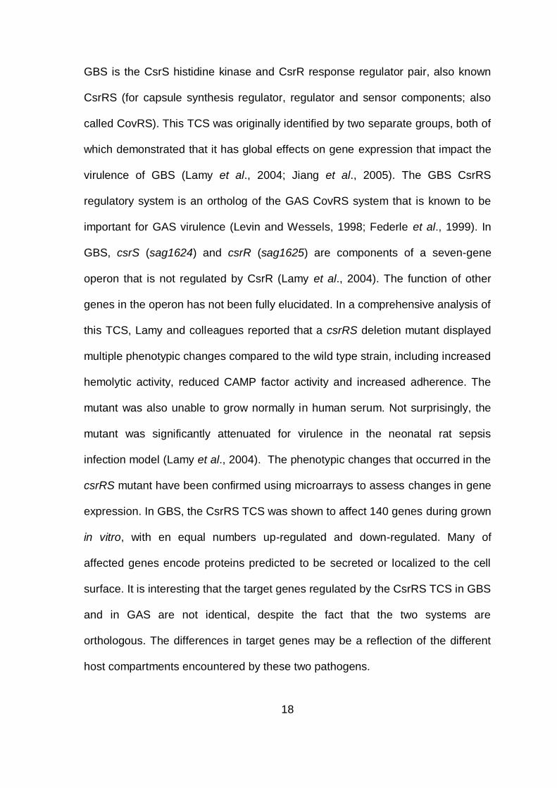
Pregnant women
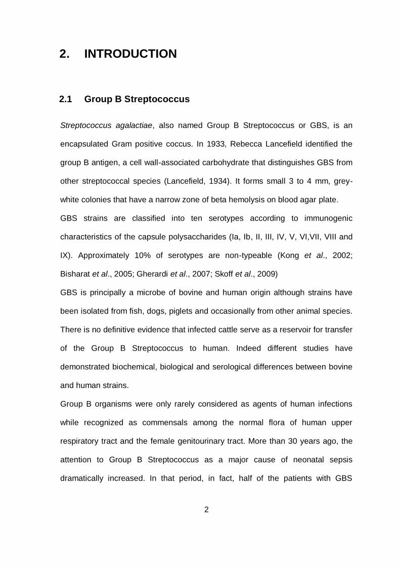
Figure 2.1 Streptococcus agalactiae. (A) Scanning Electron Microscopy (SEM) of
Streptococcus agalactiae. (B) Colonies Streptococcus agalactiae on a blood agar plate. Note
the zone of clear haemolysis.
4
Neonates
Nonpregnant adults
GBS causes a variety of perinatal infections in pregnant women, including both
symptomatic and asymptomatic bacteriuria, endometritis, amnionitis, meningitis,
pyelonephritis, and post partum wound infections (Pass et al., 1982). It also has
been suggested that GBS urinary tract infections or urinary tract, rectal, or genital
colonization in pregnant woman may lead to late term abortions and preterm and
low-birth-weight infants. Pregnant women are colonized at multiple sites,
including rectum, vagina, cervix and throat, but many of them carry GBS in
asymptomatically way (Regan et al., 1991). However, GBS colonization in
pregnant women is important because of the risk for transmission to their
newborns. Most infections and colonization of newborns are due to aspiration of
contaminated vaginal and amniotic fluid before or during parturition (Doran &
Nizet, 2004). This pathogen is the leading cause of neonatal bacterial diseases in
the United States of America; infection in newborns has been divided in early-
onset disease (EOD) and late-onset disease (LOD) depending on the infants’ age
and disease manifestations. Infants with EOD most commonly have sepsis or
pneumonia while meningitis and bone and soft tissue infections can also occur
(Edwards, 2001; Puopolo et al., 2005). LOD is less frequent than EOD and the
mortality rate is lower. In contrast morbidity is high, as around 50% of neonates
that survive to GBS infection suffer complication, including mental retardation,
hearing loss and speech and language delay (Schuchat, 1998; Schrag et al.,
2000; Edwards, 2001).
Although GBS is commonly associated with neonatal diseases (Johri et al., 2006)
and thought of as causes of disease in pregnant women, it causes substantial
5
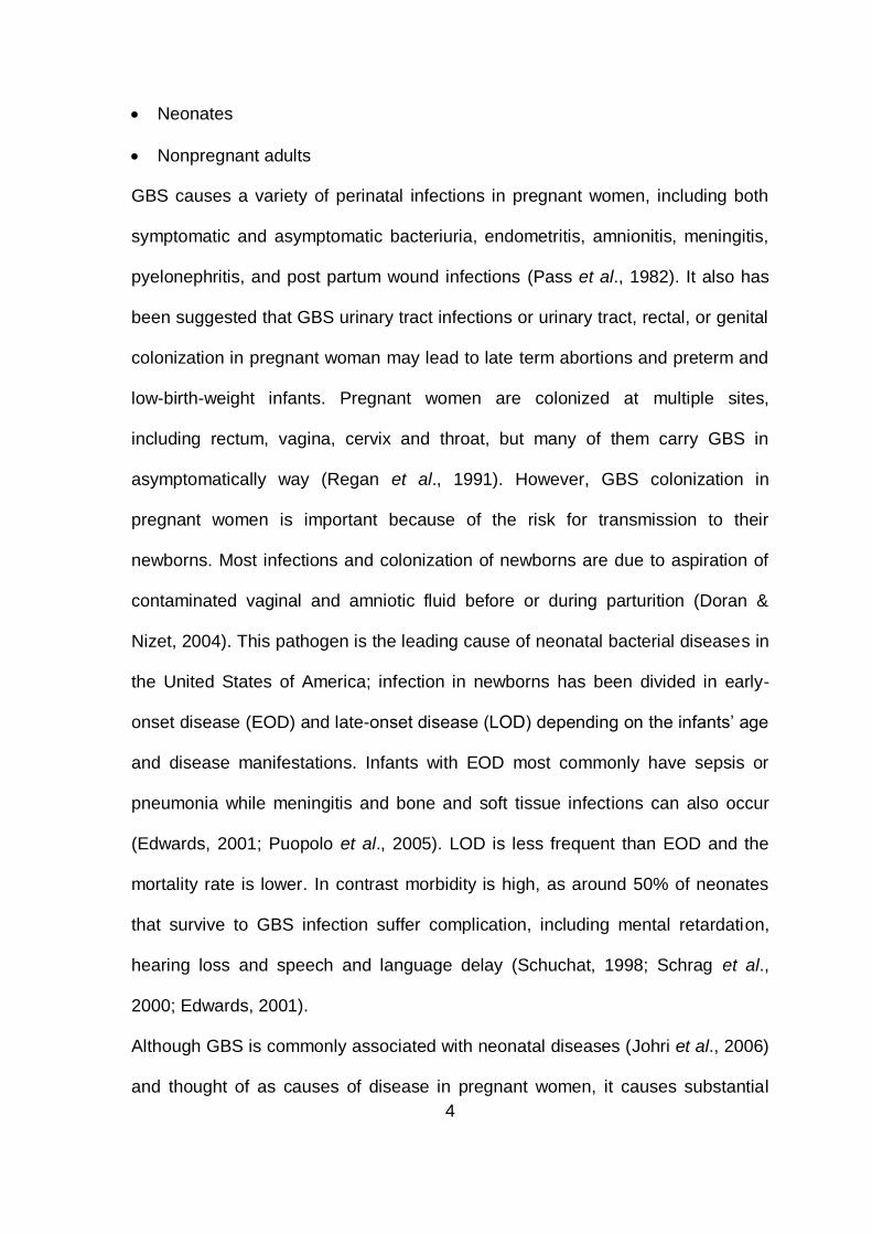
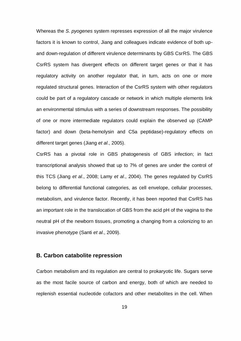
morbidity and mortality among non pregnant adults (Fig. 2.2) and appears to be
increasing in incidence in that population.
The reported annual incidence of GBS infection in nonpregnant adults in the
general population is between 4 and 7 per 100,000 (Schrag et al., 2000; Zangwill
et al., 1992 Farley et al., 1993; Blumberg et al., 1996; Phares et al., 2008; Skoff et
al., 2009). However, the risk is as high as 26 per 100,000 in patients ≥65 years of
age (Schrag et al., 2000; Phares et al., 2008). This risk reflects an increase in
incidence between 1999 and 2005 in a population-based surveillance study in ten
states in the United States (Phares et al., 2008). In view of the reductions in GBS
infection in neonates and pregnant women, GBS infection in adults is now
estimated to account for over three-fourths of invasive GBS disease in the United
States and for 90 percent of the mortality. At least 1300 GBS-related deaths
Figure 2.2 Incidence of invasive Group B streptococcal disease by age (case per 100000)
(open bars) and case fatality ratio (%) (Black line) in person > 18 years of age (Gram-positive
pathogens - Vincent A. Fischetti ,American Society for Microbiology, 2006).
6
occurred among elderly persons in 2003; the case fatality rate for elderly adults is
estimated at 15 percent (Edwards and Baker, 2005).
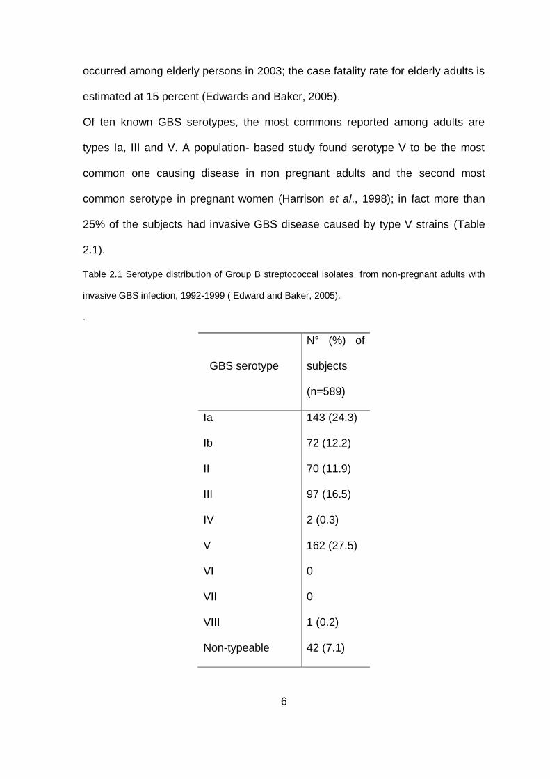
Of ten known GBS serotypes, the most commons reported among adults are
types Ia, III and V. A population- based study found serotype V to be the most
common one causing disease in non pregnant adults and the second most
common serotype in pregnant women (Harrison et al., 1998); in fact more than
25% of the subjects had invasive GBS disease caused by type V strains (Table
2.1).
Table 2.1 Serotype distribution of Group B streptococcal isolates from non-pregnant adults with
invasive GBS infection, 1992-1999 ( Edward and Baker, 2005).
.
GBS serotype
N° (%) of
subjects
(n=589)
Ia 143 (24.3)
Ib 72 (12.2)
II 70 (11.9)
III 97 (16.5)
IV 2 (0.3)
V 162 (27.5)
VI 0
VII 0
VIII 1 (0.2)
Non-typeable 42 (7.1)
7
Numerous studies have allowed description of the clinical spectrum of disease,
including clinical features, risk factors, therapy, and outcomes. The most common
syndromes caused by GBS in adults are skin, soft tissue, and bone infections.
These infections are often complications of chronic diabetes or decubitus ulcer.
Cellulitis, foot ulcers, and abscesses are the most common manifestation, but
also necrotizing fasciitis have occasionally been reported (Edwards and Baker,
2005). Patients with indwelling catheters are at higher risk for GBS bacterimia;
polymicrobial bacteremia, often with Staphylococcus aureus is identified in 26 to
30% of patients with GBS colonization (Jackson et al., 1995). GBS can also
cause pneumonia, increasing the mortality rate of patients. Less common GBS
infections such as arthritis, urinary infection, meningitis, and peritonitis can also
occur, especially in patients with common predisposing factors, such as diabetes,
osteoarthritis, and underlying joint disease. S agalactiae infection is extremely
rare in healthy individuals and is almost always associated with underlying
abnormalities. Risk factors that promote GBS infections include diabetes mellitus,
elderly, malignancy, liver disease, neurological deficits, renal failure, other forms
of immune impairment such as human immunodeficiency virus infection, cancer
and venous insufficiency (Jenkins et al., 2010).
Elderly patients with diabetes mellitus have displayed peripheral neuropathy or
peripheral vascular diseases following trauma, particularly to the lower
extremities. GBS takes advantage of this condition by crossing the endothelial
barrier and promoting bacterial invasion of the foot (Edwards and Baker, 2005).
Indeed, GBS is also found in biopsy of patients with foot infections, a common
status of patients suffering from diabetes (Urban et al., 2011). Group B
streptococcal infection in elderly people (≥70 years) is strongly linked to
8
congestive heart failure and being bedridden, with urinary tract infection,
pneumonia, and soft-tissue infection as the most common manifestations of
infection. Neurologic illness is associated with pneumonia in elderly people,
possibly due to aspiration of Group B Streptococci from the upper respiratory
tract.
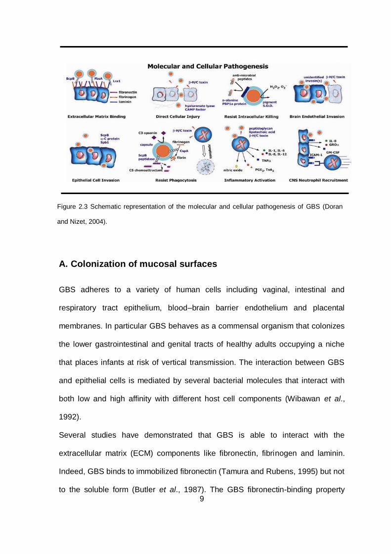
2.3 Molecular pathogenesis of GBS
Group B Streptococcus infection in human is a complex and multifactorial process
which involves several virulence determinants that contribute to neonatal disease.
An important role is mediated by the capsule, which remains together to the β-
haemolytic activity the main virulence factor for Group B Streptococcus. In
addition, a number of molecules both surface-exposed or secreted are necessary
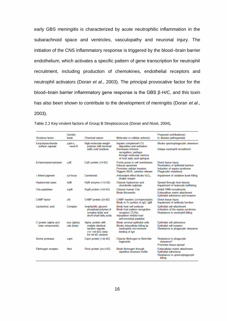
for GBS infection process (Table 2.2).
The GBS pathogenic process can be described in four main steps:
A. Colonization of mucosal surfaces;
B. Translocation through host cellular barriers;
C. Evasion of immunological clearance;
D. Activation of inflammatory response.
9
A. Colonization of mucosal surfaces
GBS adheres to a variety of human cells including vaginal, intestinal and
respiratory tract epithelium, blood–brain barrier endothelium and placental
membranes. In particular GBS behaves as a commensal organism that colonizes
the lower gastrointestinal and genital tracts of healthy adults occupying a niche
that places infants at risk of vertical transmission. The interaction between GBS
and epithelial cells is mediated by several bacterial molecules that interact with
both low and high affinity with different host cell components (Wibawan et al.,
1992).
Several studies have demonstrated that GBS is able to interact with the
extracellular matrix (ECM) components like fibronectin, fibrinogen and laminin.
Indeed, GBS binds to immobilized fibronectin (Tamura and Rubens, 1995) but not
to the soluble form (Butler et al., 1987). The GBS fibronectin-binding property
Figure 2.3 Schematic representation of the molecular and cellular pathogenesis of GBS (Doran
and Nizet, 2004).
10
seems to be associated with the surface-anchored C5a peptidase, ScpB
(Beckmann et al., 2002). The binding to fibronectin facilitates mucosal
colonization and mediates GBS internalization into host cells (Cheng et al.,
2002a). On the other hand, the adherence of GBS to laminin involves the laminin
binding protein, Lmb (Spellerberg et al., 1999), while attachment of GBS to
fibrinogen is mediated by two unrelated surface fibrinogen binding protein, FbsA
and FbsB (Gutekunst et al., 2004; Schubert et al., 2002). GBS binding to
fibrinogen and fibronectin is positively regulated by the transcriptional regulator
RogB, which modulates the expression of a number of genes coding ECM
binding proteins (Gutekunst et al., 2003). The recently described pilus-like
structures present on the surface of GBS (Lauer et al., 2005) also seems to be
involved in the adherence of bacteria to lung epithelial cells. In particular the
knock-out mutant strain for the ancillary protein shows an impaired ability to
adhere to epithelial cells (Maisey et al., 2006).
B. Translocation through host cellular barriers
The strategy used by GBS to traverse defined anatomic epithelial barriers, such
as the cervical or vaginal epithelium, entering bloodstream is still poorly
understood. However, the GBS ability to penetrate host cellular barriers is the first
distinguishing feature of its pathogenicity.
GBS has the ability to invade chorionic but not amniotic epithelial cells (Winram et
al., 1998). Nevertheless, GBS can traverse placental membranes, weak their
tensile strength and access to the fetus within the amniotic cavity. This process
induces placental membrane rupture or trigger premature delivery. After
11
aspiration of infected amniotic or vaginal fluid, the initial focus of GBS infection
takes place in the newborn lung. From there, the organism rapidly gains access
to the bloodstream and is circulated through other organs and tissues.
GBS has the ability to invade both alveolar epithelial and pulmonary endothelial
as initially noted in newborn macaques (Rubens et al., 1991), and later confirmed
in human tissue culture lines derived from both cellular barriers (Gibson et al.,
1993; Rubens et al., 1992). Cellular invasion by GBS occurs when the organism
triggers its own endocytotic uptake and enters the cell within a membrane-bound
vacuole. In the host, this process requires rearrangement of microfilament
components of the cytoskeleton and the signalling pathways mediated by PI 3-
kinase, seem to be involved (Tyrrell et al., 2002).
There is a close correlation between cellular invasion and GBS virulence
potentiality because clinical isolates from infants with bloodstream infections
invade epithelial cells better than strains from the vaginal mucosa of
asymptomatic women (Valentin-Weigand and Chhatwal, 1995). Other proteins
like ScpB or the alpha C surface protein are involved in GBS epithelial cell
invasion (Bolduc et al., 2002; Cheng et al., 2002b).
Early-onset GBS pneumonia is characterized by widespread damage to lung
epithelium and endothelium, with haemorrhage and neutrophils entering the
alveolar airspaces. GBS enters the bloodstream as a consequence of the loss of
barrier integrity and the β-haemolysin/cytolysin (β-H/C) appears the protein
largely involved in this process. cylE is the gene necessary and sufficient for GBS
β-H/C expression (Pritzlaff et al., 2001). This pore-forming toxin lyses lung
epithelial and endothelial cells and compromises their barrier function (Gibson et
al., 1999; Nizet et al., 1996). Besides, the GBS β-H/C promotes GBS intracellular
12
invasion and triggers the release of interleukin-8 (IL-8) that is the principal
chemoattractant for human neutrophils (Doran et al., 2002).
GBS is also the leading cause of bacterial meningitis in human newborns and the
bacterium has the propensity to breech the specialized endothelium comprising
the human blood–brain barrier. In fact, it has been reported that COH1, a highly
encapsulated GBS strain, was able to invade and translocate across a polarized
brain microvascular epithelial cell monolayer without marked changes in
transendothelial electrical resistance (Nizet et al., 1997). As for the epithelial cell
barriers, the GBS β-H/C is directly cytolytic for human brain endothelial cells
(Doran et al., 2003).
GBS virulence factors play a pivotal role in penetration of host cellular barriers. It
is reported that bloodstream isolates of GBS secrete high levels of an enzyme
that degrades hyaluronic acid, the main polysaccharide component of host
connective tissue (Kjems et al., 1980; Pritchard and Cleary, 1996). Another
important extracellular protein involved in invasion is CAMP factor; this protein
oligomerizes in the target membrane to form discrete pores and triggers cell lysis
(Lang and Palmer, 2003) and it is toxic when injected intravenously in rabbits
(Skalka and Smola, 1981).
Recently, it has been proposed a novel paracellular route used by GBS to
traverse epithelial cells monolayer. In this new model, the crossing of bacteria
across the cell is not associated with the loss of monolayer’s integrity suggesting
that paracellular translocation is likely to be an active but transient phenomenon
used by GBS to translocate from the site of colonization to target organs (Soriani
et al., 2006).
13
C. Evasion of immunological clearance
Phagocytic cells including polymorphonucleates (PMNs) and macrophages are
the first line of defence against GBS and their early action determines to outcome
of the infection. However, the effective uptake and killing of GBS by these cells
requires opsonization of the bacterium and deposition of complement
components (Jarva et al., 2003). Neonates that are deficient in a) phagocytic cell
function, b) specific anti-GBS immunoglobuling and c) classic and alternate
complement components, are also particularly prone to GBS invasive disease.
Polysaccharide capsule is fundamental to the avoidance of immune response.
Indeed, sialylated GBS capsule protects the bacterium by PMNs mediated
opsonophagocytic killing, preventing C3 deposition on the bacterial surface.
Isogenic GBS mutant defected for capsule synthesis is more susceptible to killing
by PMNs. As a consequence, lethal doses of capsule-deficient strains are in mice
100-fold greater than the one of the parental wild-type strain (Marques et al.,
1992; Wessels et al., 1989).
Other multifunctional GBS determinants that contribute to bacterial resistance to
the host clearance mechanisms have been identified (Jarva et al., 2003). Among
them, FbsA seems to be important for the survival of bacteria in human blood
(Schubert et al., 2002). The beta-C protein binds the Fc domain of human IgA,
potentially sequestering this important host mucosal defence molecule (Jerlstrom
et al., 1996), while GBS strains expressing the alpha-C protein appear more
resistant to phagocytic killing (Madoff et al., 1991). Besides, the presence of
tandem repeats within the alpha C sequence is correlated to antigenic variability
that allows the bacterium to avoid opsonophagocytic killing triggered by specific
14
antibody (Madoff et al., 1996). The C5a-ase possesses a domain that specifically
cleaves human complement component C5a, a chemoattractant for human
PMNs, reducing the acute neutrophils response to sites of infection (Bohnsack et
al., 1997). A recently identified novel cell surface protease named CspA targets
host fibrinogen, producing adherent fibrin-like cleavage products that coat the
bacterial surface and interfere with opsonophagocytic clearance (Harris et al.,
2003).
The application of signature-tagged mutagenesis for in vivo screening in a GBS
neonatal rat sepsis model has identify unexpected virulence genes encoding
factors that are crucial for the bacterium survival to immune clearance. For
example, ponA, which codes for an extracytoplasmic penicillin-binding protein
(PBP1a), promotes resistance to phagocytic killing independent of capsule (Jones
et al., 2003) and protects form cationic anti-microbial peptides (defensins,
cathelicidins) produced by host epithelial cells and phagocytes (Hamilton et al.,
2006). An analogue function is associated with the D-alanylation of lipotechoic
acid in the bacterial cell wall (Poyart et al., 2003).
The GBS β-H/C triggers cytolytic events in macrophages and neutrophils (Liu et
al., 2004) and can also induce macrophage apoptosis (Buratta et al., 2002; Ulett
et al., 2003) Curiously, GBS has been shown to survive for prolonged periods
within the phagolysosome of macrophages and to be >10-fold more resistant to
hydrogen peroxide killing than catalase-positive Staphylococcus aureus (Wilson
and Weaver, 1985). Superoxide dismutase (SodA) (Poyart et al., 2001) and the
orange carotenoid pigment (Liu et al., 2004) are the main defences of GBS
against oxidative stress. Indeed, the pigmentation is a property unique among
haemolytic streptococci and genetically linked to the cylE gene. Carotenoids
15
neutralize hydrogen peroxide and singlet oxygen, therefore providing a shield
against the key elements of phagocyte oxidative burst killing (Liu et al., 2004).
D. Activation of inflammatory responses
The host inflammatory response to GBS invasive infections, is associated to the
sepsis syndrome and multiorgan dysfunction. Peptidoglycan and other GBS
components associated with the cell wall, not including the surface
polysaccharide capsule, appear to be the most provocative agents in triggering
host cytokine cascades, in particular the proximal mediators tumour necrosis
factor-alpha (TNF-α) and interleukin-1 (IL-1). GBS induction of NF-kB signalling
and TNF-α release from human monocytes in vitro requires CD14 and the
receptors for complement components 3 and 4 (Medvedev et al., 1998). Recently,
the importance of complement components in amplifying GBS TNF-α induction
was corroborated when reduced levels of the cytokine were observed in the blood
of C3 or C3 receptor-deficient mice stimulated with GBS (Levy et al., 2003).
Knockout mouse studies indicate GBS cell wall peptidoglycan-induced activation
of p38 and NF-kB, depends on the cytoplasmic TLR adaptor protein MyD88, but
does not proceed via the well studied TLR2 and/or TLR4 (Henneke et al., 2002).
Of interest, GBS β-H/C and cell wall components act synergistically to induce
macrophage production of inducible nitric oxide synthase (iNOS) and generation
of nitric oxide (NO) (Ring et al., 2002), a potent factor in the sepsis cascade.
LOD is characterized by meningitis with or without accompanying sepsis
Localization of GBS in the brain and Central Nervous System (CNS) during LOD,
triggers a strong host inflammatory response. Indeed, in the infant rat model,
16
early GBS meningitis is characterized by acute neutrophilic inflammation in the
subarachnoid space and ventricles, vasculopathy and neuronal injury. The
initiation of the CNS inflammatory response is triggered by the blood–brain barrier
endothelium, which activates a specific pattern of gene transcription for neutrophil
recruitment, including production of chemokines, endothelial receptors and
neutrophil activators (Doran et al., 2003). The principal provocative factor for the
blood–brain barrier inflammatory gene response is the GBS β-H/C, and this toxin
has also been shown to contribute to the development of meningitis (Doran et al.,
2003).
Table 2.2 Key virulent factors of Group B Streptococcus (Doran and Nizet, 2004).
17
2.4 Regulation of gene expression
The GBS pathogenesis implies that this bacterium can survive in a large number
of human body compartments, encountering different environmental conditions,
such as different pH, availability of carbon source and temperature. The
modulation of gene expression allows the adaptation through different
mechanisms of regulation that control the production of proteins involved in
adhesion, nutrient acquisition, survival to host immune system (Sitkiewicz et al.,
2009; Mereghetti et al., 2008; Mereghetti et al., 2009).
A. Two component regulatory system
One common mechanism used by bacteria to regulate gene expression is the
alteration of sigma factors associated with RNA polymerase. In light of paucity of
sigma factors, GBS may rely more on other mechanisms for regulation on gene
expression. GBS strains generally possess approximately 17-20 predicted two
component regulatory systems (TCS) (Glaser et al., 2002; Tettelin et al., 2002;
Tettelin et al., 2005), a number significantly grater than has been reported for
closely related species such as GAS (13) or Lactococcus lactis (8) (Bolotin et al.,
2001; Ferretti et al., 2001). TCSs allow for the sensing of specific environmental
stimuli or conditions followed by transduction of the signal to a response
regulator. In the basic model, a membrane-bound histidine protein kinase
(sensor) is autophosphorilated when the signal is detected. The phosphoryl group
is then transferred to the cytoplasmatic response regulator. Phosphorilation of the
regulator alters its binding affinity for the promoters of target genes, thereby
affecting their transcription (Stoch et al., 2000). The best characterized TCS in
18
GBS is the CsrS histidine kinase and CsrR response regulator pair, also known
CsrRS (for capsule synthesis regulator, regulator and sensor components; also
called CovRS). This TCS was originally identified by two separate groups, both of
which demonstrated that it has global effects on gene expression that impact the
virulence of GBS (Lamy et al., 2004; Jiang et al., 2005). The GBS CsrRS
regulatory system is an ortholog of the GAS CovRS system that is known to be
important for GAS virulence (Levin and Wessels, 1998; Federle et al., 1999). In
GBS, csrS (sag1624) and csrR (sag1625) are components of a seven-gene
operon that is not regulated by CsrR (Lamy et al., 2004). The function of other
genes in the operon has not been fully elucidated. In a comprehensive analysis of
this TCS, Lamy and colleagues reported that a csrRS deletion mutant displayed
multiple phenotypic changes compared to the wild type strain, including increased
hemolytic activity, reduced CAMP factor activity and increased adherence. The
mutant was also unable to grow normally in human serum. Not surprisingly, the
mutant was significantly attenuated for virulence in the neonatal rat sepsis
infection model (Lamy et al., 2004). The phenotypic changes that occurred in the
csrRS mutant have been confirmed using microarrays to assess changes in gene
expression. In GBS, the CsrRS TCS was shown to affect 140 genes during grown
in vitro, with en equal numbers up-regulated and down-regulated. Many of
affected genes encode proteins predicted to be secreted or localized to the cell
surface. It is interesting that the target genes regulated by the CsrRS TCS in GBS
and in GAS are not identical, despite the fact that the two systems are
orthologous. The differences in target genes may be a reflection of the different
host compartments encountered by these two pathogens.
19
Whereas the S. pyogenes system represses expression of all the major virulence
factors it is known to control, Jiang and colleagues indicate evidence of both up-
and down-regulation of different virulence determinants by GBS CsrRS. The GBS
CsrRS system has divergent effects on different target genes or that it has
regulatory activity on another regulator that, in turn, acts on one or more
regulated structural genes. Interaction of the CsrRS system with other regulators
could be part of a regulatory cascade or network in which multiple elements link
an environmental stimulus with a series of downstream responses. The possibility
of one or more intermediate regulators could explain the observed up (CAMP
factor) and down (beta-hemolysin and C5a peptidase)-regulatory effects on
different target genes (Jiang et al., 2005).
CsrRS has a pivotal role in GBS phatogenesis of GBS infection; in fact
transcriptional analysis showed that up to 7% of genes are under the control of
this TCS (Jiang et al., 2008; Lamy et al., 2004). The genes regulated by CsrRS
belong to differential functional categories, as cell envelope, cellular processes,
metabolism, and virulence factor. Recently, it has been reported that CsrRS has
an important role in the translocation of GBS from the acid pH of the vagina to the
neutral pH of the newborn tissues, promoting a changing from a colonizing to an
invasive phenotype (Santi et al., 2009).
B. Carbon catabolite repression
Carbon metabolism and its regulation are central to prokaryotic life. Sugars serve
as the most facile source of carbon and energy, both of which are needed to
replenish essential nucleotide cofactors and other metabolites in the cell. When
20
faced with a wide variety of carbon and energy sources, a bacterium has to make
metabolic decisions, opting for preferential use of one source over another in
order to maintain optimal growth (Deutscher, 2008; Stulke and Hillen, 1998;
Titgemeyer and Hillen, 2002). Simultaneous utilization of all available sugars
would be metabolically inefficient and would lead to slower growth. The ability to
utilize preferred sugars depends on a regulatory process called carbon catabolite
repression (CCR) (Stulke and Hillen, 1999; Titgemeyer and Hillen, 2002; Warner
and Lolkema, 2003). CCR causes silencing of genes specific for the utilization of
nonpreferred sugars until the bacterium has consumed the preferred sugar(s).
CCR has been studied in considerable detail in the model free-living, Gram-
positive bacterium Bacillus subtilis (Stulke and Hillen, 2000; Titgemeyer and
Hillen, 2002; Warner and Lolkema, 2003). The main global regulator of CCR in
this organism is catabolite control protein A (CcpA) (Chauvaux, 1996; Henkin et
al., 1991). CcpA belongs to the LacI/GalR family of activator-repressor
transcription factors and influences the expression of a wide range of catabolic
operons in B. subtilis (Belitsky et al., 2004; Grundy et al., 1994; Grundy et al.,
1993; Henkin, 1996; Hueck and Hillen 1995; Kim et al., 2002; Simpson and
Russell, 1998; Stulke and Hillen, 2000; Warner et al., 2000; Warner and Lolkema,
2003). CcpA has also been identified to function in the regulation of catabolic
operons and catabolite repression in many streptococcal species (Iyer et al.,
2005; Asanuma et al., 2004, Dong et al., 2004, Rogers and Scannapieco, 2001,
van den Bogaard et al., 2000). Candidate genes or operons that are subject to
CcpA-dependent CCR are often identifiable by the presence of an operator
sequence, called the catabolite-repressible element (cre), to which CcpA binds
21
(Asanuma et al., 2004; Kim and Chambliss, 1997; Miwa et al., 1997; Ramseier et
al, 1995). In streptococcal species, several in vivo and in vitro studies have
shown that the cre consensus sequence is WTGNAANCGNWNNCW (N is any
base and W is A or T), where the underlined bases are involved in CcpA binding
(Tomoyasu et al., 2010; Kim and Chambliss, 1997; Miwa et al., 2000;
Schumacher et al., 2004; Warner and Lolkema. 2003).
The affinity of CcpA for cre sequences is enhanced by binding to another protein,
the histidine phosphoprotein (HPr). HPr is an integral component of the
phosphoenolpyruvate-dependent phosphotransferase system (PTS), where it
normally functions in the transfer of high-energy phosphate from
phosphoenolpyruvate to the enzyme II complex during sugar uptake (Deutscher
et al., 2006; Postma and Lengeler, 1985; Reizer et al., 1999). The presence of a
preferred sugar, such as glucose, in the medium activates phosphorylation of HPr
on a conserved serine residue at position 46 by the Hpr kinase, which itself is
activated by metabolites such as the high-energy glycolytic intermediate fructose-
1,6-bisphosphate (Brochu and Vadeboncoeur, 1999; Frey et al., 2003; Poncet et
al., 2004; Reizer et al., 1998; Thevenot, 1995). CcpA interacts with the
phosphoserine form of HPr, P∼Ser-HPr, to form a dimeric complex. This
interaction increases the affinity of CcpA for the cre box. Binding of this dimeric
complex typically causes repression of promoters, facilitating CCR (Asanuma et
al., 2004; Deutscher et al., 1995; Deutscher et al., 1994). CcpA residues involved
in binding of P∼Ser-HPr (Kraus et al., 1998) and those involved in binding of cre
(Kim and Chambliss, 1997) have been characterized, and the crystal structure of
the CcpA-P∼Ser-HPr complex has been recently solved (Schumacher et al.,
2004). However, several reports have shown that although there is slightly low
22
affinity, CcpA can recognize and bind to the cre without HPr-(Ser- 46-P) and
fructose-1,6-bisphosphate in vitro (Tomoyasu et al., 2010).
2.5 Involvement of BibA and pullulanase in GBS pathogenesis
Studies of the role of carbohydrate metabolism in streptococcal pathogenesis
have focused on two mechanisms: adherence to eukaryotic cells and acquisition
of crucial nutrients. Regulation of gene encoding for proteins involved in these
pathways could be an important basis for understanding the molecular
mechanisms underlying GBS pathogenesis.
A. BibA: Group B Streptococcus immunogenic bacterial adhesin
Recently, Santi and colleagues (Santi et al., 2007) have reported the presence of
BibA, an immunogenic surface-associated antigen expressed by GBS that is
involved in virulence. The protein product of the bibA (sag2063) gene in the GBS
strain 2603 V/R (Tettelin et al., 2002) is a polypeptide of 630 amino acids
containing a leader peptide (residues 1–27), a N-terminal domain (residues 28–
400), a proline rich region (residues 401–568) that consists of 42 copies of a
PEAK/PDVK motif, and a canonical cell wall anchoring domain (residues 596–
630). The anchoring domain is formed by the consensus LPXTG sequence,
followed by a hydrophobic transmembrane segment and a charged C-terminal
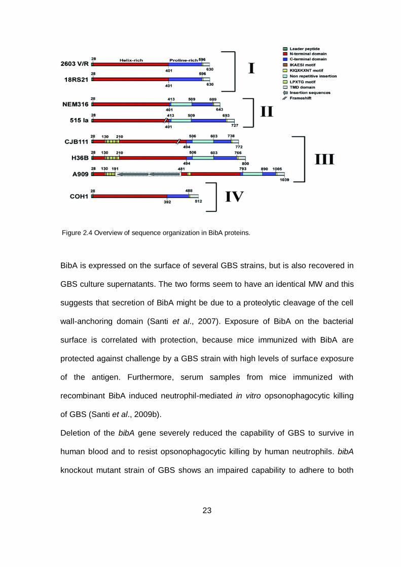
tail. As shown in figure 2.4, four allelic variants of this protein have been
identified: variant I, found in strains 2603 V/R (V) and 18RS21 (II); variant II, in
strains NEM316 (III) and 515 (Ia); variant III, in strains CJB111 (V), H36B (Ib),
and A909 (Ia); and variant IV, in the COH1 (III) strain.
23
BibA is expressed on the surface of several GBS strains, but is also recovered in
GBS culture supernatants. The two forms seem to have an identical MW and this
suggests that secretion of BibA might be due to a proteolytic cleavage of the cell
wall-anchoring domain (Santi et al., 2007). Exposure of BibA on the bacterial
surface is correlated with protection, because mice immunized with BibA are
protected against challenge by a GBS strain with high levels of surface exposure
of the antigen. Furthermore, serum samples from mice immunized with
recombinant BibA induced neutrophil-mediated in vitro opsonophagocytic killing
of GBS (Santi et al., 2009b).
Deletion of the bibA gene severely reduced the capability of GBS to survive in
human blood and to resist opsonophagocytic killing by human neutrophils. bibA
knockout mutant strain of GBS shows an impaired capability to adhere to both
Figure 2.4 Overview of sequence organization in BibA proteins.
24
human cervical and lung epithelial cells, demonstrating the effective role of BibA
as adhesin (Santi et al., 2007).
The presence of this protein on GBS surface is influenced by various
environmental factors (Mereghetti et al., 2008; Santi et al., 2009a). GBS adapts to
different environmental conditions by modulating the transcription of genes
involved in pathogen-host interaction (Boskey et al., 1999). An important
condition that affects the gene expression is the pH; in the human host, GBS
encounters pH conditions that vary from the acidic pH of the vagina or
intracellular endocytic compartments to the near-neutral pH of amniotic fluid or
the fetal. Moreover, a comparative global gene expression analysis of GBS grown
at acidic and neutral pHs has shown a down regulation of bibA expression when
GBS is grown at acidic pH compare to a grown at pH 7 (Santi et al., 2009a).
Furthermore, the regulation of the gene is under the control of two component
system CsrRS, in fact the expression level of bibA doesn’t change in the csrRS
deletion mutant strain grown in both conditions, meaning that the signal is
transducted by these regulatory components.
B. Pullulanase
The use of carbon sources is essential to the ability of bacteria to colonize the
host and potentially cause disease in humans. In particular, highly polymerized α-
glucan polysaccharides, such as starch and glycogen, are most likely to be found
in environmental niches. Indeed, it is known that dietary-derived starches are very
abundant in the human colon (Anderson et al., 1981; Levitt et al., 1987), while
glycogen is deposited in large amount in the vaginal ephitelium during times of
25
high estrogen availability. Because of the complex structures of highly
polymerized α-glucans, bacteria require an appropriate combination of enzymes
for de-polymerization to oligo- and monosaccharides. Among these enzymes are
ascribed pullulanases. Pullulanases have a glycosidic hydrolase activity towards
α-glucan polysaccharides and are considered key extracellular components in
bacterial metabolism.
Recently it is identified in GBS a novel surface-exposed α-glucan-degrading
enzyme, named SAP (Streptococcus agalactiae pullulanase), belonging to the
streptococcal family of pullulanases (Santi et al., 2008). The sap gene (sag1216)
is highly conserved among Group B streptococcus (GBS) strains; homologous
genes, such as those for pulA and spuA, are present in other pathogenic
streptococci. SAP is a member of the class 13 glycoside hydrolase (GH13; α-
amylase) family and is a type I pullulanase; in vitro studies have shown that
recombinant SAP can degrade α-glucans such as pullulan, glycogen, and starch
(Santi et al., 2008). Furthermore, fluorescence-activated scanning analysis and
confocal imaging studies performed on whole bacteria indicate that the presence
of α-glucan polysaccharides in culture medium upregulates the expression of
SAP on the bacterial surface (Santi et al., 2008). As reported for other
streptococcal pullulanases, specific anti-SAP antibodies are found in human sera
from healthy volunteers. Investigation of the functional role of anti-SAP antibodies
revealed that incubation of GBS in the presence of sera from animals immunized
with SAP reduced the ability of the bacterium to degrade pullulan.
26
3. Material and methods
3.1 Bacterial strains and growth conditions
GBS type V strain 2603V/R and isogenic mutant strain 2603ΔcsrRS have been
described previously (Tettelin et al., 2002; Jiang et al., 2005). E. coli DH10BT1
and HK100 strains were used for cloning purposes. E. coli BL21 (DE3) strain was
used for protein production. Unless otherwise specified, for experiments testing
the effects of glucose, GBS was cultured at 37°C in Todd-Hewitt broth (Difco) and
in a sugar-free complex medium (CM: 10 g/l proteose peptone, 5 g/l trypticase
peptone, 5 g/l yeast extract, 2.5 g/l KCl, 1 mM urea, 1 mM arginine). E. coli was
grown in Luria–Bertani broth and ampicillin was used at a final concentration of
100 μg/ml for recombinant strains.
3.2 Microarray analysis of gene expression
Microarray comparison was performed on the wild-type strain 2603 V/R and the
isogenic mutant strain 2603 csrRS.
The two strains were grown in THB until late exponential phase, washed in PBS
and resuspended in CM until OD600 of 0.5. The bacteria were washed in PBS and
resuspendend in CM in the absence or presence of 1% glucose at 37°C for 30
minutes of incubation. Total RNA was extracted with RNeasy Mini Kit (Qiagen)
and treated with RNase-free DNase (Qiagen) according to the manufacturer's
instructions. The concentration of total RNA was determined using a NanoDrop
ND-1000 spectrophotometer (NanoDrop Technologies). RNA integrity was
27
verified using a Bioanalyzer 1000 (Agilent). For each strain, total bacterial RNA
was isolated from four independent culture pools and the samples were sent to
Roche NimbleGen Systems, where cDNA synthesis and labeling were performed.
Changes in gene expression levels were evaluated using the NimbleGen GBS
DNA microarray (17 probes for each gene, 3 replicates for probe consisting of 60-
mer synthetic oligonucleotides for each gene). All hybridizations, staining, and
processing were performed by personnel at Roche NimbleGen, Inc. (Madison,
WI, USA).
Briefly, to synthesize double-stranded cDNA, 10 ug of RNA were retrotranscripted
using the Invitrogen SuperScript Double-Stranded cDNA Synthesis Kit. To
eliminate RNA contaminations, a step with RNase was performed. The cDNA was
precipitated with phenol:chloroform:isoamyl alcohol and quality control was
analyzed using a spectrophotometer. The cDNA was labeled using the
NimbleGen One-Color DNA Labeling Kits (labeling with Cy3).
Equal amounts of cDNA was then hybridized onto the Slide 12x135K (Design
Name: 100920_TI208435_60mer_HX12) for Streptococcus agalactiae, using the
NimbleGen Hybridization System and according to protocol "NimbleGen Arrays
User’s Guide, Gene Expression Arrays, Version 5.1" (NimbleGen Roche). cDNA
were hybridized onto microarray at +42°C for 16 hours. After hybridization,
microarray was disassembled, washed following the NimbleGen's protocol. The
slides were dried in NimbleGen Microarray Dryer (NimbleGen Roche) for 2
minutes and immediately scanned with the MS 200 Microarray Scanner
(NimbleGen Roche) following the protocol. Scanned image and data were
extracted and analyzed using NimbleScan software. NimbleScan software
normalizes expression data using quantile normalization as described by Bolstad,
28
(Bolstad et al., 2003). Gene calls are generated using the Robust Multichip
Average (RMA) algorithm as described by Irizarry (Irizarry et al., 2003a; Irizarry et
al., 2003b).
The raw data were analyzed using the DNASTAR software.
The microarray experiment has been submitted to the Array Express database of
the European Bioinformatic Institute (http://www.ebi.ac.uk/microarray-as/ae/) with
accession number A-MEXP-2195 (Chip design).
3.3 Quantitative reverse transcriptase PCR
Quantitative real-time PCR (qRT-PCR) was used to validate microarray
experiments. One microgram of RNA previously extracted for microarray was
incubated with random primers and used for cDNA synthesis (at 42°C, one hour
incubation) using ImProm-II Reverse Transcriptase (Promega). Fifteen microliters
of cDNA were used as template for PCR amplification using gene specific primers
as listed in Table 3.1. The amplification was performed using FastStart Universal
SYBR Green Master (Rox) (Roche), employing Light Cycler 480 System (Roche).
The expression levels of all the genes tested by quantitative RT-PCR were
normalized using the gyrA expression as an internal standard. Each sample was
tested in triplicate during a trial, and three independent experiments were
performed.
3.4 Flow cytometry analysis
To verify the exposure of BibA on bacterial surface, GBS was incubated for 30
minutes in CM in the absence or presence of 1% glucose employing the same

29
conditions used for microarray experiments. After the grown, the bacteria were
washed twice with PBS, suspended in newborn calf serum (Sigma), incubated for
20 min at room temperature, and dispensed in a 96-well plate (20 μl per well).
The bacteria were fixed in 1% paraformaldehyde for 15 minutes at room
temperature. Eighty microliters of anti-BibA immune rabbit serum diluted in
PBS/0.1% BSA was added to the bacterial suspension to a final dilution of 1:200.
Incubation was performed at 4°C for 1 hour. After washing, bacteria were labeled
with R-Phycoerythrin (PE)-conjugated secondary antibodies (final dilution 1:100)
(Jackson Immuno Research, PA, USA) at 4°C. Pre immune serum from rabbit
was used as a negative control. Bacterial staining was analyzed by using a FACS
CANTO II Flow cytometer equipped with three laser system (405, 488, 633 nm),
eight Color Configuration and BD FACSDivaTM v6.1.3 software (BD Bioscience,
SANJOSE, CA) and the data were analyzed with FlowJo 7.2.2 program.
3.5 SDS-PAGE and Immunoblot analysis
In order to prepare GBS extracts relative to the secreted protein fraction,
supernatant of bacteria cultures grown in the same conditions used for microarray
were collected. Proteins in 1 ml of supernatant were precipitated with 10% of
trichloroaceticacid (TCA) for 1 hr at 4°C. Protein were then pelletted, washed with
cold acetone and resuspended in Tris-HCl pH 6,8. Bacterial proteins were
separated by 4-12% SDS-polyacrylamide gel electrophoresis (SDS-PAGE) (Bio-
Rad) and transferred to nitrocellulose membranes (Bio-Rad). Membranes were
blocked with milk (5% w/v) and, then, incubated with anti-BibA immune rabbit
serum at a 1:1000 dilution, secondary antibody (ECL, horseradish peroxidase-
30
linked anti-mouse IgG, GE Healthcare) at a 1:1000 dilution and developed with
ECL enhanced chemiluminescence detection substrate (SuperSignal West Pico,
Pierce).
3.6 Cloning, production and purification of recombinant
proteins CsrR
The csrR gene (sag1625) was amplified by PCR from GBS genome using the
primers (Fw csrR, Rv csrR) listed in Table 3.1, carrying the cleavage sites of the
restriction enzyme NdeI and XhoI at 5’ end. The PCR product was digested with
the specific restriction enzymes and cloned into pET 21b (Novagen) previously
linearized with the same restriction enzymes. The construct was introduced into
BL21 (DE3) by transformation.
The recombinant bacteria were grown at 37°C to an optical density at 600 nm of
0.5 (mid exponential phase), at which time 1mM isopropyl-beta-D-
thiogalactopyranoside (IPTG) was added. After 3 hours, the cells were harvested
by centrifugation, resuspended in buffer A (50 mM Na2HPO4 [pH 8], 0.3 M NaCl)
and distrupted by sonication (10 cycles, 30 sec ON 30 sec OFF). The purification
on the soluble fraction was performed with His Gravi Trap columns (GE
Healthcare): the lysate was loaded onto the columns and after several washing in
buffer B (50 mM Na2HPO4 [pH 8], 0.3 M NaCl, 20 mM imidazole) the recombinant
proteins were eluted with high concentration imidazole buffer (50 mM Na2HPO4
[pH 8], 0.3 M NaCl, 250 mM imidazole). Protein concentration was estimated
using the Bradford assay (Bradford, M. M. 1976), and protein content was
checked by sodium dodecyl sulfate-polyacrylamide gel electrophoresis (SDS-
31
PAGE). After the purification, the protein was dialyzed against buffer A to remove
the imidazole content.
3.7 Electrophoretic mobility shift assays on bibA promoter
Elecrophoretic mobility shift assays were performed in order to verify the binding
of CsrR to the bibA promoter. Biotin-labeled primers (Fw bibA, Rv bibA, Table
3.1) were used to amplify DNA fragments corresponding to the promoter region of
bibA (sag2063). As unrelated sequence, sag0017 promoter was amplified by
PCR using the biotin-labeled primers (Fw sag0017, Rv sag0017, Table 3.1).
Various amounts of purified recombinant CsrRS (phosphorylated and not
phosphorylated) were incubated with 1 ng of labeled probes in 20 µl of buffer Z
(25 mM HEPES, pH 7.6, 50 mM KCl, 12.5 mM MgCl2, 1 mM dithiothreitol [DTT],
20% glycerol, 0.1% triton) for 20 minutes at room temperature. The reactions
were stopped with 2 µl of 50% glycerol and the protein-DNA complexes were
separated on native 6% polyacrylamide gels in 0.5X TBE (45 mM Tris, pH 8.0, 45
mM boric acid, 1 mM EDTA) at 100 V (20 V/cm) at room temperature. Afterwards,
electrophoretic transfer to a nylon membrane (GE Healthcare) was performed in
0.5X TBE at 380 mA for 45 minutes, and the transferred DNA was cross-linked to
the membrane with UV light. After incubation in blocking buffer (2% milk in PBS
with 0.5% Triton [PBS-T]) for 1 hour at room temperature, the membrane was
incubated with streptavidin-horseradish peroxidase (HRP) conjugate (Pierce) for
1 hour at room temperature at a final dilution 1:1000. The membrane was washed
and visualized with SuperSignal chemiluminescence reagent (Pierce). EMSAs
were also performed using phosphorylated His-tagged CsrRS: the in vitro
32
modification was obtained incubating 10 µg of protein with 32 mM acetyl
phosphate in freshly made phosphorylation buffer (20 mM NaH2PO4, [pH 8.0], 10
mM MgCl2, 1 mM DTT) in a total volume of 100 µl for 90 minutes at room
temperature (Jiang et al., 2004).
The specificity of CsrR binding to the bibA promoter was tested by competition
EMSA performed using increasing quantities (250x-500x) of either unlabelled
bibA promoter (used a specific competitor) or sag0017 promoter (used as a non
specific competitor).
3.8 Chromatin immunoprecipitation
The transcriptional role of CsrR in bibA gene regulation was investigated in vivo
by ChIP. GBS wild type strain and isogenic mutant strain 2603ΔcsrS grown in CM
in presence or not of glucose were fixed with 1% formaldehyde at room
temperature for 15 minutes under gentle agitation and cross linking reaction was
stopped by the addition of glicyne (0.125 M) for 10 minutes. Bacteria were
harvested by centrifugation, washed twice in 1 volume of cold phosphate-buffered
saline, washed once in 150 mM NaCl, 10 mM Tris-HCl (pH 8.0), 10 mM EDTA
(pH 8.0), and 0.25% Triton X-100, and resuspended in 2 ml TE (10 mM Tris-HCl
[pH 8.0], 1 mM EDTA). The samples were sonicated in ice and the average size
of sheared DNA was determined to be ∼0.5 kb. Cell debris was removed by
centrifugation and the supernatant was used as the chromatin input for the
immunoprecipitation reactions, after an initial stage of pre-clearing with 100 µl
50% protein A-Sepharose slurry (Pharmacia) for 45 minutes at 4°C. Precleared
cell extracts (0.9 ml) were incubated overnight with 10 µl CsrR antiserum in 1x
33
radioimmunoprecipitation assay (RIPA) buffer (140 mM NaCl, 10 mM Tris-HCl
[pH 8.0], 1 mM EDTA, 1% Triton X-100, 0.1% SDS, 0.1% Na deoxycholate) at
4°C, and CsrR-DNA complexes were immunoprecipitated with 50 µl 50% protein
A slurry (preequilibrated in 1x RIPA buffer) for 3 hours at 4°C in sterile disposable
minicolumns (Bio-Rad). After four washing steps in RIPA buffer, one in LiCl buffer
(250 mM LiCl, 10 mM Tris-HCl [pH 8.0], 1 mM EDTA, 0.5% NP-40 [Igepal], 0.5%
Na deoxycholate), and twice in TE, the immuno-complex was resuspended in 100
µl TE. Treatment for 30 minutes at 37°C with RNase A (20 µg/ml) and overnight
digestion with 50 µg/ml proteinase K in 0.5% SDS at 37°C were performed to
recover DNA. Cross-linking was reversed for 6 hours at 65 °C and DNA was
extract with organic solvents and resuspended in 100 µl of distillated water. The
presence of the target promoter sequences in the chromatin immunoprecipitates
was detected by DotBlot analysis (Danielli et al., 2006)
3.9 Cloning, expression and purification of recombinant
proteins CcpA
The ccpA gene (sag0707) was cloned into the pET-15 vector (Novagen) using the
Polymerase Incomplete Primer Extension (PIPE) methodology described by
Klock and Lesley (2009). The primers Fw ccpA, Rv ccpA, Fw pET-15, Rv pET-15
are listened in Table 3.1. PCR product and vector were directly transformed into
E.coli HK100 recipient cells. Single ampicillin resistant colonies were selected
and checked for the presence of recombinant plasmid by colony PCR. Competent
E.coli BL21 (DE3) cells were transformed with the plasmids purified from positive
clones.
34
The recombinant bacteria were grown at 37°C to an optical density at 600 nm of
0.5 (mid exponential phase), at which time 1mM isopropyl-beta-D-
thiogalactopyranoside (IPTG) was added. After 3 hours, the cells were harvested
by centrifugation, resuspended in buffer A (50 mM Na2HPO4 [pH 8], 0.3 M NaCl)
and distrupted by sonication (10 cycles, 30 sec ON 30 sec OFF). The purification
on the soluble fraction was performed with His Gravi Trap columns (GE
Healthcare): the lysate was loaded onto the columns and after several washing in
buffer B (50 mM Na2HPO4 [pH 8], 0.3 M NaCl, 20 mM imidazole) the recombinant
proteins were eluted with high concentration imidazole buffer (50 mM Na2HPO4
[pH 8], 0.3 M NaCl, 250 mM imidazole). Protein concentration was estimated
using the Bradford assay (Bradford, M. M. 1976), and protein content was
checked by sodium dodecyl sulfate-polyacrylamide gel electrophoresis (SDS-
PAGE). After the purification, the protein was dialyzed against buffer A to remove
the imidazole content.
3.10 Electrophoretic mobility shift assays on sap promoter
Elecrophoretic mobility shift assays were performed in order to verify the
interaction of CcpA with the sap promoter. A biotin labeled Fw sap and Rv sap
primers were used to amplify the promoter by PCR; as negative control of the
experiment, bibA promoter used in previous EMSA was employed. 25 ng of both
DNA were incubated with the recombinant protein in 20 µl of buffer Z (25 mM
HEPES, pH 7.6, 50 mM KCl, 12.5 mM MgCl2, 1 mM dithiothreitol [DTT], 20%
glycerol, 0.1% triton) for 20 minutes at room temperature. The DNA-protein
35
complexes were separated on 1% agarose gel, run in 1X TAE (40 mM Tris-
acetate, 1mM EDTA) at 100 V (20 V/cm) at room temperature.
To better define the binding site for CcpA on sap promoter, EMSA probes were
prepared by annealing biotin-labeled forward and reverse oligonucleotides (Fw
cre box, Rv cre box, Fw mutated cre, Rv mutated cre, Table 3.1) by incubation at
95°C for 5 min and successive gradual cooling to room temperature. Various
amounts of purified recombinant CcpA were incubated with 1 ng of labeled
probes in 20 µl of buffer Z for 20 minutes at room temperature. The reactions
were stopped with 2 µl of 50% glycerol and the protein-DNA complexes were
separated on native 6% polyacrylamide gels in 0.5X TBE (45 mM Tris, pH 8.0, 45
mM boric acid, 1 mM EDTA) at 100 V (20 V/cm) at room temperature. Afterwards,
electrophoretic transfer to a nylon membrane (GE Healthcare) was performed in
0.5X TBE at 380 mA for 45 minutes, and the transferred DNA was cross-linked to
the membrane with UV light. After incubation in blocking buffer (2% milk in PBS
with 0.5% Triton [PBS-T]) for 1 hour at room temperature, the membrane was
incubated with streptavidin-horseradish peroxidase (HRP) conjugate (Pierce) for
1 hour at room temperature at a final dilution 1:1000. The membrane was washed
and visualized with SuperSignal chemiluminescence reagent (Pierce). The
specificity of CcpA binding to the cre sequence was tested by competition EMSA
performed using increasing quantities (100x-500x) of either unlabelled cre box
(used a specific competitor) or mutated cre (used as a non specific competitor).
36
Table 3.1 Primers used for this work
Primer Sequence (5’-3’)
Fw RT gyrA CTTGACGAAGGTGAGACAATTC
Rv RT gyrA TTGAAGCGAACAGAGTAGCC
Fw RT cfb TTAAGGCTTCTACACGACTACC
Rv RT cfb CAAGTGACAACTCCACAAGTG
Fw RT bibA TGCCTACACCTGGATATTATGC
Rv RT bibA GGCTTAGCTTCTGGTTTAACG
Fw RT cylX CTGAGTTTCTTACGGAAGGTGGTG
Rv RT cylX ATCAACGACACTGCCATCAGCACA
Fw RT potB ACACCTGCTCTTACACCATTC
Rv RT potB GGGAATTAACGCCTTCTTAACC
Fw RT sag1333 AGGCGTCAATGACTTTCATGGTGC
Rv RT sag1333 ACCATATCGCCTGCTTGAACCCTA
Fw RT sag2021 CCACATGGTTCTAGTGGAAGCGTT
Rv RT sag2021 TTTCTGTTGACGGTGGTGTTGGCT
Fw RT sag0677 GTTTGCAGTTGCTGGACCACAAGA
Rv RT sag0677 GAGTTGCCTCACTAGCAGTTTCCA
Fw RT sap TGATGCGGCTGCGATTGAATTAGC
Rv RT sap ATGTTCTCCAGCCCTCACCAATCA
Fw csrR GGAATTCCATATGGGTAAAAAGATCTTAATAATCGAAG
Rv csrR CCCGCTCGAGTTTTTCACGAATCACATAGCCCATT
Fw bibA ATAATAGATTATTTTAGATAGAAACAACCC
Rv bibA CATATTCGCTCCTTTATATAGTTAGTTG
Fw sag0017 GAGGACGGTTTGCTAAATCGTTAGG
Rv sag0017 TTCATTTTTATTAAACTACTCCTTTACGAT
Fw ccpA CTGTACTTCCAGGGCATGAATACAGATGATACGATTACGATTTA
Rv ccpA AATTAAGTCGCGTTACTAATTATTTGTTGTGCCACGTTTAACAA
Fw pET-15 TAACGGACTTAATTAACGGTCTCCAGCTTGGCTGTTTTGGC

37
Rv pET-15 GCCCTGGAAGTACAGGTTTTCGTGATGATGATGATGATG
Fw sap AGGAAATTTTTGATAAAAAAGCTAGGCAATATT
Rv sap TATCATTCTCCTTTTTTAATGAATTGTTACC
Fw cre box TTACTTGTTGCAAGCGCTTGCGTAAATTG
Rv cre box CAATTTACGCAAGCGCTTGCAACAAGTAA
Fw mutated cre TTACTTGTATCGTTAAAGCTAGTAAATTG
Rv mutated cre CAATTTACTAGCTTTAACGATACAAGTAA
38
4. RESULTS
4.1 Regulation of GBS gene expression by glucose
To elucidate the response of GBS to glucose stress conditions, we performed a
comparative global gene expression analysis of the 2603 V/R GBS strain grown
at mid-exponential phase in a pepton-based complex medium (CM) devoid of
sugars versus bacteria incubated for 30 min in CM containing 1% glucose. We
found that, in such conditions, 27.5% of the genes were differentially expressed,
with 353 of them up-regulated and 225 down-regulated. As expected, among the
most regulated functional families, we found genes related to energy metabolism.
Genes encoding transport and binding proteins were also highly regulated (Fig.
4.1). Of importance, a number of virulence genes were modulates by glucose,
indicating a role in the adaptation of GBS to stress conditions. The microarray
data were validated by real-time RT-PCR on eight genes, using total RNA
isolated from wild-type and csrRS knockout strains grown in the presence or
absence of glucose (see Table 4.1). The changes in response to glucose stress
conditions observed in wild-type GBS and in the mutant strain were very similar to
those measured by global gene expression analysis.
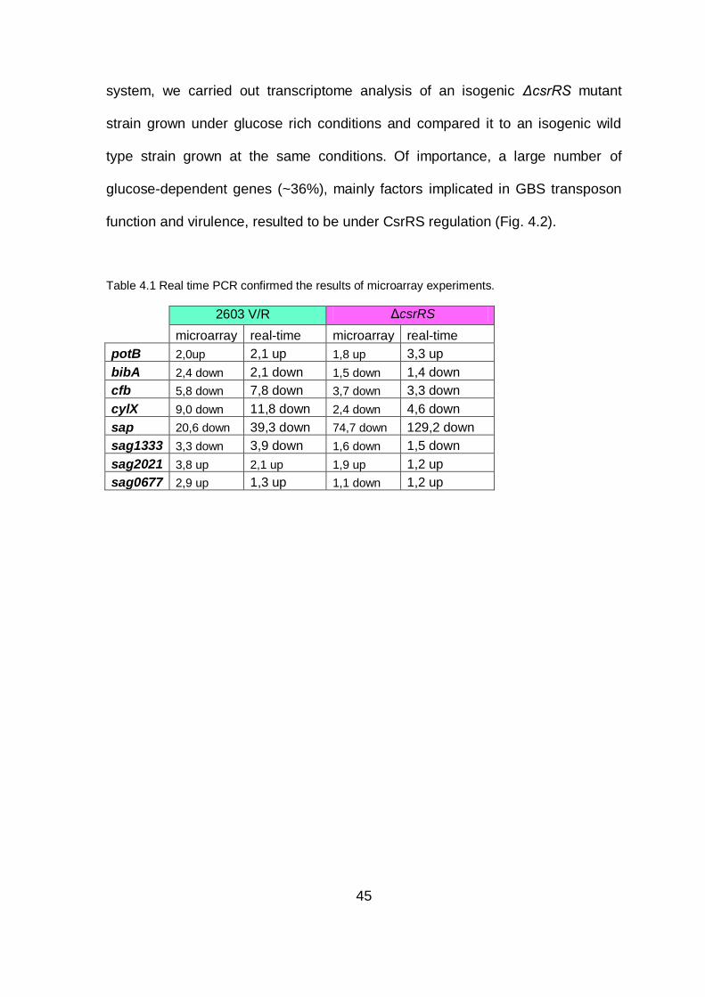
A list of the most regulated genes at high glucose conditions is reported in Table
4.2.
39
4.2 Functional categories
A. Stress response of GBS in high glucose condition
Growth in high glucose medium involves a considerable change in expression of
genes involved in adaptation and stress response. Among stress response
genes, a dramatic change was observed in the transcript for sag1677, encoding a
universal stress protein, whose expression was highly down-regulated (27-fold)
following exposure to glucose. Several genes, including sag1135, sag1136,
sag1137, encoding stress proteins were down-regulated, too. Interestingly, these
stress proteins are homologs of Gls24, a general stress protein of Enterococcus
faecalis which has been reported to have a crucial role in stress response as well
as in virulence (Teng et al., 2004).
B. Transcriptional regulators
After 30 minutes of incubation with 1% glucose several transcriptional regulators
were modulated. In particular, sag1128, sag2017, sag0554 belonging to the
putative Cro/CI family and sag1749, sag1655, sag0427, being part of the putative
Mer family of regulators, were 2-5 fold up-regulated. Of interest, the latter family
has been reported to act as a key activator in nitric oxide defense system in
pneumococci, thus ensuring both survival and systemic infection (Stroeher et al.,
2007; Brown et al., 2003)
In response to the availability of glucose sources, bacteria inhibit multiple
enzymes responsible for alternative sugar metabolism pathways through the
40
carbon catabolite repression system (CCR), mediated by the catabolite control
protein A (CcpA, sag0707). Bioinformatic analysis revealed that a number of
transcriptional regulators, that were highly down-regulated under glucose
conditions (up to 20-fold), has in their promoter region a consensus sequence for
the binding of CcpA
(http://regprecise.lbl.gov/RegPrecise/gmregulon.jsp?gmproject_id=6875). They
included: sag0277, encoding a Mga-like protein, a positive regulator of virulence
in GAS (Almengor et al., 2007; sag1348, fruR, lactose phosphotrasferase system
repressor; sag0119, rbsR, ribose operon repressor; sag0042, phosphosugar-
binding transcriptional regulator belonging to RpiR family; sag2073, a
transcriptional regulator belonging to GntR family; sag2161, encoding for a
transcriptional regulator of the Crp/Fnr family and CcpA itself.
C. Transport genes
Several genes encoding transport proteins were found to be up-regulated in high
glucose conditions. These included sag0745, coding for a putative transporter of
the NRAMP family, involved both in Mn2+ and Fe2+ uptake (Janulczyk et al., 2003;
Papp-Wallace and Maguire, 2006) and genes encoding for proteins involved in
potassium uptake, such as sag1590, sag1591, sag1631 (belonging to the trk
family) and sag1090. We also observed a five fold upregulation of the sag1711
gene, coding for a putative CorA protein involved in magnesium transport
(Warren et al., 2004). A number of genes belonging to the transport family and
found to be up-regulated by glucose are interestingly involved in peptide uptake,
a process essential to satisfy GBS growth requirement, given that this bacterium
41
has a limited capacity to synthesize amino acid. Among up-regulated genes,
although regulated at different extent, we found the dps gene (sag1444), coding
for a putative peptide/proton symporter (Samen et al., 2004); a putative histidine
ABC transporter (sag0947 to sag0949); a spermidine-putrescine transporter,
potABCD (sag1108 to sag1111), implicated in the pathogenesis of Streptococcus
pneumoniae in various infection models (Shah et al., 2011); the sag0290 to
sag0292 operon (regulated up to 8-fold), encoding a putative polar amino acid
ABC transporter; the sag1145 gene, encoding for the sodium:alanin symporter
protein; the sag0715 to sag0718 and sag0947 to sag0949 genes, all encoding for
amino acid ABC transporters. Furthermore, we found up-regulated the region
comprising the sag0241 to sag0244 genes, encoding an ABC transporter for
glycine betaine, whose accumulation in B.subtilis and L.lactis confers protection
against osmotic and cold stress to the bacterium (Hoffmann et al., 2002;
Hoffmann and Bremer, 2011; Obis et al., 1999).
As expected, in high glucose condition genes involved in the transport of complex
carbohydrates were also down-regulated, including the region from sag1441 to
sag1443, encoding for the maltose-maltodextrin transport system (malE-F-G);
sag0955 and sag1925 (msmK) genes, encoding a sugar-ABC transporter and a
sugar transport ATP-binding protein, respectively; the ribose ABC transporter
region (from sag0114 to sag0117, namely rbsA-B-C-D); the cellobiose ABC
transporter (sag0328 to sag0330). Furthermore several multiple transport
systems (PTS), which allow uptake of various carboydrate sources, such as
sag1805, sag1813, sag1814, sag1948-1951, sag1898-1902 and sag0192,
showing a specificity for different sugars, were found to be highly down-regulated
(up to 120-fold).
42
D. Wide-ranging changes in GBS adaptive metabolism
The expression of several genes involved in a wide range of metabolic pathway
was dramatically modulated by a 30 minutes incubation in 1% glucose, mirroring
a rapid adaptation of GBS cellular process metabolism to conditions and nutrients
in the new environment. We observed that bacteria grown in such a rich medium
were growing more rapidly, thus, as expected, gene encoding for DNA replication,
recombination and repair and gene encoding for membrane biosynthesis were
found to be highly up-regulated, while genes encoding for enzymes involved in
substrate degradation were found to be down-regulated. Transcript changes were
observed in aminoacid classes, in particular the sag2165 and sag2167, which
encode for a carbamate kinase and an ornithine carbamoyltransferase,
respectively, were highly down-regulated (up to 20-fold). Both enzymes are
components of the arginine deaminase system, which has been suggested to aid
bacterial survival in acidic environments by catalyzing the release of ammonia
from arginine (Gruening et al., 2006). The sag1907 gene (eda-2), encoding for
keto-hydroxyglutarate-aldolase/keto-deoxy-phosphogluconate aldolase, was
found to be highly down-regulated, too. Conversely few genes were slightly up-
regulated, such as genes belonging to the aspartate and serine families.
We found a drastic down-regulation (up to 130-fold) of genes implicated in energy
metabolism of sugars, including the sag0033 to sag0042 locus, encoding for the
sialic acid operon. The list also comprises a carbohydrate kinase (sag1906)
belonging to PfkB family; a putative hexulose-6-phosphate synthase (sag1812);
L-a ribulose-5-phosphate 4-epimerase (sag1810); a putative hexulose-6-
phosphate isomerase (sag1811); and the sag0118 gene encoding for a
43
ribokinase. Also genes encoding for proteins required for fermentation processes
were found to be down-regulated, including sag1637, sag0053 and sag0054,
annotated as alcohol dehydrogenases (adh, adhP, adhE). The repression of
genes codifying for proteins implicated in biosynthesis and degradation of
polysaccharides such as sag1901, glucuronyl hydrolase; sag0041, acetyl xylan
esterase; sag1216, pullulanase; sag0856, glycogen synthase (glgA); sag0854,
glucose-1-phosphate adenylyltransferase (glgC); sag0853 glycogen branching
enzyme (glgB) was also observed.
E. Virulence and host-pathogen interaction genes
Pathogenic bacteria by modulating the expression of surface-associated or
secreted virulence factors can adapt to host conditions and improve their capacity
to persist in specific niches. Although pathologic high glucose conditions are often
associated to an increased risk for GBS infections, we found that transcription of
known or putative virulence factors was down-regulated in response to glucose
stress conditions, indicating that in this particular scenario such determinants may
be dispensable to GBS invasiveness. In particular the expression of pore-forming
toxins including a) the cyl gene cluster (sag0662 to sag0673), required for GBS
hemolysin production, which is responsible for promoting invasion of host cells
and triggering cell lysis (Pritzlaff et al., 2001) ,and b) the cfb gene (sag2043),
encoding the CAMP factor (Lang and Palmer, 2003), was down-regulated in a
range between 6 to12 fold.
A similar pattern of expression was observed for several surface-expressed
proteins containing the LPXTG cell wall-sorting motif, such as bibA (sag2063).
44
Among virulence factors involved in host cell adherence and invasion, the
expression of the sag1234 gene encoding for the laminin binding protein, Lmb,
which promotes adherence of GBS to host cells by binding to ECM laminin
(Spellerberg et al., 1999), was repressed by glucose. Furthermore, we found that
also the expression of sodA (sag0788), a gene which plays a crucial role against
oxidative stress (Poyart et al., 2001), and of the hyaluronate lysase (sag1197),
which cleaves hyaluronic acid, a major component of the connective tissue, thus
promoting GBS spreading during infection, was significantly reduced.
On the contrary only few genes were found to be up-regulated, such as sag0677,
encoding an unknown function LPXTG protein, and sag2021, encoding for a
protein binding to human glycoprotein GP-340 thus preventing bacterial
colonization (Brady et al., 2010). The same activation trend was observed for the
sag1739 to sag1744 operon, encoding proteins which although not officially
classified among virulence factors, have been reported to be involved in the
respiration metabolism and to play a role in virulence and GBS growth in vivo
(Yamamoto et al., 2005).
4.3 The response to glucose involves the two component
system CsrRS
By comparing previously reported information on genes controlled by the CsrRS
two-component system (Lamy et al., 2004) and the array of genes modulated by
glucose, we found a number of common genes, including sag0662 to sag0673
(cyl operon), sag2043 (cfb) and sag2063 (bibA). Therefore to postulate whether
glucose-dependent regulation of gene expression was under the control of CsrRS
45
system, we carried out transcriptome analysis of an isogenic ΔcsrRS mutant
strain grown under glucose rich conditions and compared it to an isogenic wild
type strain grown at the same conditions. Of importance, a large number of
glucose-dependent genes (~36%), mainly factors implicated in GBS transposon
function and virulence, resulted to be under CsrRS regulation (Fig. 4.2).
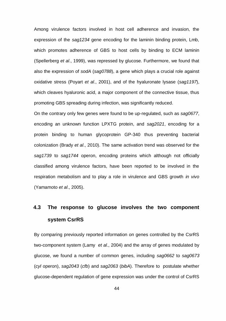
Table 4.1 Real time PCR confirmed the results of microarray experiments.
2603 V/R ΔcsrRS
microarray real-time microarray real-time
potB 2,0up 2,1 up 1,8 up 3,3 up
bibA 2,4 down 2,1 down 1,5 down 1,4 down
cfb 5,8 down 7,8 down 3,7 down 3,3 down
cylX 9,0 down 11,8 down 2,4 down 4,6 down
sap 20,6 down 39,3 down 74,7 down 129,2 down
sag1333 3,3 down 3,9 down 1,6 down 1,5 down
sag2021 3,8 up 2,1 up 1,9 up 1,2 up
sag0677 2,9 up 1,3 up 1,1 down 1,2 up
46
Table 4.2 List of genes highly regulated in GBS strain 2603V/R after incubation in high glucose
medium.
TIGR
locus
Annotation Family Fold
change
Regulation
sag0008 Transcription-repair
coupling factor (mfd)
DNA metabolism 6.1 Up
sag0185 Hypothetical protein Cell envelope 10.24 Down
sag0281 Hypothetical protein Cell envelope 7.04 Down
sag2169 Hypothetical protein Cell envelope 8.35 Up
sag0040 ROK family protein Unknown function 48.35 Down
sag1643 Glutamine
amidotransferase, class I
Unknown function 19.91 Up
sag0034 Sugar ABC transporter Transport and binding
proteins
128.03 Down
sag1949 PTS system Transport and binding
proteins
84.29 Down
sag1950 PTS system Transport and binding
proteins
119.63 Down
sag1951 PTS system Transport and binding
proteins
92.69 Down
sag1642 ABC transporter Transport and binding
proteins
19.12 Up
sag0664 CylG protein (cylG) Cellular processes 12.25 Down
sag0663 CylD protein (cylD) Cellular processes 11.63 Down
sag0667 CylA protein (cylA) Cellular processes 10.16 Down
sag0666 CylZ protein (cylZ) Cellular processes 9.92 Down
sag0662 CylX protein (cylX) Cellular processes 8.99 Down
sag0668 CylB protein (cylB) Cellular processes 8.82 Down
sag0669 CylE protein (cylE) Cellular processes 8.79 Down
sag0670 CylF protein (cylF) Cellular processes 8.63 Down
sag0671 CylJ protein (cylJ) Cellular processes 7.01 Down
sag0672 CylI protein (cylI) Cellular processes 6.76 Down
sag0673 CylK protein (cylK) Cellular processes 5.98 Down
sag2043 cAMP factor (cfb) Cellular processes 5.76 Down
sag1733 Universal stress protein
family
Cellular processes 96.38 Up
sag0777 ATP- dependent RNA
helicase
Transcription 21.94 Up
sag1216 Pullulanase (sap) Energy metabolism 20.61 Down
sag0856 Glycogen synthase (glgA) Energy metabolism 20.31 Down
sag1907 Keto-hydroxyglutarate-
aldolase (eda-2)
Amino acids
biosynthesis
48.04 Down
sag2165 Ornithine
carbamoyltransferase
(argF-2)
Amino acids
biosynthesis
10.84 Down
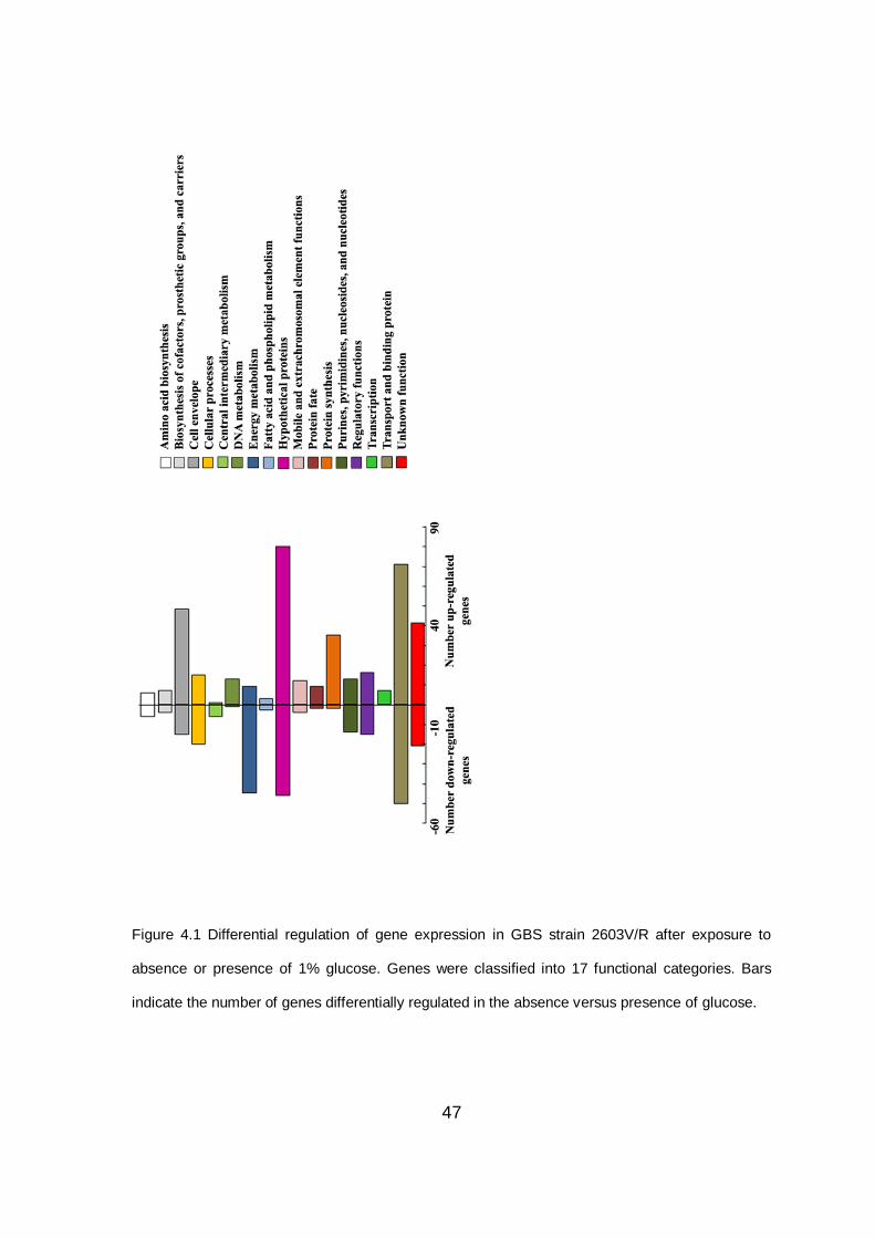
47
Figure 4.1 Differential regulation of gene expression in GBS strain 2603V/R after exposure to
absence or presence of 1% glucose. Genes were classified into 17 functional categories. Bars
indicate the number of genes differentially regulated in the absence versus presence of glucose.
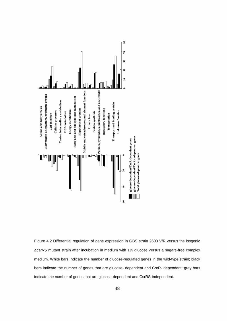
48
Figure 4.2 Differential regulation of gene expression in GBS strain 2603 V/R versus the isogenic
csrRS mutant strain after incubation in medium with 1% glucose versus a sugars-free complex
medium. White bars indicate the number of glucose-regulated genes in the wild-type strain; black
bars indicate the number of genes that are glucose- dependent and CsrR- dependent; grey bars
indicate the number of genes that are glucose-dependent and CsrRS-independent.
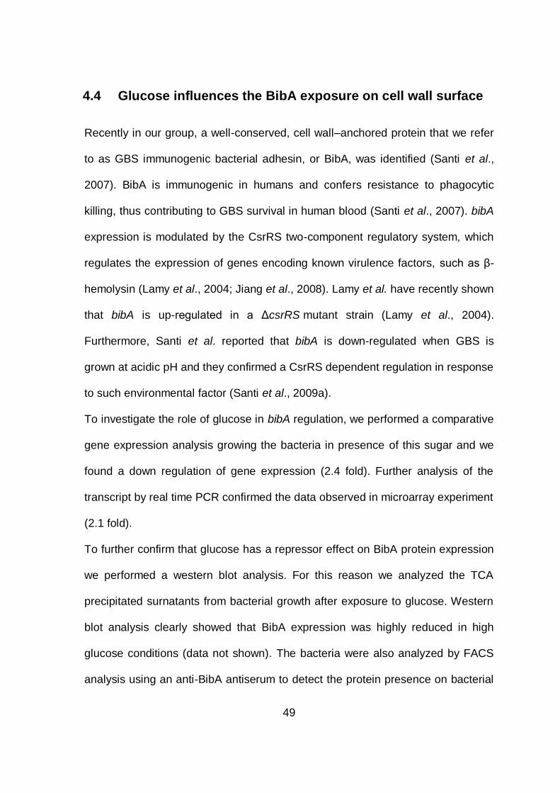
49
4.4 Glucose influences the BibA exposure on cell wall surface
Recently in our group, a well-conserved, cell wall–anchored protein that we refer
to as GBS immunogenic bacterial adhesin, or BibA, was identified (Santi et al.,
2007). BibA is immunogenic in humans and confers resistance to phagocytic
killing, thus contributing to GBS survival in human blood (Santi et al., 2007). bibA
expression is modulated by the CsrRS two-component regulatory system, which
regulates the expression of genes encoding known virulence factors, such as β-
hemolysin (Lamy et al., 2004; Jiang et al., 2008). Lamy et al. have recently shown
that bibA is up-regulated in a ΔcsrRS mutant strain (Lamy et al., 2004).
Furthermore, Santi et al. reported that bibA is down-regulated when GBS is
grown at acidic pH and they confirmed a CsrRS dependent regulation in response
to such environmental factor (Santi et al., 2009a).
To investigate the role of glucose in bibA regulation, we performed a comparative
gene expression analysis growing the bacteria in presence of this sugar and we
found a down regulation of gene expression (2.4 fold). Further analysis of the
transcript by real time PCR confirmed the data observed in microarray experiment
(2.1 fold).
To further confirm that glucose has a repressor effect on BibA protein expression
we performed a western blot analysis. For this reason we analyzed the TCA
precipitated surnatants from bacterial growth after exposure to glucose. Western
blot analysis clearly showed that BibA expression was highly reduced in high
glucose conditions (data not shown). The bacteria were also analyzed by FACS
analysis using an anti-BibA antiserum to detect the protein presence on bacterial
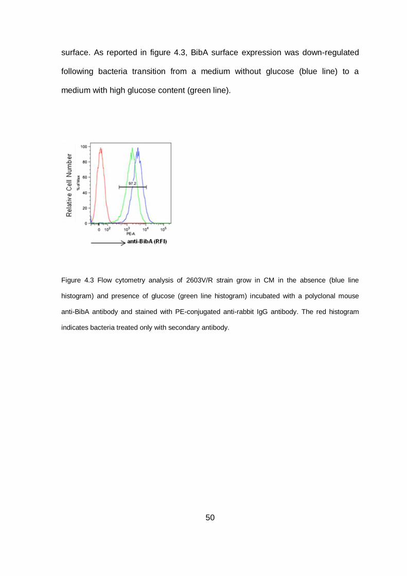
50
surface. As reported in figure 4.3, BibA surface expression was down-regulated
following bacteria transition from a medium without glucose (blue line) to a
medium with high glucose content (green line).
Figure 4.3 Flow cytometry analysis of 2603V/R strain grow in CM in the absence (blue line
histogram) and presence of glucose (green line histogram) incubated with a polyclonal mouse
anti-BibA antibody and stained with PE-conjugated anti-rabbit IgG antibody. The red histogram
indicates bacteria treated only with secondary antibody.
51
4.5 CsrR specifically binds to the bibA promoter
Lamy and colleagues (Lamy et al., 2004) identified a potential CsrR recognition
sequence, through DNase I footprinting and sequence comparisons of upstream
regions from three genes, including bibA gene from NEM 316 strain (gbs2018). A
comparison of the DNA regions protected by CsrR from DNase I cleavage
allowed to identify a highly conserved nonanucleotide motif 5’-TATTTTAAT-3’.
This sequence is 100% conserved in the CsrR binding regions of the cylX and
bibA promoters. For bibA gene, two binding sites for CsrR were identified at -60
bp and -144 bp from translational starting point.
Based on these evidences, we analyzed the bibA promoter in 2603 V/R strain
and found a putative CsrR binding site at -146 bp from translational starting point
(Fig. 4.4).
To better define CsrRS-mediated gene regulation, we deeper investigated its
mechanism of action on bibA expression. To determine whether CsrR interacts
directly with the bibA promoter region, we performed electrophoresis mobility shift
assay (EMSA) using recombinant CsrR protein. A biotin-DNA probe, which
corresponded to the promoter region of bibA, was incubated with increasing
concentrations of CsrR. As shown in figure 4.5A, CsrR induced a shift in the
mobility of the fragment at a concentration of 0.8 μg, postulating that bibA
expression could be controlled by this two component system.
As already described, CsrS is the sensor kinase protein of the CsrRS TCS
responsible for the phosphorylation of the regulator component (CsrR) at a
conserved aspartate residue. Aspartate phosphorylation usually alters the effector
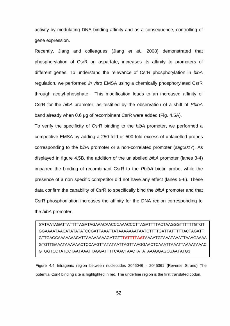
52
activity by modulating DNA binding affinity and as a consequence, controlling of
gene expression.
Recently, Jiang and colleagues (Jiang et al., 2008) demonstrated that
phosphorylation of CsrR on aspartate, increases its affinity to promoters of
different genes. To understand the relevance of CsrR phosphorylation in bibA
regulation, we performed in vitro EMSA using a chemically phosphorylated CsrR
through acetyl-phosphate. This modification leads to an increased affinity of
CsrR for the bibA promoter, as testified by the observation of a shift of PbibA
band already when 0.6 μg of recombinant CsrR were added (Fig. 4.5A).
To verify the specificity of CsrR binding to the bibA promoter, we performed a
competitive EMSA by adding a 250-fold or 500-fold excess of unlabelled probes
corresponding to the bibA promoter or a non-correlated promoter (sag0017). As
displayed in figure 4.5B, the addition of the unlabelled bibA promoter (lanes 3-4)
impaired the binding of recombinant CsrR to the PbibA biotin probe, while the
presence of a non specific competitor did not have any effect (lanes 5-6). These
data confirm the capability of CsrR to specifically bind the bibA promoter and that
CsrR phosphorilation increases the affinity for the DNA region corresponding to
the bibA promoter.
5’ATAATAGATTATTTTAGATAGAAACAACCCAAACCCTTAGATTTTACTAAGGGTTTTTTGTGT
GGAAAATAACATATATATCCGATTAAATTATAAAAAAATAATCTTTTGATTATTTTTACTAGATT
GTTGAGCAAAAAAACATTAAAAAAAAGATGTTTATTTTAATAAAATGTAAATAAATTAAAGAAAA
GTGTTGAAATAAAAAACTCCAAGTTATATAATTAGTTAAGGAACTCAAATTAAATTAAAATAAAC
GTGGTCCTATCCTAATAAATTAGGATTTTCAACTAACTATATAAAGGAGCGAATATG3
Figure 4.4 Intragenic region between nucleotides 2045046 - 2045361 (Reverse Strand) The
potential CsrR binding site is highlighted in red. The underline region is the first translated codon.
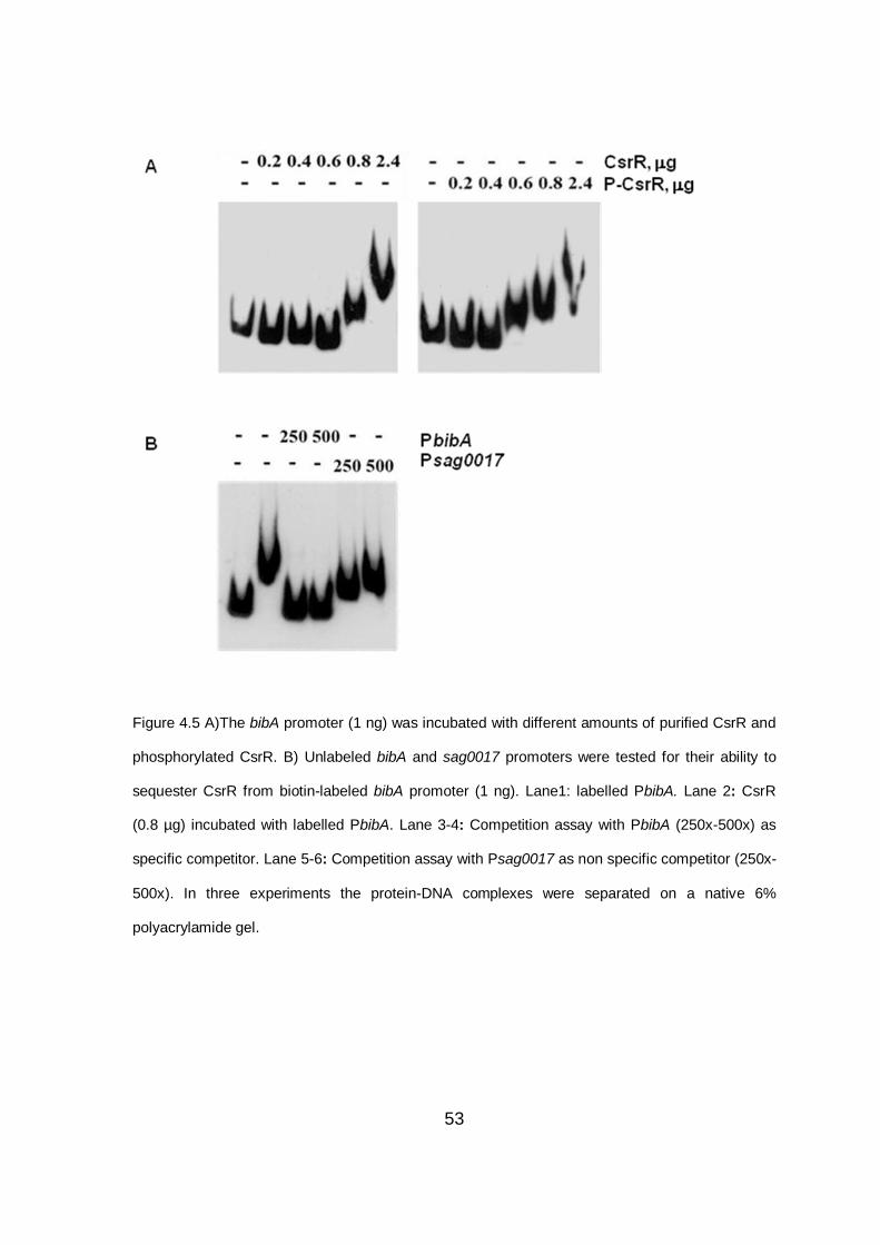
53
Figure 4.5 A)The bibA promoter (1 ng) was incubated with different amounts of purified CsrR and
phosphorylated CsrR. B) Unlabeled bibA and sag0017 promoters were tested for their ability to
sequester CsrR from biotin-labeled bibA promoter (1 ng). Lane1: labelled PbibA. Lane 2: CsrR
(0.8 µg) incubated with labelled PbibA. Lane 3-4: Competition assay with PbibA (250x-500x) as
specific competitor. Lane 5-6: Competition assay with Psag0017 as non specific competitor (250x-
500x). In three experiments the protein-DNA complexes were separated on a native 6%
polyacrylamide gel.
54
4.6 CsrR acts as repressor of bibA expression
These data by postulating a direct interaction between CsrR and the bibA
promoter suggest that CsrR controls the expression of this gene in vivo.
To investigate how the presence of glucose can influence the role of CsrR in bibA
expression we performed chromatin immuno-precipitation (ChIP) analysis.
Bacterial cultures grown to an OD600 of 0.5 were exposed for 30’ to glucose and
in vivo cross-linked with formaldehyde. After sonication, protein-DNA complexes
were immunoprecipitated with a nonsaturating amount of polyclonal CsrR
antiserum. The immunoprecipitated DNA (IP) was analyzed by dot blot analysis,
spotting on membrane the IP and flowthrough (FT) and using 32P-labelled bibA
promoter as probe. FT samples indicated that the DNA amount loaded in each
spot was comparable (data do not shown). As shown in figure 4.6, quantification
of dot blot intensity relative to DNA immunoprecipitated with CsrR antiserum
revealed a 3 fold increase in the amount of 32P-labelled bibA promoter in GBS
cultures grown in high glucose (1%) medium compared to sugar-free conditions.
The same experiment was performed using an isogenic ΔcsrRS deletion mutant
and no differences in the magnitude of immunoprecipated DNA were observed
(data not shown).
These data strongly postulate that CsrR acts as a repressor of bibA expression in
vivo.
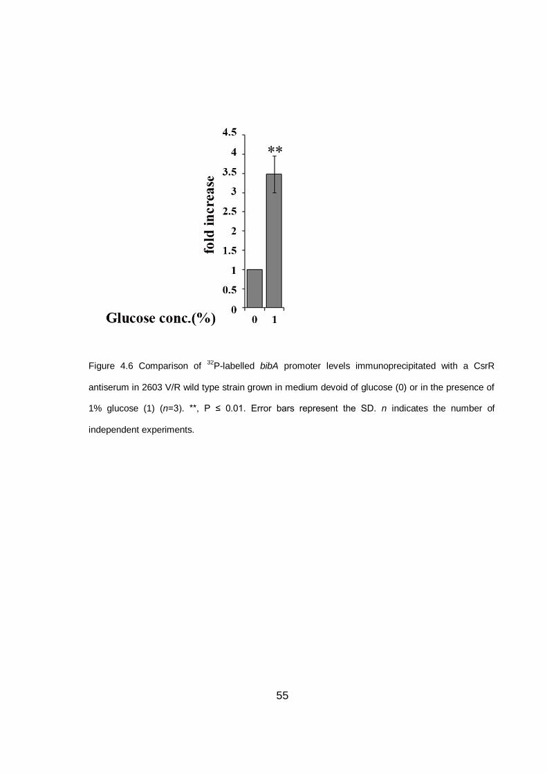
55
Figure 4.6 Comparison of 32
P-labelled bibA promoter levels immunoprecipitated with a CsrR
antiserum in 2603 V/R wild type strain grown in medium devoid of glucose (0) or in the presence of
1% glucose (1) (n=3). **, P ≤ 0.01. Error bars represent the SD. n indicates the number of
independent experiments.
56
4.7 Promoter region of sap gene shows the CcpA binding site
In addition to genes that appeared to be glucose and CsrRS dependent, we found
a large group of genes (63%), involved in GBS metabolism, transport and protein
synthesis, that was modulated by glucose both in the wild-type strain and in the
csrRS mutant strain, indicating that their expression was glucose dependent but
CsrRS independent.
GBS, as other streptococcal species (Tomoyasu et al., 2010; Abranches et al.,
2008), has a complex metabolic system that controls carbohydrate catabolism,
named CcpA regulatory system, in which enzymes necessary for the metabolism
of alternative sugar are inhibited in the presence of glucose. In particular, CcpA,
under the stimuli of environmental sugars, regulates the expression of metabolic
genes and virulence determinants (Tomoyasu et al., 2010; Abranches et al.,
2008; Almengor et al., 2007). Genes subjected to CcpA regulation are identifiable
by the presence of a target DNA sequence, called the catabolite-responsive
element (cre) site, to which CcpA binds.
To investigate the presence of the cre box in the promoter region of GBS genes,
we performed a bioinformatic analysis of the 2603 V/R genome using the
program Reg Precise
(http://regprecise.lbl.gov/RegPrecise/gmregulon.jsp?gmproject_id=6875). By this
comparative analysis we found that in 2603 V/R strain, 60 operons, conteining
139 genes showed an identical sequence as the one recognized by CcpA. In
particular, we found that 77% of them were differentially regulated in the presence
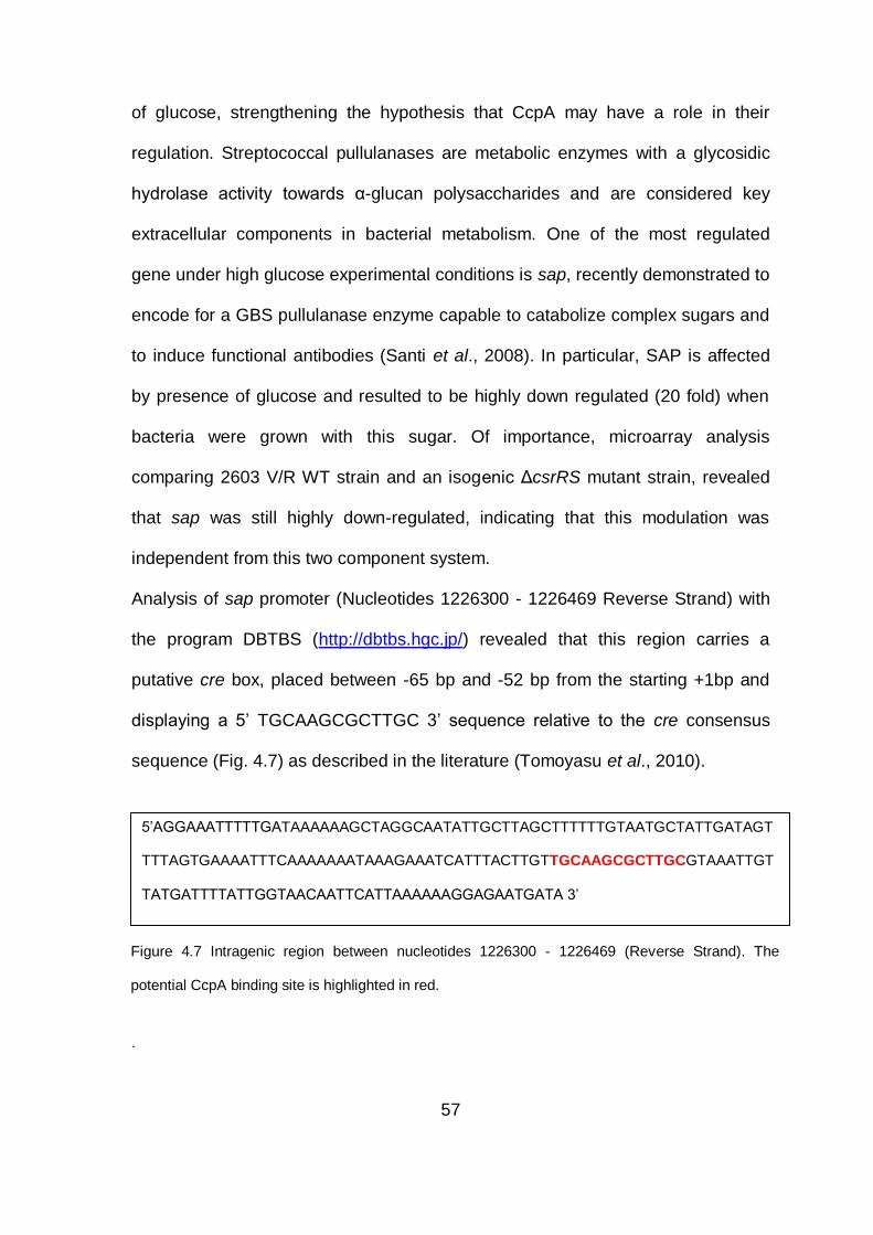
57
of glucose, strengthening the hypothesis that CcpA may have a role in their
regulation. Streptococcal pullulanases are metabolic enzymes with a glycosidic
hydrolase activity towards α-glucan polysaccharides and are considered key
extracellular components in bacterial metabolism. One of the most regulated
gene under high glucose experimental conditions is sap, recently demonstrated to
encode for a GBS pullulanase enzyme capable to catabolize complex sugars and
to induce functional antibodies (Santi et al., 2008). In particular, SAP is affected
by presence of glucose and resulted to be highly down regulated (20 fold) when
bacteria were grown with this sugar. Of importance, microarray analysis
comparing 2603 V/R WT strain and an isogenic ΔcsrRS mutant strain, revealed
that sap was still highly down-regulated, indicating that this modulation was
independent from this two component system.
Analysis of sap promoter (Nucleotides 1226300 - 1226469 Reverse Strand) with
the program DBTBS (http://dbtbs.hgc.jp/) revealed that this region carries a
putative cre box, placed between -65 bp and -52 bp from the starting +1bp and
displaying a 5’ TGCAAGCGCTTGC 3’ sequence relative to the cre consensus
sequence (Fig. 4.7) as described in the literature (Tomoyasu et al., 2010).
.
5’AGGAAATTTTTGATAAAAAAGCTAGGCAATATTGCTTAGCTTTTTTGTAATGCTATTGATAGT
TTTAGTGAAAATTTCAAAAAAATAAAGAAATCATTTACTTGTTGCAAGCGCTTGCGTAAATTGT
TATGATTTTATTGGTAACAATTCATTAAAAAAGGAGAATGATA 3’
Figure 4.7 Intragenic region between nucleotides 1226300 - 1226469 (Reverse Strand). The
potential CcpA binding site is highlighted in red.
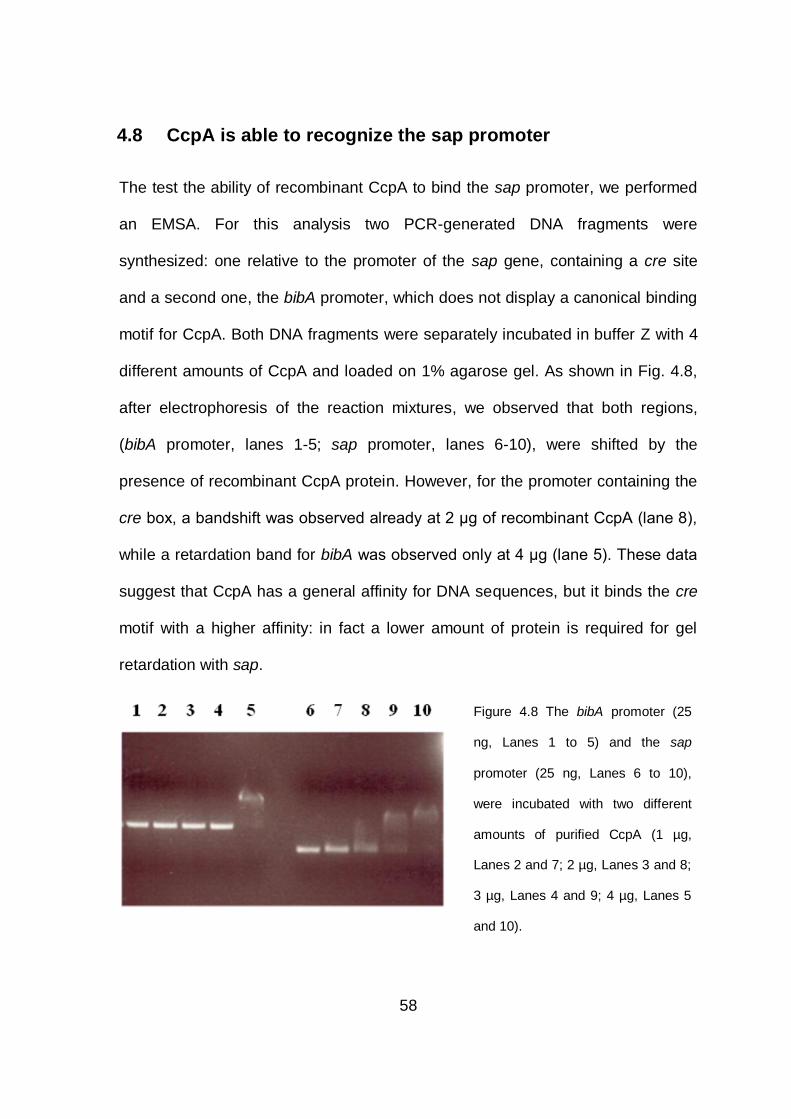
58
4.8 CcpA is able to recognize the sap promoter
The test the ability of recombinant CcpA to bind the sap promoter, we performed
an EMSA. For this analysis two PCR-generated DNA fragments were
synthesized: one relative to the promoter of the sap gene, containing a cre site
and a second one, the bibA promoter, which does not display a canonical binding
motif for CcpA. Both DNA fragments were separately incubated in buffer Z with 4
different amounts of CcpA and loaded on 1% agarose gel. As shown in Fig. 4.8,
after electrophoresis of the reaction mixtures, we observed that both regions,
(bibA promoter, lanes 1-5; sap promoter, lanes 6-10), were shifted by the
presence of recombinant CcpA protein. However, for the promoter containing the
cre box, a bandshift was observed already at 2 μg of recombinant CcpA (lane 8),
while a retardation band for bibA was observed only at 4 μg (lane 5). These data
suggest that CcpA has a general affinity for DNA sequences, but it binds the cre
motif with a higher affinity: in fact a lower amount of protein is required for gel
retardation with sap.
Figure 4.8 The bibA promoter (25
ng, Lanes 1 to 5) and the sap
promoter (25 ng, Lanes 6 to 10),
were incubated with two different
amounts of purified CcpA (1 µg,
Lanes 2 and 7; 2 µg, Lanes 3 and 8;
3 µg, Lanes 4 and 9; 4 µg, Lanes 5
and 10).
59
4.9 CcpA binds specifically the promoter of pullulanase
To assess whether the recombinant protein CcpA could bind the cre box in
pullulanase promoter, EMSA was performed with CcpA using, as probe, biotin-
labeled DNA fragment, corresponding to the cre site from the sap promoter (cre
box, Table 3.1). The probe was incubated with different concentration of
recombinant protein and the complex was resolved on native polyacrylamide gel.
The lower amount of CcpA able to induce the mobility retardation of the probe
was 0.6 μg (data do not shown).
After establishing the minimum concentration at which the shift was observed, we
performed EMSA using biotin-labeled DNA fragments, carrying both a wild type
and a mutated cre sequence (mutated cre, Table 3.1). Figure 4.9A shows that
recombinant CcpA was able to induce a shift in mobility of the cre box probe,
while it was not able to bind the fragment carrying a mutated cre sequence.
CcpA specific binding to the cre sequence was also tested by competition EMSA.
As competitors, unlabeled wild type and mutated cre sequences were used. As
shown in Fig. 4.9B CcpA binding to labelled cre box sequence was reversed
when either a 100-fold or 500-fold excess of unlabelled sap cre sequence was
added, while it was not affected by competition with identical concentrations of a
mutated cre sequence.
In conclusion, we postulate that recombinant CcpA is able to bind the cre box
present in the promoter of pullulanase in a specific manner. Indeed, CcpA does
not recognize a mutated cre sequence. The competition assay has also
demonstrated that an unlabeled wild type cre box probe, but not the mutated one,

60
competes for the binding to the protein, enforcing the specific affinity of CcpA for
this sequence. These results suggest that the sap gene could be directly
regulated by CcpA, and its repression observed under high glucose conditions
depending on this regulator.
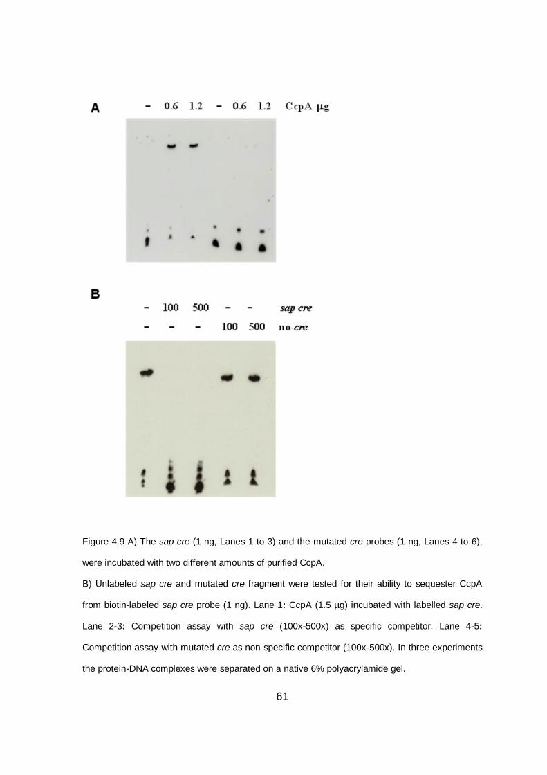
61
Figure 4.9 A) The sap cre (1 ng, Lanes 1 to 3) and the mutated cre probes (1 ng, Lanes 4 to 6),
were incubated with two different amounts of purified CcpA.
B) Unlabeled sap cre and mutated cre fragment were tested for their ability to sequester CcpA
from biotin-labeled sap cre probe (1 ng). Lane 1: CcpA (1.5 µg) incubated with labelled sap cre.
Lane 2-3: Competition assay with sap cre (100x-500x) as specific competitor. Lane 4-5:
Competition assay with mutated cre as non specific competitor (100x-500x). In three experiments
the protein-DNA complexes were separated on a native 6% polyacrylamide gel.
62
5. Discussion
Diabetes mellitus is characterized by inappropriately high concentrations of
glucose in plasma as a result of a relative or absolute lack of insulin. Patients with
uncontrolled diabetes mellitus can be more susceptible to bacterial infections,
which can lead to higher morbidity and mortality than in healthy, non-diabetic
patients because of dysfunction in polymorphonuclear neutrophil bactericidal
function, cellular immunity, and complement activation. For this reason, diabetic
patients tend to have a higher incidence and increased severity of infections than
non-diabetic patients.
Short and transient periods of hyperglycaemia result in abnormalitiesin
granulocyte adherence, chemotaxis, phagocytosis, and bactericidal function.
These impairments in immune function have been associated with an increased
risk of infections. Systemic hyperglycaemia results in derangement of the immune
system including neutrophil function, cellular immunity, and complement function.
As previous studies have shown, Klebsiella pneumoniae, followed by
Streptococcus spp. were the most commonly isolated organisms among diabetic
patients, whereas in the non-diabetic group Streptococcus spp., followed by
Staphylococcus spp., were most common (Zheng et al., 2012 ).
Although GBS is commonly associated with neonatal diseases and postpartum
infections, it is also an important cause of morbidity and mortality among adults.
GBS infection has been reported to occur in adults with serious underlying
conditions and, in particular, diabetes has been reported to be one of the most
important risk factor promoting GBS infections. Patients with diabetes display
63
peripheral neuropathy or peripheral vascular diseases following trauma,
particularly to the lower extremities. GBS takes advantage of this condition by
crossing the endothelial barrier and promoting bacterial invasion of the foot.
Indeed, GBS is also found in biopsy of patients with foot infections, a common
status of patients suffering from diabetes. The impact of hyperglycemia upon
susceptibility to GBS infection has not been fully elucidated, but, at least in part,
this effect seems to be due to impairment of neutrophil effector functions. Indeed,
Mazade et al. have reported that high glucose levels impair neutrophil-mediated
phagocytosis and killing of GBS and reduce superoxide production thus ensuring
spreading of infection (Mazade and Edwards, 2001).
Although clinical evidences indicate a strong correlation between individuals with
high blood glucose levels and the propensity to acquire GBS systemic infections,
the signaling events triggered in this bacterium by high glucose conditions and
the relative modulation of the expression of virulence determinants are still
unknown.
Glucose concentration in blood varies since 80 mg/dL in healthy people until 600
mg/dL in diabetic patients with severe complications. A clinical study has reported
a patient admitted in hospital with blood glucose concentration of 1288 mg/dL
(Yanai et al., 2012) that corresponds to ~1% of sugar. To investigate the
adaptation of GBS to high glucose presence, we performed a gene expression
analysis growing bacteria in the absence and presence of this carbon source.
One of the limitation for setting in vitro experiments looking at the effect of carbon
sources on bacteria stress response is the fact that metabolites generated during
the incubation of bacteria with sugars can alter the pH of the milieu and,
differently from what happens in vivo, where the environment (i.e. blood) is rapidly
64
buffered, may dramatically affect the activation of intracellular pathways. In order
to precisely delineate the impact of glucose, we defined that the duration of the
experiments was limited to 30 minutes, a time point at which pH was still constant
as at the initial conditions (data not shown).
We found that glucose affected transcription of ~30% of genes; in particular the
most highly regulated genes belong to transport and binding proteins and
metabolism families.
Recent reports have clearly elucidated the molecular mechanisms by which GBS
adapts to different environmental conditions, including pH, carbon source
availability, biological fluids and temperature (Mereghetti et al., 2009; Sitkiewicz et
al., 2009; Santi et al., 2009). During these events a wide range of genes involved
in adhesion to the host, nutrient acquisition and survival to the immune system
are differently modulated. For example, global gene expression analysis of GBS
grown in amniotic fluid, blood and pH stress conditions has revealed a number of
mechanisms used by this bacterium to adapt to the host. Our study proposes that
a general trend of adaptive regulation is triggered during the growth of GBS in the
hyperglycemic milieu of the blood, as for diabetic patients. In particular, we
observed that, at a gene expression level, GBS quickly adapts to environmental
carbon sources changes by modulating both transcription and translation of
genes involved in transport, metabolism and virulence.
One intriguing finding of our analysis is that the exposure of GBS to glucose
stress conditions appears to down-regulate several virulence factors, suggesting
that in such a scenario these determinants may be dispensable to bacterial
invasiveness. In this context, genes encoding for pore-forming toxins, such as
hemolysin β and CAMP factor, both known to be crucial in promoting cell invasion
65
and lysis (Pritzlaff et al., 2001; Lang et al., 2003), were found to be highly down-
regulated. Furthermore, the presence of glucose reduced the transcription of
genes involved in host cell adherence, such as Lmb (Spellerberg et al., 1999) and
serum resistance, such as bibA (Santi et al., 2009).
The down-regulation of virulence determinants in a high glucose milieu clearly
point out the reliance of GBS fitness on the access to the metabolites derived by
the catabolism of carbon sources. Indeed, over the past few years it has been
reported that catabolism of carbohydrates plays a key role in the pathogenesis of
invasive streptoccocci, including Streptococccus pneumoniae, Streptococcus
pyogenes and GBS (Almengor et al., 2007; Shelburne et al., 2008a; iyer and
Camilli, 2007; Jones et al., 2000). Johns and colleagues have recently applied
signature-tagged transposon mutagenesis to a neonatal sepsis model and
showed the importance of carbohydrate catabolism for GBS infectivity (Jones et
al., 2000). In particular, by knocking out genes involved in complex carbohydrate
metabolism, including a maltose-binding protein (mal), a phosphotransferase
(PTS) and a sucrose hydrolase (scrB), they observed an attenuated virulence
(Jones et al., 2000). Furthermore these genes, which clearly impact GBS
virulence, were also found to be down-regulated in our study, suggesting a direct
link between the modulation of genes coding for virulence factors and the one
responsible for carbohydrate utilization, leading to an attenuated status of
bacteria. Indeed, virulence is an energy consuming condition and bacteria
occupying carbohydrate-rich niches, such as the blood of hyperglycemic
individuals, may avoid the activation of virulence-associated pathways.
Response of GBS to variations in environment relies on two major types of
transcriptional regulators, the two-component gene regulatory systems (TCS) and
66
stand-alone regulators (such as CcpA). The best characterized TCS in
streptococci is the CsrR/S system, which plays a key role in connecting the
expression of complex carbohydrate metabolism genes together with that of
virulence factors, thereby contributing pathogenesis (Lamy et al., 2004;
Churchward, 2007; Mascher et al., 2003). In our study a wide range of glucose-
dependent genes, in particular factors implicated in GBS transposon function and
virulence, resulted to be under CsrRS regulation, strongly suggesting that
effectors of this TCS are involved in the response and adaptation of GBS to high
glucose condition. As previously reported for the response of GBS to pH stress
conditions (Santi et al., 2009), also in the case of high glucose conditions the
contribution of CsrRS to the transcription of glucose-dependent genes was found
to be independent on CsrR abundance, as its expression remained constant
along the time span of the experiment. This is in agreement with our findings
showing that CsrR enhances its affinity for the bibA promoter in the presence of
glucose. Based on these evidences, we speculate that as for the pH-dependent
activation of CsrR (Santi et al., 2009), the transmission of a signal through the
CsrS sensor protein during glucose-stress conditions, may be responsible for the
increased affinity for regulated promoters (Jiang et al., 2008).
The clearest link between complex carbohydrate utilization and virulence factor
production is represented by the transcriptional regulator CcpA, exhaustively
studied in GAS (Almengor et al., 2007; Shelburne et al., 2008b), but also present
in other streptococcal species (Tomoyasu et al., 2010; Abranches et al., 2008).
This complex metabolic system controls carbohydrate catabolism by inhibiting
enzymes necessary for the metabolism of alternative sugar in the presence of
glucose. In particular, CcpA, under the stimuli of environmental sugars,
67
modulates both metabolic and virulence genes by binding to the cre site and
repressing their expression (Tomoyasu et al., 2010; Abranches et al., 2008;
Almengor et al., 2007). In our study, we demonstrated that the cre box-containing
promoter of sap, one of the most regulated genes in the presence of glucose, is a
target of CcpA in vitro, suggesting that this protein may contribute to the
regulation of metabolic genes under glucose stress conditions. Indeed, we found
that numerous genes reported to be potentially controlled by CcpA were among
the most down-regulated under high glucose conditions. These data might a
starting point for future investigations to better define the role of carbon
catabolism in GBS virulence.
The need for an increased knowledge of bacterial pathogenesis to better design
curative strategies is among the most challenging objective of modern society. In
this context, the data reported in this paper by highlighting mechanisms used by
GBS to adapt to pathological conditions, may contribute to the understanding of
GBS infectivity and put the basis for the development of next generation
therapeutic and preventive agents.

68
69
6. Acknowledgements
Oltre alle persone che hanno contribuito alla realizzazione del mio progetto di
dottorato dal punto di vista scientifico, vorrei ringraziare soprattutto coloro che mi
hanno accompagnato durante questo percorso e hanno reso indimenticabili
questi tre anni.
Grazie:
Al mio supervisor Marco Soriani, per avermi dato l’opportunità di fare il dottorato,
ma soprattutto per avermi insegnato ad affrontare i confronti in modo diverso e
maturo.
A Silvia Rossi Paccani che mi ha incoraggiato a non mollare quando ero in
difficoltà.
Al mio coordinatore di dottorato Enzo Scarlato, per il confronto scientifico e per le
chiacchierate informali.
Ai miei compagni di scrivania, Gianni e Fulvia che hanno cambiato le mie lacrime
in sorrisi.
A Valentina R, rimarrai sempre la mia mamma di laboratorio.
A tutti i ragazzi del lab 227, che hanno reso le mie giornate lavorative
indimenticabili.
A tutti i partecipanti dei “viaggi della speranza” verso casa, specialmente Cristina
e Francesca Z. che con le loro chiacchierate hanno reso piacevoli tutte quelle ore
di viaggio.
A Olimpia, che mi ha ascoltato e consigliato ogni giorno rendendo più leggeri i
periodi difficili.

70
A tutti i membri del coro di S. Petronilla, siete stati la mia seconda famiglia
facendomi sentire sempre a casa.
Alle mie coinquiline di ieri e di oggi.
Ai miei amici di Napoli che non mi hanno mai fatto sentire un’estranea nonostante
le mie lunghe assenze.
A P. Carmine, che dopo 10 anni ha ancora la pazienza di sentire i miei sfoghi e
mi invoglia a sfruttare i miei “talenti”. Grazie soprattutto per avermi spronato il 21
marzo del 2008 a uscire dal guscio e volare verso orizzonti lontani.
A zia Gianna, che dimostra ogni giorno cosa vuol dire essere una donna.
A Christian, per non aver mai ostacolato il mio cammino e per avermi sempre
invogliata a cercare il meglio di me.
A mamma, papà, Silvia, Maria, Giacomo ed Elisabetta per la loro infinita pazienza
e per aver sopportato i miei sbalzi di umore e le mie paranoie. Grazie per aver
sempre creduto in me e per avermi incoraggiato quando pensavo di non farcela.
E infine grazie a Te che ci sei sempre stato e che mi hai accompagnato in questi
tre anni non lasciandomi mai sola.
71
7. References
Abranches, J., M. M. Nascimento, et al. (2008). "CcpA regulates central
metabolism and virulence gene expression in Streptococcus mutans." J
Bacteriol 190(7): 2340-9.
Almengor, A. C., T. L. Kinkel, et al. (2007). "The catabolite control protein CcpA
binds to Pmga and influences expression of the virulence regulator Mga in
the Group A Streptococcus." J Bacteriol 189(23): 8405-16.
Anderson, I. H., A. S. Levine, et al. (1981). "Incomplete absorption of the
carbohydrate in all-purpose wheat flour." N Engl J Med 304(15): 891-2.
Asanuma, N., T. Yoshii, et al. (2004). "Molecular characterization of CcpA and
involvement of this protein in transcriptional regulation of lactate
dehydrogenase and pyruvate formate-lyase in the ruminal bacterium
Streptococcus bovis." Appl Environ Microbiol 70(9): 5244-51.
Beckmann, C., J. D. Waggoner, et al. (2002). "Identification of novel adhesins
from Group B streptococci by use of phage display reveals that C5a
peptidase mediates fibronectin binding." Infect Immun 70(6): 2869-76.
Belitsky, B. R., H. J. Kim, et al. (2004). "CcpA-dependent regulation of Bacillus
subtilis glutamate dehydrogenase gene expression." J Bacteriol 186(11):
3392-8.
Bisharat, N., N. Jones, et al. (2005). "Population structure of group B
Streptococcus from a low-incidence region for invasive neonatal disease."
Microbiology 151(Pt 6): 1875-81.
Blumberg, H. M., D. S. Stephens, et al. (1996). "Invasive group B streptococcal
disease: the emergence of serotype V." J Infect Dis 173(2): 365-73.
Bohnsack, J. F., K. Widjaja, et al. (1997). "A role for C5 and C5a-ase in the acute
neutrophil response to group B streptococcal infections." J Infect Dis
175(4): 847-55.
Bolduc, G. R., M. J. Baron, et al. (2002). "The alpha C protein mediates
internalization of group B Streptococcus within human cervical epithelial
cells." Cell Microbiol 4(11): 751-8.
Bolotin, A., P. Wincker, et al. (2001). "The complete genome sequence of the
lactic acid bacterium Lactococcus lactis ssp. lactis IL1403." Genome Res
11(5): 731-53.
Bolstad, B. M., R. A. Irizarry, et al. (2003). "A comparison of normalization
methods for high density oligonucleotide array data based on variance and
bias." Bioinformatics 19(2): 185-93.
Boskey, E. R., K. M. Telsch, et al. (1999). "Acid production by vaginal flora in vitro
is consistent with the rate and extent of vaginal acidification." Infect Immun
67(10): 5170-5.

72
Bradford, M. M. (1976). "A rapid and sensitive method for the quantitation of
microgram quantities of protein utilizing the principle of protein-dye
binding." Anal Biochem 72: 248-54.
Brady, L. J., S. E. Maddocks, et al. (2010). "The changing faces of Streptococcus
antigen I/II polypeptide family adhesins." Mol Microbiol 77(2): 276-86.
Brochu, D. and C. Vadeboncoeur (1999). "The HPr(Ser) kinase of Streptococcus
salivarius: purification, properties, and cloning of the hprK gene." J
Bacteriol 181(3): 709-17.
Brown, N. L., J. V. Stoyanov, et al. (2003). "The MerR family of transcriptional
regulators." FEMS Microbiol Rev 27(2-3): 145-63.
Buratta, S., K. Fettucciari, et al. (2002). "Group B streptococcus (GBS) modifies
macrophage phosphatidylserine metabolism during induction of apoptosis."
FEBS Lett 520(1-3): 68-72.
Butler, K. M., C. J. Baker, et al. (1987). "Interaction of soluble fibronectin with
group B streptococci." Infect Immun 55(10): 2404-8.
Chauvaux, S. (1996). "CcpA and HPr(ser-P): mediators of catabolite repression in
Bacillus subtilis." Res Microbiol 147(6-7): 518-22.
Cheng, Q., S. Debol, et al. (2002). "Immunization with C5a peptidase or
peptidase-type III polysaccharide conjugate vaccines enhances clearance
of group B Streptococci from lungs of infected mice." Infect Immun 70(11):
6409-15.
Cheng, Q., D. Stafslien, et al. (2002). "The group B streptococcal C5a peptidase
is both a specific protease and an invasin." Infect Immun 70(5): 2408-13.
Churchward, G. (2007). "The two faces of Janus: virulence gene regulation by
CovR/S in group A streptococci." Mol Microbiol 64(1): 34-41.
Danielli, A., D. Roncarati, et al. (2006). "In vivo dissection of the Helicobacter
pylori Fur regulatory circuit by genome-wide location analysis." J Bacteriol
188(13): 4654-62.
Deutscher, J. (2008). "The mechanisms of carbon catabolite repression in
bacteria." Curr Opin Microbiol 11(2): 87-93.
Deutscher, J., C. Francke, et al. (2006). "How phosphotransferase system-related
protein phosphorylation regulates carbohydrate metabolism in bacteria."
Microbiol Mol Biol Rev 70(4): 939-1031.
Deutscher, J., E. Kuster, et al. (1995). "Protein kinase-dependent HPr/CcpA
interaction links glycolytic activity to carbon catabolite repression in gram-
positive bacteria." Mol Microbiol 15(6): 1049-53.
Deutscher, J., J. Reizer, et al. (1994). "Loss of protein kinase-catalyzed
phosphorylation of HPr, a phosphocarrier protein of the
phosphotransferase system, by mutation of the ptsH gene confers
catabolite repression resistance to several catabolic genes of Bacillus
subtilis." J Bacteriol 176(11): 3336-44.
73
Dillon, H. C., Jr., E. Gray, et al. (1982). "Anorectal and vaginal carriage of group B
streptococci during pregnancy." J Infect Dis 145(6): 794-9.
Dong, Y., Chen, et al. (2004). "Control of expression of the arginine deiminase
operon of Streptococcus gordonii by CcpA and Flp." J Bacteriol 186(8):
2511-4.
Doran, K. S., J. C. Chang, et al. (2002). "Group B streptococcal beta-
hemolysin/cytolysin promotes invasion of human lung epithelial cells and
the release of interleukin-8." J Infect Dis 185(2): 196-203.
Doran, K. S., G. Y. Liu, et al. (2003). "Group B streptococcal beta-
hemolysin/cytolysin activates neutrophil signaling pathways in brain
endothelium and contributes to development of meningitis." J Clin Invest
112(5): 736-44.
Doran, K. S. and V. Nizet (2004). "Molecular pathogenesis of neonatal group B
streptococcal infection: no longer in its infancy." Mol Microbiol 54(1): 23-31.
Edwards, M. S. and C. J. Baker (2005). "Group B streptococcal infections in
elderly adults." Clin Infect Dis 41(6): 839-47.
Edwards, M.S., V. Nizet, et al. (2001) “Group B Streptococcal Infections. In:
Group B Streptococcal Infections, 6th ed, Remington JS, Klein JO, Wilson
CB, Baker CJ (Eds), Elsevier Saunders, Philadelphia 2006. p.403
Farley, M. M., R. C. Harvey, et al. (1993). "A population-based assessment of
invasive disease due to group B Streptococcus in nonpregnant adults." N
Engl J Med 328(25): 1807-11.
Federle, M. J., K. S. McIver, et al. (1999). "A response regulator that represses
transcription of several virulence operons in the group A streptococcus." J
Bacteriol 181(12): 3649-57.
Ferretti, J. J., W. M. McShan, et al. (2001). "Complete genome sequence of an
M1 strain of Streptococcus pyogenes." Proc Natl Acad Sci U S A 98(8):
4658-63.
Frey, N., S. Nessler, et al. (2003). "The HPr(Ser) kinase of Streptococcus
salivarius: a hexameric bifunctional enzyme controlled by glycolytic
intermediates and inorganic phosphate." FEMS Microbiol Lett 224(1): 67-
72.
Gherardi, G., M. Imperi, et al. (2007). "Molecular epidemiology and distribution of
serotypes, surface proteins, and antibiotic resistance among group B
streptococci in Italy." J Clin Microbiol 45(9): 2909-16.
Gibson, R. L., M. K. Lee, et al. (1993). "Group B streptococci invade endothelial
cells: type III capsular polysaccharide attenuates invasion." Infect Immun
61(2): 478-85.
Gibson, R. L., V. Nizet, et al. (1999). "Group B streptococcal beta-hemolysin
promotes injury of lung microvascular endothelial cells." Pediatr Res 45(5
Pt 1): 626-34.
74
Glaser, P., C. Rusniok, et al. (2002). "Genome sequence of Streptococcus
agalactiae, a pathogen causing invasive neonatal disease." Mol Microbiol
45(6): 1499-513.
Gruening, P., M. Fulde, et al. (2006). "Structure, regulation, and putative function
of the arginine deiminase system of Streptococcus suis." J Bacteriol
188(2): 361-9.
Grundy, F. J., A. J. Turinsky, et al. (1994). "Catabolite regulation of Bacillus
subtilis acetate and acetoin utilization genes by CcpA." J Bacteriol 176(15):
4527-33.
Grundy, F. J., D. A. Waters, et al. (1993). "Identification of genes involved in
utilization of acetate and acetoin in Bacillus subtilis." Mol Microbiol 10(2):
259-71.
Gutekunst, H., B. J. Eikmanns, et al. (2003). "Analysis of RogB-controlled
virulence mechanisms and gene repression in Streptococcus agalactiae."
Infect Immun 71(9): 5056-64.
Gutekunst, H., B. J. Eikmanns, et al. (2004). "The novel fibrinogen-binding protein
FbsB promotes Streptococcus agalactiae invasion into epithelial cells."
Infect Immun 72(6): 3495-504.
Hamilton, A., D. L. Popham, et al. (2006). "Penicillin-binding protein 1a promotes
resistance of group B streptococcus to antimicrobial peptides." Infect
Immun 74(11): 6179-87.
Hansen, S. M., N. Uldbjerg, et al. (2004). "Dynamics of Streptococcus agalactiae
colonization in women during and after pregnancy and in their infants." J
Clin Microbiol 42(1): 83-9.
Harris, T. O., D. W. Shelver, et al. (2003). "A novel streptococcal surface protease
promotes virulence, resistance to opsonophagocytosis, and cleavage of
human fibrinogen." J Clin Invest 111(1): 61-70.
Henkin, T. M. (1996). "The role of CcpA transcriptional regulator in carbon
metabolism in Bacillus subtilis." FEMS Microbiol Lett 135(1): 9-15.
Henkin, T. M., F. J. Grundy, et al. (1991). "Catabolite repression of alpha-amylase
gene expression in Bacillus subtilis involves a trans-acting gene product
homologous to the Escherichia coli lacl and galR repressors." Mol
Microbiol 5(3): 575-84.
Henneke, P., O. Takeuchi, et al. (2002). "Cellular activation, phagocytosis, and
bactericidal activity against group B streptococcus involve parallel myeloid
differentiation factor 88-dependent and independent signaling pathways." J
Immunol 169(7): 3970-7.
Hoffmann, T. and E. Bremer (2011). "Protection of Bacillus subtilis against cold
stress via compatible-solute acquisition." J Bacteriol 193(7): 1552-62.
Hoffmann, T., A. Schutz, et al. (2002). "High-salinity-induced iron limitation in
Bacillus subtilis." J Bacteriol 184(3): 718-27.
75
Hueck, C. J. and W. Hillen (1995). "Catabolite repression in Bacillus subtilis: a
global regulatory mechanism for the gram-positive bacteria?" Mol Microbiol
15(3): 395-401.
Irizarry, R. A., B. M. Bolstad, et al. (2003). "Summaries of Affymetrix GeneChip
probe level data." Nucleic Acids Res 31(4): e15.
Irizarry, R. A., B. Hobbs, et al. (2003). "Exploration, normalization, and summaries
of high density oligonucleotide array probe level data." Biostatistics 4(2):
249-64.
Iyer, R., N. S. Baliga, et al. (2005). "Catabolite control protein A (CcpA)
contributes to virulence and regulation of sugar metabolism in
Streptococcus pneumoniae." J Bacteriol 187(24): 8340-9.
Iyer, R. and A. Camilli (2007). "Sucrose metabolism contributes to in vivo fitness
of Streptococcus pneumoniae." Mol Microbiol 66(1): 1-13.
Jackson, L. A., R. Hilsdon, et al. (1995). "Risk factors for group B streptococcal
disease in adults." Ann Intern Med 123(6): 415-20.
Janulczyk, R., S. Ricci, et al. (2003). "MtsABC is important for manganese and
iron transport, oxidative stress resistance, and virulence of Streptococcus
pyogenes." Infect Immun 71(5): 2656-64.
Jarva, H., T. S. Jokiranta, et al. (2003). "Complement resistance mechanisms of
streptococci." Mol Immunol 40(2-4): 95-107.
Jenkins, P. J., N. D. Clement, et al. (2010). "Invasive group B streptococcal
disease in an orthopaedic unit." J Hosp Infect 76(3): 231-3.
Jerlstrom, P. G., S. R. Talay, et al. (1996). "Identification of an immunoglobulin A
binding motif located in the beta-antigen of the c protein complex of group
B streptococci." Infect Immun 64(7): 2787-93.
Jiang, S. M., M. J. Cieslewicz, et al. (2005). "Regulation of virulence by a two-
component system in group B streptococcus." J Bacteriol 187(3): 1105-13.
Jiang, S. M., N. Ishmael, et al. (2008). "Variation in the group B Streptococcus
CsrRS regulon and effects on pathogenicity." J Bacteriol 190(6): 1956-65.
Johri, A. K., L. C. Paoletti, et al. (2006). "Group B Streptococcus: global incidence
and vaccine development." Nat Rev Microbiol 4(12): 932-42.
Jones, A. L., K. M. Knoll, et al. (2000). "Identification of Streptococcus agalactiae
virulence genes in the neonatal rat sepsis model using signature-tagged
mutagenesis." Mol Microbiol 37(6): 1444-55.
Jones, A. L., R. H. Needham, et al. (2003). "Penicillin-binding proteins in
Streptococcus agalactiae: a novel mechanism for evasion of immune
clearance." Mol Microbiol 47(1): 247-56.
Kim, H. J., A. Roux, et al. (2002). "Direct and indirect roles of CcpA in regulation
of Bacillus subtilis Krebs cycle genes." Mol Microbiol 45(1): 179-90.
Kim, J. H. and G. H. Chambliss (1997). "Contacts between Bacillus subtilis
catabolite regulatory protein CcpA and amyO target site." Nucleic Acids
Res 25(17): 3490-6.
76
Kjems, E., B. Perch, et al. (1980). "Serotypes of group B streptococci and their
relation to hyaluronidase production and hydrolysis of salicin." J Clin
Microbiol 11(2): 111-3.
Klock, H. E. and S. A. Lesley (2009). "The Polymerase Incomplete Primer
Extension (PIPE) method applied to high-throughput cloning and site-
directed mutagenesis." Methods Mol Biol 498: 91-103.
Kong, F., S. Gowan, et al. (2002). "Serotype identification of group B streptococci
by PCR and sequencing." J Clin Microbiol 40(1): 216-26.
Kraus, A., E. Kuster, et al. (1998). "Identification of a co-repressor binding site in
catabolite control protein CcpA." Mol Microbiol 30(5): 955-63.
Lamy, M. C., M. Zouine, et al. (2004). "CovS/CovR of group B streptococcus: a
two-component global regulatory system involved in virulence." Mol
Microbiol 54(5): 1250-68.
Lancefield, R. C. (1934). "A Serological Differentiation of Specific Types of Bovine
Hemolytic Streptococci (Group B)." J Exp Med 59(4): 441-58.
Lang, S. and M. Palmer (2003). "Characterization of Streptococcus agalactiae
CAMP factor as a pore-forming toxin." J Biol Chem 278(40): 38167-73.
Lauer, P., C. D. Rinaudo, et al. (2005). "Genome analysis reveals pili in Group B
Streptococcus." Science 309(5731): 105.
Levin, J. C. and M. R. Wessels (1998). "Identification of csrR/csrS, a genetic
locus that regulates hyaluronic acid capsule synthesis in group A
Streptococcus." Mol Microbiol 30(1): 209-19.
Levitt, M. D., P. Hirsh, et al. (1987). "H2 excretion after ingestion of complex
carbohydrates." Gastroenterology 92(2): 383-9.
Levy, O., R. M. Jean-Jacques, et al. (2003). "Critical role of the complement
system in group B streptococcus-induced tumor necrosis factor alpha
release." Infect Immun 71(11): 6344-53.
Liu, G. Y., K. S. Doran, et al. (2004). "Sword and shield: linked group B
streptococcal beta-hemolysin/cytolysin and carotenoid pigment function to
subvert host phagocyte defense." Proc Natl Acad Sci U S A 101(40):
14491-6.
Madoff, L. C., S. Hori, et al. (1991). "Phenotypic diversity in the alpha C protein of
group B streptococci." Infect Immun 59(8): 2638-44.
Madoff, L.C., D.L. Kasper. (1993) “Group B streptococcal infection. In: Obstetric
and Perinatal Infections, Charles, D (Ed), Mosby Year Book, Boston, p.
210
Madoff, L. C., J. L. Michel, et al. (1996). "Group B streptococci escape host
immunity by deletion of tandem repeat elements of the alpha C protein."
Proc Natl Acad Sci U S A 93(9): 4131-6.
Maisey, H. C., M. Hensler, et al. (2007). "Group B streptococcal pilus proteins
contribute to adherence to and invasion of brain microvascular endothelial
cells." J Bacteriol 189(4): 1464-7.
77
Marques, M. B., D. L. Kasper, et al. (1992). "Prevention of C3 deposition by
capsular polysaccharide is a virulence mechanism of type III group B
streptococci." Infect Immun 60(10): 3986-93.
Mascher, T., D. Zahner, et al. (2003). "The Streptococcus pneumoniae cia
regulon: CiaR target sites and transcription profile analysis." J Bacteriol
185(1): 60-70.
Mazade, M. A. and M. S. Edwards (2001). "Impairment of type III group B
Streptococcus-stimulated superoxide production and opsonophagocytosis
by neutrophils in diabetes." Mol Genet Metab 73(3): 259-67.
Medvedev, A. E., T. Flo, et al. (1998). "Involvement of CD14 and complement
receptors CR3 and CR4 in nuclear factor-kappaB activation and TNF
production induced by lipopolysaccharide and group B streptococcal cell
walls." J Immunol 160(9): 4535-42.
Mereghetti, L., I. Sitkiewicz, et al. (2008). "Extensive adaptive changes occur in
the transcriptome of Streptococcus agalactiae (group B streptococcus) in
response to incubation with human blood." PLoS One 3(9): e3143.
Mereghetti, L., I. Sitkiewicz, et al. (2009). "Identification of an unusual pattern of
global gene expression in group B Streptococcus grown in human blood."
PLoS One 4(9): e7145.
Miwa, Y., K. Nagura, et al. (1997). "Catabolite repression of the Bacillus subtilis
gnt operon exerted by two catabolite-responsive elements." Mol Microbiol
23(6): 1203-13.
Miwa, Y., A. Nakata, et al. (2000). "Evaluation and characterization of catabolite-
responsive elements (cre) of Bacillus subtilis." Nucleic Acids Res 28(5):
1206-10.
Nizet, V., R. L. Gibson, et al. (1996). "Group B streptococcal beta-hemolysin
expression is associated with injury of lung epithelial cells." Infect Immun
64(9): 3818-26.
Nizet, V., K. S. Kim, et al. (1997). "Invasion of brain microvascular endothelial
cells by group B streptococci." Infect Immun 65(12): 5074-81.
Obis, D., A. Guillot, et al. (1999). "Genetic and biochemical characterization of a
high-affinity betaine uptake system (BusA) in Lactococcus lactis reveals a
new functional organization within bacterial ABC transporters." J Bacteriol
181(20): 6238-46.
Papp-Wallace, K. M. and M. E. Maguire (2006). "Manganese transport and the
role of manganese in virulence." Annu Rev Microbiol 60: 187-209.
Pass, M. A., B. M. Gray, et al. (1982). "Puerperal and perinatal infections with
group B streptococci." Am J Obstet Gynecol 143(2): 147-52.
Phares, C. R., R. Lynfield, et al. (2008). "Epidemiology of invasive group B
streptococcal disease in the United States, 1999-2005." Jama 299(17):
2056-65.
78
Poncet, S., I. Mijakovic, et al. (2004). "HPr kinase/phosphorylase, a Walker motif
A-containing bifunctional sensor enzyme controlling catabolite repression
in Gram-positive bacteria." Biochim Biophys Acta 1697(1-2): 123-35.
Postma, P. W. and J. W. Lengeler (1985). "Phosphoenolpyruvate:carbohydrate
phosphotransferase system of bacteria." Microbiol Rev 49(3): 232-69.
Poyart, C., E. Pellegrini, et al. (2001). "Contribution of Mn-cofactored superoxide
dismutase (SodA) to the virulence of Streptococcus agalactiae." Infect
Immun 69(8): 5098-106.
Poyart, C., E. Pellegrini, et al. (2003). "Attenuated virulence of Streptococcus
agalactiae deficient in D-alanyl-lipoteichoic acid is due to an increased
susceptibility to defensins and phagocytic cells." Mol Microbiol 49(6): 1615-
25.
Pritchard, K. H. and P. P. Cleary (1996). "Differential expression of genes in the
vir regulon of Streptococcus pyogenes is controlled by transcription
termination." Mol Gen Genet 250(2): 207-13.
Pritzlaff, C. A., J. C. Chang, et al. (2001). "Genetic basis for the beta-
haemolytic/cytolytic activity of group B Streptococcus." Mol Microbiol 39(2):
236-47.
Puopolo, K. M., L. C. Madoff, et al. (2005). "Early-onset group B streptococcal
disease in the era of maternal screening." Pediatrics 115(5): 1240-6.
Ramseier, T. M., J. Reizer, et al. (1995). "In vitro binding of the CcpA protein of
Bacillus megaterium to cis-acting catabolite responsive elements (CREs)
of gram-positive bacteria." FEMS Microbiol Lett 129(2-3): 207-13.
Regan, J. A., M. A. Klebanoff, et al. (1991). "The epidemiology of group B
streptococcal colonization in pregnancy. Vaginal Infections and
Prematurity Study Group." Obstet Gynecol 77(4): 604-10.
Reizer, J., S. Bachem, et al. (1999). "Novel phosphotransferase system genes
revealed by genome analysis - the complete complement of PTS proteins
encoded within the genome of Bacillus subtilis." Microbiology 145 ( Pt 12):
3419-29.
Reizer, J., C. Hoischen, et al. (1998). "A novel protein kinase that controls carbon
catabolite repression in bacteria." Mol Microbiol 27(6): 1157-69.
Ring, A., J. S. Braun, et al. (2002). "Group B streptococcal beta-hemolysin
induces mortality and liver injury in experimental sepsis." J Infect Dis
185(12): 1745-53.
Rogers, J. D. and F. A. Scannapieco (2001). "RegG, a CcpA homolog,
participates in regulation of amylase-binding protein A gene (abpA)
expression in Streptococcus gordonii." J Bacteriol 183(11): 3521-5.
Rubens, C. E., H. V. Raff, et al. (1991). "Pathophysiology and histopathology of
group B streptococcal sepsis in Macaca nemestrina primates induced after
intraamniotic inoculation: evidence for bacterial cellular invasion." J Infect
Dis 164(2): 320-30.
79
Rubens, C. E., S. Smith, et al. (1992). "Respiratory epithelial cell invasion by
group B streptococci." Infect Immun 60(12): 5157-63.
Samen, U., B. Gottschalk, et al. (2004). "Relevance of peptide uptake systems to
the physiology and virulence of Streptococcus agalactiae." J Bacteriol
186(5): 1398-408.
Santi, I., R. Grifantini, et al. (2009a). "CsrRS regulates group B Streptococcus
virulence gene expression in response to environmental pH: a new
perspective on vaccine development." J Bacteriol 191(17): 5387-97.
Santi, I., D. Maione, et al. (2009b). "BibA induces opsonizing antibodies
conferring in vivo protection against group B Streptococcus." J Infect Dis
200(4): 564-70.
Santi, I., A. Pezzicoli, et al. (2008). "Functional characterization of a newly
identified group B Streptococcus pullulanase eliciting antibodies able to
prevent alpha-glucans degradation." PLoS One 3(11): e3787.
Santi, I., M. Scarselli, et al. (2007). "BibA: a novel immunogenic bacterial adhesin
contributing to group B Streptococcus survival in human blood." Mol
Microbiol 63(3): 754-67.
Schrag, S. J., S. Zywicki, et al. (2000). "Group B streptococcal disease in the era
of intrapartum antibiotic prophylaxis." N Engl J Med 342(1): 15-20.
Schubert, A., K. Zakikhany, et al. (2002). "A fibrinogen receptor from group B
Streptococcus interacts with fibrinogen by repetitive units with novel ligand
binding sites." Mol Microbiol 46(2): 557-69.
Schuchat, A. (1998). "Epidemiology of group B streptococcal disease in the
United States: shifting paradigms." Clin Microbiol Rev 11(3): 497-513.
Schumacher, M. A., G. S. Allen, et al. (2004). "Structural basis for allosteric
control of the transcription regulator CcpA by the phosphoprotein HPr-
Ser46-P." Cell 118(6): 731-41.
Shah, P., B. Nanduri, et al. (2011). "Polyamine biosynthesis and transport
mechanisms are crucial for fitness and pathogenesis of Streptococcus
pneumoniae." Microbiology 157(Pt 2): 504-15.
Shelburne, S. A., M. T. Davenport, et al. (2008a). "The role of complex
carbohydrate catabolism in the pathogenesis of invasive streptococci."
Trends Microbiol 16(7): 318-25.
Shelburne, S. A., 3rd, D. Keith, et al. (2008b). "A direct link between carbohydrate
utilization and virulence in the major human pathogen group A
Streptococcus." Proc Natl Acad Sci U S A 105(5): 1698-703.
Simpson, C. L. and R. R. Russell (1998). "Identification of a homolog of CcpA
catabolite repressor protein in Streptococcus mutans." Infect Immun 66(5):
2085-92.
Sitkiewicz, I., N. M. Green, et al. (2009). "Transcriptome adaptation of group B
Streptococcus to growth in human amniotic fluid." PLoS One 4(7): e6114.
80
Skalka, B. and J. Smola (1981). "Lethal effect of CAMP-factor and UBERIS-
factor--a new finding about diffusible exosubstances of Streptococcus
agalactiae and Streptococcus uberis." Zentralbl Bakteriol A 249(2): 190-4.
Skoff, T. H., M. M. Farley, et al. (2009). "Increasing burden of invasive group B
streptococcal disease in nonpregnant adults, 1990-2007." Clin Infect Dis
49(1): 85-92.
Soriani, M., I. Santi, et al. (2006). "Group B Streptococcus crosses human
epithelial cells by a paracellular route." J Infect Dis 193(2): 241-50.
Spellerberg, B., E. Rozdzinski, et al. (1999). "Lmb, a protein with similarities to the
LraI adhesin family, mediates attachment of Streptococcus agalactiae to
human laminin." Infect Immun 67(2): 871-8.
Stock, A. M., V. L. Robinson, et al. (2000). "Two-component signal transduction."
Annu Rev Biochem 69: 183-215.
Stroeher, U. H., S. P. Kidd, et al. (2007). "A pneumococcal MerR-like regulator
and S-nitrosoglutathione reductase are required for systemic virulence." J
Infect Dis 196(12): 1820-6.
Stulke, J. and W. Hillen (1998). "Coupling physiology and gene regulation in
bacteria: the phosphotransferase sugar uptake system delivers the
signals." Naturwissenschaften 85(12): 583-92.
Stulke, J. and W. Hillen (1999). "Carbon catabolite repression in bacteria." Curr
Opin Microbiol 2(2): 195-201.
Stulke, J. and W. Hillen (2000). "Regulation of carbon catabolism in Bacillus
species." Annu Rev Microbiol 54: 849-80.
Tamura, G. S. and C. E. Rubens (1995). "Group B streptococci adhere to a
variant of fibronectin attached to a solid phase." Mol Microbiol 15(3): 581-9.
Teng, F., E. C. Nannini, et al. (2005). "Importance of gls24 in virulence and stress
response of Enterococcus faecalis and use of the Gls24 protein as a
possible immunotherapy target." J Infect Dis 191(3): 472-80.
Tettelin, H., V. Masignani, et al. (2005). "Genome analysis of multiple pathogenic
isolates of Streptococcus agalactiae: implications for the microbial "pan-
genome"." Proc Natl Acad Sci U S A 102(39): 13950-5.
Tettelin, H., V. Masignani, et al. (2002). "Complete genome sequence and
comparative genomic analysis of an emerging human pathogen, serotype
V Streptococcus agalactiae." Proc Natl Acad Sci U S A 99(19): 12391-6.
Thevenot, T., D. Brochu, et al. (1995). "Regulation of ATP-dependent P-(Ser)-HPr
formation in Streptococcus mutans and Streptococcus salivarius." J
Bacteriol 177(10): 2751-9.
Titgemeyer, F. and W. Hillen (2002). "Global control of sugar metabolism: a gram-
positive solution." Antonie Van Leeuwenhoek 82(1-4): 59-71.
Tomoyasu, T., A. Tabata, et al. (2010). "Role of catabolite control protein A in the
regulation of intermedilysin production by Streptococcus intermedius."
Infect Immun 78(9): 4012-21.
81
Tyrrell, G. J., A. Kennedy, et al. (2002). "Binding and invasion of HeLa and MRC-
5 cells by Streptococcus agalactiae." Microbiology 148(Pt 12): 3921-31.
Ulett, G. C., J. F. Bohnsack, et al. (2003). "Beta-hemolysin-independent induction
of apoptosis of macrophages infected with serotype III group B
streptococcus." J Infect Dis 188(7): 1049-53.
Urban, E., G. Terhes, et al. (2011) “Common pathogens isolated from diabetic
foot infections in a university hospital, Hungary” Clinical Microbiology and
Infection. Conference: 21st ECCMID/27th ICC Milan Italy.
Valentin-Weigand, P. and G. S. Chhatwal (1995). "Correlation of epithelial cell
invasiveness of group B streptococci with clinical source of isolation."
Microb Pathog 19(2): 83-91.
van den Bogaard, P. T., M. Kleerebezem, et al. (2000). "Control of lactose
transport, beta-galactosidase activity, and glycolysis by CcpA in
Streptococcus thermophilus: evidence for carbon catabolite repression by
a non-phosphoenolpyruvate-dependent phosphotransferase system
sugar." J Bacteriol 182(21): 5982-9.
Warner, J. B., B. P. Krom, et al. (2000). "Catabolite repression and induction of
the Mg(2+)-citrate transporter CitM of Bacillus subtilis." J Bacteriol 182(21):
6099-105.
Warner, J. B. and J. S. Lolkema (2003). "CcpA-dependent carbon catabolite
repression in bacteria." Microbiol Mol Biol Rev 67(4): 475-90.
Warren, M. A., L. M. Kucharski, et al. (2004). "The CorA Mg2+ transporter is a
homotetramer." J Bacteriol 186(14): 4605-12.
Wessels, M. R., C. E. Rubens, et al. (1989). "Definition of a bacterial virulence
factor: sialylation of the group B streptococcal capsule." Proc Natl Acad Sci
U S A 86(22): 8983-7.
Wibawan, I. W., C. Lammler, et al. (1992). "Serotyping and further
characterization of group B streptococcal isolates from Indonesia."
Zentralbl Bakteriol 277(2): 260-6.
Wilson, C. B. and W. M. Weaver (1985). "Comparative susceptibility of group B
streptococci and Staphylococcus aureus to killing by oxygen metabolites."
J Infect Dis 152(2): 323-9.
Winram, S. B., M. Jonas, et al. (1998). "Characterization of group B streptococcal
invasion of human chorion and amnion epithelial cells In vitro." Infect
Immun 66(10): 4932-41.
Yamamoto, Y., C. Poyart, et al. (2005). "Respiration metabolism of Group B
Streptococcus is activated by environmental haem and quinone and
contributes to virulence." Mol Microbiol 56(2): 525-34.
Yanai, H., H. Hamsaki et al (2012). “Group B Streptococcus infection and
diabetes: a review.” Journal of Microbiology and Antimicrobials 4(1): 1-5

82
Zangwill, K. M., A. Schuchat, et al. (1992). "Group B streptococcal disease in the
United States, 1990: report from a multistate active surveillance system."
MMWR CDC Surveill Summ 41(6): 25-32.
Zheng, L., C. Yang, et al. (2012). "The clinical features of severe multi-space
infections of the head and neck in patients with diabetes mellitus compared
to non-diabetic patients." Br J Oral Maxillofac Surg.