ADAPTIVE RESPONSE OF - unina.it · ADAPTIVE RESPONSE OF ... e capace di legare il DNA senza...
138
ADAPTIVE RESPONSE OF ESCHERICHIA COLI TO ALKYLATING AGENTS: MOLECULAR ASPECTS AND BIOTECHNOLOGICAL APPLICATIONS IN THE BIOREMEDIATION FIELD Valentina Rippa Dottorato in Scienze Biotecnologiche – XXII ciclo Indirizzo Biotecnologie Industriali Università di Napoli Federico II
Transcript of ADAPTIVE RESPONSE OF - unina.it · ADAPTIVE RESPONSE OF ... e capace di legare il DNA senza...

AADDAAPPTTIIVVEE RREESSPPOONNSSEE OOFF EESSCCHHEERRIICCHHIIAA CCOOLLII TTOO AALLKKYYLLAATTIINNGG AAGGEENNTTSS:: MMOOLLEECCUULLAARR AASSPPEECCTTSS AANNDD BBIIOOTTEECCHHNNOOLLOOGGIICCAALL AAPPPPLLIICCAATTIIOONNSS IINN TTHHEE BBIIOORREEMMEEDDIIAATTIIOONN FFIIEELLDD
Valentina Rippa
Dottorato in Scienze Biotecnologiche – XXII ciclo
Indirizzo Biotecnologie Industriali Università di Napoli Federico II


Dottorato in Scienze Biotecnologiche – XXII ciclo Indirizzo Biotecnologie Industriali Università di Napoli Federico II
AADDAAPPTTIIVVEE RREESSPPOONNSSEE OOFF EESSCCHHEERRIICCHHIIAA CCOOLLII TTOO AALLKKYYLLAATTIINNGG AAGGEENNTTSS:: MMOOLLEECCUULLAARR AASSPPEECCTTSS AANNDD BBIIOOTTEECCHHNNOOLLOOGGIICCAALL AAPPPPLLIICCAATTIIOONNSS IINN TTHHEE BBIIOORREEMMEEDDIIAATTIIOONN FFIIEELLDD
Valentina Rippa Dottoranda: Valentina Rippa Relatore: Dott.ssa Angela Duilio Coordinatore: Prof. Ettore Benedetti


Learn from yesterday, live for today,
hope for tomorrow. The important thing is not to stop questioning.
Albert Einstein


INDEX
SUMMARY 1RIASSUNTO 2INTRODUCTION 8 1. Biotechnology and bioremediation: a new challenge for pollution management 9 2. Alkylating agents as environmental contaminants 10 3. Microbial utilization of alkylating compounds 12 4. The Escherichia coli adaptive response to alkylating agents 14 5. Structural properties and potential cellular function of AidB 18 6. Potential role of AidB as a detoxification enzyme: a new challenge for pollution
cleanup 21
7. Aim of the thesis 22 8. References 23RESULTS AND DISCUSSION 26 • Additional protein functions involved in the biogenesis of ribosomes and the
DNA repair mechanisms are associated with the transcriptional machinery gathered at the Escherichia coli rrnB P1 promoter
27
• Role of Escherichia coli AidB protein in the transcriptional regulation 46 • Preferential protection from DNA alkylation by the Escherichia coli AidB
protein 60
• Potential role for the the Escherichia coli AidB and the Pseudomonas putida PP4780 as detoxification enzymes
72
CONCLUSIONS 81PUBLICATIONS AND COMMUNICATIONS INDEX 83OTHER PUBLICATIONS 84


SUMMARY The increasingly stringent environmental regulations on hazardous wastes has encouraged the search for innovative solutions for the remediation of contaminated wastes. In this field, bioremediation is seen as an attractive solution due to its reputation as a low cost, environmentally friendly and publicly acceptable treatment technology. The aim of this research project was to explore new potential candidates for the bio-treatment of wastes and environments contaminated by alkylating agents. The study has been specifically focused on AidB, an enigmatic component of the response to alkylation stress in bacterium Escherichia coli. First, AidB protein was functionally characterized: it was showed to bind with high affinity DNA regions containing an upstream element and to have transcriptional activity. At this regard, it was intriguing to speculate that AidB might stimulate the transcription of genes whose products are responsible for alkylation resistance. Successively, given that the knowledge of the domain architecture is necessary for understanding the multifunctional properties of a protein, structural and functional characterization of domains present in AidB was performed. Specifically, its N-terminal region was shown to be exhibit acyl-CoA dehydrogenase activity while the short C-terminal domain was shown to be responsible for the DNA binding activity and for regulatory function. The study was then aimed at investigate the mechanism by which AidB directly protects E. coli cells against alkylating compounds. It was demonstrated that this protein prevents alkylation damage and it does so by protecting DNA and, presumably, by inactivating alkylators before they are able to react with their target. Interestingly, a recent report on the three dimensional structure of AidB bound to double strand DNA supported this model, revealing that the protein is well equipped to sterically occlude DNA from attack by damaging agents. Importantly, the unique chemical environment of FAD active site provided a rationale for a possible role of AidB in deactivation of nitrosoguanidines such as N-methyl-N′-nitro-N-nitrosoguanidine (MNNG) and N-ethyl-N′-nitro-N-nitrosoguanidine (ENNG). Coupled with structural analysis, the results obtained in this work supported the hypothesis that AidB might act as a detoxification enzyme to destroy nitrosoguanidines: indeed, it was demonstrated that aidB mutant cells display decreased resistance to MNNG and ENNG and no change in sensitivity to other classes of alkylators; besides, AidB was showed to allow more efficient gene transcription in E. coli cells exposed to nitrosoguanidines rather than to other mutagens. Therefore, AidB represents a promising tool for the bio-treatment of sites contaminated by certain alkylating agents. On the basis of data described above, this experimental work was ultimately targeted at identify as well as at characterize E. coli AidB homologues in bacteria used for bioremediation applications. Specifically, the acyl-CoA dehydrogenase coded by the PP4780 gene from Pseudomonas putida KT2440 was the object of this investigation. The PP4780 gene was expressed in ∆aidB and wild type E. coli strains and its involvement in the protection against alkylating agents was tested. Interestingly, the complementation of the ∆aidB mutation by PP4780 restored the resistance phenotype to lethal and mutagenic effects of MNNG and ENNG; besides, recombinant cells that overexpress PP4780 were shown to possess increased resistance to nitrosoguanidines as compared with wild type and aidB-overexpressing cells. On the basis of these observations, the acyl-CoA dehydrogenase from P. putida has been demonstrated to be involved in the response to alkylation stress, presumably functioning as a detoxification enzyme. In conclusion, the data obtained strongly support the possibility of developing new successful strategies for the bioremediation of sites contaminated by alkylating compounds.
1

RIASSUNTO L’impiego intensivo di composti alchilanti in diversi settori dell’attività umana (industria farmaceutica, chimica, agricola, alimentare) pone seri problemi connessi all’impatto che dette sostanze, altamente inquinanti e dotate di un elevato potere mutagenico e citotossico, hanno sull’ambiente e sulla salute degli esseri viventi. Di qui nasce l’interesse verso lo sviluppo di processi di biorisanamento economicamente sostenibili e a ridotto impatto ambientale da applicare in alternativa ai convenzionali processi di smaltimento basati sul trattamento chimico-fisico. Il biorisanamento, nello specifico, sfrutta sistemi ossidativi -microbici ed enzimatici- per la rottura e successiva trasformazione (ed eventuale completa mineralizzazione) delle molecole inquinanti. In risposta all’esposizione a composti alchilanti, numerosi microrganismi, in particolare i batteri, hanno sviluppato sistemi deputati a riparare i danni da alchilazione del DNA nonché attività enzimatiche capaci di catabolizzare, completamente o in parte, questi substrati. Tra i suddetti meccanismi, il sistema Ada-dipendente del batterio Gram negativo Escherichia coli riveste un interesse particolare. Nello specifico, la cellula batterica risponde alla condizione di stress ambientale inducendo la sintesi e l’accumulo di 4 proteine: Ada, AlkB, AlkA, capaci di riparare il DNA alchilato, e AidB, una flavoproteina non presente in batteri strettamente correlati ad E. coli (Vibrio, Shewanella, Klebsiella), omologa ad acil-CoA deidrogenasi (ACADs) e capace di legare il DNA senza specificità di sequenza (1,2,3). La funzione del fattore proteico AidB non è nota sebbene diversi studi abbiano evidenziato il suo coinvolgimento nella risposta ai danni da alchilazione. Cellule di E. coli sovraproducenti AidB risultano, infatti, meno sensibili all’azione di molecole alchilanti quali N-metil-N’-nitrosoguanidina (MNNG), ma il meccanismo responsabile dell’insorgenza di questa resistenza è tuttora sconosciuto (4). Recentemente, la risoluzione della struttura tridimensionale di AidB (5) ha fornito informazioni utili a delucidarne la funzione biologica. Tali studi strutturali hanno messo in luce che AidB è un tetramero di subunità identiche, ognuna formata da 3 domini che nel loro insieme presentano il fold tridimensionale tipico delle ACADs, e da un dominio C-terminale non riscontrato tra i membri di questa famiglia enzimatica. Nel tetramero le regioni C-terminali, ricche di amminoacidi carichi positivamente e quindi potenzialmente in grado di interagire con lo scheletro del DNA carico negativamente, risultano situate in prossimità della superficie della molecola e quindi lontane dal sito catalitico legante il coenzima FAD e le molecole di substrato. Queste osservazioni strutturali supportano l’ipotesi che l’interazione di AidB con il DNA sia finalizzata alla protezione dell’acido nucleico dall’azione dannosa di agenti mutageni. Gli studi cristallografici hanno inoltre dimostrato che il sito catalitico della proteina presenta caratteristiche strutturali non riscontrate negli omologhi ACADs. Sulla base di queste osservazioni è stato ipotizzato che substrati quali acil-CoA tioesteri a catena ramificata non sono in grado di accedere alla tasca di legame del FAD, (giustificando la debole attività acil-CoA deidrogenasica di AidB) e che la proteina possa essere capace di legare alcune classi di molecole alchilanti e catalizzarne la degradazione. Al di là dei suddetti ruoli biologici, esperimenti di proteomica funzionale condotti nel corso di questo progetto di tesi, hanno indotto ad ipotizzare che AidB abbia proprietà di regolazione dell’espressione genica. Nello specifico, AidB è stata identificata come proteina di E. coli risultante in grado di legare elementi upstream (UP): sequenze nucleotidiche, poste a monte della box -35 di varie regioni promotrici, riconosciute e legate da fattori proteici il cui ruolo è quello di attivare o reprimere la trascrizione (6). AidB, in qualità di regolatore trascrizionale, potrebbe
2

intervenire nel meccanismo di difesa della cellula batterica, inducendo la produzione di attività enzimatiche con proprietà detossificanti. Le potenzialità esibite da AidB risultano interessanti alla luce della prospettiva di realizzare organismi ingegnerizzati capaci di biodegradare sostanze alchilanti. Questo progetto di tesi si è proposto di definire la funzione biologica della proteina AidB, attraverso le seguenti tappe:
• Caratterizzazione funzionale e strutturale dei domini presenti in AidB • Caratterizzazione dell’attività trascrizionale • Analisi funzionale di AidB nella risposta a molecole alchilanti • Ricerca di proteine omologhe ad EcAidB in batteri impiegati nel campo del
biorisanamento Caratterizzazione funzionale e strutturale dei domini presenti in AidB. Le informazioni ricavate dagli allineamenti multipli di sequenze e dalla risoluzione della struttura cristallografica hanno suggerito che AidB è caratterizzata da 2 domini strutturalmente e funzionalmente indipendenti: un dominio di interazione col DNA corrispondente alla breve regione carbossi-terminale della proteina e una regione avente attività deidrogenasica corrispondente alla porzione polipeptidica contenente i 3 domini che nel loro insieme presentano il fold tipico delle ACADs. Al fine di caratterizzare i suddetti domini, è stata effettuata la produzione ricombinante della proteina intera e di due mutanti di delezione: AidBCt (il frammento carbossi-terminale) e AidB∆Ct (la porzione proteica deleta della regione C-terminale). Caretterizzazione del dominio di legame al DNA. I due mutanti e la proteina AidB, espressi in fusione con una coda di istidine, sono stati purificati e saggiati in vitro per la capacità di legare il DNA attraverso esperimenti di ritardo della mobilità elettroforetica (EMSA). E’ stata confermata la presenza di un dominio di interazione col DNA nella regione C-terminale della proteina. Esperimenti di gel filtration hanno inoltre evidenziato la struttura monomerica di tale regione. Caratterizzazione del dominio catalitico. L’attività ossido-reduttasica delle proteine AidB∆Ct e AidBCt purificate è stata misurata e confrontata con quella di AidB (campione di riferimento), registrando spettrofotometricamente l’ossidazione del substrato isovaleril-CoA. Solo il mutante AidB∆Ct è risultato in grado di catalizzare la deidrogenazione dell’isovaleril-CoA mostrando valori di attività specifica analoghi a quelli della proteina intera. L’attività catalitica risiede, quindi, nella regione N-terminale di AidB. Mediante gel filtration è stato dimostrato che AidB∆Ct è il dominio deputato alla tetramerizzazione. Caratterizzazione dell’attività trascrizionale. Allo scopo di indagare l’attività trascrizionale di AidB, si è proceduto all’identificazione di regioni promotrici legate dalla proteina in maniera sequenza-specifica. Tra queste, mediante esperimenti EMSA, è stato identificato il promotore del gene aidB (PaidB) caratterizzato dalla presenza di un elemento upstream. Il ruolo di AidB nella regolazione trascrizionale del proprio gene è stato esaminato mediante esperimenti di fusione trascrizionale. Nello specifico, il ceppo di E. coli wild type MV1161 ed il ceppo ∆aidB MV5924 sono stati trasformati con un plasmide contenente il gene reporter lacZ sotto il controllo di PaidB. Sono state allestite colture cellulari dei due ceppi ricombinanti ed è stata analizzata la quantità di β-galattosidasi prodotta. Negli estratti del ceppo mutante è stato registrato un incremento della produzione di β-galattosidasi di ∼10 volte rispetto allo strain selvatico. Questi risultati hanno suggerito
3

che, nella cellula batterica non esposta all’azione di molecole alchilanti, la proteina AidB reprime la trascrizione del proprio gene. Al fine di verificare che l’incrementata espressione del gene lacZ registrata nel ceppo mutante, fosse effettivamente imputabile all’assenza della proteina AidB, è stato effettuato un esperimento di complementazione: lo strain MV5924 è stato trasformato con un plasmide di espressione recante il gene aidB wild type. Nel ceppo complementato, i valori di attività β-galattosidasica sono risultati equiparabili a quelli registrati per le cellule wild type, dimostrando il coinvolgimento diretto di AidB nella repressione trascrizionale del suo stesso gene. Il ceppo mutante recante il costrutto reporter, è stato anche trasformato o con il plasmide esprimente la sola regione ammino-terminale di AidB o con quello contenente la porzione genica relativa al frammento carbossi-terminale. Nel ceppo complementato con il dominio C-terminale, sono stati registrati valori di attività β-galattosidasica analoghi a quelli determinati per le cellule wild type, dimostrando che questo dominio possiede attività di regolazione trascrizionale. Il coinvolgimento di AidB nella repressione trascrizionale del proprio gene è stato anche confermato mediante esperimenti di trascrizione in vitro. Analisi funzionale di AidB nella risposta a molecole alchilanti. Lo studio è stato poi incentrato sulla caratterizzazione del ruolo di AidB nel meccanismo di resistenza della cellula all’azione di substrati alchilanti. Il primo step ha previsto l’identificazione di composti mutageni a cui il ceppo ∆aidB MV5924 risulta sensibile. A tale scopo, sono state allestite crescite delle cellule wild type e ∆aidB in presenza di diversi agenti alchilanti: MMS (metilmetansulfonato), MNU (metilnitrosourea), MNNG (N-metil-N'-nitro-N-nitrosoguanidina) ed ENNG (N-etil-N'-nitro-N-nitrosoguanidina). Il ceppo mutante è risultato sensibile alle molecole MNNG ed ENNG evidenziando il coinvolgimento della proteina in un meccanismo di difesa dall’azione delle due nitrosoammine. AidB, in risposta a condizioni di stress ambientale, influenza il processo trascrizionale. Esperimenti di proteomica funzionale hanno dimostrato che AidB è parte di un complesso proteico che si assembla in maniera specifica sulla regione upstream del promotore ribosomiale rrnB P1. Da studi EMSA è risultato che questa proteina lega con elevata affinità regioni di DNA contenenti elementi UP. Al fine di attribuire un significato a queste evidenze, sono stati condotti esperimenti di fusione trascrizionale. Nello specifico, sono stati preparati costrutti reporter contenenti il gene lacZ sotto il controllo di diverse regioni promotrici:
• Il promotore ribosomiale rrnB P1 contenente e non il suo elemento UP • Il promotore leuA, esempio di sequenza promotrice non ribosomiale priva di
un elemento UP • Il promotore ompF, esempio di sequenza promotrice non ribosomiale
contenente un elemento UP Colture cellulari dei ceppi wild type e ∆aidB trasformati con i costrutti reporter sono state allestite in assenza e in presenza di composti alchilanti (MMS, MNU, MNNG, ENNG) ed è stata analizzata la quantità di β-galattosidasi presente nei corrispondenti estratti cellulari. Durante la normale crescita batterica, AidB non ha sortito alcun effetto sul processo trascrizionale; d’altro canto, sotto condizioni di stress ambientale, nello strain selvatico è stato registrato un incremento dell’espressione del gene lacZ rispetto al ceppo mutante. In particolare, è stato dimostrato che la presenza di AidB rende più efficiente la trascrizione a partire da regioni promotrici contenenti un elemento upstream. E’ stata quindi messa in luce l’attiva partecipazione della proteina nel
4

meccanismo di resistenza a molecole alchilanti. Altra osservazione interessante scaturita da tale studio è che l’espressione del gene reporter risulta maggiore quando il ceppo selvatico è esposto all’azione di MNNG o ENNG piuttosto che agli altri mutageni utilizzati (MMS e MNU), avvalorando l’ipotesi che AidB sia coinvolta nel processo di degradazione delle nitrosammine. AidB protegge il DNA dall’attacco di agenti alchilanti. Le informazioni ricavate dagli studi strutturali unitamente alla capacità di AidB di legare il DNA, hanno indotto ad ipotizzare un suo coinvolgimento in meccanismi di protezione dell’acido nucleico dall’attacco di molecole alchilanti. Il progetto si è dunque proposto di indagare, in vivo e in vitro, tale funzione. Nell’ambito degli esperimenti in vivo, crescite del ceppo di E. coli wild type e del ceppo mutante ∆aidB, trasformati con un vettore plasmidico, sono state allestite in assenza e in presenza di molecole alchilanti (MMS, MNU, MNNG, ENNG). Il DNA plasmidico è stato isolato dalle suddette colture e trattato con due enzimi, la DNA glicosilasi AlkA e la endonucleasi IV da E. coli. L’enzima AlkA riconosce e catalizza la scissione di purine e pirimidine alchilate (7), la endonucleasi IV genera rotture di singolo filamento accanto al sito abasico (8). Il trattamento con i due enzimi converte, dunque, il plasmide super-avvolto (supercoiled), contenente basi alchilate in una forma rilassata. Tali forme topologiche mostrano una differente velocità di migrazione se sottoposte a separazione elettroforetica, consentendo quindi di determinare l’esistenza e l’entità del danno subito dalle molecole di DNA in presenza e in assenza di AidB. Le analisi condotte sul DNA isolato dalle colture esposte ad agenti mutageni hanno rivelato che, nelle cellule wild type, il plasmide è presente principalmente nella forma supercoiled; in assenza di AidB, invece, è stato osservato un incremento della forma rilassata, indice dell’avvenuta alchilazione del DNA. Relativamente agli esperimenti in vitro, due tipi di reazioni sono state allestite: 1) la proteina AidB è stata incubata con il DNA plasmidico; il complesso proteina-DNA
è stato alchilato con l’agente chimico MMS e poi trattato con gli enzimi AlkA ed endonucleasi IV. Dalle analisi condotte su gel di agarosio, il plasmide è risultato presente nella forma supercoiled.
2) Il DNA plasmidico è stato dapprima alchilato con MMS e poi incubato con AidB; la miscela di reazione è stata trattata con gli enzimi AlkA ed endonucleasi IV e sottoposta ad elettroforesi su gel di agarosio. E’ stato rivelato l’incremento dell’intensità del segnale corrispondente alla forma rilassata e la conseguente scomparsa della forma supercoiled.
Complessivamente, questi esperimenti hanno dimostrato che l’interazione di AidB col DNA è finalizzata alla protezione dell’acido nucleico dall’attacco delle molecole alchilanti e non al riparo dei danni da alchilazione. I risultati conseguiti dagli studi EMSA e dagli esperimenti di fusione trascrizionale hanno indotto ad ipotizzare che la proteina AidB possa proteggere in maniera preferenziale regioni geniche contenenti un elemento UP. Al fine di investigare questa ipotesi, l’analisi di protezione è stata ristretta ad una specifica porzione: il frammento lacZ posto sotto il controllo di sequenze promotrici con e senza elemento UP. Colture dei ceppi wild type e ∆aidB, trasformati con i costrutti reporter precedentemente descritti, sono state allestite in presenza di substrati alchilanti. Il DNA plasmidico è stato isolato dalle suddette colture ed idrolizzato al fine di rilasciare il gene lacZ. Tale frammento è stato dapprima trattato con gli enzimi AlkA ed endonucleasi IV e successivamente sottoposto ad elettroforesi su gel di agarosio in condizioni denaturanti, separando i due filamenti di DNA. Questo trattamento ha permesso di
5
evidenziare l’avvenuta alchilazione del gene lacZ correlandola alla comparsa di frammenti tronchi a singolo filamento. Le analisi condotte sui campioni isolati dalle cellule mutanti hanno rivelato la completa degradazione del gene lacZ; nei campioni isolati dalle cellule wild type, invece, è stata osservata la presenza di frammenti di DNA full lenght nel caso in cui l’espressione del gene lacZ è guidata da un promotore contenente una regione UP e la presenza di alcuni frammenti tronchi nel caso in cui il gene in esame è sotto il controllo di un promotore senza elemento UP. I risultati ottenuti hanno dimostrato che l’azione protettiva di AidB si esplica in generale sul DNA e preferenzialmente sulle regioni contenenti elementi upstream. Ricerca di proteine omologhe ad EcAidB in batteri impiegati nel campo del biorisanamento. Gli studi cristallografici supportano l’ipotesi che la proteina AidB sia capace di legare specifiche classi di molecole alchilanti (le alchil-nitrosammine) ed esserne coinvolta nel processo di degradazione. Tale ipotesi è stata avvalorata dai risultati conseguiti nel corso di questo progetto di tesi: 1) la dimostrazione che il ceppo di E. coli ∆aidB è particolarmente sensibile alle alchil-nitrosammine MNNG ed ENNG; 2) l’osservazione che nelle cellule trattate con agenti alchilanti, in particolare con alchil-nitrosammine, la presenza di AidB esercita un’influenza positiva sul processo trascrizionale. La caratterizzazione delle proprietà detossificanti di AidB risulta particolarmente suggestiva alla luce della prospettiva di realizzare microrganismi ingegnerizzati capaci di biodegradare molecole alchilanti. In virtù di questa interessante potenzialità, tale progetto si è proposto di ricercare proteine omologhe ad EcAidB in sistemi microbici impiegati nel campo del biorisanamento e caratterizzarne il coinvolgimento nella risposta della cellula a substrati alchilanti. Nello specifico, da studi di allineamento di sequenza è emerso che EcAidB presenta un’elevata similarità con una putativa acil-CoA deidrogenasi (codificata dal gene PP4780) da Pseudomonas putida KT2440, un batterio Gram negativo ampiamente utilizzato nel biorisanamento (9). Alla luce di queste premesse, si è proceduto all’espressione ricombinante di PP4780 in E. coli e alla successiva analisi del coinvolgimento del corrispondente prodotto proteico nel meccanismo di resistenza ad agenti alchilanti. A tale scopo, sono state allestite colture del ceppo selvatico e delle cellule ricombinanti esprimenti PP4780 o aidB, in assenza e in presenza di MNNG o ENNG. Dall’analisi comparativa dei profili di crescita è emerso che le cellule esprimenti PP4780 risultano più resistenti, rispetto agli altri ceppi esaminati, alla condizione di stress ambientale. Ulteriore evidenza del ruolo esercito dal prodotto del gene PP4780 nella risposta a substrati alchilanti è stata ottenuta attraverso la complementazione del ceppo ∆aidB con questa molecola proteica. Dall’analisi comparativa dei profili di crescita è emerso che la putativa acil-CoA deidrogenasi da P. putida, analogamente ad AidB, è in grado di ripristinare la resistenza del batterio alla condizione di stress in atto. Tali evidenze sperimentali hanno suggerito l’attiva partecipazione di questo fattore proteico nel meccanismo di difesa della cellula batterica dall’azione di molecole alchilanti. Questo progetto, quindi, attraverso la caratterizzazione del ruolo biologico della proteina AidB e attraverso le prime indagini condotte su PP4780, ha contribuito ad acquisire informazioni utili per la realizzazione di un sistema di biorisanamento ambientale.
6

BIBLIOGRAFIA 1. Landini, P., and M. R. Volkert. 1995. Transcriptional activation of the Escherichia coli
adaptive response gene aidB is mediated by binding of methylated Ada protein. J. Biol. Chem. 270:8285-8289.
2. Landini, P., and M. R. Volkert. 2000. Regulatory responses of the adaptive response to alkylation damage: a simple regulon with complex regulatory features. J. Bacteriol. 182:6543-6549.
3. Rohankhedkar, M. S., S. B. Mulrooney, W. J. Wedemeyer, and R. P. Hausinger. 2006. The AidB component of the Escherichia coli adaptive response to alkylating agents Is a flavin-containing, DNA-binding protein. J. Bacteriol. 188:223-230.
4. Landini, P., L. I. Hajec, and M. R. Volkert. 1994. Structure and transcriptional regulation of the Escherichia coli adaptive response gene aidB. J. Bacteriol. 176:6583-6589.
5. Bowles, T., A. H. Metz, J. O'Quin, Z. Wawrzak, and B. F. Eichman. 2008. Structure and DNA binding of alkylation response protein AidB. Proc. Natl. Acad. Sci. USA 105:15299-15304.
6. Hirvonen, C. A., W. Ross, C. E. Wozniak, E. Marasco, J. R. Anthony, S. E. Aiyar, V. H. Newburn, and R. L. Gourse. 2001. Contributions of UP elements and the transcription factor FIS to expression from the seven rrn P1 promoters in Escherichia coli. J. Bacteriol. 183:6305-6314.
7. O'Brien, P. J., and T. Ellenberger. 2004. The Escherichia coli 3-methyladenine DNA glycosylase AlkA has a remarkably versatile active site. J. Biol. Chem. 279:26876-26884.
8. Levin, J. D., A. W. Johnson, and B. Demple. 1988. Homogeneous Escherichia coli endonuclease IV. Characterization of an enzyme that recognizes oxidative damage in DNA. J. Biol. Chem. 263:8066-8071.
9. Reva, O. N., C. Weinel, M. Weinel, K. Böhm, D. Stjepandic, J. D. Hoheisel, and B. Tümmler.
2006. Functional genomics of stress response in Pseudomonas putida KT2440. J. Bacteriol. 188:4079-4092.
7


Introduction
8


1. Biotechnology and bioremediation: a new challenge for pollution management Environmental pollutants are compounds that are released into the environment at high concentrations, usually as a consequence of human activities; they cause instability, disorder, harm to the ecosystem. Contaminants are either compounds of industrial origin that present chemical structures alien to the biosphere (xenobiotics), e.g. polychlorobiphenyls (PCBs), polichlorodioxins, trinitrotoluene (TNT) and azo dyes, or natural compounds that have been mobilized to a bioavailable form toxic to living organisms, e.g. hydrocarbons present in fossil fuels and heavy metals present in minerals (Fig.1). Major sources of pollution are: (i) chemical and pharmaceutical industries that produce a wide array of xenobiotics and synthetic polymers; (ii) pulp and paper bleaching, which are the main sources of chlorinated organic compounds in the environment; (iii) mining, which releases heavy metals into biogeochemical cycles; (iv) fossil fuels (coal and petroleum), which may be accidentally released in large amounts into the ecosystem (oil spills) and whose combustion increases significantly CO2 atmospheric levels (green-house effect) and causes deposition of nitric and sulfuric acids (acid rain and smog); and (v) intensive agriculture, which releases massive amounts of fertilizers, pesticides, and herbicides (Dua, M. et al., 2002; Rieger, P.G. et al., 2002). The removal of pollutants from the environment via natural physico-chemical and biological processes (natural attenuation) is, in general, a slow and unpredictable way of counteracting anthropogenic pollution and irreversible damage to the biosphere. The urgent need of rehabilitating areas fouled by pollutants has encouraged the search for innovative solutions. In this field, bioremediation is seen as an attractive strategy due to its reputation as a low cost, environmentally friendly and publicly acceptable treatment technology. The term “bioremediation” has been used to describe the process of using and manipulating detoxification abilities of living organisms to transform hazardous organic contaminants into harmless metabolites or mineralize the pollutants into carbon dioxide and water (Fig.1) (Lovley, D.R. 2003; Wackett, L.P. 2003; Wackett, L.P. et al., 2000). Although most organisms are endowed with detoxification abilities, microorganisms, particularly bacteria, have been the most well-studied and the most frequently used for bioremediation strategies. Bacteria, which evolved more than three billion years ago, have developed strategies to obtain energy from virtually every compound, playing development of the biosphere and in biogeochemimicroorganisms, together with their great ability for hohigh growth rates, allows them to evolve quickly anchanging conditions, even to extreme environments tother living organisms. The huge genetic diversity otheir great metabolic versatility (De Lorenzo, V. 200K.N. et al., 1999).
Fig.1: Main sources of pollution in the ecosystem and the factors that influence bioremediation processes.
a crucial role in sustainable cal cycles. The abundance of rizontal gene transfer and their d to adapt to environmentally
hat do not allow proliferation of f microorganisms accounts for 1; Lovley, D.R. 2003; Timmis,
9

A large number of microorganisms have been isolated and applied to both in situ and ex-situ bioremediation processes in recent years, and the identification of new microbes with novel metabolic potential offers an attractive route to solve environmental problems (Dua, M. et al., 2002). On the other hand, advances in genetic and protein engineering techniques have opened up new avenues towards the design of genetically engineered microorganisms (GEMs) and enzymes with the desired biodegradation properties (Debarati, P. et al., 2005). The relative cheapness of the processes, the wide degradation potential offered by the different types of microorganisms which can be employed, and the new frontiers opened by genetic engineering, render bioremediation one of the most promising alternatives for efficient cleanup of pollutants. 2. Alkylating agents as environmental contaminants Alkylating agents comprise a broad class of highly reactive chemical compounds that introduce alkyl groups into biologically active molecules and prevent normal functioning. Living organisms are continuously exposed to alkylating molecules released into the ecosystem at high concentrations, usually as a consequence of human activities (Vaughan, P. et al., 1991; Taverna, P. and Sedgwick, B. 1996). Alkylating compounds such as 1,2-dichloroethane, 1-chloro-2,3-epoxypropane and methyl isothiocyanate have a widespread use in the industry as solvents, intermediates in chemical synthesis and fumigant pesticides. Food (cured meats and different fish products), beverages and tobacco smoke can be a source of exposure to alkylating N-nitroso compounds (Jagerstad, M. and Skog, K. 2005), a class of agents highly reactive. Alkylating compounds found in the environment are also produced by microorganisms or may be formed by chemical reactions. Some Streptomyces sp. release alkylating antibiotics, such as streptozotocin and azaserine, into the soil creating an urgent need for an adaptive response in other microorganisms. Furthermore, certain algae and fungi, growing in saline environments generate methyl chloride (MeCl) as a product of chloride detoxification (Sedgwick, B. and Vaughan, P. 1991). Alkylating agents may be chemically formed by nitrosations, in slightly acidic conditions, of amides, amines, amino acids and peptides (Sedgwick, B. 1997; Sedgwick, B. and Vaughan, P. 1991). These reactions could occur in decaying matter, in acidic soils or in putrid water. Numerous alkylating agents found in the environment are known to be extremely cytotoxic. These chemicals react deleteriously with cellular macromolecules (DNA, RNA and proteins) either directly or following metabolic activation (Taverna, P. and Sedgwick, B. 1996; Vaughan, P. et al., 1991). Arguably, the most important cellular target is the DNA molecule. The alkylating agents can introduce methyl or larger alkyl groups into all the available nitrogen and oxygen atoms in DNA bases (Fig.2) in addition at the anionic oxygen of the phosphodiester backbone (Sedgwick, B. and Lindahl, T. 2002; Sedgwick, B. 2004). Alkyl base lesions can arrest replication, interrupt transcription, or signal the activation of cell-cycle checkpoints or apoptosis. In mammals they could be involved in carcinogenesis, neurodegenerative disease and aging.
Fig.2: Sites of alkylation on the DNA bases. Thick arrows indicate sites alkylated by most of the agents. The curly arrow indicates an additional site alkylated by alkyl radicals.
10

The majority of evidence indicates that among the 11 identified base modifications, 3-methyladenine and O6-methylguanine are mainly responsible for the biological effects of alkylation agents (Singer, B. 1976). The alkylating agent-DNA interaction can also result in cross-linking or strand-breaking reactions. Depending on their processing by the cell, these lesions can give rise either to mutation or to cell death. The relative proportions of the different base lesions depend on the nature of the alkylating agent, its reaction mechanism and the secondary structure of the DNA target (Table 1). Based on the reaction mechanism used, alkylating agents fall into two chemical categories: SN1 reagents, which typically react through a monomolecular mechanism (meaning spontaneous departure of the leaving group), and SN2 agents, that act through a bimolecular reaction (leaving group departure occurs only upon reaction with another species). The SN1 type agents such as N-methylnitrosourea (MNU), and N-methyl-N’-nitro-N-nitrosoguanidine (MNNG) introduce alkyl adducts at ring nitrogenes, exocyclic oxygens of the bases, and at oxygens of the sugar-phosphate backbone; SN2 agents, such as methyl methanesulfonate (MMS) and methyl iodide (MeI) target mainly ring nitrogenes. These lesions are less readily formed in duplex DNA because the modification sites are involved in base pairing, and are therefore shielded from alkylation. Additionally, alkylated lesions can arise through the reaction of DNA with alkyl radicals. These modified base lesions are both toxic and mutagenic (Delaney, J.C. and Essigmann, J.M. 2004; Delaney, J.C. and Essigmann, J.M. 1999) and their repair facilitates cellular survival. Environmental mutagens such as 1,2-dimethylhydrazine, tert-butylhydroperoxide and diazo-quinones generate methyl radicals that react with guanine residues in DNA to form miscoding 8-methylguanine adducts (Hix et al., 1995). One implication of the mutagenic property of alkylating agents is their carcinogenic potential. Cytotoxic alkylating agents are commonly used as drugs in cancer chemotherapy (Hurley, L.H. 2002; Chaney, S.G. and Sancar, A. 1996). DNA damage seems to be a critical component of the efficacy of these drugs, and increased DNA repair is a common cause of developed resistance (Gerson, S.L. and Willson, J.K. 1995). Both mono-and bi-functional alkylators are used as chemotherapeutic agents and these drugs function by creating a range of adducts from alkyl lesions to interstrand cross-links (Margison, G.P. et al., 2002; Ludlum, D.B. 1990).
Table 1: Relative proportions of reaction at each base position by common alkylating agents. nd= not detected.
SN2 SN1 Position of alkylation
RNAssDNA
dsDNA RNA
ssDNA dsDNA
Adenine N1 18 3.8 2.8 1.3 N3 1.4 10.4 2.6 9 N7 3.8 1.8 1.8 1.7
Guanine N3 ~1 0.6 0.4 0.8 O6 - 0.3 3 6.3 N7 68 83 69 67
Uracil/Thymine O2 nd nd nd 0.11 N3 nd nd nd 0.3 O4 nd nd nd 0.4
Cytosine O2 nd nd nd 0.1 N3 10 <1 2.3 0.6
Diester 2 0.8 ~10 17
11

UnTsrcaam 3MoaacomebMpaacer(aSba
Fig.3: Structural formulas of some alkylating agents.
seful chemotherapeutic alkylating agents include alkyl sulfonates, nitrosoureas, itrogen mustards, ethylenimines derivatives, and triazines (Fig.3). herefore, the release of alkylating molecules in the ecosystem is a remarkable ource of pollution and perturbation of life since many of these compounds are toxic, elatively new, stable, recalcitrant and less easy to degrade than many other organic ompounds. These concerns have led to new and/or stricter regulations regarding lkylating wastewater discharges. Various methods of alkylating agents removal from contaminated site have been proposed and used but bioremediation mediated by icroorganisms has emerged as friendly and cost-competitive alternative.
. Microbial utilization of alkylating compounds icroorganisms excel at using organic substances, natural or synthetic, as sources f nutrients and energy. The explanation for their remarkable range of degradative bilities is that, by the time human beings came on the scene, microorganisms had lready coexisted for billions of years with an immense variety of organic ompounds. The vast diversity of potential substrates for growth led to the evolution f enzymes capable of transforming many unrelated natural organic compounds by any different catalytic mechanisms. The resulting giant “library” of microbial nzymes serves as raw material for further evolution whenever a new chemical ecomes available (Butler, C.S. and Mason, J.R. 1997; Ellis, B.M.L. 2000). icroorganisms can use a wide array of pollutants through aerobic or anaerobic rocesses. In aerobic environments, oxygen is the most common final electron cceptor. However, many polluted environments are often anoxic, e.g., aquifers, quatic sediments and submerged soils. In such environments, biodegradation is arried out by either strict anaerobes or facultative microorganisms using alternative lectron acceptors, such as nitrate (denitrifying organisms), sulphate (sulfate educers), Fe(III) (ferric-ion reducers), CO2 (methanogens), or other acceptors chlorate, Mn, Cr, U, etc.) (Gibson, J. et al., 2002; Lovley, D.R. 2003; Widdel, F. et l., 2001). tructurally diverse alkylating molecules such as the fumigant pesticides methyl romide (MeBr), trichloronitromethane, methyl isothiocyanate (MITC) and the lkylating N-nitroso compounds are among the most prevalent pollutants released
12

into the ecosystem (Zhang, Y. et al., 2005). Bioremediation is seen as an attractive route to reduce the concentration and toxicity of these contaminants. A wide diversity of microorganisms with the capacity to biotransform alkylating compounds have been identified. Methanotrophs, pseudomonads and nitrifiers are the attractive models for such bioremediation processes. In the case of MeBr, methanotrophic and nitrifying bacteria are able of co-oxidizing MeBr to CO2 during the
oxidation of methane and ammonia, respectively (Oremland, R.S. et al., 1994; Rasche, M.E. et al., 1990). The degradation of MeBr is catalyzed by methane and ammonia monooxygenase enzyme, respectively. Trichloronitromethane can be dehalogenated by Pseudomonas species, with the major metabolic pathway occurring through three successive reductive dehalogenations to nitromethane (Castro, C.E. et al., 1983). Microorganisms responsible for degradation of MITC specifically target the isothiocyanate functional group. Recently, several bacteria expressing monooxygenase enzymes have been reported to degrade N-nitrosodimethylamine (NDMA), a potent alkylating agent that has been detected in discharges of industries as rubber manufacturing, leather tanning and, in addition, sewage treatment plant effluent (Fournier, D. et al., 2009). NDMA is a potent carcinogen, so the metabolism of NDMA and other nitrosamines by mammals has been widely studied (Mitch, W.A. and Sedlack, D.L. 2004). Metabolic conversion of NDMA is initiated by the cytochrome P450-dependent mixed function oxidase system (P-450) (Haggerty, H.G. and Holsapple, M.P. 1990; Lee, V.M. et al., 1996) and follows either the α-hydroxylation or a denitrosation pathway of the nitrosamine. The α-hydroxylation pathway results in the formation of the strongly methylating methyldiazonium ion (CH3N+≡N), which alkylates biological macromolecules such as DNA, RNA, and proteins. In this manner NDMA can exert its genotoxic effects (Sohn, S.O. et al., 2001). Alternately, NDMA can be oxidized via a denitrosation route, which leads to the formation of methylamine (CH3NH2) and formaldehyde as metabolites. Interestingly, various bacteria expressing broad-specificity monooxygenase enzymes have been observed to degrade NDMA, including Pseudomonas mendocina KR1 (toluene-4-monooxygenase; Sharp, J.O. et al., 2005), Rhodococcus ruber ENV425 (propane monooxygenase; Fournier, D. et al., 2009) and Methylosinus trichosporium OB3b (soluble methane monooxygenase; Yoshinari, T. and Shafer, D. 1990). However, few data exist concerning the pathways of NDMA and other nitrosamines metabolism in bacteria. Studying the biochemistry and genetics of the microbial pathways is crucial to develop efficient bioremediation strategies. Indeed, although microorganisms have acquired the ability to use pollutants as carbon and energy sources, their efficiency at removing such molecules might not be optimal for cleaning up present-day pollution. Microorganisms have evolved towards ecological fitness rather than biotechnological efficiency; thus, it would take a long time for bacteria capable of cleaning up anthropogenic pollution to evolve by natural selection. Hence, developing an understanding of microbial communities and their response to the natural environment and pollutants, becomes crucial to recreate and accelerate natural processes in the test tube as well as to accomplish their rational manipulation to design more efficient biocatalysts for different biotechnological applications. These include: (i) bioremediation of polluted sites, (ii) biotransformation of toxic compounds into fine chemicals and other high added-value products (green chemistry), and (iii) development of in situ biomonitoring devices and biosensors to monitor pollutant bioavailability (De Lorenzo, V. 2001; Timmis, K.N. et al, 1999).
13

4. The Escherichia coli adaptive response to alkylating agents Alkylating agents potentially cytotoxic and mutagenic occur in the environment and also in the living cells, as byproducts of normal metabolism. Since these molecules are ubiquitous and hence unavoidable, all organisms (eubacteria, archaebacteria, and eukaryotes) possess several defense systems to overcome their effects. The repair of alkylation damage to DNA involves at least four different mechanisms: (i) direct repair mediated by methyltransferases or oxidative demethylases; (ii) base excision repair initiated by DNA glycosylases; (iii) mismatch repair system; and (iv) nucleotide excision repair (Pegg, A.E. 2000). Among the systems protecting cells against the action of alkylating agents, there is the adaptive response to DNA damage. To defend against fluctuating levels of environmental alkylating compounds, many bacteria (Bacillus subtilis, Gloetrichia ghosei, Micrococcus luteus, Pseudomonas aeruginosa, Shigella sonnei, and Xanthomonas maltophilia) mount an inducible response that enhances cellular resistance to alkylation damage. The adaptive response to alkylating agents has been most extensively studied in model organism Escherichia coli. Decades ago, it was discovered that the relationship between mutation frequency in chemically-treated E. coli and the length of exposure to the alkylating agent was not linear, but rather increased initially and then reached a plateau (Cerda-Olmedo, E. and Hanawalt, P. C. 1968; Neale, S. 1972). Interestingly, the height of that plateau was related to the dose of alkylating molecule to which the bacteria were exposed (Jimenez-Sanchez, A. and Cerda-Olmedo, E. 1975). These observations, combined with the subsequent discovery that exposure of E. coli cells to sub-lethal doses of alkylating agents renders them resistant to higher concentrations of drug, led to the discovery of the so-called "adaptive response" (Jeggo, P. et al., 1977; Samson, L. and Cairns, J. 1977). The inducible protective effect required active protein synthesis (Samson, L. and Cairns, J. 1977), suggesting that it was due to the production of proteins that could defend against the toxic and mutagenic effects of alkylating agents. Later studies demonstrated that the phenomenon involved increasing expression of four genes, (ada, alkA, alkB, and aidB) (Schendel, P. F. and Robins, P. E. 1978; Evensen, G. and Seeberg, E. 1982), three of which produce proteins of established function. The first protein product of the adaptive response to be identified was Ada. The characterization of this factor revealed its bifunctional nature: Ada repairs alkylated bases and also regulates the adaptive response (Fig.4). Ada as a multi-substrate repair protein. Ada is the key enzyme of the adaptive response; this protein is composed of two major domains, a 19 kDa C-terminal (C-Ada19) and a 20 kDa N-terminal (N-Ada20), linked by a hinge region. These domains are capable of carrying out two types of DNA repair reaction: C-Ada19 directly dealkylates the mutagenic bases O6-methylguanine and O4-methylthymine, and transfers the methyl groups on to its Cys-321 residue (Demple, B. et al., 1985). N-Ada20 demethylates Sp-diastereo-isomers of the apparently innocuous methylphosphotriester lesion by methyl transfer on to the Cys-38 residue (Kondo, H. et al., 1986; Lindahl, T. et al., 1988). This methylation reaction alleviates the normal repulsion that exists between the negatively charged cysteine center and the DNA backbone (He, C. et al., 2005), increasing the affinity of Ada for DNA and converting the protein into a transcription factor regulated by post-translational modification. Cys-38 of Ada can be directly methylated by SN2 agents, such as methyl iodide (Mel), which may be an alternative of Ada activation as a positive gene regulator (Takahashi, K. et al., 1988). Ethylating agents, despite
14

produce more triesters in the DNA backbone than their corresponding methylating counterparts (Singer, B. and Kusmierek, J.T. 1982), are weaker at inducing the adaptive response. This effect is not due to an inability of the protein to remove ethyl groups, however, as the ethylphosphotriesters are repaired by Ada (Margison, G. P. et al., 1985) indicating that ethylation of the Cys-38 residue is less efficient at activating transcriptional activity than methylation. Both DNA repair actions of Ada are suicidal, resulting in loss of the repair function of the Ada protein in a stoichiometric manner, and requiring the activity to be replenished through new protein synthesis. Interestingly, when repair occurs via the Ada pathway, the restoration of a single alkylated base consumes an entire protein molecule. Although this repair method seems energetically expensive, it allows reversal of damage in a single step, without compromising the integrity of the DNA backbone or the coding content of the DNA strand. For these reasons, direct reversal of base damage, such as that exhibited by the suicidal Ada protein, can be
Fig.4: Representation of the E. coli Ada response. Ada is activated as a positive regulator by methylation of its Cys-38 in the N-terminal domain. This activation occurs by repair of methylphosphotriesters (PTE) in DNA or by direct protein methylation. Activated Ada induces expression of several genes.
advantageous. The question concerning the mechanism of the Ada response termination remains open. It has been postulated that the activated Ada protein is simply diluted by cell division after withdrawal of alkylating agents (Lindahl, T. et al., 1988). A second hypothesis is that unmethylated Ada, when all repairable methylphosphotriesters have been repaired, shuts off the adaptive response by accumulation in the cell and competition with the methylated protein for binding to the regulated genes (Saget, B.M. and Walker, G.C. 1994). This negative feedback loop to some extent mitigates the energetic burden of a suicidal protein repair system. Ada as a transcriptional regulator. The Ada protein is activated as a transcriptional factor by methylation of its Cys-38 residue in the amino-terminal half of the protein. N-methylated Ada acquires the ability to bind to specific DNA sequences in the promoter region of the ada-alkB operon and the alkA and aidB genes (Landini, P. and Volkert, M.R. 2000) (Fig.4). Binding of N-methylated Ada makes the promoters accessible to RNA polymerase and therefore activates their transcription (Nakabeppu, Y. and Sekiguchi, M. 1986). The mechanisms by which Ada interacts with the promoters of the Ada regulon and RNA polymerase have been reviewed in detail (Landini, P. and Volkert, M.R. 2000). The activation of ada, alkB, alkA and aidB genes confers increased cellular resistance to the mutagenic and cytotoxic effects of alkylating agents. Three of the four genes are known to be involved in the repair of DNA lesions and each employs a different mechanism. AlkB, a DNA dioxygenase. AlkB was shown to be an α-ketoglutarate-Fe(II)-dependent dioxygenase that hydroxylates the aberrant methyl or larger alkyl group of a range of DNA adducts (1-methyladenine, 3-methylcytosine, 3-ethylcytosine, 1-ethyladenine, hydroxyethyl,
15

propyl, hydroxypropyl lesions) creating an unstable intermediate that will decompose spontaneously to release formaldehyde and the repaired base (Trewick, S.C. et al., 2002; Choi, J. Y. et al., 2006). AlkA, a DNA glycosylase with broad substrate specificity. The E. coli alkA gene encodes a 3-methyladenine-DNA glycosylase, which is responsible for excising a broad spectrum of lethal adducts from the genome, initiating the process of base excision repair (BER). AlkA removes the abundant lesion 3-methyladenine and it also can excise the minor products 3-methylguanine, 7-methylguanine, O2-alkylpyrimidines (Thomas, L., et al., 1982; Lindahl, T. 1976; Choi, J. Y. and Guengerich, F.P. 2004). E. coli has a second 3-methyladenine-DNA glycosylase (Tag) and a second O6-methylguanine-DNA methyltransferase (Ogt) that are expressed constitutively, and will repair some 3-methyladenine and O6-methylguanine lesions in DNA during the vulnerable period in which the adaptive response is being induced (Samson, L. 1992). Ingeniously, methylphosphotriesters, a relatively innocuous type of DNA damage, that are not repaired by Ogt or any other known constitutive activity, are used as the inducing signal for the adaptive response. Repair of one of the two stereo-isomers of methylphosphotriesters therefore serves solely as a sensor for changing levels of DNA alkylation damage in bacteria (Lindahl, T. et al., 1988). AidB, the most enigmatic protein involved in the Ada-response. Despite years of effort, the function of the fourth adaptive response protein, AidB, is still unclear. In an attempt to elucidate the possible function of this protein, the deduced amino sequence of the 60.5 kDa AidB was used in homology searches in Swiss Protein and GenBank-EMBL databases. While no homology was found with any known DNA repair protein, significant homology was detected with several mammalian acyl-coenzyme A dehydrogenases (ACADs), a family of enzymes which use a flavin adenine dinucleotide (FAD) to catalyze the α,β,-dehydrogenation of acyl-coenzyme A (acyl-CoA) conjugates (Landini, P. et al., 1994; Rohankhedkar, M.S. et al., 2006). Multiple sequence alignments have showed that numerous residues of ACADs are conserved in AidB sequence; in particular, the conservation of Glu425, that corresponds to the catalytic glutamate in most of the short- and medium-chain acyl-CoA dehydrogenases, suggests that a dehydrogenase or oxidase activity is essential to the physiological function of the protein. It has been determined that AidB shows 24.6% identity with human isovaleryl-CoA dehydrogenase (IVD) precursor (Landini, P. et al., 1994), an enzyme involved in leucine metabolism in mammalian cells. AidB has been shown recently to bind stoichiometric amounts of redox active FAD, and IVD activity has been detected from both crude cell extracts overexpressing AidB and purified preparations (Landini, P. et al., 1994; Rohankhedkar, M.S. et al., 2006). However, the level of IVD activity observed in AidB is quite low compared to other ACADs. Probably the IVD activity in AidB is a side reaction that is distinct from its functional role (Rohankhedkar, M.S. et al., 2006). The Ada-dependent upregulation of AidB in vivo, in response to the presence of alkylating agents is one of two ways in which synthesis of the protein can be triggered. Expression of the aidB gene is also induced by anaerobiosis or by addition of sodium acetate to growth medium, at a slightly acidic pH (ranging 6.0 to 6.8), in an Ada-independent fashion (Volkert, M. R. et al., 1989). This second pathway of expression is mediated by rpoS (Volkert, M. R. et al., 1994), a gene which encodes
16

an alternative sigma factor of RNA polymerase, mainly active in late-logarithmic and stationary phases of E. coli growth. Discovery of the IVD activity of AidB led to the hypothesis that the protein, like other acyl-CoA dehydrogenases plays a role in energy production during fermentation and anaerobic metabolism (Brockman, H. L, and Wood, W.A. 1975). This hypothesis could explain the observed induction of AidB in response to anaerobiosis, but does not provide a explanation for its Ada-dependent induction. In an attempt to define the function of AidB in the adaptive response, the effect of its overexpression in E. coli cells exposed to methylating agent MNNG, was tested. Overexpression of AidB reduced the mutagenic effect of MNNG (Landini, P. et al., 1994) prompting the hypothesis that the protein may act as a detoxification enzyme to destroy nitrosoguanidines or their reactive intermediates. Paradoxically, strains with insertionally inactivated aidB showed two different phenotypes: those with alkylation sensitivity identical to wild-type and those that display increased resistance to both lethal and mutagenic effects of MNNG, but no alteration in sensitivity to other alkylating compounds (Volkert, M. R. et al., 1986). The finding that the same phenotype can be produced both by insertional mutation and by overexpression of the aidB gene is apparently paradoxical. It has been observed that insertion mutants studied all affected only the terminal third of the gene. It is therefore possible that these mutants produce a functional fragment of AidB protein. The observed phenotypes could be explained if expression of the first two-thirds of the gene produced a functional protein that is either more stable or more active in the resistant strains and similar to wild-type AidB in the non-resistant strains. Since sensitivity to only MNNG is affected in the insertional mutants, it has been suggested that AidB may be involved either in repair of MNNG specific DNA lesions or in an MNNG detoxification pathway. MNNG, in fact, must be metabolically activated to exert its mutagenic and lethal activity (Lawley, P. D. 1974). In E. coli such a pathway involves glutathione; interestingly, it has been detected that a nonprotein thiol: glutathione-deficient mutants are generally more resistant to MNNG (Sedgwick, B. and Robins, P. 1980). AidB might be involved in this or in an alternative detoxification pathway, either by acting directly on MNNG or by synthesis or modification of some MNNG-reactive compound. It is noteworthy that when E. coli is exposed to sodium acetate in a medium with a final pH of 6.5 (inducing conditions for aidB), it becomes more resistant to the mutagenic effect of MNNG (Oktyabrsky, O. N. et al., 1993). More recent studies have shown that the AidB protein is able to bind to dsDNA (Rohankhedkar, M.S. et al., 2006). AidB was suspected to have this ability on the basis of its coregulation with DNA repair enzymes and because a short region of its C terminus is homologous to a DNA-binding domain of human topoisomerase I. No sequence specificity was observed, but further studies are warranted to characterize this in greater detail. The presence of an enzymatically active flavin and the protein's demonstrated
DNA-binding capability led to the suggestion that AidB might catalyze the repair of alkylated DNA by a
Fig.5: Hypothesis that AidB is a methylated-base dehydrogenase. The Schiff's base product would hydrolyze to release the free base plus formaldehyde, and the reduced flavin could either react with oxygen to produce hydrogen peroxide or transfer electrons to another suitable electron acceptor.
17
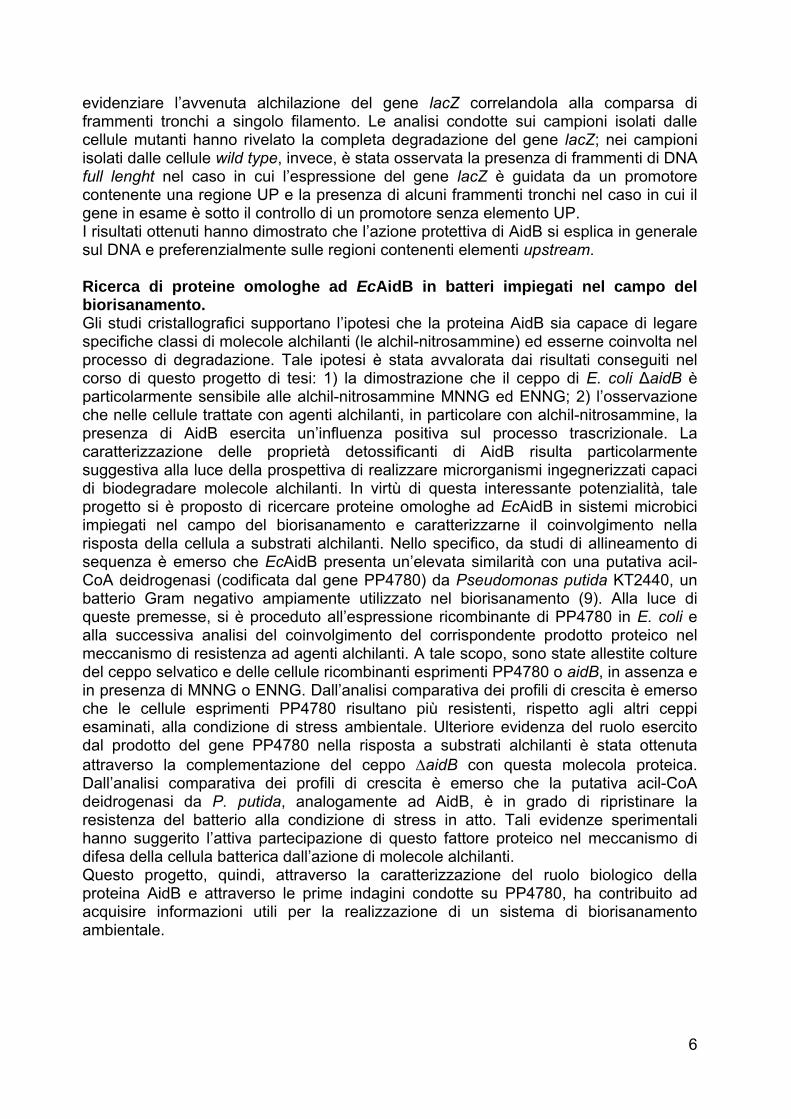
dehydrogenase mechanism, such as that shown in Fig.5 (Rohankhedkar, M.S. et al., 2006). Whether AidB acts to reduce mutagenicity by a DNA repair or a detoxification mechanism is currently a matter of speculation. However, It has been observed that, whereas ada and alkB genes form a transcriptional unit, alkA and aidB are well separated from this operon and from each other. These observations raise the possibility that AidB may be of secondary importance for repair of alkylation damage to DNA. To help resolve this issue, recently the crystal structure of the AidB protein has been determined (Bowles, T. et al., 2008). The structural analysis revealed that AidB is well equipped to sterically occlude dsDNA from chemical attack. Importantly, the structure is not consistent with a DNA repair function. Alternatively, the unique chemical environment of AidB’s putative FAD active site provides a rationale for a possible role in deactivation of alkylating agents. 5. Structural properties and potential cellular function of AidB As predicted (Landini, P. et al., 1994; Rohankhedkar, M.S. et al., 2006), the structure of E. coli AidB is representative of the ACAD family of flavoproteins. ACADs form tightly bound homodimers or homotetramers and typically have three domains: an N-terminal α-helical domain, a middle β-sheet domain, and a C-terminal helical bundle (Kim, J.J. and Miura, R. 2004). However, AidB contains unique features that distinguish it functionally from the ACAD enzymes. One AidB subunit (Fig.6A) consists of an N-terminal α-helical domain (domain I, residues 1–179), a seven-stranded β-barrel (domain II, residues 180–285), a central α-helical region (domain III, residues 286–444), and an α-helical domain at its C terminus (domain IV, residues 445–540). Domains I–III collectively constitute the ACAD core fold. Domain IV, however, is not present in the ACAD family (Fig.7). In the crystal structure, AidB forms a tetramer (Fig.6B). Sedimentation velocity ultracentrifugation showed that AidB (60,590 Da per subunit) sediments as a 234-kDa protein, and gel filtration analysis was consistent with a tetramer in solutions. The AidB tetramer is a dimer-of-dimers. As in ACAD structures, AB and CD dimers are each formed from extensive contacts between domains II and III. Two FAD molecules (one per subunit) are
A B
Fig.6: The structure of E. coli AidB. (A) Stereo image of one subunit colored according to domains. The FAD is shown as ball and stick. (B) Representation of the AidB tetramer. Four subunits A, B, C, and D associate with dihedral symmetry as a dimer of dimers. The FAD cofactors at each of the A/B and C/D dimer interfaces are shown as black CPK spheres. N-terminal extensions and L1'2' loops that form the AB/CD interface unique to AidB are labeled with the letter N and orange stars, respectively.
18
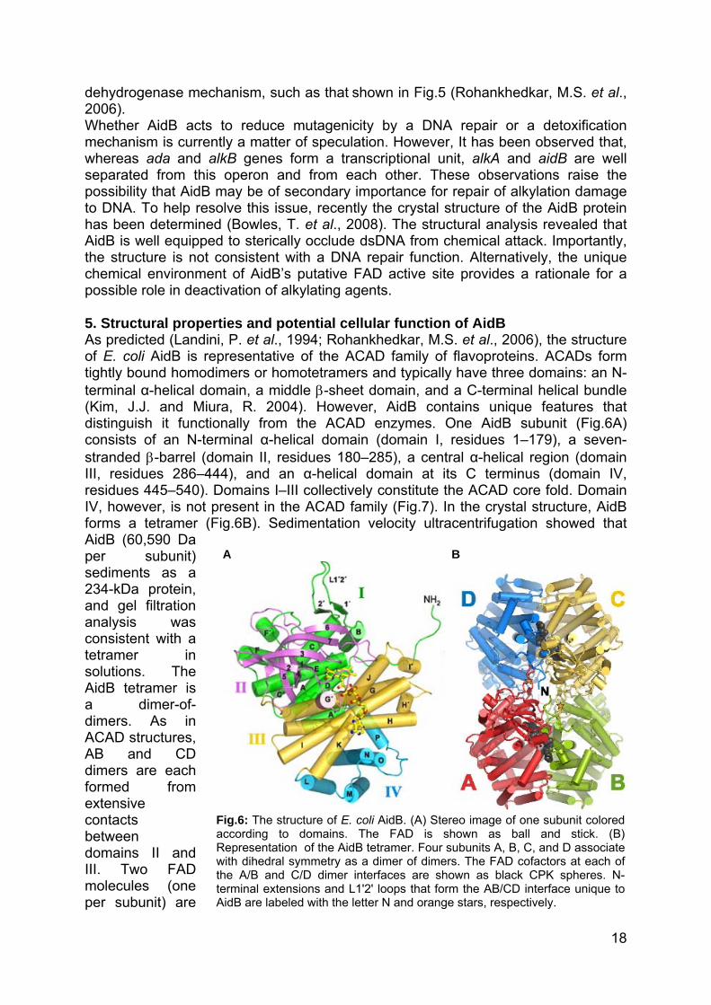
Fig.7: Structure-based sequence alignment of AidB, medium-chain acyl-CoA dehydrogenase (MCAD, PDB ID code 3MDE) (Kim, J.J. et al., 1993), isovaleryl acyl-CoA dehydrogenase (IVD, PDB ID code 1IVH) (Tiffany, K.A. et al. 1997), acyl-CoA oxidase II (ACO, PDB ID code 1IS2) (Nakajima, Y. et al., 2002), and nitroalkane oxidase (NAO, PDB ID code 2C0U) (Nagpal, A. et al., 2006). AidB residues important for tetramerization and DNA binding are highlighted gray and black, respectively, and those predicted to contact DNA are marked with black triangles. ACAD substrate binding and catalytic residues are highlighted blue and yellow, respectively. Black circles denote FAD binding residues, and AidB Trp-424 is marked with a red star.
bound at each of these A/B and C/D dimer interfaces (Fig.6B). Unlike the ACADs, however, AidB has a unique quaternary architecture formed by an extended random coil at the extreme N terminus and a β-hairpin loop (L1'2') inserted between helices αB and αC that project outward to interlock AB and CD dimers together (Fig.6B and Fig.7). Therefore AidB, as result of the novel N-terminal interface and the additional C-terminal domain IV, adopts an homotetrameric architecture different from the ACAD tetramer. In addition, several unique features distinguish AidB’s FAD cavity from the ACAD active sites despite the conservation of their general properties.
19
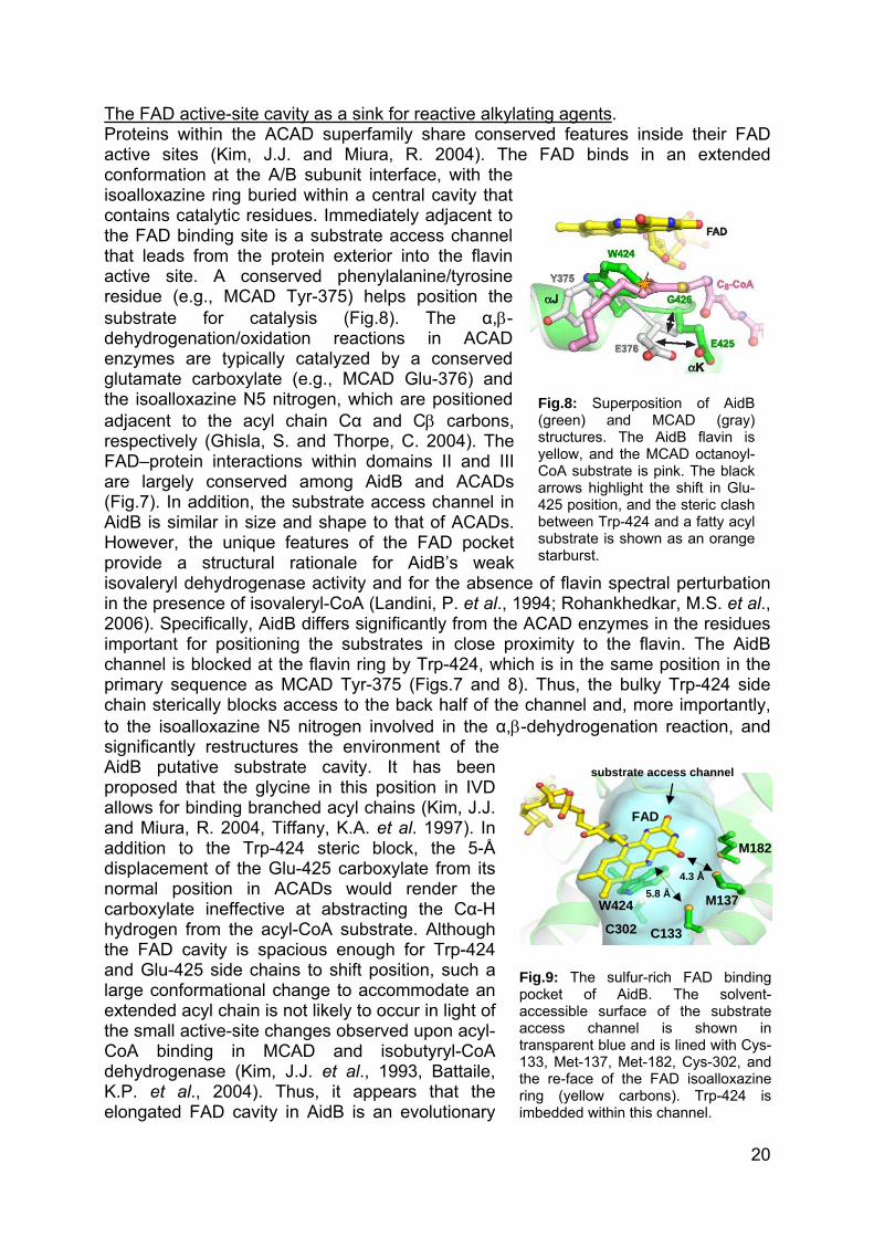
The FAD active-site cavity as a sink for reactive alkylating agents. Proteins within the ACAD superfamily share conserved features inside their FAD active sites (Kim, J.J. and Miura, R. 2004). The FAD binds in an extended conformation at the A/B subunit interface, with the isoalloxazine ring buried within a central cavity that contains catalytic residues. Immediately adjacent to the FAD binding site is a substrate access channel that leads from the protein exterior into the flavin active site. A conserved phenylalanine/tyrosine residue (e.g., MCAD Tyr-375) helps position the substrate for catalysis (Fig.8). The α,β-dehydrogenation/oxidation reactions in ACAD enzymes are typically catalyzed by a conserved glutamate carboxylate (e.g., MCAD Glu-376) and the isoalloxazine N5 nitrogen, which are positioned adjacent to the acyl chain Cα and Cβ carbons, respectively (Ghisla, S. and Thorpe, C. 2004). The FAD–protein interactions within domains II and III are largely conserved among AidB and ACADs (Fig.7). In addition, the substrate access channel in AidB is similar in size and shape to that of ACADs. However, the unique features of the FAD pocket provide a structural rationale for AidB’s weak isovaleryl dehydrogenase activity and for the absencein the presence of isovaleryl-CoA (Landini, P. et al., 192006). Specifically, AidB differs significantly from the Aimportant for positioning the substrates in close prochannel is blocked at the flavin ring by Trp-424, whichprimary sequence as MCAD Tyr-375 (Figs.7 and 8).chain sterically blocks access to the back half of the cto the isoalloxazine N5 nitrogen involved in the α,β-dsignificantly restructures the environment of the AidB putative substrate cavity. It has been proposed that the glycine in this position in IVD allows for binding branched acyl chains (Kim, J.J. and Miura, R. 2004, Tiffany, K.A. et al. 1997). In addition to the Trp-424 steric block, the 5-Å displacement of the Glu-425 carboxylate from its normal position in ACADs would render the carboxylate ineffective at abstracting the Cα-H hydrogen from the acyl-CoA substrate. Although the FAD cavity is spacious enough for Trp-424 and Glu-425 side chains to shift position, such a large conformational change to accommodate an extended acyl chain is not likely to occur in light of the small active-site changes observed upon acyl-CoA binding in MCAD and isobutyryl-CoA dehydrogenase (Kim, J.J. et al., 1993, Battaile, K.P. et al., 2004). Thus, it appears that the elongated FAD cavity in AidB is an evolutionary
Fpoacactr13thrinim
Fig.8: Superposition of AidB (green) and MCAD (gray) structures. The AidB flavin is yellow, and the MCAD octanoyl-CoA substrate is pink. The black arrows highlight the shift in Glu-425 position, and the steric clash between Trp-424 and a fatty acyl substrate is shown as an orange starburst.
of flavin spectral perturbation 94; Rohankhedkar, M.S. et al., CAD enzymes in the residues
ximity to the flavin. The AidB is in the same position in the
Thus, the bulky Trp-424 side hannel and, more importantly, ehydrogenation reaction, and
substrate access channel
ig.9: The sulfur-rich FAcket of AidB. Thecessible surface of thecess channel is s
ansparent blue and is line3, Met-137, Met-182, Cye re-face of the FAD isg (yellow carbons). Tbedded within this chann
D
2
75.8 Å 4.3 Å
M13
D b so subhow
d withs-302oallorp-4
el.
M18
C133
C302W424
FA
inding lvent-strate
n in Cys-, and
xazine 24 is
20

remnant and not a bona fide active site for fatty or amino acid acyl-CoA thioesters. The volume of the FAD binding pocket (∼290 Å2) is large enough to accommodate a small molecule. AidB has been proposed to detoxify MNNG or one of its reactive metabolites (Landini, P. et al., 1994). Unlike MMS or MNU, MNNG is activated by thiols, including glutathione, to produce highly reactive methylation agents (e.g., methyldiazonium ion) (Lawley, P. D. 1974). Interestingly, AidB’s FAD cavity is rich in thiol and methylsulfide groups (Fig.9), a characteristic distinctly different from other structural homologues. In fact, four of 13 solvent-accessible residues lining the AidB pocket are cysteine or methionine, whereas there are no such residues in contact with substrates in MCAD, IVD, ACO, or NAO (Fig.7). It is therefore intriguing to speculate that Cys-133, Met-137, Met-182, and Cys-302 may alter the redox potential of AidB’s pocket relative to those of other, functionally unrelated enzymes. In particular, Cys-133 and Met-137 sulfur atoms are positioned 5.8 and 4.3 Å from the flavin, respectively, and are oriented toward the mouth of the substrate channel (Fig.9). Thus, it appears that AidB’s putative active site may act as a sink for reactive MNNG derivatives. Structural studies also suggested that the C-terminal domain of AidB, highly electropositive, is responsible for DNA binding. Therefore AidB does not engage DNA at the acyl-CoA substrate binding cavity, the only entry point into the FAD active site. These observations argue against the notion that AidB repairs DNA by an FAD-dependent mechanism. AidB’s tetrameric assembly exposes DNA binding surfaces at each end of the tetramer. Nonspecific DNA binding at the ends of the tetramer suggests that the protein might function to protect naked DNA from attack by alkylating agents. A similar role has been observed in the Dps protein, which protects DNA in starved E. coli against oxidative damage (Almiron, M. et al., 1992; Martinez, A. and Kolter, R. 1997). Both AidB and Dps are up-regulated during stationary phase and are rpoS dependent (Volkert, M.R. et al., 1994; Altuvia, S. et al., 1994). Interestingly, endogenous methylating agents such as nitrosamines are formed as by-products of stationary-phase metabolism (Taverna, P. and Sedgwick, B. 1996). This leads to an accumulation of alkylation damage to DNA during stationary phase, as demonstrated by an increase in spontaneous mutation in methyltransferase (ada ogt)-deficient E. coli in nondividing cells (Rebeck, G.W. and Samson, L. 1991). Induction of AidB expression, therefore, could serve to prevent endogenous stationary-phase alkylation damage in a manner similar to Dps protection of oxidative damage. Dps protection of DNA is believed to occur by binding DNA duplexes within the pores of hexagonally packed Dps dodecamers (Grant, R.A. et. al, 1998). Interestingly, crystal form of AidB displays that the putative DNA binding faces are clustered symmetrically around 25-Å pore that is perfectly sized to accommodate DNA. Thus, it appears that AidB tetramers cluster around DNA to restrict access by damaging agents. Coupled with unique features of FAD site, these observations suggest that AidB may be the cell’s line of defense to prevent alkylation damage by protecting DNA and by detoxifying the alkylating compounds before they are able to react with DNA. 6. Potential role for AidB as a detoxification enzyme: a new challenge for pollution cleanup Despite detailed understanding of Ada, AlkA, and AlkB, the mechanism by which AidB protects against DNA damage in the adaptive response is less well understood. Although the specific function remains to be determined, the structural features of AidB’s unique DNA binding domain, subunit organization, and FAD chemical
21

environment help to support the involvement of the protein in a detoxification pathway of certain alkylators. The possibility that AidB might degrade alkylating molecules, among the most prevalent pollutants released into the environment, makes this protein a promising tool for bioremediation applications. Besides, interestingly, full-length AidB homologues are not present in many bacteria closely related to E. coli (Klebsiella, Vibrio, Shewanella, and Photorhabdus), but the closest hits are from some γ-proteobacteria such as Pseudomonas, Azotobacter, and Acinetobacter. This observation is intriguing because Pseudomonas species and closely related organisms are the most extensively studied and the most frequently used for bioremediation applications, owing to their ability to degrade numerous different contaminants (Wackett, L.P. 2003). The huge potential of the pseudomonads does not solely depend on a high proportion of genes responsible for the metabolism, transport and efflux of organic compounds (Nelson, K.E. et al., 2002), but also on broad capability of metabolic regulation: indeed, the control of gene expression is the key determinant of their flexibility and, in this respect, a variety of highly integrated regulatory mechanisms have been identified. Taken together, these observations suggest that the characterization of the potential ability of AidB protein and its homologues to degrade alkylators, could allow the development of new successful strategies for the bioremediation of environments and industrial effluents contaminated by alkylating compounds. 7. Aim of the thesis The aim of this research project was to explore new potential candidates for the bio-treatment of wastes and environments contaminated by alkylating agents. The study has been specifically focused on AidB protein due its potential ability to degrade certain alkylators. Initially, the work has been aimed at investigate the role of AidB in the bacterial cell; given that the knowledge of the domain architecture is necessary for understanding the multifunctional properties of a protein, structural and functional characterization of domains present in AidB was performed. Successively, the mechanism by which this protein directly protects E. coli cells against alkylating compounds has been determined. Finally, taking into account the potential role played by AidB in the detoxification of alkylating molecules, this experimental work was targeted at identify as well as at characterize E. coli AidB homologues in bacteria used for bioremediation applications. Specifically, the study has been focused on the acyl-CoA dehydrogenase coded by the PP4780 gene from Pseudomonas putida KT2440. The involvement of this protein in the response to alkylation stress has been investigated in order to enrich the knowledge of this enzyme, thus improving its potential application in bioremediation strategies. In conclusion, the data obtained in this research project support the possibility of developing new successful strategies for the bioremediation of environments and wastes contaminated by alkylating compounds.
22

REFERENCES • Battaile, K. P., Nguyen, T. V., Vockley, J., and Kim, J. J. The Journal of biological
chemistry 279(16), 16526–16534 (2004). • Brockman, H. L. and Wood, W. A. Journal of bacteriology 124(3), 1447–1453 (1975). • Butler, C. S. and Mason, J. R. Advances in microbial physiology 38, 47–84 (1997). • Castro, C. E., Wade, R. S., and Belser, N. O. Journal of Agricultural and Food
Chemistry 31(6), 1184–1187 (1983). • Cerdá-Olmedo, E. and Hanawalt, P. C. Molecular and General Genetics 101(3), 191–
202 (1968). • Chaney, S. G. and Sancar, A. J. Natl. Cancer Inst. 88(19), 1346–1360 (1996). • Choi, J. Y. and Guengerich, F. P. The Journal of biological chemistry 279(18),
19217–19229 (2004). • Choi, J. Y., Angel, K. C., and Guengerich, F. P. The Journal of biological chemistry
281(30), 21062–21072 (2006). • De Lorenzo, V. EMBO reports 2(5), 357–359 (2001). • Delaney, J. C. and Essigmann, J. M. Chemistry & Biology, 743–753 (1999). • Delaney, J. C. and Essigmann, J. M. Proceedings of the National Academy of
Sciences of the United States of America 101(39), 14051–14056 (2004). • Demple, B., Sedgwick, B., Robins, P., Totty, N., Waterfield, M. D., and Lindahl, T.
Proceedings of the National Academy of Sciences of the United States of America 82(9), 2688–2692 (1985).
• Dua, M., Singh, A., Sethunathan, N., and Johri, A.K. Applied Microbiology and Biotechnology 59(2), 143–152 (2002).
• Dungan, R. S., Gan, J., and Yates, S. R. Water, Air, and Soil Pollution 142(1), 299–310 (2003).
• Ellis, L. B. Current Opinion in Biotechnology , 232–235 (2000). • Evensen, G. and Seeberg, E. Nature 296(5859), 773–775 (1982). • Fournier, D., Hawari, J., Halasz, A., Streger, S. H., McClay, K. R., Masuda, H., and
Hatzinger, P. B. Appl. Environ. Microbiol. 75(15), 5088–5093 (2009). • Gerson, S. L. and Willson, J. K. Hematology/oncology clinics of North America 9(2),
431–450 (1995). • Ghisla, S. and Thorpe, C. European journal of biochemistry / FEBS 271(3), 494–508
(2004). • Grant, R. A., Filman, D. J., Finkel, S. E., Kolter, R., and Hogle, J. M. Nature structural
biology 5(4), 294–303 (1998). • Haggerty, H. G. and Holsapple, M. P. Toxicology 63(1), 1–23 (1990). • He, C., Hus, J. C., Sun, L. J., Zhou, P., Norman, D. P., Dötsch, V., Wei, H., Gross,
J. D., Lane, W. S., Wagner, G., and Verdine, G. L. Molecular cell 20(1), 117–129 (2005).
• Hix, S. Free Radical Biology and Medicine 19(3), 293–301 (1995). • Hurley, L. H. Nature Reviews Cancer 2(3), 188–200 (2002). • Ibekwe, A. M., Papiernik, S. K., Gan, J., Yates, S. R., Yang, C. H., and Crowley, D. E.
Appl. Environ. Microbiol. 67(7), 3245–3257 (2001). • Jagerstad, M. and Skog, K. Mutation Research/Fundamental and Molecular
Mechanisms of Mutagenesis 574(1-2), 156–172 (2005). • Jeggo, P., Defais, T. M., Samson, L., and Schendel, P. Molecular and General
Genetics 157(1), 1–9 (1977). • Jiménez-Sánchez, A. and Cerdá-Olmedo, E. Mutation research 28(3), 337–345
(1975). • Kim, J. J., Wang, M., and Paschke, R. Proceedings of the National Academy of
Sciences of the United States of America 90(16), 7523–7527 (1993). • Kim, J. J. and Miura, R. European journal of biochemistry / FEBS 271(3), 483–493
(2004).
23

• Kondo, H., Nakabeppu, Y., Kataoka, H., Kuhara, S., Kawabata, S., and Sekiguchi, M. The Journal of biological chemistry 261(33), 15772–15777 (1986).
• Landini, P. and Volkert, M. R. Journal of bacteriology 182(23), 6543–6549 (2000). • Landini, P., Hajec, L. I., and Volkert, M. R. Journal of bacteriology 176(21), 6583–
6589 (1994). • Lawley, P. D. Mutation research 23(3), 283–295 (1974). • Lee, V. M., Keefer, L. K., and Archer, M. C. Chemical research in toxicology 9(8),
1319–1324 (1996). • Lindahl, T. Nature 259(5538), 64–66 (1976). • Lindahl, T., Sedgwick, B., Sekiguchi, M., and Nakabeppu, Y. Annual review of
biochemistry 57, 133–157 (1988). • Lovley, D. R. Nature Reviews Microbiology 1(1), 35–44 (2003). • Ludlum, D. B. Mutation research 233(1-2), 117–126 (1990). • Margison, G. P., Cooper, D. P., and Brennand, J. Nucleic acids research 13(6),
1939–1952 (1985). • Margison, G. P., Santibanez Koref, M. F., and Povey, A. C. Mutagenesis 17(6), 483–
487 (2002). • Mitch, W. A. and Sedlak, D. L. Environmental science & technology 38(5), 1445–1454
(2004). • Nagpal, A., Valley, M. P., Fitzpatrick, P. F., and Orville, A. M. Biochemistry 45(4),
1138–1150 (2006). • Nakabeppu, Y. and Sekiguchi, M. Proceedings of the National Academy of Sciences
of the United States of America 83(17), 6297–6301 (1986). • Nakajima, Y., Miyahara, I., Hirotsu, K., Nishina, Y., Shiga, K., Setoyama, C.,
Tamaoki, H., and Miura, R. J Biochem 131(3), 365–374 (2002). • Neale, S. Mutation research 14(2), 155–164 (1972). • Nelson, K. E., Weinel, C., Paulsen, I. T., Dodson, R. J., Hilbert, H., Martins dos
Santos, V. A., Fouts, D. E., Gill, S. R., Pop, M., Holmes, M., Brinkac, L., Beanan, M., DeBoy, R. T., Daugherty, S., Kolonay, J., Madupu, R., Nelson, W., White, O., Peterson, J., Khouri, H., Hance, I., Chris Lee, P., Holtzapple, E., Scanlan, D., Tran, K., Moazzez, A., Utterback, T., Rizzo, M., Lee, K., Kosack, D., Moestl, D., Wedler, H., Lauber, J., Stjepandic, D., Hoheisel, J., Straetz, M., Heim, S., Kiewitz, C., Eisen, J. A., Timmis, K. N., Düsterhöft, A., Tümmler, B., and Fraser, C. M. Environmental microbiology 4(12), 799–808 (2002).
• Oktyabrsky, O. N., Golyasnaya, N. V., Smirnova, G. V., Demakov, V. A., Posokhina, N. K. h., and Kholstova, T. A. Mutation research 293(3), 197–204 (1993).
• Oremland, R. S., Miller, L. G., Culbertson, C. W., Connell, T. L., and Jahnke, L. Appl. Environ. Microbiol. 60(10), 3640–3646 (1994).
• Paul, D., Pandey, G., Pandey, J., and Jain, R. Trends in Biotechnology 23(3), 135–142 (2005).
• Pegg, A. E. Mutation research 462(2-3), 83–100 (2000). • Rasche, M. E., Hyman, M. R., and Arp, D. J. Appl. Environ. Microbiol. 56(8), 2568–
2571 (1990). • Rebeck, G. W. and Samson, L. Journal of bacteriology 173(6), 2068–2076 (1991). • Rieger, P. Journal of Biotechnology 94(1), 101–123 (2002). • Saget, B. M. and Walker, G. C. Proceedings of the National Academy of Sciences of
the United States of America 91(21), 9730–9734 (1994). • Samson, L. and Cairns, J. O. H. N. Nature 267(5608), 281–283 (1977). • Samson, L. Molecular microbiology 6(7), 825–831 (1992). • Schendel, P. F. and Robins, P. E. Proceedings of the National Academy of Sciences
of the United States of America 75(12), 6017–6020 (1978). • Sedgwick, B. and Lindahl, T. Oncogene 21(58), 8886–8894 (2002). • Sedgwick, B. and Robins, P. Molecular and General Genetics 180(1), 85–90 (1980).
24

• Sedgwick, B. and Vaughan, P. Mutation research 250(1-2), 211–221 (1991). • Sedgwick, B. Carcinogenesis 18(8), 1561–1567 (1997). • Sedgwick, B. Nature reviews. Molecular cell biology 5(2), 148–157 (2004). • Sharp, J. O., Wood, T. K., and Alvarez-Cohen, L. Biotechnology and Bioengineering
89(5), 608–618 (2005). • Singer, B. and Kusmierek, J. T. Annual review of biochemistry 51, 655–693 (1982). • Singer, B. Nature 264(5584), 333–339 (1976). • Smirnova, G. V., Oktyabrsky, O. N., Moshonkina, E. V., and Zakirova, N. V. Mutation
research 314(1), 51–56 (1994). • Sohn, O. S., Fiala, E. S., Requeijo, S. P., Weisburger, J. H., and Gonzalez, F. J.
Cancer Res 61(23), 8435–8440 (2001). • Takahashi, K., Kawazoe, Y., Sakumi, K., Nakabeppu, Y., and Sekiguchi, M. The
Journal of biological chemistry 263(27), 13490–13492 (1988). • Taverna, P. and Sedgwick, B. Journal of bacteriology 178(17), 5105–5111 (1996). • Taverna, P. and Sedgwick, B. Journal of bacteriology 178(17), 5105–5111 (1996). • Thomas, L., Yang, C. H., and Goldthwait, D. A. Biochemistry 21(6), 1162–1169
(1982). • Tiffany, K. A., Roberts, D. L., Wang, M., Paschke, R., Mohsen, A. W., Vockley, J.,
and Kim, J. J. Biochemistry 36(28), 8455–8464 (1997). • Timmis, K. Trends in Biotechnology 17(5), 200–204 (1999). • Trewick, S. C., Henshaw, T. F., Hausinger, R. P., Lindahl, T., and Sedgwick, B.
Nature 419(6903), 174–178 (2002). • Vaughan, P., Sedgwick, B., Hall, J., Gannon, J., and Lindahl, T. Carcinogenesis
12(2), 263–268 (1991). • Volkert, M. R., Hajec, L. I., and Nguyen, D. C. J. Bacteriol. 171(2), 1196–1198 (1989). • Volkert, M. R., Nguyen, D. C., and Beard, K. C. Genetics 112(1), 11–26 (1986). • Wackett, L. Current Opinion in Biotechnology 11(3), 229–231 (2000). • Wackett, L. P. Nat Biotech 21(2), 136–138 (2003). • Yoshinari, T. and Shafer, D. Canadian journal of microbiology 36(12), 834–838
(1990). • Zhang, Y., Spokas, K., and Wang, D. J Environ Qual 34(5), 1566–1572 (2005).
25


Results and discussion
The results obtained in this thesis are described in the following sections:
• Additional protein functions involved in the biogenesis of ribosomes and the DNA repair mechanisms are associated with the transcriptional machinery gathered at the Escherichia coli rrnB P1 promoter
Pag. 27
• Role of Escherichia coli AidB protein in the transcriptional regulation Pag. 46
• Preferential protection from DNA alkylation by the Escherichia coli AidB protein Pag. 60
• Potential role for the the Escherichia coli AidB and the Pseudomonas putida PP4780 as detoxification enzymes Pag. 72
26


Manuscript submitted to Molecular Microbiology
Additional protein functions involved in the biogenesis of ribosomes and the DNA repair mechanisms are associated with the transcriptional machinery gathered at the Escherichia coli rrnB P1 promoter Angela Amoresano1, 2, Claudia Cirulli1,2, Valentina Rippa1, 2, Carla Esposito1, Rosanna Papa1, 2 and Angela Duilio1, 2*
1Department of Organic Chemistry and Biochemistry, University Federico II of Naples, Naples, Italy 2School of Biotechnological Sciences, University Federico II of Naples, Naples, Italy ABSTRACT The transcriptional machinery at the ribosomal RNA rrnB P1 promoter of Escherichia coli was investigated by functional proteomics. Synthetic oligonucleotides encompassing the -35 sequence of the rrnB P1 promoter and its associated element upstream (“UP35”) were incubated with the total protein extract. Proteins found to bind exclusively to the “UP35” oligonucleotide were identified by mass spectrometry and specific protein-protein interactions were confirmed by co-immunoprecipitation. Proteins specifically binding to the rrnB P1
*Corresponding Author: Angela Duilio Dipartimento di Chimica Organica e Biochimica, Complesso Universitario di Monte Sant’Angelo Via Cinthia 4 80126 Napoli (Italy). Tel +39-081674314 Fax +39081674313; email: [email protected]
promoter region belong to different functional groups: in addition to components of the transcription machinery, we also found proteins involved in translation, DNA repair and protection mechanisms and in cell metabolic processes. Presence of several ribosomal proteins strongly suggests that the ribosome can establish direct interaction with bacterial RNA polymerase. Interestingly, we found that non-specific DNA binding proteins, i.e. Dps and AidB, are recruited into the transcriptional complex. Functional hypotheses on the possible biological significance of the different functional groups were suggested. Keywords: rRNA transcription, proteomics, cellular network, alkylation, fishing for partners, mass spectrometry. Running title: Complex transcription machinery at E. coli rrnB P1 promoter. INTRODUCTION The availability of complete genome sequences for a constantly growing number of organisms has opened the door for novel proteomic approaches aimed at identifying components of complex protein machineries and thus to elucidate their function. Moreover, microarray technologies (1) providing information on global mRNA expression, clearly demonstrated that in many cases mRNA transcripts do not directly correlate with protein expression (2,3). Therefore, the field of proteomics is challenged with the task of providing both quantitative and functional data to further complement genomics (4). In particular, functional proteomics has emerged as a new area of study aimed at isolating and identifying molecular components belonging to multi-protein complexes (5). It is now clear, in fact,
27

that fundamental biological mechanisms are carried out by a multitude of proteins that assemble to form large functional complexes (6) or specific cell pathways (7). A comprehensive description of cellular processes at the molecular level is then strictly dependent on the clear definition of the individual protein components involved in these functional entities. A key contribution to the disclosure of biological mechanisms can then be provided by functional proteomic strategies. Isolation of the entire multiprotein complexes can be essentially accomplished by either affinity based techniques (including “pull down” and “tap tag” experiments) (8-10) or immunoprecipitation procedures (11). Both methodologies take advantage from the availability of different (immuno)-affinity ligands endowed with high binding efficiency. The success of an affinity-based approach strictly depends on the absence of excessive nonspecific interactions that in turn is related to the specificity of the bait-partners recognition. When this binding specificity is extremely high, as in the case of DNA/RNA-binding proteins, a low level of false positives is expected. Ribosomal RNA (rRNA) transcription constitutes a fundamental biological process in all living cells. Several studies demonstrated that transcription of rRNA is the rate-limiting step in ribosome synthesis (12-14). Consistent with this crucial role in ribosome biogenesis, the control of rRNA transcription initiation, the efficient elongation of RNA polymerase as well as all the other steps involved in the transcription machinery are tightly regulated by a complex network of protein components (14 and references therein). Ribosomal RNA accounts for >90% of total RNA present in the bacterial cell. Operons encoding ribosomal RNA are present
in multiple copies in the genome, an unusual feature for bacterial genes; for instance, in Escherichia coli rRNA is encoded by 7 different operons. Ribosomal proteins are subject to various forms of regulation: proteins such as Fis or H-NS (15), as well as direct interaction between RNA polymerase and the ppGpp signal molecule (16,17) can modulate the activity of rRNA promoters. An important feature of ribosomal promoters is the presence of the so-called UP element, an A/T rich sequence located at 40 to 80 nucleotides upstream of the transcription start site that can stimulate transcription from the rrnB P1 promoter through direct interaction with the C-terminal domain of the RNA polymerase α subunit (18,19). In this report, we have investigated the functional protein complex that gathers at the upstream elements of the ribosomal rrnB P1 promoter of E. coli. We find that several ribosomal proteins assemble at the rrnB P1 promoter, thus suggesting direct interaction between RNA polymerase and the ribosome. In addition, proteins involved in DNA repair/protection mechanisms are also found in the promoter-binding protein complex. Altogether, our findings strongly suggest that proteins with functions not directly related to transcription associate with RNA polymerase to form multiprotein complexes. MATERIALS AND METHODS Proteomic grade trypsin, dithiothreitol (DTT), HEPES, KCl, MgCl2, glycerol, ammonium bicarbonate and triton were purchased from Sigma. Cyanogen bromide-activated agarose matrices were obtained from Pharmacia Biotech. All used solvents were of the highest purity available from Romil.
28

Escherichia coli K12 strain growth conditions and protein extract preparation. E. coli K12 strain was grown in aerobic conditions at 37°C in LB medium (20). After 16 h, bacteria were harvested by centrifugation and resuspended in Buffer Z (25 mM HEPES pH 7.6, 50 mM KCl, 12.5 mM MgCl2, 1 mM DTT, 20% glycerol, 0.1% triton) containing 1 µM phenyl methyl sulfonyl fluoride. Cells were disrupted by sonication. The suspension was centrifuged at 90,000 x g for 30 min at 4°C. After centrifugation the protein concentration of the extract was determined with Bradford assay (21). Preparation of the oligonucleotide bait and coupling to Sepharose. A 28-mer deoxynucleotide encompassing the upstream and the -35 sequence of the ribosomal promoter rrnB P1 and a randomised version of the same sequence were chemically synthesized (Table 1). The complementary oligonucleotides (500 µg) were annealed, 5’-phosphorylated and ligated as follows. Gel purified oligodeoxynucleotides were combined in 10 mM Tris pH 7.5, 50 mM NaCl, 1 mM EDTA pH 8. The total mixture was incubated at 90°C for 5 min, 65°C for 10 min, 37°C for 10 min and RT for 5 min. ATP (20 mM) and T4 polynucleotide kinase (200 units) were then added and the resulting solution was incubated at 37°C for 2 hours. The DNA was phenol extracted and then ethanol precipitated and the pellet was resuspended in H2O and the ligation reaction was initiated by the addition of 20 mM ATP and T4 DNA ligase (15 units). The mixture was incubated at 16°C and the desired multimerization grade was monitored by agarose gel. The final DNA product was phenol extracted and ethanol precipitated. The polymerised oligonucleotides were covalently linked to cyanogen bromide-
activated agarose beads following the procedure recommended by Amersham. Electrophoretic shift mobility assays. Annealed DNA oligonucleotides (fragments UP, UP35 and Neg) were 32P labelled at 5’ positions by using T4 polynucleotide kinase (20 units). Electromobility shift assays were performed in 20 µl reaction volume. Total protein extract (10 µg) or each individual recombinant protein (1 µg) were incubated with the 32P-labelled DNA (2 ng, 50,000-100,000 cpm) in buffer Z, for 20 min at RT. Mixtures were then analysed by electrophoresis on 8% native polyacrylamide gel (29:1 cross-linking ratio) in 45 mM Tris pH 8.0, 45 mM boric acid, 1 mM EDTA. Electrophoreses were performed at room temperature at 200 V (20 V/cm). The gels were dried and analysed by autoradiography. DNA affinity chromatography. A 4 ml affinity chromatography column was prepared with the agarose derivatizated beads. The column was equilibrated with 30 ml of buffer Z. The entire cell extract (300 mg) was incubated with the bait overnight at 4°C. The column was washed with 50 ml of buffer Z and the proteins selectively retained from the DNA probe were eluted with the same buffer containing 1 M KCl. The fractions containing the proteins of interest eluted from the affinity chromatography were pooled and dialyzed in H2O. The same affinity chromatography procedure was applied to the Neg oligonucleotide. Both the eluted and dialyzed fractions were loaded onto SDS-PAGE gel and visualized by colloidal Coomassie staining. In situ digestion and MALDI analysis. Protein bands stained with Coomassie brilliant blue were excised from the gel
29

and destained by repetitive washes with 0.1 M NH4HCO3 pH 7.5 and acetonitrile. Samples were reduced by incubation with 50 µl of 10 mM DTT in 0.1 M NH4HCO3 buffer pH 7.5 and carboxyamidomethylated with 50 µl of 55 mM iodoacetamide in the same buffer. Enzymatic digestion was carried out with trypsin (12.5 ng/µl) in 10 mM ammonium bicarbonate pH 7.8. Gel pieces were incubated at 4°C for 2 hours. Trypsin solution was then removed and a new aliquot of the digestion solution was added; samples were incubated for 18 hr at 37°C. A minimum reaction volume was used as to obtain the complete rehydratation of the gel. Peptides were then extracted by washing the gel particles with 10 mM ammonium bicarbonate and 1% formic acid in 50% acetonitrile at room temperature. The resulting peptide mixtures were desalted using ZipTip pipettes from Millipore, following the recommended purification procedure. MALDI Analyses. MALDI-TOF mass spectra were recorded using an Applied Biosystem Voyager DE-STR instrument. A mixture of analyte and matrix solution (alfa-cyano-hydroxycinnamic acid 10 mg/ml in 70% ACN, 0.1% citric acid, in MilliQ water) was applied to the metallic sample plate and dried at room temperature. Mass calibration was performed using external peptide standards. Raw data were analysed using the computer software provided with the instrument and reported as monoisotopic masses. Peptide masses of each digested protein were used to search for protein databases using the Mascot mass fingerprinting software (Matrix Science). Liquid Chromatography Tandem Mass Spectrometry (LC/ MS/MS) analyses. Tryptic peptide mixtures obtained from in situ digestions were also analysed
by LC/MS/MS “on-line” using a 4000Q-Trap linear trap ion mass spectrometer (Applied Biosystems) coupled to an 1100 nano HPLC system (Agilent Technologies). The mixture was loaded on an Agilent reverse-phase pre-column cartridge (Zorbax 300 SB-C18, 5x0.3 mm, solvent 0.1% formic acid, loading time 5 min). Peptides were separated on a Agilent reverse-phase column (Zorbax 300 SB-C18, 150 mm), at flow rate of 0.3 µl/min with a 5 to 65% linear gradient in 60 min (A solvent 0.1% formic acid, 2% ACN in water; B solvent 0.1% formic acid, 2% water in ACN). Nanospray source was used at 2.5 kV with liquid coupling, with a declustering potential of 50 V, using an uncoated silica tip from NewObjectives (Ringoes, NJ) (O.D. 150 µm, I.D. 50 µm, Tip.Diameter. 15 µm). Spectra acquisition was based on a survey scan (EMS) using Linear Ion Trap scanned from m/z 400 to m/z 1600 at 4000 amu/sec. This scan mode was followed by an enhanced resolution experiment (ER) for the five most intense ions of interest and then by MS2 acquisitions of the five most intense ions for peptide charge, from +2 to +3. MS2 spectra were acquired using the best collision energy calculated on the bases of m/z values and charge state (rolling collision energy). The acquired MS/MS spectra were transformed in Mascot generic file format and used for peptides identification with a licensed version of MASCOT, in a local database. Comparison with IntAct data. Validation of the proteomic results was performed by comparing our data with those provided by IntAct database (htpp://www.ebi.ac.uk/intact/search/do/search?search String=0) linked in the corresponding Swiss-Prot entries (htpp://www.expasy.org/sprot/). Each protein candidate was submitted to the
30

IntAct databank obtaining a list of all proteins involved in interaction with the query protein. Construction of expression vectors. The bacterial strains and plasmids used in this work are all reported in Table 1. The rluC, fabZ, tufA, srmB, aidB and hns genes of E. coli K12 were amplified from host DNA by polymerase chain reaction (PCR) using the forward and reverse primers listed in Table 1. To obtain RluC, FabZ, EF-Tu and H-NS tagged with c-myc epitope, the corresponding amplification products were digested with BamHI and XhoI and cloned into the pET22b-c-myc vector, respectively. To obtain pET22b-c-myc, a NdeI/BamHI digested fragment corresponding to the c-myc epitope was inserted into the pET22b (+) expression plasmid (Novagen) linearized with NdeI and BamHI. RluC, SrmB and H-NS tagged with T7 epitope were made by insertion of the corresponding amplified fragments into the BamHI and XhoI sites of pET28a (+) expression vector (Novagen). The plasmid pET28a-aidB was constructed by cloning aidB coding sequence into pET28a (+) digested with BamHI and HindIII. All plasmids (Table 1) contain the coding sequence for the corresponding recombinant protein fused to a 6X histidine tag to facilitate protein purification by Ni2+ affinity chromatography. Plasmids construction was verified by automated DNA sequencing. Production and purification of recombinant proteins. The recombinant genes were separately expressed into the E. coli strain C41 (DE3) (22). For RluC, FabZ and SrmB production, the recombinant cells were grown in LB medium at 37°C without induction until the OD600
reached 3.0. The aidB, tufA and hns genes were expressed as follows: recombinant cells were grown at 37°C to an optical density at 600 nm of ~0.5, at which time 0.05 mM isopropyl-beta-D-thiogalactopyranoside (IPTG) was added and the cultures were shifted down to 25°C for AidB production and to 28°C for EF-Tu and H-NS expression until the OD600 reached 3.0. Selective antibiotics were used at the following concentrations: 100 µg/ml ampicillin; 50µg/ml kanamicin. After incubation, cells were harvested by centrifugation at 5000 x g for 15 min at 4°C, resuspended in 50 mM Na2HPO4, pH 7.4, disrupted by passage through a French Press and centrifuged at 14000 x g for 30 min at 4°C. Recombinant proteins were purified by affinity chromatography on His-Select Nickel Affinity Gel fv (Sigma). The lysate was loaded onto His-Select Nickel Affinity Gel equilibrated with equilibration buffer (50 mM Na2HPO4, pH 7.4, 0.3 M NaCl, 10 mM imidazole). After 1 min of incubation at 4°C, the matrix was collected by centrifugation at 11000 x g for 1 min and washed 3 times with same equilibration buffer. The recombinant proteins were eluted with buffer containing 250 mM imidazole in 50 mM Na2HPO4, pH 7.4, 0.3 M NaCl. Protein concentration was estimated with Bradford reagent (Bio-Rad protein assay) and protein content was checked by SDS-polyacrilamide gel electrophoresis (SDS-PAGE). Co-immunoprecipitation and Western blotting. For co-immunoprecipitations, E. coli strain C41 (DE3) was co-transformed with the following constructs: pET28a-aidB/pET22b-c-myc-fabZ, pET28a-srmB/pET22b-c-myc-tufA, pET28a-srmB/pET22b-c-myc-rluC, pET28a-rluC/pET22b-c-myc-tufA,
31

Table 1: Bacterial strains, plasmids and oligonucleotides
Strains/plasmids Description Reference
or Source
Strains C41 (DE3) Strain that derives from BL21 [F-ompT hsdSB (rB- mB-) gal
dcm (DE3)]. This strain has at least one uncharacterized mutation that prevents cell death associated with expression of many toxic recombinant proteins
ref. 22
Plasmids pET22b(+) carries an N-terminal pelB signal sequence for potential
periplasmic localization, plus an optional C-terminal His-tag sequence
Novagen
pET28a(+) carries an N-terminal His•Tag®/thrombin/T7•Tag® configuration plus an optional C-terminal His•Tag sequence
Novagen
pET22b-c-myc pET22b∆(NdeI-BamHI)Ω(c-myc) This work pET22b-c-myc-rluC pET22b-c-myc∆(BamHI-XhoI)Ω(rluC gene) This work pET22b-c-myc-fabZ pET22b-c-myc∆(BamHI-XhoI)Ω(fabZ gene) This work pET22b-c-myc-tufA pET22b-c-myc∆(BamHI-XhoI)Ω(tufA gene) This work pET22b-c-myc-hns pET22b-c-myc∆(BamHI-XhoI)Ω(hns gene) This work pET28a-aidB pET28a∆(BamHI-HindIII)Ω(aidB gene) This work pET28a-rluC pET28a∆(BamHI-XhoI)Ω(rluC gene) This work pET28a-srmB pET28a∆(BamHI-XhoI)Ω(srmB gene) This work pET28a-hns pET28a∆(BamHI-XhoI)Ω(hns gene) This work Oligonucleotides UP Fw 5'-AGAAAATTATTTTAAATTTC-3' UP Rv 5'-GAAATTTAAAATAATTTTCT-3' UP35 Fw 5'-GAAAATTATTTTAAATTTCCTCTTGTCA-3' UP35 Rv 5'-TGACAAGAGGAAATTTAAAATAATTTTC-3' Neg Fw 5'-CAACTATAGTATAAGATATTCTTTCTTT-3' Neg Rv 5'-AAAGAAAGAATATCTTATACTATAGTTG-3' c-myc Fw 5'TTCATATGGAACAAAAACTCATCTCAGAAGAGGATCTGAATGGGGCC
GCAGGATCCTAT3’ c-myc Rv 5'ATAGGATCCTGCGGCCCCATTCAGATCCTCTTCTGAGATGAGTTTTT
GTTCCATATGAAT3’ rluC Fw 5'-ATAGGATCCATGAAAACAGAGACTCC-3' rluC-pET22b Rv 5'-ATACTCGAGTAAGCGCGCGTTACG-3' rluC-pET28a Rv 5'-ATACTCGAGTTAGCGCGCGTTACG-3' fabZ Fw 5'-ATAGGATCCTTGACTACTAACACTC-3' fabZ Rv 5'-TAACTCGAGTTAGGCCTCCCGGC-3' tufA Fw 5'-TATGGATCCGTGTCTAAAGAAAAATTTG-3' tufA Rv 5'-TATCTCGAGTAAGCCCAGAACTTTAGC-3' hns Fw 5'-TAAGGATCCATGAGCGAAGCACTTA-3' hns-pET22b Rv 5'-ATACTCGAGTAATTGCTTGATCAGG-3' hns-pET28a Rv 5'-ATACTCGAGTTATTGCTTGATCAGG-3' aidB Fw 5'-ATAGGATCCGTGCACTGGCAAACTCACACCG-3' aidB Rv 5'-CGCGAGCTCCGTAAGCTTTTACACACACACTC-3' srmB Fw 5'-ATAGGATCCATGACTGTAACGACTTTT-3' srmB Rv 5'-ATACTCGAGTTACTCTTCTGTCGTTTG-3'
32

pET28a-aidB/pET22b-c-myc-hns, pET28a-hns/pET22b-c-myc-fabZ, pET28a-hns/pET22b-c-myc-rluC, pET28a-aidB/pET22b-c-myc-rluC. After expression of the recombinant genes without induction, cells were harvested, suspended in 50 mM Na2HPO4 (pH 7.4), disrupted by passage through a French Press and centrifuged at 14000 x g for 30 min at 4°C. The supernatants were used for the co-immunoprecipitation experiments. Cell lysates (0.5 mg) were incubated with agarose-linked T7 antibody (Bethyl) and with agarose beads only (control of the experiment) at 4°C overnight. The beads were then collected by centrifugation. Precipitates were washed several times, the bound proteins were eluted with 1×SDS–PAGE sample buffer and subjected to SDS-PAGE followed by Western blot analysis that was performed by using anti-T7 mouse antibody (Novagen) and anti-c-myc mouse antibody (Calbiochem) as first antibodies and anti-mouse IgG conjugated to peroxidase as a secondary antibody (Calbiochem). RESULTS Interaction of Escherichia coli proteins with the rrnB P1 promoter upstream elements. Escherichia coli cell extracts were tested for their ability to bind to the upstream element of the rrnB P1 promoter in electrophoresis mobility shift assays (EMSA). EMSA experiments were carried out using three different DNA oligonucleotides as probes. The first oligonucleotide (20 nucleotides, oligo “UP”, Table 1) corresponds to the UP element alone, i.e. the DNA sequence directly involved in interaction with the α subunit of RNA polymerase and located immediately upstream of the -35 promoter element (23,24). The second oligonucleotide
(28 nt, “UP35”, Table 1) includes both the UP element and an additional 8 base pairs containing the TTGTCA sequence of the rrnB P1 -35 box. Finally, a third 28-nt oligonucleotide with the same nucleotide content as “UP35”, but lacking any known promoter or regulatory elements (“Neg”, Table 1) was also used as negative control in EMSA experiments (Table 1). Each probe was incubated with 10 µg of protein extracts from E. coli cells. At this protein concentration, a clear gel mobility shift band could only be detected with the “UP35” oligonucleotide, i.e. with the DNA fragment containing the upstream element and the -35 box (Fig. 1, lane 2). In contrast, no interaction was observed with either the “UP” DNA fragment containing only the upstream element or with the “Neg” oligonucleotide, suggesting that the presence of the -35 box is necessary for efficient protein binding at the concentration used (Fig. 1, lanes 1 and 3). The specific nature of protein binding to UP35 detected in EMSA was further confirmed by competition experiments: the radiolabelled “UP35” probe was incubated with the protein extract from E. coli cells and increasing quantities (100-500 fold) of either unlabelled “UP35” or “Neg” were added. Binding to the radiolabelled “UP35” by cell extract was reversed by an excess of unlabelled “UP35”, while it was not affected by competition with identical concentrations of the “Neg” probe (Fig. 1, lanes 4-7). These results demonstrated that the UP35 region could be recognised by specific protein factors and that binding to the upstream element was specific and dependent on the presence of the -35 box.
33

1 2 3 4 5 6 7
Figure 1. Electrophoresis mobility shift assays (EMSA) performed using either the UP or the UP35 elements and total protein extract from E. coli cells. Lane 1: E. coli protein extract incubated with UP; lane 2: E. coli protein extract incubated with UP35; lane 3: E. coli protein extract incubated with Neg; lane 4 - 5: Competition assay with UP35 (100x-500x); lane 6 - 7: Competition assay with Neg (100x-500x).
UP35 Neg
Probe UP35:
UPSTREAM -35
Probe UP:
UPSTREAM
Identification of proteins specifically interacting with the oligonucleotide bait. In order to identify which proteins are involved in binding to the rrnB P1 promoter upstream elements (UP element and -35 box), we performed functional proteomic assays using the “UP35” synthetic oligonucleotide linked to agarose beads (“baits”), as described in Materials and Methods. The total protein extract from E. coli K12 cells was first incubated with agarose beads to remove proteins that could bind the agarose matrix, then split in two fractions that were incubated with the agarose-linked DNA baits, i.e. the “UP35” and the “Neg” oligonucleotides, respectively. After extensive washing, the proteins specifically retained by the native and the random oligonucleotides were eluted with a strong ionic buffer containing 1 M KCl and visualized on SDS-PAGE. Figure 2 shows the corresponding Coomassie Blue stained
gel displaying a number of discrete protein bands both in the sample incubated with “UP35” (lane A) and in the negative control (sample incubated with the “Neg” oligonucleotide, lane B). Proteins specifically interacting with the native double-strand DNA probe were selected by comparing the two electrophoretic patterns: bands solely occurring in the sample and absent in the control (samples 1 to 13 in Fig. 2) were selected for identification. In addition, we excised gel slices from lane B in the positions corresponding to the bands selected for identification (samples C1 to C13 in Fig. 2), and identified the proteins as well. Common proteins identified in both the sample and the control gel slices were eliminated, thus greatly decreasing the number of false positives. Protein identification was carried out by MALDI-MS on peptides obtained by in-gel trypsin proteolytic digestion; peptides were assigned to specific proteins using the Mascot software.
34

Wfipthaginidcthsaathopaoetrawc
find σ70 or any other σ
A BMarker
Total protein extract
Figure 2. SDS PAGE fractionation of the protein complexes isolated by affinity chromatography using the UP35 bait. Coomassie Blue stained protein bands solely occurring in the sample (lane A) and absent in the control were submitted to mass spectral analyses (samples 1 to 13). Corresponding gel slices from the control lane B (samples C1 to C13) were also submitted to the identification procedure. Common proteins identified in both the sample and the control gel slices were eliminated.
henever the peptide mass ngerprinting procedure was unable to rovide unambiguous identification of e proteins, the peptide mixtures were
nalysed by LC-MS/MS techniques enerating sequence information on dividual peptides leading to the entification of the protein
omponents. The results obtained by e proteomic approach are
ummarised in Table 2. Proteomic nalysis of the excised bands identified total of 27 proteins solely present in e complex binding to the “UP35” ligonucleotide. In many cases, the eptides found in the mass spectral nalyses led to a sequence coverage f about 50% or greater as for the longation factor Tu or for yptophanase. As expected, the α, β nd β’ subunits of the RNA polymerase ere all found to be part of the “UP35”-omplex. To our surprise, we could not
factors, which are RNA polymerase subunits responsible for binding to core promoter elements such as the -35 sequence. Thus, it appears that, despite the need for the -35 sequence for specific binding (Fig. 1, lanes 1 and 2), RNA
polymerase interaction with the
“UP35” oligonucleotide mostly takes place via binding of the α subunit to the UP element. However, we cannot exclude that σ70 was not identified simply due to technical reasons. In addition to RNA polymerase, our
results suggest that a large number of proteins can bind specifically to the rrnB P1 promoter region (Table 2). Not surprisingly, some of these additional proteins are either transcription regulators or known DNA-binding proteins (e.g. H-NS, Dps). However, a significant fraction of these additional proteins are part of the translation machinery: ribosomal proteins, proteins involved in RNA binding and modification (e.g. SrmB, DeaD) and in nascent polypeptides modifications (DnaJ), possibly suggesting that the protein synthesis machinery might assemble with RNA polymerase at the rrnB P1 promoter. Finally, some proteins are involved in cell energetic and catabolic metabolism (e.g. LpdA, TnaA), and their role as part of a transcription complex is less straightforward.
35
Table 2: Putative protein interactors identified by functional proteomic analysis
Gene name Protein name
SwissProt protein
identification number
Number of
peptides found by MALDI-
MS analysis
Number of peptides found by
LC/MS/MS analysis
rpoB RNA polymerase subunit beta (RNAP β)
P0A8V2 8
rpoC RNA polymerase subunit beta' (RNAP β’)
P0A8T7 22
lon ATP-dependent protease La P0A9M0 21
rnr Ribonuclease R P21499 22 12
deaD Cold-shock DEAD box protein A (CsdA) (ATP-dependent RNA helicase DeaD)
P0A9P6 3
lpdA Dihydrolipoyl dehydrogenase P0A9P0 13 4
aidB AidB P33224 18 5
srmB ATP-dependent RNA helicase SrmB P21507 5
pssA CDP-diacylglycerol--serine O-phosphatidyltransferase
(Phosphatidylserine synthase)
P23830 17
tnaA Tryptophanase P0A853 6 2
tufA Elongation factor Tu (EF-Tu) P0A6N1 17 3
rluC Ribosomal large subunit pseudouridine synthase C (RluC)
P0AA39 33 781
ydbC Putative oxidoreductase YdbC P25906 9 185
rpoA RNA polymerase subunit alpha P0A7Z4 13 3
dnaJ Chaperone protein DnaJ P08622 3
fabZ (3R)-hydroxymyristoyl-[acyl-carrier-protein] dehydratase (FabZ)
P0A6Q6 15 9
rplI 50 S ribosomal protein L9 P0A7R1 6
rplQ 50 S ribosomal protein L17 P0AG44 4
rpsI 30 S ribosomal protein S9 P0A7X3 4
rplP 50 S ribosomal protein L16 P0ADY7 3
dps DNA protection during starvation protein P0ABT2 4
rpsF 30 S ribosomal protein S6 P02358 3
rpsL 30 S ribosomal protein S12 P0A7S3 2
rpsC 30 S ribosomal protein S3 P0A7V4 2
rpsG 30 S ribosomal protein S7 P02359 2
rpsE 30 S ribosomal protein S5 P0A7W1 2
hns DNA-binding protein H-NS P0ACF8 2
36
DNA binding properties of proteins part of the “UP35” binding complex. In addition to recruitment by protein-protein interaction, proteins may be present in the complex by binding to the upstream elements of the rrnB P1 promoter through direct DNA binding. We tested some of the proteins identified by proteomic approach for their DNA binding ability, i.e. AidB, known to be a DNA-binding protein, two RNA-binding protein (EF-Tu and RluC) and FabZ, which is not known to bind any nucleic acid. To this aim, the corresponding genes, aidB, tufA, rluC and fabZ were cloned into a commercial expression vector of the pET series and the recombinant proteins were expressed as chimeric proteins bearing a 6 His tag. The expressed proteins were then purified by affinity chromatography on Ni
2+-agarose beads and their
homogeneity was tested by SDS-PAGE and mass fingerprinting analyses. EMSA experiments were carried out by incubating each individual protein with the radioactive “UP35” oligonucleotide probe, and the randomised version of the probe used as control (data not shown). As expected, addition of AidB retarded the migration of both the “UP35” DNA fragment and the “Neg” probe, thus confirming the ability of AidB to bind double stranded DNA in a non-specific fashion (25). In contrast, neither RluC nor FabZ and nor EF-Tu were able to bind the rrnB P1 promoter upstream region. This result is not surprising. It is conceivable that regulation of the rrnB operon is accomplished by a multi protein complex gathered at the promoter region in which only few proteins can effectively directly bind the DNA sequence whereas the complex is mainly stabilised by protein-protein interactions. Validation of protein-protein
interactions by database search. In order to understand their role in the protein complexes formed at the “UP35” oligonucleotide, we verified protein-protein interactions by database search and by co-immunoprecipitation experiments. We searched each protein identified as part of the complex gathered at the upstream elements of the rrnB P1 promoter in the Swiss Prot databank (http://au.expasy.org/) and in related databases such as IntAct (http://www.ebi.ac.uk/intact/site/index.jsf). In addition to interactions among ribosomal proteins or RNA polymerase subunits, many examples of protein-protein interaction between transcription and translation machinery have already been reported; for instance, the β’ subunit of RNA polymerase has been shown to interact with ribosomal proteins S5, S7, L17 (all found in our experiments) as well as with S4, S13, L2, L3, L4, L13 and L15. Even proteins involved in cell metabolism, such as FabZ (a β-hydroxyacyl-ACP dehydratase involved in fatty acid biosynthesis) are known to interact with ribosomal proteins (S3, S5, S7 and L9) as well as with Elongation factor Tu, another factor involved in protein synthesis. With the sole exceptions of tryptophanase (TnaA) and of the YdbC putative alcohol dehydrogenase, all other proteins were found to be involved in protein-protein interactions with one or more components of the “UP35” binding complex. Validation of protein-protein interactions by co-immunoprecipitation experiments. Databases screening provided initial support to our experimental results, suggesting that the proteins identified in the functional proteomic approach might indeed be involved in a complex network of interactions. However, in
37

order to validate the observed protein-protein interactions, we performed co-immunoprecipitation experiments, using the data obtained by database search to select which proteins to test in co-immunoprecipitation experiments. Initially, we focused on interactions between proteins belonging to the same functional groups; thus, we tested potential interactions between SrmB (an RNA helicase) with either EF-Tu or RluC (23S rRNA pseudouridine synthase), both RNA-binding proteins involved in either protein synthesis or ribosome biogenesis. T7-tagged SrmB was expressed in E. coli cells together with either c-myc tagged EF-Tu or RluC and the protein extract was immunoprecipitated by anti-T7 agarose-conjugated antibody, as described in Materials and Methods. The immunoprecipitate was run on SDS-PAGE and the SrmB protein was detected by Western blot using anti-T7 antibody (Fig. 3A, 3B, left panels). c-myc-labelled proteins that co-immunoprecipitate with T7-tagged SrmB could be detected by immunodetection using antibodies directed against the c-myc peptides (Fig. 3A, 3B, right panels). We could not detect any band when antibodies directed against either T7 or c-myc were used in Western Blot experiments using samples incubated with agarose beads alone (lane 3). The bands detected by Western blot in co-immunoprecipitates perfectly match c-myc tagged EF-Tu and RluC respectively used as control (lane 2). Analogous co-immunoprecipitation experiments showed interaction between RluC and EF-Tu (Fig. 3C). Thus, our findings strongly suggest that RluC, SrmB and EF-Tu can interact and might indeed be part of the same protein complex in vivo. Co-immunoprecipitation experiments were then performed to determine whether
proteins identified as part of the “UP35”-binding protein complex, but belonging to different functional classes, could interact among each other. Indeed, we found that the transcription regulator H-NS was not only able to interact with AidB, a DNA-binding protein (Fig. 3D), but also with FabZ (involved in fatty acid metabolism, Fig. 3E) and with RluC (23S rRNA pseudouridine synthase, Fig. 3F). Likewise, AidB can establish protein-protein interactions with both FabZ (Fig. 3G) and RluC (Fig. 3H). These experimental results clearly indicated that the proteins identified in the functional proteomic experiments are involved in a complex network of interactions. DISCUSSION The ability of any organism to survive depends, in part, on mechanisms that enable it to modify its patterns of gene expression in response to extra- and intracellular signals. In the past, bacterial signalling was thought to function as a set of isolated, linear pathways. More recent studies, however, have demonstrated that many signalling pathways interact, thus forming an intricate network, which integrates both extracellular and intracellular signals, to ensure that the correct amount of the appropriate subset of genes is expressed at the proper time. Complete description of this complex signal transcription and transduction network and use of the network to predict the full range of cellular behaviours are major goals of system biology. One of the key issues in the contemporary genomic biology is to understand the structure and function of these cellular networks at different molecular levels. Among them, the transcriptional regulatory network (TRN) plays a crucial function, regulating cell physiology and metabolism through gene expression.
38
IP: anti-T7 WB: anti-T7
IP: anti-T7 WB: anti-T7
IP: anti-T7 WB: anti-T7
IP: anti-T7 WB: anti-T7
IP: anti-T7 WB: anti-c-myc
IP: anti-T7 WB: anti-c-myc
IP: anti-T7 WB: anti-c-myc
IP: anti-T7 WB: anti-c-myc
39
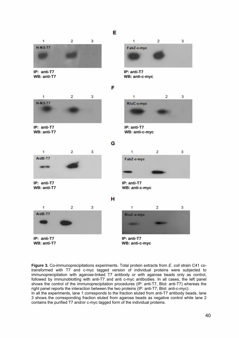
IP: anti-T7 WB: anti-c-myc
IP: anti-T7 WB: anti-c-myc
IP: anti-T7 WB: anti-c-myc
IP: anti-T7 WB: anti-c-myc
IP: anti-T7 WB: anti-T7
IP: anti-T7 WB: anti-T7
IP: anti-T7 WB: anti-T7
IP: anti-T7 WB: anti-T7
Figure 3. Co-immunoprecipitations experiments. Total protein extracts from E. coli strain C41 co-transformed with T7 and c-myc tagged version of individual proteins were subjected to immunoprecipitation with agarose-linked T7 antibody or with agarose beads only as control, followed by immunoblotting with anti-T7 and anti c-myc antibodies. In all cases, the left panel shows the control of the immunoprecipitation procedures (IP: anti-T7, Blot: anti-T7) whereas the right panel reports the interaction between the two proteins (IP: anti-T7, Blot: anti-c-myc). In all the experiments, lane 1 corresponds to the fraction eluted from anti-T7 antibody beads, lane 3 shows the corresponding fraction eluted from agarose beads as negative control while lane 2 contains the purified T7 and/or c-myc tagged form of the individual proteins.
40

In bacterial cells, regulation of ribosomal RNA (rRNA), which accounts to more than 90% of total RNA, is crucial for metabolic and energetic balance. Consistent with such an important role, ribosomal RNA gene transcription is tightly regulated. However, it is likely that not all the factors controlling rRNA transcription initiation, processing and modification have been identified to date. In the last decade, a considerable amount of attention has been given to an A+T-rich promoter sequence, the so-called UP element, located upstream of the core promoter region. In the E. coli ribosomal rrnB P1 promoter, the UP element is located in a region spanning the -40 and -60 positions, and is able to increase transcription from 30 to 70 fold by acting as a binding site for the α subunit of RNA polymerase (18). This element is a potential target for regulation of rRNA transcription by binding other putative factors able to modulate RNA polymerase activity. Indeed, a large number of promoters subjected to complex regulation possess strong α−binding sites (26-28). In this work, we used a double stranded DNA oligonucleotide carrying the UP element and the -35 sequence of the rrnB P1 promoter as a bait to isolate the transcription complexes that gather at this promoter and to identify its components. Altogether, we were able to identify 27 different proteins by mass spectrometry methodologies. Interestingly, several of the identified proteins were already known to interact, either from data obtained from the literature, or from databases such as IntAct (http://www.ebi.ac.uk/intact/site/index.jsf), thus suggesting that they might be indeed involved in functional networks. According to their reported biological activities, the various promoter
interactors can be grouped into three functional categories: DNA binding proteins and transcription factors, RNA-binding and translation factors, and cell metabolism-related proteins. As expected, we found the components of E. coli RNA polymerase, with the exception of σ70, suggesting that, despite the presence of the -35 sequence, binding to the “UP35” oligonucleotide is mediated by core RNA polymerase, probably via interaction between the α subunit and the UP element. Despite the “UP35” oligonucleotide being an artifactual system, reports of RNA polymerase binding to promoter independent of σ70 interaction with core promoter elements would support this model (26,29 and unpublished data). However, it should be considered that σ70 might have also escaped identification in the proteomic experiment simply because of technical reasons. An important component of the protein complex isolated at the upstream elements of the rrnB P1 promoter is represented by ribosomal proteins, which would suggest that the ribosome assembles with RNA polymerase at bacterial promoters. This model would be in agreement with various observations on direct protein-protein interaction between RNA polymerase subunits and ribosomal proteins (30,31), and with reports on multifunctional roles of protein synthesis factors (32). In addition, our results provide biochemical evidence that are consistent with known mechanisms of cross-talk between the ribosome and RNA polymerase, such as attenuation and stringent response. Indeed, direct interaction between transcription and translation machineries would greatly facilitate this genetic processes: for instance, ppGpp synthesis by ribosomal proteins RelA and SpoT would be relayed more
41

efficiently to RNA polymerase if both a ribosome and RNA polymerase are part of the same protein complex. Enzymes active in cell metabolism (e.g. FabZ, LpdA), as well as proteins involved in RNA modification (e.g. RluC) or post-translational maturation of nascent peptides (e.g. DnaJ, La) might be recruited to the transcription complex either by transcription factors or by ribosomal proteins, as suggested by our co-immunoprecipitation experiments (Fig. 3). Another interesting scenario is based on the nature of the DNA-binding proteins found in the “UP35”-binding complex. The H-NS protein is the only bona fide transcription regulator and it binds curved DNA regions with a strong preference for A/T rich stretches. The H-NS protein is a trans-acting regulatory factor that negatively modulates the transcription of several ribosomal RNA genes by binding to the upstream sequence of their respective promoters. This protein is overexpressed during the stationary phase when the synthesis of rRNAs has to be decreased (33). In contrast to H-NS, both AidB and Dps are non-specific DNA binding proteins (25,34) involved in the response to alkylation and oxidative damage, respectively. In this work, we suggest potential roles for the AidB protein in transcription complexes. AidB belongs to the adaptive response to alkylating agents (35,36 and references therein), which is defined by the Ada regulon including ada itself, alkA, alkB and aidB genes. The repair mechanism is governed by the Ada protein, that can repair O6-methylated guanidine residues and acts upon self-methylation as a transcription activator of the ada-alkB operon and the alkA and aidB promoters (reviewed in 37). In contrast to the situation for DNA repair proteins Ada, AlkA, and AlkB, the role of AidB in the adaptive response is still
uncharacterized. AidB has been proposed to repair alkylated DNA or detoxify alkylating reagents (25,38,39). Findings reported here demonstrated that AidB can establish direct protein-protein interaction with other components of the rrnB P1 promoter binding complex, including H-NS. Interaction with H-NS might re-direct AidB binding towards curved DNA or A/T rich regions, thus recruiting AidB to transcription initiation complexes. The role of AidB might be related to repair of single strand DNA: indeed, single strand DNA has been demonstrated to undergo alkylation processes much quicker than double strand molecules even in the absence of oxidative stress (40). During transcription, the transcriptional fork generates single strand DNA molecules that might be a target for endogenous alkylating agents. In this respect, the recruitment of AidB and its related enzymes activities directly onto the promoter might prevent alkylation to damage the transcription process. Another intriguing, but perhaps more unlikely possibility could be that AidB might be recruited to this specific promoter to counteract alkylation of the nascent rRNA molecule. Both hypotheses are currently being investigated. ACKNOWLEDGEMENTS This work was supported by grants of Ministero dell’Università e della Ricerca Scientifica (Progetti di Rilevante Interesse Nazionale 2005, 2006; FIRB Rete Nazionale di Proteomica, RBRN07BMCT). Support from the National Center of Excellence in Molecular Medicine (MIUR - Rome) and from the Regional Center of Competence (CRdC ATIBB, Regione Campania – Naples) and from NIH grant CA100122 to M.R.V is gratefully acknowledged.
42

REFERENCES 1. Shalon,D., Smith,S.J., and
Brown,P.O. (1996) A DNA microarray system for analyzing complex DNA samples using two-color fluorescent probe hybridization. Genome Res., 6, 639-645.
2. Anderson,L., and Seilhamer,J. (1997) A comparison of selected mRNA and protein abundances in human liver. Electrophoresis, 18, 533–537.
3. Gygi,S.P., Rochon,Y., Franza,B.R., and Aebersold,R. (1999) Correlation between protein and mRNA abundance in yeast. Mol. Cell. Biol., 19, 1720-1730.
4. Schmidt,F., Donahoe,S., Hagens,K., Mattow,J., Schaible,U.E., Kaufmann,S.H., Aebersold,R., and Jungblut,P.R. (2004) Complementary analysis of the Mycobacterium tuberculosis proteome by two-dimensional electrophoresis and isotope-coded affinity tag technology. Mol. Cell. Proteomics, 3, 24-42.
5. Monti,M., Orrù,S., Pagnozzi,D., and Pucci,P. (2005a) Functional proteomics. Clin. Chim. Acta, 357, 140-150.
6. Michaud,G.A., and Snyder,M. (2002) Proteomic approaches for the global analysis of proteins. Biotechniques, 33, 1308–1316.
7. Souchelnytskyi,S. (2002) Proteomics in studies of signal transduction in epithelial cells. J. Mammary Gland Biol. Neoplasia, 7, 359 – 371.
8. Terpe,K. (2003) Overview of tag protein fusions: from molecular and biochemical fundamentals to commercial systems. Appl. Microbiol. Biotechnol., 60, 523–533.
9. Reinders,J., Lewandrowski,U., Moebius,J., Wagner,Y., and Sickmann,A. (2004) Challenges in mass spectrometry-based proteomics. Proteomics, 4, 3686-3703.
10. Burckstummer,T., Bennett,K.L., Preradovic,A., Schutze,G.,
Hantschel,O., Superti-Furga,G., and Bauch,A. (2006) An efficient tandem affinity purification procedure for interaction proteomics in mammalian cells. Nat. Methods, 3, 1013-1019.
11. Monti,M., Orrù,S., Pagnozzi,D., and Pucci,P. (2005b) Interaction proteomics. Biosci Rep., 25, 45-56.
12. Schneider,D.A., Ross,W., and Gourse,R.L. (2003) Control of rRNA expression in Escherichia coli. Curr. Opin. Microbiol., 6, 151-156.
13. Paul,B.J., Ross,W., Gaal,T., and Gourse,R.L. (2004) rRNA transcription in Escherichia coli. Annu. Rev. Genet., 38, 749-770.
14. Dennis,P.P., Ehrenberg,M., and Bremer,H. (2004) Control of rRNA synthesis in Escherichia coli: a systems biology approach. Microbiol. Mol. Biol. Rev., 68, 639-668.
15. Afflerbach,H., Schroder,O., and Wagner,R. (1999) Conformational changes of the upstream DNA mediated by H-NS and FIS regulate E. coli rrnB P1 promoter activity. J. Mol. Biol., 286, 339–353.
16. Hernandez,J.V., and Cashel,M. (1995) Changes in conserved region 3 of Escherichia coli σ70 mediate ppGpp-dependent functions in vivo. J. Mol. Biol., 252, 536–549.
17. Barker,M.M., Gaal,T., Josaitis,C.A., and Gourse,R.L. (2001) Mechanism of regulation of transcription initiation by ppGpp. I. Effects of ppGpp on transcription initiation in vivo and in vitro. J. Mol. Biol., 305, 673–688.
18. Ross,W., Gosink,K.K., Salomon,J., Igarishi,K., Zou,C., Ishihama,A., Serinov,K., and Gourse,R.L. (1993) A third recognition element in bacterial promoters: DNA binding by the α subunit of RNA polymerase. Science, 262, 1407-1413.
19. Meng,W., Belyaeva,T., Savery,N.J., Busby,S.J., Ross,W.E., Gaal,T., Gourse,R.L., and Thomas,M.S. (2001) UP element-dependent transcription at the Escherichia coli rrnB P1 promoter: positional requirements and role of the RNA polymerase α subunit linker. Nucleic Acids Res., 29, 4166-4178.
43
20. Sambrook, J., and Russell, D.W. (2001) Molecular Cloning: a Laboratory Manual, 3rd ed. Cold Spring Harbor Laboratory Press, Cold Spring Harbor, NY.
21. Bradford,M.M. (1976) A rapid and sensitive method for the quantitation of microgram quantities of protein utilizing the principle of protein-dye binding. Anal. Biochem., 72, 248-254.
22. Miroux,B., and Walker,J.E. (1996) Over-production of proteins in Escherichia coli: mutant hosts that allow synthesis of some membrane proteins and globular proteins at high levels. J. Mol. Biol., 19, 289-298.
23. Ross,W., Schneider,D.A., Paul,B.J., Mertens,A., and Gourse,R.L. (2003) An intersubunit contact stimulating transcription initiation by E coli RNA polymerase: interaction of the α C-terminal domain and σ region 4. Genes Dev., 17, 1293-1307.
24. Haugen,S.P., Ross,W., and Gourse,R.L. (2008) Advances in bacterial promoter recognition and its control by factors that do not bind DNA. Nat. Rev. Microbiol., 6, 507-519.
25. Rohankhedkar,M.S., Mulrooney,S.B., Wedemeyer,W.J., and Hausinger,R.P. (2006) The AidB component of the Escherichia coli adaptive response to alkylating agents is a flavin-containing, DNA-binding protein. J. Bacteriol., 188, 223-230.
26. Landini,P., and Volkert,M.R. (1995) RNA polymerase α subunit binding site in positively controlled promoters: a new model for RNA polymerase-promoter interaction and transcriptional activation in the Escherichia coli ada and aidB genes. EMBO J., 14, 4329-4335.
27. Ross,W., Aiyar,S.E., Salomon,J., and Gourse,R.L. (1998) Escherichia coli promoters with UP elements of different strengths: modular structure of bacterial promoters. J. Bacteriol., 180, 5375–5383.
28. Boucher,P.E., Maris,A.E., Yang,M-S., and Stibitz,S. (2003) The
response regulator BvgA and RNA polymerase α subunit C-terminal domain bind simultaneously to different faces of the same segment of promoter DNA. Mol. Cell., 11, 163–173.
29. Landini,P., Bown,J.A., Volkert,M.R., and Busby,S.J. (1998) Ada protein-RNA polymerase sigma subunit interaction and alpha subunit-promoter DNA interaction are necessary at different steps in transcription initiation at the Escherichia coli ada and aidB promoters. J. Biol. Chem., 273, 13307-13312.
30. Butland,G., Peregrin-Alvarez,J.M., Li,J., Yang,W., Yang,X., Canadien,V., Starostine, A., Richards,D., Beattie,B., Krogan, N., et al. (2005) Interaction network containing conserved and essential protein complexes in Escherichia coli. Nature, 433, 531-537.
31. Arifuzzaman,M., Maeda,M., Itoh,A., Nishikata,K., Takita,C., Saito,R., Ara,T., Nakahigashi,K., Huang,H.C., Hirai,A. et al. (2006) Large-scale identification of protein–protein interaction of Escherichia coli K-12. Genome Res., 16, 686-691.
32. Hausmann,C.D., and Ibba,M. (2008) Aminoacyl-tRNA synthetase complexes: molecular multitasking revealed. FEMS Microbiology Reviews, 32, 705-721.
33. Gralla,J.D. (2005) Escherichia coli ribosomal RNA transcription: regulatory roles for ppGpp, NTPs, architectural proteins and a polymerase-binding protein. Mol. Microbiol., 55, 973-977.
34. Martinez,A., and Kolter,R. (1997) Protection of DNA during oxidative stress by the nonspecific DNA-binding protein Dps. J. Bacteriol., 179, 5188–5194.
35. Volkert,M.R. (1988) Adaptive response of Escherichia coli to alkylation damage. Environ. Mol. Mutagen., 11, 241-255.
36. Sedgwick,B., Bates,P.A., Paik,J., Jacobs,S.C., and Lindahl,T. (2007) Repair of alkylated DNA: Recent advances. DNA Repair, 6, 429-442.
44

37. Volkert,M.R., and Landini,P. (2001) Transcriptional responses to DNA damage. Curr. Opin. Microbiol., 4, 178-185.
38. Landini,P., Hajec,L.I., and Volkert,M.R. (1994) Structure and transcriptional regulation of the Escherichia coli adaptive response gene aidB. J. Bacteriol., 176, 6583-6589.
39. Bowles,T., Metz,A.H., Quin,J.O., Wawrzak,Z., and Eichman,B.F. (2008) Structure and DNA binding of alkylation response protein AidB. PNAS, 105, 15299-15304.
40. Bodell,W.J., and Singer,B. (1979) Influence of hydrogen bonding in DNA and polynucleotides on reaction of nitrogens and oxygens toward ethylnitrosourea. Biochemistry, 18, 2860-2863.
45


Manuscript to be submitted to Journal of Bacteriology
Role of Escherichia coli AidB protein in the transcriptional regulation Valentina Rippa1, 2, Carla Esposito1, Michael Volkert3 and Angela Duilio1, 2*
1Department of Organic Chemistry and Biochemistry, University Federico II of Naples, Naples, Italy 2School of Biotechnological Sciences, University Federico II of Naples, Naples, Italy 3Department of Molecular Genetics and Microbiology, University of Massachusetts Medical School, Worcester, Massachusetts, USA ABSTRACT Upon exposure to alkylating agents, Escherichia coli increases expression of aidB along with three genes (ada, alkB, and alkA) that encode DNA repair proteins. Despite extensive efforts, the molecular mechanism by which AidB protects against the mutagenic effects of DNA alkylators remains elusive. In this study, we assigned a biological role to AidB protein in the cell not exposed to alkylating agents. AidB was shown to possess a high affinity for ada/alkB, alkA and aidB promoters containing an upstream element, an AT rich transcriptional enhancer sequence. The physiological significance of this specific binding was investigated by in
*Corresponding Author: Angela Duilio Dipartimento di Chimica Organica e Biochimica, Complesso Universitario di Monte Sant’Angelo Via Cinthia 4 80126 Napoli (Italy). Tel +39-081674314 Fax +39081674313; email: [email protected]
vivo transcription assays. The results clearly demonstrated that AidB represses its own synthesis during normal cell growth. Another objective of this study was to define and characterize the domain architecture of AidB. The N-terminal region consisting of the first 439 residues was shown to exhibit isovaleryl-CoA dehydrogenase activity and to purify as a tetramer; the C-terminal region corresponding to residues 440-541 was determined to possess DNA binding activity, to purify as a monomer and to function as a transcriptional repressor in vivo. Thus, we demonstrated that the AidB protein is a modular transcription factor which requires the short C-terminal region for its regulatory function. Keywords: AidB protein, adaptive response genes, upstream element, transcription regulation. Running title: Transcriptional role of AidB protein in Escherichia coli. INTRODUCTION Transcription regulation is one of the principal strategies used by bacteria to respond to external stimuli and to adapt to a changing environment. Exposure of Escherichia coli to sublethal concentrations of alkylating agents such as methyl methanesulfonate (MMS) stimulates the expression of four genes, ada, alkB, alkA, and aidB. The activation of these genes confers increased cellular resistance to the mutagenic and cytotoxic effects of alkylating agents and is known as the adaptive response (Karran, P. et al., 1982; Kataoka, H. et al.,1983; Nakabeppu, Y. et al., 1984; Volkert, M.R. and Nguyen, D.C. 1984). The Ada protein is the key enzyme of this process; Ada acts both as a methyltransferase able to remove methyl groups from damaged DNA and as a transcriptional activator for the
46

adaptive response genes (Teo, I. et al., 1986; Nakabeppu, Y. et al., 1984; Landini, P. and Volkert, M.R. 1995). AlkA is a DNA glycosylase that catalyzes the base excision repair of alkylpurines (Nakabeppu, Y. et al., 1984). AlkB is an α-ketoglutarate-Fe(II)-dependent DNA dioxygenase that repairs 1-methyladenine and 3-methylcytosine lesions by oxidative demethylation (Trewick, S.C. et al., 2002). Despite detailed understanding of Ada, AlkA, and AlkB, the mechanism by which AidB protects against DNA damage in the adaptive response remains elusive. It has been suggested that AidB may act as a detoxification enzyme to destroy certain alkylating reagents or as a DNA repair enzyme (Landini, P. et al., 1994; Landini, P. and Volkert, M.R. 1995). AidB is a protein of 541 amino acids that is related in sequence to acyl-CoA dehydrogenases (ACADs), a family of enzymes which use a flavin adenine dinucleotide (FAD) to catalyze the α,β-dehydrogenation of acyl-CoA conjugates (Landini, P. et al., 1994; Rohankhedkar, M.S., et al., 2006). Recently, AidB has been shown to bind stoichiometric amounts of redox active FAD and to exhibit weak isovaleryl-CoA dehydrogenase (IVD) activity (Landini, P. et al., 1994; Rohankhedkar, M.S., et al., 2006). Subsequent structural studies revealed a distinctive FAD active site that provides a rationale for AidB’s limited acyl-CoA dehydrogenase activity. AidB was also shown to bind double strand DNA independently of its sequence (Rohankhedkar, M.S., et al., 2006). However, recently we demonstrated that AidB is part of the protein complex that binds to the -35 and upstream elements of the ribosomal rrnB P1 promoter but not to sequences lacking the UP element (Amoresano et al., submitted for publication), an AT rich transcriptional enhancer sequence.
This result suggested that AidB might possess sequence-specific binding. Since the -60 to -40 regions of ada/alkB, alkA and aidB promoters resemble the rrnB P1 UP element in A/T content and location relative to the core promoter (Landini, P. et al., 1998), we supposed that AidB protein might specifically interact with the promoters of the Ada-regulated adaptive response. In this work, we show that AidB displays a high affinity for UP regions of Pada/alkB, PalkA and PaidB. The functional significance of this specific binding was investigated by in vivo transcription assays using lacZ as reporter gene. AidB was shown to repress its own synthesis during normal cell growth. Another objective of this study was to define and characterize the domain architecture of AidB. The N-terminal region consisting of the first 439 residues was shown to exhibit isovaleryl-CoA dehydrogenase activity; the C-terminal region corresponding to residues 440-541 was determined to possess DNA binding activity and to function as a transcriptional repressor in vivo. Thus, the results reported here showed that the AidB protein is a modular transcription factor which requires the short C-terminal region for its regulatory function. MATERIALS AND METHODS Bacterial strains and plasmids. The bacterial strains, the plasmids and the oligonucleotides used in this work are listed in Table 1. Plasmids pMV132H, pSL101 and pSL112 were a kind gift from P. Landini (University of Milan, Italy). MV1161, MV5924, MV1601, MV6608 MV1571 and MV6607 E. coli strains were a kind gift from M. Volkert (University of Massachusetts, Worcester, MA).
47

MLbsdMka CpTwErratTvledvsFtwpa
edia and chemicals. uria-Bertani or nutrient broths (for acterial cultures and plating) and uspension medium (for bacterial ilutions) were used as described by iller (Miller, J.H. 1972). Ampicillin and
anamycin (Sigma) were used at 100 nd 50 µg/ml, respectively.
onstruction of the expression lasmids. he aidB, aidB∆Ct and Ct-aidB genes ere amplified from DNA genomic of . coli K12 by polymerase chain
eaction (PCR) using the forward and everse primers listed in Table 1. The mplified fragments were digested with
he restriction enzymes underlined in able 1 and cloned into the expression ector pET28a (Novagen) which was
inearized with the same restriction nzymes. The resulting plasmids, esignated as listed in Table 1, were erified by automated DNA equencing. or the complementation experiments,
he aidB, aidB∆Ct and Ct-aidB genes ere cloned into the expression vector ET28a-Plac. The lac promoter was mplified from DNA genomic of E. coli
by PCR using the primers listed in Table 1. The amplification product was digested with SphI and BamHI and cloned into the pET28a (+) plasmid. Then the aidB, aidB∆Ct and Ct-aidB genes were positioned downstream of the lac promoter. Plasmid construction was verified by automated DNA sequencing. Production and purification of recombinant proteins. The constructs pET28a-aidB, pET28a-aidB∆Ct, pET28a-Ct-aidB (Table 1) were individually transformed into the E. coli strain C41 (DE3). The recombinant cells were grown at 25°C to an optical density at 600 nm of ~0.5, at which time 0.05 mM isopropyl-beta-D-thiogalactopyranoside (IPTG) was added and the cultures were allowed to grow until the OD600 reached 3.0. Cells were harvested by centrifugation at 5000 x g for 15 min at 4°C, resuspended in buffer A (50 mM Na2HPO4, pH 7.4, 0.3 M NaCl, 10 mM imidazole, 1 mM PMSF), disrupted by passage through a French Press and centrifuged at 14000 x g for 30 min at 4°C.
Table 1: Bacterial strains, plasmids and oligonucleotidesStrains/plasmids
Description
Reference or source
E. coli strains C41 (DE3)
Strain that derives from BL21 [F-ompT hsdSB (rB- mB-) gal dcm (DE3)]. This strain has at least one uncharacterized mutation that prevents cell death associated with expression of many toxic recombinant proteins
Miroux and Walker, 1996
MV1161 rfa-550 derivative of AB1157 that carries the following additional markers: argE3 hisG4 leu-6 proA2 thr-1 ara-14 galK2 lacY1 mtl-1 xyl-5 rpsL31 supE44 tsx-6 rfa-550
Volkert
MV5924 Strain derivative of MG1655 in which the aidB gene had been inactivated by a tetracycline resistance cassette
Volkert
MV1601 Strain carrying a lacZ transcriptional insertion within the chromosomal alkB gene
Volkert
MV6608 Strain harboring the aidB mutation in the alkB::lacZ background
Volkert
MV1571 Strain harboring lacZ fragment in the chromosomal alkA gene Volkert MV6607 Strain harboring the aidB mutation in the alkA::lacZ
background Volkert
48

The recombinant proteins were purified by affinity chromatography on His-Select Nickel Affinity Gel (Sigma). The lysate was loaded onto His-Select Nickel Affinity Gel equilibrated with equilibration buffer (50 mM Na2HPO4, pH 7.4, 0.3 M NaCl, 10 mM imidazole).
After 1 min of incubation at 4°C, the matrix was collected by centrifugation at 11000 x g for 1 min and washed 3 times with same equilibration buffer. The recombinant proteins were eluted with buffer containing 250 mM imidazole in 50 mM Na2HPO4, pH 7.4,
pET28a(+) Plasmid carrying an N-terminal His•Tag®/thrombin/T7•Tag® configuration plus an optional C-terminal His•Tag sequence
Novagen
pET28a-aidB pET28a∆(BamHI-HindIII)Ω(aidB gene) This workpET28a-aidB∆Ct pET28a∆(BamHI-HindIII)Ω(aidB∆Ct gene) This workpET28a-Ct-aidB pET28a∆(BamHI-XhoI)Ω(Ct-aidB gene) This workpET28a-Plac pET28a∆(SphI-BamHI)Ω(lac promoter) This workpET28a-Plac-aidB pET28a-Plac∆(BamHI-HindIII )Ω(aidB gene) This workpET28a-Plac-aidB∆Ct pET28a-Plac∆(BamHI-HindIII )Ω(aidB∆Ct gene) This workpET28a-Plac-Ct-aidB pET28a-Plac∆(BamHI-XhoI)Ω(Ct-aidB gene) This workpMV132H Plasmid carrying lacZ fusion under the control of the wild type
PaidB Landini
pSL101 Plasmid derivative of pJCD01 carrying a 242 bp fragment of the wild type PaidB
Landini
pSL112 Plasmid derivative of pJCD01 carrying a 238 bp fragment of the mutant PaidB in which -12C has been substituted with the residue T
Landini
Oligonucleotides
UP35 PaidB Fw 5'-GATAAGAATGTTTTAGCAATCTCTTTCTGTCA-3' UP35 PaidB Rv 5'-TGACAGAAAGAGATTGCTAAAACATTCTTATC-3' UP35 Pada Fw 5'-GCGAAAAAAATTAAAGCGCAAGATTGTTGGTT-3' UP35 Pada Rv 5'-AACCAACAATCTTGCGCTTTAATTTTTTTCGC-3' UP35 PalkA Fw 5'-TTGCCGTCGCGACAACCGGAATATGAAAGCAA-3' UP35 PalkA Rv 5'-TTGCTTTCATATTCCGGTTGTCGCGACGGCAA-3' Neg PaidB Fw 5’-ATGAATGTACTGTATCTAGTCTACTAGTATCT-3’ Neg PaidB Rv 5’-AGATACTAGTAGACTAGATACAGTACATTCAT-3’ Neg Pada Fw 5'-TAGACTTGATCAGATGAGATGAAGTACAGATA-3' Neg Pada Rv 5'-TATCTGTACTTCATCTCATCTGATCAAGTCTA-3' Neg PalkA Fw 5'-AGTCACTGTCAGTCACAGTACAGACGACGAGA-3' Neg PalkA Rv 5'-TCTCGTCGTCTGTACTGTGACTGACAGTGACT-3' PleuA Fw 5’-GGGTTGACATCCGTTTTTGTATCCAGTAACTC-3’ PleuA Rv 5’-GAGTTACTGGATACAAAAACGGATGTCAACCC-3’ aidB Fw 5'-ATAGGATCCGTGCACTGGCAAACTCACACCG-3' aidB Rv 5'-CGCGAGCTCCGTAAGCTTTTACACACACACTC-3' aidB∆Ct Fw 5'-ATAGGATCCGTGCACTGGCAAACTC-3' aidB∆Ct Rv 5'-AATAAGCTTTTAGAGAACGCGCAACACATC-3' Ct-aidB Fw 5'-ATAGGATCCAATAAGCAAGCGGGCG-3' Ct-aidB Rv 5'-TTACTCGAGTTACACACACACTCCCC-3' Plac Fw 5’-TAAAGCATGCTTTACACTTTATGCTTCCGGTCGTATGTTGTGTGGAAAGCTTTTA-3’ Plac Rv 5’-TAAAAGCTTTCCACACAACATACGACCGGAAGCATAAAGTGTAAAGCATGCTTTA-3’
49

0.3 M NaCl. Protein concentration was estimated with Bradford reagent (Bio-Rad protein assay) and protein content was checked by SDS-polyacrilamide gel electrophoresis (SDS-PAGE). In vivo transcription assays. MV1161 (wild type) and MV5924 (∆aidB) E. coli strains were transformed with reporter plasmid pMV132H carrying the lacZ gene under the control of PaidB. The complementation experiments were performed by transforming MV5924 containing pMV132H with the following constructs: pET28a-Plac-aidB, pET28a-Plac-aidB∆Ct, pET28a-Plac-Ct-aidB. These bacterial cultures grown overnight in LB medium at 37°C, were diluted 1:100 in fresh medium. At an A600 nm of 0.4, the cultures were divided in two aliquots, and one was supplemented with 0.04% MMS to activate the adaptive response. Cellular pellets were collected during the exponential growth phase. The cells were resuspended in 50 mM Na2HPO4 (pH 7.4), disrupted by passage through a French Press and centrifuged at 14000 x g for 30 min at 4°C. The supernatant was collected and protein concentration was determined with the Bio-Rad protein assay (Bradford, M.M. 1976), using bovine serum albumine as standard. β-galactosidase activity was determined by measuring ONPG-hydrolysis, as described by Miller (Miller, J.H., 1972). In vivo transcription from the ada promoter region was measured using both MV1601 and MV6608 strains; to measure in vivo transcription from the alkA promoter we used both MV1571 and MV6607 strains. In vitro transcription assays. Transcription experiments were performed on two different DNA templates: plasmid pSL101 containing wild type PaidB and plasmid pSL112
carrying mutant PaidB with a C-T substitution at position -12. Plasmids (5 nM) and RNA polymerase holoenzyme (120 nM) (Epicentre Technology) were incubated for 20 min at 37°C, in a solution of 40 mM Hepes pH 8.0, 10 mM magnesium chloride, 200 mM potassium glutamate, 4 mM dithiothreitol and 100 µg/ml bovine serum albumin in the absence and in the presence of increasing quantities of the AidB protein (0.1-0.4 µg). Elongation step was started by the addition of a pre-warmed mixture containing nucleotides and heparin (final concentrations were 500 µM ATP, GTP and CTP, 30 µM UTP, 1 µCi of [α32P]UTP and 500 µg/ml heparin) to the template-polymerase mix and allowed to proceed for 10 min at 37°C. The reactions were stopped by the addition of stop solution (10 mM EDTA, 0.5% bromophenol blue, 0.025% xylene cyanol). After heating to 65°C, samples were subjected to electrophoresis on a 7% denaturing polyacrilamide gel in 0.5 X Tris borate-EDTA. Transcripts were detected by exposure to X-ray film overnight at -80°C. Native molecular mass of AidB∆Ct and Ct-AidB. Size exclusion chromatography was performed by using a Superdex 200 PC 3.2/30 (for AidB∆Ct) and a Superdex 75 PC 3.2/30 (for Ct-AidB) column (GE Healthcare) equilibrated in buffer containing 50 mM Tris-HCl, 150 mM NaCl, pH8. The molecular mass of the native proteins was estimated by comparing their retention time to those of molecular mass standards (thyroglobulin, 670,000 Da; bovine γ-globulin, 158,000 Da; chicken ovalbumin, 44,000 Da; equine myoglobin, 17,000 Da; vitamin B12, 1,350 Da; Bio-Rad).
50

Isovaleryl-CoA dehydrogenase activity assay.Isovaleryl-CoA dehydrogenase activity assays were carried out at room temperature in 200 mM phosphate buffer, pH 8.0, and using purified recombinant proteins that had been dialyzed to remove imidazole. For routine assays, 2 mM isovaleryl-CoA (Sigma) was used as the substrate and 0.1 mM 2,6-dichlorophenolindophenol (DCPIP) was used as the terminal electron acceptor in a final volume of 300 µl. The change in absorbance at 600 nm was monitored by using a Beckman DU 7500 spectrophotometer, and the enzyme activity was calculated by assuming an extinction coefficient of 20.6 mM−1 cm−1 for DCPIP (Engel, P.C., 1981). Electrophoretic shift mobility assays. Annealed DNA oligonucleotides (fragments UP35 PaidB, UP35 Pada, UP35 PalkA, Neg PaidB, Neg Pada, Neg PalkA) were 32P labelled at 5’ positions by using T4 polynucleotide kinase (20 units). Electromobility shift assays were performed in 20 µl reaction volume. AidB protein (2 pmol) was incubated with the 32P-labelled DNA (2 ng, 50,000-100,000 cpm) in buffer Z (25 mM HEPES pH 7.6, 50 mM KCl, 12.5 mM MgCl2, 1 mM DTT, 20% glycerol, 0.1% triton), for 20 min at RT. Mixtures were then analysed by electrophoresis on 8% native polyacrylamide gel (29:1 cross-linking ratio) in 45 mM Tris pH 8.0, 45 mM boric acid, 1 mM EDTA. Electrophoreses were performed at room temperature at 200 V (20 V/cm). The gels were dried and analysed by autoradiography. For EMSA performed by using biotinylated UP35 PaidB, protein-DNA complexes were separated on 8% nondenaturing polyacrylamide gels. After electrophoresis, gels were transferred to nylon membranes and
transferred DNA was immobilized by UV crosslinking for 3 min. For detection of bound DNA, membranes were blocked using blocking buffer followed by the addition of Streptavidin-HRPO and blots were developed according to the manufacturer’s instructions (Amersham, Arlington Heights, IL, U.S.A.). RESULTS AidB shows a high affinity for upstream regulatory regions. In a previous report (Rohankhedkar et al., 2006), it was shown that AidB binds double strand DNA independently of its sequence. However, our recent work demonstrated that AidB is part of the protein complex that gathers at the -35 and UP elements of the rrnB P1 promoter but not at identical sequences lacking the UP element (Amoresano et al., submitted for publication). This result suggested that AidB might possess sequence-specific binding. Since the -60 to -40 regions of ada/alkB, alkA and aidB promoters resemble the rrnB P1 UP element in A/T content and location relative to the core promoter (Landini et al., 1998), we supposed that AidB protein might specifically interact with the promoters of the Ada-regulated adaptive response. This hypothesis was investigated by electrophoresis mobility shift gel assays (EMSA). To this aim, the aidB gene was cloned into a commercial expression vector of the pET series and the recombinant protein was expressed as chimeric protein bearing a 6 His tag at the C-terminus. The expressed protein was then purified by affinity chromatography on Ni2+-agarose beads and its homogeneity was tested by SDS-PAGE and mass fingerprinting analyses. EMSA experiments were carried out using three different
51

Competitor UP35 PaidB - 100 500 - - PleuA - - - 100 500
1 2 3 4 5
Fig. 1. Gel retardation experiments performed incubating the AidB protein with the UP35 PaidB; competitors were included as indicated. Lane 1: AidB protein incubated with UP35 PaidB. Lane 2-3: Competition assay with UP35 PaidB (100x-500x) as a specific competitor. Lane 4-5: Competition assay with PleuA as a non specific competitor (100x-500x).
radiolabelled DNA fragments corresponding to the UP region and the -35 box of the aidB, ada/alkB and alkA promoters (“UP35 PaidB”, “UP35 Pada”, “UP35 PalkA”, Table 1). The randomised version of each UP35 probe, i.e. an oligonucleotide with the same nucleotide content as UP35, but lacking any known promoter or regulatory elements (“Neg PaidB”, “Neg Pada”, “Neg PalkA”, Table 1), was also used in EMSA experiments. As expected, addition of AidB retarded the migration of both the UP35 DNA fragments and the “Neg” probes (data not shown). The specificity of AidB binding to the three UP35 probes was tested by competition experiments: the radiolabelled “UP35” probes were incubated with the AidB protein and increasing quantities (100-500 fold) of either unlabelled “UP35” (used as a
specific competitor) or PleuA (a promoter lacking the UP element used as a non specific competitor), were added to the reaction mixture. The Fig. 1 shows that the binding to the radiolabelled “UP35 PaidB” by AidB was reversed by an excess of unlabelled “UP35” (Lanes 2-3), while it was not affected by competition with identical concentrations of PleuA (Lanes 4-5). Analogous experiments showed the specific interaction of AidB with “UP35 Pada” and “UP35 PalkA” (data not shown). Altogether, these results demonstrated that AidB possesses high binding affinity for the upstream regions of the adaptive response genes. AidB acts as a transcriptional repressor in vivo. In order to determine whether the
52

A B
0
500
1000
1500
2000
2500
3000
3500
4000
4500
5000
5500
6000
6500
7000
Prom
oter
act
ivity
(Mill
er u
nits
) MV1161
MV5924
MV5924 +pET28a-aidB
0
500
1000
1500
2000
2500
3000
3500
4000
4500
5000
5500
6000
6500
7000MV1161
MV5924
MV5924 +pET28a-aidB
Fig. 2. In vivo transcription from the aidB promoter. Vector pMV132H was introduced into MV1161 (wild type) and MV5924 (∆aidB) E. coli strains and the β-galactosidase specific activity was determined in the absence (A) and in the presence (B) of 0.04% MMS as alkylating agent. MV5924 strain was also transformed with plasmid pET28a-Plac-aidB producing functional AidB protein and the corresponding β-galactosidase activity was evaluated. Means and standard deviations have been calculated from four independent assays.
MV1161
MV5924
MV5924 + pET28a-Plac-aidB
MV59
MV1161
24
MV5924pET28a-P
+ lac-
aidB
specific binding of AidB to PaidB, Pada and PalkA might be of biological significance, we tested its effect on transcription from these promoters by in vivo β-galactosidase reporter assays. The putative AidB activity on its promoter was investigated by transforming both MV1161 (wild type) and MV5924 (∆aidB) E. coli strains with plasmid pMV132H carrying the gene coding for the β-galactosidase under the control of the aidB promoter. These strains were grown in LB medium, in the absence and in the presence of sub-inhibitory concentrations of the alkylating agent methyl methane sulfonate (MMS) and the β-galactosidase activity was monitored during the exponential growth phase. As shown in Fig. 2A, during the growth in LB medium, the ∆aidB strain exhibited levels of β-galactosidase activity 13-fold higher than that observed in the wild type cells, suggesting that transcription from
the aidB promoter is repressed by AidB during normal cell growth. The complementation of the ∆aidB mutation by pET28a-Plac-aidB restored transcription levels from the aidB promoter to wild type levels. These data clearly demonstrated that in the absence of DNA alkylation damage, the AidB protein is required for repression of its own expression. Exposure to sub-inhibitory concentrations of MMS, as expected, resulted in an increase of aidB promoter activity (comparison between Figs. 2A and 2B). Interestingly, the transcription levels in the ∆aidB strain were slightly higher (by almost 2.5-fold) than that observed for the wild type cells suggesting that the activator methylated Ada, in the absence of AidB, is more efficient in activating transcription of the aidB gene. This effect could be due to the competition between AidB and methylated Ada for binding to same site on the aidB
53

promoter. In fact, gel retardation experiments performed in this work have demonstrated that AidB is able to bind to a region of PaidB extending from the upstream element to the -35 box and previous studies (Landini, P. and Volkert, M.R. 1995) have found that methylated Ada protects the residues from -62 to -38 in the aidB promoter. Successively, we evaluated the AidB activity on the ada promoter (which controls expression of the ada and alkB genes) and the alkA promoter. In vivo transcription from the ada promoter region was measured using both MV1601 strain carrying a lacZ transcriptional insertion within the chromosomal alkB gene and MV6608 strain harboring the aidB mutation in the alkB::lacZ background. To measure in vivo transcription from the alkA promoter we used MV1571 strain which has a chromosomal lacZ operon fused to the alkA promoter and its aidB mutant derivative MV6607. We did not observe any effect of the aidB deletion on lacZ expression driven by the ada and alkA promoters: during normal cell growth, the β-galactosidase catalytic activities determined in the ∆aidB strains were low and similar to that resulting from the wild type strains (data not shown). Thus, we could not assign any role to AidB protein in transcription regulation of ada, alkB and alkA genes, although gel retardation assays have shown specific interaction between AidB and promoters under study. In vitro regulatory activity of AidB. The transcriptional activity of the AidB protein on its promoter was further investigated through in vitro transcription assays using E. coli RNA polymerase holoenzyme. Although RNA polymerase σ70 (Eσ70) is able to bind to the aidB promoter region in the absence of additional factors, it can
initiate transcription efficiently only in the presence of the activator protein Ada (Landini, P. and Volkert, M.R. 1995). Studies performed by Lacour et al., (2002) have shown that Eσ70, in the absence of others factors, is able to activate transcription from a mutant form of the aidB promoter in which the residue C at position -12 has been substituted with the nucleotide T. For this reason, in vitro transcription assays were carried out by using two different DNA templates: plasmid pSL101 containing wild type PaidB and vector pSL112 carrying mutant PaidB with a C-T substitution at position -12. The results of these experiments are shown in Fig. 3. Two distinct gel bands were detected when the RNA polymerase σ70 was incubated with the plasmid pSL112 containing mutant PaidB (Fig. 3, Lane 2). The upper band is relative to the aidB transcript while the lower band corresponds to the transcript of the RNA I promoter used as an internal control. The upper band is rather weak when the RNA polymerase σ70 was incubated with the plasmid pSL101 (Fig. 3, Lane 1) because, as described above, Eσ70
requires the activator Ada protein to initiate transcription efficiently from the wild type aidB promoter. When increasing quantities (0.1-0.4 µg) of the AidB protein were added to the transcription reaction containing vector pSL112, the band corresponding to the aidB transcript clearly disappeared (Fig. 3, Lanes 3-6) while the transcript of the RNA I promoter was not affected by the presence of AidB. In agreement with the results of the in vivo transcription experiments, AidB was shown to be involved in negative regulation of its own gene. Characterization of the functional domains in AidB. Knowledge of the domain architecture of the AidB flavoprotein is necessary
54
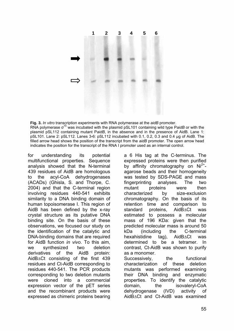
1 2 3 4 5 6
Fig. 3. In vitro transcription experiments with RNA polymerase at the aidB promoter. RNA polymerase σ70 was incubated with the plasmid pSL101 containing wild type PaidB or with the plasmid pSL112 containing mutant PaidB, in the absence and in the presence of AidB. Lane 1: pSL101. Lane 2: pSL112. Lanes 3-6: pSL112 incubated with 0.1, 0.2, 0.3 and 0.4 µg of AidB. The filled arrow head shows the position of the transcript from the aidB promoter. The open arrow head indicates the position for the transcript of the RNA I promoter used as an internal control.
for understanding its potential multifunctional properties. Sequence analysis showed that the N-terminal 439 residues of AidB are homologous to the acyl-CoA dehydrogenases (ACADs) (Ghisla, S. and Thorpe, C. 2004) and that the C-terminal region involving residues 440-541 exhibits similarity to a DNA binding domain of human topoisomerase I. This region of AidB has been defined by the x-ray crystal structure as its putative DNA binding site. On the basis of these observations, we focused our study on the identification of the catalytic and DNA-binding domains that are required for AidB function in vivo. To this aim, we synthesized two deletion derivatives of the AidB protein: AidB∆Ct consisting of the first 439 residues and Ct-AidB corresponding to residues 440-541. The PCR products corresponding to two deletion mutants were cloned into a commercial expression vector of the pET series and the recombinant products were expressed as chimeric proteins bearing
a 6 His tag at the C-terminus. The expressed proteins were then purified by affinity chromatography on Ni2+-agarose beads and their homogeneity was tested by SDS-PAGE and mass fingerprinting analyses. The two mutant proteins were then characterized by size-exclusion chromatography. On the basis of its retention time and comparison to standard proteins, AidB∆Ct was estimated to possess a molecular mass of 196 KDa: given that the predicted molecular mass is around 50 kDa (including the C-terminal hexahistidine tag), AidB∆Ct was determined to be a tetramer. In contrast, Ct-AidB was shown to purify as a monomer. Successively, the functional characterization of these deletion mutants was performed examining their DNA binding and enzymatic properties. To identify the catalytic domain, the isovaleryl-CoA dehydrogenase (IVD) activity of AidB∆Ct and Ct-AidB was examined
55

Table 2: Isovaleryl-CoA dehydrogenase activity of the full length and mutant AidB proteins was assayed as described in Materials and Methods following the isovaleryl-CoA-dependent reduction of he electron acceptor DCPIP. The data shown are averages of 10 measurements. Nd=not detected. t
AidB protein Isovaleryl-CoA dehydrogenase activity
µmol min-1 [mg protein]-1
full lenght 0.12 ± 0 AidB∆Ct 0.12 ± 0 Ct-AidB Nd
1 2 3 4
and compared with that of the full length AidB. AidB∆Ct mutant displayed low levels of isovaleryl-CoA catalytic activity that are identical to that exhibited by the full-length AidB protein (Table 2) whereas Ct-AidB mutant had no detectable enzymatic activity. Therefore, as predicted by the structural analysis, we have shown that the N-terminal region of AidB is responsible for catalytic activity. To gain insight into the domain that interacts with DNA, AidB∆Ct and Ct-AidB were assayed in vitro for DNA binding activity by gel retardation
assays. These experiments were performed using the biotin-labelled DNA fragment corresponding to the UP region and the -35 box of the aidB promoter (“UP35 PaidB”, Table 1). As shown in Fig. 4, Ct-AidB exhibited DNA binding activity (Lane 4) while AidB∆Ct was not able to bind to DNA (Lane 2). These experiments clearly demonstrated that the C-terminal domain alone is sufficient for DNA binding activity. Finally, the ability of the two mutant proteins to repress the transcription of the aidB gene was tested by in vivo β-galactosidase reporter assays. The ∆aidB strain was transformed with reporter plasmid pMV132H together with either pET28a-Plac-aidB∆Ct or pET28a-Plac-Ct-aidB. E. coli cells were grown in LB medium and the β-galactosidase activity was monitored and compared with that detected in the ∆aidB strain as well as in the ∆aidB strain carrying aidB gene on the plasmid pET28a. As shown in Table 3, Ct-AidB repressed lacZ expression with levels of β-galactosidase activity decreased to 7% compared with control cells that lacked AidB protein. In contrast, AidB∆Ct was not able to repress the PaidB-lacZ transcription. These reporter assays clearly demonstrated that Ct-AidB acts as a transcriptional repressor of the aidB gene in vivo.
Fig. 4. Gel retardation experiments carried out by incubating the full length and mutant AidB proteins with the “UP35 PaidB” probe. Lane 1: UP35 PaidB probe. Lanes 2-4: UP35 PaidB fragment incubated with AidB∆Ct, AidB and Ct-AidB proteins, respectively.
56

Table 3: β-galactosidase activity from E. coli strain MV5924 harbouring the lacZ reporter construct pMV132H and various aidB gene constructs. Means and standard deviations have been calculated rom four independent assays. f
AidB protein β-galactosidase activity Miller units %
None 3150 ± 46 100 AidB 222 ± 32 7
AidB∆Ct 3162 ± 48 100 Ct-AidB 225 ± 31 7
DISCUSSION Understanding protein functions as well as unravelling cellular mechanisms at the molecular level constitutes a major challenge in modern biology. In this paper, we report a new biological role for AidB, a component of the E. coli adaptive response to alkylating agents. While many questions about its function in the Ada-regulated response remain, we assigned a role to AidB in the cell not exposed to alkylating agents. In this study, we provided evidence that AidB shows a high affinity for the upstream regions of Pada/alkB, PalkA and PaidB. This observation lead us to a line of inquiry concerning its possible role in the transcription mechanism. This hypothesis was investigated in vivo by performing β-galactosidase reporter assays in both wild type and ∆aidB E. coli strains. In the absence of alkylation damage, a large increase in the lacZ expression driven by aidB promoter was observed in the E. coli cells lacking AidB as compared to the wild type control. In addition, the complementation of the ∆aidB mutation restored lacZ transcription to wild type levels. Taken together, these results clearly demonstrated that AidB represses its own synthesis during normal cell growth. In vitro transcription experiments performed by using a DNA template containing the aidB promoter and in the presence and absence of AidB protein confirmed
these data. Autoregulation of the aidB gene might play an important physiological role, because to some extent mitigates the energetic burden of bacterial cell not exposed to alkylating agents. In contrast, we were not able to assign any role to AidB in transcription regulation of ada, alkB and alkA genes. Another goal of this study was to identify and characterize the functional domains of AidB. The N-terminal region consisting of the first 439 residues was shown to exhibit the same levels of isovaleryl-CoA dehydrogenase (IVD) activity as the full length protein. The level of IVD activity observed in AidB is quite low (Table 2) compared to other acyl-CoA dehydrogenases: for comparison, human isovaleryl-CoA dehydrogenase exhibits specific activity of 8.2 to 11.7 µmol min-1 (mg protein)-1 (Battaile, K.P. et al., 1998; Mohsen, A. W. et al., 1998). Recent structural studies revealed several unique features that distinguish AidB’s FAD cavity from the ACAD active sites despite the conservation of their general properties (Bowles, T. et al., 2008). These observations provided a rationale for AidB’s limited acyl-CoA dehydrogenase activity, suggesting that fatty acyl-CoA are not substrates for the enzyme (Rohankhedkar, M.S. et al., 2006). The crystal structural of AidB has also identified a putative DNA binding site located in its C-terminal
57
region. In this work, we verified that the C-terminal residues 440-541 comprise a domain that is responsible for the DNA binding activity. Interestingly, this short region was also shown to function as a transcriptional repressor in vivo. Thus, the results reported here showed that the AidB protein is a modular transcription factor which requires its C-terminal region for regulatory function. The identification of two structurally and functionally independent domains suggests that AidB protein might possess multifunctional properties. Our finding that AidB has transcriptional activity does not provide any precise information on the possible role of the protein in the defence against alkylation. AidB has been hypothesized to detoxify certain alkylating reagents or to repair alkylated DNA (Landini, P. et al., 1994; Rohankhedkar, M.S. et al., 2006). However, numerous and similar examples of proteins which have more than a biological function have already been reported in the literature, i.e. Meddows et al., 2005 demonstrated that the transcription factor DksA is also involved in repair of DNA double-strand breaks. Another intriguing possibility is that AidB may stimulate the transcription of genes whose products are directly responsible for alkylation resistance. Therefore, it would be useful to perform microarray experiments to identify potential novel E. coli genes that are regulated by AidB protein in response to alkylation damage. ACKNOWLEDGEMENTS The authors wish to thank Prof. Paolo Landini from the University of Milan, Italy, for pMV132H, pSL101, and pSL112 plasmids. This work was supported by grants of Ministero dell’Università e della Ricerca Scientifica (Progetti di
Rilevante Interesse Nazionale 2004, 2005, 2006; FIRB 2001), of Programma Nazionale di Ricerche in Antartide 2004, and of Regione Campania L.R. 05/03. Support from the National Center of Excellence in Molecular Medicine (MIUR - Rome) and from the Regional Center of Competence (CRdC ATIBB, Regione Campania – Naples), Istituto Nazionale Biomolecole e Biosistemi (INBB) is gratefully acknowledged. REFERENCES
• Amoresano, A., Cirulli, C., Rippa, V., Esposito, C., Papa, R., and Duilio, A. Submitted to Molecular Microbiology.
• Battaile, K. P., McBurney, M., Van Veldhoven, P. P., and Vockley, J. Biochimica et biophysica acta 1390(3), 333–338 (1998).
• Bowles, T., Metz, A. H., O’Quin, J., Wawrzak, Z., and Eichman, B. F. Proceedings of the National Academy of Sciences of the United States of America 105(40), 15299–15304 (2008).
• Bradford, M. M. Analytical biochemistry 72, 248–254 (1976).
• Engel, P. C. (1981). Butyryl-CoA dehydrogenase from Megasphaera elsdenii. Methods Enzymol 71, 359-366.
• Ghisla, S. and Thorpe, C. European journal of biochemistry/FEBS 271(3), 494-508 (2004).
• Karran, P., Hjelmgren, T., and Lindahl, T. Nature 296(5859), 770–773 (1982).
• Kataoka, H., Yamamoto, Y., and Sekiguchi, M. J. Bacteriol. 153(3), 1301–1307 (1983).
• Lacour, S., Kolb, A., Boris Zehnder, A. J., and Landini, P. Biochemical and biophysical research communications 292(4), 922–930 (2002).
• Landini, P., Hajec, L. I., and Volkert, M. R. Journal of
58
bacteriology 176(21), 6583–6589 (1994).
• Landini, P. and Volkert, M. R. The Journal of biological chemistry 270(14), 8285–8289 (1995).
• Landini, P., Bown, J. A., Volkert, M. R., and Busby, S. J. The Journal of biological chemistry 273(21), 13307–13312 (1998).
• Meddows, T. R., Savory, A. P., Grove, J. I., Moore, T., and Lloyd, R. G. Molecular microbiology 57(1), 97–110 (2005).
• Miller, J. H. Experiments in Molecular Genetics. Cold Spring Harbor Laboratory Pr (1972).
• Miroux, B., and Walker, J. E. Journal of Molecular Biology 260(3), 289-298 (1996).
• Mohsen, A. W., Anderson, B. D., Volchenboum, S. L., Battaile, K. P., Tiffany, K., Roberts, D., Kim, J. J., and Vockley, J. Biochemistry 37(28), 10325–10335 (1998).
• Nakabeppu, Y., Kondo, H., and Sekiguchi, M. Journal of Biological Chemistry 259(22), 13723–13729 (1984).
• Rohankhedkar, M. S., Mulrooney, S. B., Wedemeyer, W. J., and Hausinger, R. P. Journal of bacteriology 188(1), 223–230 (2006).
• Teo, I., Sedgwick, B., Kilpatrick, M. W., McCarthy, T. V., and Lindahl, T. Cell 45(2), 315–324 (1986).
• Trewick, S. C., Henshaw, T. F., Hausinger, R. P., Lindahl, T., and Sedgwick, B. Nature 419(6903), 174–178 (2002).
• Volkert, M. R. and Nguyen, D. C. Proceedings of the National Academy of Sciences of the United States of America 81(13), 4110–4114 (1984).
59
Manuscript to be submitted to Molecular Microbiology
Preferential protection from DNA alkylation by the Escherichia coli AidB protein Valentina Rippa1, 2, Michael Volkert3 and Angela Duilio1, 2#. 1Department of Organic Chemistry and Biochemistry, University Federico II of Naples, Naples, Italy 2School of Biotechnological Sciences, University Federico II of Naples, Naples, Italy 3Department of Molecular Genetics and Microbiology, University of Massachusetts Medical School, Worcester, Massachusetts, USA ABSTRACT Exposure of Escherichia coli to alkylating agents activates expression of aidB along with three genes (ada, alkA, and alkB) that encode DNA repair proteins. Despite extensive efforts, the molecular function of AidB in the response to alkylation damage remains uncharacterized. AidB was recently shown to be a flavoprotein that binds to dsDNA, implicating it as a flavin-dependent DNA repair enzyme. Subsequent structural studies suggested that AidB might protect DNA from attack by alkylating agents. In this study, we demonstrate that AidB prevents DNA damage and that it does so in a preferential manner, protecting genes adjacent to promoters carrying an upstream
# Corresponding Author: Angela Duilio Dipartimento di Chimica Organica e Biochimica, Complesso Universitario di Monte Sant’Angelo Via Cinthia 4 80126 Napoli (Italy). Tel +39-081674314 Fax +39081674313; email: [email protected]
element, an AT rich transcriptional enhancer region. These include many of the housekeeping genes required for basic metabolic processes, such as the rrn operons, suggesting that E. coli and possibly other organisms preferentially protect subsets of genes from DNA damage. Keywords: AidB protein, alkylating agents, DNA protection, UP element. Running title: Protective role of AidB in Escherichia coli. INTRODUCTION Alkylating agents present in the cell and in the environment chemically modify DNA to produce cytotoxic and mutagenic lesions. Alkylation damage to DNA therefore poses a severe threat to the stability of the genome and, in mammals, can lead to genetic diseases and cancer. As a safeguard against DNA alkylation damage, all organisms have evolved multiple DNA repair mechanisms to remove these modifications and restore DNA to an undamaged state. In addition, bacteria employ an inducible response that serves to protect cells from changing levels of mutagens. In Escherichia coli, exposure to sublethal doses of alkylating agents such as methyl methanesulfonate (MMS), N-methylnitrosourea (MNU), N-methyl-N′-nitro-N-nitrosoguanidine (MNNG) and N-ethyl-N′-nitro-N-nitrosoguanidine (ENNG) stimulates the expression of four genes, ada, alkB, alkA, and aidB. The activation of these genes confers increased cellular resistance to the mutagenic and cytotoxic effects of alkylating agents and is known as the adaptive response (Kataoka, H. et al.,1983; Nakabeppu, Y. et al., 1984; Volkert, M.R. and Nguyen, D.C. 1984). The Ada protein is the key enzyme of this process; Ada acts both as a methyltransferase able to remove
60

methyl groups from damaged DNA and as a transcriptional activator for the adaptive response genes (Teo, I. et al., 1986; Nakabeppu, Y. et al., 1984; Landini, P. and Volkert, M.R. 1995). AlkA is a double-stranded DNA glycosylase that catalyzes the base excision repair of a wide variety of alkylated bases (Nakabeppu, Y. et al., 1984); AlkB is an α-ketoglutarate-Fe(II)-dependent DNA dioxygenase that repairs 1-methyladenine and 3-methylcytosine lesions by oxidative demethylation (Trewick, S.C. et al., 2002). Despite detailed understanding of Ada, AlkA, and AlkB, the mechanism by which AidB protects against DNA damage in the adaptive response remains elusive. AidB is related in sequence to acyl-CoA dehydrogenases (ACADs), which use a flavin adenine dinucleotide (FAD) to catalyze the α,β,-dehydrogenation of acyl-CoA conjugates (Landini, P. et al., 1994) and it was predicted to function in detoxification of alkylating agents. AidB was shown to be a flavoprotein and to bind to dsDNA implicating it as a flavin-dependent DNA repair enzyme (Rohankhedkar, M.S., et al., 2006). Recently, the crystal structure of AidB revealed that its DNA binding site lies within an interior channel, while its flavin binding site is accessible only from the exterior of the protein and is spatially distant from the DNA binding region (Bowles, T. et al., 2008). Based on these observations, it was suggested that AidB might bind to DNA and protect it by inactivating alkylators before they are able to react with their DNA target. The goal of this study was to determine if AidB does in fact protect E. coli cells from DNA damage by alkylating agents. Here we demonstrate that AidB prevents DNA damage and that it does so in a preferential manner, protecting genes transcribed from promoters carrying an upstream
element, an AT rich transcriptional enhancer region. These include many of the housekeeping genes required for basic metabolic processes, such as the rrn operons, suggesting that E. coli and possibly other organisms preferentially protect subsets of genes from DNA damage. MATERIALS E METHODS Bacterial strains and plasmids. The bacterial strains, the plasmids and the oligonucleotides used in this work are listed in Table 1. MG1655 and MV5924 E. coli strains were a kind gift from M. Volkert (University of Massachusetts, Worcester, MA). Media and chemicals. Luria-Bertani or nutrient broths (for bacterial cultures and plating) and suspension medium (for bacterial dilutions) were used as described by Miller (Miller, J.H. 1972). Ampicillin (Sigma) was used at 100 µg/ml. Construction of the fusion plasmids for transcription assays. The rrnB promoter P1 with and without its UP element, the leuA and the ompF promoter were amplified from DNA genomic of E. coli by PCR using the primers listed in Table 1. The amplification products were digested with SphI and HindIII and cloned into the reporter pET22b-lacZ plasmid. To obtain pET22b-lacZ, the lacZ gene was inserted into the pET22b (+) expression plasmid (Novagen) linearized with HindIII and XhoI. The resulting plasmids, designated as listed in Table 1, were verified by automated DNA sequencing. In vivo transcription assays. MG1655 and MV5924 E. coli strains were individually transformed with pET22b-lacZ, pET22b-PrrnB(+UP)-lacZ, pET22b-PrrnB(-UP)-lacZ, pET22b-PleuA-lacZ and pET22b-
61
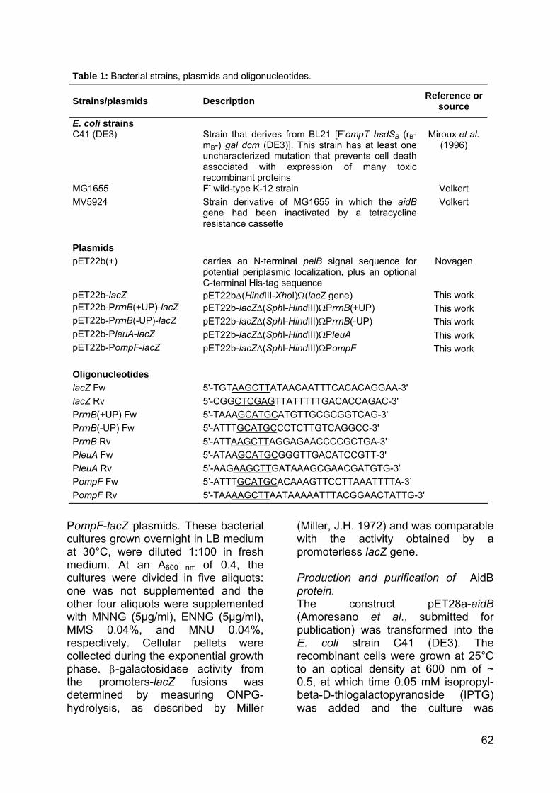
Table 1: Bacterial strains, plasmids and oligonucleotides. Strains/plasmids
Description
Reference orsource
E. coli strains C41 (DE3)
Strain that derives from BL21 [F-ompT hsdSB (rB- mB-) gal dcm (DE3)]. This strain has at least one uncharacterized mutation that prevents cell death associated with expression of many toxic recombinant proteins
Miroux et al.
(1996)
MG1655 F- wild-type K-12 strain Volkert MV5924 Strain derivative of MG1655 in which the aidB
gene had been inactivated by a tetracycline resistance cassette
Volkert
Plasmids pET22b(+) carries an N-terminal pelB signal sequence for
potential periplasmic localization, plus an optional C-terminal His-tag sequence
Novagen
pET22b-lacZ pET22b∆(HindIII-XhoI)Ω(lacZ gene) This work pET22b-PrrnB(+UP)-lacZ pET22b-lacZ∆(SphI-HindIII)ΩPrrnB(+UP) This work pET22b-PrrnB(-UP)-lacZ pET22b-lacZ∆(SphI-HindIII)ΩPrrnB(-UP) This work pET22b-PleuA-lacZ pET22b-lacZ∆(SphI-HindIII)ΩPleuA This work pET22b-PompF-lacZ pET22b-lacZ∆(SphI-HindIII)ΩPompF This work Oligonucleotides lacZ Fw 5'-TGTAAGCTTATAACAATTTCACACAGGAA-3' lacZ Rv 5'-CGGCTCGAGTTATTTTTGACACCAGAC-3' PrrnB(+UP) Fw 5'-TAAAGCATGCATGTTGCGCGGTCAG-3' PrrnB(-UP) Fw 5'-ATTTGCATGCCCTCTTGTCAGGCC-3' PrrnB Rv 5'-ATTAAGCTTAGGAGAACCCCGCTGA-3' PleuA Fw 5'-ATAAGCATGCGGGTTGACATCCGTT-3' PleuA Rv 5’-AAGAAGCTTGATAAAGCGAACGATGTG-3’ PompF Fw 5’-ATTTGCATGCACAAAGTTCCTTAAATTTTA-3’ PompF Rv 5'-TAAAAGCTTAATAAAAATTTACGGAACTATTG-3'
PompF-lacZ plasmids. These bacterial cultures grown overnight in LB medium at 30°C, were diluted 1:100 in fresh medium. At an A600 nm of 0.4, the cultures were divided in five aliquots: one was not supplemented and the other four aliquots were supplemented with MNNG (5µg/ml), ENNG (5µg/ml), MMS 0.04%, and MNU 0.04%, respectively. Cellular pellets were collected during the exponential growth phase. β-galactosidase activity from the promoters-lacZ fusions was determined by measuring ONPG-hydrolysis, as described by Miller
(Miller, J.H. 1972) and was comparable with the activity obtained by a promoterless lacZ gene. Production and purification of AidB protein. The construct pET28a-aidB (Amoresano et al., submitted for publication) was transformed into the E. coli strain C41 (DE3). The recombinant cells were grown at 25°C to an optical density at 600 nm of ~ 0.5, at which time 0.05 mM isopropyl-beta-D-thiogalactopyranoside (IPTG) was added and the culture was
62

allowed to grow until the OD600 reached 3.0. Cells were harvested by centrifugation at 5000 x g for 15 min at 4°C, resuspended in buffer A (50 mM Na2HPO4, pH 7.4, 0.3 M NaCl, 10 mM imidazole, 1 mM PMSF), disrupted by passage through a French Press and centrifuged at 14000 x g for 30 min at 4°C. The recombinant protein was purified by affinity chromatography on His-Select Nickel Affinity Gel (Sigma). The lysate was loaded onto His-Select Nickel Affinity Gel equilibrated with equilibration buffer (50 mM Na2HPO4, pH 7.4, 0.3 M NaCl, 10 mM imidazole). After 1 min of incubation at 4°C, the matrix was collected by centrifugation at 11000 x g for 1 min and washed 3 times with same equilibration buffer. The recombinant protein was eluted with buffer containing 250 mM imidazole in 50 mM Na2HPO4, pH 7.4, 0.3 M NaCl. Protein concentration was estimated with Bradford reagent (Bio-Rad protein assay) and protein content was checked by SDS-polyacrilamide gel electrophoresis (SDS-PAGE). Isolation of plasmid DNA and damage assay. The MG1655 and MV5924 E. coli strains bearing pET22b-lacZ were grown overnight in LB medium at 30°C; these bacterial cultures were then diluted 1:100 in fresh medium. At an A600 nm of 0.4, the cultures were divided in five aliquots: one was not supplemented and the other four aliquots were supplemented with MNNG (5µg/ml), ENNG (5µg/ml), MMS 0.04%, MNU 0.04%, respectively. After addition of alkylating agent, the bacterial cells were allowed to grow for 3 h; the plasmid DNA was isolated and served as a probe for the estimation of alkylated bases. The plasmids were divided into 2 aliquots, one of which was treated with the E. coli AlkA (a kind gift from Patrick J. O’Brien) and endonuclease IV (NEB); the other
aliquot did not receive further treatment (control). Treatment with AlkA was performed in 70 mM MOPS, pH 7.5, 1 mM EDTA, 1mM DTT, 5% glycerol for 30 min at 37°C, followed by treatment with Endo IV for 1 h at 37°C. Then the samples were subjected to electrophoresis in 0.8% agarose gel for ∼1 h at 80 V using 40 mM Tris, pH 7.8, 1 mM EDTA buffer. In vitro DNA binding and damage assays. An in vitro DNA binding assay was conducted incubating pET22b-lacZ plasmid (100 ng) with varying concentrations of AidB in buffer B (10 mM Tris, pH 7.8, 1 mM EDTA, 20% glycerol), for 1 h at 30°C. Mixtures were then analysed by electrophoresis in a 0.8% agarose gel using 40 mM Tris, pH 7.8, 1 mM EDTA buffer. AidB and DNA were mixed at the following ratios (w/w): 3:1, 15:1, 30:1. In DNA damage assay, AidB (3 µg) was incubated with pET22b-lacZ (100 ng) for 1 h at 30°C, either before or after the addition of 300 mM MMS to the reaction mixture. The samples were then treated with AlkA and Endo IV and subjected to electrophoresis in 0.8% agarose gel for ∼1 h at 80 V using 40 mM Tris, pH 7.8, 1 mM EDTA buffer. Determination of DNA damage on lacZ gene. MG1655 and MV5924 E. coli strains were individually transformed with pET22b-lacZ, pET22b-PrrnB(+UP)-lacZ, pET22b-PrrnB(-UP)-lacZ. These bacterial cultures grown overnight in LB medium at 30°C, were diluted 1:100 in fresh medium. At an A600 nm of 0.4, the cultures were supplemented with 0.04% MMS to activate the adaptive response and the bacterial cells were allowed to grow for 3h. Then, the plasmids under study were isolated from these bacterial cells and were
63

digested with HindIII and XhoI to release the lacZ fragment. To estimate the presence of alkyl lesions, the DNA fragments were treated and not with the AlkA and Endo IV proteins. The samples were then subjected to electrophoresis on alkaline agarose gel in 30 mM NaOH, 1 mM EDTA, pH 8 buffer, at 60 V for 3 hours at 25°C. The gel was neutralized by soaking in a solution containing 1.5 M NaCl and 1 M Tris-HCl, pH 7.6 for 1 h. Finally,
the gel was stained in TE buffer (10 mM Tris-HCl, 1 mM EDTA, pH 7.4) containing SYBR® Gold for 30 min at 25°C and the samples were then analysed for single-strand DNA breaks. RESULTS Functional analysis of AidB during transcription. Recently, using the UP element and the -35 sequence from the rrnB P1 promoter as bait and a sequence
C D
MG1655
MV5924
rrnBP1(+UP) rrnBP1(-UP) PleuA PompF rrnBP1(+UP) rrnBP1(-UP) PleuA PompF
5,62,2 2,1
MG1655
MV5924 9,6
A B
5,72,1 2,3
9,4
rrnBP1(+UP) rrnBP1(-UP) PleuA PompF
MG1655
MV5924
rrnBP1(+UP) rrnBP1(-UP) PleuA PompF
MG1655
MV5924
6,5
1,6 1 ,5 3,8
64

lacking the UP element for comparison, we isolated proteins on the basis of their preferential affinity for the UP region. Curiously, AidB was found as one of several proteins that preferentially bind to the UP element containing DNA (Amoresano et al., submitted for publication). In order to determine whether the presence of AidB at the rrnB P1 promoter might be of biological significance, we tested its effect on transcription from this promoter by in vivo transcription assays. As a control, we also investigated the putative AidB activity on other several promoter fragments: the ribosomal promoter rrnB P1 without its UP sequence, the promoter from an unrelated gene which lacks an UP element (PleuA), and the promoter from an unrelated gene which contains an UP region (ompF). The DNA fragments under study were individually fused to a promoterless lacZ gene contained in the reporter plasmid pET22b-lacZ. Both MG1655 (wild type) and MV5924 (∆aidB) E. coli strains were then transformed with the
fusion plasmids and grown in LB medium, either in the absence or in the presence of alkylating agents (MMS, MNU, MNNG, ENNG) and the β-galactosidase activity was monitored during the exponential growth phase. As shown in Fig. 1A, in all cases, wild type and mutant cells not exposed to alkylators showed identical levels of β-galactosidase activity indicating that the presence of AidB has no effect on transcription during normal cell growth. In contrast, upon treatment with alkylating agents, the transcription levels in the ∆aidB strain were lower than that observed for the wild type strain (Fig. 1B-E), suggesting a functional role for AidB on transcription during alkylation stress. Interestingly, the results from β-galactosidase assays showed that AidB strongly affects the activity of promoters containing an UP element: indeed, the mutant cells harbouring rrnB P1 and PompF, upon exposure to N-alkyl-nitrosoguanidines (MNNG, ENNG) (Fig. 1B, 1C), exhibited almost a 10-fold and 6-fold decrease of promoter
individually fused to a promoterless lacZ gene contained in the reporter plasmid pET22b-lacZ. Both MG1655 (wild type) and MV5924 (∆aidB) E. coli strains were then transformed with the
fusion plasmids and grown in LB medium, either in the absence or in the presence of alkylating agents (MMS, MNU, MNNG, ENNG) and the β-galactosidase activity was monitored during the exponential growth phase. As shown in Fig. 1A, in all cases, wild type and mutant cells not exposed to alkylators showed identical levels of β-galactosidase activity indicating that the presence of AidB has no effect on transcription during normal cell growth. In contrast, upon treatment with alkylating agents, the transcription levels in the ∆aidB strain were lower than that observed for the wild type strain (Fig. 1B-E), suggesting a functional role for AidB on transcription during alkylation stress. Interestingly, the results from β-galactosidase assays showed that AidB strongly affects the activity of promoters containing an UP element: indeed, the mutant cells harbouring rrnB P1 and PompF, upon exposure to N-alkyl-nitrosoguanidines (MNNG, ENNG) (Fig. 1B, 1C), exhibited almost a 10-fold and 6-fold decrease of promoter
3,6
Figure 1. In vivo transcription from the promoters-lacZ fusions. The pET22b-PrrnB(+UP)-lacZ, pET22b-PrrnB(-UP)-lacZ, pET22b-PleuA-lacZ and pET22b-PompF-lacZ plasmids were individually introduced into MG1655 (wild type) and MV5924 (∆aidB) E. coli strains and the β-galactosidase specific activity was determined in the absence (A) and in the presence of MNNG (5µg/ml) (B), ENNG (5µg/ml) (C), MMS 0.04% (D), MNU 0.04% (E) as alkylating agents. The activities of promoters are reported in Miller units; the activity obtained by a promoterless lacZ gene was subtracted. Numbers above bars refer to the ratio of the β-galactosidase activity of the promoter measured in the wild type cells to the activity of that same promoter in the aidB mutant strain. Means and standard deviations have been calculated from four independent assays.
MG1655
MV5924
E
rrnBP1(+UP) rrnBP1(-UP) PleuA PompF
6,6
1,7 1,4 3,6
65

activity, respectively. When the promoter regions without UP sequence were analyzed, we find that the mutant cells exhibit transcription levels slightly lower (approximately 2-fold) than that observed for the wild type cells. We also observed that AidB allows more efficient transcription in E. coli cells exposed to MNNG and ENNG (Fig. 1B, 1C) rather than to other alkylators such as MMS or MNU (Fig. 1D, 1E), supporting the hypothesis that AidB might be involved in deactivation of nitrosoguanidines. Taken together, these data strongly demonstrated that AidB affects transcription during alkylation stress and that it has more pronounced effect on the activity of promoters containing an upstream element. These results raise the possibility that AidB may prevent alkyl damage, thereby allowing more efficient transcription.
AidB reduces the level of alkylation damage in DNA. The aim of our study was to determine whether AidB might be able of preventing DNA alkylation damage. This issue was investigated by a plasmid damage assay: the plasmid DNA (pET22b-lacZ) was isolated from wild type and aidB mutant cells grown both in the absence and in the presence of alkylators (MMS, MNU, MNNG, ENNG) and served as a probe for the estimation of alkylated bases on DNA. The plasmids were divided into 2 aliquots, one of which was treated with the E. coli AlkA (a kind gift from Patrick J. O’Brien) and endonuclease IV (Endo IV) proteins; the other aliquot did not receive further treatment and served as a control. AlkA glycosylase recognizes and removes a wide variety of alkylated bases (O'Brien, P. J. and Ellenberger, T., 2004); Endo IV is an
Figure 2. Plamid damage assay. The pET22b-lacZ DNA was isolated from wild type (Lanes 1-2) and ∆aidB (Lanes 3-4) E. coli strains grown in the absence (A) or in the presence of MNNG (5 µg/ml) (B), ENNG (5 µg/ml) (C), MMS 0.04% (D), MNU 0.04% (E), digested (lanes 2, 4) or not (lanes 1, 3) with AlkA and Endo IV and subjected to agarose gel electrophoresis. Lane 5, 1 Kb DNA marker (NEB). OC: open circular; L: linear; SC: supercoiled.
wt wt ∆ ∆- + - +
wt wt ∆ ∆- + - +
wt wt ∆ ∆ - + - +
wt wt ∆ ∆ - + - +
wt wt ∆ ∆ - + - +
66

Figure 3. AidB protects DNA from alkylation damage in vitro. Lane 1-2: pET22b-lacZ plasmid not digested or digested with AlkA and Endo IV, respectively. Lanes 3-4: The pET22b-lacZ plasmid was incubated with AidB for 1 h at 37°C; the complex AidB-DNA was treated with MMS and not digested (Lane 3) or digested (Lane 4) with AlkA and Endo IV. Lanes 5-6: The pET22b-lacZ was treated with MMS; the DNA methylated was not digested or digested with AlkA and Endo IV, respectively. Lanes 7-8: The pET22b-lacZ was treated with MMS; the DNA methylated was incubated with AidB and this sample was not digested or digested with AlkA and Edo IV, respectively. Lane 9, 1 kb DNA marker (NEB).
apurinic/apyrimidinic (AP) endonuclease that converts abasic sites produced by AlkA to nicks (Ljungquist, S. 1977); therefore, the combined action of these two enzymes on a damaged plasmid results in the conversion of the covalently closed circular (supercoiled) form to the open circular form. AlkA treated and untreated plasmids were then subjected to electrophoresis on agarose gels and tested to look for loss of the supercoiled form. As shown in Fig. 2A, alkyl lesions were not detected in plasmids isolated from bacteria grown in LB medium; when plasmid DNA from cells exposed to alkylating agents was analyzed (Fig. 2B-E), we find that treatment with AlkA and Endo IV had no effect on DNA isolated from wild type cells (Lanes 2), but resulted in a complete loss of supercoiled plasmid from the aidB mutant (Lanes 4). These results strongly indicated that the presence of AidB reduces the level of alkylation damage in plasmid DNA. While the damage assay results are consistent with a role for AidB in predicted protection of DNA, its function in repair of alkyl lesions is not ruled out. AidB binds and protects plasmid DNA. In vitro experiments were performed to demonstrate conclusively that AidB binds DNA to protect it from alkylation damage. To this aim, the recombinant AidB protein bearing a His tag was produced, purified by affinity chromatography on Ni2+-agarose beads and used in a plasmid damage assay. First, gel mobility shift assays were performed to determine the amount of AidB required to completely bind the plasmid DNA. The saturation of binding was achieved at a ratio protein:DNA of 30:1 (w/w) (data not shown). Using this ratio, AidB was then incubated with plasmid DNA either before or after the addition of MMS to
the reaction mixture. MMS was used for this assay because it does not require metabolic conversion to alkylate DNA (Adler, I. D. 1980). To detect the presence of alkyl lesions, the purified plasmid DNA was cleaved with AlkA and Endo IV. Figure 3 shows that plasmid remains in the supercoiled form when AidB is added prior to MMS treatment (Lane 4), indicating that AidB protein functions in vitro and that the His-tagged form is fully active. However, when the sequence is reversed and AidB is added after MMS treatment, no effect of AidB presence is detected (Lane 8). An identical experiment using BSA protein served as a control (data not shown). Since the order of addition should not affect an AidB repair function, but protection should require the presence
67

of AidB prior to damaging treatments, we conclude that AidB protein acts by preventing damage from occurring. AidB preferentially protects DNA regions containing an UP element. Since AidB specifically binds to UP sequence containing promoters, and it affects strongly transcription at these promoters during exposure to alkylating agents, we supposed that AidB protection might be preferentially targeted to genes with an UP element. To address this question, we analyzed the effect exhibited by AidB in vivo on a portion of plasmid DNA: the lacZ fragment. In this experiment, we used plasmids carrying a lacZ gene that lacks a promoter, lacZ fused to the rrnB P1 promoter containing its UP element and lacZ fused to rrnB P1 in which UP sequence was deleted. We investigated whether the presence of AidB might affect the content of alkyl lesions within the lacZ sequences. To this aim, the plasmids under study were isolated from wild type and aidB mutant cells treated with MMS and were digested with restriction enzymes to release the lacZ fragment. To estimate the presence of alkyl lesions, the lacZ DNA fragments were gel purified, extracted and treated or not with the AlkA and Endo IV proteins. The samples were then subjected to electrophoresis on alkaline agarose gels to denature DNA and separate nicked from full-length ssDNA fragments. The lacZ integrity was then determined by the relative abundance of full length lacZ restriction fragments and nicked forms. Figure 4 shows that the mutant cells are not able to protect the lacZ gene (Lanes 8, 10, 12), since no full length lacZ fragments were detected. This confirms that the presence of AidB is required for the protection against alkyl damage. Analysis of the DNA isolated from wild type cells revealed several interesting features of AidB specificity. The lacZ
sequence fused to the UP element containing rrnB P1 was fully protected since all DNA exposed to AlkA remains as full length (Fig. 4, comparison between Lanes 3 and 4). When lacZ fused to the UP-less ribosomal promoter was tested, we find that the sample treated with AlkA (Lane 6) shows a decrease in the amount of full length fragment, as compared with the untreated control DNA (Lane 5). Interestingly, the treatment of promoterless lacZ with AlkA caused an almost complete loss of full length fragment (Lane 2). These results demonstrate that AidB preferentially protects the DNA of genes transcribed from an UP element containing promoter. However, it also protects DNA in general, since it does provide some protection to the non-
Figure 4. AidB preferentially protects DNA regions containing an UP element. The pET22b-lacZ, pET22b-PrrnB(+UP)-lacZ and pET22b-PrrnB(-UP)-lacZ plasmids were isolated from wild type (Lanes 1-6) and ∆aidB (Lanes 7-12) E. coli strains grown in the presence of MMS 0.04% and digested to release the lacZ fragment. The digested DNA samples were untreated (Lanes 1,3,5,7,9,11) or treated (Lanes 2,4,6,8,10,12) with AlkA and Endo IV and subjected to electrophoresis on alkaline agarose gels. Lanes 1,2,7,8: lacZ lacking a promoter (-P); Lanes 3,4,9,10: lacZ fused to the rrnB P1 promoter with its UP element (+UP); Lanes 5,6,11,12: lacZ fused to the rrnB P1 promoter without its UP element (-UP); Lane 13: 1Kb DNA marker (NEB).
-P -P +UP+UP-UP-UP -P -P +UP +UP-UP -UP wt wt wt wt wt wt ∆ ∆ ∆ ∆ ∆ ∆ - + - + - + - + - + - +
68

transcribing lacZ restriction fragment and the lacZ gene fused to the rrnB P1 promoter lacking the UP element. Moreover, the result that AidB protects different sequences to varying degrees is consistent with the initial observation (Fig. 2) that AidB protects whole plasmids from alkylation. The observation that the entire lacZ sequence adjacent to UP element containing promoters is protected in a better manner suggests that the promoter may function to nucleate AidB protein binding which may then polymerise along the DNA molecule. The structural studies of Bowles et al., 2008, suggest that a single AidB protein molecule can bind from 25-30 base pairs of dsDNA. Thus to protect the genome, a minimum of 160,000 AidB molecules would be required. Since AidB has a relatively weak promoter, it is unlikely that such a level would ever be achieved. Thus we suspect that the preferential binding to genes such as rrnB, and potentially to other UP element containing DNA sequences such as aidB itself and its regulatory gene ada (Landini, P. et al., 1998), recA, polA and other highly transcribed genes (Estrem, S.T. et al., 1999) may preferentially protect these DNA sequences from alkylation damage. DISCUSSION In a previous study, we found the AidB protein to be part of the protein complex that binds to the -35 and the UP elements of the rrnB P1 promoter, but not to a sequence lacking the UP element. In this work, we investigated whether the presence of AidB at the rrnB P1 promoter might be of biological significance; to this aim, we evaluated its possible role in rRNA expression by β-galactosidase reporter assays. In addition, we also tested the effect of AidB on transcription from other promoters fused to the lacZ reporter
gene. As appropriate controls, the following promoters were chosen: the ribosomal promoter rrnB P1 without its UP sequence, the promoter from an unrelated gene which lacks an UP element (PleuA) and the promoter from an unrelated gene which contains an UP region (PompF). The fusion plasmids were individually introduced into both wild type and ∆aidB E. coli strains which were then treated and not with alkylating agents. Upon alkylation stress, a decrease in the lacZ expression was observed in the E. coli cells lacking AidB as compared to the wild type control. Interestingly, AidB was found to have a more pronounced effect on the activity of promoters containing an upstream element. Our results clearly demonstrated that a functional aidB allele is necessary for efficient transcription during exposure to alkylating agents. These data lead us to a line of inquiry concerning the possible role of AidB in the transcription initiation. Its function might be related to repair of alkylated DNA given that, during transcription, the transcriptional fork generates single stranded DNA molecules that have been demonstrated to undergo alkylation processes much quicker than double stranded molecules. Another possibility is that AidB might prevent alkyl damage by protecting DNA, thereby allowing more efficient transcription. In this respect, a recent report on the three dimensional structure of AidB bound to double strand DNA provided a rationale for its possible role in damage prevention (Bowles, T. et al., 2008). The goal of our study was thus to determine the mechanism by which AidB protects against alkylating agents. First, through a plasmid damage assay we demonstrated that AidB in vivo reduces the level of alkylation damage in DNA: indeed,
69

upon exposure to alkylators, the DNA plasmid isolated from wild type cells was shown to be undamaged while alkyl lesions were detected in plasmid DNA extracted from mutant cells. While these results are consistent with a role for AidB in predicted protection of DNA, we could not rule out its possible function in repair of alkyl lesions. To unravel this issue, in vitro studies were performed incubating AidB with plasmid DNA either before or after the addition of MMS; an effect of AidB presence was detected only in the former case. Therefore, as predicted by structural analysis, we have demonstrated conclusively that AidB binds DNA to protect it from alkylation damage rather than repair it. A similar role has been observed for the Dps protein, which protects DNA in starved E. coli against oxidative damage (Almiron, M. et al., 1992; Martinez, A. and Kolter, R. 1997). Both AidB and Dps are up-regulated during stationary phase and are rpoS-dependent (Volkert, M.R. et al., 1994; Altuvia, S. et al., 1994). Interestingly, endogenous methylating agents such as nitrosamines are formed as by-products of stationary-phase metabolism (Taverna, P. and Sedgwick, B, 1996), leading to an accumulation of alkyl lesions on DNA. Induction of AidB expression, therefore, could serve to prevent endogenous stationary-phase alkylation damage in a manner similar to Dps protection of oxidative damage. Finally, since AidB was shown to specifically bind to UP sequence containing promoters and to affect strongly transcription at these promoters upon exposure to alkylating agents, we asked if AidB protection might be preferentially targeted to genes with an UP element. To address this question, we investigated whether the presence of AidB might affect the content of alkyl lesions within lacZ
sequences fused to the rrnB P1 promoter containing and not its UP element and judged by comparison with the lacZ fragment lacking a promoter. Our experiments demonstrated that AidB preferentially protects DNA sequences transcribed from promoters carrying an UP element. In conclusion, in this work we characterized the role of AidB in the E. coli adaptive response to alkylating agents: AidB functions to protect DNA from alkylation damage, by binding it and, presumably, by inactivating alkylators before they are able to react with their DNA target. In addition, we demonstrated that AidB preferentially protects DNA sequences adjacent to UP element-containing promoters, which include many of the housekeeping genes required for basic metabolic processes, such as the sequences encoding the ribosomal rRNA or the DNA polymerase I that is involved in DNA replication and repair. Further experiments are required to address the question whether the preferential protection exhibited by AidB is limited to transcriptionally active DNA. It would also be useful to examine if a similar protective mechanism is conserved and active in other organisms. ACKNOWLEDGEMENTS This work was supported by grants of Ministero dell’Università e della Ricerca Scientifica (Progetti di Rilevante Interesse Nazionale 2005, 2006), of Programma Nazionale di Ricerche in Antartide 2004, and of Regione Campania L.R. 05/03. Support from the National Center of Excellence in Molecular Medicine (MIUR - Rome) and from the Regional Center of Competence (CRdC ATIBB, Regione Campania – Naples), Istituto Nazionale Biomolecole e Biosistemi (INBB) is gratefully acknowledged.
70
REFERENCES • Adler, I. D. Mutation research
74(2), 77–93 (1980). • Almirón M., Link, A. J., Furlong, D.,
and Kolter, R. Genes & Development 6(12b), 2646–2654 (1992).
• Altuvia, S., Almirón, M., Huisman, G., Kolter, R., and Storz, G. Molecular microbiology 13(2), 265–272 (1994).
• Bouziane, M., Miao, F., Ye, N., Holmquist, G., Chyzak, G., and O’Connor, T. R. Acta biochimica Polonica 45(1), 191–202 (1998).
• Bowles, T., Metz, A. H., O’Quin, J., Wawrzak, Z., and Eichman, B. F. Proceedings of the National Academy of Sciences of the United States of America 105(40), 15299–15304 (2008).
• Kataoka, H., Yamamoto, Y., and Sekiguchi, M. J. Bacteriol. 153(3), 1301–1307 (1983).
• Landini, P., Hajec, L. I., and Volkert, M. R. Journal of bacteriology 176(21), 6583–6589 (1994).
• Landini, P. and Volkert, M. R. The Journal of biological chemistry 270(14), 8285–8289 (1995).
• Landini, P., Bown, J. A., Volkert, M. R., and Busby, S. J. The Journal of biological chemistry 273(21), 13307–13312 (1998).
• Ljungquist, S. Journal of Biological Chemistry 252(9), 2808–2814 (1977).
• Martinez, A. and Kolter, R. J. Bacteriol. 179(16), 5188–5194 (1997).
• Miller, J. H. Experiments in Molecular Genetics. Cold Spring Harbor Laboratory Pr.
• Nakabeppu, Y., Kondo, H., and Sekiguchi, M. Journal of Biological Chemistry 259(22), 13723–13729 (1984).
• O’Brien, P. J. and Ellenberger, T. Journal of Biological Chemistry 279(26), 26876–26884 (2004).
• Rohankhedkar, M. S., Mulrooney, S. B., Wedemeyer, W. J., and Hausinger, R. P. Journal of
bacteriology 188(1), 223–230 (2006).
• Taverna, P. and Sedgwick, B. Journal of bacteriology 178(17), 5105–5111 (1996).
• Teo, I., Sedgwick, B., Kilpatrick, M. W., McCarthy, T. V., and Lindahl, T. Cell 45(2), 315–324 (1986).
• Trewick, S. C., Henshaw, T. F., Hausinger, R. P., Lindahl, T., and Sedgwick, B. Nature 419(6903), 174–178 (2002).
• Volkert, M. R. and Nguyen, D. C. Proceedings of the National Academy of Sciences of the United States of America 81(13), 4110–4114 (1984).
• Volkert, M. R., Hajec, L. I., Matijasevic, Z., Fang, F. C., and Prince, R. J. Bacteriol. 176(24), 7638–7645 (1994).
71

Potential role for the Escherichia coli AidB and the Pseudomonas putida PP4780 as detoxification enzymes. INTRODUCTION Alkylating agents comprise a broad class of highly reactive chemical compounds that introduce alkyl groups into biologically active molecules and prevent normal functioning. These compounds mostly react with DNA molecule to produce cytotoxic and mutagenic lesions. Alkylation damage to DNA therefore poses a severe threat to the stability of the genome and, in mammals, can lead to genetic diseases and cancer. Living organisms are continuously exposed to alkylating agents that are among the most abundant environmental pollutants influencing health of the persons exposed: these molecules that include nitrosamines and nitrosoureas, are released into the ecosystem at high concentrations, as a consequence of human activities (Vaughan, P., et al., 1991; Taverna, P. and Sedgwick, B. 1996). Since alkylating molecules are ubiquitous and hence unavoidable, all organisms (eubacteria, archaebacteria, and eukaryotes) have evolved multiple DNA repair strategies as a safeguard against alkylation damage. In addition, many bacteria, to defend against fluctuating levels of environmental alkylators, mount an inducible response that enhances cellular resistance to alkylation damage and is known as the adaptive response. In Escherichia coli, exposure to sublethal doses of alkylating agents such as methyl methanesulfonate (MMS), N-methylnitrosourea (MNU), N-methyl-N′-nitro-N-nitrosoguanidine (MNNG), and N-ethyl-N′-nitro-N-nitrosoguanidine (ENNG) increases the expression of aidB along with three genes (ada, alkA, alkB) that encode DNA repair proteins (Kataoka, H. et al., 1983; Nakabeppu, Y. et al., 1984; Volkert, M.R. and Nguyen, D.C. 1984). The role of AidB in the response to alkylating agents was elusive for many years. AidB was shown to be a flavoprotein that has weak isovaleryl-CoA dehydrogenase activity and that binds to double strand DNA (Landini, P. et al., 1994; Rohankhedkar, M.S. et al., 2006). Our recent work demonstrated that AidB prevents alkylation damage by protecting DNA and, presumably, by inactivating alkylators before they are able to react with their target. Interestingly, a recent report on the three dimensional structure of AidB bound to dsDNA (Bowles, T. et al., 2008) supported this model, revealing that the protein is well equipped to sterically occlude DNA from attack by damaging agents. Importantly, the unique chemical environment of FAD active site provided a rationale for a possible role of AidB in deactivation of nitrosoguanidines or their reactive intermediates. In this respect, AidB’s FAD cavity was shown to be rich in thiol and methylsulfide groups and since MNNG, unlike MMS or MNU, is activated by thiols to produce highly reactive methylation agents (e.g., methyldiazonium ion) (Lawley, P. D. 1974), it is intriguing to speculate that flavin binding channel may act as a sink for reactive MNNG derivatives. Coupled with structural analysis, our recent results help to support the hypothesis that AidB may act as a detoxification enzyme: indeed, we demonstrated that AidB allows more efficient transcription during stress alkylation and that it has a more pronounced effect in E. coli cells exposed to nitrosoguanidines (MNNG, ENNG) rather than to other alkylators such as MMS or MNU. Taking into account these observations, AidB represents a promising tool for the treatment of sites contaminated by alkylating agents. Since, ideally, the design of successful strategies for the bioremediation would require the knowledge of the microorganisms present in the polluted environments, their metabolic abilities, and how they respond to changes in environmental conditions, the challenge of this experimental work was to identify E. coli AidB homologues in bacteria used for bioremediation applications
72

and to investigate the possible involvement of these gene products in the protection against alkylating agents. Particularly, the our attention has been focused on the search for AidB homologues in pseudomonads that are among extensively studied workhorses of environmental bioremediation owing to their ability to degrade numerous different contaminants (Wackett, L.P. 2003). The huge potential of the pseudomonads does not solely depend on a high proportion of genes responsible for the metabolism, transport and efflux of organic compounds, but also on broad capability of metabolic regulation (Nelson, K.E. et al ., 2002): indeed, the control of gene expression is the key determinant of their flexibility and, in this respect, a variety of highly integrated regulatory mechanisms have been identified. Hence, on the basis of the potential degradative capability offered by AidB and the great versatility of Pseudomonas species, the aim of this work was to identify new potential candidates for the bio-treatment of wastes and environments contaminated by alkylating compounds. MATERIALS AND METHODS Bacterial strains and plasmids. The bacterial strains, the plasmids and the oligonucleotides used in this work are listed in Table 1. MG1655 and MV5924 E. coli strains were a kind gift from M. Volkert (University of Massachusetts, Worcester, MA). Media and chemicals. Luria-Bertani or nutrient broths (for bacterial cultures and plating) and suspension medium (for bacterial dilutions) were used as described by Miller (Miller, J.H. 1972). Kanamycin and tetracycline (Sigma) were used at 50 and 5 µg/ml, respectively. Construction of the expression plasmids. The PP4780 gene was amplified from DNA genomic of P. putida KT2440 by polymerase chain reaction (PCR) using the forward and reverse primers listed in Table 1. The amplified fragment was digested with the restriction enzymes underlined in Table 1 and cloned into the expression vector pET28a (Novagen) which was linearized with the same restriction enzymes. The resulting plasmid, pET28a-PP4780, was verified by automated DNA sequencing. For the complementation experiments, the PP4780 gene was positioned downstream of the lac promoter into the expression vector pET28a-Plac, generating pET28a-Plac-PP4780. Production and purification of recombinant proteins. The constructs pET28a-aidB (Amoresano et al., submitted for publication) and pET28a-PP4780 (Table 1) were individually transformed into the E. coli strain C41 (DE3). The recombinant cells were grown at 25°C to an optical density at 600 nm of ~0.5, at which time 0.05 mM isopropyl-beta-D-thiogalactopyranoside (IPTG) was added and the cultures were allowed to grow until the OD600 reached 3.0. Cells were harvested by centrifugation at 5000 x g for 15 min at 4°C, resuspended in buffer A (50 mM Na2HPO4, pH 7.4, 0.3 M NaCl, 10 mM imidazole, 1 mM PMSF), disrupted by passage through a French Press and centrifuged at 14000 x g for 30 min at 4°C. The recombinant proteins were purified by affinity chromatography on His-Select Nickel Affinity Gel (Sigma). The lysate was loaded onto His-Select Nickel Affinity Gel equilibrated with equilibration buffer (50 mM Na2HPO4, pH 7.4, 0.3 M NaCl, 10 mM imidazole). After 1 min of incubation at 4°C, the matrix was collected by centrifugation at 11000 x g for 1 min and washed 3 times with same equilibration
73

Table 1: Bacterial strains, plasmids and oligonucleotides. Strains/plasmids
Description
Reference orsource
E. coli strains C41 (DE3)
Strain that derives from BL21 [F-ompT hsdSB (rB- mB-) gal dcm (DE3)]. This strain has at least one uncharacterized mutation that prevents cell death associated with expression of many toxic recombinant proteins
Miroux et al.
(1996)
MG1655 F- wild-type K-12 strain Volkert MV5924 Strain derivative of MG1655 in which the aidB gene
had been inactivated by a tetracycline resistance cassette
Volkert
Plasmids pET28a(+) Plasmid carrying an N-terminal
His•Tag®/thrombin/T7•Tag® configuration plus an optional C-terminal His•Tag sequence.
Novagen
pET28a-Plac-aidB pET28a-Plac∆(BamHI-HindIII)Ω(aidB gene) Previous workpET28a-Plac-PP4780 pET28a-Plac∆(BamHI-HindIII)Ω(PP4780 gene) This work pET28a-aidB pET28a∆(BamHI-HindIII)Ω(aidB gene)
Amoresano et al., 2009
pET28a-PP4780 pET28a(BamHI-XhoI)Ω(PP4780 gene) This work Oligonucleotides PP4780 Fw 5'-ATAGGATCCATGAGCCTGCACCAG-3' PP4780 Rv 5'-TTACTCGAGCAACAGGGGCCAGG-3' PP4780-compl Rv 5'-TTAAAGCTTCAACAGGGGCCAGG-3'
buffer. The recombinant proteins were eluted with buffer containing 250 mM imidazole in 50 mM Na2HPO4, pH 7.4, 0.3 M NaCl. Protein concentration was estimated with Bradford reagent (Bio-Rad protein assay) and protein content was checked by SDS-polyacrilamide gel electrophoresis (SDS-PAGE). Growth profiles. MV1161 (wild type) and MV5924 (∆aidB) E. coli strains were not transformed or transformed, individually, with pET28a-Plac-PP4780 and pET28a-Plac-aidB. These bacterial cultures grown overnight in LB medium at 37°C, were diluted 1:100 in fresh medium. At an A600 nm of 0.2, the cultures were divided in four aliquots supplemented with MMS 0.04%, MNU 0.04%, MNNG (5µg/ml) and ENNG (5µg/ml), respectively. After addition of alkylating agent, the bacterial cells were allowed to grow overnight. For all experiments, each growth profile was reproduced three times. Isovaleryl-CoA dehydrogenase activity assay.Isovaleryl-CoA dehydrogenase activity assays were carried out at room temperature in 200 mM phosphate buffer, pH 8.0, and using purified recombinant proteins that had been dialyzed to remove imidazole. For routine assays, 2 mM isovaleryl-CoA (Sigma) was used as the substrate and 0.1 mM 2,6-dichlorophenolindophenol (DCPIP) was used as the terminal electron acceptor in a final volume of 300 µl. The change in absorbance at 600 nm was monitored by using a Beckman DU 7500 spectrophotometer, and the enzyme activity was calculated by assuming an extinction coefficient of 20.6 mM−1 cm−1 for DCPIP.
74

RESULTS AidB increases cell survival after exposure to nitrosoguanidines. Inducible resistance of E. coli to the cytotoxic and mutagenic effects of alkylating agents involves the increased expression of the ada-alkB operon, alkA and aidB genes (Lindahl, T. et al., 1988). The DNA repair mechanisms of Ada, AlkA, and AlkB have been structurally and functionally characterized (Landini, P. and Volkert, M.R. 1995; Nakabeppu, Y. et al., 1984; Trewick, S. C. et al., 2002); recently, we demonstrated that AidB prevents alkylation damage rather than repair it. In order, to completely define the role of AidB in preventing alkylators-induced toxicity, the effect of its deletion on sensitivity to different alkylating compounds was tested. Specifically, we investigated the AidB response to four different mutagens; these include alkyl sulfonates (MMS), nitrosoureas (MNU), and nitrosoguanidines (MNNG or ENNG). To this aim, both MG1655 (wild type) and MV5924 (∆aidB) E. coli strains were grown in LB medium, in the presence of sub-inhibitory concentrations of the mutagens described above and their growth behaviour was analyzed and compared. As shown in Figs. 1A and 1B, the aidB mutant cells don’t display any change in sensitivity to MMS or MNU as compared with the wild type cells; in contrast, the inactivation of aidB gene was shown to decrease resistance to lethal and mutagenic effects of MNNG and ENNG (Figs. 1C and 1D). Interestingly, the complementation of the ∆aidB mutation by pET28a-Plac-aidB plasmid restored the cellular resistance to nitrosoguanidines; in fact, the complementated strain was shown to have a growth profile identical to wild type cells (Figs. 1C and 1D). Taken together, these results suggested that AidB plays a critical role in preventing toxicity induced by MNNG and ENNG. Since sensitivity to only nitrosoguanidines was affected in MV5924 strain, we hypothesized that AidB may be involved, by a dehydrogenase mechanism, in a detoxification pathway of this class of alkylators that constitute one of the most important environmental contaminants influencing health of the persons exposed. This result is consistent with previous structural studies performed by Bowles et al., 2008. Hence, on the basis of these observations, AidB was shown to represent a promising tool for the bio-treatment of sites contaminated by alkylating agents. AidB homologues in Pseudomonas species. Since, ideally, developing successful strategies for the bioremediation requires the knowledge of the microorganisms present in the polluted environments, their metabolic abilities, and how they respond to changes in environmental conditions, this experimental work was targeted at identify as well as at characterize E. coli AidB homologues in bacteria used for bioremediation applications. To this aim, the deduced amino sequence of the 60.5 kDa AidB was submitted to Swiss Protein and GenBank-EMBL databases. Interestingly, we found that AidB homologues are not present in many bacteria closely related to E. coli (Klebsiella, Vibrio, Shewanella, and Photorhabdus), but the closest hits are from some γ-proteobacteria (Pseudomonas, Azotobacter, and Acinetobacter), β-proteobacteria (Burkholderia, Ralstonia, Bordetella), and even gram-positive bacteria (Mycobacterium, Nocardia). This observation was intriguing because Pseudomonas species and closely related organisms are the most extensively studied and the most frequently used for bioremediation applications (Wackett, L.P. 2003), owing to their ability to degrade many several organic pollutants. Particularly, the homology search revealed that AidB shows 46% identity with the putative acyl-CoA dehydrogenase coded by the PP4780 gene from Pseudomonas putida KT2440.
75

0,010,1110
05
1015
2025
time
(h)
log OD 600 nm
MG1
655
com
plem
enta
ted
with
AidB
MV5
924
Figu
re 1
: G
row
th p
rofil
es o
f M
G16
55 (
wild
typ
e) a
nd M
V59
24 (∆a
idB
) E
. co
li st
rain
s gr
own
in t
he p
rese
nce
of M
MS
0.0
4% (
A),
MN
U 0
.04%
(B),
MN
NG
5 µ
g/m
l (C
) and
EN
NG
5 µ
g/m
l (D
), re
spec
tivel
y. In
Fig
. 1C
and
1D
, it i
s al
so re
porte
d th
e gr
owth
cur
ve o
f M
V59
24tra
nsfo
rmed
with
pET2
8a-P
lac-
aidB
plas
mid
with
the
aim
tocr
eate
anai
dB-p
rofic
ient
back
grou
nd.
0,010,1110
05
1015
2025
time
(h)
log OD 600 nm
MG
1655
MV
5924
B
D
0,010,1110
05
1015
2025
time
(h)
log OD 600 nm
MG
1655
com
plem
enta
ted
with
Aid
BM
V59
24
0,010,1110
05
1015
2025
time
(h)
log OD 600 nm
MG
1655
MV
5924
A
C
76

Interestingly, the conservation of Glu425, that corresponds to the catalytic residue in most of the short- and medium- chain acyl-CoA dehydrogenases, suggested that a dehydrogenase or oxidase activity is essential to the physiological function of the P. putida protein. Strain KT2440, whose genome has recently been sequenced (Nelson, K.E. et al., 2002), is one of the best characterized pseudomonads; its broad metabolic versatility, genetic plasticity, and ability to colonize many different environments such as bulk soil and the rhizosphere make this bacterium an ideal candidate for bioremediation applications (Clarke, P. 1982; Molina, L. et al., 1998; Espinosa-Urgel, M. et al., 2002; Timmis, K. N. 2002; Jiménez, J. I. et al., 2002; Canovas, D. et al., 2003; Santos, P. M. et al., 2004; Ramos-Gonzalez, M. I. et al., 2005; Kurbatov, L. et al., 2006; Nogales, J. et al., 2008). P. putida was shown to adapt to a variety of stress conditions and interestingly, the changes that may occur in soil include the fluctuation in levels of alkylating agents. This bacterium has also been certified as a biosafety system, which means that it can be used as a host for containment systems (Molin, S. et al., 1993) both for applications in biotechnological production and release into the environment. On the basis of these observations, it was intriguing to investigate the possible involvement of the acyl-CoA dehydrogenase from P. putida in the response to alkylation stress. Cloning and over-expression of the PP4780 gene in E. coli. To characterize the putative acyl-CoA dehydrogenase from P. putida, the first step of our work was to clone the PP4780 gene into a commercial expression vector of the pET series and to over-express the recombinant product in E. coli cells; the protein bearing a 6 His tag at the C-terminus was purified by affinity chromatography on Ni2+-agarose beads and its homogeneity was tested by SDS-PAGE and mass fingerprinting analyses. Successively, a preliminary characterization of the purified protein was performed examining its potential enzymatic properties. To this aim, the isovaleryl-CoA dehydrogenase (IVD) activity of the P. putida protein was examined and compared with that of EcAidB. The product coded by the PP4780 gene displayed low levels of isovaleryl-CoA catalytic activity that are identical to that exhibited by AidB protein (Table 2). It is intriguing to speculate that, such for AidB, fatty acyl-coA are not substrates for the P. putida enzyme but that its active site may act as a sink for certain alkylating compounds or their reactive intermediates.
Table 2: Isovaleryl-CoA dehydrogenase activity of the P. putida protein coded by the PP4780 gene and the E. coli AidB was assayed as described in Materials and Methods following the isovaleryl following the isovaleryl-CoA-dependent reduction of the electron acceptor DCPIP. The data shown are averages of 10 measurements.
Protein Isovaleryl-CoA dehydrogenase activity µmol min-1 [mg protein]-1
AidB 0.12 ± 0 PP4780 0.13 ± 0
77

PP4780 protects the bacterial cell from alkylation damage. The putative involvement of PP4780 in cellular protection against alkylating agents was investigated by transforming MV5924 (∆aidB) E. coli strain with a plasmid carrying the PP4780 gene and by analyzing the effect of this gene product on sensitivity of aidB mutant cells to nitrosoguanidines. To this aim, the strain complementated was exposed to sub-inhibitory concentrations of MNNG or ENNG and its growth profile was compared with that determined for the wild type MG1655 and ∆aidB MV5924 strains treated with nitrosoguanidines. As shown in Figs. 2A and 2B, the PP4780 gene suppressed the increased nitrosoguanidines-sensitivity caused by aidB mutation; in fact the complementated strain was shown to have a growth profile identical to wild type cells. A
0,01
0,1
1
10
0 5 10 15 20 25time (h)
log
OD
600
nm
MG1655 complementated with PP4780 MV5924
B
0,01
0,1
1
10
0 5 10 15 20 25time (h)
log
OD
600
nm
MG1655 complementated with PP4780 MV5924
Figure 2: Growth profiles of MG1655 (wild type), MV5924 (∆aidB) and MV5924 transformed with pET28a-Plac-PP4780 plasmid, grown in the presence of MNNG 5 µg/ml (A) and ENNG 5 µg/ml (B), respectively.
This finding clearly demonstrated that PP4780 expression produced a functional protein that is involved in the resistance against alkylation damage, presumably by acting as a detoxification enzyme which inactivates nitrosoguanidines.
78

In order, to further define the P. putida protein role in preventing toxicity after nitrosoguanidines challenge, we also tested whether PP4780 over-expression might confer increased resistance to E. coli cells treated with both MNNG and ENNG. Interestingly, recombinant cells that over-express PP4780 exhibited to be more resistant to nitrosoguanidines as compared with wild type and aidB-overexpressing cells (Figs. 3A and 3B).
Taken together these results, we clearly demonstrated that the P. putida protein plays a critical role in defending the bacterial cell against alkylation stress; the putative involvement of this protein in detoxification of nitrosoguanidines was considered as a possible explanation for the phenotypes observed. Clearly, further studies will be required to elucidate whether E. coli AidB and P. putida protein effectively act as detoxification enzymes to destroy nitrosoguanidines that are recognised as one of the most abundant environmental pollutants. However, the data obtained in this research project strongly support the possibility of developing new successful strategies for the bioremediation of environments and industrial effluents contaminated by alkylating compounds.
A
0,01
0,1
1
10
0 5 10 15 20 25
time (h)
log
OD
600
nm
MG1655 MG1655+ AidB MG1555 + PP4780
B
0,01
0,1
1
10
0 5 10 15 20 25
time (h)
log
OD
600
nm
MG1655 MG1655+ AidB MG1555 + PP4780
Figure 3: Growth profiles of MG1655 (wild type), MG1655 over-expressing AidB and MG1655 over-expressing PP4780, grown in the presence of MNNG 5 µg/ml (A) and ENNG 5 µg/ml (B), respectively.
79

REFERENCES • Bowles, T., Metz, A. H., O’Quin, J., Wawrzak, Z., and Eichman, B. F. Proceedings of
the National Academy of Sciences 105(40), 15299–15304 (2008). • Cánovas, D., Cases, I., and de Lorenzo, V. Environmental microbiology 5(12), 1242–
1256 (2003). • Clarke, P. Antonie van Leeuwenhoek 48(2), 105–130 (1982). • Espinosa-Urgel, M., Kolter, R., and Ramos, J. L. Microbiology (Reading, England)
148(Pt 2), 341–343 (2002). • Jiménez, J. I., Miñambres, B., García, J. L., and Díaz, E. Environmental microbiology
4(12), 824–841 (2002). • Kataoka, H., Yamamoto, Y., and Sekiguchi, M. J. Bacteriol. 153(3), 1301–1307
(1983). • Kurbatov, L., Albrecht, D., Herrmann, H., and Petruschka, L. Environmental
Microbiology 8(3), 466–478 (2006). • Landini, P. and Volkert, M. R. The Journal of biological chemistry 270(14), 8285–
8289 (1995). • Landini, P., Hajec, L. I., and Volkert, M. R. Journal of bacteriology 176(21), 6583–
6589 (1994). • Lawley, P. D. Mutation research 23(3), 283–295 (1974). • Lindahl, T., Sedgwick, B., Sekiguchi, M., and Nakabeppu, Y. Annual review of
biochemistry 57, 133–157 (1988). • Miller, J. H. Experiments in Molecular Genetics. Cold Spring Harbor Laboratory Pr. • Molin, S., Boe, L., Jensen, L. B., Kristensen, C. S., Givskov, M., Ramos, J. L., and
Bej, A. K. Annual review of microbiology 47, 139–166 (1993). • Molina, L., Ramos, C., Ronchel, M.-C., Molin, S., and Ramos, J. L. Appl. Environ.
Microbiol. 64(6), 2072–2078 (1998). • Nakabeppu, Y., Kondo, H., and Sekiguchi, M. Journal of Biological Chemistry
259(22), 13723–13729 (1984). • Nelson, K. E., Weinel, C., Paulsen, I. T., Dodson, R. J., Hilbert, H., dos Santos, M.,
Fouts, D. E., Gill, S. R., Pop, M., Holmes, M., Brinkac, L., Beanan, M., DeBoy, R. T., Daugherty, S., Kolonay, J., Madupu, R., Nelson, W., White, O., Peterson, J., Khouri, H., Hance, I., Lee, P. C., Holtzapple, E., Scanlan, D., Tran, K., Moazzez, A., Utterback, T., Rizzo, M., Lee, K., Kosack, D., Moestl, D., Wedler, H., Lauber, J., Stjepandic, D., Hoheisel, J., Straetz, M., Heim, S., Kiewitz, C., Eisen, J., Timmis, K. N., Düsterhöft, A., Tümmler, B., and Fraser, C. M. Environmental Microbiology 4(12), 799–808 (2002).
• Nogales, J., Palsson, B., and Thiele, I. BMC Systems Biology 2(1), 79+ (2008). • Ramos-Gonzalez, M. I., Campos, M. J., and Ramos, J. L. J. Bacteriol. 187(12), 4033–
4041 (2005). • Rohankhedkar, M. S., Mulrooney, S. B., Wedemeyer, W. J., and Hausinger, R. P.
Journal of bacteriology 188(1), 223–230 (2006). • Santos, P. M., Benndorf, D., and Sá-Correia, I. Proteomics 4(9), 2640–2652 (2004). • Taverna, P. and Sedgwick, B. Journal of bacteriology 178(17), 5105–5111 (1996). • Timmis, K. N. Environmental microbiology 4(12), 779–781 (2002). • Trewick, S. C., Henshaw, T. F., Hausinger, R. P., Lindahl, T., and Sedgwick, B.
Nature 419(6903), 174–178 (2002). • Vaughan, P., Sedgwick, B., Hall, J., Gannon, J., and Lindahl, T. Carcinogenesis
12(2), 263–268 (1991). • Volkert, M. R. and Nguyen, D. C. Proceedings of the National Academy of Sciences
of the United States of America 81(13), 4110–4114 (1984). • Wackett, L. P. Nat Biotech 21(2), 136–138 (2003).
80


Conclusions
81


CONCLUSIONS The strategy of using and manipulating detoxification abilities of microorganisms to reduce environmental contamination, such as in soils and waste waters, appears to be feasible alternative to physicochemical methods. This work has been aimed at exploring new potential candidates for the bio-treatment of wastes and environments contaminated by alkylating compounds that are among abundant pollutants present in the ecosystem. The study has been specifically focused on the AidB protein, a component of the adaptive response to alkylating agents in bacterium Escherichia coli. Initially, the role of AidB in the bacterial cell was investigated through structural and functional characterization of its protein domains. Successively, the mechanism by which AidB directly protects against alkylation stress has been determined: AidB prevents DNA damage, presumably by inactivating certain alkylators before they are able to react with their DNA target. Finally, taking into account its potential role in the detoxification of alkyl-nitrosoguanidines, this experimental work was targeted at identify as well as at characterize E. coli AidB homologues in bacteria used for bioremediation applications. Specifically, the study has been focused on the acyl-CoA dehydrogenase coded by the PP4780 gene from Pseudomonas putida KT2440. The involvement of this protein in preventing toxicity induced by nitrosoguanidines has been demonstrated: the complementation of the aidB mutation by PP4780 gene was shown to decrease the cellular sensitivity to nitrosoguanidines and the over-expression of PP4780 was shown to confer increased resistance to lethal and mutagenic effects of these damaging agents. The potential role for the P. putida protein in detoxification of nitrosoguanidines was considered as a possible explanation for the phenotypes observed. Clearly, characterizing the degradative properties of the acyl-CoA dehydrogenase from E. coli and P. putida KT2440 will be required to allow their potential application in bioremediation field. Besides, further work should be done aimed at exploring the entire metabolic pathways necessary for the complete degradation of alkylating compounds. In light of these observations, the current research should be focused on both identification of enzymatic activities involved in the catabolic pathway of alkylators degradation, as well as on testing their catalytic performances.
82

PUBLICATIONS • Papa R, Rippa V, Sannia G, Marino G, Duilio A (2007) An effective cold inducible
expression system developed in Pseudoalteromonas haloplanktis TAC125. J Biotechnol 127, 199-210.
• Papa R, Rippa V, Duilio A (2009) Identification of the transcription factor responsible for L-malate-dependent regulation in the marine Antarctic bacterium Pseudoalteromonas haloplanktis TAC125. FEMS Microbiol Lett 295, 177-186.
• Amoresano A, Cirulli C, Rippa V, Esposito C, Papa R, Duilio A (2009) Additional protein functions involved in the biogenesis of ribosomes and the DNA repair mechanisms are associated with the transcriptional machinery gathered at the Escherichia coli rrnB P1 promoter. Mol Microbiol-submitted.
• Rippa V, Cirulli C, Di Palo B, Doti N, Amoresano A, Duilio A (2009) The ribosomal protein L2 interacts with the RNA polymerase α subunit and acts as a transcription modulator in Escherichia coli. J Bacteriol-submitted.
COMMUNICATIONS • Cirulli C, Amoresano A, Rippa V, Esposito C, Doti N, Papa R, Duilio A. A proteomic
approach in the investigation of transcriptional regulatory network in E. coli. HIUPO 2006, Pisa, Italy.
• Rippa V, Amoresano A, Cirulli C, Esposito C, Duilio A. Investigation on transcriptional regulatory network in Escherichia coli: role of the AidB protein. Congresso Prokaryotes 2007, Cortona, Italy.
• Amoresano A, Cirulli C, Rippa V, Papa R, Esposito C, Duilio A. The transcriptional machinery gathered at the E. coli rrnB P1 promoter includes proteins involved in the biogenesis of ribosome and the DNA repair mechanisms. ItPA 2007, Catania, Italy.
• Rippa V, Esposito C, Cirulli C, Duilio A. Regolazione della trascrizione in Escherichia coli: ruolo della proteina AidB. Giornate scientifiche interpolo 2007, Napoli, Italy.
• Rippa V, Amoresano A, Duilio A. Transcriptional role of E. coli AidB protein. Congresso Prokaryotes 2008, Cortona, Italy.
• Di Palo B, Rippa V, Esposito C, Amoresano A, Duilio A. Transcriptional role of ribosomal protein L2 in Escherichia coli. Congresso Prokaryotes 2008, Cortona, Italy.
• Rippa V, Di Procolo P, Esposito C, Duilio A. Characterisation of transcriptional factor AidB in Escherichia coli. Giornate scientifiche interpolo 2008, Napoli, Italy.
INTERNATIONAL COURSES • OSSIBE 4, the 4th Oulu Summer School in Bioprocess Engineering “Protein
production- what can go wrong and haw can you improve it fast?" Oulu, Finland, June 11-15th 2007.
RESEARCH ACTIVITY IN FOREIGN LABORATORIES • From July 15th to October 31th 2009, my research activity was performed in the
Laboratory of Prof. Michael Volkert, Department of Molecular Genetics and Microbiology, University of Massachusetts Medical School, Worcester (Massachusetts).
83

Other publications
84


Manuscript submitted to Journal of Bacteriology
The ribosomal protein L2 interacts with the RNA polymerase α subunit and acts as a transcription modulator in Escherichia coli Valentina Rippa1, Claudia Cirulli1, Benedetta Di Palo1, Nunzianna Doti1, Angela Amoresano1, and Angela Duilio1* 1Department of Organic Chemistry and Biochemistry, School of Biotechnological Sciences, Federico II University of Naples - Naples – Italy. Abstract Identification of interacting proteins in stable complexes is essential to understand the mechanisms that regulate cellular processes at the molecular level. Transcription initiation in prokaryotes requires coordinated protein-protein and protein-DNA interactions often involving one or more transcription factors in addition to RNA polymerase (RNAP) subunits. The RNAPα subunit is a key regulatory element in gene transcription which functions through direct interaction with other proteins to control all stages of the process. A clear description of the RNAPα protein partners will greatly benefit the understanding of transcription modulation. A functional proteomic approach was employed to investigate protein components specifically interacting * Corresponding Author: Angela Duilio Dipartimento di Chimica Organica e Biochimica, Università di Napoli “Federico II” Complesso Universitario di Monte Sant’Angelo, Via Cinthia 4, 80126 Napoli (Italy) Tel +39-081674314 Fax +39-081674313 email: [email protected]
with RNAPα. A tagged form of the Escherichia coli RNAPα was used as a bait to fish its molecular partners out from the entire cellular extract. Among other interactors, the 50S ribosomal protein L2 (RPL2) was clearly identified by mass spectrometry. The direct interaction between RNAPα and RPL2 was further confirmed both in vivo and in vitro by co-immunoprecipitation and bacterial two-hybrid experiments. The functional role of this interaction was also investigated in the presence of a ribosomal promoter by using a β-galactosidase gene reporter assay. The results clearly demonstrated that RPL2 was able to increase β-galactosidase expression only in the presence of a specific ribosomal promoter whereas it was inactive when assayed with an unrelated promoter. Interestingly, other ribosomal proteins (L1, L3, L20, L27) did not show any influence on rRNA expression. Findings reported here strongly suggest that besides ribosome assembly the highly conserved RPL2 protein plays also a specific and direct role in transcription regulation. Keywords: L2 ribosomal protein, RNA polymerase, transcription regulation, functional proteomics, mass spectrometric procedures. Running title: Transcriptional role of ribosomal protein L2 in Escherichia coli. Introduction Understanding the mechanism and regulation of transcription in bacteria requires dissection of the specific roles of the individual components of the multi-protein transcription complex including the multi-subunit enzyme RNA polymerase (RNAP). It is well known, in fact, that one of the major factors ensuring correct gene expression in microorganisms is the

efficiency with which RNAP recognises the specific promoters of different genes. The single form of RNAP in Escherichia coli consists of a tetrameric core enzyme (ββ’α2) capable of RNA synthesis and factor-independent termination, and the σ subunit responsible for recognition of specific transcription initiation sites. However, although RNAP σ constitutes the main determinant for promoter recognition, α subunit also plays a key role in the stability of the transcription complex (3,10). The E. coli RNAP α subunit consists of two domains, the amino terminal domain (NTD) and the carboxy terminal domain (CTD) connected by a flexible linker that allows the CTD domain to interact with promoter elements located at different distances from the RNAP binding site (2). The α subunit fulfils at least two functions during genes transcription: it is involved in both the assembly of the core enzyme and the regulation of transcription initiation. The role of the two domains is clear: NTD is needed for dimerization and interaction with the β and β’ subunits whereas CTD is important for both DNA binding and protein-protein interactions. Transcription initiation requires coordinated protein-protein and protein-DNA interactions often involving one or more transcription factors in addition to RNAP subunits (19). Transcription factors can interact with nearly all RNAP components, although αCTD constitutes the most frequent target. Class I activators bind to an upstream site and contact the αCTD thereby recruiting RNAP to the promoter (6, 20). The α subunit is then a key regulatory element in gene transcription which functions through direct interaction with other proteins to control all stages of the process. A clear description of the α subunit protein partners will greatly benefit the understanding of transcription
modulation. Recently functional proteomics approaches essentially based on pull-down or immunoprecipitation experiments (17) have been introduced to investigate protein-protein interactions both in vitro and in vivo. The association of a specific protein with molecular partners belonging to protein complexes involved in particular mechanisms can greatly contribute to the description of the cellular processes at the molecular level (9, 12). In this work, a 6-His tagged recombinant form of the α subunit from E. coli RNAP (RNAPα) was used as a bait to fish its molecular partners out from the entire cellular extract. Among other interactors, the 50S ribosomal protein L2 (RPL2) was clearly identified by mass spectrometric procedures. The direct interaction between RNAPα and RPL2 was further confirmed both in vivo and in vitro by co-immunoprecipitation experiments and two hybrid technique. The functional role of this interaction was investigated by in vivo transcription assays using lacZ as reporter gene. RPL2 was shown to increase β-galactosidase expression only when a specific ribosomal promoter was used in the assay, whereas it was inactive in the presence of an unrelated promoter. Interestingly, other ribosomal proteins (L1, L3, L20, L27) did not show any effect on rRNA expression. Findings reported here strongly suggest that besides ribosome assembly the highly conserved RPL2 protein plays also a specific and direct role in transcription regulation. Material and methods Bacterial strains and plasmids. The bacterial strains and the plasmids used in this work are listed in Table 1. Media and chemicals. Luria-Bertani or nutrient broths (for

Table 1: Bacterial strains and plasmids.
Strains/plasmids Description Reference or Source
Strains
E. coli K12 wild type DSMZ C41 (DE3) Strain that derives from BL21 [F-ompT hsdSB (rB-
mB-) gal dcm (DE3)]. This strain has at least one uncharacterized mutation that prevents cell death associated with expression of many toxic recombinant proteins
ref. 15
R721 71/18 glp O-P434/p22lacZ ref. 5 Plasmids pET22b(+) carries an N-terminal pelB signal sequence, plus
an optional C-terminal His-tag sequence; ampicillin resistance
Novagen
pET28a(+) carries an N-terminal His•Tag®/thrombin/T7•Tag® configuration, plus an optional C-terminal His•Tag sequence; kanamycin resistance
Novagen
pET22b-αpol pET22b(+)∆(NdeI-XhoI)Ω(rpoA gene) This work pET22b-c-Myc pET22b(+)∆(NdeI-BamHI)Ω(c-myc tag) This work pET22b-c-Myc-L2 pET22b-c-Myc∆(BamHI-XhoI)Ω(rplB gene) This work pET22b-c-Myc-Thioredoxin
pET22 b-c-Myc∆(BamHI-XhoI)Ω(trxC gene) This work
pET28a-αpol pET28a∆(BamHI- XhoI)Ω(rpoA gene) This work pcIP22 pC132 derivative carrying N-terminal end of p22
repressor; ampicillin resistance ref. 5
pcI434 pACYC177 derivative carrying N-terminal end of 434 repressor; chloramphenicol resistance
ref. 5
pcIP22-RNAPα pcI P22∆(SalI- BamHI)Ω(rpoA gene) This work pcI434-RNAPα pcI434∆(SalI- BamHI)Ω(rpoA gene) This work pcI434-RPL2 pcI434∆(SalI- BamHI)Ω(rplB gene) This work pcI434-L2 pcI434∆(NdelI- BamHI)Ω(rplB gene) This work pcI434-L1 pcI434∆(NdelI- BamHI)Ω(rplA gene) This work pcI434-L3 pcI434∆(NdelI- BamHI)Ω(rplC gene) This work pcI434-L20 pcI434∆(NdelI- BamHI)Ω(rplT gene) This work pcI434-L27 pcI434∆(NdelI- BamHI)Ω(rpmA gene) This work pET22b-PrrnD pET22b∆(SphI-HindIII)Ω(rrsD promoter) This work pET22b-PaidB pET22b∆(SphI-HindIII)Ω(aidB promoter) This work pET22b-PrrnD-lacZ pET22b-PrrnD∆(HindIII-XhoI)Ω(lacZ gene) This work pET22b-PaidB-lacZ pET22b-PaidB∆(HindIII-XhoI)Ω(lacZ gene) This work bacterial cultures and plating) and suspension medium (for bacterial dilutions) were used as described by Miller (14). Ampicillin, kanamycin and chloramphenicol (Sigma) were used at 100, 50 and 34 µg/ml, respectively. Construction of the expression plasmids. The rpoA, rplA, rplB, rplC, rplT, rpmA and trxC genes were amplified from
DNA genomic of E. coli by PCR using the specific primers listed in Table 2. The amplified fragments were digested with the restriction enzymes underlined in Table 1 and cloned into the commercial expression vectors which were linearized with the same restriction enzymes. To obtain pET22b-c-Myc, a NdeI/BamHI digested fragment corresponding to the c-Myc epitope was inserted into the

pET22b(+) expression plasmid (Novagen) linearized with NdeI and BamHI. The resulting plasmids, designated as listed in Table 1, were verified by automated DNA sequencing. Production and purification of recombinant proteins. The constructs pET22b-αpol, pET22b-c-Myc-L2, pET22b-c-Myc-Thioredoxin and pET28a-αpol (Table 1) were individually transformed into the E. coli strain C41 (DE3). For αRNAP and Thioredoxin production, the recombinant cells were grown in LB at 37°C without induction until the OD600 reached 3.0. The rplB gene was expressed as follows: recombinant cells were grown at 37°C to an optical density at 600 nm of ~ 0.9, at which time 0.05 mM isopropyl-beta-D-thiogalactopyranoside (IPTG) was added and the cultures were allowed to grow until the OD600 reached 3.0. Cells were harvested by centrifugation at 5000 x g for 15 min at 4°C, resuspended in buffer A (50 mM Na2HPO4, pH 7.4, 0.3 M NaCl, 10 mM imidazole, 1 mM PMSF), disrupted by passage through a French Press and centrifuged at 14000 x g for 30 min at 4°C. The recombinant proteins were purified by affinity chromatography on His-Select Nickel Affinity Gel (Sigma) according to slightly modified manufacturer’s instructions. The lysate was loaded onto His-Select Nickel Affinity Gel equilibrated with equilibration buffer (50 mM Na2HPO4, pH 7.4, 0.3 M NaCl, 10 mM imidazole). After 1 min of incubation at 4°C, the matrix was collected by centrifugation at 11000 x g for 1 min and washed 3 times with washing buffer (50 mM Na2HPO4, pH 7.4, 0.8 M NaCl, 10 mM imidazole). The recombinant proteins were eluted with buffer containing 250 mM imidazole in 50 mM Na2HPO4, pH 7.4, 0.3 M NaCl. Protein concentration
was estimated with Bradford assay (1) and protein content was checked by SDS-polyacrylamide gel electrophoresis (SDS-PAGE). Fishing for partners. 100 µl of His-Select Nickel Affinity Gel (Sigma) were incubated twice with 10 mg of bacterial protein extract for 2 hours at 4°C as preclearing step. The unbound protein extract was then incubated with 6HisRNAPα linked onto agarose beads by His-Ni(2+) interactions for 2 hours at 4°C. The matrix was collected by centrifugation at 5000 x g for 10 min and washed 4 times with washing buffer. The retained proteins were eluted with 100µl of Laemmli buffer (62.5 mM Tris-HCl, pH 6.8, 10% glycerol, 2% sodium dodecyl sulfate (SDS), and 0.1% bromophenol blue) containing 0.1 M DTT (4). The eluate was fractionated by 10 % monodimensional SDS-PAGE and stained with Brilliant Blue G-colloidal Coomassie (Sigma). In situ digestion. Coomassie blue-stained protein bands were excised from SDS-PAGE gels and washed in deionised MilliQ grade water (three times, 10 min). The excised spots were then washed first with acetonitrile and then with 0.1 M ammonium bicarbonate (three times, 15 min). Protein bands were then in gel digested as previously described (16). MALDI MS analyses. MALDI-TOF mass spectra were recorded using an Applied Biosystem Voyager DE STR instrument. A mixture of analyte and matrix solution (alfa-cyano-hydroxycinnamic acid 10 mg/ml in 66% ACN, 10 mM citric acid, in MilliQ water) was applied to the metallic sample plate and dried down at room temperature. Mass calibration was performed using external peptide standards.

Raw data were analysed using the computer software provided by the manufacturer and reported as monoisotopic masses. Peptide masses of tryptic fragments from each digested protein were used to search for protein databases using an in-house version of the Mascot software (Matrix Science). Nano-Liquid Chromatography-ESI Tandem Mass Spectrometry (LC/MS/MS). Tryptic peptide mixtures obtained as
previously described were also analysed by nano-LC/ES/MS/MS on-line using a linear ion trap instrument (4000Q-trap Applied Biosystems). The proteolytic digest was fractionated on a HP 1100 nano HPLC apparatus (Hewlett-Packard, Palo Alto, CA) using a capillary C18 column (75 µm x 150 mm, 300 Å) (Torrance, CA) with 0.1% (v/v) formic acid, 2% (v/v) ACN in H2O (solvent A) and 0.1% (v/v) formic acid, 2% (v/v) H2O in ACN (solvent B) by means of a linear gradient from 5 to
Table 2: Oligonucleotides.
rpoA-pET22b Fw 5’ ATACATATGATGCAGGGTTCTGTGACAG 3’ rpoA-pET22b Rv 5’ TAACCTGTGCTCGAGTAACTCGTCAGCG 3’ c-myc Fw 5'ATTCATATGGAACAAAAACTCATCTCAGAAGAGGATCTGAAT
GGGGCCGCAGGATCCTAT3’
c-myc Rv 5'ATAGGATCCTGCGGCCCCATTCAGATCCTCTTCTGAGATGAGTTTTTGTTCCATATGAAT3’
rplB-pET22b Fw 5’ TATGGATCCATGGCAGTTGTTAAATG 3’ rplB-pET22b Rv 5’ ATACTCGAGTAATTTGCTACGGCGAC 3’ trxC-pET22b Fw 5’ CCGGATCCATGAATACCGTTTGTACCCATTG 3’ trxC-pET22b Rv 5’ CCCTCGAGTAAAAGAGATTCGTTCAGCCAG 3’ rpoA-pET28a Fw 5’ GAGGGATCCATGCAGGGTTCTGTGACAG 3’ rpoA-pET28a Rv 5’ GTGCTCGAGTTACTCGTCAGCGATGC 3’ rpoA-pcI Fw 5' TATCTAGAGCGTCGACCATGCAGGGTTCTGTGACAG 3’ rpoA-pcI Rv 5' ATACTCGAGCGGGATCCTTACTCGTCAGCGATGCTT 3’ rplB-pcI434 Fw 5' TATCTAGAGCGTCGACCATGGCAGTTGTTAAATGTA 3’ rplB-pcI434 Rv 5’ ATACTCGAGCGGGATCCTTATTTGCTACGGCGACG 3’ L2-pcI434 Fw 5’ TAACATATGATGGCAGTTGTTAAATGTAA 3’ L2-pcI434 Rv 5’ TTAGGATCCTTATTTGCTACGGCGACG 3’ rplA-pcI434 Fw 5’ GAGCCATATGCATGGCTAAACTGACCAAGCG 3’ rplA-pcI434 Rv 5’ GCGGGATCCTTAGTTTACAGAAGCGCTCAGG 3’ rplC-pcI434 Fw 5’ GAGCCATATGCATGATTGGTTTAGTCGGTAAA 3’ rplC-pcI434 Rv 5’ GCGGGATCCTTACGCCTTCACAGCTGGTTT 3’ rplT-pcI434 Fw 5’ GAGCCATATGCATGGCTCGCGTAAAACGTGG 3’ rplT-pcI434 Rv 5’ GCGGGATCCTTATGCCAGAGCTGCTTTCGC 3’ rpmA-pcI434 Fw 5’ GAGCCATATGCATGGCACATAAAAAGGCTGG 3’ rpmA-pcI434 Rv 5’ GCGGGATCCTTATTCAGCTTCGATGCTGATAAA 3’ PrrnD-pET22b Fw 5’ GTGCGCATGCACAGAAAAAAAGATC 3’ PrrnD-pET22b Rv 5 ’TCGAAGCTTCGGAGGCGCATTATAG 3’ PaidB-pET22b Fw 5’ GTGCGCATGCATAAGAATGTTTTAGC 3’ PaidB-pET22b Rv 5’ TCGAAGCTTCACCATTAGTATGGTC 3’ lacZ-pET22b Fw 5’ TGTAAGCTTATAACAATTTCACACAGGAA 3’ lacZ-pET22b Rv 5’ CGGCTCGAGTTATTTTTGACACCAGAC 3’

70% solvent B for 60 min at a flow rate of 0.2 µl/min. The column was directly connected to the ion source through the nanospray probe and both ES/MS and ES/MS/MS spectra were acquired throughout the entire analysis by dependent data scanning, monitoring the five most intense ions. Co-immunoprecipitation and Western blotting analysis. For co-immunoprecipitations, E. coli strain C41 (DE3) was co-transformed with pET22b-c-Myc-L2/pET28a-αpol and with pET22b-c-Myc-Thioredoxin/pET28a-αpol as control of the experiment. 2 ml of cultures grown overnight were inoculated into 200 ml of LB medium containing 100 µg/ml ampicillin and 50 µg/ml kanamicin. After expression of the recombinant genes without induction, cells were harvested by centrifugation at 5000 x g for 15 min at 4°C, resuspended in 50 mM Na2HPO4 (pH 7.4), 1mM PMSF, disrupted by passage through a French Press and centrifuged at 14000 x g for 30 min at 4°C. The protein contents were checked by Western blot analysis. The supernatants were then used for the co-immunoprecipitation experiments. Cell lysates were incubated with agarose-linked T7 antibody (Bethyl) and with agarose beads alone (control of the experiment) at 4°C overnight. The beads were then collected by centrifugation and washed; the bound proteins were eluted with 1×SDS–PAGE sample buffer (2% SDS, 10% glycerol, 62.5 mM Tris-HCl [pH 6.8], 100 mM dithiothreitol, 0.01% bromophenol blue) and subjected to SDS-PAGE followed by Western blot analysis that was performed by using anti-T7 mouse antibody (Novagen) and anti-c-Myc mouse antibody (Calbiochem) as first antibodies and anti-mouse IgG conjugated to peroxidase as a secondary antibody (Calbiochem).
The two-hybrid system: growth conditions and dimerization assay. The plasmids pcIP22, pcI434 and their derivates needed for the two hybrid experiment (Table 1) were introduced into R721 competent cells. The recombinant cells were inoculated into 0.5 ml LB medium, diluted 1:1000 in 10 ml LB supplemented with 1x10⁻⁴ M IPTG and grown with aeration at 34 °C for about 5h. At this time the OD600 should range between 0.3 and 0.4 (5). Assay of β-galactosidase activity was performed as described by Miller (14). The activity was represented in Miller units and was calculated as follows: activity (Miller units) = 1,000 × A420/(time [min] × volume [culture] × optical density at 600 nm). Construction of pET22b-PrrnD-lacZ and pET22b-PaidB-lacZ for transcription assays. The rrnD P1 and the aidB promoter were amplified from DNA genomic of E. coli by PCR using the primers listed in Table 2. The amplified products were digested with SphI and HindIII and cloned into the pET22b(+) vector which was linearized with the same restriction enzymes. Then the lacZ gene was positioned downstream the rrnD P1 and the aidB promoter using HindIII and XhoI. Plasmid constructions were verified by automated DNA sequencing. In vivo transcription assays. The C41 cells transformed with pET22b-PrrnD-lacZ and the C41 cells co-transformed with pET22b-PrrnD-lacZ/pcI434-L2 were grown overnight in LB medium at 37°C and then were diluted 1:100 in fresh medium and selective antibiotics were added. As control of the experiment, the C41 cells transformed with pET22b-PaidB-lacZ and the C41 cells co-transformed with pET22b-PaidB-lacZ/pcI434-L2 were used. At an optical density at 600 nm

of 0.9, IPTG was added (final concentration 0.05 mM) and cellular pellets were collected during the growth. The cells were resuspended in 50 mM Na2HPO4 (pH 7.4), disrupted by passage through a French Press and centrifuged at 14000 x g for 30 min at 4°C. The supernatant was collected and protein concentration was determined with the Bio-Rad protein assay (1), using bovine serum albumine as standard. β-galactosidase activity was determined by measuring ONPG-hydrolysis, as described by Miller (14). The putative effect of other ribosomal proteins (L1, L3, L20, L27) on rRNA transcription was also tested by in vivo transcription assays. To this aim, the C41 cells were co-transformed with the following constructs: pET22b-PrrnD-lacZ/pcI434-L1, pET22b-PrrnD-lacZ/pcI434-L3, pET22b-PrrnD-lacZ/pcI434-L20, pET22b-PrrnD-lacZ/pcI434-L27 and the β-galactosidase activity was monitored during the exponential growth phase. As control of the experiment, the C41 cells co-transformed with pET22b-PaidB-lacZ and pcI434-L1/L3/L20/L27 were used. Results and discussion Identification of novel proteins associated with the RNAP alpha subunit. Understanding protein functions as well as unravelling cellular mechanisms at the molecular level constitutes a major challenge in modern biology. Both targets can be addressed through the identification of interacting protein partners in vivo. The association of an unknown protein with components belonging to a specific protein complex involved in a particular mechanism would in fact be strongly suggestive of its biological function (11, 13, 17). Furthermore, a detailed description of the cellular signalling pathways might greatly benefit from
the elucidation of protein-protein interactions in the cell (7, 8). Identification of interacting proteins in stable complexes in a cellular system by functional proteomic approach is essentially accomplished by affinity-based procedures. The basic idea is to express the protein of interest with a suitable tag to be used as a bait to fish its specific partners out from the entire cellular extract. Individual components within the multi-protein complex are then fractionated by SDS-PAGE and identified by mass spectrometric methodologies. Investigation of the regulatory transcriptional network in E. coli was carried out by identifying transcriptional modulators interacting with the RNAP α subunit employing a "fishing for partners" strategy combined with mass spectrometric procedures. The recombinant plasmid pET22b-αpol was introduced into the E. coli cells and the resulting expressed 6x-His tagged RNAP α subunit (6HisRNAPα) was purified by affinity chromatography on a His-Select Nickel Affinity Gel. From 1 liter of bacterial growth about 20 mg of 6HisRNAPα pure protein was obtained. In a classical pull down experiment, the 6HisRNAPα was linked to agarose beads by His-Ni(2+) interactions and the clear bacterial extract was incubated with the bait. The immobilized protein formed stable non-covalent interactions with specific partners occurring in the E. coli extract that were retained on the insoluble matrix, while the unbound proteins were eluted by repetitive washings. After mild washes to eliminate non-specific interactions, the protein components specifically recognised by the bait were eluted in Laemli buffer and fractionated onto a 10% mono-dimensional SDS PAGE. An equal amount of bacterial extract was incubated with His-Select Nickel Affinity Gel beads lacking RNAPα

under the same experimental conditions as negative control. Figure 1 shows the resulting SDS-PAGE gel following colloidal Coomassie staining where lane 4 and lane 5 correspond to the negative control and the sample, respectively. Due to the complexity of the gel patterns and the low resolution of 1D electrophoresis, several proteins can occur in the same gel band. Therefore, protein bands specifically present in the sample lane and absent in the control lane cannot be identified by simply comparing the two gel profiles. The entire lines from the sample and the control were then cut in slices (38 slices each) and each gel slice was submitted to the identification procedure. Proteins contained in the slices were reduced, alkylated and in situ digested with trypsin. The resulting peptide mixtures were directly analysed by mass spectrometry methodologies using both the peptide mass fingerprinting procedure and LC-MS/MS analyses (12, 17comparison between the samthe control was then performedbasis of the proteins efidentified in each gel slice. Cproteins identified in both theand the sample slices were diand only those proteins identified in the sample slicabsent in the control were seleputative RNAPα interactors. Fthis procedure, the bait protein
Figure 1. Fractionation of RNAPα interactors by mono-dimensional SDS PAGE. After a preclearing step, the bacterial extract was incubated with the 6HisRNAPα linked to agarose beads and the protein components specifically recognised by the bait were eluted in Laemli buffer. Lane 1, molecular markers; lane 2, total bacterial extract; lane 3, unbound proteins not retained by the bait; lane 4, protein extract retained by His-Select Nickel Affinity Gel alone and eluted with Laemli buffer (control); lane 5, proteins specifically retained by the bait.
). The ple and on the
fectively ommon control scarded
solely es and cted as ollowing RNAPα
was only identified in the sample slice C19 with an apparent molecular mass of about 40 kDa, whereas it was absent in the corresponding control slice 19 and it was not detected in any gel slice from the control lane. Among many others, we focused our attention on the protein occurring in band C25 at about 30 kDa (Figure 1, lane 5). The MALDI-MS spectrum obtained from an aliquot of the tryptic digest of band C25 is shown in Figure
Figure 2. MALDI MS analysis of the tryptic digest of band C25. Band C25 was in situ digested with trypsin and the resulting peptide mixture extracted from the gel and directly analysed by MALDI MS. Peaks labelled with an asterisk were used to search for a non redundant sequence database using the MASCOT software, leading to the identification of RPL2.
2. Peaks labelled with an asterisk were used to search for a non redundant sequence database using a in-house version of the MASCOT software. The query returned a highly significant match (score 160) with ribosomal protein L2, whose predicted molecular mass of 30 kDa corresponded to the observed electrophoretic mobility of band C25. The mass spectral analyses led to a sequence coverage greater than 50% of RPL2. The remaining portion of the tryptic digest was analysed by LC/MS/MS. Peptides were fractionated by nano-HPLC directly inserted into the nanoelectrospray source and their mass values accurately determined. Peptide ions were simultaneously isolated and fragmented into the mass spectrometer producing daughter ion spectra from which sequence
information could be inferred. Several sequence stretches were obtained that matched peptide fragments occurring within RPL2 sequence thus confirming previous MALDI data and indicating RPL2 as a putative RNAPα interactor. No trace of RPL2 could be observed in any gel slice from the control lane.
The interaction between RNAPα subunit and RPL2 proposed by the functional proteomic approach had to be confirmed by both in vitro and in vivo investigations. First, this interaction was verified by a totally independent approach based on co-immunoprecipitation experiments. Both T7-tagged RNAPα and c-Myc labelled RPL2 were co-expressed in E. coli C41 cells under the control of
Verification of RNAPα–RPL2 interaction by co-immunoprecipitation.

an IPTG inducible promoter. However, since it is well known that pET vectors always express a small amount of the recombinant protein even in the absence of induction, stimulation of the recombinant cells with IPTG was omitted to avoid unnecessary overproduction of the two proteins. The protein extract from recombinant cells was fractionated by SDS-PAGE and stained by colloidal Coomassie as control. The protein bands
corresponding to RNAPα and RPL2 could not be detected, thus ruling out overexpression of the two proteins. The total protein extract was immunoprecipitated by anti-T7 antibody linked to agarose beads and underivatised agarose beads as control. Figure 3A shows the Western blot of the T7-RNAPα immunoprecipitate developed with anti c-Myc antibodies to highlight the presence of RPL2. A protein band
Figure 3. Co-immunoprecipitation experiments. Total protein extracts from E. coli strain C41 co-transformed with pET28a-αpol/pET22b-c-Myc-L2 and with pET28a-αpol/pET22b-c-Myc-Tioredoxin were subjected to immunoprecipitation with agarose-linked T7 antibody and with agarose beads alone as control, followed by immunoblotting with anti-c-Myc antibody (left panel) and with anti-T7antibody (right panel). In this experiment the recombinant RNAPα contains a T7 tag. In all the experiments (A-D), lanes 1-3 corresponding to the unbound, the wash and the elution fraction when anti-T7 antibody beads were used while lane 4 corresponds to the fraction eluted from agarose beads alone. The panels A and B represent the T7-RNAPα:c-Myc-L2 coimmunoprecipitation. In A and B, lane 5 contains the purified T7-tagged form of RNAPα and lane 6 contains the purified c-Myc tagged form of RPL2. The panels C and D represent the absence of T7-RNAPα:c-Myc-Tioredoxin coimmunoprecipitation. In C, lane 5 contains the purified c-Myc tagged form of Thioredoxin and lane 6 contains the purified T7-tagged form of RNAPα. In D, lane 5 contains the purified T7-tagged form of RNAPα and lane 6 contains the purified c-Myc tagged form of Thioredoxin.

positive to anti c-Myc antibody was clearly detected in lane 3 corresponding to the anti T7 immunoprecipitate. A pure c-Myc tagged RPL2 sample was loaded in lane 6 as control and it was positively stained by the antibody whereas pure T7-RNAPα was not recognised as expected (lane 5). The presence of T7-RNAPα in the immunoprecipitate was verified by developing the Western blot with anti-T7 antibodies (Figure 3B). A protein band of about 40 KDa was clearly detected by the antibody in lane 3, that perfectly corresponded to pure T7-RNAPα loaded in lane 5 as control. As expected, pure c-Myc RPL2 was not recognised by the anti T7 antibody (lane 6) thus ruling out any cross reactivity in the experiment. No signal was generated in the fraction eluted from agarose beads alone (lanes 4, Figure 3A-B). A co-immunoprecipitation experiment using T7-tagged RNAPα and an unrelated protein, a c-Myc labelled Thioredoxin was performed as control to rule out any possible artefact in the co-IP between RNAPα and RPL2 and to confirm the physiological relevance of this interaction. Thioredoxin is a cytosolic oxidoreductase involved in the reduction of other proteins by cysteine thiol-disulfide exchange and then it is completely unrelated with the transcriptional mechanism. The recombinant proteins were produced in E. coli C41 cells using the same experimental conditions described above and the total protein extract was immunoprecipitated by anti-T7 agarose-conjugated antibody and underivatised agarose beads as control. As shown in Figure 3D, when the Western blot was developed with anti-T7 antibodies, a protein band corresponding to pure T7-RNAPα loaded in lane 5 as control was clearly visible in the lane 3 corresponding to
fraction eluted from anti-T7 antibody beads. On the contrary, Figure 3C shows that, when the anti-c-Myc antibody was used, a protein band corresponding to pure thioredoxin (loaded in lane 5 as control) was only detected in lane 1 containing the unbound material. No trace of this band could be observed in the IP lane (Fig. 3C, lane 3). These results demonstrated that thioredoxin did not interact with RNAPα, indicating that, at least in the conditions used, no aspecific aggregation of the expressed proteins had occurred and ruling out any possible artefact in the co-IP experiments. A biologically significative interaction seems to take place between RNAPα and RPL2 confirming previous proteomic data. Investigation of RNAPα-RPL2 interaction. A further question to be addressed was whether RNAPα and RPL2 proteins gave origin to a direct interaction or they just belong to the same multi-protein complex. To address this point, we designed a biochemical experiment based on the bacterial two hybrid system (5). The genes coding for RNAPα and RPL2 were cloned in the two plasmid vectors pcIP22 and pcI434 which contained the N-terminal domain of phage 434 and P22 repressor, respectively. The recombinant plasmids obtained were used to transform the E. coli strain R721 that harbours the 434-P22 chimeric operator located upstream the lacZ gene. If RNAPα and RPL2 had given origin to a direct interaction, a functional repressor would be produced and the β-galactosidase activity expressed by lacZ gene would be decreased. The ability of the reconstituted repressor to bind to the chimeric operator and to affect enzymatic

arfwrpRRtraSfRcAofadTaia
Table 3. Two-hybrid assay. (a) The pcI plasmids were inserted into E. coli strain R721 by transformation and the ability of the reconstituted repressors to bind to the chimeric operator was then tested by measuring the residual β-galactosidase activity. (b) Two-hybrid assay to investigate the interaction between RNAPα and RPL2. The plasmids pcIP22-RNAPα and pcI434-L2 were both expressed in E. coli R721 cells and the residual β-galactosidase activity was recorded. In the absence of any pcI plasmid, strain R721 produced 2500 Miller units of β-galactosidase activity (mean of 5 independent experiments). Other values are the mean of 9 independent experiments.
pcI plasmids β-galactosidase proportion of residual (Miller units) β-galactosidase activity (%)
(a) pcI434 2350±2.5 95% pcIP22 2517±3.4 100% pcIP22+pcI434 2373±1.5 94.1% pcI434-434 1886±2.6 75% pcIP22-434 2113±3.1 86% pcI434-434+pcIP22-434 351±2.7 14%
(b)
pcIP22-RNAPα 1915±1.8 76.2% pcI434-RPL2 1829±2.5 72.7% pcI434-RNAPα 2200±3.4 88% pcIP22-RNAPα+pcI434-RPL2 880±2.9 35% pcIP22-RNAPα+pcI434-RNAPα 401±3.2 16%
ctivity was tested by measuring the esidual β-galactosidase activity ollowing induction of the bacterial cells ith 0.1 mM IPTG. The results are
eported in Table 3. When the lasmids pcIP22-RNAPα and pcI434-PL2 were both expressed in E. coli 721 cells, a considerable decrease in
he β-galactosidase activity was ecorded with the residual enzymatic ctivity dropping down to 35%. ince RNAPα is known to form a
unctional dimer, the plasmids pcIP22-NAPα and pcI434-RNAPα were also o-expressed in R721 cells as control. s expected, when RNAPα gave rigin to the dimeric structure, a unctional repressor was produced nd the β-galactosidase activity ecreased to 16% of the initial value. he results of the bacterial two hybrid ssay demonstrated that a direct
nteraction occurs between RNAPα nd RPL2 within the multi-protein
complex. RPL2 is a transcriptional modulator. Finally we addressed the point whether the binding of RPL2 to RNAPα might have a functional role in modulating the transcription mechanism in E. coli. Since RPL2 is essential for growth and survival of E. coli, we could not design a knock-out gene experiment. The putative transcriptional role of the interaction between RNAPα and RPL2 was then investigated by in vivo transcription experiments using a classic gene reporter assay. The lacZ gene coding for E. coli β-galactosidase was cloned into the pET22b(+) expression vector, under the control of the ribosomal promoter rrnD P1, originating the recombinant expression vector pET22b-PrrnD-lacZ. The rrnD promoter P1 used contains the DNA sequence extending from -61 to +1 including the core promoter plus

FigTheandrecphagrerepstan
thegentherecL2.vecE. ctranvecwergalabotresthecleapro
ure 4. In vivo transcription assays. E. coli C41 cells were transformed with pET22b-PrrnD-lacZ or with pET22b-PaidB-lacZ vector the β-galactosidase specific activity was monitored in the absence and in the presence of the
ombinant proteins L1, L2, L3, L20 and L27. The β-galactosidase activity was evaluated at log se. White bars represent the β-galactosidase activity in the absence of recombinant production; y bars represent the β-galactosidase activity when recombinant L2 is overproduced; black bars resent the β-galactosidase activity measured in the presence of L1/L3/L20 or L27. Means and dard deviations have been calculated from three independent assays.
upstream element (Table 2). The e coding for RPL2 was cloned into pcI434 plasmid, generating the ombinant expression vector pcI434- The two compatible expression tors were then used to co-transform oli C41 cells. The same cells were sformed with pET22b-PrrnD-lacZ tor alone as control. E. coli cells e allowed to grow and the β-ctosidase activity was monitored in
h cell cultures. Figure 4 shows the ults obtained. A large increase in β-galactosidase activity could rly be observed in the E. coli cells
ducing recombinant RPL2 as
compared to the control. These finding suggested that the ribosomal protein might act as a transcriptional activator of the ribosomal operon rrnD. The specificity of RPL2 toward the rrnD promoter P1 was tested by an in vivo transcription assay using β-galactosidase as reporter gene under the control of an unrelated promoter, the aidB promoter. The PaidB used contains the DNA sequence extending from -61 to +1 including the core promoter plus the upstream element (Table 2). As shown in Fig 4, recombinant RPL2 did not affect β-galactosidase expression when the

aidB promoter was used in the assay, as the enzymatic activity in cell cultures transformed with the constructs pET22b-PaidB-lacZ and pcI434-L2 remains unchanged. These data confirmed previous results, that RPL2 is a transcriptional regulator specific for the ribosomal promoter rrnD P1. In addition, to ensure that the effect on rrnD P1 could be specifically attributed to RPL2, the putative role of other ribosomal proteins on rRNA transcription was tested. As appropriate controls, four proteins of the bacterial ribosome were chosen: L1 and L3 that are relatively large ribosomal proteins such as L2; L20, a ribosomal protein that participates in the early assembly steps of the 50 S ribosomal subunit (18) and L27 that is a relatively late assembly protein (22). As clearly shown in Fig. 4, L1, L3, L20 and L27 were unable to affect lacZ expression driven by both rrnD P1 and PaidB, thus demonstrating the specific and direct role of RPL2 on rRNA expression. Besides their role in ribosome assembling, several ribosome proteins have been shown to fulfil other biological functions in the cell. One of the best characterised functions is the ability of some E. coli ribosomal proteins to regulate the translation of their own multicistronic mRNAs (23). Other E. coli ribosomal proteins have been demonstrated to play roles in transcriptional antitermination (S4 and S10) or to be involved in DNA repair and replication (S9 and L14) (21). Data reported in this paper showed that the highly conserved RPL2 protein is involved in transcription regulation by acting as an activator of the ribosomal operon transcription. Moreover, the functional proteomic approach indicated that RPL2 belongs to a large multi-protein transcription complex that gathers at the RNAPα subunit. Finally,
we demonstrated that the transcriptional activator function exerted by RPL2 is carried out through the direct and specific binding of the ribosomal protein to the α subunit of the RNA polymerase. ACKNOWLEDGEMENTS We are indebted to Prof. Paolozzi and Prof. Di Lallo from the University Tor Vergata di Roma for the bacterial two-hybrid system. This work was supported by grants of Ministero dell’Università e della Ricerca Scientifica (Progetti di Rilevante Interesse Nazionale 2004, 2005, 2006; FIRB 2001), of Programma Nazionale di Ricerche in Antartide 2004, and of Regione Campania L.R. 05/03. Support from the National Center of Excellence in Molecular Medicine (MIUR - Rome) and from the Regional Center of Competence (CRdC ATIBB, Regione Campania – Naples), Istituto Nazionale Biomolecole e Biosistemi (INBB) is gratefully acknowledged. References
1. Bradford, M.M. (1976). A rapid and sensitive method for the quantitation of microgram quantities of protein utilizing the principle of protein-dye binding. Anal. Biochem. 72:248-254.
2. Browning, D. F., and S. J. Busby. (2004). The regulation of bacterial transcription initiation. Nat. Rev. Microbiol. 2:57-65.
3. Busby, S., and R. H. Ebright. (1999). Transcription activation by catabolite activator protein (CAP). J. Mol. Biol. 293:199–213.
4. Chen, J., C. Röcken, C. Lofton-Day, H.U. Schulz, O. Müller, N. Kutzner, P. Malfertheiner, and M.P. Ebert. (2005). Molecular analysis of APC promoter methylation and protein expression in colorectal cancer metastasis. Carcinogenesis. 26:37-43.
5. Di Lallo, G., L. Castagnoli, P. Gherardini, and L. Paolozzi.

(2001). A two-hybrid system based on chimeric operator recognition for studying protein homo/heterodimerization in Escherichia coli. Microbiology 147:1651-1656.
6. Ebright, R. H. (1993). Transcription activation at class I CAP-dependent promoters. Mol. Microbiol. 8:797-802.
7. Fraldi, A., E. Zito, F. Annunziata, A. Lombardi, M. Cozzolino, M. Monti, C. Spampanato, A. Ballabio, P. Pucci, R. Sitia, and M. P. Cosma. (2008). Multistep, sequential control of the trafficking and function of the Multiple Sulfatase Deficiency gene product, SUMF1 by PDI, ERGIC-53 and ERp44. Hum. Mol. Genet. 17:2610-2621.
8. Gavin, A.C., M. Bösche, R. Krause, P. Grandi, M. Marzioch, A. Bauer, J. Schultz, J. M. Rick, A. M. Michon, C. M. Cruciat, M. Remor, C. Höfert, M. Schelder, M. Brajenovic, H. Ruffner, A. Merino, K. Klein, M. Hudak, D. Dickson, T. Rudi, V. Gnau, A. Bauch, S. Bastuck, B. Huhse, C. Leutwein, M. A. Heurtier, R. R. Copley, A. Edelmann, E. Querfurth, V. Rybin, G. Drewes, M. Raida, T. Bouwmeester, P. Bork, B. Seraphin, B. Kuster, G. Neubauer, and G. Superti-Furga. (2002). Functional organization of the yeast proteome by systematic analysis of protein complexes. Nature 415:141-147.
9. Gingras, A.C., M. Gstaiger, B. Raught, and R. Aebersold. (2007). Analysis of protein complexes using mass spectrometry. Nat. Rev. Mol. Cell Biol. 8:645-654.
10. Gourse, R.L., W. Ross, and T. Gaal. (2000). Ups and downs in bacterial transcription initiation: the role of the alpha subunit of RNA polymerase in promoter recognition. Mol. Microbiol. 37:687–695.
11. Ho, Y., A. Gruhler, A. Heilbut, G. D. Bader, L. Moore, S. L. Adams, A. Millar, P. Taylor P, K. Bennett, K.
Boutilier, L. Yang, C. Wolting, I. Donaldson, S. Schandorff, J. Shewnarane, M. Vo, J. Taggart, M. Goudreault, B. Muskat, C. Alfarano, D. Dewar, Z. Lin, K. Michalickova, A. R. Willems, H. Sassi, P. A. Nielsen, K. J. Rasmussen, J. R. Andersen, L. E. Johansen, L. H. Hansen, H. Jespersen, A. Podtelejnikov, E. Nielsen, J. Crawford, V. Poulsen, B. D. Sørensen, J. Matthiesen, R. C. Hendrickson RC, F. Gleeson, T. Pawson, M. F. Moran, D. Durocher, M. Mann, C. W. Hogue, D. Figeys, and M. Tyers. (2002). Systematic identification of protein complexes in Saccharomyces cerevisiae by mass spectrometry. Nature 415:180-183.
12. Köcher, T., and G. Superti-Furga. (2007). Mass spectrometry-based functional proteomics: from molecular machines to protein networks. Nat. Methods. 4:807-815.
13. Lewis, T.S., J. B. Hunt, L. D. Aveline, K. R. Jonscher, D. F. Louie, J. M. Yeh, T. S. Nahreini, K. A. Resing, and N. G. Ahn. (2000). Identification of novel MAP kinase pathway signaling targets by functional proteomics and mass spectrometry. Mol. Cell. 6:1343-1354.
14. Miller, J.H. (1972). Experiments in molecular genetics. Cold Spring Harbor Laboratory Press, Cold Spring Harbor, N.Y.
15. Miroux, B., and J. E. Walker. (1996). Over-production of proteins in Escherichia coli: mutant hosts that allow synthesis of some membrane proteins and globular proteins at high levels. J. Mol. Biol. 260:289-298.
16. Monti, G., L. De Napoli, P. Mainolfi, R. Barone, M. Guida, G. Marino, and A. Amoresano. (2005). Monitoring Food quality by microfluidic electrophoresis, gas chromatography, and mass spectrometry techniques: effects of aquaculture on the sea bass

(Dicentrarchus labrax). Anal. Chem. 77:2587–2594.
17. Monti, M., S. Orrù, D. Pagnozzi, and P. Pucci. (2005). Interaction proteomics. Biosci. Rep. 25:45-56.
18. Raibaud, S., I. Lebars, M. Guillier, C. Chiaruttini, F. Bontems, A. Rak, M. Garber, F. Allemand, M. Springer and F. Dardel. (2002). NMR structure of bacterial ribosomal protein L20: implications for ribosome assembly and translational control. J. Mol. Biol. 323:143-151.
19. Rojo, F. (1999) Repression of transcription in bacteria. J. Bacteriol. 181:2987-2991
20. Savery, N., V. Rhodius, and S. Busby. (1996). Protein-protein interactions during transcription activation: the case of Escherichia coli cyclic AMP receptor protein. Philos. Trans. R. Soc. Lond. B. Biol. Sci. 351:543-550.
21. Torres, M., C. Condon, J. M. Balada, C. Squires, and C. L. Squires. (2001). Ribosomal protein S4 is a transcription factor with properties remarkably similar to NusA, a protein involved in both non-ribosomal and ribosomal RNA antitermination. EMBO J. 14:3811-3820.
22. Wower, I.K., J. Wower, and R.A. Zimmermann. (1998). Ribosomal protein L27 participates in both 50 S subunit assembly and the peptidyl transferase reaction. J. Biol. Chem. 273: 19847-19852.
23. Zengel, J.M., and L. Lindahl. (1994). Diverse mechanisms for regulating ribosomal protein synthesis in Escherichia coli. Prog. Nucleic Acid Res. Mol. Biol. 47:331-370.

A
cpbOar©
K
1
iop
BCf
0
Journal of Biotechnology 127 (2007) 199–210
An effective cold inducible expression system developedin Pseudoalteromonas haloplanktis TAC125
Rosanna Papa a, Valentina Rippa a, Giovanni Sannia a,Gennaro Marino a,b, Angela Duilio a,∗
a Department of Organic Chemistry and Biochemistry, Federico II University of Naples, Napoli, Italyb School of Biotechnological Sciences, Federico II University of Naples, Napoli, Italy
Received 30 November 2005; received in revised form 18 May 2006; accepted 2 July 2006
bstract
A regulative two-component system previously identified in Pseudoalteromonas haloplanktis TAC125 was used to construct aold inducible expression system that is under the control of l-malate. Performances of the inducible system were tested for bothsychrophilic and mesophilic protein production using two “difficult” proteins as control. The results obtained demonstrated thatoth psychrophilic -galactosidase and yeast -glucosidase are produced in a fully soluble and catalytically competent form.
ptimal conditions for protein production, including growth temperature, growth medium and l-malate concentration werelso investigated. Under optimized conditions yields of 620 and 27 mg/l were obtained for -galactosidase and -glucosidase,espectively.
2006 Elsevier B.V. All rights reserved.
m; -Ga
ati
eywords: Psychrophiles; Protein production; Cold inducible syste
. Introduction
Incorrect folding of the nascent polypeptide chains
s one of the main problems occurring during heterol-gous protein production in bacteria. A key role in thisrocess is played by intermolecular hydrophobic inter-∗ Corresponding author at: Dipartimento di Chimica Organica eiochimica, Complesso Universitario di Monte Sant’Angelo, Viainthia, 80126 Napoli, Italy. Tel.: +39 081674314;
ax: +39 081674313.E-mail address: [email protected] (A. Duilio).
(sohiaaaa
168-1656/$ – see front matter © 2006 Elsevier B.V. All rights reserved.doi:10.1016/j.jbiotec.2006.07.003
lactosidase; -Glucosidase
ctions taking place among partly folded intermediateshat cause protein molecules to stick together thus driv-ng them away from the productive folding pathwayGeorgiou and Valax, 1996). Since formation of inclu-ion bodies often impairs the recombinant productionf valuable proteins, many experimental approachesave been explored to minimize this undesirable effect,ncluding expression of chimerical proteins (Mitra et
l., 2005) and co-expression with chaperonines (Luond Hua, 1998). Expression of “difficult” proteins haslso been carried out by lowering the temperaturet the physiological limit allowed for the growth of
2 iotechn
mEhbmsyuam
tp0tb(hpmtdTeteshgietTpufetstoi(
ptso
wpmicsPgeyc
2
2
(baN1wcb11wicAaa1mbc
2
c
00 R. Papa et al. / Journal of B
esophilic host organisms (between 15 and 18 C forscherichia coli). Lowering the temperature, in fact,as a pleiotropic effect on the folding process, desta-ilising the hydrophobic interactions needed for inter-ediates aggregation (Jeon et al., 1995). Although in
ome cases this approach has been reported to increaseields of soluble and active recombinant protein prod-cts, the exploitation of an industrial process performedt suboptimal growth condition of the expression hostight hardly be considered.A rational alternative to mesophilic organisms is
he use of naturally cold-adapted bacteria as hosts forrotein production at low temperature (even at aroundC). The development of a shuttle genetic system for
he transformation of the cold adapted Gram-negativeacterium Pseudoalteromonas haloplanktis TAC125PhTAC125) (Birolo et al., 2000; Tutino et al., 2001)as already been reported. This system has made itossible the isolation of constitutive psychrophilic pro-oters and the construction of cold expression sys-
ems for the homologous/heterologous protein pro-uction at low temperatures (Duilio et al., 2004a,b).he described expression system represented the firstxample of heterologous protein production based on arue cold-adapted replicon (Duilio et al., 2003). How-ver, the development of an effective cold expressionystem needs to be finely tuned possibly using adoc promoters. Physical separation between bacterialrowth phase and expression of the desired proteins,n fact, cannot only improve the productivity of thentire system but can also play an important role inhe production of proteins toxic for the host cells.hese goals can only be achieved by using regulatedromoters and efficient induction strategies. Recently,sing a proteomic approach and taking advantagerom the genome sequence of PhTAC125 (Mediguet al., 2005) we isolated and characterized a func-ionally active two-component system. The regulatoryystem (PSHAb0361–PSHAb0362) is involved in theranscriptional regulation of the gene coding for anuter membrane porin (PSHAb0363), and it is stronglynduced by the presence of l-malate in the mediumPapa et al., in press).
In this paper we used the regulative region com-
rising the two-component system located upstreamhe PSHAb0363 gene to construct an inducible expres-ion vector, named pUCRP that is under the controlf l-malate. Performances of the inducible systemefP
ology 127 (2007) 199–210
ere tested for both psychrophilic and mesophilicrotein production using two “difficult” proteins asodel systems. Moreover, an evaluation of optimal
nduction conditions for protein production was alsoarried out. Data presented in this paper demon-trated that both psychrophilic -galactosidase fromhTAE19 (Hoyoux et al., 2001) and mesophilic -lucosidase from Saccharomyces cerevisiae (Kopetzkit al., 1989a,b) are produced in PhTAC125 in goodields and in a completely soluble and catalyticallyompetent form.
. Materials and methods
.1. Bacterial strains, DNA constructs and media
Pseudoalteromonas haloplanktis TAC125PhTAC125) (Birolo et al., 2000), a Gram-negativeacterium was grown in aerobic conditions at 4nd 15 C in minimal medium (1 g/l KH2PO4, 1 g/lH4NO3, 10 g/l NaCl, 0.2 g/l MgSO4 × 7H2O,0 mg/l FeSO4, 10 mg/l CaCl2 × 2H2O, supplementedith casamino acid 0.5% w/v and different con-
entration of l-malate when requested) and in TYProth (16 g/l yeast extract, 16 g/l bacto tryptone,0 g/l marine mix) at pH 7.5, supplemented with00 g/ml ampicillin if transformed. The plasmidsere mobilized into P. haloplanktis TAC125 by
nterspecific conjugation using the transformed E.oli S17-1 (λpir) cells as donor (Tascon et al., 1993).liquots (100 l) of logarithmic cultures of the donor
nd the recipient strains were mixed and spotteds a drop onto TYP plate. After 16 h of mating at5 C, the cells were resuspended in 200 l of TYPedium. Psychrophilic transconjugants were selected
y plating serial dilutions at 4 C on TYP platesontaining 100 g/ml ampicillin (Tutino et al., 2001).
.2. Construction of the expression vectors
Plasmids are all reported in Table 1.Genomic DNA preparation from PhTAC125 was
arried out as previously described (Tosco et al., 2003).
The pUCRP inducible cold-adapted gene-xpression vector was constructed by cloning DNAragment corresponding to the cold-active promoter of(PSHAb0363) vector (Papa et al., in press; http://

R. Papa et al. / Journal of Biotechnology 127 (2007) 199–210 201
Table 1Plasmids and oligonucleotides
Plasmid Description References
P(PSHAb0363) pPLB containing the PSHAb0363 promoter region (PhTAC125genome chromosome II from 423068 to 419631 bp)
Papa et al. (in press)
pPLB Promoter-trap cold-adapted vector containing the promoter-lessPhTAE79 lacZ gene
Duilio et al. (2004a,b)
pUCLT/Rterm Vector deriving from the pUC18 plasmid, containing the T/R boxand the transcription termination signal from PhTAC125 aspC gene
Tutino et al. (2002)
pUCRP pUCLT/Rterm containing PSHAb0363 promoter region This workpPM4 pUCLT/Rterm containing P4 PhTAC125 promoter region (422 bp,
GenBank accession number AJ557253)Duilio et al. (2004a,b), Siani et al. (in press)
pPM4GLUCPl Expression vector containing ScGLUCPl gene Duilio et al. (2003)pKK-177GLUCPl Recombinant vector for the cytoplasmic, IPTG-inducible
production of ScGLUCPl in Escherichia coliKopetzki et al. (1989a)
pUCRPGLUCPl pUCRP containing ScGLUCPl gene This work
OligonucleotidePSHAb0363a Fw 5′-CAAAGCTAGGCAAAGCTTAATTATAC-3′PSHAb0363a Rv 5′-CCTGGATCCAATATCGATAGTTTTACG-3′PSHAb0363b Fw 5′-GATGGACGTCTAGAACTATCGATATTAG-3′PSHAb0363b Rv 5′-CCTTCAATCTAGATATCTGCAGGAGTATC-3′PSHAb0363c Fw 5′-GATACTCCTGCAGATATATTAATTG-3′PSHAb0363c Rv 5′-GTTTAACGTGTCTGCAGTTTATCATATGGTGTCC-3′
ATATGTTTCG
wpT
pdd(FgfPdrfipwbnow
to
2iapt1apt
2pα
Bgtdu
AG1 5′-CAAGCTTACACAGGAAGAGAAATCAG2 5′-GGGGAATCTGGTAAACCAGGACG
ww.bioinfo.hku.hk/GenoList/index.pl?database=sychrolist) into pUCLT/Rterm vector (Fig. 1,able 1).
P(PSHAb0363) promoter region was inserted intoUCLT/Rterm vector by a three steps procedure asescribed in Fig. 1. The first step consisted in theouble HindIII/ClaI digestion of a 1700 bp fragmentPCR amplified by using the primers PSHAb0363aw and PSHAb0363a Rv and the PhTAC125 DNAenomic as template). In the second step a 750 bpragment was amplified using PSHAb0363bFw andSHAb0363b Rv as primers and double ClaI/PstIigested. Finally, the third fragment containing the 3′egion of P(PSHAb0363) promoter region was ampli-ed using PSHAb0363c Fw and PSHAb0363c Rv asrimers and PstI digested. The amplified fragmentsere cloned into pUCLT/Rterm corresponding sitesy using two consecutive ligation reactions, and theirucleotide sequence checked to rule out the occurrencef any mutation during synthesis. The resulting vector
as called pUCRP (Table 1).The amplifications were performed using in a mix-ure containing 60 ng of template, 50 pmol of eachligonucleotide primer, 1.8 mM, MgCl2, 50 mM KCl,
2(
a
ACTATTTC-3′AATAC-3′
0 mM Tris–HCl pH 8.3, 0.1% gelatine, 200 M dNTPn a final volume of 50 l. The mixtures were incubatet 95 C for 10 min, after which 1.25 U of Taq DNAolymerase were added. Twenty cycles of amplifica-ion (consisting of 1 min at 95 C, 1.5 min at 58 C andmin at 72 C) were carried out and were followed bycycle in which the extension reaction at 72 C was
rolonged for 10 min in order to complete DNA syn-hesis.
.3. Construction of expression vectors for theroduction of Saccharomyces cerevisiae-glucosidase in PhTAC125
The S. cerevisiae MAL12 gene (ScMAL12, Gen-ank accession no. E.C.3.2.1.20), encoding the -lucosidase (Kopetzki et al., 1989a) was inserted intohe pUCRP expression vector by a single step proce-ure, consisting in the double NdeI/BamHI digestionsing pPM4GLUCPl plasmid as template (Duilio et al.,
003). The resulting vector was called pUCRPGLUCPlTable 1).pPM4GLUCPl expression vector was generated bytwo-step procedure, the first one consisting in the

202 R. Papa et al. / Journal of Biotechnology 127 (2007) 199–210
F DNAP moter-le
datItpPlptird
2
wll1
E5a9
Rs
arpfs(d
P
ig. 1. Construction of pUCRP expression vector. (A) PhTAC125(PSHAb0363) vector (Papa et al., in press). (B) pUCLT/Rterm pro
ouble NdeI/PstI digestion of a 580 bp fragment (PCRmplified by using the AG1 and AG2 primers andhe expression vector pKK-177GLUCPl as template).n the second step, a 1180 bp fragment, containinghe 3′ region of MAL12 gene, was recovered fromKK-177GLUCPl (Kopetzki et al., 1989a) by a doublestI/BamHI digestion. Finally, the two fragments were
igated into the pPM4 vector (Siani et al., in press),reviously digested with NdeI and BamHI, generatinghe plasmid pPM4GLUCPl (Table 1). A DNA sequenc-ng reaction was performed on the ScMAL12 amplifiedegion to rule out the occurrence of mutations intro-uced by the PCR synthesis.
.4. Enzymatic assays and analytical procedures
Duplicate cultures of recombinant PhTAC125 cells
ere aerobically grown in liquid culture and cellu-ar pellets (corresponding to 25 OD600 nm) were col-ected during the growth. The cells were resuspended inml of lysis buffer (100 mM sodium phosphate, 5 mM
mp6ε
genomic fragment corresponding to the cold-active promoter ofss vector. (C) pUCRP expression vector.
DTA, 2% Triton X-100, 1 mM PMSF, 1 mM DTT, andmg/ml lisozyme, pH 7.8), and incubated for 20 mint 15 C. After a centrifugation step of 20 min at 4 C500 × g, the supernatant was collected.
Protein concentration was determined with the Bio-ad protein assay (Bradford, 1976), using bovine
erum albumine as standard.Recombinant cold-active -galactosidase was
ssayed spectrophotometrically at 25 C as previouslyeported (Hoyoux et al., 2001), and calculation wereerformed on the basis of an extinction coefficientor o-nitrophenol of 3.5 mM−1 cm−1 at 410 nm andpecific activity of 138.2 U/mg purified enzymeHoyoux et al., 2001). Catalytic parameters wereetermined as reported in Hoyoux et al., 2001.
-Glucosidase production was assayed using 5 mMNPG (p-nitrophenyl--d-glucopyranoside) as chro-
ogenic substrate at 25 C in 100 mM potassium phos-hate, 1 mM MgCl2, 100 mM 2-mercaptoethanol, pH.8, and calculated on the basis of the p-nitrophenol410 nm = 1.8 mM−1 cm−1 and specific activity of

iotechn
1CK
2
(waci153rsatsroTd
2
AeiphTpbttsa
3
aPp
dlgmimcbfadapyiba
3P
tvfPgetclCtipbommssot
R. Papa et al. / Journal of B
30 U/mg purified enzyme (Kopetzki et al., 1989a).atalytic parameters were determined as reported inopetzki et al. (1989a,b).
.5. In situ digestion
The protein bands stained by Colloidal ComassiePierce) and containing the proteins to be identifiedere excised, cut in small pieces and washed in 50 mM
mmonium bicarbonate pH 8.0 in 50% acetonitrile to aomplete destaining. The gel pieces were re-suspendedn 50 mM ammonium bicarbonate pH 8.0, reduced with0 mM DTT at 56 C for 45 min and alkylated with a5 mM solution of iodoacetamide in the same buffer for0 min at room temperature in the dark. The excess ofeagent was descarded and the gel pieces were washedeveral times with the buffer, resuspended in 50 mMmmonium bicarbonate and incubated with 100 ng ofrypsin for 2 h at 4 C and overnight at 37 C. Theupernatant containing peptides was removed and theemaining gel pieces were washed with acetonitrile inrder to extract the peptides still present in the gel.hese two fractions were then collected and freeze-ried.
.6. MALDI MS analysis and protein identification
MALDI mass spectra were recorded on anpplied Biosystem Voyager DE-PRO mass spectrom-
ter equipped with a reflectron analyser and usedn delayed extraction mode. A 1 l of peptide sam-le was mixed with an equal volume of -cyano-4-ydroxycynnamic acid as matrix (10 mg/ml in 0.2%FA in 70% acetonitrile), applied to the metallic sam-le plate and air dried. Mass calibration was performedy using the standard mixture provided by manufac-urer. Raw data, reported as monoisotopic masses, werehen introduced into MASCOT peptide fingerprintingearch program (by Matrix Science, Boston, USA)vailable on the net and used for protein identification.
. Results and discussion
The isolation and the structural and functional char-cterization of a l-malate inducible promoter fromhTAC125 has already been reported (Papa et al., inress). This promoter, named P(PSHAb0363), is able to
p
tw
ology 127 (2007) 199–210 203
irect the expression of a promoter-less psychrophilicacZ gene, exhibiting a 13-fold increase in the -alactosidase activity when l-malate was added to theedium (Papa et al., in press). Performances of this
nducible system were tested both for psychrophilic andesophilic protein production using the psychrophilic-galactosidase from P. haloplanktis TAE79 and the S.erevisiae -glucosidase. These proteins were chosenecause they can hardly be expressed in recombinantorm in mesophilic hosts even at sub-optimal temper-ture conditions. When the -galactosidase was pro-uced in E. coli cells at 18 C, 20 mg of catalyticallyctive enzyme was produced per liter of culture (Feller,ersonal communication). Analogously, recombinanteast -glucosidase produced in E. coli aggregates innsoluble form, the active soluble amount of proteineing less than 1% of the total production (Le Thanhnd Hoffmann, 2005).
.1. Psychrophilic protein production inhTAC125
The cold-active promoter P(PSHAb0363) was usedo construct an inducible cold-adapted gene-expressionector named pUCRP containing the gene codingor the -galactosidase from P. haloplanktis TAE79.hTAC125 cells harbouring -galactosidase wererown in liquid culture in minimal medium in the pres-nce and in the absence of l-malate at 15 C up tohe stationary phase. An aliquot of each culture wasollected and the soluble protein content was ana-yzed by SDS gel electrophoresis. Fig. 2A shows theoomassie blue-stained gels corresponding to the pro-
ein extracts from PhTAC125 recombinant cells grownn the absence and in the presence of l-malate. Theresence of a strong 118 kDa extra-band could clearlye detected in the cellular extract grown in the presencef the inducer (panel A, lane 3). In the absence of l-alate, only a tiny band with the same electrophoreticobility was observed (panel A, lane 2). These data
uggest that under the control of the inducible expres-ion system -galactosidase is produced in PhTAC125nly in the presence of l-malate, indicating an unde-ectable basal activity of the inducible P(PSHAb0363)
romoter.The unambiguous identification and a detailed struc-ure characterization of the expressed -galactosidaseere obtained by mass spectrometric analysis of the

204 R. Papa et al. / Journal of Biotechnology 127 (2007) 199–210
Fig. 2. Recombinant production of the thermally labile -galactosidase from PhTAE19 in PhTAC125 cells. Panel A: 7.5% SDS PAGE gelelectrophoresis of protein extracts from PhTAC125 cells harboring P(PSHAb0363) and grown in minimal medium in the absence (lane 2) andin the presence (lane 3) of l-malate, in comparison with -galactosidase from PhTAE79 used as control (lane 1); molecular weight marker (lane4). The recombinant protein is indicated by an open box. Panel B: MALDI-TOF analysis of the -galactosidase peptides generated by trypticdigestion. Panel C: Mascot Search Results.

iotechnology 127 (2007) 199–210 205
c(tarttotMtbutg
ggatabTatTt
drur4tdsp
Table 2Catalytic parameters of psychrophilic -galactosidase and meso-philic -glucosidase produced in PhTAC125 in comparison withthose determined for the purified enzymes
Proteins KM (mM)
-Galactosidase produced in PhTAC125 0.25 ± 0.04Purified -galactosidase (Feller, personal
communication)0.21
-Glucosidase produced in PhTAC125 0.18 ± 0.02Purified -glucosidase (Kopetzki et al., 1989a) 0.2
KM value for -galactosidase was determined according to the assayconditions reported in Hoyoux et al. (2001) using -oNPG as sub-saa
fsgtdut
3
afpTde(
T
1
MM
D
R. Papa et al. / Journal of B
orresponding Coomassie blue stained protein bandFig. 2A, lane 3) following in situ digestion withrypsin. The resulting peptide mixture was directlynalysed by MALDI mass spectrometry and the accu-ate mass values recorded in the spectra were usedo search for the entire NCBI protein databank usinghe MASCOT software. Fig. 2C shows the outcomef the search procedure that unambiguously identifiedhe protein band as the P. haloplanktis -galactosidase.
ass spectral data allowed us to verify a large por-ion of the -galactosidase (Fig. 2B), ruling out possi-le modification and/or mutation of the protein prod-ct. This structural characterization demonstrated thathe recombinant product was indeed the expected -alactosidase from PhTAE79.
To test the catalytic activity of recombinant -alactosidase, PhTAC125 recombinant cells wererown in liquid culture at 4 and 15 C both in thebsence and in the presence of l-malate up to the sta-ionary phase. -Galactosidase catalytic activity wasssayed in the cell lysates as previously describedy Hoyoux et al. (2001) using -ONPG as substrate.he KM for the synthetic substrate was determinednd found to be almost identical to that obtained forhe purified enzyme (Feller, personal communication;able 2), thus indicating that the protein had assumed
he correct native conformation.Since wild type PhTAC125 does not show any
etectable -galactosidase activity, the lysate fromecombinant cells harbouring the void vector wassed as negative control. The specific activity of theecombinant enzyme is reported in Table 3. Both at
and 15 C enzymatic activity greatly increases in
he presence of l-malate, being the largest increaseetected at 15 C. These data demonstrated that aoluble psychrophilic -galactosidase is efficientlyroduced in PhTAC125 in a catalytically competentct
i
able 3-Galactosidase and -glucosidase specific activities (U/mg) from PhTAC5 C grown in minimal medium (MM) in the presence and in the absence o
Specific activity (U/mg) ± S.D.
-Galactosidase
4 C 15 C
M 0.21 ± 0.05 0.86 ± 0M + l-malate 2.12 ± 0.05 11.42 ± 1
ata shown are the average of 12 measurements. S.D.: standard deviation.
trate. KM value for -glucosidase was determined according to thessay conditions reported in Kopetzki et al. (1989a) using -pNPGs substrate.
orm. The transcriptional activity of the inducibleystem was further investigated by monitoring -alactosidase activity during the exponential andhe late-stationary growth phase. These experimentsemonstrated that -galactosidase activity remainednchanged during cellular growth from the exponentialo the late stationary phase (data not shown).
.2. Induction conditions
Once the production of a recombinant biologicallyctive -galactosidase in PhTAC125 was defined, dif-erent induction conditions were tested to optimize theroductivity of the P(PSHAb0363) inducible system.his procedure consisted in two consecutive steps: (i)efinition of the optimal cellular growth phase in whichxpression of the heterologous gene has to be induceddefinition of t0); (ii) definition of the optimal inducer
oncentration to obtain higher levels of expression ofhe psychrophilic gene (definition of C0).PhTAC125 recombinant cells were grown in min-mal medium in the absence of l-malate at 15 C.
125 cells harbouring P(PSHA0363) and pUCRPGLUCPl at 4 andf l-malate, respectively
-Glucosidase
4 C 15 C
.02 0.03 ± 0.00 0.03 ± 0.00
.05 0.31 ± 0.03 0.75 ± 0.05
2 iotechnology 127 (2007) 199–210
Ibd6±10aiFatctioneciw
sombltoryotfst(ialwltc(
tP
Fig. 3. Optimal conditions of induction for the -galactosidaseproduction. (A) Time of induction (t0). -Galactosidase produc-tion yield in PhTAC125 cells harbouring P(PSHAb0363) grownin minimal medium (MM) obtained by adding l-malate 0.2% atdifferent time (t0) during cellular growth curve. The correspond-ing optical density before l-malate addiction were: early expo-nential phase t0 = 5 h (OD 600 nm = 0.32 ± 0.03); mid exponentialphase t0 = 7 h (OD 600 nm = 0.53 ± 0.04); early stationary phaset0 = 10 h (OD 600 nm = 1.31 ± 0.08); mid stationary phase t0 = 23 h(OD 600 nm = 1.92 ± 0.10). (B) Concentration of inductor (C0).-Galactosidase production yield in PhTAC125 cells harbouringP(PSHAb0363) grown in minimal medium (MM) obtained by addingl-malate at mid-exponential phase at four different concentrations(C0). -Galactosidase activity was monitored before the additionof inducer (as negative control of the experiment), and 2–8–24 ha(f
iAcn
06 R. Papa et al. / Journal of B
nduction of psychrophilic gene expression was testedy adding l-malate 0.2% at four different phasesuring cellular growth, i.e. early (t0 = 5 h; OD00 nm = 0.32 ± 0.03) and mid (7 h; OD 600 nm = 0.530.04) exponential phase, early (10 h; OD 600 nm =
.31 ± 0.08) and mid (t0 = 23 h; OD 600 nm = 1.92 ±
.10) stationary phase. Recombinant -galactosidasectivity was monitored before the addition of thenducer and 2, 8 and 24 h after the induction (t1).ig. 3A shows the yield of -galactosidase, expresseds mg of protein/l (Hoyoux et al., 2001), and the induc-ion ratios (IR) obtained at the four different phases cal-ulated as a ratio of -galactosidase yield at a specificime after the induction (t1) with respect to that beforenduction (t0). The best induction ratios (t1/t0) werebserved when l-malate was added during the expo-ential phase, with the highest ratio detected in the mid-xponential phase. High levels of recombinant enzymean be already detected on cell lysates after 8 h fromnduction, but maximal -galactosidase productionas recorded at t1 = 24 h from induction (716 mg/l).The second step in the optimization procedure con-
isted in the definition of the optimal concentrationf inductor (C0). Four different concentrations of l-alate 0.2%, 0.4%, 1% and 5%, were added to recom-
inant PhTAC125 cells, at mid-exponential phase. Cellysates were collected at 2, 8 and 24 h from induc-ion and -galactosidase activity was assayed. Yieldf -galactosidase and calculated induction ratios areeported in Fig. 3B. Lower concentrations of inducerielded the highest IR values with the maximum effectbtained using 0.4% l-malate. In these experiments,he results obtained at 0.2% l-malate, were slightly dif-erent from those reported in Fig. 3A possibly due to alightly higher basal -galactosidase activity, althoughhe final enzyme production at 24 h was very similar561 mg/l). Higher concentration of l-malate resultedn a strong reduction in the IR value, probably due ton effect of the dicarboxylic acid on the osmotic equi-ibrium of the cell; in particular when 5% l-malateas added to recombinant cells, a decrease of cellu-
ar growth was observed (data not shown). Also inhis case, the highest production was obtained whenell lysates were collected 24 h after the induction
620 mg/l).Finally, effect of the growth medium on produc-ion of the recombinant enzyme was investigated.hTAC125 recombinant cells were grown at 15 C
fsic
fter the induction (t1). Numbers above bars refer to induction ratiost1/t0). Means and standard deviations have been calculated fromour independent assays.
n both minimal (MM) and complex medium (Typ).ccording to the conditions previously determined,
ells were induced with 0.4% l-malate at mid expo-ential phase and cell lysates were collected after 24 h
rom the induction. Table 4 summarises the resultshowing that recombinant -galactosidase productionn induced cells is always higher than that observed forells not treated with l-malate. The inducer has a much
R. Papa et al. / Journal of Biotechn
Table 4-Galactosidase and -glucosidase production yields fromPhTAC125 cells harbouring P(PSHA0363) and pUCRPGLUCPlgrown at 15 C in minimal medium (MM) and in rich medium(Typ) in the presence and in the absence of l-malate, calculatedaccordingly to Hoyoux et al. (2001) and Kopetzki et al. (1989a),respectively
Production yield (mg/1) ± S.D.
-Galactosidase -Glucosidase
MM 12.8 ± 0.3 0.64 ± 0.00MM + l-malate 620 ± 24 26.7 ± 1.8TYP 5.6 ± 1.0 0.43 ± 0.04T
Td
hii4aetm
3P
getvpmmpfAscdetui
di
rrticaaittyisimtg
wrg1frer
calac
pgyia
tPi
YP + l-malate 25.0 ± 0.9 9.22 ± 0.04
he data shown are the average of 12 measurements. S.D.: standardeviation.
igher effect (almost one order of magnitude) in min-mal than in rich medium, showing a 48-fold increasen -galactosidase production in MM compared to the.5-fold observed in Typ medium. Many reasons canccount for this observation including competition forntry and interference with regulation networks due tohe presence of many related metabolites in the rich
edium.
.3. Production of a mesophilic protein inhTAC125
For the expression of the S. cerevisiae -lucosidase, the ScMAL12 gene encoding the yeastnzyme was inserted into the inducible expression vec-or pUCRP, as described in Section 2. The recombinantector was mobilized into PhTAC125 cells and thesychrophilic transconjugants were grown in minimaledium both in the absence and in the presence of l-alate at 15 C up to the late exponential phase. Cell
ellets were collected, disrupted and the soluble proteinractions were analysed by SDS gel electrophoresis.s shown in Fig. 4A, the presence of a protein band
howing an electrophoretic mobility similar to that ofommercial yeast -glucosidase (lane 1) was clearlyetectable (lane 3). This band is not present in thextract from recombinant PhTAC125 cells grown inhe absence of l-malate (lane 2), suggesting that a sol-ble -glucosidase is produced by the psychrophilic
nducible system only upon treatment with l-malate.Cellular insoluble fractions were also analysed toetect any recombinant -glucosidase aggregated innclusion bodies. However, the SDS-PAGE analysis
malw
ology 127 (2007) 199–210 207
uled out the occurrence of any protein band cor-esponding to the yeast enzyme (data not shown)hus demonstrating that the recombinant -glucosidases only produced in soluble form in the psy-hrophilic system. The unambiguous identification anddetailed structure characterization of the expressed-glucosidase were obtained by mass spectrometricnalysis as described above. The results are shownn Fig. 4B and C. Mass spectral data were used byhe MASCOT software to search for the NCBI pro-ein databank leading to a clear identification of theeast enzyme (Fig. 4B). The peptide mass fingerprint-ng procedure showed that the identified peptides werecattered along the entire -glucosidase sequence, rul-ng out the possible occurrence of genetic rearrange-
ent. This structure characterization demonstrated thathe recombinant product was indeed the expected -lucosidase from Saccharomyces cerevisiae.
The biological activity of the recombinant enzymeas tested by enzymatic assay following the
elease of p-nitrophenol from p-nitrophenyl--d-lucopyranoside (PNPG) at 410 nm (Kopetzki et al.,989a). As for -galactosidase, the calculated KMor the synthetic substrate was nearly identical to theeported by Kopetzki et al. (1989a) for the purifiednzyme, indicating a correct folding process of theecombinant protein (Table 2).
PhTAC125 recombinant cells were grown in liquidulture at 4 and 15 C up to the mid exponential phasend then treated or not treated with 0.4% l-malate. Cel-ular lysates were collected 24 h after induction and themount of recombinant -glucosidase assessed by itsatalytic activity as reported in Kopetzki et al. (1989a).
Table 3 reports the specific activity of recombinant-glucosidase obtained. The highest -glucosidaseroduction was observed in lysates from induced cellsrown at 15 C (0.75 U/mg), demonstrating that theeast -glucosidase is efficiently expressed in thenducible psychrophilic host system in a totally solublend catalytically competent form.
Effect of the growth medium in the production ofhe recombinant -glucosidase was also investigated.hTAC125 recombinant cells were grown in duplicate
n liquid culture at 15 C in minimal and in complex
edium; induction was performed with 0.4% l-malatet the mid exponential phase and cell lysates were col-ected 24 h after induction. Recombinant -glucosidaseas produced in induced PhTAC125 cells in a totally

208 R. Papa et al. / Journal of Biotechnology 127 (2007) 199–210
Fig. 4. Recombinant production of the mesophilic -glucosidase from Saccharomyces cerevisiae in PhTAC125 cells. Panel A: 7.5% SDS PAGEg g pUCRt -glucos( B: MALd
siih4
4
em
ptogiebi
el electrophoresis of protein extract from PhTAC125 cells harborinhe presence (lane 3) of l-malate, in comparison with commercial
lane 4). The recombinant protein is indicated by an open box. Paneligestion. Panel C: Mascot Search Results.
oluble and active form in both growth media. Accord-ng to previous results, the effect of l-malate inductions observed in both conditions (Table 4) but is muchigher in minimal than in rich medium (induction ratio1 versus 21, total yield 26.7 mg/l).
. Conclusions
An inducible cold expression system, which isffective in the production of both psychrophilic andesophilic proteins, was developed in the Antarctic
aths
PGLUCPl grown in minimal medium in the absence (lane 2) and inidase from yeast used as control (lane 1); molecular weight markerDI-TOF analysis of the -glucosidase peptides generated by tryptic
sychrophilic bacterium PhTAC125. Performances ofhis inducible system were tested using two “ardu-us” proteins. Both recombinant psychrophilic -alactosidase and yeast -glucosidase were producedn PhTAC125 as soluble and catalytically activenzymes. Structural and kinetic analyses of the recom-inant proteins showed that both enzymes were nearlydentical to their native counterparts. The absence of
ggregated protein material might certainly be dueo the lower expression temperature that destabilisesydrophobic interactions (Jeon et al., 1995). However,ince the optimal expression temperature determined
iotechn
ff1a
daideTbbii
(cphfeoyb
ttc
titsbnscrir
A
d
d2i0ltReLf(icv
R
B
B
D
D
D
G
H
J
K
R. Papa et al. / Journal of B
or PhTAC125 is only marginally lower than that usedor proteins production in E. coli (15 C compared to8 C), other factors must play an effect in preventingggregation.
Experimental conditions for optimal protein pro-uction in the cold inducible expression system werelso defined. Low concentrations of l-malate and longnduction time are effective for maximal protein pro-uction. Higher l-malate concentration was shownither to be less effective or to impair cellular growth.he unusually long time of induction might possiblye ascribed to the long time of duplication displayedy PhTAC125 when grown in minimal medium. Thenducible system exhibits better performances in min-mal medium when compared with rich medium.
At optimal expression conditions, recombinant-galactosidase is produced in an high yields
620–720 mg/l), indicating that the inducible systeman be very effective in the expression of psychrophilicroteins that are usually poorly produced in mesophilicosts. A significant lower production yield is observedor yeast -glucosidase putatively due to the differ-nt codon usage between the eukaryotic and bacterialrganisms. Nevertheless, the cold expression systemielded a satisfactory amount of this protein in a solu-le and active form.
Both recombinant proteins were structurally charac-erized in details by mass spectrometric analyses andheir KM values were determined showing to be almostoincident with those defined for the native enzymes.
The cold inducible expression system described inhis paper has a number of interesting features, includ-ng the natural low temperature of cell growth andhe possibility of fine tuning the recombinant expres-ion via the inducible promoter. These aspects mighte useful for biotechnological applications, althougho attempts were performed to define how the expres-ion system works upon up-scaling which really is therucial step to disclose industrial perspectives. In thisespect, the better performances of the system in min-mal medium must be underlined both for economicaleasons and for the utilization in bioreactors.
cknowledgments
This work was supported by grants of Ministeroell’Universita e della Ricerca Scientifica (Progetti
K
ology 127 (2007) 199–210 209
i Rilevante Interesse Nazionale 2003 to GM; FIRB001 to GM), of Programma Nazionale di Ricerchen Antartide 2004, and of Regione Campania L.R.5/03. Support from the National Center of Excel-ence in Molecular Medicine (MIUR, Rome) and fromhe Regional Center of Competence (CRdC ATIBB,egione Campania, Naples) is gratefully acknowl-dged. We also thank Prof. G. Feller (University ofiege, Belgium) for kindly providing the DNA coding
or -galactosidase from PhTAE79 and Dr. E. KopetzkiBoehringer Mannheim GmbH, Department of Genet-cs, Penzberg, F.R.G.) for kindly providing the DNAoding for -glucosidase from Saccharomyces cere-isiae.
eferences
irolo, L., Tutino, M.L., Fontanella, B., Gerday, C., Mainolfi, K.,Pascarella, S., Sannia, G., Vinci, F., Marino, G., 2000. Aspar-tate aminotransferase from the Antarctic bacterium Pseudoal-teromonas haloplanktis TAC125. Cloning, expression, proper-ties, and molecular modelling. Eur. J. Biochem. 267, 2790–2802.
radford, M.M., 1976. A rapid and sensitive method for the quanti-tation of microgram quantities of protein utilizing the principleof protein-dye binding. Anal. Biochem. 72, 248–254.
uilio, A., Marino, G., Mele, A., Sannia, G., Tutino, M.L., 2003. Sis-tema di espressione di proteine ricombinanti a basse temperature.Uff. It. Brev. Marchi RM2003/A000155.
uilio, A., Madonna, S., Tutino, M.L., Pirozzi, M., Sannia, G.,Marino, G., 2004a. Promoters from a cold-adapted bacterium:definition of a consensus motif and molecular characterizationof UP regulative elements. Extremophiles 8, 125–132.
uilio, A., Tutino, M.L., Marino, G., 2004b. Recombinant proteinproduction in antarctic Gram-negative bacteria in recombinantgene expression. Methods Mol. Biol. 267, 225–238.
eorgiou, G., Valax, P., 1996. Expression of correctly folded proteinsin Escherichia coli. Curr. Opin. Biotechnol. 7, 190–197.
oyoux, A., Jennes, I., Dubois, P., Genicot, S., Dubail, F., Fran-cois, J.M., Baise, E., Feller, G., Gerday, C., 2001. Cold-adapted beta-galactosidase from the Antarctic psychrophilePseudoalteromonas haloplanktis. Appl. Environ. Microbiol. 67,1529–1535.
eon, Y.H., Negishi, T., Shirakawa, M., Yamazaki, T., Fujita, N., Ishi-hama, A., Kyogoku, Y., 1995. Solution structure of the activatorcontact domain of the RNA polymerase alpha subunit. Science270, 1495–1497.
opetzki, E., Buckel, P., Schumacher, G., 1989a. Cloning and char-acterization of baker’s yeast alpha-glucosidase: over-expression
in a yeast strain devoid of vacuolar proteinases. Yeast 5, 11–24.opetzki, E., Schumacher, G., Buckel, P., 1989b. Control of forma-tion of active soluble or inactive insoluble baker’s yeast alpha-glucosidase PI in Escherichia coli by induction and growth con-ditions. Mol. Gen. Genet. 216, 149–155.
2 iotechn
L
L
M
M
P
S
T
T
T
10 R. Papa et al. / Journal of B
e Thanh, H., Hoffmann, F., 2005. Optimized production of activealpha-glucosidase by recombinant Escherichia coli. Evaluationof processes using in vivo reactivation from inclusion bodies.Biotechnol. Prog. 21, 1053–1061.
uo, Z.H., Hua, Z.C., 1998. Increased solubility of glutathione S-transferase-P16 (GST-pl6) fusion protein by co-expression ofchaperones groes and groel in Escherichia coli. Biochem. Mol.Biol. Int. 46, 471–477.
edigue, C., Krin, E., Pascal, G., Barbe, V., Bernsel, A., Bertin,P.N., Cheung, F., Cruveiller, S., D’Amico, S., Duilio, A., Fang,G., Feller, G., Ho, C., Mangenot, S., Marino, G., Nilsson, J.,Parrilli, E., Rocha, E.P.C., Rouy, Z., Sekowska, A., Tutino,M.L., Vallenet, D., von Heijne, G., Danchin, A., 2005. Copingwith cold: the genome of the versatile marine Antarctica bac-terium Pseudoalteromonas haloplanktis TAC125. Genome Res.10, 1325–1335.
itra, A., Chakrabarti, K.S., Shahul Hameed, M.S., Srinivas, K.V.,
Senthil Kumar, G., Sarma, S.P., 2005. High level expression ofpeptides and proteins using cytochrome b5 as a fusion host. Pro-tein Exp. Purif. 41, 84–97.apa, R., Glagla, S., Krin, E., Danchin, A., Schweder, T., Marino, G.,Duilio, A., in press. Identification of a two-component regulatory
T
ology 127 (2007) 199–210
system in Pseudoalteromonas haloplanktis TAC125 by using aproteomic approach. Extremophiles.
iani, L., Papa, R., Di Donato, A., Sannia, G., in press. Recombinantexpression of toluene o-xylene monooxygenase (ToMO) fromPseudomonas stutzeri OX1 in the marine Antarctic bacteriumPseudoalteromonas haloplanktis TAC125. J. Biotechnol.
ascon, R.I., Rodriguez-Ferri, E.F., Gutierrez-Martin, C.B.,Rodriguez-Barbosa, I., Berche, P., Vazquez-Boland, J.A., 1993.Transposon mutagenesis in Actinobacillus pleuropneumoniaewith a Tn10 derivative. J. Bacteriol. 175, 5717–5722.
osco, A., Birolo, L., Madonna, S., Lolli, G., Sannia, G., Marino,G., 2003. GroEL from the psychrophilic bacterium Pseudoal-teromonas haloplanktis TAC125: molecular characterization andgene cloning. Extremophiles 7, 17–28.
utino, M.L., Duilio, A., Parrilli, E., Remaut, E., Sannia, G., Marino,G., 2001. A novel replication element from an Antarctic plas-mid as tool for the expression of proteins at low temperatures.
Extremophiles 5, 257–264.utino, M.L., Parrilli, E., Giaquinto, L., Duilio, A., Sannia, G.,Feller, G., Marino, G., 2002. Secretion of a-Amylase from Pseu-doalteromonas haloplanktis TAB23: two different pathways indifferent hosts. J. Bacteriol. 184, 5814–5817.

R E S E A R C H L E T T E R
Identi¢cationofthe transcription factor responsible forL-malate-dependent regulation in themarineAntarcticbacteriumPseudoalteromonashaloplanktisTAC125Rosanna Papa1, Valentina Rippa2 & Angela Duilio2,3
1Department of Public Health Sciences, La Sapienza University, Rome, Italy; 2Department of Organic Chemistry and Biochemistry, Universita Federico II,
Naples, Italy; and 3School of Biotechnological Sciences, Universita Federico II, Naples, Italy
Correspondence: Angela Duilio,
Dipartimento di Chimica Organica e
Biochimica, Complesso Universitario Monte
Sant’Angelo, Universita Federico II, Via
Cinthia 4, 80126 Naples, Italy. Tel.: 139 081
674 314; fax: 139 081 674 313; e-mail:
Received 15 January 2009; revised 10 March
2009; accepted 12 March 2009.
First published online 15 April 2009.
DOI:10.1111/j.1574-6968.2009.01589.x
Editor: Ezio Ricca
Keywords
two-component regulatory system;
DNA–protein interaction; transcriptional
regulation; s54; psychrophile.
Abstract
Two-component systems are widespread in nature and constitute the most
common mechanism of transmembrane signal transduction in bacteria. Recently,
a functionally active two-component system consisting of malS and malR genes
possibly involved in the expression of a C4-dicarboxylate transporter system
(dctAB operon) was identified in the marine Antarctic bacterium Pseudoalteromo-
nas haloplanktis TAC125. In this paper, we performed a functional analysis of the
two-component system and demonstrated its involvement in the regulation of the
expression of C4-dicarboxylate transporter genes. The expression of the C4-
dicarboxylate transporter genes was induced by L-malate with the promoter
element located upstream of the dctA gene being active only in the presence of
the inducer. A s54 promoter responsible for the L-malate dependent transcription
regulation was identified and functionally characterized. The molecular mechan-
ism involves an inverted repeat sequence located upstream the s54 promoter that
was shown to bind regulatory proteins only in the presence of L-malate. The
protein factor responsible for the induction of the dctAB operon expression was
eventually identified as the transcriptional regulatory protein MalR. MalR is the
first transcriptional factor identified in P. haloplanktis TAC125 and one of the few
transcriptional modulators reported so far in cold adapted bacteria.
Introduction
Two-component regulatory systems are widespread in nat-
ure and exist not only in nearly all prokaryotes and many
Archaea but also in eukaryotes such as plants, fungi and
yeasts (West & Stock, 2001). A typical two-component
regulatory system consists of a signalling histidine kinase
(sensor kinase) and a cytoplasmic response regulator, usual-
ly a transcription factor acting as activator or repressor.
Binding of an environmental signal ligand to the sensory
domain of the histidine kinase induces autophosphorylation
of the transmitter domain that in turn serves as phospho-
donor for its cognate response regulator, thus activating the
control protein and leading to transcriptional regulation
(Wolanin et al., 2002). In bacteria, these regulatory systems
are involved in many signal-processing mechanisms, from
chemotaxis and flagellar movement to internal nitrogen
availability or pathogenicity, from the control of gene
expression for nutrient acquisition and virulence to antibio-
tic resistance (Galperin, 2004). Two-component regulatory
systems are often used by free-living bacteria that have to
adapt to frequent changes in nutritional availability and,
more generally, in environmental conditions in which they
need to finely tune gene expression. By contrast, bacteria that
live in stable niches (e.g. symbionts of aphids, rickettsias,
extremophiles or some marine hydrocarbon-degrading bac-
teria) seem to have less-regulated promoters and less regula-
tory factors (Cases et al., 2003; Cases & de Lorenzo, 2005).
The Antarctic Pseudoalteromonas haloplanktis TAC125 is
the cold-adapted bacterium, so far characterized, endowed
with the highest specific growth rate at low temperature. It
was considered a likely candidate to study the molecular
basis of physiological adaptation with the added value of
having interesting biotechnological features. This microor-
ganism was, in fact, suggested as a promising novel host
system for the recombinant protein production at low
FEMS Microbiol Lett 295 (2009) 177–186 c 2009 Federation of European Microbiological SocietiesPublished by Blackwell Publishing Ltd. All rights reserved

temperatures (Duilio et al., 2004a, b). Using genome se-
quencing, corroborated by in silico and in vivo analyses,
exceptional genomic and metabolic features have been
uncovered (Medigue et al., 2005). Annotation of the genome
highlighted the presence of a large number of regulatory
mechanisms including typical two-component systems,
although P. haloplanktis TAC125 usually lives in stable
environmental niches. Using a proteomic approach, we
identified a functionally active two-component system con-
sisting of two Coding DNA Sequences (CDSs) PSHAb0361
and PSHAb0362 (Papa et al., 2006). This regulatory ele-
ment was suggested to be involved in the expression of a
C4-dicarboxylate transporter system comprising CDSs
PSHAb0363 and PSHAb0364, coding for an outer membrane
porin and a putative periplasmic transporter of dicarboxylic
acids, respectively (Papa et al., 2006). Pseudoalteromonas
haloplanktis TAC125 metabolizes L-malate very efficiently.
In silico analyses, revealed the presence of a gene presumably
coding for malic enzyme (PSHAa2725, EC No. 1.1.1.40).
These enzymes play a key role in allowing cellular growth on
L-malate, a key intermediate of Kreb’s cycle (Iwakura et al.,
1979). In this paper, we performed a functional analysis of
the two-component system with the aim to investigate its
involvement in the regulation of the expression of C4-
dicarboxylate transporter.
Materials and methods
Methods
Bacterial strains, DNA constructs and media
Pseudoalteromonas haloplanktis TAC125 (Birolo et al., 2000)
was collected in 1992 from seawater near the French
Antarctic Station Dumont d’Urville (601400; 401010E).
It was routinely grown in aerobic conditions at 150 r.p.m.
at 15 1C in minimal medium, containing 1 g L1 KH2PO4,
1 g L1 NH4NO3, 10 g L1 NaCl, 0.2 g L1 MgSO4 7H2O,
10 mg L1 FeSO4, 10 mg L1 CaCl2 2H2O, supplemented
with 0.5% casamino acid and 0.2% L-malate as carbon
source (Papa et al., 2006) and 100mg mL1 ampicillin when
transformed.
Reverse transcriptase (RT)-PCR
Total RNA was extracted from P. haloplanktis TAC125
cells grown up to 3 OD600 nm as described previously (Tosco
et al., 2003) in minimal medium in the presence and in the
absence of 0.2% L-malate. RNA was reverse transcribed
using SuperScript II RNAse H Reverse Transcriptase
(Invitrogen) according to the manufacturer’s instructions.
cDNA was amplified using specific oligonucleotides designed
on malRS and dctAB respectively as primers (Table 1), and
Taq polymerase (Promega, Madison, WI) according to the
manufacturer’s instructions. The reaction mixture was am-
plified (95 1C for 4500; 58 1C for 4500; 72 1C for 6000, 35 cycles),
and the products were detected by gel electrophoresis. As a
control of the experiment, PCR amplifications on genomic
DNA with the same oligonucleotide pairs used for RT-PCR,
were performed. Densitometric analysis of the bands for the
quantification of transcription levels in the presence and in
the absence of L-malate was performed using QUANTITY ONE
Biorad software.
Primer extension analysis
Total RNA for primer extension analysis was extracted from
recombinant P(PSHA0363) P. haloplanktis TAC125 cells
(Papa et al., 2006) grown in minimal medium in the
presence of L-malate. Primer extension experiments were
performed as described previously (Tosco et al., 2003), using
the specific PSHAb0363PE Rev oligonucleotide as primer
(Table 1).
Preparation of protein extracts
Protein extracts were performed as described previously
(Papa et al., 2006).
Construction of promoter and UP (upstream)deletion mutants
Two mutants P(PSHAb0363s70) and P(PSHAb0363s54)were constructed from P(PSHAb0363) vector applying the
one-step overlap extension PCR strategy (Urban et al.,
1997). Either the fw/rev s70 or fw/rev s54 primers were
designed to introduce two single-point mutations, thereby
destroying the respective putative s70 10 box and s54
24 box (Table 1). Further, a vector containing a mutated
version of the upstream sequence containing only the
proximal portion of the upstream region was generated and
named P(PSHAb0363)UP. The P(PSHAb0363) vector was
hydrolysed with ClaI and EcoRV enzymes. ClaI is located
about 1600 bp upstream of the inverted repeat sequence,
while EcoRV is located within the inverted repeat sequence
(Fig. 2a and c). This enzymatic digestion separates the distal
upstream region from the proximal region. A PCR-ampli-
fied fragment was obtained using an oligonucleotide con-
taining ClaI in its original position and a mutated
oligonucleotide containing EcoRV site immediately up-
stream the inverted repeat sequence (Table 1). This frag-
ment, opportunely controlled by sequencing, was then
inserted into the P(PSHAb0363) hydrolysed vector to gen-
erate the P(PSHAb0363)UP vector.
FEMS Microbiol Lett 295 (2009) 177–186c 2009 Federation of European Microbiological SocietiesPublished by Blackwell Publishing Ltd. All rights reserved
178 R. Papa et al.

Transcriptional fusion assays
Recombinant strains were grown either in the presence or in
the absence of L-malate up to midstationary phase (24 h).
Reporter assays of transcriptional fusions were performed by
measuring b-galactosidase activity as described by Duilio
et al. (2004a, b).
Electromobility shift assay (EMSA)
A 120-bp DNA fragment containing the inverted repeat
sequence located upstream dctA gene (UPdctA) was ob-
tained by PCR amplification using the specific oligonucleo-
tide pairs indicated in Fig. 2a and Table 1. The obtained
fragment was 32P labelled with the random primed DNA
labelling kit (Roche) according to the manufacturer’s in-
structions. EMSAs were performed in 20-mL reaction vo-
lume, in binding buffer (10 mM Tris, pH 7.5, 10 mM EDTA,
0.1 mM PMSF). Total protein extract (10mg) was incubated
with the 32P-labelled DNA (2 ng, 50 000–100 000 c.p.m.) in
binding buffer in the presence of 8 mg of poly(dI–dC) as a
nonspecific competitor for the binding reaction, for 20 min
at room temperature. A 120-bp PCR-amplified fragment of
the intragenic region of dctA was used as control (for
oligonucleotide pairs, see Table 1). Mixtures were then
analysed by electrophoresis on 6% native polyacrylamide
gel (29 : 1 cross-linking ratio) in TBE buffer (45 mM
Tris-HCl, pH 8.0, 45 mM boric acid, 1 mM EDTA). Electro-
phoreses were performed at room temperature at 200 V. The
gels were dried and analysed by autoradiography. In compe-
tition experiments, incubations were performed after the
addition of five- to 50-fold molar excesses of unlabelled
competitor DNA to the reaction mixture, containing pro-
teins and poly(dI-dC).
Western blot analysis
An antiserum against MalR was raised in rabbit (Primm,
Milan, Italy), using the synthetic peptide comprising the
region 122–136 of the protein (NH2-CDKRSLVMENRALKR-
COOH) as antigen. Protein extracted from P. haloplanktis
TAC125 cells grown both in the presence and in the absence
of L-malate, were resolved by sodium dodecyl sulphate
polyacrylamide gel electrophoresis (SDS-PAGE) under re-
ducing conditions using standard procedures (Sambrook &
Russell, 2001). The proteins were transferred on to a
polyvinylidene difluoride (PVDF) membrane using an elec-
troblotting transfer apparatus (Trans-Blot Semi-Dry Trans-
fer Cell, Bio-Rad). MalR protein was detected using the
Table 1. Plasmids and oligonucleotides
Description References
Plasmids
P(PSHAb0363) pPLB containing the PSHAb0363 promoter region (3600 bp) Papa et al. (2006)
Oligonucleotides
PSHAbMalS RT-PCR Fw 50-GGTTTGGTCGAATACATGACTTAGAGC-30 This work
PSHAbMalS RT-PCR Rv 50-CATCAACCATAACAACCGAGGTAACTGC-3 0 This work
PSHAbMalR RT-PCR Fw 50-GATGAGGCGATGATCCGCGATTCG-30 This work
PSHAbMalR RT-PCR Fw 50-CTAAACCCACCTTCATTCACCAATGCC-30 This work
PSHAbdctA RT-PCR Fw 50-CCGATACAGGCGTTATGCGCTACGC-30 This work
PSHAbdctA RT-PCR Rv 50-CACTCTGAGTGTTGATACAAAGCACC-30 This work
PSHAbdctB RT-PCR Fw 50-GTAGCCGCGCCTTATGCTCATCC-3 0 This work
PSHAbdctB RT-PCR Rv 50-CCACTAAACTCGCCTAGCAAAGGT-30 This work
PSHAbMalSR RT-PCR Fw 50-CTAAGGCCGCACCCTTTGTCGGC-30 This work
PSHAbMalSR RT-PCR Rv 50-GTAACGTGCAACCAGCTCTTTGC-30 This work
P(PSHAb0363) PE Rev 50-CGTTAAGTGGGCTATGTGC-30 This work
PSHAbdctA bandshift Fw 50-GGGCATGTAGTAGTGTGCG-30 This work
PSHAbdctA bandshift Rv 50-GATGGCCAACTACTAAATAAAC-3 0 This work
dctA bandshift control Fw 50-CTTACCTAATATGGTATTAGC-30 This work
dctA bandshift control Rv 50-GGCGCTTCTATTTGTGTGCCTTC-30 This work
PSHAb0363s70 Fw 50-GGGTGTACAACAAAGAGTAGATTC-30 This work
PSHAb0363s70 Rv 50-GAATCTACTCTTTGTTGTACACCC-3 0 This work
PSHAb0363s54 Fw 50-GTAGTTAACCATCATTTTGCTTAATGG-30 This work
PSHAb0363s54 Rev 50-CCATTAAGCAAAATGATGGTTAACTAC-30 This work
PSHAb0363UP Fw 50-CGTAAAACTATCGATATTAGATACAGG-30 This work
PSHAb0363UP Rv 50-CACTTTTATTTAAAGATATCACCTTTATCG-30 This work
PSHAb0363C Fw 50-GATACTTCCTGCAGATATATTAATTG-30 Papa et al. (2006)
PSHAb0363C Rev 50-CCTGTGTCCCGGGTATCATCATGTGTCC-30 Papa et al. (2006)
PSHAb0363 PE Rv 50-GCTCTAAGTCATGTATTCGACC-3 0 This work
Underlined sequences represent restriction sites inserted.
FEMS Microbiol Lett 295 (2009) 177–186 c 2009 Federation of European Microbiological SocietiesPublished by Blackwell Publishing Ltd. All rights reserved
179Malate regulation in P. haloplanktis

anti-rabbit polyclonal antibody produced on the 122–136
target peptide of the protein (1 : 500) and peroxidase-con-
jugated anti-rabbit secondary antisera (1 : 20 000) (A9169,
Sigma). The membrane was developed using SuperSignal
West Femto Maximum Sensitivity Substrate detection kit
(Pierce) according to the manufacturer’s instructions.
Southwestern blot and cross-interaction analyses
Proteins extracted from P. haloplanktis TAC125 cells grown
both in the presence and in the absence of L-malate, were
resolved by SDS-PAGE and transferred on to a PVDF
membrane as described previously. The UPdctA 32P-labelled
fragment used for EMSA was also used for the Southwestern
experiment. The PVDF membrane was first washed three
times in 10 mM Tris-HCl, pH 7.5, containing 5% nonfat dry
skimmed milk, 10% glycerol, 2.5% Triton X-100, 0.1 mM
dithiothreitol and 150 mM NaCl at 25 1C. The membrane
was then soaked in binding buffer (10 mM Tris-HCl, pH 7.5,
0.125% milk, 8% glycerol, 1 mM dithiothreitol, 1 mM EDTA
and 40 mM NaCl). Incubation with UPdctA fragment
(500 000 c.p.m. mL1) was performed in 3 mL of binding
buffer containing 5 mM MgCl2 at 25 1C for 16 h with gentle
agitation. The membrane was then washed three times in
10 mM Tris-HCl, pH 7.5, and 50 mM NaCl for 15 min. The
membrane was then exposed for autoradiography. The
PVDF membrane preincubated with the anti-MalR sera
was then incubated in the presence of the radiolabelled
UPdctA fragment in binding buffer for 16 h with gentle
agitation at 25 1C. The membrane was then washed three
times in 10 mM Tris-HCl, pH 7.5, and 50 mM NaCl for
15 min and exposed for autoradiography.
Results
Functional analysis of the two-componentregulatory system
We identified four genes possibly involved in the uptake of
C4-dicarboxylic acids (Papa et al., 2006). These genes,
schematically shown in Fig. 1c, are located on P. haloplanktis
TAC125 chromosome II. malS and malR encode for a
putative C4-dicarboxylate sensor kinase and a putative C4-
dicarboxylate response regulator, respectively. Downstream
the malR gene, dctA and dctB are coding for an outer
membrane porin and a putative transporter of tri-dicarb-
oxylic acids, respectively. The transcriptional regulation
of malRS and dctAB loci under different conditions was
300 bp
malS fw
149 bp 185 bp–77 bp
malR malS
malR fw malR rv malS rv dctA fw dtcA rv dctB fw dtcBrv
dctA fw dtcB rv malRS fw malRS rv
(c)
(a) malS + PC
malR + PC
dctA+ PC
dctB + PC
23S rRNA gene16S rRNA gene
5S rRNA gene
(b)malRS
+ PC
dctAB + PC
23S rRNA gene16S rRNA gene
5S rRNA gene
– – – – – –
I IIII III IVI
dctA dctB
179 bp12 bp
Fig. 1. Schematic organization and transcriptional analysis of malRS two-component regulatory system. (a) L-Malate-dependent transcriptional induction of
the malRS and dctAB genes, monitored by RT-PCR (top panels) and visualization of the same samples in an agarose–formaldehyde gel
of total RNA (bottom panels). Total RNA samples were extracted from cells grown in minimal medium in the presence (1) and in the absence ( ) of
L-malate. Pseudoalteromonas haloplanktis TAC125 genomic DNA was used as positive control (PC). (b)Transcriptional analysis of malRS and dctAB loci both
in the presence and in the absence of L-malate (top panels) and visualization of the same samples in an agarose–formaldehyde gel of total RNA (bottom
panels). Pseudoalteromonas haloplanktis TAC125 genomic DNA was used as positive control (PC). (c) A schematic representation of the malRS and dctAB
locus and their transcriptional organization. The position of the oligonucleotides used for the amplifications (’) and the corresponding transcripts are also
indicated. , putative promoter element; , transcript detected in the absence of L-malate; , transcript detected in the presence of L-malate.
FEMS Microbiol Lett 295 (2009) 177–186c 2009 Federation of European Microbiological SocietiesPublished by Blackwell Publishing Ltd. All rights reserved
180 R. Papa et al.

investigated by RT-PCR. RT-PCR analyses were performed
on the RNA extracted from P. haloplanktis TAC125 cultured
in minimal medium both in the presence and in the absence
of L-malate. The results are shown in Fig. 1a. malS was not
detected in the absence of L-malate, whereas a transcription
product was weakly detected in the presence of the inducer
(Fig. 1a). The light bands in panel II seem to show that
transcription of the malR gene occurred essentially at the
same level under both conditions. Analogously, a dctA
transcription product was detectable in both conditions
although it appeared strongly induced by the presence of
L-malate (Fig. 1a). By contrast, transcription of the dctB gene
did not seem to be strongly affected by the presence of L-
malate, showing a band under both conditions (Fig. 1a).
However, in the absence of malate, the band in panel IV of
Fig. 1a appeared weaker than the band detected when malate
was added. A densitometric analysis of the transcripts was
then performed using the QUANTITY ONE Biorad software and
the data are reported in Table 2. A slight but clear increase in
the intensity of the transcript band was observed following
malate addition.
We also analysed the possible presence of bicistronic
transcripts for both malRS and dctAB loci (Fig. 1b, Table 1).
A cotranscriptional regulation for malS and malR genes
could not be detected, although previous sequence analysis
had revealed that these two genes are partially overlapped
(7 bp). This result could be probably due to the very low
expression level of this locus. As shown in Fig. 1b, a very
small amount of the bicistronic transcription product
encompassing the dctAB locus could only be detected when
L-malate was added to the minimal medium. A schematic
view of the transcriptional regulation of malRS and dctAB
loci is reported in Fig. 1c.
The occurrence of both the single dctA and the dctA-dctB
bicistronic messengers was further investigated by primer
extension experiments. Figure 2b shows the occurrence of
(b) (c)
(a)
GGGCATGTAGTAGTGTGCGATAAAGGTGATGTTTTTAAATAAAAGTGTGCGGATATCCGCACACTTTTTTGCT
TTAAAAACAGTGTTAATCATTTAACTAGCTGAAATTTATGTTTATTTAGTAGTTGGCCATCATTTTGCTTAAT
GGGTGTACAACAATAAGTAGATTCTAATTTTTAGGACACATG
σ CONSENSUS SITE ( ) Y T G G C A C G N N N N T T G C W
σ CONSENSUS SITE ( ) T R G R T W –- (14-19 bp) -- T A T R A Y
Y: C,T W: A,T R: A,GN: A,G,C,T
+1
σ –24 box σ –12 box
σ –10 box
σ –35 box
dctA EMSA Fw
dct EMSA rvSD+1
T G C A
P1
P2
Fig. 2. Transcriptional analysis of dctAB operon
by primer extension analysis. (a) The nucleotide
sequence upstream of the dctA gene. The putative
P1 and P2 promoters are underlined and
highlighted in bold. The corresponding –10 and
–35 regions of s70 core promoter (indicated with
broken lines) and the corresponding –12 and –24
regions of s54 core promoter (indicated with
continuous lines) are in bold. The transcriptional
start sites are underlined and highlighted in grey.
The ribosome-binding site (SD) is indicated in italics,
while the dctA start codon is indicated in bold.
Specific oligonucleotide pairs used to PCR amplify
the region are shown. The putative binding site
recognized by MalR is highlighted in black (EcoRV
endonuclease site is shown). (b) Primer extension
analysis of the dctA transcript. A 19-bp
oligonucleotide was annealed to recombinant
P(PSHAb0363) Pseudoalteromonas haloplanktis
TAC125 total RNA and extended using reverse
transcriptase. The nucleotide sequence of the
upstream region was determined using the same
oligonucleotide as primer (lane T, G, C and A).
(c) Putative hairpin structure within the 53-bp-long
inverted repeat sequence (EcoRV endonuclease site
is shown in grey).
FEMS Microbiol Lett 295 (2009) 177–186 c 2009 Federation of European Microbiological SocietiesPublished by Blackwell Publishing Ltd. All rights reserved
181Malate regulation in P. haloplanktis

two different transcription initiation sites. A proximal start
site, corresponding to an adenine base, was identified only
13 bp upstream of the dctA start codon (P1), while a distal
start site (adenine) was identified 33 bp upstream of the
translational start site (P2). The nucleotide sequence up-
stream of the dctA gene was then carefully examined. A
putative s70 promoter element was identified upstream of
the proximal start site, according to the P. haloplanktis
TAC125 consensus sequence previously defined (Duilio
et al., 2004a, b). Moreover, a putative s54 promoter element
was detected upstream of the distal transcriptional start
site. The consensus sequence required to define the s54
-dependent core promoter was derived from a collection of
186 promoters from 47 different bacterial species (Barrios
et al., 1999). In contrast to s70, it is well known that s54 can
originate a transcriptionally competent open complex only
in the presence of a transcriptional activator, known as
bacterial enhancer-binding protein (EBP), usually bound at
least 100 bp upstream of the promoter site (Studholme &
Dixon, 2003). The DNA region located upstream of the s54
core promoter was then examined revealing the presence of a
53-bp long sequence corresponding to a perfect inverted
repeat element positioned 71-bp upstream of the P2 tran-
scription initiation site (Fig. 2a–c).
To investigate which promoter is involved in the
L-malate-dependent transcriptional regulation, the 10 box
of the s70 and the 24 box of the s54 core promoters were
individually mutated to destroy the respective promoter con-
sensus sequence. P(PSHAb0363s54) and P(PSHAb0363s70)were obtained by mutating the nucleotide residues mainly
conserved within the corresponding consensus sequences
(Barrios et al., 1999; Paget & Helmann, 2003). The nucleo-
tide sequences of the two mutated promoters are shown in
Fig. 3a. The activity of the promoters was investigated by
transcriptional fusion experiments. The two mutated DNA
sequences were individually fused to a promoter-less lacZ
gene contained in the pPLB plasmid (Duilio et al., 2004a, b).
Recombinant cells harbouring the mutated and wild-type
promoters were grown in minimal medium either in the
absence or in the presence of L-malate and recombinant cells
were collected in midstationary growth phase, as the max-
imum production of b-galactosidase was observed during
this growth phase (Papa et al., 2007). As expected, mutation
of each core promoter sequence resulted in a clear decrease
of b-galactosidase activity in comparison with the corre-
sponding value obtained with the wild-type promoter
(Fig. 3b). This result very likely indicates a cooperative effect
of two promoters that could be disturbed by the introduc-
tion of a mutation in one of two core promoters. However,
the transcriptional efficiency of the two mutated promoters
was very differently affected by the presence of L-malate. The
b-galactosidase activity remained unchanged when L-malate
was supplied to cells containing the s54-mutated vector
(PSHAb0363s54). On the contrary, cells harbouring the
s70 mutated promoter (PSHAb0363s70) showed an in-
crease in the b-galactosidase activity when L-malate was
added. These findings clearly indicated that only the s54
promoter is involved in the transcriptional regulation
mediated by L-malate. Moreover, a transcriptional lacZ
fusion construct containing a deletion of the distal upstream
region (UP) was also prepared. The deletion destroys the
inverted and repeat sequence, thus preventing the formation
of the hairpin structure (Fig. 2c). Cells harbouring
(PSHAb0363)UP were grown in the presence and in the
absence of L-malate. As shown in Fig. 3b, the b-galactosidase
activity remained unchanged when L-malate was supplied,
suggesting that an intact UP element is needed for transcrip-
tion regulation.
Identification of a cis-regulatory element
Functional analysis of the inverted repeat element located
upstream of the P2 transcriptional start site, was carried out
by EMSA to investigate whether this region might be able to
interact with P. haloplanktis TAC125 proteins. A 120-bp
DNA fragment encompassing the inverted repeat sequence
and the 24 box of the s54 promoter (UPdctA) (Fig. 2a),
was incubated with the entire protein extracts from cells
grown in minimal medium either in the absence or in the
presence of L-malate. A clear gel mobility shift band could
Table 2. Densitometric analysis of malRS and dctAB transcripts and ribosomal RNA, in the presence and in the absence of L-malate, as reported in Fig. 1
malS malR dctA dctB dctAB
malate 1 malate malate 1 malate malate 1 malate malate 1 malate malate 1 malate13.45 308.97 141.36 190.12 208.31 2004.22 523.93 888.59 14.23 381.94
23S rRNA gene 16S rRNA gene 5S rRNA gene
malate 1 malate malate 1 malate malate 1 malate1667.66 1735.55 1579.79 1590.87 699.20 673.78
To calculate the intensity of the band, the same area was used for each transcript. The value reported for each condition corresponds to the total
intensity of all the pixels in the volume divided by the area of the volume (density), as reported in user guide of the QUANTITY ONE software. Estimated errors
are c. 5% of the value.
FEMS Microbiol Lett 295 (2009) 177–186c 2009 Federation of European Microbiological SocietiesPublished by Blackwell Publishing Ltd. All rights reserved
182 R. Papa et al.

only be detected when the inverted repeat fragment was
incubated with the protein extract from cells grown in the
presence of L-malate, whereas no binding was observed in
the absence of the inducer. A 120-bp DNA fragment of the
intragenic region of dctA was incubated with the entire
protein extracts from cells grown in minimal medium either
in the absence or in the presence of L-malate, as control. The
free probe is shown for comparison (Fig. 4a). The specificity
of the binding was further demonstrated by competition
EMSA experiments (Fig. 4b). The specific labelled probe was
incubated with total protein extracts in the presence of
increasing amounts (five-, 20- and 50-fold) of unlabelled
specific and nonspecific DNA competitors.
Identification of the transcriptionalregulatory factor
Bacterial EBPs typically consist of three domains, the
N-terminal regulatory domain acts either positively or
negatively on ring formation and ATPase activity while
the central domain with ATPase activity contains the signa-
ture GAFTGA motif, which mediates interactions with s54
(Bose et al., 2008). Finally, the C-terminal DNA-binding
domain enables specific promoter recognition by allowing
EBPs to bind to enhancer-like sequences located c.
100–150 bp upstream of the transcription start site. An in
silico INTERPROSCAN analysis of MalR revealed domain archi-
tecture similar to the nitrogen assimilation regulatory
protein NtrC, with an N-terminal response regulator recei-
ver domain that contains the phosphorylation site, a central
output domain directly responsible for the interaction with
the s54 holoenzyme form of RNAP and a C-terminal DNA-
binding domain. Mal R also presents the motif that mediates
interactions with s54 at residues 223–228, but with a
tyrosine replacing the phenylalanine (GAYTGA). For this
reason, MalR was suggested to be the likely candidate for the
regulation of dctAB via s54 activation.
Southwestern blot analysis was then designed to investi-
gate the ability of MalR to specifically recognize the cis-
regulatory element. The radiolabelled UPdctA was incubated
with protein extracts from cells grown in the presence and in
the absence of L-malate. Figure 5 shows that a single
hybridized band with an apparent molecular mass of about
50 kDa was clearly detected in the presence of the inducer.
The tiny band observed in the absence of L-malate indicated
that recruitment of the regulatory factor also occurred in the
(a)σ PROMOTER SITE T G G C C A T c a t t T T G C T
P(PSHAb0363σ *) T A A C C A T c a t t T T G C T
σ PROMOTER SITE T T G C T T –- (14-19 bp) -- T A A G T A
P(PSHAb0363σ *) T T G C T T –- (14-19 bp) -- A G A G T A
(b)
0
2
4
6
8
10
12
P (
PS
HA
b036
3)
+ L-malate
- L-malate
β-G
al a
ctiv
ity
P (
PS
HA
b036
3sig
ma7
0*)
P (
PS
HA
b036
3sig
ma5
4*)
P (
PS
HA
b036
3)U
P*
Fig. 3. Functional characterization of s54 and s70
core promoters by two single-point mutations.
(a) The two core promoter sequences are
underlined. Single-point mutations for each
promoter are highlighted in grey. (b) Profiles of
b-galactosidase activity of P(PSHAb0363),
P(PSHAb0363s70), P(PSHAb0363s54) and
P(PSHAb0363)UP transcriptional fusion
constructs measured in midstationary phase in
Pseudoalteromonas haloplanktis TAC125 cells
grown in minimal medium in the presence and in
the absence of L-malate.
FEMS Microbiol Lett 295 (2009) 177–186 c 2009 Federation of European Microbiological SocietiesPublished by Blackwell Publishing Ltd. All rights reserved
183Malate regulation in P. haloplanktis

absence of the inducer, although it was strongly induced
when L-malate was added to the medium. The identity of the
50-kDa protein involved in the binding to the regulatory
element was univocally assessed by two further experiments.
First, the same membrane was developed with antibodies
raised against the MalR protein. A clear immunostained
band perfectly matching the radiolabelled signal was de-
tected by the anti-MalR antibody when the nucleotide probe
was incubated with the protein extract from cells grown in
the presence of L-malate. According to the transcriptional
data, an identical immunostained band could also be
detected when L-malate was omitted from the medium.
Second, the specificity of the interaction between the MalR
and the DNA cis-regulatory element was confirmed by a
specific cross-competition experiment. The membrane con-
taining the protein extracts from cells grown in the presence
and in the absence of L-malate was first incubated with anti-
MalR serum and then hybridized with the radiolabelled
DNA fragment. No hybridization bands could be detected
following interaction between the radiolabelled DNA frag-
ment and the cell extract, as it was clearly observed in the
previous experiment (Fig. 5), suggesting that anti-MalR
antibodies recruited MalR preventing its interaction with
the DNA fragment. As a whole, these findings demonstrated
that MalR is able to specifically recognize the cis-regulatory
element located upstream of dctA gene and to regulate the
L-malate dependent expression of the downstream genes.
Discussion
A functionally active two-component system was recently
identified in the Antarctic bacterium P. haloplanktis TAC125
by differential proteomic experiments (Papa et al., 2006).
This regulatory element consists of PSHAb0361 and
PSHAb0362 and was suggested to be involved in the expres-
sion of a C4-dicarboxylate transporter system comprising
PSHAb0363 and PSHAb0364 (Papa et al., 2006). The pro-
teins encoded by PSHAb0361 and PSHAb0362, hereby
indicated as malS and malR, exhibited 31.7% and 58.3%
identity with a C4-dicarboxylate transport sensor protein
(Q87R82) and a C4-dicarboxylate transport transcriptional
regulatory protein (Q8ECK1) from Vibrio parahaemolyticus
and Shewanella oneidensis, respectively. PSHAb0363 encoded
a putative protein sharing 33.1% identity with an outer
membrane porin from S. oneidensis (Q8EGP2). PSHAb0364
coding for a putative protein sharing 46.3% identity with a
di-tricarboxylate transporter from Corynebacterium gluta-
micum (Q8NTS7) was found 12 bp downstream of the outer
membrane porin gene, thus suggesting the involvement of
these two CDSs in the transport and the uptake of C4-
dicarboxylic acids. Accordingly, these CDSs were renamed
dctA and dctB (C4-dicarboxylate transport), respectively.
Functional analysis of the malRS and dctAB loci seems to
indicate that each gene is independently transcribed by its
own promoter in both loci. However, functional differences
exist among the various promoters. The promoter elements
located upstream of the malS gene and, particularly, the dctA
gene were strongly affected by L-malate with the former
I
+
50 kDa
II III
– + – + –
Fig. 5. Southwestern blot analysis of UPdctA region and total protein
extract of Pseudoalteromonas haloplanktis TAC125 cells grown in the
presence and in the absence of L-malate. Panel I, Southwestern assay
showing the interaction between the UPdctA used as probe and a
50-kDa protein in the cellular extract of cells grown both in the presence
(1) and in the absence ( ) of L-malate. Panel II, Western immunoblot
with anti-MalR antibodies showing the presence of MalR protein in the
cellular extract of cells grown both in the presence (1) and in the
absence ( ) of L-malate. Panel III, Cross-competition assay. The PVDF
membrane containing the protein extracts from cells grown in the
presence (1) and in the absence ( ) of L-malate was first incubated
with anti-MalR serum and then hybridized with the radiolabelled UPdctA
fragment.
(a) (b)
1 2 3 4 5 1 2 3 4 5 6Specific probe Nonspecific probe
Fig. 4. EMSAs performed using UPdctA region and total protein extract
of Pseudoalteromonas haloplanktis TAC125 cells grown in the presence
and in the absence of L-malate. (a) Lane 1, free probe; lane 2, nonspecific
probe incubated with proteins extracted from cells grown in the absence
of L-malate; lane 3, specific probe incubated with proteins extracted
from cells grown in the absence of L-malate; lane 4, nonspecific probe
incubated with proteins extracted from cells grown in the presence of L-
malate; lane 5, specific probe incubated with proteins extracted from
cells grown in the presence of L-malate. (b) Competition assays per-
formed by incubating specific radiolabelled probe with total protein
extract of P. haloplanktis TAC125 grown in the presence of L-malate, and
increasing quantity (five-, 20- and 50-fold) of unlabelled nonspecific and
specific DNA competitors.
FEMS Microbiol Lett 295 (2009) 177–186c 2009 Federation of European Microbiological SocietiesPublished by Blackwell Publishing Ltd. All rights reserved
184 R. Papa et al.

being active only in the presence of the inducer. On the
contrary, the promoter located upstream of the malR and
dctB genes seem to be insensitive or slightly responsive to the
presence of L-malate, as in the case of dctB. The malate
dependence of the promoter located upstream of the dctA
gene confirmed the involvement of the corresponding porin
in the L-malate uptake, as reported previously (Papa et al.,
2006). The occurrence of two different transcription initia-
tion sites upstream of the dctAB locus was demonstrated.
A putative s70 promoter element was identified upstream of
the proximal start site. Moreover, a putative s54 promoter
element was inferred upstream of the distal transcriptional
start site (Barrios et al., 1999). The s54 core promoters seem
to be remarkably highly conserved both in sequence and in
structure as compared with the more variable s70 promo-
ters. This conservation very likely reflects the strict require-
ments for promoter recognition and function needed for a
highly controlled regulation. Functional analysis of the two
promoters demonstrated that only the activity of the s54
promoter was affected by L-malate. However, the dctA gene
was found to be expressed from both promoters, the
constitutive s70 promoter active also in the absence of L-
malate and responsible for the basal level of the dctA
transcript, and the inducible s54 promoter, which assures
high levels of transcript in the presence of the inducer.
In contrast to s70 RNAP bound at its cognate promoter
sites, s54 RNAP is unable to spontaneously isomerize from a
closed complex to a transcriptionally competent open com-
plex (Studholme & Dixon, 2003). To proceed with initiation
of transcription, the closed complex must participate in an
interaction with transcriptional activators known as bacter-
ial EBPs, involving nucleotide hydrolysis. Transcriptional
activators usually bind at least 100 bp upstream of the
promoter site, and DNA looping is required for the activator
to contact the closed complex and catalyse the formation of
the open promoter complex. Accordingly, the molecular
mechanism exerted by L-malate on the expression of the
dctAB operon involves the inverted repeat sequence located
upstream of the s54 promoter. This DNA sequence repre-
sents a cis-acting region able to bind P. haloplanktis TAC125
regulatory proteins only when the cells were grown in the
presence of L-malate. The protein factor responsible for the
L-malate dependent induction was identified as the tran-
scriptional regulatory protein MalR.
In silico analysis revealed a close similarity of MalR
sequence with NtrC, a typical bacterial EBP. Two-compo-
nent systems have evolved to allow bacteria to sense and
respond to a wide range of stresses and environmental cues,
using specialized EBPs. Examples include the nitrogen
assimilation regulatory protein NtrC and the dicarboxylic
acid transport regulator DctD (Studholme & Dixon, 2003).
Bacterial EBPs show a classical architecture consisting of
three domains, the N-terminal regulatory domain acting
either positively or negatively on ring formation and ATPase
activity, the central AAA1domain endowed with ATPase
activity and the C-terminal DNA-binding domain enabling
specific promoter recognition.
Binding of MalR to the inverted repeat sequence pro-
duced a very highly retarded shift band in the EMSA
experiment, suggesting something very large binding to it,
either RNAP or a multimer of MalR. Indeed, upon phos-
phorylation, NtrC forms oligomers (possibly an octamer)
that can hydrolyse ATP and couple the energy available from
ATP hydrolysis to the formation of an open complex with
s54 holoenzyme (Lee et al., 2001). Interaction of MalR with
the DNA inverted repeat sequence very likely stabilizes the
interaction of the RNAP on the s54 core promoter thus
activating the transcription of downstream genes. However,
transcription of malR gene and production of MalR protein
seem to occur at the same level both in the absence and in
the presence of L-malate. A possible explanation for this
apparent discrepancy is that L-malate induces the expression
of the MalS histidine kinase that can act as phospho-donor
for its cognate MalR response regulator increasing the
amount of the active form of the control protein and leading
to transcriptional activation.
Acknowledgement
This work was supported by grants of MURST (PRIN 2003,
and FIRB 2001 to Gennaro Marino), of PNR in Antartide
2004 to Gennaro Marino, and of Regione Campania L.R.
05/03.
References
Barrios H, Valderrama B & Morett E (1999) Compilation and
analysis of s54-dependent promoter sequences. Nucleic Acids
Res 27: 4305–4313.
Birolo L, Tutino ML, Fontanella B, Gerday C, Mainolfi K,
Pascarella S, Sannia G, Vinci F & Marino G (2000) Aspartate
aminotransferase from the Antarctic bacterium
Pseudoalteromonas haloplanktis TAC 125. Cloning, expression,
properties, and molecular modelling. Eur J Biochem 267:
2790–2802.
Bose D, Joly N, Pape T, Rappas M, Schumacher J, Buck M &
Zhang X (2008) Dissecting the ATP hydrolysis pathway of
bacterial enhancer-binding proteins. Biochem Soc T 36: 83–88.
Cases I & de Lorenzo V (2005) Promoters in the environment:
transcriptional regulation in its natural context. Nature Rev
Genet Microbiol 3: 105–118.
Cases I, de Lorenzo V & Ouzounis CA (2003) Transcription
regulation and environmental adaptation in bacteria. Trends
Microbiol 11: 248–253.
Duilio A, Madonna S, Tutino ML, Pirozzi M, Sannia G & Marino
G (2004a) Promoters from a cold-adapted bacterium:
FEMS Microbiol Lett 295 (2009) 177–186 c 2009 Federation of European Microbiological SocietiesPublished by Blackwell Publishing Ltd. All rights reserved
185Malate regulation in P. haloplanktis

definition of a consensus motif and molecular characterization
of UP regulative elements. Extremophiles 8: 125–132.
Duilio A, Tutino ML & Marino G (2004b) Recombinant protein
production in Antarctic Gram-negative bacteria. Meth Mol
Biol 267: 225–237.
Galperin MY (2004) Bacterial signal transduction network in a
genomic perspective. Environ Microbiol 6: 552–567.
Iwakura M, Hattori J, Arita Y, Tokushige M & Katsuki HW (1979)
Studies on regulatory functions of malic enzymes. VI.
Purification and molecular properties of NADP-linked malic
enzyme from Escherichia coli W. J Biochem (Tokyo) 85:
1355–1365.
Lee YS, Han JS, Jeon Y & Hwang DS (2001) The arc two-component
signal transduction system inhibits in vitro Escherichia coli
chromosomal initiation. J Biol Chem 276: 9917–9923.
Medigue C, Krin E, Pascal G et al. (2005) Coping with cold: the
genome of the versatile marine Antarctica bacterium
Pseudoalteromonas haloplanktis TAC125. Genome Res 15:
1325–1335.
Paget MSB & Helmann JD (2003) The s70 family of sigma
factors. Genome Biol 4: 203–208.
Papa R, Glagla S, Danchin A, Schweder T, Marino G & Duilio A
(2006) Proteomic identification of a two-component
regulatory system in Pseudoalteromonas haloplanktis TAC125.
Extremophiles 10: 483–491.
Papa R, Rippa V, Sannia G, Marino G & Duilio A (2007) An
effective cold inducible expression system developed in
Pseudoalteromonas haloplanktis TAC125. J Biotechnol 127:
199–210.
Sambrook J & Russell DW (2001) Molecular Cloning: A
Laboratory Manual, 3rd edn. Cold Spring Harbor Laboratory
Press, Cold Spring Harbor, NY.
Studholme DJ & Dixon R (2003) Domain architectures of
s54-dependent transcriptional activators. J Bacteriol 185:
1757–1767.
Tosco A, Birolo L, Madonna S, Lolli G, Sannia G & Marino G
(2003) GroEL from the psychrophilic bacterium
Pseudoalteromonas haloplanktis TAC125: molecular
characterization and gene cloning. Extremophiles 7:
17–28.
Urban A, Neukirchen S & Jaeger KE (1997) A rapid and
efficient method for site-directed mutagenesis using
one-step overlap extension PCR. Nucleic Acids Res 25:
2227–2228.
West AH & Stock AM (2001) Histidine kinases and response
regulator proteins in two-component signaling systems.
Trends Biochem Sci 26: 369–377.
Wolanin PM, Thomason PA & Stock JB (2002) Histidine protein
kinases: key signal transducers outside the animal kingdom.
Genome Biol 3: 3013.1–3013.8.
FEMS Microbiol Lett 295 (2009) 177–186c 2009 Federation of European Microbiological SocietiesPublished by Blackwell Publishing Ltd. All rights reserved
186 R. Papa et al.