ACTA ODONTOLOGICA LATINOAMERICANA · Argentina, and had received oral rehabilitation with implants...
84
ACTA ODONTOLOGICA LATINOAMERICANA Vol. 29 Nº 3 2016 ISSN 1852-4834 on line version versión electrónica
Transcript of ACTA ODONTOLOGICA LATINOAMERICANA · Argentina, and had received oral rehabilitation with implants...

ACTA ODONTOLOGICALATINOAMERICANAVol. 29 Nº 3 2016
ISSN 1852-4834 on line versionversión electrónica
AOL32016:32011 15/02/2017 12:51 Página 1
AOL32016:32011 15/02/2017 12:51 Página 2

Honorary EditorEditor honorarioRómulo Luis Cabrini(Universidad de Buenos Aires, Argentina)
Scientific EditorEditor CientíficoMaría E. Itoiz(Universidad de Buenos Aires, Argentina)
Associate EditorsEditores AsociadosCarlos E. BozziniRicardo MacchiAngela M. Ubios(Universidad de Buenos Aires, Argentina)Amanda E. Schwint(Comisión Nacional de Energía Atómica, Argentina)
Assistant EditorsEditores AsistentesPatricia MandalunisSandra J. Renou(Universidad de Buenos Aires, Argentina)
Technical and Scientific AdvisorsAsesores TécnicoCientíficosLilian Jara TracchiaLuciana M. SánchezTammy SteimetzDelia Takara(Universidad de Buenos Aires, Argentina)
Editorial BoardMesa EditorialEnri S. Borda (Universidad de Buenos Aires, Argentina)
Noemí E. Bordoni (Universidad de Buenos Aires, Argentina)
Fermín A. Carranza (University of California, Los Angeles, USA)
José Carlos Elgoyhen (Universidad del Salvador, Argentina)
Fernando Goldberg (Universidad del Salvador, Argentina)
Andrea Kaplan (Universidad de Buenos Aires, Argentina)
Andrés J.P. KleinSzanto (Fox Chase Cancer Center, Philadelphia, USA)
Héctor E. Lanfranchi Tizeira (Universidad de Buenos Aires,Argentina)
Susana Piovano (Universidad de Buenos Aires, Argentina)
Guillermo Raiden (Universidad Nacional de Tucumán, Argentina)
Sigmar de Mello Rode (Universidade Estadual Paulista,Brazil)
Cassiano K. Rösing (Federal University of Rio Grande do Sul, Brazil)
PublisherProducción Gráfica y PublicitariaImageGraf / email: [email protected]
Acta Odontológica Latinoamericana is the officialpublication of the Argentine Division of the InternationalAssociation for Dental Research.
Revista de edición argentina inscripta en el RegistroNacional de la Propiedad Intelectual bajo el N° 284335.Todos los derechos reservados.Copyright by:ACTA ODONTOLOGICA LATINOAMERICANAwww.actaodontologicalat.com
Vol. 29 Nº 3 / 2016 ISSN 1852-4834 Acta Odontol. Latinoam. 2016
ACTA ODONTOLÓGICA LATINOAMERICANAAn International Journal of Applied and Basic Dental Research
POLÍTICA EDITORIAL
El objetivo de Acta OdontológicaLatinoamericana (AOL) es ofrecer a lacomunidad científica un medio adecuadopara la difusión internacional de los trabajos de investigación, realizados preferentemente en Latinoamérica, dentro delcampo odontológico y áreas estrechamente relacionadas. Publicará trabajos originales de investigación básica, clínica yepidemiológica, tanto del campo biológico como del área de materiales dentales ytécnicas especiales. La publicación de trabajos clínicos será considerada siempreque tengan contenido original y no seanmeras presentaciones de casos o series. Enprincipio, no se aceptarán trabajos de revisión bibliográfica, si bien los editorespodrán solicitar revisiones de temas departicular interés. Las ComunicacionesBreves, dentro del área de interés de AOL,serán consideradas para su publicación.Solamente se aceptarán trabajos no publicados anteriormente, los cuales no podránser luego publicados en otro medio sinexpreso consen timiento de los editores.
Dos revisores, seleccionados por lamesa editorial dentro de especialistas encada tema, harán el estudio crítico de losmanuscritos presentados, a fin de lograr elmejor nivel posible del contenido científico de la revista.
Para facilitar la difusión internacional,se publicarán los trabajos escritos eninglés, con un resumen en castellano o portugués. La revista publicará, dentro de laslimitaciones presupuestarias, toda información considerada de interés que se lehaga llegar relativa a actividades conexasa la investigación odontológica del árealatinoamericana.
EDITORIAL POLICY
Although Acta Odontológica Lati noamericana (AOL) will accept originalpapers from around the world, the principal aim of this journal is to be an instrumentof communication for and among LatinAmerican investigators in the field of dental research and closely related areas.
AOL will be devoted to original articlesdealing with basic, clinic and epidemiological research in biological areas or thoseconnected with dental materials and/orspecial techniques.
Clinical papers will be published aslong as their content is original and notrestricted to the presentation of singlecases or series.
Bibliographic reviews on subjects ofspecial interest will only be published byspecial request of the journal.
Short communications which fall within the scope of the journal may also besubmitted. Submission of a paper to thejournal will be taken to imply that it presents original unpublished work, not underconsideration for publication elsewhere.
By submitting a manuscript the authorsagree that the copyright for their article istransferred to the publisher if and whenthe article is accepted for publication. Toachieve the highest possible standard inscientific content, all articles will be refereed by two specialists appointed by theEditorial Board. To favour internationaldiffusion of the journal, articles will bepublished in English with an abstract inSpanish or Portuguese.
The journal will publish, within budgetlimitations, any data of interest in fieldsconnected with basic or clinical odontological research in the Latin America area.
Este número se terminó de editar el mes de Diciembre de 2016
AOL32016:32011 15/02/2017 12:51 Página 195

CONTENTS / ÍNDICE
GENETICRELATEDNESS OF PERIIMPLANTS AND BUCCAL CANDIDA ALBICANS ISOLATES DETERMINED BY RAPDPCRRELACIÓN GENÉTICA DE AISLAMIENTOS DE CANDIDA ALBICANS POR RAPDPCR EN SURCOS PERIIMPLANTARIOS DE CAVIDAD BUCALAdriana M. Bertone, Alcira C. Rosa, Natalia Nastri, Hector D. Santillán, Yamila Ariza, Cristina A. Iovannitti, Virginia M. Jewtuchowicz .................................................................................... 197
ASSESSMENT OF KNOWLEDGE ON TEMPOROMANDIBULAR DISORDERS AMONG MEXICAN DENTAL EDUCATORSEVAUACIÓN DEL CONOCIMIENTO SOBRE TRASTORNOS TEMPOROMANDIBULARES EN DOCENTES DE ODONTOLOGÍA EN MÉXICOIrene A. Espinosa, Edgar M. Pérez, Yoly M. Gonzalez, Alejandro Corona ........................................................................................................................................................................................ 206
PERCEPTION OF DISCOMFORT DURING INJECTION AND THE NEED FOR SUPPLEMENTAL ANESTHESIA IN THE INTRAOSSEOUS TECHNIQUE USING 4% ARTICAINEPERCEPCIÓN DE INCOMODIDAD DURANTE LA INYECCIÓN Y NECESIDAD DE ANESTESIA SUPLEMENTARIA EN ANESTESIA INTRAÓSEA USANDO ARTICAÍNA AL 4%Adel Martínez Martínez, María del Pilar Lujan Pardo, Jonathan Harris Ricardo ................................................................................................................................................................................ 214
TEMPOROMANDIBULAR JOINT INVOLVEMENT IN RHEUMATOID ARTHRITIS PATIENTS: ASSOCIATION BETWEEN CLINICAL AND TOMOGRAPHIC DATAENVOLVIMENTO DA ARTICULAÇÃO TEMPOROMANDIBULAR EM PACIENTES COM ARTRITE REUMATOIDE – ASSOCIAÇÃO ENTRE DADOS CLÍNICOS E TOMOGRÁFICOSPatrícia C. F. Cordeiro, Josemar P. Guimaraes, Viviane A. de Souza, Isabela M. Dias, Jesca N. N. Silva, Karina L. Devito, Leticia L. Bonato .............................................................................. 219
FUNGAL CONTAMINATION AND DISINFECTION OF DENTAL CHAIRS, TERESINA, PIAUI, BRAZILCONTAMINAÇÃO DE FUNGOS E DESINFECÇÃO EM CADEIRAS ODONTOLOGICAS, TERESINA, PIAUÍ, BRASILAntônio I. V. de Almondes, Jhonatta O. P. de Araújo, Larissa Mendes de Sirqueira Amaral, Renandro C. Reis, Jhonatas C. S. Porto, João Batista Teles, Thiago L. Monte, Iraci de M.S. Noleto, Tanit C. Santos, Ivonizete P. Ribeiro, Mitra Mobin .............................................................................................................................. 225
MUCOEPIDERMOID CARCINOMA OF THE SALIVARY GLANDS. A RETROSPECTIVE STUDY OF 51 CASES AND REVIEW OF THE LITERATURECARCINOMA MUCOEPIDERMOIDE DE GLÁNDULAS SALIVALES. ESTUDIO RETROSPECTIVO DE 51 CASOS Y REVISIÓN DE LA LITERATURAJanet O. GuevaraCanales, Rafael MoralesVadillo, Guillermo GuzmánArias, Carlos E.CavaVergiú, Henry GuerraMiller, Jaime E. MontesGil .......................................................................................................................................................................................................................................... 230
MICROBIOLOGICAL CONTAMINATION IN DIGITAL RADIOGRAPHY: EVALUATION AT THE RADIOLOGY CLINIC OF AN EDUCATIONAL INSTITUTIONCONTAMINAÇÃO MICROBIOLÓGICA EM RADIOGRAFIAS DIGITAIS: AVALIAÇÃO DA CLÍNICA DE RADIOLOGIA DE UMA INSTITUIÇÃO DE ENSINOCristiana P. Malta, Naiana N. L. Damasceno, Rosangela A. Ribeiro, Carolina S. F. Silva, Karina L. Devito .................................................................................................................................... 239
EVALUATION OF FRACTURE TORQUE RESISTANCE OF ORTHODONTIC MINIIMPLANTSRESISTÊNCIA DE FRATURA AO TORQUE DE MINIIMPLANTES ORTODÔNTICOSFernando Dalla Rosa, Paola F. P. Burmann, Henrique C. Ruschel, Ivana A. Vargas, Paulo F Kramer................................................................................................................................................ 248
GENERALIZED AGGRESSIVE PERIODONTITIS: MICROBIOLOGICAL COMPOSITION AND CLINICAL PARAMETERS IN NONSURGICAL THERAPYPERIODONTITIS AGRESIVA GENERALIZADA: COMPOSICIÓN MICROBIOLÓGICA Y PARÁMETROS CLÍNICOS EN LA TERAPIA NO QUIRÚRGICAMaría M. Usin, Sandra M. Tabares, Julieta Menso, Estela R. de Albera, Adela Sembaj .................................................................................................................................................................... 255
EFECTIVENESS OF THE WAVEONE AND PROTAPER D SYSTEMS FOR REMOVING GUTTAPERCHA WITH OR WITHOUT A SOLVENTEFICIÊNCIA DOS SISTEMAS WAVEONE E PROTAPER D NA REMOÇÃO DE GUTAPERCHA COM OU SEM USO SOLVENTEAna Paula M. Colombo, Carlos E. Fontana, Aline Godoy, Alexandre S. De Martin, Augusto S. Kato, Daniel GP. Rocha, Rina A. Pelegrine, Carlos E. S. Bueno.................................................................................................................................................................................................................................................. 262
Acta Odontol. Latinoam. 2016 ISSN 1852-4834 Vol. 29 Nº 3 / 2016
ACTA ODONTOLÓGICA LATINOAMERICANAAn International Journal of Applied and Basic Dental Research
Contact us Contactos: Cátedra de Anatomía Patológica, Facultad de Odontología, Universidad de Buenos AiresM.T. de Alvear 2142 (1122) Buenos Aires, Argentina Fax: (5411) 4 508[email protected] http://www.actaodontologicalat.com/contacto.htmlLa Pampa 2487(1428) Buenos AiresArgentina Fax:(5411) 47847007; [email protected]
ACTA ODONTOLÓGICA LATINOAMERICANA
A partir del Volumen 27 (2014) AOL se edita en formato digital con el Sistema de Gestión de Revistas Electrónicas (Open Journal System, OJS). La revista es de accesoabierto (Open Access). Esta nueva modalidad no implica un aumento en los costos de publicación para los autores.
Comité Editorial
ACTA ODONTOLÓGICA LATINOAMERICANA
From volume 27 (2014) AOL is published in digital format with the Open Journal System (OJS). The journal is Open Access. This new modality does not implyan increase in the publication fees.
Editorial Board
AOL32016:32011 15/02/2017 12:51 Página 196
RESUMENLas técnicas moleculares se han utilizado en estudios recientespara identificar una gran diversidad de patógenos bacterianosde surcos periimplantarios de cavidad bucal. Sin embargo, laprevalencia y epidemiología molecular de especies de levadurasen relación con la periimplantitis son aún desconocidas. Elobjetivo de este estudio fue determinar la prevalencia ydistribución de las levaduras en la biopelícula periimplantaria yestudiar la relación genética de Candida albicans. Se estudiaron40 pacientes inmunocompetentes no fumadores que se asistieronen la clínica dental de la Asociación ImplantodontológicaArgentina, Buenos Aires, Argentina, y que habían recibidorehabilitación oral con implantes durante más de cinco años. Las levaduras aisladas de las muestras de biopelículaperiimplantaria (n = 89) y bucales (n = 120), fueron identificadas
por métodos micológicos tradicionales y moleculares. Se obtuvoel ADN de C. albicans y se realizaron estudios moleculares porRAPD PCR. La prevalencia de levaduras en el surco alrededordel implante fue de 73 % (n = 29). C. albicans fue la especie másfrecuente identificada en esta población de estudio. El análisisRAPD permitió identificar idénticos genotipos de C. albicans enambos nichos ecológicos estudiados, periimplantar y bucal. Según los resultados obtenidos, el surco periiplantario es unnicho ecológico que favorece el crecimiento de especies delevaduras del género Candida. La mayoría de los aislamientosde C. albicans periimplantarios se originan a partir de lainfección endógena causada por cepas comensales.
Palabras clave: Implantes; biopelícula; Candida albicans;RAPDPCR; periimplantitis.
ABSTRACTMolecular techniques have been used in recent studies toidentify a wide range of potential bacterial pathogens in periimplant pockets of the oral cavity. However, the prevalenceand molecular epidemiology of yeasts and species distributionrelated to periimplantitis are as yet unknown. The aim of thisstudy was to determine the prevalence and distribution ofyeasts in periimplant biofilm and to study genetic relatednessof Candida albicans.Yeasts recovered from periimplant biofilm samples (n=89) andbuccal samples (n=120) were studied in 40 immunocompetentnonsmoking patients who visited the dental clinic of theAsociación Implantodontológica Argentina, Buenos Aires,Argentina, and had received oral rehabilitation with implants
for more than five years. Yeasts recovered from samples werestudied by typing assays using RAPDPCR. The prevalence ofyeasts in the periimplant sulcus was 73% (n=29). C. albicanswas the most prevalent species identified in this studypopulation. The RAPD analysis showed identical genotypes inmost C. albicans spp. from the two different sampling sites:buccal and periimplant. These findings suggest that periimplant biofilm is an ecological niche that favors the growth ofyeast species. Most C. albicans found in periimplant biofilmoriginate from the endogenous infection caused by commensalstrains.
Key words: Implants; biofilm; Candida albicans; RAPDPCR;periimplantitis.
197
Vol. 29 Nº 3 / 2016 / 197-205 ISSN 1852-4834 Acta Odontol. Latinoam. 2016
GENETIC-RELATEDNESS OF PERI-IMPLANTS AND BUCCAL CANDIDA ALBICANS ISOLATES DETERMINED BY RAPD-PCR
Adriana M. Bertone1, Alcira C. Rosa2, Natalia Nastri2, Hector D. Santillán3, Yamila Ariza1, Cristina A. Iovannitti1, Virginia M. Jewtuchowicz1,3
1 Department of Microbiology, Parasitology and Immunology, School of Medicine, University of Buenos Aires, Argentina.
2 Department of Microbiology and Parasitology, School of Dentistry, University of Buenos Aires, Argentina.
3 Hospital HIGA Gandulfo, Ministry of Health, Buenos Aires, Argentina.
RELACIÓN GENÉTICA DE AISLAMIENTOS DE CANDIDA ALBICANS POR RAPD-PCR EN SURCOS PERI-IMPLANTARIOS DE CAVIDAD BUCAL
INTRODUCTIONThe use of osseointegrated implants, as well as theircomplications and problems, have increased inrecent decades. Successfully osseointegratedtitanium implants usually harbor low quantities ofplaque and present little marginal inflammation.
Supra and subgingival microbiota at wellmaintained implant sites seem to resemble themicrobiota associated with healthy gingiva. Anincreased proportion of putative periodontalpathogens has been documented at implant sites,suggesting that the periodontal pocket may serve as
AOL32016:32011 15/02/2017 12:51 Página 197

a reservoir for colonization of titanium implants.Periimplantitis is a chronic progressive marginalinfection, defined as an inflammatory reaction thataffects the tissue surrounding osseointegrated dentalimplants, resulting in the loss of the supporting bone.Microbiota resembling that of adult peiodontitis hasbeen found in periimplantitis 14. Extensive antibiotic treatment and irrigation withchlorhexidine may cause etiological changes.Microorganisms not primarily associated withperiodontitis, such as Staphylococcus spp., entericsand Candida spp., have also been isolated 25.Molecular techniques have been used in recentstudies to identify a wide range of potentialbacterial pathogens in periimplant pockets 6,7.However, the prevalence of yeasts and speciesdistribution related to periimplantitis are as yetunknown.The same has been found to be true for dentalbiofilm 2,8 . Dahlen et al. 9, and Reynaud et al.10
claim that there was colonization by the genusCandida spp. in periodontal pockets, refractoryperiodontitis3,10,11, and implant failure. Other studiesreport presence of Candida albicans in thesubgingival plaque microbiota of human immuno deficiency virus (HIV) positive individuals 12.In recent years, several molecular typing methodshave been used to characterize Candida spp.isolates and to delineate strain relatedness, the mostwidely used being polymerase chain reaction (PCR)based methods. Among these, the random amplifiedpolymorphic DNA (RAPD) method of DNAfingerprinting has become quite popular for allinfectious fungi and has been successfully appliedto assess the genetic relatedness of Candida spp. 1318.These methods have greatly enhanced knowledgeon the epidemiology of oral and subgingivalCandida spp., and can provide valuable informationthrough their ability to distinguish distinct isolates of the same species. Some studies havedemonstrated that commensal yeasts dominate inoral candidiasis, whereas controversial evidenceshows that genetically homogeneous, hypervirulentstrains of C. albicans are involved in the disease19. Since there is no available data on the epidemiologyof yeasts and genetic characterization of periimplant C. albicans, the aim of this study was tocharacterize periimplant biofilm and mucosal C. albicans isolates recovered from immuno competent subjects with more than 5 years of
implant treatment, and to assay the genetic similarityof C. albicans isolates from the two niches in thesame patient by RAPD.
MATERIAL AND METHODSStudy populationThis study was approved by the Ethics Committeeof the School of Pharmacy and Biochemistry,University of Buenos Aires (Res. 41, File 727.071/10).Yeasts recovered from periimplant plaque (n=89)and buccal samples (n=120) were studied in 40immunocompetent nonsmoking patients with more than five years of implant treatment on oralprosthesis who attended the dental clinic of theAsociación Implantodontológica Argentina, BuenosAires, Argentina. Evaluations included clinical examination andradiographs with clinical measurements: pocketdepth (PD), considered regular up to 3 mm aroundimplants, plaque index, gingival index 11,20 andbleeding on probing. Measurements were taken at four sites per tooth (mesial, buccal, distal andlingual positions) on 15 teeth, excluding thirdmolars. Bone resorption was assessed by comparing theradiographic examination in the patients’ medicalrecords taken at the time of implant placement tothose taken at the appointment for this study. Inorder to analyze bone resorption, implants wereclassified into two groups according to time ofimplant placement: “immediately loaded implants”if they were placed during the same session as toothextraction or “delayed loaded implants” if theywere placed on healed bone, months or years afterextraction. Participation in our survey was voluntary and allpatients provided written informed consent. The volunteers were requested to rinse their mouthsthoroughly with sterile distilled water, after whichsterile swabs were used to take samples fromtongue, palate and cheek. The dental professional then isolated the area usingcotton rolls and a highspeed suction device.Following removal of the supragingival plaque usinga Teflon curette to avoid salivary contamination,periimplant biofilm was collected from theinterdental plate by inserting 34 sterile paper pointsnumber 303540 for 1530 minutes in the four sites:mesial, buccal, distal and lingual positions. Sampleswere cultured in a differential chromogenic medium
198 Adriana M. Bertone, et al.
Acta Odontol. Latinoam. 2016 ISSN 1852-4834 Vol. 29 Nº 3 / 2016 / 197-205
AOL32016:32011 15/02/2017 12:51 Página 198

(CHROMagar Candida, Paris, France). Yeast isolateswere identified using conventional mycologicalmethods: colony color on the chromogenic medium,micromorphology in agar milk with 1% Tween80 21,carbohydrate assimilation tests using a commerciallyavailable kit API ID 32D (BioMérieux, Lyon,France), and specific PCR22.
Random amplified polymorphic DNA (RAPD)analysisYeast DNA was isolated using a technique describedpreviously 2224. Five different primers were includedin the typing assays. Primer sequences were asfollows:OPA 02 (TGCCGAGCTG), OPA 09 (GGGTAACGCC),M13F (CGACGTTGTAAAACGACGCCCAGT),M13R (CAGGAAACAGCTATGAC), and OCP 5(GATGACCGCC). They were all used in RAPDPCR, following the method developed by Williamset al.23. Arbitrary amplification was performed in atotal volume of 50 µl containing: 1_buffer 2.5 mMMgCl2, 0.2 mM each of the dNTP, 0.5 mM of theprimer, 1.25 U Taq DNA polymerase (Invitrogen),and 75 ng of template DNA. The cycling programconsisted of 4 min at 94ºC, 35 1minute cycles at94ºC, 1 min at 25ºC, 2 min at 72ºC followed by afinal extension of 5 min at 72ºC.These steps were carried out in a Minicycler DNAthermal cycler (TM MJ Research Inc., NY, USA).Products were separated by electrophoresis in 1.4%agarose gel and stained with ethidium bromide.They were visualized under UV light and digitalizedby image analyzer software (EPIChemi Darkroom.UVP Laboratory Products, California, USA). Bandprofiles were analyzed and compared visually. Eachband was scored as positive or negative for allisolates; and the presence or absence of each bandwas recorded for each isolate. The resulting matrixwas interpreted using the Treecon program, whereisolates were grouped according to the resemblanceof their patterns. Based on matrix of similaritycoefficients (SC), a dendrogram was generated bythe unweighted pair group method using arithmeticaverages (UPGMA). The criterion used for genotypingwas as follows: arbitrary threshold at an SC of 90%for closely related isolates.
Statistical analysis Statistical analysis was performed using Statistix7.0 and the SPSS 11.0 software. Confidence interval
was 95% (CI 95%). Fisher and ANOVA werecalculated at 95% using the EpiInfo 6.04 program(Atlanta University, GA).
RESULTSClinical featuresThe 40 subjects included in the study ranged in agefrom 33 to 76 years (mean age 56 years), 50% were female (20/40). None of them had receivedantibacterial or antifungal agents before thistreatment. Of the total population, 68% were nonsmokers. This population had an average of 12.80teeth and 2.58 implants; 1.85 loaded implants and0.38 nonloaded implants. Of the total number of original implants (n=103)in the study population, we found that only 89 were present. The percentage of bone resorption in immediately loaded implants (n=13), wassignificantly higher (p<0.001) than in delayedloaded implants (n=76) (Fig.1).Comparison of bone resorption in relation to thekind of prosthesis placed on the implants (n=89)showed significantly higher resorption rates (p<0.001) in the group with removable prostheses(36/48) than in the group with fixed prostheses(6/26) and without load (7/15) (Table 1).Pocket depth (PD) was more than 3mm in 18/40patients and less than 3 mm in 22/40 patients (Table 2).
Carriage of C. albicans and other yeast speciesThe prevalence of yeasts in the periimplant sulcuswas 73% (n=29, CI 95%:55.9 84.9). In buccalmucosa, the distribution of yeasts was: 73% inpalate and cheek (n=29), CI 95%; 0.559 0.859), and
Genetics of peri-implant Candida albicans 199
Vol. 29 Nº 3 / 2016 / 197-205 ISSN 1852-4834 Acta Odontol. Latinoam. 2016
Fig. 1: Percentage of bone resorption in immediatelyloadedand delayedloaded implants. (N=89).
AOL32016:32011 15/02/2017 12:51 Página 199

85% in lingual mucosa (n=34, CI 95%; 70.2 94.3),representing a high statistically significant prevalence(p<0.001) (Table 3).Table 4 summarizes species distribution of yeastisolates in periimplant biofilm and buccal mucosa.Of the 140 yeasts recovered, C. albicans was thespecies most frequently found in all niches, periimplant and mucosa. The prevalence of C. albicans was 55% (n=22) inperiimplant biofilm. Other nonC. albicans spp.and other yeasts were found: C. dubliniensis (n=11),C. parapsilosis (n=5), Saccharomyces cereviciae
(n=5), C. krusei (n=2), C. tropicalis (n=1), C. lusitaniae (n=1) and Rhodotorula spp. (n=1).The occurrence of two or three coisolated specieswas observed in 22/120 buccal mucosa samples. C. albicans and C. krusei (n=6) followed bySaccharomyces cereviciae and C. dubliniensis(n=4) were the associations most frequentlyobserved. The combinations in periimplant sulcus was 16.7%(n=8). Of the associations of the species found, themost predominant were C. dubliniensis with C.krusei, and C. albicans with C. glabrata (2% each)(Table 5).In relation to pocket depth and presence of yeasts,patients with periimplant sulcus >3 mm exhibitedan increase in positive cultures (83%, 15/18)compared to negative cultures (17%, 3/18),whereas patients with periimplant sulcus ≤3 mm,positive cultures (59%, 13/22) and negativecultures (41%, 9/22) exhibited much lowerdiscrepancy. This difference was not statisticallysignificant (Table 6).Of the 89 implants studied, 43 showed nocolonization by Candida, of which 23 had boneresorption (53 %) and 20 did not (47%). Of the 46implants where there was colonization by Candida,26 had resorption (47%) while the other 20 did not(43%). In all four cases, the percentages weresimilar. According to these results, periimplantCandida colonization would not be the determiningcause of bone resorption around implants. (Fig. 2)
200 Adriana M. Bertone, et al.
Acta Odontol. Latinoam. 2016 ISSN 1852-4834 Vol. 29 Nº 3 / 2016 / 197-205
Table 1: Study of bone resorption in 89 implants.
Prosthetic load With bone Without boneresorption resorption
Totals 89 49 40
Fixed prosthesis 26 6 20
Removable prosthesis 48 36 12
Without prosthesis 15 7 8
Table 2: Pocket depth greater and smaller than 3mm.
Cultures PD>3mm. PD≤3mm. Total
Positive 15 83% 13 59% 28
Negative 3 17% 9 41% 12
Total patients 18 100% 22 100% 40
Table 3: Prevalence of yeasts in the peri-implant sulcus and mucosa.
Cultures Cheek IC95% Tongue* IC95% Palate IC95% Sulcus IC95%
Positive 29 (73%) 55.9 84.9 34 (85%) 70.2 94.3 29 (73%) 55.9 84.9 29 (73%) 55.9 84.9
Negative 11(27%) 15.1 44.1 6 (15%) 05.7 29.8 11 (27%) 15.1 44.1 11 (27%) 15.1 44.1
*p<0.001
Table 4: Prevalence of Candida albicans in peri-implant sulcus.
Yeast Species Sulcus % IC95%
C. albicans 22 55.0 38.7 70.4
C.dubliniensis 11 27.5 15.1 44.1
C. parapsilosis 5 12.5 4.2 26.8
C.tropicalis 1 2.5 0.1 13.2
C. guilliermondii 0
C. krusei 2 5.0 0.6 16.9
Saccharomyces cerevisiae 5 12.5 4.2 26.8
C. glabrata 0
C. lusitaniae 1 2.5 0.1 13.2
Rhodotorula spp. 1 2.5 0.1 13.2
Total 48
AOL32016:32011 15/02/2017 12:51 Página 200

Implants with removable prostheses exhibitedsignificantly higher (p<0.001) rates of Candidaspp. colonization (19/22) than those with fixedprostheses (9/18) (Table 7).
RAPDPCR ASSAYWe selected five RAPD primers, based on theirreproducibility, after the prescreening process inorder to analyze 68 C. albicans isolates. The number
of bands ranged from two to three splitters (M13r)to 12 (M13f). Three of five primers were the mostinformative (M13f, OPA 9 and OPC5) and generatedthe highest number of band patterns (10 to 12).The dendrogram generated by the UPGMAclustering method, using the RAPDPCR technique
Genetics of peri-implant Candida albicans 201
Vol. 29 Nº 3 / 2016 / 197-205 ISSN 1852-4834 Acta Odontol. Latinoam. 2016
Table 5: Distribution of yeasts in mucosa.
Colonization of yeasts in mucosa CHEEK TONGUE PALATE TOTAL CULTURES Percentage of total
Negative 11 6 11 28
C. albicans 14 11 12 37 40%
C. dubliniensis 5 3 4 12 13%
C. parapsilosis 2 5 2 9 10%
Saccharomyces cerevisiae 3 2 1 6 7%
C. tropicalis 0 1 1 2 2%
C. glabrata 0 1 0 1 1%
C. krusei 0 1 0 1 1%
C. guillermondii 1 0 0 1 1%
C. lusitaniae 1 0 0 1 1%
C. krusei and C. albicans 1 3 2 6 7%
Saccharomyces cerevisiae and C. dubliniensis 1 2 1 4 5%
C. parapsilosis and C. tropicalis 0 1 2 3 3%
C. parapsilosis and C. albicans 0 2 0 2 2%
C. parapsilosis and dubliniensis 0 0 2 2 2%
C. guillermondii and C. tropicalis 0 1 1 2 2%
Saccharomyces cerevisiae and C. glabrata 0 1 0 1 1%
C. glabrata and C. dubliniensis 1 0 0 1 1%
C. krusei and C. dubliniensis 0 0 1 1 1%
Total Positive 29 34 29 92 100%
Fig. 2: Percentage of implants with and without Candidacolonization.
Table 6: Presence of yeasts in relation to pocket depth.
Cultures PD>3mm. PD≤3mm. Total
Positive 15 83% 13 59% 28
Negative 3 17% 9 41% 12
Total patients 18 100% 22 100% 40
Table 7: Colonization of Candida spp. in implants withremovable and fixed prosthesis.
Culture Fixed prosthesis Removable prosthesis Total
Positive 9 50% 19 86% 28
Negative 9 50% 3 14% 12
Total 18 100% 22 100% 40
AOL32016:32011 15/02/2017 12:51 Página 201

for C. albicans in oral cavity, tongue (LE), palate(PA), cheek (CA), and periimplant sulcus (I) showssimilarity coefficient (SC) ranging from 60% to100%. Thirteen genetic clusters and nine maingenotypes were obtained at a similarity coefficient(SC) of 90%, genotypes I, II, III, IV, and V. (Fig 3)
DISCUSSIONIn this study, 40 immunocompetent adult patientswith more than 5 years’ treatment were recruitedand grouped according to their health status andpocket depth into periimplantitis or healthy. Asexpected, patients with periimplantitis presentedmore infectious sites, including higher rates ofpercentage similarity (PS) (Anova Test p< 0.001).Eightynine periimplant sulcus samples and 120swabs from buccal mucosa were cultured directly
in CHROMagar Candida medium to enable thepresumptive identification of C. albicans or C.dubliniensis, C. tropicalis and C. krusei. This alsoenabled identification of the presence of infectionscaused by more than one species simultaneously.Similar findings have been reported by otherauthors who analyzed other populations22,25,2628. The prevalence of yeasts in sulcus was 73% (n=29),showing that the surrounding ecological niche andperiimplant sulcus enabled yeast growth. Otherstudies have reported the presence of Candida spp.in periimplant lesions29,30, and found Candida spp.in 55% of periimplant sites. The comparison of yeast distribution in relation toclinical markers of periimplantitis revealed nosignificant difference in the prevalence of yeasts atsites with PD >3 mm or at sites with bone resorption.
202 Adriana M. Bertone, et al.
Acta Odontol. Latinoam. 2016 ISSN 1852-4834 Vol. 29 Nº 3 / 2016 / 197-205
Fig. 3: The dendrogram generated by the UPGMA clustering method, using the coefficient of similarity between RAPDPCR of C.albicans in oral cavity, tongue (LE), palate (PA), cheek (CA), and periimplant sulcus (I) shows that the similarity coefficient (SC)ranged from 60 to 100%. Thirteen genetic clusters and nine main genotypes were obtained at a similarity coefficient (SC) of 90%,genotypes I, II, III, IV and V.
AOL32016:32011 15/02/2017 12:51 Página 202

These findings revealed the presence of yeastspecies in periimplant sulcus as well at sites withor without periimplantitis. Of the 120 buccal mucosa samples studied here, thetongue was the site with highest prevalence ofCandida spp. (85, CI95%, 0.702 0.943), in contrastto cheek and palate, with a statistically significantdifference (p<0.001). Candida spp. prevalence was higher in our study thanin previously reported series3134 in which it rangedfrom 25% to 65%, suggesting that the presence ofimplants in our study population increases prevalence.In relation to the type of implant rehabilitation −fixedor removable− the latter yielded significantly higher(p<0.001) prevalence of yeasts. It is worth notingthat these findings suggest that periimplant plaqueis an ecological niche that favors the growth of yeastspecies; especially in implants with removablerehabilitation, even though they can be removed forcleaning. Moreover, these implants are made ofacrylic, which favors adhesion of Candida spp.These are the first data results reported in Argentina. The use of buccal devices induces alterations withinthe oral cavity. Hägg et al.35 observed that the presenceof prosthesis or other buccal devices increases thenumber of Candida spp., not only at the site butthroughout the mucosa. Dental prostheses are made ofacrylic resins in which surface defects favor thedevelopment of plaque and prevent its removal36. Thesurface of the prosthesis is very porous and thussusceptible to being colonized by large numbers ofmicroorganisms, which may give rise to differentpathologies in the oral cavity.Comparison of the two study samples showed“high” concordance, with colonization or infectionby the same yeast in both ecological niches in 95%of the patients (Kappa=0.8).In relation to the distribution of yeast species, C.albicans spp. was the most prevalent (55%, n=22),but it is important to highlight that nonC. albicansspp. were also found in periimplant sulcus: C.dubliniensis 27.5% (n=11), C. parapsilosis 12.5%(n=5), Saccaromyces cereviciae 12.5% (n=5), C.tropicalis, C. lusitaniae and Rhodotorula spp. 2.5%(n=1), and C. krusei 5% (n=2), (Table 1). Many ofthese less prevalent species are emerging andcharacterized by the presence of diminishedsensitivity to antifungals37. No data is available inthe literature reviewed.Epidemiological surveillance is very important foridentifying the prevalence of yeast species in the
biofilm of periimplant sulcus since they createreservoirs for opportunistic microorganisms which,in certain clinical situations such as patients withimmune deficiencies, play a significant role indiseases such as buccal candidiasis and disseminateddiseases 34, 38. In this study, C. albicans isolates from the buccalcavity and periimplant sulcus of the same patientwere considered to be closely related in 90% of thecases (16/20) according to RAPDPCR. Similarityamong isolates from both ecological nichessuggests that the source of C. albicans colonizationin periimplant biofilm is the patient’s buccal cavity.Thus, it can be assumed that most C. albicans spp.found in periimplant biofilm originate fromendogenous infection by commensal strains.Coincidentally, other authors have found identicalgenetic patterns in yeasts from different anatomicalsites in the same patient. However, the resultsobtained highlight the fact that the same patientcarries different species39. It is important to considerthat C. albicans colonization in periimplant sulcuscould also occur due to the presence strainsadaptable to the periimplant environment, whichis likely as a result of genetic variations such as geneconversion and/or chromosomal translocations 15, 19. To date, scientific literature has not provided anyinformation on the genetic characterization of C.albicans isolates in periimplant sulcus. Hence,yeast isolates were analyzed by RAPDPCR, whichhas proved to be a rapid, simple, costeffectivetechnique and discriminatory for the moleculartyping of C. albicans isolates. Other authors haveused the same techniques to assay several yeastsspecies 13, 1517, 22. This is the first study conducted in Argentina on themolecular characterization of clinical C. albicansisolates in periimplant sulcus by RAPDPCR.We confirm that the periimplant plaque is anecological niche that favors the growth of yeastspecies; especially in implants with removablerehabilitation.C. albicans spp. were the most prevalent in periimplant samples, but it is important to highlight thatnonC. albicans spp. were also found in periimplant sulcus, e.g. C. dubliniensis, C. parapsilosis,Saccaromyces cereviciae, C. tropicalis, C. lusitaniaeand C. krusei.The findings suggest that most periimplant C.albicans originate from endogenous infection bycommensal strains.
Genetics of peri-implant Candida albicans 203
Vol. 29 Nº 3 / 2016 / 197-205 ISSN 1852-4834 Acta Odontol. Latinoam. 2016
AOL32016:32011 15/02/2017 12:51 Página 203

REFERENCES1. Mombelli A. Aging and the periodontal and periimplant
microbiota. Periodontol 2000 1998; 16:4452.2. Marsh PD. Dental plaque: biological significance of a
biofilm and community lifestyle. J Clin Periodontol 2005;32:715.
3. Listgarten MA, Lai CH, Young V. Microbial compositionand pattern of antibiotic resistance in subgingival microbialsamples from patients with refractory periodontitis. J ClinPeriodontol 1993; 64:155161.
4. Marsh PD. Plaque as a biofilm: pharmacological principlesof drug delivery and action in the sub and supragingivalenvironment. Oral Dis 2003; 9:1622.
5. Pye AD, Lockhart DE, Dawson MP, Murray CA, Smith AJ.A review of dental implants and infection. J Hosp Infect.2009; 72:104110.
6. Maximo MB, De Mendonca AC, Renata Santos V,Figueiredo LC, Feres M, Duarte PM. Shortterm clinicaland microbiological evaluations of periimplant diseasesbefore and after mechanical antiinfective therapies. ClinOral Implants Res 2009; 20:99108.
7. Norowski PA Jr, Bumgardner JD. Biomaterial andantibiotic strategies for periimplantitis: a review. J BiomedMater Res B Appl Biomater 2009; 288:530543.
8. Jarvensivu A, Hietanen J, Rautemaa R, Sorsa T, RichardsonM. Candida yeasts in chronic periodontitis tissues andsubgingival microbial biofilms in vivo. Oral Dis 2004;10:106112.
9. Dahlen G, Wikstrom M. Occurrence of enteric rods,staphylococci and Candida in subgingival samples. OralMicrobiol Immunol 1995; 10:4246.
10. Reynaud AH, NygaardOstby B, Boygard GK, Eribe ER,Olsen I, Gjermo P. Yeasts in periodontal pockets. J ClinPeriodontol 2001; 28:860864.
11. Pizzo G, Giammanco GM, Pecorella S, Campisi G,Mammina C, D’Angelo M. Biotypes and randomlyamplified polymorphic DNA (RAPD) profiles ofsubgingival Candida albicans isolates in HIV infection.New Microbiol 2005; 28:7582.
12. Brady LJ, Walker C, Oxford G, Stewart C, Magnusson I,McArthur W. Oral diseases, mycology and periodontalmicrobiology of HIV1infected women. Oral MicrobiolImmunol 1996; 11:371380.
13. Jain P, Khan ZK, Bhattacharya E, Ranade SA. Variation inrandom amplified polymorphic DNA (RAPD) profilesspecific to fluconazoleresistant and sensitive strains ofCandida albicans. Diag Microbiol Infect Dis 2001; 41:113119.
14. Dassanayake RS, Samaranayake LP. Amplificationbasednucleic acid scanning techniques to assess geneticpolymorphism in Candida. Crit Rev Microbiol 2003; 29:124.
15. Montour L, Tey R, Xu J. Isolation of Candida dubliniensisin an aboriginal community in Ontario, Canada. J ClinMicrobiol 2003; 41:34233426.
16. Samaranayake YH, Samaranayake LP, Dassanayake RS,Yau JY, Tsang WK, Cheung BP, Yeung KW. Genotypicshuffling’ of sequential clones of Candida albicans in HIVinfected individuals with and without symptomatic oralcandidiasis. J Med Microbiol 2003; 52:349359.
17. Costa F, Manaia CM, Figueiral MH, Pinto E. Genotypicanalysis of Candida albicans isolates obtained fromremovable prosthesis wearers. Lett Appl Microbiol 2008;46:445449.
18. Jewtuchowicz VM, Mujica MT, Malzone MC, Cuesta A,Nastri ML, Iovannitti CA, Rosa AC. Genetic relatedness ofsubgingival and buccal Candida dubliniensis isolates inimmunocompetent subjects assessed by RAPDPCR. J OralMicrobiol 2009; 15:17. DOI: 10.3402/jom.v1i0.2003.
19. Song X, Eribe ER, Sun J, Hansen BF, Olsen I. Geneticrelatedness of oral yeasts within and between patients withmarginal periodontitis and subjects with oral health. JPeriodon Res 2005; 40:446452.
20. Silness J, Loe H. Periodontal Disease in Pregnancy.Correlation between Oral Hygiene and Periodontal Condition.Acta Odontol Scand 1964; 22:121135.
21. Jitsurong S, Kiamsiri S, Pattararangrong N. New milkmedium for germ tube and chlamydoconidia production byCandida albicans. Mycopathologia 1993; 123:9598.
22. Jewtuchowicz VM, Mujica MT, Brusca MI, Sordelli N,Malzone MC, Pola SJ, Iovannitti CA, Rosa AC. Phenotypicand genotypic identification of Candida dubliniensisfrom subgingival sites in immunocompetent subjects inArgentina. Oral Microbiol Immunol 2008; 23:505509.
23. Scherer S, Stevens DA. Application of DNA typingmethods to epidemiology and taxonomy of Candidaspecies. J Clin Microbiol 1987; 25:675679.
24. Duran EL, Mujica MT, Jewtuchowicz VM, FinquelievichJL, Pinoni MV, Iovannitti CA. Examination of the geneticvariability among biofilmforming Candida albicansclinical isolates. Rev Iberoam Micol 1987; 24:268271.
25. Williams JG, Kubelik AR, Livak KJ, Rafalski JA, TingeySV. DNA polymorphisms amplified by arbitrary primersare useful as genetic markers. Nucleic Acids Res 1990;18:65316535.
26. Soll DR. The ins and outs of DNA fingerprinting theinfectious fungi. Clin Microbiol Rev 2000; 13:332370.
27. Mujica MT, Finquelievich JL, Jewtuchowicz V, IovannittiCA. Prevalence of Candida albicans and Candida nonalbicans in clinical samples during 19992001. Rev ArgentMicrobiol 2004; 36:107112.
28. Jewtuchowicz VM, Brusca MI, Mujica MT, Gliosca LA,Finquelievich JL, Lovannitti CA, Rosa AC. Subgingival
204 Adriana M. Bertone, et al.
Acta Odontol. Latinoam. 2016 ISSN 1852-4834 Vol. 29 Nº 3 / 2016 / 197-205
ACKNOWLEDGEMENTSThe authors would like to thank Prof. Ricardo Macchi for hisadvice on statistics (School of Dentistry, University of BuenosAires, Argentina).This study was supported by grants UBACYT 20020100200204and 20020150100219BA from the University of Buenos Aires.
CORRESPONDENCE Dra. Virginia Marta Jewtuchowicz.Loria 574 18ª, Lomas de Zamora, Bs As, CP: 1832.Argentinaemail: [email protected]
AOL32016:32011 15/02/2017 12:51 Página 204
distribution of yeast and their antifungal susceptibility inimmunocompetent subjects with and without dentaldevices. Acta Odontol Latinoam 2007; 20:1722.
29. Laine P, Salo A, Kontio R, Ylijoki S, Lindqvist C, SuuronenR. Failed dental implants clinical, radiological andbacteriological findings in 17 patients. J Craniofac Surg2005; 33:212217.
30. Salvi GE, Furst MM, Lang NP, Persson GR. Oneyearbacterial colonization patterns of Staphylococcus aureusand other bacteria at implants and adjacent teeth. Clin OralImplants Res 2008; 19:242248.
31. Kleinegger CL, Lockhart SR, Vargas K, Soll DR.Frequency, intensity, species, and strains of oral Candidavary as a function of host age. J Clin Microbiol 1996;34:22462254.
32. Aguirre Urizar JM. Oral candidiasis. Rev Iberoam Micol2002; 19:1721.
33. Negroni M, Gonzalez MI, Levin B, Cuesta A, Iovanniti C.Candida carriage in the oral mucosa of a studentpopulation: adhesiveness of the strains and predisposingfactors. Rev Argent Microbiol 2002; 34:2228.
34. Luque AG, Biasoli MS, Tosello ME, Binolfi A, Lupo S,Magaro HM. Oral yeast carriage in HIVinfected and noninfected populations in Rosario, Argentina. Mycoses 2009;52:5359.
35. Hägg U, Kaveewatcharanont P, Samaranayake YH,Samaranayake LP. The effect of fixed orthodonticappliances on the oral carriage of Candida species andEnterobacteriaceae. Eur J Orthod 2004; 26:623629.
36. Verran J, Maryan CJ. Retention of Candida albicans onacrylic resin and silicone of different surface topography. JProsthet Dent 1997; 77:535539.
37. Hazen KC. New and emerging yeast pathogens. ClinMicrobiol Rev 1995; 8:462478.
38. Gonzalez Gravina H, Gonzalez de Moran E, Zambrano O,Lozano Chourio M, Rodriguez de Valero S, Robertis S,Mesa L. Oral Candidiasis in children and adolescents withcancer. Identification of Candida spp. Med Oral Patol OralCir Bucal 2007; 12:419423.
39. Badoc C, De Meeus T, Bertout S, Odds FC, Mallie M,Bastide JM. Clonality structure in Candida dubliniensis.FEMS Microbiol Lett 2002; 209:249254.
Genetics of peri-implant Candida albicans 205
Vol. 29 Nº 3 / 2016 / 197-205 ISSN 1852-4834 Acta Odontol. Latinoam. 2016
AOL32016:32011 15/02/2017 12:51 Página 205

206
Acta Odontol. Latinoam. 2016 ISSN 1852-4834 Vol. 29 Nº 3 / 2016 / 206-213
RESUMENLos Trastornos Témporomandibulares (TTM) incluyen ungrupo de condiciones musculoes que léticas y neuromuscularesque afectan a la Articulación Temporomandibular (ATM), losmúsculos masticadores y otros tejidos asociados. Debido al número relativamente alto de pacientes con TTM enla población, la educación en esta área de la salud debe serincluida en las currículas de las escuelas de odontología. A pesar de que el nivel de conocimiento sobre TTM ha sidoevaluado en diversos países, esto no ha sido realizado enMéxico, por lo que el objetivo del presente estudio fue evaluarel nivel de conocimiento sobre los TTM de los profesores deodontología en cinco universidades de Puebla, México.Bajo un diseño observacional, se administró una encuesta a161 docentes de odontología para evaluar el nivel deconocimiento sobre los TTM. La encuesta incluyó cuatrodominios: a) patofisiología; b) psicofisiología; c) trastornospsiquiátricos y d) dolor crónico. Se usaron las respuestas
otorgadas con un consenso de expertos como estándar dereferencia1 para evaluar el nivel global de conocimiento sobrelos TTM. Los resultados mostraron que los docentes tuvieronun nivel global de conocimiento del 55% de acuerdo alestándar de referencia. El dominio psicofisiológico indivi dualmente fue el mejor reconocido con el 77% de acuerdocon los expertos; las respuestas correctas en los otrosdominios oscilaron entre el 38% y el 56%. El presente estudiodemostró la necesidad de incorporar educación sobre losTTM estandarizada en la currícula de las escuelas ofacultades de odontología en las universidades mexicanas.Hasta que esto suceda, las generaciones de odontólogos notienen el conocimiento ni la experiencia necesarios paradiagnosticar y manejar a los pacientes con TrastornosTemporomandibulares.
Palabras clave: Trastornos Temporomandibulares; educacióndental; enseñanza.
INTRODUCTION Temporomandibular disorders are recognized bythe American Association of Dental Research(AADR) as a collective term that embraces a groupof musculoskeletal and neuromuscular conditions
that involve the temporomandibular joints, themuscles and all associated tissues 2. TMDs havebeen identified as a major cause of nondental painin the orofacial region and are considered to be asubclassification of musculoskeletal disorders 3. It
ABSTRACTTemporomandibular disorders (TMDs) is an umbrella term thatembraces a group of musculoskeletal and neuromuscularconditions that involve the temporomandibular joints, musclesand all associated tissues. Because of the relatively highnumber of patients with TMDs in the population, instruction inthis area of health care should be included on all dentalcurricula. Although levels of knowledge among dentists havebeen evaluated in several countries, they have not been inMexico. This study evaluates the dental faculty’s range ofknowledge about TMD at five dental schools in Puebla,Mexico.Using an observational design, a survey was administered to161 educators in order to assess their knowledge of TMD. Fourdomains were assessed, including: a) pathophysiology; b)
psychophysiology; c) psychiatric disorders; and d) chronicpain. Overall knowledge of TMD was measured using a consensus of TMD experts’ answers as a referencestandard1The results show that educators’ overall knowledgehad 55% agreement with the reference standard. Individually,the psychophysiological domain was correctly recognized by77.7% of the educators; correct responses on the other domainsranged from 38% to 56%. This study demonstrates the need toincorporate standardized TMDs instruction into the dentalcurricula at Mexican Universities, without which graduatingdentists will lack the necessary knowledge or experience todiagnose and manage their TMD patients.
Key words: Temporomandibular joint disorders; dental education;teaching.
ASSESSMENT OF KNOWLEDGE ON TEMPOROMANDIBULAR DISORDERS AMONG MEXICAN DENTAL EDUCATORS
Irene A. Espinosa1, Edgar M. Pérez1, Yoly M. Gonzalez2, Alejandro Corona3
1 School of Stomatology, Benemerita Universidad Autonoma de Puebla, México 2 Department of Oral Diagnostic Sciences, State University New York at Buffalo, USA3 Undergraduate Student in Psychology, State University New York at Buffalo, USA
EVAUACIÓN DEL CONOCIMIENTO SOBRE TRASTORNOS TEMPOROMANDIBULARES EN DOCENTES DE ODONTOLOGÍA EN MÉXICO
AOL32016:32011 15/02/2017 12:51 Página 206

has been speculated that the onset of TMD iscomplex and multifactorial, and such factors havebeen classified as predisposing, precipitating andperpetuating 4.
The reported prevalence of TMD, according topopulationbased studies, ranges from 6.3% to 15%in women and 2.8% to 10% in men. TMDconditions have been found to have an agespecificpattern, peaking at 35 to 45 years of age 510. Studieshave shown that the prevalence of signs orsymptoms associated to TMD can be observed inup to 50% of the general population, of which only3% to 7% seek professional help, depending on theseverity of their symptoms 5. Additionally, it hasbeen demonstrated that patients with more than oneTMD diagnosis have a greater chronicity as well asgreater psychosocial involvement 1013.
In the United States, there have been severalattempts to improve education in this field. Since1990, the First Educational Conference to Developthe Curriculum in Temporomandibular Disordersand Orofacial Pain proposed several curriculummodels specifically for predoctoral, postdoctoral,and continuing education 1422. A second educationalconference was held in 1992, at which theeducational methodologies for the implementationof formal curriculum guidelines in dental education,problembased learning, decision analysis, andcomputer technology were discussed 23. Finally, in2000, the Third Educational Conference was held,sponsored by the American Academy of OrofacialPain, the Association of University TMD andOrofacial Pain Programs, the American Academyof Oral Medicine, the Canadian Academy of Oraland Maxillofacial Pathology and Oral Medicine,and the Association of Canadian Faculties ofDentistry. Over 130 educators participated with thegoal of improving the teaching of TMD and OFP atpredoctoral level 23,24.
Today, TMDs are being studied and treated with amedical perspective that involves orthopedicprinciples combined with a biopsychosocialunderstanding of how chronic pain disorders affectthose who suffer them 25,26. Despite this progress,there are still controversies among those in the fieldof dental and advanced dental education. LeRescheet al. 1 evaluated the extent of knowledge of TMDin a random sample of general dentists and TMDspecialists. They reported that practicing dentiststended to agree with the opinion of experts on
psychophysiological aspects, but they generallydisagreed on issues related to the domain ofpathophysiology. The study concluded that there isa high degree of consensus in knowledge amongspecialists and general dentists on some items;however, there is a need to reach a more consistentconsensus among all domains.Based on the information presented above, there is no doubt that teaching TMD should be afundamental component of the dental curriculum,not only at the didactic level, but also incorporatedinto the student’s clinical experiences, whichinfluence knowledge and skills for treating TMDpatients 13,15,26,2732. As far as we know, there is nopublished study evaluating the knowledge of dentaleducators or clinicians in the area of TMD inMexico. Therefore, the aim of this study was to evaluate knowledge of TMD among dentaleducators at five dental schools in Puebla, Mexico.
MATERIALS AND METHODSAn observational, crosssectional study wasconducted on 161 dental educators from five, out of atotal of twentyone, dental schools in the city ofPuebla, Mexico. All twentyone universities wereinvited to participate, but only 5 accepted toparticipate voluntarily and obtained approval fromthe relevant institutions. A published surveyconducted in Seattle was used as reference1 in whichthirteen researchers who publish extensively in therefereed TMD literature formed the TMD expertgroup. These experts belonged to the InternationalAssociation for Dental Research (IADR) and/or theInternational Association for the Study of Pain(IASP), and all had extensive clinical and/or researchexperience with TMD patients. The Seattle study wastranslated and adapted by an expert panel intoSpanish. This survey consisted of 35 items dividedinto four domains: a) pathophysiology: assessingknowledge of biomedical or biomechanical aspectsof TMD etiology, diagnosis and treatment, b)psychophysiology: assessing knowledge of theinteraction of physical and psychological factors inTMD etiology, diagnosis and treatment, c) psychiatricdisorders: assessing knowledge about anxiety,depression and somatization disorders associatedwith TMD, and d) chronic pain: assessing knowledgeabout the causes, diagnosis and appropriate treatmentof chronic pain conditions as applied to TMD,according with survey proposal by LeResche et al 1.
Education Regarding Temporomandibular Disorders 207
Vol. 29 Nº 3 / 2016 / 206-213 ISSN 1852-4834 Acta Odontol. Latinoam. 2016
AOL32016:32011 15/02/2017 12:51 Página 207
In the original Seattle study, the statements wereevaluated by panels of experts. The expertresponses used in the Seattle study were also usedfor the present study. The statements were said togenerate expert consensus if more than 75% of theexperts in the designated group endorsed an “agree”response (scored 7 to 10) or a “disagree” response(scored 0 to 3). The answers were considered “correct” if theresponse matched the reference standard orresponse provided by the consensus of TMDexperts. Otherwise, the responses were considered“incorrect”, even those in which the participantsanswered “I don’t know”.All the participants answered the survey at theirrespective institutions in the presence of theresearcher.
Statistical AnalysisDescriptive statistics including mean, median,standard deviation (SD), and percentages arepresented. In addition, the median percentage ofcorrect responses for each domain and totalinstrument scores were calculated. Comparisons by
gender and by year of graduation from dental schoolwere performed using the MannWhitney test.Comparison by academic level was performedusing the Kruskal Wallis test. A significance α levelof 0.05 was used. SPSS version 17 was used for thestatistical analysis. The dependent variable was TMDknowledge in dental educators. The independentvariables were: gender, academic level, and year ofgraduation.
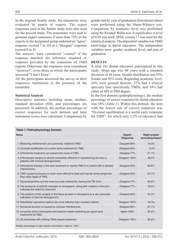
RESULTSA total 161 dental educators participated in thisstudy. Mean age was 40 years with a standarddeviation of 10 years. Gender distribution was 55%female and 45% male. Regarding academic level,24% were general dentists, 37% had a clinicalspecialty (not specifically TMD), and 39% hadeither an MS or PhD degree.In the first domain (pathophysiology), the medianpercentage of correct responses by dental educatorswas 38% (Table 1). Within this domain, the itemwith the lowest rate of correct responses was“Occlusal equilibration is a useful early treatmentfor TMD”, for which only 2,5% of educators had
208 Irene A. Espinosa, et al.
Acta Odontol. Latinoam. 2016 ISSN 1852-4834 Vol. 29 Nº 3 / 2016 / 206-213
Table 1: Pathophysiology Domain.
Items
1. Balancing interferences are commonly related to TMD.
2. Occlusal equilibration is a useful early treatment for TMD.
3. Orthodontic treatment can prevent the onset of TMD.
4. Arthroscopic surgery is almost completely effective in repositioning the disc inpatients with internal derangements.
5. Orthodontic therapy is the best treatment to resolve TMD in a patient with a skeletalmalocclusion.
6. TMD caused by trauma is much more difficult to treat and has far worse prognosisthan other types of TMD.
7. Transcranial films are the most accurate method for viewing the TM Joint.
8. The presence of arthritic changes on tomograms, along with crepitus in the jointindicates the need for treatment.
9. The position of the condyle in the fossa as seen in tomograms is a very accurateindication of internal derangement.
10. Mandibular repositions splints are more effective than maxillary splints.
11. Nocturnal bruxism is caused by occlusal interferences.
12. Ice packs and/or heat packs and passive muscle stretching are good early treatments for TMD.
13. All individuals with clicking TMJs require treatment.
Median percentage of right answers according to experts = 38.4
Expert Response
Disagree 85%
Disagree 85%
Disagree 77%
Disagree 100%
Disagree 92%
Disagree 83%
Disagree 77%
Disagree 77%
Disagree 92%
Disagree 100%
Disagree 85%
Agree 100%
Disagree 100%
Right answeraccording Expert
14.3%
2.5%
21.1%
38.5%
65.8%
54.7%
50.9%
18.0%
40.4%
45.3%
67.7%
58.4%
36.0%
AOL32016:32011 15/02/2017 12:51 Página 208

adequate knowledge. The item with the highestnumber of correct responses in the same domainwas “Nocturnal bruxism is caused by occlusalinterferences”, for which 68% of educators hadadequate knowledge. This particular domainpresented a wide range of variability.In the second domain (psychophysiology), dentaleducators had better knowledge of the subject, andthe median percentage of total correct answers was78% (Table 2).Within this domain, the item with thelowest rate of correct answers was “Stress is a majorfactor in the development of TMD”, with only 47%of the educators demonstrating adequate knowledge.The item with the highest percentage of correctanswers was “Stress management is indicated formany TMD patients”, with 88% of the educatorshaving adequate knowledge. In the third domain (psychiatric disorders), themedian percentage of total correct answers bydental educators was 50% (Table 3). The item with
the lowest rate of correct answers was “Clinicaldepression is rare in chronic TMD patients”, withonly 47% of educators having adequate knowledge.The item with the highest percentage of correctanswers in this domain was “Depression can be animportant etiologic factor in chronic pain”, with 62% of educators demonstrating adequateknowledge.Finally, in the fourth domain (chronic pain), themedian percentage of correct answers was 56%(Table 4). Within this domain, the item with thelowest rate of correct answers was “Prescription ofnarcotics, as needed for pain as treatment of choicewhen TMD pain is severe”, where only 26% of theparticipants had adequate knowledge. The item withthe highest rate of correct answers in this domainwas “Behavior modification treatments areappropriate for patients with chronic TMD pain”,where 63% of dental educators agreed with expertson TMD.
Education Regarding Temporomandibular Disorders 209
Vol. 29 Nº 3 / 2016 / 206-213 ISSN 1852-4834 Acta Odontol. Latinoam. 2016
Table 2: Psychophysiologic Domain.
Items
1. The mechanisms of acute and chronic pain are the same.
2. Biofeedback can be useful for treating TMD.
3. Oral parafunctional habits are often significant in the development of TMD.
4. Patients with TMD who clench/brux do so either during the day or at night, but notboth.
5. Stress management is indicated for many TMD patients.
6. Stress is a major factor in the development of TMD.
7. Tension and stress increase jaw muscle EMG levels in susceptible patients.
8. Progressive muscle relaxation is not an effective treatment for TMD.
9. Information on the daily pattern of TMD symptoms can be helpful for identifying contributing factors.
Median percentage of right answers according to experts = 77.7
Expert Response
Disagree 100%
Agree 100%
Agree 85%
Agree 85%
Agree 77%
Agree 100%
Disagree 82%
Agree 92%
Disagree 92%
Right answeraccording Expert
79.5%
65.2%
72.7%
72.7%
88.2%
46.6%
76.4%
54.0%
79.5%
Table 3: Psychiatric Disorders Domain.
Items
1. Clinical depression is rare in chronic TMD patients.
2. Depressed mood is fair common in chronic TMD patients.
3. Anxiety disorders are more common in TMD patients than in the population at large.
4. Depression can be an important etiologic factor in chronic pain.
Median percentage of right answers according to experts = 50.0
Expert Response
Disagree 100%
Agree 86%
Agree 79%
Agree 79%
Right answeraccording Expert
47.2%
52.8%
59.0%
62.1%
AOL32016:32011 15/02/2017 12:51 Página 209

210 Irene A. Espinosa, et al.
Acta Odontol. Latinoam. 2016 ISSN 1852-4834 Vol. 29 Nº 3 / 2016 / 206-213
Table 4: Chronic Pain Domain.
Items
1. Chronic TMD patients should be advised to rest and limit their work and social activities when they are experiencing pain.
2. PRN narcotics (i.e., "as needed " for pain) are a treatment of choice when TMD painis severe.
3. Antidepressants are never indicated in the management of TMD.
4. An extensive history of previous treatment failures in a TMD patient is usually anindication for surgery.
5. Chronic pain is a behavioral as well as a physical problem.
6. Although some TMD patients have psychological problems, these problems are usually unrelated to their pain.
7. Difficulty with sleep is a common finding in chronic pain.
8. Some patients use pain as an excuse to avoid unpleasant chores.
9. Behavior modification treatments are appropriate for patients with chronic TMD pain.
Median percentage of right answers according to experts = 55.5
Expert Response
Disagree 85%
Disagree 93%
Disagree 88%
Disagree 100%
Agree 96%
Disagree 85%
Agree 96%
Agree 89%
Agree 88%
Right answeraccording Expert
46.6%
25.5%
49.1%
55.3%
36.0%
37.9%
58.4%
60.9%
63.4%
Table 5: Comparison by gender.
Domain Male (n=72) Female (n=89) p*Median Correct Percentage Median Correct Percentage
Pathophysiology 40.2 38.7 0.564
Psychophysiologic 70.0 70.7 0.837
Chronic Pain 47.7 48.3 0.768
Psychiatric disorders 53.4 56.2 0.576
Across all domains 52.8 53.1 0.816
*U de Mann-Whitney
Comparison by gender (Table 5), year of graduation(Table 6), and academic level (Table 7) showed nostatistically significant difference among groups(p>0.05).
DISCUSSIONThis research shows that participating dentaleducators’ knowledge ofTMD differs greatly fromthe knowledge of experts in TMD reported in the literature1,33. Several countrieshave madeefforts to assess knowledge of TMD amongdentists1,25,3136,38,39. Researchers have shown thateven among professionals with advanced educationin TMD, there is no homogeneity of concepts onthe pathophysiology of these conditions 1,34,3638. InMexico there is no specialty in TMD, and patientswith this condition are treated by specialists in
different areas of stomatology and general dentists.This study represents the first evaluation conductedin Mexico, and clearly indicated the inconsistencyof knowledge and understanding of these disorders,and consequently, the low priority that has beingassigned to the field of TMD in dental education.We believe that this study highlights the need fordental educators to be prepared and teach the mostupdated knowledge in the field to their dentalstudents.Our results are consistent with data previouslyreported by several researchers. No difference wasfound by gender, academic level and year ofgraduation1,35. This is also consistent with Glaros etal 33 who claims that general dentists and specialistsin areas other than the TMD do not differ inknowledge about these disorders. However, other
AOL32016:32011 15/02/2017 12:51 Página 210

authors have found controversial results, withspecialists obtaining better scores 34.
Our data are also consistent with previouslyreported results on the pathophysiological domain,representing the lowest rate of 38% 1,33,35,38. Theresults illustrate a poor understanding of theetiology, diagnosis, and treatment of TMD. Ourresearch showed the greatest weakness (only 2.5%of correct answers according to the experts) is inthe belief that occlusal balance is a useful option inearly treatment of temporomandibular disorders.Occlusal equilibrations are still being used inMexico for the early management of patients withTMD, despite the vast worldwide evidence againstsuch treatment. This particular finding contrastswith values from other previously reported studiesin which the percentage of agreement of generaldentists and other specialists was about 30% and26%1,33. On the other hand, the correct percentage,according to the experts in this research, about thestatement “orthodontic treatment can preventTMD” (21%), was slightly lower in studies byGlaros et al 33 (19%) and Le Resche 1(14%),although all results are low.
Conversely, it is noteworthy that the domain ofpsychophysiology (mechanisms of acute andchronic pain, biofeedback, oral parafunctionalhabits, stress, etc.) in the etiology oftemporomandibular disorders was well recognizedby the participants (78%). This highlights theunderstanding of most educators of the role ofpsychophysiological factors in the field of TMD.Previous studies 1,33 have shown correct knowledgeof this domain in 50% to 90% of general dentistsand other specialists, consistent with the results ofour study (46% to 88%).With respect to the domainof psychiatric disorders, our study has found thatdepression and anxiety are recognized asdetermining factors in patients with TMD, with52% to 62% of participants answering those itemscorrectly. Studies by Le Resche1 and Glaros33 foundsuccess rates higher than those reported in ourstudy. Finally, domain analysis of chronic paindenotes that participants have acceptableknowledge of said domain (55%). However, issuessuch as “PRN narcotics (i.e., “as needed” for pain)are a treatment of choice when TMD pain is severe”and “Chronic pain is a behavioral as well as a
Education Regarding Temporomandibular Disorders 211
Vol. 29 Nº 3 / 2016 / 206-213 ISSN 1852-4834 Acta Odontol. Latinoam. 2016
Table 6: Comparison by time that the educators finished the last academic level.
Domain Under 15 years (n=63) 15 and over years (n=98) p*Median Correct Percentage Median Correct Percentage
Median Median
Pathophysiology 38.4 38.4 0.932
Psychophysiologic 66.6 77.7 0.084
Chronic Pain 55.5 49.9 0.926
Psychiatric disorders 50.0 50.0 0.856
Across all domains 52.6 55.0 0.531
*U de Mann-Whitney
Table 7: Comparison by academic level.
Domain General Dentists Dental Specialists Dentists with MD/PHD p*
Median Correct Percentage
Pathophysiology 39.4 41.4 37.5 0.369
Psychophysiologic 68.3 71.4 70.8 0.676
Chronic Pain 50.2 46.0 48.6 0.526
Psychiatric disorders 62.5 52.5 52.7 0.275
Across all domains 54.6 52.5 52.3 0.635
*Kruskal Wallis
AOL32016:32011 15/02/2017 12:51 Página 211

physical problem”, remain poorly understood byparticipants. Despite the high prevalence of TMD reported in theliterature, knowledge of TMD among dentaleducators needs improvement, as previous studieshave reported 1,33,34,37,38. The results denote a high level of variability in the domain of the
pathophysiology diagnosis and treatment as well asa need to improve in the other domains. Knowledgeamong educators is not influenced by gender,academic level, or year of graduation. These resultssupport the conclusion that there is an importantneed for improvement in the knowledge of TMD inthe dental educational system in Puebla, México.
212 Irene A. Espinosa, et al.
Acta Odontol. Latinoam. 2016 ISSN 1852-4834 Vol. 29 Nº 3 / 2016 / 206-213
ACKNOWLEDGMENTThe authors would like to thank the BenemeritaUniversidad Autonoma de Puebla and the ConsejoNacional de Ciencia y Tecnología (CONACYT) forproviding financial support for PhD. Irene Espinosaduring her International Scholarship Award, and forMauricio Perez during his Master’s studies.
CORRESPONDENCE: Dr. Irene Aurora Espinosa Tlacomulco 4513, Col. Ampliación Reforma Sur. CP 72160 Puebla, Pue. México. [email protected]
REFERENCES1. Le Resche L, Truelove EL, Dworkin SF. Temporomandibu
lar disorders: a survey dentists’ knowledge and beliefs. JAm Dent Assoc 1993; 124:97106.
2. Greene CS. Managing the care of patients with temporoman di bu lar disorders: a new guideline for care. J Am Dent Assoc2010; 141:10861088.
3. De Leeuw R. Orofacial Pain Guidelines for Assessment,Diagnosis and Management.Quintessence Books. 4th ed.Chicago, USA: The American Academy of Orofacial Pain,2008:131132.
4. Okeson JP. Management of temporomandibular disordersand occlusion. 5th ed. St Louis: Mosby, 2003:143189.
5. Le Resche L. Epidemiology of TemporomandibularDisorders: Implications for the Investigation of EtiologicFactors. Crit Rev Oral Biol Med 1997; 8:291305.
6. Isong U, Gansky S, Plesh O. Temporomandibular joint andmuscle disordertype pain in the US adults: the NationalHealth Interview Survey. J Orofac Pain 2008; 22:317322.
7. National Institute of Dental and Craniofacial ResearchFacial Pain. 2010. NIH Publication No. 103487.: URL:http://www.nidcr.nih.gov/OralHealth/Topics/TMJ/TMJDisorders.htm
8. Gonzalez YM. Are temporomandibular disorders a publichealth problem? Alpha Omegan 2003;96:1114
9. Sessle JB, Lavigne JG, Lund JP. Orofacial Pain From Basic Sciences to Clinical Management. 1st ed. Chicago:Quintessence Book, 2001:1719.
10. Slade GD, Bair E, Kunthel B, Mulkey F, BaraianC, Rothwell R, Reynolds M, Miller V, et al. Study Methods,Recruitment, Sociodemographic Findings, and DemographicRepresentativeness in the OPPERA Study. J Pain 2011;12:T12–T26.
11. Espinosa SI, Lara MC, Lara CA, Saavedra GM, Vargas GH.Comparación de losaspectospsicosociales (eje II) de lospacientes con trastornos teporomandibulares, de acuerdo ala combinación de diagnósticosfísicos (eje I) de los criteriosdiagnósticos para la investigación de los trastornos tempo romandibulares (CDI/TTM). Rev Oral 2009;10: 477481.
URL:http://www.medigraphic.com/pdfs/oral/ora2009/ora0930b.pdf
12. Slade GD, Diatchenko L, Bhalang K, Sigurdsson A,Fillingim RB, Belfer I, Max MB, Goldman D, et al.Influence of psychological factors on risk of temporoman dibular disorders. J Dent Res 2007; 86:11201125.
13. Klasser GD, Greene CS. Predoctoral teaching of temporo mandibular disorders: a survey of U.S. and Canadian dentalschools. J Am Dent Assoc 2007; 138:231237.
14. Gontyy AA.Teaching a comprehensive orofacial pain coursein the dental curriculum. J Dent Educ 1990; 54:319322.
15. Solberg WK, Fricton JR. The role of the dental school inteaching TMD and orofacial pain. J Craniomandib Disord1992; 6:107110.
16. Greene CS, Stockstill JW, Clark GT. Predoctoral educationfor TMD and orofacial pain: a philosophical overview. JCraniomandib Disord 1992; 6:111112.
17. Attanasio R, Mohl ND. Suggested curriculum guidelinesfor the development of predoctoral programs in TMD andorofacial pain. J Craniomandib Disord 1992; 6:113116.
18. Attanasio R, Mohl ND. Suggested curriculum guidelines forthe development of continuing education programs in TMDand orofacial pain. J Craniomandib Disord 1992; 6:137140.
19. Attanasio R, Mohl ND. Suggested curriculum guidelinesfor the development of postdoctoral programs in TMD andorofacial pain. J Craniomandib Disord 1992; 6:126134.
20. Stockstill JW. Curriculum outline for adjunctive predoctoraleducation in TMD and orofacial pain. J CraniomandibDisord 1992; 6:117122.
21. Fricton JR, Pullinger AG, Mohl ND. Postdoctoral educationfor TMD and orofacial pain. A philosophical overview. JCraniomandib Disord 1992; 6:123125.
22. McNeill C, Falace D, Attanasio R. Continuing educationfor TMD and orofacial pain: a philosophical overview. JCraniomandib Disord 1992; 6:135136.
23. Mohl ND, Attanasio R. The Third Educational Conferenceto Develop the Curriculum in Temporomandibular Disordersand Orofacial Pain: introduction. J Orofac Pain 2002;16:173175.
AOL32016:32011 15/02/2017 12:51 Página 212

24. Mohl ND. The Third Educational Conference to Develop theCurriculum in Temporomandibular Disorders and Orofacialpain: Summary/Conclusions. J Orofac Pain 2002; 16:198199.
25. Shankland W. Temporomandibular disorders: standardtreatment options. Gen Dent 2004; 52:349355.
26. Klasser GD, Greene CS. The changing field of temporo man di bular disorders: what dentists need to know? J CanDent Assoc 2009; 75:4953.
27. McKinney JF, Mosby EL. Temporomandibular disorders: whatto teach in dental school. J Craniomandib Disord 1990; 4:1719.
28. Douglass GD. Making a comprehensive diagnosis in a comprehensive care curriculum. J Dent Educ 2002; 66:414420.
29. Gonzalez Y, Mohl ND. Care of patients with temporoman di bular disorders: an educational challenge. J Orofac Pain2002;16:200206.
30. Alsafi Z, Michelotti A, Ohrbach R, Nilner M, List T.Achieved competences in temporomandibular disorders/orofacial pain: a comparison between two dental schools inEurope. Eur J Dent Educ 2015;19:161168.
31. AlKhotani A, Björnsson O, NaimiAkbar A, Christidis N,Alstergren P. Study on selfassessment regarding knowledgeof temporomandibular disorders in children/adolescents bySwedish and Saudi Arabian dentist. Acta Odontol Scand2015; 73:522529.
32. Alonso AA, Heima M, Lang LA, Teich ST. Dental students’perceived level of competence in orofacial pain. J DentEduc 2014; 78:13791387.
33. Glaros AG, Glass EG, McLaughlin L. Knowledge andbeliefs of dentists regarding temporomandibular disordersand chronic pain. J Orofac Pain 1994; 8:216222.
34. Baharvand M, Sedaghat Monfared M, Hamian M, JalaliMoghaddam E, Sadat Hosseini F, Alavi KA. Temporo mandibular disorders: knowledge, attitude and practiceamong dentists in Tehran, Iran. J Dent Res Dent ClinProspect 2010; 4:9094.
35. Just JK, Perry HT, Greene CS. Treating TM disorders: asurvey on diagnosis, etiology and management. J Am DentAssoc 1991; 122:5560.
36. Tegelberg Å, Wenneberg B, List T. General practice dentists’knowledge of temporomandibular disorders in children andadolescents. Eur J Dent Educ. 2007; 11:216221.
37. Patil S, Iyengar AR, Ramneek. Assessment of knowledge,attitude and practices of dental practitioners regardingtemporomandibular joint disorders in India. J Adv Clin ResInsights 2016;3:6471.
38. Lee WY, Choi JW, Lee JW. A study of dentists´ knowledgeand beliefs regarding temporomandibular disorders inKorea. CRANIO 2000; 18:142146.
Education Regarding Temporomandibular Disorders 213
Vol. 29 Nº 3 / 2016 / 206-213 ISSN 1852-4834 Acta Odontol. Latinoam. 2016
AOL32016:32011 15/02/2017 12:51 Página 213

214
Acta Odontol. Latinoam. 2016 ISSN 1852-4834 Vol. 29 Nº 3 / 2016 / 214-218
RESUMENLos autores condujeron un estudio experimental para deter minar la eficacia de la técnica anestésica intraósea usandoarticaína al 4% con epinefrina 1:100.000, en pacientes conpulpitis aguda en molares mandibulares. En diferentes sesionesclínicas, los miembros del equipo de investigadores usaronarticaína al 4% con epinefrina 1:100.000 para induciranestesia mandibular con la técnica intraósea (Grupo 1) o conel bloqueo del nervio alveolar inferior (Grupo 2), se aplicó cadatécnica en 35 pacientes con diagnóstico de pulpitis aguda enmolares inferiores. En cada grupo, se determinó la necesidadde hacer anestesia complementaria y la comodidad del paciente
con un test Escala Visual Analoga. Un total de 70 pacientesfueron enrolados en este estudio (35 sujetos por grupo). En elgrupo de intraósea no fue necesaria la aplicación de técnicascomplementarias en 22 pacientes (31.4%), resultados similaresen la técnica alveolar inferior (n: 23 32.8%). La técnicaintraósea demostró ser más cómoda al compararla con técnicamandibular (18 pacientes 25.7%). Este estudio demostró queel uso de la técnica intraósea conarticaína al 4%, arrojóresultados prometedores en lo que a comodidad y reducción enla anestesia complementaria hace referencia.
Palabras clave: Articaína; anestesia local; anestesia dental.
INTRODUCTIONPain control in dentistry is based on the use of different anesthetics. When conventionaltechniques do not provide adequate anesthesia,alternatives are needed. The success rate ofanesthesia is variable, with 10% to 20% failurereported when inferior alveolar nerve block is used1.It is therefore necessary to include new techniques1,2
as well as active principles such as 4% articaine,which supplements good anesthesia technique
using an active principle which was introduced inthe USA in the year 20003. Articaine appears to havegreater residual diffusion capacity, providing anintermediate period of anesthesia. Although it is an amidetype anesthesia, it has an additional ester group and is thus less toxic, since 90% ismetabolized by plasma estearates. Its higherliposolubility enables greater diffusion than otheramides in soft tissues and bone, so it has beenproposed for use in infiltrations in the mandibular
ABSTRACTThe authors conducted an experimental study to determinepatient perception of discomfort during injection and the needfor supplemental anesthesia using the intraosseous techniquewith 4% articaine with 1:100,000 epinephrine in patients withsymptomatic pulpitis in mandibular molars. At different clinicalsessions, researchers used 4% articaine with 1:100,000epinephrine to apply intraosseous injection (Group 1) orinferior alveolar nerve block (Group 2). Each technique wasapplied in 35 patients. In each group, the need for additionalanesthesia was determined and patient discomfort during
injection was assessed with a Visual Analogue Scale (VAS) test.In the intraosseous group, no supplemental technique wasneeded in 22 patients (62.85 %), and results were similar forthe inferior alveolar technique (n: 23 65.71%). Theintraosseous technique proved to be more comfortable than themandibular technique (18 patients – 25.7%). This study foundthat the use of intraosseous technique with 4% articaine showspromising results regarding patient comfort and reducing theneed for additional anesthesia.
Key words:Articaine; lidocaine; local anesthesia dental anesthesia.
PERCEPTION OF DISCOMFORT DURING INJECTION AND THE NEED FOR SUPPLEMENTAL ANESTHESIA IN THE INTRAOSSEOUS TECHNIQUE USING 4% ARTICAINE
Adel Martínez Martínez1, María del Pilar Lujan Pardo2, Jonathan Harris Ricardo3
1 Departamento de Medicina Bucal. Facultad de Odontología, Universidad de Cartagena, Colombia
2 Departamento de Odontopediatría. Programa de Odontología, Corporación Universitaria Rafael Núñez, Cartagena, Colombia.
3 Departamento de Cirugía Oral. Programa de Odontología, Corporación Universitaria Rafael Núñez, Cartagena, Colombia.
PERCEPCIÓN DE INCOMODIDAD DURANTE LA INYECCIÓN Y NECESIDAD DE ANESTESIA SUPLEMENTARIA EN ANESTESIA INTRAÓSEA USANDO ARTICAÍNA AL 4%
AOL32016:32011 15/02/2017 12:51 Página 214

molar region3, 4.The onset of the anesthetic effectoccurs within 16 minutes, and it lasts for about anhour, with maximum effect at 25 minutes.5
The use of intraosseous anesthesia dates back to1910, when Masselink BH published a techniquefor placing the solution inside the medullar bonethrough a perforation in the cortical bone made witha round carbide drill.6 Intraosseous anesthesia isindicated for removing deep caries, pulpotomy,carving abutments in living teeth, endodontictreatment of teeth with pulpitis and exodontic treat ment of permanent teeth. Its advantages includeproviding deep pulp anesthesia which is highlyeffective for endodontic treatment of teeth withpulpitis. Its use is somewhat limited by the fact thatit requires a special kit for application, and shouldtherefore be planned before beginning a procedureso that it may be used immediately as a supplementduring the operation.710Potential complicationsreported are perforation of the roots of the toothunder treatment or neighboring teeth, laceration of the inferior alveolar nerve and perforation of the maxillary sinus.11Different authors haverecommended infiltration of the mandible with 4%articaine 4%, demonstrating that anesthesia efficacyimproves when using this anesthetic solution.1214
This paper reports the results of a clinical study todetermine the efficacy of intraosseous anesthetictechnique using 4% articaine with 1:100,000epinephrine compared to conventional mandibularblock technique in patients diagnosed with acutepulpitis in mandibular molars.
MATERIALS AND METHODSA doubleblind randomized uncontrolled study was performed to determine patients’ perception of discomfort during use of the intraosseoustechnique, and the need for supplemental injection
with 4% articaine with 1:100,000 epinephrine incases of symptomatic pulpitis in mandibularmolars. Sample size (N = 70) was obtained usingEpi Info. As there was no quantifiable targetpopulation, nonprobabilistic sampling by criterionwas used and a technique assigned randomly.Patients underwent clinical and radiographicexamination and sensitivity tests to confirm thediagnosis of acute symptomatic pulpitis. Exclusioncriteria were periapical lesions, temporary dentition,allergy to anesthetic solution and radiologicevaluation determining existence of anatomicalstructures preventing the application of theintraosseous technique. Seventy patients weredivided randomly into two groups (n = 35): Group 1,intraosseous technique and Group 2, inferioralveolar nerve block. Anesthetics were applied by astandardized researcher. The need for supplementalanesthesia during root canal treatment wasdetermined and patient perception of discomfortafter the use of either the intraosseous technique orinferior alveolar never block was assessed using aVisual Analogue Scale (VAS). Data were recorded in standardized collectioninstruments and tabulated on an Excel spreadsheet.Statistical analyses included descriptive statistics,frequency tables and comparison of variables. TheChi2 test with Yates correction was appliedassuming p <0.05, using STATA 9® statistic software.Ethical considerations were followed as set forth inColombia’s Ministry of Health Resolution 008430of 1993. All participants signed informed consent.
RESULTSDemographics: 70 patients were treated: 35 in theintraosseous injection group and 35 in the inferioralveolar nerve block group. There was no statisticaldifference between groups for age and sex (Table 1).
Discomfort in Intraosseous Technique 215
Vol. 29 Nº 3 / 2016 / 214-218 ISSN 1852-4834 Acta Odontol. Latinoam. 2016
Table 1: Demographic Data.
INTRAOSSEOUS GROUP IANB GROUP P Value
GENDER n (%) n (%) n (%)
MALE 19 (45.71 ) 16 (54.29) 35 (100)0.47
FEMALE 16 (45.71) 19 (54.29) 35 (100)
AGE (YEARS)
MEAN ± STD DEV 42.14±13.95 41.82±18.83 70 (100) 0.93
*STD DEV: Standard Deviation,*IANB: Inferior Alveolar Nerve Block
AOL32016:32011 15/02/2017 12:51 Página 215

Supplemental Injection: 35.71% (95% CI: 25.18 –47.83) (n= 25) of the sample required supplementalinjection (Table 2). 37.15% of the patients in themandibular intraosseous injection group requiredsupplemental injection compared to 34.28% in theinferior alveolar nerve block group (Table 2)(p=0.80).Patient perception of injection and discomfort: themean values on the visual analogue scale (VAS)were 0.94 (SD: 1.21) for Group 1 (mandibularintraosseous injection) and 2.6 (SD: 1.24) for Group 2 (alveolar nerve block group), differingstatistically (p=0.00) (Fig. 1).
DISCUSSIONPain control in mandibular molars with pulpitis isone of the main challenges during pulp removal forcanal preparation, so alternatives to conventionalnerve blockage are needed in order to achieve deepanesthesia in the mandibular molar zone. One of thebest options is to combine an active principle withhigh bone diffusion, e.g. 4% articaine, using theintraosseous anesthetic technique. In this study,intraosseous technique was proposed as the primary
anesthesia with 4% articaine including 1:100,000epinephrine for mandibular molars with asympto matic pulpitis. The intraosseous technique wasfound to be similar to the conventional techniquewith regard to the need for supplemental anesthesia:13 (18.57%) and 12 patients (17.14 %), respectively.With regard to patient discomfort during theinjection of anesthesia, as reported on the VisualAnalog Scale (VAS), 18 patients (25.79%) reportedno pain during the intraosseous technique, incontrast to the mandibular technique group, inwhich only 2 (2.85%) reported no pain. This showsthat the intraosseous technique with the X Tip Kitis more comfortable than puncture using theconventional technique at the level of the inferiordental orifice. The results of this research differfrom those reported by Pereira LA et al.Faltancitas10 a la 14 inclusive15 in a double blind study in 60patients comparing anesthetic effectiveness andcardiovascular changes after the application of 0.9ml of 4% articaine with 1:100,000 and 1:200,000epinephrine using the intraosseous technique, inmandibular molars with acute pulpitis, whichshowed that both solutions had a highly anestheticeffect (96.8% and 93.1% respectively) withminimal cardiovascular effects. Their results arecomparable with our study if we consider that for the intraosseous technique, 18.57% requiredsupplemental anesthesia, reflecting 81.43%effectiveness. Mohammad et al.16 conducted adouble blind clinical trial in 100 patients diagnosedwith irreversible pulpitis in maxillary teeth, whoreceived infiltrative anesthesia with 2 ml of 4% articaine with 1:100,000 epinephrine or with2% lidocaine with 1:80,000 epinephrine. Theresponse was evaluated with an electronic pulp measuring device and found no significantstatistical difference compared to 4% articaine with1:100,000 epinephrine and lidocaine with 1:80,000epinephrine, in anesthesia of maxillary teethinfiltrated in the buccal zone. They additionally
216 Adel Martínez Martínez, et al.
Acta Odontol. Latinoam. 2016 ISSN 1852-4834 Vol. 29 Nº 3 / 2016 / 214-218
Table 2: Supplementary Anesthesia.
INTRAOSSEOUS GROUP IANB GROUP Total P Valuen (%) n (%) n (%)
No 22 (62,85) 23 ( 65,71) 64,28%0.80
YES 13 ( 37,15) 12 (34,28) 35,71%
*IANB: Inferior Alveolar Nerve Block
Fig. 1: Perception of injection and perforation discomfortduring intraosseous injection and inferior alveolar nerveblock.
AOL32016:32011 15/02/2017 12:51 Página 216

determined that only 67% of the patients expressednumbness with the mandibular technique and33.3% required a supplemental technique. Ourstudy found that only 13 patients requiredsupplemental anesthesia (18.57%). Inferior alveolarnerve blockage is the technique most often used tonumb the mandibular posterior teeth in root canaltreatments. Jung IY et al.17 report that the localanesthesia used to block the inferior alveolar nervecan provide a success rate of 70% in noninflamedpulps, but the success rate decreases dramaticallyto 30% in patients with irreversible pulpitis, who are 8 times more likely than normal patients to undergo failure. These are the cases for which we highly recommend the use of intraosseousinjection with 4% articaine as a primary technique,
improving the success ratio during the preparationof root canals. Nussteinet al18 recommend the intraosseoustechnique due to a 98% success rate with lidocaineand 1:100,000 epinephrine with rapidonset pulpalanesthesia. Our study suggests that intraosseousanesthesia can be used as a primary technique inroot canal treatments in patients with symptomaticpulpitis because it provides an adequate level ofanesthesia and a high degree of comfort. The use of intraosseous technique with 4% articaineshows promising results regarding comfort andreduction of the need for additional anesthesia. Theintraosseous technique is probably the bestalternative when deep pulpar anesthesia is requiredin mandibular molars
Discomfort in Intraosseous Technique 217
Vol. 29 Nº 3 / 2016 / 214-218 ISSN 1852-4834 Acta Odontol. Latinoam. 2016
ACKNOWLEDGEMENTSThe authors are grateful to the patients and volunteers whoparticipated in this study and toDr. Miguel Simancas for hisinvaluable contribution in the statistical analysis.
CORRESPONDENCEDr. AdelMartinezMartinez. Campus Zaragocilla.Faculty of Dentistry.University of Cartagena.Cartagena, Colombia. email: [email protected]
REFERENCES1. Haas DA. Alternative mandibular nerve block techniques: a
review of the GowGates and AkinosiVazirani closedmouth mandibular nerve block techniques. J Am Dent Assoc.2011; 142 Suppl 3:8S12S.
2. Goldberg S, Reader A, Drum M, Nusstein J, Beck M.Comparison of the anesthetic efficacy of the conventionalinferior alveolar, GowGates, and VaziraniAkinosi techniques.J Endod. 2008;34:13061311.
3. Moore PA, Cuddy MA, Cooke MR, Sokolowski CJ.Periodontal ligament and intraosseous anesthetic injectiontechniques: alterna tives to mandibular nerve blocks. J AmDent Assoc. 2011; 142Suppl 3:13S8S.
4. Coggins R, Reader A, Nist R, Beck M, Meyers WJ.Anesthetic efficacy of the intraosseous injection in maxillaryand mandibular teeth. Oral Surg Oral Med Oral Oral PatholOral Radiol Endod 1996;81:634641.
5. Replogle K, Reader A, Nist R, Beck M, Weaver J, MeyersWJ. Anesthetic efficacy of the intraosseous injection of 2%lidocaine (1:100,000 epinephrine) and 3% mepivacaine inmandibular first molars. Oral Surg Oral Med Oral PatholOral Radiol Endod.1997;83:3037.
6. Nusstein J, Kennedy S, Reader A.I, Beck M, WeaverJ. Anesthetic efficacy of the supplemental Xtip intraosseousinjection in patients with irreversible pulpitis. J Endod.2003; 29: 724728.
7. Bigby J, Reader A, Nusstein J, Beck M, Weaver J. Articainefor supplemental intraosseous anesthesia in patients withirreversible pulpitis. J Endod. 2006; 32:10441047.
8. Sixou JL, BarbosaRogier ME. Efficacy of intraosseousinjections of anesthetic in children and adolescents. OralSurg Oral Med Oral Pathol Oral Radiol Endod 2008;106:173178.
9. PenarrochaOltra D, AtaAli J, OltraMoscardo M J,PenarrochaDiago M A, Penarrocha M. Comparative studybetween manual in jection intraosseous anesthesia andconventional oral anesthesia. Med Oral Patol Oral CirBucal 2012; 17: e233235.
10. Verma PV, Srivastava R, Ramesh K M.. Anesthetic efficacyof Xtip intraosseous injection using 2% lidocaine with1:80,000 epinephrine in patients with irreversible pulpitisafter inferior alveolar nerve block: A clinical study. JConserv Dent 2013;16:162166.
11. Graetz C, FawzyElSayed K M, Graetz N, Dörfer CE. Rootdamage indu ced by intraosseous anesthesia – an in vitroinvestigation. Med Oral Patol Oral Cir Bucal. 2013 1; 18e1: 30134.
12. Katyal V. The efficacy and safety of Articaine versus lignocainein dental treatments: a metaanalysis. J Dent 2010; 38:307317.
13. Nuzum FM, Drum M, Nusstein J, Reader A, Beck M.Anesthetic efficacy of articaine for combination labial pluslingual infiltrations versus labial infiltration in the mandibularlateral incisor. J Endod 2010; 36:952956.
14. Meechan JG. The use of the mandibular infiltration anesthetictechnique in adults.J Am Dent Assoc 2011 ;142 Suppl 3:19S24S.
15. Pereira LA, Groppo FC, Bergamaschi CD, Meechan JG,Ramacciato JC, Motta RH, Ranali J. Articaine (4%) withepinephrine (1:100,000 or 1:200,000) in intraosseous
AOL32016:32011 15/02/2017 12:51 Página 217

injections in symptomatic irreversible pulpitis ofmandibular molars: anesthetic efficacy and cardiovasculareffects. Oral Surg Oral Med Oral Pathol Oral Radiol 2012;Epub ahead of print, 10.1016/j.oooo. 2011.10.045.
16. Kanaa M D, Whitworth J M, Meechan J. A comparison ofthe efficacy of 4% articaine with 1:100,000 epinephrineand 2% lidocaine with 1:80,000 epinephrine in achievingpulpal anesthesia in maxillary teeth with irreversiblepulpitis. J Endod 2012; 38: 279282.
17. Jung I Y, Kim J H, Kim E S, Lee C Y, Lee S J. An evaluationof buccal infiltrations and inferior alveolar nerve blocks inpulpal anesthesia for mandibular first molars. J Endod2008; 34:1113.
18. Nusstein J, Wood M, Reader A, Beck M, Weaver J.Comparison of the degree of pulpal anesthesia achievedwith the intraosseous injection and infiltration injectionusing 2% lidocaine with 1:100,000 epinephrine. Gen Dent2005 ; 53:5053.
218 Adel Martínez Martínez, et al.
Acta Odontol. Latinoam. 2016 ISSN 1852-4834 Vol. 29 Nº 3 / 2016 / 214-218
AOL32016:32011 15/02/2017 12:51 Página 218

RESUMOA artrite reumatoide (AR) é uma doença sistêmica, autoimune,caracterizada por inflamação crônica e hiperplasia sinovial,que usualmente afeta múltiplas articulações. Dentre estas, aarticulação temporomandibular (ATM), tornase susceptívelao desenvolvimento de alterações. O estudo objetiva avaliar apresença de desordem temporomandibular (DTM) e altera çõesósseas degenerativas da ATM (articulação temporo man di bu lar) de pacientes diagnosticados com AR (artrite reumatóide).Como metodologia, aplicouse o questionário ResearchDiagnostic Criteria for Temporomandibular Disorder (RDC/TDM) em para avaliação clínica da ATM e classificação dadesordem temporomandibular em 49 pacientes de ambos ossexos e idade variável. Foram excluídos os indivíduos que jáhaviam realizado tratamento prévio para DTM e/ou comhistórico de traumatismo crâniofacial. Posteriormente osparticipantes foram submetidos a exames de tomografiacomputadorizada de feixe cônico (TCFC) para avaliação depossíveis alterações degenerativas no côndilo mandibular e naeminência articular. Foram apresentadas as frequências das
alterações encontradas e verificouse a associação entre osachados clínicotomográficos por meio do teste do Quiquadrado. Após a avaliação clínica verificouse que 75% dospacientes possuíam queixas de dor na região orofacial,variando entre a presença de artralgia, mialgia ou ambas.Quanto aos diagnósticos, 100% da amostra apresentoudiagnóstico do Grupo III do RDC/TMD (artralgia, osteoartriteou osteoartrose). A presença de alterações ósseas degenera tivas foi encontrada em 90% dos indivíduos avaliados, sendoque as mais prevalentes foram aplainamento (78,7%) eosteófito (39,3%). O teste de associação sugeriu uma maiortendência de desenvolvimento de alterações degenerativas nosindivíduos assintomáticos (p = 0.01). O caráter assintomáticodo envolvimento da ATM na AR pode ocultar danos estruturaisvistos em imagem. Assim, ressaltase a importância dodiagnóstico e tratamento precoces para redução de danosestruturais e funcionais.
Palavraschave: Artrite reumatoide; articulação temporoman dibular; tomografia computadorizada de feixe cônico.
ABSTRACTRheumatoid arthritis (RA) is a systemic autoimmune diseasecharacterized by chronic inflammation and synovial hyperplasia,which usually affects multiple joints. The temporomandibularjoint (TMJ) becomes susceptible to the development of changesresulting from RA. The aim of this study was to evaluate thepresence of TMD and degenerative bone changes in TMJ inpatients diagnosed with RA (rheumatoid arthritis). The ResearchDiagnostic Criteria for Temporomandibular Disorders (RDC/TMD) questionnaire was used for clinical evaluation of the TMJand for TMD classification of 49 patients of both sexes and allages. Individuals who had already undergone prior treatment forTMD and/or with a history of craniofacial trauma were excluded.The participants underwent cone beam computed tomography(CBCT) exams to assess possible degenerative changes in themandibular condyle and the articular eminence. The frequenciesof the changes found are presented and the possible associations
between clinical and CT findings analyzed using the chisquaretest. It was found that 75% of the patients had complaints of pain in the orofacial region, including arthralgia, myalgia or both. As for the diagnoses, 100% of the sample wasdiagnosed as RDC/TMD Group III (arthralgia, osteoarthritisor osteoarthrosis). The presence of degenerative bone changeswas found in 90% of the subjects, the most prevalent beingflattening (78.7%) and osteophytes (39.3%). The associationtest suggested a greater tendency to develop degenerativechanges in asymptomatic individuals (p = 0.01). Theasymptomatic nature of the involvement of the TMJ in RA canhide structural damage seen in imaging. Thus, the importanceof early diagnosis and treatment to reduce structural andfunctional damage is emphasized.
Key words: Rheumatoid arthritis; temporomandibular joint;cone beam computed tomography, temporomandibular disorder.
219
Vol. 29 Nº 3 / 2016 / 219-224 ISSN 1852-4834 Acta Odontol. Latinoam. 2016
TEMPOROMANDIBULAR JOINT INVOLVEMENT IN RHEUMATOID ARTHRITIS PATIENTS: ASSOCIATION BETWEEN CLINICAL AND TOMOGRAPHIC DATA
Patrícia C. F. Cordeiro1, Josemar P. Guimaraes1, Viviane A. de Souza2, Isabela M. Dias1, Jesca N. N. Silva1, Karina L. Devito1, Leticia L. Bonato3
1 School of Dentistry, Federal University of Juiz de Fora, Juiz de Fora, Minas Gerais, Brazil.2 School of Medicine, Federal University of Juiz de Fora, Juiz de Fora, Minas Gerais, Brazil.3 Estácio Juiz de Fora University, Juiz de Fora, Minais Gerais, Brazil.
ENVOLVIMENTO DA ARTICULAÇÃO TEMPOROMANDIBULAR EM PACIENTES COM ARTRITE REUMATOIDE – ASSOCIAÇÃO ENTRE DADOS CLÍNICOS E TOMOGRÁFICOS
AOL32016:32011 15/02/2017 12:51 Página 219

INTRODUCTIONRheumatoid arthritis (RA) is an autoimmunesystemic disease characterized by chronicinflammation and synovial hyperplasia whichusually affects multiple joints. Currently, it isconsidered the main inflammatory joint disorder,with a prevalence of 0.51% in the generalpopulation1. The incidence is higher in women, withvarying proportions documented in the literature:4:12, 7:13, and 9:14.While its autoimmune pattern is emphasized,various factors contribute to development of thedisease, such as genetic predisposition, micro biological, hormonal and environmental factors58.Extensive infiltration of immune cells occurs, withhigh levels of inflammatory mediators in synovialtissue of peripheral joints9,10, and the abnormalitiesin the structure and function of such joints appearas the primary and characteristic manifestations inthese patients.The temporomandibular joint (TMJ) becomessusceptible to the development of changes resultingfrom RA. However, it is rarely the first joint to beaffected5,6,11,12. It is estimated that more than half ofthe patients with RA present clinical evidence ofinvolvement of the TMJ5,7,9,10, the most frequentbeing bilateral involvement.Clinically, the involvement of the TMJ follows thesame destructive path as the other joints, correlatingdirectly with the severity and duration of the RAthat has taken hold7,13. In the initial phase, there maybe synovial hyperemia, lymphocyte infiltration,fibrinoid degeneration and pannus formation(granulation tissue). The cartilage is destroyed andthe granulation tissue can be seen in the joint cavity.From that point on, there may be fibrosis andscarring, generating fibrous adhesion. Fibrousankylosis is the rarest and final stage that the TMJaffected by RA can attain10,14.The most common clinical signs and symptoms inthe TMJ are arthralgia, swelling, stiffness duringmouth opening and upon waking, weakness of themasticatory muscles with decreased bite force, jointnoises, and limited joint function2,57,10,15.Studies using cone beam computed tomography(CBCT), considered the gold standard for theanalysis of degenerative bone changes of the TMJ,demonstrated that it is possible to find, in the TMJof patients with RA, high prevalence of reducedjoint space and serious erosion of cortical or
subcortical bone, which can lead to destruction ofthe mandibular condyle11,16. Bone sclerosis,osteophytes and subcortical cysts may also occur6,7. However, the presence of changes in the TMJ of RApatients is often ignored by rheumatologists andeven by patients, especially when treatment isfocused on other joints of the upper extremities and/or weightsupporting extremities. These changes inthe TMJ may lead to complications and disability,making its treatment and monitoring extremelyimportant.The aim of this study was to evaluate the presenceof TMD and degenerative bone changes in TMJ inpatients diagnosed with RA by performing a clinicalexamination and analysis of CBCT images.
MATERIAL AND METHODSThis is a crosssectional, observational, descriptivestudy, approved by the Research Ethics Committeeof the Federal University of Juiz de Fora (UFJF),under code number 771.869. Being an observational study, the sample wasconsisted of patients recruited randomly in one year,totalizing 49 volunteers.The study assessed patients previously diagnosedwith RA treated at the Rheumatology Clinic of theUniversity Hospital, UFJF. The diagnosis of RAwas based on criteria established by the AmericanCollege of Rheumatology, 1987, and ACR/EULAR201017. The patients diagnosed with RA were informedabout the study and we included those who agreedto participate. All patients signed informed consentprior to participating in the research. Participationin the study was open to patients of both sexes andall ages. Exclusion criteria were pregnant women,individuals who had already undergone priortreatment for TMD and/or with a history of cranio facial trauma.The assessment was divided into two stages:assessment of clinical signs and symptoms in theTMJ and analysis of CBCT images. To assess theclinical signs and symptoms, we used Axis I of theResearch Diagnostic Criteria for Temporomandi bular Disorders (RDC/TMD), the gold standard forclinical diagnosis of TMD18, which enabled patientsto be classified into distinct groups according to thetype of disorder: Group I) muscle disorders; GroupII) disk displacement, and Group III) arthralgia,osteoarthritis and osteoarthrosis. The questionnaire
220 Patrícia C. F. Cordeiro, et al.
Acta Odontol. Latinoam. 2016 ISSN 1852-4834 Vol. 29 Nº 3 / 2016 / 219-224
AOL32016:32011 15/02/2017 12:51 Página 220

was administered to all patients by a single trainedand calibrated examiner according standardsestablished by RDC/TMD18.CBCT images were obtained using theiCat NextGeneration system (Imaging Sciences International,Hatfield, Pennsylvania, USA) operating at 120 kVand 38 mA, with the following acquisitionprotocol: 26.9 s exposure time, 8 cm FOV and 0.25mm voxel. All images were obtained in the closedmouth position (maximum habitual intercuspation MHI). For the examination of each TMJ, arepresentative coronal section (most central region)of the condyle was selected, obtained via the TMJwindow of the ICatVision software (ImagingSciences International, Hatfield, Pennsylvania, USA).From this coronal section, sequential parasagittalsections were generated, where the articular bonechanges were evaluated.The bone surfaces of the mandibular condyle andthe articular eminence of each TMJ were classifiedas: healthy (no change); flattening (loss of roundedcontour of surfaces); with erosion (loss of continuityin cortical bone); with osteophytes (exophytic forma tion growing from surfaces); and with sclerosis (anyincrease in cortical thickness in loadbearing areas).Each possible change could appear alone or incombination. To identify the presence of bonechanges, the condition had to be observed in at leasttwo sequential parasagittal sections19. This evaluationwas performed by a radiologist with experience inTMJ CBCT images, without knowledge of thepatient’s clinical data. The evaluation was based on established criteria for tomographic analysis ofthe TMJ19.
The absolute and relative frequencies of thechanges found were presented and the associationbetween clinical and CT findings were verifiedusing the chisquare test. Calculations wereperformed using version 14.0 of the SPSS forWindows software, with a significance level of 5%(p≤0.05) and a 95% confidence interval.
RESULTSInitially, 49 individuals who were undergoingclinical examination (RDC/TMD) for diagnosis ofTMD were included in the study. Sixteen of themdid not return to the institution for the CBCT exam,and were therefore excluded, with 33 subjectsremaining in the study. All participants were female,with mean age 59.4 (± 10.3) years.
Mean RA duration was 12.04 (± 8.57) years, being13.36 (± 8.48) years in participants with presenceof degenerative bone changes, and 3.66 (± 1.24) inindividuals with no changes observed in CT.Through the combination of the RDC/TMD and theCBCT images, it was possible to observe that allsubjects were diagnosed with TMD, presenting at least one diagnosis of Group III (arthralgia,osteoarthritis or osteoarthrosis).Of the 33 participants, 8 (24.2%) presented osteo arthritis, 21 (63.6%) had osteoarthritis, and 4(12.1%) presented arthralgia. In association withthe diagnosis of Group III changes, 13 (39.3%)individuals were also diagnosed with musculardisorder (Group I), and only 1 (3.0%) participantwas diagnosed with Group II TMD (discdisplacement).In relation to the degenerative bone changes in theTMJ diagnosed in CBCT examinations, there wasgreater involvement of the condyle than thearticular eminence, particularly from the flatteningof the bone surfaces. It is also worth noting that agreater number of individuals with associatedconditions of degenerative bone changes wasobserved. Considering the combined assessment ofcondyle and articular eminence, only 3 (9%) of thepatients presented healthy structures, 11 (33.3%)had only one isolated bone change, and most (19 /57.5%) presented more than one bone changesimultaneously. Table 1 shows the prevalence of thedegenerative bone changes of the TMJ, classifiedinto flattening, osteophytes, erosion, and sclerosis.A total of 12 (36.36%) participants were consideredas having arthralgia: four diagnosed with arthralgiaalone and eight who also had osteoarthritis. Amongthe 30 (90.9%) individuals who had bone changes,nine (27.27%) also had arthralgia and 21 (60%)were asymptomatic. The three (9.09%) individualswithout degenerative bone changes had joint pain,and no participant was free of the two conditionssimultaneously. From the chisquare test, there wasa significant association between the groups(p=0.01), suggesting a greater tendency to developdegenerative bone changes in asymptomatic indivi duals, compared to those who felt pain (Table 2).Similarly, the association was tested between thepresence of degenerative changes and the cases ofmyalgia (Group I). Among the patients whoseimages showed condylar and/or articular eminencechanges, 40% had muscle pain while 60% were
TMJ Involvement in RA Patients 221
Vol. 29 Nº 3 / 2016 / 219-224 ISSN 1852-4834 Acta Odontol. Latinoam. 2016
AOL32016:32011 15/02/2017 12:51 Página 221

asymptomatic. Among the patients with healthybone structures, 66.6% presented no pain symptoms.As shown in Table 3, there was no significantassociation between the groups (p=0.82).
DISCUSSIONRheumatoid arthritis acts as a systemic etiologicalfactor with major impact on the development oftemporomandibular disorders. However, since itsclinical manifestations are often silent, theinvolvement of the TMJ in patients with RA hasbeen ignored5,20. However, it was observed that 75%of the patients had complaints of pain in theorofacial region including arthralgia, myalgia, orboth. In addition, 100% of the sample was diagno sed as RDC/TMD Group III, with impairment ofthe TMJ.By means of the association between the clinical(RDC/TMD) and the CT exams −considered thegold standard for the diagnosis of degenerativebone changes associated with Group III21− thepresence of changes was found in 90% of theparticipants. Flattening of the condylar surface, thebone change with the highest prevalence in the
study, can be understood as a sign of adaptation andremodeling due to the person’s age and/or degreeof disk displacement; however the degree offlattening observed may be related to condylardestruction in these individuals19. The presence ofosteophytes is directly related to the progression of osteoarthritis21. Erosion, observed in eightparticipants, seen as a cortical bone discontinuity,is characteristic of inflammatory degenerativeprocesses such as in RA. While subcortical sclerosisof the condylar surface or fossa is considered avariation, especially when it comes to aging, remo deling, or association with mandibular hyperfunctionas an attempt at protection. However, manifestationof generalized sclerosis of the subchondral bone isassociated with joint degradation and may be aresult of the presence of RA19.The duration of RA, especially when it is more thanfive years, is regarded as an aggravating factor forthe involvement of the TMJ5,22. Degenerative bonechanges such as the presence of erosion andflattening in the mandibular condyle, are usuallynoticed in CT scans 5 to 10 years after the onset ofsymptoms4. Corroborating this statement, in the
222 Patrícia C. F. Cordeiro, et al.
Acta Odontol. Latinoam. 2016 ISSN 1852-4834 Vol. 29 Nº 3 / 2016 / 219-224
Table 1: Prevalence of degenerative bone changes of the TMJ by individual.
Healthy Flattening Osteophytes Erosion SclerosisN (%) N (%) N (%) N (%) N (%)
Condyle 4 (12.1) 26 (78) 13 (39.3) 8 (24.2) 8 (24.2)
Eminence 17 (51.5) 12 (36.3) 0 (0) 1 (0.3) 5 (15.1)
Table 2: Association between the presence of degenerative change and arthralgia.
Arthralgia
Presence Absence p-value OR
Presence of degenerative change 9 (30%) 21 (70%)0.01 0 (0-1.20)
Absence of degenerative change 3 (100%) 0(0)
Table 3: Association between the presence of degenerative change and myalgia.
Myalgia
Presence Absence p-value OR
Presence of degenerative change 12 (40%) 18 (60%)0.82 1.33 (0.08-41.99)
Absence of degenerative change 1 (33.3%) 2 (66.6%)
AOL32016:32011 15/02/2017 12:51 Página 222

present study the patients with degenerativechanges had an average RA duration of 13.36 (±8.48) years, while patients with bilaterally healthybone surfaces had an average RA duration of 3.66(± 1.24) years. Therefore, the study suggests thatthe involvement of the articular bone structuresoccurs in more advanced stages of the disease.Nevertheless, it is worth noting that among patientswith degenerative changes, the presence of jointpain in the TMJ was not proportional to thepresence of bone changes. Of the 30 participantswho had imagological changes, only ninecomplained of pain in the joint region, and 12reported myalgia. Results show that the presence ofarthralgia in the TMJ is associated with the earlystages of the disease, acting as a signal ofimpairment in this joint in RA patients, as patientswith advanced joint damage reported no pain.Similarly, one study7 showed images of the TMJwhich revealed erosions in RA patients who hadonly mild discomfort or appeared free fromsymptoms in the TMJ, characterizing the silentmanifestation in this joint. It should be considered,however, that the TMJ is structurally different fromall the other joints, since it has retrodiscal tissue,rich in blood vessels, which can act as a highlyefficient drainage system for common exudate.Thus, joint effusion and pain can be minimized oreven extinguished in this joint5,6. However, theabsence of pain in the RA patient does not mean theabsence of TMJ impairment.
Temporomandibular disorders generally haveprevalence characteristics similar to those affectingpatients with RA, higher prevalence in females andthe most common age group is 30 to 65 years, andthus may cause dentists to disregard the presence ofRA. Similarly, due to the silent manifestation ofTMJ, rheumatologists dismiss recommendationsand treatments for this joint5,20. Thus, the quality oflife and the pain levels in these patients may becomecompromised, making their treatment even morecomplex. A multidisciplinary team should work on thiscomplex type of patient in an attempt to reduce thephysical and psychological consequences of RA23.In addition, casecontrol studies and longitudinalstudies on larger samples would help understandthe involvement of the TMJ and its evolution inpatients with RA, with the aim of helping them toachieve better quality of life.
CONCLUSIONAccording to this study, arthralgia of the TMJ inpatients with RA acts as a signal of the onset of anactive inflammatory process, in an attempt atprotection, since individuals with advanced lesionsdid not complain of pain. The asymptomatic natureof the involvement of the TMJ in RA can hidestructural damage seen in imaging. Therefore, theimportance is emphasized of early diagnosis andtreatment to reduce structural and functionaldamage.
TMJ Involvement in RA Patients 223
Vol. 29 Nº 3 / 2016 / 219-224 ISSN 1852-4834 Acta Odontol. Latinoam. 2016
CORRESPONDENCEDr. Patrícia Cataldo de Felipe CordeiroDoutor Constantino Paleta St., 30/1301, Center Juiz de Fora/MG – CEP: 36015450, [email protected]
REFERENCES1. Silman AJ, Pearson JE. Epidemiology and genetics of
rheumatoid arthritis. Arthritis Res 4 2002;Suppl 3:S265272.2. Aliko A, Ciancaglini R, Alushi A, Tafaj A, Ruci D. Temporo
mandibular joint involvement in rheumatoid arthritis,systemic lupus erythematosus and systemic sclerosis. Int JOral Maxillofac Surg 2011; 40: 704709.
3. Helenius LM, Tervahartiala P, Helenius I, AlSukhunJ, Kivisaari L, Suuronen R, Kautiainen H, Hallikainen D, etal. Clinical, radiographic and MRI findings of the temporo man dibular joint in patients with different rheumaticdiseases. Int J Oral Maxillofac Surg 2006; 35: 983989.
3. BessaNogueira RV, Vasconcelos BC, Duarte AP, Góes PS,Bezerra TP.Targeted assessment of the temporomandibularjoint in patients with rheumatoid arthritis. J Oral MaxillofacSurg 2008; 66: 18041811.
4. Lin YC, Hsu ML, Yang JS, Liang TH, Chou SL, Lin HY. Temporomandibular joint disorders in patients with rheumatoid arthritis. J Chin Med Assoc 2007; 70: 527534.
5. Mehra P, Wolford LM, Baran S, Cassano DS. Singlestagecomprehensive surgical treatment of the rheumatoid arthritistemporomandibular joint patients. J Oral Maxillofac Surg2009; 67: 18591872.
AOL32016:32011 15/02/2017 12:51 Página 223

6. Yilmaz HH, Yildirim D, Ugan Y, Tunc SE, Yesildag A,Orhan H, Akdag C. Clinical and magnetic resonanceimaging findings of the temporomandibular joint andmasticatory muscles in patients with rheumatoid arthritis.Rheumatol Int 2012; 32: 11711178.
7. Symmons DP, Barrett EM, Bankhead CR, Scott DG, SilmanAJ. The incidence of rheumatoid arthritis in the UnitedKingdom: results from the Norfolk Arthritis Register. Br JRheumatol 1994;33: 735739.
8. Hajati A, Näsström K, Alstergren P, Bratt J, Kopp S.Temporomandibular joint bone tissue resorption in patientswith early rheumatoid arthritis can be predicted by jointcrepitus and plasma glutamate level. Mediators Inflamm2010; 627803. DOI: 10.1155/2010/627803.
9. Ueno T, Kagawa T, Kanou M, Ishida N, Fujii T,Fukunaga J, Mizukawa N, Sugahara T. Pathology of thetemporomandibular joint of patients with rheumatoidarthritis. Case reports of secondary amyloidosis andmacrophage populations. J Craniomaxillofac Surg 2003;31:252256.
10. Klareskog L, Gregersen PK, Huizinga TW. Prevention ofautoimmune rheumatic disease: state of the art and futureperspectives. Ann Rheum Dis2010; 69: 20622066.
11. Moen K, Kvalvik AG, Hellem S, Jonsson R, Brun JG. The longterm effect of anti TNFalpha treatment ontemporomandibular joints, oral mucosa, and salivary flowin patients with active rheumatoid arthritis: a pilot study.Oral Surg Oral Med Oral Pathol Oral Radiol Endod 2005;100: 433440.
12. Sasaguri K, IshizakiTakeuchi R, Kuramae S, Tanaka EM,Sakurai T, Sato S. The temporomandibular joint in arheumatoid arthritis patient after orthodontic treatment.Angle Orthod 2009; 79: 804811.
13. Cunha CO, Pinto LM, de Mendonça LM, Saldanha AD,Conti AC, Conti PC. Bilateral asymptomatic fibrousankylosis of the temporomandibular joint associated withrheumatoid arthritis: a case report. Braz Dent J 2012; 23:779782.
14. Delantoni A, Spyropoulou E, Chatzgiannis J, PapademitriouP. Sole radiographic expression of rheumatoid arthritis inthe temporomandibular joints: a case report. Oral Surg OralMed Oral Pathol Oral Radiol Endod 2006; 102: e3740.
15. Kobayashi R, Utsunomiya T, Yamamoto H, Nagura H.Ankylosis of the temporomandibular joint caused byrheumatoid arthritis: a pathological study and review. J OralSci 2001; 43: 97101.
16. Kay J, Upchurch KS. ACR/EULAR 2010 rheumatoidarthritis classification criteria. Rheumatology 2012; 51:vi59.
17. Dworkin S F, Le Resche L. Research diagnostic criteria fortemporomandibular disorders: review, criteria, examinationsand specifications, critique. J Craniomand Disord 1992; 6:301355.
18. Ahmad M, Hollender L, Anderson Q, Kartha K, OhrbachRK, Truelove EL, John MT, Schiffman EL. ResearchDiagnostic Criteria for Temporomandibular Disorders(RDC/TMD): Development of image analysis criteria andexaminer reliability for image analysis. Oral Surg Oral MedOral Pathol Oral Radiol Endod 2009; 107: 844860.
19. Melchiorre D, Calderazzi A, MaddaliBongi S, CristofaniR, Bazzichi L, Eligi C, Maresca M, Ciompi M. A comparisonof ultrasonography and magnetic resonance imaging in theevaluation of temporomandibular joint involvement inrheumatoid arthritis and psoriatic arthritis. Rheumatology2003; 42: 673676.
20. Look JO, Schiffman EL, Truelove EL, Ahmad M.Reliability and validity of Axis I of the Research DiagnosticCriteria for Temporomandibular Disorders (RDC/TMD)with proposed revisions. J Oral Rehabil 2010; 37: 744759.
21. Celiker R, GökceKutsal Y, Eryilmaz M. Temporo mandibular joint involvement in rheumatoid arthritis.Relationship with disease activity. Scand J Rheumatol1995; 24:2225.
22. Sidebottom AJ, Salha R: Management of the temporoman dibular joint in rheumatoid disorders. Br J Oral MaxillofacSurg 2013; 51:191198.
224 Patrícia C. F. Cordeiro, et al.
Acta Odontol. Latinoam. 2016 ISSN 1852-4834 Vol. 29 Nº 3 / 2016 / 219-224
AOL32016:32011 15/02/2017 12:51 Página 224
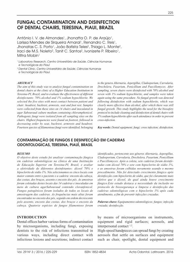
RESUMO O objetivo deste estudo foi analisar contaminação fúngica em cadeiras odontológicas na clínica de uma Instituição de Educação Superior em TeresinaPI, Brasil, e avaliar a efetividade de diferentes desinfetantes: álcool 70% ehipoclorito de sódio 1%. Nós selecionamos os cinco locais commaior contato entre o paciente e a cadeira: encosto da cabeça,das costas, dos braços, assento e encosto dos pés. As amostrasforam coletadas destes locais das 14 cadeiras e inoculadas emmeio de cultura agarSabouraud contendo cloranfenicol.Fungos patogênicos foram isolados de todos os locais deamostragem das cadeiras. As frequências mais altas foramencontradas no encosto dos pés, seguido em ordem decrescentepelo assento, encosto das costas, dos braços e encosto dacabeça. Quatorze espécies de fungos filamentoso foram
identificados, pertencente aos gêneros Alternaria, Aspergillus,Cladosporium, Curvularia, Drechslera, Fusarium, Penicilliume Paecillomyces. Após a coleta, sete cadeiras foram desinfe tadas com álcool 70% e sete com hipoclorito de sódio 1%, e as amostras foram colhidas novamente usando o mesmoprocedimento. Não foi detectado crescimento fúngico apósdesinfecção com hipoclorito de sódio, que foi claramente maisefetivo que o álcool, do qual ainda houve crescimentofúngico.Este estudo destaca a necessidade da inclusão noprotocolo de biossegurança a limpeza e desinfecção dascadeiras odontológicas com o hipoclorito 1% após cadaatendimento, a fim de prevenir infecções cruzadas.
Palavras chave: Equipamentos odontológicos; fungos; infecçãocruzada; desinfecção.
INTRODUCTIONDental offices harbor various forms of contaminationby microorganisms, including fungi, exposingdentists to the risk of infections transmitted invarious ways, including direct contact withinfectious lesions and secretions; indirect contact
by means of microorganisms on instruments,equipment and rigid surfaces; aerosols, andinterpersonal contact 1,2.Highspeed handpieces can spread fungi by creatingaerosols that settle on surfaces and equipment such as chair, spotlight, dental equipment and
ABSTRACTThe aim of this study was to analyze fungal contamination ondental chairs at the clinic of a Higher Education Institution inTeresinaPI, Brazil, and to evaluate the effectiveness of differentdisinfectants: 70% alcohol and 1% sodium hypochlorite. Weselected the five sites with most contact between patient andchair: headrest, backrest, armrests, seat and foot rest. Sampleswere collected from these sites on 14 chairs and inoculated inagar Sabouraud culture medium containing chloramphenicol.Pathogenic fungi were isolated from all sampling sites on thechairs. Highest frequencies were found on footrest, followed indecreasing order by seat, backrest, armrests and headrest.Fourteen species of filamentous fungi were identified, belonging
to the genera Alternaria, Aspergillus, Cladosporium, Curvularia,Drechslera, Fusarium, Penicillium and Paecillomyces. Aftersampling, seven chairs were disinfected with 70% alcohol andseven with 1% sodium hypochlorite, and samples were takenagain using the same procedure. No fungal growth was detectedfollowing disinfection with sodium hypochlorite, which wasclearly more effective than alcohol, after which there was stillfungal growth. This study highlights the need for the biosafetyprotocol to include cleaning and disinfection of dental chairs with1% sodium hypochlorite after each attendance in order to preventcrossinfection.
Key words: Dental equipment; fungi; cross infection; disinfection.
225
Vol. 29 Nº 3 / 2016 / 225-229 ISSN 1852-4834 Acta Odontol. Latinoam. 2016
FUNGAL CONTAMINATION AND DISINFECTION OF DENTAL CHAIRS, TERESINA, PIAUI, BRAZIL
Antônio I. V. de Almondes1, Jhonatta O. P. de Araújo1, Larissa Mendes de Sirqueira Amaral1, Renandro C. Reis1, Jhonatas C. S. Porto1, João Batista Teles2, Thiago L. Monte2, Iraci de M.S. Noleto2, Tanit C. Santos2, Ivonizete P. Ribeiro1, Mitra Mobin1
1 Laboratory Research, Centro Universitário de Saúde, Ciências Humanas e Tecnológicas do Piauí
2 Dental Clinic, Centro Universitário de Saúde, Ciências Humanas e Tecnológicas do Piauí.
CONTAMINAÇÃO DE FUNGOS E DESINFECÇÃO EM CADEIRAS ODONTOLOGICAS, TERESINA, PIAUÍ, BRASIL
AOL32016:32011 15/02/2017 12:51 Página 225
instruments. This is particularly risky when airconditioning further aggravates the occurrence offungi and the area is not cleaned regularly 3,4.Biosafety in dentistry includes a set of actions to protect staff and patients in a clinical setting,including ergonomic practices, control of physicaland chemical hazards, use of specific protocols, andappropriate handling of products and equipment, inaddition to the sterilization process, disinfection,antisepsis, use of barriers and individual protectiveequipment (IPE).5
Controlling disease transmission is a challenge todentists, since the oral cavity contains microorga nisms, many of which can cause diseases ranging froma mild cold to pneumonia and from tuberculosis toherpes. These infections may be transmitted by
contaminated saliva, blood, fluid from the gingivalsulcus, and even the patient’s respiratory secretions 2,6.This study analyzed fungal contamination on dentalchairs at the clinic in a Higher Education Institutionin TeresinaPI, and evaluated the effectiveness of twodisinfecting agents: 70% alcohol and 1% sodiumhypochlorite.
MATERIAL AND METHODSThis was a quantitative and descriptive studyperformed at the Research Laboratory of theUniversity Center UNINOVAFAPI in TeresinaPIfrom January to November 2015, after authorizationfrom the management.Samples were collected from 14 (93.3%) of thedental chairs by rubbing sterile swabs soaked in
226 Antônio I. V. de Almondes, et al.
Acta Odontol. Latinoam. 2016 ISSN 1852-4834 Vol. 29 Nº 3 / 2016 / 225-229
Fig. 1: Micromorphology of filamentous fungi isolated in dental chairs of a Higher Education Institution (HEI):A Penicillium oxalicum; B Aspergillus flavus; C Fusarium aff. incarnatum; D Aspergillus carnus; E Drechslera biseptata; F Cladosporium cladosporioides; G Aspergillus niger; H Curvularia clavata; I Curvularia brachyspora; J Penicillium piceum; K Cladosporium oxysporum; L Penicillium decumbens.
AOL32016:32011 15/02/2017 12:51 Página 226

saline on the five sites selected: headrest, backrest,armrests, seat and footrest.From each sample, 100µL were inoculated in Petridishes containing Sabouraud Dextrose agar(DifcoTM) culture medium plus chloramphenicol(0.05g/L) and incubated at room temperature toenable the growth of the fungal colonies. After growth of the colonies, microcultures weremounted for recognition of species using identifica tion keys previously described7,8.The same procedure was used to test the disinfectantefficacy of 70% ethanol and 1% sodium hypochlorite.For statistical analysis, the chisquare test for goodnessof fit was applied with a significance level (α) of 5%.
RESULTSWe identified 14 species belonging to the generaAlternaria, Aspergillus, Cladosporium, Curvularia,Drechslera, Fusarium, Penicillium and Paecillomyces(Fig.1).Aspergillus niger was the most frequent species,being found at all sites of the dental chair. Curvulariaclavata and Alternaria infectoria were identifiedfrom three site seach (Table 1).The region with the highest level of contaminationwas footrest, with 50.0%, followed by seat
(42.9%), backrest and armrest (35.7% each), andfinally headrest (21.4%) (Table 2).Following disinfection with 70% alcohol, fungalgrowth occurred in the samples from all sites, whereasfollowing disinfection with 1% hypochlorite, nofungal growth was observed.
DISCUSSIONDental chairs can become contaminated by fungi inseveral ways: via air conditioning, failure to applybiosafety standards, lack of internal protocolsand/or failure to comply with them, and invasiveprocedures performed during treatment.
Contamination and Disinfection of Dental Chairs 227
Vol. 29 Nº 3 / 2016 / 225-229 ISSN 1852-4834 Acta Odontol. Latinoam. 2016
Table 1: Presence of fungal species per dental chair site at a clinic at a Higher Education Institution (HEI).
Fungal Species Regions
Headrest Backrest Seat Armrest Footrest
Fusarium aff. Incarnatum •
Paecilomyces variotii •
Curvularia lunata • • •
Alternaria infectoria • • •
Aspergillus niger • • • • •
Aspergillus flavus • •
Cladosporium oxysporum •
Cladosporium cladosporioides • •
Drechslera biseptata •
Penicillium decumbens • •
Penicillium oxalicum •
Aspergillus carneus •
Penicillium piceum
Curvularia brachyspora
Source: Research Laboratory UNINOVAFAPI.
Table 2: Frequency of fungi on the dental chair, Teresina – PI, 2015.
Dental chair sites Number of fungal species %*
Headrest 3 21.4
Backrest 5 35.7
Seat 6 42.9
Armrest 5 35.7
Footrest 7 50.0
Total found 14 100.0
(*) amounts to more than 100% because more than one type of fungus may be found per location.
AOL32016:32011 15/02/2017 12:51 Página 227

All the species found in this study are pathogenicand may cause infections ranging from cutaneousto systemic infections. Aspergillus niger, the mostcommon species in this study, causes pulmonaryaspergillosis, endophthalmitis, endocarditis, perito nitis and cutaneous infections7. The table 3 showsthe main diseases caused by the species found inthis study.There is high risk of crossinfection in the dentaloffice as a result of the invasive proceduresperformed and environmental contamination bybiological agents or bioaerosols which may comefrom internal or external air conditioning, furnitureand carpets 2, 3, 9.Mobin and Salmito found the following pathogenicfungal genera in air conditioners of an Intensive CareUnit (ICU): Acremonium, Aspergillus, Paecillomyces,Penicillium, Trichoderma, Cladosporium, Curvulariaand Nigrospora. Several other studies show that airconditioners provide a favorable environment forfungal growth, thus actively contributing to worseninghealth status of patients, whether in ICUs or dentaloffices 10.Sousa and Fortuna studied air conditioned dentaloffices in Bahia, and in all cases found the highestfrequency of microorganisms such as Aspergillus
and Fusarium near the spittoon 3. This might be dueto highspeed drills spreading microorganismsthrough aerosols, which may become attached toequipment and accessories as well as remainingairborne.A study comparing microbial load between a dentalclinic and a nondental public area found that therisk in the dental clinic may be greater than in thepublic area due to the diversity of microorganisms,susceptibility of the host and exposure time. Inaddition to bacteria, they found fungi, as Aspergillusniger and Aspergillus flavus 11.Another study reports high number of aerosol andbioaerosol particles during dental procedures and avariety of microorganisms present in a dental office,including the fungal genera Penicillium, Cladosporiumand Alternaria 9.Internal air conditioning also contributes toproliferation and spread of fungi in dental offices.Recirculating air and airing the room after eachpatient is recommended to prevent accumulation offungal spores 10.After disinfecting the chairs with 70% alcohol,there was still fungal growth from the samples,showing the method to be less effective than use of1% sodium hypochlorite. Although alcohol is a
228 Antônio I. V. de Almondes, et al.
Acta Odontol. Latinoam. 2016 ISSN 1852-4834 Vol. 29 Nº 3 / 2016 / 225-229
Table 3: Disease occurrence caused by fungi isolated from dental chairs from a HEI in Teresina - PI.
Fungal Species Diseases7
Penicillium piceum Raper&Fennell Fungaemia
Penicillium oxalicum Currie & Thom Eye infection
Aspergillus flavus Link: Fr. Allergic bronchial aspergillosis, lung infections, ear infections, sinus infections and endocarditis.
Fusarium aff. incarnatum (Rob.) Sacc. Endocarditis
Paecilomyces variotii Bain. Pneumonia, sinusitis, endophthalmitis
Drechslera biseptata (Sac. & Roum.) Richardson & Fraser Sinusitis
Cladosporium cladosporioides (Fres.) (de Vries) Lung and cutaneous infection
Cladosporium oxysporum Berk. & Curt. Keratitis and cutaneous infection
Aspergillus carneus (v. Tiegh.) Blochwitz Lung infections
Curvularia clavata Jain Sinusitis, cerebritis
Alternaria infectoria Simmons Phaeohyphomycosis
Penicillium decumbens Thom Fungaemia
Aspergillus niger v. Tiegh. Pulmonary aspergillosis, endophthalmitis, endocarditis, peritonitis, onychomycosis, cutaneous infections
Curvularia brachyspora Boedijn Keratitis and cutaneous infection
Source: De Hoog et al. 2000.
AOL32016:32011 15/02/2017 12:51 Página 228

commonly used disinfectant, the results of this andof other studies suggest that it is ineffective againstfungi 12.Regarding the action of alcohol on bacteria, severalstudies claim that its effect is more bacteriostaticthan bactericidal. Moreover, there are studies thatclaim that the use of 70% alcohol is inappropriate
for removing saliva layers on instruments, and ithas been demonstrated that even water is moreappropriate than alcohol for removing blood andorganic matter 13. The biosafety protocol should therefore includecleaning and disinfecting chairs with 1% hypochloriteafter each patient in order to prevent crossinfection.
Contamination and Disinfection of Dental Chairs 229
Vol. 29 Nº 3 / 2016 / 225-229 ISSN 1852-4834 Acta Odontol. Latinoam. 2016
ACKNOWLEDGEMENTSThe authors thank Edivan Pereira da Silva, responsible for themicrobiology laboratory, for technical support to the research.
CORRESPONDENCEDra. Mitra MobinCentro Universitário da Saúde, Ciências Humanas e Tecnológicas do Piauí.Rua Vitorino Orthiges Fernandes, 6123 – Uruguai Teresina Piauí, Brasil. CEP: 64073505 [email protected]
REFERENCES1. Cardoso CT, Pinto Júnior JR, Pereira EA, Barros LM, Freitas
ABDA. Contaminação de Tubos de Resina Composta manipu lados sem barreira de proteção. ROBRAC. 2010; 48: 7175.
2. Costa Arantes D, de Andrade Hage C, Silva do NascimentoL, Sirotheau Correa Pontes F. Biossegurança aplicada àOdontologia na Universidade Federal do Pará, Cidade deBelém, Estado do Pará, Brasil. Rev PanAmaz Saúde.2015;6:1118.
3. Silva de Sousa, K, Fortuna, JL. Microrganismos em ambien tes climatizados de consultórios odontológicos em umacidade do extremo sul da Bahia. Revista Baiana de SaúdePública 2011; 35: 250263. DOI: 10.5327/Z010002332015390300002.
4. Vilibor Xavier F, dos Santos Paiva MC, Roselino RibeiroAL, Goncalves Krakhecke A. Fungos potencialmente pato gê nicos isolados de água de equipos odontológicos. JOdontol FACIT.2015; 2:2228.
5. Aleixo RQ, Queiroz RC, Custódio VC, Moura JA.Contaminação dos tubos de resina composta utilizados naclínica odontológica. Clipe Odonto– UNITAU 2010; 1: 3945.
6. Azeredo F, Macedo de Menezes L, Medina da Silva R, DeonRizzatto SM, Gressler Garcia G, Revers K. Análisesmicrobiológicas de alicates ortodônticos.Dental Press JOrthod 2011; 16: 103112.
7. De Hoog GS, Guarro J, Gene J, Figueras MJ. Atlas ofclinical fungi. Washington: ASM Press, 2000.1126p
8. Lacaz CS, Porto E, Martins JEC,HeinsVaccari EM, MeloNT. Tratado de micologia Médica. São Paulo:Sarvier, 2002.1120p
9. Polednik B. Aerosol and bioaerosol particles in a dentaloffice. Environ Res. 2014; 134: 405409.
10. Mobin M, do Amparo Salmito M. Microbiota fúngicados condicionadores de ar nas unidadesde terapiaintensiva de Teresina, PI. Rev Soc Bras Med Trop. 2006;39: 556559.
11. Kimmerle H, WiedmannAlAhmad M, Pelz K, Wittmer A,Hellwig E, AlAhmad A. Airborne microbes in differentdental environments in comparison to a public area. ArchOral Biol 2012; 57: 689696.
12. da Silva Aquino I, Porto JC, da Silva JL, Morais KF, CoelhoFA, de Sousa Lopes T, Ribeiro IP, Noleto IS, do AmparoSalmito M, Mobin M. Evaluation of disinfectants forelimination of fungal contamination of patient beds in areference hospital in Piauí, Brazil. Environ Monit Assess.2016; 188: 24.
13. Ferreira AM, de Andrade D, Rigotti MA, de Almeida MT,Guerra OG, dos Santos Junior AG.Assessment of disinfectionof hospital surfaces using different monitoringmethods.RevLat Am Enfermagem. 2015; 23: 466474.
AOL32016:32011 15/02/2017 12:51 Página 229

230
Acta Odontol. Latinoam. 2016 ISSN 1852-4834 Vol. 29 Nº 3 / 2016 / 230-238
RESUMENEl propósito de este estudio es presentar la casuística delcarcinoma mucoepidermoide de glándulas salivales de pacientesdiagnosticados en el Instituto Nacional de EnfermedadesNeoplásicas “Dr. Eduardo Cáceres Graziani” Lima, Perú, desdeel 2002 hasta el 2012.Realizamos un estudio retrospectivo en el cual fueron incluidossujetos con diagnóstico primario de carcinoma mucoepidermoideen glándulas salivales. Entre enero de 2002 y diciembre de 2012,se registraron 51 casos. El número de pacientes de sexo femeninofue mayor, con 28 casos (54,9%) y con respecto a la distribuciónpor edades, el 33,3% de los pacientes eran menores de 30 añosde edad. El dolor fue uno de los síntomas principales. El 74,5%
de los carcinomas mucoepidermoides se localizaron en laglándula parótida. De los hallazgos obtenidos se concluye principalmente que enlo que respecta a la distribución epidemiológica de edad ygénero de los 51 casos analizados estas variaron en el mismorango de otros estudios. También se distingue que el mayornúmero de casos estuvieron localizados en glándulas salivalesmayores, dato en concordancia con otras poblaciones reporta das. Las demás características presentaron una distribuciónhomogénea.
Palabras clave: Neoplasias de las glándulas salivales; carcinomamucoepidermoide; epidemiología; neoplasias.
INTRODUCTIONNeoplasms of the major and minor salivary glandsare a challenge for clinicians and histopathologistsbecause they are infrequent and have a wide rangeof histological, clinical, epidemiological anddevelopmental characteristics1.In salivary glands, the most frequent benign tumoris pleomorphic adenoma and the most frequent
malignant tumor is mucoepidermoid carcinoma(MC), according to the Armed Forces Institute ofPathology (AFIP) in Washington2.MC is defined as a malignant epithelial neoplasmof the salivary glands caused by proliferation ofsecretory cells, formed by a variable proportion of mucous, epidermoid, intermediate, columnar and clear cells, often with a cystic component. Its
ABSTRACTThe aim of this study is to present the casuistic of mucoepidermoidcarcinoma of salivary glands in patients diagnosed at “Dr.Eduardo Cáceres Graziani” National Institute for NeoplasticDiseases, Lima, Perú.From January 2002 to December 2012, 51 cases werediagnosed as mucoepidermoid carcinoma. The number offemale patients was higher, with 28 cases (54.9%), andregarding age distribution, 33.3% of the patients were under 30 years old. Pain was one of the main symptoms, and
74.5% of the mucoepidermoid carcinomas were located in theparotid gland.It is concluded that epidemiology regarding age and gender ofthe 51 cases analyzed was in the same range as other studies,and that most cases were located in major salivary glands, inagreement with reports on other populations. Other characte ristics showed a homogeneous distribution.
Key words: Salivary gland neoplasms; mucoepidermoid tumor,epidemiology, neoplasms.
MUCOEPIDERMOID CARCINOMA OF THE SALIVARY GLANDS. A RETROSPECTIVE STUDY OF 51 CASES AND REVIEW OF THE LITERATURE
Janet O. Guevara-Canales1, Rafael Morales-Vadillo1, Guillermo Guzmán-Arias1, Carlos E.Cava-Vergiú1, Henry Guerra-Miller2, Jaime E. Montes-Gil2
1 Universidad de San Martín de Porres (USMP), Facultad de Odontología, Instituto de Investigación, Lima, Perú.
2 National Institute for Neoplastic Diseases, Lima, Peru.
CARCINOMA MUCOEPIDERMOIDE DE GLÁNDULAS SALIVALES. ESTUDIO RETROSPECTIVO DE 51 CASOS Y REVISIÓN DE LA LITERATURA
AOL32016:32011 15/02/2017 12:51 Página 230

biological behavior is related to the histologictumor grade (low, intermediate or high)24.MC was studied, described and reported for the firsttime by Stewart, Foote and Becker in 1945with regard to tumors with double metaplasia ordouble constitution, epidermoid cells and mucousproducing cells5.
EtiologyLittle is known regarding the etiological agents ofsalivary gland neoplasms. Apparently, lowdoseradiotherapy used in benign disorders such as acneor obstructive lesions of the lymphoid tissue in theoral cavity or nasopharynx is the main factorinvolved in the genesis of these tumors. They havenot been associated to radiotherapy for thetreatment of malignant neoplasms, suggesting thatirradiation at high doses is a lower risk factor thanirradiation at low doses. Local trauma has also beenimplicated in the genesis of MC in minor salivaryglands6.
EpidemiologyNeoplasms of the salivary glands are rare, accountingfor less than 2%7 of all human neoplasms and about3% of head and neck tumors7,8. Malignant tumors ofthe salivary glands are infrequent and account forabout 3% of all malignant neoplasms of head andneck8.Malignant neoplasms originating in minor salivaryglands are less than 25% of all salivary neoplasms9,10,
and most of the tumors arising in minor salivaryglands are malignant9,11.MC is the most common malignant neoplasm in majorand minor salivary glands12, accounting fornearly 30%of all malignant neoplasms of the salivary glands.Approximately half the MCs occur in the majorsalivary glands, with 80% in the parotid gland, 8 to13% in the submaxillary gland and 2 to 4% in thesublingual gland13. Central mucoepidermoid tumorslocated in the jaws are a recognized entity. Browandand Waldron reported 9 cases and examined the 41previously published cases14. Some authors report that MC is evenly distributedbetween sexes15, but most authors report thatglandular MC is more frequent in females, with afemale:male ratio of 2:16,16 or 3:217,18.The onset occurs between the 2nd and 8th decades oflife, and it is the most frequent malignant tumor inpersons under 20 year of age, in whom there is apredilection for the hard palate. There is also clearpredilection for white race6,16.
Clinical manifestationBetween 70% and 80% of neoplasms in general ofthe salivary glands are located in the parotid, whilethe palate is the most common site for neoplasmsof minor salivary glands2. Similarly, just over 70%of MCs are located in major glands, with the parotidbeing the most frequent site19, (with almost half thecases) (Fig. 1 and 2), followed by the submandibulargland and sublingual gland3. MCs represent 23% of
Mucoepidermoid Carcinoma of Salivary Glands 231
Vol. 29 Nº 3 / 2016 / 230-238 ISSN 1852-4834 Acta Odontol. Latinoam. 2016
Fig. 1: Female patient, 30years old. Tumorous lesion in
left parotid gland. Patientcomplained of pain and
paresthesia. One year ofillness with slow, progressive
growth. Diagnosed with highgrade MC and died six
months later, after radiationtreatment.
Fig. 2: Male patient, 48 yearsold. Tumorous lesion in left
parotid gland. Patientcomplained of paresthesia.Diagnosed with high grade
MC. Seventeen months aftersurgery and radiotherapy,
presented pulmonarymetastasis and died
5 months later.
AOL32016:32011 15/02/2017 12:51 Página 231

all tumors in the minor salivary glands20, with thepalate being the most commonly affected site (withalmost half the cases in hard palate, the second mostfrequent location after parotid gland), and they arefound less frequently in other minor salivary glandssuch as those of the tongue, floor of the mouth, gum,lips and cheek mucosa, the ectopic salivary tissuebeing another location, though exceptional.Clinically, in major glands, MCs appear as solitary,asymptomatic enlargements of the parotid body orpole, or of the submaxillary region8. In minor glandsthey appear as blue or purplishred fluctuatingmasses with a smooth surface, and are often clinicallymistaken for mucoceles21.The average latency period is one year, but mayvary widely. MCs sometimes grow rapidly after aperiod of quiescence. In highgrade lesions there is onset of pain, facial paralysis and fixation inneighboring structures8.Discovery in minor glands is sometimes accidentalduring a routine mouth exploration. Rapidlygrowing tumors are very unusual. The surface isusually smooth, but if it is ulcerated, it is usuallyassociated to more aggressive forms. If it is locatedat the base of the tongue it may cause dysphagia,while if it affects the bone it may cause insensitivityin teeth6.
HistologyAccording to the bicellular theory, salivary glandtumors are formed by22:a) Duct luminal cells and/or acinar cells plus
myoepithelial cells,b) Duct luminal cells or acinar cells, orc) Myoepithelial cells only.
Histopathology thus identifies mucosecretory,epidermoid, intermediate8,17,18,21, columnar or clearcells, proliferating alone or in different combinations,in a cystic or solid pattern8,21.In addition to this cell pattern, there is extracellularmatrix produced by the neoplastic myoepithelialcells, collagen, elastic fibers, glycoproteins,glycosaminoglycans and proteoglycans23.The anatomical pathology shows they are partiallyencapsulated, with full encapsulation being veryrare8,21.Most authors consider three grades of differen tiation8,21 depending on intracystic component,neural invasion, necrosis, mitotic activity and
pleomorphism24. The histological grade in thisstudy was classified following AFIP25, published inthe World Health Organization Classification oftumours26 and shown in Table 1 and Figs. 3, 4 and 5.
DiagnosisThe differential diagnoses considered are: necrotizingsialometaplasia (of the palate), mucocele, invertedpapilloma or cystadenoma, cystadenocarcinoma,primary or metastatic epidermoid carcinoma, andlowgrade polymorphic adenocarcinoma17.The most useful and popular techniques forevaluating neoplasms of the salivary glands arecurrently Computerized Axial Tomography (CAT)and biopsy by needle aspiration. The latter,however, should be taken with precaution becauseits diagnostic precision is 6080%, sometimesrequiring a repetition of the aspiration or surgicalbiopsy. The histological information from thebiopsy and delimitation of the lesion provided byCAT enable appropriate medical and surgicalmanagement27.
TreatmentThe treatment depends on the location, clinicalaspect and histopathological grade. Lowgrade MCis generally treated with surgery only, while highgrade tumors also require radiation and dissectionof neck lymph nodes28. The management ofintermediategrade tumors is controversial, perhapsreflecting the controversy in tumor classification29.For tumors in accessory salivary glands, surgicalexcision is recommended, leaving 12 cm safetymargins around the tumor8.For treatment of tumors in major glands, glandexeresis is recommended (superficial or deepparotidectomy, submaxilectomy)8.Cervical lymph notes are removed when lymphnodes are clinically affected (higher incidence oflymph node metastasis in submaxillary location)8.Radiotherapy after surgical treatment with tumorfree margins does not increase local control, as itdoes in advanced cases, cases with infiltratedresection margins and cases located at the base ofthe tongue30. Response to chemotherapy is low (bestresults have been achieved with methotrexate ycisplatin)31.Standard treatment for the main types of salivarygland cancer is surgical resection with adjuvantradiation to reduce failure rates3234 and even though
232 Janet O. Guevara-Canales, et al.
Acta Odontol. Latinoam. 2016 ISSN 1852-4834 Vol. 29 Nº 3 / 2016 / 230-238
AOL32016:32011 15/02/2017 12:51 Página 232

the role of adjuvant chemotherapy has not beenproven, it has been used to treat distant metastasisand nonexcisable disease, and to reduce the effectsof local/regional recurrence35.Adjuvant radiotherapy has been shown to have anadvantage in survival of patients with tumors largerthan 4 cm, but little benefit for patients with smalltumors, suggesting that, together with the marginsinvolved, tumors larger than 4 cm are an absoluteindication for postsurgical radiotherapy32,3638.Further progress in therapy is needed to improvethe outcomes of histologic highgrade disease39.
PrognosisPrognosis depends on clinical stage40, (related toanatomical location41,42), histologic grade (patientswith low or intermediategrade tumors have localcontrol and favorable survival rates39) and treatment40.Variable prognostic factors have also been reported,such as neural invasion, vascular invasion, and localor distant metastasis, which are associated toclinical outcome for patients25.Other authors mention the same factors thatinfluence survival, such as histologic grade, clinicalstage, paralysis of facial nerve and lymph node
Mucoepidermoid Carcinoma of Salivary Glands 233
Vol. 29 Nº 3 / 2016 / 230-238 ISSN 1852-4834 Acta Odontol. Latinoam. 2016
Table 1: Histopathologic features used in grading MC. World Health Organization26.
Histopathologic feature Point value
Intracystic component <20% 2
Neural invasion 2
Mitosis (≥4/10 HPFs*) 3
Necrosis 3
Anaplasia 4
Tumor grade Point score
Low grade 0 - 4
Intermediate grade 5 - 6
High grade ≥7
*HPF, High-Power Fields
Fig. 3: MC, low grade. Histological image showing cysticstructures lined with mucinous, squamous and intermediatecells with slightly atypical nuclei and low mitotic activity.Adjacent connective tissue without perineural invasion (H&EOrig. Mag. 100x).
Fig. 4: MC, intermediate grade. Histological image showingtumor with small cystic structures, predominance ofintermediate cells, with mucosecretory and epidermoid cells(H&E Orig. Mag. 100x).
Fig. 5: MC, high grade. Histological image showingproliferations of solid islands of epidermoid and intermediatecells with few mucinous cells, with atypical nuclei and greatermitotic activity (H&E Orig. Mag. 100x).
AOL32016:32011 15/02/2017 12:51 Página 233

metastasis, in addition to location (worse prognosis insubmaxillary), age and sex (better prognosis in youngpeople and females). Mucoepidermoid tumor is oneof the few tumors of salivary glands in which flowcytometry has high prognostic value, with greatersurvival in patients with diploid DNA patterns6.Regarding histologic grade, it has been reported thatat the same histopathological grade, tumors in theparotid gland have better prognosis than tumors inthe submandibular gland43. Prognosis is better forlowgrade lesions and for highgrade lesions whenthey arein stage I or II32,36,37,44. MC prognosisappears to depend largely on tumor grade, withreports of 5year MC survival rates of 92%100%for low grade, 6292% for intermediate grade and043% for high grade tumors25. MCs with highgrade malignancy have a 50% probability ofpresenting metastasis17.Some molecular factors of malignant cells alsoinfluence survival rate, e.g. p27, which is a highlyfavorable prognostic factor for MC, whereas Ki67is not identified as a survival indicator45.Even with complete resection, there is still asubstantial risk of local recurrence (16%27%) anddistant metastasis (13%26%)46,47.The aim of this study is to present the epidemiologyof salivary gland MC through thecasuistic inpatients diagnosed at the “Dr. Eduardo CáceresGraziani” National Institute for Neoplastic Diseasesfrom 2002 to 2012.
MATERIALS AND METHODSThis is an observational, descriptive, crosssectional,retrospective study which included subjects withprimary diagnosis of mucoepidermoid carcinomaof the salivary glands diagnosed at “Dr. EduardoCaceres Graziani” National Institute for NeoplasticDiseases, Lima, Peru, from January 2002 toDecember 2012. Clinical records with incompletedata were excluded.The study was approved by the institutional reviewboard (protocol: INEN 13:27) and conducted inaccordance with the World Medical AssociationDeclaration of Helsinki on medical researchprotocols and ethics.The collected data were transferred to a MicrosoftExcel program, on which the table was prepared. The analysis was performed using Windows XP®
Operative System (Washington, USA), with theassistance of the statistical program SPSS
(Statistical Package for Social Sciences) version20.0 for Windows (SPSS, Chicago, IL, USA).All values found in the different statistical testswere considered with a significance level of 0.05(p<0.05).
RESULTSBetween January 2002 to December 2012, 349 newcases were found of malignant tumors in salivaryglands, of which 4.61% (51)cases were mucoepidermoid carcinoma.All cases were diagnosed by pathological anatomystudies. Table 2 shows the distribution of the differentcharacteristics evaluated according to sex. Thereare more female patients (28 cases; 54.9%;female:male ratio 1.2:1). Age range was 12 to 88years, with 64.71% of patients aged 60 years or less.In most cases (62.75%), tumor size was smaller orequal to 4 cm at the time of the first visit, and themost frequent location was parotid gland (74.51%).No significant difference was found for thesecharacteristics. Among the signs and symptomsevaluated, the presence of paresthesia wasstatistically significant (p=0.038). Considering theTNM (T: primary tumor, N: cervical nodules, andM: distant metastasis) staging system for salivarygland carcinoma, cases were most frequently stageT2 (43.14%), which are tumors measuring 24 cmwith no macroscopic extracapsular extension, N0(86.27%), which are lesions without metastasis inregional nodules and M0 (68.63%), which is whenthere are tumors without distant metastasis. Withregard to clinical stage, the largest number of caseswas in clinical stage IV C (31.37%), followed byclinical stage II (29.41%). Regarding histologicalgrade, cases were distributed evenly between lowand high grade, with 39.22% each, and regardingtreatment, surgery combined with radiotherapy wasthe treatment of choice, with 42.18% of the cases.
DISCUSSIONMost epidemiological studies of MC have foundfemales to be affected more frequently than males,e.g Goode et al. studied 234 MC of the majorsalivary glands, finding that females accounted for51.3%43. In our study, females accounted for 54.9%,a value similar to those reported by Villavicencio etal.48 and Schwarz et al.49. Some authors report a 3:2female:male ratio16,18, and McHugh et al. report a
234 Janet O. Guevara-Canales, et al.
Acta Odontol. Latinoam. 2016 ISSN 1852-4834 Vol. 29 Nº 3 / 2016 / 230-238
AOL32016:32011 15/02/2017 12:51 Página 234

Mucoepidermoid Carcinoma of Salivary Glands 235
Vol. 29 Nº 3 / 2016 / 230-238 ISSN 1852-4834 Acta Odontol. Latinoam. 2016
Table 2: Clinical pathological features of subjects with MC of the salivary glands.
Clinical characteristics Female Male p Value
n % n %
Age (months) Average 43.107 49.04
Minimum 18 12
Maximum 79 88
Up to 60 years 20 60.6 13 39.4 0.268
Over 60 years 8 44.4 10 55.6
Size (cm) Average 4.25 3.56 5.17 2.96
Minimum 1.5 1
Maximum 20 12
Up to 4 cm 20 62.5 12 37.5 0.157
Over 4 cm 8 42.1 11 57.9
Location Parotid gland 18 47.4 20 52.6 0.165
Submaxillary gland 5 71.4 2 28.6
Minor glands 5 83.3 1 16.7
Signs Ulcer No 24 57.1 18 42.9 0.487
Yes 4 44.4 5 55.6
Pain No 17 54.8 14 45.2 0.991
Yes 11 55.0 9 45.0
Paresthesia No 25 62.5 15 37.5 0.038*
Yes 3 27.3 8 72.7
Dysphagia No 27 56.3 21 43.7 0.439
Yes 1 33.3 2 66.7
Trismus No 27 54.0 23 46.0 0.360
Yes 1 100 0 00.0
TNM (T) T1 7 70.0 3 30.0 0.235
T2 13 59.1 9 40.9
T3 6 50.0 6 50.0
T4A 1 16.7 5 83.3
T4B 1 100 0 00.0
TNM (N) N0 25 56.8 19 43.2 0.224
N1 3 75.0 1 25.0
N2A 0 00.0 2 100
N2B 0 00.0 1 100
TNM (M) M0 20 57.1 15 42.9 0.634
M1 8 50.0 8 50.0
Clinical stage I 4 66.7 2 33.3 0.051
II 9 60.0 6 40.0
III 4 57.1 3 42.9
IV A 1 50.0 1 50.0
IV B 2 40.0 3 60.0
IV C 8 50.0 8 50.0
Histological grade Low grade 14 70.0 6 30.0 0.068
Intermediate grade 7 63.3 4 36.4
High grade 7 35.0 13 65.0
Treatment Sg 10 66.7 5 33.3 0.504
Sg + Rt 10 47.6 11 52.4
Sg + Rt + Cht 0 0.0 1 100
Rt + Cht 1 100 0 0.0
Rt 7 53.8 6 46.2
* Statistically significant valueTNM: Classification of Malignant Salivary Gland Tumor Stages.Cht Chemotherapy; Rt Radiotherapy; Sg Surgery.
AOL32016:32011 15/02/2017 12:51 Página 235

1:1.2 male:female ratio39. Another recent study isconsistent with the above, finding 67% of the casesin females29.MC is the most common malignant neoplasm of thesalivary glands in persons over 40 years of age13,50.Chomette et al. report that the onset of MCaccording to age is variable and may occur betweenthe 2nd and 8th decades of life6, in agreement withour study, in which on set ranged from the 2nd to 9th
decades of life. Villavicencio et al. report onsetfrom the 4th to 6th decades of life48, and Rapidis etal. report 3rd to 9th decades of life17. Average age inall the aforementioned studies was the 6th decade oflife, thus, average age of on set is reported as 52.2years by Chomette et al.6; 54.3 years byVillavicencio et al.48, and 56.7 years by Rapidis etal.17. These values are similar to the average agefound in our study, which was 45.78 years. It isworth noting that this study found 8 cases ofpatients aged 19 years or under over the 10yearstudy period (0.8 cases per year), similar to findingsreported by Techavichit et al. of 14 cases over aperiod of 15 years (0.9 cases per year) in patientsaged 19 years or under51.Villavicencio et al. report that the clinical size ofthe primary tumor varies according to theanatomical site affected; with median size 8 cm (4 to 10 cm) in submaxillary gland and 3 cm (1 to5 cm) in minor salivary glands48. Rapidis et al.found no statistically significant differencebetween tumor size according to site17. Katabi et al.report that tumor size was 0.5 to 9 cm, with anaverage value of 1.77 cm29. In our study, the averagesize was 4.67 cm.MC is a neoplasm that usually affects majorsalivary glands, although 10% can arise in minoror accessory salivary glands in the head and neckarea24, including maxillary sinus, nasopharynx,nasal cavity, oropharynx, larynx and trachea. Ourstudy was consistent with this observation, finding74.51% of the cases in parotid gland, 13.73% insubmaxillary gland and 11.76% in minor glands.Ellis et al. report that 74.5% of neoplasms of thesalivary glands in general are found in the parotidgland2, with a percentage identical to the onefound in our study. The location of MC in ourstudy was also consistent with Villavicencio et al.who reported 74.5% in major salivary glands (32cases in parotid and 3 cases in submaxillarygland)48; Védrine et al., who reported 18 cases, of
which 77.78% (14 cases) were in parotid gland,11.11% (2 cases) in submaxillary gland and11.11% (2 cases) in minor salivary glands7;Schwarz et al. who reported 60% (24 cases) inparotid gland, 10% (4 cases) in submandibulargland and 30% (12 cases) in minor glands49, andRapidis et al. who reported 18 cases, of which66.6% (12 cases) were in major salivary glands(10 in parotid and 2 in submaxillary)17. Regardinglocation in minor salivary glands, Triantafillidouet al. report 56.25% in palate40, while in our study,100% of the minor salivary gland cases werelocated in the palate.Signs and symptoms are varied, with noneappearing as characteristic or pathognomonic, butthe most outstanding in our study was pain, inagreement with Villavicencio et al., who report painin 55.3% of their 47 cases48. It is worth noting thatit is statistically significant (p=0.038) that mostpatients do not report paresthesia as a symptom atthe time of the initial clinical examination.Regarding the clinical stage of MC, Schwarz et al.report declining percentages for increasing stages,with 40% (16 cases) Type I, 30% (12 cases) TypeII, 15% (6 cases) Type III and 15% (6 cases) typeIV49. In our study, the highest number of cases wasfound for the last stage, IV (16 cases), followed bystage II (15 cases).In most epidemiological studies presenting data onthe histological grade of the disease, the distributionof the number of MC cases generally declines asthe level of histologic grade rises, i.e. there is aninverse relationship. Rapidi et al. report a declineaccording to histological grade, with 9 tumorsclassified as low grade, 5 as intermediate grade and4 as high grade17; Katabi et al. report 90.38% (47cases) low grade, 3.85% (2 cases) intermediategrade and 5.77% (3 cases) high grade29; Védrine etal. report 68.75% (11 cases) low grade, 18.75% (3cases) intermediate grade and 12.5% (2 cases) highgrade7, and Schwarz et al. report 67.5% (27 cases)low grade, 7.5% (3 cases) intermediate grade and25% (10 cases) high grade49. Our study found equalnumbers of cases for low grade and high grade with39.22% (20 cases) each, and 21.56% (11 cases)intermediate grade. This higher percentage of caseswith high histological grades may be due to thehealth culture in the Peruvian population, sincepeople tend to consult a professional only when thedisease is at an advanced stage.
236 Janet O. Guevara-Canales, et al.
Acta Odontol. Latinoam. 2016 ISSN 1852-4834 Vol. 29 Nº 3 / 2016 / 230-238
AOL32016:32011 15/02/2017 12:51 Página 236

Treatment is fairly well established, with mostpapers reporting surgical treatment and adjuvantradiation therapy, similarly to our casuistic, whichshows that 41.18% was treated thus, which dependson the histologic grade. This is also reflected byfollowup case research such as Chen et al., whichfollows a series of 61 cases, all of which weretreated with surgery and postoperative radiation, forover 10 years52, and other papers which reportcasuistic of surgical treatment only, such as Rapidiset al., who treated 9 cases of parotid gland tumorswith superficial or total parotidectomy, all withradical dissection of neck or suprahyoid dissection,
with one parotid gland case being treated withhemimandibulectomy and suprahyoid dissection17.Based on the abovementioned findings, otherstudies were conducted which, in addition to theepidemiological contribution regarding this salivarygland disease, present the respective patient followup analyses for survival.It is concluded that the epidemiology regarding ageand gender of the 51 cases analyzed was in the samerange as other studies, and that most cases werelocated in major salivary glands, in agreement withreports on other populations. Other characteristicsshowed a homogeneous distribution.
Mucoepidermoid Carcinoma of Salivary Glands 237
Vol. 29 Nº 3 / 2016 / 230-238 ISSN 1852-4834 Acta Odontol. Latinoam. 2016
CorrespondenceDr. JanetOfelia GuevaraCanalesAv. Julio Bayletti 620 San Borja, Lima41, Lima, [email protected]
REFERENCES1. Speight PM, Barrett AW. Salivary gland tumours. Oral Dis
2002; 8:229240.2. Ellis GL, Auclair PL. Atlas of tumor pathology: Tumors of
the major salivary glands. Washington DC, USA: ArmedForces Institute of Pathology (AFIP), 2008.
3. Luna MA. Salivary mucoepidermoid carcinoma: revisited.Adv Anat Pathol 2006; 13:293307.
4. Ozawa H, Tomita T, Sakamoto K, Tagawa T, Fujii R,Kanzaki S, Ogawa K, Kameyama K, et al. Mucoepidermoidcarcinoma of the head and neck: clinical analysis of 43patients. Jpn J Clin Oncol 2008; 38:414418.
5. Stewart FW, Foote FW, Becker WF. Mucoepidermoidtumors of salivary glands. Ann Surg 1945; 122:820844.
6. Chomette G, Auriol M, Tereau Y, Vaillant JM. Mucoepidermoidtumors of minor salivary glands. Clinical and pathologiccorrelations. Histoenzymologic and ultrastructural studies.Ann Pathol 1982; 2:2940.
7. Védrine PO, Coffinet L, Temam S, Montagne K, LapeyreM, Oberlin O, Orbach D, Simon C, et al. Mucoepidermoidcarcinoma of salivary glands in the pediatric age group: 18clinical cases, including 11 second malignant neoplasms.Head Neck 2006; 28:827833.
8. Spiro RH. Salivary neoplasms: overview of a 35yearexperience with 2,807 patients. Head Neck Surg 1986; 8:177184.
9. World Health Organization. Classification of tumours.Pathology and genetics of head and neck tumours. Lyon,France: IARC Press, 2005.
10. Copelli C, Bianchi B, Ferrari S, Ferri A, Sesenna E.Malignant tumors of intraoral minor salivary glands. OralOncol 2008; 44:658663.
11. Chou C, Zhu G, Luo M, Xue G. Carcinoma of the minorsalivary glands: results of surgery and combined therapy. JOral Maxillofac Surg 1996; 54:448453.
12. Auclair PL, Ellis GL. Mucoepidermoid carcinoma. In: EllisGL, Auclair PL, Gnepp DR, editors: Surgical pathology ofthe salivary glands. Philadelphia, USA: W.B. Saunders,1991:269298.
13. Brandwein MS, Ivanov K, Wallace DI, Hille JJ, Wang B,Fahmy A, Bodian C, Urken ML, et al. Mucoepidermoidcarcinoma: a clinicopathologic study of 80 patients withspecial reference to histological grading. Am J Surg Pathol2001; 25:835845.
14. Browand BC, Waldron CA. Central mucoepidermoid tumorsof the jaws. Report of nine cases and review of the literature.Oral Surg Oral Med Oral Pathol 1975; 40:631643.
15. Raspall G. Glándulas Salivales. In: Dias H, editor: CirugíaMaxilofacial Patología quirúrgica de la cara, boca, cabezay cuello. Madrid, España: Editorial Médica Panamericana,1997:439.
16. Myer C, Cotton RT. Salivary gland disease in children: areview. Part 1: Acquired nonneoplastic disease. ClinPediatr (Phila) 1986; 25:314322.
17. Rapidis AD, Givalos N, Gakiopoulou H, Stavrianos SD,Faratzis G, Lagogiannis GA, Katsilieris I, Patsouris E.Mucoepidermoid carcinoma of the salivary glands. Reviewof the literature and clinicopathological analysis of 18patients. Oral Oncol 2007; 43:130136.
18. Kokemueller H, Brueggemann N, Swennen G, Eckardt A.Mucoepidermoid carcinoma of the salivary glands—clinical review of 42 cases. Oral Oncol 2005; 41:310.
19. Pires FR, Pringle GA, de Almeida OP, Chen SY. Intraoralminor salivary gland tumors: a clinicopathological study of546 cases. Oral Oncol 2007;43:463470.
20. Spiro RH, Huvos AG, Berk R, Strong EW. Mucoepidermoidcarcinoma of salivary gland origin. A clinicopathologicstudy of 367 cases. Am J Surg 1978; 136:461468.
21. Ellis GL, Auclair PL, Gnepp DR. Surgical pathology of thesalivary glands. Philadelphia, USA: W.B. Saunders;1991.
AOL32016:32011 15/02/2017 12:51 Página 237

22. Allon I, Vered M, Buchner A, Dayan D. Stromal differencesin salivary gland tumors of a common histopathogenesisbut with different biological behavior: a study withpicrosirius red and polarizing microscopy. Acta Histochem2006; 108:259264.
23. Dardick I. Histogenesis and morphogenesis of salivaryglands neoplasms. In: Ellis GL, Auclair PL, Gnepp DR,editors: Surgical pathology of the salivary glands.Philadelphia, USA: W.B. Saunders, 1991:108.
24. Auclair PL, Goode RK, Ellis GL. Mucoepidermoidcarcinoma of intraoral salivary glands. Evaluation andapplication of grading criteria in 143 cases. Cancer 1992;69:20212030.
25. Ellis GL, Auclair PL. Atlas of tumor pathology: Tumors ofthe major salivary glands. Washington DC, USA: ArmedForces Institute of Pathology (AFIP),1996:173.
26. Barnes EL, Eveson JW AP, Reichart P, Sidransky D. WorldHealth Organization. Classification of tumours. Pathologyand genetics of head and neck tumours. Lyon, France:IARC Press, 2005.
27. Myer C, Cotton RT. Salivary gland disease in children: areview. Part 2: Congenital lesions and neoplastic disease.Clin Pediatr (Phila) 1986; 25:353357.
28. Herd MK, Murugaraj V, Ghataura SS, Brennan PA, AnandR. Lowgrade mucoepidermoid carcinoma of the palateapreviously unreported case of metastasis to the liver. J OralMaxillofac Surg 2012; 70:23432346.
29. Katabi N, Ghossein R, Ali S, Dogan S, Klimstra D, GanlyI. Prognostic features in mucoepidermoid carcinoma ofmajor salivary glands with emphasis on tumour histologicgrading. Histopathology 2014; 65:793804.
30. Hosokawa Y, Shirato H, Kagei K, Hashimoto S, NishiokaT, Tei K, Ono M, Ohmori K, et al. Role of radiotherapy formucoepidermoid carcinoma of salivary gland. Oral Oncol1999;35:105111.
31. Kaplan MJ, Johns ME, Cantrell RW. Chemotherapy forsalivary gland cancer. Otolaryngol Head Neck Surg 1986;95:165170.
32. Armstrong JG, Harrison LB, Spiro RH, Fass DE, StrongEW, Fuks ZY. Malignant tumors of major salivary glandorigin. A matchedpair analysis of the role of combinedsurgery and postoperative radiotherapy. Arch OtolaryngolHead Neck Surg 1990; 116:290293.
33. Leverstein H, van der Wal JE, Tiwari RM, Tobi H, van derWaal I, Mehta DM, Snow GB. Malignant epithelial parotidgland tumours: analysis and results in 65 previouslyuntreated patients. Br J Surg 1998; 85:12671272.
34. Harrison LB, Armstrong JG, Spiro RH, Fass DE, StrongEW. Postoperative radiation therapy for major salivarygland malignancies. J Surg Oncol 1990; 45:5255.
35. Hocwald E, Korkmaz H, Yoo GH, Adsay V, Shibuya TY,Abrams J, Jacobs JR. Prognostic factors in major salivarygland cancer. Laryngoscope 2001; 111:1434149.
36. Renehan AG, Gleave EN, Slevin NJ, McGurk M. Clinicopathological and treatmentrelated factors influencingsurvival in parotid cancer. Br J Cancer 1999; 80:12961300.
37. Slevin N, Frankenthaler R. The role of radiotherapy in themanagement of salivary gland cancer. In: McGurk M,Renehan A, editors: Controversies in the management of
salivary gland disease. Oxford, USA: Oxford UniversityPress, 2001:163172.
38. Frankenthaler RA, Luna MA, Lee SS, Ang KK, Byers RM,Guillamondegui OM, Wolf P, Goepfert H. Prognosticvariables in parotid gland cancer. Arch Otolaryngol HeadNeck Surg 1991; 117:12511256.
39. McHugh CH, Roberts DB, ElNaggar AK, Hanna EY,Garden AS, Kies MS, Weber RS, Kupferman ME.Prognostic factors in mucoepidermoid carcinoma of thesalivary glands. Cancer 2012; 118:39283936.
40. Triantafillidou K, Dimitrakopoulos J, Iordanidis F,Koufogiannis D. Mucoepidermoid carcinoma of minorsalivary glands: a clinical study of 16 cases and review ofthe literature. Oral Dis 2006; 12:364370.
41. Cheuk W, Chan JK. Advances in salivary gland pathology.Histopathology 2007; 51:120.
42. Yih WY, Kratochvil FJ, Stewart JC. Intraoral minor salivarygland neoplasms: review of 213 cases. J Oral MaxillofacSurg 2005; 63:805810.
43. Goode RK, Auclair PL, Barnes GL. Mucoepidermoidcarcinoma of the major salivary glands: clinical andhistopathologic analysis of 234 cases with evaluation ofgrading criteria. Cancer 1998; 82:12171224.
44. Vander Poorten VL, Balm AJ, Hilgers FJ, Tan IB, Keus RB,Hart AA. Stage as major long term outcome predictor in minorsalivary gland carcinoma. Cancer 2000; 89:11951204.
45. Miyabe S, Okabe M, Nagatsuka H, Hasegawa Y, Inagaki A,Ijichi K, Nagai N, Eimoto T, et al. Prognostic significance ofp27Kip1, Ki67, and CRTC1MAML2 fusion transcript inmucoepidermoid carcinoma: a molecular and clinicopathologicstudy of 101 cases. J Oral Maxillofac Surg 2009; 67:14321441.
46. Spiro RH, Armstrong J, Harrison L, Geller NL, Lin SY, StrongEW. Carcinoma of major salivary glands. Recent trends. ArchOtolaryngol Head Neck Surg 1989; 115:316321.
47. Kane WJ, McCaffrey TV, Olsen KD, Lewis JE. Primaryparotid malignancies. A clinical and pathologic review.Arch Otolaryngol Head Neck Surg 1991; 117:307315.
48. VillavicencioAyala B, ReséndizColosia JA, LabastidaAlmendaro S, TorresNúñe MG, PeñaTorres LM, GallegosHernández JF. Prognostic factors in head and neckmucoepidermoid carcinoma. Cir Cir 2008; 76:109117.
49. Schwarz S, Stiegler C, Müller M, Ettl T, Brockhoff G, ZenkJ, Agaimy A. Salivary gland mucoepidermoid carcinoma isa clinically, morphologically and genetically heterogeneousentity: a clinicopathological study of 40 cases with emphasison grading, histological variants and presence of the t(11;19)translocation. Histopathology 2011; 58:557570.
50. Boahene DK, Olsen KD, Lewis JE, Pinheiro AD, PankratzVS, Bagniewski SM. Mucoepidermoid carcinoma of theparotid gland: the Mayo clinic experience. Arch OtolaryngolHead Neck Surg 2004; 130:849856.
51. Techavichit P, Hicks MJ, LópezTerrada DH, Quintanilla NM,Guillerman RP, Sarabia SF, Sayeed H, Nuchtern JG et al.Mucoepidermoid carcinoma in children: asingle institutionalexperience. Pediatr Blood Cancer 2016; 63:2731.
52. Chen AM, Lau VH, Farwell DG, Luu Q, Donald PJ.Mucoepidermoid carcinoma of the parotid gland treated bysurgery and postoperative radiation therapy: clinicopathologiccorrelates of outcome. Laryngoscope 2013;123:30493055.
238 Janet O. Guevara-Canales, et al.
Acta Odontol. Latinoam. 2016 ISSN 1852-4834 Vol. 29 Nº 3 / 2016 / 230-238
AOL32016:32011 15/02/2017 12:51 Página 238
RESUMOO objetivo neste estudo foi avaliar o índice de contaminação dosequipamentos de radiografias digitais intra e extrabucais daclínica de radiologia odontológica de uma instituição públicade ensino. As amostras foram coletadas em três dias distintos,em dois momentos: pela manhã, antes dos atendimentos clínicos,e ao final do dia, após os atendimentos e antes dos procedimen tos de limpeza e desinfecção. As amostras foram coletadas doaparelho de raios X periapical (cabeçote, braço articular, painelde controle e botão disparador); do aparelho de raios Xpanorâmico (apoio temporal, bloco de mordida, painel decontrole e botão disparador); do sistema digital intrabucal(sensor); dos computadores dos sistemas digitais (teclado emouse). As amostras foram semeadas em diferentes meios decultura e, após incubação, foram contadas as unidadesformadoras de colônia (UFC/mL). Testes bioquímicos foram
realizados para colônias suspeitas de Staphylococcus,Streptococcus e bastonetes Gram negativos(BGN). Os fungosforam diferenciados visualmente em fungos filamentosos eleveduras. Os resultados indicaram crescimento de fungos eStaphylococcus em todos os locais amostrados. Em relação aosBGN, houve crescimento em todos os locais coletados doequipamento radiográfico intrabucal. No aparelho panorâmico,houve crescimento de BGN apenas no botão disparador, tecladoe mouse. De maneira geral, houve maior número de UFC/mLantes do uso. Pode se concluir que é necessário implantarprotocolos mais rigorosos de controle de infecção na práticaradiológica, evitando que a obtenção de exames radiográficosseja um veículo para contaminação cruzada na FO/UFJF.
Palavraschave: Radiologia; Radiografia Digital; Controle deInfecção; Microorganismos; Bactérias; Fungos.
INTRODUCTIONRadiographic exams area complementary tool fordiagnosing major diseases of the oral cavity. Theirincreasing popularity has encouraged private
colleges and clinics to adopt them1. The advantagesof digital radiography include reduced radiationexposure, rapid image acquisition, easy digitalstorage, and electronic image transmission, as well
ABSTRACTThe aim of this study was to evaluate the contamination rate ofintra and extraoral digital Xray equipment in a dentalradiology clinic at a public educational institution. Sampleswere collected on three different days, at two times in the day:in the morning, before attending patients, and at the end of theday, after appointment hours and before cleaning anddisinfection procedures. Samples were collected from theperiapical Xray machine (tube head, positioning device,control panel and activator button), the panoramic Xraymachine (temporal support, bite block, control panel andactivator button), the intraoral digital system (sensor), and thedigital system computers (keyboard and mouse). The sampleswere seeded in different culture media, incubated, and colonyforming units (CFU/mL) counted. Biochemical tests were
performed for suspected colonies of Staphylococcus,Streptococcus and Gramnegative bacilli (GNB). Fungi werevisually differentiated into filamentous fungi and yeasts. Theresults indicated the growth of fungi and Staphylococcus fromall sampling locations. GNB growth was observed from all sites sampled from the intraoral Xray equipment. On thepanoramic unit, GNB growth was observed in samples fromactivator button, keyboard and mouse. In general, a highernumber of CFU/mL was present before use. It can be concludedthat more stringent protocols are needed to control infectionand prevent Xray exams from acting as vehicle for crosscontamination.
Key words: Radiology; digital radiography; infection control;microorganisms; bacteria; fungi.
239
Vol. 29 Nº 3 / 2016 / 239-247 ISSN 1852-4834 Acta Odontol. Latinoam. 2016
MICROBIOLOGICAL CONTAMINATION IN DIGITAL RADIOGRAPHY: EVALUATION AT THE RADIOLOGY CLINIC OF AN EDUCATIONAL INSTITUTION
Cristiana P. Malta1, Naiana N. L. Damasceno1, Rosangela A. Ribeiro1, Carolina S. F. Silva2, Karina L. Devito1
1 Schoolof Dentistry, Federal University of Juiz de Fora, Brazil.2 Biological Sciences Institute, Federal University of Juiz de Fora, Brazil.
CONTAMINAÇÃO MICROBIOLÓGICA EM RADIOGRAFIAS DIGITAIS: AVALIAÇÃO DA CLÍNICA DE RADIOLOGIA DE UMA INSTITUIÇÃO DE ENSINO
AOL32016:32011 15/02/2017 12:51 Página 239

as elimination of darkroom requirements and thepossibility of image quality enhancement, such aschanges in contrast and density24.In digital radiography, image receptors can be oftwo types: 1) solid electronic sensors such aschargecoupled devices (CCD) and complementarymetal oxide semiconductor (CMOS) sensors, whichproduce images directly; and 2) photostimulablestorage phosphor (PSP) plates that must be scannedto convert latent images into visible images3,58.Digital radiography has many advantages overconventional techniques that use radiographic filmas the image receiver. However, since the receptoris reused a number of times, as opposed to the singleuse of radiographic film, digital systems clearlyincrease problems associated with infectioncontrol1. The sensors or phosphor plates cannot besterilized; thus, it is important to use effectivephysical barriers to protect them7,911. At dentalschools, the sensors are handled by a large numberof operators and used ona great many patients,further challenging the effectiveness of infectioncontrol3. In addition to the sensors, care should betaken with the other equipment involved withdigital systems, such as the computer, particularlythe keyboard and the mouse, and the actual Xraymachine, whether intraoral or extraoral.Given the need to evaluate the infection controlprotocol followed at the Radiology Clinic (Schoolof Dentistry), the aim of this study was to evaluatemicrobial contamination on the surfaces of theintraoral and extraoral digital radiology equipmentused at the Radiology Clinic to identify the differentgroups of microorganisms present on them, and tocompare contamination rates before and after clinicoffice hours.
MATERIALS AND METHODSSamples were collected at the Radiology Clinic overthree nonconsecutive random days at two differenttimes: in the morning, before attending patients, andat the end of the day, after appointment hours andbefore cleaning and disinfection procedures.Samples were collected from the following surfacesof the radiological equipment and accessories: 1)digital intraoral system: periapical Xray machine(tube head, positioning device, control panel and activator button), digital system (sensor), andcomputer (mouse and keyboard); 2) digital extraoralsystem: panoramic Xray machine (temporal support,
bite block, control panel and activator button) andcomputer (mouse and keyboard).Material was collected using a sterile swab soakedin a test tube containing 10 mL sterile salinesolution (0.85% NaCl). After using the swab, it wasstored in the same tube until samples wereprocessed. Sterilized 5x5 cm2 templates were usedto standardize the sampling area. The total areasampled per device was 125 cm2. For the activatorbutton, sensor and bite block, where the surface isextremely small, the entire area was used forcollection. Throughout the collection process,personal protective equipment was used to avoidcross contamination.The collected samples were subjected to serialdilution in which 1 mL aliquots were transferred to tubes containing 9 mL sterile saline solution(0.85% NaCl), and so on, until 102 dilution. Afterhomogenization, 100 µL aliquots were dispensedusing a pipette and seeded with a Drigalski loop onthe surfaces of plates, in duplicate. The followingculture media were used: mannitol agar (HiMedia,Mumbai, India), which is selective for the isolationof staphylococci; MacConkey agar (HiMedia,Mumbai, India), which is selective for the isolationof enterobacteria and other Gramnegative nonfermenting bacilli; Sabouraud dextrose agar(HiMedia, Mumbai, India), which is used for theisolation of molds and yeasts; and sheep blood agar(HiMedia, Mumbai, India), which is used for therecovery of streptococci/enterococci, staphylococciand Gramnegative bacilli.The seeded plates with mannitol agar and MacConkeyagar culture media were incubated at 35 ± 2°C in abacteriological incubator for 24 to 48 hours. The platescontaining the blood agar medium were subjected tomicroaerophilic conditions with a candle in anaugmented atmosphere of 5% CO2 at 35 ± 2°C for 24to 72 hours. The Sabouraud agar plates were incubatedat 27 ± 2°C for up to seven days. After the incubationperiod, the colonies were counted. The dilutionformula was applied to calculate colonyforming unitsper milliliter (CFU/mL) of each sampling site andaveraged, considering the duplication.Colonies suggestive of staphylococci that wereidentified in mannitol agar and blood agar weresubcultured in tryptone soya agar (TSA; HiMedia,Mumbai, India) to remove selective agent interfe rence for Gram stain test confirmation and forbiochemical analyses. In the Gram stain test, the
240 Cristiana P. Malta, et al.
Acta Odontol. Latinoam. 2016 ISSN 1852-4834 Vol. 29 Nº 2 / 2016 / 239-247
AOL32016:32011 15/02/2017 12:51 Página 240

observation of grouped Grampositive cocci isconfirmatory of the group. The catalase test was usedto differentiate staphylococci from streptococci/enterococci because staphylococci behave ascatalasepositive bacteria. The coagulase and DNasetests served to differentiate Staphylococcus aureusfrom other species. At all stages, reference cultureswere used as positive controls (StaphylococcusaureusATCC 33591 and Staphylococcus epidermidisATCC 12228). Novobiocin antimicrobial suscepti bility testing was used to differentiate betweenStaphylococcus epidermidis and Staphylococcussaprophyticus; the positive control for sensitivity inthis test was the presence of a halo equal to or greaterthan 15 mm. To avoid overestimating the results, thecount of staphylococcal colonies on blood agar wasdisregarded for the locations that presented growthin both mannitol agar and blood agar in the samecollection.Colonies suggestive of streptococci/enterococci inthe blood agar medium were subcultured in TSAfor confirmation by the Gram stain test, in whichGrampositive cocci are observed in long chains,while for colonies suggestive of enterococci, Grampositive cocci are observed in short chains or inpairs. To differentiate staphylococci from streptococci/enterococci, the colonies subcultured in TSA weresubjected to the catalase test, where catalasenegativecolonies were selected. These colonies were furthersubcultured in blood agar and incubated undermicroaerophilic conditions for 24 hours to observethe hemolysis pattern.Colonies suggestive of Gramnegative bacilli (GNB)in MacConkey agar and blood agar were subculturedin TSA and submitted to the Gram stain test. Todifferentiate between glucosefermenting and nonfermenting bacilli, the fermentation test was conductedin a glucose broth. Oxidase test strips were used todifferentiate between oxidasepositive and oxidasenegative colonies. Tests were also run using the Bactraysystem (Laborclin, Pinhais, Paraná, Brazil) for thebiochemical identification of GNB via pH changes,substrate hydrolysis, and metabolic production. Toavoid overestimating the results, the count of GNBcolonies in blood agar was disregarded for the locationsthat presented growth in both MacConkey agar andblood agar in the same collection.Fungi growing in Sabouraud agar were visuallydifferentiated into filamentous fungi and yeasts. Nobiochemical test was performed.
The KolmogorovSmirnov test was used to verifythat the values were normally distributed. TheWilcoxon test allowed a comparison of the CFUcounts before and after the clinic’s activities. Weused SPSS version 13.0 (SPSS Inc, Chicago, USA).The significance level was 5% (p≤0.05).
RESULTSAltogether, 78 samples were collected from theRadiology Clinic, with half the samples (three persite) collected before the clinical procedures and the other half (three per site) collected after clinicoffice hours and before cleaning and disinfectionprocedures.Tables 1 and 2 present the data describing thequantitative distribution of the different microorga nisms from the collection sites for intraoral andextraoral digital systems, respectively. Fungi werepresent at all collection sites, both before and afterclinic office hours. Staphylococci were also highlyprevalent, being absent only in a few specificcollection periods. In contrast, GNB were foundless frequently, although they were present at all thecollection sites before the use of the intraoralequipment.In assessing the growth of Staphylococcus/Streptococcus in the intraoral digital Xray system,all the colonies tested positive for catalase, i.e., no growth of Streptococcus/ enterobacteria wasobserved. After coagulase and DNase testing,Staphylococcus aureus was not isolated. Regardingthe novobiocin susceptibility profile, 23.81% wereresistant and were identified as Staphylococcussaprophyticus, whereas 76.19% were sensitive andcharacterized as Staphylococcus epidermidis. In theextraoral digital Xray system, only one sample,which was collected after the use of the controlpanel, had a negative result for catalase and wasvisible as Grampositive cocci in long chains, whichare representative of Streptococcus. This colonywas subcultured in blood agar and incubated undermicroaerophilic conditions for 24 hours, at whichpoint a partial hemolysis pattern was observed thatwas alphahemolytic. The rest of the colonies,which were catalasepositive bacteria, weresubjected to coagulase and DNase testing. Only onecolony, which was obtained from the activatorbutton before use, showed positive results for bothtests and was identified as S. aureus. Regarding thenovobiocin susceptibility profile, 31.34% were
Contamination in Digital Radiography 241
Vol. 29 Nº 2 / 2016 / 239-247 ISSN 1852-4834 Acta Odontol. Latinoam. 2016
AOL32016:32011 15/02/2017 12:51 Página 241

resistant and identified as S. saprophyticus, whereas64.93% were sensitive and characterized as S.epidermidis (Fig. 1).In the evaluation of GNB growth on the intraoraldigital Xray system, all samples were negative forthe glucose fermentation test. Approximately 30%of the samples were oxidasepositive GNB, and70% were oxidasenegative GNB. Tests wereconducted using the Bactray system, whichidentified Proteus mirabilis on the positioningdevice, control panel, tube head and keyboardbefore use and on the sensor and keyboard after usein 52.94% of the samples, with a 100% probability.Pseudomonas pseudoalcaligenes was identified on the tube head, positioning device, control panel, button and mouse before use in 29.41% of the samples, with a probability of 83.18%.Acinetobacterbaumannii/ calcoaceticus was identifiedon the sensor, positioning device and tube headbefore use in 17.65% of the samples, with a
probability of 65.5%. Regarding the glucosefermentation test for the colonies identified on theextraoral digital Xray system, 23.08% of thesamples were fermentationpositive bacteria, and76.92% were fermentationnegative bacteria. Onlyone oxidasepositive sample was identified. TheBactray system identified Acinetobacter baumannii/calcoaceticus on the keyboard before use and onthe keyboard and activator button after use in53.85% of the samples, with a probability of 65.5%.Serratiaplymuthica was identified on the keyboardand mouse before use in 23.08% of the samples, witha probability of 62.84%. Klebsiellarhinoscleromatiswas identified on the keyboard after use in 15.38%of the samples, with a probability of 30%.Burkholderiapseudomallei was identified on thekeyboard after use in 7.69% of the samples, with aprobability of 48.78% (Fig. 1).In the evaluation of fungal growth fromthe intraoraldigital Xray system, 3.64% filamentous fungi and
242 Cristiana P. Malta, et al.
Acta Odontol. Latinoam. 2016 ISSN 1852-4834 Vol. 29 Nº 2 / 2016 / 239-247
Table 1: Descriptive data (mean, median, minimum, and maximum) of the quantitative distribution of thevarious microorganisms in the respective collection locations in the intraoral digital system. Data are presented in CFU/mL.
BEFORE USE
Cocci* GNB Fungi
Mean Med Min Max Mean Med Min Max Mean Med Min Max
Tube head 0.03 0 0 0.10 0.88 0.40 0 2.25 8.46 0.35 0.05 25
Positioning device 0.16 0.05 0.05 0.40 15.66 0 0 47 0.51 0.05 0 1.50
Control panel 0.01 0 0 0.05 0.16 0.10 0.05 0.35 2.16 0.90 0.05 5.55
Activator button 0 0 0 0 0.15 0 0 0.45 0.06 0.10 0 0.10
Sensor 0.01 0 0 0.05 0.05 0 0 0.15 0.11 0.10 0 0.25
Mouse 0.05 0.05 0.05 0.05 0.01 0 0 0.05 5.90 0.15 0.05 17.50
Keyboard 0.10 0.05 0 0.25 0.01 0 0 0.05 0.78 0.20 0.15 2
AFTER USE
Cocci* GNB Fungi
Mean Med Min Max Mean Med Min Max Mean Med Min Max
Tube head 0.01 0 0 0.05 0 0 0 0 0.06 0.05 0 0.15
Positioning device 0.10 0.10 0 0.20 0 0 0 0 0.03 0 0 0.10
Control panel 0.01 0 0 0.05 0 0 0 0 0.01 0 0 0.05
Activator button 0.01 0 0 0.05 0 0 0 0 0.03 0 0 0.10
Sensor 0.08 0.10 0.05 0.10 0.06 0 0 0.20 0.03 0 0 0.10
Mouse 0 0 0 0 0 0 0 0 0.05 0.05 0 0.10
Keyboard 0.01 0 0 0.05 0.46 0 0 1.40 0.13 0.10 0 0.30
*Cocci: Staphylococcus
AOL32016:32011 15/02/2017 12:51 Página 242

Contamination in Digital Radiography 243
Vol. 29 Nº 2 / 2016 / 239-247 ISSN 1852-4834 Acta Odontol. Latinoam. 2016
Table 3: Comparison of CFU/mL counts (median) before and after the clinical procedures, for the intraoral digital X-ray system.
Cocci* GNB Fungi
Before After p Before After p Before After p
Tube head 0 0 0.17 0.40 0 0.06 0.35 0.05 0.02*
Positioning device 0.05 0.10 0.46 0 0 0.17 0.05 0 0.46
Control panel 0 0 1.00 0.10 0 0.10 0.90 0 0.02*
Activator button 0 0 0.17 0 0 0.17 0.10 0 0.17
Sensor 0 0.10 0.06 0 0 0.46 0.10 0 0.17
Mouse 0.05 0 1.00 0 0 0.17 0.15 0.05 0.02*
Keyboard 0.05 0 0.06 0 0 0.17 0.20 0.10 0.02*
*Cocci: Staphylococcusp: significance levelp≤0.05: statistically significant difference by Wilcoxon test.
Table 2: Descriptive data (mean, median, minimum, and maximum) of the quantitative distribution of thevarious microorganisms in the respective collection locations in the extraoral digital system. Data are presented in CFU/mL.
BEFORE USE
Cocci* GNB Fungi
Mean Med Min Max Mean Med Min Max Mean Med Min Max
Temporal support 0.08 0 0 0.25 0 0 0 0 0.06 0 0 0.20
Biteblock 0 0 0 0 0 0 0 0 0.16 0.20 0.10 0.20
Control panel 0 0 0 0 0 0 0 0 0.85 0 0 2.55
Activator button 16.56 0 0 49.70 0 0 0 0 0.23 0.30 0 0.40
Mouse 0.03 0 0 0.10 0.01 0 0 0.05 0.18 0.25 0 0.30
Keyboard 0.46 0.40 0.15 0.85 0.20 0.15 0 0.45 0.36 0.40 0.20 0.50
AFTER USE
Cocci* GNB Fungi
Mean Med Min Max Mean Med Min Max Mean Med Min Max
Temporal support 0.10 0.15 0 0.15 0 0 0 0 1.83 0.05 0 5.45
Biteblock 0.06 0.05 0 0.15 0 0 0 0 0.96 0.30 0 2.60
Control panel 4.11 0.05 0 12.30 0 0 0 0 0.06 0.05 0 0.15
Activator button 0.15 0.15 0 0.30 2.41 0 0 7.25 0.45 0.30 0.05 1
Mouse 0.10 0.10 0.05 0.15 0 0 0 0 0.01 0 0 0.05
Keyboard 0.20 0.05 0.05 0.50 0.36 0.30 0 0.80 0.16 0.15 0.05 0.30
*Cocci: Staphylococcus e Streptococcus
96.36% yeasts were distinguished visually. For theextraoral system, 68.74% filamentous fungi and31.26% yeasts were distinguished (Fig. 1).The results of the comparisons between CFU/mLcounts before and after the clinical procedures are
shown in Tables 3 and 4. For the intraoral digitalsystem, the Wilcoxon test indicated significantdifferences only for the fungi collected from thetube head, control panel, mouse and keyboard. Forthe extraoral digital system, the CFU/mL counts
AOL32016:32011 15/02/2017 12:51 Página 243

before and after clinical procedures showeda significant difference only for the fungicollected from the keyboard. Notably, thenumber of colonies was always higher beforeclinical procedures.
DISCUSSIONDigital Xray is a major advance in radiogra phic imaging and is increasingly beingadopted by dental professionals. It is alreadyused even in undergraduate courses indentistry and has great potential to aiddiagnosis and treatment procedures and tofacilitate image storage, transfer andretrieval4,6,8,12. However, the substitution offilms by sensors or phosphor plates does notfree the digital equipment from crosscontamination; on the contrary, the reuse ofthe image receptors increases the importanceof infection control.Bacterial, viral, and fungal infections pose asignificant hazard in dental practice, andbiosafety principles must be followed toprevent contamination of equipment,operators and patients. Dental radiography,which is normally overlooked because it isnot routinely associated with needles, sharpinstruments and waste blood, has recentlybecome a concern because infectious
244 Cristiana P. Malta, et al.
Acta Odontol. Latinoam. 2016 ISSN 1852-4834 Vol. 29 Nº 2 / 2016 / 239-247
Table 4: Comparison of CFU/mL counts (median) before and after the clinical procedures, for the extraoral digital X-ray system.
Cocci* GNB Fungi
Before After p Before After p Before After p
Temporal support 0 0.15 0.91 0 0 1.00 0 0.05 0.06
Biteblock 0 0.05 0.06 0 0 1.00 0.2 0.3 0.46
Control panel 0 0.05 0.06 0 0 1.00 0 0.05 0.46
Activator button 0 0.15 0.91 0 0 0.17 0.3 0.3 0.06
Mouse 0 0.1 0.06 0 0 0.17 0.25 0 0.06
Keyboard 0.4 0.05 0.11 0.15 0.3 0.46 0.4 0.15 0.02*
*Cocci: Staphylococcus e Streptococcusp: significance levelp≤0.05: statistically significant difference by Wilcoxon test.
Fig. 1: Isolation percentage of the different microorganisms detected.
AOL32016:32011 15/02/2017 12:51 Página 244
diseases may be transmitted by contamination ofthe materials and equipment used to obtain intraoraland extraoral radiographs.The protocols suggested as universal precautionsfor infection control emphasize the use of barriersor the chemical disinfection of surfaces. However,the barriers used for radiographic equipment shouldallow visual access to the control panel and shouldnot interfere with the configuration of the machine.Moreover, disinfecting surfaces with chemicals isnot common practice with electronic equipment13.Digital Xray sensors, which cannot be sterilizedby heat, should be covered with protectivebarriers2,14 However, some studies have shown thatreceptors for digital images are potential sources ofcontamination even when surrounded by a plasticbarrier10,11,15,16.In dental schools, problems with infection controlare more critical because of the large number ofpatients and radiographic equipment operatorsinvolved. In addition, the inexperience of mostoperators (undergraduate students) can furthercomplicate the installation of strict infection controlprotocols. Some authors have suggested gassterilization with ethylene oxide for digital sensorsat dental schools3. Additionally, for installations thatdo not have access to gas sterilization systems, adisinfectant with adequate cleaning effectivenessand short contact time, such as propanolethanol ora chlorinebased disinfectant3 can be considered.Although the literature describes the actions that should be taken to control infection before,during and after radiographic film exposure andemphasizes that aseptic practices applied in dentalradiography are relatively simple and inexpensive,a great amount of negligence by operators stilloccurs17,18. Qudeimat et al reported that changinggloves between patients was rare among dentistsand dental assistants in a teaching center in Jordan19. McCarthy and MacDonald, who comparedinfection control practices among general dentistsand specialist groups, indicated that bettercompliance with infection control practices isneeded in both groups20. These situations indicatethat crossinfection control issues do not arouseinterest among dental surgeons or that there is adeficiency in continuing education regarding howto avoid crossinfection in dental practice. Biosafetyin radiology should be more clearly required by theauthoritative bodies, not only for students in dental
programs but also for professionals in public andprivate practices21.Many microorganisms are related to dental practice.Even though some of them belong the normalmicrobiota, they should be considered opportunisticpathogens which can cause human infectionsconstituting a disease when introduced intounprotected sites or in situations of immune systemdeficiency, depending on their virulence factors.The upper respiratory tract and oral cavity arecolonized by numerous microorganisms such asStaphylococcus, Streptococcus, Porphyromonas,Prevotella, Haemophilus, Eubacterium,Enterobacteriaceae, Actinomyces, Acinetobacterand Candida22,23.The genus Staphylococcus is composed of diversespecies that can be found in human clinical samples.These microorganisms are important pathogensand, in general, involved in various diseasesmediated by toxins, such as skin diseases, bacte remia, endocarditis, pneumonia, osteomyelitis,septic arthritis, urinary tract infections andopportunistic infections. The species mostcommonly associated with human diseases areStaphylococcus aureus – the most virulent and bestknown member of the genus– and Staphylococcusepidermidis, Staphylococcus haemolyticus,Staphylococcus lugdunensis and Staphylococcussaprophyticus22. The results of the present studyindicated growth of Staphylococcus from allsampling sites, and Staphylococcus aureus,Staphylococcus saprophyticus and Staphylococcusepidermidis were also identified. The rate ofcontamination with Staphylococcus is of concernbecause, although these bacteria are members of thenormal microbiota of the skin and mucousmembranes of humans, they also cause suppuration,abscess formation, various pyogenic infections, andeven fatal septicemia22.Streptococci and enterococci are Grampositive,catalasenegative, and oxidasenegative bacteriathat usually grow into pairs and in chains. Among theimportant streptococci is Streptococcus pyogenes,which is responsible for suppurative afflictions such as pharyngitis, soft tissue infections, andstreptococcal toxic shock syndrome and for nonsuppurative disorders such as rheumatic fever andglomerulonephritis. Streptococcus agalactiae isresponsible for diseases in newborns and infectionsin pregnant women. Several βhemolytic streptococci
Contamination in Digital Radiography 245
Vol. 29 Nº 2 / 2016 / 239-247 ISSN 1852-4834 Acta Odontol. Latinoam. 2016
AOL32016:32011 15/02/2017 12:51 Página 245

are important. Streptococcus viridans is responsiblefor abscess formation in deep tissue; Streptococcusanginus, for septicemia in neutropenic patients;Streptococcus mitis and Streptococcus salivarius,for subacute endocarditis; Streptococcus mutans,for tooth decay; Streptococcus bovis, for cancer ofthe gastrointestinal tract; Streptococcus pneumoniae,for pneumonia, meningitis, and bacteremia22.According to Jorge23, only four groups areconsidered oral streptococci, i.e., the mutans,anginosus, mitis, and salivarius groups. In thisstudy, streptococcal contamination of the extraoralXray device control panel proved the existence oforal cavity microorganisms on the radiographicequipment; these microorganisms are often presentin oral mucosa and in saliva22.The bacilli or Gramnegative rods belonging to theEnterobacteriaceae family are widely distributedin nature; found in soil, water, vegetables, and theintestinal tracts of humans and animals24. Theycause a variety of diseases in humans, including30% to 35% of all bacteremia, over 70% of urinarytract infections, and a large number of intestinalinfections. The Salmonella serotype Typhi, andShigella and Yersinia pestis species are alwaysassociated with human disease, while other speciessuch as Escherichia coli, Klebsiellapneumoniae,and Proteus mirabilis are commensal members ofthe normal microbiota that can cause opportunisticinfections22. Escherichia coli, KlebsiellasppandYersinia have been detected in the human oralcavity and subgingival samples23. Gramnegativenonfermenting bacilli are a nonsporeformingaerobic group that do not use carbohydrates as anenergy source or degrade them by fermentativepathways, and have special requirements forgrowth24. They constitute a group of opportunisticpathogens of plants, animals and human beings, and their taxonomic classification has undergonechanges in recent years. However, most of the clinically significant isolates are members of five genera: Pseudomonas, Burkholderia,Stenotrophomonas, Actinobacter and Moraxella22.In the present study, GNB growth was observedfrom all locations sampled from the intraoralradiographic equipment. On the panoramic unit, GNB growth was observed only from theactivator button, keyboard and mouse. Thefollowing species were identified: Proteusmirabilis, Pseudomonas pseudoalcaligenes,
Acinetobacterbaumannii/ calcoaceticus,Serratiaplymuthica, Klebsiellarhinoscleromatisand Burkholderiapseudomallei.Approximately 80,000 identified species of fungiexist; however, less than 400 are medicallyimportant, and fewer than 50 species causeapproximately 90% of fungal infections. Mostpathogenic fungi are exogenous, and their naturalhabitats are water, soil and organic waste.Candidiasis and dermatophytosis are the fungalinfections (mycoses) of highest incidence, causedby fungi of the resident microbiota which are highlyadapted to survival in the human host25. In thepresent study, although fungi were the mostprevalent microorganisms, with growth in allsamples and from all sampling sites, they were onlyidentified visually as filamentous fungi or yeasts.Although no study in the literature has determinedthe maximum amount of microorganisms allowedin a clinical dental setting, the goal is to reduce thisamount as much as possible to promote health andprevent disease. Importantly, we can never becertain whether we are dealing with animmunocompromised patient during a radiographicexamination. For these patients, a low number ofmicroorganisms can cause disease, or normalmicrobiota can cause opportunistic infections.The results of this study also showed a higherCFU/mL count before the use of the radiographicequipment, possibly due to the timing of thesampling, because the swabbing actions duringcollection before the use of the equipment may havecleaned the collection sites. A second hypothesis isthat the equipment and surfaces are poorlysanitized.Because potentially infectious individuals are notalways identified through information from theirmedical history or through physical, clinical, andlaboratory exams, protective measures should beadopted to prevent or reduce the transmission ofpathogenic microorganisms that can cause varioustypes of infectious or contagious diseases. Thus, thedentist is primarily responsible for crossinfectioncontrol in the clinical workplace, and must maintainasepsis while conducting Xray exams and verifythat the necessary measures for safe and effectiveinfection control are being followed by all teammembers. Methods for sterilization, disinfection,mechanical barriers and personal protectiveequipment should be used in all dental specialty
246 Cristiana P. Malta, et al.
Acta Odontol. Latinoam. 2016 ISSN 1852-4834 Vol. 29 Nº 2 / 2016 / 239-247
AOL32016:32011 15/02/2017 12:51 Página 246

work, including radiology, to ensure a favorableenvironment for maintaining the health of staff andpatients26.Based on the results of this study, acquisition ofintraoral and extraoral digital radiographs increases
the possibility of crossinfection, creating the needfor more stringent protocols for infection control inradiological practice, in order to prevent Xrayexams from being vehicles for cross contamination,particularly at educational institutions.
Contamination in Digital Radiography 247
Vol. 29 Nº 2 / 2016 / 239-247 ISSN 1852-4834 Acta Odontol. Latinoam. 2016
CORRESPONDENCEDr. Karina Lopes DevitoFaculdade de Odontologia – Universidade Federal de Juiz de ForaCampus Universitário, s/n, CEP: 36036900, Juiz de Fora, Minas Gerais, [email protected]
REFERENCES1. Kalathingal S, Youngpeter A, Minton J, Shrout M,
Dickinson D, Plummer K, Looney S. An evaluation ofmicrobiologic contamination on a phosphor plate system:is weekly gas sterilization enough? Oral Surg Oral MedOral Pathol Oral Radiol Endod 2010; 109: 457462.
2. American Dental Association Council on Scientific Affairs:The use of dental radiographs: update and recommendations.J Am Dent Assoc 2006; 137: 13041312.
3. Kalathingal SM, Moore S, Kwon S, Schuster GS, Shrout MK,Plummer K.An evaluation of microbiologic contaminationon phosphor plates in a dental school. Oral Surg Oral MedOral Pathol Oral Radiol Endod 2009; 107: 279282.
4. Christensen GJ.Is the current generation of technologyfacilitating better dentistry? J Am Dent Assoc 2011; 142:959963.
5. ADA Council on Scientific Affairs. An update onradiographic practices: information and recommendations.ADA Councilon Scientific Affairs. J Am Dent Assoc 2001;132: 234238.
6. MacDonald R. Digital imaging for dentists. Aust Dent J2001; 46: 301305.
7. Parks ET, Williamson GF. Digital radiography: anoverview. J Contemp Dent Pract 2002; 3: 2339.
8. Van Der Stelt PF. Filmless imaging: the uses of digitalradiography in dental practice. J Am Dent Assoc 2005; 136:13791387.
9. Wenzel A, Frandsen E, Hintze H.Patient discomfort andcross infection control in bitewing examination with astorage phosphor plate and a CCDbased sensor. J Dent1999; 27: 243246.
10. Hokett SD, Honey JR, Ruiz F, Baisden MK, Hoen MM.Assessing the effectiveness of direct digital: radiographybarrier sheaths and finger cots. J Am Dent Assoc 2000; 131:463467.
11. MacDonald DS, Waterfield JD. Infection control in digitalintraoral radiography: evaluation of microbiologicalcontamination of photostimulable phosphor plates in barrierenvelopes. J Can Dent Assoc 2011; 77: b93.
12. Farman AG, Levato CM, Gane D, Scarfe WC. In practice:how going digital will affect the dental office. J Am DentAssoc 2008; 139: 14S19S.
13. Wyche CJ. Infection control protocols for exposing andprocessing radiographs. J Dent Hyg 1996; 70: 122126.
14. Negron W, Mauriello SM, Peterson CA, Arnold R. Crosscontamination of the PSP sensor on a preclinical setting. JDent Hyg 2005; 79: 8.
15. Hubar JS, Oeschger MP. Optimizing efficiency of radiographdisinfection. Gen Dent 1995; 43: 360362.
16. Kuperstein AS.Defective plastic infectioncontrol barriersand faulty technique may cause PSP plate contaminationused in digital intraoral radiography. J Evid Base Dent Pract2012; 12: 4647.
17. Bartoloni JA, Charlton DG, Flint DJ. Infections controlpractices in dental radiology. Gen Dent 2003; 51: 264271.
18. Palenik CJ. Infection control practices for dental radiography.Dent Today 2004; 23: 5255.
19. Qudeimat MA, Farrah RY, Owais AI. Infection controlknowledge and practices among dentists and dental nursesat a Jordanian university teaching center. Am J InfectControl 2006; 34: 218222.
20. McCarthy GM, MacDonald JK.A comparison of infectioncontrol practices of different groups of oral specialists andgeneral dental practitioners. Oral Surg Oral Med OralPathol Oral Radiol Endod 1998; 85: 4754.
21. Yuzbasioglu E, Saraç D, Canbaz S, Saraç S, Gengiz S.Asurvey of crossinfection control procedures: knowledgeand attitudes of Turkish dentists. J Appl Oral Sci 2009; 17:565569.
22. Murray PR, Rosenthal KS, Pfaller MA: MicrobiologiaMédica. Rio de Janeiro, RJ, Brasil: Elsevier, 2009.
23. Jorge AOC:Microbiologia e Imunologia Oral. Rio deJaneiro, RJ, Brasil: Elsevier, 2012.
24. WinKoneman EW, WinnJrW, Allen S, Janda W, Procop G,Schreckenberger P, Woods G:Diagnóstico Microbiológico:Texto e Atlas Colorido. Rio de Janeiro, RJ, Brasil: GuanabaraKoogan, 2008.
25. Brooks GF, Carroll KC, Butel JS, Morse AS, MietznerTA:Microbiologia Médica de Jawetz, Melnick e Adelberg.Porto Alegre, RS, Brasil: AMGH, 2012.
26. Silva MAS, Martins MV, Medici Filho E, Moraes LC,Castilho JCM, Jorge AOC. Evaluation of the efficiency ofan infection control protocol in dental radiology by means ofmicrobiological analysis. Cienc Odontol Bras 2004; 7:1521.
AOL32016:32011 15/02/2017 12:51 Página 247

248
Acta Odontol. Latinoam. 2016 ISSN 1852-4834 Vol. 29 Nº 3 / 2016 / 248-254
RESUMOO objetivo do presente estudo foi avaliar a resistência defratura ao torque de miniimplantes ortodônticos. Foramutiliza das cinco marcas comerciais (SIN®, CONEXÃO®,NEODENT®, MORELLI® e FORESTADENT®). Para cadadiâmetro, de cada marca comercial, foram testados 10 miniimplantes, totalizando 100 amostras. Os miniimplantes foramsubmetidos a um Ensaio Estático de Torção, conforme a normatécnica ASTM F543. Os resultados foram submetidos à Análisede Variância (ANOVA) complementado pelo teste de compa rações múltiplas de Tukey. Os valores médios de resistência defratura ao torque variaram de 15,7 a 70,4 N·cm e mini
implantes de maior diâmetro apresentaram maiores valores de torque máximo de fratura e de limite de escoamento,independente da marca comercial. Além disso, foram obser vadas diferenças significativas entre as marcas comerciaisquando agrupadas de acordo com o diâmetro. Concluise queminiimplantes de maior diâmetro apresentaram maioresvalores de resistência de fratura ao torque. Informações sobreo torque máximo de fratura das diferentes marcas comerciaispodem aumentar o índice de sucesso deste método deancoragem ortodôntica.
Palavraschave: Implantes dentários; ortodontia; torque.
INTRODUCTIONThe control of loads placed on teeth and their bony foundations is one of the principles oforthodontics1. For every action force there is areaction force of equal size and opposite direction,which causes movement of the anchorage unit2.Therefore, management of orthodontic anchorage,which may be defined as the resistance offered by a
group of teeth or extraoral supports when a force isapplied, thus preventing or limiting unwantedmovement, is essential to the success of orthodontictreatment 3, 4. In recent years, alternative orthodonticanchorage methods have become the focus ofsubstantial research and miniimplants have beenintroduced into the market, broadening the range ofoptions available 5, 6.
ABSTRACTThis study sought to assess the fracture torque resistanceof miniimplants used for orthodontic anchorage. Fivecommercially available brands of miniimplants were used (SIN®, CONEXÃO®, NEODENT®, MORELLI®, andFORESTADENT®). Ten miniimplants of each diameter of eachbrand were tested, for a total 100 specimens. The miniimplantswere subject to a static torsion test as described in ASTMstandard F543. Analysis of variance (ANOVA) with the Tukeymultiple comparisons procedure was used to assess results.Overall, mean fracture strength ranged from 15.7 to 70.4 N·cm.
Miniimplants with larger diameter exhibited higher peaktorque values at fracture and higher yield strength, regardlessof brand. In addition, significant differences across brandswere observed when implants were stratified by diameter. Inconclusion, larger miniimplant diameter is associated withincreased fracture torque resistance. Additional informationon peak torque values at fracture of different commercialbrands of miniimplants may increase the success rate of thisorthodontic anchorage modality.
Key words: Dental implants, orthodontics, torque.
EVALUATION OF FRACTURE TORQUE RESISTANCE OF ORTHODONTIC MINI-IMPLANTS
Fernando Dalla Rosa1, Paola F. P. Burmann2, Henrique C. Ruschel3, Ivana A. Vargas4, Paulo F Kramer5
1 Private practice, Caxias do Sul, RS, Brazil.2 Private practice, Santo Ângelo, RS, Brazil.3 Department of Oral Histology and Department of Pediatric Dentistry, Universidade Luterana do Brasil (ULBRA), Canoas, RS, Brazil.
4 Department of Orthodontics, ULBRA, Canoas, RS, Brazil.5 Department of Pediatric Dentistry, ULBRA, Canoas, RS, Brazil.
RESISTÊNCIA DE FRATURA AO TORQUE DE MINI-IMPLANTES ORTODÔNTICOS
AOL32016:32011 15/02/2017 12:51 Página 248
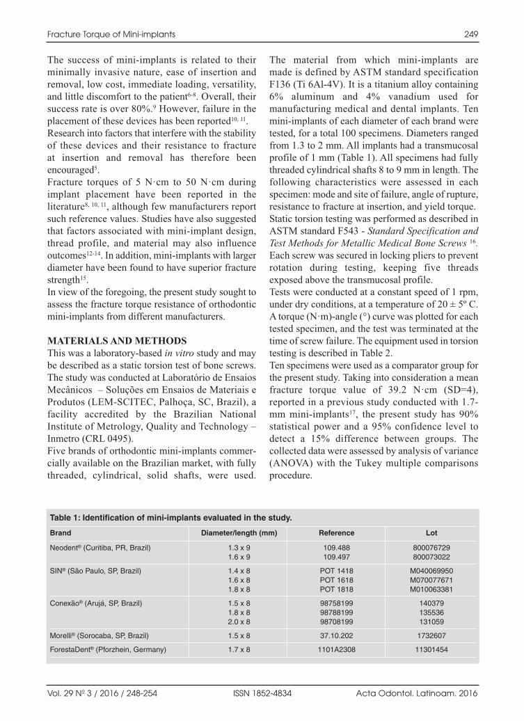
The success of miniimplants is related to theirminimally invasive nature, ease of insertion andremoval, low cost, immediate loading, versatility,and little discomfort to the patient68. Overall, theirsuccess rate is over 80%.9 However, failure in theplacement of these devices has been reported10, 11.Research into factors that interfere with the stabilityof these devices and their resistance to fracture at insertion and removal has therefore beenencouraged5.Fracture torques of 5 N·cm to 50 N·cm duringimplant placement have been reported in theliterature8, 10, 11, although few manufacturers reportsuch reference values. Studies have also suggestedthat factors associated with miniimplant design,thread profile, and material may also influenceoutcomes1214. In addition, miniimplants with largerdiameter have been found to have superior fracturestrength15.In view of the foregoing, the present study sought toassess the fracture torque resistance of orthodonticminiimplants from different manufacturers.
MATERIALS AND METHODSThis was a laboratorybased in vitro study and maybe described as a static torsion test of bone screws.The study was conducted at Laboratório de EnsaiosMecânicos – Soluções em Ensaios de Materiais eProdutos (LEMSCITEC, Palhoça, SC, Brazil), afacility accredited by the Brazilian NationalInstitute of Metrology, Quality and Technology –Inmetro (CRL 0495).Five brands of orthodontic miniimplants commer cially available on the Brazilian market, with fullythreaded, cylindrical, solid shafts, were used.
The material from which miniimplants are made is defined by ASTM standard specificationF136 (Ti 6Al4V). It is a titanium alloy containing6% aluminum and 4% vanadium used formanufacturing medical and dental implants. Tenminiimplants of each diameter of each brand weretested, for a total 100 specimens. Diameters rangedfrom 1.3 to 2 mm. All implants had a transmucosalprofile of 1 mm (Table 1). All specimens had fullythreaded cylindrical shafts 8 to 9 mm in length. Thefollowing characteristics were assessed in eachspecimen: mode and site of failure, angle of rupture,resistance to fracture at insertion, and yield torque.Static torsion testing was performed as described inASTM standard F543 Standard Specification andTest Methods for Metallic Medical Bone Screws 16.Each screw was secured in locking pliers to preventrotation during testing, keeping five threadsexposed above the transmucosal profile.Tests were conducted at a constant speed of 1 rpm,under dry conditions, at a temperature of 20 ± 5º C.A torque (N·m)angle (°) curve was plotted for eachtested specimen, and the test was terminated at thetime of screw failure. The equipment used in torsiontesting is described in Table 2.Ten specimens were used as a comparator group forthe present study. Taking into consideration a meanfracture torque value of 39.2 N·cm (SD=4),reported in a previous study conducted with 1.7mm miniimplants17, the present study has 90%statistical power and a 95% confidence level todetect a 15% difference between groups. Thecollected data were assessed by analysis of variance(ANOVA) with the Tukey multiple comparisonsprocedure.
Fracture Torque of Mini-implants 249
Vol. 29 Nº 3 / 2016 / 248-254 ISSN 1852-4834 Acta Odontol. Latinoam. 2016
Table 1: Identification of mini-implants evaluated in the study.
Brand Diameter/length (mm) Reference Lot
Neodent® (Curitiba, PR, Brazil) 1.3 x 9 109.488 8000767291.6 x 9 109.497 800073022
SIN® (São Paulo, SP, Brazil) 1.4 x 8 POT 1418 M0400699501.6 x 8 POT 1618 M0700776711.8 x 8 POT 1818 M010063381
Conexão® (Arujá, SP, Brazil) 1.5 x 8 98758199 1403791.8 x 8 98788199 1355362.0 x 8 98708199 131059
Morelli® (Sorocaba, SP, Brazil) 1.5 x 8 37.10.202 1732607
ForestaDent® (Pforzhein, Germany) 1.7 x 8 1101A2308 11301454
AOL32016:32011 15/02/2017 12:51 Página 249

RESULTSRupture was the characteristic mode of failure forthe tested miniimplants. Fractures occurred alongthe free end formed by the five exposed screwthreads, with the fracture angle ranging from 89° to 406.8°. On ANOVA with Tukey multiplecomparisons, neither failure site nor rupture anglewere significantly associated with miniimplantbrand or diameter at the 5% significance level.Table 3 shows the fracture torque resistance andyield torque values of the tested miniimplants.Mean fracture strength at insertion and yield limitranged from 15.7 to 70.4 N·cm and 9.2 to 53.1N·cm, respectively. Minimum and peak torquecurves obtained during mechanical testing areshown in Figs. 1 and 2.Significant differences were observed betweenbrands. In addition, miniimplants with larger
diameter exhibited superior fracture strength andyield limits, regardless of brand. The worstperformance was observed for the specimen withthe narrowest diameter (NEODENT® 1.3) and thebest performance for the specimen with the largestdiameter (CONEXÃO® 2.0).Table 4 shows the results for miniimplantsstratified into three groups by diameter: small (1.3mm/1.4 mm/1.5 mm), medium (1.6 mm/1.7 mm) orlarge (1.8 mm/1.9 mm/2.0 mm). Mean fracturestrength for small, medium, and large specimenswas 25.9 N·cm, 33.9 N·cm, and 54.2 N·cm,respectively. In the smalldiameter group, MORELLI®
brand miniimplants had the best performance. In the mediumdiameter group, the SIN® andNEODENT® brands stood out, whereas in the largediameter group, CONEXÃO® brand miniimplantsexhibited superior resistance to fracture at insertion.
250 Fernando Dalla Rosa, et al.
Acta Odontol. Latinoam. 2016 ISSN 1852-4834 Vol. 29 Nº 3 / 2016 / 248-254
Table 2: Identification of mini-implants evaluated in the study.
Internal reference Description Manufacturer/model Certificate of calibration
IM 0143 Single-axis torsion testing machine – OFServo Mecânica 9 (OFCME 30 Nm) —
IM 0144 Motion control motor OF INMETRODIMCI 0789/2014
23/04/2015
IM 0145 Torque measurement system – OF (OFTCN 20 KC) K&L2Nm-Maq 9 S371806/2013 20/08/2014
IM 0036 Digital caliper, 300 mm INSIZE CERTI(IS11137-300) 0244/14
05/02/2015
Table 3: Mean (standard deviation) fracture torque resistance and yield torque of orthodontic mini-implants.
Brand – diameter (mm) Fracture torque resistance (N·cm) Yield torque (N·cm)
SIN® – 1.4 26.1EF (0.6) 17.3E (0.6)
SIN® – 1.6 36.1D (2.7) 24.5C (1.9)
SIN® – 1.8 50.2B (1.5) 33.6B (1.0)
FORESTADENT® – 1.7 28.1E (0.5) 20.1D (0.8)
MORELLI® – 1.5 37.7D (2.9) 25.2C (2.8)
CONEXÃO® – 1.5 24.2F (1.2) 16.3E (1.6)
CONEXÃO® – 1.8 45.9C (0.8) 32.7B (1.5)
CONEXÃO® – 2.0 70.4A (3.1) 53.1A (3.7)
NEODENT® – 1.3 15.7G (0.7) 9.2F (0.4)
NEODENT® – 1.6 37.5D (0.8) 23.1C (0.9)
Means followed by different capital letters indicate significant differences according to ANOVA followed by the Tukey multiple comparisons test, at a significance level of 5%.
AOL32016:32011 15/02/2017 12:51 Página 250
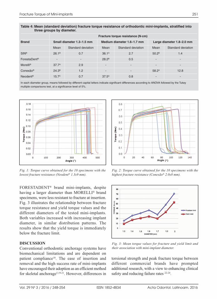
FORESTADENT® brand miniimplants, despitehaving a larger diameter than MORELLI® brandspecimens, were less resistant to fracture at insertion.Fig. 3 illustrates the relationship between fracturetorque resistance and yield torque values and thedifferent diameters of the tested miniimplants.Both variables increased with increasing implantdiameter, in similar distribution patterns. Theresults show that the yield torque is immediatelybelow the fracture limit.
DISCUSSIONConventional orthodontic anchorage systems havebiomechanical limitations and are dependent onpatient compliance18. The ease of insertion andremoval and the high success rate of miniimplantshave encouraged their adoption as an efficient methodfor skeletal anchorage5,1921. However, differences in
torsional strength and peak fracture torque betweendifferent commercial brands have promptedadditional research, with a view to enhancing clinicalsafety and reducing failure rates 2224.
Fracture Torque of Mini-implants 251
Vol. 29 Nº 3 / 2016 / 248-254 ISSN 1852-4834 Acta Odontol. Latinoam. 2016
Fig. 1: Torque curve obtained for the 10 specimens with thelowest fracture resistance (Neodent® 1.3x9 mm).
Fig. 2: Torque curve obtained for the 10 specimens with thehighest fracture resistance (Conexão® 2.0x8 mm).
Fig. 3: Mean torque values for fracture and yield limit andtheir association with miniimplant diameter.
Table 4: Mean (standard deviation) fracture torque resistance of orthodontic mini-implants, stratified into three groups by diameter.
Fracture torque resistance (N·cm)
Brand Small diameter 1.3–1.5 mm Medium diameter 1.6–1.7 mm Large diameter 1.8–2.0 mm
Mean Standard deviation Mean Standard deviation Mean Standard deviation
SIN® 26.1B 0.7 36.1A 2.7 50.2B 1.4
ForestaDent® - - 28.2B 0.5 - -
Morelli® 37.7A 2.9 - - - -
Conexão® 24.3B 1.2 - - 58.2A 12.8
Neodent® 15.7C 0.7 37.5A 0.8 - -
In each diameter group, means followed by different capital letters indicate significant differences according to ANOVA followed by the Tukeymultiple comparisons test, at a significance level of 5%.
AOL32016:32011 15/02/2017 12:51 Página 251

The usual length of orthodontic miniimplantsranges from 5 to 12 mm, while diameter andtransmucosal profile usually range from 1.2 to 2mm and from 0 to 3 mm, respectively10,11,25. Studieshave shown that a progressive increase in implantdiameter provides improved primary stability dueto increased bone contact area4,2630, but alsoincreases the risk of damage to surroundingstructures, particularly to the roots of adjacentteeth3133.Miniimplants with smaller diameter and length,however, have increased risk of fracture due tolower mechanical resistance. Despite its importanceto periimplant health, the transmucosal profile hasno influence on resistance to fracture at implantinsertion 34,35.In the present study, static torsion testing wasperformed in accordance with ASTM standardF543. This method ensures replicability of the studyand prioritizes mechanical analysis of the specimen,regardless of substrate. Mechanical data areobtained from the specimen alone, without externalinterference, as the implant is isolated and securedin a clamp. Conversely, studies performed inacrylic, porcine bone, and artificial bone are subjectto interference from other variables 32,33, 36, 37.In addition, a previous study evaluated titaniumalloy quality and microstructure of the miniimplantbrands tested herein14. According to the authors,these devices were free from internal structuraldefects and compliant with current standards14.In the present study, the characteristic mode offailure was miniimplant rupture. Site of failurealong the exposed threads and angle of rupture werenot associated with brand or diameter of the devicesevaluated. Technically, fracture sites may occurrandomly, as all screw threads are subject to thesame strain condition and intensity. The ruptureangle should preferably be high, as this would allowthe practitioner to detect during insertion that theimplant is undergoing elastic deformation and notdriving into bone, halt the procedure, and alter thetechnique accordingly before fracture occurs.Mean fracture torque resistance ranged from 15.7 to70.5 N·cm. These values are consistent with thosereported in studies that employed similar methods.Lima et al.15 observed values ranging from 30 to 36N·cm in 1.6mm NEODENT® implants, whereas
Wilmes et al.11, in a study of 41 commerciallyavailable brands 1.3 to 2 mm in diameter, reportedvalues ranging from 10.9 to 64.1 N·cm.Our results also showed that fracture strength isdirectly related to miniimplant diameter. Miniimplants with larger diameter exhibited higherfracture torque resistance, regardless of manufacturer.In the present study, the CONEXÃO® 2.0mm miniimplants performed best overall. According toBarros et al.13, a 0.1mm increase in miniimplantdiameter significantly reduces the risk of fracture.Toyoshima and Wakabayashi38 also observed thatincreasing diameter improves fracture torqueresistance.Yield torque represents the time point at which alloydeformation shifts from elastic (reversible) toplastic (irreversible). Optimally, in clinical practice,dentists should always work within the elastic limitof the alloy, thus preventing permanent deformationof the device. The higher the yield limit of a device,the greater its ability to resist plastic deformation.According to the results obtained, the yield limit behaves similarly to and is immediately belowthe fracture limit of these devices. Therefore,manufacturers should adopt this limit as a referencevalue, as it represents the point at which fatigue anddeformation occur; devices torqued beyond thislimit may be at increased risk of fracture duringremoval. Further research into this mechanicalparameter is warranted before this paradigm can beadopted and thus increase operator safety.Stratification of the miniimplants into groups bydiameter revealed differences across the testedbrands. MORELLI® and CONEXÃO® branddevices exhibited the best fracture torque resistancein the smalldiameter and largediameter groups,respectively.The use of miniimplants for orthodontic anchorageis effective and widespread in clinical practice.However, the success of this method depends largelyon primary stability. Fracture torque thus plays acritical role in clinical protocols involving placementof these devices. Precise information on the peakfracture torque and yield limit of miniimplantsshould be made available by manufacturers. Inaddition, torquesensing instruments should alwaysbe coupled to miniimplant drivers in order to ensuremeasurement of the forces applied.
252 Fernando Dalla Rosa, et al.
Acta Odontol. Latinoam. 2016 ISSN 1852-4834 Vol. 29 Nº 3 / 2016 / 248-254
AOL32016:32011 15/02/2017 12:51 Página 252

REFERENCES1. Petrey JS, Saunders MM, Kluemper GT, Cunningham LL,
Beeman CS. Temporary anchorage device insertion variables:effects on retention. Angle Orthod 2010; 80:446453.
2. Kim HJ, Yun HS, Park HD, Kim DH, Park YC. Softtissueand corticalbone thickness at orthodontic implant sites.Am J Orthod Dentofacial Orthop 2006; 130:177182.
3. Tseng YC, Hsieh CH, Chen CH, Shen YS, Huang IY, ChenCM. The application of miniimplants for orthodonticanchorage. Int J Oral Maxillofac Surg 2006; 35:704707.
4. Lim SA, Cha JY, Hwang CJ. Insertion torque of orthodonticminiscrews according to changes in shape, diameter andlength. Angle Orthod 2008; 78:234240.
5. Park HS, Jeong SH, Kwon OW. Factors affecting theclinical success of screw implants used as orthodonticanchorage. Am J Orthod Dentofacial Orthop 2006; 130:1825.
6. Mattos CT, Ruellas ACO, Elias CN. Is it possible to reuseminiimplants for orthodontic anchorage? Mat Res 2010;13:521525.
7. Cheng SJ, Tseng IY, Lee JJ, Kok SH. A prospective studyof the risk factors associated with failure of miniimplantsused for orthodontic anchorage. Int J Oral MaxillofacImplants 2004; 19:100106.
8. Florvaag B, Kneuertz P, Lazar F, Koebke J, Zoller JE,Braumann B, Mischkowski RA. Biomechanical propertiesof orthodontic miniscrews. An invitro study. J OrofacOrthop 2010; 71:5367.
9. Papadopoulos MA, Papageorgiou SN, Zogakis IP. Clinicaleffectiveness of orthodontic miniscrew implants: a metaanalysis. J Dent Res 2011; 90:969976.
10. Wilmes B, Drescher D. Impact of bone quality, implanttype, and implantation site preparation on insertion torquesof miniimplants used for orthodontic anchorage. Int J OralMaxillofac Surg 2011; 40:697703.
11. Wilmes B, Panayotidis A, Drescher D. Fracture resistanceof orthodontic miniimplants: a biomechanical in vitrostudy. Eur J Orthod 2011; 33:396401.
12. Kim JW, Baek SH, Kim TW, Chang YI. Comparison ofstability between cylindrical and conical type miniimplants.Mechanical and histological properties. Angle Orthod 2008;78:692698.
13. Barros SE, Janson G, Chiqueto K, Garib DG, Janson M.Effect of miniimplant diameter on fracture risk and selfdrilling efficacy. Am J Orthod Dentofacial Orthop 2011;140:e181192.
14. Burmann PF, Ruschel HC, Vargas IA, de Verney JC, KramerPF. Titanium alloy orthodontic miniimplants: scanningelectron microscopic and metallographic analyses. ActaOdontol Latinoam 2015; 28:4247.
15. Lima GM, Soares MS, Penha SS, Romano MM. Comparisonof the fracture torque of different Brazilian miniimplants.Braz Oral Res 2011; 25:116121.
16. American Society for Testing and Materials. ASTM F54300,Standard Specification and Test Methods for Metallic MedicalBone Screws, ASTM International, West Conshohocken, PA,2002.
17. Pithon MM, Nojima MG, Nojima LI. In vitro evaluation ofinsertion and removal torques of orthodontic miniimplants.Int J Oral Maxillofac Surg 2011; 40:8085.
18. Papadopoulos MA, Tarawneh F. The use of miniscrewimplants for temporary skeletal anchorage in orthodontics:a comprehensive review. Oral Surg Oral Med Oral PatholOral Radiol Endod 2007; 103:e615.
19. Chen CH, Chang CS, Hsieh CH, Tseng YC, Shen YS,Huang IY, Yang CF, Chen CM. The use of microimplantsin orthodontic anchorage. J Oral Maxillofac Surg 2006;64:12091213.
20. Moon CH, Lee DG, Lee HS, Im JS, Baek SH. Factorsassociated with the success rate of orthodontic miniscrewsplaced in the upper and lower posterior buccal region.Angle Orthod 2008; 78:101106.
21. Wiechmann D, Meyer U, Buchter A. Success rate of miniand microimplants used for orthodontic anchorage: aprospective clinical study. Clin Oral Implants Res 2007;18:263267.
22. Buchter A, Wiechmann D, Koerdt S, Wiesmann HP, PiffkoJ, Meyer U. Loadrelated implant reaction of miniimplantsused for orthodontic anchorage. Clin Oral Implants Res2005; 16:473479.
23. Mah J, Bergstrand F. Temporary anchorage devices: a statusreport. J Clin Orthod 2005; 39:132136; discussion 136;quiz 153.
24. Melsen B. Miniimplants: Where are we? J Clin Orthod2005; 39:539547.
25. Wilmes B, Rademacher C, Olthoff G, Drescher D.Parameters affecting primary stability of orthodontic miniimplants. J Orofac Orthop 2006; 67:162174.
26. Wu X, Deng F, Wang Z, Zhao Z, Wang J. Biomechanicaland histomorphometric analyses of the osseointegration ofmicroscrews with different surgical techniques in beagledogs. Oral Surg Oral Med Oral Pathol Oral Radiol Endod2008; 106:644650.
27. Heidemann W, Terheyden H, Gerlach KL. Analysis of theosseous/metal interface of drill free screws and selftappingscrews. J Craniomaxillofac Surg 2001; 29:6974.
28. Kim JW, Ahn SJ, Chang YI. Histomorphometric and mechanicalanalyses of the drillfree screw as orthodontic anchorage. Am JOrthod Dentofacial Orthop 2005; 128:190194.
29. Schatzle M, Mannchen R, Zwahlen M, Lang NP. Survivaland failure rates of orthodontic temporary anchoragedevices: a systematic review. Clin Oral Implants Res 2009;20:13511359.
30. Chen Y, Kyung HM, Gao L, Yu WJ, Bae EJ, Kim SM. Mechani cal properties of selfdrilling orthodontic microimplantswith different diameters. Angle Orthod 2010; 80:821827.
Fracture Torque of Mini-implants 253
Vol. 29 Nº 3 / 2016 / 248-254 ISSN 1852-4834 Acta Odontol. Latinoam. 2016
CORRESPONDENCEDr.Fernando Dalla RosaRua Sinimbú, 1878, sala 1106, bairro Centro95020002 Caxias do Sul, RS [email protected]
AOL32016:32011 15/02/2017 12:51 Página 253

31. Kang YG, Kim JY, Lee YJ, Chung KR, Park YG. Stabilityof miniscrews invading the dental roots and their impacton the paradental tissues in beagles. Angle Orthod 2009;79:248255.
32. Chen YH, Chang HH, Chen YJ, Lee D, Chiang HH, YaoCC. Root contact during insertion of miniscrews fororthodontic anchorage increases the failure rate: an animalstudy. Clin Oral Implants Res 2008; 19:99106.
33. Chen Y, Shin HI, Kyung HM. Biomechanical andhistological comparison of selfdrilling and selftappingorthodontic microimplants in dogs. Am J OrthodDentofacial Orthop 2008; 133:4450.
34. Araújo TM, Nascimento MHA, Bezerra F, Sobral MC.Ancoragem esqueletica em Ortodontia com miniimplantes.R Dental Press Ortodon Ortop Facial 2006; 11:126156.
35. Nova MFP, Carvalho FR, Elias CN, Artese F. Avaliação dotorque para inserção, remoção e fratura de diferentes miniimplantes ortodônticos. Rev Dental Press Ortodon OrtopFacial 2008; 13:7687.
36. Wilmes B, Ottenstreuer S, Su YY, Drescher D. Impact ofimplant design on primary stability of orthodontic miniimplants. J Orofac Orthop 2008; 69:4250.
37. Katic V, Kamenar E, Blazevic D, Spalj S. Geometricaldesign characteristics of orthodontic miniimplantspredicting maximum insertion torque. Korean J Orthod2014; 44:177183.
38. Toyoshima Y, Wakabayashi N. Load limit of miniimplantswith reduced abutment height based on fatigue fractureresistance: experimental and finite element study. Int J OralMaxillofac Implants 2015; 30:e1016.
254 Fernando Dalla Rosa, et al.
Acta Odontol. Latinoam. 2016 ISSN 1852-4834 Vol. 29 Nº 3 / 2016 / 248-254
AOL32016:32011 15/02/2017 12:51 Página 254
RESUMENEn este trabajo, nos propusimos determinar las variaciones delos parámetros periodontales y la composición microbiológicade las bolsas periodontales al inicio, a los 3 y 6 meses despuésdel tratamiento en pacientes con periodontitis agresivageneralizada (GAP), sometidos a tratamiento periodontal no quirúrgico combinado con clorhexidina y antibióticossistémicos. Se elaboró historia médica y dental en 10 sujetos,con una edad media de 30,6 ± 2,7 años, con diagnóstico deGAP. Se les practicó tratamiento periodontal no quirúrgicocombinado con clorhexidina al 0,12%, 875 mg de amoxicilinay 500 mg de metronidazol. Los antibióticos se prescribieroncada 12 horas durante diez días. Se registraron: la placabacteriana (BP), sangrado al sondaje (BOP), la profundidadde sondaje (PD), el nivel de inserción clínica (NIC),hipermovilidad y lesiones de furcación. En cada visita, setomaron las mediciones, y se tomó una muestra de la placa
subgingival en sitio de la mayor profundidad al sondaje encada sextante para identificar mediante técnica de biologíamolecular: Porphyromonas gingivalis, Treponema denticola,forsythia Tannerella, Prevotella intermedia, y Aggregatibacteractinomycetemcomitans. Después de 6 meses, el análisis de laprueba de Wilcoxon mostró un aumento de 0,97 mm de CAL (p= 0,0047) y 2,54 mm en la PB (p = 0,009). Se definió sitio sano,cuando se determinó un PD <5 mm, BOP negativo, y no sedetectaron bacterias patógenas a los 6 meses, lo que indicóuna mejora significativa (p = 0,008), con (IC 95%) = 4,7(1,1022 a 20,11). Con el protocolo de tratamiento presentado,es posible especular que a los 6 meses después del tratamiento,un paciente puede tener aproximadamente 4 veces másposibilidades de presentar una PD<5 mm y bolsillosperiodontales sin bacterias patógenas.
Palabras clave: periodontitis agresiva, tratamiento periodontal.
ABSTRACTThe aim of this study was to determine the variations inperiodontal parameters and microbiological composition inperiodontal pockets at the baseline and 3 and 6 months posttreatment in patients with Generalized Aggressive Periodontitis(GAP) undergoing nonsurgical periodontal treatment combinedwith chlorhexidine and systemic antibiotics. Medical and dentalhistory was taken from 10 subjects, average age 30.6±2.7 years,diagnosed with GAP. A nonsurgical periodontal treatmentcombined with 0.12% chlorhexidine, 875 mg amoxicillin and500 mg metronidazole every 12 hours for ten days wasconducted. At each visit, the following measurements wererecorded: bacterial plaque (BP), bleeding on probing (BOP),probing depth (PD), clinical attachment level (CAL),hypermobility, and furcation lesions, and a sample of subgingival
plaque was taken from the site of the deepest probing depth ofeach sextant to identify Porphyromonas gingivalis, Treponemadenticola, Tannerella forsythia, Prevotella intermedia andAggregatibacter actinomycetemcomitans using molecularbiology techniques. After 6 months, the Wilcoxon test showed anincrease of 0.97 mm in CAL (p=0.0047) and 2.54 mm in PD(p=0.009). A healthy site was defined as having a PD <5 mm,negative BOP and no pathogenic bacteria detected at 6 months,indicating significant improvement (p=0.008), with OR (95%CI) =4.7 (1.102220.11). With the treatment protocol used in thisstudy, 6 months after treatment, patients had an approximately4fold higher possibility of presenting PD <5 mm andperiodontal pockets without periodontal pathogenic bacteria.
Key words: Aggressive periodontitis, periodontal treatment.
255
Vol. 29 Nº 3 / 2016 / 255-261 ISSN 1852-4834 Acta Odontol. Latinoam. 2016
GENERALIZED AGGRESSIVE PERIODONTITIS: MICROBIOLOGICAL COMPOSITION AND CLINICALPARAMETERS IN NON-SURGICAL THERAPY
María M. Usin1, 3, Sandra M. Tabares2, Julieta Menso1, Estela R. de Albera3, Adela Sembaj2
1 Periodontology B Department, School of Dentistry, National University of Cordoba, Argentina.2 Biochemistry and Molecular Biology, School of Medical Science, National University of Cordoba, Argentina.
3 Independencia Foundation, Córdoba, National University of Cuyo, Mendoza, Argentina.
PERIODONTITIS AGRESIVA GENERALIZADA: COMPOSICIÓN MICROBIOLÓGICA Y PARÁMETROS CLÍNICOS EN LA TERAPIA NO QUIRÚRGICA
INTRODUCTIONGeneralized Aggressive Periodontitis (GAP)comprises a group of periodontal pathologiescharacterised by rapid evolution, severe bone loss,
few clinical manifestations, lowgrade inflammationand an amount of microorganisms which does notappear to be proportional to the severity ofdestruction 1. The condition is diagnosed when
AOL32016:32011 15/02/2017 12:51 Página 255

more than 30% of the teeth are affected, includingat least three permanent teeth other than incisorsand first molars, and is mainly diagnosed insystemically healthy persons at an early age 1. It isalso speculated that there is a hereditary componentin susceptibility 2.A highly virulent microbial flora has been described,with Aggregatibacter actinomycetemcomitansconsidered to be a risk factor because its persistenceis associated with recurrence3,4. Other authorssuggest that Porphyromonas gingivalis, Tannerelladenticola and Prevotella intermedia, togetherwith A. actinomycetemcomitans, are responsiblefor the low response to treatment in GAP patientsbecause of a relationship between the rapid loss of periodontal insertion in patients undergo ing maintenance and the persistence of thesespecies 5,6.The primary objective of treating this kind ofperiodontal disease is to modify the pathogenicsubgingival microbiota to nonpathogenic and toprevent the reestablishment of virulent bacteria inthe subgingival biofilm. These changes shouldimprove clinical parameters such as bleeding onprobing (BOP), probing depth (PD) and clinicalattachment level (CAL) 7,8. The identification of thebacterial species that colonise the pockets might bea tool for predicting the outcome or success oftreatment, especially with the use of additionalantiseptic and antibiotic treatment 9, 10.Several studies have reported the effectiveness andclinical safety of nonsurgical periodontal therapycombined with systemic antibiotics for thetreatment of GAP 11 13. However, there is no clearconsensus on the choice of antibiotics or theprotocol for the use and duration of antibiotictreatment. Moreover, several important issuesrelated to the therapy have yet to be clarified. Thereis no previous report in Argentina about theeffectiveness of nonsurgical therapy combined withantibiotics such as amoxicillin and metronidazolefor the management of periodontal flora in GAP.The aim of this study was to analyze the periodontalclinical parameters and the microbiologicalcomposition of periodontal pockets in patientsdiagnosed with GAP, and their response to nonsurgical periodontal treatment combined with 12% chlorhexidine and 875 Amoxicillin plusMetronidazole 500 mg by comparing baseline tothe situation at 3 and 6 months.
MATERIALS AND METHODSStudy PopulationTen unrelated patients of both sexes, aged 15 to 35years, were recruited from the Department ofPeriodontics of the Independencia Foundation inthe city of Cordoba, Argentina. Detailed systemicand oral anamnesis procedures were performed to assess the inclusion criteria, and GAP wasdiagnosed according to the definition of theAmerican Academy of Periodontology 14. Theinclusion criteria for the patients were: 1) having atleast 20 teeth, presenting interproximal bone loss inat least 3 teeth other than incisors and first molarsand 2) being systemically healthy, having noprevious history of periodontal treatment or historyof prescribed antibiotic medication within theprevious 6 months. Patients who were pregnant,breastfeeding, diabetic, smokers, immunosuppressedor drug addicts were excluded. Teeth with lesionswith furcation were excluded. The Ethics andDiscipline Committee of the Independence Foundationapproved the protocol. All patients received aprecise explanation of the research protocol, signedan informed consent to take part in the study, andwere treated following the principles of the HelsinkiDeclaration.
Clinical Records and Treatment ProtocolA complete medical and clinical dental history,including serial radiographies and periodontalcharting (Go Probe* R), was performed on eachpatient. Researchers MMU and JM recorded allclinical parameters at the beginning of the protocoland at 3 and 6 months after treatment. The followingclinical parameters were recorded: 1) presence/absence of supragingival bacterial plaque (BP),presence/absence of bleeding on probing (BOP),dental hypermobility, PD in mm, and loss of clinicalattachment level (CAL) in mm. The measurementswere taken from 6 different sites per tooth,excluding the third molar, using a Hu Friedyperiodontal probe (PCPUNC15, HuFriedy,Chicago, IL). The interrater correlation coefficientfor the PD measurements was 0.85, indicating ahigh level of reliability between MMU and JM. Periodontal treatment consisted of detailed oralhygiene technique instructions, followed by rootscaling and planing, and rinsing with a solution of0.12% chlorhexidine digluconate (GlaxoSmithKline).Following their oral hygiene routine, patients were
256 María M. Usin, et al.
Acta Odontol. Latinoam. 2016 ISSN 1852-4834 Vol. 29 Nº 3 / 2016 / 255-261
AOL32016:32011 15/02/2017 12:51 Página 256

instructed to rinse with 15 ml clorhexidine for 30 seconds twice a day for 60 days, beginning afterthe first session of mechanical debridement.Mechanical debridement consisted of progressivesessions of root scaling and planing once a weekdivided into approximately 5 to 7 sessions, dependingon the case. The treatment was performed underlocal anaesthesia, by quadrant and with specificGracey curettes (HuFriedy, Chicago, IL) for eachsector and face of the teeth. The treatment wasconsidered complete when BP was less 10%, therewas no bleeding or any visible sign of inflammation,and no calculus was detected by the calculusdetection device (HuFriedy, Chicago, IL). Patientsreceived a prescription of 875 mg amoxicillin and500 mg metronidazole every 12 hours for 10 daysfollowing the first scaling session. The dosage andduration of the antibiotic prescription are in relationto the pharmacological presentation. After completingthe basic treatment, the clinical parameters were reevaluated after 3 and 6 months with reinforcementof the hygiene instructions.
Sample Collection for the MicrobiologicalIdentification of Periodontal PathogensThe samples for microbiological analysis weretaken at the beginning of the nonsurgicalperiodontal treatment and at 3 and 6 months. Thematerial for the analysis was collected on the sitepresenting the deepest PD on each subgingivalsector of each sextant, on the proximal faces:mesialvestibular, mesiopalatal, distovestibular anddistopalatal. Prior to taking samples, the supragingival plaquewas removed from the interproximal surfaces usinga sterile curette. The surfaces were isolated withsterile gauze and five consecutive #35 paper pointswere inserted with catheterisation movements to thedepth of the periodontal pocket for 60 seconds each.The cones were placed into Eppendorf tubes andstored at 4°C until processing.
Sample Processing1 DNA extraction:Two hundred microliters of sterile water were addedto the Eppendorf tubes containing the paper pointsimpregnated with the material extracted from theperiodontal pockets. Each tube was incubated at37ºC for 10 minutes and centrifuged at 14,000 g for5 minutes. DNA was extracted using conventional
techniques 15. To verify the presence of DNA in thesupernatant, electrophoresis was performed on0.8% agarose gel stained with ethidium bromideand visualised using ultraviolet (UV) light.
2 Identification by conventional PCR:First, we amplified a highly preserved specificsequence (960 bp) of the 16S rRNA gene to identifygramnegative bacteria in the sample. Once Gramnegative bacteria were confirmed in the sample,PCRs with specific oligonucleotides for A.actinomycetemcomitans, T. forsythia, P. gingivalis,P. intermedia, and T. denticola were performedfollowing the protocol described by Ashimoto et al 16.The assay was performed twice for each sample,including a negative control by adding to eachreaction one tube without DNA and one tube withDNA from organisms isolated in a culture anddonated by the Bacteriology Laboratory at theReina Fabiola Hospital (Catholic University ofCordoba). The PCR products were analysed byelectrophoresis on 1.6% agarose gels. The gels werestained with ethidium bromide and photographedunder UV light. A molecular marker indicated thesize of the amplification product in the gel (PerkinElmer 100bp Marker). For doubtful results, thesamples were sequenced to corroborate thespecificity of the amplified fragment (ABI PRISM310, Applied Biosystems, USA).
Statistical analysisStatistical analysis was performed using SPSS (Ver.9; SPSS, Inc.) and Infostat vs2007e data analysissoftware. Significance was defined as p valuesunder 0.05 (p<0.05). A sample size of 10 subjectswas needed to provide an 80% power for detectingan average 0.75% reduction in probing depthbetween baseline and 6 months. Once patients hadbeen selected for treatment, it was not possible toinclude additional patients to compensate for anypotential dropouts. The intraclass correlationcoefficients for mean PD and CAL were 0.92 and0.91 respectively. The examiner’s reproducibilityof measurements taken at baseline and at 6 monthswas good (0.93 and 0.92 respectively).The quantitative variables (PD, CAL) are expressedas means and standard deviations, and thequalitative variables BP, BOP, S and presence ofbacteria in the pockets are expressed as absoluteand percent frequencies. The statistical significance
Generalized Aggressive Periodontitis 257
Vol. 29 Nº 3 / 2016 / 255-261 ISSN 1852-4834 Acta Odontol. Latinoam. 2016
AOL32016:32011 15/02/2017 12:51 Página 257

between the mean values for the same periods ofassessment was evaluated using the paired StudentT test, and the differences between 2 periods oftreatment were assessed using the Wilcoxon SignedRank test. The presence of bacteria and the CALand PD values at the end of the treatment werecorrelated with 2x2 contingency tables, usingPearson’s chisquare test or Fisher’s exact test whenfew samples were analysed. Subsequently, raw oddsratios (ORs) and 95% confidence intervals (95%CIs) were used to calculate the relative risk ofdisease. Status was considered healthy when aftersix months, pathogenic bacteria were not detected,BOP was negative and PD<5 mm was recorded inthe treated pockets.
RESULTSThis study analysed 60 periodontal sites in 4 malesand 6 females, average age 30.6±2.7 years (range2135 years) who completed all stages of theproject. Patients had an average 27 tooth, although
one patient lost two teeth and another patient lostone tooth during the treatment. The complete oralexamination revealed an average 42±7.9 pocketsover 5 mm and 121.5±6.9 pockets under 5 mm.BOP was present in the upper maxilla in 47.6% ofthe cases, and in the mandible in 47.9%. Table 1summarises the changes in periodontal parametersat the different time points of the assessment. MeanPD and CAL values in mm decreased significantlythroughout the treatment in all patients (p<0.035).After six months posttreatment, according to theWilcoxon Signed Rank test, there was a 0.97 mmincrease in CAL (p=0.0047) and a 2.54mmrecovery in PD (p=0.009). Positive BOP decreasedby 40.47% in the upper maxilla and by 37.2% in thelower maxilla after treatment. At six months, thenumber of periodontal pockets deeper than 5 mmdecreased by 36.6%, whereas the frequency ofpockets less than 5 mm deep increased significantly. Regarding the distribution throughout treatment ofthe five pathogens identified in the periodontalpockets at all sites, P. gingivalis, T. denticola, andT. forsythia were identified with greater frequencyat the beginning of the treatment. P. gingivalis wasnot detected at the 3month followup and onlyrecognised at one site six months posttreatment,whereas T. denticola and T. forsythia persisted atall sites after completion of the 6month period.P. intermedia was identified in association with A. actinomycetemcomitans only at the beginning of the study. These bacteria were not isolated again at any site throughout the study. The absence of P. gingivalis, P. intermedia and A.actinomycetemcomitans after treatment concurredwith the increase in nonvirulent Gramnegativebacteria during the 3 and 6month periodontalmaintenance period (Table 2).To analyse whether the reduction in PD observed at the 6month followup was associated with
258 María M. Usin, et al.
Acta Odontol. Latinoam. 2016 ISSN 1852-4834 Vol. 29 Nº 3 / 2016 / 255-261
Table 1: Periodontal Clinical Parameters duringthe treatment.
Baseline 3m 6m p(n=60) (n=60) (n=60)
PD(mm) 7.0±2.6* 4.63±3.8 4.46±3.7 0.002
CAL(mm) 7.6±6.5* 6.8±4.5 6.63±5.4 0.035
Upper BOP (%) 47.6 42.9 4.047 0.007*
Lower BOP (%) 47.9 39.08 37.19 0.039*
Pockets>5mm 42*±0.9 25±0.2 22±0.3 0.037
Pockets<5mm 121*±6.4 137±0.1 140±04 0.045
The scores represent means ± standard error; in millimeters of probing depth (PD) and clinical attachment level (CAL), and the number ofpockets of more than or less than 5 mm at all sites measured at eachstage of treatment at 3m ( 3 months) and 6 m ( 6 months). BOP bleedingon probing; the numbers represents the percentage of the averagescore. *Significant p value between baseline and 6m.
Table 2: Distribution of Periodontal Bacteria at baseline and 3- and 6- months post-treatment.
Pg Sites (%) Td Sites (%) Tf Sites (%) Pi Sites (%) Aa Sites (%) Others Sites (%)
Baseline 13 (43.3) 15 (47) 13 (40) 12 (37) 4 (10) 2 (6)
3 months 0 6 (20) 7 (23) 0 0 18 (60)
6 months 1 (3) 5 (17) 8 (27) 0 0 16 (53)
The numbers represent the number of sites where each bacterium was identified; percentages between brackets. Porphiromonas gingivalis (Pg), Treponema denticola (Td), Tannerella forsythia (Tf) Prevotella intermedia (Pi) and Agreggatibacter actinomycemcomitans (Aa), Others: Gram-negativebacteria.
AOL32016:32011 15/02/2017 12:51 Página 258

the presence/absence of periodontal bacteria, aunivariate analysis was performed to correlate thepresence of 1 or more pathogenic bacteria with PDvalues compatible with clinical improvement. Forthis purpose, healthy status was considered to bePD<5 mm plus absence of pathogenic bacteria inthe pocket and absence of BOP. The chisquare test revealed a statistically significant associationof Χ2=5.6, p=0.008, a statistically significantimprovement between baseline and the 6monthmeasurement, with OR (95% CI) = 4.7 (1.102220.11). This finding indicated that the chance ofpockets improving to PD<5 mm at 6 months posttreatment was 4fold higher when periodontalpathogenic bacteria were absent from the periodontalpocket.
DISCUSSION In our study, the analysis of clincal parametersbefore and after treatment revealed positive resultsfor mechanical periodontal therapy combined with chlorhexidine and antibiotics in patients with GAP, showing significant improvements inclinical periodontal variables such as decreased PD,increased CAL, and decreased percentage of plaqueand sites with BOP, which were maintained at the6month followup. Notably, the average increasein CAL was 0.97 mm at six months after thebeginning of the treatment, and the PD decreasedby 2.54 mm during the same period. This clinicalsuccess included a significant reduction in periodontalpathogens at the treated sites. These results are inagreement with other studies reporting that nonsurgical treatment including the use of amoxicillinand metronidazole leads to clinical benefits andsignificant reduction in periodontal pathogens.10 13.
Hughes et al.17 conducted a study on patients withGAP following treatment with superficial rootdebridement and oral hygiene instructions. In thereassessment, they report a mean decrease of 2.11mm in PD and an increase of 1.77 mm in CAL.They report that 32% of the patients with a negativeresponse to treatment were smokers and suggestthat smoking is the most important factor associatedwith treatment failure 17. The fact that our studyexcluded smokers may explain our higher PDvalues after 6 months. At the 3 and 6month followups we found signi ficant reduction in the presence of P. gingivalis, P.
intermedia and A. actinomycetemcomitans in theperiodontal pockets treated, indicating the efficacyof mechanical therapy combined with 875 mgamoxicillin and 500 mg metronidazole every 12hours for 10 days and Clorhexidine 0.12% rinses toeliminate pathogens and thereby restore periodontaltissue. It is also worth noting that the T. denticolaand T. forsythia were identified at all stages of thetreatment, and although they were detected in fewer pockets, the association between T. denticolaand T. forsythia may be resistant to the combinedtreatment. Other authors have described thebacterial relationship between P. gingivalis, A.actinomycetemcomitans and T. forsythia in GAP 18.We note that different studies use different dosesand duration of the antibiotic treatment 11. Theamoxicillin dose used in our study has not beenreported in other studies. For example, GomesBaeta et al. 19 prescribed 500 mg amoxicillin and250 mg metronidazole 3 times per day for 10 days,and detected A. actinomycetemcomitans in allsamples up to the 9month posttreatment followup session. Haffajee et al. 20 showed that systemicadministration of these antibiotics may suppressperiodontal bacteria efficiently and thereby improvethe therapeutic response 18. We believe that oneweakness of the current study is that the high dosesof amoxicillin used did not prevent the persistenceof T. denticola or T. forsythia either at 3 or at 6months. Further studies are needed to analysewhether a combination of amoxicillin/metronidazolemight induce persistence or antibiotic resistance inthe periodontal microflora. The risk of side effectsor adverse events also remains to be addressed 12.
Mechanical instrumentation significantly changedthe composition of the subgingival microflora bydecreasing virulent microorganisms and increasingbeneficial cocci and bacilli, with this bacterial patternleading to a healthy bacteriological profile 20. Someauthors suggest a limited effect of root scaling andplanning on certain pathogenic species becausetheir complete elimination might be difficult as aresult of reinfection of successfully treated sites.Johnson et al.21 suggest that after 6 months, thesebacteria recolonise the plaque when they remain inthe epithelial cells of the oral mucosa and areunaltered by treatment.21 These data support thehypothesis that extracrevicular bacterial depositscontribute to the development of recurrent diseases
Generalized Aggressive Periodontitis 259
Vol. 29 Nº 3 / 2016 / 255-261 ISSN 1852-4834 Acta Odontol. Latinoam. 2016
AOL32016:32011 15/02/2017 12:51 Página 259
in some patients 19,20. We observed recolonizationby P. gingivalis in 1 patient at the 6month followup. According to our data, P. gingivalis in aperiodontal pocket might be a risk factor forunsuccessful treatment or relapse. It is important to note that the reinforcement of oral hygiene andreinstrumentation at followup appointmentscontributed to the continuous reduction of certainpathogenic species. Our observations, as well asstudies by other authors, have noted a significantdecrease in the bacterial population in the treatedpockets at three months, and repopulation at sixmonths 22. This repopulation may be caused by theuse of systemic antibiotic therapeutic regimen.While adjunctive therapy is effective, few studieshave reported whether periodontal species havedeveloped resistance to antibiotics prescribed 18.Because GAP is rarely diagnosed in our location,the sample size in our study is limited. Furthermore,the 6month followup period might be too brief forlongitudinal assessment. An increase in the numberof patients and a longer followup period wouldenhance the study’s strength and contribute toestablishing appropriate intervals for maintainingperiodontal health. Notably, although the clinicaloutcome was positive after 6 months, the elimina tion of certain bacteria was shown to be transitory.This persistence may contribute to the recurrenceof the disease in some patients. To achieve longterm stability in clinical outcomes, it is essential toimplement strict maintenance programmes. Webelieve that controlling the microbiologicalcomposition of the pocket may help reduce the use
of antibacterial agents and the risk of producingbacterial resistance. The rationale for adding amoxicillin to theprescription of this combined drug regimen is based on a synergistic effect of amoxicillin onmetronidazole and its hydroxymetabolite against A.actinomycetemcomitans 23. In our study, only 10%of the periodontal pockets were found to contain A.actinomycetemcomitans at baseline. Bazzano et al.report similar frequencies in patients with severechronic periodontitis and treated only with rootscaling and planning 24. A study that evaluatedmetronidazole, alone or combined with amoxicillin,as an adjunct to nonsurgical therapy in chronicperiodontitis subjects, showed that the clinical andmicrobiological benefits of both treatment optionswere very similar 25. Further studies are needed to discuss the balance of the risk/benefits ratio ofthe prescription adjunctive antimicrobial regimen,in particular when high doses of amoxicillin are used (825mg/10 days/ 2 times a day/) and A.actinomycetemcomitans is identified in low frequency. Our study verified that in patients diagnosedclinically, radiologically and microbiologicallywith GAP, virulent bacterial species may beeliminated by mechanical treatment combined withspecific systemic antibiotics (875 mg amoxicillinand 500 mg metronidazole), antiseptics (such as0.12% chlorhexidine digluconate) and a 6monthmaintenance period. The above protocol leads to asignificant recovery in periodontal parameters suchas PD and BOP, achieving values that arecompatible with periodontal health.
260 María M. Usin, et al.
Acta Odontol. Latinoam. 2016 ISSN 1852-4834 Vol. 29 Nº 3 / 2016 / 255-261
ACKNOWLEDGEMENTSThe authors thank the patients who participated in this study. Thisresearch was partly financed by the Secretary of Science andTechnology of the Córdoba National University (SECyTUNC,according to its initials in Spanish) and by personal funds of theauthors.
CORRESPONDENCEDra. Adela Sembaj. Cátedra de Bioquímica y Biología Molecular. Facultad de Ciencias Médicas. Universidad Nacional de Córdoba. 2º piso Pabellón Argentina. Ciudad Universitaria. Córdoba, [email protected]
REFERENCES1. Armitage CG, Cullinan M. Comparison of the clinical
features of chronic and aggressive periodontitis. Periodontol2000. 2010; 53:12–27.
2. Vieira AR, Albandar JM. Role of genetic factors in thepathogenesis of aggressive periodontitis. Periodontol 2000.2014; 65: 92106.
3. Fine DH, Markowitz K, Furgang D. Aggregatibacteractynomicetemcomitans and its relationship to initiation of
localized aggressive periodontitis: Longitudinal cohort studyof initially healthy adolescents. J Clin Microbiol. 2007;45:38593869.
4. Slots J, Ting M. Actinobacillus actynomicetemcomitans andPorphiromonas gingivalis in human periodontal disease:occurrence and treatment. Periodontol 2000. 1999; 20:82121.
5. Liu J, Zhao J, Li C, Yu N, Zhang D, Pan Y. Clinical andmicrobiologic effect of nonsurgical periodontal therapy on
AOL32016:32011 15/02/2017 12:51 Página 260

patients with chronic or aggressive periodontitis. QuintessenceInt 2013; 44:575583.
6. Shiloah J, Patters MR, Dean III JW, Bland P, Toledo G. Theprevalence of Actinobacillus actynomicetemcomitans,Porphiromonas gingivalis and Bacteroides forsythus inhumans 1 year after 4 randomized treatment modalities. JPeriodontol 1998; 69:13641372.
7. Heller D, Varela VM, SilvaSenem MX, Torres MC, FeresFilho EJ, Colombo AP. Impact of systemic antimicrobialscombined with antiinfective mechanical debridement onthe microbiota of generalized aggressive periodontitis: A 6month RCT. J Clin Periodontol 2011; 38:355364.
8. Albandar JM, Olsen I, Gjermo P. Association between sixDNA probedetected periodontal bacteria and alveolar boneloss and other clinical signs of periodontitis. Acta OdontolScand 1999; 48: 415423.
9. MeyerBaumer A, Pristch M, Cosgarea R, Sayed NE, KimTS, Eickholz P, Pretzl B. Prognostic value of theperiodontal risk assessment in patients with aggressiveperiodontitis. J Clin Periodontol 2012; 39:651658.
10. Rodrigues A, Lourençäo DS, Lima NE, Pannuti CM,Dominguez R, Hirata M, Lotufo RF, De Micheli G. Clinicaland Microbiologic Evaluation, by RealTime PolymeraseChain Reaction, of NonSurgical Treatment of AggressivePeriodontitis Associated With Amoxicillin and Metronidazole.J Periodontol 2012; 83:744756.
11. Sgolastra, F, Petrucci, A, Gatto, R, Monaco, A. Effectivenessof systemic amoxicillin/metronidazole as an adjunctivetherapy to full mouth scaling and root planning in thetreatment of aggressive periodontitis: a systematic reviewand metaanalysis. J Periodontol 2012; 83, 731743.
12. Yek EC, Cintan S, Topcuoglu N, Kulekci G, Issever H,Kantarci A. Efficacy of amoxicillin and metronidazolecombination for the management of generalized aggressiveperiodontitis. J Periodontol 2010; 81:964974.
13. Haffajee AD, Socransky SS, Gunsolley JC. Systemic antiinfective periodontal therapy. A systematic review. Annalsof Periodontol 2003; 8:115181.
14. Armitage GC. Development of a classification system forperiodontal diseases and conditions. Ann Periodontol 1999;4:16.
15. Sambroock J, Russell DW. Molecular cloning: A laboratorymanual.3th ed Cold Spring Harbor Laboratory Press, ColdSpring Harbor, New York; 2001.
16. Ashimoto A, Chen C, Bakker I, Slots J. Polymerase chainreaction detection of 8 putative periodontal pathogens in
subgingival plaque of gingivitis and advanced periodontitislesions. Oral Microbiol Immunol 1996; 11:266273.
17. Hughes FJ, Syed M, Koshy B, Marinho V, Bostanci N,McKay IJ, Curtis MA, Croucher RE, Marcenes W.Prognostic factors in the treatment of generalizedaggressive periodontitis: I. Clinical features and initialoutcome. J Clin Periodontol 2006:33:663670.
18. Guerrero A, Nibali L, Lambertenghi R, Ready D, Suvan J,Griffiths GS, Wilson M, Tonetti MS. Impact of baselinemicrobiological status on clinical outcomes in generalizedaggressive periodontitis patients treated with or withoutadjunctive amoxicillin and metronidazole: an exploratoryanalysis from a randomized controlled clinical trial. J ClinPeriodontol 2014;41:10801089.
19. Lourenço TG, Heller D, do Souto RM, SilvaSenem MX,Varela VM, Torres MC, FeresFilho EJ, Colombo AP Longterm evaluation of the antimicrobial susceptibility andmicrobial profile of subgingival biofilms in individualswith aggressive periodontitis. Braz J Microbiol 2015; 46:493500.
20. Haffajee AD, Teles RP, Socransky SS. The effect ofperiodontal therapy on the composition of the subgingivalmicrobiota. Periodontol 2000 2006;42:219258.
21. Johnson JD, Chen R, Lenton PA, Zhang G, Hinrichs JE,Rudney JD. Persistence of extracrevicular bacterialreservoirs after treatment of aggressive periodontitis. JPeriodontol 2008; 79: 23052312.
22. Mullally BH, Dace B, Shelburne CE, Wolff LF, CoulterWA. Prevalence of periodontal pathogens in localized andgeneralized forms of earlyonset periodontitis. J PeriodontalRes 2000; 35: 232234.
23. Pavicic MJ, van Winkelhoff AJ, PavicicTemming YA, de GJ.Amoxycillin causes an enhanced uptake of metronidazole inActinobacillus actinomycetemcomitans: a mechanism ofsynergy. J Antimicrob Chemother 1994; 34:10471050.
24. Bazzano G, Parodi R, Tabares S, Sembaj A. Evaluation ofperiodontal mechanical therapy in deep pockets: Clinicaland bacteriological response. Rev. Clin. PeriodonciaImplantol. Rehabil. Oral 2012; 5; 123128. http://dx.doi.org/10.4067/S071901072012000300004
25. Soares, GM, Mendes, JA, Silva, MP, Faveri, M, Teles, R,Socransky, SS, Wang,X, Figueiredo, LC, Feres, M.Metronidazole alone or with amoxicillin as adjuncts tononsurgical treatment of Chronic Periodontitis: a secondaryanalysis of microbiological results from a randomizedclinical trial. J Clin Periodontol 2014; 41:366377.
Generalized Aggressive Periodontitis 261
Vol. 29 Nº 3 / 2016 / 255-261 ISSN 1852-4834 Acta Odontol. Latinoam. 2016
AOL32016:32011 15/02/2017 12:51 Página 261

262
Acta Odontol. Latinoam. 2016 ISSN 1852-4834 Vol. 29 Nº 3 / 2016 / 262-267
RESUMONo retratamento endodôntico, a completa remoção do materialobturador e o acesso ao forame apical são necessários parapermitir a limpeza do sistema de canais. O propósito deste estudofoi avaliar a eficácia do sistema reciprocante WaveOne ecomparála ao sistema rotatório ProTaper Universal, com ousem o uso de solvente, na remoção do material obturador.O temponecessário a cada técnica empregada foi determinado ecomparado. Quarenta prémolares inferiores humanos extraídoscom canal único, reto e achatado foram preparados e obturados.Foram então divididos em quatro grupos (n = 10) de acordo como sistema utilizado, como segue. Grupo 1: ProTaper D Niti;Grupo 2: Sistema ProTaper D com solvente; Grupo 3: SistemaWaveOne instrumento Primary; e Grupo 4: Sistema WaveOneinstrumento Primary com solvente, sendo o tempo registrado.
Os dentes foram clivados longitudinalmente e fotografadosutilizando microscópio operatório com aumento de 5 vezes.Aquantidade de material remanescente foi avaliada com o uso dosoftware Image Tool 3.0. Os resultados foram comparadosutilizando o teste de KruskalWallis(p < 0.05). Em relação aosresultados, não houve diferença significativa entre os gruposquanto à quantidade de material obturador residual (p > 0.05).O tempo operatório no Grupo 3 foi significativa mente maior doque nos grupos 1, 2 e 4 (p < 0.05).O sistema WaveOne foi tão efetivo quanto o ProTaper D, com ou sem solvente.temponecessário à desobturação dos canais no Grupo 3 (WaveOne semsolvente) foi significativamente maior do que nos demais grupos.
Palavraschave: Gutapercha; canal radicular; movimentoreciprocante.
INTRODUCTIONAlthough endodontic treatments have a high success rate, they sometimes require a secondintervention1,2. Endodontic retreatment requires thecomplete removal of the filling material and access
to the apical foramen for adequate cleansing andrefilling of the root canal system3. Conventional techniques using GatesGlidden andmanual files have been losing their popularity dueto the need for solvents. More current techniques
ABSTRACTEndodontic retreatment requires complete removal of the fillingmaterial and access to the apical foramen. The purpose of thisstudy was to evaluate the effectiveness of the WaveOnereciprocating system and compare it to the ProTaper D rotarysystem, with or without the use of a solvent, in removing fillingmaterial from root canals. The time required for each fillingremoval technique employed was also determined andcompared. Forty extracted human mandibular premolars witha single, straight, flattened canal were prepared and filled.They were divided into four groups (n = 10): Group 1:ProTaper D NiTi rotary instruments; Group 2: ProTaper D NiTirotary instruments, with a solvent; Group 3: WaveOne primaryinstrument; and Group 4: WaveOne primary instrument, with a
solvent. The teeth were then split along their long axis andphotographed using an operating microscope with 5Xmagnification. The amount of remaining filling material wasassessed with Image Tool software. The results were comparedusing the KruskalWallis test (p <0.05). There was nosignificant difference between groups regarding the amount ofresidual filling material (p > 0.05). Operative time wassignificantly longer in Group 3 than in groups 1, 2 and 4 (p <0.05). The WaveOne system and the ProTaper D system wereequally effective, with or without a solvent. The time requiredto remove the filling material from the canals was significantlylonger in Group 3 than in the other groups.
Key words: GuttaPercha; root canal; reciprocating.
EFECTIVENESS OF THE WAVEONE AND PROTAPER D SYSTEMS FOR REMOVING GUTTA-PERCHA WITH OR WITHOUT A SOLVENT
Ana Paula M. Colombo, Carlos E. Fontana, Aline Godoy, Alexandre S. De Martin, Augusto S. Kato, Daniel G-P. Rocha, Rina A. Pelegrine, Carlos E. S. Bueno
Department of Endodontics, Dental Research Center, São Leopoldo Mandic University, Campinas, SP, Brazil.
EFICIÊNCIA DOS SISTEMAS WAVEONE E PROTAPER D NA REMOÇÃO DE GUTA-PERCHA COM OU SEM USO SOLVENTE
AOL32016:32011 15/02/2017 12:51 Página 262

using motors facilitate endodontic retreatment andreduce working time.Reciprocating systems were introduced4 with the aimof improving resistance to fracture, since thereciprocating motion reduces the cyclic and torsionalfatigue to which the instrument is submitted5,6
.Furthermore, the instruments in these systems aremade from an alloy subjected to a thermal treatment,known as MWire and considered more resistant thanthe conventional NiTi alloy7. The WaveOne system(Dentsply Maillefer, Ballaigues, Switzerland) wasdesigned specifically for use in reciprocating motion.It comprises three instruments: small (21.06),primary (25.08 in three first milimeters) and large(40.08 in three first millimeters)8.Reciprocating systems were not originally designedfor use in retreatment procedures; nevertheless, Rioset al.9 compared the efficacy of two reciprocatingsystems and one rotary system in removing fillingmaterial from straight canals. They observed that thereciprocating systems and the rotary system testedwere equally effective in removing filling material.Others studies have also compared reciprocatingfiles in retreatment 26,27.Zuolo et al.10 compared the effectiveness of areciprocating system and a rotary system to that of amanual instrumentation technique in removingfilling material from root canals. Chloroform wasused as a solvent in all groups. The authors concludedthat the operative time involved in using thereciprocating system with a solvent was significantlyshorter than that required by the other systems tested.In spite of these results, no consensus has beenreached to date on the effectiveness of reciprocatingsystems for endodontic retreatment procedures. Theuse of a solvent as an aid in removing the fillingmass is also somewhat controversial. Some authorshave suggested that its use could increase workingtime due to the formation of a residual slurry whichadheres to the canal walls11, whereas others do notconfirm these results12,13. Some authors point outthat a solvent may be responsible for improvedcleanliness of the root canal system ¹⁴, whereasothers do not observe this correlation13,15. Tagger’s hybrid filling technique was chosen toplasticize guttapercha and produces good adaptationof the filling material to the canal walls16.The purpose of this study was to evaluate theeffectiveness of the WaveOne reciprocating system,with or without the use of a solvent, and compare it
to that of the ProTaper D rotary system in removingfilling material from straight canals. The timerequired by each retreatment technique was alsodetermined and compared.
MATERIALS AND METHODSpecimen preparationAll the specimens used in this study were obtainedfrom the tooth bank at the Dental Research Center,São LeopoldoMandic University, Campinas, SP,Brazil. The study protocol was reviewed andapproved by the Research Ethics Committee of thesame institution (protocol no. 2012/0420).Forty extracted human mandibular premolars wereused for this study. All teeth had a single, straight,flattened canal, a completely formed root and nocalcification or internal resorption. The teeth wereselected based on an evaluation of apical radiographstaken in orthoradial and mesiodistal direction. Theteeth were preserved in a 0.1% thymol solution(Fórmula e Ação, São Paulo, SP, Brazil).The crowns were removed with a diamond disc (KGSorensen, Cotia, SP, Brazil), and root length wasstandardized at 16 mm. A #10 Ktype file (DentsplyMaillefer) was then inserted into the canal until itcould be seen at the apical foramen with an operatingmicroscope (D. F. Vasconcelos, São Paulo, SP,Brazil) at 5X magnification. The working length wasdetermined 1 mm short of this measurement.
Initial endodontic treatmentA single operator instrumented the canals using therotary technique. Preflaring was performed withGates Glidden burs (Dentsply Maillefer) and the SXand S1 files of the ProTaper Universal rotary system.Apical preparation was performed with ProTaper S1,S2, F1 and F2 files. The canal was irrigated with 2mL 2.5% sodium hypochlorite (Fórmula e Ação) aftereach instrument change. Once the instrumentationwas completed, the canal was irrigated with 17%EDTA (Fórmula e Ação) for 3 minutes followed by 5mL 2.5% sodium hypochlorite.The canals were dried with absorbent paper pointsand filled with ProTaper F2 guttapercha cones(Dentsply Maillefer), accessory cones (DentsplyMaillefer), and AH plus sealer (Dentsply Maillefer)using Tagger’s hybrid technique16, 1 mm short ofthe apical foramen. Filling quality was confirmedwith periapical radiographs taken in the orthoradialand mesiodistal direction.
Efficiency of Systems for Endodontic Retreatment 263
Vol. 29 Nº 3 / 2016 / 262-267 ISSN 1852-4834 Acta Odontol. Latinoam. 2016
AOL32016:32011 15/02/2017 12:51 Página 263
The coronal access was sealed with Cavit Gtemporary filling material (3M Espe, Seefeld,Germany), and the teeth were stored under 100%humidity for 30 days for subsequent removal of theroot canal filling.
Endodontic retreatmentThe 40 teeth were randomly divided into 4 groupswith 10 specimens assigned to each group using acomputerized algorithm.The crowns were removed and the roots werestandardized at 16 mm to avoid any possibleinterferences related to canal access and crownanatomy11,14,21. After filling the teeth, they werestored at 100% humidity for 30 days to ensurecomplete setting of the endodontic cement22,23. Afterremoving the temporary seal, the filling materialwas removed using one of the following techniques:
Group 1, filling removal with ProTaper D – Thefilling material was removed using ProTaper D1 and D2 retreatment files, in that order, up to the established working length. The files were drivenby an XSmart electrical motor (Dentsply Maillefer)in continuous rotation at a constant speed of 500 rpmfor instrument D1, and 400 rpm for instrument D2,with a torque of 4 Ncm. After applying the last rotaryinstrument, canal patency was confirmed byintroducing a #10 manual file up to the foramen.
Group 2, filling removal with ProTaper D +chloroform – The filling material was removedusing a technique similar to that employed in Group 1; however, after using the D1 file, 0.1 mLchloroform (Fórmula e Ação) was placed in thecanal with a micropipette (Digipet, Curitiba, PR,Brazil). The D2 instrument was then applied up tothe working length.
Group 3, filling removal with WaveOne – Fillingmaterial removal was performed with the WaveOneprimary instrument driven by the XSmart Pluselectrical motor in WaveOne system mode,according to the manufacturer’s instructions, withthe reciprocating handpiece used with peckingmotion. This instrument was inserted in 3 mm steps,always accompanied by irrigation, until theworking length was reached. Canal patency wasconfirmed by inserting a #10 manual file up to theforamen.
Group 4, filling removal with WaveOne +chloroform – In this group, 0.1 mL chloroform wasplaced at the canal orifice, and the WaveOneprimary instrument was then applied as in Group 3.Each file of the ProTaper system was discarded afteruse in 5 canals11,17,18 , and each file of the WaveOnesystem was discarded after use in 4 canals19.After each use, the instruments were thoroughlycleaned by removing any filling material residue.The irrigating solution used during retreatment was2.5% sodium hypochlorite, with a total 20 mL perspecimen. All teeth were retreated by a singleoperator.Filling material removal was considered completedwhen no filling material residue was observed onthe endodontic instruments or detected inside theroot canal with the operating microscope under12.5X magnification. The actual time spent on the removal procedure wasmeasured and recorded for each specimen with adigital stopwatch. The stopwatch was started at thebeginning of the filling removal procedure andstopped when it ended.
Filling removal evaluationAfter completing the filling removal procedures,grooves were made along the long axis of the toothin bucolingual direction, and the grooves weredeepened with a diamond disc up to the vicinity ofthe canal wall. The specimens were then cleavedinto two halves, mesial and distal, and both halveswere assessed10. All of the specimens thus obtained were coded andphotographed with a Canon T3I camera (Canon,Inc., Taichung City, Taiwan) and an F1.4 50 mm lens(Nagasaki Canon Inc., Nagasaki, Japan) coupled toan operating microscope set at a 5X magnification. Image Tool 3.00 software (University of TexasHealth Science Center, San Antonio CA, USA) wasused to measure both the canal area and the fillingmaterial remainder area, given in square pixels (Fig. 1). The data were converted into percentagesto allow a comparison among techniques employedand among specimens with different areas.
Statistical analysisThe KruskalWallis test was used to compare theamount of remaining filling material and the timerequired for the filling removal procedure in thedifferent study groups (p < 0.05).
264 Ana Paula M. Colombo, et al.
Acta Odontol. Latinoam. 2016 ISSN 1852-4834 Vol. 29 Nº 3 / 2016 / 262-267
AOL32016:32011 15/02/2017 12:51 Página 264

RESULTSAll teeth examined showed some amount of residualfilling material inside the canals. The amount offilling remainder in each group is shown in Table 1.There was no significant difference between groupsregarding the amount of residual filling material(p > 0.05). Table 2 shows that the operative timerequired in Group 3 was significantly longer thanthat required in groups 1, 2 and 4 (p < 0.05). Therewas no significant difference between groups 1, 2and 4 as regards operative time.
DISCUSSIONThe complete removal of filling material from theroot canal system is one of the main objectives ofnonsurgical endodontic retreatment. Only byattaining this goal can the apical foramen beaccessed and the action of endodontic instrumentsand irrigating solutions used during instrumen tation be effective, preventing the necrotic tissueand microorganisms from remaining inside thecanal3.We chose to work with mandibular premolarsbecause they are flattened mesiodistally and have agreater buccolingual dimension, making it harderfor endodontic instruments to touch all of thedentinal walls20. Thus, adequate removal of thefilling material in these canals is also rendered moredifficult21, a fact that cannot be ignored whencomparing the effectiveness of filling removaltechniques. The use of a solvent as an aid in removing the fillingmass is somewhat controversial. A solvent canfacilitate penetration of the instrument into thefilled canal24,25, but its use may result in theformation of a thin and hardtoremove layer offilling material adhered to the dentinal walls11,12.Chloroform is the most widely used solvent, and itis ability to dissolve guttapercha is superior to thatof most other solvents25. Furthermore, it can besafely used in endodontic procedures owing to itslimited toxicity. None of the techniques employed in this study waseffective in completely removing filling material,confirming the results obtained in previousstudies9,10,21,23. However, the working time requiredin Group 3 was significantly longer than thatrequired in groups 1, 2 and 4. The first instrument (D1) in Group 1 (ProTaper Dwithout solvent) has a working tip which makes it
Efficiency of Systems for Endodontic Retreatment 265
Vol. 29 Nº 3 / 2016 / 262-267 ISSN 1852-4834 Acta Odontol. Latinoam. 2016
Fig. 1: Evaluation of filling material remainder.
Table 1: Means and standard deviations of residual filling material (expressed as percentagearea) on canal walls after application ofthe filling material removal methods.
Groups Proportion (%) of residual material in relation to canal total area
1 3.08 (2.11)
2 2.08 (1.25)
3 1.45 (0.86)
4 2.52 (3.28)
p value 0.2431
Table 2: Means and standard deviations of the time (in seconds) required for the filling removalprocedure in the different study groups (p < 0.01).
Group 1 Group 2 Group 3 Group 4
285.40 327.50 676.00 372.70(64.33)a (49.14)a (218.36)b (141.95)a
Different letters indicate significant statistical difference (p < 0.01).
easier for it to penetrate the filling mass. Thefollowing instrument (D2) penetrates the fillingmaterial and causes it to be removed en bloc. Thismay explain the shorter working time recorded for this system compared to Group 3 (WaveOnewithout solvent). However, it is important to notethat, owing to its working tip, instrument D1 mayonly be used safely in straight canals; its use incurved canals may cause deviations from thecanal’s original path.
AOL32016:32011 15/02/2017 12:51 Página 265

Even though a solvent softens guttapercha, nostatistically significant difference was observedbetween Group 1 (ProTaper D without solvent) andGroup 2 (ProTaper D with solvent) as regardsoperative time, confirming the results of previousstudies12,13. This may be explained by the effectivenessof the ProTaper D system in removing the filling mass,rendering it unnecessary to use a solvent to soften theguttapercha12. These results differ from those of thestudy conducted by Takahashi et al.11, in which usinga solvent increased the working time required by theProTaper D system to remove the filling material.The longer working time was recorded for Group 3(WaveOne without solvent). This may be explainedby the difficulty, perceived by the operator, inintroducing the WaveOne primary instrument intothe filling mass. The use of a solvent with this system(Group 4) softened the guttapercha, rendering instru ment penetration easier, thus significantly reducingworking time. Even though the solvent used here proved beneficialin reducing operative time with the WaveOnesystem, the use of solvents in general should beundertaken in a controlled manner owing to the riskof extrusion into the periapical region. Furthermore,studies have shown that singlefile systems workingin reciprocating motion produce significantly moredebris then systems working in continuous rotationduring the canal instrumen tation phase. It couldtherefore be hypothesized that combining the useof solvents with reciprocating systems mightaggravate the risk of extrusion. Comparing the effectiveness of a manual instru men tation technique to that of the Mtwo rotarysystem (VDW, Munich, Germany) and of the
Reciproc (VDW) reciprocating system in removingfilling material with the use of chloroform, Zuoloet al.10 observed that operative time was signifi cantly shorter in the reciprocating system than in the other techniques. In contrast, our study found no significant difference in operative timebetween the ProTaper D and WaveOne systems(with solvent). This discrepancy is probably relatedto the fact that Zuolo et al.10 used a sequence of 4 instruments of the Mtwo system, in addition to Mtwo R instruments (15.05 and 25.05), toreinstrument the canals, whereas we used asequence of only 2 instruments of the ProTaper Dsystem in the present study. Considering that reciprocating systems arespecifically designed to reduce the taperlock effect, hence imparting greater safety to theinstrumentation procedure6,7; and, further, that three clockwise and counterclockwise motions arerequired to complete a whole turn of the instrumentinside the canal, it could be assumed thatreciprocating systems would be in disadvantagecompared to rotary systems in terms of fillingremoval operative time. This alleged disadvantageseemed to have been reflected in our results, sincethe working time for Group 3 (WaveOne withoutsolvent) was the longest. On the other hand,reciprocating systems are more resistant to fracture,and thus safer, particularly in retreatment of curvedcanals. We conclude that the WaveOne system andthe ProTaper D system were equally effective, withor without a solvent, in removing filling material.Removing the filling material from the canals tooksignificantly longer in Group 3 (WaveOne withoutsolvent) than in the other groups.
266 Ana Paula M. Colombo, et al.
Acta Odontol. Latinoam. 2016 ISSN 1852-4834 Vol. 29 Nº 3 / 2016 / 262-267
CORRESPONDENCEDr. Aline GodoyAv. Dr. Renato de Toledo Porto, número 481Santa Marta, São CarlosSP [email protected]
REFERENCES1. Ricucci D, Siqueira JF Jr. Recurrent apical periodontitis and
late endodontic treatment failure related to coronal leakage:a case report.. J Endod 2011; 37: 11711175.
2. Vieira AR, Siqueira JF Jr, Ricucci D, Lopes WS. Dentinal tubuleinfection as the cause of recurrent disease and late endodontictreatment failure: a case report. J Endod 2012; 38: 250254.
3. Torabinejad M, Corr R, Handysides R, Shabahang S. Outcomesof nonsurgical retreatment and surgery: a systematic review. JEndod 2009; 35: 930937.
4. Yared G. Canal preparation using only one NiTi rotatorinstrument. Int Endod J 2008; 41: 339344.
5. Gavini G, Caldeira CL, Akisue E, Candeiro GT, KawakamiDA. Resistance to flexural fatigue of Reciproc R25 filesunder continuous rotation and reciprocating movement. JEndod 2012; 38: 684687.
6. Kim HC, Kwak SW, Cheung GS, Ko DH, Chung SM, LeeW. Cyclic fatigue and torsional resistance of two new nickeltitanium instruments used in reciprocation motion: Reciprocversus WaveOne. J Endod 2012; 38: 541544
AOL32016:32011 15/02/2017 12:51 Página 266

7. Plotino G, Grande NM, Testarelli L, Gambarini G. Cyclicfatigue of Reciproc and WaveOne reciprocating instruments.Int Endod J 2012; 45: 614618.
8. Webber J, Machtou P, Pertot W, Kutter S, Ruddle C, WestJ. The WaveOne singlefile reciprocating system. Roots2011; 1:2833. http://www.endoexperience.com/documents/WaveOne.pdf
9. Rios MA, Villela AM, Cunha RS, Velasco RC, De MartinAS, Kato AS, Bueno CE. Efficacy of 2 reciprocatingsystems compared with a rotary retreatment system forguttapercha removal. J Endod 2014; 40: 543546.
10. Zuolo AS, Mello JE Jr, Cunha RS, Zuolo ML, BuenoCE.Efficacy of reciprocating and rotary techniques forremoving filling material during root canal retreatment. IntEndod J 2013; 46: 947943.
11. Takahashi CM, Cunha RS, De Martin AS, Fontana CE,Silveira CF, da Silveira Bueno CE. In vitro evaluation ofthe effectiveness of ProTaper universal rotary retreatmentsystem for guttapercha removal with or without a solvent.J Endod 2009; 35: 15801583.
12. Kfir A, Tsesis I, Yakirevich E, Matalon S, Abramovitz I.Theefficacy of five techniques for removing root filling material:microscopic versus radiographic evaluation. Int Endod J2012; 45: 3541.
13. Hülssmann M, Bluhm V. Efficacy, cleaning ability andsafety of different rotary NiTi instruments in root canalretreatment. Int Endod J 2004; 37: 468476.
14. Ferreira JJ, Rhodes JS, Pitt Ford TR. The efficacy of guttapercha removal using Profiles. Int Endod J 2001; 34: 267274.
15. Wilcox LR. Endodontic retreatment with halothane versuschloroform solvent. J Endod 1995; 21: 305307.
16. Tagger M, Tamse A, Katz A, Korzen B. Evaluation of theapical seal produced by a hybrid root canal filling method,combining lateral condensation and thermatic compaction.J Endod 1984; 10: 299303.
17. Marques da Silva B, BarattoFilho F, Leonardi DP, HenriqueBorges A, Volpato L, Branco Barletta F. Effectiveness ofProTaper, DRaCe, and Mtwo retreatment files with andwithout supplementary instruments in the removal of rootcanal filling material.Int Endod J 2012; 45: 927932.
18. Abramovitz I, RellesBonar S, Baransi B, Kfir A.Theeffectiveness of a selfadjusting file to remove residualguttapercha after retreatment with rotary files. Int Endod J2012; 45: 386392.
19. Bürklein S, Schäfer E. Apically extruded debris with reciprocating singlefile and fullsequence rotary instrumentationsystems. J Endod 2012; 38: 850852.
20. Alves FR, Rôças IN, Almeida BM, Neves MA, Zoffoli J,Siqueira JF Jr. Quantitative molecular and culture analysesof bacterial elimination in ovalshaped root canals by asinglefile instrumentation technique.Int Endod J 2012, 45:871877.
21. Masiero AV, Barletta FB. Effectiveness of different techniquesfor removing guttapercha during retreatment. Int Endod J2005; 38: 27.
22. Ersev H, Yılmaz B, Dinçol ME, Dağlaroğlu R.Theefficacy of ProTaper Universal rotary retreatment instru mentation to remove single guttapercha cones cementedwith several endodontic sealers.Int Endod J 2012; 45:756762.
23. Bramante MB, Fidelis NS, Assumpção TS, BernardinelliN, Garcia RB, Bramante AS, Moraes IG. Heat release, timerequired, and cleaning ability of Mtwo R and ProTaperuniversal retreatment systems in the removal of fillingmaterial. J Endod 2010; 36: 18701873.
24. Dezan Júnior E, Holland R, Lopes HP. Retratamentoendodôntico: avaliação da qualidade de resíduos após adesobturação com ou sem o uso de solvente. Rev BrasOdontol 1995; 52: 25.
25. Barletta FB, de Sousa Reis M, Wagner M, Borges JC,Dall’Agnol C. Computed tomography assessment of threetechniques for removal of filling material. Aust Endod J.2008 34:101105.
26. Hunter KR, Doblecki W, Pelleu GB Jr. Halothane andEucalyptol as alternative to chloroform for softening guttapercha. J Endod 1991; 17: 310313.
27. Silva EJ, Orlowsky NB, Herrera DR, Machado R, KrebsRL, CoutinhoFilhoTde S. Effectiveness of rotatory andreciprocating movements in root canal fillingmaterialremoval. Braz Oral Res. 2015; 29:16.
Efficiency of Systems for Endodontic Retreatment 267
Vol. 29 Nº 3 / 2016 / 262-267 ISSN 1852-4834 Acta Odontol. Latinoam. 2016
AOL32016:32011 15/02/2017 12:51 Página 267

Acta Odontol. Latinoam. 2016 ISSN 1852-4834 Vol. 29 Nº 3 / 2016
activin receptors................................................ 138adhesives .............................................................. 7adolescent .......................................................... 93aggressive periodontitis .................................... 255aging .................................................................... 7alendronate........................................................ 168allergy and immunology .................................... 42angiotensinconverting enzyme inhibitors. ........ 60articaine ............................................................ 214associated factors ................................................ 93atherosclerosis .................................................... 29bacteria.............................................................. 239biofilm .............................................................. 197biomarkers .......................................................... 49blood .................................................................. 42blotting.............................................................. 178bone tissue ........................................................ 168brain .................................................................... 49candida albicans................................................ 197ceramic.............................................................. 162color .................................................................. 162communication media ........................................ 14composite resins.................................................... 7computed tomography ................................ 54, 105cone beam computed tomography .................... 219creatine deficiency .............................................. 49cross infection .................................................. 225dental anesthesia .............................................. 214dental caries ...................................................... 178dental education ................................................ 206dental equipment .............................................. 225dental implants.................................................. 248dental informatics .............................................. 14dental materials .................................................. 76dental plaque prevention and control.................. 82dental porcelain ................................................ 162dental research .................................................... 14digital radiography............................................ 239disinfection ...................................................... 225edentulous ........................................................ 123electrophoresis .................................................. 178endodontics .................................................. 54, 76endothelium ........................................................ 29epidemiology .................................................... 230esthetics .............................................................. 37facial pain............................................................ 23
factor beta1 ...................................................... 138fibrosis .............................................................. 138finite element analysis ...................................... 130friction .............................................................. 130fungi.......................................................... 225, 239gas chromatography............................................ 49geriatric assessment .......................................... 151guttapercha........................................................ 262halitosis .............................................................. 93hydrogen peroxide ........................................ 37, 68hypersensitivity .................................................. 42hypertension........................................................ 60immunoglobulin A .......................................... 178implants ............................................................ 197incisor................................................................ 115infection control................................................ 239lidocaine............................................................ 214local anesthesia ................................................ 214mastication........................................................ 123metal ceramic alloys ........................................ 162microorganisms ................................................ 239morphology ...................................................... 144mucoepidermoid tumor .................................... 230myocardial infarction.......................................... 23myocardial ischemia .......................................... 23neoplasms ........................................................ 230nickel .................................................................. 42oral health .......................................... 14, 151, 186oral hygiene ........................................................ 82orthodontic brackets.......................................... 130orthodontics ........................................ 42, 115, 248overdenture ...................................................... 123ozone .................................................................. 68patient satisfaction ............................................ 123perception ........................................................ 151periimplantitis .................................................. 197periodontal diseases. .......................................... 60periodontal treatment ........................................ 255periodontitis ...................................................... 138pregnancy.......................................................... 186prevalence .......................................................... 93quality of life .................................... 123, 151, 186radiology .......................................................... 239RAPDPCR ...................................................... 197reciprocating .................................................... 262reference standards .......................................... 115
INDEXES TO VOLUME 29 (2016)
SUBJECT INDEX
AOL32016:32011 15/02/2017 12:51 Página 268

Vol. 29 Nº 3 / 2016 ISSN 1852-4834 Acta Odontol. Latinoam. 2016
rheumatoid arthritis .......................................... 219root canal .................................................... 54, 262root canal therapy .............................................. 76saliva .......................................................... 49, 178salivary gland neoplasms.................................. 230salivary proteins and peptides .......................... 178strontium ranelate ............................................ 168submandibular gland ........................................ 138teaching ............................................................ 206temporomandibular disorder ............................ 219temporomandibular joint .................................. 219
temporomandibular joint disorders .................. 206third molar ........................................................ 144tooth .................................................................. 144tooth bleaching.............................................. 37, 68tooth extraction ................................................ 105tooth socket ...................................................... 105torque ................................................................ 248transforming growth ........................................ 138vasodilation ........................................................ 29western.............................................................. 178
INDEXES TO VOLUME 29 (2016)
SUBJECT INDEX
AOL32016:32011 15/02/2017 12:51 Página 269
Acta Odontol. Latinoam. 2016 ISSN 1852-4834 Vol. 29 Nº 3 / 2016
AgudeloSuarez, AA..........................................151Albuquerque, VC ................................................54Álvarez, R ............................................................23Amer, MAR ..............................................138, 144Amorim, NLS ......................................................54Arashiro, FN ........................................................76Ariza, Y..............................................................197Avila, J ..............................................................115Barone, D ............................................................14Basting, RT ..........................................................68Beraldo, DZ ........................................................76Bertone, AM ......................................................197Bezard, M ............................................................49Bonato, LL ........................................................219Bonta, H ............................................................105Borges, T de F....................................................123Botero, JE ..........................................................130Bozzini, CE........................................................168Bridi, EC ..............................................................68Brunotto, M..........................................................49Bueno, CES........................................................262Burmann, PFP....................................................248Cari M. Pieper, CM............................................162Caride, F ............................................................105CarneiroJr, AM ..................................................37Carranza, N ........................................................105Carrasco, R ........................................................115Castro, RJ ..........................................................178CavaVergiú, CE ................................................230Colombo, APM..................................................262Colussi, PRG........................................................93Corona, A ..........................................................206Costa, OR ............................................................60Dalla Rosa, F ....................................................248Damasceno, NNL ..............................................239Das Neves, FD ..................................................123De Albera, ER....................................................255De Almondes, AIV ............................................225De Araújo, JOP ..................................................225De Campos, RA ....................................................7De la Cruz, G ......................................................60De Lima, CJ ........................................................68De Martin, AS....................................................262De Oliveira, TRC ..............................................123De Souza, VA ....................................................219Devito, KL ................................................219, 239
Dias, IM ............................................................219Díaz, DH............................................................168Do Amaral, FLB ..................................................68Do Prado, CJ......................................................123Dodelson de Kremer, R ......................................49Dos Santos, PH ......................................................7EscobarArregoces, F ..........................................29Escudero, ND ....................................................168Espinosa, IA ......................................................206Fagundes, TC ........................................................7Falcão, CAM........................................................54Farias, AB ............................................................76Fernandes, AB......................................................68Ferraz, MAAL ....................................................54FerroCamargo, MB ............................................29Figueiredo, Marcia Ç ..........................................14Fontana, CE ......................................................262França, FMG........................................................68Freitas, SAP ........................................................54Galli, FG ............................................................105Garbin, AJI ........................................................186Garbin, CAS ......................................................186Garcia, NL ........................................................130Giacaman, RA....................................................178Godoy, A ............................................................262Gómez, SL ........................................................130Gonçalves, LC ..................................................123González, CR ....................................................138Gonzalez, YM....................................................206GonzálezCalvar, SI ..........................................138Grifo, MB ..........................................................144GRossi, GN..........................................................82Gualtieri, AF ......................................................105Guedes, VL ..........................................................37GuerraMiller, H ................................................230GuevaraCanales, JO ........................................230Guilherme B. Camacho, ..................................162Guimaraes, JP ....................................................219GuzmánArias, G ..............................................230Herrera, R ..........................................................178Iovannitti, CA ....................................................197Isberg, A ..............................................................23Jewtuchowicz, VM ............................................197Kato, AS ............................................................262Klemonskis, GL ..................................................82Kramer, PF ........................................................248
INDEXES TO VOLUME 29 (2016)
AUTHOR INDEX
AOL32016:32011 15/02/2017 12:51 Página 270

Vol. 29 Nº 3 / 2016 ISSN 1852-4834 Acta Odontol. Latinoam. 2016
Kreiner, M............................................................23LatorreUriza, C ..................................................29Lemos, CAA..........................................................7Lewin, PG............................................................60Linden, MSS........................................................93Lisboa, GM..........................................................37Lopes, MHB ........................................................93López Jordi, M del C ..........................................14Luna, M do RML ................................................37Machado, LS..........................................................7Malta, CP ..........................................................239Mandalunis, PM ................................................168Marques, LS ........................................................42Martínez Martínez, A ........................................214Martínez, LD........................................................49Matos, FTC..........................................................54Mauro, SJ ..............................................................7Mendes, FA........................................................123MenesesGómez, EJ ..........................................151Menso, J ............................................................255Michelis, V ..........................................................23Moimaz, SAS ....................................................186Monte, TL ..........................................................225MontesGil, JE ..................................................230Montoya, Y ........................................................130MoralesVadillo, R ............................................230Moyaho, A ........................................................115Muniz, FWMG ....................................................93Nastri, N ............................................................197Navarro, RS ........................................................68Nicolosi, LN ........................................................60Nieto, M ..............................................................60Noleto, I de MS..................................................225Paiva, SM ............................................................42Pardo, M del PL ................................................214Pari, F ................................................................144Patrícia C. F. Cordeiro, PCF ..............................219Pazzini, CA..........................................................42Peconick, AP........................................................42Pelegrine, RA ....................................................262Pereira, C ............................................................14Pereira, KFS ........................................................76Pereira, LJ ............................................................42Pérez, EM ..........................................................206
Porto, JCS ..........................................................225PosadaLópez, A ................................................151Reis, RC ............................................................225Ribeiro, RA........................................................239Ricardo, JH ........................................................214Rocha, DGP......................................................262Rocha, NB..........................................................186Rodas, JA ..........................................................168Rojas, MA..........................................................105Rosa, AC............................................................197Rösing, CK ..........................................................93Rubio, MC ..........................................................60Ruiz, AJ................................................................29Ruschel, hc ........................................................248Salgado, PA..........................................................82Saliba, O ............................................................186Sánchez, GA ......................................................144Santana, MSCA ..................................................68Santillán, HD ....................................................197Santos, TC............................................................54Sarudiansky, AN ..................................................60Sembaj, A ..........................................................255Silva, CSF ..........................................................239Silva, JNN..........................................................219Sorazabal, AL ......................................................82Squassi, AF ..........................................................82Stegun, RC ..........................................................37Tabares, SM ......................................................255Teles, JB ............................................................225Trinks, PW ........................................................144Turssi, CP ............................................................68UrizaCarrasco, LF ..............................................29Usin, MM ..........................................................255Vacas, MI ..........................................................138Vargas, IA ..........................................................248Velasco, C ..........................................................115VelosaPorras, J....................................................29Virgen, AL ........................................................130Vitullo, AD ........................................................138Waldemarin, RFA ..............................................162Waldenström, A ..................................................23Yoshinari, FMS....................................................76Zafalon, EJ ..........................................................76Zataráin, B ........................................................115
INDEXES TO VOLUME 29 (2016)
AUTHOR INDEX
AOL32016:32011 15/02/2017 12:51 Página 271

AOL32016:32011 15/02/2017 12:51 Página 272

AOL32016:32011 15/02/2017 12:51 Página 273

AOL32016:32011 15/02/2017 12:51 Página 274

AOL32016:32011 15/02/2017 12:51 Página 275

AOL32016:32011 15/02/2017 12:51 Página 276