


Le lingue
Pagine
Legale

Ematologia di LaboratorioEmatologia di Laboratorio
Paolo Paolo DorettoDorettoPiero CappellettiPiero Cappelletti
Milano 11 novembre 2010Milano 11 novembre 2010
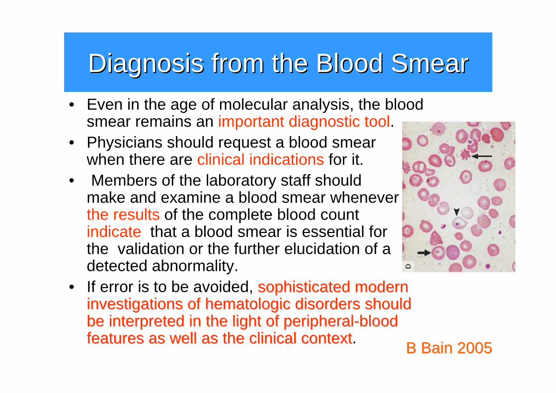
DiagnosisDiagnosis fromfrom the the BloodBlood SmearSmear
B B BainBain 20052005
• Even in the age of molecular analysis, the bloodsmear remains an important diagnostic tool.
• Physicians should request a blood smearwhen there are clinical indications for it.
• Members of the laboratory staff shouldmake and examine a blood smear wheneverthe results of the complete blood countindicate that a blood smear is essential forthe validation or the further elucidation of a detected abnormality.
• If error is to be avoided, sophisticated sophisticated modernmoderninvestigationsinvestigations ofof hematologichematologic disordersdisorders shouldshouldbebe interpretedinterpreted in the light in the light ofof peripheralperipheral--bloodbloodfeaturesfeatures asas wellwell asas the the clinicalclinical contextcontext.
White White BloodBlood CellCell MorphologyMorphology in the in the BalanceBalance
Like many in the hematology laboratoryprofession, I have spent countless hoursreading blood films, bone marrows, and body fluids. But did I ever seriously ask if I was makingmaking anan impact on impact on medicalmedicaldecisionmakingdecisionmaking with my morphologyassessments?
B B HouwenHouwen 20052005
Esiste e cosEsiste e cos’è’è ll’’ematologia di ematologia di laboratorio?laboratorio?
• Ematologia di Laboratorio�disciplina morfologica�disciplina tecnologica�disciplina clinica
•• Logica diagnostica Logica diagnostica delldell’’ematologia di laboratorioematologia di laboratorio
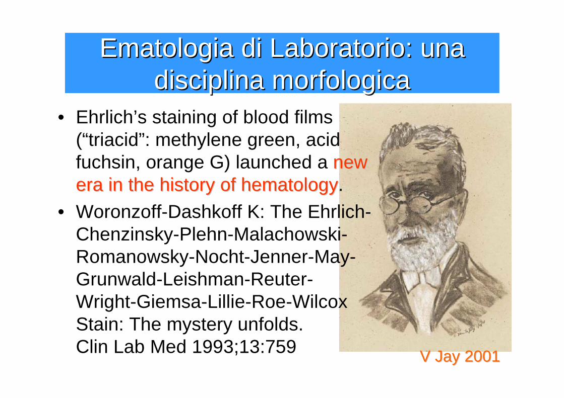
Ematologia di Laboratorio: una Ematologia di Laboratorio: una disciplina morfologicadisciplina morfologica
• Ehrlich’s staining of blood films(“triacid”: methylene green, acid fuchsin, orange G) launched a newnewera in the era in the historyhistory ofof hematologyhematology.
• Woronzoff-Dashkoff K: The Ehrlich-Chenzinsky-Plehn-Malachowski-Romanowsky-Nocht-Jenner-May-Grunwald-Leishman-Reuter-Wright-Giemsa-Lillie-Roe-Wilcox Stain: The mystery unfolds. Clin Lab Med 1993;13:759
V Jay 2001V Jay 2001
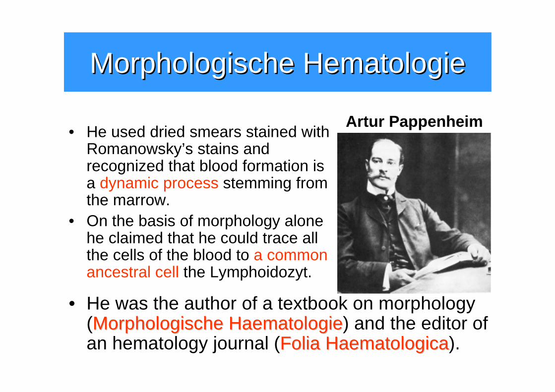
MorphologischeMorphologische HematologieHematologie
• He was the author of a textbook on morphology(MorphologischeMorphologische HaematologieHaematologie) and the editor ofan hematology journal (FoliaFolia HaematologicaHaematologica).
Artur Pappenheim• He used dried smears stained with
Romanowsky’s stains and recognized that blood formation isa dynamic process stemming fromthe marrow.
• On the basis of morphology alone he claimed that he could trace allthe cells of the blood to a common ancestral cell the Lymphoidozyt.
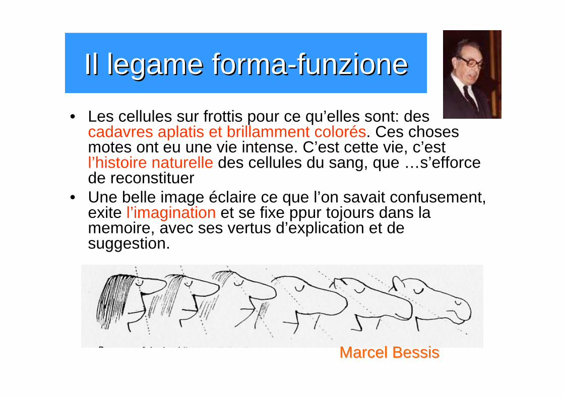
Il legame formaIl legame forma--funzionefunzione
• Les cellules sur frottis pour ce qu’elles sont: descadavres aplatis et brillamment colorés. Ces chosesmotes ont eu une vie intense. C’est cette vie, c’est l’histoire naturelle des cellules du sang, que …s’efforcede reconstituer
• Une belle image éclaire ce que l’on savait confusement, exite l’imagination et se fixe ppur tojours dans la memoire, avec ses vertus d’explication et de suggestion.
Marcel Marcel BessisBessis

La storia della leucemiaLa storia della leucemia
• Virchow, R. (1856) DieDieLeukLeukäämiemie. In: GesammelteAbhandlungen ZurWissenschaftlichen Medizin, pp. 190-211. Meidinger, Frankfurt.
• “It is moreover, the sameconclusion which Bennett came to in the much-discussed matter of prioritybetween us when heobserved a case ofindubitable leukaemia, some months before I saw my first case” R. Virchow 1858
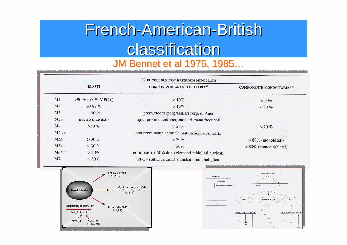
FrenchFrench--AmericanAmerican--BritishBritishclassificationclassification
JM JM BennetBennet etet al 1976, 1985al 1976, 1985……

FAB M7: Acute FAB M7: Acute MegakarioblasticMegakarioblasticLeukemiaLeukemia –– BeyondBeyond MorphologyMorphology
• The criteria for M7 represent a significant departure for the FAB group in that ultrastructuralcytochemistry or immunologictechniques, or both, are nowrequired to make the diagnosis
• …are we ready for a a totallytotally newnewclassificationclassification based primarly on cytogenetic or immunologiccharacterization…?
CDCD BloomfieldBloomfield, RD , RD BrunningBrunning 19851985
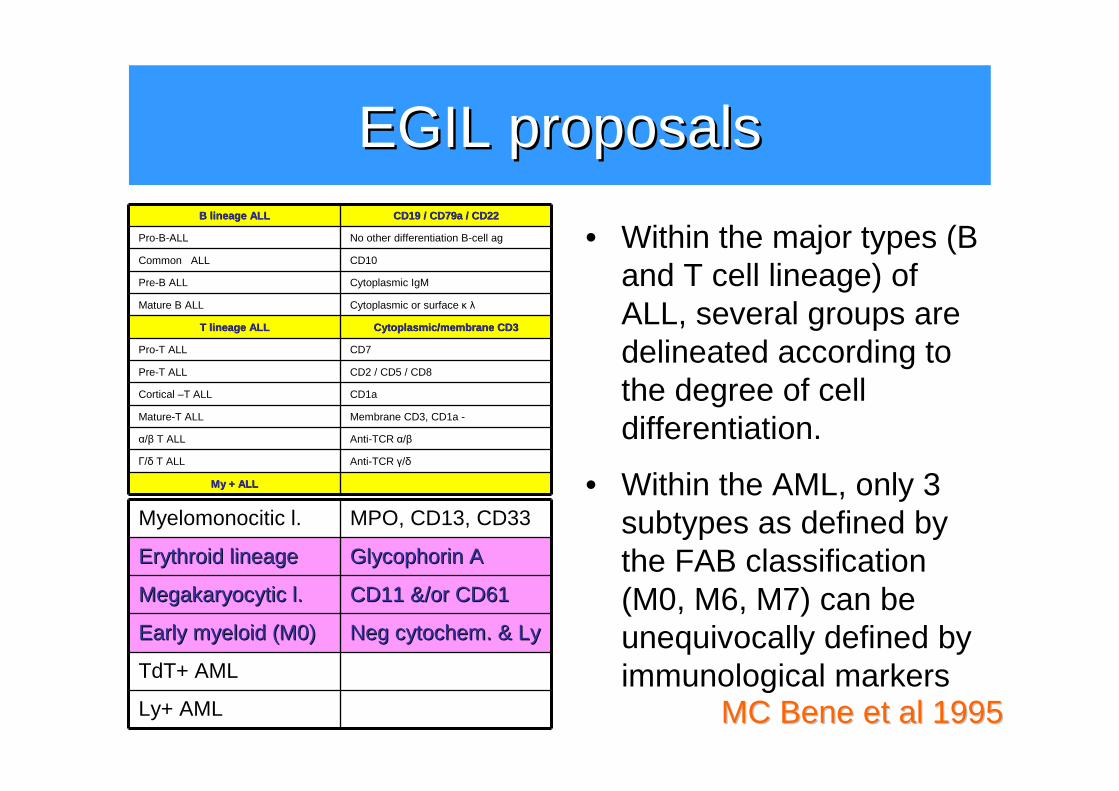
EGIL EGIL proposalsproposals
• Within the AML, only 3 subtypes as defined bythe FAB classification(M0, M6, M7) can beunequivocally defined byimmunological markers
B B lineagelineage ALLALL CD19 / CD79a / CD22CD19 / CD79a / CD22
Pro-B-ALL No other differentiation B-cell ag
Common ALL CD10
Pre-B ALL Cytoplasmic IgM
Mature B ALL Cytoplasmic or surface κ λ
T T lineagelineage ALLALL CytoplasmicCytoplasmic /membrane CD3/membrane CD3
Pro-T ALL CD7
Pre-T ALL CD2 / CD5 / CD8
Cortical –T ALL CD1a
Mature-T ALL Membrane CD3, CD1a -
α/β T ALL Anti-TCR α/β
Γ/δ T ALL Anti-TCR γ/δ
MyMy + ALL+ ALL
Myelomonocitic l. MPO, CD13, CD33
ErythroidErythroid lineagelineage GlycophorinGlycophorin AA
MegakaryocyticMegakaryocytic l.l. CD11 &/or CD61CD11 &/or CD61
EarlyEarly myeloidmyeloid (M0)(M0) NegNeg cytochemcytochem. & . & LyLy
TdT+ AML
Ly+ AML MC Bene MC Bene etet al 1995al 1995
• Within the major types (B and T cell lineage) ofALL, several groups are delineated according tothe degree of celldifferentiation.
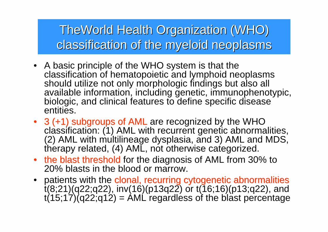
TheWorldTheWorld HealthHealth OrganizationOrganization (WHO) (WHO) classificationclassification ofof the the myeloidmyeloid neoplasmsneoplasms
• A basic principle of the WHO system is that the classification of hematopoietic and lymphoid neoplasmsshould utilize not only morphologic findings but also allavailable information, including genetic, immunophenotypic, biologic, and clinical features to define specific diseaseentities.
•• 3 (+1) 3 (+1) subgroupssubgroups ofof AMLAML are recognized by the WHO classification: (1) AML with recurrent genetic abnormalities, (2) AML with multilineage dysplasia, and 3) AML and MDS, therapy related, (4) AML, not otherwise categorized.
•• the the blastblast thresholdthreshold for the diagnosis of AML from 30% to20% blasts in the blood or marrow.
• patients with the clonalclonal, , recurringrecurring cytogeneticcytogenetic abnormalitiesabnormalitiest(8;21)(q22;q22), inv(16)(p13q22) or t(16;16)(p13;q22), and t(15;17)(q22;q12) = AML regardless of the blast percentage
TheWorldTheWorld HealthHealth OrganizationOrganization (WHO) (WHO) classificationclassification ofof the the myeloidmyeloid neoplasmsneoplasms
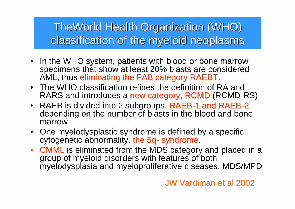
• In the WHO system, patients with blood or bone marrowspecimens that show at least 20% blasts are consideredAML, thus eliminatingeliminating the FAB the FAB categorycategory RAEBTRAEBT.
• The WHO classification refines the definition of RA and RARS and introduces a newnew categorycategory, RCMD, RCMD (RCMD-RS)
• RAEB is divided into 2 subgroups, RAEBRAEB--1 and RAEB1 and RAEB--22, depending on the number of blasts in the blood and bonemarrow
• One myelodysplastic syndrome is defined by a specificcytogenetic abnormality, the 5qthe 5q-- syndromesyndrome.
•• CMMLCMML is eliminated from the MDS category and placed in a group of myeloid disorders with features of bothmyelodysplasia and myeloproliferative diseases, MDS/MPD
JW JW VardimanVardiman etet al 2002al 2002
TheWorldTheWorld HealthHealth OrganizationOrganization (WHO) (WHO) classificationclassification ofof the the myeloidmyeloid neoplasmsneoplasms
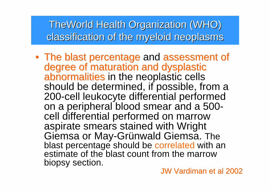
•• The The blastblast percentagepercentage and assessmentassessment ofofdegreedegree ofof maturationmaturation and and dysplasticdysplasticabnormalitiesabnormalities in the neoplastic cellsshould be determined, if possible, from a 200-cell leukocyte differential performedon a peripheral blood smear and a 500-cell differential performed on marrowaspirate smears stained with Wright Giemsa or May-Grünwald Giemsa. The blast percentage should be correlated with anestimate of the blast count from the marrowbiopsy section.
JW JW VardimanVardiman etet al 2002al 2002
TheWorldTheWorld HealthHealth OrganizationOrganization (WHO) (WHO) classificationclassification ofof the the myeloidmyeloid neoplasmsneoplasms
•• ““blastblast equivalentsequivalents”” = myeloblasts, monoblastsmonoblasts and and promonocytespromonocytes in acute monoblastic/monocytic and acute and chronic myelomonocytic leukemia and the megakaryoblastsmegakaryoblasts in acute megakaryoblastic leukemia
•• the the abnormalabnormal promyelocytepromyelocyte, in acute promyelocyticleukemia (APL)
•• ErythroidErythroid precursorsprecursors ((erythroblastserythroblasts)) are not included in the blast count except in the rare instance of “pure”erythroleukemia.
•• DysplasticDysplastic micromegakaryocytesmicromegakaryocytes are also excluded• the percentage of CD34+ CD34+ cellscells should not be considered a
substitute for a blast count from the smears or an estimate from the bone marrow biopsy. Although CD34 hematopoietic cells generally are blasts, not all blastsexpress CD34.
JW JW VardimanVardiman etet al 2002al 2002
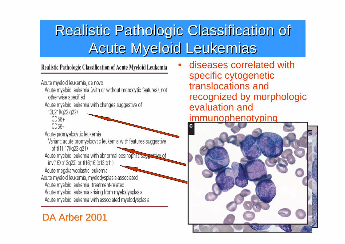
RealisticRealistic PathologicPathologic ClassificationClassification ofofAcute Acute MyeloidMyeloid LeukemiasLeukemias
• diseases correlated withspecific cytogenetictranslocations and recognized by morphologicevaluation and immunophenotyping
DA DA ArberArber 20012001
Integrazione dei datiIntegrazione dei dati
...a realistic pathologicclassification AMLsthat would containdisease types thatcan be recognizedby a a combinationcombination ofofmorphologicmorphologic, , cytochemicalcytochemical, and , and immunophenotypingimmunophenotypingstudiesstudies.
• From our experience, wesuggest that togethertogether withwithmorphologicmorphologic and and cytochemicalcytochemical examinationexamination, , a a panelpanel ofof mAbsmAbs againstMPO, cyCD3, cyCD79a, CD13, CD33, CD10, CD19, CD2, and CD117 might be a cost-effective, highly predictivescreening tooltool toto predictpredictlineagelineage differentiationdifferentiation ofofacute acute leukemiasleukemias.
ThalhammerThalhammer--ScherrerScherrer etet al 2002al 2002DA DA ArberArber 20012001
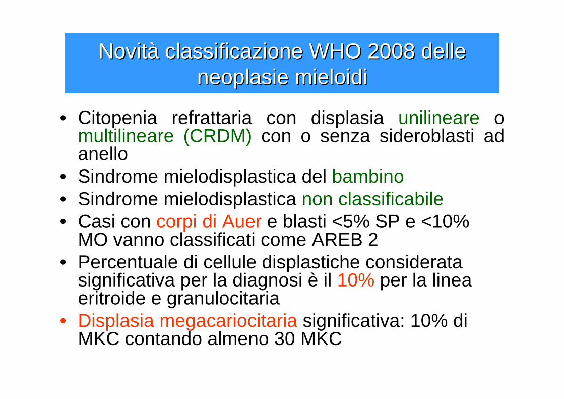
NovitNovitàà classificazione WHO 2008 delle classificazione WHO 2008 delle neoplasie mieloidi neoplasie mieloidi
• Incorporazione formale delle anomalie genetiche (traslocazioni cromosomiche e mutazioni geniche) nell’algoritmo diagnostico per la diagnosi di AML– t(6;9)(p22;q23), inv(3)(p21;q26.2) o t(3;3)(p22;q23),
t(1;22)(p13;q13), con mutazione NPM1, CEBPA– Leucemie acute di lineage ambiguo– Proliferazioni mieloidi correlate a sindrome di Down– Neoplasia a cellule dendritiche blastiche plasmacitoidi
• Da integrare con i dati clinici, morfologici e/o immunofenotipici
NovitNovitàà classificazione WHO 2008 delle classificazione WHO 2008 delle neoplasie mieloidi neoplasie mieloidi
• Citopenia refrattaria con displasia unilineare o multilineare (CRDM) con o senza sideroblasti ad anello
• Sindrome mielodisplastica del bambino• Sindrome mielodisplastica non classificabile• Casi con corpi di Auer e blasti <5% SP e <10%
MO vanno classificati come AREB 2• Percentuale di cellule displastiche considerata
significativa per la diagnosi è il 10% per la lineaeritroide e granulocitaria
• Displasia megacariocitaria significativa: 10% diMKC contando almeno 30 MKC
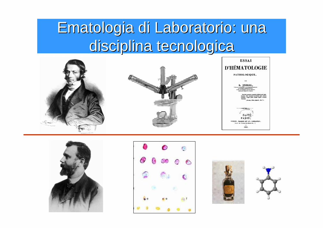
Ematologia di Laboratorio: una Ematologia di Laboratorio: una disciplina tecnologicadisciplina tecnologica
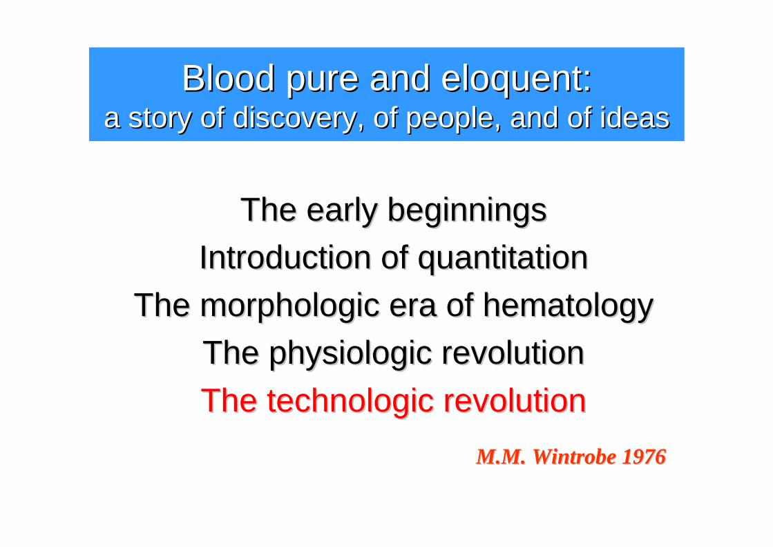
BloodBlood pure and pure and eloquenteloquent: : a story a story ofof discoverydiscovery, , ofof people, and people, and ofof ideasideas
The The earlyearly beginningsbeginnings
IntroductionIntroduction ofof quantitationquantitation
The The morphologicmorphologic era era ofof hematologyhematology
The The physiologicphysiologic revolutionrevolution
The The technologictechnologic revolutionrevolution
M.M. M.M. WintrobeWintrobe 19761976
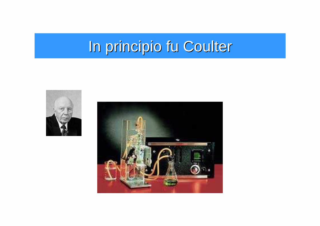
In principio fu In principio fu CoulterCoulter
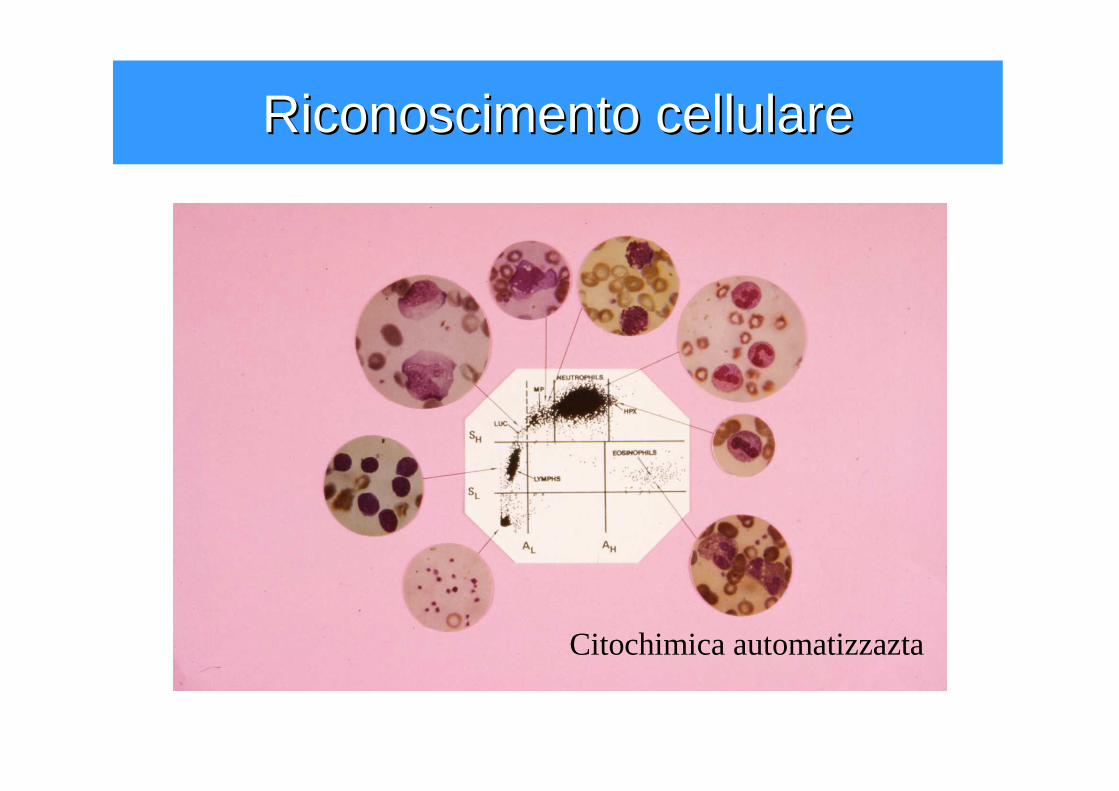
Riconoscimento cellulareRiconoscimento cellulare
Citochimica automatizzazta
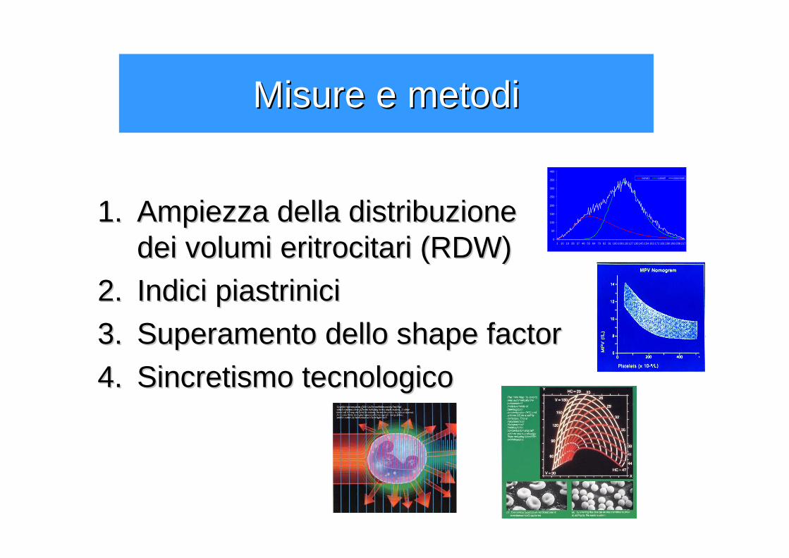
Misure e metodiMisure e metodi
1.1. Ampiezza della distribuzione Ampiezza della distribuzione dei volumi dei volumi eritrocitarieritrocitari (RDW)(RDW)
2.2. Indici piastriniciIndici piastrinici
3.3. Superamento dello Superamento dello shapeshape factorfactor
4.4. Sincretismo tecnologicoSincretismo tecnologico
0
50
100
150
200
250
300
350
400
1 10 19 28 37 46 55 64 73 82 91 100 109 118 127 136145 154 163 172 181190 199 208 217
curva1 curva2 osservati
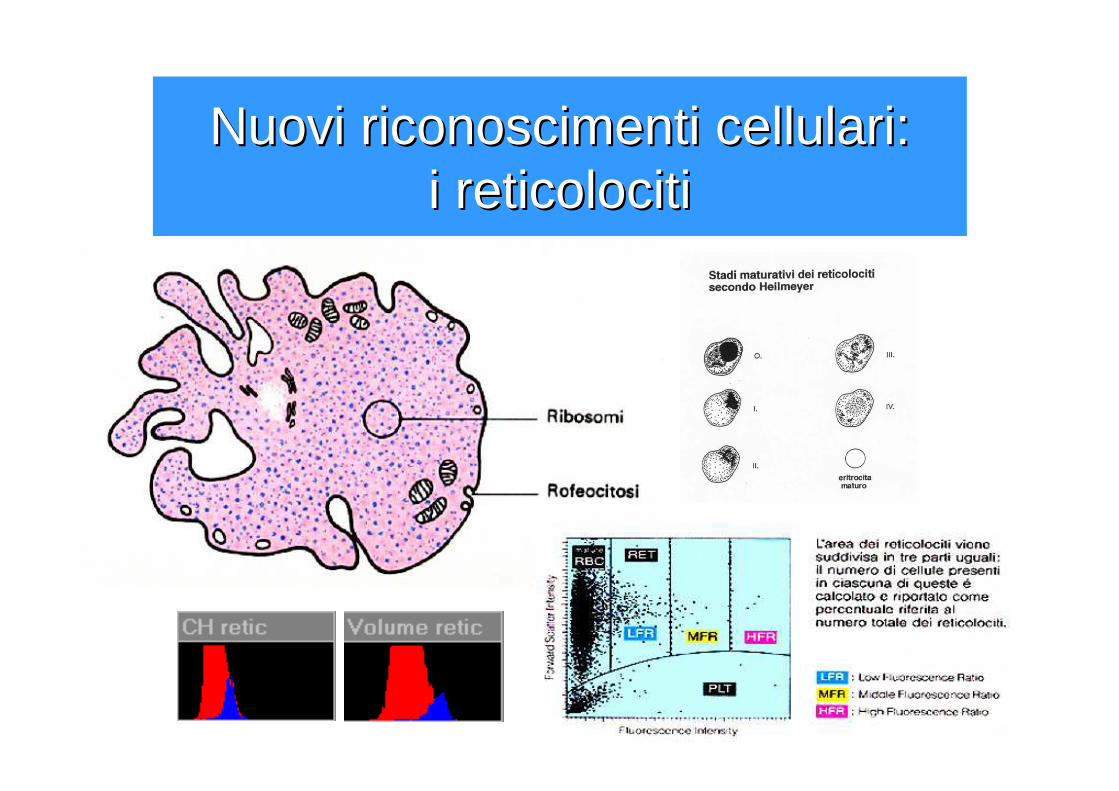
Nuovi riconoscimenti cellulari: Nuovi riconoscimenti cellulari: i i reticolocitireticolociti
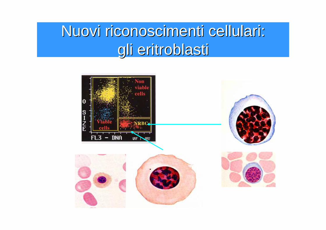
Nuovi riconoscimenti cellulari:Nuovi riconoscimenti cellulari:gli eritroblastigli eritroblasti
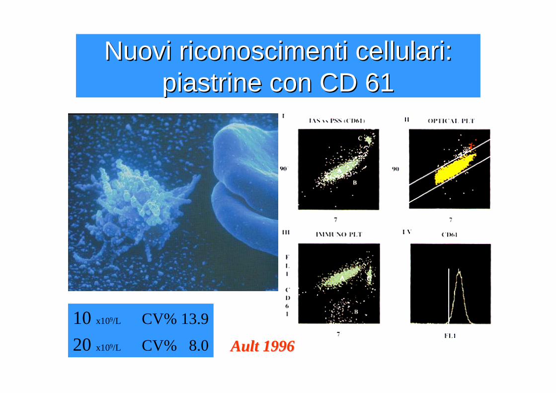
Nuovi riconoscimenti cellulari: Nuovi riconoscimenti cellulari: piastrine con piastrine con CDCD 6161
10 x109/L CV% 13.9
20 x109/L CV% 8.0 AultAult 19961996
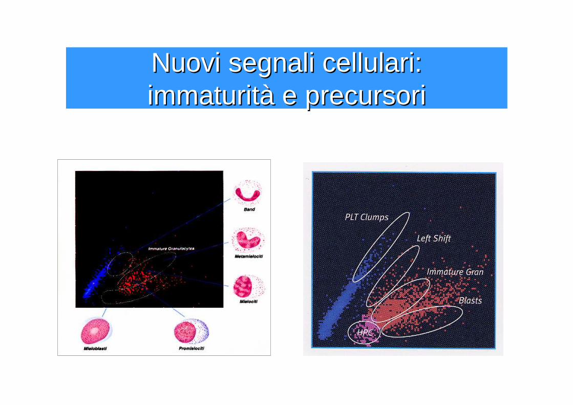
Nuovi segnali cellulari: Nuovi segnali cellulari: immaturitimmaturitàà e precursorie precursori
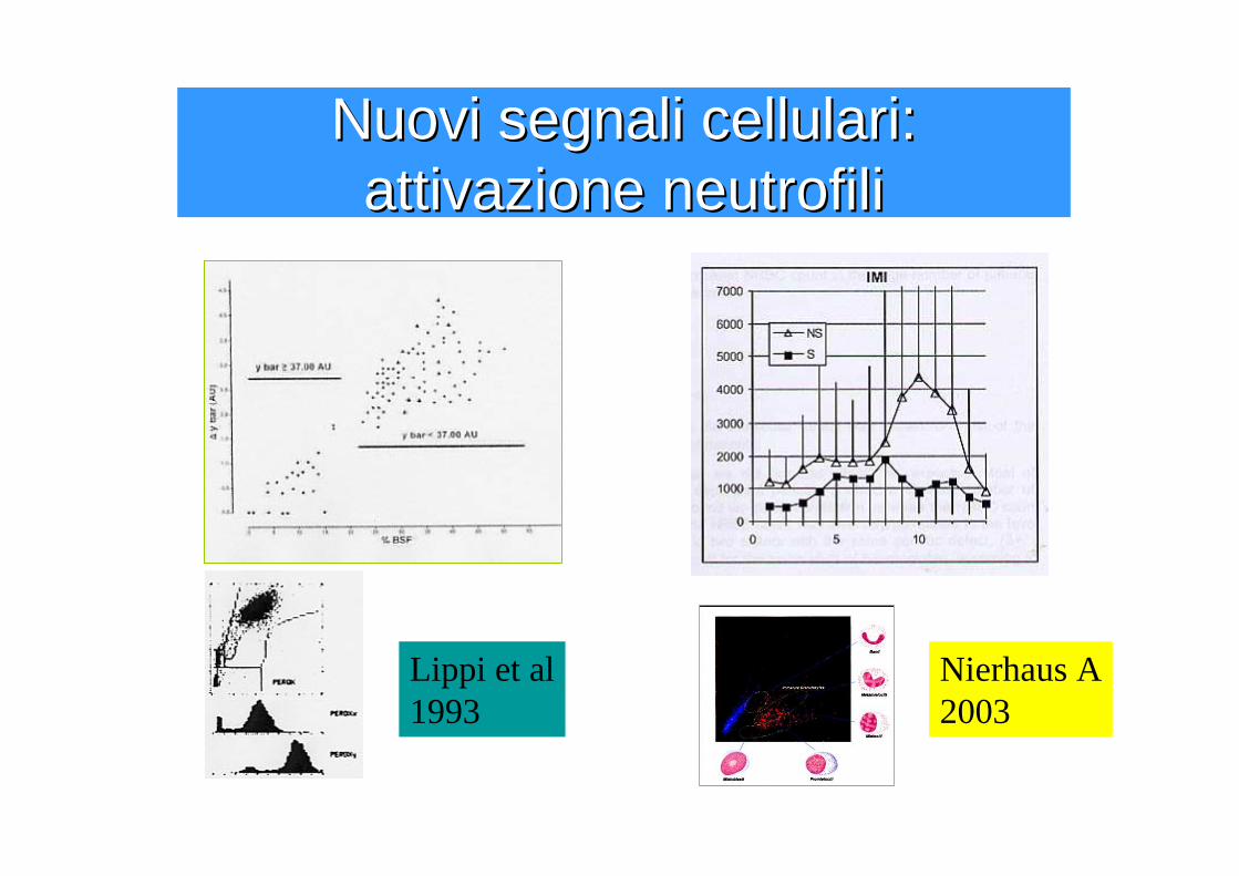
Nuovi segnali cellulari: Nuovi segnali cellulari: attivazione neutrofiliattivazione neutrofili
Lippi et al1993
Nierhaus A2003
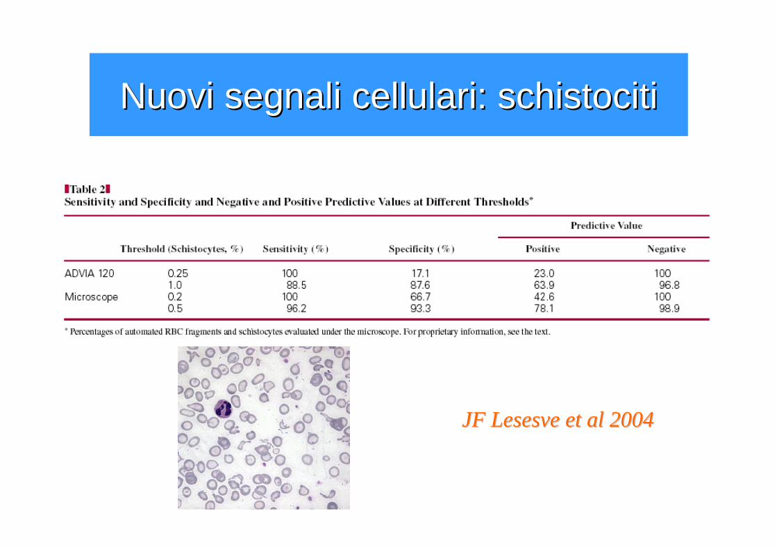
Nuovi segnali cellulari: Nuovi segnali cellulari: schistocitischistociti
JF JF LesesveLesesve etet al 2004al 2004
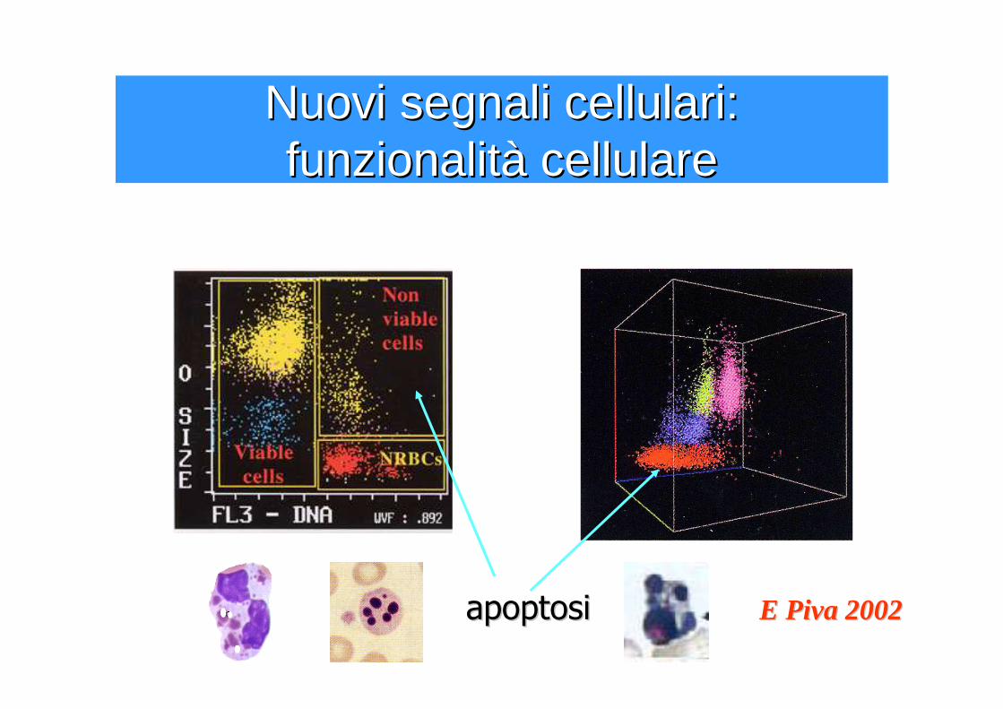
Nuovi segnali cellulari: Nuovi segnali cellulari: funzionalitfunzionalitàà cellularecellulare
apoptosiapoptosi E Piva 2002E Piva 2002
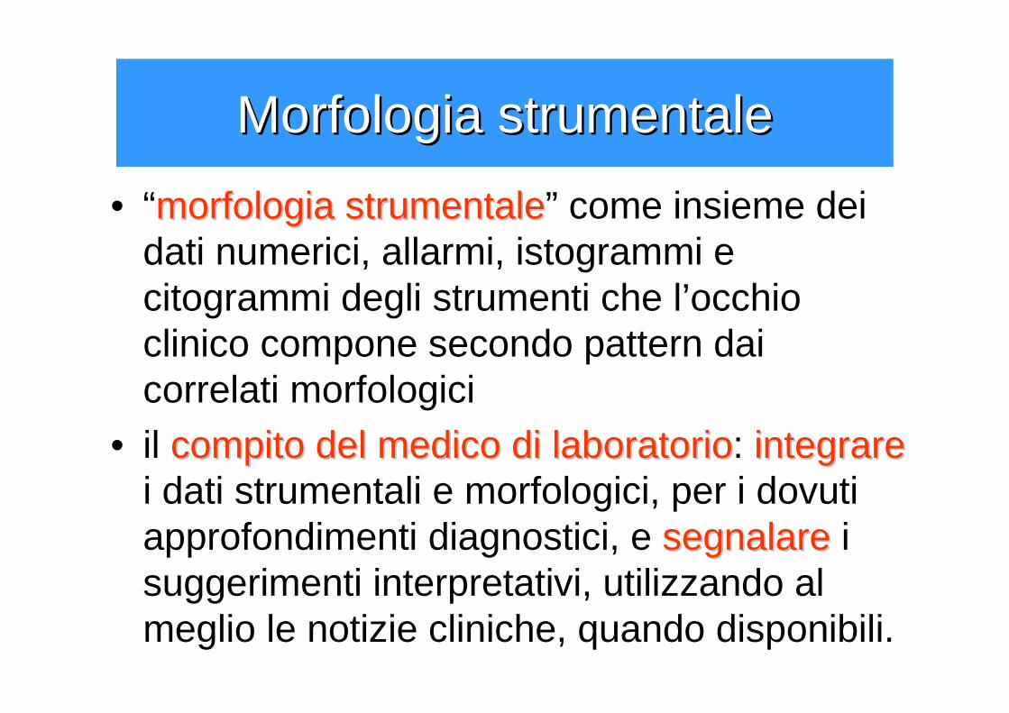
Morfologia strumentaleMorfologia strumentale
• “morfologia strumentalemorfologia strumentale” come insieme dei dati numerici, allarmi, istogrammi e citogrammi degli strumenti che l’occhio clinico compone secondo pattern dai correlati morfologici
• il compito del medico di laboratoriocompito del medico di laboratorio: integrareintegrarei dati strumentali e morfologici, per i dovuti approfondimenti diagnostici, e segnalaresegnalare i suggerimenti interpretativi, utilizzando al meglio le notizie cliniche, quando disponibili.
P Cappelletti P Cappelletti etet al 1989al 1989
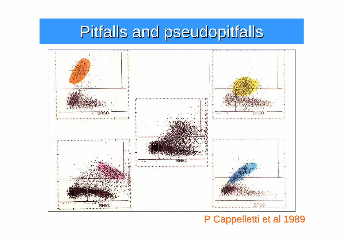
PitfallsPitfalls and and pseudopitfallspseudopitfalls
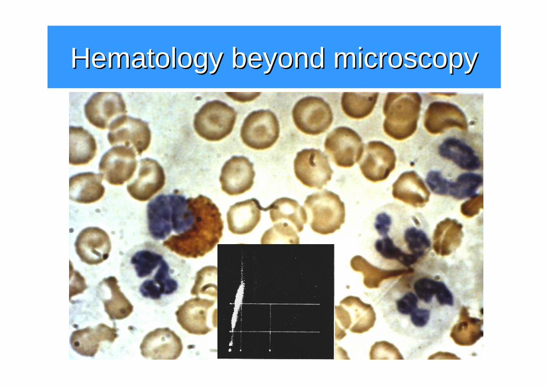
HematologyHematology beyondbeyond microscopymicroscopy
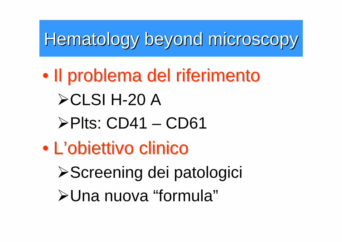
HematologyHematology beyondbeyond microscopymicroscopy
•• Il problema del riferimentoIl problema del riferimento�CLSI H-20 A�Plts: CD41 – CD61
•• LL’’obiettivo clinicoobiettivo clinico�Screening dei patologici�Una nuova “formula”
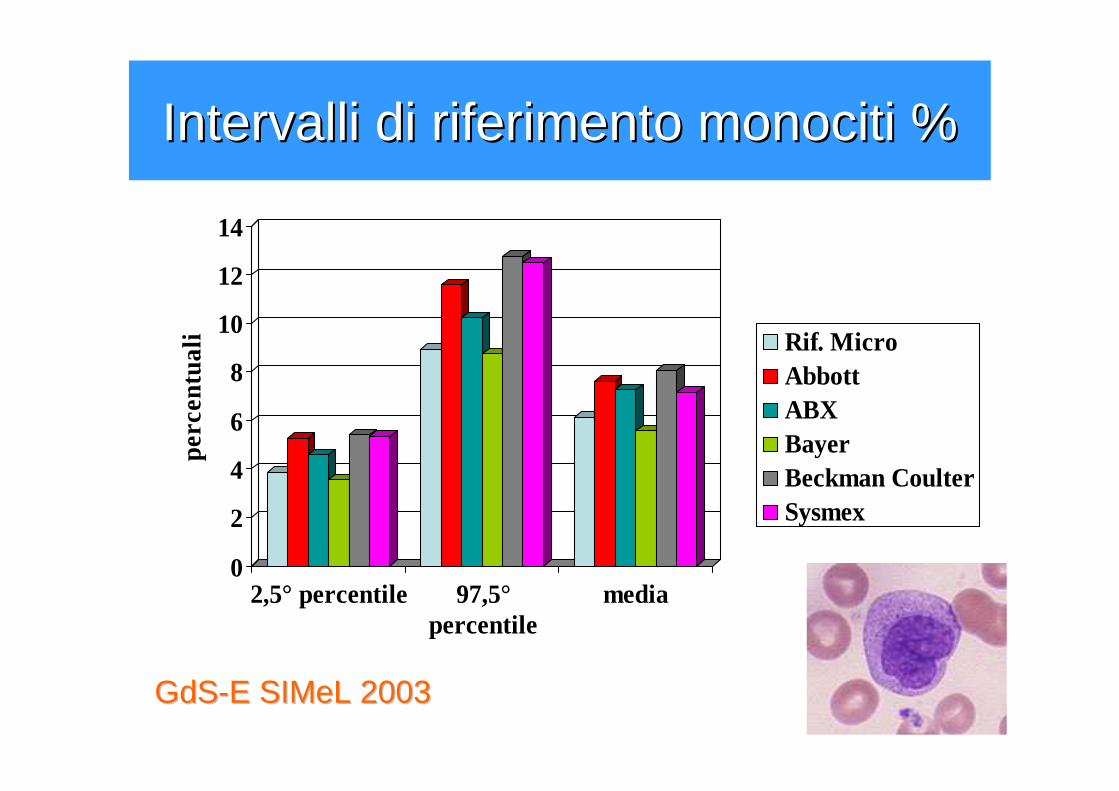
Intervalli di riferimento monociti %Intervalli di riferimento monociti %
0
2
4
6
8
10
12
14pe
rcen
tual
i
2,5° percentile 97,5°percentile
media
Rif. MicroAbbottABXBayerBeckman CoulterSysmex
GdSGdS--EE SIMeLSIMeL 20032003
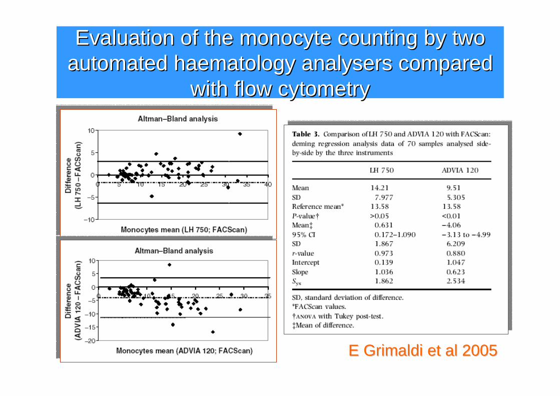
EvaluationEvaluation ofof the the monocytemonocyte countingcounting byby twotwoautomatedautomated haematologyhaematology analysersanalysers comparedcompared
withwith flow flow cytometrycytometry
E Grimaldi E Grimaldi etet al 2005al 2005
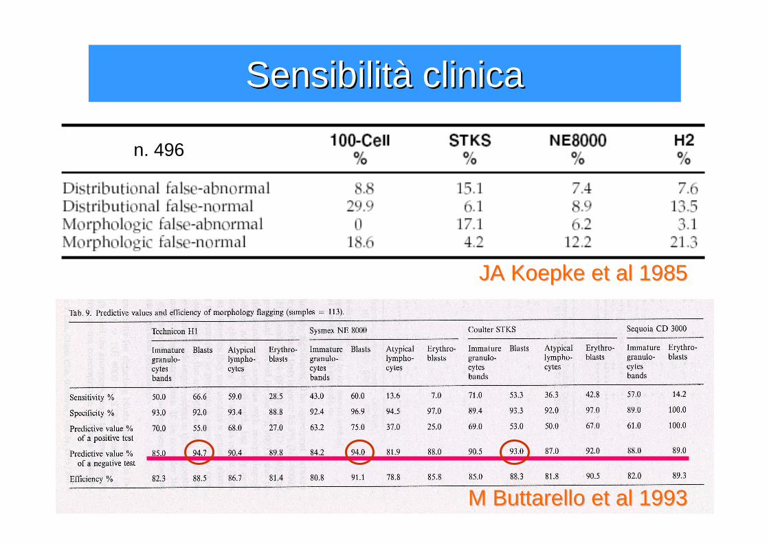
SensibilitSensibilitàà clinicaclinica
M M ButtarelloButtarello etet al 1993al 1993
JA JA KoepkeKoepke etet al 1985al 1985
n. 496
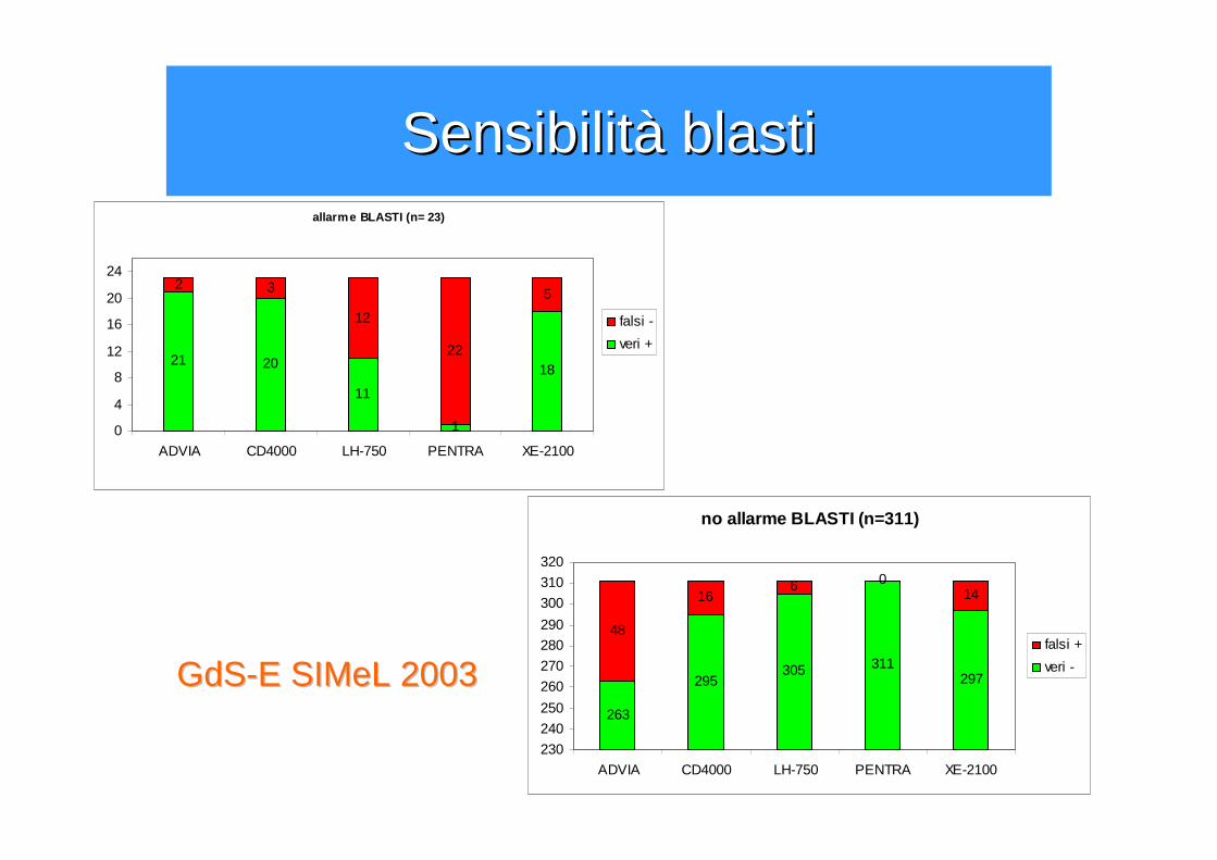
allarme BLASTI (n= 23)
21 20
11
1
18
2 3
12
22
5
0
4
8
12
16
20
24
ADVIA CD4000 LH-750 PENTRA XE-2100
falsi -
veri +
no allarme BLASTI (n=311)
263
295305 311
297
48
166 0
14
230
240
250
260
270
280
290
300
310
320
ADVIA CD4000 LH-750 PENTRA XE-2100
falsi +
veri -GdSGdS--EE SIMeLSIMeL 20032003
SensibilitSensibilitàà blastiblasti
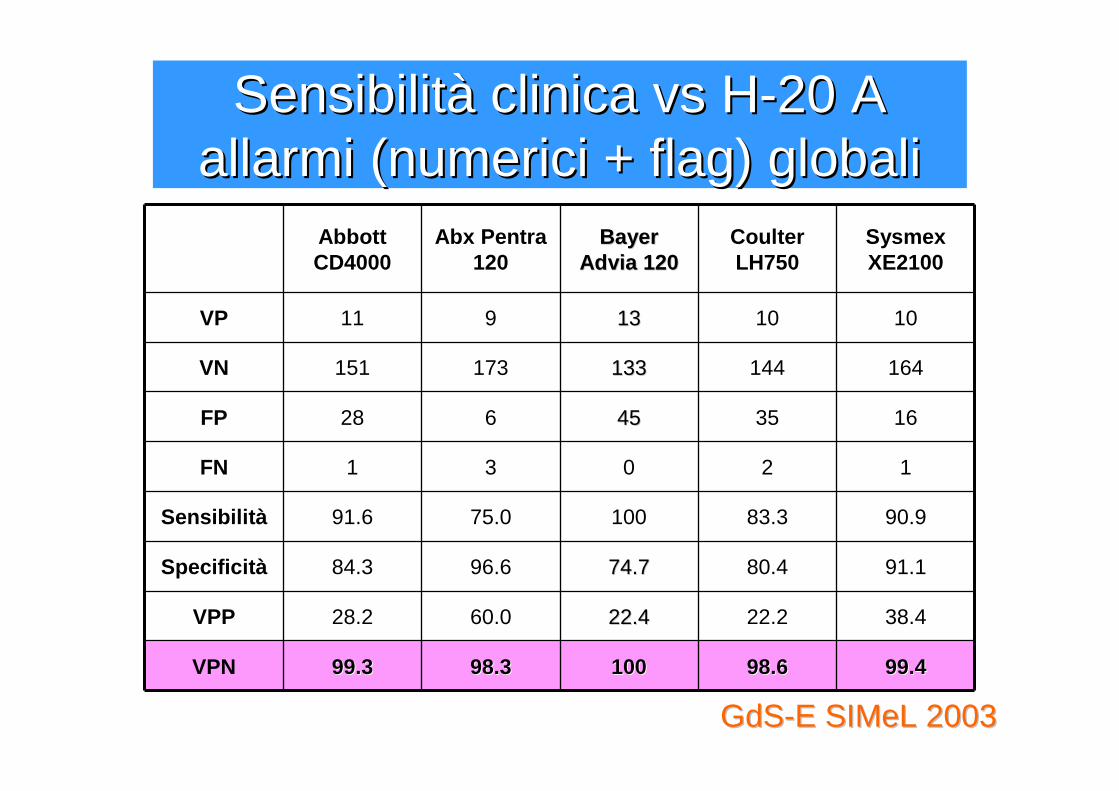
Abbott CD4000
Abx Pentra120
Bayer Bayer AdviaAdvia 120120
CoulterLH750
SysmexXE2100
VP 11 9 1313 10 10
VN 151 173 133133 144 164
FP 28 6 4545 35 16
FN 1 3 0 2 1
Sensibilità 91.6 75.0 100 83.3 90.9
Specificità 84.3 96.6 74.774.7 80.4 91.1
VPP 28.2 60.0 22.422.4 22.2 38.4
VPN 99.399.3 98.398.3 100100 98.698.6 99.499.4
SensibilitSensibilitàà clinica vs Hclinica vs H--20 A20 Aallarmi (numerici + allarmi (numerici + flagflag) globali) globali
GdSGdS--EE SIMeLSIMeL 20032003
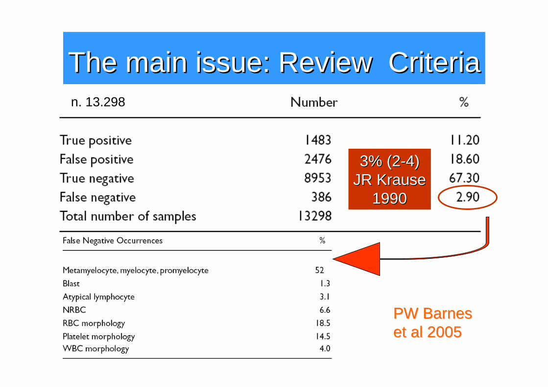
The The mainmain issueissue: : ReviewReview CriteriaCriterian. 13.298
PW PW BarnesBarnesetet al 2005al 2005
3% (23% (2--4)4)JR JR KrauseKrause
19901990
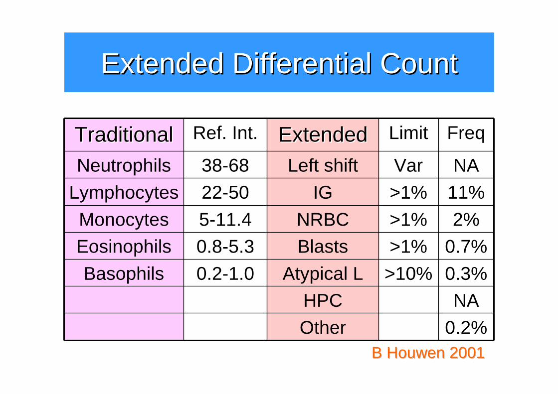
ExtendedExtended DifferentialDifferential CountCount
TraditionalTraditional Ref. Int. ExtendedExtended Limit Freq
Neutrophils 38-68 Left shift Var NA
Lymphocytes 22-50 IG >1% 11%
Monocytes 5-11.4 NRBC >1% 2%
Eosinophils 0.8-5.3 Blasts >1% 0.7%
Basophils 0.2-1.0 Atypical L >10% 0.3%
HPC NA
Other 0.2%B B HouwenHouwen 20012001
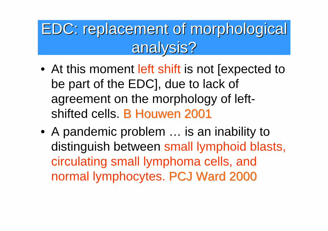
EDC: EDC: replacementreplacement ofof morphologicalmorphologicalanalysisanalysis??
• At this moment left shift is not [expected to be part of the EDC], due to lack of agreement on the morphology of left-shifted cells. B B HouwenHouwen 20012001
• A pandemic problem … is an inability to distinguish between small lymphoid blasts, circulating small lymphoma cells, and normal lymphocytes. PCJ Ward 2000PCJ Ward 2000
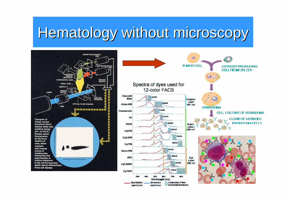
HematologyHematology withoutwithout microscopymicroscopy
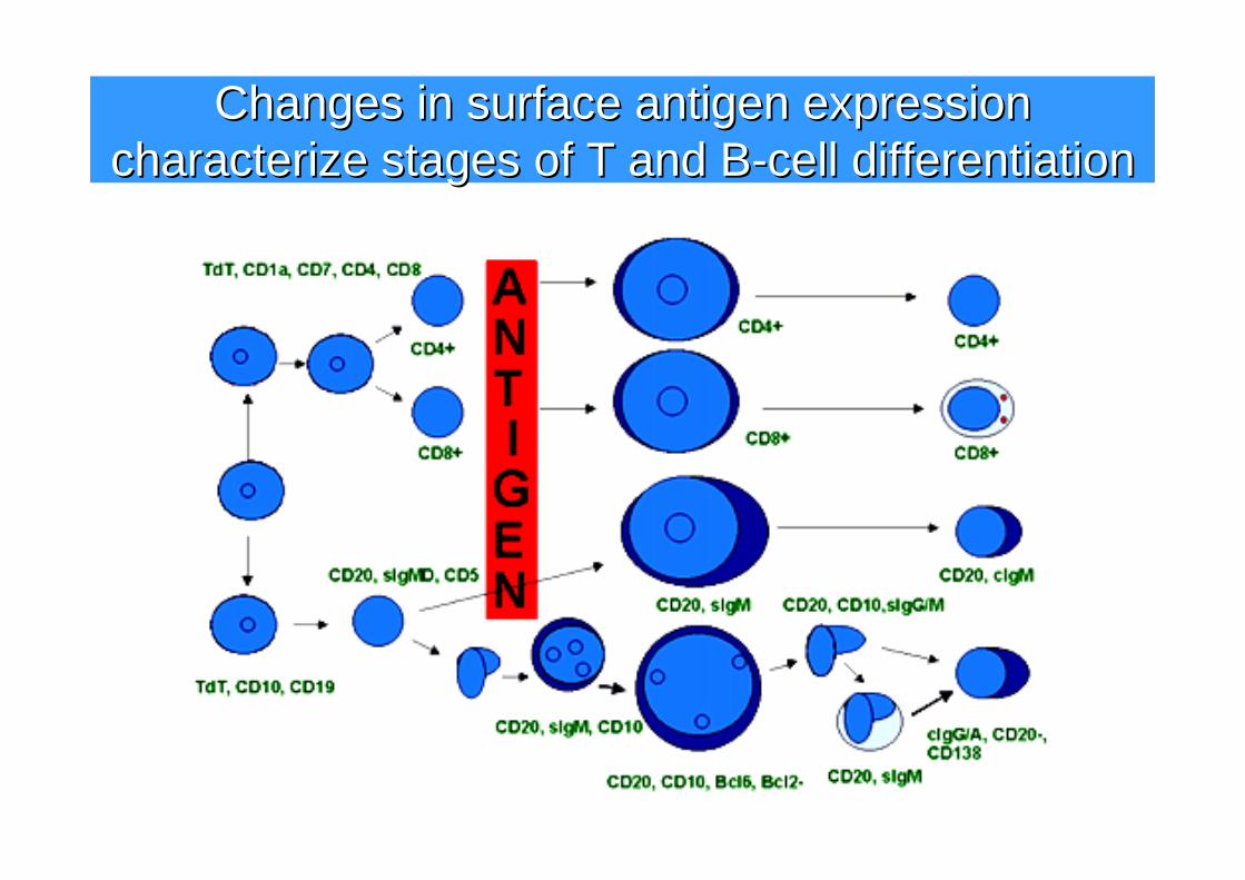
Changes in surface antigen expression Changes in surface antigen expression characterize stages of T and Bcharacterize stages of T and B--cell differentiationcell differentiation
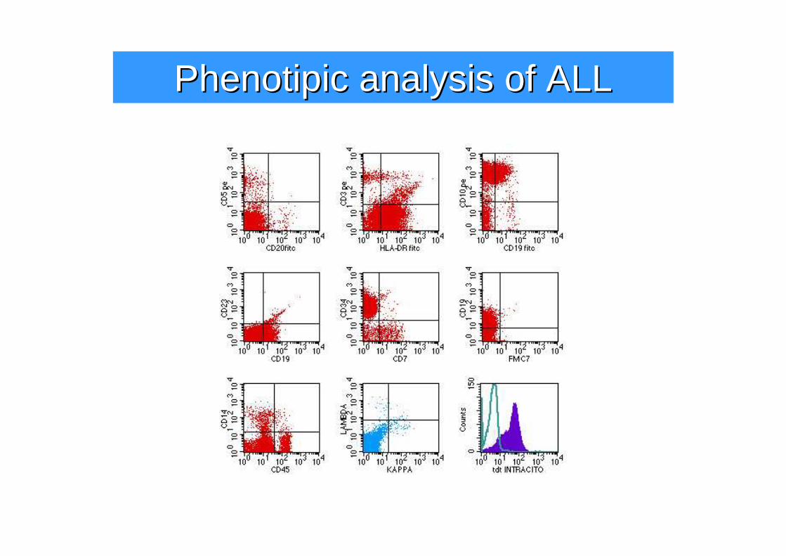
PhenotipicPhenotipic analysisanalysis ofof ALLALL
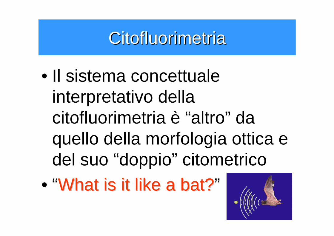
CitofluorimetriaCitofluorimetria
• Il sistema concettuale interpretativo della citofluorimetria è “altro” da quello della morfologia ottica e del suo “doppio” citometrico
• “WhatWhat isis itit likelike a a batbat??”
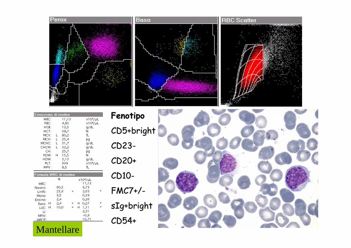
Mantellare
FenotipoFenotipo
CD5+brightCD5+bright
CD23CD23--
CD20+CD20+
CD10CD10--
FMC7+/FMC7+/--
sIg+brightsIg+bright
CD54+CD54+
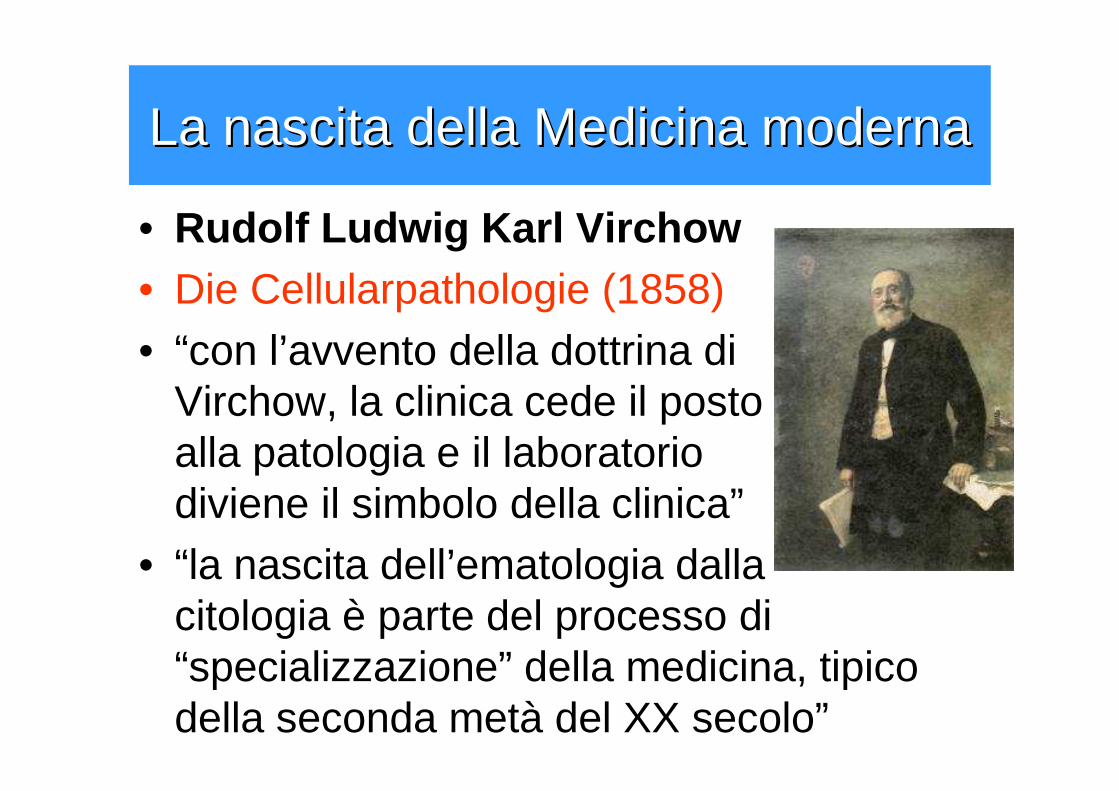
La nascita della Medicina modernaLa nascita della Medicina moderna
• Rudolf Ludwig Karl Virchow• Die Cellularpathologie (1858)• “con l’avvento della dottrina di
Virchow, la clinica cede il posto alla patologia e il laboratorio diviene il simbolo della clinica”
• “la nascita dell’ematologia dalla citologia è parte del processo di “specializzazione” della medicina, tipico della seconda metà del XX secolo”
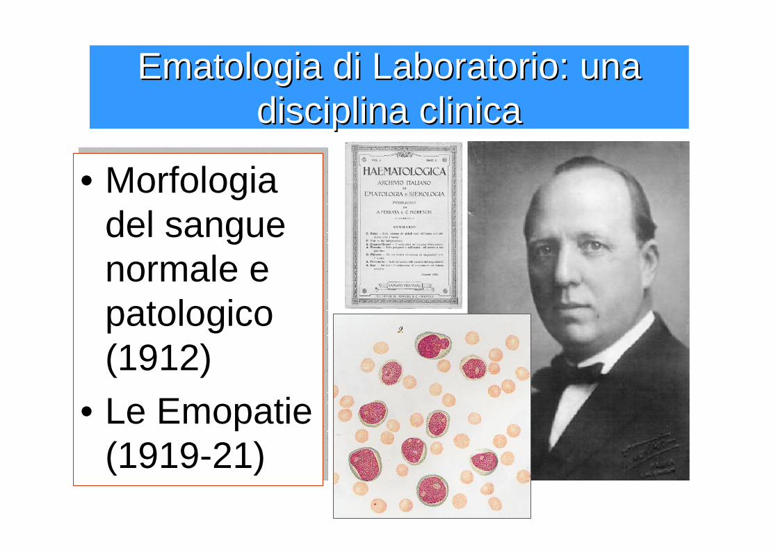
Ematologia di Laboratorio: una Ematologia di Laboratorio: una disciplina clinicadisciplina clinica
• Morfologia del sangue normale e patologico (1912)
• Le Emopatie (1919-21)
• Morfologia del sangue normale e patologico (1912)
• Le Emopatie (1919-21)
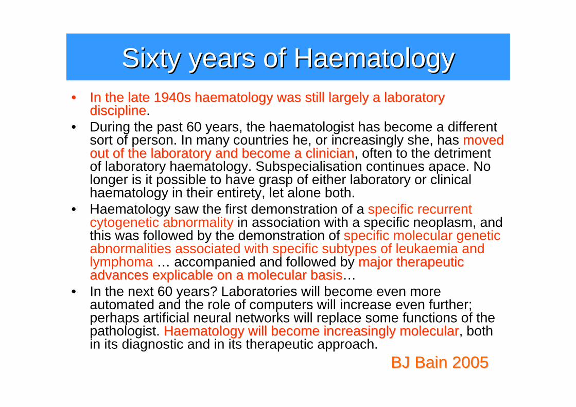
SixtySixty yearsyears ofof HaematologyHaematology•• In the late 1940s In the late 1940s haematologyhaematology waswas stillstill largelylargely a a laboratorylaboratory
disciplinediscipline. • During the past 60 years, the haematologist has become a different
sort of person. In many countries he, or increasingly she, has movedmovedout out ofof the the laboratorylaboratory and and becomebecome a a clinicianclinician, often to the detrimentof laboratory haematology. Subspecialisation continues apace. No longer is it possible to have grasp of either laboratory or clinicalhaematology in their entirety, let alone both.
• Haematology saw the first demonstration of a specific recurrentcytogenetic abnormality in association with a specific neoplasm, and this was followed by the demonstration of specific molecular geneticabnormalities associated with specific subtypes of leukaemia and lymphoma … accompanied and followed by major major therapeutictherapeuticadvancesadvances explicableexplicable on a on a molecularmolecular basisbasis…
• In the next 60 years? Laboratories will become even more automated and the role of computers will increase even further; perhaps artificial neural networks will replace some functions of the pathologist. HaematologyHaematology willwill becomebecome increasinglyincreasingly molecularmolecular, bothin its diagnostic and in its therapeutic approach.
BJ BJ BainBain 20052005
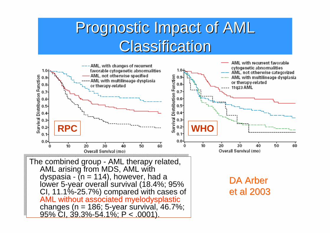
PrognosticPrognostic Impact Impact ofof AML AML ClassificationClassification
The combined group - AML therapy related, AML arising from MDS, AML withdyspasia - (n = 114), however, had a lower 5-year overall survival (18.4%; 95% CI, 11.1%-25.7%) compared with cases ofAML without associated myelodysplasticchanges (n = 186; 5-year survival, 46.7%; 95% CI, 39.3%-54.1%; P < .0001).
The combined group - AML therapy related, AML arising from MDS, AML withdyspasia - (n = 114), however, had a lower 5-year overall survival (18.4%; 95% CI, 11.1%-25.7%) compared with cases ofAML AML withoutwithout associatedassociated myelodysplasticmyelodysplasticchanges (n = 186; 5-year survival, 46.7%; 95% CI, 39.3%-54.1%; P < .0001).
RPC WHO
DA DA ArberArberetet al 2003al 2003
E n EE n EEEEE
EMATOLOGO
M
M
G
D
M
S
M
D
Ematologia di LaboratorioEmatologia di Laboratorio
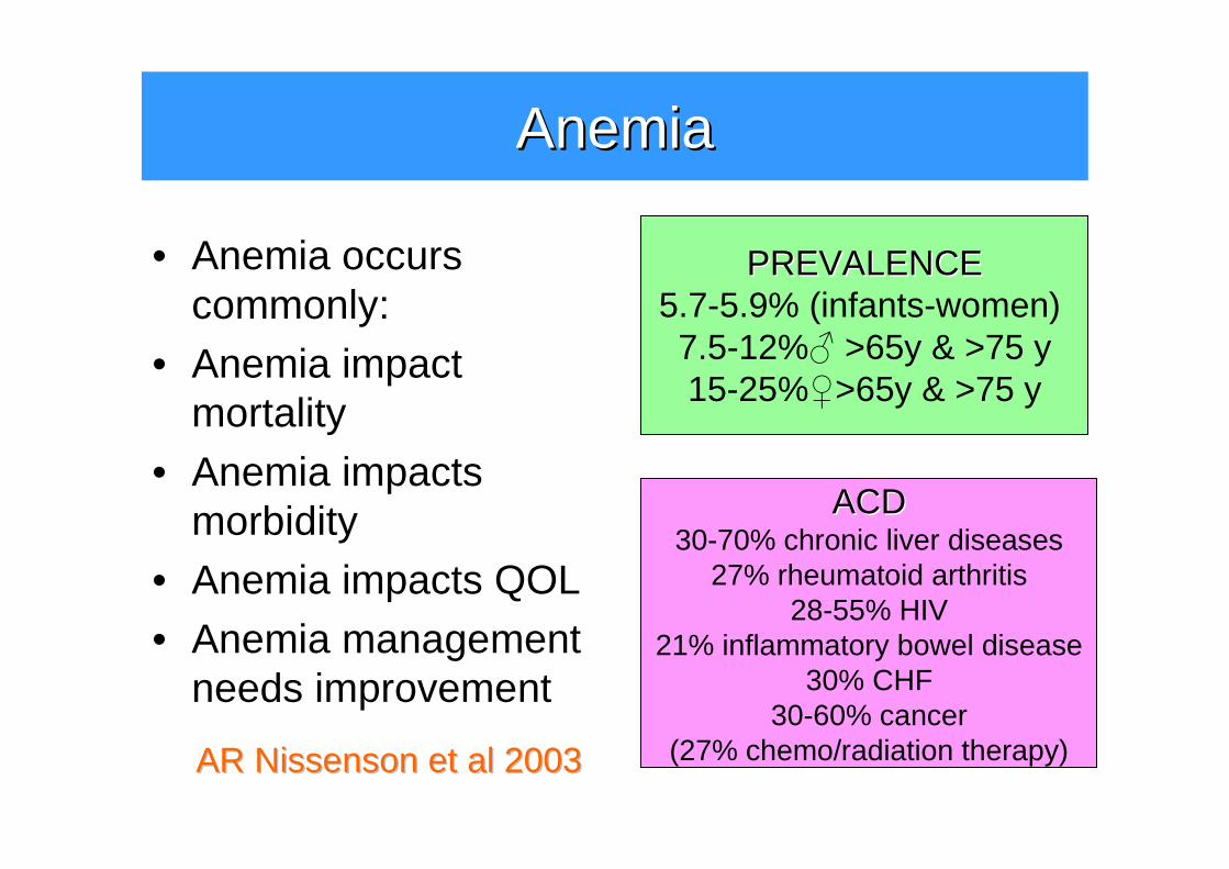
AnemiaAnemia
• Anemia occurscommonly:
• Anemia impact mortality
• Anemia impactsmorbidity
• Anemia impacts QOL
• Anemia management needs improvement
AR AR NissensonNissenson etet al 2003al 2003
PREVALENCEPREVALENCE5.7-5.9% (infants-women) 7.5-12%♂ >65y & >75 y 15-25%♀>65y & >75 y
ACDACD30-70% chronic liver diseases
27% rheumatoid arthritis28-55% HIV
21% inflammatory bowel disease30% CHF
30-60% cancer(27% chemo/radiation therapy)
C Thomas, L Thomas 2002C Thomas, L Thomas 2002
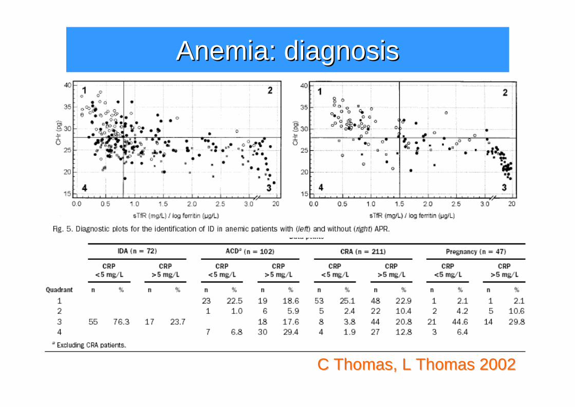
Anemia: Anemia: diagnosisdiagnosis
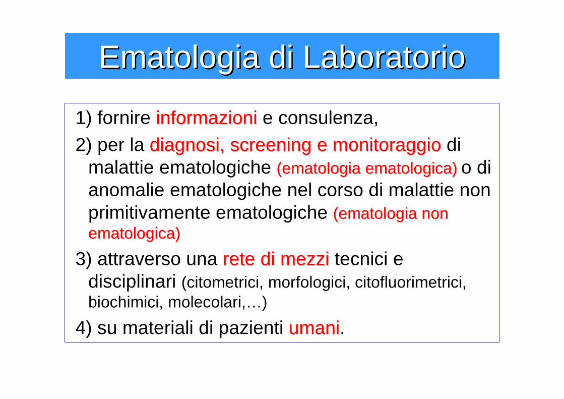
Ematologia di LaboratorioEmatologia di Laboratorio
1) fornire informazioniinformazioni e consulenza,2) per la diagnosi, screening e monitoraggiodiagnosi, screening e monitoraggio di
malattie ematologiche (ematologia ematologica)(ematologia ematologica) o di anomalie ematologiche nel corso di malattie non primitivamente ematologiche (ematologia non (ematologia non ematologica)ematologica)
3) attraverso una rete di mezzirete di mezzi tecnici e disciplinari (citometrici, morfologici, citofluorimetrici, biochimici, molecolari,…)
4) su materiali di pazienti umaniumani.
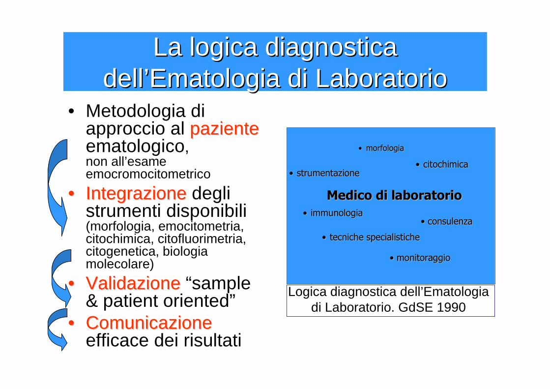
La logica diagnostica La logica diagnostica delldell’’Ematologia di LaboratorioEmatologia di Laboratorio
• Metodologia di approccio al paziente paziente ematologico, non all’esame emocromocitometrico
•• IntegrazioneIntegrazione degli strumenti disponibili(morfologia, emocitometria, citochimica, citofluorimetria, citogenetica, biologia molecolare)
•• ValidazioneValidazione “sample & patient oriented”
•• ComunicazioneComunicazioneefficace dei risultati
Medico di laboratorioMedico di laboratorio
•• morfologiamorfologia
•• citochimicacitochimica
•• immunologiaimmunologia
•• tecniche specialistichetecniche specialistiche
•• strumentazionestrumentazione
•• consulenzaconsulenza
•• monitoraggiomonitoraggio
Logica diagnostica dell’Ematologia di Laboratorio. GdSE 1990

Le basi della conoscenzaLe basi della conoscenza
ottima e aggiornata conoscenza
� ematologia di laboratorio� Tecnologia utilizzata
�Nuove tecnologie
� ematologia clinica�Diagnosi
�Prognosi� Terapia
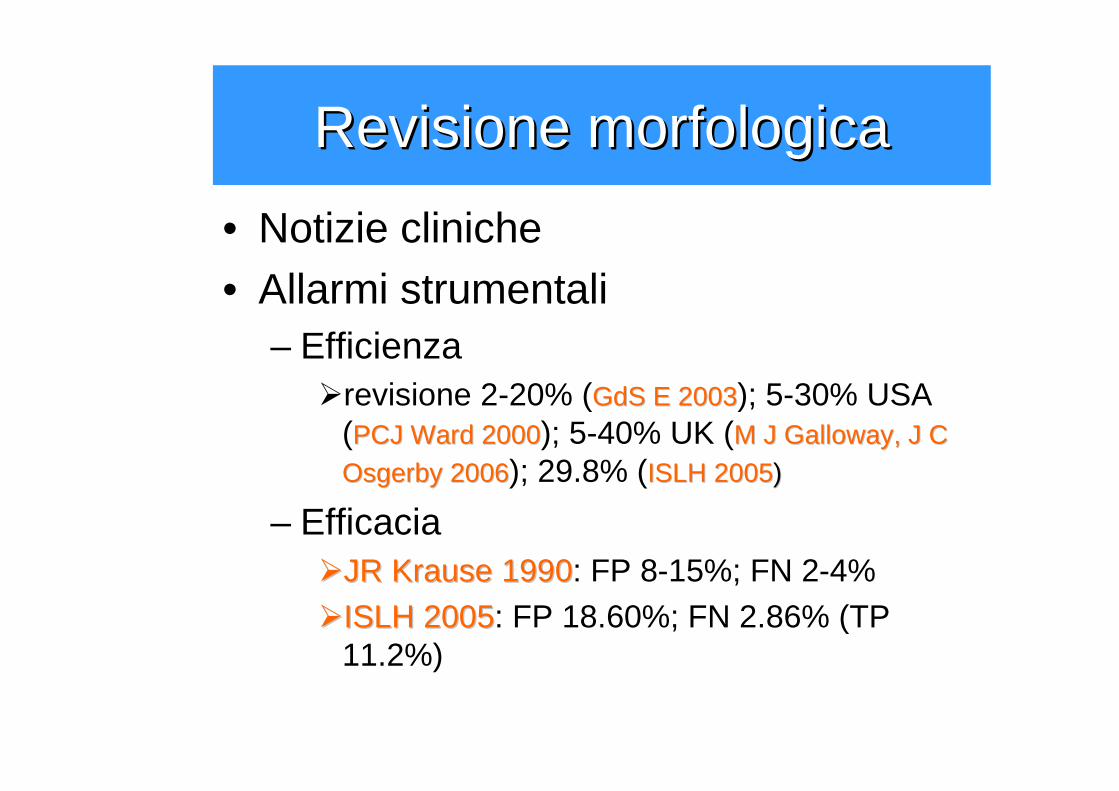
Revisione morfologicaRevisione morfologica
• Notizie cliniche• Allarmi strumentali
– Efficienza�revisione 2-20% (GdSGdS E 2003E 2003); 5-30% USA
(PCJ PCJ WardWard 20002000); 5-40% UK (M J M J GallowayGalloway, J C , J C
OsgerbyOsgerby 20062006); 29.8% (ISLH 2005ISLH 2005))
– Efficacia ��JR JR KrauseKrause 19901990: FP 8-15%; FN 2-4%
��ISLH 2005ISLH 2005: FP 18.60%; FN 2.86% (TP 11.2%)
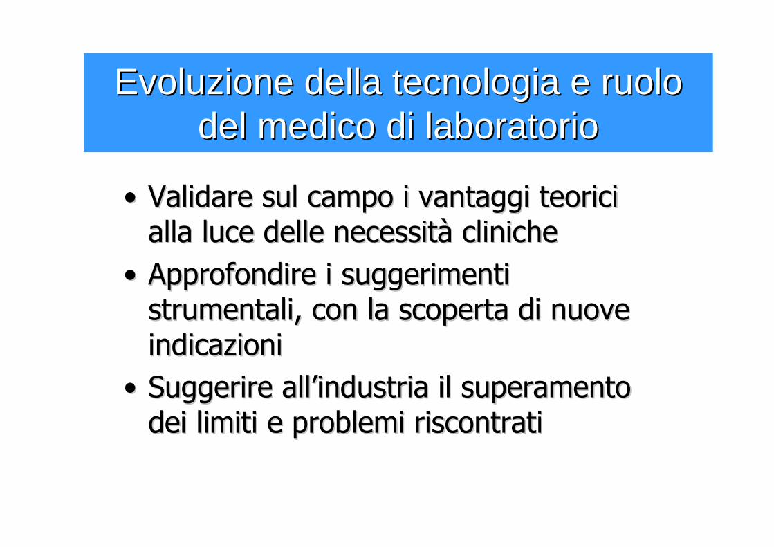
Evoluzione della tecnologia e ruolo Evoluzione della tecnologia e ruolo del medico di laboratoriodel medico di laboratorio
•• Validare sul campo i vantaggi teorici Validare sul campo i vantaggi teorici alla luce delle necessitalla luce delle necessitàà clinichecliniche
•• Approfondire i suggerimenti Approfondire i suggerimenti strumentali, con la scoperta di nuove strumentali, con la scoperta di nuove indicazioniindicazioni
•• Suggerire allSuggerire all’’industria il superamento industria il superamento dei limiti e problemi riscontratidei limiti e problemi riscontrati
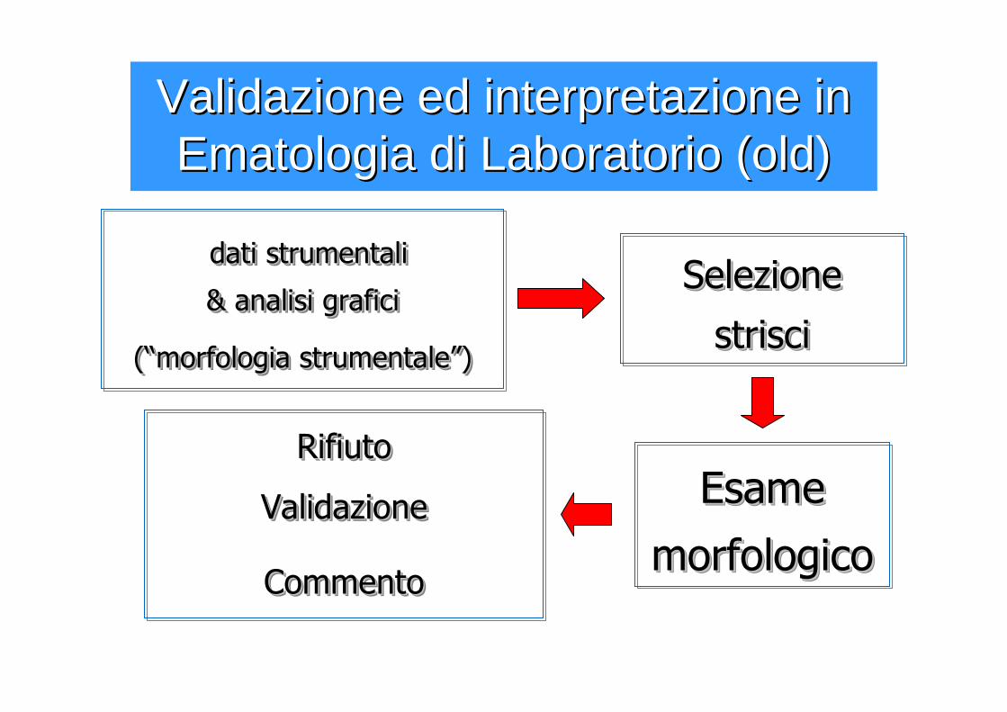
dati strumentali
& analisi grafici
(“morfologia strumentale”)
dati strumentali dati strumentali
& analisi grafici& analisi grafici
((““morfologia strumentalemorfologia strumentale””))
Selezione
strisci
Selezione Selezione
striscistrisci
Esame
morfologico
Esame Esame
morfologicomorfologico
Validazione ed interpretazione in Validazione ed interpretazione in Ematologia di Laboratorio (Ematologia di Laboratorio (oldold))
Rifiuto
Validazione
Commento
RifiutoRifiuto
Validazione Validazione
CommentoCommento
dati strumentalidati strumentalidati strumentali
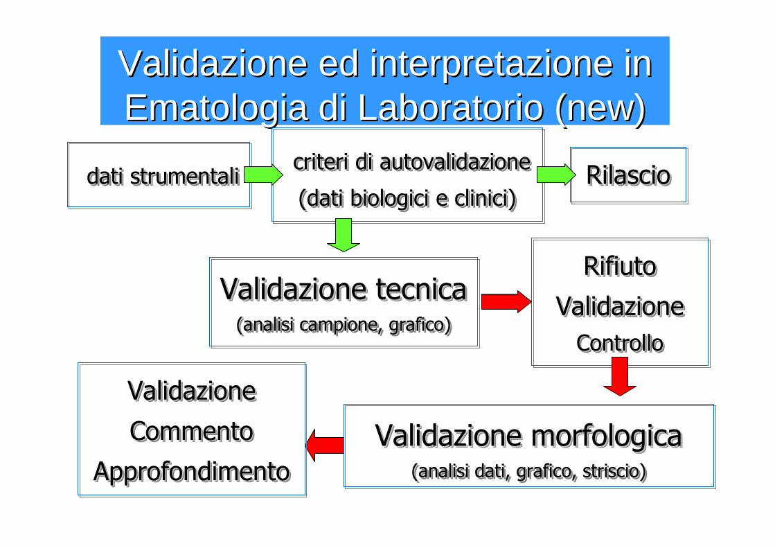
Validazione ed interpretazione in Validazione ed interpretazione in Ematologia di Laboratorio (Ematologia di Laboratorio (newnew))
Rifiuto
Validazione
Controllo
Rifiuto Rifiuto
ValidazioneValidazione
ControlloControllo
criteri di autovalidazione
(dati biologici e clinici)
criteri di criteri di autovalidazioneautovalidazione
(dati biologici e clinici)(dati biologici e clinici)RilascioRilascioRilascio
Validazione tecnica (analisi campione, grafico)
Validazione tecnica Validazione tecnica (analisi campione, grafico)(analisi campione, grafico)
Validazione morfologica (analisi dati, grafico, striscio)
Validazione morfologica Validazione morfologica (analisi dati, grafico, striscio)(analisi dati, grafico, striscio)
Validazione
Commento
Approfondimento
Validazione Validazione
Commento Commento
ApprofondimentoApprofondimento
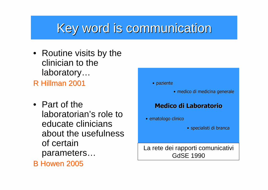
Key word Key word isis communicationcommunication
• Routine visits by the clinician to the laboratory…
R R HillmanHillman 20012001
• Part of the laboratorian’s role toeducate cliniciansabout the usefulnessof certainparameters…
B B HowenHowen 20052005
Medico di LaboratorioMedico di Laboratorio
•• pazientepaziente
•• specialisti di brancaspecialisti di branca
•• ematologo clinicoematologo clinico
•• medico di medicina generalemedico di medicina generale
La rete dei rapporti comunicativiGdSE 1990
MissionMission
1. Promuovere lo studio e la conoscenza 1. Promuovere lo studio e la conoscenza delldell’’ematologia di laboratorioematologia di laboratorio
2.2. Formare medici di laboratorio esperti nella Formare medici di laboratorio esperti nella diagnostica ematologicadiagnostica ematologica
3.3. Standardizzare le metodiche, utilizzare Standardizzare le metodiche, utilizzare pienamente i vantaggi della tecnologiapienamente i vantaggi della tecnologia
4.4. Perseguire lPerseguire l’’appropriatezza attraverso appropriatezza attraverso lineelinee--guida e referti interpretativiguida e referti interpretativi
Morfologia/MorfologieMorfologia/Morfologie
“Si vorrebbe essere riusciti a documentare l’interesse che trasfigura il fenomeno morfologico quand’esso è messo in relazione col fenomeno funzionale, cosicché sempre si ribadisce il potere interpretativo e l’efficacia formativa che la morfologia possiede, nella ricerca e rispettivamente nell’educazione ematologica.”
A A AscenziAscenzi, G , G MotturaMottura 19701970
Top Related