


Le lingue
Pagine
Legale

ACTA ODONTOLOGICALATINOAMERICANAVol. 30 Nº 1 2017
ISSN 1852-4834 on line versionversión electrónica
AOL12017:32011 03/07/2017 11:25 Página 1
AOL12017:32011 03/07/2017 11:25 Página 2
Vol. 30 Nº 1 / 2017 ISSN 1852-4834 Acta Odontol. Latinoam. 2017
Honorary EditorEditor honorarioRómulo Luis Cabrini(Universidad de Buenos Aires, Argentina)
Scientific EditorEditor CientíficoMaría E. Itoiz(Universidad de Buenos Aires, Argentina)
Associate EditorsEditores AsociadosRicardo MacchiAngela M. Ubios(Universidad de Buenos Aires, Argentina)Amanda E. Schwint(Comisión Nacional de Energía Atómica, Argentina)
Assistant EditorsEditores AsistentesPatricia MandalunisSandra J. Renou(Universidad de Buenos Aires, Argentina)
Technical and Scientific AdvisorsAsesores TécnicoCientíficosLilian Jara TracchiaLuciana M. SánchezTammy SteimetzDelia Takara(Universidad de Buenos Aires, Argentina)
Editorial BoardMesa EditorialEnri S. Borda (Universidad de Buenos Aires, Argentina)
Noemí E. Bordoni (Universidad de Buenos Aires, Argentina)
Fermín A. Carranza (University of California, Los Angeles, USA)
José Carlos Elgoyhen (Universidad del Salvador, Argentina)
Fernando Goldberg (Universidad del Salvador, Argentina)
Andrea Kaplan (Universidad de Buenos Aires, Argentina)
Andrés J.P. KleinSzanto (Fox Chase Cancer Center, Philadelphia, USA)
Susana Piovano (Universidad de Buenos Aires, Argentina)
Guillermo Raiden (Universidad Nacional de Tucumán, Argentina)
Sigmar de Mello Rode (Universidade Estadual Paulista,Brazil)
Hugo Romanelli (Universidad Maimónides, Argentina)
Cassiano K. Rösing (Federal University of Rio Grande do Sul, Brazil)
PublisherProducción Gráfica y PublicitariaImageGraf / email: [email protected]
Acta Odontológica Latinoamericana is the officialpublication of the Argentine Division of the InternationalAssociation for Dental Research.
Revista de edición argentina inscripta en el RegistroNacional de la Propiedad Intelectual bajo el N° 284335.Todos los derechos reservados.Copyright by:ACTA ODONTOLOGICA LATINOAMERICANAwww.actaodontologicalat.com
ACTA ODONTOLÓGICA LATINOAMERICANAAn International Journal of Applied and Basic Dental Research
POLÍTICA EDITORIAL
El objetivo de Acta OdontológicaLatinoamericana (AOL) es ofrecer a la
comunidad científica un medio adecuado
para la difusión internacional de los tra
bajos de investigación, realizados prefe
rentemente en Latinoamérica, dentro del
campo odontológico y áreas estrechamen
te relacionadas. Publicará trabajos origi
nales de investigación básica, clínica y
epidemiológica, tanto del campo biológi
co como del área de materiales dentales y
técnicas especiales. La publicación de tra
bajos clínicos será considerada siempre
que tengan contenido original y no sean
meras presentaciones de casos o series. En
principio, no se aceptarán trabajos de revi
sión bibliográfica, si bien los editores
podrán solicitar revisiones de temas de
particular interés. Las Comunicaciones
Breves, dentro del área de interés de AOL,
serán consideradas para su publicación.
Solamente se aceptarán trabajos no publi
cados anteriormente, los cuales no podrán
ser luego publicados en otro medio sin
expreso consen timiento de los editores.
Dos revisores, seleccionados por la
mesa editorial dentro de especialistas en
cada tema, harán el estudio crítico de los
manuscritos presentados, a fin de lograr el
mejor nivel posible del contenido científi
co de la revista.
Para facilitar la difusión internacional,
se publicarán los trabajos escritos en
inglés, con un resumen en castellano o por
tugués. La revista publicará, dentro de las
limitaciones presupuestarias, toda infor
mación considerada de interés que se le
haga llegar relativa a actividades conexas
a la investigación odontológica del área
latinoamericana.
EDITORIAL POLICY
Although Acta Odontológica Lati noamericana (AOL) will accept original
papers from around the world, the princi
pal aim of this journal is to be an instrument
of communication for and among Latin
American investigators in the field of den
tal research and closely related areas.
AOL will be devoted to original articles
dealing with basic, clinic and epidemio
logical research in biological areas or those
connected with dental materials and/or
special techniques.
Clinical papers will be published as
long as their content is original and not
restricted to the presentation of single
cases or series.
Bibliographic reviews on subjects of
special interest will only be published by
special request of the journal.
Short communications which fall with
in the scope of the journal may also be
submitted. Submission of a paper to the
journal will be taken to imply that it pres
ents original unpublished work, not under
consideration for publication elsewhere.
By submitting a manuscript the authors
agree that the copyright for their article is
transferred to the publisher if and when
the article is accepted for publication. To
achieve the highest possible standard in
scientific content, all articles will be ref
ereed by two specialists appointed by the
Editorial Board. To favour international
diffusion of the journal, articles will be
published in English with an abstract in
Spanish or Portuguese.
The journal will publish, within budget
limitations, any data of interest in fields
connected with basic or clinical odonto
logical research in the Latin America area.
Este número se terminó de editar el mes de Junio de 2017
AOL12017:32011 03/07/2017 11:25 Página 1
CONTENTS / ÍNDICE
In Memoriam. Prof. Dr. Carlos Eduardo José Bozzini .............................................................................................................................................................................................................. 3
Effect of silver diamine fluoride (SDF) on the dentinpulp complex. Ex vivo histological analysis on human primary teeth and rat molarsEfecto del diamino fluoruro de plata (DFP) sobre complejo dentinopulpar. Análisis histológico ex vivo en dientes primarios humanos y molares de rataGlenda Rossi, Aldo Squassi, Patricia Mandalunis, Andrea Kaplan .............................................................................................................................................................................................................................. 5
Surface detail reproduction and dimensional accuracy of molds: Influence of disinfectant solutions and elastomeric impression materialsReprodução de detalhes da superfície e estabilidade dimensional de moldes: influência das soluções desinfetantes e elastômerosRicardo D. Guiraldo , Sandrine B. Berger , Ronaldo M. T. Siqueira , Victor H. Grandi , Murilo B. Lopes , Alcides GoniniJúnior , Rodrigo V. Caixeta ,
Rodrigo V. de Carvalho, Mário A. C. Sinhoreti ............................................................................................................................................................................................................................................................ 13
Analysis of the adverse events reported to the office of the clinical director at a dental school in Bogotá, ColombiaAnálisis de los eventos adversos reportados a Dirección de Clínicas en una Facultad de Odontología de BogotáColombiaMaría F. Huertas, Juliana Gonzalez, Sandra Camacho, Ana L. Sarralde, Adriana Rodríguez ...................................................................................................................................................................................... 19
Evaluation of pH and calcium ion diffusion from calcium hydroxide pastes and MTA Evaluación del pH y la difusión de iones calcio de pastas de hidróxido de calcio y MTAMaría del M. Sáez, Gabriela L. López, Diana Atlas, María L. de la Casa .................................................................................................................................................................................................................... 26
Root surface temperature variation during mechanical removal of root canal filling material. An in vitro studyVariaciones térmicas en la superficie radicular durante la desobturación mecánica del conducto. Estudio in vitroMartín GarcíaCuerva, Lucía Horvath, Laura Pinasco, Verónica Ciparelli, Hernán Tartacovsky, Ariel Gualtieri,
Ana C Casadoumecq, Pablo Rodríguez, Carlos GonzalezZanotto .............................................................................................................................................................................................................................. 33
Methodological aspects in the study of periodontal breakdown in rats: influence of the presence and time of ligatureAspectos metodológicos no estudo da destruição periodontal em ratos: influência da presença e tempo de ligaduraJuliano Cavagni, Luise Seibel, Eduardo J. Gaio, Cassiano K. Rösing .......................................................................................................................................................................................................................... 39
Acta Odontol. Latinoam. 2017 ISSN 1852-4834 Vol. 30 Nº 1 / 2017
ACTA ODONTOLÓGICA LATINOAMERICANAAn International Journal of Applied and Basic Dental Research
Contact us Contactos: Cátedra de Anatomía Patológica, Facultad de Odontología, Universidad de Buenos AiresM.T. de Alvear 2142 (1122) Buenos Aires, Argentina Fax: (5411) 4 508[email protected] http://www.actaodontologicalat.com/contacto.html
ACTA ODONTOLÓGICA LATINOAMERICANA
A partir del Volumen 27 (2014) AOL se edita en formato digital con el Sistema de Gestión de Revistas Electrónicas (Open Journal System, OJS). La revista es de accesoabierto (Open Access). Esta nueva modalidad no implica un aumento en los costos de publicación para los autores.
Comité Editorial
ACTA ODONTOLÓGICA LATINOAMERICANA
From volume 27 (2014) AOL is published in digital format with the Open Journal System (OJS). The journal is Open Access. This new modality does not implyan increase in the publication fees.
Editorial Board
AOL12017:32011 03/07/2017 11:25 Página 2
Vol. 30 Nº 1 / 2017 ISSN 1852-4834 Acta Odontol. Latinoam. 2017
Con profundo pesar comunicamos el fallecimiento del Dr. Eduardo Carlos Bozzini, Editor Asociado de nuestra revista, ocurridoel 24 de marzo.
Hace ya algo más de treinta años, el Dr. Bozzini, junto con el Dr. Cabrini, impulsó la idea de crear Acta Odontológica Latinoame ricana, con el fin de promover la publicación de los trabajos de investigación realizados en Latinoamérica. Desde entonces, hacolaborado siempre en la edición y revisión de trabajos.
Bozzini era doctor en Odontología, Universidad de Buenos Aires (UBA, 1957) y doctor en Ciencias Biológicas (UBA, 1991)Desde su graduación, se dedicó a la docencia y a la investigación con dedicación exclusiva. Fue Profesor Titular de Fisiología de La Facultad de Odontologia, UBA, entre los años 1971 y 1998, continuando luego sus tareas como profesor Emérito. Fuemiembro de la Carrera del Investigador del Consejo Nacional de Investigaciones (CONICET), alcanzando la posición de Investigador Superior.
Fue distinguido entre los Grandes Maestros de la UBA y como Maestro de la Odontología por la Asociación Odontológica Argentina.
Sus numerosas actividades científicas, entre las que se destacan aproximadamente 200 publicaciones de trabajos, mas de unaveintena de tesis doctorales dirigidas y participaciones en conferencias, simposios y mesas redondas en Argentina, Estados Unidos, Perú, Bolivia, Austria, Alemania, Francia e Inglaterra, colocaron a la Cátedra de Fisiología y a la Facultad, en unaimportante posición de prestigio internacional en el ámbito de su principal línea de trabajo: Regulación de la eritropoyesis yfisiología de la altura.
Lo recordaremos siempre como un gran científico, colaborador y amigo.
We deeply regret to announce the passing of Dr. Eduardo Carlos Bozzini, Associate Editor or our journal, on March 24.
For over thirty years, Dr. Bozzini, in collaboration with Dr. Cabrini, drove the idea of creating Acta Odontológica Latinoamericanawith the aim of promoting publication of research in Latin America. Since then, he continuously cooperated with editing andpublishing papers.
Dr. Bozzini was a Doctor in Dentistry, Buenos Aires University (UBA, 1957) and Doctor in Biological Science (UBA, 1991). Aftergraduating he did fulltime teaching and research. He was Full Professor of Physiology at the UBA School of Dentistry from 1971to 1998, after which he continued as Professor Emeritus. He had a permanent researcher position (Carrera del Investigador) withthe Argentine National Research Council (Consejo Nacional de Investigaciones, CONICET), attaining the position of SeniorResearcher.
He was distinguished among the Great Masters at UBA and Master of Dentistry by the Argentine Dental Association (AsociaciónOdontológica Argentina).
His numerous scientific activities, including approximately 200 published papers, direction of over twenty PhD theses and participation in conferences, symposiums and round tables in Argentina, USA, Peru, Bolivia, Austria, Germany, France and England,brought major international prestige to the Department of Physiology and the School of Dentistry in the context of his main line ofwork: regulation of erythropoiesis and high altitude physiology.
We will always remember him as a great scientist, collaborator and friend.
IN MEMORIAM
Prof. Dr. Carlos Eduardo José Bozzini(1932 2017)
AOL12017:32011 03/07/2017 11:25 Página 3
RESUMEN El objetivo del trabajo fue determinar del efecto del DFP encomplejo dentinopulpar aplicando dos modelos: piezasdentarias luego de su aplicación (ex vivo) y en molares deanimales experimentales. Se realizó un estudio descriptivoaplicando dos modelos: en piezas dentarias primarias (ex vivo)con caries amelodentinarias sin compromiso pulpar que hayansido sometidas previamente con DFP 38%, mediante dosevaluaciones: Microscopía electrónica de Barrido (MEB) ydetector de energía dispersiva de rayos X (EDS) a fin dedeterminar su composición cuali y cuantitativa y Microscopíaóptica de campo claro (MCC) mediante la técnica descalcifi cación y en molares de animales de laboratorio donde seutilizaron 12 ratas Wistar macho. La técnica fue estandarizadaen la fosa distal de la cara oclusal del primer molar inferior, serealizó una cavidad amelodentinaria aprox. 0.5 mm de profun didad, en ambos molares. En un molar se aplicó la solución DFP al 38 % y el opuesto como control. Se realizaron corteshistológicos y se evaluó en forma cualitativa la pulpa dental en ambos grupos. En las piezas ex vivas mediante MEB
se observaron áreas de hipermineralización en la dentinaintertubular y escasos conductillos obliterados y por EDS sedetectó Ag en el centro de la lesión (7.34%), disminuyendo suconcentración en los límites (1,71%) y no se detectó en las zonasmás alejadas de la misma. En MCC se observó DFP sellando losconductillos sólo en sitio de colocación y con una penetraciónlimitada, por debajo, los conductillos se observaron de aspectonormal y el tejido pulpar asociado con la caries tratada ha mostrado un infiltrado inflamatorio crónico y formación de dentina terciaria, sin observarse precipitado de Ag. En elmodelo experimental en las cavidades expuestas con DFP enmolares no se alteró en forma relevante la histología pulpar. Lasobservaciones realizadas con las diferentes técnicas y en tejidosdentarios sugieren que el DFP genera mínimos efectos adversos.Los resultados de este estudio contribuirían a continuar coninvestigaciones que permitan recomendar el producto como unaestrategia costo efectivo para el tratamiento de la enfermedad.
Palabras clave: Caries dental; fluoruros; diamino fluoruro deplata.
ABSTRACTThe aim of this study was to determine the effect of SDF on thedentinpulp complex using two models: teeth after SDFapplication (ex vivo) and experimental animal molars. Adescriptive study was performed using two models. In the firstmodel, primary teeth (ex vivo) with enameldentin caries, withoutpulp involvement and previously treated with 38% SDF, wereevaluated by means of two techniques: (a) Scanning ElectronMicroscopy (SEM) and energydispersive Xray detector (EDS)to determine qualitative and quantitative composition, and (b)brightfield optical microscopy (OM) after decalcification. Thesecond model used laboratory animal molars from 12 maleWistar rats. Standardized enameldentin cavities approximately0.5 mm deep were made the distal fossa of the occlusal face ofboth first lower molars, to one of which a 38% SDF solution wasapplied, while the other was used as a control. Histologicalsections were prepared and dental pulp was evaluated
qualitatively in both groups. SEM on ex vivo teeth showed areasof hypermineralization in the intertubular dentin and few blockedtubules, while EDS detected Ag in the center of the lesion(7.34%), its concentration declining at the edges (1.71%), withnone in the areas farthest from the lesion. OM showed SDFsealing the tubules only at the site where it had been placed, withlimited penetration beneath, the tubules appeared normal andthe pulp tissue associated to treated caries showed chronicinflammatory infiltrate and formation of tertiary dentin, with noAg precipitate. In the experimental animal model, pulp histologywas not significantly altered in the molar cavities exposed toSDF. The observations using the different techniques on dentaltissues suggest that SDF causes minimal adverse effects. Theresults of this study may contribute to further studies on thesuitability of SDF as a costeffective strategy for treating caries.
Key words: Dental caries; fluorides; silver diamine fluoride.
5
Vol. 30 Nº 1 / 2017 / 5-12 ISSN 1852-4834 Acta Odontol. Latinoam. 2017
Effect of silver diamine fluoride (SDF) on the dentin-pulp complex. Ex vivo histological analysis on human primary teeth and rat molars
Glenda Rossi 1, Aldo Squassi 1, Patricia Mandalunis 2, Andrea Kaplan 3
1 Universidad de Buenos Aires, Facultad de Odontología, Cátedra de Odontología Preventiva y Comunitaria.
2 Universidad de Buenos Aires, Facultad de Odontología, Cátedra de Histología y Embriología.3 Universidad de Buenos Aires, Facultad de Odontología, Cátedra de Materiales Dentales.
Efecto del diamino fluoruro de plata (DFP) sobre complejo dentino-pulpar. Análisis histológico ex vivo en dientes primarios humanos y molares de rata
AOL12017:32011 03/07/2017 11:25 Página 5
INTRODUCTIONThe antimicrobial agent silver nitrate (AgNO3) was
used industrially for over 100 years to make water
potable. AgNO3 is used medically in eye drops to
prevent infections in newborns, and in dentistry
it is often used in stomatological treatments for
mouth ulcers1,2. In 1969, silver diamine fluoride
[F(NH3)2Ag] (SDF) solution was synthesized for
dental treatments35. Since then, it has been used in
Japan as Saforide® Solution (J Morita Company,
Japan) for application to caries lesions due to its
capacity as an antimicrobial agent and to stabilize
caries processes, particularly in primary teeth,
thanks to which it has an important role in pediatric
dentistry6,7.
SDF is a colorless solution which is used at
3840%, pH 810. On contacting the caries surface
it produces calcium fluoride (CaF2) and silver
phosphate (Ag3PO4)7. The F:Ag ion ratio is
44,800:255,000 ppm6,812.
SDF is manufactured and marketed in South America
as Fluoroplat® (Laboratorios Naf, Buenos Aires,
Argentina), in Australia (Creighton Pharmaceutical,
Sydney, Australia) and in Brazil as Safluoride di
Walter® in 10% solution (Polidental, Río de Janeiro,
Brazil).
The mechanism of action of SDF on caries has been
related to the formation of silver phosphate by
reaction with the tooth enamel surface. When the
dentin is compromised, the compound penetrates
the tubules, partially or totally, blocking their
lumen. In addition, it has an antimicrobial effect,
inactivating cariogenic bacteria it contacts 1,2,710.
Silver fluoride (AgF2) is much more soluble in
water than other silver halides. Silver diamine
fluoride (SDF) contains ammonium in addition to
AgF2. The ammonium ions combine with the silver
ions to produce a complex ion called silver diamine
ion [Ag (NH3)2], which is reversible and more
stable than AgF2. It can thus be kept at a constant
concentration for a longer time.
Craig et al.13 and Gotjamanos14 showed that silver
fluoride (AgF2) is effective in arresting caries in
primary molars in children.
Different studies have evaluated the potential
toxicity of SDF in children1517. Gotjamanos
and Afonso report that commercial 40% AgF2
preparations contain high concentrations of
fluorides and if used for treatment of young patients
may cause fluorosis15. Western Australia Dental
Health Services conducted a study using AgF2 and
found no evidence of its appropriate use causing
fluorosis18. There is no clinical report of fluorosis
as a result of using SDF.
Chu et al.19 conducted an 18month study on 375
children and reported 7083% effectiveness of SDF
applied on primary central incisors. Llodra et al 7
found similar effectiveness in a controlled cohort
clinical study on a Cuban population of 373 6year
old schoolchildren over 36 months, finding 80%
effectiveness on canines and primary molars, and
65% effectiveness on permanent first molars.
Chu and Lo 1920 and Zhi et al 21 report that SDF
application once or twice a year significantly reduces
the incidence of caries and reduces the substantial
risk of adverse events. Llodra et al 7 and Chu et al6
report that SDF applications produce reversible
gingival irritation, although this disadvantage is
minimized when SDF is applied using an adequate
relative isolation protocol. Sharma et al 22 claim that
SDF is efficacious for arresting caries lesions.
Gao et al 23 and Mei et al 24 conclude that professional
use of 5% sodium fluoride varnish can remineralize
enamel caries and that 38% SDF can arrest dentin
caries.
In vitro studies on the penetration of SDF into
the tooth structure found that it penetrates
approximately 2 µm into enamel and 50200 µm
into dentin, while in arrested lesions it reaches a
thickness of approximately 150 µm 25.
Different authors have reported the antimicrobial
effect of SDF. Among the most relevant studies is
Chun et al.10, reporting that SDF has antimicrobial
effect against cariogenic S. mutans or A. naeslundiibiofilm on dentin surfaces. These findings agree
with Mei et al., who report the same conclusions
with 38% SDF solution12.
De Almeida et al.26 confirmed the antimicrobial
effect of SDF at commercial concentrations of 12%
and 30%, which are lower than the concentration
used in our study.
One of the main drawbacks of SDF is esthetic
because of the dark stain it produces on the tooth
surface. Knight et al.911 therefore conducted in vitrostudies combining SDF with potassium iodide (IK),
which lessened the stain while preserving the
antimicrobial properties. Although the product
containing SDF and IK emerged on the Australian
market, it has not become well known in the rest of
the countries that use SDF27.
6 Glenda Rossi, et al.
Acta Odontol. Latinoam. 2017 ISSN 1852-4834 Vol. 30 Nº 1 / 2017 / 5-12
AOL12017:32011 03/07/2017 11:25 Página 6
SDF is considered a simple, lowcost therapeutic
alternative which does not require training for
application by health professionals and has a
significant benefit for individuals and populations,
based on biological sealing22,2829.
Although satisfactory results have been obtained
using SDF, to date, its potential toxic action and
mode of interaction with dental tissues have not
been fully elucidated.
Thus, the aim of this study was to determine the
effect of SDF on the dentinpulp complex by using
two models: (a) ex vivo teeth after SDF application
and (b) molars in experimental animals.
MATERIALS AND METHODSWe performed a descriptive study on the effect of
SDF on the dentinpulp complex by using two
models: ex vivo human teeth after application and
molars in experimental animal.
Histological Study of human teeth treated with SDFWe used 8 human primary teeth obtained by
exfoliation or indicated extraction. Inclusion
criteria were: teeth with dentinenamel caries
without pulp involvement and previously subject to
SDF treatment prior to exfoliation or extraction due
to persistence (approximately 1 year after SDF
application).
The following protocol was used for applying 38%
SDF:
Relative isolation of the lesion, removal of affected
dentin using hand instruments, application of
38% SDF by rubbing for 1 minute with a manual
applicator soaked in the solution, followed by
rinsing with distilled water.
This study was partly performed within the
framework of the project “Strategic approach for
reconversion of barriers to access to dental care in
highly vulnerable groups”, which was reviewed and
approved by the Ethics Committee at the Buenos
Aires University School of Dentistry (UBACYT
U20020120100324BA). In order to include children
in this study, we obtained informed consent from
their legal guardians and formal acceptance
from each child. An authorization for donation was
attached to the consent forms describing how
the tooth would be used, the research aims, and a
statement that refusal to participate would not
generate any conflict with participation in the project.
Four teeth were cut in half using a diamond disc for
Scanning Electron Microscope (SEM) observations.
The other four teeth were decalcified for observation
under brightfield microscopy.
In each histological analysis of human teeth (exvivo) the zone opposite the treated lesion was used
as control.
Evaluation using Scanning Electron Microscopy (SEM)Sections were prepared from the 4 primary teeth
which had been previously cut to expose the lesion.
Residue was removed from the sections by
ultrasound and they were dehydrated in an alcohol
concentration gradient (100, 96, 70 and 50%).
Samples were sputtercoated with goldpalladium
(using a Termo VG Scientific SC 7620 sputter coater)
for SEM observation (Scanning electron microscope
model SUPRA 40 Gemini II, Carl Zeiss). One of the
teeth was also studied using an energydispersive
Xray detector (EDS) in order to determine its
qualitative and quantitative composition.
Evaluation by brightfield optical microscopy (OM): Decalcification techniqueTeeth were fixed in 10% formalin buffer for at least
48 hrs, after which they were decalcified in 7.5%
nitric acid for 7 days. Then they were embedded in
paraffin, and mesiodistal histological sections
approximately 8 µm thick were cut. Sections were
stained with hematoxylin and eosin and a qualitative
histological evaluation of dental pulp was performed
under brightfield optical microscopy.
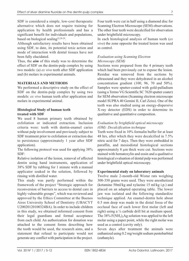
Experimental study on laboratory animals Twelve male 2monthold Wistar rats weighing
300350 grams were used. They were anesthetized
(ketamine 50ml/kg and xylazine 15 ml/kg i.p.) and
placed on an adapted operating table. The lower
jaw was isolated and the following standardize
technique applied. An enameldentin hole about
0.5 mm deep was made in the distal fossa of the
occlusal face of each lower first molar (left and
right) using a ¼ carbide drill bit at medium speed.
The 38% F(NH3)2Ag solution was applied to the left
molar using a paper point, while the right molar was
used as a control (cavity only).
Seven days after treatment the animals were
euthanized using 0.2 mg/weight sodium pentobarbital
(euthanyle).
Effect of silver diamine fluoride on the dentin-pulp complex 7
Vol. 30 Nº 1 / 2017 / 5-12 ISSN 1852-4834 Acta Odontol. Latinoam. 2017
AOL12017:32011 03/07/2017 11:25 Página 7
Lower jaws were extracted and fixed in 10% buffered
formalin for 48 hs, decalcified in 10% EDTA pH 7.2
for 30 days, processed histologically and embedded
in paraffin. Histological mesiodistal sections were
prepared and stained with hematoxylin and eosin, and
qualitative histological evaluation of the dental pulp
was performed on both treated and untreated molars
under brightfield optical microscopy.
The experimental protocol is in keeping with the
National Institutes of Health Guidelines for the Care
and Use of Laboratory Animals. The procedure
described above is shown in Fig. 1.
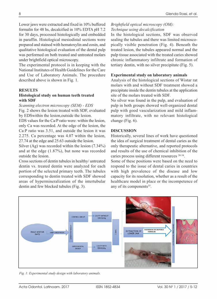
RESULTSHistological study on human teeth treated with SDFScanning electron microscopy (SEM) EDSFig. 2 shows the lesion treated with SDF, evaluated
by EDSwithin the lesion,outside the lesion.
EDS values for the Ca/P ratio were: within the lesion,
only Ca was recorded. At the edge of the lesion, the
Ca:P ratio was 3.51, and outside the lesion it was
2.275. Ca percentage was 4.07 within the lesion,
27.74 at the edge and 25.63 outside the lesion.
Silver (Ag) was recorded within the lesion (7.34%)
and at the edge (1.87%), but none was recorded
outside the lesion.
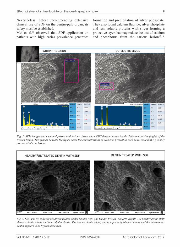
Cross sections of dentin tubules in healthy/ untreated
dentin vs. treated dentin were analyzed for each
portion of the selected primary teeth. The tubules
corresponding to dentin treated with SDF showed
areas of hypermineralization of the intertubular
dentin and few blocked tubules (Fig. 3).
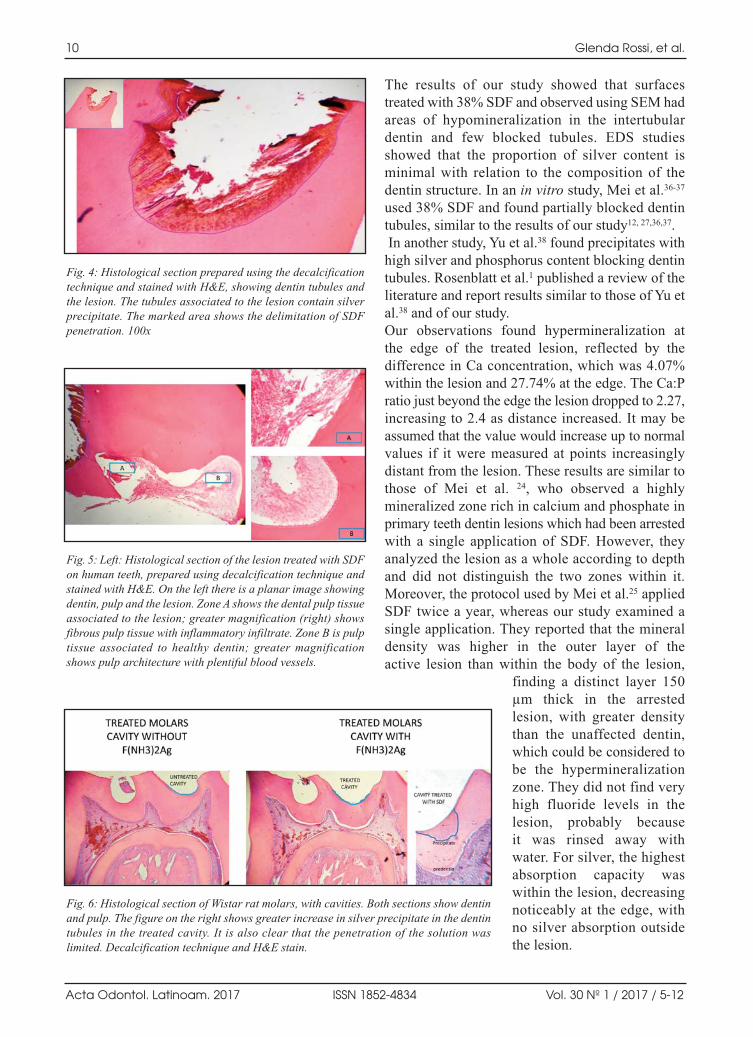
Brightfield optical microscopy (OM): Technique using decalcificationIn the histological sections, SDF was observed
sealing the tubules and there was limited microsco
pically visible penetration (Fig. 4). Beneath the
treated lesion, the tubules appeared normal and the
pulp tissue associated with the treated caries showed
chronic inflammatory infiltrate and formation of
tertiary dentin, with no silver precipitate (Fig. 5).
Experimental study on laboratory animalsAnalysis of the histological sections of Wistar rat
molars with and without SDF treatment showed a
precipitate inside the dentin tubules at the application
site of the molars treated with SDF.
No silver was found in the pulp, and evaluation of
pulp in both groups showed wellorganized dental
pulp with good vascularization and mild inflam
matory infiltrate, with no relevant histological
change (Fig. 6).
DISCUSSIONHistorically, several lines of work have questioned
the idea of surgical treatment of dental caries as the
only therapeutic alternative, and reported protocols
and results of the use of chemical inhibition of the
caries process using different resources 3034.
Some of these positions were based on the need to
respond to the issue of dental caries in countries
with high prevalence of the disease and low
capacity for its resolution, whether as a result of the
healthcare model in place or the incompetence of
any of its components35.
8 Glenda Rossi, et al.
Acta Odontol. Latinoam. 2017 ISSN 1852-4834 Vol. 30 Nº 1 / 2017 / 5-12
Fig. 1: Experimental study design with laboratory animals.
AOL12017:32011 03/07/2017 11:25 Página 8
Nevertheless, before recommending extensive
clinical use of SDF on the dentinpulp organ, its
safety must be established.
Mei et al.12 observed that SDF application on
patients with high caries prevalence generates
formation and precipitation of silver phosphate.
They also found calcium fluoride, silver phosphate
and less soluble proteins with silver forming a
protective layer that may reduce the loss of calcium
and phosphorus from the carious lesion12,19.
Effect of silver diamine fluoride on the dentin-pulp complex 9
Vol. 30 Nº 1 / 2017 / 5-12 ISSN 1852-4834 Acta Odontol. Latinoam. 2017
Fig. 2: SEM images show enamel prisms and lesions. Insets show EDS determination inside (left) and outside (right) of thetreated lesion. The graphs beneath the figure show the concentrations of elements present in each zone. Note that Ag is onlypresent within the lesion.
Fig. 3: SEM images showing healthy/untreated dentin tubules (left) and tubules treated with SDF (right). The healthy dentin (left)shows a dentin tubule and intertubular dentin. The treated dentin (right) shows a partially blocked tubule and the intertubulardentin appears to be hypermineralized.
AOL12017:32011 03/07/2017 11:25 Página 9
The results of our study showed that surfaces
treated with 38% SDF and observed using SEM had
areas of hypomineralization in the intertubular
dentin and few blocked tubules. EDS studies
showed that the proportion of silver content is
minimal with relation to the composition of the
dentin structure. In an in vitro study, Mei et al.3637
used 38% SDF and found partially blocked dentin
tubules, similar to the results of our study12, 27,36,37.
In another study, Yu et al.38 found precipitates with
high silver and phosphorus content blocking dentin
tubules. Rosenblatt et al.1 published a review of the
literature and report results similar to those of Yu et
al.38 and of our study.
Our observations found hypermineralization at
the edge of the treated lesion, reflected by the
difference in Ca concentration, which was 4.07%
within the lesion and 27.74% at the edge. The Ca:P
ratio just beyond the edge the lesion dropped to 2.27,
increasing to 2.4 as distance increased. It may be
assumed that the value would increase up to normal
values if it were measured at points increasingly
distant from the lesion. These results are similar to
those of Mei et al. 24, who observed a highly
mineralized zone rich in calcium and phosphate in
primary teeth dentin lesions which had been arrested
with a single application of SDF. However, they
analyzed the lesion as a whole according to depth
and did not distinguish the two zones within it.
Moreover, the protocol used by Mei et al.25 applied
SDF twice a year, whereas our study examined a
single application. They reported that the mineral
density was higher in the outer layer of the
active lesion than within the body of the lesion,
finding a distinct layer 150
µm thick in the arrested
lesion, with greater density
than the unaffected dentin,
which could be considered to
be the hypermineralization
zone. They did not find very
high fluoride levels in the
lesion, probably because
it was rinsed away with
water. For silver, the highest
absorption capacity was
within the lesion, decreasing
noticeably at the edge, with
no silver absorption outside
the lesion.
10 Glenda Rossi, et al.
Acta Odontol. Latinoam. 2017 ISSN 1852-4834 Vol. 30 Nº 1 / 2017 / 5-12
Fig. 4: Histological section prepared using the decalcificationtechnique and stained with H&E, showing dentin tubules andthe lesion. The tubules associated to the lesion contain silverprecipitate. The marked area shows the delimitation of SDFpenetration. 100x
Fig. 6: Histological section of Wistar rat molars, with cavities. Both sections show dentinand pulp. The figure on the right shows greater increase in silver precipitate in the dentintubules in the treated cavity. It is also clear that the penetration of the solution waslimited. Decalcification technique and H&E stain.
Fig. 5: Left: Histological section of the lesion treated with SDFon human teeth, prepared using decalcification technique andstained with H&E. On the left there is a planar image showingdentin, pulp and the lesion. Zone A shows the dental pulp tissueassociated to the lesion; greater magnification (right) showsfibrous pulp tissue with inflammatory infiltrate. Zone B is pulptissue associated to healthy dentin; greater magnificationshows pulp architecture with plentiful blood vessels.
AOL12017:32011 03/07/2017 11:25 Página 10
In another study, Mei et al.12 concluded that
application of 38% SDF arrests the caries process by
reducing demineralization and collagen destruction.
In addition, the presence of high fluoride and silver
concentrations may inhibit microbial growth of the
species present in cariogenic biofilm. It has also
been suggested that SDF has an inhibitory effect on
metalloproteinases, thereby protecting collagen
from destruction in carious lesions, and acting as
another form of protection against dentin degradation.
In another in vitro study, Mei 2 et al. found that
the primary components of SDF appear to react
with dentin tissues, forming calcium fluoride, a
compound that protects against caries.
Dentin microhardness changes according to its
mineral content. Any changes produced by applying
SDF may thus also be evaluated by microhardness,
and it would be useful to supplement our observations
with mechanical determinations on dentin in order
to assess its potential correlation36, 37.
The evaluation of experimental animal molars
showed that SDF has limited penetration. We found
no major alteration to dental pulp or presence of sil
ver in the pulp of either human teeth or experimental
animal molars. These findings agree with Korwar et
al.,39 who reported absence of inflammatory symp
toms in ex vivo teeth with cavities treated with SDF.
Our paper also describes the presence of tertiary
dentin adjacent to the treated cavity.
Internationally, Japan has marketed products
containing SDF for over 80 years and the US Food
and Drug Administration (FDA) approved its use in
the year 201440.
Dental caries is a highly prevalent disease in
children in developing countries41. Under many
circumstances, conventional methods for prevention
and treatment of caries are unavailable or unafford
able to communities in those regions.
The use of unconventional protocols for treatment
of caries lesions, including agents that stabilize the
process, is essential to the development of dental
care programs for highly vulnerable sectors of
society and/or sectors with barriers to healthcare
access. Traditional approaches for treating caries in
populations with these barriers provide temporary
benefits due to the high relapse rates in individuals
with greater burden of disease.
The results of our study may contribute to establishing
the absence of potential adverse effects in dental
tissues subject to topical application of SDF and
thereby enable continued research which may
ultimately enable SDF to be recommended as a low
cost, highly effective strategy for treating caries.
Through the use of different histological study models
(tooth substrates and observation techniques) this
study suggests that SDF produces minimal adverse
effects on the structures described. However, the
toxicity and biocompatibility of silver compounds
require further evaluation before its safety can be
established and its application recommended as a
therapeutic measure in programs intended for
populations with barriers to conventional dental care.
Effect of silver diamine fluoride on the dentin-pulp complex 11
Vol. 30 Nº 1 / 2017 / 5-12 ISSN 1852-4834 Acta Odontol. Latinoam. 2017
ACKNOWLEDGMENTSHistological technicians Mariela Lacave and Ivanna Sanchez
Rojas from the Department of Histology and Embryology,
School of Dentistry, Buenos Aires University.
This work was partly funded by Grants 20020120100109BA
and U20020120100324BA from Buenos Aires University Sci
entific Programming.
CORRESPONDENCEDr. Glenda Natalia Rossi.
Cátedra de Odontología Preventiva y Comunitaria,
Facultad de Odontología, Universidad de Buenos Aires.
Marcelo T. de Alvear 2142 5to B. C1122AAH.
Ciudad Autónoma de Buenos Aires. Argentina.
REFERENCES1. Rosenblatt A, Stamford TC, Niederman R. Silver Diamine
Fluoride: A Caries “SilverFluoride Bullet”. J Dent Res
2009; 88:11625.
2. Mei ML, Ito L, Cao Y, Li Q, Chu CH, Lo EC. The inhibitory
effects of silver diamine fluoride on cysteine cathepsins. J
Dent 2014; 42:329335.
3. Yamaga R, Nishino M, Yoshida S, Yokomizo I. Diammine
silver fluoride and its clinical application. J Osaka Univ
Dent Sch 1972; 12:120.
4. Fung M, Wong M, Lo EC, Chu CH. Arresting Early
Childhood Caries with Silver Diamine FluorideA Literature
Review. Oral Hyg Health 2013; 1:117. doi: 10.4172/2332
0702.1000117
5. VinodB M, Koppolu M, Nuvulla S, Thangala V, Redderu
K R. Antimicrobial efficiency of silver diamine fluoride as
an endodontic medicament An ex vivo study. Contemp
Clin Dent 2012; 3:262264.
6. Chu CH, Lo EC, Lin HC. Effectiveness of silver diamine
fluoride and sodium fluoride varnish in arresting dentin
caries in Chinese preschool children. J Dent Res 2002;
81:767770.
7. Llodra JC, Rodriguez A, Ferrer B, Menardia V, Ramos T,
Morato M. Efficiency of silver diamine fluoride for caries
AOL12017:32011 03/07/2017 11:25 Página 11
reduction in primary teeth and first permanent molars of
school children: 36month clinical trial. J Dent Res 2005;
84:721724.
8. Vasquez E, Zegarra G, Chirinos E, Castillo J, et al. Short
term serum pharmacokinetics of diammine silver fluoride
after oral application. BMC Oral Health 2012; 12:60.
9. Knight GM, McIntyre JM, Craig GG, Mulyani, Zilm PS,
Gully NJ. Differences between normal and demineralized
dentine pretreated with silver fluoride and potassium iodide
after an in vitro challenge by Streptococcus mutans. Aust
Dent J 2007; 52:1621.
10. Chu CH, Mei L, Seneviratne CJ, LO EC. Effects of silver
diamine fluoride on dentine carious lesions induced by
Streptococcus mutans and Actinomyces naeslundii biofilms.
Int J Paediatr Dent 2012; 22:210.
11. Knight GM, McIntyre JM, Craig GG, Mulyani, Zilm PS,
Gully NJ. An in vitro model to measure the effect of a silver
fluoride and potassium iodide treatment on the permeability
of demineralized dentine to Streptococcus mutans. Aust
Dent J 2005; 50:242–245.
12. Mei ML, Li Q, Chun CH, Lo EC, Samaranayake LP.
Antibacterial effects of silver diamine fluoride on multi
species cariogenic biofilm on caries. Ann Clin Microbiol
Antimicrob 2013; 12:17.
13. Craig GG, Powell KR, Cooper MH. Caries progression in pri
mary molars: 24month results from a minimal treatment
pro gramme. Community Dent Oral Epidemiol 1981; 9:260265.
14. Gotjamanos T. Pulp response in primary teeth with deep residual
caries treated with silver fluoride and glass ionomer cement
(‘atraumatic’ technique). Aust Dent J 1996; 41:328334.
15. Gotjamanos T, Afonso F. Unacceptably high levels of fluoride
in commercial preparations of silver fluoride. Aust Dent J
1997; 42:5253.
16. Gotjamanos T, Orton V. Abnormally high fluoride levels in
commercial preparations of 40 per cent silver fluoride
solution: Contraindications for use in children. Aust Dent J
1998; 43:422427.
17. Gotjamanos T, Orton V. Fluorideion concentration in 40
per cent silver fluoride solutions determined by ion
selective electrode and ion chromatography techniques.
Aust Dent J 1998; 43: 5556.
18. Neesham DC. Fluoride concentration in AgF2 and dental
fluorosis. Letter to the Editor. Aust Dent J 1997; 42:268269.
19. Chu CH, Lo EC. Promoting caries arrest in children with
silver diamine fluoride: a review. Oral Health Prev Dent
2008; 6:315321.
20. Chu CH, Lo EC. Microhardness of dentine in primary teeth
after topical fluoride applications. J Dent 2008; 36:387391.
21. Zhi, Q, Lo EC, Lin HC. Randomized clinical trial on
effectiveness of silver diamine fluoride and glass ionomer
in arresting dentine caries in preschool children. J Dent
2012; 40:962967.
22. Sharma G, Puranik MP, KR S. Approaches to Arresting Dental
Caries: An Update. J Clin Diagn Res 2015; 9:ZE08ZE11.
23. Gao SS, Zhang S, Mei ML, Lo EC, Chu CH. Caries
remineralisation and arresting effect in children by
professionally applied fluoride treatment – a systematic
review. BMC Oral Health 2016; 16:12. doi: 10.1186/
s1290301601716.
24. Mei ML, Lo EC, Chu CH. Clinical Use of Silver Diamine
Fluoride in Dental Treatment. Compend Contin Educ Dent
2016; 37:9398.
25. Mei ML, Ito L, Cao Y, Lo EC, Li QL, Chu CH. An ex vivo
study of arrested primary teeth caries with silver diamine
fluoride therapy. J Dent 2014; 42:395402.
26. de Almeida L de F, Cavalcanti YW, Valença AM. In Vitro
Antibacterial Activity of Silver Diamine Fluoride in
Different Concentrations. Acta Odontol Latinoam 2011;
24:127131.
27. Hamama HH, Yiu CK, Burrow MF. Effect of silver diamine
fluoride and potassium iodide on residual bacteria in
dentinal tubules. Aust Dent J 2015; 60:8087.
28. Kidd EA. How ‘Clean’ Must a Cavity Be before Restoration?
Caries Res 2004; 38:305313.
29. MattosSilveira J, Floriano I, Ferreira FR, Viganó ME,
Frizzo MA, Reyes A, Novaes TF, Moriyama CM, et al.
New proposal of silver diamine fluoride use in arresting
approximal caries: study protocol for a randomized
controlled trial. Trials 2014; 15:448. doi: 10.1186/1745
621515448.
30. Hyde EJ. Cariesinhibiting action of three topically applied
agents in incipient lesions in newly erupted teeth: results
after 24 months. J Can Dent Assoc (Tor) 1973; 39:189193.
31. Mercer VH, Mulher J. The clinical demostration of caries
arrestment following topical stannous fluoride treatments.
ASDC J Dent Child 1965; 32:6572.
32. Nishino M, Massler M. Immunisation of caries susceptible
pits and fissures with diammine silver fluoride solution. J
Pedod 1977; 2:1625.
33. Mc Donald SP, Sheiham A. A clinical comparison of non
traumatic methods of treating dental caries. Int Dent J 1994;
44:465470.
34. Klein U, Kanellis MJ, Drake D. Effects of four anticaries
agents on lesions depth progression in an in vitro caries
model. Pediatr Dent 1999; 21:176180.
35. Frencken JE, Songpaisan Y, Phantuvanit P, Pilot T. An
atraumatic restorative treatment (ART) technique: Evaluation
after one year. Int Dent J 1994; 44:460464.
36. Mei ML, Ito L, Cao Y, Li QL, Lo EC, Chu CH. Inhibitory
effect of silver diamine fluoride on dentine demineralisation
and collagen degradation. J Dent 2013; 41:809817.
37. Mei ML, Chu CH, Low KH, Che CM, Lo EC. Caries
arresting effect of silver diamine fluoride on dentine carious
lesion with S. mutans and L. acidophilus dualspecies
cariogenic biofilm. Med Oral Patol Oral Cir Bucal 2013;
18:e824831.
38. Yu DG, Kimura Y, Fujita A, Hossain M, Kinoshita JI,
Suzuki N, Matsumoto K. Study on acid resistance of human
dental enamel and dentin irradiated by semiconductor laser
with Ag (NH3)2F solution. J Clin Laser Med Surg 2001;
19:141146.
39. Korwar A, Sharma S, Logani A, Shah N. Pulp response to
high fluoride releasing glass ionomer, silver diamine
fluoride, and calcium hydroxide used for indirect pulp
treatment: An invivo comparative study. Contemp Clin
Dent 2015; 6:288292.
40. Horst JA, Ellenikiotis H, Milgrom PL. UCSF Protocol for
Caries Arrest Using Silver Diamine Fluoride: Rationale,
Indications, and Consent. J Calif Dent Assoc 2016; 44:
1628.
41. Boyce WT, Den Besten PK, Stamperdahl J, Zhan L, Jiang Y,
Adler NE, Featherstone JD. Social Inequalities in Childhood
Dental Caries: The Convergent Roles of Stress, Bacteria and
Disadvantage. Soc Sci Med 2010; 71:16441652.
12 Glenda Rossi, et al.
Acta Odontol. Latinoam. 2017 ISSN 1852-4834 Vol. 30 Nº 1 / 2017 / 5-12
AOL12017:32011 03/07/2017 11:25 Página 12
INTRODUCTIONHighaccuracy impression materials (elastomeric
impression materials) were first used in dentistry in
the 1950s1. Currently, four different elastomeric
impression materials are used: polysulfide, polyether,
polydimethylsiloxane and polyvinylsiloxane,
each of which has specific chemical reactions and
setting characteristics1. Impression materials should
RESUMOEste estudo comparou a reprodução de detalhes da superfície eestabilidade dimensional de moldes obtidos após desinfecçãoutilizando hipoclorito de sódio 2%, digluconato de clorexidina2%, ou ácido peracético 0,2% a moldes que não foramdesinfetados com quatro elastômeros: polissulfeto (Light BodiedPermlastic), polieter (Impregum Soft), silicona reação porcondensação (Oranwash L) e silicona reação por adição (AquasilUltra LV). Os moldes foram preparados sobre matriz contendolinhas de 20, 50 e 75 µm realizado sob pressão com moldeira de metal perfurada. Os moldes foram removidos após apolimerização e desinfetados (utilizando uma das soluções porimersão, armazenados em frascos fechados durante 15 minutos)ou não desinfetados. Assim, as amostras foram divididas em 16grupos (n=5). A reprodução detalhes da superfície e a precisão
dimensional foram avaliadas usando microscopia óptica na linha20 µm com 25 mm de comprimento, de acordo com a norma ISO4823. Os resultados de precisão dimensional (%) foramsubmetidos à análise de variância (ANOVA) e as médiascomparadas pelo teste de Tukey com 5% de nível de significância.A linha de 20 µm foi completamente reproduzida por todos oselastômeros, independentemente do processo de desinfecção. Nãohouve diferença estatisticamente significativa entre o grupocontrole e moldes desinfetados com acido peracético para oselastômeros Impregum Soft (polieter) e Aquasil Ultra LV (siliconareação por adição). O desinfetante de alto nível ácido peracéticoseria o material de escolha para a desinfecção.
Palavraschave: Estabilidade dimensional; desinfetante dental;materiais de moldagem.
ABSTRACTThis study compared the surface detail reproduction anddimensional accuracy of molds after disinfection using 2%sodium hypochlorite, 2% chlorhexidine digluconate or 0.2%peracetic acid to those of molds that were not disinfected, forfour elastomeric impression materials: polysulfide (Light BodiedPermlastic), polyether (Impregum Soft), polydimethylsiloxane(Oranwash L) and polyvinylsiloxane (Aquasil Ultra LV). Themolds were prepared on a matrix by applying pressure, using aperforated metal tray. The molds were removed followingpolymerization and either disinfected (by soaking in one of thesolutions for 15 minutes) or not disinfected. The samples werethus divided into 16 groups (n=5). Surface detail reproductionand dimensional accuracy were evaluated using optical
microscopy to assess the 20µm line over its entire 25 mmlength. The dimensional accuracy results (%) were subjectedto analysis of variance (ANOVA) and the means were comparedby Tukey’s test (α=5%). The 20µm line was completelyreproduced by all elastomeric impression materials, regardlessof disinfection procedure. There was no significant differencebetween the control group and molds disinfected with peraceticacid for the elastomeric materials Impregum Soft (polyether)and Aquasil Ultra LV (polyvinylsiloxane). The highleveldisinfectant peracetic acid would be the choice material fordisinfection.
Key words: Dimensional accuracy; dental disinfectant; dentalImpression materials.
13
Vol. 30 Nº 1 / 2017 / 13-18 ISSN 1852-4834 Acta Odontol. Latinoam. 2017
Surface detail reproduction and dimensional accuracy of molds: Influence of disinfectant solutions and elastomeric impression materials
Ricardo D. Guiraldo1, Sandrine B. Berger1, Ronaldo M. T. Siqueira1, Victor H. Grandi1, Murilo B. Lopes1, Alcides Gonini-Júnior1, Rodrigo V. Caixeta1, Rodrigo V. de Carvalho1, Mário A. C. Sinhoreti 2
1 Departamento de Odontologia Restauradora, Faculdade de Odontologia, Universidade Norte do Paraná – UNOPAR, Londrina, PR, Brasil
2 Departamento de Odontologia Restauradora, Faculdade de Odontologia de Piracicaba, Universidade Estadual de Campinas – UNICAMP, Piracicaba, SP, Brasil
Reprodução de detalhes da superfície e estabilidade dimensional de moldes: influência das soluções desinfetantes e elastômeros
AOL12017:32011 03/07/2017 11:25 Página 13
reproduce hard and soft tissues in order to
obtain biologically, mechanically, functionally and
esthetically acceptable restorations2, and in addition
to being capable of recording the anatomic
topography of the desired area, they should remain
dimensionally stable3. The dimensional accuracy of
a material is usually timedependent; for example,
a material may be highly dimensionally accurate
soon after its initial polymerization but less accurate
after storage for a period of time4. Dimensional
changes may occur in the molds as a result of
features inherent to the impression materials such
as wettability, handling properties and viscosity, or
to thickness of the material between the oral
structures and tray, method of fixing the impression
material to the tray, time elapsed until cast pouring,
material’s hydrophilicity, byproduct loss, polyme
rization shrinkage, thermal shrinkage due temperature
change (from mouth to room temperature),
incomplete elastic recovery, and, in some cases,
soaking1.
Disinfection is defined as a clinical stage designed
to destroy most microorganisms (viruses, bacteria
and spores) from the surface of an impression5, and
is an important biosafety measure. In absence of
disinfection, treatment procedures can expose
dentists, hygienists and laboratory workers to direct
or crosscontamination5,6. During the impression
procedure, the materials come into contact with
fluids such as blood and saliva, which may contain
pathogenic microorganisms capable of transmitting
infectious diseases such as herpes, hepatitis,
tuberculosis or AIDS5,7.
Disinfection can be accomplished by physical or
chemical action. However, physical action may
result in temperature increase, which can cause
measurable deformations in the molds5. For
impression materials, the use of solutions with
chemical action is recommended5. Disinfectants
must perform effectively as antimicrobial agents
while not adversely affecting the dimensional
accuracy or feature fidelity of the impression
material and resulting gypsum cast8. Disinfection
should be carried out with the product that requires
the least amount of time for the disinfection
process9. The most frequently used disinfectants are
glutaraldehyde, formaldehyde, alcohol, iodine
solution, synthetic phenol, sodium hypochlorite and
other chlorinereleasing solutions5. Other potential
disinfectants may be used to eliminate pathogens,
provided they do not alter the properties of
elastomeric impression materials. Peracetic acid has
been cited in the literature as a promising alternative
for disinfection due to its antimicrobial efficiency10, but there is no report on its use as a disinfectant
for elastomeric impression materials.
This study compared the surface detail reproduc
tion and dimensional accuracy of elastomeric
molds prepared using polysulfide, polyether,
polydimethylsiloxane or polyvinylsiloxane elasto
meric impression materials and disinfected using
2% sodium hypochlorite, 2% chlorhexidine
digluconate or 0.2% peracetic acid, to those of
models produced using molds that were not
disinfected. The null hypotheses tested were that
surface detail reproduction and dimensional
accuracy of elastomeric molds are not affected by
either [1] the elastomeric impression material or [2]
the disinfectant solution.
MATERIALS AND METHODSThis study used the lightbody elastomeric
impression materials polysulfide (Light Bodied
Permlastic, batch number 11311, Kerr, Romulus,
MI, USA), polyether (Impregum Soft, batch
number 1220700759, 3M Deutschland, Seefeld,
Germany), polydimethylsiloxane (Oranwash L,
batch number 133520, Zhermack, Badia Polesine,
RO, Italy) and polyvinylsiloxane (Aquasil Ultra LV,
batch number 100223, Dentsply Caulk, Milford,
DE, USA).
Dimensional accuracy and surface detail reproduction
were evaluated in accordance with ISO 482311.
The molds were prepared on a matrix (38 mm
outer diameter and 29.97 mm internal diameter)
containing three parallel lines 20, 50, and 75 µm
wide and 25 mm long, spaced 2.5 mm apart. Two
additional lines marked X and X′ were used to
determine the dimensional accuracy and surface
detail reproduction on the 20 µm line.
Before the impression procedure, the matrix was
cleaned ultrasonically and dried with compressed
air. The elastomeric impression materials were
prepared according to the manufacturers’ instructions.
A perforated metal tray (31 mm internal diameter
× 5 mm high) was placed on a glass plate and filled
with the molding material. The tray was joined to
the matrix and a 20 N force was applied using a
pneumatic press to simulate the impression process
and permit leakage of excess material5.
14 Ricardo D. Guiraldo, et al.
Acta Odontol. Latinoam. 2017 ISSN 1852-4834 Vol. 30 Nº 1 / 2017 / 13-18
AOL12017:32011 03/07/2017 11:25 Página 14
The molds were removed 3 min after polyme rization
of the elastomeric materials (polymeri zation
time was consistent with the minimum time recom
mended by the manufacturers)5 and disinfected by
soaking for 15 minutes at 37º C in 2% sodium
hypochlorite solution (Qboa, batch number L11212,
Indústria Anhenbi S/A, Osasco, SP, Brazil), 2%
chlorhexidine digluconate solution (Riohex 2%,
batch number R1202994, Indústria Farmacêutica
Rioquímica LTDA, São José do Rio Preto, SP,
Brazil), or 0.2% peracetic acid solution (Peresal, bath
number 4232AP0504, Ecolab Deutschland GmbH,
Düsseldorf, Germany). Control samples were not
disinfected. The samples were divided into 16 groups
(n=5) according to disinfectant procedure and
elastomeric impression material: Group 1: No
disinfectant (control group) + polysulfide; Group 2:
No disinfectant (control group) + polyether; Group
3: No disinfectant (control group) + polydimethylsi
loxane; Group 4: No disinfectant (control group) +
polyvinylsiloxane; Group 5: 2% Sodium hypochlo
rite solution + polysulfide; Group 6: 2% Sodium
hypochlorite solution + polyether; Group 7: 2%
Sodium hypochlorite solution + polydimethylsiloxa
ne; Group 8: 2% Sodium hypochlorite solution +
polyvinylsiloxane; Group 9: 2% Chlorhexidine
digluconate solution + polysulfide; Group 10: 2%
Chlorhexidine digluconate solution + polyether;
Group 11: 2% Chlorhexidine digluconate solution +
polydimethylsiloxane; Group 12: 2% Chlorhexidine
digluconate solution + polyvinylsiloxane; Group 13:
0.2% Peracetic acid solution + polysulfide; Group
14: 0.2% Peracetic acid solution + polyether; Group
15: 0.2% Peracetic acid solution + polydimethylsilo
xane; Group 16: 0.2% Peracetic acid solution +
polyvinylsiloxane.
Surface detail reproduction was measured using an
optical microscope (SZM, Bel Engineering srl, MI,
Italy). The molds were examined under lowangle
illumination at a magnification of 4x to 12x to
determine whether the 20 µmline was completely
reproduced over the full length of 25 mm between
the intersecting reference lines (X and X′), in
accordance with ISO 482311.
Dimensional accuracy was measured on the molds
using an optical microscope (STM, Olympus Optical
Co Ltd, Japan) with an accuracy of 0.0005 mm.
Dimensional accuracy expressed as a percentage
(L) was calculated in accordance with ISO 482311
using the equation:
L= [(L2 – L1) / L1] x 100, where L1 is the distance
between the lines on the matrix and L2 is the
distance between the lines on the impression
material.
Then, 100% was added to the results of the
equation12 and the dimensional accuracy results (%)
were subject to the KolmogorovSmirnov test for
normality, twoway ANOVA (material x disinfectant),
and the means were compared by Tukey’s test at 5%
significance levels.
RESULTSThe surface detail reproduction of all the elastomeric
impression materials was completely reproduced on
the 20 µm line regardless of disinfection procedure
(100% of the 5 samples in all 16 groups).
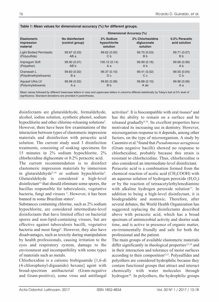
There was a statistically significant difference in
the mean values of dimensional accuracy in the
interaction between disinfectant procedure and
elastomeric impression material (p = 0.00001).
The dimensional accuracy of nondisinfected
Aquasil Ultra LV (polyvinylsiloxane) (Table 1)
was statistically higher than that of Oranwash L
(polydimethylsiloxane); however Impregum
Soft (polyether) and Light Bodied Permlastic
(polysulfide) did not differ from the others.
There was no significant difference between the
control group and the molds disinfected with
peracetic acid for the elastomeric materials
Impregum Soft (polyether) and Aquasil Ultra LV
(polyvinylsiloxane).
DISCUSSIONThe success of some forms of dental treatment
depends upon the accuracy with which a restoration
can be manufactured in the laboratory, using models
constructed from impressions13. Clearly, the
precision of the initial impression, in terms of both
dimensional accuracy and detail reproduction,
is a prerequisite for success13. The risk of cross
infection from a patient to a dental technician is a
matter of concern14, and in order to protect the
members of the dental team, a high standard of
hygiene and disinfection of dental equipment,
including dental impressions, is recommended6. A
disinfectant has dual requirements: it must be an
effective antimicrobial agent yet cause no adverse
response to the dimensional accuracy and surface
texture features of the impression material and
resultant plaster cast8. The most frequently used
Disinfection of elastomeric materials 15
Vol. 30 Nº 1 / 2017 / 13-18 ISSN 1852-4834 Acta Odontol. Latinoam. 2017
AOL12017:32011 03/07/2017 11:25 Página 15
disinfectants are glutaraldehyde, formaldehyde,
alcohol, iodine solution, synthetic phenol, sodium
hypochlorite and other chlorinereleasing solutions5.
However, there have been few examinations of the
interaction between types of elastomeric impression
materials and disinfection with peracetic acid
solution. The current study used 3 disinfection
treatments, consisting of soaking specimens for
15 minutes in 2% sodium hypochlorite, 2%
chlorhexidine digluconate or 0.2% peracetic acid.
The current recommendation is to disinfect
elastomeric impression materials by immersion
in glutaraldehyde5,14 or sodium hypochlorite5.
Glutaraldehyde is considered a highlevel
disinfectant15 that should eliminate some spores, the
bacillus responsible for tuberculosis, vegetative
bacteria, fungi and viruses 3. However, it has been
banned in some Brazilian states3.
Substances containing chlorine, such as 2% sodium
hypochlorite, are considered intermediatelevel
disinfectants that have limited effect on bacterial
spores and nonlipidcontaining viruses, but are
effective against tuberculosis bacilli, vegetative
bacteria and most fungi3. However, they also have
disadvantages, such as toxicity during manipulation
by health professionals, causing irritation to the
eyes and respiratory system, damage to the
environment and incompatibility with some types
of materials such as metals.
Chlorhexidine is a cationic bisbiguanide [1,6di
(4chlorophenyldiguanido) hexane] agent with
broadspectrum antibacterial (Gramnegative
and Grampositive), some virus and antifungal
activities9. It is biocompatible with oral tissues9 and
has the ability to remain on a surface and be
released gradually9,16. Its excellent properties have
motivated its increasing use in dentistry. However,
microorganism response to it depends, among other
factors, on the type of microorganism. A study by
Casemiro et al.9 found that Pseudomonas aeruginosa(Gram negative bacilli) showed no response to
chlorhexidine, probably because this strain is
resistant to chlorhexidine. Thus, chlorhexidine is
also considered an intermediatelevel disinfectant.
Peracetic acid is a combination formed from the
chemical reaction of acetic acid (CH3COOH) with
an aqueous solution of hydrogen peroxide (H2O2)
or by the reaction of tetraacetylethylenediamine
with alkaline hydrogen peroxide solution17. In
addition to being a highlevel disinfectant, it is
biodegradable and nontoxic. Therefore, after
several debates, the World Health Organization has
suggested replacing the disinfectants described
above with peracetic acid, which has a broad
spectrum of antimicrobial activity and shorter soak
time, and is active in presence of organic matter,
environmentally friendly and safe for both the
professional and the patient.
The main groups of available elastomeric materials
differ significantly in rheological properties12,18 and
in their interaction and tolerance of moist surfaces
according to their composition4,13. Polysulfides and
polyethers are considered hydrophilic because they
contain functional groups that attract and interact
chemically with water molecules through
hydrogen18. In polyethers, the hydrophilic groups
16 Ricardo D. Guiraldo, et al.
Acta Odontol. Latinoam. 2017 ISSN 1852-4834 Vol. 30 Nº 1 / 2017 / 13-18
Table 1: Mean values for dimensional accuracy (%) for different groups.
Elastomeric impression material
Light Bodied Permlastic(Polysulfide)
Impregum Soft (Polyether)
Oranwash L (Polydimethylsiloxane)
Aquasil Ultra LV(Polyvinylsiloxane)
No disinfectant (control group)
99.87 (0.03)AB a
99.90 (0.01)AB b
99.83 (0.02)B a
99.98 (0.02)A a
2% Sodium hypochlorite
solution
99.62 (0.05)C b
100.12 (0.14)A a
99.37 (0.10)D c
99.82 (0.08)B b
2% Chlorhexidinedigluconate
solution
99.70 (0.03)B b
99.99 (0.16)A b
99.41 (0.02)C c
99.88 (0.10)A ab
0.2% Peracetic acid solution
99.71 (0.07)B b
99.96 (0.06)A b
99.55 (0.04)C b
99.96 (0.06)A a
Dimensional Accuracy (%)
Mean values followed by different lowercase letters in rows and uppercase letters in columns differed statistically by Tukey’s test at 5% level of significance. Standard deviations are provided in parentheses.
AOL12017:32011 03/07/2017 11:25 Página 16
are the carbonyl (C==O) and ether (COC) groups,
while polysulfide, the hydrophilic groups are the
disulfide (—SS—) and mercapto (—SH) groups18.
Our results showed that the 20µm line was
completely reproduced by all the elastomeric
materials; however, although there was no change
in the 20µm line for the Light Bodied Permlastic
(polysulfide) and Impregum Soft (polyether)
elastomeric materials, their surfaces appeared
porous when disinfected with sodium hypochlorite.
Acceptable methods of measuring the dimensional
accuracy of casts include measuring calipers9,20,
micrometers21, dial gauges22 and measuring
microscopes3. A microscope was used in this study
due to its high accuracy (0.0005 mm). An ideal
impression material would be dimensionally
accurate over time, and could therefore be poured
at the operator’s convenience23. One study found
that the impression material polyvinylsiloxane
presents ideal dimensional stability23. Another study
found that polyether presented better dimen
sional precision than the polydimethylsiloxane
and polysulfide materials24, while in another25,
polyether presented intermediate behavior between
polydimethylsiloxane and polyvinylsiloxane. Thus,
although these studies used different methodologies,
by analogy, polyvinylsiloxane appears to have the
best dimensional accuracy, followed by polyether.
In the present study, for nondisinfected molds,
dimensional accuracy (Table 1) was statistically
higher for Aquasil Ultra LV (polyvinylsiloxane)
than for Oranwash L (polydimethylsiloxane), while
Impregum Soft (polyether) and Light Bodied
Permlastic (polysulfide) did not differ from the
others. The lower dimensional accuracy for
Oranwash L may be the result of ethanol being
formed as a byproduct during its polymerization
reaction and being lost through evaporation from
the surface of the material before disinfection.
Although polydimethylsiloxane has greater
polymerization shrinkage, it is hydrophobic, being
less susceptible to water sorption by immersion in
disinfectant solutions5. Thus, the lower dimensional
accuracy results for Oranwash L may be attributed
to the time elapsed (15 min) during disinfection.
Table 1 shows that the samples immersed in 2%
sodium hypochlorite, 2% chlorhexidine digluconate
or 0.2% peracetic acid showed no similar patterns
after disinfection. The results of this study show no
significant difference between the control group
and the molds disinfected with peracetic acid for
the elastomeric materials Impregum Soft (polyether)
and Aquasil Ultra LV (polyvinylsiloxane). For
Oranwash L (polydimethylsiloxane) and Light
Bodied Permlastic (polysulfide), the significant
difference between the control group and the molds
disinfected with peracetic acid was probably related
to leaching of alcohol or water in the disinfecting
solutions. Thus, peracetic acid would be the
material of choice for disinfection. As previously
mentioned, polyethers can be considered hydrophilic,
which was verified in the interaction Impregum
Soft – sodium hypochlorite. However, dimensional
accuracy of about 0.1 to 0.8% is compensated at
some stages during the laboratory steps required in
the preparation of the restorations26. Despite the
diversity of results in the literature regarding the
effect of disinfectant solutions on the dimensional
stability of elastomeric materials5, the dimensional
variations observed in this study cannot be
considered sufficient to create significant distortions
which could compromise the accuracy of prosthetic
restorations. Disinfection is an essential step which
cannot be omitted.
Based on the results of this study, the first null hypo
thesis was accepted and the second was rejected, as
there was no difference in [1] the surface detail
reproduction, although [2] significant differences
were found in the dimensional accuracy of elasto
meric molds. The authors conclude that although
there are differences in dimensional accuracy of
elastomeric molds when they are disinfected, this
change has no clinical affect. Moreover, peracetic
acid only promoted a significant difference from
the control group (dimensional accuracy) when
compared to Oranwash L (polydimethylsiloxane)
and Light Bodied Permlastic (polysulfide), which
was probably not a result of the use of this disinfec
tant. Thus, the highlevel disinfectant peracetic acid
would be the material of choice for disinfection.
Further studies are needed to prove its effectiveness
in disinfection of elastomeric impression materials.
CONCLUSIONUnder the conditions and within the limitations of
the current study, it can be concluded that the high
level disinfectant peracetic acid would be the
material of choice for disinfection.
Disinfection of elastomeric materials 17
Vol. 30 Nº 1 / 2017 / 13-18 ISSN 1852-4834 Acta Odontol. Latinoam. 2017
AOL12017:32011 03/07/2017 11:25 Página 17
ACKNOWLEDGEMENTSThe study was supported by FUNADESP (Fundação Nacional
de Desenvolvimento do Ensino Superior Particular). The authors
wish to thank Engineer Marcos Blanco Cangiani (Faculdade de
Odontologia de Piracicaba) for assistance with making the
matrix.
CORRESPONDENCEProf. Dr. Ricardo Danil Guiraldo
Universidade Norte do Paraná – UNOPAR
Rua Marselha, 183
86041 140 Londrina, PR Brasil
e mail: [email protected]
18 Ricardo D. Guiraldo, et al.
Acta Odontol. Latinoam. 2017 ISSN 1852-4834 Vol. 30 Nº 1 / 2017 / 13-18
REFERENCES1. Vitti RP, CorrerSobrinho L, Sinhoreti MA. Dimensional
accuracy of stone casts made by a monophase impression
technique using different elastomeric impression materials.
Braz J Oral Sci 2011; 10:175179.
2. Perakis N, Belser UC, Magne P. Final impressions: a review
of material properties and description of a current technique.
Int J Periodontics Restorative Dent 2004;24:109117.
3. Guiraldo RD, Borsato TT, Berger SB, Lopes MB, Gonini
Jr A, Sinhoreti MA. Surface detail reproduction and
dimensional accuracy of stone models: influence of
disinfectant solutions and alginate impression materials.
Braz Dent J 2012; 23:417421.
4. Petrie CS, Walker MP, O’mahony AM, Spencer P.
Dimensional accuracy and surface detail reproduction of
two hydrophilic vinyl polysiloxane impression materials
tested under dry, moist, and wet conditions. J Prosthet Dent
2003; 90:365372.
5. Carvalhal CI, Mello JA, Sobrinho LC, Correr AB, Sinhoreti
MA. Dimensional change of elastomeric materials after
immersion in disinfectant solutions for different times. J
Contemp Dent Pract 2011; 12:252258.
6. Kimondollo PM. Guidelines for developing a dental
laboratory infectioncontrol protocol. Int J Prosthodont
1992; 5:452456.
7. Adabo GL, Zanarotti E, Fonseca RG, Cruz CA. Effect of
disinfectant agents on dimensional stability of elastomeric
impression materials. J Prosthet Dent 1999: 81:621624.
8. Taylor RL, Wright PS, Maryan C. Disinfection procedures:
their effect on the dimensional accuracy and surface quality
of irreversible hydrocolloid impression materials and
gypsum casts. Dent Mater 2002; 18:103110.
9. Casemiro LA, PiresdeSouza FC, Panzeri H, Martins CH,
Ito IY. In vitro antimicrobial activity of irreversible
hydrocolloid impressions against 12 oral microorganisms.
Braz Oral Res 2007; 21:323329.
10. Salvia AC, Teodoro GR, Balducci I, KogaIto CY, Oliveira
SH. Effectiveness of 2% peracetic acid for the disinfection
of guttapercha cones. Braz Oral Res 2011; 25:2327.
11. ISO 4823 “Dentistry: elastomeric impression materials”
Geneva Switzerland, 2000.
12. Guiraldo RD, Moreti AF, Martinelli J, Berger SB,
Meneghel LL, Caixeta RV, Sinhoreti MA. Influence of
alginate impression materials and storage time on surface
detail reproduction and dimensional accuracy of stone
models. Acta Odontol Latinoam 2015; 28:156161.
13. German MJ, Carrick TE, McCabe JF. Surface detail
reproduction of elastomeric impression materials related to
rheological properties. Dent Mater 2008; 24:951956.
14. Melilli D, Rallo A, Cassaro A, Pizzo G. The effect of
immersion disinfection procedures on dimensional stability
of two elastomeric impression materials. J Oral Sci 2008;
50:441446.
15. Omidbakhsh N. A new peroxidebased flexible endoscope
compatible highlevel disinfectant. Am J Infect Control
2006; 34:571577.
16. Ramer MS, Gerhardt DE, McNally K. Accuracy of
irreversible hydrocolloid impression material mixed
with disinfectant solutions. J Prosthodont 1993; 2:156
158.
17. Fernandes FH, Orsi IA, Villabona CA. Effects of the
peracetic acid and sodium hypochlorite on the colour
stability and surface roughness of the denture base acrylic
resins polymerised by microwave and water bath methods.
Gerodontology 2013; 30:1825.
18. Berg JC, Johnson GH, Lepe X, AdanPlaza S. Temperature
effects on the rheological properties of current polyether
and polysiloxane impression materials during setting. J
Prosthet Dent 2003; 90:150161.
19. Chong YH, Soh G, Setchell DJ, Wickens JL. Relationship
between contact angles of die stone on elastomeric
impression materials and voids in stone casts. Dent Mater
1990; 6:162166.
20. Woodward JD, Morris JC, Khan Z. Accuracy of stone casts
produced by perforated trays and nonperforated trays. J
Prosthet Dent 1985; 53:347350.
21. Rueggeberg FA, Beall FE, Kelly MT, Schuster GS. Sodium
hypochlorite disinfection of irreversible hydrocolloid
impression material. J Prosthet Dent 1992; 67:628631.
22. Millstein PL. Determining the accuracy of gypsum casts
made from type IV dental stone. J Oral Rehabil 1992;
19:239243.
23. Donovan TE, Chee WW. A review of contemporary
impression materials and techniques. Dent Clin N Am
2004; 48:445470.
DOI dx.doi.org/10.1016/j.cden.2003.12.014.
24. Shah S, Sundaram G, Bartlett D, Sherriff M. The use of a
3D laser scanner using superimpositional software to assess
the accuracy of impression techniques. J Dent 2004;
32:653658.
25. Lacy AM, Fukui H, Bellman T, Jendresen MD. Time
dependent accuracy of elastomer impression materials Part
II: polyether, polysulfides, polyvinylsiloxane. J Prosthet
Dent 1981; 45:329333.
26. Johnson GH, Chellis KD, Gordon GE, Lepe X. Dimensional
stability and detail reproduction of irreversible hydrocolloid
and elastomeric impressions disinfected by immersion. J
Prosthet Dent 1998; 79:446453.
AOL12017:32011 03/07/2017 11:25 Página 18
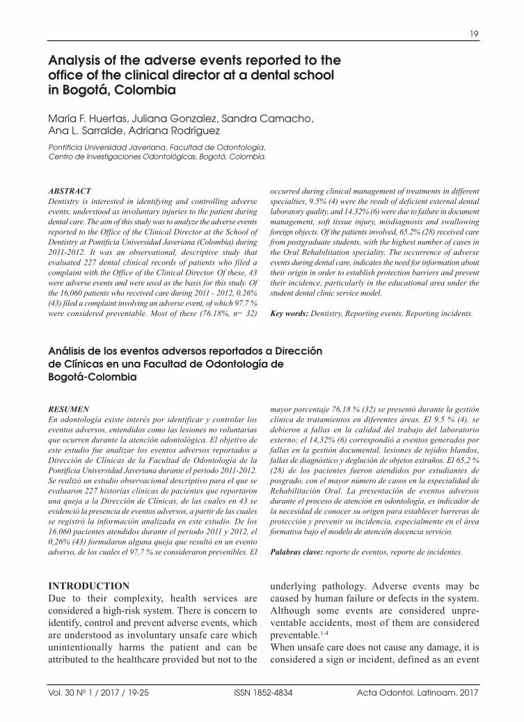
RESUMENEn odontología existe interés por identificar y controlar loseventos adversos, entendidos como las lesiones no voluntariasque ocurren durante la atención odontológica. El objetivo deeste estudio fue analizar los eventos adversos reportados aDirección de Clínicas de la Facultad de Odontología de laPontificia Universidad Javeriana durante el periodo 20112012.Se realizó un estudio observacional descriptivo para el que seevaluaron 227 historias clínicas de pacientes que reportaronuna queja a la Dirección de Clínicas, de las cuales en 43 seevidenció la presencia de eventos adversos, a partir de las cualesse registró la información analizada en este estudio. De los16.060 pacientes atendidos durante el periodo 2011 y 2012, el0,26% (43) formularon alguna queja que resultó en un eventoadverso, de los cuales el 97,7 % se consideraron prevenibles. El
mayor porcentaje 76,18 % (32) se presentó durante la gestiónclínica de tratamientos en diferentes áreas. El 9,5 % (4), sedebieron a fallas en la calidad del trabajo del laboratorioexterno; el 14,32% (6) correspondió a eventos generados porfallas en la gestión documental, lesiones de tejidos blandos,fallas de diagnóstico y deglución de objetos extraños. El 65,2 %(28) de los pacientes fueron atendidos por estudiantes deposgrado, con el mayor número de casos en la especialidad deRehabilitación Oral. La presentación de eventos adversosdurante el proceso de atención en odontología, es indicador dela necesidad de conocer su origen para establecer barreras deprotección y prevenir su incidencia, especialmente en el áreaformativa bajo el modelo de atención docencia servicio.
Palabras clave: reporte de eventos, reporte de incidentes.
INTRODUCTIONDue to their complexity, health services are
considered a highrisk system. There is concern to
identify, control and prevent adverse events, which
are understood as involuntary unsafe care which
unintentionally harms the patient and can be
attributed to the healthcare provided but not to the
underlying pathology. Adverse events may be
caused by human failure or defects in the system.
Although some events are considered unpre
ventable accidents, most of them are considered
preventable.14
When unsafe care does not cause any damage, it is
considered a sign or incident, defined as an event
ABSTRACTDentistry is interested in identifying and controlling adverseevents, understood as involuntary injuries to the patient duringdental care. The aim of this study was to analyze the adverse eventsreported to the Office of the Clinical Director at the School ofDentistry at Pontificia Universidad Javeriana (Colombia) during20112012. It was an observational, descriptive study thatevaluated 227 dental clinical records of patients who filed acomplaint with the Office of the Clinical Director. Of these, 43were adverse events and were used as the basis for this study. Ofthe 16,060 patients who received care during 2011 2012, 0.26%(43) filed a complaint involving an adverse event, of which 97.7 %were considered preventable. Most of these (76.18%, n= 32)
occurred during clinical management of treatments in differentspecialties, 9.5% (4) were the result of deficient external dentallaboratory quality, and 14.32% (6) were due to failure in documentmanagement, soft tissue injury, misdiagnosis and swallowingforeign objects. Of the patients involved, 65.2% (28) received carefrom postgraduate students, with the highest number of cases inthe Oral Rehabilitation speciality. The occurrence of adverseevents during dental care, indicates the need for information abouttheir origin in order to establish protection barriers and preventtheir incidence, particularly in the educational area under thestudent dental clinic service model.
Key words: Dentistry, Reporting events, Reporting incidents.
19
Vol. 30 Nº 1 / 2017 / 19-25 ISSN 1852-4834 Acta Odontol. Latinoam. 2017
Analysis of the adverse events reported to the office of the clinical director at a dental school in Bogotá, Colombia
María F. Huertas, Juliana Gonzalez, Sandra Camacho, Ana L. Sarralde, Adriana Rodríguez
Pontificia Universidad Javeriana, Facultad de Odontología, Centro de Investigaciones Odontológicas, Bogotá, Colombia.
Análisis de los eventos adversos reportados a Dirección de Clínicas en una Facultad de Odontología de Bogotá-Colombia
AOL12017:32011 03/07/2017 11:25 Página 19
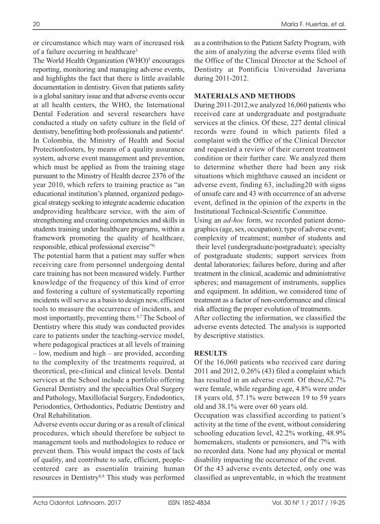
or circumstance which may warn of increased risk
of a failure occurring in healthcare3.
The World Health Organization (WHO)5 encourages
reporting, monitoring and managing adverse events,
and highlights the fact that there is little available
documentation in dentistry. Given that patients safety
is a global sanitary issue and that adverse events occur
at all health centers, the WHO, the International
Dental Federation and several researchers have
conducted a study on safety culture in the field of
dentistry, benefitting both professio nals and patients4.
In Colombia, the Ministry of Health and Social
Protectionfosters, by means of a quality assurance
system, adverse event manage ment and prevention,
which must be applied as from the training stage
pursuant to the Ministry of Health decree 2376 of the
year 2010, which refers to training practice as “an
educational institution’s planned, organized pedago
gical strategy seeking to integrate academic education
andproviding healthcare service, with the aim of
strengthening and creating competencies and skills in
students training under healthcare programs, within a
framework promoting the quality of healthcare,
responsible, ethical professional exercise”6.
The potential harm that a patient may suffer when
receiving care from personnel undergoing dental
care training has not been measured widely. Further
knowledge of the frequency of this kind of error
and fostering a culture of systematically reporting
incidents will serve as a basis to design new, efficient
tools to measure the occurrence of incidents, and
most importantly, preventing them.4,7 The School of
Dentistry where this study was conducted provides
care to patients under the teachingservice model,
where pedagogical practices at all levels of training
– low, medium and high – are provided, according
to the complexity of the treatments required, at
theoretical, preclinical and clinical levels. Dental
services at the School include a portfolio offering
General Dentistry and the specialties Oral Surgery
and Pathology, Maxillofacial Surgery, Endodontics,
Periodontics, Orthodontics, Pediatric Dentistry and
Oral Rehabilitation.
Adverse events occur during or as a result of clinical
procedures, which should therefore be subject to
management tools and methodologies to reduce or
prevent them. This would impact the costs of lack
of quality, and contribute to safe, efficient, people
centered care as essentialin training human
resources in Dentistry8,9. This study was performed
as a contribution to the Patient Safety Program, with
the aim of analyzing the adverse events filed with
the Office of the Clinical Director at the School of
Dentistry at Pontificia Universidad Javeriana
during 20112012.
MATERIALS AND METHODSDuring 20112012,we analyzed 16,060 patients who
received care at undergraduate and postgraduate
services at the clinics. Of these, 227 dental clinical
records were found in which patients filed a
complaint with the Office of the Clinical Director
and requested a review of their current treatment
condition or their further care. We analyzed them
to determine whether there had been any risk
situations which mighthave caused an incident or
adverse event, finding 63, including20 with signs
of unsafe care and 43 with occurrence of an adverse
event, defined in the opinion of the experts in the
Institutional TechnicalScientific Committee.
Using an adhoc form, we recorded patient demo
graphics (age, sex, occupation); type of adverse event;
complexity of treatment; number of students and
their level (undergraduate/postgraduate); specialty
of postgraduate students; support services from
dental laboratories; failures before, during and after
treatment in the clinical, academic and administrative
spheres; and management of instruments, supplies
and equipment. In addition, we considered time of
treatment as a factor of nonconformance and clinical
risk affecting the proper evolution of treatments.
After collecting the information, we classified the
adverse events detected. The analysis is supported
by descriptive statistics.
RESULTS Of the 16,060 patients who received care during
2011 and 2012, 0.26% (43) filed a complaint which
has resulted in an adverse event. Of these,62.7%
were female, while regarding age, 4.8% were under
18 years old, 57.1% were between 19 to 59 years
old and 38.1% were over 60 years old.
Occupation was classified according to patient’s
activity at the time of the event, without considering
schooling education level, 42.2% working, 48.9%
homemakers, students or pensioners, and 7% with
no recorded data. None had any physical or mental
disability impacting the occurrence of the event.
Of the 43 adverse events detected, only one was
classified as unpreventable, in which the treatment
20 María F. Huertas, et al.
Acta Odontol. Latinoam. 2017 ISSN 1852-4834 Vol. 30 Nº 1 / 2017 / 19-25
AOL12017:32011 03/07/2017 11:25 Página 20
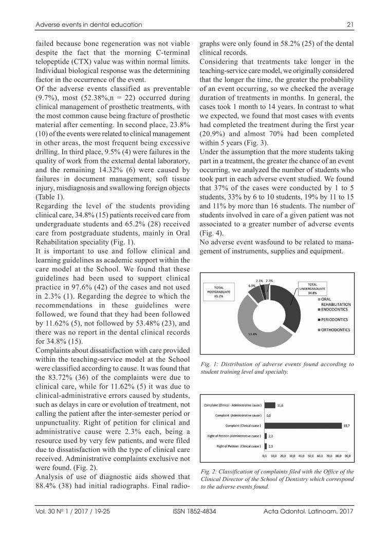
failed because bone regeneration was not viable
despite the fact that the morning Cterminal
telopeptide (CTX) value was within normal limits.
Individual biological response was the determining
factor in the occurrence of the event.
Of the adverse events classified as preventable
(9.7%), most (52.38%,n = 22) occurred during
clinical management of prosthetic treatments, with
the most common cause being fracture of prosthetic
material after cementing. In second place, 23.8%
(10) of the events were related to clinical management
in other areas, the most frequent being excessive
drilling. In third place, 9.5% (4) were failures in the
quality of work from the external dental laboratory,
and the remaining 14.32% (6) were caused by
failures in document management, soft tissue
injury, misdiagnosis and swallowing foreign objects
(Table 1).
Regarding the level of the students providing
clinical care, 34.8% (15) patients received care from
undergraduate students and 65.2% (28) received
care from postgraduate students, mainly in Oral
Rehabilitation speciality (Fig. 1).
It is important to use and follow clinical and
learning guidelines as academic support within the
care model at the School. We found that these
guidelines had been used to support clinical
practice in 97.6% (42) of the cases and not used
in 2.3% (1). Regarding the degree to which the
recommen dations in these guidelines were
followed, we found that they had been followed
by 11.62% (5), not followed by 53.48% (23), and
there was no report in the dental clinical records
for 34.8% (15).
Complaints about dissatisfaction with care provided
within the teachingservice model at the School
were classified according to cause. It was found that
the 83.72% (36) of the complaints were due to
clinical care, while for 11.62% (5) it was due to
clinicaladministrative errors caused by students,
such as delays in care or evolution of treatment, not
calling the patient after the intersemester period or
unpunctuality. Right of petition for clinical and
administrative cause were 2.3% each, being a
resource used by very few patients, and were filed
due to dissatisfaction with the type of clinical care
received. Administrative complaints exclusive not
were found. (Fig. 2).
Analysis of use of diagnostic aids showed that
88.4% (38) had initial radiographs. Final radio
graphs were only found in 58.2% (25) of the dental
clinical records.
Considering that treatments take longer in the
teachingservice care model, we originally considered
that the longer the time, the greater the probability
of an event occurring, so we checked the average
duration of treatments in months. In general, the
cases took 1 month to 14 years. In contrast to what
we expected, we found that most cases with events
had completed the treatment during the first year
(20.9%) and almost 70% had been completed
within 5 years (Fig. 3).
Under the assumption that the more students taking
part in a treatment, the greater the chance of an event
occurring, we analyzed the number of students who
took part in each adverse event studied. We found
that 37% of the cases were conducted by 1 to 5
students, 33% by 6 to 10 students, 19% by 11 to 15
and 11% by more than 16 students. The number of
students involved in care of a given patient was not
associated to a greater number of adverse events
(Fig. 4).
No adverse event wasfound to be related to mana
gement of instruments, supplies and equipment.
Adverse events in dental education 21
Vol. 30 Nº 1 / 2017 / 19-25 ISSN 1852-4834 Acta Odontol. Latinoam. 2017
Fig. 1: Distribution of adverse events found according tostudent training level and specialty.
Fig. 2: Classification of complaints filed with the Office of theClinical Director of the School of Dentistry which correspondto the adverse events found.
AOL12017:32011 03/07/2017 11:25 Página 21
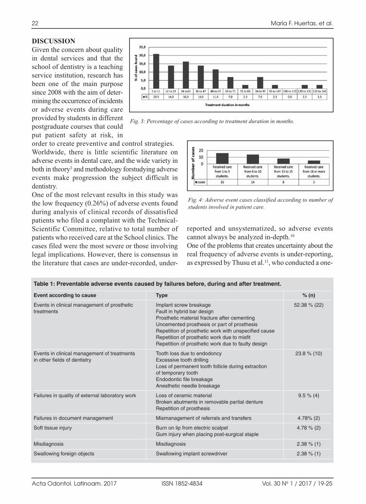
DISCUSSIONGiven the concern about quality
in dental services and that the
school of dentistry is a teaching
service institution, research has
been one of the main purpose
since 2008 with the aim of deter
mining the occurrence of incidents
or adverse events during care
provided by students in different
postgraduate courses that could
put patient safety at risk, in
order to create preventive and control strategies.
Worldwide, there is little scientific literature on
adverse events in dental care, and the wide variety in
both in theory3 and methodology forstudying adverse
events make progression the subject difficult in
dentistry.
One of the most relevant results in this study was
the low frequency (0.26%) of adverse events found
during analysis of clinical records of dissatisfied
patients who filed a complaint with the Technical
Scientific Committee, relative to total number of
patients who received care at the School clinics. The
cases filed were the most severe or those involving
legal implications. However, there is consensus in
the literature that cases are underrecorded, under
reported and unsystematized, so adverse events
cannot always be analyzed indepth.10
One of the problems that creates uncertainty about the
real frequency of adverse events is underreporting,
as expressed by Thusu et al.11, who conducted a one
22 María F. Huertas, et al.
Acta Odontol. Latinoam. 2017 ISSN 1852-4834 Vol. 30 Nº 1 / 2017 / 19-25
Fig. 3: Percentage of cases according to treatment duration in months.
Fig. 4: Adverse event cases classified according to number ofstudents involved in patient care.
Table 1: Preventable adverse events caused by failures before, during and after treatment.
Event according to cause
Events in clinical management of prosthetic treatments
Events in clinical management of treatments in other fields of dentistry
Failures in quality of external laboratory work
Failures in document management
Soft tissue injury
Misdiagnosis
Swallowing foreign objects
Type
Implant screw breakageFault in hybrid bar designProsthetic material fracture after cementing Uncemented prosthesis or part of prosthesisRepetition of prosthetic work with unspecified cause Repetition of prosthetic work due to misfitRepetition of prosthetic work due to faulty design
Tooth loss due to endodoncyExcessive tooth drillingLoss of permanent tooth follicle during extraction of temporary toothEndodontic file breakageAnesthetic needle breakage
Loss of ceramic materialBroken abutments in removable partial dentureRepetition of prosthesis
Mismanagement of referrals and transfers
Burn on lip from electric scalpelGum injury when placing post-surgical staple
Misdiagnosis
Swallowing implant screwdriver
% (n)
52.38 % (22)
23.8 % (10)
9.5 % (4)
4.78% (2)
4.78 % (2)
2.38 % (1)
2.38 % (1)
AOL12017:32011 03/07/2017 11:25 Página 22
year study based on dental reports from the database
of the United Kingdom’s National Patient Safety
Agency. They found a low rate of reports on dental
incidents, possibly due to the voluntary nature of
reporting and the reluctance of dental practitioners
to disclose incidents for fear of some kind of
professional inconvenience.
A study conducted on the database from the Spanish
Observatory for Dental Patient Safety (OESPO),
analyzed 415 law suits and reported that 40%
were caused by errors (conscious event), 40% by
complications and 20% by accidents (conceptually
defined as an adverse event). It concluded that the
use of this source has limitations because dentists
report few adverse events since they perceive their
procedures as being less complex than medical pro
ce dures and not lifethreatening, although the same
study found 11 cases of death attributed to the
dentist’s confidence during treatment (allergy, endo
car ditis caused by lack of prophylaxis, hemorrhages
in anticoagulated patients and infections in immuno
compromised patients, among others), pointing to
the need for detailed clinical records10, Obadan et
al.12 report that 24.1% of adverse events required
that the patient be transferred to an emergency
department, of which 11.1% resulted in death of the
affected patient.
In our study, the most severe event (2.38%) of the
43 found was due to swallowing a prosthodontic
screwdriver. The patient was taken to emergency
room and the device had to be removed under
medical care at a hospital. Although events such as
this are infrequent, they are important because of
their potential complications which may lead to
death of a patient. There are reports of a wide range
of ingested items, such as fixed prosthesis,
orthodontic items13,14metal restorations, crowns,
cores, endodontic files, and ultrasonic tips, among
others15.
In their analysis of the those cases, Obinata et al
found that ingestions occurred more frequently
during treatment of lower molars, and suggest
keeping the patient’s head inclined towards the side
being treated so that objects fall in the buccal pouch.
Cases of ingestion occurred more frequently when
the procedures were performed by professionals
with less than 5 years of experience. Therefore, and
considering the risk created by these accidents,
dentists should take meticulous precautions and be
prepared to deal with this kind of emergency15.
When there is inhalation, the risk is greater, so it
is suggested that dental offices should have
emergency protocols for dealing with it promptly14.
In addition, patients at greater risk of ingestion or
aspiration of objects should be identified and extra
precautions takento prevent such complications.
Zitzmann et al.16provide guidelines for managing
inhaled or ingested objects during dental treatment.
In addition to the issue of underreporting, the source
of information on which an analysis is based
modifies the casuistic and results found on the
subject. The other methodologies most frequently
used for detection and analysis of adverse events
are direct review of clinical records and surveys.
These methods usually increase the number of
events reported. Our research group conducted a
study on adverse events in the field of endodontics,
finding reports in 74.4% of the records analyzed
over two years. It is interesting to note that most of
them (81.3%) are considered preventable17similarly
to the current study, in which only one event was
not preventable.
A study by Hiivala et al.4 used an internet survey
of dentists who worked at public and private
institutions. It reports 872 patient safety incidents,
of which 53% were considered adverse events, 45%
incidents and 13% severe events potentially causing
permanent damage, as a result of factors caused by
application of local anesthesia, allergic reaction,
exposure to radiation and extracting wrong teeth,
among others. Anotherstudy conducted on the
database of adverse events reported to national
supervision and administrative institutions in the
healthcare sector found that 32% occurred at private
dental offices, 62.9% were preventable, 4.1% not
preventable and 33% could not be evaluated18.
The most frequent types of adverse events have
been reported in most fields of dental care. In
our study, the highest frequency occurred in
clinical management of prosthetic treatments, with
52.4%, followed by another clinical management
treatments in other fields of dentistry, 28.8%. These
results were similar to those reported by Hivalla
et al.18, who rank prosthodontics in first place
with 16.4%, restoration 9.5%, implants 8.4%,
endodontics 6.6%, orthodontics 3.6% and periodon
tics 1.8%. Similarly, Tiwana et al.19 report events in
the field of prosthodontics in first place. Perea
Perez10 finds the highest frequency of events in the
Adverse events in dental education 23
Vol. 30 Nº 1 / 2017 / 19-25 ISSN 1852-4834 Acta Odontol. Latinoam. 2017
AOL12017:32011 03/07/2017 11:25 Página 23
field of implantology, followed by endodontics, oral
surgery, prosthodontics and orthodontics.
A range of factors have been found to influence the
occurrence of an adverse event. For example, poor
management of the patient’s medical records
considered relevant to patient safety, as well
professional skill during the clinical interview,
particularly regarding sensitive issues such as
HIV20. Our study only found one adverse event as a
result of diagnosis (2.38%), which occurred due to
the lack of a comprehensive diagnosis. Tiwanaet
al.19found insufficient or erroneous records in
35.6% and in complete medical recordsin 15.1%.
Considering the importance of diagnostic help such
as diagnostic support and followup of treatments,
the School of Dentistry policy is that all patients
begin with an initial radiograph.It was found that
approximately 10% of the cases did not have
radiographs, which the students may have removed
to perform the diagnosis and failed to return to the
clinical file. The problem has now been overcome
by the use of digital clinical records, which were
implemented 4 years ago at the School.
Among other predisposing factors for adverse
events are patient care by a large number of
students21 attempting to perform procedures that are
beyond the professional’s technical skill, lack of
consultation with experts, overconfidence in own
skills and knowledge, ignoring evidencebased
medicine, operator fatigue, lack of awareness of
risks, lack of communication leading to procedural
errors through mismanagement of referrals and
transfers22 and following guidelines18,20,21,23. We
looked at these factors during this study, but found
no association between the occurrence of adverse
events and the demographic variable sage, sex,
level or any influence of number of students or use
of guidelines. Regarding age, adverse events have
been reported more frequently in adults (2560
years), with no difference between sexes.12
Regarding timing, we found that most events
occurred within the first 5 years of treatment,
including the intersemester periods, given the
modality of university service provided. In a study
on dentists, Hiivala et al.18 found a time of 17
months, with adverse events attributed mainly to
communication breakdowns in the organization.
With regard to the main factors that contribute to
preventing adverse events for professionals and
university teachers, Bailey20 mentions knowledge
of the patient’s medical record as having the
greatest impact, as well as quality and adequacy
of the recordtaking. The pedagogical model
implemented at our School includes in its clinical
administrative competences, knowledge of how to
use the dental clinical record as an essential safety
factor when providing service.
Ten years ago, the School implemented patient
safety and service quality committees with the aim
of providing a safety policy contributing to
systematic followup, implementing safety barriers,
research and knowledge management in the area,
with the aim of creating an impact on students in
the exercise of their future profession.
One of the weaknesses of the patient safety culture
is the lack of understanding of the concept of
adverse event and the lack of research, which does
not allow learning how to prevent adverse events,
guidelines for improving quality such as detailed
monitoring of critical biosafety processes and
sterilization, proper medication prescription, control
of unnecessary radiation and checklists for all
surgical procedures, among others, to help improve
quality and patient safety10.
The greatest impact on the culture of patient safety
is achieved through training. It is at theacademy that
knowledge is consolidated with principles of ethics
and responsibility. Methodical, systematic rigor in
the adherence to safe practices by all staff involved
in patient care will reduce the occurrence of adverse
events.
Clear, prompt communication skills with patients are
essential for decision making by patients regarding
treatments and contribute to creating trust. There is a
need in both dental professionals and students who
are undergoing training to gain deeper knowledge
and research of adverse events in order to prevent
them from occurring again, thus contributing to
improving patient safety and student training.
24 María F. Huertas, et al.
Acta Odontol. Latinoam. 2017 ISSN 1852-4834 Vol. 30 Nº 1 / 2017 / 19-25
CORRESPONDENCEDr.Juliana González.
Carrera 7 No. 4062, edificio 26 piso 3. Bogotá, Colombia.
AOL12017:32011 03/07/2017 11:25 Página 24
REFERENCES1. Luengas S. Modelo para gestionar la seguridad del paciente
en las instituciones de salud. Vía Salud 2008; 43:25.
http://www.cgh.org.co/imagenes/calidadycultura.pdf
2. Franco A. Fundamentos en seguridad al paciente para
disminución de los errores médicos. 1ª ed. Cali, Colombia:
Editorial Universidad del Valle; 2006.
http://www.worldcat.org/title/fundamentosdeseguridad
alpacienteparadisminuirerroresmedicos/oclc/2371934
53/viewport
3. Ministerio de la Protección Social República de Colombia.
Lineamientos para la implementación de la política de
seguridad del paciente. Noviembre 2008.
https://www.minsalud.gov.co/sites/rid/Lists/BibliotecaDigi
tal/RIDE/DE/CA/LINEAMIENTOS_IMPLEMENTACIO
N_POLITICA_SEGURIDAD_DEL_PACIENTE.pdf
4. Hiivala N, MussaloRauhamaa H, Murtomaa H. Patient
safety incident prevention and management among Finnish
dentists. Acta Odontol Scand 2013; 71:16631670.
5. Organización Mundial de la Salud. Alianza Mundial para
la Seguridad del Paciente. La Investigación en Seguridad
del Paciente. Mayor Conocimiento para una atención más
segura. 2008.
http://www.who.int/patientsafety/information_centre/docu
ments/ps_research_brochure_es.
6. Ministerio de Protección Social República de Colombia.
Sistema de Información para la Calidad y se adoptan los
indica dores de monitoría del Sistema Obligatorio de
Garantía de Calidad de la Atención en Salud. Resolución
1446 de 8 de mayo de 2006.
https://www.minsalud.gov.co/Normatividad_Nuevo/RESO
LUCI%C3%93N%201446%20DE%202006%20
%20ANEXO%20T%C3%89CNICO.pdf
7. Kalenderian E, Walji MF, Tavares A, Ramoni RB. An
adverse event trigger tool in dentistry: a new methodology
for measuring harm in the dental office. J Am Dent Assoc
2013;144:808814.
8. Maher S. The Francis Report The importance of person
centred health and care. Dent Update 2015; 42:210212.
9. Bissell V, Felix DH. The Francis reportImplications for
the education and training of dental professionals.Dent
Update 2015; 42:215218.
10. PereaPérez B, LabajoGonzález E, AcostaGío AE,
Yamalik N. Eleven basic procedures/practices for dental
patient safety. J Patient Saf 2015; 9.
DOI: 10.1097/PTS.0000000000234.
11. Thusu S, Panesar S, Bedi R. Patient safety in dentistry
state of play as revealed by a national database of errors. Br
Dent J 2012; 213(3):e3. DOI: 10.1038/sj.bdj.2012.669.
12. Obadan EM, Ramoni RB, Kalenderian E. Lessons learned
from dental patient safety case reports. J Am Dent Assoc
2015; 146:318326.
13. AlWahadni A, Al Hamad KQ, AlTarawneh A. Foreign
body ingestion and aspiration in dentistry: a review of the
literature and reports of three cases. DentUpdate 2006;33:
569 570.
14. Bilder L, HazanMolina H, Aizenbud D. Medical
emergencies in a dental office. Inhalation and ingestion of
orthodontic objects. J Am Dent Assoc 2011; 142:4552.
15. Obinata K, Satoh T, Towfik AM, Nakamura M. An investi
gation of accidental ingestion during dental procedures. J
Oral Sci 2011; 53:495500.
16. Zitzmann NU, Fried R, Elsasser S, Marinello CP. [The
aspiration and swallowing of foreign bodies. The manage
ment of the aspiration or swallowing of foreign bodies
during dental treatment]. Schweiz Monatsschr Zahnmed
2000; 110:619632.
17. Tafur MC, Camacho LD, Mejía SH, González J, Huertas
MF. Frecuencia de eventos adversos de la terapia endodón
tica y seguimiento de pacientes atendidos en el posgrado
de Endodoncia de la Pontificia Universidad Javeriana
(20072008). Univ Odontol 2014; 33. DOI:1011144/javeriana.
uo3371.feat
18. Hiivala N, MussaloRauhamaa H, Tefke HL, Murtomaa H.
An analysis of dental patient safety incidents in a patient
complaint and healthcare supervisory database in Finland.
Acta Odontol Scand 2016; 74:8189.
19. Tiwana KK, Morton T, Tiwana PS. Aspiration and ingestion
in dental practice: a 10year institutional review. J Am Dent
Assoc 2004; 135:12871291.
20. Bailey E. Contemporary views of dental practitioners’ on
patient safety. Br Dent J 2015; 219:535539.
21. AguirreGas HG, VázquezEstupiñán F. Medical error:
adverse events. CirCir 2006; 74:495503.
http://www.redalyc.org/pdf/662/66274614.pdf
22. Yamalik N, Van Dijk W. Analysis of the attitudes and
needs/demands of dental practitioners in the field of patient
safety and risk management. IntDent J 2013; 63:291297.
23. Franco A. La seguridad clínica de los pacientes: entendiendo
el problema. Colomb Med. 2005; 36:130133.
https://tspace.library.utoronto.ca/bitstream/1807/8998/1/rc
05020.pdf
Adverse events in dental education 25
Vol. 30 Nº 1 / 2017 / 19-25 ISSN 1852-4834 Acta Odontol. Latinoam. 2017
AOL12017:32011 03/07/2017 11:25 Página 25
26
Acta Odontol. Latinoam. 2017 ISSN 1852-4834 Vol. 30 Nº 1 / 2017 / 26-32
RESUMEN El objetivo de este estudio ex vivo fue evaluar los cambios en elpH y la difusión a través de la dentina radicular de iones calcioa partir de pastas de hidróxido de calcio (Ca (OH)2) y trióxidomineral agregado (MTA), durante 7, 30 y 60 días; y la relaciónentre el pH y la difusión de iones. Se utilizaron 32 premolareshumanos. Las coronas fueron seccionadas, los conductos radicu lares fueron instrumentados y obturados con las siguientespreparaciones: 1) Ca (OH)2 + agua destilada (n = 7); 2) Ca (OH)2 + gluconato de clorhexidina (CHX) al 0,1% (n = 7);3) MTA + agua destilada (n = 7); 4) MTA + gluconato declorhexidina al 0,1% (n = 7); 5) agua destilada (n = 2) (control);6) gluconato de clorhexidina al 0,1% (n = 2) (control). El ápicey la apertura coronaria se sellaron con IRM. Las raíces se
colocaron en tubos Eppendorf con 1 ml de agua destilada a 37 °C y 100% de humedad. Se midió el pH inicial y a los 7, 30 y60 días, con pHmetro, y se analizó el contenido de iones calcioen la solución por espectrofotometría de absorción atómica. Losdatos fueron analizados estadísticamente con ANOVA, análisisde regresión lineal y correlación de Pearson. Los valores de pHmás altos se obtuvieron con las pastas de hidróxido de calcio alos 60 días (p ≤ 0,05). Todos los grupos mostraron liberación deiones calcio. La pasta de hidróxido de calcio con agua destiladamostró el valor más alto de iones calcio a los 60 días (p ≤ 0,01).Hubo una correlación positiva entre los valores de pH y calcio.
Palabras clave: pH, hidróxido de calcio; compuesto de trióxidomineral.
INTRODUCTIONCalcium hydroxide (Ca(OH)2) has been widely used
as an intracanal medication1. It is a strongly alkaline
substance (pH 12.512.8) with high antibacterial
activity against oral bacteria. The effectiveness of
Ca(OH)2 for inactivation of microorganisms and
tissue healing is directly related to its dissociation
into calcium and hydroxyl ions. Hydroxyl ions
diffuse through the dentinal tubules and inactivate
Gramnegative bacterial lipopolysaccharide (LSP)2.
In addition, their high pH activates tissue enzymes,
causing a mineralization effect3.
Drug vehicles play an important role in the ion
dissociation rate, causing the paste to be solubilized
ABSTRACTThe aim of this ex vivo study was to evaluate changes in pH andcalcium ion diffusion through root dentin from calcium hydroxide(Ca (OH)2) and mineral trioxide aggregate (MTA) pastes at 7,30 and 60 days; and the relationship between pH and iondiffusion. Thirtytwo human premolars were used. Crowns weresectioned and root canals instrumented and filled in with thefollowing preparations: 1) Ca(OH)2 + distilled water (n=7); 2)Ca(OH)2 + 0.1% chlorhexidine gluconate (n=7); 3) MTA +distilled water (n=7); 4) MTA + 0.1% chlorhexidine gluconate(CHX) (n=7); 5) distilled water (n=2) (control); 6) 0.1%chlorhexidine gluconate (n=2) (control). The apex and coronaryopening were sealed with IRM. Roots were placed in Eppendorf
tubes with 1 ml distilled water at 37°C and 100% humidity. Atbaseline, 7, 30 and 60 days, pH was measured with pH meter,and calcium ion content in the solution was analyzed by atomicabsorption spectrophotometry. The data were statisticallyanalyzed using ANOVA, simple linear regression analysis andPearson’s correlation test. The highest pH values were achievedwith calcium hydroxide pastes at 60 days (p ≤ 0.05). Calciumions were released in all groups. The calcium hydroxide pastewith distilled water at 60 days had the highest calcium ion value(p ≤ 0.01). There was a positive correlation between calcium andpH values.
Key words: pH; calcium hydroxide; mineral trioxide aggregate.
Evaluation of pH and calcium ion diffusion from calcium hydroxide pastes and MTA
María del M. Sáez1,2, Gabriela L. López2,3, Diana Atlas2, María L. de la Casa2
1 Universidad Nacional de Tucumán. Facultad de Odontología. Cátedra de Práctica Final Obligatoria. Tucumán, Argentina.
2 Universidad Nacional de Tucumán. Facultad de Odontología. Cátedra de Endodoncia, Tucumán, Argentina.
3 Universidad Nacional de Tucumán. Facultad de Odontología. Cátedra de Química Biológica., Tucumán, Argentina
Evaluación del pH y la difusión de iones calcio de pastas de hidróxido de calcio y MTA
AOL12017:32011 03/07/2017 11:25 Página 26

and resorbed by the periapical tissues from within
the root canal4. Calcium hydroxide was pioneered by
Heithersay5 and Frank6 in apexification treatment,
which provided adequate apical healing through the
induction of an apical barrier. However, longterm
calcium hydroxide treatment lasting more than 30
days may reduce dentin fracture strength7. Due to its
alkaline nature, calcium hydroxide denatures some
of the acidic dentin proteins which act as bonding
agents, thereby weakening the dentin and increasing
risk of fracture8.
Mineral trioxide aggregate (MTA) powder is
basically a mixture of 75% Portland cement, 20%
bismuth oxide and 5% gypsum9. It was developed in
1990 by Torabinejad at Loma Linda University10.
MTA has been recognized as a bioactive11, hard tissue
conductive12, hard tissue inductive, biocompatible
material13. It has been used for apical barriers, root
end fillings, perforation repairs, regenerative therapy,
pulp capping and pulpotomies14. During the setting
process, MTA pH is initially 10.2, increasing to 12.5
during the first few hours. MTA has also been shown
to have antibacterial activity15.
Chlorhexidine gluconate (CHX) is widely used as
an irrigant for the treatment of infected root canal
systems to reduce endodontic microbiota16. Many
studies suggest the use of a combination of CHX
with Ca(OH)2 for greater antimicrobial action17.
Chlorhexidine is a broad spectrum antibacterial
agent with effectiveness against Enterococcusfaecalis and Candida albicans18. CHX can be
adsorbed onto dental tissue, resulting in substantive
antibacterial activity19. Stowe et al.15 demonstrated
that the substitution of sterile water by 0.12% CHX
in toothcolored ProRoot MTA enhanced the
antimicrobial effect of the MTA in vivo.
MTA could be an alternative to Ca(OH)2 to halt
resorption without the disadvantage of weakening
tooth structure20. Use of MTA must be considered
permanent because it is difficult to remove once it
has set21.
The aim of this ex vivo study was to evaluate the
relationship between pH and diffusion of calcium
ions through root dentin using Ca(OH)2 and MTA
pastes with different vehicles for 7, 30 and 60 days.
MATERIALS AND METHODSPreparation of specimensThirtytwo singleroot human mandibular premolars,
recently extracted for orthodontic reasons, were
selected on the basis of their similarity in
morphology and size. Crowns were amputated at
cementoenamel junction level using a high speed
bur #2200 (KG Sorensen, SP, Brazil) and water
irrigation. The cementum was then removed using
Gracey curettes. Root canals were enlarged up to
file #45 (Maillefer, East Lansing, MI, USA), and
cleaned and shaped using the stepback technique.
After each instrument change, root canals were
irrigated with 2 ml 1% sodium hypochlorite, and
17% EDTA was used as final irrigation for 2
minutes. Then root canals were rinsed with distilled
water, dried with absorbent paper points, randomly
divided into six groups and filled with the following
preparations: Group I: 1 g calcium hydroxide
(Farmadental Lab, Buenos Aires, Argentina) with
1.5 ml distilled water (Ca(OH)2 + DW); Group II:
1 g calcium hydroxide with 1.5 ml 0.1%
chlorhexidine (Ca(OH)2 + CHX); Group III: MTA
(CPMTM, Egeo S.R.L., Buenos Aires, Argentina)
with distilled water (MTA + DW). The powder/
liquid ratio was 0.33/1 (w/w). Group IV: MTA with
0.1% chlorhexidine (MTA + CHX). The powder/
liquid ratio was 0.33/1 (w/w). Group V: distilled
water as a control group (DW); Group VI: 0.1%
chlorhexidine (CHX), prepared from a 20%
chlorhexidine solution, as a control group.
In the experimental groups, the paste was placed
using a Lentulo spiral (Dentsply, Mailifer,
Switzerland). Once the root canal was filled
completely, the apical foramen and root canal
openings were sealed with restorative temporary
cement, IRMTM (Denstply, USA). The roots were
then stored in individual plastic tubes (Eppendorf)
containing 1 ml distilled water (Fig. 1). They were
kept at 37 ºC and 100% relative air humidity
throughout the testing period. After 7, 30 and 60
days, the water was assessed for pH and calcium
ion release.
Analyses of pH and calcium ion releasepH readingsThe pH was determined with a digital pH meter
(BroadleyJames Irvine, California, USA) for small
volumes (sensitivity: 0.01 pH units), calibrated to
pH 7 and 4 with standard buffer solutions before
use. The pH was determined by placing the
refillable calomel electrode in 30 µl of sample on a
slide for 10 seconds. The electrode was washed with
distilled water and wiped dry between readings.
Calcium hydroxide and MTA, pH and diffusion 27
Vol. 30 Nº 1 / 2017 / 26-32 ISSN 1852-4834 Acta Odontol. Latinoam. 2017
AOL12017:32011 03/07/2017 11:25 Página 27
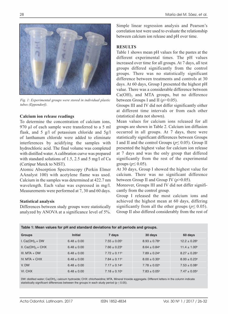
Calcium ion release readingsTo determine the concentration of calcium ions,
970 µl of each sample were transferred to a 5 ml
flask, and 5 g/l of potassium chloride and 5g/l
of lanthanum chloride were added to eliminate
interferences by acidifying the samples with
hydrochloric acid. The final volume was completed
with distilled water. A calibration curve was prepared
with standard solutions of 1.5, 2.5 and 5 mg/l of Ca
(Certipur Merck to NIST).
Atomic Absorption Spectroscopy (Perkin Elmer
AAnalyst 100) with acetylene flame was used.
Calcium in the samples was determined at 422.7 nm
wavelength. Each value was expressed in mg/l.
Measurements were performed at 7, 30 and 60 days.
Statistical analysisDifferences between study groups were statistically
analyzed by ANOVA at a significance level of 5%.
Simple linear regression analysis and Pearson’s
correlation test were used to evaluate the relationship
between calcium ion release and pH over time.
RESULTSTable 1 shows mean pH values for the pastes at the
different experimental times. The pH values
increased over time for all groups. At 7 days, all test
groups differed significantly from the control
groups. There was no statistically significant
difference between treatments and controls at 30
days. At 60 days, Group I presented the highest pH
value. There was a considerable difference between
Ca(OH)2 and MTA groups, but no difference
between Groups I and II (p˃0.05).
Groups III and IV did not differ significantly either
at different time intervals or from each other
(statistical data not shown).
Mean values for calcium ions released for all
groups are shown in Table 2. Calcium ion diffusion
occurred in all groups. At 7 days, there were
statistically significant differences between Groups
I and II and the control Groups (p≤ 0.05). Group II
presented the highest value for calcium ion release
at 7 days and was the only group that differed
significantly from the rest of the experimental
groups (p≤ 0.05).
At 30 days, Group I showed the highest value for
calcium. There was no significant difference
between Group II and Group IV (p˃0.05).
Moreover, Groups III and IV did not differ signifi
cantly from the control group.
Group I released the most calcium ions and
achieved the highest mean at 60 days, differing
significantly from all the other groups (p≤ 0.05).
Group II also differed considerably from the rest of
28 María del M. Sáez, et al.
Acta Odontol. Latinoam. 2017 ISSN 1852-4834 Vol. 30 Nº 1 / 2017 / 26-32
Table 1: Mean values for pH and standard deviations for all periods and groups.
Groups Initial 7 days 30 days 60 days
I. Ca(OH)2 + DW 6.48 ± 0.00 7.55 ± 0.05b 8.93 ± 0.78a 12.2 ± 0.26b
II. Ca(OH)2 + CHX 6.48 ± 0.00 7.66 ± 0.23b 8.64 ± 0.84a 11.4 ± 1.00b
III. MTA + DW 6.48 ± 0.00 7.72 ± 0.11b 7.89 ± 0.24a 8.27 ± 0.26a
IV. MTA + CHX 6.48 ± 0.00 7.84 ± 0.11b 8.00 ± 0.30a 8.00 ± 0.23a
V. DW 6.48 ± 0.00 7.17 ± 0.14a 7.78 ± 0.02a 7.53 ± 0.08a
VI. CHX 6.48 ± 0.00 7.18 ± 0.10a 7.83 ± 0.05a 7.47 ± 0.05a
DW: distilled water; Ca(OH)2: calcium hydroxide; CHX: chlorhexidine; MTA, Mineral trioxide aggregate. Different letters in the column indicate statistically significant differences between the groups in each study period (p ≤ 0.05).
Fig. 1: Experimental groups were stored in individual plastictubes (Eppendorf).
AOL12017:32011 03/07/2017 11:25 Página 28
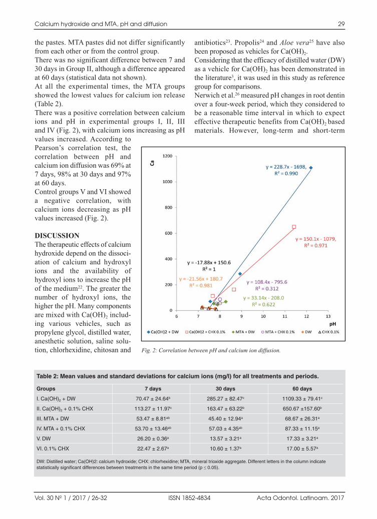
the pastes. MTA pastes did not differ significantly
from each other or from the control group.
There was no significant difference between 7 and
30 days in Group II, although a difference appeared
at 60 days (statistical data not shown).
At all the experimental times, the MTA groups
showed the lowest values for calcium ion release
(Table 2).
There was a positive correlation between calcium
ions and pH in experimental groups I, II, III
and IV (Fig. 2), with calcium ions increasing as pH
values increased. According to
Pearson’s correlation test, the
correlation between pH and
calcium ion diffusion was 69% at
7 days, 98% at 30 days and 97%
at 60 days.
Control groups V and VI showed
a negative correlation, with
calcium ions decreasing as pH
values increased (Fig. 2).
DISCUSSIONThe therapeutic effects of calcium
hydroxide depend on the dissoci
ation of calcium and hydroxyl
ions and the availability of
hydroxyl ions to increase the pH
of the medium22. The greater the
number of hydroxyl ions, the
higher the pH. Many components
are mixed with Ca(OH)2 includ
ing various vehicles, such as
propylene glycol, distilled water,
anesthetic solution, saline solu
tion, chlorhexidine, chitosan and
antibiotics23. Propolis24 and Aloe vera25 have also
been proposed as vehicles for Ca(OH)2.
Considering that the efficacy of distilled water (DW)
as a vehicle for Ca(OH)2 has been demonstrated in
the literature3, it was used in this study as reference
group for comparisons.
Nerwich et al.26 measured pH changes in root dentin
over a fourweek period, which they considered to
be a reasonable time interval in which to expect
effective therapeutic benefits from Ca(OH)2 based
materials. However, longterm and shortterm
Calcium hydroxide and MTA, pH and diffusion 29
Vol. 30 Nº 1 / 2017 / 26-32 ISSN 1852-4834 Acta Odontol. Latinoam. 2017
Table 2: Mean values and standard deviations for calcium ions (mg/l) for all treatments and periods.
Groups 7 days 30 days 60 days
I. Ca(OH)2 + DW 70.47 ± 24.64b 285.27 ± 82.47c 1109.33 ± 79.41c
II. Ca(OH)2 + 0.1% CHX 113.27 ± 11.97c 163.47 ± 63.22b 650.67 ±157.60b
III. MTA + DW 53.47 ± 8.81ab 45.40 ± 12.94a 68.67 ± 26.31a
IV. MTA + 0.1% CHX 53.70 ± 13.46ab 57.03 ± 4.35ab 87.33 ± 11.15a
V. DW 26.20 ± 0.36a 13.57 ± 3.21a 17.33 ± 3.21a
VI. 0.1% CHX 22.47 ± 2.67a 10.60 ± 1.37a 17.00 ± 5.57a
DW: Distilled water; Ca(OH)2: calcium hydroxide; CHX: chlorhexidine; MTA, mineral trioxide aggregate. Different letters in the column indicate statistically significant differences between treatments in the same time period (p ≤ 0.05).
Fig. 2: Correlation between pH and calcium ion diffusion.
AOL12017:32011 03/07/2017 11:25 Página 29
Ca(OH)2 treatment may reduce dentin fracture
strength7,8. MTA could be a treatment option in
cases with external inflammatory root resorption,
particularly when root fracture could be a risk20.
The diffusion of Ca(OH)2 through dentinal tubules
has been evaluated in several studies22, 2729. In this
study, calcium ion release from MTA and Ca(OH)2
was evaluated over two months. The accuracy of the
technique used is important for precise assessment
of the amount of calcium ion released. Several
methods such as atomic absorption spectrometry,
ultraviolet spectrophotometer, fluorometry, flame
photometry and complexometric titration with
EDTA have been used30. The present study used an
atomic absorption spectrometer.
In the present study, all the experimental groups
showed an increase in pH over time. This is in
agreement with Duarte et al.31, who found that all
pastes behaved similarly in terms of pH and calcium
ion release for the different study periods.
The vehicles employed in calcium hydroxide pastes
and MTA influence their diffusion capacity. Estrela
et al.3 explained that distilled water allows the
fastest and most significant dissociation. This is
consistent with our study, where the highest levels
of calcium ion release were obtained when distilled
water was used as a vehicle. The highest pH values
were also observed with distilled water at 60 days.
Hansen et al.32 found different pH values at different
levels of the root, possibly related to the number
and direction of dentinal tubules at each level. Perez
et al.33 showed that the pH of dentin depends on the
type, location and duration of Ca(OH)2 application.
In agreement with this, our study found higher pH
and calcium ion values at 60 days.
Evans et al.34 reported that Enterococcus faecaliswas resistant to Ca(OH)2 at pH 11.1, but at pH 11.5,
99.9% were killed. In a previous study, we found
that the pH values of the experimental groups were
not alkaline enough to eliminate E. faecalis from
the root canal in the lapse of a week or even a
month35. However, in the present study, Ca(OH)2
with DW at 60 days had a pH value (12.2 ± 0.26)
high enough to kill E. faecalis.
Due to the antimicrobial effectiveness of CHX, it
has been suggested as a vehicle for Ca(OH)2 pastes.
The antimicrobial activity of the association of
Ca(OH)2 and CHX has already been evaluated,
revealing effectiveness against several bacterial
populations17, 36. Stowe et al.15 observed that adding
CHX to MTA could enhance antimicrobial activity.
According to our results, CHX would be an efficient
vehicle for Ca(OH)2 allowing the diffusion of
calcium ions. Moreover, for MTA pastes using CHX
as vehicle, calcium ion diffusion was similar or
higher than for MTA with distilled water.
Mori et al.37 concluded that the concentration of CHX
influenced calcium diffusion. They demonstrated
that 2% CHX allowed higher diffusion than 0.2%
CHX. In contrast, the present study found higher
calcium ion diffusion with 0.1% chlorhexidine than
the results reported by Mori et al.37. Another
interesting factor to consider is the higher viscosity
and easier preparation of the pastes with CHX
compared to those prepared with distilled water.
This study showed that at 30 days there was no
difference in the pH values between Ca(OH)2 pastes
and MTA pastes. However, Heward et al.20 found that
after four weeks, intracanal placement of MTA
resulted in significantly higher pH than did Ca(OH)2.
Differences in methodologies might explain this
apparent disparity.
Ozdemir et al.38 showed diffusion of calcium
through the defects in the dentin in MTAfilled
roots with a significant increase in concentration
over time. Each drug presents different diffusion
characteristics, which are directly related to its
interaction with the tooth structure39. In the present
study, MTA pastes present the lowest calcium ion
release values at all experimental times. In contrast,
TonomaruFilho40 reported that MTA and Sealer 26
presented the highest values of calcium ion release
after 28 days, out of six sealer materials evaluated.
Similarly to our results, TonomaruFilho40 also
reported that control group released calcium ions,
even though the teeth were unfilled, because the
tooth itself can release calcium ions from its
structure.
In this in vitro study, all the pastes analyzed increased
the pH values over time, although calcium hydroxide
and distilled water pastes showed the highest pH
values and calcium ion diffusion. The greatest
diffusion of ions took place between 30 and 60 days.
Calcium hydroxide paste groups showed better
diffusion capacity than MTA. Nevertheless, further
studies should be undertaken to determine whether
MTA could be a good option for teeth at risk of
fracture.
30 María del M. Sáez, et al.
Acta Odontol. Latinoam. 2017 ISSN 1852-4834 Vol. 30 Nº 1 / 2017 / 26-32
AOL12017:32011 03/07/2017 11:25 Página 30
ACKNOWLEDGMENTSThis study was partially supported by a grant from SCAIT (Sec
retariat of Science, Arts and Technological Innovation). The
authors wish to thank Alberto Marcial Manlla for assistance
with statistical procedures.
CORRESPONDENCEDra. María del Milagro Sáez
Cátedra de Endodoncia, Facultad de Odontología,
Universidad Nacional de Tucumán
Av. Benjamín Aráoz 800
San Miguel de Tucumán (4000), Argentina.
Calcium hydroxide and MTA, pH and diffusion 31
Vol. 30 Nº 1 / 2017 / 26-32 ISSN 1852-4834 Acta Odontol. Latinoam. 2017
REFERENCES 1. Ruparel NB, Teixeria FB, Ferraz CC, Diogenes A. Direct
effect of intracanal medicaments on survival of stem cells
of the apical papilla. J Endod 2012; 38:13721375.
2. SafaviKE, Nichols FC. Alteration of biological properties
of bacterial lipopolysaccharide by calcium hydroxide
treatment. J Endod 1994; 20:127129.
3. Estrela C, Sydney GB, Bammann LL, Felippe Júnior O.
Mechanism of action of calcium and hydroxyl ions of calcium
hydroxide on tissue and bacteria. Braz Dent J 1995; 6:8590.
4. Fava LR, Saunders WP. Calcium hydroxide pastes: classifi
ca tion and clinical indications. Int Endod J 1999; 32:257282.
5. Heithersay Gs. Calcium Hydroxide in the treatment of
pulpless teeth with associated pathology. J Br Endond Soc
1975; 8:7493.
6. Frank AL. Therapy for the divergent pulpless tooth by
continued apical formation. J Am Dent Assoc 1966; 72:
8793.
7. Andreasen JO, Farik B, Munksgaard EC. Longterm
calcium hydroxide as a root canal dressing may increase
risk of root fracture. Dent Traumatol 2002; 18:1122.
8. Sahebi S, Moazami F, Abbott P. The effects of shortterm
calcium hydroxide application on the strehgth of dentine.
Dent Traumatol 2010; 26:4346.
9. Islam I, Chmg HK, Yap AUJ. Comparison of the phisical
and mechanical properties of MTA and Portland Cement. J
Endod 2006; 39:747754.
10. Torabinejad, M.; Watson, T. F.; Pitt Ford, T. R. Sealing
ability of a mineral trioxide aggregate as a retrogate root
filling material. J Endod 1993; 19:591595.
11. Enkel B, Dupas C, Armengol V. Bioactive materials in
endodontics. Expert Rev Med Devices 2008; 5:475494.
12. Moretton TR, Brown CE Jr, Legan JJ, Kafrawy AH. Tissue
reactions after subcutaneous and intraosseous implantation
of mineral trioxide aggregate and ethoxybenzoic acid
cement. J Biomed Mater Res 2000; 52:528533.
13. Torabinejad M, Hong CU, McDonald F, Pitt Ford TR.
Physical and chemical properties of a new rootend filling
material. J Endod 1995; 21:349353.
14. Parirokh M, Torabinejad M. Mineral trioxide aggregate: a
comprehensive literature review part III. Clinical applica
tions, drawbacks, and mechanism of action. J Endod 2010;
36:400413.
15. Stowe TJ, Sedgley CM, Stowe B, Fenno JC.The effects of
chlorhexidine gluconate (0.12%) on the antimicrobial
properties of toothcolored ProRoot mineral trioxide
aggregate. J Endod 2004; 30:429431.
16. Ohara P, Torabinejad M, Kettering JD. Antibacterial effect
of various endodontic irrigants on selected anaerobic
bacteria. Endod Dent Traumatol 1993; 9:95100.
17. Siqueira JF Jr, Paiva SS, Roças In. reduction in the cultiva
ble bacterial populations in infected root canals by a
chlorhexidine based antimicrobial protocol. J Endod 2007;
33:541547.
18. Waltimo TM, Orstavik D, Sirén EK, Haapasalo MP. In
vitro susceptibility of Candida albicans to four disinfec
tants and their combinations. Int Endod J 1999; 32:
421429.
19. Parsons GJ, Patterson SS, Miller CH, Katz S, Kafrawy AH,
Newton CW. Uptake and release of chlorhexidine by bovine
pulp and dentin specimens and their subsequent acquisition
of antibacterial properties. Oral Surg Oral Med Oral Pathol
1980; 49:455459.
20. Heward S, Sedgley CM. Effects of intracanal mineral
trioxide aggregate and calcium hydroxide during four weeks
on pH changes in simulated root surface resorption defects:
an in vitro study using matched pairs of human teeth. J
Endod 2011; 37:4044.
21. Boutsioukis C, Noula G, Lambrianidis T. Ex vivo study of
the efficiency of two techniques for the removal of mineral
trioxide aggregate used as a root canal filling material. J
Endod 2008; 34:12391242.
22. Safavi KE, Nakayama TA. Influence of mixing vehicle on
dissociation of calcium hydroxide in solution. J Endod
2000; 26:649651.
23. Pacios MG, de la Casa ML, Bulacio MA, López ME.
Influence of difference vehicles on the pH of calcium
hydroxide pastes. J Oral Sci 2004; 46:107111.
24. Montero JC, Mori GG. Assessment of ion diffusion from a
calcium hydroxidepropolis paste through dentin. Braz Oral
Res 2012; 26:318322.
25. de Souza Batista V, Dáquila Olian D, Garrido Mori G.
Diffusion of hydroxyl ions from calcium hydroxide and
Aloe vera pastes. Braz Dent J 2014; 25:212216.
26. Nerwich A, Figdor D, Messer HH. pH changes n root dentn
over a 4week period following root canal dressing with
calcium hydroxide. J Endod 1993; 19:302305.
27. Duarte MA, Demarchi AC, Giaxa MH, Kiga MC, Fraga
SC, de Souza LC. Evaluation of pH and calcium ion release
of three root canal sealers. J Endod 2000; 26:389390.
28. Azabal Arroyo M, MenasalvasRuiz G, MartínAlonso J,
Arroquia JJ, Vegadel Barrio JM. Loss of hydroxyl
ions from guttapercha points with calcium hydroxide
in their composition: an in vivo study. J Endod 2002;
28:697698.
29. de Andrade Ferreira FB, Silva e Souza Pde A, do Vale MS,
de Morales IG, Granjeiro JM. Evaluation of pH levels
and calcium ion release in various calcium hydroxide
endodontic dressings. Oral Surg Oral Pathol Oral Radiol
Endod 2004; 97:388392.
AOL12017:32011 03/07/2017 11:25 Página 31
30. Robertson WG, Marshall RW. Calcium measurements in
serum and plasma total and ionized. Cri Rev Clin Lab Sci
1979; 11:271304.
31. Duarte MA, Midena R, Zeferino M, Ricci R, Tanamaru
Filho M. Evaluation of pH and Calcium Ion Release of
Calcium Hydroxide Pastes Containing Different Substances.
J Endod 2009; 35:12741277.
32. Hansen SW, Marshall G, Sedgley CM. Comparison of
intracanal EndoSequence Root Repair Material and ProRoot
MTA to induce pH changes in simulated root resorption
defects over 4 weeks in matched pairs of human teeth. J
Endod 2011; 37: 502506.
33. Pérez F, Franchi M, Péli JF. Effect of calcium hydroxide
form and placement on root dentin pH. Int Endod J 2001;
34:417423.
34. Evans MD, Baumgartner JC, Khemaleelakul SU, Xia T.
Efficacy of calcium hydroxide: chlorhexidine paste as an
intracanal medication in bovine dentin. J Endod 2003;
29:338339.
35. de la Casa ML, Sáez MM, López GL, López ME, Cecilia
M. Acción antibacteriana de pastas de hidróxido de calcio
frente a Enterococcus faecalis. Endodoncia 2012; 30:6063.
36. Gomes BP, Vianna ME, Sena NT, Zaia AA, Ferraz CC,
de Souza Filho FJ. In vitro evaluation of the antimi crobial
activity of calcium hydroxide combined with chlorhexidine
gel used as intracanal medicament. Oral Surg Oral Med
Oral Pathol Oral Radiol Endod 2006; 102:544550.
37. Mori GG, Ferreira FC, Batista FR, Godoy AM, Nunes DC.
Evaluation of the diffusion capacity of calcium hydroxide
pastes through the dentinal tubules. Braz Oral Res 2009;
23:113118.
38. Ozdemir HO, Ozçelik B, Karabucak B, Cehreli ZC.
Calcium ion diffusion from mineral trioxide aggregate
through simu lated root resorption defects. Dent Traumatol
2008; 24:7073.
39. Camões IC1, Salles MR, Chevitarese O, Gomes LN.
Diffusion of Ca(OH)2 associated with different vehicles:
chromatographic study (highperformance liquid chroma
to graphy). J Endod 2004; 30:3034.
40. TanomaruFilho M, Chaves Faleiros FB, Saçaki JN,
Hungaro Duarte MA, GuerreiroTanomaru JM. Evaluation
of pH and calcium ion release of rootend filling materials
containing calcium hydroxide or mineral trioxide aggregate.
J Endod 2009; 35:14181421.
32 María del M. Sáez, et al.
Acta Odontol. Latinoam. 2017 ISSN 1852-4834 Vol. 30 Nº 1 / 2017 / 26-32
AOL12017:32011 03/07/2017 11:25 Página 32
RESUMENEl objetivo del presente trabajo fue estudiar los cambios térmicosin vitro en la superficie externa de la raíz del diente, generadosdurante los procedimientos de desobturación mecánica.Se utilizaron 30 premolares inferiores unirradiculares reciente mente extraídos, que fueron seccionados transversalmente a16 mm del ápice para estandardizar la longitud de lasmuestras. Se realizó luego su tratamiento endodóntico. Lasmediciones de temperatura fueron realizadas mediante undispositivo digital con termocupla, en la superficie externa adiferentes tiempos (t = 0, 2, 4, 6, 8, 10 y 15 segundos), durantela desobturación con fresas de Gates Glidden (G1, G2, G3);de Peeso (P1, P2, P3) y la correspondiente a PostecPlus FRC(FRC). Los registros de temperatura fueron comparadosmediante pruebas de ANOVA de medidas repetidas, seguidaspor comparaciones entre pares mediante la prueba de Tukey. Con todas las fresas se encontró una variación significativa dela temperatura entre los diferentes tiempos (p<0.05). Las compa raciones entre pares indicaron que la temperatura se incrementó
con el tiempo en todas las fresas (p<0.05). Se detectarondiferencias significativas de temperatura entre diferentes fresasdespués de 0, 2, 4, 6, 8, 10 y 15 segundos (p<0.05). El aumento de la temperatura de la superficie radicular fue
importante ya que los valores registrados superan los 50ºC,teniendo en cuenta que el aumento de temperatura mayor fuede 16ºC. Si partimos de los 37ºC (temperatura fisiológica), latemperatura presente en los tejidos circundantes; cemento,periodonto y hueso; alcanzaría los 53ºC. Un aumento de 10ºCpor encima de la temperatura corporal en la superficieradicular podría causar lesiones en los tejidos circundantes.La utilización de refrigeración, el control del tiempo de accióny el uso de un instrumental en estado óptimo son parámetrosineludibles debido a que los mismos pueden contribuir adisminuir el calor generado y trasmitido hacia la superficieexterna de la raíz durante la desobturación.
Palabras clave: Preparación del conducto radicular; temperaturade transición; endodoncia.
ABSTRACTThe aim of this study was to analyze in vitro temperature changeson the outer surface of the dental root during mechanical fillingremoval procedures.Thirty recently extracted singlerooted lower premolars were cuttransversally at 16 mm from the apex in order to standardizesample length. Endodontic treatment was performed on them. Thefilling material was subsequently removed using Gates Glidden(G1, G2, G3); Peeso (P1, P2, P3) and PostecPlus FRC (FRC)reamers while temperatures were measured on the outer surfaceusing a digital device with thermocouple at 0, 2, 4, 6, 8, 10 and 15seconds. Temperatures were compared using repeated measuresANOVA followed by pairwise comparison with Tukey’s test. All reamers caused significant temperature variation betweendifferent times (p<0.05). Pairwise comparisons indicated thattemperature increased with time for all reamers (p<0.05).
Significant differences in temperature were found betweendifferent reamers after 0, 2, 4, 6, 8, 10 and 15 seconds (p<0.05). Temperature at the root surface increased considerably. Valueshigher than 50ºC were recorded, the greatest increase frombaseline being 16ºC. Accordingly, if the procedure were begunat 37ºC (physiological temperature), the temperature in thesurrounding tissues cementum, periodontium and bone wouldrise to 53ºC. An increase in 10ºC above body temperature at theroot surface may cause lesions in surrounding tissues. Whileremoving filling material, it is essential to cool, control actiontime and use instruments in perfect condition, all of which maycontribute to reducing the heat generated and transmitted tothe outer root surface.
Key words: Root canal preparation; transition temperature;endodontic.
33
Vol. 30 Nº 1 / 2017 / 33-38 ISSN 1852-4834 Acta Odontol. Latinoam. 2017
Root surface temperature variation during mechanical removal of root canal filling material. An in vitro study
Martín García-Cuerva1, Lucía Horvath1, Laura Pinasco2, Verónica Ciparelli1, Hernán Tartacovsky1, Ariel Gualtieri3, Ana C Casadoumecq4, Pablo Rodríguez2, Carlos Gonzalez-Zanotto1
1 Universidad de Buenos Aires. Facultad de Odontología. Cátedra de Clínica I de Operatoria2 Universidad de Buenos Aires. Facultad de Odontología. Cátedra de Endodoncia3 Universidad de Buenos Aires. Facultad de Odontología, Cátedra de Biofisica4 Universidad de Buenos Aires. Facultad de Odontología.Cátedra de Fisiología
Variaciones térmicas en la superficie radicular durante la desobturación mecánica del conducto. Estudio in vitro
AOL12017:32011 03/07/2017 11:25 Página 33
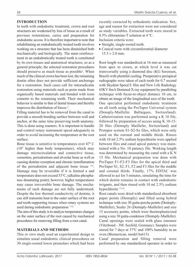
INTRODUCTIONIn teeth with endodontic treatment, crown and root
structures are weakened by loss of tissue as a result of
previous restorations, caries and preparation for
endodontic access. It is therefore important to note that
rehabilitating an endodontically treated tooth involves
working on a structure that has been diminished both
mechanically and biologically. The main reinforce
ment in an endodontically treated tooth is constituted
by its own tissues and anatomical structures, so as a
general principle; the selected restoration procedures
should preserve as much tissue as possible1. When
much of the clinical crown has been lost, the remaining
dentin often does not provide sufficient anchorage
for a restoration. Such cases call for intraradicular
restoration using materials such as posts made from
organically based materials and bonded with resin
cements to the remaining tooth. Their mechanical
behavior is similar to that of dental tissues and thereby
improves the distribution of forces 2.
Filling material has to be removed from the canal to
provide a smooth bonding surface between wall and
anchor, at the same time preserving tooth anatomy.
This is done using reamers. It is important to handle
and control rotary instrument speed adequately in
order to avoid increasing the temperature at the root
surface.
Bone tissue is sensitive to temperatures over 47º C
(10° higher than body temperature), which may
damage microcirculation and connective tissue,
cementim, periodontium and alveolar bone as well as
causing dentine resorption and chronic immflama tion
of the periodontim and adjacent bone tissue 36.
Damage may be reversible if it is limited a and
temperature does not exceed 53°C, (alkaline phospha
tase denaturing point); however, higher temperatures
may cause irreversible bone damage. The mecha
nisms of such damage are not fully understood.
Despite the low thermal conductivity of dentine, it
can still transmite heat to the outer surface of the root
and toothsupporting tissues when rotary systems are
used during endodontic preparation 712.
The aim of this study is to analyze temperature changes
on the outer surface of the root caused by mechanical
procedures for removing filling from a root canal.
MATERIALS AND METHODS This in vitro study used an experimental design to
simulate usual endodontic clinical procedures on
30 singlerooted lower premolars which had been
recently extracted by orthodontic indication. Sex,
age and reason for extraction were not considered
as study variables. Extracted teeth were stored in
0.5% chloramineT solution at 4 ºC.
Inclusion criteria were:
• Straight, singlerooted teeth.
• Conical roots with circumferential diameter
15.5 ± 2.0 mm.
Root length was standardized at 16 mm as measured
from apex to crown, at which level it was cut
transversally using a diamond disc (KG Sorensen,
Brazil) with plentiful cooling. Preoperative periapical
radiographs were taken of each tooth for 0.7 second
with Skydent Speed E film and New Life Radiology
65KV 8mA Denimed Xray equipment by paralleling
technique with focustoobject distance 10 cm, to
obtain an image of the longitudinal axis of the tooth.
One specialist performed endodontic treatment
on all teeth using the ProTaper Universal system
(DensplyMaillefer. Ballaigues, Switzerland).
Catheterization was performed using a K 10 file,
followed by preparation of access using K 1015
20 files (Dentsply Maillefer, Switzerland) and
Protaper system S1S2Sx files, which were only
used on the coronal and middle thirds. Rinses
with 10 ml 2.5% sodium hypochlorite were applied
between files and canal apical patency was main
tained with a No. 10 patency file. Working length
was determined by measuring the canal with a K N
15 file. Mechanical preparation was done with
ProTaper F1F2F3 files for the apical third and
ProTaper S1, S2, F1, F2 and F3 files for the middle
and coronal thirds. Finally, 17% EDTAC was
allowed to act for 5 minutes, simulating the time for
which dentin remains in contact with endodontic
irrigants, and then rinsed with 10 ml 2.5% sodium
hypochlorite 13,14.
Root canals were dried with standardized absorbent
paper points (Dentsply) and filled using hybrid
technique with size 30 guttapercha points (Dentsply
Maillefer), Sealer 26 (DentsplyMaillefer) and size
15 accessory points, which were thermoplasticized
using a size 30 guttacondensor (DentsplyMaillefer).
Canal openings were sealed with glass ionomer
(Vitrebond 3M. Seefeld, Germany). Samples were
stored for 7 days at 37ºC and 100% humidity in an
oven (Biomerican, model bs615).
Canal preparation and filling removal were
performed by one standardized operator in order to
34 Martín García-Cuerva, et al.
Acta Odontol. Latinoam. 2017 ISSN 1852-4834 Vol. 30 Nº 1 / 2017 / 33-38
AOL12017:32011 03/07/2017 11:25 Página 34
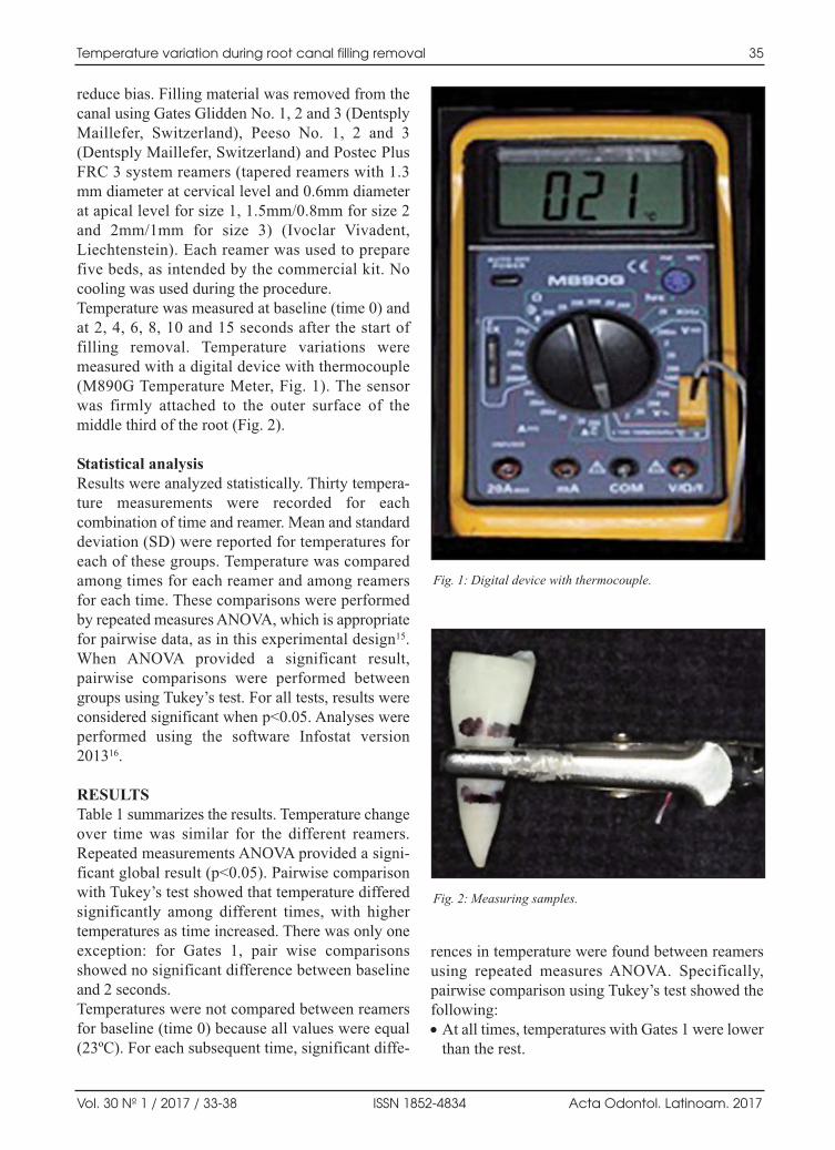
reduce bias. Filling material was removed from the
canal using Gates Glidden No. 1, 2 and 3 (Dentsply
Maillefer, Switzerland), Peeso No. 1, 2 and 3
(Dentsply Maillefer, Switzerland) and Postec Plus
FRC 3 system reamers (tapered reamers with 1.3
mm diameter at cervical level and 0.6mm diameter
at apical level for size 1, 1.5mm/0.8mm for size 2
and 2mm/1mm for size 3) (Ivoclar Vivadent,
Liechtenstein). Each reamer was used to prepare
five beds, as intended by the commercial kit. No
cooling was used during the procedure.
Temperature was measured at baseline (time 0) and
at 2, 4, 6, 8, 10 and 15 seconds after the start of
filling removal. Temperature variations were
measured with a digital device with thermocouple
(M890G Temperature Meter, Fig. 1). The sensor
was firmly attached to the outer surface of the
middle third of the root (Fig. 2).
Statistical analysisResults were analyzed statistically. Thirty tempera
ture measurements were recorded for each
combination of time and reamer. Mean and standard
deviation (SD) were reported for temperatures for
each of these groups. Temperature was compared
among times for each reamer and among reamers
for each time. These comparisons were performed
by repeated measures ANOVA, which is appropriate
for pairwise data, as in this experimental design15.
When ANOVA provided a significant result,
pairwise comparisons were performed between
groups using Tukey’s test. For all tests, results were
considered significant when p<0.05. Analyses were
performed using the software Infostat version
201316.
RESULTS Table 1 summarizes the results. Temperature change
over time was similar for the different reamers.
Repeated measurements ANOVA provided a signi
ficant global result (p<0.05). Pairwise comparison
with Tukey’s test showed that temperature differed
significantly among different times, with higher
temperatures as time increased. There was only one
exception: for Gates 1, pair wise comparisons
showed no significant difference between baseline
and 2 seconds.
Temperatures were not compared between reamers
for baseline (time 0) because all values were equal
(23ºC). For each subsequent time, significant diffe
rences in temperature were found between reamers
using repeated measures ANOVA. Specifically,
pairwise comparison using Tukey’s test showed the
following:
• At all times, temperatures with Gates 1 were lower
than the rest.
Temperature variation during root canal filling removal 35
Vol. 30 Nº 1 / 2017 / 33-38 ISSN 1852-4834 Acta Odontol. Latinoam. 2017
Fig. 1: Digital device with thermocouple.
Fig. 2: Measuring samples.
AOL12017:32011 03/07/2017 11:25 Página 35
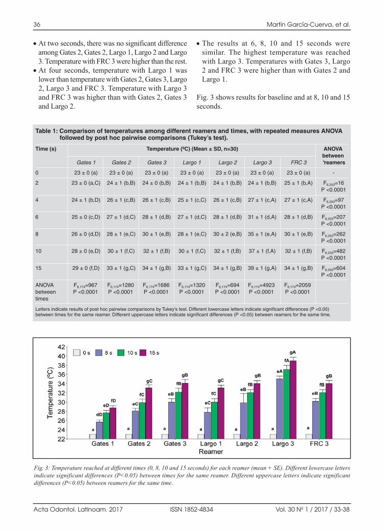
• At two seconds, there was no significant difference
among Gates 2, Gates 2, Largo 1, Largo 2 and Largo
3. Temperature with FRC 3 were higher than the rest.
• At four seconds, temperature with Largo 1 was
lower than temperature with Gates 2, Gates 3, Largo
2, Largo 3 and FRC 3. Temperature with Largo 3
and FRC 3 was higher than with Gates 2, Gates 3
and Largo 2.
• The results at 6, 8, 10 and 15 seconds were
similar. The highest temperature was reached
with Largo 3. Temperatures with Gates 3, Largo
2 and FRC 3 were higher than with Gates 2 and
Largo 1.
Fig. 3 shows results for baseline and at 8, 10 and 15
seconds.
36 Martín García-Cuerva, et al.
Acta Odontol. Latinoam. 2017 ISSN 1852-4834 Vol. 30 Nº 1 / 2017 / 33-38
Table 1: Comparison of temperatures among different reamers and times, with repeated measures ANOVA followed by post hoc pairwise comparisons (Tukey’s test).
Time (s) Temperature (ºC) (Mean ± SD, n=30) ANOVAbetween
Gates 1 Gates 2 Gates 3 Largo 1 Largo 2 Largo 3 FRC 3 *reamers
0 23 ± 0 (a) 23 ± 0 (a) 23 ± 0 (a) 23 ± 0 (a) 23 ± 0 (a) 23 ± 0 (a) 23 ± 0 (a) -
2 23 ± 0 (a,C) 24 ± 1 (b,B) 24 ± 0 (b,B) 24 ± 1 (b,B) 24 ± 1 (b,B) 24 ± 1 (b,B) 25 ± 1 (b,A) F6,203=16P <0.0001
4 24 ± 1 (b,D) 26 ± 1 (c,B) 26 ± 1 (c,B) 25 ± 1 (c,C) 26 ± 1 (c,B) 27 ± 1 (c,A) 27 ± 1 (c,A) F6,203=97P <0.0001
6 25 ± 0 (c,D) 27 ± 1 (d,C) 28 ± 1 (d,B) 27 ± 1 (d,C) 28 ± 1 (d,B) 31 ± 1 (d,A) 28 ± 1 (d,B) F6,203=207P <0.0001
8 26 ± 0 (d,D) 28 ± 1 (e,C) 30 ± 1 (e,B) 28 ± 1 (e,C) 30 ± 2 (e,B) 35 ± 1 (e,A) 30 ± 1 (e,B) F6,203=262P <0.0001
10 28 ± 0 (e,D) 30 ± 1 (f,C) 32 ± 1 (f,B) 30 ± 1 (f,C) 32 ± 1 (f,B) 37 ± 1 (f,A) 32 ± 1 (f,B) F6,203=482P <0.0001
15 29 ± 0 (f,D) 33 ± 1 (g,C) 34 ± 1 (g,B) 33 ± 1 (g,C) 34 ± 1 (g,B) 39 ± 1 (g,A) 34 ± 1 (g,B) F6,203=604P <0.0001
ANOVA F6,174=967 F6,174=1280 F6,174=1686 F6,174=1320 F6,174=694 F6,174=4923 F6,174=2059between P <0.0001 P <0.0001 P <0.0001 P <0.0001 P <0.0001 P <0.0001 P <0.0001times
Letters indicate results of post hoc pairwise comparisons by Tukey’s test. Different lowercase letters indicate significant differences (P <0.05)between times for the same reamer. Different uppercase letters indicate significant differences (P <0.05) between reamers for the same time.
Fig. 3: Temperature reached at different times (0, 8, 10 and 15 seconds) for each reamer (mean + SE). Different lowercase lettersindicate significant differences (P<0.05) between times for the same reamer. Different uppercase letters indicate significantdifferences (P<0.05) between reamers for the same time.
AOL12017:32011 03/07/2017 11:25 Página 36
DISCUSSIONThe harmful effects of heat on the outer root surface
during filling removal procedures are clinically
important because an increase greater than 10°C
above physiological temperature (37ºC) can alter
the viability of supporting tissues and cause bone
necrosis, cell apoptosis and sometimes, in more
severe cases, tooth ankylosis9. Heat generation
while removing filling from the canal system can
be modified in various ways, including type of
instrument used (acuity, sharpness and size),
condition of instrument cutting edges, rotation,
cutting pressure applied and contact time with tooth
structure.
This study found temperature increases of up to
approximately 16ºC at 15 seconds during canal
preparation. Wider reamers were associated to
greater heat generation. Manufacturers’ protocols
for filling removal say that working time should be
shorter than 1 minute. Based on our results, we
suggest that this time limit should be considerably
lower.
Another factor to consider during filling removal is
how thick the remaining dentin is in the canal walls.
Stripping dentin tissue excessively from the canal
walls during preparation with the aim of increasing
anchorage and achieving better fit of the element to
be bonded is contraindicated. It will not only weaken
the walls, but also increase heat transmission
outward. Excessive removal of root dentin is known
to compromise the root, and preserving root dentin
is directly related to root strength 17,18. Thus,
knowledge of the internal tooth anatomy contributes
to dental practice which is more conservative of
tissues, and avoids causing excessive damage to teeth
and tooth supporting tissues during preparation.
Customizing posts prior to bonding may contribute
to reducing the occurrence of irreversible injury.
Lubieniecka et al.19 replicated the clinical situation
of removing filling from canals during post space
preparation and analyzed it with a thermal imaging
camera. The effect of cooling was clear in the
cervical region of the tooth, where temperature was
very close to initial temperature reading. The
highest temperature on the surface of the root
corresponded to the greatest depth that the drill
reached. The periapical zone experienced very little
or no temperature increase19. Weller et al.20 recorded
the highest temperature increase at the most coronal
part of the root, which is closely related to larger
width of reamers, more guttapercha in this part of
the tooth and thinner dentin walls. Other authors
such as Lima Machado and Antoniazzi21 reported
higher temperatures for instrumentation of the
cervical third than for the middle and apical thirds.
Some authors suggest that clinicians should take
into account that dentin is considered to be a good
thermal insulator, and the thicker it is, the less heat
will be transferred to the outer surface of the root2224.
However, depending on their anatomy, not all teeth
have a thick layer of dentin, e.g. lower incisors have
very thin walls. Care must therefore be exercised to
avoid damaging tooth support tissue when remaining
dentin is less than 1 mm thick22.
Further studies could consider working length, type
and size of reamer used and cooling while preparing
root canal. The use of anatomic posts could also be
considered, since they are more respectful of tooth
anatomy, requiring less thinning of dentin walls and
consequently providing treatment that is more
conservative of remaining dentin structures.
CONCLUSIONWithin the limitations of the present study, the
increase in temperature at the root surface during
mechanical removal of filling was important, since
the values recorded at the outer root surface were
high enough to damage tissues surrounding the tooth.
Even though temperatures increased to critical values
for surrounding tissues, the absolute results of this invitro study are not directly transferable to real clinical
situations, since they would be influenced by
periodontal tissues, periodontal blood circulation and
the oral environment.
These results suggest the need for further studies to
enable current protocols to be adapted.
Temperature variation during root canal filling removal 37
Vol. 30 Nº 1 / 2017 / 33-38 ISSN 1852-4834 Acta Odontol. Latinoam. 2017
CORRESPONDENCEDr. Martin García Cuerva
Cátedra de Clínica I de Operatoria, Facultad de Odontología,
Universidad de Buenos Aires
Marcelo T. de Alvear 2142, Piso 9°B,
(CP1122AAH) C.A.B.A., Argentina.
Email: [email protected]
AOL12017:32011 03/07/2017 11:25 Página 37
REFERENCES 1. Bertoldi Hepburn. Rehabilitación posendodontica. Capitulo
2, Postura filosófica para la rehabilitacion posendodóntica.
Buenos Aires, Argentina. Editorial Médica Panamericana
S.A.C.F., 2012: 219.
2. Menezes MS, Queiroz EC, Campos RE, Martins LRM, et
al. Influence of endodontic sealer on fibreglass post bond
strength to root dentin. Int Endod J 2008; 41:476484.
3. Eriksson A, Albrektsson T, Grane B, Mcqueen D. Thermal
injury to bone. A vitalmicroscopic description of heat
effects. Int J Oral Surg 1982; 11:115121.
4. Bhaskar SN, Lilly GE. Intrapulpal temperature during
cavity preparation. J Dent Res 1965; 44:644647.
5. Tjan AH, Abbate MF. Temperature rise at root surface
during post space preparation. J Prosth Dent 1993; 69:4145.
6. Saunders EM, Saunders WP. The heat generated on the
external root surface during post space preparation. Int
Endod J 1989; 22:169173.
7. Eriksson JH, Sundström F. Temperature rise at root surface
during root canal preparation a possible cause of damage to
tooth and periodontal tissue. Swed Dent J 1984; 8:217223.
8. Cohen BI, Deutsch AS, Musikant BL. Effect of power
settings on temperature change at the root surface when
using a Holmium YAG laser in enlarging the root canal. J
Endod 1996; 22:596599.
9. Eriksson AR, Albrektsson T. Temperature threshold levels
for heatinduced bone tissue injury: A vitalmicroscopic
study in the rabbit. J Prosthet Dent 1983; 50:101107.
10. Kilic K, Er O, Kilinc HI, Aslan T, et al. Infrared thermographic
comparison of temperature increases on the root surface
during dowel space preparations using circular versus oval
fiber dowel systems. J Prosthodont 2013; 22:203207.
11. Ratih DN, Palamara JEA, Messer HH. Temperature change,
dentinal fluid flow and cuspal displacement during resin
composite restoration. J Oral Rehabil 2007; 34:693701.
12. Lipski M, Mrozek J, Drozdzik A. Influence of water
cooling on root surface temperature generated during post
space preparation. J Endod 2010; 36:713716.
13. Cunha RS, De Martin AS, Barros PP, da Silva FM, et al. In
vitro evaluation of the cleansing working time and analysis
of the amount of guttapercha or Resilon remnants in the
root canal walls after instrumentation for endodontic
retreatment. J Endod 2007; 33:14261428.
14. Galafassi D, Colucci V, Cecchin D, Scatena C, et al. Effect
of Endodontic Irrigants on Microtensile Bond Strength to
Dentin After Thermocycling and LongTerm Water Storage.
J Dent (Tehran) 2013; 10:426435.
15. Davis C. NormalTheory Methods: Repeated Measures
ANOVA. En: Statistical methods for the analysis of repeated
measurements, 1ra ed. New York: SpringerVerlag, 2002:
103116.
16. Di Rienzo JA, Casanoves F, Balzarini MG, Gonzalez L, et
al. InfoStat versión 2013p, Grupo InfoStat, FCA, Univer
sidad Nacional de Córdoba, Argentina. 2013.
17. Lertchirakarn V, Timyam A, Messer HH. Effects of root
canal sealers on vertical root fracture resistance of endodon
ti cally treated teeth. J Endod 2002; 28:217219.
18. Fukui Y, Komada W, Yoshida K, Otake S, et al. Effect of
reinforcement with resin composite on fracture strength of
structurally compromised roots. Dent Mater J 2009; 28:
602609.
19. Lubieniecka J, Lukasiewicks J, Bozyk J, Kleinrok J. The
evaluation of increase and distribution of temperature
during the dental drilling using a thermal imaging camera.
FLIR technical series. Application note for research and
science. 2011: 17.
20. Weller RN, Kimbrough WF, Anderson RW. Root surface
temperatures produced during post space preparation. J
Endod 1996,22: 304307.
21. Lima Machado ML, Antoniazzi JH. In Vitro evaluation of
the temperature achieved by GatesGlidden. Largo and
modified largo drills during chemosurgical preparation of
root canals. Ecler endond. Vol 1 n3 Sao Paulo, 1999.
22. Kwon SJ, Park YJ, Jun SH, Ahn JS, et al. Thermal irritation
of teeth during dental treatment procedures. Restor Dent
Endod 2013; 38: 105112.
23. Ottl P, Lauer HC. Temperature response in the pulpal
chamber during ultrahighspeed tooth preparation with
diamond burs of different grit. J Prosthet Dent 1998; 80:1219.
24. Dominici JT, Clark S, Scheetz J, Eleazer PD. Analysis of
heat generation using ultrasonic vibration post removal. J
Endod 2005; 31:301303.
38 Martín García-Cuerva, et al.
Acta Odontol. Latinoam. 2017 ISSN 1852-4834 Vol. 30 Nº 1 / 2017 / 33-38
AOL12017:32011 03/07/2017 11:25 Página 38
RESUMOObjetivo: avaliar o efeito de diferentes períodos experimentaise se a presença de ligadura em um dos lados afeta a perdaóssea alveolar (POA) no lado contralateral. O presente estudo tratase de uma análise secundária dosbancos de dados de estudos realizados na UniversidadeFederal do Rio Grande do Sul. Os estudos incluídosutilizaram o modelo de indução de doença periodontal porligadura em ratos. Os estudos necessitavam possuir grupocontrole sem ligadura, assim como controle intragrupo.Foram incluídos 3 estudos, com diferentes períodos deanálise: 2 semanas com ligadura e 8 semanas sem ligadura;5 semanas com ligadura e 17 semanas sem ligadura; 22semanas com e sem ligadura. Os ratos foram criados nasmesmas condições, sacrificados por decapitação, as maxilasretiradas e os tecidos moles removidos com hipoclorito desódio 9%. Tomadas fotográficas foram realizadas e cincomensurações foram obtidas de cada imagem. A presença de
ligadura gerou uma perda óssea alveolar significativamentemaior quando comparado ao lado sem ligadura. Nos ladoscom ligadura um período de 2 semanas mostra menor perdaóssea alveolar que 5 e 22 semanas. Lados sem ligaduraforam avaliados e não observouse diferença significativaentre 8 e 17 semanas para periodontite espon tânea. Noentanto a partir de 22 semanas os animais exibiram signifi cativamente maior perda óssea alveolar quando compa radoaos demais tempos expe rimentais. A presença de ligaduraem um dos lados não influenciou a perda óssea do ladocontralateral. Duas semanas de doença periodontal induzidapor ligadura parece ser suficiente para demonstrar perdaóssea significativa e a utilização de lados contralaterais dedentes com ligadura é possível de ser considerada comocontroles saudáveis, reduzindo o número de animais empesquisa.
Palavraschave: Doenças periodontais; periodontite; ratos.
ABSTRACTThe aim of the present work was to evaluate the effect of differenttimes on alveolar bone loss (ABL) and whether the presence ofligature on one side affects ABL on the contralateral site.This is a secondary analysis of databases from studies conductedat the Federal University of Rio Grande do Sul. Included studiesused ligatureinduced periodontal disease in rats. In order to beincluded, the studies were required to have a control group withoutany ligatures and an intragroup control. Three studies wereincluded, which used different time periods: 2 weeks with ligatureand 8 weeks without ligature; 5 weeks with ligature and 17 weekswithout ligature; 22 weeks with and without ligature. Animals wereraised similarly and sacrificed by decapitation. Maxillae weredefleshed with 9% sodium hypochlorite. Pictures were taken andfive measurements were obtained from each image.
The presence of ligature generated significantly greater ABLcompared to sides without ligature. Comparing sides withligature, ABL was lower at 2 weeks than at 5 and 22 weeks.Sides without ligature showed no significant difference between8 and 17 weeks for spontaneous periodontitis. However, after22 weeks, animals exhibited significantly greater ABL whencompared to other periods. The presence of ligature on oneside did not influence ABL on the contralateral side.Two weeks of ligatureinduced periodontal disease seems to besufficient to demonstrate significant ABL. Teeth withoutligature contralateral to teeth with ligature may be consideredsound controls, thereby reducing the amount of animals neededin periodontal research.
Key words: Periodontal diseases; periodontitis; rats.
39
Vol. 30 Nº 1 / 2017 / 39-45 ISSN 1852-4834 Acta Odontol. Latinoam. 2017
Methodological aspects in the study of periodontal breakdown in rats: influence of the presence and time of ligature
Juliano Cavagni , Luise Seibel , Eduardo J. Gaio, Cassiano K. Rösing
Universidade Federal do Rio Grande do Sul, Faculdade de Odontologia, Departamento de Periodontia, Porto Alegre, Brazil
Aspectos metodológicos no estudo da destruição periodontal em ratos: influência da presença e tempo de ligadura
INTRODUCTIONPeriodontitis is highly prevalent worldwide and is a
major cause of tooth loss in adults1, 2. It affects the
underlying supporting structures of the teeth,
resulting in loss of connective tissue and bone
support3, 4.
AOL12017:32011 03/07/2017 11:25 Página 39
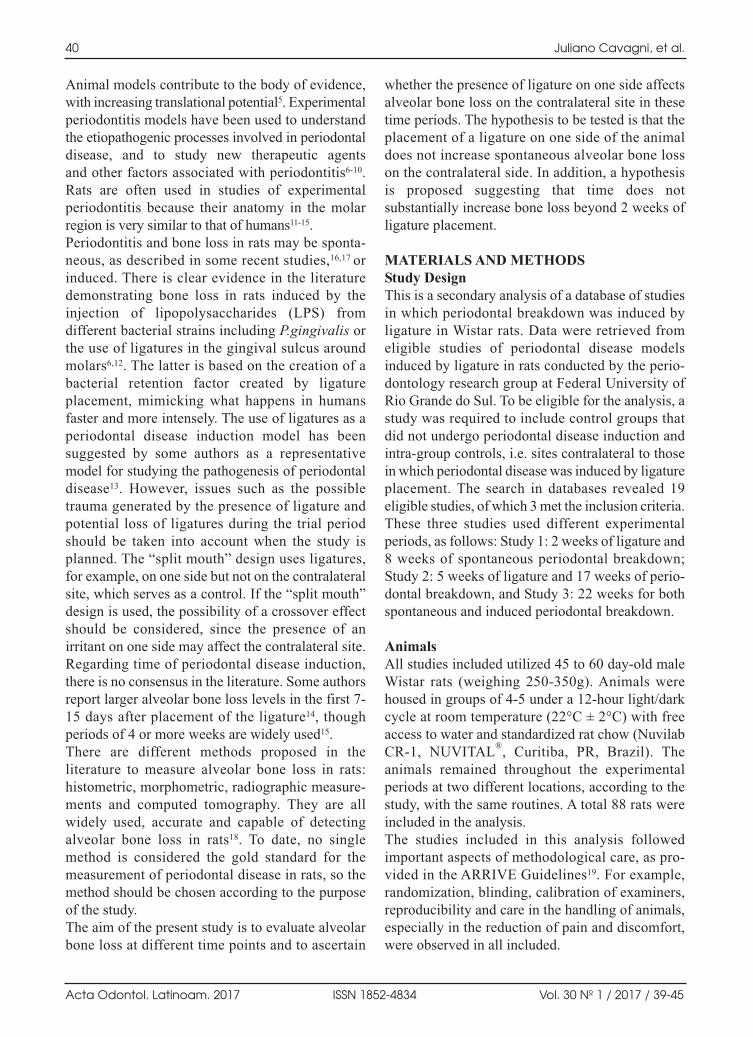
Animal models contribute to the body of evidence,
with increasing translational potential5. Experimental
periodontitis models have been used to understand
the etiopathogenic processes involved in periodontal
disease, and to study new therapeutic agents
and other factors associated with periodontitis610.
Rats are often used in studies of experimental
periodontitis because their anatomy in the molar
region is very similar to that of humans1115.
Periodontitis and bone loss in rats may be sponta
neous, as described in some recent studies,16,17 or
induced. There is clear evidence in the literature
demonstrating bone loss in rats induced by the
injection of lipopolysaccharides (LPS) from
different bacterial strains including P.gingivalis or
the use of ligatures in the gingival sulcus around
molars6,12. The latter is based on the creation of a
bacterial retention factor created by ligature
placement, mimicking what happens in humans
faster and more intensely. The use of ligatures as a
periodontal disease induction model has been
suggested by some authors as a representative
model for studying the pathogenesis of periodontal
disease13. However, issues such as the possible
trauma generated by the presence of ligature and
potential loss of ligatures during the trial period
should be taken into account when the study is
planned. The “split mouth” design uses ligatures,
for example, on one side but not on the contralateral
site, which serves as a control. If the “split mouth”
design is used, the possibility of a crossover effect
should be considered, since the presence of an
irritant on one side may affect the contralateral site.
Regarding time of periodontal disease induction,
there is no consensus in the literature. Some authors
report larger alveolar bone loss levels in the first 7
15 days after placement of the ligature14, though
periods of 4 or more weeks are widely used15.
There are different methods proposed in the
literature to measure alveolar bone loss in rats:
histometric, morphometric, radiographic measure
ments and computed tomography. They are all
widely used, accurate and capable of detecting
alveolar bone loss in rats18. To date, no single
method is considered the gold standard for the
measurement of periodontal disease in rats, so the
method should be chosen according to the purpose
of the study.
The aim of the present study is to evaluate alveolar
bone loss at different time points and to ascertain
whether the presence of ligature on one side affects
alveolar bone loss on the contralateral site in these
time periods. The hypothesis to be tested is that the
placement of a ligature on one side of the animal
does not increase spontaneous alveolar bone loss
on the contralateral side. In addition, a hypothesis
is proposed suggesting that time does not
substantially increase bone loss beyond 2 weeks of
ligature placement.
MATERIALS AND METHODSStudy DesignThis is a secondary analysis of a database of studies
in which periodontal breakdown was induced by
ligature in Wistar rats. Data were retrieved from
eligible studies of periodontal disease models
induced by ligature in rats conducted by the perio
dontology research group at Federal University of
Rio Grande do Sul. To be eligible for the analysis, a
study was required to include control groups that
did not undergo periodontal disease induction and
intragroup controls, i.e. sites contralateral to those
in which periodontal disease was induced by ligature
placement. The search in databases revealed 19
eligible studies, of which 3 met the inclusion criteria.
These three studies used different experimental
periods, as follows: Study 1: 2 weeks of ligature and
8 weeks of spontaneous periodontal breakdown;
Study 2: 5 weeks of ligature and 17 weeks of perio
dontal breakdown, and Study 3: 22 weeks for both
spontaneous and induced periodontal breakdown.
AnimalsAll studies included utilized 45 to 60 dayold male
Wistar rats (weighing 250350g). Animals were
housed in groups of 45 under a 12hour light/dark
cycle at room temperature (22°C ± 2°C) with free
access to water and standardized rat chow (Nuvilab
CR1, NUVITAL®, Curitiba, PR, Brazil). The
animals remained throughout the experimental
periods at two different locations, according to the
study, with the same routines. A total 88 rats were
included in the analysis.
The studies included in this analysis followed
important aspects of methodological care, as pro
vided in the ARRIVE Guidelines19. For example,
randomization, blinding, calibration of examiners,
reproducibility and care in the handling of animals,
especially in the reduction of pain and discomfort,
were observed in all included.
40 Juliano Cavagni, et al.
Acta Odontol. Latinoam. 2017 ISSN 1852-4834 Vol. 30 Nº 1 / 2017 / 39-45
AOL12017:32011 03/07/2017 11:25 Página 40
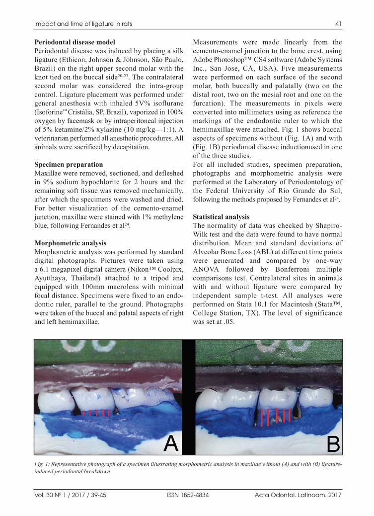
Periodontal disease modelPeriodontal disease was induced by placing a silk
ligature (Ethicon, Johnson & Johnson, São Paulo,
Brazil) on the right upper second molar with the
knot tied on the buccal side2023. The contralateral
second molar was considered the intragroup
control. Ligature placement was performed under
general anesthesia with inhaled 5V% isoflurane
(Isoforine™ Cristália, SP, Brazil), vaporized in 100%
oxygen by facemask or by intraperitoneal injection
of 5% ketamine/2% xylazine (10 mg/kg—1:1). A
veterinarian performed all anesthetic procedures. All
animals were sacrificed by decapitation.
Specimen preparationMaxillae were removed, sectioned, and defleshed
in 9% sodium hypochlorite for 2 hours and the
remaining soft tissue was removed mechanically,
after which the specimens were washed and dried.
For better visualization of the cementoenamel
junction, maxillae were stained with 1% methylene
blue, following Fernandes et al24.
Morphometric analysisMorphometric analysis was performed by standard
digital photographs. Pictures were taken using
a 6.1 megapixel digital camera (Nikon™ Coolpix,
Ayutthaya, Thailand) attached to a tripod and
equipped with 100mm macrolens with minimal
focal distance. Specimens were fixed to an endo
dontic ruler, parallel to the ground. Photographs
were taken of the buccal and palatal aspects of right
and left hemimaxillae.
Measurements were made linearly from the
cementoenamel junction to the bone crest, using
Adobe Photoshop™ CS4 software (Adobe Systems
Inc., San Jose, CA, USA). Five measurements
were performed on each surface of the second
molar, both buccally and palatally (two on the
distal root, two on the mesial root and one on the
furcation). The measurements in pixels were
converted into millimeters using as reference the
markings of the endodontic ruler to which the
hemimaxillae were attached. Fig. 1 shows buccal
aspects of specimens without (Fig. 1A) and with
(Fig. 1B) periodontal disease inductionused in one
of the three studies.
For all included studies, specimen preparation,
photo graphs and morphometric analysis were
performed at the Laboratory of Periodontology of
the Federal University of Rio Grande do Sul,
following the methods proposed by Fernandes et al24.
Statistical analysisThe normality of data was checked by Shapiro
Wilk test and the data were found to have normal
distribution. Mean and standard deviations of
Alveolar Bone Loss (ABL) at different time points
were generated and compared by oneway
ANOVA followed by Bonferroni multiple
comparisons test. Contralateral sites in animals
with and without ligature were compared by
independent sample ttest. All analyses were
performed on Stata 10.1 for Macintosh (Stata™,
College Station, TX). The level of significance
was set at .05.
Impact and time of ligature in rats 41
Vol. 30 Nº 1 / 2017 / 39-45 ISSN 1852-4834 Acta Odontol. Latinoam. 2017
Fig. 1: Representative photograph of a specimen illustrating morphometric analysis in maxillae without (A) and with (B) ligatureinduced periodontal breakdown.
AOL12017:32011 03/07/2017 11:25 Página 41
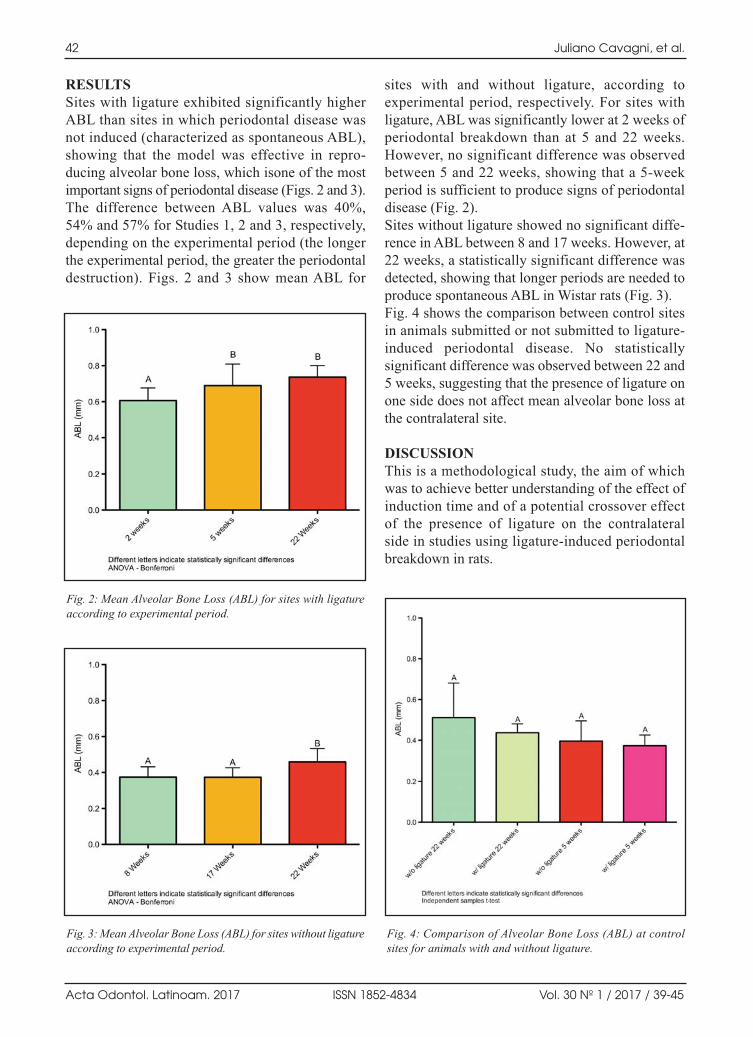
RESULTSSites with ligature exhibited significantly higher
ABL than sites in which periodontal disease was
not induced (characterized as spontaneous ABL),
showing that the model was effective in repro
ducing alveolar bone loss, which isone of the most
important signs of periodontal disease (Figs. 2 and 3).
The difference between ABL values was 40%,
54% and 57% for Studies 1, 2 and 3, respectively,
depending on the experimental period (the longer
the experimental period, the greater the periodontal
destruction). Figs. 2 and 3 show mean ABL for
sites with and without ligature, according to
experimental period, respectively. For sites with
ligature, ABL was significantly lower at 2 weeks of
periodontal breakdown than at 5 and 22 weeks.
However, no significant difference was observed
between 5 and 22 weeks, showing that a 5week
period is sufficient to produce signs of periodontal
disease (Fig. 2).
Sites without ligature showed no significant diffe
rence in ABL between 8 and 17 weeks. However, at
22 weeks, a statistically significant difference was
detected, showing that longer periods are needed to
produce spontaneous ABL in Wistar rats (Fig. 3).
Fig. 4 shows the comparison between control sites
in animals submitted or not submitted to ligature
induced periodontal disease. No statistically
signi ficant difference was observed between 22 and
5 weeks, suggesting that the presence of ligature on
one side does not affect mean alveolar bone loss at
the contralateral site.
DISCUSSIONThis is a methodological study, the aim of which
was to achieve better understanding of the effect of
induction time and of a potential crossover effect
of the presence of ligature on the contralateral
side in studies using ligatureinduced periodontal
breakdown in rats.
42 Juliano Cavagni, et al.
Acta Odontol. Latinoam. 2017 ISSN 1852-4834 Vol. 30 Nº 1 / 2017 / 39-45
Fig. 4: Comparison of Alveolar Bone Loss (ABL) at controlsites for animals with and without ligature.
Fig. 2: Mean Alveolar Bone Loss (ABL) for sites with ligatureaccording to experimental period.
Fig. 3: Mean Alveolar Bone Loss (ABL) for sites without ligatureaccording to experimental period.
AOL12017:32011 03/07/2017 11:25 Página 42
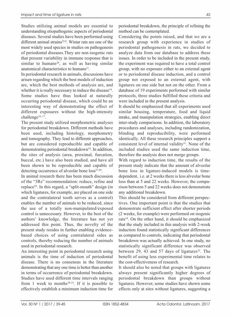
Studies utilizing animal models are essential to
understanding etiopathogenic aspects of periodontal
diseases. Several studies have been performed using
different animal strains25,8. Wistar rats are one of the
most widely used species in studies on pathogenesis
of periodontal diseases.They are nonisogenic rats
that present variability in immune response that is
similar to humans26, as well as having similar
anatomical characteristics to humans11.
In periodontal research in animals, discussions have
arisen regarding which the best models of induction
are, which the best methods of analysis are, and
whether it is really necessary to induce the disease14.
Some studies have thus looked at naturally
occurring periodontal disease, which could be an
interesting way of demonstrating the effect of
different exposures without the highintensity
challenge16, 17.
The present study utilized morphometric analyses
for periodontal breakdown. Different methods have
been used, including histology, morphometry
and tomography. They lead to different approaches,
but are considered reproducible and capable of
demonstrating periodontal breakdown18. In addition,
the sites of analysis (area, proximal, furcation,
buccal, etc.) have also been studied, and have all
been shown to be reproducible and capable of
detecting occurrence of alveolar bone loss27,28.
In animal research there has been much discussion
of the “3Rs” recommendation: reduce, refine and
replace29. In this regard, a “splitmouth” design (in
which ligatures, for example, are placed on one side
and the contralateral tooth serves as a control)
enables the number of animals to be reduced, since
the use of a totally nonmanipulated/exposed
control is unnecessary. However, to the best of the
authors’ knowledge, the literature has not yet
addressed this point. Thus, the novelty of the
present study resides in further enabling evidence
based choices of using contralateral sides as
controls, thereby reducing the number of animals
used in periodontal research.
An interesting point in periodontal research using
animals is the time of induction of periodontal
disease. There is no consensus in the literature
demonstrating that any one time is better than another
in terms of occurrence of periodontal breakdown.
Studies have used different time intervals ranging
from 1 week to months30,14. If it is possible to
effectively establish a minimum induction time for
periodontal breakdown, the principle of refining the
method can be contemplated.
Considering the points raised, and that we are a
research group with experience in studies of
periodontal pathogenesis in rats, we decided to
analyze data from our database to address these
issues. In order to be included in the present study,
the experiment was required to have a total control
group, with no exposure either to an external agent
or to periodontal disease induction, and a control
group not exposed to an external agent, with
ligatures on one side but not on the other. From a
database of 19 experiments performed with similar
protocols, three studies fulfilled these criteria and
were included in the present analysis.
It should be emphasized that all experiments used
similar housing, temperature, food and liquid
intake, and manipulation strategies, enabling direct
interstudy comparisons. In addition, the laboratory
procedures and analyses, including randomization,
blinding and reproducibility, were performed
identically. All these research principles support a
consistent level of internal validity31. None of the
included studies used the same induction time,
therefore the analysis does not merge groups.
With regard to induction time, the results of the
present study indicate that the amount of alveolar
bone loss in ligatureinduced models is time
dependent, i.e. at 2 weeks there is less alveolar bone
loss than at 5 and 22 weeks. However, the compa
rison between 5 and 22 weeks does not demonstrate
any additional breakdown.
This should be considered from different perspec
tives. One important point is that the studies that
demonstrate sufficient effect after shorter periods
(2 weeks, for example) were performed on isogenic
rats14. On the other hand, it should be emphasized
that the study included in this analysis with 2week
induction found statistically significant differences
as compared to controls, indicating that periodontal
breakdown was actually achieved. In one study, no
statistically significant difference was observed
between 29, 43 and 57 days of ligatures32. The
benefit of using less experimental time relates to
the costeffectiveness of research.
It should also be noted that groups with ligatures
always present significantly higher degrees of
periodontal breakdown than groups without
ligatures. However, some studies have shown some
effects only at sites without ligatures, suggesting a
Impact and time of ligature in rats 43
Vol. 30 Nº 1 / 2017 / 39-45 ISSN 1852-4834 Acta Odontol. Latinoam. 2017
AOL12017:32011 03/07/2017 11:25 Página 43
potential increased challenge that may mask the
effect of the presence of naturally occurring
biofilm33. This is not supported by the mechanical
effect of the presence of the ligature, since germfree
animals exposed to ligatureinduced periodontal
disease did not present significant periodontal
breakdown34. Thus, a 2week period seems to be
sufficient for ligatureinduced periodontal break
down; however, depending on the exposure variable
to be tested, additional time may be necessary, and
after 5 weeks the breakdown seems to leveloff.
In animals with spontaneous alveolar bone loss,
periodontal breakdown takes longer. Moreover,
the studies use different times, since the whole
experimental time frame is considered, not only the
induction time.
The present analysis also studied the crossover
effect with the aim of better understanding one of
the supposed biases of splitmouth designs. This
bias is considered “supposed” because no published
paper has provided support to this hypothesis. The
present study endeavors to shed some light on this
discussion, restricted to animal studies, which could
nevertheless be further investigated in human
clinical studies. The main basis for the hypothesis
is that no drug or event with a known systemic
effect should be part of the experiment.
The analysis in the present study demonstrates that
the presence of a silk ligature on one molar has no
statistically significant effect on the contralateral side.
This is supported by the fact that the values of
periodontal breakdown encountered at sites without
ligature from animals exposed to ligature on the
contralateral side does not differ from the mean values
of spontaneous periodontal bone loss in animals not
exposed to any additional manipulation. The present
results thus suggest that total control groups are
unnecessary in periodontal disease studies in rats.
To conclude, two weeks of ligatureinduced perio
dontal disease seem to be sufficient to demonstrate
significant bone loss, and teeth without ligature
contralateral to teeth with ligature may be
considered sound controls, thereby reducing the
amount of animals needed in periodontal research.
44 Juliano Cavagni, et al.
Acta Odontol. Latinoam. 2017 ISSN 1852-4834 Vol. 30 Nº 1 / 2017 / 39-45
CORRESPONDENCEDr. Juliano Cavagni
Av. Neusa Goulart Brizola, 555/ap. 403
Porto Alegre/RS, 90460230, Brazil
Email: [email protected]
REFERENCES 1. Albandar JM, Rams TE. Global epidemiology of periodontal
diseases: an overview. Periodontol 2000 2002; 29:710.
2. Oppermann RV, Haas AN, Rosing CK, Susin C. Epidemiology
of periodontal diseases in adults from Latin America.
Periodontol 2000 2015; 67:1333.
3. Page RC, Kornman KS. The pathogenesis of human perio
dontitis: an introduction. Periodontol 2000 1997; 14:911.
4. Pihlstrom BL, Michalowicz BS, Johnson NW. Periodontal
diseases. Lancet 2005; 366:18091820.
5. Hackam DG, Redelmeier DA. Translation of research evidence
from animals to humans. JAMA 2006; 296:17311372.
6. de Molon RS, de Avila ED, Boas Nogueira AV, Chaves de
Souza JA, et al. Evaluation of the host response in various
models of induced periodontal disease in mice. J Period
2014; 85:46577.
7. Fine DH. Of mice and men: animal models of human
periodontal disease. J Clin Period 2009; 36:913934.
8. Oz HS, Puleo DA. Animal models for periodontal disease. J
biomed biotechnol 2011; 2011:754857.
9. Struillou X, Boutigny H, Soueidan A, Layrolle P. Experi
mental animal models in periodontology: a review. Open
Dent J 2010; 4:3747.
10. Weinberg MA, Bral M. Laboratory animal models in
periodontology. J Clin Period 1999; 26:335340.
11. Listgarten MA. Similarity of epithelial relationships in the
gingiva of rat and man. J Period 1975; 46:677680.
12. Genco CA, Van Dyke T, Amar S. Animal models for
Porphyromonas gingivalismediated periodontal disease.
Trends Microbiol 1998; 6:444449.
13. de Souza JA, Nogueira AV, de Souza PP, Cirelli JA, et al.
Expression of suppressor of cytokine signaling 1 and 3 in
ligatureinduced periodontitis in rats. Arch Oral Biol 2011;
56:11201128.
14. Bjornsson MJ, Velschow S, Stoltze K, HavemosePoulsen
A, et al. The influence of diet consistence, drinking water
and bedding on periodontal disease in SpragueDawley
rats. JPeriodRes 2003; 38:543550.
15. Cavagni J, Soletti AC, Gaio EJ, Rosing CK. The effect of
dexamethasone in the pathogenesis of ligatureinduced
periodontal disease in Wistar rats. BrazOral Res 2005;
19:290294.
16. Cavagni J, Wagner TP, Gaio EJ, Rego RO, et al. Obesity
may increase the occurrence of spontaneous periodontal
disease in Wistar rats. Arch Oral Biol 2013; 58:1034
1039.
AOL12017:32011 03/07/2017 11:25 Página 44
17. Oballe HJ, Gaio EJ, Spuldaro T, Cavagni J, et al. Effects of
alcohol and/or tobacco exposure on spontaneous alveolar
bone loss in rat. Braz DentJ 2014; 25:197202.
18. Li CH, Amar S. Morphometric, histomorphometric, and
microcomputed tomographic analysis of periodontal
inflammatory lesions in a murine model. J Period 2007;
78:11201128.
19. Kilkenny C, Browne WJ, Cuthi I, Emerson M, et al.
Improving bioscience research reporting: the ARRIVE
guidelines for reporting animal research. Vet ClinPathol
2012; 41:2731.
20. Daudt LD, Cavagni J, Gaio EJ, Souza A, et al. Effect of
inhaled corticosteroid on TNFalpha production and
alveolar bone loss in Wistar rats. Arch Oral Biol 2011;
56:13981403.
21. Fernandes MI, Gaio EJ, Susin C, Rosing CK, et al. Effect
of nifedipine on gingival enlargement and periodontal
breakdown in ligatureinduced periodontitis in rats.
ArchOral Biol 2010; 55:523529.
22. Galvao MP, Chapper A, Rosing CK, Ferreira MB, et al.
Methodological considerations on descriptive studies of
induced periodontal diseases in rats. Pesqui Odontol Bras
2003; 17:5662.
23. Sallay K, Sanavi F, Ring I, Pham P, et al. Alveolar bone
destruction in the immunosuppressed rat. J PeriodRes 1982;
17:263274.
24. Fernandes MI, Gaio EJ, Oppermann RV, Rados PV, et al.
Comparison of histometric and morphometric analyses of
bone height in ligatureinduced periodontitis in rats. Braz
Oral Res 2007; 21:216221.
25. Madden TE, Caton JG. Animal models for periodontal
disease. Methods Enzymol 1994; 235:106119.
26. Shapira L, Wilensky A, Kinane DF. Effect of genetic
variability on the inflammatory response to periodontal
infection. J Clin Period 2005; 32 Suppl 6:7286.
27. Azambuja CB, Cavagni J, Wagner MC, Gaio EJ, et al.
Correlation analysis of alveolar bone loss in buccal/palatal
and proximal surfaces in rats. Braz Oral Res 2012; 26:
571577.
28. Liberman DN, Pilau RM, Orlandini LF, Gaio EJ, et al.
Comparison of two methods for alveolar bone loss
measure ment in an experimental periodontal disease model
in rats. Braz Oral Res 2011; 25:8084.
29. Osborne NJ, Payne D, Newman ML. Journal editorial
policies, animal welfare, and the 3Rs. Am J Bioeth 2009;
9:5559.
30. Benzen BH, Grauballe MCB, Bjornsson M, Stoltze K, et al.
A comparison of two models of experimental periodontitis
in rats. Scand J Lab Anim Sci 2005; 2:7380.
31. Festing MF, Altman DG. Guidelines for the design and
statistical analysis of experiments using laboratory animals.
ILAR journal / National Research Council, Institute of
Laboratory Animal Resources 2002; 43:244258.
32. Susin C, Rosing CK. Effect of variable moderate chronic
stress on ligatureinduced periodontal disease in Wistar
rats. Acta Odont Scand 2003; 61:273277.
33. Liberman DN, Pilau RM, Gaio EJ, Orlandini LF, et al. Low
concentration alcohol intake may inhibit spontaneous
alveolar bone loss in Wistar rats. Arch Oral Biol 2011;
56:109113.
34. Crawford JM, Taubman MA, Smith DJ. The natural history
of periodontal bone loss in germfree and gnotobiotic rats
infected with periodontopathic microorganisms. J Period
Res 1978; 13:1625.
Impact and time of ligature in rats 45
Vol. 30 Nº 1 / 2017 / 39-45 ISSN 1852-4834 Acta Odontol. Latinoam. 2017
AOL12017:32011 03/07/2017 11:25 Página 45
AOL12017:32011 03/07/2017 11:25 Página 46
AOL12017:32011 03/07/2017 11:25 Página 47
AOL12017:32011 03/07/2017 11:25 Página 48
AOL12017:32011 03/07/2017 11:25 Página 49
AOL12017:32011 03/07/2017 11:25 Página 50
Top Related